microvascular substance p binding to normal and inflamed
TRANSCRIPT

Vol. 267, No. 2Printed in U.S.A.
ABSTRACT
951
0022.3565/93/2672-0951$03.OO/OTHE JOURNAL OF PHARMACOLOGY AND EXPERIMENTAL THERAPEUTICS
Copyright © 1993 by The American Society forPharmacology and Experimental Therapeutics
Microvascular Substance P Binding to Normal and Inflamed Ratand Human Synovium1
DAVID A. WALSH,2 MICHAEL SALMON, PAUL I. MAPP,3 JOHN WHARTON, NEIL GARRETT, DAVID R. BLAKE4 andJULIA M. POLAK
Department of Histochemistry, Royal Postgraduate Medical School (D.A. W., MS., J.W., J.M.P.) and the Inflammation Group, Arthritis andRheumatism Council Building, The London Hospital, Whitechapel, London, United Kingdom (D.A.W., P.I.M., N.G., ORB.)
Accepted for publication June 28, 1993
The regulatory peptide substance P has been implicated in the
development and persistence of inflammatory synovitis. Theauthors used quantitative in vitro receptor autoradiography tocompare synovial binding of 125lodine-Bolton Hunter-labeled sub-stance P ([125l]BH-SP) in rats and humans and between unin-flamed and persistently inflamed synovium. [125l]BH-SP bindingto microvascular endothelium paralleled the distribution of sub-stance P-immunoreactive nerves and had characteristics of theneurokinin (NK) 1 class of tachykinin receptor. Specific bindingwas inhibited by the selective NK1 receptor antagonist, FK888,and the dual NK1/NK2 receptor antagonist FK224, with Hillcoefficients near unity. FK888 was > 1 000 times and FK224>1 0 times more potent at inhibiting binding in human comparedwith rat synovium. Synovium from patients and rats with chronicarthritis contained heterogeneously distributed inflammatory cell
infiltrates. For the 1 0 microvessels with the densest [125I]BH-SPbinding in each section, no significant differences in bindingdensity, affinity, or K1 values for substance P, FK888 or FK224were found between synovium from naive and monoarthnticrats, nor between that from patients with rheumatoid arthritis orosteoarthritis. However, in both rat and human specimens, mi-croscopic examination suggested that microvascular [1251]BH-SPbinding in intensely infiltrated regions of synovium was less densethan in adjacent, less infiltrated areas. It was concluded that NK1receptors are similarly distributed in rat and human synovium butshow major differences in selectivity for antagonists such asFK888. NK1 receptors in synovium may mediate proinflamma-tory actions of locally released substance P; defective neurovas-cular regulation may contribute to the persistence of chronicarthritis.
Substance P is a member of the tachykinin group of regula-
tory peptides and it is localized to perivascular and free nervefibers in both human and rat synovium (Mapp et al., 1990;
Hukkanen et al., 1992). It is released from the peripheral
terminals of stimulated sensory nerves and acts locally onspecific cell surface receptors. The acute effects of substance Pon the joint are largely proinflammatory and increase plasmaextravasation and vasodilation (Lam and Ferrell, 1991). Sub-stance P is an important component of acute neurogenic in-flammation (Ferrell and Russell, 1986) and may be involved inother acute models of articular inflammation (Lam and Ferrell,1989). Substance P may also chronically modify tissue perfu-
Received for publication March 19, 1993.1 This work was funded by the Arthritis & Rheumatism Council and the
Nuffield Foundation.2 Arthritis & Rheumatism Council Clinical Research Fellow.
3 Arthritis & Rheumatism Council Research Fellow.
4 Arthritis & Rheumatism Council Professor of Rheumatology.
sion by inducing vascular proliferation (Ziche et al., 1990) and
it has been shown in vitro to modulate the activities of other
cell types, which include synoviocytes and lymphocytes (Lotz
et al., 1987; Payan, 1989). Intact sensory nerves are importantfor normal tissue repair (Kjartansson et al., 1987) and wepropose that defective neurovascular regulation may contribute
to the persistence of tissue damage that occurs in chronic
arthritis.
Immunohistochemical studies have indicated a reduction of
substance P-containing nerves in the superficial layers of
chronically inflamed synovium, both in animal models (Huk-
kanen et al., 1991), and in human rheumatoid arthritis (Mapp
et al., 1990). Chronically inflamed synovium is, however, het-
erogeneous, with regions of intense cellular infiltration adjacent
to less inflamed areas where nerves persist. There is an abun-
dance of membrane peptidases capable of degrading substance
P within inflamed synovium, particularly localized aroundblood vessels (Walsh et al., 1993; Mapp et al., 1992). The activity
ABBREVIATIONS: DMSO, dimethyl sulfoxide; GTP-YS, guanosine 5’-0-(3-thiotnphosphate); CP-96,345, [cis-2-(diphenylmethyl)-N-[(2-methoxy-phenyl)-methylj-1-azabicyclo[2.2.2)octan-3-amine]; [1251JBH-SP, 125lodine-Bolton Hunter-labeled substance P; mBSA, methylated bovine serumalbumin; NKA, neurokinin A; NKB, neurokinin B; NK1 , neurokinin 1 receptor; NK2, neurokinin 2 receptor; vWF, von Willebrand factor; Bc,, equilibriumbinding densities; n�; Hill coefficient; FK888, N2-[(4R)-4-hydroxy-1 -(1 -methyl-i H-indol-3-yI)carbonyl-L-prolyl]-N-methyl-N-phenylmethyl-3-(2-naph-thyl) L-alaninamide. FK224,L-allo-threonylj-L-asparaginyl]-L-sehne-v-lactone. _______________________
at ASPE
T Journals on M
ay 8, 2016jpet.aspetjournals.org
Dow
nloaded from

952 Walsh et al. Vol. 267
of substance P is therefore likely to be restricted to the imme-
diate vicinity of its release and chronically inflamed synoviummay contain regions with defective neurovascular regulation
alongside others exposed to the proinflammatory effects ofsubstance P.
Many of the vascular actions of substance P are mediated by
the NK1 class of tachykinin receptor, which displays a higheraffinity for substance P than the related tachykinins, NKA andNKB (Regoli and Nantel, 1991). NK1 receptor antagonists maybe helpful in elucidating the roles of substance P in chronicarthritis and may be of therapeutic benefit in limiting neuro-genic inflammation. However, interspecies differences in tachy-kinin receptors have been noted in extra-articular systems,
both in the distribution of receptors on different tissue struc-
tures (Frossard and Advenier, 1991) and in their selectivity forspecific tachykinin antagonists (Beresford, et al., 1991; Fujii et
aL, 1992). Furthermore, selective tachykinin analogs haveraised the possibility of tissue-specific receptor subtypes (Pe-
titet et at., 1992) and in vivo evidence suggests that receptor
expression may be modified during articular inflammation(Scott et aL, 1992). The choice of appropriate animal modelsand pharmacological agents is therefore essential in the inves-tigation of the role of substance P in chronic synovial inflam-mation.
We previously described the localization and characterization
of binding sites for [‘�I]BH-SP to microvascular endotheliumin synovium from patients with rheumatoid arthritis (Walsh et
at., 1992a). In the present article, we investigated the distribu-tion of [1251]BH-SP binding sites with characteristics of NK1
receptors in the rat knee and compared this with humansynovium to assess whether the rat may be an appropriatemodel for investigating the effects of substance P on synovium.
Furthermore, we investigated interspecies differences in NK1
receptors by using inhibition by selective antagonists of [12511
BH-SP binding to microvessels in both rat and human syno-vium and examined the effects of chronic inflammation on ratand human NK1 receptor distribution and characteristics.
Methods
Animals. Experimental monoarthritis was induced by challengingsensitized rats with intra-articular mBSA (Van Noorden et al., 1988).
Adult male Wistar rats (150 g) were sensitized to mBSA by multiplesubcutaneous injection of the antigen (0.5 ml of 5 mg/ml in Freund’s
complete adjuvant) on days 0 and 7. Arthritis was then induced on day
21 by a single injection of 100 zl of 5 mg/mI mBSA in sterile saline
into the suprapatellar pouch of the right knee joint. Three control
groups of littermates consisted of naive animals, sensitized saline-challenged animals and unsensitized mBSA-challenged animals.
All animals were sacrificed 21 days after challenge (42 days after
sensitization) by asphyxiation in carbon dioxide and cervical disloca-
tion. Their knees were dissected free of overlying skin and removedtogether with distal femur and proximal tibia. Both knees were collectedfrom naive and mBSA-sensitized/challenged animals and the right
knee only from other control groups.Human tissues. Human knee synovium was collected at surgery
from patients undergoing total knee replacement for established rheu-matoid arthritis (n = 13; five men and eight women; mean age, 60
years; range, 22-77 years) or osteoarthritis (n = 10; three men and
seven women; mean age, 72 years; range, 59-79 years) or carbon fiber
resurfacing of articular cartilage for early osteoarthritis (n = 4; one
man and three women; mean age, 36 years; range, 19-44 years). Com-
parisons of Kd, K1 and Bm,.� values were made on data from six patients
with rheumatoid arthritis (two men and four women; mean age, 58
years) and six with osteoarthritis (two men and four women; mean age,
65 years).
Tissue processing. For receptor autoradiography and immuno-
staining for vascular markers, tissue samples were immediately em-
bedded in Tissue-Tek (Miles Inc., Elkhart, IN) and frozen to corkmounts in melting isopentane without prior fixation. For determining
substance P-like immunoreactivity, normal male Wistar rats were
sacrificed by cervical dislocation. Their knees were injected with Zam-
boni’s fixative (Stefani et al., 1967), removed and immersed overnight
in Zamboni’s fixative and then transferred to phosphate-buffered saline
containing 15% sucrose at 4”C until frozen to cork mounts similar tothe fresh tissues.
Specimens were stored at -40”C until use. Sagittal sections (10-sm
thick for receptor autoradiography and vWF staining, 20-sm thick for
substance P immunostaining) were cut on a cryostat using a tungsten
carbide knife, thaw mounted onto Vectabond (Vector Laboratories,
Peterborough, UK)-treated slides, air dried and either used immediatelyor stored at -20”C with silica gel for up to 5 days until use.
Ligand binding. Sections of rat knees were preincubated for 20
mm in 10 mM HEPES, pH 7.4, containing 130 mM NaC1, 4.7 mM
KC1, 5 mM MgCl2 and 1 mM EGTA (buffer A). Excess buffer was
removed and sections were incubated for 45 mm with ligand in bufferB (buffer A plus 1% BSA, 0.1% bacitracin, 100 �iM chymostatin and
100 �M leupeptin). The ligand was 0.5 nM [1251]BH-SP alone (totalbinding) or together with an excess (1 �M) of unlabeled substance P
(nonspecific binding). The sections were subsequently washed twice
for 5 mm at 4”C in buffer A and rinsed in distilled water before being
rapidly dried under a stream of cold air. Any bone fragments that had
detached from the slides were dissected away from the specimen before
it was apposed to autoradiography film. Except where stated, incuba-
tions were performed at 22’C. Binding of [mnI]BH�SP to human syno-
vium was assessed by a modification of this protocol as previously
described (Walsh et aL, 1992a).
Quantification. Labeled sections were apposed to radiosensitivefilm (Hyperfilm-3H, Amersham, Little Chalfont, UK) and exposed for
5 days at 4”C. The film was developed in Kodak D9 (Eastman Kodak,
Rochester, NY) for 3 mm at 15”C. Autoradiographic images were
analyzed using an IBAS 386 (Kontron, Watford, UK) image-processing
system. In each case, binding to synovial microvessels was identified,
where necessary, by a comparison of the film with tissue sectionscounterstained with hematoxylin and eosin and the 10 microvessels
(diameter < 100 �m) with the densest binding were delineated inter-
actively. Binding density was derived from gray values by comparison
with ‘9 standards (Amersham) coexposed with each batch of film and
corrected for the activity data of the ligand.Microautoradiography. Tissue sections labeled with [‘9}BH-SP
were fixed in Bouin’s solution for 1 hr at room temperature, washed in
phosphate-buffered saline, rinsed in distilled water and dried in a flow
of cold air. Sections were dipped in radiosensitive emulsion (Ilford KS,Ilford Anitec U.K., Londou) at 42”C and dried at room temperature.
After exposure for 3 to 5 weeks at 4”C, the dipped slides were developedsimilar to the film autoradiograms, then counterstained with hematox-
ylin and eosin, dehydrated and mounted in DPX (Raymond Lamb,
London, UK).
Characterization of binding sites. Specific binding of [‘2511BH-SP was defined as the total minus the nonspecific binding. The asso-
ciation rate of 0.5 nM [‘9]BH-SP to specific binding sites was assessed
in normal rat synovium. Dissociation was measured as the decline in
specific binding after equilibration with 0.5 nM [‘251]BH-SP and the
transfer of sections to an excess of buffer A without added ligand.
The saturability ofbinding was measured by “hot” saturation studieswith 0.05 to 1.1 nM ‘�I-BH-SP and by “cold” saturation with total
substance P concentrations of 0.5 to 10.5 nM. For the latter, sections
were incubated with 0.5 nM [‘�I]BH-SP together with increasing
concentrations ofunlabeled substance P and total bound ligand (labeledplus unlabeled) calculated as ([L] + [L]) x B/[�L], where [‘L] is the
concentration of free [‘9]BH-SP, [L] is the concentration of free
unlabeled substance P and ‘B is the measured bound [‘9]BH-SP.
at ASPE
T Journals on M
ay 8, 2016jpet.aspetjournals.org
Dow
nloaded from

Fig. 1. [125l]BH-SP binding to a normal rat knee. A) Total binding; B)nonspecific binding in presence of 1 �M unlabeled substance P. Specificvascular binding is demonstrated in the synovium, s, and penosteum,open arrows; f, femur. Reversal prints of film autoradiograms. Bar = 2mm.
1993 Substance P Binding in Synovium 953
The specificity ofbinding was measured by binding inhibition studies
using unlabeled ligands, including substance P and the related peptides
NKA and NKB and unrelated human calcitonin gene-related peptide
�3 and porcine neuropeptide Y. In addition, binding inhibition studies
were performed using FK224, a dual NK1/NK2 receptor antagonist
(Morimoto et at., 1992) and FK888, a selective NK1 receptor antagonist
(Fujii et a!., 1992). NKB, FK224 and FK888 were each initially dissolved
in DMSO and subsequently diluted in incubation buffer to give a
maximum concentration of DMSO of 0.001 % . In separate experiments,DMSO was not found to affect [‘251]BH-SP binding at concentrations
up to 0.005% (data not shown).The guanine nucleotide sensitivity of [‘251]BH-SP binding was as-
sessed by coincubating sections with 0.5 nM ‘251-BH-SP and 1 �M
GTP�yS and confirmed by measuring the dissociation of [1251]BH-SP
in the presence and absence of the guanine nucleotides, GDP (100 �M)
and GTP (100 SM).Guanine nucleotide-induced desaturation of specific binding sites
was performed by preincubation of tissue sections with GTP (100 �M)
or GDP (100 tiM) for 5 mm at 22”C, followed by three 5-mm washes
in buffer A, before incubation with [1251]BH-SP as described above.
Controls were included to confirm the dissociation of bound ligand and
that desaturated binding sites subsequently bound [‘251JBH-SP. In
consecutive sections, equilibrium binding with 0.5 nM [‘251]BH-SP or
saturation of unoccupied binding sites with an excess (0.1 jIM) of
unlabeled substance P was followed by GTP-induced desaturation.
Statistical analysis. Kinetic and equilibrium constants were de-
rived from specific binding values by iterative nonlinear regression
assuming single-site models using GraphPAD (InPlot4.0, San Diego,
CA). B,�, values were obtained with 0.5 nM [1251]BH-SP incubated for
45 mm at 22”C. Values of association (k+1) and dissociation (k_1) rate
constants were derived from kinetic experiments using 0.5 nM [125!]
BH-SP and equilibrium dissociation constants (Kd) and maximal bind-ing densities (B,,,51) from binding inhibition studies using 0.5 nM [125!]
BH-SP and 0 to 1000 nM unlabeled substance P (DeBlasi et al., 1989).
Values for binding inhibition constants (K) and �Hj1I were derived by
fitting to sigmoid curves. Descriptive data are expressed as the means
(95% confidence interval) and between-group comparisons were made
using analysis of variance and Student’s t test with Bonferroni’s
correction on geometric data.
Immunohistochemical tests. To confirm the binding of [125I]BH-SP to synovial microvessels, consecutive sections to microautoradi-
ograms were immunostained for vWF using previously characterized
rabbit polyclonal antibodies (A082, Dako, High Wycombe, UK) and
the avidin-biotin complex method of Hsu et al., (1981). The antiserumto vWF shows full cross-reactivity with rat vWF. Cryosat sections (10
�zm) were fixed in acetone for 5 mm at 4’C, incubated with primary
antibody overnight at 4”C, with biotinylated goat antirabbit immuno-
globulin G for 2 hr at room temperature and with avidin-biotin complex
for 30 minutes. Finally, they were developed in diaminobenzidine,
dehydrated and mounted in DPX.
To compare the distribution of [1251]BH-SP binding sites with thatof substance P-immunoreactive nerves, normal rat knees were stained
using a rabbit polyclonal antibody to substance P (Merighi et al., 1988)
and the avidin-biotin complex method with glucose oxidase/nickelenhancement (Shu et al., 1988).
Materials. [‘251]BH-SP (specific activity, 2000 Ci/mmol), 125! stand-
ards and Hyperfilm-3H were obtained from Amersham International.
FK224 and FK888 were kindly provided by Fujisawa PharmaceuticalsLtd (Osaka, Japan). Other unlabeled peptides, guanine nucleotides,
enzyme inhibitors and enzyme-free BSA were obtained from Sigma
Chemical Co., Poole, UK. Antisera to vWF was purchased from Dako
Ltd. and biotinylated goat antirabbit affinity-purified immunoglobulinG and avidin-biotin complex were from Vector.
Results
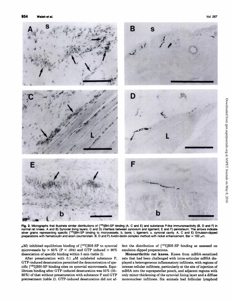
Normal rat knees. [‘251]BH-SP bound specifically to mi-
crovessels in normal rat knees (figs. 1-3). Emulsion-dipped
preparations revealed that microvessels with intense binding
were particularly localized immediately beneath the synoviallining layer, at interfaces between synovium and ligaments, insynovium at entheses where the joint capsule attaches to bone
and in periosteum (fig. 2). The distribution of microvessels with
specific [‘251]BH-SP binding was similar to that of substance
P-immunoreactive nerves (fig. 2). Microvessels within striated
muscle also displayed intense [‘251]BH-SP binding; larger ar-
terioles and venules (diameters, > 100 �tm), both in synoviumand muscle, exhibited less dense [1251]BH-SP binding comparedwith smaller microvessels (fig. 3). Specific binding was not
observed to cells in bone and cartilage, nor to synovial liningcells. Immunostaining of consecutive sections for vWF con-firmed that structures binding [1251]BH-SP were positive for
the endothelial marker (fig. 3).Kinetic studies with 0.5 nM [‘251]BH-SP were consistent
with a diffusion limited association rate (k+1 1.7 X 10⁶ M’
sec’; 95% CI, 0.8-3.7 x 10⁶) and a monophasic dissociation
(k_1 = 10 x iO-5 M’ sec�; 95% CI, 7-16 x 106), with 45%
dissociation within the first 120 mm (fig. 4A). Biphasic modelsof association and dissociation did not fit the data significantly
better than did monoexponential models. “Hot” saturation
studies indicated Kd and Bmax values of 1.6 nM and 61 amol
mm2, respectively (means of three animals), but did not pro-duce full saturation at the highest concentration of [‘2511BH-
SP used (1.1 nM). “Cold” saturation and binding inhibition
studies with unlabeled substance P confirmed the saturability
of high-affinity binding sites with Bmax 94 amol mm2 (70-
127) and Kd 1.6 nM (1.1-2.4, fig. 4B). Binding inhibition
studies with related tachykinins indicated a rank order of
potencies substance P > NKA > NKB, which suggests a NK1
subclass of tachykinin receptor (table 1, fig. 4C). The unrelatedpeptides calcitonin gene-related peptide 13and neuropeptide Y
(each 1 �M) did not significantly inhibit specific [‘251]BH-SPbinding. Binding inhibition studies with the tachykinin antag-onists FK224 and FK888 indicated a rank order of potency
substance P > > FK888 = FK224 (table 1, fig. 5A). GTP”YS (1
at ASPE
T Journals on M
ay 8, 2016jpet.aspetjournals.org
Dow
nloaded from
954 Walsh etal.
B S
Vol. 267
j
I
. ‘ ) .‘ a’ � a � � �
.‘
1’
r
D
� .,� ‘-
I L;2, ,/ -
“�s P � 7
E
. )#{149}_
.�- . �
_#;�:�:... �
.1�,
�
4�’I��5 � � .i�,
..� , . ‘. �%_� ‘4 �, . �,:� - ,. �#{188}�: .
� - . .�. - ;. . . .)
� �. ..�.. �- ::..�Z.-�- � � .
,‘ &� .q� .�. _� - -�
Fig. 2. Micrographs that illustrate similar distributions of [125l]BH-SP binding (A, C and E) and substance P-like immunoreactivity (B, D and F) innormal rat knees. A and B) Synovial lining layers; C and D) interface between synovium and ligament; E and F) periosteum. The arrows indicatesilver grains representing specific [125I]BH-SP binding to microvessels; b, bone; I, ligament; s, synovial cavity. A, C and E) Emulsion-dippedpreparations with hematoxylin and eosin counterstain. B, D and F) Avidin-biotin complex method with nickel enhancement. Bar = 100 �m.
F
� � L� a
b ______
� ,
�M) inhibited equilibrium binding of [‘251]BH-SP to synovial
microvessels by > 85% (P < .004) and GTP induced > 90%
dissociation of specific binding within 5 mm (table 2).After presaturation with 0.1 �iM unlabeled substance P,
GTP-induced desaturation permitted the demonstration of spe-
cific [125I]BH-SP binding sites on synovial microvessels. Equi-
librium binding after GTP-induced desaturation was 55% (35-
85%) of that without presaturation with substance P and GTP
pretreatment (table 2). GTP-induced desaturation did not af-
fect the distribution of [‘251]BH-SP binding as assessed onemulsion-dipped preparations.
Monoarthritic rat knees. Knees from mBSA-sensitized
rats that had been challenged with intra-articular mBSA dis-played a heterogeneous inflammatory infiltrate, with regions of
intense cellular infiltrate, particularly at the site of injection of
mBSA into the suprapatellar pouch, and adjacent regions with
only minor thickening of the synovial lining layer and a diffuse
mononuclear infiltrate. Six animals had follicular lymphoid
at ASPE
T Journals on M
ay 8, 2016jpet.aspetjournals.org
Dow
nloaded from

1993 Substance P Binding in Synovium 955
- ��--- �
A:-’’4�...‘ � \_ � � �
. . “� ..- � . .: #{149},� .
?‘ � � �
‘ � � . v�, 4%�
B,.,
.: � ;4.�. ‘ -. .ied�$ �
� *�
�
0 30 60 90 120 150
Time (minutes)
a)
C
E0)C 0)�.
DC �
.0D
U C- 3
�- 0- .0Ua) U0.
U) �-
a)0.U)
�
U
�- 004)0.
0�LI)
:i:
5)(\J E- 3
E�0C X3 Co0 C
.05-5
U
Ua)0.
LI)
0 5 10
Total free (nM)
-11 -10 -9 -8 -7 -6 -5
Fig. 3. A) [125l]BH-SP binding in normal rat synovium; B) consecutivesection to (A) stained for the endothelial marker, vWF. Arrows indicatespecific [1251]BH-SP binding to vWF-immunoreactive microvessels; s,synovial cavity. A) Emulsion-dipped preparation with hematoxylin andeosin counterstain; B) avidin-biotin complex method. Bar = 100 �zm.
infiltrates in the challenged knee and these cases were used for
subsequent comparative studies. Contralateral knees and kneesof all control animals showed no signs of inflammation.
In emulsion-dipped preparations, specific [‘251]BH-SP bind-ing to microvessels was observed in infiltrated and uninfiltratedregions of inflammatory synovium. Staining of consecutive
sections for the endothelial marker, vWF, confirmed that struc-
tures displaying specific [‘251]BH-SP binding corresponded tomicrovessels. Microvessels in inflamed knees in regions with
intense inflammatory infiltrates exhibited less dense [‘251]BH-SP binding than did those in adjacent, less inflamed areas (fig.
6). This differential distribution of [‘251]BH-SP binding wasalso observed after GTP-induced desaturation.
Film autoradiograms were quantified for the 10 synovial
microvessels with the densest binding in each section. No
significant differences were found in the equilibrium bindingdensity of 0.5 nM [‘25I]BH-SP between inflamed knees frommonoarthritic rats and their contralateral, uninflamed knees
nor between knees from arthritic rats and those from any of
the three control groups (n = 6 each group, data not shown).
Subsequent comparisons were made between inflamed kneesfrom monoarthritic rats and naive rat knees. In separate ex-periments to those described earlier, no significant differences
in equilibrium binding of [‘25I]BH-SP to synovial microvessels
were observed between naive and monoarthritic rat knees ir-
respective of prior GTP-induced desaturation of endogenous
[Competing ligand] (log M)
Fig. 4. Specific synovial microvascular [125l]BH-SP binding in normal ratknees. A) Association of 0.5 nM [1�I]BH-SP (#{149})and dissociation afterthe transfer of sections to an excess of buffer without ligand at 60 mm(arrow) (0). The values are means ± S.E.M. of five cases, expressed asthe percent of specific binding at 60 mm. B) “Cold” saturation with 0.5nM [125l]BH-SP and 0 to 10 nM unlabeled substance P. The values aremeans ± S.E.M. of iO cases. Insert) Scatchard plot of mean data. C)Inhibition of 0.5 nM [125l]BH-SP binding by unlabeled substance P (#{149}),NKA (A) and NKB J). The values are means ± S.E.M. of 6 cases,expressed as the percentages of specific binding in the absence ofunlabeled ligand.
binding (table 2). Both groups demonstrated similar reductions
in equilibrium binding after GTP-induced desaturation. Maxi-
mal binding density, Kd values and inhibitory potencies of
FK224 and FK888 were similar for vessels with the densest
[‘251]BH-SP binding in inflamed and uninflamed knees (table1, fig. 5A, B).
Human Synovium. Synovium from patients with rheuma-
toid arthritis all contained inflammatory cell infiltrates with
samples from four specimens containing follicular lymphoid
aggregates. Osteoarthritic synovium often also contained dif-
fuse mononuclear inflammatory cell infiltrates, although in thesynovium from patients undergoing carbon fiber resurfacing,
at ASPE
T Journals on M
ay 8, 2016jpet.aspetjournals.org
Dow
nloaded from

956 Walsh et al. Vol. 267
TABLE 1Characteristics of 1�l-Bolton Hunter-labeled substance P binding to rat and human synovial microvesselsValues are expressed as geometn c means (95% confidence intervals).
Rat Human
Naive (n = 6) mBSA (n = 6) Osteoarthritis(n = 6) RheumatoidArthritis (n = 6)
B� (amol mm2) 94 (70-i 27) 63 (28-1 46) 172 (1 36-21 8) 100 (41 -244)K, values (nM)
Substance P 1 .i�’ (0.8-1 .8) 1 7b (0.9-3.2) 0.5c (0.4-0.8) 0.3” (0.2-0.7)NKA 340w (1 50-800) NA NA NAFK888 440k (260-750) 430� (200-900) 0.3c (0.1-0.6) 0.1” (0.06-0.3)FK224 660” (280-i 600) 670� (200-2260) 46 (22-82) 21 (7-66)
NKB 3600 (1 700-7500) NA NA NAflHl
Substance P -1 .2 (0.9 to -1 .5) -1 .1 (-0.9 to -1 .4) -1 .1 (-0.9 to -1 .4) -1 .2 (-0.7 to -1.7)NKA -0.9 (0.7 to -1 .3) NA NA NAFK888 -1 .2 (-0.8 to -2.0) -1 .1 (-0.6 to -2.0) -1 .3 (-0.6 to -2.0) -1 .1 (-0.5 to -1.6)FK224 -1 .2 (-0.9 to -1 .6) -1 .1 (-0.8 to -1 .4) -1 .0 (-0.6 to -1 .4) -0.9 (-0.4 to -1.4)NKB -1 .6 (-0.9 to -3.0) NA NA NA
a Substance P was more potent than NKA (P < .001) and NKA was more potent than NKB (P < .001).a FK224 was of similar potency to FK888 (P > .05) and substance P was more potent than either FK224 (P < .001) or FK888 (P < .001) in each of the naive and
arthritic rat groups.C FK888 was more potent than substance P in both osteoarthntis (P < .05) and � rheumatoid arthritis (P < .02).
. Not assessed.
such infiltrates were, at most, sparse. Individual samples ofrheumatoid or osteoarthritic synovium contained regions withintense inflammatory cell infiltration adjacent to less inflamed
areas.In emulsion-dipped preparations of synovia from patients in
each disease group, specific [‘9]BH-SP binding was localizedto endothelia of synovial microvessels and was particularly
dense on microvessels localized immediately beneath the syn-ovial lining layer (fig. 7). Specific binding was not identified tolining cells themselves nor to infiltrating inflammatory cells.Specific binding of [‘25I]BH-SP appeared less dense to micro-vessels in intensely inflamed regions of synovium compared
with an abundance of densely binding microvessels in less
inflamed areas (fig. 6). This differential distribution was notaffected by prior desaturation with GTP.
Specific binding of [1251]BH-SP to human synovial microves-sels was reversible (k_1 2.3 x i0� s’; 95% CI, 1.7-3.1 x i0�,
fig. 8A). It was sensitive to guanine nucleotides as demonstrated
by an almost complete abolition of equilibrium binding in the
presence of GTP�yS (B� = 1.9 amol mm2; 95% CI, 1.7-2.1)compared with that in the absence of guanine nucleotides (B�= 67 amol mm2; 95% CI, 33-137). The addition of GTP orGDP (each 100 jzM) to dissociation buffers markedly enhanced
the dissociation rate of specific [‘251]BH-SP binding, with >
90% dissociation within 5 mm (table 2, fig. 8). As observed inrat knees, equilibrium binding after guanine nucleotide-induceddesaturation was significantly less than that in the absence ofdesaturation (table 2, fig. 8B). Presaturation of binding sites
with substance P or increasing the number or duration of
washes between desaturation and rebinding by more than threewashes for 5 mm each did not significantly affect equilibriumbinding after guanine nucleotide-induced desaturation (fig. 7B).
As previously described (Walsh et at., 1992a), binding sitesfor [125I]BH-SP had characteristics of the NK1 subclass oftachykinin receptor and showed a higher affinity for unlabeledsubstance P than for NKA or NKB. Binding inhibition studieswith the tachykinin antagonists FK224 and FK888 demon-strated complete inhibition of binding with nH,ll values notsignificantly different from unity (table 1, fig. 5C, D). FK888
was significantly more potent than substance P in inhibiting
microvascular [‘�I]BH-SP binding in both osteoarthritic (P <
.05) and rheumatoid arthritic (P < .02) synovium (paired t
tests). FK224 was some two orders of magnitude less potent.
No significant differences were found for microvessels with thedensest [‘251]BH-SP binding between osteoarthritic and rheu-matoid synovium in K1 values for substance P, FK224 or FKSSSor Bmax (table 1, fig. 5C, D), nor in equilibrium binding using0.5 nM [‘25I]BH-SP (table 2).
FK224 was >10 times and FK888 was >1000 times morepotent in inhibiting [‘251]BH-SP binding to synovial microves-sels in human than in rat knees (analysis of variance with
Bonferroni corrected P values < .001, table 1, fig. 5). No
significant differences were found for K, values for substance
P between the two species.
Discussion
We demonstrated binding sites for [‘251]BH-SP to microvas-cular endothelium in both human and rat synovium. The bind-ing was saturable and high affinity. Although association and
dissociation time course, saturation and binding inhibition datadid not fit significantly better to two-site rather than to single-
site models, the limited number of time points and concentra-
tions studied and the inherent variability in the data resulted
in a low power to discriminate between multiple-site models.The enhancement of dissociation rates by the guanine nucleo-tides GTP and GDP indicated two interchangeable affinity
states (high and low) and this finding was consistent with
binding to G protein-coupled receptors (Iyengar et at., 1980).
The dissociation constants derived from kinetic data (approx-
imately 60 pM for rat microvessels) were lower than thosederived from saturation studies (1.6 nM) and this may have
resulted from a preferential characterization of high-affinitysites when using low (0.5 nM) rather than high (up to 10.5 nM)
ligand concentrations in the two types of experiments.Binding of [‘251]BH-SP to both rat and human synovial
microvessels displayed specificity for tachykinins and relatedcompounds. The rank order of potencies of tachykinins sub-
stance P > NKA > NKB was consistent with a NK1 class oftachykinin receptor and was supported by complete inhibition
of binding by the selective NK1 antagonist FK888 (Fujii et at..,
1992). The distribution of ‘25I-BH-SP binding sites paralleled
at ASPE
T Journals on M
ay 8, 2016jpet.aspetjournals.org
Dow
nloaded from

1993
I
-10 -9 -8 -7 -6
(Co� �] (log N)
12!
125
1�
Ir(,�. 25
-5
[Cc�eting ligsnd] (log N)
(C� �] Qog N)
-ii -10 -9 -8 -‘� -6 -5
(Co�eting ligind] (log N)
Fig. 5. Inhibition of microvascular binding of specific [125l]BH-SP insynovium from knees of naive rats (A), from knees of rats with mBSA-induced monoarthntis (B) and from humans with osteoarthntis (C) orrheumatoid arthritis (D). Sections were incubated with 0.5 nM [1251JBH-sP together with increasing concentrations of unlabeled substance P(#{149}),FK888 4 or FK224 (A). FK888 was > 1000 times and FK224,>I 0 times more potent at inhibiting [125l}BH-SP binding to microvessels ininflamed synovium in the human than in the rat (P < .001). The valuesare means ± S.E.M. of six cases, expressed as percentages of specificbinding in the absence of unlabeled ligand.
that demonstrated by ourselves and others for substance P.
immunoreactive nerves (Hukkanen et at., 1992), which sug-gested that they may represent functional receptors for locallyreleased substance P.
In preliminary studies, the equilibrium binding of [‘251]BH-
SP to 60 vessels with the densest binding in human synovium
was positively skewed. The modal binding density to microves-
sels in the sample could not be determined because microvessels
Substance P Binding in Synovium 957
with low-density binding identified on emulsion-dipped prepa-
rations could not be reliably distinguished from nonspecificbinding on film autoradiograms. In this study, quantification
was performed on the 10 vessels with the densest binding of[‘251]BH-SP in each section to obtain accurate determinationsof Kd, K1 and nH,11 values. However, the resulting � and Bmax
values would have been influenced by morphometric factors
known to vary in synovial inflammation, such as the number
and size of vessels within the samples (Stevens et al., 1991).
These factors and the regional heterogeneity of ‘251-BH-SPbinding observed in individual samples may have obscured thepossible differences in density of microvascular [‘25I]BH-SPbinding between disease groups. Nonetheless, the current study
confirmed our previous findings that relatively uninflamed
regions of arthritic synovium possess dense microvascularendothelial [‘251-BH-SP] binding sites (Walsh et al., 1992a) inaddition to perivascular substance P-immunoreactive nerves(Mapp et al., 1990). This suggests that these less inflamed
regions of synovium continue to be subject to neurovascular
regulation and also to the acute, proinflammatory effects ofsubstance P.
Guanine nucleotide-induced desaturation has been describedas a method to investigate the influence of receptor occupancy
on ligand binding (Bertherat et at., 1991). Both GTP and GDPincrease the dissociation rates of [‘25I]BH-SP from microvas-cular binding sites in both human and rat synovium, as has
been described for agonists at other G-protein coupled receptors
(Iyengar et at., 1980). However, agonist binding after guanine
nucleotide-induced desaturation requires the return of the
receptor to a high affinity state, probably involving the reas-
sociation of the low-affinity receptor and G-protein. The con-
sistent reduction in equilibrium binding of agonist observed
after GTP-induced desaturation indicated that this recouplingprocess was less than 100% efficient in tissue slice preparations,
limiting the ability of guanine nucleotide-induced desaturation
experiments to provide quantitative information on receptoroccupancy when receptors are identified by labeled agonists.
Qualitative analysis at the microscopic level indicated thatmicrovessels in intensely inflamed areas of synovium displayed
less intense binding of [‘251]BH-SP than did adjacent, less
inflamed areas. Such heterogeneity of [‘251]BH-SP binding
density persisted after GTP-induced desaturation, which mdi-cated that it was not caused by prior receptor occupancy by
endogenous substance P but might indicate variations in recep-
tor expression or in the efficacy of receptor-G-protein coupling.
Low [‘251]BH-SP binding in intensely inflamed regions of sy-
novium suggested a reduced responsiveness to substance P. Wealso observed a reduction of substance P-immunoreactive
nerves in inflamed compared with less inflamed regions ofsynovium from patients and animals with chronic arthritis
(Mapp et at., 1990; Hukkanen et al., 1991). We propose there-
fore, that microvessels in intensely inflamed regions of syno-
vium are not subject to normal neurovascular regulation.NK1 receptors display heterogeneity in sensitivity to selec-
tive antagonists. The nonpeptide NK1 antagonist CP-96,345has a higher affinity for human and guinea pig NK1 receptors
than for those in the rat (Beresford et at., 1991). FK888 has
similarly been found to have a higher affinity for the guineapig than for the rat NK1 receptor (Fujii et al., 1992). There is
also evidence of differences in NK1 receptor specificities be-
tween different tissues of a single species (Petitet et at., 1992)and suggestions that receptor expression and/or affinity may
at ASPE
T Journals on M
ay 8, 2016jpet.aspetjournals.org
Dow
nloaded from

t,� - ‘#{149}i�’� -� a. � ‘� -
- ,,-. . ��:.-;‘� � ,; L
. �.-�:ts�’- � �
:��:‘:%�‘: a �,�&;:. . ‘. :- ‘ � � � IL�E��% � ��‘. C. � ‘ � � -; �r � �
‘�“
. . � ‘ . I C � ‘#� �
.... �. �.. � � �� C,’.. �: � #{149}:� �
I,). � � :4
�k. . :.%�4S� 4”�’’� q� � -� � �
-- . . � S
S
� . �!‘ �
�
�. � � � ik1i� I�.� �4. . ‘a
� I #{149} � .. ; �-.�a�1‘4_ . :r
, ,. -� _‘.I.i
�r �
‘a�� I�? � ��
� .J’,.1�...1 �
- �
Fig. 6. [125l]BH-SP binding to synovial microvessels (arrow) in arthritic synovium in the rat (A and B) and in the human (C and D). A and C) Regionsof synovium showing dense microvascular [125l]BH-SP binding in areas with sparse inflammatory cell infiltration; B and 0) less dense microvascularbinding in intensely infiltrated areas adjacent to A and C, respectively. A and B) mBSA-induced monoarthritis; C and 0) human seronegativeinflammatory arthritis. 5, synovial cavity. Emulsion-dipped preparations with hematoxylin and eosin counterstain. Bar = 100 �m.
: �
958 Walsh et al. Vol. 267
TABLE 2
Effect of 5-mm incubation with 100 �M GTP on specific binding of [125IJBHS P to rat and human synovial microvesselsSections were exposed to 1 00 MM GTP for 5 mm at room temperature either before (bind -, desaturate + and bind +) or after (bind +, desaturate + and bind -)
incubation with 0.5 nM [‘251]BH-SP. GTP induced dissociation of almost all specific binding but permitted rebinding of [‘25l]BH-SP. Specific binding after GTP-induceddesaturation was less than that without prior exposure to GTP. No significant differences were found between naive and monoarthntic rats, either without desaturation[ratio mBSA/naive = 0.8 (0.5-1 .1), P = .1 3] or with desaturation [ratio mBSA/naive = 0.8 (0.5-1 .2), P = .3] nor between OA and RA synovium either without desaturation[ratio RA/OA = 0.9 (0.6-1 .4), P = .6] or with desaturation [ratio RA/OA = 1 .4 (0.7-2.4), P = .3]. Equilibrium binding values expressed in amol mm2 as geometric means(95% Cl).
Bind Desaturate �ndRat Human
-�- Naive(n=6) mBSA (n = 6) OA (n = 7) RA (n = 6)’
- - + 44 (34-58) 33 (27-42) 43 (32-57) 39 (25-60)- + + 24* (18-33) 20** (15-25) 21*** (12-35) 28*** (19-40)+ + - 1 .6 (1 .5-1 .7) 1 .8 (1 .6-2.0) 2.5 (2.1 -3.0) 2.5 (2.4-2.6)
a Osteoarthntis.b Rheumatoid arthritis.
a p = .008, compared with binding without prior desaturation (for following P values as well).*.P= .004.“P=.003.
‘‘P=.08.
A� � �#{149} � .�. :.%.� . ; ‘ . I. � . . . >..
. *. � � .1 , � - -I’� #{149}�,-....,. . &‘��.‘_,_ �
� . - 1� � � .�;; Tf�� �
� � -. �_�-�Jp � �
‘ � .�:. � .;4�i ...�. � ..: �
..tJ� � �!j� �i ‘�:r&:#{163}
-. � �: � .
*� 4 ,‘ �
. .,. .. :k�.�. , �&,.. . -..
- C j�--: - �
� \ :1#{149}:��, -..
‘44 --,.- � � �
: � � �‘1��/z��. � � � � ..�
� � � �
be influenced by inflammation (Scott et at., 1992). Therefore,
conclusions from studies on different tissues in different species
or in different pathological states should be interpreted with
caution.
The [‘251]BH-SP binding sites investigated in this study were
on microvascular endothelia in both human and rat synovium,
which eliminated the influence from possible NK1 receptor
heterogeneity between tissues. No significant differences were
observed between disease groups within the same species in
affinities of microvascular binding sites for substance P, FK888
or FK224, which suggested that chronic synovial inflammation
does not affect the affinity or selectivity of NK1 receptors.
at ASPE
T Journals on M
ay 8, 2016jpet.aspetjournals.org
Dow
nloaded from

a
C.�
A
Substance P BInding in Synovium 959
S-%, #{149},a�#{149}‘.�
�-
U
-a,.-.�1
. a ‘� . � - �
� �:..:.a,, ,;i. ..�
0 30 60 90 120use (minutes)
rrn� (-a)
- +
+ +
- - + + + +
+
+ + + + + +
- 10 10 25 40 70- + + + + +
Fig. 7. SpecIfic [‘�l]BH-SP binding to subintimal microvessels (arrow) inosteoarthrltic human synovium. S. synovial cavity. Emulsion-dipped prep-aration with hematoxylin and eosin counterstain. Bar = 100 �m.
However, FK888 was > 1000 times and FK224, >10 times morepotent in human than in rat tissue in regard to inhibiting
binding of iThI�BH�SP � synovial microvessels. These dataextend previous observations of differences in potency of
FK888 between rat brain and guinea pig lung and suggest that,as identified using CP 96,345, the human and guinea pig NK1receptors have similar specificities but are substantially differ-
ent from the rat NK1 receptors.Heterogeneity between species is an important factor in
choosing appropriate animal models for the development ofspecific NK1 receptor antagonists. Our findings suggest thatthe rat knee closely parallels the human knee in terms of NK1
receptor distribution and responses to chronic inflammation
and may therefore be an appropriate model for investigatingthe effects of substance P and its antagonists in arthritis.However, pharmacological tools used in such studies should be
chosen with the specificity of the rat NK1 receptor in mindand the rat may not be an optimal model for the assessment ofantagonists such as FKSS8 that are highly selective for thehuman NK1 receptor.
Human rheumatoid arthritis and osteoarthritis are chronicconditions, typically with repeated acute exacerbations. Weconsider that acute inflammation is essentially an appropriate,
protective response to tissue injury but, if inadequately regu-
lated, it may itself be damaging. Substance P is proinflamma-tory in acute models (Lam and Ferrell, 1989; Scott et at., 1992)and pharmacological doses of substance P may exacerbatechronic arthritis (Levine et at., 1984). However, the physiolog-ical release of substance P from sensory nerves may also be a
protective response to tissue injury, increase perfusion of hy-
poxic synovium and contribute to tissue repair (Walsh et at.,
1992b).
We previously demonstrated a perivascular distribution of
membrane peptidases capable of degrading substance P in
normal and, especially, inflamed synovium and proposed thatsubstance P is localized by these enzymes to the immediate
vicinity of its release, which results in a functional compart-mentalization of synovium (Mapp et aL, 1992; Walsh et at.,
1993).Our findings suggest a heterogeneous model for chronic in-
flammation with regions of intense cellular infiltration, loss of
substance P-immunoreactive nerves and reduction in endothe-
hal NK1 receptors separated by membrane peptidases from
Fig. 8. A) Enhancement by guanine nucleotides of the dissociation rateof specific [125l]BH-SP binding from microvessels in osteoarthritic humansynovium. Sections were equilibrated with 0.5 nM [‘�l]BH-SP and trans-ferred at time 0 to an excess of buffer WithOUt [1�l]BH-SP with noguanine nucleotide (#{149}),100 MM GTP (0) or 1 00 �M GDP (s). The valuesare expressed as means ± S.E.M. B) Effect of guanine nucleotide-induced desaturation of substance P binding sites on subsequent bindingof [125l]BH-SP to microvessels in human osteoarthntic synovium. Sec-tions were variously incubated with exogenous unlabeled substance P(1 00 nM) or buffer for 10 mm (± presaturate) then incubated with 0.5 nM[1251]BH-SP for 45 mm (+ bind) or incubated with 100 �M GDP for 5 mm(+ desaturate), rinsed for 1 0 to 70 mm in two changes of buffer and thenincubated with 0.5 nM [1�l]BH-SP for 45 mm, before further rinsing,drying and exposure to film. � with unlabeled substance Pnotfollowed by GDP incubation signfficantly inhibited subsequent bindingof [1�l]BH-SP compared with binding in the absence of presaturationand compared with binding after presaturation and GDP-InCIUCed desat-uration (all corrected P values < .01). b��jf� binding after GDP-induceddesaturation was significantly less than that in the absence of desatu-ration (P = .004). No significant differences in binding were observedafter GDP-IndUCed desaturation between sections presaturated or notpresaturated with unlabeled substance P nor between different durationsof rinses between GDP-induced desaturation and incubation with [‘al]BH-SP. The values are expressed as the percent of specific binding inthe absence of presaturation and desaturation as the means ± S.E.M.of five cases. Statistical analyses were performed on original pairedgeometric data.
adjacent areas with less intense inflammatory infiltrates, per-sistent innervation and abundant NK1 receptors. We suggest
that a failure of neurovaseular regulation in intensely inflamedregions may contribute to the failure of resolution in chronic
arthritis; excessive neurogenic inflammation in less infiltratedregions may contribute to the acute exacerbations of arthritis.Specific antagonists such as FKSS8 and FK224, which interactwith the human NK1 receptor, may prove to be useful in
investigating the mechanisms underlying acute exacerbations
of human arthritis.
Acknowledgments
The authors are grateful to the orthopedic surgeons who assi8ted in thecollection of human synovial samples used in this study.
at ASPE
T Journals on M
ay 8, 2016jpet.aspetjournals.org
Dow
nloaded from

960 Walsh et al.
References
BERE5FORD, I. J. M., BIRCH, P. J., HAGAN, R. M. AND IRELAND, S. J.: Investi-gation into species variants in tachykinin NK1 receptors by use of the non-peptide antagonist, CP 96,345. Br. J. Pharmacol. 104: 292-293, 1991.
BERTHERAT, J., SLAMA, A., K0RD0N, C., VIDEAU, C. AND EPELBAUM, J.: Char-acterisation of pericellular [‘�‘I]Tyro DTrp8 somatostatin binding sites in therat arcuate nucleus by a newly developed method: Quantitative high-resolutionlight microscopic radioautography. Neuroscience 41: 571-579, 1991.
DEBLASI, A., O’REILLY, K. AND MOTULSKY, H. J.: Calculating receptor numberfrom binding experiments using same compound as radioligand and competitor.Trends Pharmacol. Sci. 101: 227-229, 1989.
FERRELL, W. R. AND RUSSELL, N. J. W.: Extravasation in the knee induced byantidromic stimulation of articular C fibre afferents of the anaesthetized cat.J. Physiol. (Lond.) 379: 407-416, 1986.
FR055ARD, N. AND ADVENIER, C.: Tachykinin receptors and the airways. LifeSci. 49: 1941-1953, 1991.
FUJII, T., MuRAl, M., M0RIM0T0, H., MAEDA, Y., YAMAOKA, M., HAGIWARA,
D., MIYAKE, H., IKARI, N. AND MA’rsuo, M.: Pharmacological profile of a highaffinity dipeptide NK1 receptor antagonist, FK888. Br. J. Pharmacol. 107:785-789, 1992.
Hsu, S.-M., RAINE, L. AND FANGER, H.: Use of avidin-biotin-peroxidase complex(ABC) method in immunoperoxidase techniques: A comparison between ABCand unlabelled antibody (PAP) procedures. J. Histochem. Cytochem. 29: 577-580, 1981.
HUKKANEN, M., GRONBLAD, M., REE5, R., KONrFINEN, Y. T., GIBsoN, S. J.,
HIETANEN, J., POLAK, J. M. AND BREWERTON, D. A.: Regional distribution ofmast cells and peptide-containing nerves in normal and adjuvant arthritic ratsynovium. J. Rheumatol. 18: 177-183, 1991.
HUKKANEN, M., KONTFINEN, Y. T., REE5, R. G., SANTAVIRTA, S., TERENGHI,
G. AND POLAK, J. M.: Distribution of nerve endings and sensory neuropeptidesin rat SynOVium, meniscus and bone. Int. J. Tissue React. 14: 1-10, 1992.
JYENGAR, R., ABRAMOWITZ, J., B0RDEL0N-RI5ER, M., BLUME, A. J. AND BIRN-
BAUMER, L.: Regulation of hormone-receptor coupling to adenylyl cyclase:Effects of GTP and GDP. J. Biol. Chem. 255: 10312-10321, 1980.
KJARTANSSON, J., DALSGA.ARD, C.-J. AND JONS50N, C. E.: Decreased survival ofexperimental critical flaps in rats after sensory denervation with capsaicin.Plast. Reconstr. Surg. 79: 218-221, 1987.
LAM, F. Y. AND FERRELL, W. R.: Inhibition ofcarrageenan induced inflammationin the rat knee joint by substance P antagonist. Ann. Rheum. Dis. 48: 928-
932, 1989.LAM, F. Y. AND FERRELL, W. R.: Specific neurokinin receptors mediate plasma
extravasation in the rat knee joint. Br. J. Pharmacol. 103: 1263-1267, 1991.LEVINE, D. J., CLARK, R., DEv0R, M., HELMS, C., MosKoWITz, M. A. AND
BASBAUM, A. I.: Intraneuronal substance P contributes to the severity ofexperimental arthritis. Science (Washington D.C.) 226: 547-549, 1984.
LoTz, M., CARSON, D. A. AND VAUGHAN, J. H.: Substance P activation ofrheumatoid synoviocytes: Neural pathway in pathogenesis of arthritis. Science(Washington D.C.) 235: 893-895, 1987.
MAPP, P. I., KIDD, B. L., GIssoN, S. J., TERRY, J. M., REVELL, P. A., IBRAHIM,
N. B. N., BLAKE, D. R. AND POLAK, J. M.: Substance P-, calcitonin gene-related peptide- and C-flanking peptide of neuropeptide Y-immunoreactive
Vol. 267
fibres are present in normal synovium but depleted in patients with rheumatoidarthritis. Neuroscience 37: 143-153, 1990.
MAPP, P. I., WALSH, D. A., KIDD, B. L., CRUWYS, S. C., POLAK, J. M. AND
BLAKE, D. R.: Localisation of the enzyme neutral endopeptidase to the humansynovium. J. Rheumatol. 19: 1838-1844, 1992.
MERIGHI, A., POLAK, J. M., GIBsoN, S. J., GULBENKIAN, S., VALENTINO, K. L.AND PERIONE, S. M.: Ultrastructural studies on calcitonin gene-relatedpeptide-‘ tachykinins- and somatostatin-immunoreactive neurones in rat dorsal rootganglia: Evidence for the colocalisation of different peptides in single secretorygranules. Cell Tissue Res. 254: 101-109, 1988.
M0RIM0T0, H., MURAl, M., MAEDA, Y., YAMAOKA, M., NISHIKAWA, M., Kiyo-TOH, S. AND FUJn, T.: FK224, a novel cyclopeptide substance P antagonistwith NK, and NK2 receptor selectivity. J. Pharmacol. Exp. Ther. 262: 398-402, 1992.
PAYAN, D. G.: Neuropeptides and inflammation: The role of substance P. Annu.Rev. Med. 40: 341-352, 1989.
PETITET, F., SAFFROY, M., TORRENS, Y., LAVIELLE, S., CHA55AING, G., LOEUIL-LET, D., GLOWINSKI, J. AND BEAUJOUAN, J. C.: Possible existence of a newtachykinin receptor subtype in the guinea pig ileum. Peptides 13: 383-388,1992.
REG0LI, D. AND NANTEL, F.: Pharmacology of neurokinin receptors. Biopolymers31: 777-783, 1991.
Scorr, D. T., LAM, F. Y. AND FERRELL, W. R.: Acute inflammation enhancessubstance P-induced plasma protein extravasation in the rat knee joint. Regul.Pept. 39: 227-235, 1992.
SHU, S., JU, G. AND FAN, L.: The glucose oxidase-DAB-nickel method inperoxidase histochemistry of the nervous system. Neurosci. Lett. 85: 169-171,1988.
STEFANINI, M., DEMARTINO, C. AND ZAMBONI, T.: Fixation of ejaculated sperm
for electron microscopy. Nature (Lond.) 216: 173-174, 1967.STEVENS, C. R., BLAKE, D. R., MERRY, P., REVELL, P. A. AND LEVICK, J. R. A
comparative study by morphometry of the microvasculature in normal andrheumatoid synovium. Arthritis Rheum. 34: 1508-1513, 1991.
VAN NOORDEN, C. J. F., SMITH, R. E. AND RESNICK, D.: Cysteine proteinaseactivity in arthritic rat knee joints and the effects of a selective systemicinhibitor Z-Phe-A1aCH2F. J. Rheumatol. 15: 1525-1535, 1988.
WALSH, D. A., MAPP, P. I., WHARTON, J., POLAK, J. M. AND BLAKE, D. R.:Neuropeptide degrading enzymes in normal and inflamed human synovium.Am. J. Pathol. 142: 1610-1621, 1993.
WALSH, D. A., MAPP, P. I., WHARTON, J., RUTHERFORD, R. A. D., KIDD, B. L.,
REVELL, P. A., BLAKE, D. R. AND POLAK, J.M.: Localisation and characteris-ation of substance P binding to human synovium in rheumatoid arthritis. Ann.Rheum. Dis. 51: 313-317, 1992a.
WALSH, D. A., WHARTON, J., BLAKE, D. R. AND POLAK, J. M.: Neural andendothelial regulatory peptides, their possible involvement in inflammation.mt. �i. Tissue React. 14: 101-111, 1992b.
ZICHE, M., MORBIDELLI, L., PAcINI, M., GEPPETTI, P., ALESSANDRI, G. AND
MAGGI, C. A.: Substance P stimulates neovascularization in vivo and prolifer-ation of cultured endothelial cells. Microvasc. Res. 40: 264-278, 1990.
Send reprint requests to: Dr. D. A. Walsh, Department of Histochemistry,Royal Postgraduate Medical School, Du Cane Road, London W12 ONN, UK.
at ASPE
T Journals on M
ay 8, 2016jpet.aspetjournals.org
Dow
nloaded from