microspheres sized for success - boston scientific
TRANSCRIPT
Shaped and Sized for Embolization Success: Expert Case Experiences
Sponsored by Boston Scientific Corporation
Treatment of Pedunculated Fibroid With Embozene™
BY MICHAEL SCHMIDLING, MD; RAJESH PATEL, MD; MITCHELL BREZEL, MD;
AND NICHOLAS J. PETRUZZI, MD
CASE PRESENTATIONA 52-year-old gravida 0 para 0 woman with a 2-year his-tory of intermittent heavy menstrual bleeding was hos-pitalized for transfusion due to severe anemia. She also complained of pelvic pain and bulk symptoms including frequent urination. A recent PAP smear and endometrial biopsy both revealed benign cytology. Conservative mea-sures to control bleeding, including hormonal therapy and dilation and curettage had been unsuccessful. The patient wished to pursue uterine artery embolization (UAE).
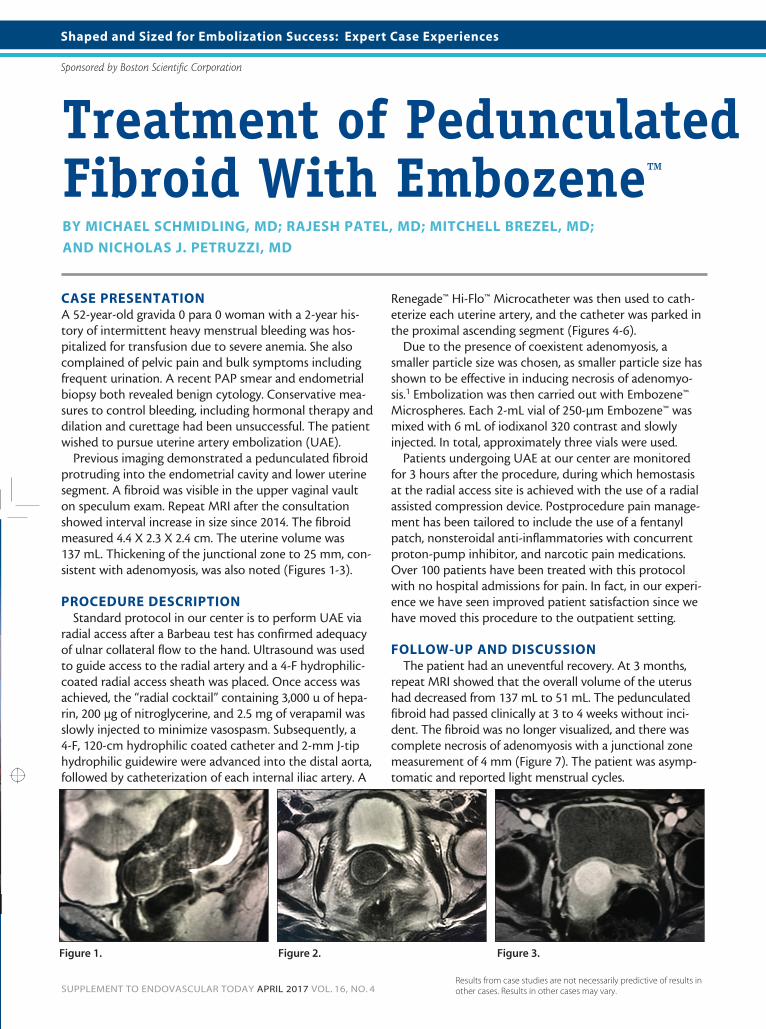
Previous imaging demonstrated a pedunculated fibroid protruding into the endometrial cavity and lower uterine segment. A fibroid was visible in the upper vaginal vault on speculum exam. Repeat MRI after the consultation showed interval increase in size since 2014. The fibroid measured 4.4 X 2.3 X 2.4 cm. The uterine volume was 137 mL. Thickening of the junctional zone to 25 mm, con-sistent with adenomyosis, was also noted (Figures 1-3).
PROCEDURE DESCRIPTIONStandard protocol in our center is to perform UAE via
radial access after a Barbeau test has confirmed adequacy of ulnar collateral flow to the hand. Ultrasound was used to guide access to the radial artery and a 4-F hydrophilic-coated radial access sheath was placed. Once access was achieved, the “radial cocktail” containing 3,000 u of hepa-rin, 200 μg of nitroglycerine, and 2.5 mg of verapamil was slowly injected to minimize vasospasm. Subsequently, a 4-F, 120-cm hydrophilic coated catheter and 2-mm J-tiphydrophilic guidewire were advanced into the distal aorta,followed by catheterization of each internal iliac artery. A
Renegade™ Hi-Flo™ Microcatheter was then used to cath-eterize each uterine artery, and the catheter was parked in the proximal ascending segment (Figures 4-6).
Due to the presence of coexistent adenomyosis, a smaller particle size was chosen, as smaller particle size has shown to be effective in inducing necrosis of adenomyo-sis.1 Embolization was then carried out with Embozene™ Microspheres. Each 2-mL vial of 250-μm Embozene™ was mixed with 6 mL of iodixanol 320 contrast and slowly injected. In total, approximately three vials were used.
Patients undergoing UAE at our center are monitored for 3 hours after the procedure, during which hemostasis at the radial access site is achieved with the use of a radial assisted compression device. Postprocedure pain manage-ment has been tailored to include the use of a fentanyl patch, nonsteroidal anti-inflammatories with concurrent proton-pump inhibitor, and narcotic pain medications. Over 100 patients have been treated with this protocol with no hospital admissions for pain. In fact, in our experi-ence we have seen improved patient satisfaction since we have moved this procedure to the outpatient setting.
FOLLOW-UP AND DISCUSSIONThe patient had an uneventful recovery. At 3 months,
repeat MRI showed that the overall volume of the uterus had decreased from 137 mL to 51 mL. The pedunculated fibroid had passed clinically at 3 to 4 weeks without inci-dent. The fibroid was no longer visualized, and there was complete necrosis of adenomyosis with a junctional zone measurement of 4 mm (Figure 7). The patient was asymp-tomatic and reported light menstrual cycles.
Figure 1. Figure 2. Figure 3.
SIZED FOR SUCCESSONCOZENE™ MICROSPHERES
More control and confidence in your embolization procedures with the most precisely calibrated microspheres.
Visit bostonscientific.com/Oncozene to learn more.
©2016 Boston Scientific Corporation or its affiliates. PI-420006-AA SEP2016
PI-420006-AA_Oncozene_Sized-For-Success_EVT Half PageAd_vF.indd 1 4/4/17 9:24 AM
Results from case studies are not necessarily predictive of results in other cases. Results in other cases may vary.SUPPLEMENT TO ENDOVASCULAR TODAY APRIL 2017 VOL. 16, NO. 4

SUPPLEMENT TO ENDOVASCULAR TODAY APRIL 2017 VOL. 16, NO. 4
Shaped and Sized for Embolization Success: Expert Case Experiences
Sponsored by Boston Scientific Corporation
This case highlights several considerations for UAE. Recommendations for treatment of pedunculated fibroids has been modified over time from more conservative to currently more liberal recommendations. We generally will offer treatment for all pedunculated fibroids after discussions with the patient and referring gynecologist about associated risks of fibroid impaction. This case is particularly interest-ing noting complete necrosis of the stalk and passage of the entire fibroid. One could postulate this may be related to the smaller particle size used. This case also highlights the safety and efficacy of UAE as an outpatient procedure using radial artery access. n
1. Kim MD, Kim YM, Kim HC, et al. Uterine artery embolization for symptomatic adenomyosis: a new technical development of the 1-2-3 protocol and predictive factors of MR imaging affecting outcomes. J Vasc Interv Radiol. 2011;22:497–502.
Figure 4.
Figure 7.
Figure 5. Figure 6.
Michael Schmidling, MDSection ChiefDivision of Interventional Radiology AtlantiCare Regional Medical Center Atlantic Medical Imaging Galloway, New JerseyDisclosures: None.
Rajesh Patel, MDAttending Interventional Radiologist AtlantiCare Regional Medical Center Atlantic Medical Imaging Galloway, New JerseyDisclosures: None.
Mitchell Brezel, MDChairman of Radiology AtlantiCare Regional Medical Center Atlantic Medical Imaging Galloway, New JerseyDisclosures: None.
Nicholas J. Petruzzi, MDAttending Interventional RadiologistAtlantiCare Regional Medical CenterDirector of Interventional OncologyAtlantic Medical ImagingGalloway, New Jersey(609) 652-6094; [email protected] Disclosures: None.

SUPPLEMENT TO ENDOVASCULAR TODAY APRIL 2017 VOL. 16, NO. 4
Shaped and Sized for Embolization Success: Expert Case Experiences
Sponsored by Boston Scientific Corporation
Embozene MicrospheresCAUTION: Federal law (USA) restricts this device to sale by or on the order of a physician. Rx only. Prior to use, please see the complete “Directions for Use” for more information on Indications, Contraindications, Warnings, Precautions, Adverse Events, and Operator’s Instructions.
INDICATIONSEmbozene Microspheres are indicated for the embolization of hyper-vascular tumors and arteriovenous malformations (AVMs).
CONTRAINDICATIONSEmbolization procedures shall not be performed if:• Patient is unable to tolerate vascular occlusion procedures.• Vascular anatomy precludes correct catheter placement or embolic injection.• Presence or likely onset of vasospasm.• Presence of a blood coagulation disorder that would prohibit arterial punctures.• Presence of severe atheromatous disease that would preclude correct catheter placement.• Presence of patent extra-to-intra-cranial anastomoses or shunts from the arterial to the venous circulation.• Presence of collateral vessel pathways which could potentially endan-ger non-targeted tissue during an embolization procedure.• Presence of any vasculature where Embozene Microspheres could pass directly into the central nervous system, central circulatory system or other nontarget territories.• Patient has high-flow arteriovenous shunt with diameter greater than the selected Embozene Microspheres.• Patient is pregnant.• Patient has known allergies to barium sulfate, 3-aminopropyltrialk-oxysilane, polyphosphazene or IV radiopaque contrast agent.
WARNINGSVascular embolization is a high risk procedure. The procedure should be performed by specialized physicians trained in vascular emboliza-tion procedures. Complications can occur at any time during or after the procedure, and may include, but not limited to:
• Undesirable reflux or passage of Embozene Microspheres into normal arteries adjacent to the targeted lesion or through the lesion into other arteries or arterial beds.
• Embolization of the wrong artery or migration of the micro-spheres to other parts of the body, which may necessitate further treatment.
• Hematoma, or bruising, at the incision site for arterial access.• Arterial aneurysm at the incision site for arterial access.• Deep vein thrombosis, or clotting of a deep vein in patient’s leg(s).• Thrombosis of the artery at the incision site for arterial access.• Pulmonary embolization. • Ischemia at an undesirable location.• Capillary bed saturation and tissue damage.• Ischemic stroke or ischemic infarction.• Vessel or lesion rupture and hemorrhage.• Neurological deficits including cranial nerve palsies.• Vasospasm.• Recanalization.• Foreign body reactions necessitating medical intervention.• Infection necessitating medical intervention.• Clot formation at the tip of the catheter and subsequent
dislodgement.• Allergic reaction.• Risks of radiation from angiography and fluoroscopy used to
visualize the blood vessels during embolization, which may include a radiation burn and risks to future fertility.
• Death.Do not use Embozene Microspheres in conjunction with embolization
devices based on organic solvents such as ethyl alcohol or dimethyl sulfoxide (DMSO) at the same embolization site.Do not use ionic contrast agent with this product. Ionic contrast agents could alter the microsphere characteristics resulting in microsphere deformation and procedure failure.
PRECAUTIONSTo maintain safety, the following precautions shall be considered:
• Safety and effectiveness of Embozene Microspheres in the treat-ment of uterine fibroids has not been established
• Safety and effectiveness of Embozene Microspheres for hepatic and renal embolization uses has not been established.
• The physician should carefully select the size and quantity of Embozene Microspheres according to the lesion to be treated based on the physician’s education and training and currently available scientific evidence.
• Physicians must decide the most appropriate time to stop the infusion of Embozene Microspheres. Typically the artery will ac-cept fewer Embozene Microspheres as the treatment progresses. Proximal slowing or termination of flow may indicate that the vessel or the target area is occluded by Embozene Microspheres. Careful fluoroscopic monitoring is required.
• Microparticle embolization must be performed slowly. The injec-tion speed and manner must be controlled. Excessive injection rate may result in retrograde flow in the vessel leading to embo-lization of other non-target healthy tissue or organs
• The color of the Embozene Microspheres may be visible through the skin if injected into superficial arteries.
• If arteriovenous anastomoses, branch vessels which lead away from the targeted embolization area, or emergent vessels not evi-dent prior to embolization are present, it can lead to non-targetedembolization and cause severe complications for the patient.
• Microspheres smaller than 100 μm can migrate to distal anasto-motic feeders and embolize circulation to distal tissue. For this reason, smaller microspheres have a greater likelihood of caus-ing unwanted ischemic injury. This should be considered prior to starting the embolization procedure. Possible consequences include, but are not limited to, paralysis, necrosis, swelling, ab-scess formation and more severe post-embolization syndrome.
• Ischemia of tissue adjacent to the targeted area may result from post-embolization swelling. Therefore, special care should be taken to avoid such ischemia of non-tolerant, non-targeted tis-sue such as the nervous system.
• Consider upsizing Embozene Microspheres if angiographic ap-pearance of embolization does not quickly appear during injec-tion of the microspheres.
• If there are any symptoms of unwanted embolization during injec-tion, consider stopping the procedure to evaluate the possibility of shunting. Such symptoms may include changes in patient vital signs, such as hypoxia or central nervous system changes.

Shaped and Sized for Embolization Success: Expert Case Experiences
Sponsored by Boston Scientific Corporation
SUPPLEMENT TO ENDOVASCULAR TODAY APRIL 2017 VOL. 16, NO. 4
Renegade STC 18 Microcatheter Renegade Fiber Braided MicrocatheterRenegade HI-FLO MicrocatheterCAUTION: Federal law (USA) restricts this device to sale by or on the order of a physician. Rx only. Prior to use, please see the complete “Directions for Use” for more information on Indications, Contraindications, Warnings, Precautions, Adverse Events, and Operator’s Instructions.
INTENDED USE/INDICATIONS FOR USE The Renegade STC 18 Microcatheter, Renegade Fiber Braided Microcath-eter, and the Renegade HI-FLO Microcatheter are intended for peripheral vascular use. The microcatheter can be coaxially tracked over a steerable guidewire in order to access distal, tortuous vasculature. Once the subse-lective region has been accessed, the microcatheter can be used for the controlled and selective infusion of diagnostic, embolic, or therapeutic materials into vessels. Diagnostic, embolic, therapeutic agents to be used in accordance with specifications outlined by the manufacturer.
CONTRAINDICATIONS None Known.
WARNING The Renegade STC 18 Microcatheter, Renegade Fiber Braided Micro-catheter, and the Renegade HI-FLO Microcatheter are not intended for use in the coronary vasculature or the neurovasculature.
PRECAUTIONS • This device should be used only by physicians thoroughly trained
in percutaneous, intravascular techniques and procedures. • Never advance or withdraw an intravascular device against
resistance until the cause of the resistance is determined by fluoroscopy. Movement of the microcatheter or guidewire against resistance may result in separation of the microcatheter or guidewire tip, damage to the microcatheter or guidewire tip, or vessel perforation.
• Because the microcatheter may be advanced into narrow subse-lective vasculature, repeatedly assure that the microcatheter has not been advanced so far as to interfere with its removal.
ADVERSE EVENTS The Adverse Events include, but are not limited to: vessel trauma, em-bolism, hemorrhage/hematoma, vasospasm, infection, air embolism, allergic reaction.