microneedle-mediated delivery of copper peptide … ghkcu.pdfresearch paper microneedle-mediated...
TRANSCRIPT

RESEARCH PAPER
Microneedle-Mediated Delivery of Copper Peptide Through Skin
Hairui Li & Yong Sheng Jason Low & Hui Ping Chong & Melvin T. Zin & Chi-Ying Lee & Bo Li & Melvina Leolukman & Lifeng Kang
Received: 3 November 2014 /Accepted: 5 February 2015 /Published online: 19 February 2015# Springer Science+Business Media New York 2015
ABSTRACTPurpose Copper peptide (GHK-Cu) plays an important role inskin regeneration and wound healing. However, its skin absorp-tion remains challenging due to its hydrophilicity. Here we usepolymeric microneedle array to pre-treat skin to enhance GHK-Cu skin penetration.Methods Two in vitro skin models were used to assess the ca-pability of microneedles in facilitating skin delivery of GHK-Cu.Histological assay and confocal laser scanning microscopy wereperformed to characterize and quantify the microconduits createdby the microneedles inside skin. Cellular and porcine modelswere used to evaluate the safety of microneedle-assisted copperpeptide delivery.Results The depth and percentage of microneedle penetrationwere correlated with application forces, which in turn influencedthe extent of enhancement in the skin permeability of GHK-Cu. In9 h, 134±12 nanomoles of peptide and 705±84 nanomoles ofcopper permeated though the microneedle treated human skin,while almost no peptide or copper permeated through intacthuman skin. No obvious signs of skin irritation were observedwith the use of GHK-Cu after microneedle pretreatment.Conclusions It is effective and safe to enhance the skinpermeation of GHK-Cu by using microneedles. This ap-proach may be useful to deliver similar peptides or mineralsthrough skin.
KEY WORDS application forces . copper peptide .microneedle . skin penetration . transdermal
ABBREVIATIONSAAS Atomic absorption spectroscopyDMEM Dulbecco’s modified Eagle’s mediumDMSO Dimethyl sulfoxideGHK Glycyl-L-histidyl-L-lysylGHK-Cu Copper peptideHaCaT Human adult low calcium high temperatureHDF Human dermal fibroblastsHPLC High performance liquid chromatographyMN MicroneedleMSS Microchannel Skin SystemMTT 3-(4,5-dimethylthiazol-2-yl)-2,5-diphenyl tetrazoli-
um bromidePBS Phosphate buffered saline
INTRODUCTION
Glycyl-L-histidyl-L-lysyl (GHK) is a naturally occurring carri-er tripeptide with high affinity for copper ions. GHK was firstisolated from human plasma by Dr L. Pickart because of itsactivity to prolong survival of normal liver cells (1).Subsequently, researchers found that GHK-Cu can stimulatethe synthesis of extracellular matrix macromolecules, such ascollagen and glycosaminoglycan (2–4). It can activate the pro-duction of metalloproteinases and anti-proteases that removedamaged proteins from the extracellular matrix macromole-cules (5). GHK-Cu was also found to increase decrorin expres-sion and decrease TGF-beta expression, which is beneficial fora scar-free healing (6,7). The increased expression of p63 ofkeratinocytes by both GHK-Cu and GHK suggests thatGHK and its copper complex can promote the survival ofbasal stem cells in skin (8,9). These contribute to the wound-
Electronic supplementary material The online version of this article(doi:10.1007/s11095-015-1652-z) contains supplementary material, which isavailable to authorized users.
H. Li : Y. S. J. Low :H. P. Chong : L. Kang (*)Department of Pharmacy, National University of Singapore, 18 ScienceDrive 4, Singapore, Singapore 117543e-mail: [email protected]
H. Li :M. T. Zin : C.<Y. Lee : B. Li :M. Leolukman3M Innovation Singapore, 100 Woodlands Avenue,Singapore, Singapore 738205
Pharm Res (2015) 32:2678–2689DOI 10.1007/s11095-015-1652-z

healing and skin remodelling effects of GHK-Cu (2,10–14).The biochemical action of GHK and GHK-Cu on skin cellswere summarized in SI 1. Recent genomic studies revealedthat GHK can directly modulate the expression of a largenumber of human genes and reverse gene expression to ahealthier state, whichmay explain the diversity of its biologicalactions (15–17). Furthermore, in vivo studies showed thatGHK-Cu can improve hair growth, skin regeneration andwound healing (10).
Minerals are essential for human health and mineral sup-plementation is recommended to complement dietary intake.Supplementation of minerals via oral route is the most com-mon way. However, oral mineral absorption is affected bymany factors including interactions with other dietary compo-nents in the gastrointestinal tract (18). Gastrointestinal sideeffects have also been reported with oral ingestion of someminerals, such as copper and iron. In addition, oral liquidforms of iron supplementation may cause teeth stains (19).To this end, transdermal delivery of minerals may be a usefulalternative but remains largely unexplored. Copper as an es-sential trace element plays a critical role in diverse biologicalprocesses, such as haemoglobin synthesis and the stimulationof skin biomarkers including collagen and elastin (20). Copperalso has an important role in the activation of key enzymesspecific to tissue repair and in the cross-linking andmaturationof collagen in healing wounds (19). Because of the gastrointes-tinal irritation that oral intake of copper can cause, transder-mal delivery of copper can act as a good alternative, althoughskin absorption of charged ions is very low (21,22). It wasfound that when GHK is coupled with copper, the peptidesilences the redox activity of copper, hence permitting thedelivery of copper in a non-toxic form that can be subsequent-ly utilized by the cells (8,23).
Although its biological actions start at picomole level, farhigher dosages have been used in clinical trials, since theGHK-Cu uptake levels are very low through skin. On theother hand, if GHK-Cu is injected intradermally, GHK israpidly cleared (95% clearance in 1 min). The fragility andrapid breakdown of GHK is the major obstacle for clinicaland cosmetic applications (24). In clinical trials involvingGHK-Cu, large variations were observed and its efficacy to-wards the healing of indolent human wounds or skin ulcerswere not found (10,25,26). To address the concerns, a varietyof chemical modification to GHK has been carried out toproduce breakdown-resistant copper complexes, but nonewere found better than GHK-Cu (10).
The low uptake of GHK-Cu by skin is a common problem,similar to many other peptides and molecules. This is as theskin naturally functions to protect the human body from theexternal environment. So the molecules that can be deliveredthough skin are limited. As a general rule, only hydrophobicmolecules with a molecular weight less than 500 Da are ableto passively diffuse through the skin (27). GHK-Cu with a log
P of −4.5 can represent hydrophilic peptides that have diffi-culty in penetrating into skin (11). Because of the diverse bio-logical effects of GHK-Cu, along with its potential as a sourceof copper supplementation, it is of interest to find out a meth-od to enhance its skin absorption.
To this end, we propose the use of microneedles, a mini-mally invasive but effective skin permeation enhancementmethod, to facilitate the effective and sustained delivery ofGHK-Cu through skin. If an effective skin uptake of GHK-Cu is possible in a sustained manner with microneedle pre-treatment, GHK-Cu is expected to better fulfil its biologicaleffects.
Microneedles with lengths in the micron ranges can en-hance skin permeability with no pain by breaching the stratumcorneum layer of skin with self-administration (28). It has beenshown that microneedles are associated with a lower risk ofmicrobial infection than hypodermal needles (29). There aredifferent methods of drug delivery to skin with microneedles,and microneedle pretreatment followed by a topical formula-tion has the advantage of possible extended release (30).Studies have shown that pretreatment of skin withmicroneedles can enhance delivery of topically applied formu-lations (31,32), including peptides (33).
In this study, we investigate the effectiveness of a commer-cial microneedle product, i.e., 3M™ Microchannel SkinSystem (MSS), in enhancing the skin permeability of copperpeptide, using both rat and human skin models. Since MSS ishand-applied by using an applicator, the effect of differentapplication forces was studied. In addition, we also carriedout cell and animal testing to verify its safety.
MATERIALS AND METHODS
Materials
The 3M™ Microchannel Skin System (MSS) is illustrated inFig. 1a. The plastic microneedle patch comprises of a rectan-gular grid of needles (13 by 27 array, or 351 needles) centredon an oval array. The microneedles have square pyramidalshape with a needle height of about 700 μm and a tip-to-tipneedle spacing of 500 μm. GHK-Cu and GHK werepurchased from McBiotec, Nanjing, China. The ratioof GHK to Cu is 2:1 (manufacturer data). Copper stan-dard solution (1 g/L), and trypan blue solution (0.4%) werepurchased from Sigma-Aldrich, Singapore. Concentrated ni-tric acid (69% w/w) was purchased from VWR InternationalS.A.S, Singapore. Rhodamine B was purchased from AlfaAesar (Lancaster, UK). Phosphate buffered saline (PBS)(pH 7.4, 10×) was obtained from Vivantis, Malaysia. PDMS(Sylgard® 184 Silicone Elastomer Kit) was purchased fromDow Corning (Midlan, MI, USA). Solvable™, an aqueousbased tissue solubilizer, was purchased from PerkinElmer
Microneedle-Mediated Delivery of Copper Peptide 2679
(Watham, MA, USA). Pierce® BCA protein assay kitwas purchased from Thermo Scientific (Rockford, IL,USA). 3-(4,5-dimethylthiazol-2-yl)-2,5-diphenyl tetrazoli-um bromide (MTT) and dimethyl sulfoxide (DMSO)were purchased from MP Biomedicals, Singapore.Dulbecco’s modified Eagle’s medium (DMEM) and fetalbovine serum were purchased from Life Technology,Singapore. Penicillin streptomycin solution was purchasedfrom PAN-Biotech GmbH, Germany. All chemicals wereused as supplied.
Skin and Microneedle Application
Human dermatomed skin was obtained from Science Care(Phoenix, AZ, USA). The skin tissues were excised from thethighs of Caucasian female cadaver, who died at the age of 92.
Besides human skin, rat skin was also chosen for the in vitroskin permeation and penetration study. Skin of rodents, in-cluding rats, is the most commonly used for in vitropercunaeous permeation studies because of its availability(34). Rat abdominal skins were obtained from National
f e
b
50µm50µm
a
0
50
100
150
200
250
300
350
0 10 20 30
Dep
th o
f p
enet
rati
on
(µ
m)
Force of MN application (N)
Rat skin
Human skin
dc
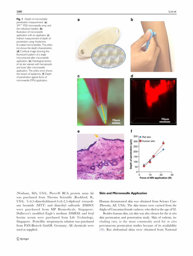
Fig. 1 Depth of microneeldepenetration measurement. (a)3M™ MSS microneedle array andthe individual needles. (b)Illustration of microneedleapplication with an applicator. (c)Indirect measurement of depth ofpenetration using rhodamineB-coated micrroneedles. The yellowline shows the depth of penetration.(d) Confocal image showing thefluorescent pattern of a singlemicrochannel after microneedleapplication. (e) Histological sectionof rat skin stained with hematoxylinand eosin after microneedleapplication. The yellow arrow showsthe breach of epidermis. (f) Depthof penetration against force ofmicroneedle (MN) application.
2680 Li et al.

University of Singapore Comparative Medicine through thetissue sharing program. The hair on rat skin was removed byan electrical shaver followed by hair removal cream (Veet®)application and removal after 2 min. Subcutaneous fat andconnective tissues were also trimmed off (35).
A 6 mm thick PDMS substrate was used to support the skinfor microneedle application as reported (36). The skin wasspread over the PDMS substrate mounted on a Styrofoamboard with epidermis side up. A microneedle array was thenput on the skin sample. Four different forces were appliedthrough the applicator (Fig. 1b) at 4.5, 10.0, 13.3 and22.2 N for 10 s, respectively, using a force gauge (HF-10,JISC, Japan).
The use of animal skin was approved by the NationalUniversity of Singapore Institutional Animal Care and UseCommittee. The use of human skin was approved by theNational University of Singapore Institutional ReviewBoard.
Depth of Microneedle Penetration Measurement
Microneedles were first primed with oxygen plasma in a plas-ma cleaner (Harrick Plasma, US) for 3 min to render themhydrophilic. The arrays were then flood-coated with 70 μl of0.1% w/w rhodamine B solution and dried at 35°C for 1 h.The rhodamine B coated arrays were then applied with aforce of 4.5, 10.0, 13.3 and 22.2 N for 10 s respectively onrat and human dermatomed skin samples. The depth of pen-etration was measured indirectly by recording the distancefrom the tip of the microneedles to the boundary where therhodamine B coating was wiped off after skin insertion (31).To analyze the penetration depth of the microneedles, themicroneedles were imaged using a stereoscopic microscope(Nikon SMZ25, Japan). The depth of penetration for eacharray was then determined by measuring 35 out of the total351 microneedles per array. Three arrays were tested for eachforce.
Confocal laser scanning microscopy was used to visualizethe micro-conduits created in rat skin placed on a piece ofglass slide. The scanning started from the stratum corneum side,through the z-axis of the microscope (A1R+si, Nikon, Japan)at 10× magnification. The excitation wavelength was 562 nmand fluorescence emission was at 570–620 nm for rhodamineB.
Histological Examination
Histological examination of rat skin was carried out by cuttingthe microneedle treated skin samples into 10 μm sectionsusing a Microcryostat (Leica, Germany). The histological sec-tions were stained with hematoxylin and eosin and imagedusing a microscope (Olympus, Japan).
Percentage of Penetration Measurement
The percentage of penetration was tested using trypan bluecoated microneedles. Microneedle arrays were first treatedwith oxygen plasma for 3 min, flood-coated with 70 μl oftrypan blue solution and then dried at 35°C for 1 h. Afterapplication of a microneedle array with the specified forceon skin, the skin was cleansed with water. Application siteswere then imaged by using the stereoscopic microscope. Thevisible insertions were counted and the percentage of penetra-tion (the number of stained dots divided by the total numberof array microneedles) was calculated. At least 3 replicateswere performed at each force.
In Vitro Skin Permeation Study
Vertical Franz diffusion cells with an effective exposed area of1 cm2 were used. The pretreated skin samples were mountedonto Franz diffusion cells with epidermis facing up. The intactskin was used as control. The donor cell contained 2 ml of5.8 mM (GHK)2Cu (the molar ratio of GHK to Cu in the rawmaterial was 2 to 1) solution while the receptor cell contained4.8ml of 1× PBS. The cells were placed inside a chamber withtemperature controlled at 32°C (comparable to the physiolog-ical temperature of the skin surface). Magnetic stirrers in thereceptor cells stirred at a speed of 100 rpm. The receptorsolutions were withdrawn at pre-set time intervals and re-placed with fresh ones. The receptor solutions were subjectedto the measurement of copper permeated through the skin byatomic absorption spectroscopy (AAS) and the measurementof peptide permeated by high performance liquid chromatog-raphy (HPLC). At completion, the skin surface was washed byrinsing the donor and receptor compartments with 1 ml water3 times, respectively. The skin was then removed from thediffusion cells, dried with Kimwipes and collected intoFalcon tubes for the measurement of copper retained in skinby AAS (37). Three replicates were conducted for each group.
AAS Method
Copper concentrations were determined by using an atomicabsorption spectrometer (PinAAcle 900T, PerkinElmer) withacetylene flow rate of 2.5 L/min and compressed air flow rateof 10 L/min. The instrument was calibrated with Cu workingstandards (1.57–62.95 μM) made from Cu stock standardsolution (1 g/L) with 2% nitric acid as the diluent. Dilutedpermeation samples collected from receptor solutions werethen aspirated into the air/acetylene flame where Cu atomsabsorb light of 324.75 nm. All readings of standards and sam-ples were conducted with the instrument in the absorbancemode. To digest skin tissues prior to analysis, concentratednitric acid was used, as previous studies have shown that itquantitatively releases trace elements from biological tissues
Microneedle-Mediated Delivery of Copper Peptide 2681

(38). Nitric acid of 2.5ml was added to each of the Falcon tubescontaining skin samples and heated at 80°C using a thermo-static water bath until complete digestion of the skin samples(38). After that, they were topped up to 10 ml using deionizedwater and filtered using 0.22 μm polytetrafluoroethylenemembrane. The filtered solution was used for the measure-ment of the amount copper retained in skin.
HPLC Method
The amount of GHK permeated was determined by usingHitachi L2000 LaChrome Elite HPLC system with AgilentBio SCX NP3 column (50 mm×4.6 mm, 3 μm). The mobilephase consisted of Mobile A (Millipore water) and Mobile B(10 mM NaH2PO4 with 0.5 M NaCl) with a gradient elutionprogram with a mixture of solvents A and B as follows: 0% Bfor 1 min, 1–100% B for 1–5 min, 100% B for 5–8 min, 100–0% B for 8–8.1 min, and 0% B for 8.1–11 min. The flow ratewas set at 0.5 ml/min. The injection volume was 20 μl foreach sampling and ultraviolet detection was performed at awavelength of 218 nm. A calibration curve was conductedusing GHK standard solution from 1.18 to 587.58 μM.
Skin Total Protein Determination
Skin samples were put into a 2 ml tube with 1.5 ml Solvable™added. Then the tube was heated at 60°C for 4 h to allow thecompletely dissolution of skin. The total protein level of skinwas determined by using a Pierce® BCA protein assay kitafter the skin digestion solution was diluted appropriately withwater.
Cytotoxicity Assay of GHK-Cu on Skin Cells
The cytotoxicity of different concentrations of GHK-Cuagainst human adult low calcium high temperature(HaCaT) keratinocytes and human dermal fibroblasts (HDF)were studied by MTT assay in 6 replicates. Briefly, 5000 cellsin 200 μl culture medium (DMEM supplemented with 10%fetal bovine serum and 1% penicillin-streptomycin solution)per well were seeded into 96-well plates and incubated for24 h. The culture medium was then removed. Subsequently,180 μl fresh culture medium and 20 μl of GHK-Cu samples(0.058–58,000 μM (GHK)2Cu in PBS) were added per welland incubated for 24, 48 and 72 h, respectively. For controlgroup, 180 μl fresh culture medium and 20 μl of PBS wereadded. At the respective analysis point, the medium was re-moved. The wells are washed with 200 μl PBS andreplenished with 200 μl fresh medium per well. Twenty μlMTT solution (5 mg/ml in PBS) was added to each well, afterwhich the plates were incubated for an additional 4 h. Thesupernatant was then removed and formazan crystals weresolubilized in 150 μl DMSO. Absorbance was recorded at
595 nm with a microplate reader (Tecan, Switzerland). Wellscontaining DMSO alone were used as the blank. Percentage ofcel l viabil i ty was expressed as (Asample−ADMSO)/(Acontrol−ADMSO) × 100%.
In Vivo Irritation Test on Pigs
Young adult swine (Yorkshire X), ranging from 10 to 40 kgwere used for the study. The animals were first sedated withketamine (10 mg/kg) and then anesthetized with isofluranegas. Atropine was administered to reduce salivary, tracheo-bronchial, and pharyngeal secretions. The ham area of pigswas shaved with an electrical shaver followed by a disposableshaver. The microneedles were applied on the skin with ahand force of around 20 N (estimated by using the forcegauge) for 10 s. Then the skin was observed for irritationand imaged with time. In another testing, immediately aftermicroneedle treatment, the application site was covered bygauze saturated with GHK-Cu solution, then fixed with plas-ter. After 8 h, the gauze was removed and the skin was im-aged. The studies were repeated on 3 to 4 pigs. The pigs werelater recovered. The procedure for animal testing was ap-proved by the National University of Singapore InstitutionalAnimal Care and Use Committee.
Statistical Analysis
All results were presented as mean±standard deviation.Statistical analysis was performed by one-way analysis of var-iance followed by Tukey post hoc test using IBM SPSS Statistics19. A probability value of p<0.05 was considered statisticallysignificant.
RESULTS
Depth of Microneedle Penetration
In Fig. 1c, the yellow line indicated the distance fromthe tip of the microneedles to the boundary where therhodamine B coating has been wiped off after skin in-sertion. The value of the distance was taken as depth ofpenetration of microneedles. Figure 1d showed the directmeasurement of the depth of penetration using confocal laserscanning microscopy by measuring the depth of themicroneedle fluorescence pattern inside the skin. The depthof penetration associated with an application force of 13.3 Non rat skin was measured to be 130 μm with directmeasurement and 146 μm using indirect measurement.It was found that the results obtained from indirectdepth of penetration measurement method had no dif-ference from those obtained by direct measurementmethod (p>0.05). Hence, the indirect method was used
2682 Li et al.

for this study for its convenience. Furthermore, Fig. 1efurther ascertained the presence of microscale passagescreated by microneedles inside the rat skin. Figure 1fshowed that the depth of penetration of microneedlesincreased linearly with application force both on rat(r2=0.9977) and human (r2=0.9758) skin samples inthe tested force range from 4.5 to 22.2 N.
Percentage of Penetration
The percentage of microneedle penetration in rat skin wasmeasured using trypan blue coated microneedles. Figure 2aand b shows the representative image of rat skin, humandermatomed skin after application of trypan blue-coatedmicroneedles at 22.2 N, respectively. The blue dots thatremained on the skin indicated the successful penetration ofmicroneedles into skin. The number of the blue dots was usedto calculate the percentage of penetration per microneedlearray. Figure 2c showed that a higher force of microneedleapplication resulted in a higher percentage of penetration.
The increase in force of microneedle application on rat skinresulted in a sharper increase in the percentage of penetrationas compared to that on human dermatomed skin. The highestforce of 22.2 N made the percentage of penetration to bealmost 100% on rat skin and 30% on human dermatomedskin.
In Vitro Skin Permeation Study
To assess the enhancing effects of the microneedles for GHK-Cu to permeate through skin at different forces of application,we performed in vitro drug permeation study. Upon topicaladministration, the copper ions are subjected to dynamic li-gand exchange inside skin (37). Therefore, quantitative assess-ment of skin penetration of GHK-Cu is challenging. Hence,we analysed the permeation of copper and GHK separatelyby using AAS and HPLC. Figure 3a and b showed that theamounts of copper and peptide permeated through rat skinwere significantly increased after microneedle pretreatment.Figure 3c and d showed that for non-treated humandermatomed skin, there was no peptide detected in the recep-tor solution, while only trace amount of copper was detected,which may be from the skin itself (data not shown). With themicroneedle pretreatment, copper and peptide were detectedin the receptor solution. However, substantial high amounts ofcopper and peptide were detected only when a high force(22.2 N) was used.
Figure 4 showed that there was no significant difference inthe amount of copper retained in skin samples after 9 h’ per-meation study among the microneedle pretreated skinsamples and the control at various forces. However, significantdifference was found between rat and human skin.
Cytotoxicity Assay of GHK-Cu on Cells
Figure 5 showed that (GHK)2Cu was not toxic to eitherHaCaT keratinocytes or HDF cells in the range of 0.0058–5800 μM. Besides, GHK-Cu at 5800 μM showed certainstimulatory effect on HDF proliferation. Other researchgroups have also shown that GHK-Cu can stimulate thegrowth of dermal fibroblasts (39,40).
In Vivo Irritation Test on Pig Skin
For blank microneedles, minimal erythema was observedimmediately after removal of microneedles. Mild erythe-ma was observed at 5 min but almost not visible after25 min (Fig. 6a). For GHK-Cu application immediately aftermicroneedle pretreatment, no erythema or edema was ob-served for 8 h (Fig. 6b).
a
c
b
Rat skin,MN 22.2 N
Human skin, MN 22.2 N
0
20
40
60
80
100
120
0 10 20 30
Per
cen
tag
e o
f M
N p
enet
rati
on
(%
)
Force of MN application (N)
Rat skinHuman skin
Fig. 2 Percentage of penetration. Representative images of 22.2 N MNapplication force on rat skin (a) and human dermatomed skin (b). (c) Percent-age of penetration against force.
Microneedle-Mediated Delivery of Copper Peptide 2683

DISCUSSION
Microneedles can painlessly enhance the skin permeation of awide range of therapeutic compounds which barely penetratehuman skin (30). It has been reported that the microneedleapplication force can influence its enhancing effect (41–44). Inthis study we evaluated the skin permeation of topical GHK-Cu in vitro using a microneedle array from 3MCompany. The3M™ MSS is designed for manual application with a plasticapplicator. The application forces tested were: 4.5 N (low),10 N (medium), 13.3 N (medium) and 22.2 N (high).
To study the depth of needle penetration, two approacheshave been used. One is the direct measurement of themicrochannels inside skin by confocal laser scanning micros-copy and the other is the indirect measurement. In the indirectmeasurement, the depth of penetration was obtained by mea-suring the length of needle tip where rhodamine B dye waswiped off (31). But the coating procedure of rhodamine B ontothe microneedles was complicated. To simplify themicroneedle coat ing procedure, we treated themicroneedles with oxygen plasma, which was found tobe very effective for rhodamine B coating.
0
100
200
300
400
500
600
700
800
0 5 10
Cu
mu
lati
ve p
erm
eate
d p
epti
de
(nan
om
ole
s)
Time (h)
ControlMN, 4.5 NMN, 10.0 NMN, 13.3 NMN, 22.2N
0
100
200
300
400
500
600
700
800
0 5 10
Cu
mu
lati
ve p
erm
eate
d c
op
per
(n
ano
mo
les)
Time (h)
ControlMN, 4.5 NMN, 10.0 NMN, 13.3 NMN, 22.2N
0
500
1000
1500
2000
2500
3000
0 5 10
Cu
mu
lati
ve p
erm
eate
d c
op
per
(n
ano
mo
les)
Time (hours)
ControlMN, 4.5 NMN, 10.0 NMN, 13.3 NMN, 22.2 N
a b
0
500
1000
1500
2000
2500
3000
0 5 10
Cu
mu
lati
ve p
erm
eate
d p
epti
de
(n
ano
mo
les)
Time (hours)
ControlMN, 4.5NMN, 10.0 NMN, 13.3 NMN, 22.2 N
c d
Rat skinRat skin
Human skin
Human skin
Fig. 3 In vitro skin permeationstudy. The cumulative amount ofcopper (a) and peptide (b)permeated through rat skinpretreated with varying applicationforces. The cumulative amounts ofcopper (c) and peptide (d)permeated through humandermatomed skin pretreated withvarying application forces.
0
100
200
300
400
500
600
700
Clean skin Control MN, 4.5N MN, 10.0 N MN, 13.3 N MN, 22.2 N
Am
ou
nt
of
cop
per
per
ski
n
(nan
om
ole
s)
Rat skin
Human skin
Fig. 4 Cumulative amount ofcopper retained in skin after 9 hpermeation study. The ‘Clean skin’refers to the skin that was not usedin permeation study.
2684 Li et al.

The linear relationship between depth of penetration andmicroneedle application force indicates that these two factorsare strongly correlated. However, we cannot assume that thelinearity will be the same when the force is out of the range(<4.5 N or >22.2 N) (45). The force range would be from 13to 63mN/needle in our study if converted to force per needle.Some of these forces are even lower than the insertion forces ofreported microneedles. For example, Park et al. reports
measured microneedle insertion force of 37 mN with a tip20 μm in diameter (46). It indicated that our microneedlearray had a high efficiency for skin insertion.
To study the percentage of penetration, trypan blue coatedmicroneedles were used. Trypan blue is known to specificallystain the sites of stratum corneum perforation (47). A commonprocedure is to treat the skin first with microneedles, and thentransfer trypan blue solution on the treated area for targeted
0
20
40
60
80
100
120
0 0.0058 0.058 0.58 5.8 58 580 5800
Cel
l via
bilit
y (%
)(GHK)2Cu concentration (µM)
24 h
48 h
72 h
0
20
40
60
80
100
120
140
0 0.0058 0.058 0.58 5.8 58 580 5800
Cel
l via
bilit
y (%
)
(GHK)2Cu concentration (µM)
24 h
48 h
72 h
a
b*
Fig. 5 Cytotoxicity study. Viabilityof HaCaT keratinocytes (a) andHDF (b) after incubation with GHK-Cu for 24, 48 and 72 h.
a 0 min 5 min 25 min bFig. 6 Representative images ofin vivo irritation test on pig skin. (a)The skin conditions after MNapplication. (b) The condition ofMN pre-treated skin, followed byapplication of 5.8 mM copperpeptide for 8 h. The markedrectangle area indicated themicroneedle treated area.
Microneedle-Mediated Delivery of Copper Peptide 2685

staining (42,47). However, we found that wrinkles and hairfollicles can also be easily stained. Besides, trypan blue solutionhas limited contact with skin surfaces to ensure staining of allthe microchannels because of the hydrophobic nature of skinsurfaces. To solve this problem, we coated trypan blue directlyonto the microneedles and then conducted the penetrationtest on skin. The false staining on the skin surface was thenwiped off with water. With the new method, we can easily getclear staining images indicating the successful penetration ofmicroneedles.
The results showed that with a higher force, the percentageof penetration increased. However, the effects were differenton rat and human skins. Rat skin was much easier to bestained than human dermatomed skin. Trypan blue stainsdamaged cells selectively because the damaged cell mem-branes allow trypan blue to enter and stain the nucleiand cytoplasm (48). Since the stratum corneum is made upof layers of highly keratinized cells it will not be stainedby trypan blue. Trypan blue can stain the epidermis atthe sites of stratum corneum perforation (36). Although ratfull skin and human dermatomed skin samples had thesimilar thickness, around 500 μm, rat skin has a muchthinner layer of epidermis (~15 μm, Fig. 1e) than hu-man skin (~200 μm). After the stratum corneum of rat skinis penetrated by microneedles, the viable epidermis belowmay be easily stained. On the other hand, the human skin hasa more complicated structure. The layer below the stratumcorneum is the stratum granulosum. Since the stratum granulosum ismade up of partially keratinized cells without nuclei, it is moredifficult to stain (49).
In general, microneedle pretreatment can increaseskin delivery of GHK-Cu, although the results werequite different on rat and human skin. The permeationof copper peptide was observed for non-treated rat skin, butalmost none for intact human dermatomed skin. Withmicroneedle pretreatment, the permeated amount of copperand peptide increased significantly than those through non-treated rat skin (Fig. 3a and b). For human skin, microneedlepretreatment made permeated copper and peptide detect-able. But substantial increase in the skin permeation was onlyobserved at 22.2 N (Fig. 3c and d).
We noticed that the depth of penetration was proportionalto application forces in both rat skin and human dermatomedskin models. For the rat skin, however, the microneedles caneasily penetrate the entire epidermis at all the forces, as thethickness of epidermis of rat skin is ~15 μm (Fig. 1e).Epidermis, especially, its outermost layer, i.e., the stratumcorneum, is the main barrier. Thus, the permeation of copperpeptide through rat skin was independent of depth of pene-tration in rat skin model, similar to another finding (36).Bachhav et al. also found that the transport of lidocaine wasindependent of the depth of the microchannels created bylaser after removal of stratum corneum (50).
In case of human dermatomed skin model, whenmicroneedle application forces were no higher than 13.3 N,increase in skin permeation was observed because the stratumcorneum was penetrated, while the distinct increase in the skinpermeation at 22.2 N may be due to the penetration of wholeepidermis layer (the epidermis layer of the human skin is~200 μm). It indicated that viable epidermis layers are alsosignificant permeation barriers (51).
Apart from the two types of skin models (rat full skin andhuman dermatomed skin), we also tested human epidermis.Human epidermis has been widely used in in vitro skin perme-ation experiment to study the conventional transdermal drugdelivery (52). Human epidermis has also been used to studymicroneedle assisted transdermal drug delivery (44,53).However, in our study, it was shown that human epidermis,without the dermis layer, showed a much higher percentage ofpenetration with microneedles as compared to humandermatomed skin (SI 2 and SI 3). It may be because the thickdermis in the dermatomed skin could provide cushion effectand have absorbed some of the force applied onto the epider-mal layer. Furthermore, a sharp increase in skin permeation ofcopper peptide was observed even with a mild microneedleapplication force on human epidermis (SI 3). As such rat skinand human epidermis may have their limitations as models tostudy the physical enhancement of skin by using microneedles(SI 4).
Furthermore, higher skin permeation of GHK-Cu can beobtained with the use of a higher concentration of GHK-Cusolution with microneedle treatment. However, no perme-ation of GHK-Cu was observed through intact humandermatomed skin even when the GHK-Cu concentration in-creased (SI 5). It suggested that increasing GHK-Cu concen-tration alone in topical formulations might not be a goodchoice to improve the effect of the GHK-Cu creams/gels.
The ratio of copper to peptide permeated though rat skinwas around 1:1, while when human skin models were used,the ratio wasmuch higher. It may be partly due to the fact thatmore copper were retained in rat skin than in human skin.Moreover, GHK-Cu complex can permeate at a faster ratethan GHK alone (SI 6), which suggested a potentially usefulapproach to deliver minerals through skin.
Although it was found that application force was correlatedwith copper permeation through skin, no correlation wasfound between application force and copper retention insideskin. Partitioning of copper can be influenced by complexa-tion with proteins in the skin (54). In this case, the skin proteinswere probably saturated with copper at the end of the perme-ation study, thus no difference of copper was found inside theskin samples. On the other hand, the difference between ratskin and human dermatomed skin may be due to the differ-ence of protein content in the two types of skin. The weight ofskin was around 0.40 and 0.10 g while the protein content wasaround 36 and 10 mg for one piece of rat skin and human
2686 Li et al.

dermatomed skin used in permeation study, respectively. Theretained peptide in skin was not studied in this in vitro modelsince GHK peptide would degrade rapidly because of theexistence of enzymes in vivo (55).
Besides the efficacy, the skin irritancy potential of thisapproach was also tested, using MTT assay to monitor themetabolic activity of keratinocytes and fibroblasts (56). Thecytotoxicity assay showed that GHK-Cu was not toxic toHaCaT keratinocytes and human dermal fibroblasts in therange of 0.0058–5800 μM (Fig. 5). From the in vitro perme-ation study, the highest amount of copper retained in skin wasaround 600 nanomoles (Fig. 5). The weight of skin was ~0.4 g.So the retained copper can reach a concentration of ~150 μMby calculation, which is less than 5800 μM. Thus it is notexpected to cause any skin irritations. Furthermore, irritationtest on pig skin confirmed that the skin recovered quickly aftermicroneedle application and the combination of microneedlesand topical GHK-Cu formulation did not cause any skin irri-tation. The skin of the domestic pig is widely accepted as asuitable model for human skin for dermatological researchand for percutaneous permeation studies because pig skin isusually considered as the closest in structure to human skinamong different animal species (57). Pigs have also been usedin the studies of microneedle assisted topical drug delivery(31,58). Nevertheless, it is still necessary to further confirmthe safety of microneedle assisted GHK-Cu application onhuman subjects.
The recommended intake of copper for healthy adults is0.9 and 1.3 mg/day by American Food and Nutrition Boardand World Health Organization, respectively (59,60). In ourstudy, about 705 nanomoles/cm2 of copper can permeatethrough human skin with the assistance of microneedles in9 h. And a steady flux of 48. 1 nanomoles/h/cm2 was calcu-lated from the permeation curve (Fig. 3c). Hence about 721nanomoles/cm2 of copper was expected to permeate throughthe skin in the next 15 h. Altogether there would be about 1426nanomoles/cm2 (which is about 0.091 mg/ cm2) of copperpermeating through human skin in 24 h with the assistanceof microneedles. As a result, a patch size of 9 to 15 cm2 maybe designed to meet the daily intake requirement of copper.
CONCLUSION
The application force was found to affect the percutaneousdelivery of copper peptide by influencing the depth and per-centage of microneedle penetration through skin. The skinpermeation of copper peptide can be enhanced bymicroneedle pretreatment at a high application force withminimal skin disturbance. In 9 h, 134±12 nanomoles of pep-tide and 705±84 nanomoles of copper can permeate thoughthe pretreated human skin. It indicates that microneedles maybe useful to deliver similar peptides or minerals through skin.
Besides, human epidermis and rat skin may not be suitablemodels for in vitro skin permeation study when microneedlesare used.
ACKNOWLEDGMENTS AND DISCLOSURES
The study is supported by a Singapore EDB-IPP grant (grantnumber: RL2012-035). We thank Siew Ping Yeo from 3MInnovation Singapore for helping with the scanning electronmicroscopy imaging.
REFERENCES
1. Pickart L, Thaler MM. Tripeptide in human serum which prolongssurvival of normal liver cells and stimulates growth in neoplastic liver.Nat New Biol. 1973;243(124):85–7.
2. Maquart FX, Pickart L, Laurent M, Gillery P, Monboisse JC, BorelJP. Stimulation of collagen synthesis in fibroblast cultures by thetripeptide-copper complex glycyl-L-histidyl-L-lysine-Cu2+. FEBSLett. 1988;238(2):343–6.
3. Buffoni F, Pino R, Dal Pozzo A. Effect of tripeptide-copper com-plexes on the process of skin wound healing and on cultured fibro-blasts. Arch Int Pharmacodyn Ther. 1995;330(3):345–60.
4. Wegrowski Y, Maquart FX, Borel JP. Stimulation of sulfated glycos-aminoglycan synthesis by the tripeptide-copper complex glycyl-L-histidyl-L-lysine-Cu2+. Life Sci. 1992;51(13):1049–56.
5. Siméon A, Emonard H, Hornebeck W, Maquart F-X. Thetripeptide-copper complex glycyl-L-histidyl-L- lysine-Cu2+ stimu-lates matrix metalloproteinase-2 expression by fibroblast cultures.Life Sci. 2000;67(18):2257–65.
6. Siméon A, Wegrowski Y, Bontemps Y, Maquart FX. Expression ofglycosaminoglycans and small proteoglycans in wounds: modulationby the tripeptide-copper complex glycyl-L-histidyl-L-lysine-Cu(2+). JInvestig Dermatol. 2000;115(6):962–8.
7. McCormack MC, Nowak KC, Koch RJ. The effect of coppertripeptide and tretinoin on growth factor production in a serum-free fibroblast model. Arch Facial Plast Surg. 2001;3(1):28–32.
8. Kang YA, Choi HR, Na JI, Huh CH, Kim MJ, Youn SW, et al.Copper-GHK increases integrin expression and p63 positivity bykeratinocytes. Arch Dermatol Res. 2009;301(4):301–6.
9. Choi HR, Kang YA, Ryoo SJ, Shin JW, Na JI, Huh CH, et al. Stemcell recovering effect of copper-free GHK in skin. J Pept Sci.2012;18(11):685–90.
10. Pickart L. The human tri-peptide GHK and tissue remodeling. JBiomater Sci Polym Ed. 2008;19(8):969–88.
11. Hostynek JJ, Dreher F, Maibach HI. Human skin retentionand penetration of a copper tripeptide in vitro as function of skinlayer towards anti-inflammatory therapy. Inflamm Res. 2010;59(11):983–8.
12. Philips N, Hwang H, Chauhan S, Leonardi D, Gonzalez S.S t imu la t ion o f ce l l p ro l i f e ra t i on and expre s s i on o fmatrixmetalloproteinase-1 and interluekin-8 genes in dermal fibro-blasts by copper. Connect Tissue Res. 2010;51(3):224–9.
13. Maquart FX, Bellon G, Chaqour B, Wegrowski J, Patt LM, TrachyRE, et al. In vivo stimulation of connective tissue accumulation by thetripeptide-copper complex glycyl-L-histidyl-L-lysine-Cu2+ in rat ex-perimental wounds. J Clin Invest. 1993;92(5):2368–76.
14. Gorouhi F, Maibach HI. Role of topical peptides in preventing ortreating aged skin. Int J Cosmet Sci. 2009;31(5):327–45.
15. Lamb J. The connectivity map: a new tool for biomedical research.Nat Rev Cancer. 2007;7(1):54–60.
Microneedle-Mediated Delivery of Copper Peptide 2687

16. Iorio F, Bosotti R, Scacheri E, Belcastro V, Mithbaokar P, FerrieroR, et al. Discovery of drug mode of action and drug repositioningfrom transcriptional responses. Proc Natl Acad Sci U S A.2010;107(33):14621–6.
17. Campbell JD, McDonough JE, Zeskind JE, Hackett TL, PechkovskyDV, Brandsma CA, et al. A gene expression signature of emphysema-related lung destruction and its reversal by the tripeptide GHK.Genome Med. 2012;4(8):67.
18. Harvey L. Mineral bioavailability. Nutr Food Sci. 2001;31(4):179–82.
19. Driscoll MS, Kwon EK, Skupsky H, Kwon SY, Grant-Kels JM.Nutrition and the deleterious side effects of nutritional supplements.Clin Dermatol. 2010;28(4):371–9.
20. Chan S, Gerson B, Subramaniam S. The role of copper, molybde-num, selenium, and zinc in nutrition and health. Clin Lab Med.1998;18(4):673–85.
21. Araya M, Pena C, Pizarro F, Olivares M. Gastric response to acutecopper exposure. Sci Total Environ. 2003;303(3):253–7.
22. Reynolds JEF, Prasad AB, editors. Martindale, the extra pharmaco-poeia. 28th ed. London: The Pharmaceutical Press; 1982.
23. Pickart L, Freedman JH, Loker WJ, Peisach J, Perkins CM,StenkampRE, et al. Growth-modulating plasma tripeptidemay func-tion by facilitating copper uptake into cells. Nature. 1980;288(5792):715–7.
24. Pickart L. The need for improved skin regenerative copper peptides.Skinbiology.com. 2014. http://skinbiology.com/copper-peptides-need-for-improved.html. Accessed 17 Apr 2014.
25. Mulder GD, Patt LM, Sanders L, Rosenstock J, Altman MI, HanleyME, et al. Enhanced healing of ulcers in patients with diabetes bytopical treatment with glycyl-l-histidyl-l-lysine copper. WoundRepair Regen. 1994;2(4):259–69.
26. Bishop JB, Phillips LG, Mustoe TA, VanderZee AJ, Wiersema L,Roach DE, et al. A prospective randomized evaluator-blinded trialof two potential wound healing agents for the treatment of venousstasis ulcers. J Vasc Surg. 1992;16(2):251–7.
27. Prausnitz MR, Langer R. Transdermal drug delivery. NatBiotechnol. 2008;26:1261–8.
28. Li H, Yu Y, Faraji Dana S, Li B, Lee CY, Kang L. Novel engineeredsystems for oral, mucosal and transdermal drug delivery. J DrugTarget. 2013;21(7):611–29.
29. Donnelly R, Singh T, Tunney M, Morrow DJ, McCarron P,O’Mahony C, et al. Microneedle arrays allow lower microbial pene-tration than hypodermic needles in vitro. Pharm Res. 2009;26(11):2513–22.
30. Kim YC, Park JH, PrausnitzMR.Microneedles for drug and vaccinedelivery. Adv Drug Deliv Rev. 2012;64(14):1547–68.
31. Duan D, Moeckly C, Gysbers J, Novak C, Prochnow G, SiebenalerK, et al. Enhanced delivery of topically-applied formulations follow-ing skin pre-treatment with a hand-applied, plastic microneedle ar-ray. Curr Drug Deliv. 2011;8:557–65.
32. Wu Y, Qiu Y, Zhang S, Qin G, Gao Y. Microneedle-based drugdelivery: studies on delivery parameters and biocompatibility.Biomed Microdevices. 2008;10(5):601–10.
33. Mohammed YH, Yamada M, Lin LL, Grice JE, Roberts MS,Raphael AP, et al. Microneedle enhanced delivery of cosmeceuticallyrelevant peptides in human skin. PLoS One. 2014;9(7):e101956.
34. Godin B, Touitou E. Transdermal skin delivery: predictions forhumans from in vivo, ex vivo and animal models. Adv Drug DelivRev. 2007;59(11):1152–61.
35. Mah CS, Kochhar JS, Ong PS, Kang L. A miniaturized flow-through cell to evaluate skin permeation of endoxifen. Int J Pharm.2013;441(1–2):433–40.
36. Kochhar JS, Quek TC, Soon WJ, Choi J, Zou S, Kang L. Effect ofmicroneedle geometry and supporting substrate on microneedle ar-ray penetration into skin. J Pharm Sci. 2013;102(11):4100–8.
37. Hostynek JJ, Dreher F, Maibach HI. Human skin penetration of acopper tripeptide in vitro as a function of skin layer. Inflamm Res.2011;60(1):79–86.
38. Tinggi U, Maher W. Determination of trace elements in biologicaltissues by aluminum block digestion and spike-height flame atomicabsorption spectrometry. Microchem J. 1986;33(3):304–8.
39. Huang PJ, Huang YC, Su MF, Yang TY, Huang JR, Jiang CP. Invitro observations on the influence of copper peptide aids for theLED photoirradiation of fibroblast collagen synthesis. PhotomedLaser Surg. 2007;25(3):183–90.
40. Pollard JD, Quan S, Kang T, Koch RJ. Effects of copper tripeptideon the growth and expression of growth factors by normal and irra-diated fibroblasts. Arch Facial Plast Surg. 2005;7(1):27–31.
41. Oh JH, Park HH, Do KY, Han M, Hyun DH, Kim CG, et al.Influence of the delivery systems using a microneedle array on thepermeation of a hydrophilic molecule, calcein. Eur J PharmBiopharm. 2008;69(3):1040–5.
42. Verbaan FJ, Bal SM, van den Berg DJ, Dijksman JA, van Hecke M,Verpoorten H, et al. Improved piercing of microneedle arrays indermatomed human skin by an impact insertion method. J ControlRelease. 2008;128(1):80–8.
43. Wu XM, Todo H, Sugibayashi K. Effects of pretreatment of needlepuncture and sandpaper abrasion on the in vitro skin permeation offluorescein isothiocyanate (FITC)-dextran. Int J Pharm. 2006;316(1–2):102–8.
44. YanG,Warner KS, Zhang J, Sharma S,Gale BK. Evaluation needlelength and density of microneedle arrays in the pretreatment of skinfor transdermal drug delivery. Int J Pharm. 2010;391(1–2):7–12.
45. Davis SP, Landis BJ, Adams ZH, AllenMG, Prausnitz MR. Insertionof microneedles into skin: measurement and prediction of insertionforce and needle fracture force. J Biomech. 2004;37(8):1155–63.
46. Park JH, Yoon YK, Choi SO, Prausnitz MR, Allen MG. Taperedconical polymer microneedles fabricated using an integrated lenstechnique for transdermal drug delivery. IEEE Trans Biomed Eng.2007;54(5):903–13.
47. Kochhar JS, Goh WJ, Chan SY, Kang L. A simple method ofmicroneedle array fabrication for transdermal drug delivery. DrugDev Ind Pharm. 2012;39(2):299–309.
48. Tran S-L, Puhar A, Ngo-Camus M, Ramarao N. Trypan blue dyeenters viable cells incubated with the pore-forming toxin HlyII ofbacillus cereus. PLoS ONE. 2011;6(9):e22876.
49. Marieb EN, Hoehn K. Human anatomy & physiology. Boston:Pearson; 2007.
50. Bachhav YG, Summer S, Heinrich A, Bragagna T, Böhler C,Kalia YN. Effect of controlled laser microporation on drugtransport kinetics into and across the skin. J ControlRelease. 2010;146:31–6.
51. Andrews SN, Jeong E, Prausnitz MR. Transdermal delivery of mol-ecules is limited by full epidermis, not just stratum corneum. PharmRes. 2013;30(4):1099–109.
52. Robert LB, Jeffrey JY, Margaret EKK. Determination of percutane-ous absorption by in vitro techniques. Percutaneous Absorption:CRC Press; 2005. p. 265–9.
53. Park JH, Allen MG, Prausnitz MR. Biodegradable polymermicroneedles: fabrication, mechanics and transdermal drug delivery.J Control Release. 2005;104(1):51–66.
54. Hostynek JJ. Factors determining percutaneous metal absorption.Food Chem Toxicol. 2003;41(3):327–45.
55. Endo T, Miyagi M, Ujiie A. Simultaneous determination ofglycyl-L-histidyl-L-lysine and its metabolite, L-histidyl-L-lysine,in rat plasma by high-performance liquid chromatographywith post-column derivatization. J Chromatogr B Biomed SciAppl. 1997;692(1):37–42.
56. Gibbs S. In vitro irritation models and immune reactions. SkinPharmacol Physiol. 2009;22(2):103–13.
2688 Li et al.

57. Simon GA, Maibach HI. The pig as an experimental animal modelof percutaneous permeation in man: qualitative and quantitative ob-servations—an overview. Skin Pharmacol Appl Ski Physiol.2000;13(5):229–34.
58. Zhang Y, Brown K, Siebenaler K, Determan A, Dohmeier D,Hansen K. Development of lidocaine-coated microneedle productfor rapid, safe, and prolonged local analgesic action. Pharm Res.2012;29:170–7.
59. Food and Nutrition Board, Institute of Medicine. Dietary referenceintakes for vitamin A, vitamin K, arsenic, boron, chromium, copper,iodine, iron, manganese, molybdenum, nickel, silicon, vanadium,and zinc. 2015. http://www.nal.usda.gov/fnic/DRI/DRI_Vitamin_A/vitamin_a_full_report.pdf. Accessed 19 Jan 2015.
60. WHO/FAO/IAEA. Trace elements in human nutrition andhealth. 2015. http://whqlibdoc.who.int/publications/1996/9241561734_eng_fulltext.pdf. Accessed 19 Jan 2015.
Microneedle-Mediated Delivery of Copper Peptide 2689