microfluidicpreparationofliposomestodetermineparticlesize ...mml.umd.edu/mml/papers/andar - cell...
TRANSCRIPT

RESEARCH PAPER
Microfluidic Preparation of Liposomes to Determine Particle SizeInfluence on Cellular Uptake Mechanisms
Abhay U. Andar & Renee R. Hood & Wyatt N. Vreeland & Don L. DeVoe & Peter W. Swaan
Received: 3 May 2013 /Accepted: 28 July 2013 /Published online: 3 October 2013# Springer Science+Business Media New York 2013
ABSTRACTPurpose This study investigates the cellular uptake and traffickingof liposomes in Caco-2 cells, using vesicles with distinct averagediameters ranging from 40.6 nm to 276.6 nm. Liposomes wereprepared by microfluidic hydrodynamic flow focusing, producingnearly-monodisperse populations and enabling size-dependentuptake to be effectively evaluated.Methods Populations of PEG-conjugated liposomes of various dis-tinct sizes were prepared in a disposable microfluidic device using asimple continuous-flow microfluidic technique. Liposome cellular up-take was investigated using flow cytometry and confocal microscopy.Results Liposome uptake by Caco-2 cells was observed to bestrongly size-dependent for liposomes with mean diametersranging from 40.6 nm to 276.6 nm. When testing these lipo-somes against endocytosis inhibitors, cellular uptake of the largest(97.8 nm and 162.1 nm in diameter) liposomes were predom-inantly subjected to clathrin-dependent uptake mechanisms, themedium-sized (72.3 nm in diameter) liposomes seemed to beinfluenced by all investigated pathways and the smallest liposomes(40.6 nm in diameter) primarily followed a dynamin-dependentpathway. In addition, the 40.6 nm, 72.3 nm, and 162.1 nmdiameter liposomes showed slightly decreased accumulationwithin endosomes after 1 h compared to liposomes which were
97.8 nm in diameter. Conversely, liposome co-localization withlysosomes was consistent for liposomes ranging from 40.6 nm to97.8 nm in diameter.Conclusions The continuous-flow synthesis of nearly-monodisperse populations of liposomes of distinct size via amicrofluidic hydrodynamic flow focusing technique enabledunique in vitro studies in which specific effects of particle size oncellular uptake were elucidated. The results of this study highlightthe significant influence of liposome size on cellular uptake mech-anisms and may be further exploited for increasing specificity,improving efficacy, and reducing toxicity of liposomal drug deliverysystems.
KEY WORDS endocytosis . liposomes . microfluidics .nanoparticles
ABBREVIATIONSAF4 assymetric flow field-flow fractionationBSA bovine serum albuminCaco-2 cancer colon (human colorectal adenocarcinoma
cells)DAPI 4’,6-diamino-2-phenylindoleDiI C18 1,1′-dioctadecyl-3,3,3′,3′-
tetramethylindocarbocyanine perchlorateDMEM Dulbecco’s modified eagle’s mediumDPBS Dulbecco’s phosphate buffer salineDPPC DipalmitoylphosphatidylcholineDYN dynasoreEDTA ethylenediamine tetra acetic acidEEA-1 early endosome antigen -1FIL filipinFRR flow rate ratioHBSS Hank’s balanced salt solutionLAMP-1 lysosome-associated membrane protein - 1MALLS multiangle laser light scatteringMDC monodansyl cadaverinePBS phosphate buffer saline
Abhay U. Andar and Renee R. Hood contributed equally to this work.
A. U. Andar : P. W. Swaan (*)Center for Nanomedicine and Cellular DeliveryDepartment of Pharmaceutical ScienceUniversity of Maryland, Baltimore, Maryland USAe-mail: [email protected]
R. R. Hood :D. L. DeVoeMaryland MEMS and Microfluidics LaboratoryDepartment of Mechanical EngineeringUniversity of Maryland, College Park, Maryland USA
R. R. Hood :W. N. VreelandBiomolecular Measurement DivisionNational Institute for Standards and TechnologyGaithersburg, Maryland, USA
Pharm Res (2014) 31:401–413DOI 10.1007/s11095-013-1171-8

PDI poly dispersity indexPDMS poly (dimethylsiloxane)PEG poly (ethylen glycol)PEG2000-PE
dipalmitoylphosphatidylethanolamine-poly(ethyleneglycol) 2000
QELS quasi-elastic light scatteringWORT wortmanninWST-1 water-soluble tetrazolium salt
INTRODUCTION
Liposomes have received a great deal of attention as drugdelivery vehicles owing to their ability to transport a rangeof therapeutic agents. Such advances in liposome useshave been highlighted and discussed within review papers(1–4). Currently, the most widespread form of liposomaldrug delivery is Doxil, which is the first FDA approvedliposome based drug used in anti-cancer therapy (5,6).While the utility of liposomes as delivery vehicles fortherapeutics and vaccines has been widely recognized,there remain many hurdles for effective delivery totargeted tissues and cells (7). In particular, current lipo-some preparation techniques rely on bulk-scale synthesisand typically result in a limited range of size and unifor-mity (8). Due to the inevitable size variability within asingle population of liposomes, determination of an effi-cient size-based targeting mechanism remains a challenge.Liposome size and variability in size are important factorsthat can influence its drug dosage, targeting and clearance(9,10). Initial cellular recognition of nanoparticles used indrug delivery depends on particle surface chemistry, size,and morphology (11–14). Size has also been shown to bean important factor for efficient cellular uptake and ex-tended circulation time (9,10), hence the reduction in sizevariability has the potential to improve performance inliposomal drug delivery systems.
Established bulk-scale processes offer limited ability toproduce monodisperse liposomes within the size rangedeemed acceptable for nanoparticle drug carriers (50 nmto 200 nm in diameter) (15). Liposome populations pro-duced from bulk-scale techniques encompass a great dealof variability in average diameter and homogeneity(16–20),even after multiple post-processing steps to reduce therange of size distribution. In contrast, a demonstratedmicrofluidic-based technique for liposome synthesis pre-sents a new avenue for producing populations of liposomeswith tunable size and exceptionally low polydispersity com-pared to traditional methods (21–23). The microfluidicmethod is an implementation of the alcohol-injection tech-nique for liposome production within a microfluidic net-work, exploiting hydrodynamic focusing for controlled
diffusive mixing of chemical species at the nanoscale toenable precise self-assembly of lipids into liposomes whosesizes may be dynamically adjusted by simply modifying theflow conditions within the device. Unlike traditionalmethods, whose bulk-scale synthesis processes rely on tur-bulent flows to induce liposome self-assembly in anunpredictable manner and are thus followed by requisiteadditional processing steps for size homogenization, themicrofluidic technique exploits exquisitely controlled diffu-sive transport and laminar convective flows within amicrofluidic channel network to enable one-step,continuous-flow liposome production without post-processing steps for further size regularization. This tech-nique is a significant advancement in the preparation ofliposomes, enabling formation of liposomes ranging from30 nm to several hundred nanometers with minimal sizevariability within a given population (21).
Here, we have investigated how endocytosis behavior inCaco-2 cells is affected by particle size by using liposomesynthesized through the microfluidic technique, thus en-abling inspection of each phenomenon with much finerresolution than studies involving traditional preparationmethods. Cellular uptake mechanisms were investigatedthrough the use of various endocytosis inhibitors, in addi-tion to intracellular trafficking of varied liposome vesiclesize was investigated over time using confocal microscopy,distinguishing the internalization pathways and uptakemechanisms used by the Caco-2 cells upon exposure toliposomes of various sizes.
MATERIALS AND METHODS
Microfluidic Preparation of Liposomes
Device Fabrication
Microfluidic devices were fabricated using soft lithographytechniques. Microchannel designs were created usingAutoCAD software (Autodesk, San Rafael, CA) and printedonto a photomask (Fineline Imaging, Colorado Springs, CO).To create features for imprinting microchannels, SU-8 nega-tive photoresist (MicroChem Corp., Newton, MA) was spin-coated onto a 4-inch silicon wafer (University Wafer, SouthBoston, MA), exposed to ultraviolet light through the photo-mask on an automated EVG 620 mask aligner (EV Group,Germany), and developed to establish a master mold withraised features which may be used repeatedly to createmicrochannels in poly(dimethylsiloxane) (PDMS) elastomer.The SU-8 mold was placed in a plastic petri dish and a 10:1(w:w) mixture of pre-polymer PDMS elastomer and curingagent (Sylgard 184, Dow Corning Corp. Midland, MI) waspoured over the mold. Vacuum was applied to the petri dish
402 Andar et al.

to remove air bubbles before being placed in a convectionoven at 80°C for 4 h to ensure complete curing of thePDMS. The PDMS was carefully removed from the SU-8mold and sectioned into devices by a scalpel. Inlet andoutlet holes were made using a biopsy punch (Harris Uni-Core, Ted Pella, Inc., Redding, CA). The bonding sur-faces of the PDMS and a glass slide were cleaned usingisopropanol and DI water then exposed to oxygen plasmain a March Jupiter III Reactive Ion Etcher (NordsonCorp., Concord, CA). The PDMS and glass pieces weremated and placed in a convection oven at 80°C for 2 hfor bonding to occur. Microfluidic device channels were15 μm wide and 150 μm high.
Lipid Mixture and Hydration Buffer Preparation
Dipalmitoylphosphatidylcholine (DPPC), cholesterol, anddipalmitoylphosphatidylethanolamine-poly(ethylene glycol)2000 (PEG2000-PE) (all from Avanti Polar Lipids Inc., Ala-baster, AL) were mixed in chloroform (Mallinckrodt BakerInc., Phillipsburg, NJ) at a molar ratio of 5:4:1, respectively.The lipid mixture was prepared in glass scintillation vials thenplaced in a vacuum desiccator for at least 24 h to ensurecomplete solvent removal. The dried lipid mixtures were thenredissolved in anhydrous ethanol (Sigma Aldrich) with 1 wt%of a lipophilic membrane dye, 1,1′-dioctadecyl-3,3,3′,3′-tetramethylindocarbocyanine perchlorate (DiI-C18) (LifeTechnologies, Carlsbad, CA), Carlsbad, CA)) for a total lipidconcentration of 15 mM. A 10 mM phosphate buffered saline(PBS) (Sigma Aldrich) solution at pH 7.4 was used as ahydration buffer. All fluids (solvent and buffer) were passedthrough 0.22 μm filters (Millipore Corp., New Bedford, MA)before being introduced to the microfluidic device.
Microfluidic Liposome Synthesis
The PDMS-glass microfluidic devices were used to formPEGylated liposomes by including PEG-conjugated lipidsduring liposome synthesis with phosphate buffered saline(PBS) as a hydration buffer using the method demonstratedprevious (24). Briefly, PBS was injected into two side channelsintersecting with a center channel containing theethanol/lipid mixture. The flow rate ratio (FRR), de-fined as the ratio of volumetric flow rate of aqueousbuffer to that of solvent, was varied from 3-15 toproduce liposomes of various sizes. The linear flowvelocity of the combined fluid streams for all FRRswas kept constant at 0.20 m/s. The hydrodynamicfocusing region within the microfluidic device was mon-itored with a TE-2000 S epifluorescence inverted mi-croscope (Nikon, Melville, NY) during liposome forma-tion to ensure the presence of consistent and symmetricflow conditions.
Liposome Characterization via Asymmetric Flow Field-FlowFractionation with MALLS and QELS
High-resolution size-based separation of the liposome popu-lations was carried out with Asymmetric Flow Field-FlowFractionation (AF4). PBS buffer was used for the AF4 separa-tions, which were combined in-line with multi-angle laser lightscattering (MALLS) and quasi-elastic light scattering (QELS)for liposome detection and characterization (DAWNHELEOS and QELS, Wyatt Technology, Santa Barbara,CA). A vendor-supplied spacer (250 μm thickness) was usedto define the flow channel thickness with a 10 kDa molecularweight cut off (MWCO) regenerated cellulose membrane forthe cross-flow partition (Millipore, Bedford, MA). The flowwas controlled with Eclipse 2 software (Wyatt Technology,Santa Barbara, CA). A sample volume of 10 μL of eachliposome population was injected at a flow rate of 0.1 μL/minwhile focusing at 1.5 mL/min for 5 min. The injection step wassucceeded by a second focusing step of 1.5 mL/min for 5 min.The crossflow was linearly decreased from 0.2 to 0mL/min overan 18 min while eluting the separated particles at 1 mL/min.The radii of the separated particles were determined using theMALLS and QELS detectors with data processing usingASTRA software (Wyatt Technology, Santa Barbara, CA).Static light scattering intensity (λ=690 nm) was measured at15 angles simultaneously. Data was collected at 1 Hz byMALLS and 0.2Hz byQELS. The autocorrelation function ofthe QELS was fitted to a single-mode exponential decay modelto calculate the hydrodynamic radii of the liposomes. A coated-sphere light scattering model (i.e. a spherical structure with tworadial regions of differing refractive index) was used to calculatethe geometric radii of the fractionated liposome samples.
Cellular Uptake Studies
Caco-2 Cell Culture
Caco-2 cells were cultured at 37°C in an atmosphere of 95%relative humidity and 5%CO2 (vol%). Cells weremaintained inDulbecco’s Modified Eagle’s Medium (DMEM) supplementedwith 10% fetal bovine serum (FBS), 1% non-essential aminoacids, 10,000 units/mL penicillin, 10,000 μg/mL streptomycinand 25 μg/mL amphotericin B. Media was changed everysecond day and cells were passaged at approximately 90%confluence using a 0.25% trypsin/ethylendiamine tetraaceticacid (EDTA) solution. Caco-2 cells were obtained from Ameri-can Type Culture Collection (ATCC)(Rockville, MD)
Cytotoxicity of Endocytosis Inhibitors
Cytotoxicity of endocytosis inhibitors was assessed in Caco-2cells to ensure the cell viability over short-term exposure tothese chemicals during uptake experiments. Endocytosis
Microfluidic Liposomes for Measuring Size-Dependent Uptake 403

inhibitors were prepared at a range of concentrations knownto reduce the different pathways (purchased from Sigma Al-drich, St. Louis, MO). Inhibitors used for their respectivepathways were as follows: monodansyl cadaverine (MDC)(150 μM to 600 μM) for clathrin mediated endocytosis; filipin(FIL) (2 μM to 8 μM) for reduction of caveolin-mediatedendocytosis; dynasore (DYN) (25 μM to 100 μM) fordynamin-dependent endocytosis; and wortmannin (WORT)(50 nM to 200 nM) for macropinocytosis (The range of all theinhibitors was determined through literature (25–27) Cytotox-icity of the inhibitors was assessed by the water-soluble tetra-zolium salt (WST-1) assay (Roche Applied Science, Indianap-olis, IN). Caco-2 cells were seeded at 50,000 cells per well in a96-well plate (Corning, NY). Cells were incubated at 37°C,95% relative humidity and 5% CO2 for 48 h. Cells werewashed with warm Hank’s balanced salt solution (HBSS)buffer and incubated for 2 h with 100 μL solutions containinga varied concentration of endocytosis inhibitors. The solutionswere removed after 2 h and cells were washed twice withHBSS buffer. WST-1 assay reagent solution was added toeach well (10 μL of WST-1 in 100 μL of HBSS for each well)and incubated for 4 h at 37°C. Following the incubation time,the plate was mixed well and then measured for absorbanceon a plate reader. Absorbance at 460 nm and background at600 nm were measured using a SpectraMax plate reader(Molecular Devices, Sunnyvale, CA). Cells incubated inHBSS were used as the negative control for 100% viabilityand cells incubated in Triton-X were used as the positivecontrol. Cell viability ≥80% was classified as concentrationsacceptable for uptake studies. As represented in Table II,MDC at 300 μM, WORT at 100 nM, FIL 4 μM and DYNat 50 μM were used for the cellular uptake studies.
Cellular Uptake
Cellular uptake of different sized liposomes was determined inthe presence and absence of endocytosis inhibitors. Inhibitorsused were at concentrations that showed a minimum of 85%of viability during the 2 h period of the assay incubationperiod. Cells were seeded at 150,000 cell/well in a 12-wellplate (Corning, NY) and grown for 3 days - 4 days at 37°C,95% relative humidity and 5% CO2. Culture medium wasthen removed and cells were washed twice with warm HBSS.The required amount of HBSS and liposome solution wasadded to each well such that the total volume was 400 μL.Cells were incubated with the liposome solution for 15 minand 1 h at 37°C. The liposome mixture was then removedand cells were washed twice with a 1 mL solution of ice-coldDulbecco’s phosphate buffered saline (DPBS). Cells were thenincubated with trypsin for 5 min after which cell culturemedium was added to halt this detachment process. Cellswere removed from plates, transferred to microcentrifugetubes, and centrifuged for 4 min at 1500 rpm. After removing
the supernatant, cells were washed in DPBS and finally fixedin 1% (wt%) paraformaldehyde (in DPBS) solution. Flowcytometry was used to measure the cellular fluorescence usingthe BD LSR flow cytometer (Becton Dickenson, FranklinLakes, NJ) with filters, Ex - 552 nm; Em - 577/10 nm. A200 μL solution of 0.4% (wt%) Trypan Blue was added toeach sample and 10,000 events to 20,000 events were takenper sample for 4 repeat experimental samples. Percentageuptake was determined for different cell populations by theshift in mean fluorescence (region of interest was determinedusing FlowJo software) in the presence of endocytosis inhibi-tors compared to the controls in HBSS. Uptake analysis forthe influence of liposome size on cell populations was deter-mined by comparing the shift in means between each lipo-some size sample. The data for fluorescence intensity eventsfor all liposome sizes recorded by flow cytometry was normal-ized to the total uptake of liposomes at 37°C in the absence ofany inhibitor (this gives the 100% value on the graph) and tothe difference between the uptake in presence of inhibitors byits uptake value at 4°C (for each liposome size the 4°C uptakewas different since this represents the passive uptake andhence highlights the region of interest). Hence the valuespresented in Fig. 4 represent the percentage of liposomes notaffected by the inhibitor.
Intracellular Co-localization
Intracellular trafficking of liposomes of different sizes withinCaco-2 cells was also investigated in addition to their uptakemechanism. Caco-2 cells were seeded at 5000 cell/cm2 on 8-chamber slides. Slides were used after 3 days to 5 days ofincubation at 37°C, 95% relative humidity and 5% CO2.Cells were washed with warm HBSS and then incubated withliposomes in HBSS for 15 min to 1 h. After which cells werewashed with ice-cold DPBS, and fixed with 4% (wt%) para-formaldehyde, 4% (wt%) sucrose in DPBS for 15 min. Cellswere washed with permeabilization buffer (PBS containing300mM sucrose, 50mMNaCl, 3mMMgCl2·6H2O, 20mMHEPES, 0.5% Triton X 100 (vol%), pH 7.2), which wasadded to the samples and kept at 4°C for 5 min. The perme-abilization buffer washed out twice with DPBS and laterincubated at 37°C with blocking buffer (3% bovine serumalbumin (BSA) (wt/vol) in DPBS) for 5 min. Cells werewashed with DPBS and primary antibodies for earlyendosomes (rabbit polyclonal early endosome antigen -1(EEA-1; Molecular Probes) and lysosome-associated mem-brane protein 1 (rabbit polyclonal lysosome antigen – 1(LAMP-1); Molecular Probes) (both solutions prepared at1:500 vol.ratio in 3% BSA solution) were added to separatecell sample chambers and left for 1 h in the incubator at 37°C.The primary antibody was then removed and the cells werewashed 3 times with blocking buffer and then the secondaryantibody Alexa flour-488 goat anti-rabbit IgG (Molecular
404 Andar et al.

probes, Carlsbad, CA) was added at 1:1000 in the blockingagent solution for 1 h. The cells were then washed with 0.5 vol(vol%) Tween 20 in DPBS and finally three times withDPBS. Cells were then incubated with 300 nM 4’,6-diamidino-2-phenylindole (DAPI) for 10 min to stain thenuclei. The cells were then washed twice with DPBSand the chambers were removed. The slides weremounted and covered with glass coverslips. They wereallowed to dry for a few hours before they were sealedwith nail varnish and stored at 4°C. Images were ac-quired using a Nikon A1 Inverted confocal laser-scanning microscope (Nikon Instruments, Melville, NY).Co-localization between liposomes with early endosomesand lysosomes was quantified using the Elements soft-ware supplied with the Nikon microscope. The extent ofco-localization between the overlapping channels wasdetermined using the Mander’s overlap coefficient(Mx(oc)). The extent of co-localization between the redand green channels (Mx(oc)) was calculated using thefollowing equation:
Mx ocð Þ ¼
X
i
xi;colocX
i
xi
where xi, coloc is the value of voxels of the overlapped red withgreen components, and xi is the value of the red component.Mx(oc) is reported for each treatment as an average of the 6images where the threshold was adjusted to 5% for all images,in order to compensate for any background noise. This meth-od of calculating the co-localization was adopted from apreviously published journal article (26), however here weused Nikon Elements for acquisition and the Volocity 3Dimaging software (Improvision, Lexington, MA) to determineour co-localization values.
RESULTS
Microfluidic Liposome Synthesis
Liposome solutions prepared by microfluidic flow focusingwere characterized using MALLS and QELS in-line withAF4 to determine liposome diameter, size distribution, andfinal number concentration of liposomes in solution. Themean diameters of liposome populations used for cellularuptake experiments ranged from 40.6 nm to 276.6 nm(Fig. 1, Table I). The relationship between FRR and averageliposome diameter of populations made within the PDMS-glass device followed trends of previously demonstrated stud-ies (22,28), showing a decrease in liposome size correspondingto an increase in FRR. The populations of liposomes created
for this study exhibited low levels of polydispersity with verydistinct diameters.
Cellular Uptake Studies
Cytotoxicity of Liposome Populations
Cytotoxicity tests were performed to ensure that toxicity ofliposomes would have no influence on cellular uptake. TheWST-1 reagent assay was used to examine the viability ofCaco-2 cells in presence of the differently sized liposomes atvaried concentrations. Caco-2 cells, seeded at 5×10⁴ cells/well, were incubated for 2 days at 37°C, 95% relative humid-ity and 5% CO2. Cells were incubated with the test liposomesolution and assessed by the WST-1 assay at different concen-trations. 90% to 95% viability was chosen as the minimumallowable concentration for use in uptake studies.
Size Dependent Uptake of Liposomes in Caco-2 Cells
Caco-2 cells were incubated with liposomes, with the number ofliposomes introduced from each sample held constant. Thevalues for total cellular uptake were determined by flow cytom-etry with the signal normalized to liposome surface area (DiI dyeis encapsulated within the liposomal bilayer). The resulting fluo-rescence data from flow cytometry indicated that cellular uptakewas dependent on liposome size (Fig. 2). Liposome uptake wasmeasured for two different incubation time points, 15 min and1 h, both at 37°C. The total cellular uptake for the smallestliposomes (40.6 nm) was significantly higher, almost a 12-foldincrease, than the largest liposomes (162.1 nm and 276.6 nm).Cellular uptake of the medium-sized liposomes (86.2 nm and97.8 nm) demonstrated an intermediate level of uptake com-pared to the liposomes on opposite ends of this size spectrum.
Endocytosis Inhibitors and Their Influence on Liposome Uptake
Four endocytosis inhibitors were used to examine the path-ways of cellular uptake as a function of liposome size. Beforeinitiating the uptake and transport studies, Caco-2 cell viabil-ity during 2 h assay time was confirmed in the presence of theendocytosis inhibitors and the concentrations were deter-mined based on those found in literature (25–27). The inhib-itor concentrations that showed cell viability values≥80%were selected for uptake experiments (results presented inFig. 3 and Table II). Monodansyl cadeverin (MDC), aclathrin-mediated endocytosis pathway inhibitor, was used ata concentration of 300 μM for cell studies. At this concentra-tion, little toxicity was observed compared to the maximumtested concentration of 600 μM. Filipin (FIL), which inhibitsthe caveolae-mediated endocytosis pathway by binding tocholesterol inside caveolar pits and disrupting the pathwaycycle, was used at a concentration of 4 μM for cell uptake
Microfluidic Liposomes for Measuring Size-Dependent Uptake 405

experiments. Wortmannin (WORT), which inhibits the pro-cess of macropinocytosis, did not show a considerable level oftoxicity at any of the tested concentrations (50 nM to 200 nM),hence 100 nM concentration was used for inhibition experi-ments as shown previously in literature (27). Dynasore (DYN),an inhibitor which is responsible for vesicle scission duringboth clathrin- and caveolin- mediated pathways and selective-ly inhibits dynamin 1 and dynamin 2 GTPases during uptake(8,26), showed increasing toxicity over the concentrationstested with an acceptable viability value at 50 μM, the valuechosen for subsequent experiments.
The literature shows that liposomes are predominantlyendocytosed through either the clathrin- or caveolin- mediatedendocytosis (29–31). Since liposomes prepared viamicrofluidicsexhibit significantly decreased variability in size and polydisper-sity compared to those made through traditional processes, thepresent experiments are able to distinguish the uptake pathwaysexperienced by each population of distinctly-sized liposomesand elucidate specific relationships between endocytic fate andvesicle size. The effect of vesicle size on cellular uptake wascompared at different conditions of temperature (37°C and4°C) and in the presence of each respective endocytosis path-way inhibitor at 37°C. Uptake of liposomes at 4°C representspassive cellular uptake: at this low temperature there is a globalsuppression of all active endocytotic pathways, thus any lipo-some uptake at 4°C is completely dependent on the ability of
the particle to be passively internalized through the cell mem-brane. Conversely, the uptake of liposomes at 37°C in theabsence of endocytosis inhibitors represents total cellular uptakewhen all available endocytosis mechanisms within Caco-2 cellsmay be fully utilized. In Fig. 4, uptake effect in presence ofendocytosis inhibitors and at 4°C are all normalized to the totaluptake at 37°C (which represents 100% total uptake in absenceof endocytosis inhibitors). Therefore, any difference between theuptake at 37°C and the uptake under the influence of inhibitorsrepresents the liposome uptake hampered by the inhibitor.However, the difference between the uptake of liposome inpresence of endocytosis inhibitors and the uptake at 4°C repre-sents the amount of liposomes not influenced by the pathway.
The presence of MDC (clathrin-mediated pathway inhib-itor) caused a much greater inhibition of cellular uptake of72.3 nm diameter liposome than compared to the other sizes,162.1 nm, 97.8 nm and 40.6 nm (Fig. 4). Although cellularuptake of the larger liposomes appeared to be affected byMDC, when compared to the total uptake, an almost com-plete suppression of uptake was mainly observed for themedium sized liposomes (72.3 nm). Suggesting that the72.3 nm liposomes may rely on the clathrin-dependent uptakemechanim for entry into Caco-2 cells. In contrast, MDCcaused minimal effect on the uptake of the smallest liposomes(40.6 nm). These observations suggest that 40.6 nm diameterliposomes might be exploiting a clathrin-independent path-way for entry into Caco-2 cells.
The presence of WORT, (macropinocytosis uptake path-way inhibitor), the smaller liposomes (40.6 nm) experiencedlittle change in their uptake as compared to the other sizes,suggesting that this pathway may not significantly influencetheir entry into Caco-2 cells. WORT pathway inhibitor likethe MDC inhibitor, caused almost complete inhibition incellular uptake of 72.3 nm liposomes (Fig. 4), suggesting thatthis size range may also be majorly influenced by themacropinocytosis uptake mechanism. The larger 162.1 nm
Fig. 1 Diameter rangedetermined by MALLS and QELS inline with AF4. Data show size andsize distribution of liposomes (innm) prepared by microfluidics.Mean diameter for liposomes ispresented with ± distribution ofdiameters (nm) for each sample.Liposome populations representedhere are referenced by modaldiameter throughout the text.
Table I Liposome Parti-cles Represented as MeanDiameter and Polydisper-sity Index (PDI) After BeingPrepared and CollectedThrough MicrofluidicsChannels at Their Re-spective Flow Rate Ratios(FRR)
FRR Mean diameter ± StDev PDI
5 276.6±58.2 nm 0.044
7 162.1±16.7 nm 0.011
9 97.8±3.6 nm 0.001
10 86.2±9.4 nm 0.012
12 72.3±14.4 nm 0.040
15 40.6±5.4 nm 0.018
406 Andar et al.

and 97.8 nm liposomes were not very greatly influenced bythis pathway in comparison to the other sizes.
Upon exposure to FIL (Fig. 4), (caveolar-mediated endo-cytosis inhibitor), there was very little inhibition in cellularuptake of the larger liposomes (97.8 nm and 162.1 nm), butan increased inhibition of small- and medium-sized liposomes(40.6 nm and 72.3 nm). These results suggest the possibility ofsmall- and medium-sized liposomes following caveolae-mediated (clathrin-independent) endocytosis pathways, as op-posed to their larger counterparts.
Under the presence of DYN (dynamin inhibitor), nearlycomplete inhibition of cellular uptake of small- and medium-sized liposomes (40.6 nm and 72.3 nm), whereas larger97.8 nm and 162.1 nm diameter liposomes in comparisonwere not affected much by the inhibitor.
In conclusion, exposure to the combination of inhibitors ofspecific endocytosis pathways and liposomes of various sizes
has been studied here to elucidate size-based uptake mecha-nisms experienced by Caco-2 cells in culture.When deriving acomparison between the effect of inhibitor on cellular uptakeof liposomes of different sizes, the size ranges of 162.1 nm and97.8 nm appeared to not be influenced by most of the testedpathway inhibitors. However when this comparison is madebetween the different inhibitor within the same size, for ex-ample with the 162.1 nm, the MDC and DYN has a slightlymore effect on the uptake than compared to the FIL andWORT. This may suggest that this size could possibly beinfluenced by a clathrin- and dynamin- dependent endocytosispathways. For the 97.8 nm the MDC seems to have the mosteffect compared to the other pathway inhibitors FIL, DYNand WORT, hence suggesting that the clathrin-dependentpathway may also be influencing this size range. All pathwayinhibitors tested here influenced the uptake of 72.3 nm diam-eter liposomes indicating that liposomes of this size may have
Fig. 2 Size dependent uptake ofliposomes. Flow cytometry resultsfor Caco-2 cells incubated for15 min and 1 h at 37°C withliposomes ranging from 40.6 nm to276.6 nm in mean diametercontaining DiI-C18 lipophilic dye.Fluorescence data was normalizedto liposome diameter, revealing thehighest liposome uptake for40.6 nm. Each cell population wasincubated with a consistent particleconcentration (2×10¹⁰ liposomes/mL). *** indicates a significantdifference (p<0.001) by One-wayANOVA test when compared withcontrol sample without liposomes.
Fig. 3 Cytotoxicity of endocytosisinhibitors. Cell populations wereincubated for 2 h at 37°C withvarious endocytosis inhibitors. Thegraph indicates the percentage ofcell viability for the differentconcentrations for each endocytosisinhibitor and results are reported asMean ± Standard Deviation withn=6.
Microfluidic Liposomes for Measuring Size-Dependent Uptake 407
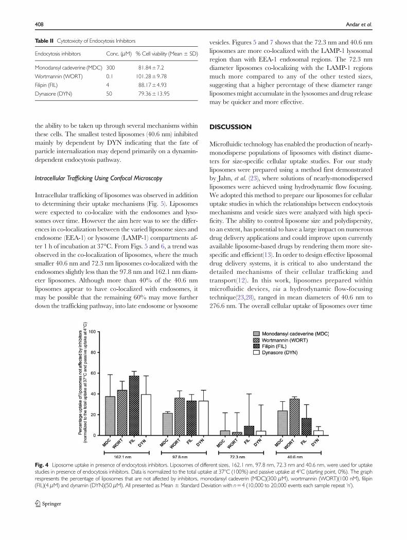
the ability to be taken up through several mechanisms withinthese cells. The smallest tested liposomes (40.6 nm) inhibitedmainly by dependent by DYN indicating that the fate ofparticle internalization may depend primarily on a dynamin-dependent endocytosis pathway.
Intracellular Trafficking Using Confocal Microscopy
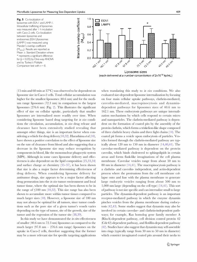
Intracellular trafficking of liposomes was observed in additionto determining their uptake mechanisms (Fig. 5). Liposomeswere expected to co-localize with the endosomes and lyso-somes over time. However the aim here was to see the differ-ences in co-localization between the varied liposome sizes andendosome (EEA-1) or lysosome (LAMP-1) compartments af-ter 1 h of incubation at 37°C. From Figs. 5 and 6, a trend wasobserved in the co-localization of liposomes, where the muchsmaller 40.6 nm and 72.3 nm liposomes co-localized with theendosomes slightly less than the 97.8 nm and 162.1 nm diam-eter liposomes. Although more than 40% of the 40.6 nmliposomes appear to have co-localized with endosomes, itmay be possible that the remaining 60% may move furtherdown the trafficking pathway, into late endosome or lysosome
vesicles. Figures 5 and 7 shows that the 72.3 nm and 40.6 nmliposomes are more co-localized with the LAMP-1 lysosomalregion than with EEA-1 endosomal regions. The 72.3 nmdiameter liposomes co-localizing with the LAMP-1 regionsmuch more compared to any of the other tested sizes,suggesting that a higher percentage of these diameter rangeliposomesmight accumulate in the lysosomes and drug releasemay be quicker and more effective.
DISCUSSION
Microfluidic technology has enabled the production of nearly-monodisperse populations of liposomes with distinct diame-ters for size-specific cellular uptake studies. For our studyliposomes were prepared using a method first demonstratedby Jahn, et al. (23), where solutions of nearly-monodispersedliposomes were achieved using hydrodynamic flow focusing.We adopted this method to prepare our liposomes for cellularuptake studies in which the relationships between endocytosismechanisms and vesicle sizes were analyzed with high speci-ficity. The ability to control liposome size and polydispersity,to an extent, has potential to have a large impact on numerousdrug delivery applications and could improve upon currentlyavailable liposome-based drugs by rendering them more site-specific and efficient(13). In order to design effective liposomaldrug delivery systems, it is critical to also understand thedetailed mechanisms of their cellular trafficking andtransport(12). In this work, liposomes prepared withinmicrofluidic devices, via a hydrodynamic flow-focusingtechnique(23,28), ranged in mean diameters of 40.6 nm to276.6 nm. The overall cellular uptake of liposomes over time
Table II Cytotoxicity of Endocytosis Inhibitors
Endocytosis inhibitors Conc. (μM) % Cell viability (Mean ± SD)
Monodansyl cadeverine (MDC) 300 81.84±7.2
Wortmannin (WORT) 0.1 101.28±9.78
Filipin (FIL) 4 88.17±4.93
Dynasore (DYN) 50 79.36±13.95
Fig. 4 Liposome uptake in presence of endocytosis inhibitors. Liposomes of different sizes, 162.1 nm, 97.8 nm, 72.3 nm and 40.6 nm, were used for uptakestudies in presence of endocytosis inhibitors. Data is normalized to the total uptake at 37°C (100%) and passive uptake at 4°C (starting point, 0%). The graphrespresents the percentage of liposomes that are not affected by inhibitors, monodansyl cadeverin (MDC)(300 μM), wortmannin (WORT)(100 nM), filipin(FIL)(4 μM) and dynamin (DYN)(50 μM). All presented as Mean ± Standard Deviation with n=4 (10,000 to 20,000 events each sample repeat ‘n’).
408 Andar et al.
(15min and 60min at 37°C) was observed to be dependent onliposome size in Caco-2 cells. Total cellular accumulation washigher for the smallest liposomes (40.6 nm) and for the medi-um range liposomes (72.3 nm) in comparison to the largestliposomes (276.6 nm) (Fig. 2). This illustrates the significanteffect of size on cellular uptake, particularly that smallerliposomes are internalized more readily over time. Whenconsidering liposome based drug targeting for in vivo condi-tions the circulation, accumulation, in vivo drug release andclearance have been extensively studied revealing thatamongst other things, size is an important factor when con-sidering a vehicle for drug delivery(10,32).Harashima et al.(10),have shown a positive correlation to the effect of liposome sizeon the rate of clearance from blood and also suggesting that adecrease in the liposome size may reduce recognition bycomponents in blood, like the mononuclear phagocytic system(MPS). Although in some cases liposome delivery and effec-tiveness is also dependent on the lipid composition (25,33,34)and surface charge or chemistry (35–37), it has been shownthat size is also a major factor determining effectiveness ofdrug delivery. When considering liposome delivery forantitumor drugs, size appears to be a major factor affectingdrug permeation into the in vivo tumor environment and localtumor tissue, where the optimal size has been shown to be inthe range of ≤100 nm (10,32). This size range has also beenshown to accumulate more within tumor tissues compared tomuch larger sizes (10). However, a liposome size of 100 nmmay not always be optimal for all tumors, since tumor condi-tions such as the pore size of a given tumor’s vessels variesdepending on the type of tumor, site of the growth, size of thetumor and the regression of the tumor site (38,39).
In this study we have demonstrated the in vitro effectivenessof smaller (40.6 nm to 72.3 nm) liposomes as compared to themuch larger (97.8 nm – 276.6 nm range) liposomes on theuptake in Caco-2 cells, therefore suggesting that the formermay be a more relevant size for specific targeting applications
when translating this study to in vivo conditions. We alsoevaluated size-dependent liposome internalization by focusingon four main cellular uptake pathways, clathrin-mediated,caveolin-mediated, macropinocytosis and dynamin-dependent pathways for liposomes sizes of 40.6 nm to162.1 nm. These endocytosis pathways are unique internali-zation mechanisms by which cells respond to certain microand nanoparticles. The clathrin-mediated pathway is depen-dent on the formation of coated pits by the assembly of theprotein clathrin, which forms a triskelion-like shape composedof three clathrin heavy chains and three light chains (14). Thecoated pit forms a vesicle upon endocytosis of particles. Ves-icles formed through the clathrin-mediated pathway are typ-ically about 120 nm to 150 nm in diameter (14,40,41). Thecaveolae-mediated pathway is dependent on the proteincaveolin, which binds cholesterol to sphingolipids in certainareas and forms flask-like invaginations of the cell plasmamembrane. Caveolae vesicles range from about 50 nm to80 nm in diameter (14,41). The macropinocytosis pathway isa clathrin- and caveolin- independent, and actin-dependentprocess where the protrusions from the cell membrane col-lapse onto and fuse with the plasma membrane to generatelarge endocytic vesicles ranging from about 500 nm to1,000 nm large (depending on the cell type) (14,41). This sortof pathway is not size specific and can internalize small to largeparticles. The dynamin-dependent pathway is an importantreceptor-mediated pathway in which the enzyme dynaminpinches vesicles from the plasma membrane during endocy-tosis (42,43). Some studies suggest that dynamin may also beinvolved in certain caveolae- and clathrin-independent path-ways; for example, Ras homolog gene family member A(RhoA)-dependent pathway, cell division control protein 42(Cdc42)-dependent pathway, and flotillin-dependent pathway(42). Studies have also suggest that dynaminmay self-assembleinto rings (typically range from 30 nm to 50 nm in diameter)which constrict invaginated coated pits around their necks to
Fig. 5 Co-localization ofliposomes with EEA-1 and LAMP-1.Intracellular trafficking of liposomeswas measured after 1 h incubationwith Caco-2 cells. Co-localizationbetween liposomes andendosomes (EEA1)/lysosomes(LAMP1) was measured usingMander’s overlap coefficient(Mx(oc)). Results are reported asMean ± Standard Deviation where* represents a significant differencefor (p<0.05) by One-way ANOVAand by Tuckey’s MultipleComparison test with n=8.
Microfluidic Liposomes for Measuring Size-Dependent Uptake 409

form budding coated vesicles (44). In our experiments weobserved that the dynamin-dependent pathway specificallyinfluenced the smaller 40.6 nm liposomes. Interestingly,72.3 nm diameter liposomes also seemed to be affected bythis pathway. Whereas the 97.8 nm to 162.1 nm liposomeswere not affected as much by this pathway compared to theother smaller sizes. The larger, 97.8 nm to 162.1 nm, were notinfluenced by any of the pathways tested as much as thesmaller 72.3 nm and 40.6 nm. The larger liposomes162.3 nm seemed to be more affected by the MDC andDYN inhibitors than the FIL and WORT inhibitors, which
may suggest that this size range may be influenced slightly bythe clathrin- and dynamin- dependent pathways.Whereas the97.8 nm liposomes were influenced by the MDC when com-pared to the other inhibitors FIL, WORT and DYN, thussuggesting that this size range may be influenced more by theclathrin-dependent pathway. In contrast, the 72.3 nm lipo-some seemed to be affected by all tested pathway inhibitors.Hence suggesting that this might be a useful size range fordrug delivery since it utilizes most of the major traffickingpathways within cells. Since chemical inhibitors have a variedeffectiveness in different cell lines and can somewhat be non-
Fig. 6 Confocal imaging of co-localization of liposomes withendosomal regions. Caco-2 cellsincubated for 1 h with DiI-C18 (red)fluorescently labeled liposomes, (a)40.6 nm, (b) 72.3 nm, (c) 97.8 nmand (d) 162.1 nm liposomes.Endosomes were stained usingAlexaFluor 488 (green) for anti EEA1antibody. These samples wereexamined by confocal microscope(Nikon A1). The nucleus wasstained with Dapi. The arrows inthe merge image point to the co-localized regions. Scale bar is 5 μm.
410 Andar et al.

specific (26), hence liposome particles may behave differentlydepending on cell type and our results may be true for Caco-2cellular uptake in specific. The intracellular trafficking exper-iments, using the confocal microscopy, shed further light onthe environment that liposomes encounter after cellular up-take. Co-localization between the liposomes of varied size withendosomal and lysosomal markers was reported in Caco-2cells. Early endosomal accumulation of 97.8 nm diameterliposomes was much greater than lysosomal accumulationafter 1 h of incubation. For the 72.3 nm liposomes after 1 hincubation they seemed to be more accumulated within
lysosomal regions than compared to endosomal regions. Asimilar trend was observed for the 40.6 nm diameter lipo-somes where there was slightly higher lysosomal accumulationof these liposomes compared to the endosomal regions.Therefore a greater amount of the 40.6 nm and 72.3 nmdiameter liposomes travel to the lysosome for degradation,which is a key step in the effective targeting and drug release(14). Thus, we have shown that total liposome uptake isdependent on size and that there are several mechanisms thatcome into play during the uptake process that may also beinfluenced by size.
Fig. 7 Confocal imaging of co-localization of liposomes withlysosomal regions. Caco-2 cellsincubated for 1 h with DiI-C18 (red)fluorescently labeled liposomes, (a)40.6 nm, (b) 72.3 nm, (c) 97.8 nmand (d) 162.1 nm liposomes.Lysosomes were stained usingAlexaFluor 488 (green) for antiLamp-1 antibody. These sampleswere examined by confocalmicroscope (Nikon A1). Thenucleus was stained with Dapi.Arrow in the merge images point tothe co-localized regions . Scale baris 10 μm.
Microfluidic Liposomes for Measuring Size-Dependent Uptake 411

Previous studies probing the cellular uptake and traf-ficking of liposomes have been based on populations ofvesicles with high levels of poly-dispersity due to thelimitations of contemporary synthesis methods (8,28),thus the ability to highlight the true fate of each indi-vidual vesicle size within the range of interest waslimited. Microfluidic mixing of chemical species providesthe unique ability to produce nearly-monodisperse pop-ulations of liposomes with distinct, varying size, this is ahighly advantageous quality for increasing specificityand efficacy while reducing toxicity of liposomes fordrug delivery applications. Microfluidic liposome prepa-ration also uses much less quantities of solution andmay be a more cost effective method for liposomeproduction(28) (problems concerning expenses for acade-mia and industry mentioned in a review by Barenholz, et al.(45)). The ability to synthesize homogenous populationsof liposomes also permits a higher level of control overthe intracellular fate of the drug carrier, thus aiding inmore sophisticated design of liposomal drug deliverysystems.
CONCLUSIONS
Here we reported a detailed study of cellular uptake mecha-nisms and intracellular trafficking related to the uptake ofliposome populations prepared by a novel microfluidic flowfocusing technique described by Jahn et al.(28). The uniqueability of microfluidic flow focusing to produce nearly-monodispersed populations of distinct sizes is highly advanta-geous for increasing the specificity for drug delivery applica-tions. We found that the liposome uptake is size dependentand increases with a decrease in diameter. The mechanismsinvolved in the uptake of liposomes are also dependent on thesizes, where the smaller 40.6 nm liposomes seem to depend ona dynamin dominant pathway. However the slightly largerdiameter liposomes (97.8 nm to 162.1 nm), when compared tothe smaller sizes, did not show any significant dependency onany particular uptake pathways that were tested, but when thelarger liposomes were compared among themselves theyshowed slight dependency on the clathrin-mediated pathway.The uniqueness observed in this study was the intermediatesize range of 72.3 nm that seemed to be dependent on almostall the tested mechanisms. Also this size range showed themost acculmulation within the lysosomal regions compared tothe other tested liposome diameters. In this study we foundthat size plays a big role in facilitating the recognition ofparticles by a particular mechanism. Knowledge of detailedmechanisms of liposome sizes was better facilitated with theuse of microfluidic flow focusing and this may assist in design-ing better liposome populations for drug delivery for variousapplications in the future.
ACKNOWLEDGMENTS AND DISCLOSURES
Certain commercial equipment, instruments, or materials areidentified in this paper to foster understanding. Such identifi-cation does not imply recommendation or endorsement bythe National Institute of Standards and Technology, nor doesit imply that the materials or equipment identified are neces-sarily the best available for the purpose.
Financial support was provided by the Department ofEnergy (DOE), grant number DE-FG02-08CH11527. CathyStorer, B.S., Regina Harley, M.S. and Marcelo Sztein, M.D.from the Flow Cytometry Core Facilities, Centre for VaccineDevelopment, School of Medicine, University of Maryland,Baltimore, USA.
REFERENCES
1. Torchilin VP. Recent advances with liposomes as pharmaceuticalcarriers. Nat Rev Drug Discov. 2005;4(2):145–60.
2. Allen TM. Liposomes - opportunities in drug delivery. Drugs.1997;54(Supplement 4):8–14.
3. StormG, Crommelin DJ. Liposomes: quo vadis? Pharm Sci TechnolToday. 1998;1(1):19–31.
4. Lasic DD. Novel applications of liposomes. Trends Biotechnol.1998;16(7):307–21.
5. Gabizon A, Shmeeda H, Barenholz Y. Pharmacokinetics ofpegylated liposomal Doxorubicin: review of animal and humanstudies. Clin Pharmacokinet. 2003;42(5):419–36.
6. Barenholz Y. Doxil®–the first FDA-approved nano-drug: lessonslearned. J Control Release: Off J Control Release Soc.2012;160(2):117–34. Elsevier B.V.
7. Gregoriadis G. Engineering liposomes for drug delivery: progressand problems. Trends Biotechnol. 1995;13(12):527–37.
8. Edwards KA, Baeumner AJ. Liposomes in analyses. Talanta.2006;68(5):1421–31.
9. Litzinger DC, Buiting AMJ, Van Rooijen N, Huang L. Effectof liposome size on the circulation time and intraorgan distri-bution of amphipathic poly(ethylene glycol)-containing lipo-s ome s . B i o ch im B iophy s Ac t a (BBA ) B i omembr .1994;1190(1):99–107.
10. Nagayasu A,UchiyamaK,KiwadaH. The size of liposomes: a factorwhich affects their targeting efficiency to tumors and therapeuticactivity of liposomal antitumor drugs. Adv Drug Deliv Rev.1999;40(1–2):75–87.
11. Lee R, Low P. Delivery of liposomes into cultured KB cells via folatereceptor-mediated endocytosis. J Biol Chem. 1994;259(5):3196–204.
12. Petros RA, DeSimone JM. Strategies in the design of nanoparticlesfor therapeutic applications. Nat Rev Drug Discov. 2010;9(8):615–27. Nature Publishing Group.
13. Wang J, Byrne JD, Napier ME, DeSimone JM. More effectivenanomedicines through particle design. Small. 2011;7(14):1919–31.WILEY-VCH Verlag.
14. Hillaireau H, Couvreur P. Nanocarriers’ entry into the cell:relevance to drug delivery. Cell Mol life Sci: CMLS.2009;66(17):2873–96.
15. Allen TM, Cullis PR. Drug delivery systems: entering the main-stream. Science (New York, NY). 2004;303(5665):1818–22.
16. Wagner A, Vorauer-Uhl K, Kreismayr G, Katinger H. Thecrossflow injection technique: an improvement of the ethanol injec-tion method. J Liposome Res. 2002;12(3):259–70.
412 Andar et al.
17. Olson F, Hunt CA, Szoka FC, Vail WJ, Papahadjopoulos D. Prep-aration of liposomes of defined size distribution by extrusion throughpolycarbonate membranes. Biochim Biophys Acta (BBA) Biomembr.1979;557(1):9–23.
18. Szoka F Jr, Papahadjopoulos D. Comparative properties andmethods of preparation of lipid vesicles (liposomes). Annu RevBiophy. 1980;9:467–508.
19. Batzri S, Korn ED. Single bilayer liposomes prepared without sonica-tion. Biochim Biophys Acta (BBA) Biomembr. 1973;298(4):1015–9.
20. Kremer J. Vesicles of variable diameter prepared by a modifiedinjection method. Biochemistry. 1977;16(17):3932–5.
21. Jahn A, Stavis SM, Hong JS, Vreeland WN, DeVoe DL, Gaitan M.Microfluidic mixing and the formation of nanoscale lipid vesicles.ACS Nano. 2010;4(4):2077–87.
22. Jahn A, Reiner JE, Vreeland WN, DeVoe DL, Locascio LE, GaitanM. Preparation of nanoparticles by continuous-flow microfluidics. JNanopart Res. 2008;10(6):925–34.
23. Jahn A, Vreeland WN, Gaitan M, Locascio LE. Controlled vesicleself-assembly in microfluidic channels with hydrodynamic focusing. JAm Chem Soc. 2004;126(9):2674–5.
24. Hood R, Shao C, Omiatek D, Vreeland W, DeVoe D. Microfluidicsynthesis of PEG- and folate-conjugated liposomes for one-step for-mation of targeted stealth nanocarriers. Pharm Res . Springer US;2013;1–11.
25. Allen TM, Austin GA, Chonn A, Lin L, Lee KC. Uptake of liposomesby cultured mouse bone marrow macrophages: influence of liposomecomposition and size. Biochim Biophys Acta. 1991;1061(1):56–64.
26. Goldberg DS, Ghandehari H, Swaan PW. Cellular entry of G3.5poly (amido amine) dendrimers by clathrin- and dynamin-dependentendocytosis promotes tight junctional opening in intestinal epithelia.Pharm Res. 2010;27(8):1547–57.
27. Pollock S, Antrobus R, Newton L,Kampa B, Rossa J, Latham S, et al.Uptake and trafficking of liposomes to the endoplasmic reticulum.FASEB J: Off Publ Fed Am Soc Exp Biol. 2010;24(6):1866–78.
28. Jahn A, Vreeland WN, DeVoe DL, Locascio LE, Gaitan M.Microfluidic directed formation of liposomes of controlled size. Lang-muir: ACS J Surf Colloids. 2007;23(11):6289–93.
29. Straubinger RM, Hong K, Friend DS, Papahadjopoulos D. Endo-cytosis of liposomes and intracellular fate of encapsulated molecules:encounter with a low pH compartment after internalization in coatedvesicles. Cell. 1983;32(4):1069–79.
30. Kamps J, Scherphof G. Receptor versus non-receptor mediatedclearance of liposomes. Adv Drug Deliv Rev. 1998;32(1–2):81–97.
31. Hong K, Yoshimura T, Papahadjopoulos D. Interaction of clathrinwith liposomes: pH-dependent fusion of phospholipid membranesinduced by clathrin. FEBS Lett. 1985;191(1):17–23.
32. Drummond DC,Meyer O, Hong K, Kirpotin DB, PapahadjopoulosD. Optimizing liposomes for delivery of chemotherapeutic agents tosolid tumors. Pharm Rev. 1999;51(4):691–743.
33. Barza M, Stuart M, Szoka F. Effect of size and lipid composition onthe pharmacokinetics of intravitreal liposomes. Invest OphthalmolVis Sci. 1987;28(5):893–900.
34. Mayer LD, Tai LC, Ko DS, Masin D, Ginsberg RS, Cullis PR, et al.Influence of vesicle size, lipid composition, and drug-to-lipid ratio onthe biological activity of liposomal doxorubicin in mice. Cancer Res.1989;49(21):5922–30.
35. Souhami RL, Patel HM, Ryman BE. The effect of reticuloendothe-lial blockade on the blood clearance and tissue distribution of lipo-somes. Biochim Biophys Acta (BBA) Gen Subj. 1981;674(3):354–71.
36. Lee R, Low P. Folate-mediated tumor cell targetting of liposome-entrapped doxorubicin. Biochim Biophys Acta (BBA) Biomembr.1995;1233:134–44.
37. Koren E, Apte A, Jani A, Torchilin VP. Multifunctional PEGylated2C5-immunoliposomes containing pH-sensitive bonds and TAT pep-tide for enhanced tumor cell internalization and cytotoxicity. J ControlRelease: Off J Control Release Soc. 2012;160(2):264–73. Elsevier B.V.
38. Hobbs SK, MonskyWL, Yuan F, Roberts WG, Griffith L, TorchilinVP, et al. Regulation of transport pathways in tumor vessels: role oftumor type and microenvironment. Proc Natl Acad Sci U S A.1998;95(8):4607–12.
39. Hirano A, Matsui T. Vascular structures in brain tumors. HumanPathol. 1975;6(5):611–21.
40. Bareford L, Swaan P. Endocytic mechanisms for targeted drugdelivery. Adv Drug Deliv Rev. 2007;59(8):748–58.
41. Wang J, Byrne J, Napier M, DeSimone J. More effectivenanomedicines through particle design. Small. 2011;7(14):1919–31.
42. Sever S. Dynamin and endocytosis. Curr Opin Cell Biol.2002;14:463–7.
43. Gu C, Yaddanapudi S, Weins A, Osborn T, Reiser J, Pollak M, et al.Direct dynamin-actin interactions regulate the actin cytoskeleton.EMBO J. 2010;29(21):3593–606. Nature Publishing Group.
44. Hinshaw JE, Schmid SL. Dynamin self-assembles into ringssuggesting a mechanism for coated vesicle budding. Nature. 1995.p. 190–2.
45. Barenholz Y. Liposome application: problems and prospects. CurrOpin Colloid Interface Sci. 2001;6(1):66–77.
Microfluidic Liposomes for Measuring Size-Dependent Uptake 413