microfluidic devices for drug discovery and analysis microfluidic drug.pdf · microfluidic devices...
TRANSCRIPT

© Woodhead Publishing Limited, 2013
231
7 Microfluidic devices for drug discovery
and analysis
J. S. KOCHHAR , S. Y. CHAN and P. S. ONG,
National University of Singapore, Singapore, W. G. LEE ,
Kyung Hee University, Republic of Korea and L. KANG, National
University of Singapore, Singapore
DOI : 10.1533/9780857097040.2.231
Abstract : Microdevices, since their inception in the last decade of the twentieth century, have changed our view of science, due to their potential applications in fi elds ranging from optics, semiconductors and the microelectronics industry to drug discovery and development, point-of-care clinical diagnostics, sensitive bioanalytical systems and other areas of the biological sphere. In this chapter we review the potential applications of microfl uidic platforms for drug discovery applications comprising high-throughput screening in target selection, lead identifi cation/optimization and preclinical testing. Application of microfl uidics in chemical analysis, as well as analysis of metabolites in blood for studying pathology, is also discussed.
Key words : microfl uidics, drug discovery, high-throughput screening, drug analysis, point-of-care diagnostics.
7.1 Introduction
The ascent of microfabrication research and development at the turn of the
century has opened several avenues for the biomedical sector. Micron-scale
chips, with micrometre dimension channels, can be used to manipulate fl uid
fl ow at the micron/submicron scale. The spatial control offered by this tech-
nology, known as ‘microfl uidics’, has potential applications in handling, pro-
cessing and analysis of fl uids (Whitesides, 2006). The miniaturized scale of
these devices requires lower sample volumes (in nanolitres) than conven-
tional microplate assays (requiring hundreds of microlitres), hence making
them economical alternatives. The minute dimensions of the devices offer
shorter diffusion path lengths, allowing faster analysis and precise control
of fl uid fl ow, and leading to specifi city in chemical microreactors. Design
manipulation, easily achievable by conventional lithographical and novel

232 Microfl uidic devices for biomedical applications
© Woodhead Publishing Limited, 2013
nanotechnology techniques, provides versatility in mixing fl uids that can be
controlled by external physical forces such as magnetic and electric fi elds.
These microdevices may either be integrated into existing macrodevices, or
constitute comprehensive analytical systems by themselves. The miniaturi-
zation provided by these high-throughput devices allows for a large number
of replicates on a small chip, enabling massive parallelization and thereby
increasing effi ciency and lowering costs (Lombardi and Dittrich, 2010). The
fl uid fl ow properties at microscale are very different from those at macro-
scale, and this can be exploited using microfl uidic devices (Beebe et al ., 2002).
These advantages make them an ideal choice in disciplines spanning across
molecular analysis, biodefence programmes, and the discovery and develop-
ment of new drugs in the pharmaceutical and biotechnological industries.
Initially, the concept of microfl uidics was applied to the fi eld of analyt-
ical chemistry. Lithographic patterning/etching, used to produce chemical
sensors and analytical techniques on glass/silicon substrates, provided proof
of concept for their applicability (Harrison et al ., 1993; Manz et al ., 1990).
Afterwards, chemical and biological sensors that could thwart the threats of
bioterrorism, and aid in biodefence sample testing (Liszewski, 2003), were
developed. With the surge in biotechnological methods, proteomics, genom-
ics, and the discovery of protein-based therapeutics, microfl uidics offers
brighter prospects in DNA sequencing and genotyping as well as in pro-
tein separation and analysis (Chen et al ., 2010; Gomez, 2011). Microfl uidic
devices have also provided valuable opportunities for drug discovery
and development processes, with their benefi ts at each stage from target
identifi cation (Malmstadt et al ., 2006) to lead identifi cation/optimization
(Jones et al ., 2005), and further to preclinical studies (Matsui et al ., 2006),
clinical trials (Herr et al ., 2007), formulation development (Alsenz and
Kansy, 2007) and the manufacturing stage (Szita et al ., 2005). Additionally,
these devices have been used for improved confi nement of cells in three-
dimensional (3D) scaffolds, cell-based testing and cell component analy-
sis. The cellular and molecular interactions at a scale proportional to their
dimensions (Whitesides, 2003) are very different from those observed at
macroscale volumes. An interesting application has been in the fi eld of tis-
sue engineering, whereby microfl uidic platforms provide 3D scaffolds mim-
icking the natural environment for growth and mutual interaction between
cells (Li et al ., 2012; Yamada et al ., 2012). They have also been investigated
for transdermal and pulmonary delivery of drugs (Ashraf et al ., 2011; Yeo
et al ., 2010), as well as for personalized diagnostic kits (Yager et al ., 2006).
In this chapter we will present an overview of the microfl uidic devices
that have been researched for drug discovery and drug analysis. First, we
discuss the role played by microfl uidics in the current paradigm for drug
discovery, in identifying druggable targets, and in the progress achieved by
high throughput screening (which has allowed thousands of molecules to be

Microfl uidic devices for drug discovery and analysis 233
© Woodhead Publishing Limited, 2013
screened on a chip), followed by optimizing few lead molecules and assess-
ing their pharmacokinetic and pharmacodynamic properties in preclinical
systems. Later, we discuss the application of microfl uidic devices in chemical
analysis.
7.2 Microfluidics for drug discovery
Discovering new therapeutics for a pathophysiological condition involves
identifying a specifi c target (Kang et al ., 2008). With the help of computa-
tional biology and/or experimental methods, such targets can be identifi ed.
This is followed by validating the target by a series of complicated cell-based
or animal experiments. Once validated, screening of drug libraries, pro-
duced by combinatorial chemistry, composed of millions (usually >10 6 com-
pounds) of drug molecules to fi nd a few lead molecules for clinical trials, is
carried out. This is aimed at getting the safest, most reliable and effi cacious
pharmaceutical compound, which is then fi led as a new drug application
for approval by regulatory agents such as the United States Food and Drug
Administration (FDA). The complex and lengthy procedure of discovering
a suitable drug candidate is exemplifi ed by the fact that it takes 10–15 years
for a drug to go from bench to bedside, and it has been estimated to cost
approximately 1 billion USD (Wu et al ., 2010). The attrition, from thousands
of new chemical structures in the drug library, to a few lead compounds, to
a single successful therapeutic agent, is a result of the ineffi cient procedures
used in the conventional/current drug discovery and development process.
Progress in the use of microscale platforms aids the process of drug dis-
covery through effi cient and expeditious design of therapeutics and pro-
vision of information on biological targets (Lal and Arnsdorf, 2010).
High-throughput microfl uidic devices have shown considerable promise
over the conventional methods, which required long processing times and
expensive equipment, thereby delaying the whole drug discovery process.
In the following sections, we describe the contribution of microfl uidics to
various segments of drug discovery.
7.2.1 Identifi cation of druggable targets
The process of drug discovery begins with the identifi cation of the func-
tion of a potential drug target and comprehending its role in the disease
process. Discovering pharmacological activities was conventionally carried
out by testing various substances (usually plant extracts) in living organ-
isms to observe the changes caused in a particular phenotype. However,
towards the end of the twentieth century, this process of phenotype-based
target identifi cation was largely replaced by a target-based approach. With
progressive acquisition of knowledge in the fi eld of molecular biology and
234 Microfl uidic devices for biomedical applications
© Woodhead Publishing Limited, 2013
improvement in isolation techniques, identifi cation of complex systems
that are responsible for a drug’s pharmacological response has evolved to
be the new approach in identifi cation of drug targets and it has reduced the
use of living organisms and living tissues (Terstappen et al ., 2007).
Drug targets, which may be a cellular receptor, an ion channel, nucleic
acids, DNA or RNA, enzymes, polysaccharides and lipids, are chemically
well-defi ned molecular structures capable of interacting with therapeutic
drug moieties (Imming et al ., 2006). This interaction leads to downstream
clinical effects. Common drug targets belong to the classes of kinases, pro-
teases, phosphatases and G protein coupled receptors. Ion channel proteins
represent another attractive target in the drug discovery paradigm, as they
have been implicated in several cardiovascular and neuronal disorders
(Dunlop et al ., 2008). Around 40% of targets in drug discovery belong to
the class of ligand-gated ion channels (Yin et al ., 2008).They act as the main
targets for the currently available pharmaceutical agents, as well as majority
of those agents in the drug development phase, and hence have been the
focus of intense research resulting in dedicated conferences and numerous
publications (Perrin et al ., 2006; Zagnoni, 2012).
As most of these targets are a part of the cell membrane lipid bilayer
structure, their functionality depends on the membrane integrity. The
proteins may be denatured once dissociated from the membrane, and
hence are required to be integrated into the membrane throughout the
analytical procedure (Suzuki et al ., 2004). Target validation employing
isolated membrane proteins and ion channels offers many technologi-
cal challenges, as reproducing these nano-scale systems is very complex
(2007). However, incorporating these drug targets in artifi cially synthe-
sized lipid bilayer membranes, and by specifi cally controlling the mem-
brane architecture and surface characteristics, simulating the natural
environment of a drug target is envisaged as an option for target identi-
fi cation (Zagnoni, 2012).
Microfl uidic technology has played a key role in the fabrication of bilayer
lipid membranes (BLM) (Mayer et al ., 2003). Micron-sized BLMs with
integrated membrane proteins and ion channels are advantageous over
macro-systems, providing economical and time saving analysis platforms.
These BLMs bear remarkable electric sealing, and hence are amenable to
recording electrical signals across single membrane protein. On-chip pla-
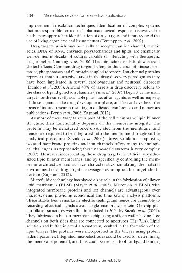
nar bilayer structures were fi rst introduced in 2004 by Suzuki et al . (2004).
They fabricated a bilayer membrane chip using a silicon wafer having fl ow
channels on both sides that are connected to apertures (Fig. 7.1a). Lipid
solution and buffer, injected alternatively, resulted in the formation of the
lipid bilayer. The proteins were incorporated in the bilayer using protein
laden liposomes. Integrated microelectrodes could be used for determining
the membrane potential, and thus could serve as a tool for ligand-binding

Microfl uidic devices for drug discovery and analysis 235
© Woodhead Publishing Limited, 2013
studies. However, silicon-based devices suffer from many disadvantages,
including high dielectric loss of silicon leading to high electrical noise. Apart
from that, the manufacture of silicon-based devices is time consuming, and
the reproducibility of the BLMs is questionable. Other materials used for
fabrication include epoxy photoresist (Cheng et al ., 2001), glass (Fertig et al ., 2002) and Tefl on (Mayer et al ., 2003), but the resultant BLMs were fragile
and unstable.
Polymeric microfl uidic devices have the potential to overcome these
drawbacks, offering advantages of economy and ease of fabrication. Poly
(methyl methacrylate) (PMMA) has been seen as viable alternative, due
Taperedapertures
Electrodes (B)Channel (B)
GlassSiliconGlass
Out (B)
Out (U)
Planar lipid bilayer
Glass
Glass
Upperchannel
Lowerchannel
Membraneprotein
Micro-electrode
Channel (U)
Electrodes (U)
Buffer in (U)(a)Buffer in
(B)Lipid in
(U)
Flow Buffer
Buffer
Patch-clampamplifier
(b) 1 2
3 4
BLMEdge
7.1 Formation of bilayer lipid membranes (BLM) on microfl uidic chips.
(a) Conceptual diagram of a membrane fl uid chip having fl uid channels
and apertures. Alternate fl ow of lipid and buffer solutions leads to
formation of BLMs. (Suzuki et al ., 2004). (b) A microfl uidic device with
a channel extending from a trench, where electrodes are inserted in
both the upper well (containing lipid) and the lower channel (containing
buffer). The bilayer is formed within an aperture upon exposure to air
(left), the growth of which is monitored over 20 s (right). The setup was
placed over a microscope to observe BLM formation that appeared as a
bright region in the centre (Sandison et al ., 2007).
236 Microfl uidic devices for biomedical applications
© Woodhead Publishing Limited, 2013
to its good optical and dielectric properties, low glass transition tempera-
ture, ease of processing, and ability to bond other materials, unlike Tefl on
(Sandison et al ., 2007). Suzuki et al . modifi ed their previous silicon-based
design, to make a PMMA-based device providing a tapered aperture for
lipid fl ow, and hence achieve a constant amount of lipid solution at the aper-
ture. Further application of a static pressure to control fi lm thickness yielded
a more reproducible (90%) bilayer. With further optimization, embedding
of four lipid bilayers on a single chip and gramicidin peptide, a monovalent
cation channel, incorporated into the bilayer, was achieved (Suzuki et al ., 2006). One of the unique advantages of this microfl uidic device is that it
facilitates easy microscopic observation of the bilayer (Suzuki et al ., 2007).
Sandison et al . created microfl uidic channels on PMMA-coated glass sub-
strates by using hot embossing and laser micromachining (Fig. 7.1b). PMMA
surface was chemically treated to render it hydrophobic. Lower channel was
fi lled with buffer, and lipid solution was applied to the upper well, which was
later fi lled with the buffer. Lipid bilayers could be achieved by exposure of
the top surface to air (Sandison et al ., 2007).
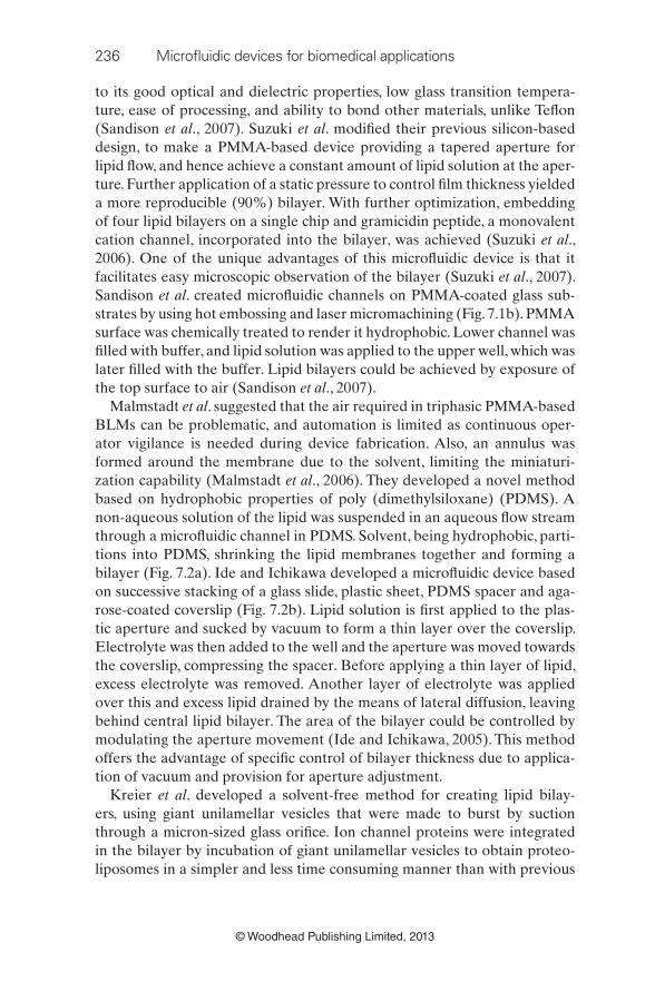
Malmstadt et al . suggested that the air required in triphasic PMMA-based
BLMs can be problematic, and automation is limited as continuous oper-
ator vigilance is needed during device fabrication. Also, an annulus was
formed around the membrane due to the solvent, limiting the miniaturi-
zation capability (Malmstadt et al ., 2006). They developed a novel method
based on hydrophobic properties of poly (dimethylsiloxane) (PDMS). A
non-aqueous solution of the lipid was suspended in an aqueous fl ow stream
through a microfl uidic channel in PDMS. Solvent, being hydrophobic, parti-
tions into PDMS, shrinking the lipid membranes together and forming a
bilayer (Fig. 7.2a). Ide and Ichikawa developed a microfl uidic device based
on successive stacking of a glass slide, plastic sheet, PDMS spacer and aga-
rose-coated coverslip (Fig. 7.2b). Lipid solution is fi rst applied to the plas-
tic aperture and sucked by vacuum to form a thin layer over the coverslip.
Electrolyte was then added to the well and the aperture was moved towards
the coverslip, compressing the spacer. Before applying a thin layer of lipid,
excess electrolyte was removed. Another layer of electrolyte was applied
over this and excess lipid drained by the means of lateral diffusion, leaving
behind central lipid bilayer. The area of the bilayer could be controlled by
modulating the aperture movement (Ide and Ichikawa, 2005). This method
offers the advantage of specifi c control of bilayer thickness due to applica-
tion of vacuum and provision for aperture adjustment.
Kreier et al . developed a solvent-free method for creating lipid bilay-
ers, using giant unilamellar vesicles that were made to burst by suction
through a micron-sized glass orifi ce. Ion channel proteins were integrated
in the bilayer by incubation of giant unilamellar vesicles to obtain proteo-
liposomes in a simpler and less time consuming manner than with previous
Microfl uidic devices for drug discovery and analysis 237
© Woodhead Publishing Limited, 2013
Analyte solution
organicphase
Solventextraction
Solventextraction
PDMS channelwalls
(a) (b)(i)
(ii)
(ii)(iii)
(iii) (iv)
(v)(iv)
(i)
Lipidmolecule Organic
Aqueous
Aqueousphase
Lipid solution
Organic phase
7.2 Formation of bilayer lipid membranes (BLM). (a) By microfl uidic
solvent extraction, (i) droplet of organic solvent with dissolved lipid
is formed in an aqueous stream of fl uid. Lipids are organized on the
hydrophobic – hydrophilic interface (inset). (ii) As solvent enters the
PDMS, the two interfaces approach one another. (iii) Finally, only
the lipid layers are left behind, forming a bilayer membrane. (iv)
Images showing solvent extraction from a lipid solution droplet in a
microfl uidic channel, over a period of time (minutes:seconds), the BLM,
although not visible in the last image, was formed and confi rmed by
electrical measurements (Malmstadt et al ., 2006). (b) By microfl uidic
bilayer chamber method, (i) A drop of electrolyte was applied to the
well of spacer. (ii) A plastic sheet was placed on the spacer and moved
downward until the aperture hit the bottom. Then excess electrolyte
was removed with a pipette. (iii) Small amount of lipid solution and a
sample solution were added sequentially. Alternatively, lipid solution
was sprayed through a fi ne pipette to the edge of the aperture with
bubbling without removing the electrolyte in (ii). (iv) After formation
of a thick membrane across the aperture, the plastic sheet was moved
upwards. The membrane expanded, reached the agarose layer and
thinned to form a bilayer. (v) Successive bright-fi eld images of BLM
formation (Ide and Ichikawa, 2005).

238 Microfl uidic devices for biomedical applications
© Woodhead Publishing Limited, 2013
techniques. Typical gating phenomena were observed by changes in pH and
membrane voltage in the outer membrane protein OmpF obtained from
Escherichia coli (Kreir et al ., 2008). Chip-based bilayers have been used for
bacterial toxin binding studies. Using total internal refl ection fl uorescence
microscopy, cholera toxin B subunit and tetanus toxin C fragment could be
detected as low as 100 pM (Moran-Mirabal et al ., 2005). It was suggested
that this method is adaptable for proteins and nucleic acids as well.
These techniques to fabricate BLMs in vitro provide a good platform to
identify ion channel proteins as drug targets. Also, once identifi ed, these tar-
gets can then be used to screen new therapeutic agents and identify lead
compounds for preclinical studies. They can also be used for determination
of membrane properties under non-physiological conditions and gain access
to ion channels in intracellular membranes (Kreir et al ., 2008).
Cellular receptors and the downstream signal transduction pathways
are being increasingly recognized to play a critical role in drug action and
astounding progress has been made in characterizing their behaviour. Signal
transduction has also been enormously researched with many companies
having dedicated programmes for signal transduction based drug discov-
ery (Anonymous, 2000). Enzymes such as tyrosine kinase play an impor-
tant role in phosphorylating proteins, forming the essential links in signal
transduction pathways (Wang et al ., 2008). Wang et al . recently developed
a novel microfl uidic device combining the function of electroporation and
fl ow cytometry to measure the translocation of fl uorescently tagged tyrosine
kinase to the cell membrane, at a single cell level. It was demonstrated that
cells stimulated through antigen receptor retained more kinase than their
non-stimulated counterparts. These results could have a marked impact on
target-based drug discovery, as kinases are frequently involved in common
diseases such as cancer (Wang et al ., 2008).
Analysis of protein molecules from a single cell has been envisaged as a
potential tool to identify specifi c targets. Recently, single cell analysis has
gained considerable importance in microfl uidics-based drug discovery, as
these devices are able to perform manipulation, lysis, labelling, separation
and quantifi cation of the protein contents in a single cell (Huang et al ., 2007).
Although this technique is not amenable to live cell monitoring, it provides
for simultaneous detection of multiple targets, combining higher sensitivity
with higher throughput. Using a single cell analysis chip, the number of β 2
adrenergic receptors was determined. The integrated microfl uidic chip facil-
itated cell and chemical handling, cell lysis, electrophoretic separation, and
detection of lysate using laser-induced fl uorescence (Gao et al ., 2004; Wu
et al ., 2004). Separation of proteins and peptides has also been achieved on
miniaturized electrophoretic cells (Schulze and Belder, 2012; Sikanen et al ., 2012). Some of these techniques have been dealt with in greater detail in the
subsequent section on analysis.

Microfl uidic devices for drug discovery and analysis 239
© Woodhead Publishing Limited, 2013
Understanding of the interactions between receptors and their ligands
provide insightful information on disease progression, and exploration of
such drug – receptor pairs provides us an opportunity to discover drugs selec-
tively targeting a particular receptor (Goldberg et al ., 2009). Modulation of
physiological events, such as cell differentiation and death, release of neu-
rotransmitters and hormones, is a result of activation/suppression of signal
transduction pathways, which are often coupled to cellular receptors. This
activation/suppression is in turn due to binding of specifi c ligands to these
receptors. Much of the research work in discovering new receptor ligands
has been focused on binding studies of low molecular weight molecules to
macromolecular receptors, followed by screening of biochemical changes.
However, it has been reported that lack of a particular biochemical event
does not necessarily translate into lack of receptor activation. Other cellular
components and events, such as second messengers, downstream processes,
gene transcription and change in receptor confi guration, have to be looked
into. This, however, is not possible with the conventional assay procedures
(Gurwitz and Haring, 2003). High-throughput ligand-binding assays provide
a suitable alternative to perform multiple tasks on a small chip. Moreover,
the discovery of many new ‘orphan’ receptors, for which no ligands are cur-
rently known, offers a promising avenue for drug discovery.
Microfl uidic devices are benefi cial for ligand-binding studies, as they
reduce interaction times, enhance sensitivity and throughput (Kang et al ., 2008), and aid in separation of complexed and uncomplexed molecules
(Bange et al ., 2005). For these binding studies, receptor or ligand molecules
can be immobilized on a PDMS substrate by adsorption (Makamba et al ., 2005), or covalent bonding (Sui et al ., 2006), or by microcontact printing
as achieved for solution hybridized oligonucleotides (Razumovitch et al ., 2009). These binding interactions are usually quantifi ed by the measurement
of equilibrium dissociation constant ( K d ) of the ligand – receptor complex.
Goldberg et al . demonstrated the interaction of glycopeptide antibiot-
ics, teicoplanin and vancomycin, immobilized on a PDMS microchannel
with 5-carboxyfl uorescein- d -Ala- d -Ala- d -Ala (5-FAM-(DA) 3 ). The K d
was reported to be similar to previously reported values as measured by
commercial systems, even though it utilized a smaller amount of reagents
(Goldberg et al ., 2009). Centrifugal microfl uidic platforms, which are disc-
shaped microfl uidic devices, have also been developed whereby the fl uid
fl ows by simple rotation of the disc. Interaction between phenothiazine anti-
depressants and calmodulin, attached to a green fl uorescent protein, was
studied. Drug binding affected the fl uorescence properties, and hence con-
centration of the drug bound to the protein receptor could be determined
(Puckett et al ., 2004). The BLMs described earlier have been used exten-
sively for ligand-binding studies over the past two decades. Recently, phos-
pholipid bilayers were patterned with bovine serum albumin by lithography.

240 Microfl uidic devices for biomedical applications
© Woodhead Publishing Limited, 2013
Following repeated cycles of patterning, ganglioside GM1 was coated along
the microfl uidic channels in different concentrations, and its interaction
with varying concentrations of cholera toxin B was studied (Shi et al ., 2008).
Javanmard et al . demonstrated a novel method of coupling a microfl uidic
device with shear force spectroscopy to study the interaction between pro-
tein molecules and DNA base pairs. The method could be used to measure
the affi nity of bonding between the interacting molecules by measuring the
drag force required to detach the ligand bound to the microfl uidic channel
when the receptor attached to the surface of microbeads is pressure driven
through these channels (Javanmard et al ., 2010).
7.2.2 Hit identifi cation and lead optimization
After the identifi cation of a particular druggable target, the next step in
the drug discovery process is to identify a ‘hit’, which involves the phases of
hit identifi cation (HI), lead identifi cation (LI), and lead optimization (LO).
A ‘hit’ is a particular chemical or biological moiety that binds to a specifi c
target which has been implicated in an ailment. Screening and optimization
of millions of ‘hits’ results in several ‘lead’ compounds. This whole multi-
phase process, in which ‘leads’ are optimized by an initial screening involv-
ing multiple ‘hits’, is described as a ‘hit-to-lead’ process (Goodnow, 2006 ).
Synthesizing and screening the right drugs which can potentially be used,
carried forward through a drug development programme, and enter a clinic,
starts from correct identifi cation of hits and leads. These steps are impera-
tive, since drug discovery is an expensive process. An error at this stage may
lead to an expensive failure at a later stage.
Drug candidates may either be derived from combinatorial libraries or be
of natural origin. Drug libraries have been estimated to be in the order of
10 63 (Bohacek et al ., 1996). Microfl uidic chip-based combinatorial chemistry
and high-throughput screening, together aim to result in a paradigm shift,
leading to the development of methods of sequential synthesis and testing
of thousands of compounds in parallel (Knight, 2000).
Synthesis of drug libraries
Recognition of drug targets has kept pace with the fast progress in genomic
and proteomic tools. Pharmaceutical companies on the other hand are fac-
ing challenges to generate drug compounds at the fastest possible rate, in
an inexpensive manner. Synthesis of drug libraries has been described as
the biggest impediment in the drug discovery process (Jones et al ., 2005).
Improved methods in combinatorial chemistry have resulted in rapid syn-
thesis of large number of chemical compounds, and have produced enor-
mous drug libraries. This has been further accelerated by the improvement

Microfl uidic devices for drug discovery and analysis 241
© Woodhead Publishing Limited, 2013
in the design of the microfl uidic reactors. These microfl uidic reactors can be
classifi ed into three types, based on the fl ow pattern, namely (i) fl ow-through
type, (ii) droplet or slug type, and (iii) batch type. The most common fl ow-
through type enables multiple reagents to be maintained at a temperature,
and be pressure driven through the channels. These reactors have been used
widely in extraction procedures as well as in multiple chemical syntheses
(Keng et al ., 2012).
Parallel combinatorial synthesis in multiple microfl uidic reactors has also
been demonstrated utilizing the continuous fl ow of reagents in microfl uidic
channels. A multiple microfl uidic reactor assembly was fabricated to synthe-
size carbamates in a multistep procedure (Sahoo et al ., 2007). However, this
method sacrifi ces the advantages of an integrated system for several reac-
tions to be carried out on a single chip. Researchers then looked to fabricate
a consolidated device with multiple layers of parallel chips. A multilayer
glass chip was developed for a 2 × 2 series synthesis in parallel (Kikutani
et al ., 2002). The complexity and expense of fabrication of this multi-lay-
ered device was a concern. Recently, Dexter and Parker exhibited parallel
combinatorial synthesis of compounds on a single-layered microfl uidic chip
(Fig. 7.3a). They fabricated a single layer PDMS chip for synthesizing a 2 × 2
series of amide formation products (Dexter and Parker, 2009).
However, continuous fl ow reactors are not suitable for multistep reac-
tions, especially those involving sequential synthesis. A modifi ed technique,
termed batch microfl uidics, in which specifi c microvalves control the deliv-
ery of reagents in batches, has been developed. These isolated batches can
be delivered to the microfl uidic reactor chamber at specifi c time points in a
reaction cycle, exercising greater control over the reaction (Lee et al ., 2005).
A fl uoride radiolabelled imaging probe, in nano/microgram scale, was syn-
thesized in fi ve sequential processes involving fl uoride concentration, water
evaporation, radiofl uorination, solvent exchange, and hydrolytic deprotec-
tion (Fig. 7.3b).
A newer technology, known as droplet microfl uidics, has recently come
to the fore. It is based on compartmentalization of each assay in a small
droplet, usually in the range of 1 pL–10 nL, surrounded by an immiscible
oil, which can be manipulated and processed in a high-throughput manner
(Brouzes, 2012). Each of these droplets can act as a tiny microfl uidic reactor,
notably reducing the reagent volumes required. A mesh-grid design microw-
ell array was fabricated by Um et al ., which allows for continuous addition
and trapping of picolitre single cell droplets in the microwells (Fig. 7.3c).
Due to miniaturization, the device provides high-throughput screening of
the droplets (Um et al ., 2012), but multistep reactions using these devices
are still a big challenge.
In addition, these microfl uidic reactors have also been used for synthe-
sis of biological molecules, such as DNA. Short synthetic oligonucleotides

242 Microfl uidic devices for biomedical applications
© Woodhead Publishing Limited, 2013
(a)
(b)
(i) Single-cell droplet generation
(ii) Droplet transfer to microarray
Cell loading Waste
Oil inlet
1. T-junction
2. Pinched flow
Outlet
(iv) Additional droplet transfer
(iii) Droplet trapping
(v) Droplet merging in the array
(c)
7.3 Different types of microfl uidic reactors. (a) A continuous PDMS-
based microfl uidic fl ow reactor for 2 × 2 parallel combinatorial
synthesis. The tubing has been inserted at each inlet and outlet port
(Dexter and Parker, 2009). (b) Optical micrograph of a batch type
microfl uidic reactor with actual dimension (inset) (Lee et al ., 2005).
(c) Schematic of a microdroplet manipulator, including functions for
(i) droplet generation, (ii) transfer of droplets to a microwell array,
(iii) migration of droplets into the wells, (iv) trapping of second droplets
and (v) oil change to induce droplet merging (Um et al ., 2012).

Microfl uidic devices for drug discovery and analysis 243
© Woodhead Publishing Limited, 2013
were joined under thermal cycling in a microfl uidic picoArray device to
form DNA constructs up to 10 kb instantaneously. The fabricated DNA
construct was shown to express relevant proteins and may be used for
cell free protein expression on a large scale (Zhou et al ., 2004). Mei
et al . developed a microfl uidic array device for synthesis of chloram-
phenicol acetyl-transferase and luciferase, and reported the yield to be
13–22 times higher than that achieved in microcentrifuge tube, with a
5–10 times longer lasting protein expression. The device is composed of
an array of units that allowed for fabrication of different proteins, pro-
tein expression and nutrient supply. The device is also capable of syn-
thesis and analysis of proteins on a single chip, potentially eliminating
the need to harvest proteins, thereby reducing wastage and increasing
process effi ciency (Mei et al ., 2007). A droplet-based microfl uidic method
was recently developed for on-chip protein synthesis. Production of a
water-in-oil-in-water (W/O/W) emulsion was accomplished by formation
of a water-in-oil emulsion on a poly (methyl methacrylate) chip, up fi rst,
followed by complete emulsion formation on a PDMS/glass microchip.
Synthesis and expression of a green fl uorescent protein from a DNA
template was successfully demonstrated using a microfl uidic platform
(Wu et al ., 2011).
Most of the devices developed use PDMS as the substrate material due
to its excellent optical properties as well as its mouldability. However,
PDMS is incompatible with many organic solvents and adsorbs many
hydrophobic compounds due to its surface properties. Keng et al . fab-
ricated a microfl uidic platform that is operated by electrowetting-on-
dielectric (EWOD). The device was made from inorganic materials
coated with perfl uoropolymer, and offers fl exibility in use with organic
and hydrophobic reagents (Keng et al ., 2012). The device was shown to
be suitable for diverse chemical reactions with minimal consumption of
reagents, with suitability for multistep procedures requiring several sol-
vent exchange rounds.
These devices have been put to effi cient use to generate drug libraries,
which provide a powerful source that needs to be screened to explore new
drugs. To screen these large combinatorial libraries of compounds, the
pharmaceutical industry has looked at high-throughput screening (HTS)
methodologies over the past two decades. Conventional screening meth-
ods were able to screen 5000–20 000 compounds over a few years, result-
ing in ineffi cient screening of only 2–20% of the compounds on the whole
library. However, HTS, or newly termed ultra-high throughput screening
(uHTS), methodologies aim to screen 10 000–100 000 compounds over
a period of 24 h, resulting in generation of 2–18 million screening results
per year (Beggs, 2001). This logarithmic increase in screening capability
has given a boost to the hit-to-lead discovery process.

244 Microfl uidic devices for biomedical applications
© Woodhead Publishing Limited, 2013
High-throughput screening
Traditionally, high density microplates including 96, 384, 1536 and those
with >1536 wells have been used extensively for HTS (Battersby and Trau,
2002; Brandish et al ., 2006). However, liquid handling on a microlitre scale
in these microplates was found to be diffi cult due to their inability to be
integrated with robotic liquid handling technologies as well as suitable
detection platforms. Microfl uidic platforms can further miniaturize the
HTS platforms, lowering the assay volume required. Also, these platforms
can be easily modelled for convenient liquid handling and integrated with
analytical devices. Microfl uidic HTS platforms for confi ning reagents have
been studied in both serial and parallel confi gurations. Using the serial
method, compounds are screened successively with only one detector unit.
However, in this approach, the throughput is largely dependent on fl ow rate
and concentration of the sample, as well as acquisition speed of the detec-
tor. In contrast, parallel screening offers faster analysis, segregating multiple
samples into miniaturized compartments of a high density microplate, and
analysing by a single detector. Parallel analysis is, however, limited by the
miniaturization capacity and hence the extent of parallelization (Thorsen,
2004). Nevertheless, both methods have been extensively used in microfl u-
idic HTS.
Microfl uidic microwell arrays are versatile tools for cell culture and
high-throughput experimentation through cell-based assays, particularly
important in drug screening and offering a potential alternative to animal-
based testing. Nearly 50% of all drug discovery processes rely on cell-based
assays (Fox et al ., 2006). Seeding many cell types on a single chip offers
the advantages of testing the effect of drugs on different cells types. It also
offers the potential of testing many compounds on a single cell type in high
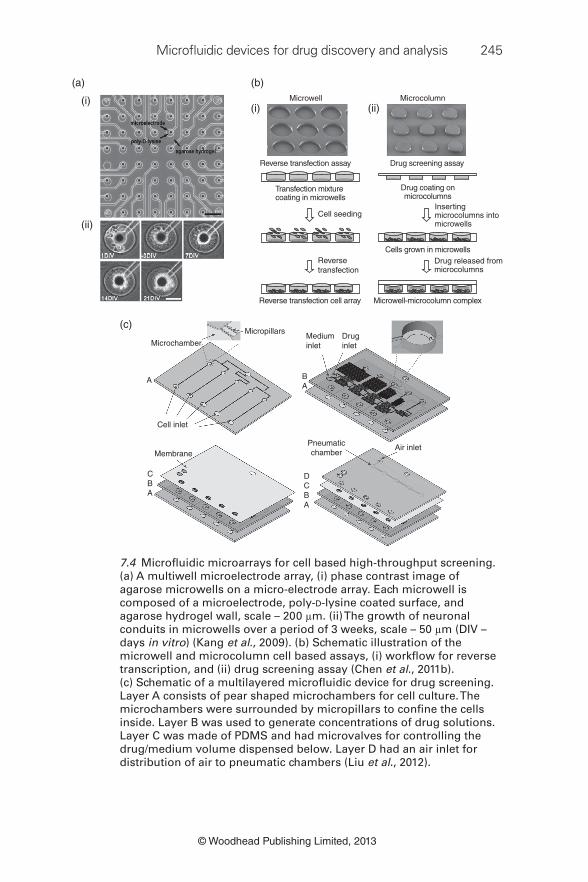
throughput. A multiwell microelectrode array was fabricated using PDMS
by conventional soft lithographic process. The array was then coated with
a cell-adhesive layer of poly-D-lysine followed by patterning a non-con-
ducting agarose gel layer to isolate the individual neuronal micro-circuits
and record individual action potentials of drugs such as bicuculline and
N-methyl-D-aspartic acid (Kang et al ., 2009) (Fig. 7.4a). Chen et al . devel-
oped a complementary microwell and microcolumn system for screening
of drugs (Fig. 7.4b). They used microelectro-mechanical systems (MEMS)
to fi rst fabricate a microwell array on a glass substrate to culture the cells.
Employing a similar process, they fabricated complementary microcolumns
that would carry the drugs to be topically applied onto the cells. The system
was found to be suitable for delivering high-throughput identifi cation of
epidermal growth factor receptor inhibitors (Chen et al ., 2011c). An inte-
grated multilayer microdevice incorporating a drug/medium concentration
gradient generator, fl ow controlling microvalves, and microchambers for
Microfl uidic devices for drug discovery and analysis 245
© Woodhead Publishing Limited, 2013
(a)
(i)
(ii)
(c)
Microchamber
Micropillars
Pneumaticchamber
Cell inlet
MembraneAir inlet
Mediuminlet
Druginlet
BA
A
DCBA
CBA
7.4 Microfl uidic microarrays for cell based high-throughput screening.
(a) A multiwell microelectrode array, (i) phase contrast image of
agarose microwells on a micro-electrode array. Each microwell is
composed of a microelectrode, poly-D-lysine coated surface, and
agarose hydrogel wall, scale – 200 μ m. (ii) The growth of neuronal
conduits in microwells over a period of 3 weeks, scale – 50 μ m (DIV –
days in vitro ) (Kang et al ., 2009). (b) Schematic illustration of the
microwell and microcolumn cell based assays, (i) workfl ow for reverse
transcription, and (ii) drug screening assay (Chen et al ., 2011b).
(c) Schematic of a multilayered microfl uidic device for drug screening.
Layer A consists of pear shaped microchambers for cell culture. The
microchambers were surrounded by micropillars to confi ne the cells
inside. Layer B was used to generate concentrations of drug solutions.
Layer C was made of PDMS and had microvalves for controlling the
drug/medium volume dispensed below. Layer D had an air inlet for
distribution of air to pneumatic chambers (Liu et al ., 2012).
Microwell Microcolumn
Reverse transfection assay
Cell seeding
Drug released frommicrocolumns
Drug coating onmicrocolumns
Cells grown in microwells
Transfection mixturecoating in microwells
Reverse transfection cell array Microwell-microcolumn complex
Reversetransfection
Drug screening assay
(i) (ii)
(b)
Insertingmicrocolumns intomicrowells

246 Microfl uidic devices for biomedical applications
© Woodhead Publishing Limited, 2013
cell culture was recently fabricated by Liu et al . for testing the apoptosis
behaviour in a cisplatin-resistant cancer cell line (Liu et al ., 2012). A vertical
perfusion mode was adopted in this device, as shear stress due to horizontal
fl uid fl ow can adversely impact the cells. Using the set-up, sequential load-
ing of cells, medium, drugs and air was achieved in successive layers of the
device (Fig. 7.4c).
Despite a lot of progress in developing microscale arrays for cell culture,
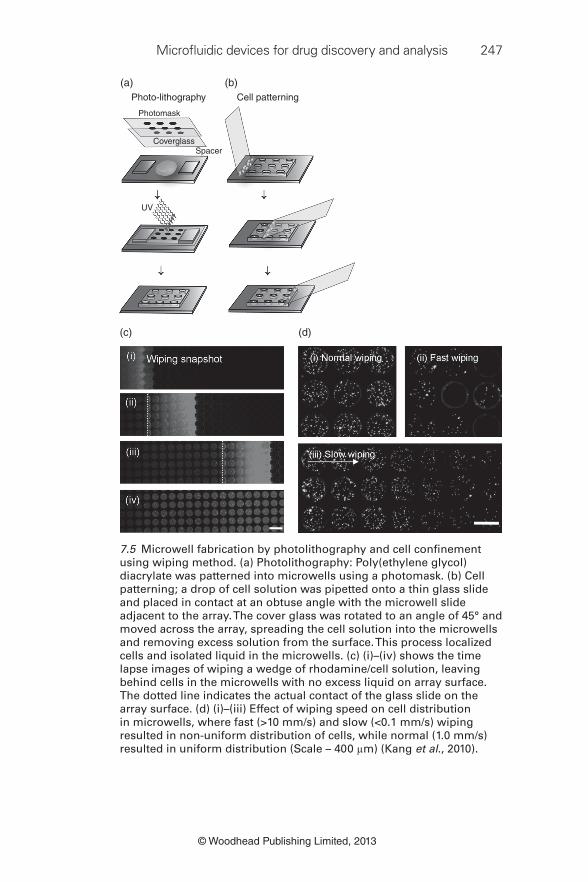
cell seeding in these arrays is a challenge. Kang et al . addressed this issue by
developing a simple wiping method to seed cells in microwells (Fig. 7.5). A
coverslip was used to slowly wipe the cells suspended in the growth medium
across the surface of the microwell array. Cell concentration, microwell
geometry and wiping speed controlled the cell seeding density (Kang et al ., 2010). They also developed an algorithm and software for automatic count-
ing of cells in a microwell array. The software, named Arraycount, detects
the cell count from the fl uorescent cell images in high throughput. The
results were in close correlation with cell counts from the manual methods
(Kachouie et al ., 2009).
Studying single cell characteristics offers an advantage over observing the
behaviour of a group of cells, as single cell characteristics might be hugely
different from the entire population of cells. Microwell arrays have been
developed to confi ne single cells, for observation of these and their prog-
eny over a period of time. One of the fi rst studies pertaining to single cell
confi nement in microwell arrays for drug screening was reported by Rettig
and Folch (2005). PDMS microwells were fabricated by conventional soft
lithography, and controlled seeding of single cells into microwells could be
achieved by optimizing the geometry of the microwells. It was observed that
microwells with an aspect ratio (diameter: depth) close to 1 had more than
85% wells with single cell occupancy for both adherent and non-adherent
cells (Rettig and Folch, 2005). An interesting round bottom microwell array
was recently developed by Liu et al . by creating PDMS microwell arrays
by reverse moulding using polystyrene microspheres melted on a glass
substrate (Liu et al ., 2010). The size of these microwells could be tuned to
10–20 μ m, which is diffi cult to achieve with conventional soft lithography.
The PDMS microwells were then used to confi ne single cells by pouring
excess cell suspension over the microwells, which allowed the cells to settle
in. The enzymatic activity of the cells was studied by carrying out the car-
boxylesterase assay using calcein AM. Fluorescence intensities from single
cells could be captured to reveal different kinetic behaviour of entrapped
cells, which was related to cell viability (Fig. 7.6). Another novel way of con-
straining single cells in microwells was demonstrated by Wang et al . (2012).
The fl exibility of PDMS was exploited by stretching the patterned PDMS
array using a tube that delivered the cells onto the array. After loading, the
tube was withdrawn and cells settled in the microwells, which were then
Microfl uidic devices for drug discovery and analysis 247
© Woodhead Publishing Limited, 2013
Photo-lithography
(a) (b)
(c) (d)
Photomask
CoverglassSpacer
UV
Cell patterning
7.5 Microwell fabrication by photolithography and cell confi nement
using wiping method. (a) Photolithography: Poly(ethylene glycol)
diacrylate was patterned into microwells using a photomask. (b) Cell
patterning; a drop of cell solution was pipetted onto a thin glass slide
and placed in contact at an obtuse angle with the microwell slide
adjacent to the array. The cover glass was rotated to an angle of 45° and
moved across the array, spreading the cell solution into the microwells
and removing excess solution from the surface. This process localized
cells and isolated liquid in the microwells. (c) (i)–(iv) shows the time
lapse images of wiping a wedge of rhodamine/cell solution, leaving
behind cells in the microwells with no excess liquid on array surface.
The dotted line indicates the actual contact of the glass slide on the
array surface. (d) (i)–(iii) Effect of wiping speed on cell distribution
in microwells, where fast (>10 mm/s) and slow (<0.1 mm/s) wiping
resulted in non-uniform distribution of cells, while normal (1.0 mm/s)
resulted in uniform distribution (Scale – 400 μ m) (Kang et al ., 2010).

248 Microfl uidic devices for biomedical applications
© Woodhead Publishing Limited, 2013
amenable to further analytical treatment. They also demonstrated that cells
within the microwells could be isolated by deforming the PDMS substrate
using a microneedle (Wang et al ., 2012). A further example was illustrated
by Lew and co-workers, who devised a plastic microwell array by using eco-
nomical materials such as shrink wrap fi lm and tape. A carbon dioxide laser
was used to cut holes in the tape, which acted as a mask to etch wells in the
shrink wrap by oxygen plasma (Lew et al ., 2011).
Oxygen plasma
Glass
Glass
Glass
glass
Drop suspension of PS microspheres
Hydrophilic surfaceon glass substrate
Assembly of PS microsphereson glass surface
Partial melting anddeformation of PS microspheres
Curing PDMS on PSmicrospheres template (80ºC, 2 h)
Microwell arrayson PDMS replica
Heat on hot plate (240ºC, 3 min)
1) Silanize PS surface2) Cast PDMS
Peel off PDMS
PDMS
10∼20 μm
10∼20 μm
PS microspheres
(a)
(b)
7.6 Microwell arrays for single cell analysis. (a) Fabrication of PDMS-
based microwell arrays using polystyrene microbeads, and (b)
micrograph image of Ramos cells in each microwell (Scale 20 μ m) (Liu
et al ., 2010).

Microfl uidic devices for drug discovery and analysis 249
© Woodhead Publishing Limited, 2013
Apart from multiwell arrays, multiplexed screening platforms have
also been developed to screen multiple samples in one run. The ability to
analyse multiple proteins, nucleic acids as well as small molecules, reduces
assay time, reagent volume and cost. Multiplexed measurements provide
the ability to increase the throughput without a simultaneous increase in
the density of the microfl uidic array. Multiplexing technology has been
applied to two different types of microfl uidic platforms: planar arrays
and suspension (particle based) arrays. For protein and DNA analysis,
planar arrays have been used, whereby protein molecules have been pat-
terned as microarrays onto substrates using lithography (MacBeath and
Schreiber, 2000). Such systems offer application specifi c advantages, rang-
ing from study of protein – protein interactions to establishing proteins as
targets for small molecules and specifi c functions of enzymes. Suspension
arrays, on the other hand, offer the advantages of studying the properties
of compounds in solution, thereby providing ease of sample modifi cation,
higher throughput and increased batch-to-batch uniformity (Nolan and
Sklar, 2002).
A multiplexed system could be used to screen a compound against
multiple kinases, or study protein – protein interaction and detect changes
in enzyme conformation (Xue et al ., 2001). In this report, four kinases
were screened against a substrate. The reaction products/substrates
could be separated by electrophoretic separation on a chip and analysed.
Multiplexed screening of picolitre-sized droplets that could be manipu-
lated using an array of electrodes has also been reported. For example,
caspase-3, a marker of apoptosis, which is an important tool in cancer
drug discovery, was measured after human cervical adenocarcinoma
HeLa cells were treated with different concentrations of staurosporine.
The technique termed as digital microfl uidics was compared with conven-
tional techniques involving 96-well plate. It resulted in a 33-fold reduc-
tion in sample volume together with a lower detection limit for caspase-3
analysis compared with conventional techniques. This can be attributed
to the lack of delamination in apoptotic cells in the digital microfl uidics
platform that uses the droplet manipulation system instead of pipetting
or aspiration of liquids with conventional techniques (Bogojevic et al ., 2012).
Analysing multiple samples by multiplexing, however, poses a challenge
in sample recognition. Hence, it is necessary to have an encoding scheme
integrated into the system to allow for rapid and precise analyte identifi ca-
tion. Encoding schemes based on spectrometric (Han et al ., 2001), graphi-
cal (Evans et al ., 2003), electronic (Service, 1995) and physical techniques
(Vaino and Janda, 2000) have been developed. An exhaustive review
of various encoding techniques has been published by Braeckmans et al . (2002). Spectrometric techniques utilize specifi c wavelengths to analyse a

250 Microfl uidic devices for biomedical applications
© Woodhead Publishing Limited, 2013
compound. In contrast, graphical methods use certain optical elements that
are chemically patterned onto the microarray. These techniques require
much sophistication, and are expensive and may require a considerable
amount of time for fabrication and integration.
Pregibon et al . recently developed a novel encoding scheme for multi-
plexed platforms (Pregibon et al ., 2007). In this system, two poly(ethylene
glycol)-based monomer solutions, one being a fl uorescent dye and the other
being an acrylated probe, were made to fl ow through microfl uidic chan-
nels. The solutions during fl ow were exposed to ultraviolet light using con-
ventional techniques of continuous fl ow lithography to develop a patterned
particle (Pregibon et al ., 2007). The morphological properties of the particles
were determined by a photomask, inserted into a fl uorescence microscope
(Pregibon et al ., 2007). A simple dot coding scheme was used on the photo-
mask that could generate over two million particles, with each having a unique
code. Although the particle size achieved in this method was larger than in
previous methods, the authors demonstrated that the sample volume required
was manageable, together with providing higher sensitivity and reproducibil-
ity. The system was able to detect DNA at very low concentrations, without
signal amplifi cation, proving it to be a completely integrated encoding device,
with advantages of low cost, high effi ciency with virtually unlimited number
of codes possible, and all this achievable with the services of a simple fl uores-
cence microscope.
Inkjet printing technology has been purported as a highly effi cient
screening alternative, providing effi ciencies greater than 200 000 com-
pounds per day, currently achievable with the microfl uidic platforms
described earlier. The technology offers capabilities to simultaneously
deposit cells and drugs to be tested in a small picolitre volume. Post-
processing, the cell characteristics can be studied to evaluate the drug
effects. Such a novel platform was developed by Rodr í guez-D é vora et al . (2012). They developed an inkjet printer-based method to pattern green
fl uorescent protein expressing Escherichia coli cells grown on a soy agar
medium, on a coverslip. Live/Dead ™ assay, used to assess bacterial cell
viability, demonstrated high rate of cell survival after imprinting. Fast
screening utilizing low volumes to assess the effect of three antibiot-
ics patterned together with the bacterial cells could be carried out. This
bioprinting approach was compared to the standard micro-pipetting
approach and was found to yield similar results at much lower volumes
(Rodr í guez-D é vora et al ., 2012).
These microfl uidic platforms have signifi cantly enhanced the profi le of
HTS, leading to optimization of hits and leads, before the leads are put
through preclinical testing for evaluation of their preliminary pharmacoki-
netic and toxicological properties.
Microfl uidic devices for drug discovery and analysis 251
© Woodhead Publishing Limited, 2013
7.2.3 Preclinical evaluation
Interaction with the molecular targets begins the journey of the drug
in the human body. When a drug is administered, it has to be absorbed
across mucous membranes, followed by its distribution to its target site
and metabolism to an inactive metabolite to be eliminated from the
body. It should also be devoid of any toxic effects. These characteristics,
respectively known as absorption, distribution, metabolism, elimination
and toxicology (ADMET) are essential factors in determining the path of
the drug in the later stages of the drug discovery process. A fi ne balance
between these pharmacokinetic characteristics is needed for the develop-
ment of a drug from a chemical entity (Muster et al ., 2008). Unsatisfactory
ADMET profi les account for attrition of 50–60% drug candidates at the
preclinical development stage (Smith, 2007), with lack of effi cacy and
undesirable toxicity being the major causes (Kramer et al ., 2007). It has
been reported that the lack of effi cacy accounts for 30% of failures of new
drug entities and toxicity further accounts for another 30%. If these are
detected at later stages in the drug development process, the overall cost
of the programme will be increased, as cost escalates with each stage (Kola
and Landis, 2004). This is why pharmaceutical companies are nowadays
adopting the ‘ fail early; fail cheap ’ approach to identifying the toxicolog-
ical properties of drug compounds. This is done in lieu of savings in the
event that toxicological properties are identifi ed at a much later stage or
even after the launch of the product, necessitating an inevitable and highly
expensive market recall. It has been reported that market recalls, as a per-
centage of approvals, in the United States has fallen from 27.2% in 1980s
to 5.2% in 2000s (Qureshi et al ., 2011). This has, in part, been the contri-
bution of more novel and effi cient toxicity screening platforms that have
been developed in the past two decades. It also underlines the importance
of profi cient preclinical programmes, and the role played by them in drug
development.
In vitro toxicological testing in cell models provides useful infor-
mation about the drug candidates, much before the expensive animal
experiments and first-in-human clinical trials are conducted. In vitro
experiments have been long touted to replace animal testing, espe-
cially due to the ethical concerns surrounding animal experimentation
(Wen et al ., 2012). Moreover, in vitro toxicity in excised animal organs
may not be extrapolated to correctly reflect human toxicities. On the
other hand, in vivo preclinical testing in live animals requires a large
amount of compound under investigation, which is usually available in
limited quantities and may be prohibitively expensive (Muster et al ., 2008).
252 Microfl uidic devices for biomedical applications
© Woodhead Publishing Limited, 2013
In vitro evaluation
Three-dimensional (3D) cell culture mimics the natural environment of
the cells, including cell – cell and cell – extracellular matrix interactions, as
opposed to planar two-dimensional (2D) cultures that are used to main-
tain cells (Pampaloni et al ., 2007). An excellent collation of advantages of
3D cell culture over the 2D format has been provided by Zhang and van
Noort (2011). Also, these 3D cultures offer an ex vivo alternative to live
animal testing, and potentially reduce the cost of toxicity screening during
drug development. Nonetheless, 3D cell cultures present a few shortcom-
ings, especially with sample handling and imaging. Since these cultures are
thicker than conventional ‘petri-dish’ cultures, they are diffi cult to adapt to
conventional microscopic techniques. Liquid handling in patterned micro-
structures requires sophisticated micro/nanolitre scale devices. However,
the advantages of studying the cells in an environment outweigh the tech-
nological shortcomings, which, too, are being addressed simultaneously.
As hepatotoxicity has been the leading cause of failure at the clinical
trial stages and post launch market withdrawals, many researchers have
looked at developing in vitro cell-based hepatotoxicity assays. It is impor-
tant to notice here that most of these agents went through preclinical ani-
mal testing and were assumed to be safe (Kaplowitz, 2005). Microfl uidic 3D
cell culture platforms aim to address this problem, and have been designed
to provide deeper insights into cell behaviour when exposed to cytotoxic
agents. A multiwell 3D cell culture platform was designed using soft lithog-
raphy to co-culture primary hepatocytes with mouse 3T3-J2 fi broblasts
(Fig. 7.7a). A PDMS stencil containing through-holes in a 24-well format
was fi rst applied to a polystyrene plate, followed by application of collagen-
I through the holes. After removal of the PDMS stencil and application of
a 24-well PDMS blank, hepatocytes were cultured on the 24 wells, which
attached to the collagen, surrounded by fi broblasts. The hepatocyte mor-
phology was maintained in the wells for 4–6 weeks. Albumin and urea syn-
thesis, measured as markers of protein synthesis and nitrogen metabolism
and typically considered as a measure of liver function, were reported to be
normal. On the other hand, pure cultures were reported to be morphologi-
cally unstable and there was a loss of albumin and urea synthesis (Khetani
and Bhatia, 2008).
Kane et al . designed a microfl uidic 8 × 8 array, composed of PDMS. Each
well in the array had two chambers. The primary chamber’s bottom was
made of glass coated with collagen, for co-culturing rat hepatocytes and
3T3-J2 fi broblasts; the collagen aided selective adhesion of hepatocytes,
while continuous perfusion of medium and removal of waste products was
achieved by microfl uidic tubing connected to the chamber. The secondary
chamber, which was separated from the primary chamber by a thin PDMS

Microfl uidic devices for drug discovery and analysis 253
© Woodhead Publishing Limited, 2013
7.7 Multiwell culture for in vitro toxicity testing. (a) Schematic of the
fabrication process (left panel) with photomicrographs of each step
(right panel). A PDMS stencil in a 24-well format with through holes at
the bottom of each well is sealed to a polystyrene plate, with collagen-I
adsorbed on exposed polystyrene. The stencil is then peeled off
followed by be application of blank PDMS stencil before cell seeding.
Hepatocytes are then seeded which selectively attach to collagen-I,
allowing fi broblasts to be seeded in other bare areas (Khetani and
Bhatia, 2008). (b) A mathematical PBPK model and a corresponding
physical μ CCA based on the human body. A μ CCA consists of liver,
tumour and marrow chambers, interconnected with channels mimicking
the blood fl ow pattern in the human body (Sung and Shuler, 2009).
Multiwell elastomeric device
(a)
PDMS stencil
Micropatternedextracellular matrix
Micropatternedhepatocytes
Micropatternedcoculture
Physisorbextracellularmatrix protein
Removestencil
Seedhepatocytes
Seedstromalcells
Colon tumor
Liver
Marrow Blo
od
PBPK model microCCA
3 cm
3 cm
(b)
Tumor
Liver Marrow

254 Microfl uidic devices for biomedical applications
© Woodhead Publishing Limited, 2013
membrane, was linked to microfl uidic channels supplying humidifi ed air
with 10% carbon dioxide at 37°C. They also reported similar results, with
increased albumin and urea production (Kane et al ., 2006). Such microfl u-
idic platforms have also been used to assess cardiotoxicity, neurotoxicity,
embryotoxicity and cytolysis, a summary of which has been provided in a
review by Wen et al . (2012). These microfl uidic devices, which can emulate a
particular organ in vitro , are referred to as organ-on-a-chip devices.
Although the above listed cell-based assays provide information about a
compound’s therapeutic and toxic properties on the tissue under consider-
ation, they do not tell anything about the effect on the whole body or inter-
actions with other organs and related dose dynamics. As a drug in the body
goes through the complex process of ADME, collectively called as phar-
macokinetics (PK), with contributions from different organs, cell culture
using cells-on-a-chip or organ-on-a-chip technology, fails to capture these
responses. Of late, scientists have developed miniaturized multi-compart-
ment cell culture platforms better known as body-on-a-chip devices. These
can promote tissue – tissue interactions by creation of environment and fl ow
conditions scaled down to in vivo tissue sizes. They can also aid in study-
ing interactions between organs in a high-throughput manner, enabling
the study of multiorgan metabolic and toxicity profi les of a compound.
Microscale systems designed for physiologically based pharmacokinetic
modelling (PBPK), having different compartments for different tissues,
can assist in understanding parameters such as tissue-to-blood perfusion,
enzyme kinetics, liquid-to-cell ratio and physiological stress on a particular
tissue/organ (Esch et al ., 2011).
A novel microfl uidic system, microscale cell culture analogue ( μ CCA),
has been developed for multiorgan toxicity analysis. A multiorgan culture
system, the integrated discrete multiple organ culture or ‘wells-within-a-
well’ system, was designed by Li et al . Cells from different organs were cul-
tured in small wells in their respective media in a bigger well. They cultured
primary cells from liver, kidney, lungs, central nervous system, blood vessels
as well as human breast adenocarcinoma cancer cell line, MCF-7. For testing
the toxicity of a model drug, the bigger wells were fl ushed with a medium
containing the drug, tamoxifen. The effect of tamoxifen was evaluated and
its comparative toxicity towards various organs was also examined. Apart
from this, the system offers another advantage in enabling the analysis of
the anticancer activity of a drug with respect to its effect on normal tis-
sues. Although the authors did not delve into multiorgan interactions, this
in principle can be adapted for this purpose and its capabilities should be
further investigated (Li et al ., 2004).
In a model based on PBPK to emulate the dynamics of the human body,
different compartments hosting different cell types were connected through
microfl uidic channels to mimic blood circulation. Four different cells were
Microfl uidic devices for drug discovery and analysis 255
© Woodhead Publishing Limited, 2013
cultured on a μ CCA, including hepatocytes (HepG2/C3A), bone marrow
cells (MEG-01), uterine cancer cells (MES-SA), and a multidrug resistant
(MDR) uterine cancer cell line (MES-SA/DX-5). In a combination drug
therapy of chemotherapeutic doxorubicin, with MDR modulators cyclo-
sporine and nicardipine, treated for 24 h or 72 h, a selective toxicity towards
MES-SA/DX-5 was detected, a synergy not observed in conventional 96-well
plate assays. This device could thus be used in drug screening and selection
of potential MDR modulators, as well as gather dose required and dose
response curves for subsequent in vivo animal experiments or clinical trials
(Tatosian and Shuler, 2009). 3D hydrogel cultures in μ CCA format were
developed by Sung and Shuler (Fig. 7.7b). Three types of cells, hepatocytes
(HepG3/C3A), myeloblasts (Kasumi-1) and colon cancer cells (HCT-116),
were embedded in different chambers in 3D hydrogels, representing dif-
ferent organs. The cytotoxic effect of tegafur, a prodrug of active antican-
cer drug, 5-fl uorouracil, commonly used in colon cancer was tested using
this device. An interesting revelation, as compared to conventional 96-well
plate assay was that, although, the liver cells in μ CCA showed metabolism
of tegafur similar to 96-well plate, the metabolism led to the death of hepa-
tocytes, an effect which was unnoticeable in well plate assays (Sung and
Shuler, 2009). The literature is replete with tegafur toxicity data, particularly
its hepatotoxicity (Maruyama et al ., 1995). In such a scenario, development
of microfl uidic systems providing critical toxicity information in in vitro
models bodes well for preclinical drug testing.
Ex vivo evaluation
Apart from in vitro microfl uidic cell culture platforms, some researchers have
also looked at ex vivo microfl uidic platforms by isolating animal tissues, par-
ticularly liver, and culturing excised explants to analyse the toxicity of vari-
ous compounds. It has been reported that precision cut liver slices fare better
than hepatocytes alone with respect to metabolic activity (Graaf et al ., 2007).
Continuous perfusion of nutrient medium can further reduce the loss of met-
abolic activity and prolong protein expression in these slices. Microfl uidic
devices have been designed to continuously replenish the spent medium
and remove waste material from these slices. van Midwoud et al . designed a
PDMS-based ‘perifusion’ device, with liver slices supported on polycarbonate
membranes (Fig. 7.8a). The term ‘perifusion’ was used instead of perfusion,
as the medium fl owed around the slices. PDMS membranes were purposely
kept thin so as to allow for effi cient gaseous exchange. Metabolic activity of
7-ethoxycoumarin observed was comparable to the well plate-based method
was observed in this device (van Midwoud et al ., 2010). Another PDMS-based
device was developed to analyse ethanol toxicity in liver explants. Using this
device, concentrations as low as 20 mM produced a decrease in mitochondrial

256 Microfl uidic devices for biomedical applications
© Woodhead Publishing Limited, 2013
Outlet
2-3 mm (10)
PDMS membrane(250 μm)
PDMS membrane(250 μm)
Inlet
Polycarbonatemembrane
(10 μm, 8 μm pores)
Polycarbonatemembrane
(10 μm, 8 μm pores)
Liver slice
“o-ring”
250 μm (9)350 μm (8)±50 μm (7)500 μm (6)
1.5 mm (5)
±50 μm (4)350 μm (3)250 μm (2)2-3 mm (1)
(i)(a)
(ii)
Platelet
LeukocyteErythrocyte
Flow direction
Microcavity array
(b)
PumpWaste
reservoir
7.8 Microfl uidic platforms for ex vivo experiments. (a) For culturing
liver slices. (i) Cross-sectional view of a PDMS-based biochip for
culturing liver slices that were supported on a polycarbonate
membrane. The device was termed as a ‘perifusion’ device as
culture medium fl owed around the slices. Thin PDMS membranes
allowed for effi cient gaseous exchange. (ii) A photograph of the
device mentioned in (i) (van Midwoud et al ., 2010). (b) For leukocyte
counting and assessment of haematotoxicity. A microcavity array
was created by PDMS to form a sieve like structure that retained
the leukocytes, while allowing other blood cells to pass through.
The cells can then be separately analysed for toxicity due to various
drugs (Hosokawa et al ., 2012).

Microfl uidic devices for drug discovery and analysis 257
© Woodhead Publishing Limited, 2013
metabolic activity as well increased lactate dehydrogenase activity, a marker
of cell death. These effects were observed in a concentration dependent man-
ner, together with a decrease in albumin and urea synthesis (Hattersley et al ., 2011). Such devices utilizing excised tissues represent clinically more relevant
models to replace animal experimentation.
In vivo evaluation
Microfl uidic platforms have also been used to assist in vivo animal experi-
ments, for blood sampling, sample preparation and analysis (Kang et al ., 2008). An automatic blood collection microfl uidic chip-based on PDMS
was developed by Wu et al . for withdrawal of blood from mice without the
need of trained personnel. The device consisted of two layers, holding chan-
nels for blood inlet, outlet, heparin block, blood reservoir and sample wells.
A microfl uidic device was used for processing blood samples from mice
for determining haematotoxicity (Fig. 7.8b). In this device, a microcavity
array was created by master-moulding PDMS structures to form a sieve-like
structure that separated leukocytes from other blood cells. Benzene toxicity
was assessed by staining the leukocytes and counting them over a period of
2 weeks (Hosokawa et al ., 2012). Microfl uidic platforms have been designed
to be integrated with novel analytical techniques such as matrix assisted
laser/desorption ionization–mass spectrometry (MALDI-MS), which can
facilitate fast sample analysis with high precision and resolution of many
metabolites in biological samples (Lee et al ., 2008; Xu et al ., 2006).
These microfl uidic systems have thus played a critical role in various
stages of the drug development process. Beginning with the identifi cation
of targets, to synthesis of compounds for generation big compound librar-
ies, to HTS and preclinical development, microfl uidics has been effectively
adapted to reduce the consumption of reagents and make the drug discov-
ery process more effi cient and cost effective. In the subsequent section, we
will discuss about the application of microfl uidics in analysis of chemical
and biological drugs. We also discuss the role played by these devices in
detection of diseases and routine diagnostic purposes, which may reduce
the healthcare costs.
7.3 Microfluidics for drug analysis and diagnostic applications
Miniaturization of analytical tools has been propelled by the recent surge
in the development and maturation of microfabrication techniques. The
better control of physical processes at the micron-scale has further fuelled
the interest in micro-analytical systems as new paradigms for pharmaceuti-
cal analysis. These systems are aimed at reducing both the sample volume
258 Microfl uidic devices for biomedical applications
© Woodhead Publishing Limited, 2013
and time of analysis, besides being amenable to integration with the other
platforms and potential for high throughput. High parallelization, thus now
possible, had made analysis of multiple compounds fast and easy (Lee et al ., 2009). Design modifi cation can provide integrated facilities for handling
fl uids, and thermal and spatial control for targeting specifi c detection com-
ponents to enhance selectivity (DeMello, 2006). With the mass fabrication
of micron-sized platforms now made possible through sophisticated instru-
ments, the cost of production of these devices has come down, providing an
opportunity to develop single use analytical devices, thereby reducing the
possibility of cross contamination (Lion et al ., 2004).
7.3.1 Microfl uidics for drug analysis
Although there have been many mechanistic and experimental advance-
ments in analysis of drugs, the basic analytical equipment and components
have not changed much over the past few decades. Recently, with the appli-
cation of microscale techniques adopted from the semiconductor industry,
scientists are now poised for choice to carry out analytical assessments at
an order 5–9 times lower than conventional counterparts (DeMello, 2006).
Microscale analytical devices, also termed as micro-total analytical systems
( μ TAS), comprise microchannel networks that aim to replicate the analy-
sis procedures on physically shrunk platforms, without compromising the
analytical effi ciency or sensitivity. Apart from this, μ TAS can be designed to
attain a high level of automation, thereby making multiplexed assays pos-
sible and providing a system that reduces manual errors, helping to increase
the assay accuracy. These devices have been more popular in the analysis
of biological molecules such as proteins and nucleic acids (Guo et al ., 2012;
Meagher and Thaitrong, 2012), and have been the subject of other chapters
in this book. Here, we would limit our focus to discussion about the applica-
tion of microfl uidics in the analysis of drug entities.
Analysis of pharmaceutical compounds has been carried out using high
performance liquid chromatography (HPLC), linked to various detection
modules including ultraviolet and fl uorescence spectrophotometers as well
as mass spectrometry, electrophoresis, potentiometry, colorimetry, radioiso-
topic assay, microbiological methods, enzymatic methods, surface plasmon
resonance based biosensor assays and chemiluminescence, among many
others. Microfl uidic platforms, due to the fl exibility in their design, are doc-
ile to integration with most of the above listed analytical methods. With
the increasing demand for highly sensitive and minuscule working volume
microfl uidic platforms, it is imperative that the chosen method is able to
detect low amounts of the analyte. Chemiluminescence (CL) was reported
to be a highly sensitive technique, with vitamin B 12 concentrations as low as
5 pg/mL being detected easily (Kumar et al ., 2009). CL-based methods have

Microfl uidic devices for drug discovery and analysis 259
© Woodhead Publishing Limited, 2013
been used for microfl uidic detection of vitamin B 12 and L-phenylalanine.
Luminol oxidation by hydrogen peroxide in the presence of externals
catalyst ions, such as cobalt (II) and copper (II) and amino acids such as
L-phenylalanine under alkaline conditions, is the basis of this test. The resul-
tant product is a blue compound (3-aminopthalate ion) that can be detected
at a wavelength of 425 nm (Chen et al ., 2007; Wang et al ., 2007).
Lok et al . developed a microfl uidic chip to detect the concentration of
vitamin B 12 using a continuous fl ow microfl uidic chip (Fig. 7.9). The device
consisted of a two passive mixing reaction chambers and a double spirally
wound microchannel network as an optical detection unit, spanning the
three sequentially fabricated layers. Microchannels in the second layer were
(a)E
D
B
W
A
C
(b)
7.9 Microfl uidic chip for chemiluminescence based detection of vitamin
B 12 . (a) Schematic of the chip with fi ve inlets, two mixing chambers and
two spiral detection units, and the fabricated chip (b) (Lok et al ., 2012).
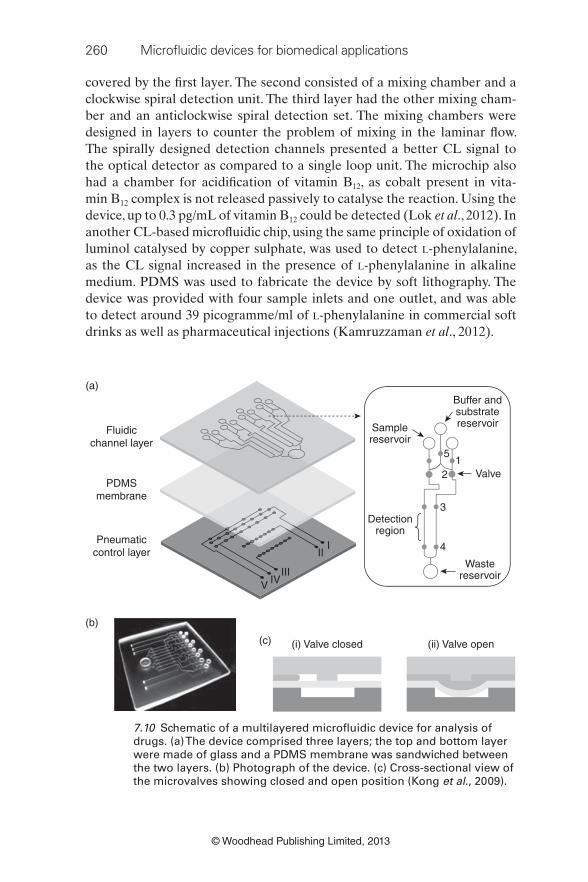
260 Microfl uidic devices for biomedical applications
© Woodhead Publishing Limited, 2013
PDMSmembrane
Pneumaticcontrol layer
IVIII
III
V
Fluidicchannel layer
(a)
Detectionregion
Wastereservoir
Valve
4
3
21
5
Samplereservoir
Buffer andsubstratereservoir
(b)
(i) Valve closed(c) (ii) Valve open
7.10 Schematic of a multilayered microfl uidic device for analysis of
drugs. (a) The device comprised three layers; the top and bottom layer
were made of glass and a PDMS membrane was sandwiched between
the two layers. (b) Photograph of the device. (c) Cross-sectional view of
the microvalves showing closed and open position (Kong et al ., 2009).
covered by the fi rst layer. The second consisted of a mixing chamber and a
clockwise spiral detection unit. The third layer had the other mixing cham-
ber and an anticlockwise spiral detection set. The mixing chambers were
designed in layers to counter the problem of mixing in the laminar fl ow.
The spirally designed detection channels presented a better CL signal to
the optical detector as compared to a single loop unit. The microchip also
had a chamber for acidifi cation of vitamin B 12 , as cobalt present in vita-
min B 12 complex is not released passively to catalyse the reaction. Using the
device, up to 0.3 pg/mL of vitamin B 12 could be detected (Lok et al ., 2012). In
another CL-based microfl uidic chip, using the same principle of oxidation of
luminol catalysed by copper sulphate, was used to detect L-phenylalanine,
as the CL signal increased in the presence of L-phenylalanine in alkaline
medium. PDMS was used to fabricate the device by soft lithography. The
device was provided with four sample inlets and one outlet, and was able
to detect around 39 picogramme/ml of L-phenylalanine in commercial soft
drinks as well as pharmaceutical injections (Kamruzzaman et al ., 2012).

Microfl uidic devices for drug discovery and analysis 261
© Woodhead Publishing Limited, 2013
Won et al . developed a microfl uidic device on glass slides for simultaneous
detection of fi ve sulphonamide drugs (Won et al ., 2012). The device was pro-
vided with modules for pre-concentration and electrokinetic separation of
drugs using the fi eld amplifi ed sample stacking (FASS) and fi eld amplifi ed
sample injection (FASI) techniques in two parallel channels (Shiddiky and
Shim, 2007). Subsequent electrochemical detection of sulphonamides was
carried out at the end of the separation channel, which consisted of a sil-
ver/silver chloride, platinum wire and aluminium-gold nanoparticles modi-
fi ed carbon paste electrode. The device was able to detect femto-molar level
concentrations of these drugs and provide an opportunity to simultaneously
detect these drugs in clinical samples. A microfl uidic chip integrated to a
laser-induced fl uorescence scanner was developed for the detection of β 2
agonist drugs such as clenbuterol (Fig. 7.10). These drugs increase muscle
mass and have been often misused in farm animals (Martinez-Navarro,
1990), as well as in power sports by athletes (Delbeke et al ., 1995; Hesketh
et al ., 1992), making it vital to analyse them in a rapid and accurate man-
ner. The three layers of the device consisted of a fl uidic channel, a PDMS
membrane, and a pneumatic control layer interspersed with many pneu-
matic microvalves and micropumps to enable the delivery of reagents. Glass
was used to fabricate the fl uidic channel and pneumatic control layer by
standard lithography and etching to create microchannels. The PDMS mem-
brane was then sandwiched between the two layers and generated pneu-
matic valve and pump effect due to defl ection by compressed air. The drugs
could be detected within 30 min and at a concentration as low as 0.088 ng/
mL (Kong et al ., 2009). Thus, microfl uidic devices can play a crucial role in
detection drugs and pharmaceuticals, and can be routinely used in chemical,
pharmaceutical and clinical settings with high precision in an economical
way.
7.3.2 Microfl uidics for diagnostic purposes
The conventional diagnostic techniques based on sophisticated macroscopic
equipment such as gas chromatography – mass spectrometry are only fea-
sible in big air-conditioned laboratories, which are equipped with trained
workforces and devices for sample handling, together with ample ancillary
resources needed for effi cient diagnosis. But this is not attainable outside
the realm of these laboratories, especially in the developing world, particu-
larly in rural areas (Lee et al ., 2010; Yager et al ., 2006). Miniaturized versions
of analytical platforms have been recently conceptualized, primarily based
on microfl uidic technology, to perform diagnostic analysis, consuming mini-
mal amounts of reagents with high effi ciency and speed, making the device
a portable point-of-care, self-usable system. This not only reduces the logis-
tical issues with sample handling and transfer, but also provides patients
262 Microfl uidic devices for biomedical applications
© Woodhead Publishing Limited, 2013
with the luxury of testing for various markers, such as blood glucose, in the
comfort of their homes, which is particularly suitable for geriatric patients.
These microfl uidic diagnostic devices may reduce the healthcare costs asso-
ciated with diagnosis.
Microfl uidic biosensors, as they are commonly known, have been fabri-
cated for a variety of purposes. Particularly important among these are on-
chip enzymatic assays. As enzymes have the potential of converting a large
number of substrate molecules into product in a fraction of a second, in a
highly selective manner, they offer an exciting avenue for chemical analy-
sis. Enzyme assay on a chip may be either homogenous or heterogeneous.
In the former, all the reactants are in solution phase (Hadd et al ., 1997),
while in the latter either of the enzyme/substrate/inhibitor needs to be
immobilized on a solid surface (Mao et al ., 2002; Krenkova and Foret, 2004).
Mostly, enzyme immobilization is carried out on microchannel walls or onto
some support inside the channels. This provides the advantages of enzyme
recycling, placement of enzyme at specifi c locations on microchannels, and
analysis in a continuous fl ow environment, making immobilized assays a
preferable choice (Kim et al ., 2009).
Enzyme immobilization has been mostly carried out on microspheres, also
known as microbeads, due to their similar size properties to microchannels,
as well as large surface area for enzyme support (Peterson, 2005). They offer
the advantage of being contained at appropriate locations by using mechan-
ical barriers or magnetic devices. Kim et al . developed a microfl uidic device
for glucose detection. The device consisted of two separate chambers, for
reaction and detection, respectively. In the reaction chamber, microbeads
were covalently bound to an enzyme, glucose oxidase, and were supported
by microfi lters. A poly (ethylene glycol)-based microarray (fabricated by
photolithography) encapsulating a horseradish peroxidize, formed the
detection chamber. The bienzymatic reaction was used to detect the conver-
sion of non-fl uorescent substrate (Amplex Red fl uorescence indicator) to a
fl uorescent resorufi n, with glucose concentrations in the range of 1–10 mM
detected successfully by fl uorescence microscopy and quantifi ed by soft-
ware (Kim et al ., 2009). In a modifi ed version, Sheng et al . used glucose oxi-
dase modifi ed magnetic nanoparticles, constrained in the microchannel with
the aid of external magnetic fi eld, for the amperometric analysis of glucose.
The device offered a simple alternative to other such devices, as no mixing
was needed, achieving higher sensitivity. Detection was linear with a range
between 25 μ M and 15 mM. The device also possessed a separation channel
that avoided the entry of macromolecules, thereby eliminating the need to
pre-process the sample. This allowed the serum samples to be directly used
for glucose analysis (Sheng et al ., 2012). Other devices integrating enzyme
and immunoassays have been fabricated for simultaneous detection of glu-
cose and insulin (Wang et al ., 2003).

Microfl uidic devices for drug discovery and analysis 263
© Woodhead Publishing Limited, 2013
Herr et al . designed a microchip-based device for the detection of dis-
ease biomarkers in periodontal disease (Fig. 7.11). The multifaceted device
had integrated capabilities for sample pretreatment, including fi ltering, mix-
ing and enrichment of the saliva, which could be withdrawn in a hands-free
manner. The device, a microchip electrophoretic immunoassay ( μ CEI), was
provided with molecular sieves fabricated using hydrogel, to enrich the sam-
ple, followed by electrophoretic separation to resolve a fl uorescent antibody
bound to an enzyme, responsible for tissue decay. Using the method, they
could dispense with the need for using matched antibody pairs as well as to
immobilize the antibody (Herr et al ., 2007).
Enzyme-linked immunosorbent assay (ELISA) has been the mainstay of
clinical diagnostics for detection of antigens and antibodies. However, the
Membrane
SW
BW
S
B
B B
mAb*
Detectionpoint
7.11 Microchip electrophoretic immunoassay ( μ CEI) chip. The device
had fl uid wells for: S, sample; B, buffer; SW, sample waste; BW,
buffer waste; mAb*, fl uorescently labelled monoclonal antibody.
The device possessed molecular sieves to enrich the sample and
electrophoretically separate the molecular antibody that is bound to an
enzyme (Herr et al ., 2007).

264 Microfl uidic devices for biomedical applications
© Woodhead Publishing Limited, 2013
1211
8
9
2
14
15
3
65
113
TMB
Stop
Sample Blank
WasteWaste
BloodWashingbuffer 4
7
10
Mixingchamber
DetectionchamberPl
asm
ase
para
tion
(a)
(b)
Mixing chamber
Detecting chamber
14
18
153
12
8
7
179
Plasma
13
16
19
Waste
Rotation
Immunoassay part
Biochemical analysis part
Waste
TMBAntibody2nd
Washingbuffer 2
StopWashingbuffer 1
4
7.12 Microfl uidic lab-on-a-disc platforms. (a) Schematic of a fully
automatic disposable ELISA platform, with facilities for plasma
separation, storage of buffers, reagents, mixing the reactants and
collection of waste. ELISA assay could be performed in half as volume
and less than 30 min. The numbers indicate the sequence of reaction (Lee
et al ., 2009). (b) Schematic of an improvised platform with facilities for
assessing both immunoassay and clinical chemistry of blood, provided
on a single disc. The detection chamber could be used at 10 different
wavelengths to accommodate a range of reactions (Lee et al ., 2011).
conventional macroscale ELISA protocols are laborious, sluggish, require
multiple reagent addition and washing steps, and often result in inconsistent
results due to manual glitches. Furthermore, commercially available ELISA
kits, as well as the instruments used, are costly (Lai et al ., 2004). Chip-based
ELISA methods offer the advantage of faster antigen – antibody reaction
with the consumption of signifi cantly less reagent (Cesaro-Tadic et al ., 2004;
Murakami et al ., 2004). Microfl uidic ELISA platforms have been researched
in great detail in the past few years (Herrmann et al ., 2006; Holmes et al ., 2007). Lee et al . developed a fully automatic ELISA platform for detecting
antigen and antibody for hepatitis B virus, on a disposable plastic disc, made
of poly (methyl methacrylate), having arrangements for conducting immu-
noassays from whole blood. The device had facilities for plasma separation
and chambers for storage of buffers, reagents, substrates, collection of waste,
Microfl uidic devices for drug discovery and analysis 265
© Woodhead Publishing Limited, 2013
mixing the reagents, and detection of the product. Using just 150 μ L of blood,
the assay could be performed as opposed to double this volume with con-
ventional methods; the whole assay could be carried out in 30 min, whereas
conventional well plate-based ELISA yields results in a minimum of 2 h (Lee
et al ., 2009). Miniaturization did not compromise the sensitivity of the device,
and a similar detection limit could be achieved (Fig. 7.12a). Recently, they
developed an advanced chip to carry out the immunoassay as well as bio-
chemical assessment of whole blood (Fig. 7.12b). The chip had an automated
arrangement for plasma separation, mixing, incubation and detection. The
freeze-dried reagents for both assays were stored in dedicated compartments,
and the detection was carried out by optical measurements at ten different
wavelengths to accommodate various reactions (Lee et al ., 2011). Their group
has also developed a multiplexed immunoassay, based on three different bio-
markers to improve detection effi ciency (Park et al ., 2012).
Droplet-based microfl uidic approaches have been used for analysis of
human physiological fl uids such as blood, serum, plasma, sweat and tears.
These might be very useful in a point-of-care setting, whereby a patient
can use a microdroplet of his/her sample, with minimal training, without
much inconvenience, and read the results in real time. A monolithic elec-
trowetting platform consisting of a photolithographically patterned metal
electrode array on a glass substrate was designed and the sample was sand-
wiched between this array and another plate of glass. The sample droplet
was encapsulated in silicone oil to prevent evaporation and ensure smooth
transport, at frequencies of 20 Hz and voltage less than 65 V. Glucose con-
centration was determined by a colorimetric assay in various physiologi-
cal samples and results were found to be comparable to reference values
(Srinivasan et al ., 2004).
Electrochemical methods of detection are recognized to be among the
most sensitive, as they do not involve any label tagging for studying the fate
of biological compounds. Electrodes serving as sensors have found their
niche in medical diagnostics, due to their relative ease of fabrication and
integration with analytical devices. With several markers being pliable for
electrochemical analysis, many methods of electrode fabrication have been
pursued (Wartelle et al ., 2005; Quinton et al ., 2011). Screen printed elec-
trodes (SPE) based on carbon are another form of electrochemical analyti-
cal device that has been studied for the detection of chemicals, such as nitric
oxide (Miserere et al ., 2006), and biomarkers for cancer (Wan et al ., 2011).
They are useful for electrochemical immunosensor assays, as they offer the
advantages of low background current, ease of chemical modifi cation on
carbon surface and relative inertness of carbon-based materials. A variety
of materials such as nylon, glass, alumina, and organic fi lms have been used
for electrode fabrication and present an interesting alternative for point-of-
care testing (Miserere et al ., 2006; Schuler et al ., 2009).
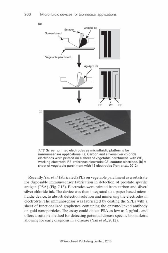
266 Microfl uidic devices for biomedical applications
© Woodhead Publishing Limited, 2013
Screen boardScraper
(a)Carbon ink
Vegetable parchment
Ag/AgCl ink
CE REWE
(b)
7.13 Screen printed electrodes as microfl uidic platforms for
immunosensor applications. (a) Carbon and silver/silver chloride
electrodes were printed on a sheet of vegetable parchment, with WE,
working electrode; RE, reference electrode; CE, counter electrode. (b) A
sheet of vegetable parchment with 18 electrodes (Yan et al ., 2012).
Recently, Yan et al . fabricated SPEs on vegetable parchment as a substrate
for disposable immunosensor fabrication in detection of prostate specifi c
antigen (PSA) (Fig. 7.13). Electrodes were printed from carbon and silver/
silver chloride ink. The device was then integrated to a paper-based micro-
fl uidic device, to absorb detection solution and immersing the electrodes in
electrolyte. The immunosensor was fabricated by coating the SPEs with a
sheet of functionalized graphenes, containing the enzyme-linked antibody
on gold nanoparticles. The assay could detect PSA as low as 2 pg/mL, and
offers a suitable method for detecting potential disease specifi c biomarkers,
allowing for early diagnosis in a disease (Yan et al ., 2012).

Microfl uidic devices for drug discovery and analysis 267
© Woodhead Publishing Limited, 2013
A microfl uidic magnetic disc has been used to separate rare, circulating
endothelial cells (CEC) from peripheral blood mononuclear cells (Chen
et al ., 2011b). These CECs have been associated with many diseases, and
their low concentration in blood impedes their detection. A magnetic disc
was used to trap the cells attached to immunomagnetic beads. Human
umbilical vein endothelial cells (HUVEC) were used as a model for CECs
and stained with anti-CD146-phycoerythrin antibody, which was tagged to
anti-phycoerythrin magnetic beads that attracted these cells to the magnetic
disc. This magnetic disc had an inlet channel, connecting channels and a
waste reservoir. When the disc was rotated, the centrifugal force propelled
away the non-magnetic cells through the connecting channels to the waste
reservoir, effectively retaining target cells (HUVECs) in the inlet reservoir,
which had a multistage magnet over it.
As a basic criterion, the point-of-care diagnostic devices meant for the
developing world must be inexpensive and integrated, dispensing the ser-
vices of ancillary equipments (Mabey et al ., 2004; Martinez et al ., 2008a). In
the wake of this cost consideration, paper-based microfl uidic devices offer
a potential alternative to glass/polymer-based open channel microsystems.
These devices can also enable a multitude of sample outlets from a sin-
gle inlet, ensuring simultaneous analytical assays, without the need for an
external pumping device. Being light in weight and easy to stack makes
their shipment logistically much easier than glass/polymer-based devices.
Paper-based microfl uidic devices have been researched in detail by the lab-
oratory of George Whitesides at Harvard, called 3D microfl uidic paper ana-
lytical devices ( μ PAD) (Bruzewicz et al ., 2008; Martinez et al ., 2007, 2008b,
2008c).
Microfl uidic devices have also been developed for diagnosis of lyso-
somal storage disorders (Shen et al ., 2012), cancer (Lien et al ., 2010; Chen
et al ., 2011a; Tang et al ., 2012), H1N1 infl uenza (Lee et al ., 2012) and herpes
simplex virus (Zubair et al ., 2011), sickle cell disease (Hersher, 2012) and
Johne’s disease in cattle (Wadhwa et al ., 2012). In principle, all microfl uidic
diagnostic devices consist of a molecular sensing unit coupled to a signal
converter (transducer) that reads out the results quantitatively. An elabo-
rate review of different mechanistic approaches of biosensors is provided by
(Mohanty and Kougianos, 2006). Since the biosensors vary greatly in their
design, and hence their sensitivity and effi ciency, the reader is referred to a
few state-of-the-art reviews for more detailed information (Choi et al ., 2011;
Mohammed and Desmulliez, 2011).
It is expected that microfl uidic technology will play a crucial role in med-
ical diagnostics in the coming years, essentially with the development of
disposable, sample-to-result devices, making routine diagnosis a more per-
sonalized approach (Eicher and Merten, 2011; Foudeh et al ., 2012).
268 Microfl uidic devices for biomedical applications
© Woodhead Publishing Limited, 2013
7.4 Conclusion and future trends
Conclusively, microfl uidic devices have shown initial promise in all areas of
drug discovery and development, as well as in the domains of miniaturized
diagnostic devices. The future of microfl uidic devices for application in drug
discovery appears bright with a lot of research activity being focused on the
development of miniaturized chips. However, the concern remains about
the integration of these devices with ancillary equipment, including electri-
cal accessories, pressure pumps and platforms for analysis of samples. Due
to their increased acceptance and potential benefi ts as economical alterna-
tives to conventional bench-top macroscale equipments, it is important to
develop integrated ‘everything-on-a-chip’ systems that are widely accepted
in all stages of drug discovery and development. This may entail an inter-dis-
ciplinary effort from engineers and researchers working on fl uid dynamics
to design micropumps, analytical equipment manufacturers to scale down
analysis systems to commensurate with the chip size, and fi nally researchers
working on microfabrication to further miniaturize the platforms and mak-
ing them adaptable to these ancillary systems.
Industry has played a crucial role in this respect so far. Fluidigm Corporation
(http://www.fl uidigm.com/), a venture by Dr Stephen Quake from Caltech,
has developed various platforms for microfl uidic device integration. Based
on the technology known as multilayer soft lithography, 3D structures can
be created from elastomers to form integrated valves (NanoFlex ™ ), pumps
and channels. Besides this, Caliper Life Sciences (http://www.caliperls.com/)
have also developed several automated/semi-automated robotics controlled
liquid handling systems (Zephyr®) that can be potentially integrated with
microfl uidic devices. These microfl uidic integration tools are expected to aid
microfl uidic-based drug discovery by improving effi ciency and scalability.
With the rate of approval of new drugs declining in the past few years and
the pharmaceutical industry still lacking effective tools in discovering new
drugs, microfl uidic platforms come with a ray of hope, chaperoning routine
assays in a more effi cient manner and hopefully allowing more highly effi ca-
cious and safe drugs to be discovered.
On the other hand, miniaturized devices as diagnostic kits have made
inroads into the households of many diabetic patients as blood glucose moni-
tors. Miniaturized devices for other applications not only face fabrication
and technical issues of being an integrated and comprehensive system, but
they also face the challenge of patient acceptance, having a direct interface
with the end user. Although their acceptance is propelled by the convenience
of use, the future of such devices for routine practice in other pathologi-
cal conditions would rely on how the users perceive them. A sizeable pop-
ulation, particularly in remote villages and tribal areas in developing world,
lack good education, and training to use such devices might prove to be a
Microfl uidic devices for drug discovery and analysis 269
© Woodhead Publishing Limited, 2013
daunting task. Moreover, it would require sincere effort on the part of cli-
nicians and marketing professionals, to persuade the patients in developed
world, who have been so used to visiting a clinic to get their routine bio-
chemical check-ups, to adopt such self-usable devices. In addition, it has to
be ensured that such devices are safe to use and dispose, without causing any
serious environmental hazards.
7.5 Sources of further information and advice
Although we have tried to encompass the major developments on the drug
discovery and analysis front in this chapter, the reader is referred to the
following for further details. A review refl ecting on the origins and the cur-
rent status of microfl uidics by George Whitesides is highly recommended
(Whitesides, 2006). Another comprehensive review, by Yeo et al ., that
describes the role of microfl uidics in various biological applications is also
recommended (Yeo et al ., 2011). Other important works that appear in lit-
erature and circumscribe the two areas of interest in this chapter include
(Imming et al ., 2006; Yager et al ., 2006; Kang et al ., 2008; Wen and Yang, 2008;
Lombardi and Dittrich, 2010; Livak-Dahl et al ., 2011).
7.6 References Alsenz, J. and Kansy, M. (2007). High throughput solubility measurement in drug
discovery and development. Adv Drug Deliv Rev , 59 , 546–67. DOI 10.1016/j.
addr.2007.05.007
Anonymous (2000). Signal transduction as a drug-discovery platform. Nat Biotechnol , 18 , 37–9.
Ashraf, M. W., Tayyaba, S. and Afzulpurkar, N. (2011). Micro electromechanical sys-
tems (mems) based microfl uidic devices for biomedical applications. Int J Mol Sci , 12 , 3648–704. DOI 10.3390/ijms12063648
Bange, A., Halsall, H. B. and Heineman, W. R. (2005). Microfl uidic immunosensor
systems. Biosens Bioelectron , 20 , 2488–503. DOI 10.1016/j.bios.2004.10.016
Battersby, B. J. and Trau, M. (2002). Novel miniaturized systems in high-throughput
screening. Trends Biotechnol , 20 , 167–73.
Beebe, D. J., Mensing, G. A. and Walker, G. M. (2002). Physics and applications
of microfl uidics in biology. Annu Rev Biomed Eng , 4 , 261–86. DOI 10.1146/
annurev.bioeng.4.112601.125916
Beggs, M. (2001). HTS – where next? Drug Discov World , 2 , 125–34.
Bogojevic, D., Chamberlain, M. D., Barbulovic-Nad, I. and Wheeler, A. R. (2012). A
digital microfl uidic method for multiplexed cell-based apoptosis assays. Lab Chip , 12 , 627–34. DOI 10.1039/c2lc20893h
Bohacek, R. S., Mcmartin, C. and Guida, W. C. (1996). The art and practice
of structure-based drug design: a molecular modeling perspective. Med Res Rev , 16 , 3–50. DOI 10.1002/(SICI)1098–1128(199601)16:1<3::AID-
MED1>3.0.CO;2–6

270 Microfl uidic devices for biomedical applications
© Woodhead Publishing Limited, 2013
Braeckmans, K., De Smedt, S. C., Leblans, M., Pauwels, R. and Demeester, J. (2002).
Encoding microcarriers: present and future technologies. Nat Rev Drug Discov ,
1 , 447–56. DOI 10.1038/nrd817
Brandish, P. E., Chiu, C. S., Schneeweis, J., Brandon, N. J., Leech, C. L., Kornienko,
O., Scolnick, E. M., Strulovici, B. and Zheng, W. (2006). A cell-based ultra-high-
throughput screening assay for identifying inhibitors of D-amino acid oxidase.
J Biomol Screen , 11 , 481–7. DOI 10.1177/1087057106288181
Brouzes, E. (2012). Droplet microfl uidics for single-cell analysis. Methods Mol Biol , 853 , 105–39. DOI 10.1007/978–1–61779–567–1_10
Bruzewicz, D. A., Reches, M. and Whitesides, G. M. (2008). Low-cost printing of
poly(dimethylsiloxane) barriers to defi ne microchannels in paper. Anal Chem ,
80 , 3387–92. DOI 10.1021/Ac702605a
Cesaro-Tadic, S., Dernick, G., Juncker, D., Buurman, G., Kropshofer, H., Michel, B.,
Fattinger, C. and Delamarche, E. (2004). High-sensitivity miniaturized immu-
noassays for tumor necrosis factor a using microfl uidic systems. Lab Chip , 4 ,
563–9. DOI 10.1039/B408964b
Chen, H., Gao, F., He, R. and Cui, D. (2007). Chemiluminescence of luminol cata-
lyzed by silver nanoparticles. J Colloid Interface Sci , 315 , 158–63. OI 10.1016/j.
jcis.2007.06.052
Chen, J. K., Bai, B. J. and Chang, F. C. (2011a). Diagnosis of breast cancer recurrence
using a microfl uidic device featuring tethered cationic polymers. Appl Phys Lett , 99 , 1–3. DOI 10.1063/1.3608239
Chen, K. C., Lee, T. P., Pan, Y. C., Chiang, C. L., Chen, C. L., Yang, Y. H., Chiang, B. L.,
Lee, H. and Wo, A. M. (2011b). Detection of circulating endothelial cells via a
microfl uidic disk. Clin Chem , 57 , 586–92. DOI 10.1373/clinchem.2010.157404
Chen, P. C., Huang, Y. Y. and Juang, J. L. (2011c). MEMS microwell and microcol-
umn arrays: novel methods for high-throughput cell-based assays. Lab Chip , 11 ,
3619–25. DOI 10.1039/c0lc00696c
Chen, Y. J., Roller, E. E. and Huang, X. (2010). DNA sequencing by denaturation:
experimental proof of concept with an integrated fl uidic device. Lab Chip , 10 ,
1153–9. DOI 10.1039/b921417h
Cheng, Y. L., Bushby, R. J., Evans, S. D., Knowles, P. F., Miles, R. E. and Ogier, S. D.
(2001). Single ion channel sensitivity in suspended bilayers on micromachined
supports. Langmuir , 17 , 1240–42.
Choi, S., Goryll, M., Sin, L. Y. M., Wong, P. K. and Chae, J. (2011). Microfl uidic-
based biosensors toward point-of-care detection of nucleic acids and proteins.
Microfl uid Nanofl uidics , 10 , 231–47.
Delbeke, F. T., Desmet, N. and Debackere, M. (1995). The abuse of doping agents in
competing body builders in Flanders (1988–1993). Int J Sports Med , 16 , 66–70.
DOI 10.1055/s-2007–972966
Demello, A. J. (2006). Control and detection of chemical reactions in microfl uidic
systems. Nature , 442 , 394–402. DOI 10.1038/nature05062
Dexter, J. P. and Parker, W. (2009). Parallel combinatorial chemical synthesis using
single-layer poly(dimethylsiloxane) microfl uidic devices. Biomicrofl uidics , 3 ,
34106. DOI 10.1063/1.3230501
Dunlop, J., Bowlby, M., Peri, R., Vasilyev, D. and Arias, R. (2008). High-throughput
electrophysiology: an emerging paradigm for ion-channel screening and physi-
ology. Nat Rev Drug Discov , 7 , 358–68. DOI 10.1038/nrd2552
Eicher, D. and Merten, C. A. (2011). Microfl uidic devices for diagnostic applications.
Expert Rev Mol Diagn , 11 , 505–19.DOI 10.1586/ERM.11.25
Microfl uidic devices for drug discovery and analysis 271
© Woodhead Publishing Limited, 2013
Esch, M. B., King, T. L. and Shuler, M. L. (2011). The role of body-on-a-chip devices
in drug and toxicity studies. Annu Rev Biomed Eng , 13 , 55–72. DOI 10.1146/
annurev-bioeng-071910–124629
Evans, M., Sewter, C. and Hill, E. (2003). An encoded particle array tool
for multiplex bioassays. Assay Drug Dev Technol , 1 , 199–207. DOI
10.1089/154065803321537926
Fertig, N., Blick, R. H. and Behrends, J. C. (2002). Whole cell patch clamp recording
performed on a planar glass chip. Biophys J , 82 , 3056–62.
Foudeh, A. M., Fatanat Didar, T., Veres, T. and Tabrizian, M. (2012). Microfl uidic
designs and techniques using lab-on-a-chip devices for pathogen detection for
point-of-care diagnostics. Lab Chip , 12 , 3249–66.
Fox, S., Farr-Jones, S., Sopchak, L., Boggs, A., Nicely, H. W., Khoury, R. and Biros, M.
(2006). High-throughput screening: update on practices and success. J Biomol Screen , 11 , 864–9. DOI 10.1177/1087057106292473
Gao, J., Yin, X. F. and Fang, Z. L. (2004). Integration of single cell injection, cell lysis,
separation and detection of intracellular constituents on a microfl uidic chip.
Lab Chip , 4 , 47–52. DOI 10.1039/b310552k
Goldberg, M. D., Lo, R. C., Abele, S., Macka, M. and Gomez, F. A. (2009). Development
of microfl uidic chips for heterogeneous receptor-ligand interaction studies.
Anal Chem , 81 , 5095–8. DOI 10.1021/ac9006649
Gomez, F. A. (2011). Microfl uidics in protein chromatography. Methods Mol Biol , 681 , 137–50. DOI 10.1007/978–1–60761–913–0_8
Goodnow, R.A. (2006). Hit and lead identifi cation: integrated technology based
approaches. Drug Discovery Today , 3 , 367–75.
Graaf, I. a. M., Groothuis, G. M. M. and Olinga, P. (2007). Precision-cut tissue slices
as a tool to predict metabolism of novel drugs. Exp Opin Drug Metab Toxicol , 3 , 879–98. DOI 10.1517/17425255.3.6.879
Guo, M. T., Rotem, A., Heyman, J. A. and Weitz, D. A. (2012). Droplet microfl uid-
ics for high-throughput biological assays. Lab Chip , 12 , 2146–55. DOI 10.1039/
c2lc21147e
Gurwitz, D. and Haring, R. (2003). Ligand-selective signaling and high-content
screening for GPCR drugs. Drug Discov Today , 8 , 1108–9.
Hadd, A. G., Raymond, D. E., Halliwell, J. W., Jacobson, S. C. and Ramsey, J. M. (1997).
Microchip device for performing enzyme assays. Analy Chem , 69 , 3407–12.
Han, M., Gao, X., Su, J. Z. and Nie, S. (2001). Quantum-dot-tagged microbeads for
multiplexed optical coding of biomolecules. Nat Biotechnol , 19 , 631–5. DOI
10.1038/90228
Harrison, D. J., Fluri, K., Seiler, K., Fan, Z., Effenhauser, C. S. and Manz, A. (1993).
Micromachining a miniaturized capillary electrophoresis-based chemical anal-
ysis system on a chip. Science , 261 , 895–7. DOI 10.1126/science.261.5123.895
Hattersley, S. M., Greenman, J. and Haswell, S. J. (2011). Study of ethanol induced
toxicity in liver explants using microfl uidic devices. Biomed Microdevices , 13 ,
1005–14. DOI 10.1007/s10544–011–9570–2
Herr, A. E., Hatch, A. V., Throckmorton, D. J., Tran, H. M., Brennan, J. S., Giannobile,
W. V. and Singh, A. K. (2007). Microfl uidic immunoassays as rapid saliva-based
clinical diagnostics. Proc Natl Acad Sci U S A , 104 , 5268–73. DOI 10.1073/
pnas.0607254104
Herrmann, M., Veres, T. and Tabrizian, M. (2006). Enzymatically-generated fl uorescent
detection in micro-channels with internal magnetic mixing for the development
of parallel microfl uidic ELISA. Lab Chip , 6 , 555–60. DOI 10.1039/b516031f
272 Microfl uidic devices for biomedical applications
© Woodhead Publishing Limited, 2013
Hersher, R. (2012). Microfl uidic chips promise better diagnosis for sickle cell disease.
Nat Med , 18 , 475. DOI 10.1038/nm0412–475
Hesketh, J. E., Campbell, G. P., Lobley, G. E., Maltin, C. A., Acamovic, F. and Palmer,
R. M. (1992). Stimulation of actin and myosin synthesis in rat gastrocnemius
muscle by clenbuterol; evidence for translational control. Comp Biochem Physiol C , 102 , 23–7.
Holmes, D., She, J. K., Roach, P. L. and Morgan, H. (2007). Bead-based immunoas-
says using a micro-chip fl ow cytometer. Lab Chip , 7 , 1048–56. DOI 10.1039/
B707507n
Hosokawa, M., Asami, M., Yoshino, T., Tsujimura, N., Takahashi, M., Nakasono, S.,
Tanaka, T. and Matsunaga, T. (2012). Monitoring of benzene-induced hemato-
toxicity in mice by serial leukocyte counting using a microcavity array. Biosens Bioelectron , 40 , 110–14.
Huang, B., Wu, H., Bhaya, D., Grossman, A., Granier, S., Kobilka, B. K. and Zare,
R. N. (2007). Counting low-copy number proteins in a single cell. Science , 315 ,
81–4. DOI 10.1126/science.1133992
Ide, T. and Ichikawa, T. (2005). A novel method for artifi cial lipid-bilayer formation.
Biosens Bioelectron , 21 , 672–7. DOI 10.1016/j.bios.2004.12.018
Imming, P., Sinning, C. and Meyer, A. (2006). Drugs, their targets and the nature
and number of drug targets. Nat Rev Drug Discov , 5 , 821–34. DOI 10.1038/
nrd2132
Javanmard, M., Babrzadeh, F. and Davis, R. W. (2010). Microfl uidic force spectros-
copy for characterization of biomolecular interactions with piconewton resolu-
tion. Appl Phys Lett , 97 , 173704. DOI 10.1063/1.3491547
Jones, R., Godorhazy, L., Szalay, D., Gerencser, J., Dorman, G., Urge, L. and Darvas, F.
(2005). A novel method for high-throughput reduction of compounds through
automated sequential injection into a continuous-fl ow microfl uidic reactor.
QSAR Comb Sci , 24 , 722–27. DOI 10.1002/qsar.200540006
Kachouie, N., Kang, L. and Khademhosseini, A. (2009). Arraycount, an algorithm
for automatic cell counting in microwell arrays. Biotechniques , 47 , 10–16. DOI
10.2144/000113202
Kamruzzaman, M., Alam, A. M., Kim, K. M., Lee, S. H., Kim, Y. H., Kim, G. M. and
Dang, T. D. (2012). Microfl uidic chip based chemiluminescence detection of
L-phenylalanine in pharmaceutical and soft drinks. Food Chem , 135 , 57–62.
DOI 10.1016/j.foodchem.2012.04.062
Kane, B. J., Zinner, M. J., Yarmush, M. L. and Toner, M. (2006). Liver-specifi c func-
tional studies in a microfl uidic array of primary mammalian hepatocytes. Anal Chem , 78 , 4291–8. DOI 10.1021/ac051856v
Kang, G., Lee, J. H., Lee, C. S. and Nam, Y. (2009). Agarose microwell based neuronal
micro-circuit arrays on microelectrode arrays for high throughput drug testing.
Lab Chip , 9 , 3236–42. DOI 10.1039/b910738j
Kang, L., Chung, B. G., Langer, R. and Khademhosseini, A. (2008). Microfl uidics
for drug discovery and development: from target selection to product lifecycle
management. Drug Discov Today , 13 , 1–13. DOI 10.1016/j.drudis.2007.10.003
Kang, L., Hancock, M. J., Brigham, M. D. and Khademhosseini, A. (2010). Cell con-
fi nement in patterned nanoliter droplets in a microwell array by wiping. J Biomed Mater Res A , 93 , 547–7. DOI 10.1002/jbm.a.32557
Kaplowitz, N. (2005). Idiosyncratic drug hepatotoxicity. Nat Rev Drug Discov , 4 ,
489–99. DOI 10.1038/nrd1750

Microfl uidic devices for drug discovery and analysis 273
© Woodhead Publishing Limited, 2013
Keng, P. Y., Chen, S., Ding, H., Sadeghi, S., Shah, G. J., Dooraghi, A., Phelps, M.
E., Satyamurthy, N., Chatziioannou, A. F., Kim, C. J. and Van Dam, R. M.
(2012). Micro-chemical synthesis of molecular probes on an electronic
microfl uidic device. Proc Natl Acad Sci U S A , 109 , 690–5. DOI 10.1073/
pnas.1117566109
Khetani, S. R. and Bhatia, S. N. (2008). Microscale culture of human liver cells for
drug development. Nat Biotechnol , 26 , 120–6. DOI 10.1038/nbt1361
Kikutani, Y., Horiuchi, T., Uchiyama, K., Hisamoto, H., Tokeshi, M. and Kitamori, T.
(2002). Glass microchip with three-dimensional microchannel network for 2 x
2 parallel synthesis. Lab Chip , 2 , 188–92. DOI 10.1039/b208382p
Kim, D. N., Lee, Y. and Koh, W. G. (2009). Fabrication of microfl uidic devices
incorporating bead-based reaction and microarray-based detection system
for enzymatic assay. Sens Actuators B Chem , 137 , 305–12. DOI 10.1016/j.
snb.2008.12.042
Knight, A. R. (2000). HTS – A strategy for drug discovery. Drug Disov World , 1 , 32–8.
Kola, I. and Landis, J. (2004). Can the pharmaceutical industry reduce attrition rates?
Nat Rev Drug Discov , 3 , 711–15. DOI 10.1038/Nrd1470
Kong, J., Jiang, L., Su, X., Qin, J., Du, Y. and Lin, B. (2009). Integrated microfl uidic
immunoassay for the rapid determination of clenbuterol. Lab Chip , 9 , 1541–7.
DOI 10.1039/b818430e
Kramer, J. A., Sagartz, J. E. and Morris, D. L. (2007). The application of discovery
toxicology and pathology towards the design of safer pharmaceutical lead can-
didates. Nat Rev Drug Discov , 6 , 636–49. DOI 10.1038/nrd2378
Kreir, M., Farre, C., Beckler, M., George, M. and Fertig, N. (2008). Rapid screening
of membrane protein activity: electrophysiological analysis of OmpF reconsti-
tuted in proteoliposomes. Lab Chip , 8 , 587–95. DOI 10.1039/b713982a
Krenkova, J. and Foret, F. (2004). Immobilized microfl uidic enzymatic reactors.
Electrophoresis , 25 , 3550–63. DOI 10.1002/elps.200406096
Kumar, S. S., Chouhan, R. S. and Thakur, M. S. (2009). Enhancement of chemilumi-
nescence for vitamin B12 analysis. Anal Biochem , 388 , 312–6. DOI 10.1016/j.
ab.2009.02.029
Lai, S., Wang, S., Luo, J., Lee, L. J., Yang, S. T. and Madou, M. J. (2004). Design of
a compact disk-like microfl uidic platform for enzyme-linked immunosorbent
assay. Anal Chem , 76 , 1832–7. DOI 10.1021/ac0348322
Lal, R. and Arnsdorf, M. F. (2010). Multidimensional atomic force microscopy for
drug discovery: a versatile tool for defi ning targets, designing therapeutics and
monitoring their effi cacy. Life Sci , 86 , 545–62. DOI 10.1016/j.lfs.2009.02.030
Lee, B. S., Lee, J. N., Park, J. M., Lee, J. G., Kim, S., Cho, Y. K. and Ko, C. (2009). A fully
automated immunoassay from whole blood on a disc. Lab Chip , 9 , 1548–55.
DOI 10.1039/b820321k
Lee, B. S., Lee, Y. U., Kim, H. S., Kim, T. H., Park, J., Lee, J. G., Kim, J., Kim, H., Lee,
W. G. and Cho, Y. K. (2011). Fully integrated lab-on-a-disc for simultaneous
analysis of biochemistry and immunoassay from whole blood. Lab Chip , 11 ,
70–8. DOI 10.1039/c0lc00205d
Lee, C. C., Sui, G. D., Elizarov, A., Shu, C. Y. J., Shin, Y. S., Dooley, A. N., Huang,
J., Daridon, A., Wyatt, P., Stout, D., Kolb, H. C., Witte, O. N., Satyamurthy, N.,
Heath, J. R., Phelps, M. E., Quake, S. R. and Tseng, H. R. (2005). Multistep syn-
thesis of a radiolabeled imaging probe using integrated microfl uidics. Science ,
310 , 1793–6. DOI 10.1126/science.1118919
274 Microfl uidic devices for biomedical applications
© Woodhead Publishing Limited, 2013
Lee, K. G., Lee, T. J., Jeong, S. W., Choi, H. W., Heo, N. S., Park, J. Y., Park, T. J. and Lee,
S. J. (2012). Development of a plastic-based microfl uidic immunosensor chip
for detection of H1N1 infl uenza. Sensors , 12 , 10810–19.
Lee, S. H., Lee, C. S., Kim, B. G. and Kim, Y. K. (2008). An integrated microfl uidic
chip for the analysis of biochemical reactions by MALDI mass spectrometry.
Biomed Microdevices , 10 , 1–9. DOI 10.1007/s10544–007–9104–0
Lee, W. G., Kim, Y. G., Chung, B. G., Demirci, U. and Khademhosseini, A. (2010).
Nano/Microfl uidics for diagnosis of infectious diseases in developing countries.
Adv Drug Deliv Rev , 62 , 449–57. DOI 10.1016/j.addr.2009.11.016
Lew, V., Nguyen, D. and Khine, M. (2011). Shrink-induced single-cell plastic microw-
ell array. J Lab Autom , 16 , 450–6. DOI 10.1016/j.jala.2011.06.003
Li, A. P., Bode, C. and Sakai, Y. (2004). A novel in vitro system, the integrated dis-
crete multiple organ cell culture (IdMOC) system, for the evaluation of human
drug toxicity: comparative cytotoxicity of tamoxifen towards normal human
cells from fi ve major organs and MCF-7 adenocarcinoma breast cancer cells.
Chem Biol Interact , 150 , 129–36. DOI 10.1016/j.cbi.2004.09.010
Li, X. J., Valadez, A. V., Zuo, P. and Nie, Z. (2012). Microfl uidic 3D cell culture:
potential application for tissue-based bioassays. Bioanalysis , 4 , 1509–25. DOI
10.1063/1.3687398
Lien, K. Y., Chuang, Y. H., Hung, L. Y., Hsu, K. F., Lai, W. W., Ho, C. L., Chou, C. Y.
and Lee, G. B. (2010). Rapid isolation and detection of cancer cells by utilizing
integrated microfl uidic systems. Lab Chip , 10 , 2875–86. DOI 10.1039/c005178k
Lion, N., Reymond, F., Girault, H. H. and Rossier, J. S. (2004). Why the move to micro-
fl uidics for protein analysis? Curr Opin Biotechnol , 15 , 31–7. DOI 10.1016/j.
copbio.2004.01.001
Liszewski, K. (2003). Broader uses for microfl uidics technologies – Applications
expand from drug discovery to battlefi eld. Gen Eng News , 23 , 66–7.
Liu, C., Liu, J., Gao, D., Ding, M. and Lin, J. M. (2010). Fabrication of microwell arrays
based on two-dimensional ordered polystyrene microspheres for high-through-
put single-cell analysis. Anal Chem , 82 , 9418–24. DOI 10.1021/ac102094r
Liu, C., Wang, L., Xu, Z., Li, J. M., Ding, X. P., Wang, Q. and Li, C. Y. (2012). A multi-
layer microdevice for cell-based high-throughput drug screening. J Micromech Microeng , 22 , 1–7. DOI 10.1088/0960–1317/22/6/065008
Livak-Dahl, E., Sinn, I. and Burns, M. (2011). Microfl uidic chemical analysis
systems. Annu Rev Chem Biomol Eng , 2 , 325–53. DOI 10.1146/annurev-
chembioeng-061010–114215
Lok, K. S., Abdul Muttalib, S. Z., Lee, P. P., Kwok, Y. C. and Nguyen, N. T. (2012).
Rapid determination of vitamin B12 concentration with a chemiluminescence
lab on a chip. Lab Chip , 12 , 2353–61. DOI 10.1039/c2lc00037g
Lombardi, D. and Dittrich, P. S. (2010). Advances in microfl uidics for drug discovery.
Exp Opin Drug Discov , 5 , 1081–94. DOI 10.1517/17460441.2010.521149
Mabey, D., Peeling, R. W., Ustianowski, A. and Perkins, M. D. (2004). Diagnostics for
the developing world. Nat Rev Microbiol , 2 , 231–40. DOI 10.1038/Nrmicro841
Macbeath, G. and Schreiber, S. L. (2000). Printing proteins as microarrays for high-
throughput function determination. Science , 289 , 1760–63.
Makamba, H., Hsieh, Y. Y., Sung, W. C. and Chen, S. H. (2005). Stable permanently
hydrophilic protein-resistant thin-fi lm coatings on poly(dimethylsiloxane) sub-
strates by electrostatic self-assembly and chemical cross-linking. Anal Chem ,
77 , 3971–8. DOI 10.1021/ac0502706
Microfl uidic devices for drug discovery and analysis 275
© Woodhead Publishing Limited, 2013
Malmstadt, N., Nash, M. A., Purnell, R. F. and Schmidt, J. J. (2006). Automated forma-
tion of lipid-bilayer membranes in a microfl uidic device. Nano Lett , 6 , 1961–5.
DOI 10.1021/nl0611034
Manz, A., Graber, N. and Widmer, H. M. (1990). Miniaturized total chemical-analy-
sis systems – a novel concept for chemical sensing. Sens Actuators B Chem , 1 ,
244–48.
Mao, H., Yang, T. and Cremer, P. S. (2002). Design and characterization of immobi-
lized enzymes in microfl uidic systems. Anal Chem , 74 , 379–85.
Martinez-Navarro, J. F. (1990). Food poisoning related to consumption of illicit beta-
agonist in liver. Lancet , 336 , 1311.
Martinez, A. W., Phillips, S. T., Butte, M. J. and Whitesides, G. M. (2007). Patterned
paper as a platform for inexpensive, low-volume, portable bioassays. Angew Chem Int Ed , 46 , 1318–20. DOI 10.1002/anie.200603817
Martinez, A. W., Phillips, S. T., Carrilho, E., Thomas, S. W., 3rd, Sindi, H. and Whitesides,
G. M. (2008a). Simple telemedicine for developing regions: camera phones and
paper-based microfl uidic devices for real-time, off-site diagnosis. Anal Chem ,
80 , 3699–707. DOI 10.1021/ac800112r
Martinez, A. W., Phillips, S. T. and Whitesides, G. M. (2008b). Three-dimensional
microfl uidic devices fabricated in layered paper and tape. Proc Natl Acad Sci U S A , 105 , 19606–11. DOI 10.1073/pnas.0810903105
Martinez, A. W., Phillips, S. T., Wiley, B. J., Gupta, M. and Whitesides, G. M. (2008c).
FLASH: a rapid method for prototyping paper-based microfl uidic devices. Lab Chip , 8 , 2146–50. DOI 10.1039/b811135a
Maruyama, S., Hirayama, C., Abe, J., Tanaka, J. and Matsui, K. (1995). Chronic active
hepatitis and liver cirrhosis in association with combined tamoxifen/tegafur
adjuvant therapy. Dig Dis Sci , 40 , 2602–7.
Matsui, N., Kaya, T., Nagamine, K., Yasukawa, T., Shiku, H. and Matsue, T. (2006).
Electrochemical mutagen screening using microbial chip. Biosens Bioelectron ,
21 , 1202–9. DOI 10.1016/j.bios.2005.05.004
Mayer, M., Kriebel, J. K., Tosteson, M. T. and Whitesides, G. M. (2003). Microfabricated
tefl on membranes for low-noise recordings of ion channels in planar lipid bilay-
ers. Biophys J , 85 , 2684–95.
Meagher, R. J. and Thaitrong, N. (2012). Microchip electrophoresis of DNA follow-
ing preconcentration at photopatterned gel membranes. Electrophoresis , 33 ,
1236–46. DOI 10.1002/elps.201100675
Mei, Q., Fredrickson, C. K., Simon, A., Khnouf, R. and Fan, Z. H. (2007). Cell-free
protein synthesis in microfl uidic array devices. Biotechnol Prog , 23 , 1305–11.
DOI 10.1021/bp070133p
Miserere, S., Ledru, S., Ruille, N., Griveau, S., Boujtita, M. and Bedioui, F. (2006).
Biocompatible carbon-based screen-printed electrodes for the electrochemi-
cal detection of nitric oxide. Electrochem Commun , 8 , 238–44. DOI 10.1016/j.
elecom.2005.11.016
Mohammed, M. I. and Desmulliez, M. P. (2011). Lab-on-a-chip based immunosensor
principles and technologies for the detection of cardiac biomarkers: a review.
Lab Chip , 11 , 569–95. DOI 10.1039/c0lc00204f
Mohanty, S. P. and Kougianos, E. (2006). Biosensors: a tutorial review. Potentials, IEEE , 25 , 35–40. DOI 10.1109/mp.2006.1649009
Moran-Mirabal, J. M., Edel, J. B., Meyer, G. D., Throckmorton, D., Singh, A. K. and
Craighead, H. G. (2005). Micrometer-sized supported lipid bilayer arrays for

276 Microfl uidic devices for biomedical applications
© Woodhead Publishing Limited, 2013
bacterial toxin binding studies through total internal refl ection fl uorescence
microscopy. Biophys J , 89 , 296–305. DOI 10.1529/biophysj.104.054346
Murakami, Y., Endo, T., Yamamura, S., Nagatani, N., Takamura, Y. and Tamiya, E.
(2004). On-chip micro-fl ow polystyrene bead-based immunoassay for quantita-
tive detection of tacrolimus (FK506). Anal Biochem , 334 , 111–6. DOI 10.1016/j.
ab.2004.07.029
Muster, W., Breidenbach, A., Fischer, H., Kirchner, S., Muller, L. and Pahler, A.
(2008). Computational toxicology in drug development. Drug Discov Today ,
13 , 303–10. DOI 10.1016/j.drudis.2007.12.007
Nolan, J. P. and Sklar, L. A. (2002). Suspension array technology: evolution of the
fl at-array paradigm. Trends Biotechnol , 20 , 9–12.
Pampaloni, F., Reynaud, E. G. and Stelzer, E. H. K. (2007). The third dimension
bridges the gap between cell culture and live tissue. Nat Rev Mol Cell Biol , 8 ,
839–45. DOI 10.1038/Nrm2236
Park, J., Sunkara, V., Kim, T. H., Hwang, H. and Cho, Y. K. (2012). Lab-on-a-disc
for fully integrated multiplex immunoassays. Anal Chem , 84 , 2133–40. DOI
10.1021/ac203163u
Perrin, D., Fremaux, C. and Scheer, A. (2006). Assay development and screening of a
serine/threonine kinase in an on-chip mode using caliper nanofl uidics technol-
ogy. J Biomol Screen , 11 , 359–68. DOI 10.1177/1087057106286653
Peterson, D. S. (2005). Solid supports for micro analytical systems. Lab Chip , 5 , 132–
9. DOI 10.1039/b405311g
Pregibon, D. C., Toner, M. and Doyle, P. S. (2007). Multifunctional encoded particles
for high-throughput biomolecule analysis. Science , 315 , 1393–6. DOI 10.1126/
science.1134929
Puckett, L. G., Dikici, E., Lai, S., Madou, M., Bachas, L. G. and Daunert, S. (2004).
Investigation into the applicability of the centrifugal microfl uidics develop-
ment of protein-platform for the ligand binding assays incorporating enhanced
green fl uorescent protein as a fl uorescent reporter. Anal Chem , 76 , 7263–8. DOI
10.1021/Ac049758h
Quinton, D., Girard, A., Thi Kim, L. T., Raimbault, V., Griscom, L., Razan, F., Griveau,
S. and Bedioui, F. (2011). On-chip multi-electrochemical sensor array platform
for simultaneous screening of nitric oxide and peroxynitrite. Lab Chip , 11 ,
1342–50. DOI 10.1039/c0lc00585a
Qureshi, Z. P., Seoane-Vazquez, E., Rodriguez-Monguio, R., Stevenson, K. B. and
Szeinbach, S. L. (2011). Market withdrawal of new molecular entities approved
in the United States from 1980 to 2009. Pharmacoepidemiol Drug Saf , 20 , 772–
7. DOI 10.1002/pds.2155
Razumovitch, J., Meier, W. and Vebert, C. (2009). A microcontact printing approach
to the immobilization of oligonucleotide brushes. Biophys Chem , 139 , 70–4.
DOI 10.1016/j.bpc.2008.10.005
Rettig, J. R. and Folch, A. (2005). Large-scale single-cell trapping and imaging using
microwell arrays. Anal Chem , 77 , 5628–34. DOI 10.1021/Ac0505977
Rodr í guez-D é vora, J. I., Zhang, B., Reyna, D., Shi, Z. D. and Xu, T. (2012). High
throughput miniature drug-screening platform using bioprinting technology.
Biofabrication , 1–8.
Sahoo, H. R., Kralj, J. G. and Jensen, K. F. (2007). Multistep Continuous-Flow
Microchemical Synthesis Involving Multiple Reactions and Separations. Angew Chem Int Ed , 46 , 5704–8. DOI 10.1002/anie.200701434

Microfl uidic devices for drug discovery and analysis 277
© Woodhead Publishing Limited, 2013
Sandison, M. E., Zagnoni, M. and Morgan, H. (2007). Air-exposure technique for the
formation of artifi cial lipid bilayers in microsystems. Langmuir , 23 , 8277–84.
DOI 10.1021/la7007528
Schuler, T., Asmus, T., Fritzsche, W. and Moller, R. (2009). Screen printing as cost-effi -
cient fabrication method for DNA-chips with electrical readout for detection of
viral DNA. Biosens Bioelectron , 24 , 2077–84. DOI 10.1016/j.bios.2008.10.028
Schulze, M. and Belder, D. (2012). Poly(ethylene glycol)-coated microfl uidic
devices for chip electrophoresis. Electrophoresis , 33 , 370–8. DOI 10.1002/
elps.201100401
Service, R. F. (1995). Chemistry – radio tags speed compound synthesis. Science , 270 , 577.
Shen, J., Zhou, Y., Lu, T., Peng, J., Lin, Z., Huang, L., Pang, Y., Yu, L. and Huang, Y.
(2012). An integrated chip for immunofl uorescence and its application to analyze
lysosomal storage disorders. Lab Chip , 12 , 317–24. DOI 10.1039/c1lc20845d
Sheng, J., Zhang, L., Lei, J. and Ju, H. (2012). Fabrication of tunable microreac-
tor with enzyme modifi ed magnetic nanoparticles for microfl uidic electro-
chemical detection of glucose. Anal Chim Acta , 709 , 41–6. DOI 10.1016/j.
aca.2011.10.008
Shi, J., Yang, T. and Cremer, P. S. (2008). Multiplexing ligand-receptor binding mea-
surements by chemically patterning microfl uidic channels. Anal Chem , 80 ,
6078–84. DOI 10.1021/ac800912f
Shiddiky, M. J. A. and Shim, Y. B. (2007). Trace analysis of DNA: Preconcentration,
separation, and electrochemical detection in microchip electrophoresis using
Au nanoparticles. Anal Chem , 79 , 3724–33. DOI 10.1021/Ac0701177
Sikanen, T., Aura, S., Franssila, S., Kotiaho, T. and Kostiainen, R. (2012). Microchip
capillary electrophoresis-electrospray ionization-mass spectrometry of intact
proteins using uncoated Ormocomp microchips. Anal Chim Acta , 711 , 69–76.
DOI 10.1016/j.aca.2011.10.059
Smith, C. (2007). Tools for drug discovery: tools of the trade. Nature , 446 , 219–22.
DOI 10.1038/446219a
Srinivasan, V., Pamula, V. K. and Fair, R. B. (2004). An integrated digital microfl uidic
lab-on-a-chip for clinical diagnostics on human physiological fl uids. Lab Chip ,
4 , 310–15. DOI 10.1039/b403341h
Sui, G., Wang, J., Lee, C. C., Lu, W., Lee, S. P., Leyton, J. V., Wu, A. M. and
Tseng, H. R. (2006). Solution-phase surface modification in intact
poly(dimethylsiloxane) microfluidic channels. Anal Chem , 78 , 5543–51.
DOI 10.1021/ac060605z
Sung, J. H. and Shuler, M. L. (2009). A micro cell culture analog (microCCA) with 3-D
hydrogel culture of multiple cell lines to assess metabolism-dependent cyto-
toxicity of anti-cancer drugs. Lab Chip , 9 , 1385–94. DOI 10.1039/b901377f
Suzuki, H., Tabata, K., Kato-Yamada, Y., Noji, H. and Takeuchi, S. (2004). Planar
lipid bilayer reconstitution with a micro-fl uidic system. Lab Chip , 4 , 502–5. DOI
10.1039/b405967k
Suzuki, H., Tabata, K. V., Noji, H. and Takeuchi, S. (2006). Highly reproducible method
of planar lipid bilayer reconstitution in polymethyl methacrylate microfl uidic
chip. Langmuir , 22 , 1937–42. DOI 10.1021/La052534p
Suzuki, H., Tabata, K. V., Noji, H. and Takeuchi, S. (2007). Electrophysiological
recordings of single ion channels in planar lipid bilayers using a polymethyl
methacrylate microfl uidic chip. Biosens Bioelectron , 22 , 1111–15. DOI 10.1016/j.
bios.2006.04.013

278 Microfl uidic devices for biomedical applications
© Woodhead Publishing Limited, 2013
Szita, N., Boccazzi, P., Zhang, Z. Y., Boyle, P., Sinskey, A. J. and Jensen, K. F. (2005).
Development of a multiplexed microbioreactor system for high-throughput
bioprocessing. Lab Chip , 5 , 819–26. DOI 10.1039/B504243g
Tang, C. K., Vaze, A. and Rusling, J. F. (2012). Fabrication of immunosensor microw-
ell arrays from gold compact discs for detection of cancer biomarker proteins.
Lab Chip , 12 , 281–6. DOI 10.1039/c1lc20833k
Tatosian, D. A. and Shuler, M. L. (2009). A novel system for evaluation of drug mix-
tures for potential effi cacy in treating multidrug resistant cancers. Biotechnol Bioeng , 103 , 187–98. DOI 10.1002/bit.22219
Terstappen, G. C., Schlupen, C., Raggiaschi, R. and Gaviraghi, G. (2007). Target
deconvolution strategies in drug discovery. Nat Rev Drug Discov , 6 , 891–903.
DOI 10.1038/nrd2410
Thorsen, T. A. (2004). Microfl uidic tools for high-throughput screening. Biotechniques ,
36 , 197–9.
Um, E., Rha, E., Choi, S. L., Lee, S. G. and Park, J. K. (2012). Mesh-integrated
microdroplet array for simultaneous merging and storage of single-cell drop-
lets. Lab Chip , 12 , 1594–7. DOI 10.1039/c2lc21266h
Vaino, A. R. and Janda, K. D. (2000). Euclidean shape-encoded combinatorial chem-
ical libraries. Proc Natl Acad Sci U S A , 97 , 7692–6.
Van Midwoud, P. M., Groothuis, G. M. M., Merema, M. T. and Verpoorte, E. (2010).
Microfl uidic biochip for the perifusion of precision-cut rat liver slices for metab-
olism and toxicology studies. Biotechnol Bioeng , 105 , 184–94. DOI 10.1002/
Bit.22516
Wadhwa, A., Foote, R. S., Shaw, R. W. and Eda, S. (2012). Bead-based microfl uidic
immunoassay for diagnosis of Johne’s disease. J Immunol Methods , 382 , 196–
202. DOI 10.1016/j.jim.2012.06.006
Wan, Y., Deng, W., Su, Y., Zhu, X., Peng, C., Hu, H., Peng, H., Song, S. and Fan, C.
(2011). Carbon nanotube-based ultrasensitive multiplexing electrochemi-
cal immunosensor for cancer biomarkers. Biosens Bioelectron , 30 , 93–9. DOI
10.1016/j.bios.2011.08.033
Wang, J., Bao, N., Paris, L. L., Wang, H. Y., Geahlen, R. L. and Lu, C. (2008). Detection
of kinase translocation using microfl uidic electroporative fl ow cytometry. Anal Chem , 80 , 1087–93. DOI 10.1021/Ac702065e
Wang, J., Ibanez, A. and Chatrathi, M. P. (2003). On-chip integration of enzyme and
immunoassays: simultaneous measurements of insulin and glucose. J Am Chem Soc , 125 , 8444–5. DOI 10.1021/ja036067e
Wang, L., Yang, P., Li, Y. X., Chen, H. Q., Li, M. G. and Luo, F. B. (2007). A fl ow
injection chemiluminescence method for the determination of fl uoro-
quinolone derivative using the reaction of luminol and hydrogen perox-
ide catalyzed by gold nanoparticles. Talanta , 72 , 1066–72. DOI 10.1016/j.
talanta.2006.12.050
Wang, Y., Shah, P., Phillips, C., Sims, C. E. and Allbritton, N. L. (2012). Trapping cells
on a stretchable microwell array for single-cell analysis. Anal Bioanal Chem ,
402 , 1065–72. DOI 10.1007/s00216–011–5535–9
Wartelle, C., Schuhmann, W., Blochl, A. and Bedioui, F. (2005). Integrated compact
biocompatible hydrogel-based amperometric sensing device for easy screen-
ing of drugs involved in nitric oxide production by adherent cultured cells.
Electrochim Acta , 50 , 4988–4994. DOI 10.1016/j.electacta.2004.12.048

Microfl uidic devices for drug discovery and analysis 279
© Woodhead Publishing Limited, 2013
Wen, Y. and Yang, S. T. (2008). The future of microfl uidic assays in drug development.
Exp Opin Drug Discov , 3 , 1237–53. DOI 10.1517/17460440802434510
Wen, Y., Zhang, X. and Yang, S. T. (2012). Medium to high throughput screening:
microfabrication and chip-based technology. Adv Exp Med Biol , 745 , 181–209.
DOI 10.1007/978–1–4614–3055–1_11
Whitesides, G. M. (2003). The ‘right’ size in nanobiotechnology. Nat Biotechnol , 21 ,
1161–5. DOI 10.1038/nbt872
Whitesides, G. M. (2006). The origins and the future of microfl uidics. Nature , 442 ,
368–73. DOI 10.1038/nature05058
Won, S. Y., Chandra, P., Hee, T. S. and Shim, Y. B. (2012). Simultaneous detection of
antibacterial sulfonamides in a microfl uidic device with amperometry. Biosens Bioelectron , 39 , 204–9. DOI 10.1016/j.bios.2012.07.043
Wu, H., Wheeler, A. and Zare, R. N. (2004). Chemical cytometry on a picoliter-scale
integrated microfl uidic chip. Proc Natl Acad Sci U S A , 101 , 12809–13. DOI
10.1073/pnas.0405299101
Wu, M. H., Huang, S. B. and Lee, G. B. (2010). Microfl uidic cell culture systems for
drug research. Lab Chip , 10 , 939–56. DOI 10.1039/b921695b
Wu, N., Oakeshott, J. G., Easton, C. J., Peat, T. S., Surjadi, R. and Zhu, Y. (2011). A dou-
ble-emulsion microfl uidic platform for in vitro green fl uorescent protein expres-
sion. J Micromech Microeng , 21 , 1–7. DOI 10.1088/0960–1317/21/5/054032
Xu, Y., Little, M. W. and Murray, K. K. (2006). Interfacing capillary gel microfl u-
idic chips with infrared laser desorption mass spectrometry. J Am Soc Mass Spectrom , 17 , 469–74. DOI 10.1016/j.jasms.2005.12.003
Xue, Q., Wainright, A., Gangakhedkar, S. and Gibbons, I. (2001). Multiplexed
enzyme assays in capillary electrophoretic single-use microfl uidic devices.
Electrophoresis , 22 , 4000–7.
Yager, P., Edwards, T., Fu, E., Helton, K., Nelson, K., Tam, M. R. and Weigl, B. H.
(2006). Microfl uidic diagnostic technologies for global public health. Nature ,
442 , 412–8. DOI 10.1038/Nature05064
Yamada, M., Sugaya, S., Naganuma, Y. and Seki, M. (2012). Microfl uidic synthesis
of chemically and physically anisotropic hydrogel microfi bers for guided cell
growth and networking. Soft Matter , 8 , 3122–30. DOI 10.1039/C2sm07263g
Yan, M., Zang, D., Ge, S., Ge, L. and Yu, J. (2012). A disposable electrochemical
immunosensor based on carbon screen-printed electrodes for the detection
of prostate specifi c antigen. Biosens Bioelectron , 38 , 355–61. DOI 10.1016/j.
bios.2012.06.019
Yeo, L. Y., Chang, H. C., Chan, P. P. and Friend, J. R. (2011). Microfl uidic devices for
bioapplications. Small , 7 , 12–48. DOI 10.1002/smll.201000946
Yeo, L. Y., Friend, J. R., Mcintosh, M. P., Meeusen, E. N. T. and Morton, D. a. V. (2010).
Ultrasonic nebulization platforms for pulmonary drug delivery. Exp Opin Drug Deliv , 7 , 663–79. DOI 10.1517/17425247.2010.485608
Yin, H. B., Pattrick, N., Zhang, X. L., Klauke, N., Cordingley, H. C., Haswell, S. J. and
Cooper, J. M. (2008). Quantitative comparison between microfl uidic and micro-
titer plate formats for cell-based assays. Anal Chem , 80 , 179–85.
Zagnoni, M. (2012). Miniaturised technologies for the development of artifi cial lipid
bilayer systems. Lab Chip , 12 , 1026–39. DOI 10.1039/c2lc20991h
Zhang, C. and Van Noort, D. (2011). Cells in microfl uidics. Top Curr Chem , 304 ,
295–321. DOI 10.1007/128_2011_147

280 Microfl uidic devices for biomedical applications
© Woodhead Publishing Limited, 2013
Zhou, X., Cai, S., Hong, A., You, Q., Yu, P., Sheng, N., Srivannavit, O., Muranjan, S.,
Rouillard, J. M., Xia, Y., Zhang, X., Xiang, Q., Ganesh, R., Zhu, Q., Matejko, A.,
Gulari, E. and Gao, X. (2004). Microfl uidic PicoArray synthesis of oligodeoxy-
nucleotides and simultaneous assembling of multiple DNA sequences. Nucleic Acids Res , 32 , 5409–17. DOI 10.1093/nar/gkh879
Zubair, A., Burbelo, P. D., Vincent, L. G., Iadarola, M. J., Smith, P. D. and Morgan, N.
Y. (2011). Microfl uidic LIPS for serum antibody detection: demonstration of a
rapid test for HSV-2 infection. Biomed Microdevices , 13 , 1053–62. DOI 10.1007/
s10544–011–9575-x