microdeletion and prenatal fish probes
TRANSCRIPT

Microdeletion and Prenatal FISH Probes
Features• �Improve�confidence�in�result�interpretation�
with�high�intensity�signals�and�minimal�background
• �Enhance�detection�and�scoring�accuracy�with�robust,�easy-to-analyse�probes
• �Save�time�and�minimise�mixing�errors�with�easy-to-use,�pre-mixed�probes
• �Optimise�stock�levels�and�minimise�wastage�with�flexible�pack�sizes�to�meet�your�needs

Microdeletion and Prenatal FISH Probes
Microdeletion Testing
Microdeletion syndromes are a group of clinically-recognisable disorders brought about by the deletion of specific regions of chromosomal DNA, causing haploinsufficiencies of important genes. These deletions are difficult to visualise using standard cytogenetic techniques such as karyotyping, however FISH can resolve these submicroscopic deletions.
Our comprehensive range of microdeletion probes include products for some of the rarest human genetic syndromes. All of our microdeletion probes are available in economical 5, or standard 10 test kits.
2
Why Choose CytoCell Probes?
Customers can have confidence in CytoCell’s bright, tight signals and minimal background.
The OGT Partnership
Behind every sample is a life that can be improved through the right care decisions. The OGT partnership approach is key to providing the highest level of service, working closely with you to understand your unique challenges, customising our approach to meet your exact needs. Choose CytoCell probes for your FISH analysis; our effective, accurate and simple to use products help clinical decision makers to reach the right decisions for each patient.
Microdeletion and Prenatal FISH Probes
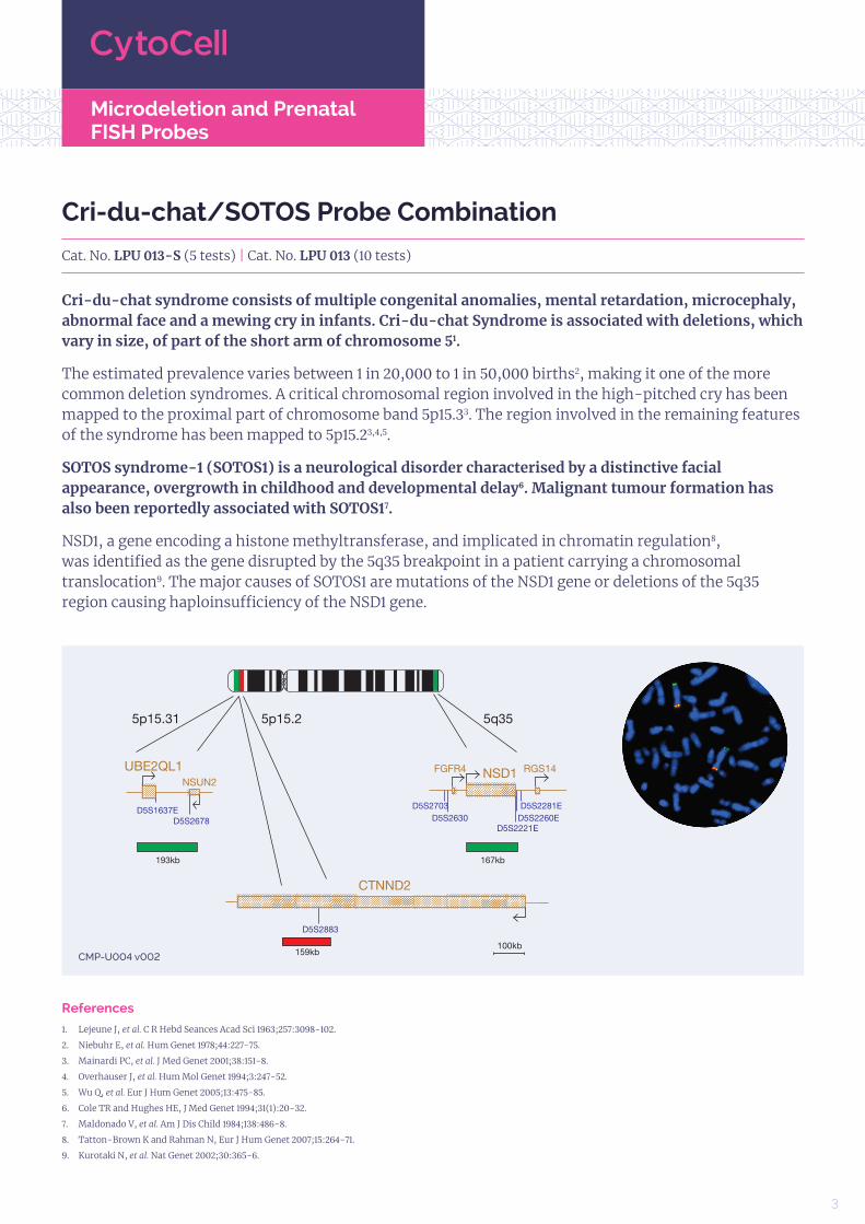
Cri-du-chat/SOTOS Probe Combination
Cat. No. LPU 013-S (5 tests) | Cat. No. LPU 013 (10 tests)
Cri-du-chat syndrome consists of multiple congenital anomalies, mental retardation, microcephaly, abnormal face and a mewing cry in infants. Cri-du-chat Syndrome is associated with deletions, which vary in size, of part of the short arm of chromosome 51.
The estimated prevalence varies between 1 in 20,000 to 1 in 50,000 births2, making it one of the more common deletion syndromes. A critical chromosomal region involved in the high-pitched cry has been mapped to the proximal part of chromosome band 5p15.33. The region involved in the remaining features of the syndrome has been mapped to 5p15.23,4,5.
SOTOS syndrome-1 (SOTOS1) is a neurological disorder characterised by a distinctive facial appearance, overgrowth in childhood and developmental delay6. Malignant tumour formation has also been reportedly associated with SOTOS17.
NSD1, a gene encoding a histone methyltransferase, and implicated in chromatin regulation8, was identified as the gene disrupted by the 5q35 breakpoint in a patient carrying a chromosomal translocation9. The major causes of SOTOS1 are mutations of the NSD1 gene or deletions of the 5q35 region causing haploinsufficiency of the NSD1 gene.
References
1. Lejeune J, et al. C R Hebd Seances Acad Sci 1963;257:3098-102.
2. Niebuhr E, et al. Hum Genet 1978;44:227-75.
3. Mainardi PC, et al. J Med Genet 2001;38:151-8.
4. Overhauser J, et al. Hum Mol Genet 1994;3:247-52.
5. Wu Q, et al. Eur J Hum Genet 2005;13:475-85.
6. Cole TR and Hughes HE, J Med Genet 1994;31(1):20-32.
7. Maldonado V, et al. Am J Dis Child 1984;138:486-8.
8. Tatton-Brown K and Rahman N, Eur J Hum Genet 2007;15:264-71.
9. Kurotaki N, et al. Nat Genet 2002;30:365-6.
CMP-U004�v002
32

Microdeletion and Prenatal FISH Probes
DiGeorge and 22q13.3 Deletion Syndrome Probe Combinations
DiGeorge Syndrome
DiGeorge syndrome1, and a variety of congenital malformation syndromes including Velocardiofacial syndrome (VCFS)2, have in common deletions of chromosome 22 at 22q11.22,3,4,5. These syndromic phenotypes are collectively coined CATCH22, a mnemonic that covers the clinical findings of Cardiac abnormality, Abnormal facies, Thymic aplasia, Cleft palate and Hypocalaemia/Hyperthyroidism due to a chromosome 22 deletion. In addition, around 29% of nonsyndromic patients with isolated conotruncal defects have been shown to have a 22q11.2 microdeletion6. The incidence of these anomalies is estimated to be 1:4000 to 1:9700 live births7 and the deletion of 22q11.2 therefore represents one of the most common genetic defects. A region of approximately 2Mb, referred to as the DiGeorge Critical Region (DGCR), is the most commonly deleted region and occurs in up to 90% of patients5,8,9. Within the DGCR, a minimal critical region of 300- 480kb has been described10,11, containing several genes, including TUPLE1 (HIRA), TBX1, SLC25A1 (CTP) and CLTD.
22q13.3 Deletion Syndrome
The 22q13.3 deletion syndrome presents a recognisable phenotype characterised by hypotonia, delay or absence of expressive speech, global developmental delay, normal to accelerated growth and mild dysmorphic features12,13. Some deletions of the terminal region of chromosome 22q are cytogenetically visible. However, a few cases of cryptic deletions have been reported12,14, suggesting that the actual incidence of 22q telomere deletion may be higher than previously thought. Several observations of patients with 22q13.3 deletion showed that the SHANK3 (ProSAP2)20 gene, encoding a structural protein of the postsynaptic density of excitation synapses and expressed in the cortex and cerebellum of the brain15, was disrupted15,16,17 or deleted18, making it a candidate causative gene for this syndrome. The deletion varies dramatically in size from 130kb to 9Mb18,19,20. The use of 22q subtelomeric probes, distal to the ARSA gene, have therefore been recommended for examining all 22q13.3 deletions20,21.
References
1. Pinsky L, DiGeorge AM, J Pediatr 1965;66:1049-54.
2. Shprintzen RJ, et al. Cleft Palate J 1978;15:56-62.
3. Burn J, et al. J Med Genet 1993;30:822-4.
4. Wilson DI, et al. J Med Genet 1993;30:852-6.
5. Driscoll DA, et al. J Am Hum Genet 1992;50:924-33.
6. Goldmuntz E, et al. J Med Genet 1993;30:807-12.
7. Tezenas Du Montcel S, et al. J Med Genet 1996;33:719.
8. Driscoll DA, et al. Am J Med Genet 1992;44(2):261-8.
9. Scambler PJ, et al. Genomics 1991;10:201-6.
10. Halford S, et al. Hum Mol Genet 1993;2(12):2099-107.
11. Carlson C, et al. Am J Hum Genet 1997;61:620-9.
12. Phelan MC, et al. Am J Med Genet 2001;101(2):91-9.
13. Phelan MC. Orphanet Journal of Rare Diseases 2008,3:14.
14. Prasad C, et al. Clin Genet 2000;57(2):103-9.
15. Beeckers TM, et al. J Neurochem 2002;81(5):903-10.
16. Bonaglia MC, et al. Am J Hum Genet 2001;69(2):261-8.
17. Anderlid BM, et al. Hum Genet 2002;110(5):439-43.
18. Wilson HL, et al. J Med Genet 2003;40(8):575-84.
19. Dupont C, et al. French Speaking Cytogeneticists Association Congress 2003.
20. Luciani J, et al. J Med Genet 2003;40(9):690-6.
21. Chen CP, et al. Prenat Diagn 2003;23(6):504-8.
4

Microdeletion and Prenatal FISH Probes
DiGeorge/VCFS TUPLE1 & 22q13.3 Deletion Syndrome Probe Combination
Cat. No. LPU 004-S (5 tests) | Cat. No. LPU 004 (10 tests)
The TUPLE1 probe is 113kb, labelled in red, and covers most of the TUPLE1 (HIRA) gene. The N85A3 (44kb) probe, labelled in green, is located within 22q13.3 and covers the telomeric end of the SHANK3 gene. The two unique sequences provide control probes for each other and allow identification of chromosome 22.
CMP-U008�v005
5

Microdeletion and Prenatal FISH Probes
DiGeorge/VCFS N25 & 22q13.3 Deletion Syndrome Probe Combination
Cat. No. LPU 010-S (5 tests) | Cat. No. LPU 010 (10 tests)
The N25 probe is 63kb, labelled in red and covers a region including the D22S75 marker and the centromeric end of the CLTCL1 gene. The N85A3 (44kb), labelled in green, is located within the 22q13.3 band and covers the telomeric end of the SHANK3 gene. The two unique sequences provide control probes for each other and allow identification of chromosome 22.
CMP-U007�v003
6

Microdeletion and Prenatal FISH Probes
DiGeorge/TBX1 & 22q13.3 Deletion Syndrome Probe Combination
Cat. No. LPU 014-S (5 tests) | Cat. No. LPU 014 (10 tests)
The TBX1 probe is 211kb, labelled in red, and covers the entire TBX1 gene including the D22S1627 marker. The N85A3 (44kb), labelled in green, is located within 22q13.3 and covers the telomeric end of the SHANK3 gene. The two unique sequences act as control probes for each other and allow identification of chromosome 22.
CMP-U006�v003
76

Microdeletion and Prenatal FISH Probes
DiGeorge II (10p14)
Cat. No. LPU 015-S (5 tests) | Cat. No. LPU 015 (10 tests)
DiGeorge syndrome1, and a variety of congenital malformation syndromes including velocardiofacial syndrome (VCFS)2, share the deletion of chromosome 22 at 22q11.22,3,4,5. These chromosome 22 deletions are collectively coined CATCH22, a mnemonic that covers the clinical findings of Cardiac abnormality, Abnormal facies, Thymic aplasia, Cleft palate and Hypocalaemia/Hyperthyroidism due to a chromosome 22 deletion. In DiGeorge syndrome, however, cases have also been found in which patients have a deletion on chromosome 10p13-p14 (DGS2) instead of chromosome 226,7,8.
The deletion of the DGS2 locus on 10p may be 50 times less frequent than that of the DGS1 locus on 22q and has been estimated to occur in 1 in 200,000 live births9. The CELF2 gene has been identified within the 300kb minimally deleted region of DGS2 and is postulated to be involved in the DGS2 deletion10. CELF2 is a candidate gene for the heart defect and thymus hypoplasia/aplasia associated with partial monosomy 10p10 and may be involved in atrial septal defects (ASDs), a common cardiac anomaly associated with DGS211.
References
1. DiGeorge AM, J Pediatr 1965;67:907.
2. Shprintzen RJ, et al. Cleft Palate J 1978;15:56-62.
3. Wilson DI, et al. J Med Genet 1993;30:852-6.
4. Driscoll DA, et al. J Med Genet 1992;50:924-33.
5. Burn J, et al. J Med Genet 1993;30:822-4.
6. Schuffenhauer S, et al. Ann Genet 1995;38(3):162-7.
7. Daw SC, et al. Nat Genet 1996;13:458-60.
8. Dasouki M, et al. Am J Med Genet 1997;73(1):72-5.
9. Berend SA, et al. Am J Med Genet 2000;91(4):313-7.
10. Lichtner P, et al. J Mol Med 2002;80:431-42.
11. Yatsenko SA, et al. Clin Genet 2004;66:128-36.
CMP-U005�v002�
8

Microdeletion and Prenatal FISH Probes
Prader-Willi/Angelman (SNRPN)
Cat. No. LPU 005-S (5 tests) | Cat. No. LPU 005 (10 tests)
Prader-Willi Syndrome (PWS) and Angelman Syndrome (AS) are distinct neurogenetic disorders caused by the loss of function of genes on chromosome 15 (bands 15q11-13), on either the paternally or maternally inherited chromosome, respectively1.
In 70% of patients, a large interstitial deletion of 3-4Mb is observed1,2. In around 3% of patients, an imprinting defect is observed, caused by either an epimutation or a microdeletion of the Imprinting Centre (IC)1,3. Uniparental disomy, in which both chromosome 15s are inherited from the same parent, accounts for most of the remaining patients with PWS/AS1.
The SNRPN gene is one of four imprinted loci that are expressed from the paternal chromosome 15 region (15q11-13) and maps to the minimally deleted region (MDR) involved in PWS5. Its chromosomal location and imprinting status suggest it plays a possible role in the aetiology of PWS4.
The imprinting centre (IC) maps to a 100kb region proximal to SNRPN. Parental deletions or mutations in the IC impair the imprinting process in 15q11-13 and cause one of two distinct diseases in their offspring5,6. Most of the PWS imprinting deletions involve SNRPN and are approximately 200kb in size. The AS imprinting deletions are small (approximately 40kb), involve the BD3 region, and do not include SNRPN.
References
1. Butler MG, Curr Genomics. 2011 May;12(3):204-215.
2. Clayton-Smith J and Pembrey M. J Med Genet 1992;29:412-5.
3. Buiting K, et al. Am J Hum Genet 1998;63(1):170-80.
4. Glenn C, et al. Am J Hum Genet 1996;58:335-46.
5. Buiting K, et al. Nat Genet 1995;9:395-400.
6. Dittrich B, et al. Nat Genet 1996;14:163-70.
CMP-U013�v002
98

Microdeletion and Prenatal FISH Probes
Prenatal Testing
The CytoCell prenatal FISH assays are designed for the rapid and accurate detection of the most common foetal chromosomal disorders:
• Down syndrome1 (Trisomy 21)
• Edwards syndrome2 (Trisomy 18)
• Patau syndrome3 (Trisomy 13)
• Sex chromosome disorders4 (Copy number changes of X and/or Y chromosomes)
Prenatal Enumeration Kits
Our Prenatal Kits contain FISH probes for the identification of trisomies 21, 18 and 13, as well as sex chromosome aneuploidies utilising an overnight protocol.
FAST FISH Prenatal Kits
When rapid results really matter, choose CytoCell’s FAST prenatal FISH probes; utilise our 2-hour hybridisation protocol to meet the demands of urgent turnaround times, without compromising on signal quality.
Features of our Prenatal range:
• Larger 30 test and 50 test packs for the most commonly performed investigations
• The option of a 2-hour protocol with FAST Prenatal Kits
10
References
1. Lejeune et al. C. R. Acad. Sci. 1959; 248: 1721-1722.
2. http://www.ojrd.com/content/7/1/81/abstract.
3. http://ghr.nlm.nih.gov/condition/trisomy-13.
4. Visootsak and Graham. Orphanet Journal of Rare Diseases 2006, 1:42.

Microdeletion and Prenatal FISH Probes
Prenatal FISH Probe Range
Probe Description Chromosome Region No. Tests Cat. No.*
X,�Y,�18,�13�and�21
Probe�Set�1
X�centromere�Xp11.1-q11.1�(DXZ1)�Green
5,�10,�30�or�50FAST�FISH:�LPF�001 Standard:�LPA�001
Y�centromere�Yp11.1-q11.1�(DYZ3)�Orange
18�centromere�18p11.1-q11.1�(D18Z1)�Blue
Probe�Set�213�unique�sequence�(13q14.2)�Green
21�unique�sequence�(21q22.13)�Orange
X,�Y�and�18
X�centromere�Xp11.1-q11.1�(DXZ1)�Green
5�or�10FAST�FISH:�LPF�002 Standard:�LPA�002
Y�centromere�Yp11.1-q11.1�(DYZ3)�Orange
18�centromere�18p11.1-q11.1�(D18Z1)�Blue
13�and�2113�unique�sequence�(13q14.2)�Green
5�or�10FAST�FISH:�LPF�003 Standard:�LPA�003
21�unique�sequence�(21q22.13)�Orange
13,�18�and�21
13�unique�sequence�(13q14.2)�Green
5�or�10 LPA�00518�centromere�18p11.1-q11.1�(D18Z1)�Blue
21�unique�sequence�(21q22.13)�Orange
18�Centromere 18�centromere�18p11.1-q11.1�(D18Z1)�Blue 5�or�10 LPA�004
*For�5,�30�or�50�test�kit,�add�-X�to�the�catalogue�number�e.g�LPF�###-S,�LPF�###-30,�LPF�###-50.
11

Cytocell Ltd., Oxford Gene Technology, 418 Cambridge Science Park, Milton Road, Cambridge, CB4 0PZ, UK
CytoCell: This document and its contents are © Oxford Gene Technology IP Limited – 2021. All rights reserved. Trademarks: OGT™ (Oxford Gene Technology IP Ltd); CytoCell® is a trademark of Cytocell Ltd. IVD: For in vitro diagnostic use. Some products may not be available in your region.
990251 01/21 v2
Microdeletion and Prenatal FISH Probes
12
Probe NameChromosome
RegionNo. Tests Cat. No.†
Angelman�(UBE3A/D15S10) 15q11.2-q12 5�or�10 LPU�006
Cri-du-chat�and�Sotos�Probe�Combination 5p15.31/5p15.2/5q35 5�or�10 LPU�013
DiGeorge�II�(10p14) 10p14 5�or�10 LPU�015
DiGeorge/VCFS�N25�&�22q13.3�Deletion�Syndrome�Probe�Combination 22q11.2/22q13.3 5�or�10 LPU�010
DiGeorge/VCFS�TUPLE1�&�22q13.3�Deletion�Syndrome�Probe�Combination 22q11.2/22q13.3 5�or�10 LPU�004
DiGeorge/TBX1�&�22q13.3�Deletion�Syndrome�Probe�Combination 22q11.2/22q13.3 5�or�10 LPU�014
Kallmann�(KAL1)�and�Steroid�Sulphatase�Deficiency�(STS)�Probe�Combination Xp22.31 5�or�10 LPU�016
Prader-Willi/Angelman�(SNRPN)� 15q11.2 5�or�10 LPU�005
Saethre-Chotzen/Williams-Beuren�Combination 7p21.1/7q11.23 5�or�10 LPU�024
SHOX Xp22.33/Yp11.32 5�or�10 LPU�025
Smith-Magenis�(RAI1)/Miller-Dieker�Probe�Combination 17p13.3/17p11.2 5�or�10 LPU�019
SRY Yp11.31 5�or�10 LPU�026
Williams-Beuren 7q11.23 5�or�10 LPU�011
Wolf-Hirschhorn 4p16.3 5�or�10 LPU�009
Microdeletion Probe Range
†For�5�test�kit�add�-S�to�catalogue�number,�e.g:�LPU�###-S.
Ordering information
UK +44 (0) 1223 294048
ogt.com