microbiome analysis of pacific white shrimp gut and ... · 8,000 reads were performed in rdp...
TRANSCRIPT

Submitted 15 June 2018Accepted 25 September 2018Published 30 October 2018
Corresponding authorHan Ming Gan,[email protected]
Academic editorHauke Smidt
Additional Information andDeclarations can be found onpage 16
DOI 10.7717/peerj.5826
Copyright2018 Md Zoqratt et al.
Distributed underCreative Commons CC-BY 4.0
OPEN ACCESS
Microbiome analysis of Pacific whiteshrimp gut and rearing water fromMalaysia and Vietnam: implications foraquaculture research and managementMuhammad Zarul Hanifah Md Zoqratt1,2,*, Wilhelm Wei Han Eng1,2,Binh Thanh Thai3, Christopher M. Austin1,2,4,5 and Han Ming Gan1,2,4,5,*
1 School of Science, Monash University Malaysia, Petaling Jaya, Selangor, Malaysia2Genomics Facility, Tropical Medicine and Biology Platform, Monash University Malaysia, Petaling Jaya,Selangor, Malaysia
3 Fisheries and Technical, Economical College, Dinh Bang, Tu Son, Vietnam4Centre for Integrative Ecology, School of Life and Environmental Sciences, Deakin University, Geelong,Victoria, Australia
5Deakin Genomics Centre, Deakin University, Geelong, Victoria, Australia*These authors contributed equally to this work.
ABSTRACTAquaculture production of the Pacific white shrimp is the largest in the world forcrustacean species. Crucial to the sustainable global production of this importantseafood species is a fundamental understanding of the shrimp gut microbiota and itsrelationship to the microbial ecology of shrimp pond. This is especially true, giventhe recently recognized role of beneficial microbes in promoting shrimp nutrientintake and in conferring resistance against pathogens. Unfortunately, aquaculture-related microbiome studies are scarce in Southeast Asia countries despite the severeimpact of early mortality syndrome outbreaks on shrimp production in the region.In this study, we employed the 16S rRNA amplicon (V3–V4 region) sequencing andamplicon sequence variants (ASV) method to investigate the microbial diversity ofshrimp guts and pond water samples collected from aquaculture farms located inMalaysia and Vietnam. Substantial differences in the pond microbiota were observedbetween countries with the presence and absence of several taxa extending to the familylevel. Microbial diversity of the shrimp gut was found to be generally lower than thatof the pond environments with a few ubiquitous genera representing a majority ofthe shrimp gut microbial diversity such as Vibrio and Photobacterium, indicating host-specific selection of microbial species. Given the high sequence conservation of the 16SrRNA gene, we assessed its veracity at distinguishing Vibrio species based on nucleotidealignment against type strain reference sequences and demonstrated the utility of ASVapproach in uncovering awider diversity ofVibrio species compared to the conventionalOTU clustering approach.
Subjects Aquaculture, Fisheries and Fish Science, Marine Biology, MicrobiologyKeywords Metagenomics, Aquaculture, Litopenaeus vannamei, 16S ribosomal RNA ampliconssequencing, Vibrio parahaemolyticus
How to cite this article Md Zoqratt et al. (2018), Microbiome analysis of Pacific white shrimp gut and rearing water from Malaysia andVietnam: implications for aquaculture research and management. PeerJ 6:e5826; DOI 10.7717/peerj.5826
INTRODUCTIONLitopenaeus vannamei (Boone, 1931), also known as the Pacific white shrimp or Whitelegshrimp, is a major aquaculture commodity with a production of 3.69 million tonnes valuedat 18 billion USD revenue (FAO, 2016). In recent years, significant outbreaks of acutehepatopancreatic necrosis disease (AHPND), also known as early mortality syndrome(EMS) have been reported in a number of white shrimp-producing countries. EMS wasfirst reported in China in 2009 and subsequently spread to Southeast Asian countriesincluding Vietnam, Malaysia, and Thailand (Foo et al., 2017; Kondo et al., 2014; Tran etal., 2013). The causative agent of EMS has been reported to be Vibrio parahaemolyticusstrains harbouring a plasmid containing the pirA- and pirB- like genes encoding for toxinscapable of severely damaging the shrimp gut (Han et al., 2015; Lee et al., 2015). To date,EMS has caused an estimated one billion USD of losses to the shrimp industry worldwide(De Schryver, Defoirdt & Sorgeloos, 2014; Lee et al., 2015).
Monitoring and control of pond water quality play a crucial role in managing andpreventing disease outbreak in aquaculture. However, the current practice of water qualitymonitoring usually focuses on the measurement of chemical and physical parameters suchas oxygen, pH, temperature, salinity, turbidity and nitrogen compounds. The importanceof microbial communities in influencing or responding to variation in aquaculture pondwater quality has only been recognized in recent years (Bentzon-Tilia, Sonnenschein &Gram, 2016). This is especially relevant to managing the water quality of aquacultureponds and their cultured biomass because microbes carry out important biological servicesin aquaculture environment including nutrient cycling, probiotic/pathogenic activity andnutrient acquisition in addition to potentially acting as a rapid biological indicator ofcritical chemical changes in the rearing water (Cardona et al., 2016; Cornejo-Granadoset al., 2017; Costa, Pérez & Kreft, 2006; Emerenciano, Gaxiola & Cuzon, 2013; Grotkjær etal., 2016; Jinbo et al., 2017; Liu et al., 2015; Wright, Konwar & Hallam, 2012; Zeng et al.,2017; Zhu et al., 2016). Microbes can also be used to improve the water quality of ponds.For example, adding denitrifying bacteria to biofilters has been shown to reduce theconcentration of ammonia and its immediate derivatives, which are detrimental to shrimphealth (Saffran et al., 2001).
Recognizing the importance of microbial biomass and diversity on the health andproduction of cultured invertebrates, several microbiome studies have analysed the gutmicrobiome of wild-caught shrimps (Cornejo-Granados et al., 2017; Phayungsak et al.,2018; Rungrassamee et al., 2016) as well as cultured shrimps under different abiotic andbiotic factors. However, most studies have been restricted to a specific country especiallyChina, focusing only on one or very few localized ponds (Cornejo-Granados et al., 2017;Huang et al., 2016; Rungrassamee et al., 2016; Tang et al., 2014; Xiong et al., 2015; Zhang etal., 2014; Zhu et al., 2016). A study in Thailand used denaturing gradient gel electrophoresisprofiling and barcoded pyrosequencing to demonstrate a greater survivability of Litopenaeusvannamei and improved the resilience of its gut microbiome upon Vibrio harveyi exposurerelative to that of Penaeus monodon (Rungrassamee et al., 2016). Another more recent studyin Vietnam utilised a standard Illumina 16S rRNA gene amplicon sequencing method to
Md Zoqratt et al. (2018), PeerJ, DOI 10.7717/peerj.5826 2/22

investigate the effect of EMS outbreak on the microbial interaction networks in shrimp guts(Chen et al., 2017b). Thus, despite the emergence of Southeast Asia (SEA) as an aquaculturehub, studies on any significant geographic scale are relatively scarce in this region. Further,to our knowledge, all recent shrimp aquaculture microbiome studies still employ theoperational taxonomic units (OTU) clustering approach in marker-gene data analysisdespite recent calls for the replacement of this approach with exact sequence variantswhich can resolve single-base differences among biological sequences in the sample thusproviding a more comprehensive and accurate view of microbial communities (Callahan,McMurdie & Holmes, 2017; Eren et al., 2014; Utter, Mark Welch & Borisy, 2016).
To initiate a more broad-based investigation of shrimp microbiomes directly relevantto the aquaculture industry in SEA, we performed Illumina 16S rRNA gene ampliconsequencing of L. vannamei guts and rearing water from aquaculture farms located in twoSEA countries with contrasting climates at the time of sampling i.e., Malaysia (warmand humid, 30 ◦C) and Vietnam (cool and dry, 20 ◦C). For the first time in a shrimpaquaculture microbiome study, we applied the recently advocated ASV-method (Edgar,2016) to: (1) compare shrimp intestinal microbial diversity and their pond environments;(2) compare these microbial communities between Malaysia and Vietnam; and (3) assessthe performance of the 16S rRNA V3–V4 hypervariable region and clustering approachesin capturing the genetic diversity of Vibrio.
METHODSSample collectionSampling in Vietnam was performed at two separate shrimp farms in the Quang Ninhprovince (approximately 20 km apart), while sampling in Malaysia was performed ata large shrimp farm with multiple pond systems located in Perak state (Table 1). TwomL of pond water was sampled from 2–4 distant location (corners) of each pond,pelleted via centrifugation at 7,000 rpm for 10 min and resuspended in RNA/DNA shield(ZymoResearch, Irvine, CA, USA). Shrimp intestinal samples were collected by dissectingout the shrimp guts followed by homogenization in RNA/DNA shield (ZymoResearch,Irvine, CA, USA). Sampling was performed with the permission and under the supervisionof the aquaculture manager from the respective farms. No field permit was required forthis study because samples were collected from private fields. As per the request of theaquaculture manager, exact sampling location of some farms was not disclosed in thisstudy to protect the identity of the farm.
DNA extraction, amplification, purification and sequencingGenomic DNA was extracted from the RNA/DNA shield lysate using DNA Clean &ConcentratorTM-5 (ZymoResearch, Irvine, CA, USA) according to the manufacturer’sinstructions. The V3–V4 region of the 16S rRNA gene was amplified using forward primer5′–TCGTCGGCAGCGTCAGATGTGTATAAGAGACAGCCTACGGGNGGCWGCAG–3′ and reverse primer 5′–GTCTCGTGGGCTCGGAGATGTGTATAAGAGACAGGACTACHVGGGTATCTAATCC–3′ containing partial Illumina Nextera adapter. PCRreaction (∼10 ng input DNA/ reaction) and barcode incorporation were performed as
Md Zoqratt et al. (2018), PeerJ, DOI 10.7717/peerj.5826 3/22
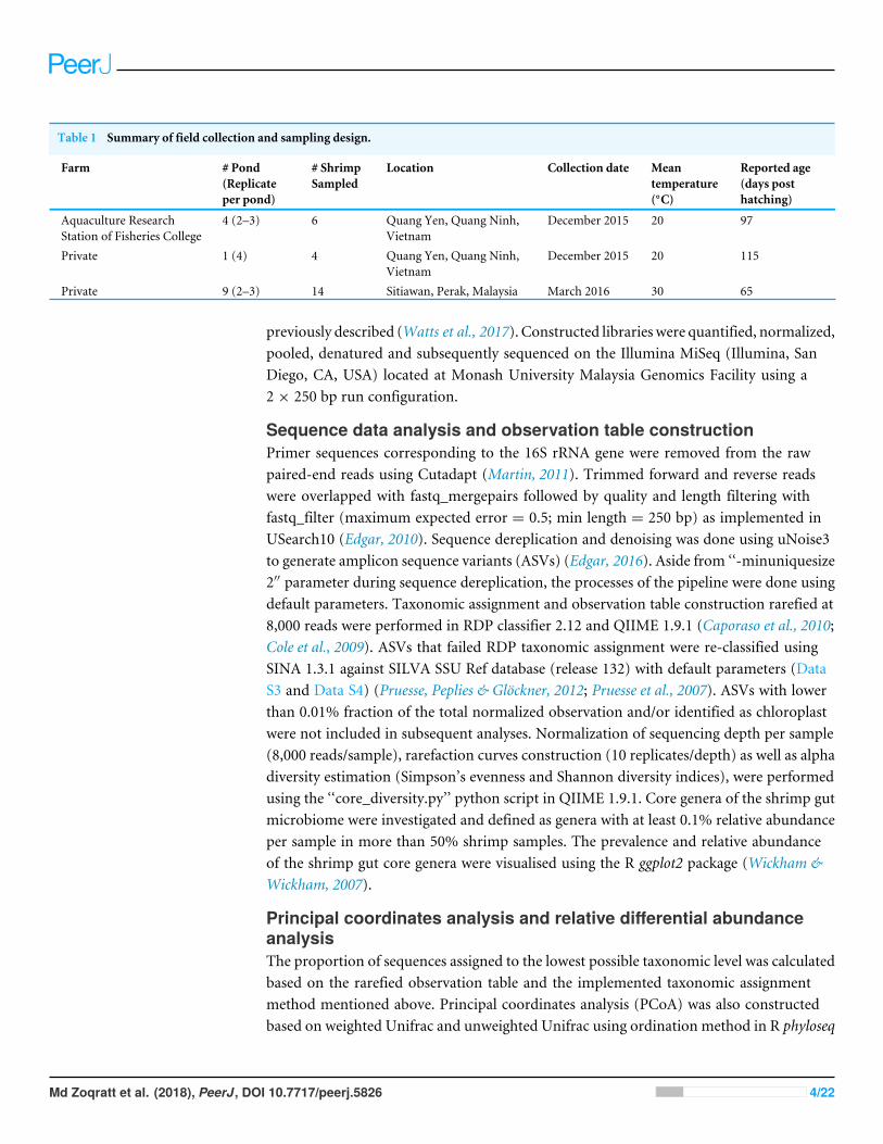
Table 1 Summary of field collection and sampling design.
Farm # Pond(Replicateper pond)
# ShrimpSampled
Location Collection date Meantemperature(◦C)
Reported age(days posthatching)
Aquaculture ResearchStation of Fisheries College
4 (2–3) 6 Quang Yen, Quang Ninh,Vietnam
December 2015 20 97
Private 1 (4) 4 Quang Yen, Quang Ninh,Vietnam
December 2015 20 115
Private 9 (2–3) 14 Sitiawan, Perak, Malaysia March 2016 30 65
previously described (Watts et al., 2017). Constructed libraries were quantified, normalized,pooled, denatured and subsequently sequenced on the Illumina MiSeq (Illumina, SanDiego, CA, USA) located at Monash University Malaysia Genomics Facility using a2 × 250 bp run configuration.
Sequence data analysis and observation table constructionPrimer sequences corresponding to the 16S rRNA gene were removed from the rawpaired-end reads using Cutadapt (Martin, 2011). Trimmed forward and reverse readswere overlapped with fastq_mergepairs followed by quality and length filtering withfastq_filter (maximum expected error = 0.5; min length = 250 bp) as implemented inUSearch10 (Edgar, 2010). Sequence dereplication and denoising was done using uNoise3to generate amplicon sequence variants (ASVs) (Edgar, 2016). Aside from ‘‘-minuniquesize2′′ parameter during sequence dereplication, the processes of the pipeline were done usingdefault parameters. Taxonomic assignment and observation table construction rarefied at8,000 reads were performed in RDP classifier 2.12 and QIIME 1.9.1 (Caporaso et al., 2010;Cole et al., 2009). ASVs that failed RDP taxonomic assignment were re-classified usingSINA 1.3.1 against SILVA SSU Ref database (release 132) with default parameters (DataS3 and Data S4) (Pruesse, Peplies & Glöckner, 2012; Pruesse et al., 2007). ASVs with lowerthan 0.01% fraction of the total normalized observation and/or identified as chloroplastwere not included in subsequent analyses. Normalization of sequencing depth per sample(8,000 reads/sample), rarefaction curves construction (10 replicates/depth) as well as alphadiversity estimation (Simpson’s evenness and Shannon diversity indices), were performedusing the ‘‘core_diversity.py’’ python script in QIIME 1.9.1. Core genera of the shrimp gutmicrobiome were investigated and defined as genera with at least 0.1% relative abundanceper sample in more than 50% shrimp samples. The prevalence and relative abundanceof the shrimp gut core genera were visualised using the R ggplot2 package (Wickham &Wickham, 2007).
Principal coordinates analysis and relative differential abundanceanalysisThe proportion of sequences assigned to the lowest possible taxonomic level was calculatedbased on the rarefied observation table and the implemented taxonomic assignmentmethod mentioned above. Principal coordinates analysis (PCoA) was also constructedbased on weighted Unifrac and unweighted Unifrac using ordination method in R phyloseq
Md Zoqratt et al. (2018), PeerJ, DOI 10.7717/peerj.5826 4/22
package (Lozupone & Knight, 2005; McMurdie & Holmes, 2013). The resulting PCoA plotswere then visualized using R ggplot2 package (Wickham &Wickham, 2007). Strength andsignificance of grouping were calculated using the compare_categories.py python scriptin QIIME which implements ANOSIM analysis using the default 999 permutations.Relative differential abundance test was also conducted at phylum and family levels usingTukey-Kramer post-hoc in conjunction with analysis of variance (ANOVA) statistical testin STAMP (Parks et al., 2014). Multiple hypothesis testing was done for the four genericgroups namely Malaysian Farm, Malaysian Shrimp, Vietnamese Farm and VietnameseShrimp. A significant differential abundance of phylum distribution was defined asBenjamini–Hochberg-corrected probability p-value of ≤ 0.01 and was observed only forthe top 10 most abundant phyla. A significant differential abundance of family distributionwas defined as Benjamini–Hochberg corrected p-value of ≤ 0.01 and eta squared ≥ 0.3.
Comparison of Vibrio diversity using different clustering methodsVibrio diversity was compared using different methods of 16S marker-gene data analysis,namely conventional operational taxonomic unit (OTU) clustering and amplicon sequencevariants (ASV), using UParse and uNoise3 respectively (Edgar, 2013; Edgar, 2016). Exceptfor -minuniquesize of 2 during sequence dereplication, both pipelines were conductedusing default parameters. ASVs and OTUs assigned to the genus Vibrio with at leastcumulative read abundance of more than 200 were retained for blastN similarity search(E-value< 1e−100) against 16S rRNA sequences ofVibrio type strain curated in EzBioCloud(as of 18th May 2018) (Yoon et al., 2017).
RESULTSShrimp intestinal and pond microbial communities are distinctA total of 2,731,818 successfully merged reads were generated in this study with 2,144,192reads (median of 32,648 reads/sample; min= 9,948; max= 66,590) confidently mapped tothe ASVs. 92.12% and 7.77% of the mapped reads correspond to RDP-classified and SINA-classified ASVs respectively, while the remaining mapped reads belong to ASVs withoutconfident taxonomic assignment at the kingdom rank. A majority of reads recovered fromshrimp intestine and ponds were assigned to members from the phyla Proteobacteria,Actinobacteria, Bacteroidetes and Fusobacteria (Fig. 1). Significant differences in therelative abundance of bacteria phyla were observed among samples isolated from pondsand shrimp guts. Reads mapping to Actinobacteria and Bacteroidetes are more abundantin ponds (p< 0.01 and p< 0.001, respectively), while shrimp guts have a significantlyhigher relative abundance of Proteobacteria (p< 0.001). At a finer level, Malaysian shrimpintestinalmicrobiome containsmore readsmapping to the phylumFusobacteria (p< 0.01).Notable, this phylum is also near absent in three out of four Malaysian shrimps noted tobe unhealthy based on morphological observation by the aquaculture manager (Fig. 1).
Rarefaction curves based on alpha diversity metrics, number of observed ASVs andPD (Phylogenetic diversity) whole tree, indicated that 8,000 sequences per sample aresufficient for capturing the alpha diversity of microbial communities in both shrimp gutsand ponds. Inverse Simpson’s and Shannon’s indices showed higher species richness and
Md Zoqratt et al. (2018), PeerJ, DOI 10.7717/peerj.5826 5/22
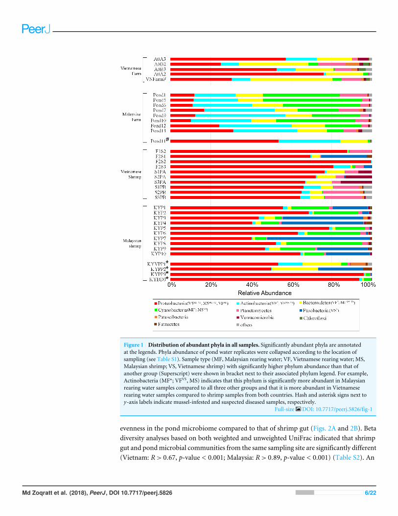
Figure 1 Distribution of abundant phyla in all samples. Significantly abundant phyla are annotatedat the legends. Phyla abundance of pond water replicates were collapsed according to the location ofsampling (see Table S1). Sample type (MF, Malaysian rearing water; VF, Vietnamese rearing water; MS,Malaysian shrimp; VS, Vietnamese shrimp) with significantly higher phylum abundance than that ofanother group (Superscript) were shown in bracket next to their associated phylum legend. For example,Actinobacteria (MF*; VFVS, MS) indicates that this phylum is significantly more abundant in Malaysianrearing water samples compared to all three other groups and that it is more abundant in Vietnameserearing water samples compared to shrimp samples from both countries. Hash and asterisk signs next toy-axis labels indicate mussel-infested and suspected diseased samples, respectively.
Full-size DOI: 10.7717/peerj.5826/fig-1
evenness in the pond microbiome compared to that of shrimp gut (Figs. 2A and 2B). Betadiversity analyses based on both weighted and unweighted UniFrac indicated that shrimpgut and pondmicrobial communities from the same sampling site are significantly different(Vietnam: R> 0.67, p-value < 0.001; Malaysia: R> 0.89, p-value < 0.001) (Table S2). An
Md Zoqratt et al. (2018), PeerJ, DOI 10.7717/peerj.5826 6/22

Figure 2 Rarefaction curves and alpha diversity plots of each sample group. (A) Rarefaction curveconstructed based on observed ASVs. (B) Rarefaction curve constructed based on phylogenetic distance(PD_whole_tree). (C) Shannon’s evenness index box plot. (D) Shannon index box plot.
Full-size DOI: 10.7717/peerj.5826/fig-2
even stronger separation was also observed among pond samples from different samplingsites/ countries (Fig. 3, Table S2). Furthermore, samples from one of the Malaysian ponds(MF_Pond11) noted by the shrimp farmer to be infested by mussels (Table S1) was distinctfrom other Malaysian pond sample samples (Fig. 3). It is worth noting that rearing watersamples collected from different parts of the same pond have minimal spatial variationin microbial composition as evidenced by the general tight clustering of pond replicates,suggesting homogenous microbial community in the rearing water and indicating thatthe sampling protocols are efficient for capturing pond diversity. Although the separationbetween Malaysian and Vietnamese shrimp gut samples was less obvious in both PCoAplots with occasional overlap, their microbial community structure appears to differmoderately (R< 0.67) with good statistical support (p-value < 0.001) based on ANOSIManalysis (Table S2).
Md Zoqratt et al. (2018), PeerJ, DOI 10.7717/peerj.5826 7/22
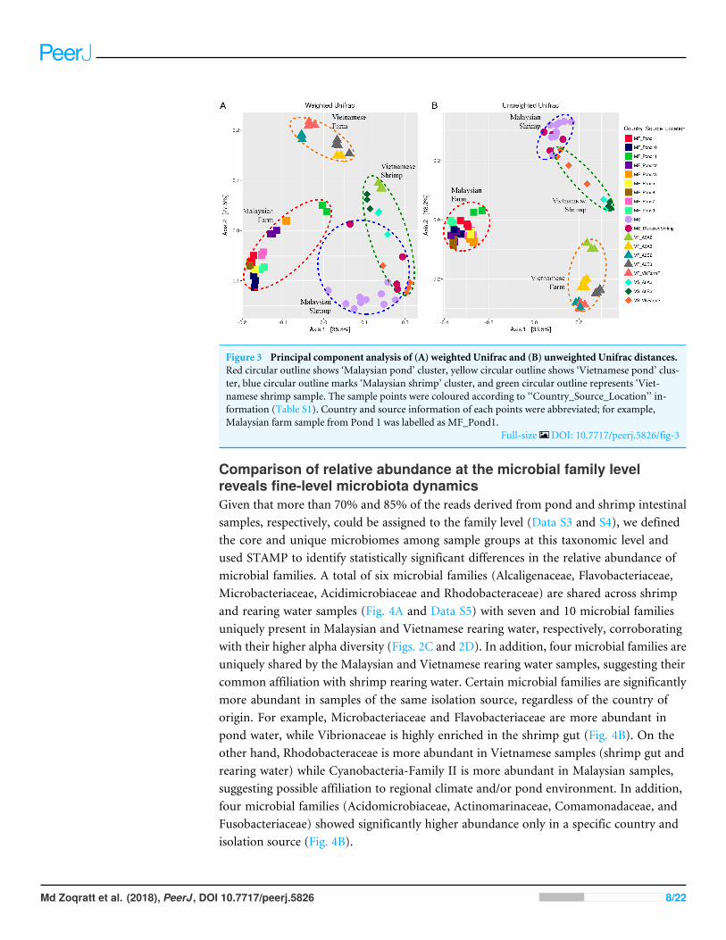
Figure 3 Principal component analysis of (A) weighted Unifrac and (B) unweighted Unifrac distances.Red circular outline shows ‘Malaysian pond’ cluster, yellow circular outline shows ‘Vietnamese pond’ clus-ter, blue circular outline marks ‘Malaysian shrimp’ cluster, and green circular outline represents ‘Viet-namese shrimp sample. The sample points were coloured according to ‘‘Country_Source_Location’’ in-formation (Table S1). Country and source information of each points were abbreviated; for example,Malaysian farm sample from Pond 1 was labelled as MF_Pond1.
Full-size DOI: 10.7717/peerj.5826/fig-3
Comparison of relative abundance at the microbial family levelreveals fine-level microbiota dynamicsGiven that more than 70% and 85% of the reads derived from pond and shrimp intestinalsamples, respectively, could be assigned to the family level (Data S3 and S4), we definedthe core and unique microbiomes among sample groups at this taxonomic level andused STAMP to identify statistically significant differences in the relative abundance ofmicrobial families. A total of six microbial families (Alcaligenaceae, Flavobacteriaceae,Microbacteriaceae, Acidimicrobiaceae and Rhodobacteraceae) are shared across shrimpand rearing water samples (Fig. 4A and Data S5) with seven and 10 microbial familiesuniquely present in Malaysian and Vietnamese rearing water, respectively, corroboratingwith their higher alpha diversity (Figs. 2C and 2D). In addition, four microbial families areuniquely shared by the Malaysian and Vietnamese rearing water samples, suggesting theircommon affiliation with shrimp rearing water. Certain microbial families are significantlymore abundant in samples of the same isolation source, regardless of the country oforigin. For example, Microbacteriaceae and Flavobacteriaceae are more abundant inpond water, while Vibrionaceae is highly enriched in the shrimp gut (Fig. 4B). On theother hand, Rhodobacteraceae is more abundant in Vietnamese samples (shrimp gut andrearing water) while Cyanobacteria-Family II is more abundant in Malaysian samples,suggesting possible affiliation to regional climate and/or pond environment. In addition,four microbial families (Acidomicrobiaceae, Actinomarinaceae, Comamonadaceae, andFusobacteriaceae) showed significantly higher abundance only in a specific country andisolation source (Fig. 4B).
Md Zoqratt et al. (2018), PeerJ, DOI 10.7717/peerj.5826 8/22

Figure 4 Microbial community dynamics at the family level. (A) Venn diagram illustrating the num-ber of unique and overlapping microbial families among shrimp guts and rearing water. To be consideredas present, a microbial family must be detected in at least 90% of the samples from the same group. (B)Tukey post-hoc pairwise comparison in conjunction with analysis of variance (ANOVA) between ‘‘Coun-try_Source’’ groups of nine significant microbial families. Mean proportion values of families that are sig-nificantly different are shown in the bar plots on the left section, while the differences in the pairwise com-parison mean proportion with 95% confidence interval are shown on the right section.
Full-size DOI: 10.7717/peerj.5826/fig-4
Md Zoqratt et al. (2018), PeerJ, DOI 10.7717/peerj.5826 9/22
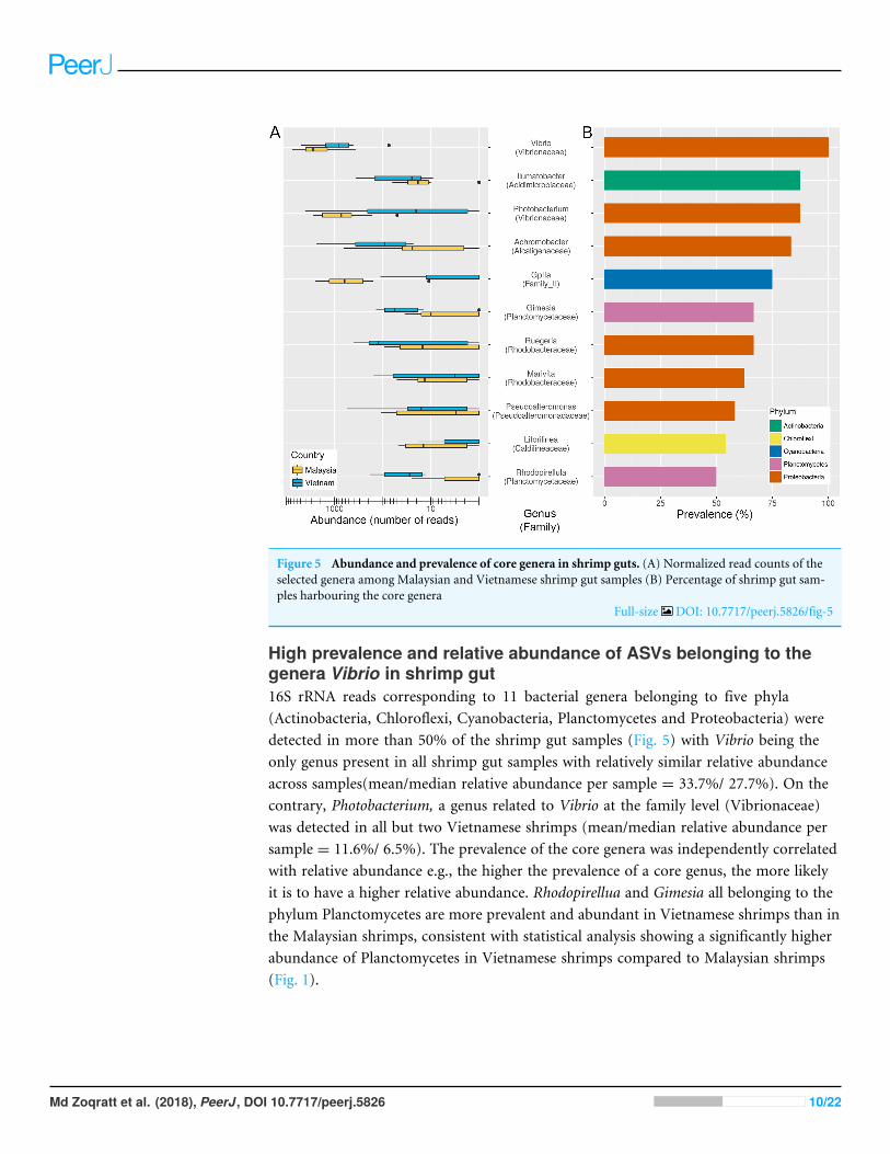
Figure 5 Abundance and prevalence of core genera in shrimp guts. (A) Normalized read counts of theselected genera among Malaysian and Vietnamese shrimp gut samples (B) Percentage of shrimp gut sam-ples harbouring the core genera
Full-size DOI: 10.7717/peerj.5826/fig-5
High prevalence and relative abundance of ASVs belonging to thegenera Vibrio in shrimp gut16S rRNA reads corresponding to 11 bacterial genera belonging to five phyla(Actinobacteria, Chloroflexi, Cyanobacteria, Planctomycetes and Proteobacteria) weredetected in more than 50% of the shrimp gut samples (Fig. 5) with Vibrio being theonly genus present in all shrimp gut samples with relatively similar relative abundanceacross samples(mean/median relative abundance per sample = 33.7%/ 27.7%). On thecontrary, Photobacterium, a genus related to Vibrio at the family level (Vibrionaceae)was detected in all but two Vietnamese shrimps (mean/median relative abundance persample = 11.6%/ 6.5%). The prevalence of the core genera was independently correlatedwith relative abundance e.g., the higher the prevalence of a core genus, the more likelyit is to have a higher relative abundance. Rhodopirellua and Gimesia all belonging to thephylum Planctomycetes are more prevalent and abundant in Vietnamese shrimps than inthe Malaysian shrimps, consistent with statistical analysis showing a significantly higherabundance of Planctomycetes in Vietnamese shrimps compared to Malaysian shrimps(Fig. 1).
Md Zoqratt et al. (2018), PeerJ, DOI 10.7717/peerj.5826 10/22

Substantial underestimation of Vibrio diversity using the OTUclustering approachThe high cumulative abundance of reads assigned to the genus Vibrio in shrimp gutsindicates that some members of this genus are endogenous to the shrimp gut microbiota.UPARSE using the default 97% sequence similarity cut-off setting identified two abundantOTUs assigned to Vibrio. This contrasts greatly with the ASV approach which identifiedsubstantially more biological sequences classified as Vibrio (Fig. 6, Data S3 and S4). Amajority of the constructed ASVs do not have an exact sequence match to the constructedOTUs and more importantly, some could be assigned to a single Vibrio species (ASV22,Vibrio jasicida TCFB 0772T; ASV19,Vibrio neocaledonicusNC470T). OTU2 and OTU10 areidentical in both sequence length and identity to ASV2 and ASV14, respectively. Similaritysearch of OTU2/ASV2 revealed an exact sequence match toV. rotiferianus LMG21460T andV. campbellii CAIM519T, indicating a limitation to the use of V3–V4 hypervariable regionin delimiting some Vibrio species (Fig. 6). The high ratios of ASVs-to-OTUs observed forOTU2 and OTU10 strongly suggests that imposing a fixed dissimilarity threshold usingconventional OTU clustering underestimates the true microbial diversity for Vibrio speciesin this study and that resolving amplicon sequence variants (ASV) from amplicons datawhich is sensitive down to single-nucleotide differences, through read de-replication anderror correction, substantially increased the number of observed Vibrio species from theidentical dataset. Unlike the ASVs associated with OTU2, nearly all ASVs associated withOTU10 have at least two mismatches to known Vibrio species, indicating the presence ofunculturable or yet-to-be-cultured Vibrio strains in the shrimp gut. Of even more interest,the ASV approach revealed the presence of V. parahaemolyticus (ASV235 in Fig. 6) thatwas missed by the OTU clustering approach presumably due to its overall low relativeabundance across samples (Table S5).
DISCUSSIONDespite the immense scale of shrimp aquaculture in South East Asia and the major impactsof aquaculture disease outbreaks in tropical regions, we are only starting to understand themicrobial composition of the shrimp guts and their relationship to rearing water in theregion (Leung & Bates, 2013). Most shrimp-related microbiome studies have been limitedto a few farms in a particular country; most of which have been conducted in countriesoutside of SEA such as China and Mexico. Litopenaeus vannamei microbiome studieshave so far investigated the microbial composition of wild-type shrimps serving as animportant baseline for future comparative studies (Cornejo-Granados et al., 2017) as wellas the impacts of disease exposure (Chen et al., 2017b; Cornejo-Granados et al., 2017; Jinboet al., 2017; Rungrassamee et al., 2016; Xiong et al., 2015; Zhu et al., 2016), developmentalstages (Huang et al., 2016), nutrition (Zhang et al., 2014) and temperature (Tang et al.,2014) on shrimp intestinal microbiome. We have contributed new findings to the growingliterature by providing the first data on gut and pond water microbiome of Malaysiancultured shrimps. Furthermore, we compared bacterial communities of shrimp guts andpond water from multiple aquaculture farms in two distinct climatic regions (Malaysia
Md Zoqratt et al. (2018), PeerJ, DOI 10.7717/peerj.5826 11/22
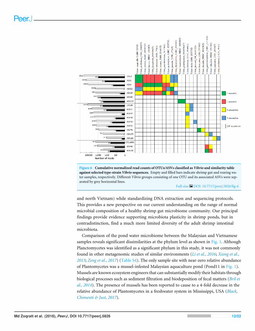
Figure 6 Cumulative normalized read counts of OTUs/ASVs classified as Vibrio and similarity tableagainst selected type-strain Vibrio sequences. Empty and filled bars indicate shrimp gut and rearing wa-ter samples, respectively. Different Vibrio groups consisting of one OTU and its associated ASVs were sep-arated by grey horizontal lines.
Full-size DOI: 10.7717/peerj.5826/fig-6
and north Vietnam) while standardizing DNA extraction and sequencing protocols.This provides a new perspective on our current understanding on the range of normalmicrobial composition of a healthy shrimp gut microbiome community. Our principalfindings provide evidence supporting microbiota plasticity in shrimp ponds, but incontradistinction, find a much more limited diversity of the adult shrimp intestinalmicrobiota.
Comparison of the pond water microbiome between the Malaysian and Vietnamesesamples reveals significant dissimilarities at the phylum level as shown in Fig. 1. AlthoughPlanctomycetes was identified as a significant phylum in this study, it was not commonlyfound in other metagenomic studies of similar environments (Li et al., 2016; Xiong et al.,2015; Zeng et al., 2017) (Table S4). The only sample site with near-zero relative abundanceof Plantomycetes was a mussel-infested Malaysian aquaculture pond (Pond11 in Fig. 1).Mussels are known ecosystem engineers that can substantiallymodify their habitats throughbiological processes such as sediment filtration and biodeposition of fecal matters (Bril etal., 2014). The presence of mussels has been reported to cause to a 4-fold decrease in therelative abundance of Plantomycetes in a freshwater system in Mississippi, USA (Black,Chimenti & Just, 2017).
Md Zoqratt et al. (2018), PeerJ, DOI 10.7717/peerj.5826 12/22

Although Flavobacteriaceae and Microbacteriaceae are prevalent in the rearing waterand shrimp gut samples, they exhibited significantly higher relative abundance in therearing water samples. Albeit initially associated with a fairly general ecological functione.g., simple mineralization-based commensalism (Kirchman, 2002), emerging evidencesuggests that some members within the marine Flavobactericeae clade are algal-associatedspecies that exhibit growth promoting and inhibiting effects to its host and other algalspecies, respectively (Bowman, 2006). Genera such as Cellulophaga, Psychroserpens andFormoasa have been previously reported to produce toxic secondary metabolites againstdinoflagellates, commonly associated with algal bloom (Adachi et al., 2002; Egan et al.,2000). Thus, the significant abundance of Flavobacteriacea in both rearing water could belinked to the natural occurrence of algal and diatom species in the rearing water some ofwhich were co-amplified by the V3–V4 primers in this study (Data S3 and Table S3). On thecontrary, most described members from the family Microbacteriaceae were not associatedwith marine environmental and were typically isolated from the terrestrial environment(Evtushenko & Takeuchi, 2017). High abundance of Microbacteriaceae (unclassified at thegenus level) in shrimp rearing water particularly during the post-larvae stage has beenpreviously observed in a commercial marine shrimp hatchery in Hainan, China (Zeng etal., 2017) which was suggested to be a temporal-specific bacterial family caused by changesin the shrimp diet during different growth stages. On the contrary, four microbial familiesnamely, Cryomorphaceae, Rhodospirillaceae, Bacteriovoracaceae and Saprospiraceae, areexclusively found across both Malaysian and Vietnamese shrimp rearing water samples,indicating their specific adaptation to shrimp rearing water or more generally the marineaquatic environment. For example, members of the family Rhodospirillaceae are purplenon-sulfur andmostly nitrogen-fixing photosynthetic bacteria. Their absence in the shrimpgut is consistent with their strict requirement for light to grow and proliferate which is notsufficiently present in the shrimp gut environment.
Despite the conspicuous difference in the shrimp pond microbiota between twocountries, the shrimp intestinal microbiota are more similar to each other which ispresumably due to host selection for microbial strains that adapt to or exploit the shrimpgut environment as corroborated by their lower alpha diversity indices compared to thatof rearing water samples (Xiong et al., 2017). However, the presence of several microbialfamilies in both shrimp and rearing water samples indicates that the shrimp gut microbiotamaybe significantly affected by the microbial communities present in their aquaticenvironment e.g., rearing water and pond sediments (Chen et al., 2017a; Cornejo-Granadoset al., 2017) as opposed to being maternally influenced as observed in some mammalsand other animals with forms of parental care (Jakobsson et al., 2014; Kohl & Dearing,2012; Zhang et al., 2014). Moulting e.g., shedding of shrimp ectodermal gut tissue alsoprovides a new opportunity for shrimp stomach and guts to be colonised by the bacterialcommunity of the pond (Moss, LeaMaster & Sweeney, 2000). Furthermore, crustaceans,including L. vannamei, also consume their exuvia, which provides another opportunityfor microbial recolonization of the shrimp guts and also the transmission of intestinalmicrobiome across shrimps throughout their developmental stages (Martínez-Córdova &Peña Messina, 2005).
Md Zoqratt et al. (2018), PeerJ, DOI 10.7717/peerj.5826 13/22
Although the micro-clustering of shrimp gut samples based on country and/or farmof origin may be associated with the difference in their respective growth environment,variation in developmental stage may also contribute to the observed clustering as theshrimps in this study were collected at different adult growth stages (Table 1). Theabundance of members from the family Fusobacteriaceae is highly dependent on theshrimp developmental stage with a near-zero abundance in young shrimp larvae gut andsubsequently making up a substantial portion of the microbiome in the adult stage (75-daypost-hatching) (Chen et al., 2017b; Zeng et al., 2017). Intriguingly, although most of theVietnamaese shrimps were 97-day post-hatching during sampling, the low abundance andprevalence of Fusobactericeae in the their guts indicates microbiome resemblance to that ofyounger shrimps (Zeng et al., 2017). In contrast, Fusobacteriaceae is prevalent in Malaysianshrimps that are relatively young e.g., 65-day post- hatching. However, it is worth notingthat Sitiawan, the closest city to where the Malaysian farm is located, is warm (30 oC)throughout the year. Such a climate may support faster shrimp growth thus enabling themto reach adulthood earlier (Kumlu, Türkmen & Kumlu, 2010; Wyban, Walsh & Godin,1995). Members from the family Fusobacteriaceae are microaerotolerant to obligateanaerobic Gram-negative rods bacteria that derive energy through the fermentation of avariety of carbohydrates, amino acids and peptides. Such a metabolic profile is consistentwith the higher prevalence and abundance of Fusobacteriaceae in mature shrimp intestinalsystems that typically exhibit a better digestive ability (Parte et al., 2011; Schock et al., 2013).Unfortunately, despite exhibiting a wide ecological diversity as evidenced by their diverseisolation source, the symbiotic relationship of Fusobacteriaceae towards its host has yetbeen properly demonstrated (Nelson, Rogers & Brown, 2013). Future work consisting ofmetatranscriptome and metagenome sequencing of the shrimp gut microbiota will benecessary to shed light on the role of Fusobactericeae in the shrimp gut.
Vibrio and Photobacterium belonging to the Vibrionaceae family are both abundantand prevalent in nearly all of the shrimp samples, an observation that is consistent withprevious reports (Cornejo-Granados et al., 2017; Rungrassamee et al., 2016; Xiong et al.,2017). In contrast, Zeng et al. (2017) did not identify any Vibrio-specific OTUs in theirsampling. Such anomalies are unlikely to be biological but rather due to technical andanalytical differences, such as the choice of the 16S rRNA gene region sequenced (V4- vs.V3–V4-hypervariable region) and bioinformatic analysis settings. High abundance andprevalence of Vibrio and Photobacterium genera in shrimp guts suggest that they are morelikely to be endogenous rather than pathogenic strains (Kriem et al., 2015). However, thisalso reflects the persistence and adaptation of members from these genera to the shrimpgut environments and may potentially explain the susceptibility of shrimps to non-nativepathogenic Vibrio and Photobacterium strains (Kondo et al., 2014;Wang & Chen, 2006).
The use of ASVs reveals a wider diversity of Vibrio species, suggesting that previousshrimp microbiome analyses that employed the common 97% similarity cut-off forclustering will risk masking the trueVibrio diversity in the shrimp gut (Callahan, McMurdie& Holmes, 2017; Chen et al., 2017b). Fortuitously, despite the observed low resolution ofthe V3–V4 hypervariable region for Vibrio species, this region appears to be distinct in V.parahaemolyticus, which exhibits at least two diagnostic nucleotides that are absent from
Md Zoqratt et al. (2018), PeerJ, DOI 10.7717/peerj.5826 14/22

all known type strains of Vibrio species (Fig. 6). The lack of an OTU with exact matchto ASV235 suggests that analysis using the OTU clustering approach will fail to reportthe presence of V. parahaemolyticus and/or undescribed Vibrio strains sharing the same16S rRNA gene sequence with V. parahaemolyticus if they are present at low abundancein the dataset. This can have critical implications for aquaculture microbial managementespecially in the early detection of V. parahaemolyticus infection. Since shrimp gut canharbour both pathogenic and native Vibrio species, complementing 16S rRNA-basedamplicon sequencing with an alternative genetic marker such as pyrH may enable a moreaccurate quantification of Vibrio diversity and abundance in aquaculture environment(Tall et al., 2013; Thompson et al., 2005). Given the high diversity of shrimp gut-associatedVibrio as revealed for the first time by the ASV approach, shallow shotgun metagenomesequencing will also be instructive to obtain species/strain-level taxonomic resolution ofthe abundant endogenous microbes in shrimp guts particularly those belonging to thegenera Vibrio and Photobacterium.
Shrimp gut microbiomes vary due to biological differences (shrimp strains), differencesin environmental or farming practice (temperature, diet, probiotic, wild capture) or evenbiases from different laboratory procedures such as the sequencing platform and thedifferent partial 16S sequence target (Cornejo-Granados et al., 2017; Tremblay et al., 2015).Considering the crucial functions undertaken by microbial communities and the potentialuse of the pond microbiome for pond health surveillance, investment in measuring awide variety of chemical and physical parameters would allow us to better correlate therelationship between microbiomes and rearing water quality and therefore improving ourunderstanding of aquaculture microbiomes.
CONCLUSIONSUsing a standardized Illumina 16S rRNA amplicons sequencing protocol, we report for thefirst time, amplicon sequence variants (ASV)-based analysis of aquaculture rearing waterand shrimp gut microbiota from two South East Asia countries with different climates.Despite substantial difference in the microbial composition of shrimp rearing waterbetween farms in Malaysia and Vietnam, adult shrimp guts are more similar and exhibit agenus level core microbiome with the genus Vibrio being the most prevalent and abundantgroup. In addition, compared to OTU clustering approach, the ASV method improvedthe identification of closely related and/or rare Vibrio species, which is of relevance to theshrimp aquaculture industry. The high abundance of Vibrio in shrimp gut also suggeststhat some Vibrio species are endogenous and non-virulent to shrimps with functional andecological roles that remain to be elucidated in the future.
ACKNOWLEDGEMENTSWe thank the Monash University Malaysia Genomics Facility for the provision ofcomputational resources. We are also extremely grateful to the aquaculture managersfor providing access to their farms and sharing information.
Md Zoqratt et al. (2018), PeerJ, DOI 10.7717/peerj.5826 15/22

ADDITIONAL INFORMATION AND DECLARATIONS
FundingThis work was supported by the Tropical and Medicine Biology Platform, MonashUniversity. The funders had no role in study design, data collection and analysis, decisionto publish, or preparation of the manuscript.
Grant DisclosuresThe following grant information was disclosed by the authors:Tropical and Medicine Biology Platform, Monash University.
Competing InterestsThe authors declare there are no competing interests.
Author Contributions• Muhammad Zarul Hanifah Md Zoqratt performed the experiments, analyzed the data,prepared figures and/or tables, authored or reviewed drafts of the paper, approved thefinal draft.• Wilhelm Wei Han Eng performed the experiments, approved the final draft.• Binh Thanh Thai conceived and designed the experiments, contributed reagents/mate-rials/analysis tools, approved the final draft.• Christopher M. Austin conceived and designed the experiments, contributedreagents/materials/analysis tools, authored or reviewed drafts of the paper, approved thefinal draft.• Han Ming Gan conceived and designed the experiments, performed the experiments,analyzed the data, prepared figures and/or tables, authored or reviewed drafts of thepaper, approved the final draft.
DNA DepositionThe following information was supplied regarding the deposition of DNA sequences:
All FastQ raw data may be accessed through SRA accession number SRP126985 or NCBIBioProject PRJNA422950.
Data AvailabilityThe following information was supplied regarding data availability:
FASTA files for OTU and ASV in addition to their taxonomic assignments are availablein the Supplemental File.
Supplemental InformationSupplemental information for this article can be found online at http://dx.doi.org/10.7717/peerj.5826#supplemental-information.
Md Zoqratt et al. (2018), PeerJ, DOI 10.7717/peerj.5826 16/22

REFERENCESAdachi M, Fukami K, Kondo R, Nishijima T. 2002. Identification of marine algicidal
Flavobacterium sp. 5 N-3 using multiple probes and whole-cell hybridization.Fisheries Science 68:713–720 DOI 10.1046/j.1444-2906.2002.00484.x.
Bentzon-Tilia M, Sonnenschein EC, Gram L. 2016.Monitoring and managing microbesin aquaculture—towards a sustainable industry.Microbial Biotechnology 9:576–584DOI 10.1111/1751-7915.12392.
Black EM, Chimenti MS, Just CL. 2017. Effect of freshwater mussels on the vertical dis-tribution of anaerobic ammonia oxidizers and other nitrogen-transforming microor-ganisms in upper Mississippi river sediment. PeerJ 5:e3536 DOI 10.7717/peerj.3536.
Bowman JP. 2006. The marine clade of the family Flavobacteriaceae: the generaAequorivita, Arenibacter, Cellulophaga, Croceibacter, Formosa, Gelidibacter, Gillisia,Maribacter,Mesonia,Muricauda, Polaribacter, Psychroflexus, Psychroserpens,Robiginitalea, Salegentibacter, Tenacibaculum, Ulvibacter, Vitellibacter and Zobellia.In: Dworkin M, Falkow S, Rosenberg E, Schleifer K-H, Stackebrandt E, eds. Theprokaryotes: volume 7: Proteobacteria: delta, epsilon subclass. New York: Springer NewYork, 677–694.
Bril JS, Durst JJ, Hurley BM, Just CL, Newton TJ. 2014. Sensor data as a measureof native freshwater mussel impact on nitrate formation and food digestion incontinuous-flow mesocosms. Freshwater Science 33:417–424 DOI 10.1086/675448.
Callahan BJ, McMurdie PJ, Holmes SP. 2017. Exact sequence variants should replaceoperational taxonomic units in marker-gene data analysis. The ISME Journal11:2639–2643 DOI 10.1038/ismej.2017.119.
Caporaso JG, Kuczynski J, Stombaugh J, Bittinger K, Bushman FD, Costello EK,Fierer N, Peña AG, Goodrich JK, Gordon JI. 2010. QIIME allows analysis ofhigh-throughput community sequencing data. Nature Methods 7:335–336DOI 10.1038/nmeth.f.303.
Cardona E, Gueguen Y, Magré K, Lorgeoux B, Piquemal D, Pierrat F, Noguier F,Saulnier D. 2016. Bacterial community characterization of water and intestine ofthe shrimp Litopenaeus stylirostris in a biofloc system. BMCMicrobiology 16:157DOI 10.1186/s12866-016-0770-z.
Chen C-Y, Chen P-C,Weng FC-H, Shaw GT-W,Wang D. 2017a.Habitat and in-digenous gut microbes contribute to the plasticity of gut microbiome in orien-tal river prawn during rapid environmental change. PLOS ONE 12:e0181427DOI 10.1371/journal.pone.0181427.
ChenW-Y, Ng TH,Wu J-H, Chen J-W,Wang H-C. 2017b.Microbiome dynamics in ashrimp grow-out pond with possible outbreak of acute hepatopancreatic necrosisdisease. Scientific Reports 7:9395 DOI 10.1038/s41598-017-09923-6.
Cole JR,Wang Q, Cardenas E, Fish J, Chai B, Farris RJ, Kulam-Syed-Mohideen AS,McGarrell DM,Marsh T, Garrity GM, Tiedje JM. 2009. The ribosomal databaseproject: improved alignments and new tools for rRNA analysis. Nucleic AcidsResearch 37:D141–D145 DOI 10.1093/nar/gkn879.
Md Zoqratt et al. (2018), PeerJ, DOI 10.7717/peerj.5826 17/22
Cornejo-Granados F, Lopez-Zavala AA, Gallardo-Becerra L, Mendoza-Vargas A,Sánchez F, Vichido R, Brieba LG, VianaMT, Sotelo-Mundo RR, Ochoa-LeyvaA. 2017.Microbiome of pacific whiteleg shrimp reveals differential bacterialcommunity composition between wild, aquacultured and AHPND/EMS outbreakconditions. Scientific Reports 7:11783 DOI 10.1038/s41598-017-11805-w.
Costa E, Pérez J, Kreft J-U. 2006.Why is metabolic labour divided in nitrification?Trends in Microbiology 14:213–219 DOI 10.1016/j.tim.2006.03.006.
De Schryver P, Defoirdt T, Sorgeloos P. 2014. Early mortality syndrome outbreaks:a microbial management issue in shrimp farming? PLOS Pathogens 10:e1003919DOI 10.1371/journal.ppat.1003919.
Edgar RC. 2010. Search and clustering orders of magnitude faster than BLAST. Bioinfor-matics 26:2460–2461 DOI 10.1093/bioinformatics/btq461.
Edgar RC. 2013. UPARSE: highly accurate OTU sequences from microbial ampliconreads. Nature Methods 10:996–998 DOI 10.1038/nmeth.2604.
Edgar RC. 2016. UNOISE2: improved error-correction for Illumina 16S and ITSamplicon sequencing. bioRxiv ArXiv preprint. arXiv:081257.
Egan S, Thomas T, Holmström C, Kjelleberg S. 2000. Phylogenetic relationshipand antifouling activity of bacterial epiphytes from the marine alga Ulva lactuca.Environmental Microbiology 2:343–347 DOI 10.1046/j.1462-2920.2000.00107.x.
EmerencianoM, Gaxiola G, Cuzon G. 2013. Biofloc technology (BFT): a review foraquaculture application and animal food industry. In: Biomass now-cultivation andutilization. London: InTech.
Eren AM,Morrison HG, Lescault PJ, Reveillaud J, Vineis JH, SoginML. 2014.Min-imum entropy decomposition: unsupervised oligotyping for sensitive partition-ing of high-throughput marker gene sequences. The Isme Journal 9:968–979DOI 10.1038/ismej.2014.195.
Evtushenko LI, Takeuchi M. 2017. The family microbacteriaceae. Growth 18:2–35.Foo SM, EngWWH, Lee YP, Gui K, Gan HM. 2017. New sequence types of Vib-
rio parahaemolyticus isolated from a Malaysian aquaculture pond, as re-vealed by whole-genome sequencing. Genome Announcements 5:e00302–17DOI 10.1128/genomeA.00302-17.
Food and Agriculture Organization (FAO). 2016. The state of world fisheries andaquaculture 2016. Contributing to food security and nutrition for all. FAO, RomeAvailable at http://www.fao.org/ fi/ oldsite/ eims_search/1_dett.asp?pub_id=316888.
Grotkjær T, Bentzon-Tilia M, D’Alvise P, Dourala N, Nielsen KF, Gram L. 2016.Isolation of TDA-producing Phaeobacter strains from sea bass larval rearing unitsand their probiotic effect against pathogenic Vibrio spp. in Artemia cultures.Systematic and Applied Microbiology 39:180–188 DOI 10.1016/j.syapm.2016.01.005.
Han JE, Tang KFJ, Tran LH, Lightner DV. 2015. Photorhabdus insect-related (Pir)toxin-like genes in a plasmid of Vibrio parahaemolyticus, the causative agent ofacute hepatopancreatic necrosis disease (AHPND) of shrimp. Diseases of AquaticOrganisms 113:33–40 DOI 10.3354/dao02830.
Md Zoqratt et al. (2018), PeerJ, DOI 10.7717/peerj.5826 18/22
Huang Z, Li X,Wang L, Shao Z. 2016. Changes in the intestinal bacterial communityduring the growth of white shrimp, Litopenaeus vannamei. Aquaculture Research47:1737–1746 DOI 10.1111/are.12628.
Jakobsson HE, Abrahamsson TR, JenmalmMC, Harris K, Quince C, Jernberg C,Björkstén B, Engstrand L, Andersson AF. 2014. Decreased gut microbiota diversity,delayed Bacteroidetes colonisation and reduced Th1 responses in infants delivered bycaesarean section. Gut 63:559–566 DOI 10.1136/gutjnl-2012-303249.
Jinbo X, Jinyong Z,Wenfang D, Chunming D, Qiongfen Q, Chenghua L. 2017. Inte-grating gut microbiota immaturity and disease-discriminatory taxa to diagnose theinitiation and severity of shrimp disease. Environmental Microbiology 19:1490–1501DOI 10.1111/1462-2920.13701.
Kirchman DL. 2002. The ecology of Cytophaga—Flavobacteria in aquatic environments.FEMS Microbiology Ecology 39:91–100 DOI 10.1111/j.1574-6941.2002.tb00910.x.
Kohl KD, DearingM-D. 2012. Experience matters: prior exposure to plant toxinsenhances diversity of gut microbes in herbivores. Ecology Letters 15:1008–1015DOI 10.1111/j.1461-0248.2012.01822.x.
Kondo H, Tinwongger S, Proespraiwong P, Mavichak R, Unajak S, Nozaki R, Hirono I.2014. Draft genome sequences of six strains of Vibrio parahaemolyticus isolated fromearly mortality syndrome/acute hepatopancreatic necrosis disease shrimp in Thai-land. Genome Announcements 2:e00221–00214 DOI 10.1128/genomeA.00221-14.
KriemMR, Banni B, El Bouchtaoui H, Hamama A, El Marrakchi A, Chaouqy N,Robert-Pillot A, Quilici ML. 2015. Prevalence of Vibrio spp. in raw shrimps(Parapenaeus longirostris) and performance of a chromogenic medium forthe isolation of Vibrio strains. Letters in Applied Microbiology 61:224–230DOI 10.1111/lam.12455.
KumluM, Türkmen S, KumluM. 2010. Thermal tolerance of Litopenaeus vannamei(Crustacea: Penaeidae) acclimated to four temperatures. Journal of Thermal Biology35:305–308 DOI 10.1016/j.jtherbio.2010.06.009.
Lee C-T, Chen I-T, Yang Y-T, Ko T-P, Huang Y-T, Huang J-Y, HuangM-F, Lin S-J,Chen C-Y, Lin S-S, Lightner DV,Wang H-C,Wang AH-J, Wang H-C, Hor L-I, LoC-F. 2015. The opportunistic marine pathogen Vibrio parahaemolyticus becomesvirulent by acquiring a plasmid that expresses a deadly toxin. Proceedings of theNational Academy of Sciences of the United States of America 112:10798–10803DOI 10.1073/pnas.1503129112.
Leung TL, Bates AE. 2013.More rapid and severe disease outbreaks for aquaculture atthe tropics: implications for food security. Journal of Applied Ecology 50:215–222DOI 10.1111/1365-2644.12017.
Li L, Yan B, Li S, Xu J, An X. 2016. A comparison of bacterial community struc-ture in seawater pond with shrimp, crab, and shellfish cultures and in non-cultured pond in Ganyu, Eastern China. Annals of Microbiology 66:317–328DOI 10.1007/s13213-015-1111-4.
Md Zoqratt et al. (2018), PeerJ, DOI 10.7717/peerj.5826 19/22
Liu S, Ren H, Shen L, Lou L, Tian G, Zheng P, Hu B. 2015. pH levels drive bacterialcommunity structure in sediments of the Qiantang River as determined by 454pyrosequencing. Frontiers in Microbiology 6:285 DOI 10.3389/fmicb.2015.00285.
Lozupone C, Knight R. 2005. UniFrac: a new phylogenetic method for comparingmicrobial communities. Applied and Environmental Microbiology 71:8228–8235DOI 10.1128/AEM.71.12.8228-8235.2005.
MartinM. 2011. Cutadapt removes adapter sequences from high-throughput sequencingreads. EMBnet Journal 17:10–12 DOI 10.14806/ej.17.1.200.
Martínez-Córdova LR, PeñaMessina E. 2005. Biotic communities and feeding habitsof Litopenaeus vannamei (Boone 1931) and Litopenaeus stylirostris (Stimpson1974) in monoculture and polyculture semi-intensive ponds. Aquaculture Research36:1075–1084 DOI 10.1111/j.1365-2109.2005.01323.x.
McMurdie PJ, Holmes S. 2013. phyloseq: an R package for reproducible interac-tive analysis and graphics of microbiome census data. PLOS ONE 8:e61217DOI 10.1371/journal.pone.0061217.
Moss SM, LeaMaster BR, Sweeney JN. 2000. Relative abundance and species com-position of gram-negative, aerobic bacteria associated with the gut of juve-nile white shrimp Litopenaeus vannamei reared in oligotrophic well water andeutrophic pond water. Journal of the World Aquaculture Society 31:255–263DOI 10.1111/j.1749-7345.2000.tb00361.x.
Nelson TM, Rogers TL, BrownMV. 2013. The gut bacterial community of mammalsfrom marine and terrestrial habitats. PLOS ONE 8:e83655DOI 10.1371/journal.pone.0083655.
Parks DH, Tyson GW, Hugenholtz P, Beiko RG. 2014. STAMP: statistical analysis oftaxonomic and functional profiles. Bioinformatics 30:3123–3124DOI 10.1093/bioinformatics/btu494.
Parte A, Krieg NR, LudwigW,WhitmanWB, Hedlund BP, Paster BJ, Staley JT,Ward N, Brown D. 2011. Bergey’s manual of systematic bacteriology: volume 4: theBacteroidetes, Spirochaetes, Tenericutes (Mollicutes), Acidobacteria, Fibrobacteres,Fusobacteria, Dictyoglomi, Gemmatimonadetes, Lentisphaerae, Verrucomicrobia,Chlamydiae, and Planctomycetes. New York: Springer.
PhayungsakM, Phimsucha B,Wanilada R, Sopacha A, Sirawut K, PiamsakM, Sage C.2018. Bacterial community composition and distribution in different segments of thegastrointestinal tract of wild-caught adult Penaeus monodon. Aquaculture Research49:378–392 DOI 10.1111/are.13468.
Pruesse E, Peplies J, Glöckner FO. 2012. SINA: accurate high-throughput multiplesequence alignment of ribosomal RNA genes. Bioinformatics 28:1823–1829DOI 10.1093/bioinformatics/bts252.
Pruesse E, Quast C, Knittel K, Fuchs BM, LudwigW, Peplies J, Glöckner FO. 2007.SILVA: a comprehensive online resource for quality checked and aligned ribosomalRNA sequence data compatible with ARB. Nucleic Acids Research 35:7188–7196DOI 10.1093/nar/gkm864.
Md Zoqratt et al. (2018), PeerJ, DOI 10.7717/peerj.5826 20/22
RungrassameeW, Klanchui A, Maibunkaew S, Karoonuthaisiri N. 2016. Bacterialdynamics in intestines of the black tiger shrimp and the Pacific white shrimpduring Vibrio harveyi exposure. Journal of Invertebrate Pathology 133:12–19DOI 10.1016/j.jip.2015.11.004.
Saffran K, Cash K, Hallard K, Neary B,Wright R. 2001. Canadian water qualityguidelines for the protection of aquatic life. CCMEWater Quality Index 1:34–31.
Schock TB, Duke J, Goodson A,Weldon D, Brunson J, Leffler JW, Bearden DW.2013. Evaluation of Pacific White Shrimp (Litopenaeus vannamei) health during asuperintensive aquaculture growout using NMR-based metabolomics. PLOS ONE8:e59521 DOI 10.1371/journal.pone.0059521.
Tall A, Hervio-Heath D, Teillon A, Boisset-Helbert C, Delesmont R, Bodilis J, Touron-Bodilis A. 2013. Diversity of Vibrio spp. isolated at ambient environmental temper-ature in the Eastern English Channel as determined by pyrH sequencing. Journal ofApplied Microbiology 114:1713–1724 DOI 10.1111/jam.12181.
Tang Y, Tao P, Tan J, MuH, Peng L, Yang D, Tong S, Chen L. 2014. Identification ofbacterial community composition in freshwater aquaculture system farming ofLitopenaeus vannamei reveals distinct temperature-driven patterns. InternationalJournal of Molecular Sciences 15:13663–13680 DOI 10.3390/ijms150813663.
Thompson F, Gevers D, Thompson C, Dawyndt P, Naser S, Hoste B, Munn C, SwingsJ. 2005. Phylogeny and molecular identification of vibrios on the basis of multi-locus sequence analysis. Applied and Environmental Microbiology 71:5107–5115DOI 10.1128/AEM.71.9.5107-5115.2005.
Tran L, Nunan L, Redman RM,Mohney LL, Pantoja CR, Fitzsimmons K, LightnerDV. 2013. Determination of the infectious nature of the agent of acute hepatopan-creatic necrosis syndrome affecting penaeid shrimp. Diseases of Aquatic Organisms105:45–55 DOI 10.3354/dao02621.
Tremblay J, Singh K, Fern A, Kirton E, He S, Woyke T, Lee J, Chen F, Dangl J, TringeS. 2015. Primer and platform effects on 16S rRNA tag sequencing. Frontiers inMicrobiology 6:771 DOI 10.3389/fmicb.2015.00771.
Utter DR, MarkWelch JL, Borisy GG. 2016. Individuality, stability, and variability of theplaque microbiome. Frontiers in Microbiology 7:564 DOI 10.3389/fmicb.2016.00564.
Wang F-I, Chen J-C. 2006. Effect of salinity on the immune response of tiger shrimpPenaeus monodon and its susceptibility to Photobacterium damselae subsp. damselae.Fish & Shellfish Immunology 20:671–681 DOI 10.1016/j.fsi.2005.08.003.
Watts MP, Spurr LP, Gan HM,Moreau JW. 2017. Characterization of an autotrophicbioreactor microbial consortium degrading thiocyanate. Applied Microbiology andBiotechnology 101:5889–5901 DOI 10.1007/s00253-017-8313-6.
WickhamH,WickhamMH. 2007. The ggplot package. Available at https:// cran.r-project.org/web/packages/ ggplot2/ index.html .
Wright JJ, Konwar KM, Hallam SJ. 2012.Microbial ecology of expanding oxygen mini-mum zones. Nature Reviews Microbiology 10:381–394 DOI 10.1038/nrmicro2778.
Md Zoqratt et al. (2018), PeerJ, DOI 10.7717/peerj.5826 21/22

Wyban J, WalshWA, Godin DM. 1995. Temperature effects on growth, feeding rateand feed conversion of the Pacific white shrimp (Penaeus vannamei). Aquaculture138:267–279 DOI 10.1016/0044-8486(95)00032-1.
Xiong J, Wang K,Wu J, Qiuqian L, Yang K, Qian Y, Zhang D. 2015. Changes in intesti-nal bacterial communities are closely associated with shrimp disease severity. AppliedMicrobiology and Biotechnology 99:6911–6919 DOI 10.1007/s00253-015-6632-z.
Xiong J, Zhu J, DaiW, Dong C, Qiu Q, Li C. 2017. Integrating gut microbiota immatu-rity and disease-discriminatory taxa to diagnose the initiation and severity of shrimpdisease. Environmental Microbiology 19:1490–1501 DOI 10.1111/1462-2920.13701.
Yoon SH, Ha SM, Kwon S, Lim J, Kim Y, Seo H, Chun J. 2017. Introducing EzBioCloud:a taxonomically united database of 16S rRNA gene sequences and whole-genomeassemblies. International Journal of Systematic and Evolutionary Microbiology67:1613–1617 DOI 10.1099/ijsem.0.001755.
Zeng S, Huang Z, Hou D, Liu J, Weng S, He J. 2017. Composition, diversity and functionof intestinal microbiota in pacific white shrimp (Litopenaeus vannamei) at differentculture stages. PeerJ 5:e3986 DOI 10.7717/peerj.3986.
ZhangM, Sun Y, Chen K, Yu N, Zhou Z, Chen L, Du Z, Li E. 2014. Characterization ofthe intestinal microbiota in Pacific white shrimp, Litopenaeus vannamei, fed dietswith different lipid sources. Aquaculture 434:449–455DOI 10.1016/j.aquaculture.2014.09.008.
Zhu J, DaiW, Qiu Q, Dong C, Zhang J, Xiong J. 2016. Contrasting ecological processesand functional compositions between intestinal bacterial community in healthy anddiseased shrimp.Microbial Ecology 72:975–985 DOI 10.1007/s00248-016-0831-8.
Md Zoqratt et al. (2018), PeerJ, DOI 10.7717/peerj.5826 22/22