microbial fossils from the kheinjua … research, 21 (1983) 247--271 247 elsevier science publishers...
TRANSCRIPT

Precambrian Research, 21 (1983) 247--271 247 Elsevier Science Publishers B.V., Amsterdam - Printed in The Netherlands
MICROBIAL FOSSILS FROM THE KHEINJUA FORMATION, MIDDLE PROTEROZOIC SEMRI GROUP (LOWER VINDHYAN)~ SON VALLEY AREA, CENTRAL INDIA
DIANNA SCHULTE McMENAMIN
Department of Geological Sciences, Preston Cloud Research Laboratory, University of California, Santa Barbara, California 93106 (U.S.A.)
SURENDRA KUMAR
Department of Geology, Lucknow University, Lucknow, 226007, U.P. (India)
STANLEY M. AWRAMIK
Department of Geological Sciences, Preston Cloud Research Laboratory, University of California, Santa Barbara, California 93106 (U.S.A.)
(Received May 11, 1982; accepted November 18, 1982)
ABSTRACT
McMenamin, D.S., Kumar, S. and Awramik, S.M., 1983. Microbial fossils from the Khein- jua Format ion, Middle Proterozoic Semri Group (Lower Vindhyan), Son Valley area, central India. Precambrian Res., 21: 247--271.
Abundant coccoid and filamentous microfosslls are found in petrographic thin sections of stratiform stromatoli t ic cherts from the z1200 Ma-old Fawn Limestone of the Khein- jua Format ion, lower Vindhyan Supergroup, central India. The assemblages is dominated by coccoid forms of probable chroococcacean and entophysalidacean affinities; most filaments closely resemble the osciliatoriacean Gunflintia minuta.
Unlike other Middle to Late Proterozoic stromatoli t ic microbiotas, the Kheinjua is dominated by microfossils < 10 , m in diameter. Another notable feature of the assem- blage is that its microfossiis are found in extensive sheets of amorphous organic matrix, which also contains rare bizarre morphs unique to this formation.
Taxonomically, the Kheinjua microbiota most closely resembles Proterozoic micro- biotas described from the Belcher Islands (Kasegalik Formation), McArthur Group, Bitter Springs Format ion, and the Yudoma Suite.
The following taxa are formally described: C h r o o c o c c a c e a e - Myxococcoides minor Schopf, Eosynechococcus isolatus n.sp., Tetraphycus congregatus n.sp., Kheinjuasphaera vulgaris n.gen., n.sp., Glenobotrydion aenigmatis Schopf, Melasmatosphaera media Hot- mann; Entophysalidaceae - - Eoentophysalis belcherends Hofmann, E. magna n. sp.; Oscfl- latoriaceae - - cf. Gunflinta minuta Barghoorn, Eomycetopsis? siberiensis Lo.
INTRODUCTION
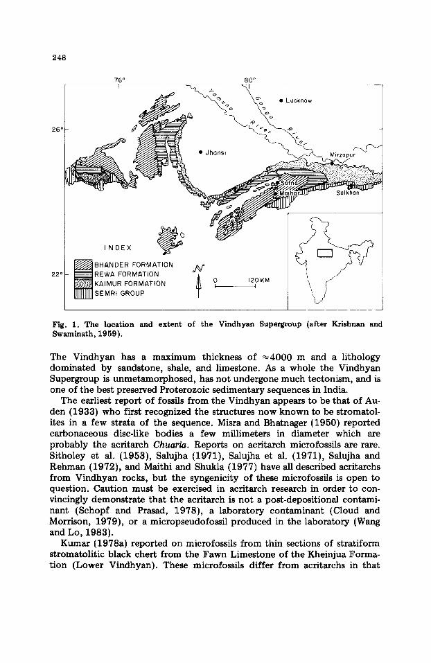
The Middle to Late Proterozoic Vindhyan Supergroup covers an area of ~104 000 krn 2 (Fig. 1) between Bihar in the east and Rajasthan to the west.
0301-9268/83/$03.00 © 1983 Elsevier Science Publishers B.V.
76 ° i
26 °
22 °
248
INDEX ~ ( b
~ BHANDER FORMATION REWA FORMATION KAIMUR FORMATION SEMRI GROUP
80 °
.Luckoow
• Jhonsi
~ ~ r IJ.U.~ ~o IKn~
N 0 120 KM I I
Fig. 1. The location and extent of the Vindhyan Supergroup (after Krishnan and Swaminath, 1959).
The Vindhyan has a maximum thickness of ~ 4 0 0 0 m and a lithology dominated by sandstone, shale, and limestone. As a whole the Vindhyan Supergroup is unmetamorphosed, has not undergone much tectonism, and is one of the best preserved Proterozoic sedimentary sequences in India.
The earliest report of fossils f rom the Vindhyan appears to be that of Au. den (1933) who first recognized the structures now known to be stromatol- ites in a few strata of the sequence. Misra and Bhatnager (1950) reported carbonaceous disc-like bodies a few millimeters in diameter which are probably the acritarch Chuaria. Reports on acritarch microfossils are rare. Sitholey et al. (1953), Salujha (1971), Salujha et al. (1971), Salujha and Rehman (1972), and Malthi and Shukla (1977) have all described acritarchs from Vindhyan rocks, but the syngenicity of these microfossils is open to question. Caution must be exercised in acritarch research in order to con- vincingly demonstrate that the acritarch is not a post-depositional contami- nant (Schopf and Prasad, 1978), a laboratory contaminant (Cloud and Morrison, 1979), or a micropseudofossil produced in the laboratory (Wang and Lo, 1983).
Kumar (1978a) reported on microfossils from thin sections of stratiform stromatolitic black chert from the Fawn Limestone of the Kheinjua Forma- tion (Lower Vindhyan). These microfossils differ from acritarchs in that

249
they are found in petrographic thin section and have affinities among modern cyanobacterial groups, whereas the category 'acritarch' is generally reserved for incertae sedis microfossils found in acid residues.
Overall, the taxonomic composition of the microbiota from the Kheinjua Formation resembles other Proterozoic stratiform stromatolitic microbiotas, such as those from the ~1900 Ma-old Belcher Islands (Hofmann, 1976), the ~1500 Ma-old McArthur Group (Muir, 1976; D.Z. Oehler, 1978), the ~850 Ma-old Bitter Springs Formation (Schopf and Blacic, 1971), and the ~.650 Ma-old Yudoma Suite (Lo, 1980). However, this microbiota is unusual and differs significantly from these and many other stromatolitic microbiotas in that: (1) it is dominated by small-sized (< 10 u m diameter) individuals; (2) many of the microfossils are found in an abundant organic matrix which partly defines the stromatolitic lamination; and (3) there are some bizarre, mul t icomponent morphs present that defy placement in any known taxo- nomic category.
GEOLOGIC SETTING
The Vindhyan Supergroup has been recognized as an important strati- graphic unit since its establishment by Oldham (1856). The Supergroup is relatively thick (~4000 m), extensive (outcrops cover ~ 104000 kin2), and unmetamorphosed in most areas, but its stratigraphic position and age have been subject to debate for many years (Gupta, 1977).
Following the studies of Auden (1933), the Vindhyan is divided into two lithostratigraphic units; the lower unit, the Semri Group or Lower Vindhyan~ is unconformably overlain by the Upper Vindhyan Group (Fig. 1). The Sem- ri Group is best exposed in the Son Valley where it crops out in small hillocks and scarps. Here, four lithostratigraphic units are distinguished; the Basal Formation, the Porcellanite Formation, the Kheinjua Formation and the Rohtas Formation (Fig. 2). The Rohtas Formation is unconformably over- lain by argillacious--arenaceous rocks of the Upper Vindhyan Kaimur Formation. Stromatolite occurrences have been recorded from the Basal, Kheinjua and Rohtas Formations (Kumar, 1978b).
The Kheinjua Formation is further subdivided into three conformable lithological members. These are, in ascending stratigraphic order, the Olive Shale Member, the Fawn Limestone Member and the Glauconitic Sandstone Member. The Fawn Limestone contains the microfossiliferous cherts.
AGE
The Vindhyan Supergroup unconformably overlies the slightly metamor- phosed Bijawar Group with a R b - S r age of ~2500 Ma (Crawford and Compston, 1970). Despite the research activity on the Vindhyan Supergroup during the last 15 years (see Singh, 1973; Sahni, 1975; Kumar, 1978b), most radiometric age data are K--Ar dates of a mid-1960's vintage.

250
2 4 ° 3 0 '
• . ° ° . . . . . . .
. . . . . . . . , o ° °
I~ e wo r iT~!..w,_:~ ' ' "
8:5 °
' " . (Upper . ' . " KAIMUR FORMATION vindhyan) . ~ ROHTAS FORMATION -~
"..:i.:-i GLAUCONITIC SANDSTONE FAWN LIMESTONE
OLIVE SHALE
- ' ~ PORCELLA~IITE FORMATION l l l l ml~ KAJRHAHAT LIMESTONE "~ Basol con-
m BASAL CONGLOMERATE glomerote
. . . . FAULT
LOCATION OF THE SAMPLES
8.4 KM I
N o , I
Fig. 2. The occurrence of the Semri Group in the Son Valley (after Auden, 1933).
The Glauconitic Sandstone Member that conformably overlies the Fawn Limestone is an important glauconite-bearing horizon in the Son Valley. The most quoted age for these glauconites is a K--Ar age of 1100 -+ 60 Ma (Vino- gradov et al., 1964). This has been recalculated with later recommended de- cay constants to 1080 -+ 40 Ma (Kreuzer et al., 1977). Glauconite-bearing layers in the Kaimur Formation (Upper Vindhyan) have K - A r ages of 940-- 910 Ma (Vinogradov et al., 1964) which have been recalculated to 890 -+ 40 Ma (Kreuzer et al., 1977).
Ages based on biostratigraphically characteristic stromatolites are in general agreement with the K--Ar dates on glauconites. In the Kheinjua Formation (K--At age ;>1100 Ma), t he Fawn Limestone contains the typical Middle Riphean (1350--950 Ma) stromatolite Conophyton gargangcum (Kumar, 1976). Additional Middle Riphean stromatolites (Tungussia and Baicalia) are found in the Rhotas Formation (Kumar, 1980). However, as in most stroma- tolite work of this type, some of the data are inconsistent: Boxonia (an Upper Riphean to Lower Cambrian group) has been reported from the Upper Vindhyan Bhander Formation (Rao et al., 1977).
251
The age of the Fawn Limestone, Kheinjua Formation, seems to be be- tween 1300 and 1100 Ma-old (Kumar, 1978b) or ~ 1 2 0 0 Ma-old.
THE MICROFOSSILIFEROUS CHERT
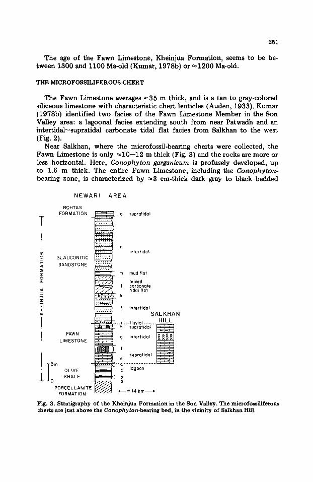
The Fawn Limestone averages ~ 3 5 m thick, and is a tan to gray-colored siliceous limestone with characteristic chert lenticles (Auden, 1933). Kumar (1978b) identified two facies o f the Fawn Limestone Member in the Son Valley area: a lagoonal facies extending south from near Patwadh and an intertidal--supratidal carbonate tidal fiat facies from Salkhan to the west (Fig. 2).
Near Salkhan, where the microfossil-bearing cherts were collected, the Fawn Limestone is only ~ 1 0 - - 1 2 m thick (Fig. 3) and the rocks are more or less horizontal. Here, Conophyton garganicum is profusely developed, up to 1.6 m thick. The entire Fawn Limestone, including the Conophyton- bearing zone, is characterized by ~ 3 cm-thick dark gray to black bedded
NEWARI AREA
z o k-
n~ o
D
Z
,,i
I
ROHTAS FORMATION o supratidal
GLAUCONITIC SANDSTONE
intertidal
m mud flat
mixed I carbonate
tidal flat k
FAWN j h LIMESTONE g intertidal
f supratidal
e r'-d
i bm " lagoon OLIVE c SHALE ~ b
O - a PORCELLANITE
FORMATION *--~ 14 km-----~
intertidal SALKHAN
-fluvial ....... HILL supratidol I : ~
Fig. 3. Stratigraphy of the Kheinjua Formation in the Son Valley. The microfossiliferous cherts are just above the Conophyton-bearing bed, in the vicinity of Salkhan Hill.
252
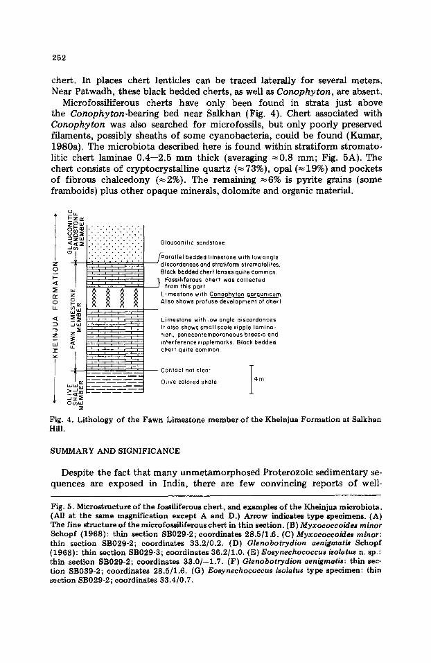
chert. In places chert lenticles can be traced laterally for several meters. Near Patwadh, these black bedded cherts, as well as Conophyton, are absent.
Microfossiliferous cherts have only been found in strata just above the Conophyton-bearing bed near Satkhan (Fig. 4). Chert associated with Conophyton was also searched for microfossils, but only poorly preserved filaments, possibly sheaths of some cyanobacteria, could be found (Kumar, 1980a). The microbiota described here is found within stratiform stromato- litic chert laminae 0 .4- -2 .5 mm thick (averaging ~ 0 . 8 mm; Fig. 5A). The chert consists of cryptocrystalline quartz (~73%), opal ( ~ 1 9 % ) a n d pockets of fibrous chalcedony (~2%). The remaining ~6% is pyrite grains {some framboids) plus other opaque minerals, dolomite and organic material.
Ld l - "v"
° t d
Z 0
0 LI-
• , , . . . . , . . • • .o.
iiii!iil iiiii!ili Glauconitic sandstone
~ / - ~ a r a l l e L bedded limestone with Iowangle discordances and strotiform stromatolites Black bedded chert lenses quite common
. ~ Eossiliferous chert was collected from this port
Limestone with Conophyton go rganicum Also shows profuse development of chert
Limestone with low angle discordances. It also shows small scale ripple lamina- tion, penecontemporaneous breccia and interference ripplemarks. Block bedded chert quite common
" r
Contact not cleor J
Olive colored shoie 1 4 m
0o3,,1
Fig. 4. Lithology of the Fawn Limestone member of the Kheinjua Formation at Salkhan Hill.
SUMMARY AND SIGNIFICANCE
Despite the fact that many unmetamorphosed Proterozoic sedimentary se- quences are exposed in India, there are few convincing reports of well-
Fig. 5. Microstructure of the fossiliferous chert, and examples of the Kheinjua microbiota. (All at the same magnification except A and D.) Arrow indicates type specimens. (A) The fine structure of the microfosailiferous chert in thin section. (B) Myxococcoides minor Schopf (1968): thin section SB029-2; coordinates 28.5/1.6. (C) Myxococcoides minor: thin section SB029-2; coordinates 33.2/0.2. (D) Glenobotrydion aenigmatis Schopf (1968): thin section SB029-3; coordinates 36.2/1.0. (E)Eosynechococcus isolatus n. sp.: thin section SB029-2; coordinates 33.0/--1.7. (F)Glenobotrydion aenigmatis: thin sec- tion SB039-2; coordinates 28.5/1.6. (G) Eosynechococcus isolatus type specimen: thin section SB029-2 ; coordinates 33.4/0.7.

253
lOpm

254
preserved microfossils. Based on studies of petrographic thin sections, con- vincing Indian microfossils have been reported from the 1700- 2000 Ma-old Babadudan Iron Formation (Viswanathiah and Venkatachalapathy, 1980); the 1100--2500 Ma-old Aravalli Group (Bannerjee, 1973); the ~1400 Ma- old Vempalle Formation (Schopf and Prasad, 1978); the ~1100 Ma-old Deoban Group (Kumar and Singh, 1979); and the ~970 Ma-old Jammu Limestone (Raha et al., 1978; Raha, 1980). Kumar (1978a; 1980) published preliminary reports on this 1100--1300 Ma-old Kheinjua microbiota which we examine here in greater detail. This report significantly expands the avail- able information on stromatolite-building microorganisms from the Protero- zoic of India.
One of the most striking features of the Kheinjua microbiota is the general- ly small size of the microfossils. Though two of the new species described here are larger than related species described elsewhere, the modal size distribution of both coccoids and filaments is ~7 pm. Coccoids range from 2.6 to 33.6 #m in diameter, averaging 5.2 pro, with a mode between 3 and 6 gm. Filamentous microfossils are similarly small, having a range from 0.57 to 7.5 ~m (average 2.03 pm), and a modal distribution from 0.6 to 2.3 pm. These sizes are unusually small for a microbiota of this age when com- pared to the size data given by Schopf (1977): for microbiotas younger than 1400 Ma, the average size of coccoids is 13 gm, and acellular filaments range from 2 to 20 pm in diameter (no mean calculated). Hence, the Kheinjua microbiota deviates from the tendency noted by Schopf (1977) for micro- fossils to increase in mean diameter through Proterozoic time.
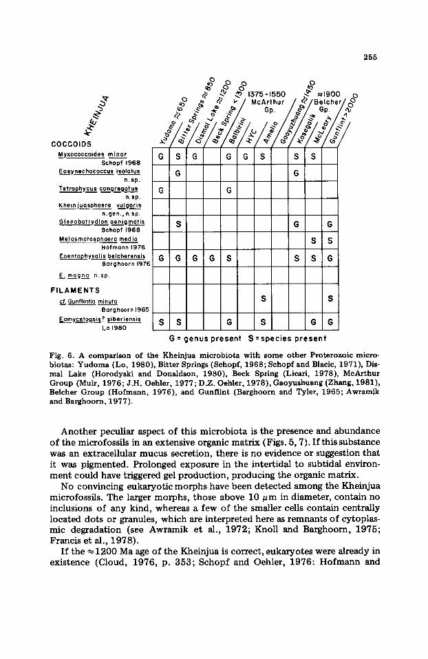
Taxonomically, the Kheinjua microbiota resembles other stratiform stromatolitic microbiotas. Taxonomic similarity is a common feature of Proterozoic stratiform stromatolites from intertidal to subtidal environments (Hofmann, 1976; Awramik, 1981). In particular, the Kheinjua is similar to microbiotas from the ~1900 Ma-old Belcher Islands (Hofmann, 1976); the ~1500 Ma-old McArthur Group (Muir, 1976; D.Z. Oehler, 1978); the ~-850 Ma-old Bitter Springs (Schopf, 1968; Schopf and Blacic, 1971 ); and the ~ 650 Ma-old Yudoma Suite (Lo, 1980). Of the 9 genera and 10 species described from the Kheinjua, 5 genera and 3 species are in common with the Bitter Springs, 5 genera and 4 species are in common with the McArthur Group, and 6 genera and 3 species are in common with the Belcher Islands (Fig. 6). When compared to the Yudoma microbiota, only 4 genera and 1 species are in common. All these deposits share similar environmental settings.
Anomalously, the filaments of the Kheinjua are predominantly cf. Gun- flintia minuta. The only other microbiotas where Gunflintia minuta is the major filamentous component are in Early Proterozoic iron-formations such as the Gunflint Iron Formation (Barghoorn and Tyler, 1965; Awramik 1976); the Sokoman Iron Formation (Knoll and Simonson, 1981); the Frere Formation (Walter et al., 1976); and the Tyler Formation (Cloud and Morri- son, 1980). Unlike these formations, the Kheinjua microbiota is not associat- ed with iron, is found only in stratiform stromatolites, and is significantly younger.
255
/ COCCOIDS
M_~xococcoides minor Schopf 1968
Eo$~fnechococcus isolatus n.sp.
Tetrophycu$ congregotus n. sp.
Kheinjuasphaera vu..~Ioor is n.gen, t n.sp
.Glenobot rydion oenigmatis Schopf 1968
Melasmatosphoero media Hofmann 1976
Eoentophysolis belcherensis G Borghoorn 1976
E~ m.~agna n.sp.
FILAMENTS cf. Gunflintio minuta
Barghoorn 1965
Eomycetopsis ? siberiensi$ S Lo 1980
G S
G
G G G
McArthur Gp.
S S
G
~1900
,I v.,
oo
G G G S
G G
S S
S S G
S S
S G S G G
G=genuspresent S=species present
Fig. 6. A comparison of the Kheinjua microbiota with some other Proterozoic micro- biotas: Yudoma (Lo, 1980), Bitter Springs (Schopf, 1968; Schopf and Blacic, 1971), Dis- mal Lake (Horodyski and Donaldson, 1980), Beck Spring (Licari, 1978), McArthur Group (Muir, 1976; J.H. Oehler, 1977; D.Z. Oehler, 1978), Gaoyushuang (Zhang, 1981), Belcher Group (Hofmann, 1976), and Gunflint (Barghoorn and Tyler, 1965; Awramik and Barghoorn, 1977).
Another peculiar aspect of this microbiota is the presence and abundance of the microfossils in an extensive organic matrix (Figs. 5, 7). If this substance was an extracellular mucus secretion, there is no evidence or suggestion that it was pigmented. Prolonged exposure in the intertidal to subtidal environ- ment could have triggered gel production, producing the organic matrix.
No convincing eukaryotic morphs have been detected among the Kheinjua microfossils. The larger morphs, those above 10/~m in diameter, contain no inclusions of any kind, whereas a few of the smaller cells contain centrally located dots or granules, which are interpreted here as remnants of cytoplas- mic degradation (see Awramik et al., 1972; Knoll and Barghoorn, 1975; Francis et al., 1978}.
If the ~1200 Ma age of the Kheinjua is correct, eukaryotes were already in existence (Cloud, 1976, p. 353; Schopf and Oehler, 1976: Hofmann and

256
Toxo
go~ o
©
®
Postulated Aff in i t ies
Super kingdom Procoryota
Kingdom MONERA
Phylum unknown
Phylum CYANOBACTERIA (CYANOPHYTA)
Class Hormogoneae
Order Oscillatoriales
Family Oscillatoriaceoe
Class Coccogoneae
Order Chroococcales
Family Chroococcaceoe
Family Entophysolidoceoe
M yxococcoides minor Schopf 1968
Eosynechococcus isolotu$ n. sp
Tetrophycus congre.gotus n . s p
Kheinjuasphaera vulgaris n.gen., n.sp
Eoentophysalis belcherensis
E...~ m__ogn._o n. sp Hofmonn 1976
Glenobotrydion oenlgmotis Schopf 1968
Melosmotosphaera media Hofmonn 1976
cf.(~unflintio minula Borghoorn 1965
. / ~ . . - ' ~ Eomycetopsis ? siberiensis Lo 1980
m
?
?
ml ? ? m
Size
outside
d iameter
or
w i d t h
(~m)
I0 20 50
I i
mR
m
m m
Fig. 7. Aff in i t ies and relat ive a b u n d a n c e s of specms m
q) (.3
(.3 o o
A abund0nt C common R rare
R
A
A
A
C
C
R
R
A
C
t he Khe in jua mic rob io ta .
Chen, 1980; Awramik, 1981). By analogy with modern stromatolitic com- munities, environments like that of the Fawn Limestone in the Kheinjua Formation are unlikely places for protoctistan eukaryotes. Eukaryotes would more likely be found in offshore, permanently submerged stromatolitic, or non-stromatolitic clastic facies.
The similarity of the Kheinjua microfossils to older microbiotas in size and taxonomic composition may reflect environmental conditions which favored small microorganisms and excluded large microbes. The details of these environmental conditions are uncertain. Alternatively, there might be peculiarities in the diagenetic and taphonomic history of the Kheinjua that destroyed most of the larger, possibly more delicate morphs. Another expla- nation for the anachronistic characteristics of the microbiota is the possibili- ty of an error in the age assignment of the Kheinjua.
257
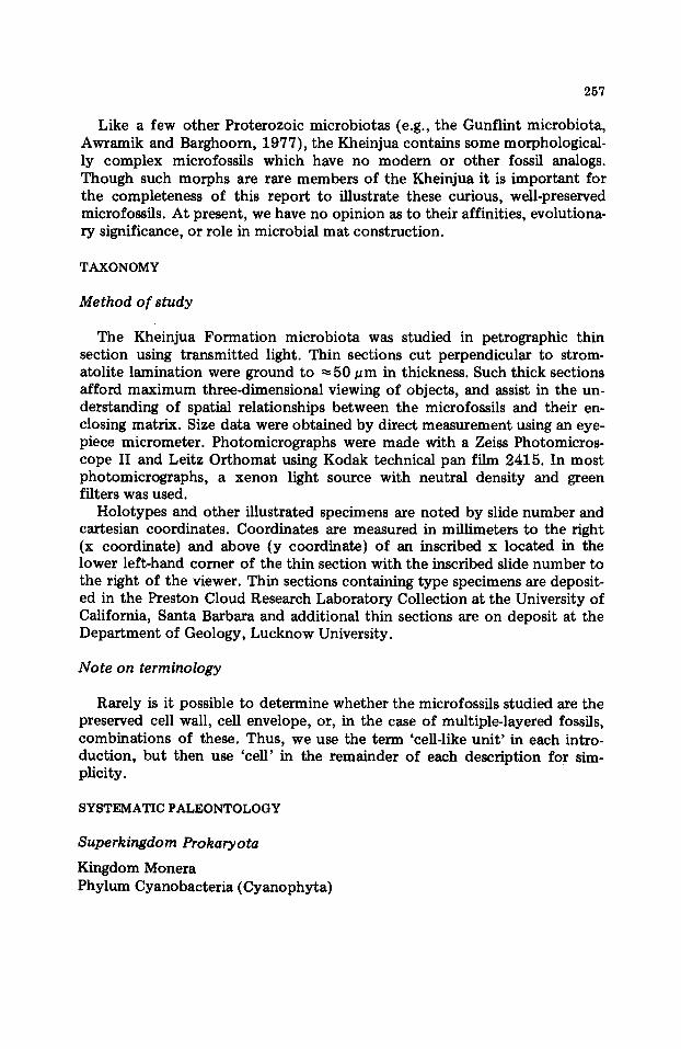
Like a few other Proterozoic microbiotas (e.g., the Gunflint microbiota, Awramik and Barghoorn, 1977), the Kheinjua contains some morphological- ly complex microfossils which have no m o d e m or other fossil analogs. Though such morphs are rare members of the Kheinjua it is important for the completeness of this report to illustrate these curious, well-preserved microfossils. At present, we have no opinion as to their affinities, evolutiona- ry significance, or role in microbial mat construction.
TAXONOMY
Method of study
The Kheinjua Formation microbiota was studied in petrographic thin section using transmitted light. Thin sections cut perpendicular to strom- atolite lamination were ground to ~ 5 0 ~m in thickness. Such thick sections afford maximum three-dimensional viewing of objects, and assist in the un- derstanding of spatial relationships between the microfossils and their en- closing matrix. Size data were obtained by direct measurement using an eye- piece micrometer. Photomicrographs were made with a Zeiss Photomicros- cope II and Leitz Orthomat using Kodak technical pan film 2415. In most photomicrographs, a xenon light source with neutral density and green filters was used.
Holotypes and other illustrated specimens are noted by slide number and cartesian coordinates. Coordinates are measured in millimeters to the right (x coordinate) and above (y coordinate) of an inscribed x located in the lower left-hand comer of the thin section with the inscribed slide number to the right of the viewer. Thin sections containing type specimens are deposit- ed in the Preston Cloud Research Laboratory Collection at the University of California, Santa Barbara and additional thin sections are on deposit at the Depar tment of Geology, Lucknow University.
Note on terminology
Rarely is it possible to determine whether the microfossils studied are the preserved cell wall, cell envelope, or, in the case of multiple-layered fossils, combinations of these. Thus, we use the term 'cell-like unit ' in each intro- duction, bu t then use 'cell' in the remainder of each description for sim- plicity.
SYSTEMATIC PALEONTOLOGY
Superkingdom Prokaryota
Kingdom Monera Phylum Cyanobacteria (Cyanophyta)

258
Class Coccogoneae Order Chroococcales
Family Chroococcaceae Genus Myxococcoides Schopf (1968)
Type species. Myxococcoides minor Schopf (Schopf, 1968; pp. 183 and 676, Plate 81, Figs. 1 and 10, and Table 4), Bitter Springs Formation, Northern Territory, Australia.
Myxococcoides minor Schopf (1968) (Fig. 5B, C)
Description. Single-walled cell-like units, spheroidal to ellipsoidal; often de- formed by mutual compression when in groups. Habit solitary or clustered in groups of up to 30 cells. Groups vary in shape from roughly spheroidal to elongate. Cell unit diameter 6.3--12.75 pm, average 8.2 pm (38 cells measur- ed). Few cells contain an inclusion. Long axes of ellipsoids have no preferred orientation with respect to bedding or stromatolite lamination. Discussion. Though the Kheinjua form has a somewhat wider size range and an average diameter slightly less than the type material described from the Bitter Springs Formation (Fig. 8), it conforms to all other diagnostic features of M. minor Schopf (1968). The Kheinjua Myxococcoides differs in size from other described species of Myxococcoides (Fig. 8). Myxococcoides minor may also be compared to Glenobotrydion aenigmatis Schopf {1968). As noted by Hofmann (1976), M. minor and G. aenigmatis overlap in group organization, dimensions, and appearance. In the Kheinjua, M. minor and G. aenigmatis are differentiated on the basis of colonial organization; G. aen- igmatis forms pseudofilamentous colonies, sometimes containing very large numbers of cells (Fig. 5D, F). Intracellular inclusions are not considered taxonomically diagnostic.
Genus Eosynechococcus Hofmann (1976)
Type species. Eosynechococcus moorei Hofmann (Hofmann, 1976; pp. 1057--1058 Plate 2, Figs. 1--7), Kasegalik Formation, Belcher Islands, Canada.
Eosynechococcus isolatus n. sp. (Fig. 5E, G).
D/agnos/s. CeU-like units, rod-shaped to ellipsoidal, occasionally slightly curved. Cells found loosely associated in groups of 30--60 ceils; cells rarely touch and lack individual sheaths, but are usually in a common organic matrix. Ceils measure 1.7-6.8 pm in the short dimension and 1.7-8.5 pm in the long dimension, averaging 2.7 pm by 4.07 pm (91 specimens measur- ed). Cells are organized into ovoidal groups, 10--30 pm by 20- 60 pm (4 measured).
Etymology: isolatus, Latin, with reference to the isolated nature of in- dividuals within a group.

259
Glenobotrydion
G_.:mojorinum Schopf 8 Rlocic 1971 Hofmonn 1976
G. vorioforme Zhong 1981
6,oenigmotis Schopf 1968 present paper
_Myxococcoides M, minor Rchopf 1968
Muir 197R Hofmonn 1976 p r e s e n t p a p e r
M.crocens O.Z. Oehler 1978
M_: kingii Muir 1976 J.H. Oehler 1977 D.Z.Oehler 1978
M. minuta Muir 1976 D. Z. Oehler 1978
M. reticulota $chopf 1968
M. inornata Schopf 1968
Hofmonn 1976
M. konzolovoe Muir 1974
M.; sto philydion LO 1980
M_. q_..gyo ngen sis Yin 1981
M_;reniformis Muir 1974
M. sp. Hofmonni976 M. sp. D.Z.Oehler 1978
s p e c i e s R e p o r t
: C 0
I
I
I
C : I
', C :
: C
I 0
: C :
~--?~
RANGE
MEAN
0
I
° ,0 ' .
: C I
I 0 I , C .
I I ! I I i i i I ] I i I i I i i , i l , , R ~ I
i 2 3 4 5 s ? s 9 Io 15 2o z5 size bLm
Fig. 8. The sizes of Glenobotrydion and Myxococcoides individuals in several micro- biotas.
Type locality. Upper Fawn Limestone, Kheinjua Formation, above Cono- phyton garganicum, Salkhan Hill, Son Valley area, Mirzapur district, Uttar Pradesh, India.
Type specimen. 33.4/0.7 in thin section SB029-2, illustrated in Fig. 5G.
Discussion. Although the dimensions of E. isolatus are similar to E. moorei Hofmann (1976), the Kheinjua species differs in the arrangement of cells. Eosynechococcus isolatus is not grouped in small stacks and chains, and cells are not in contact with one another. Eosynechococcus isolatus shows more size variation within a group and individuals tend to be rounder (Fig. 9). Groups of E. isolatus are occasionally surrounded by an amorphous organic matrix. Individuals of E. isolatus resemble the solitary Pilauia macul- ata Oehler (1978), which does not form groups. Occasionally, E. isolatus contains a single centrally or eccentrically positioned inclusion.

260
6
",7, 0 4
~3
I
A . = 8 0 % of population
~ = 9 5 % of population
X = average size
• = individual cells
~ o
I I I I i I 2 3 4 5 ainu
I I I I I I I I I 2 3 4 ,~ 6 7 8 ~m
long axis
io
x
=o co 5
B.
E: i S 0.~I a t_u_s T E grandis -- Ho-'~-~monn 1976
.~ , Z , E medius Hofrnonn 1976 I£
I • ,I~ I
1=*E.moorei Hofmann 1976
I I I 5 I0 15 20 pm
long axis
Fig. 9. (A) Size distribution of Eosynechococcus isolatus n. sp. Measured population is 101 individuals (omu = ocular micrometer units; raw data). (B) Comparison with other described species of Eosynechococcus.
Genus Glenobotrydion Schopf (1968)
Type species. Glenobotrydion aenigmatis Schopf (Schopf, 1968; pp. 681--683, Plate 83, Fig. 9; Plate 84, Figs. 4 and 5; text--Fig. 6, and Table 4), Bitter Springs Formation, Northern Territory, Australia.
Glenobotrydion aenigmatis Schopf (1968) (Fig. 5D, F )
Description. Cell-like units, spheroidal to discoidal; 5.1-12.75 pm in diam- eter, average 9.3 pm (43 individuals measured); usually arranged in pseudo- filamentous colonies containing from 7 to over 100 cells. Colony dimensions range from 10 X 35 ~m to 50 X 300 pm (3 colonies measured); long axes of colonies are roughly parallel to stromatolitic lamination. Short axis of compressed or flattened cells is parallel to pseudofilament. Reproduction by fission observed in one or two planes. Discussion. In the Bitter Springs, G. aenigmatis is characterized by an inclu sion which has been used as evidence for a eukaryotic affinity (Schopf, 1968). However, such inclusions are more likely the result of cytoplasmic degradation (Awramik et al., 1972} and should not be used as a diagnostic taxonomic feature. The Kheinjua form is similar in size and morphology to the Bitter Springs G. aenigmatis (Schopf, 1968, Plate 84, Fig. 5), except that Kheinjua cells generally lack inclusions.
Individual cells of G. aenigmatis and Myxococcoides minor are indistin- guishable; we refer to cells organized into pseudofilaments as G. aenigmatis. Also, solitary spheroids of the correct size found closely associated with G. aenigmatis colonies are assigned to that species on the assumption that they may have broken off from the parent colony. Single-walled, solitary

261
spheroids not closely associated with any colony are grouped in the new genus Kheinjuasphaera (see below). Based on the size and shape of indi- viduals and mode of reproduction, G. aenigmatis may be a member of the Chroococcaceae, but a pseudofflamentous arrangement is a common feature in the Entophysalidaceae (Geitler, 1932).
Genus Melasmatosphaera Hofmann (1976)
Type species. Melasmatosphaera magna Hofmann (Hofrnann, 1976; p. 1066, Plate 8, Figs. 1--2), Mc Leafy Formation, Belcher Islands, Canada.
Melasmatosphaera media Hofmann (1976)
Description. Solitary, dark spheroid 11.4 ~m across, containing numerous clustered and scattered granules of sub-micron size. Discussion. Only one specimen has been found, but it is identical to the diag- nosis and illustrations of Hofmann (1976, Plate 8, Fig. 5). Following Hof- mann (1976), we recognize that Melasmatosphaera may represent a degrada- tional morph of other taxa; in this assemblage, possible candidates include Kheinjuasphaera vulgaris, or solitary individuals of Myxococcoides minor or Glenobotrydion aenigmatis.
Family Entophysalidaceae
Genus Eoentophysalis Hofmann (1976)
Type species. Eoentophysalis belcherensis Hofmann (Hofmann, 1976; pp. 1070--1072, Figs. 1--5; Plate 5, Figs. 3--6; Plate 6, Figs. 1--14), Kasegalik Formation, Belcher Islands, Canada.
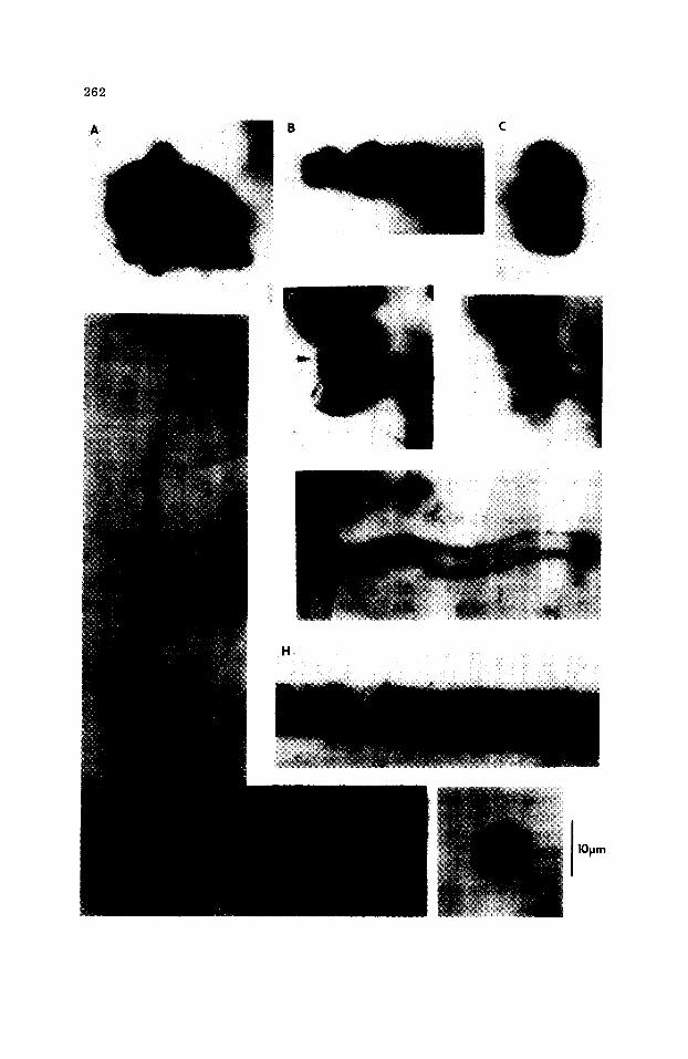
Eoentophysalis belcherensis Hofmann (1976) (Fig. 10A--C)
Description. Cell-like units, spheroidal to polyhedral, often deformed by mutual compression. Found in spheroidal or roughly linear clumps of 5-- 25 cells with a diameter of 3.99--5.7/~m, averaging 4.89/~m (10 cells measured). Discussion. The type material of E. belcherensis occurs in extensive monospe- cific mats. In the Kheinjua, E. belcherensis is only found in small clusters as- sociated with other members of the microbiota; nowhere was it observed to form mats. The aggregation in Fig. 10B illustrates the sub4inear organization that Oehler (1978) has interpreted as indicative of a young colony. Such an arrangement could also be viewed as an isolated, detached remnant of a more extensive colony.
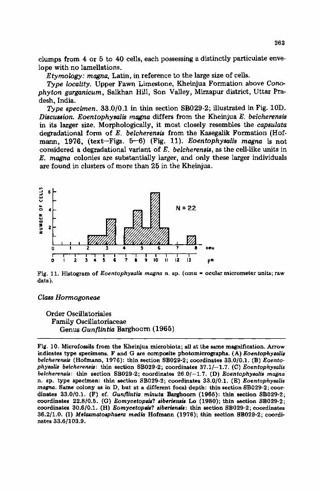
Eoentophysalis magna n. sp. (Fig. 10D, E)
Diagnosis. Cell-like units, spheroidal, eUipsoidal, or subpolyhedral; diameter 4.25--9.12 ~m, average 5.41 ~m (22 specimens measured). Occurs in irregular
2 6 2
B C
H i~! i ' i ! i ¸ ¸ i ~ !!! ~!~il ~!~ii i̧̧
1Opm
263
clumps from 4 or 5 to 40 cells, each possessing a distinctly particulate enve- lope with no lamellations.
Etymology: magna, Latin, in reference to the large size of cells. Type locality. Upper Fawn Limestone, Kheinjua Formation above Cono-
phyton garganicum, Salkhan Hill, Son Valley, Mirzapur district, Uttar Pra- desh, India.
Type specimen. 33.0/0.1 in thin section SB029-2; illustrated in Fig. 10D. Discussion. Eoentophysalis magna differs from the Kheinjua E. belcherensis in its larger size. Morphologically, it most closely resembles the capsulata degradational form of E. belcherensis from the Kasegalik Formation (Hof- mann, 1976, (text--Figs. 5--6) (Fig. 11). Eoentophysalis magna is not considered a degradational variant of E. belcherensis, as the cell-like units in E. magna colonies are substantially larger, and only these larger individuals are found in clusters of more than 25 in the Kheinjua.
6
o 4 N=22
~ z
I I I I ~ / ~ J 0 I 2 3 4 5 G ? 8 0mu
I I ! I I I I I I I I I I 1 0 I 2 .3 4 5 S 7' I I g I 0 II 12 13 IJm
Fig. 11. Histogram of Eoentophysalis magna n. sp. (omu ffi ocular micrometer units; raw data).
Class Hormogoneae
Order Oscillatoriales Family Oseillatoriaceae
Genus Gunflintia Barghoorn (1965)
Fig. 10. Microfossils from the Kheinjua microbiota; all at the same magnification. Arrow indicates type specimens. F and G are composite photomicrographs. (A) Eoentophysalis belcherensis (Hofmann, 1976): thin section SB029-2; coordinates 33.0/0.1. (B) Eoento- physalis belcherensis: thin section SB029-2; coordinates 37.1/--1.7. (C)Eoentophysa l i s belcherensis: thin section SB029-2; coordinates 26 .0 / -1 .7 . (D) Eoentophysa|is magna n. sp. type specimen: thin section SB029-2; coordinates 33.0/0.1. (E) Eoentophysalis magna. Same colony as in D, but at a different focal depth: thin section SB029-2; coor- dinates 33.0/0.1. (F) cf. Gunflintia minuta Barghoorn (1965): thin section SB029-2; coordinates 22.8/0.5. (G) Eomycetopsis? siberiensis Lo (1980); thin section SB029-2; coordinates 30.6/0.1. (H) Eomycetopsis? siberiensis: thin section SB029-2; coordinates 36.2/1.0. (I) Melasmatosphaera media Hofmann (1976); thin section SB029-2; coordi- nates 33.6/103.9.
264
Type species. Gunflintia minuta Barghoorn (Barghoorn and Tyler, 1965; Fig. 4, parts 6, 8; Fig. 6, part 1), Gunflint Iron Formation, Ontario, Canada.
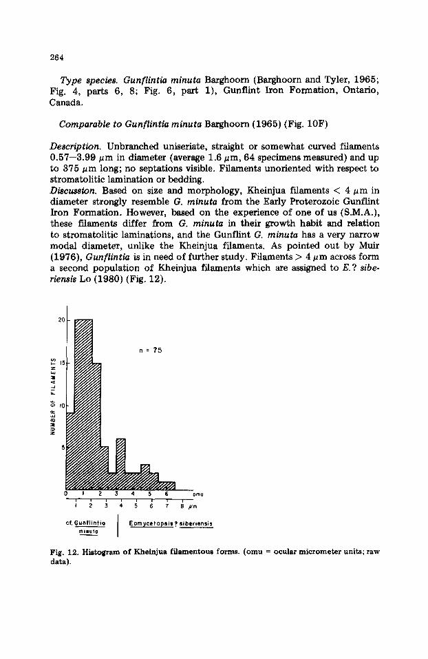
Comparable to Gunflintia minuta Barghoorn (1965) (Fig. 10F)
Description. Unbranched uniseriate, straight or somewhat curved filaments 0.57--3.99 #m in diameter (average 1.6 ~m, 64 specimens measured) and up to 375 pm long; no septations visible. Filaments unoriented with respect to stromatolitic lamination or bedding. Discussion. Based on size and morphology, Kheinjua filaments < 4 ~m in diameter strongly resemble G. minuta from the Early Proterozoic Gunflint Iron Formation. However, based on the experience of one of us (S.M.A.), these filaments differ from G. minuta in their growth habit and relation to stromatolitic laminations, and the Gunflint G. minuta has a very narrow modal diameter, unlike the Kheinjua filaments. As pointed out by Muir (1976), Gunflintia is in need of further study. Filaments > 4 pm across form a second population of Kheinjua filaments which are assigned to E. ? sibe- riensis Lo (1980) (Fig. 12).
2o I
~E
..J b_
=
i 2 3 4 5 6 i i i i i i i I 2 3 4 5 6 7
cf. GunflintiOminuto J
omu
Eornycetops s ? siberiensis
Fig. 12. Histogram of Kheinjua filamentous forms. ( o m u = ocular micrometer units; raw data).
265
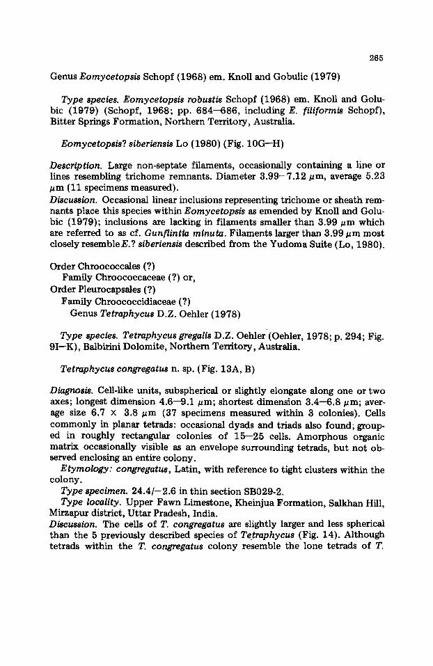
Genus Eomycetopsis Schopf (1968) em. Knoll and Gobulic (1979)
Type species. Eomycetopsis robustis Schopf (1968) em. Knoll and Golu- bic (1979) (Schopf, 1968; pp. 684--686, including E. filiformis Schopf), Bitter Springs Formation, Northern Territory, Australia.
Eomycetopsis? siberiensis Lo (1980) (Fig. 10G--H)
Description. Large non-septate filaments, occasionally containing a line or lines resembling tr ichome remnants. Diameter 3 .99-7 .12 pm, average 5.23 ~m (11 specimens measured). Discussion. Occasional linear inclusions representing tr ichome or sheath rem- nants place this species within Eomycetopsis as emended by Knoll and Golu- bic (1979); inclusions are lacking in filaments smaller than 3.99 ~zm which are referred to as cf. Gunflintia minuta. Filaments larger than 3.99/~m most closely resemble E.? siberiensis described from the Yudoma Suite (Lo, 1980).
Order Chroococcales (?) Family Chroococcaceae (?) or,
Order Pleurocapsales (?) Family Chroococcidiaceae (?)
Genus Tetraphycus D.Z. Oehler (1978)
Type species. Tetraphycus gregalis D.Z. Oehler (Oehler, 1978; p. 294; Fig. 9I--K), Balbirini Dolomite, Northern Territory, Australia.
Tetraphycus congregatus n. sp. (Fig. 13A, B)
Diagnosis. Cell-like units, subspherical or slightly elongate along one or two axes; longest dimension 4.6--9.1 ~m; shortest dimension 3.4--6.8/~m; aver- age size 6.7 X 3.8 ~m (37 specimens measured within 3 colonies). Cells commonly in planar tetrads: occasional dyads and triads also found, group- ed in roughly rectangular colonies of 15--25 cells. Amorphous organic matrix occasionally visible as an envelope surrounding tetrads, but not ob- served enclosing an entire colony.
Etymology: congregatus, Latin, with reference to tight clusters within the colony.
Type specimen. 24 .4 / -2 .6 in thin section SB029-2. Type locality. Upper Fawn Limestone, Kheinjua Formation, Salkhan Hill,
Mirzapur district, Uttar Pradesh, India. Discussion. The cells of T. congregatus are slightly larger and less spherical than the 5 previously described species of Tetraphycus (Fig. 14). Although tetrads within the T. congregatus colony resemble the lone tetrads of T.
266
h lo~m
G
2 6 7
o
8 . 7
7 - 6
-~ 6 "5 o 5 "4 t..
2 2 -
I I -
A
J I l [ I I * * I I
I 2 3 4 5 6 7 e 9 IO~m long oxis
E :I B
T con u n c t u m 7 . ~ _ _ | _ _ (Lo , 1979)
;,° 6 : ' I "°'"°°" ....
5 L ' "'J~i ' I T o 4 -- YS.-~..;.,97e)J. 1
I
T.:.gr_eeq;oli~S (Oeh le r , 1 9 7 8 )
T. q l i m i n u t i v u s (Oehter , 1978 )
, .
I 2 3 4 5 6 7 8 pm long axis
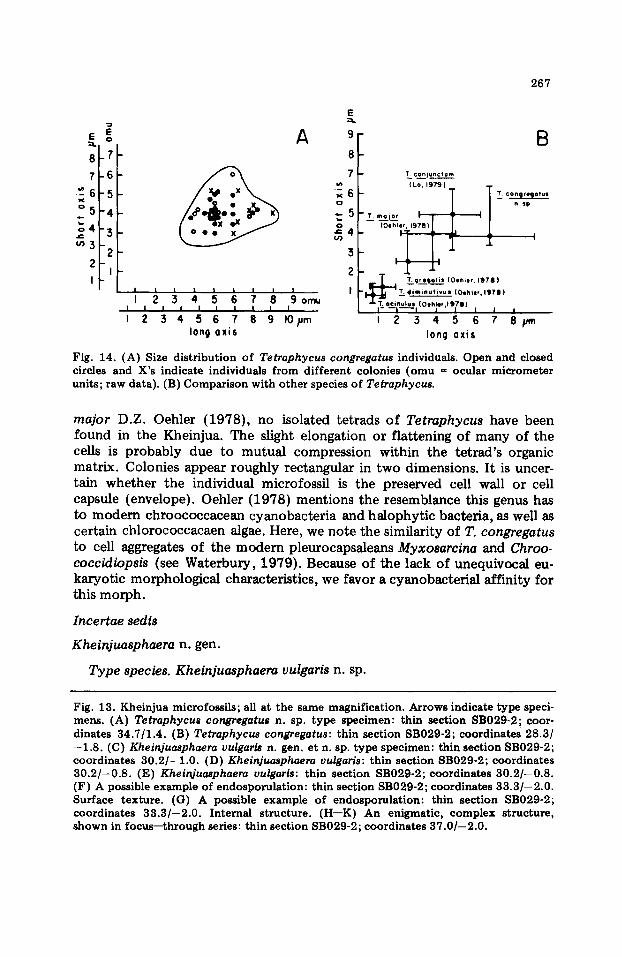
Fig. 14. (A) Size distr ibution of Tetraphycus congregatus individuals. Open and closed circles and X's indicate individuals from different colonies (omu = ocular micrometer units; raw data). (B) Comparison with other species of Tetraphycus.
major D.Z. Oehler (1978), no isolated tetrads of Tetraphycus have been found in the Kheinjua. The slight elongation or flattening of many of the cells is probably due to mutual compression within the tetrad's organic matrix. Colonies appear roughly rectangular in two dimensions. It is uncer- tain whether the individual microfossil is the preserved cell wall or cell capsule (envelope). Oehler (1978) mentions the resemblance this genus has to m o d e m chroococcacean cyanobacteria and halophytic bacteria, as well as certain chlorococcacaen algae. Here, we note the similarity of T. congregatus to cell aggregates of the modern pleurocapsaleans Myxosarcina and Chroo- coccidiopsis (see Waterbury, 1979). Because of the lack of unequivocal eu- karyotic morphological characteristics, we favor a cyanobacterial affinity for this morph.
Incertae sedis
Kheinjuasphaera n. gen.
Type species. Kheinjuasphaera vulgaris n. sp.
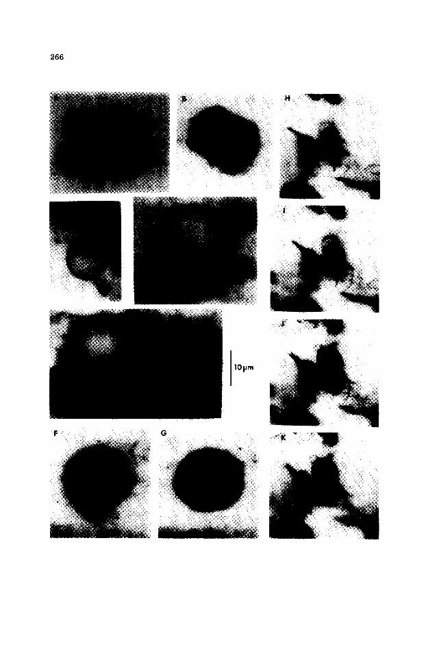
Fig. 13. Kheinjua microfossils; all at the same magnification. Arrows indicate type speci- mens. (A) Tetraphycus congregatus n. sp. type specimen: thin section SB029-2; coor- dinates 34.7/1.4. (B) Tetraphycus congregatus: thin section SB029-2; coordinates 28.3/ - 1 . 8 . (C) Kheinjuasphaera vulgaris n. gen. et n. sp. type specimen: thin section SB029-2; coordinates 30.2/--1.0. (D) Kheinjuasphaera vulgaris: thin section SB029-2; coordinates 30.2/--0.8. (E) Kheinjuasphaera vulgaris: thin section SB029-2; coordinates 30.2/--0.8. (F) A possible example of endosporulat ion: thin section SB029-2; coordinates 33.3/--2.0. Surface texture. (G) A possible example of endosporulat ion: thin section SB029-2; coordinates 33.3/--2.0. Internal structure. (H--K) An enigmatic, complex structure, shown in focus--through series: thin section SB029-2; coordinates 37.0/--2.0.
268
Type locality. Upper Fawn Limestone, Kheinjua Formation, Salkhan Hill, Son Valley area, Mirzapur District, Uttar Pradesh, India.
Etymology. With reference to its occurrence in the Kheinjua Formation; sphaera, Latin, spherical shape.
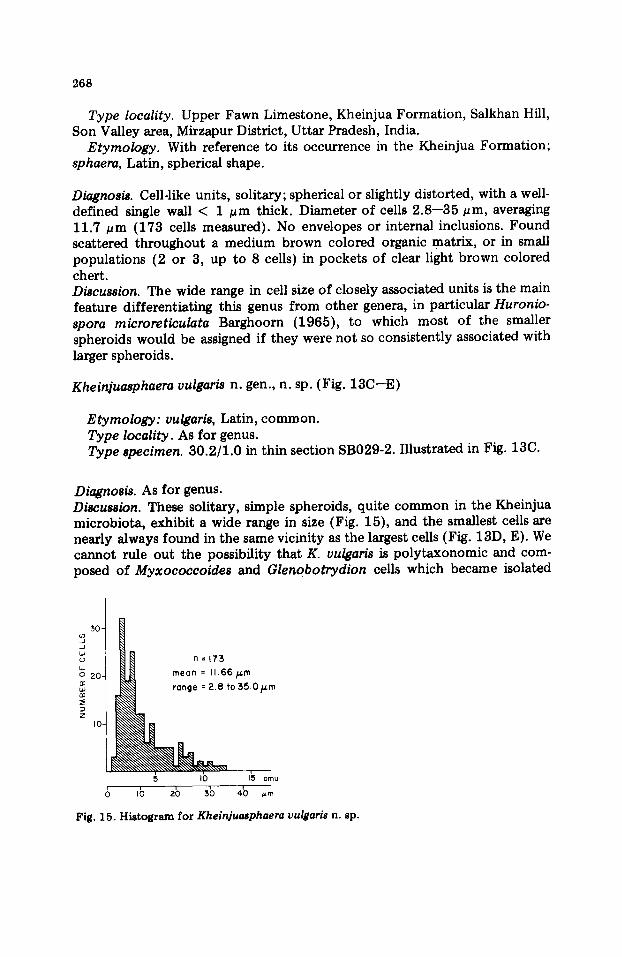
Diagnosis. Cell-like units, solitary; spherical or slightly distorted, with a well- defined single wall < 1 ~m thick. Diameter of cells 2.8--35 #rn, averaging 11.7 /~m (173 cells measured). No envelopes or internal inclusions. Found scattered throughout a medium brown colored organic matrix, or in small populations (2 or 3, up to 8 cells) in pockets of clear light brown colored chert. Discussion. The wide range in cell size of closely associated units is the main feature differentiating this genus from other genera, in particular Huronio- spora microreticulata Barghoorn (1965), to which most of the smaller spheroids would be assigned if they were not so consistently associated with larger spheroids.
Kheinjuasphaera vulgaris n. gen., n. sp. (Fig. 13C--E)
Etymology: vulgaris, Latin, common. Type locality. As for genus. Type specimen. 30.2/1.0 in thin section SB029-2. Illustrated in Fig. 13C.
Diagnosis. As for genus. Discussion. These solitary, simple spheroids, quite common in the Kheinjua microbiota, exhibit a wide range in size (Fig. 15), and the smallest cells are nearly always found in the same vicinity as the largest cells (Fig. 13D, E). We cannot rule out the possibility that K. vulgaris is polytaxonomic and com- posed of Myxococcoides and Glenobotrydion cells which became isolated
3 0 -
..J L~
2 0 -
Z I0 -
~ ~ range : 2.8 to 35.0 ~m
g Ib I~ o~o 6 Ib 2b sb 4b ~
Fig. 15. Histogram for Khein]u~phaem vulgari8 n. sp.

269
from their parent colonies. However, the lack of prominent breaks in the size distribution of closely associated cells and the uniformity of morphology make this a relatively well defined species.
Kheinjuasphaera vulgaris is associated with the enigmatic microfossil illustrated in Fig. 13F, G. This association suggests that K. vulgaris may be derived from the complex microfossil and represents spores, fruiting bodies, or some other type of developmental morph. We place K. vulgaris in Insertae sedis.
ACKNOWLEDGEMENTS
We thank Mark McMenamin and David Pierce for helpful comments; David Pierce for assistance with photographs and technical matters; and David Crouch for drafting figures. This paper was written during the tenure of an Alexander yon Humboldt Foundation Fellowship granted to one of us (S.K.). Contribution No. 116 of the Preston Cloud Research Laboratory.
REFERENCES
Auden, J.B., 1933. Vindhyan sedimentation in Son Valley, Mirzapur district. Mere. Geol. Surv. India, 62: 141--250.
Awramik, S.M., 1976. Gunflint stromatolites: microfossil distribution in relation to stromatolite morphology. In: M.R. Walter (Editor), Stromatolites. Elsevier, Amster- dam, pp. 311--320.
Awramik, S.M., 1981. The pre-Phanerozoic biosphere -- three billion years of crises and opportunities. In: M.H. Nitecki (Editor), Biotic Crises in Ecological and Evolutionary Time. Academic Press, New York, pp. 83--102.
Awramik, S.M. and Barghoorn, E.S., 1977. The Gunflint microbiota. Precambrian Res., 5: 121--142.
Awramik, S.M., Golubic, S. and Barghoorn, E.S., 1972. Blue-green algal cell degradation and its implication for the fossil record. Geol. Soc. Am. Abs. with Programs, 4: 438.
Bannerjee, D.M., 1973. Microfossil from late Precambrian phosphatic Aravalli stromatol- ires of Udalpur, Rajasthan, India. In: V.K. Verma, R.K. Misra, S.S. Merh, V.K. Gaur and K.S. Valdiya (Editors), Recent Researches in Geology. Hindustan, Delhi, India, pp. 263--268.
Barghoorn, E.S. and Tyler, S.A., 1965. Microorganisms from the Gunflint chert. Sci- ence, 147: 563--577.
Cloud, P., 1976. Beginnings of biospheric evolution and their biogeochemical conse- quences. Paleobiology, 2: 351--387.
Cloud, P. and Morrison, K., 1979. On microbial contaminants, micropseudofossils, and the oldest records of life. Precambrian Res., 9: 81---91.
Cloud, P. and Morrison, K., 1980. New microbial fossils from 2 Gyr old rocks in nor- thern Michigan. Geomicrobiology J., 2: 161--178.
Crawford, A.R. and Compston, W., 1970. The age of the Vindhyan System of Peninsular India. Q. J. Geol. Soc. London, 125: 351- 371.
Francis, F., Barghoorn, E.S. and Margulis, L., 1978. On the experimental silieification of microorganisms. IH. Implications of the preservation of the green pokaryotic alga Prochloron and other coccoids for interpretation of the microbial fossil record. Pre- cambrian Res., 7: 377--383.
270
Geitler, L., 1932. Cyanophyceae. Akademische Verlagsgeselischaft m.b.H., Leipzig. Re- printed by Johnson Reprint Corporation, New York, 1971, 1196 pp.
Gupta, V.J., 1977. Indian Precambrian Stratigraphy. Hindustan, Delhi, India, 328 pp. Hofmann, H.J., 1976. Precambrian microflora, Belcher Islands, Canada: significance and
systematics. J. Paleont., 50: 1040--1073. Hofmann, H.J. and Chen, J., 1980. Carbonaceous megafossils from the Precambrian
(1800 Ma) near Jixian, northern China. Can. J. Earth Sci., 18: 443--447. Horodyski, R.J. and Donaldson, J.A., 1980. Microfossils from the Middle Proterozoic
Dismal Lakes Group, Arctic Canada. Precambrian Res., 11: 125--159. Knoll, A.H. and Barghoorn, E.S., 1975. Precambrian eucaryotic organisms: a reassess-
ment of the evidence. Science, 190: 52--54. Knoll, A.H. and Golubic, S., 1979. Anatomy and taphonomy of a Precambrian algal
stromatolite. Precambrian Res., 10: 115--151. Knoll, A.H. and Simonson, B., 1981. Early Proterozoic microfossils and penecontempora-
neous quartz cementat ion in the Sokoman Iron Formation, Canada. Science, 211: 478--480.
Kreuzer, H., Harre, W., Kiirsten, M., Schnitzer, W.A., Mufti, K.S. and Srivastava, N.K., 1977. K/At dates of two glauconites from the Chandarpur Series (Chhattisgarh/In- dia). Geol. Jahrb., B28: 23--26.
Krishnan, M.S. and Swaminath, J., 1959. The great Vindhyan basin of northern India. J. Geol. Soc. India, 1: 10--30.
Kumar, S., 1976. Stromatoli tes from the Vindhyan rocks of Son Valley--Maihar Area, Districts Mirzapur (U.P.) and Satna (M.P.). J. Palaeont. Soc. India, 18: 13--21.
Kumar, S., 1978a. Discovery of micro-organisms from the black cherts of the Fawn Limestone (Late Precambrian), Semri Group, Son Valley, Mirzapur district, Uttar Pradesh. Curt. Sci., 47: 461.
Kumar, S., 1978b. Stromatoli tes and environment of deposition of the Vindhyan Super- group of Central India. J. Palaeont. Soc. India., 21: 33--43.
Kumar, S., 1980. Stromatoli tes and Indian biostratigraphy: a review. J. Paleont. Soc. India, 23: 166--183.
Kumar, S. and Singh, S., 1979. Discovery of micro-organisms from the bedded cherts of the Deoban Limestone (Late Precambrian), Lesser Himalaya, Uttar Pradesh. Curt. Sci., 48: 209--211.
Licari, G.R., 1978. Biogeology of the late pre-Phanerozoic Beck Spring Dolomite of eastern California. J. Paleont., 52: 767--792.
Lo, S.C., 1980. Microbial fossils from the Lower Yudoma Suite, earliest Phanerozoic, eastern Siberia. Precambrian Res., 13: 109--166.
Maithi, P.K. and Shukla, M., 1977. Microbiota from the Suket Shales, Rampura, Vind- hyan System (Late Precambrian) Madhya Pradesh. Palaeobotanist, 23: 176--188.
Miara, R.C. and Bhatnager, G.S., 1950. On carbonaceous discs and algal dust from the Vindhyans pre-Cambrian. Curt. Sei., 19: 88- 89.
Muir, M.D., 1976. Proterozoic microfossils from the Amelia Dolomite, McArthur Basin, Northern Territory. Alcheringa, 1: 143--158.
Oehler, D.Z., 1977. Pyrenoid-like structures in Late Precambrian algae from the Bitter Springs Format ion of Australia. J. Paleont., 51 : 885--901.
Oehler, D.Z., 1978. Microflora of the middle Proterozoic Balbirini Dolomite (McArthur Group) of Australia. Aleheringa, 2: 269--309.
Oehler, J.H., 1977. Microflora of the H.Y.C. Pyritic Shale Member of the Barney Creek Format ion (McArthur Group), middle Proterozoic of northern Australia. Alcheringa, 1: 315--349.
Oldham, T., 1856. Account of the results arrived at from investigations in central India during the past season. J. Asiat. Soc. Bengal, 25 : 249--255.
Raha, P.K., 1980. Proterozoic mierobiota from stromatoli t ic black chert of Jammu Limestone, Udhampur district, Jammu and Kashmir, India. J. Geol. Soc. India, 21: 572--574.
271
Raha, P.K., Chandy, K.C. and Balasubrahmanyan, M.N., 1978. Geochronology of the Jammu Limestone, Udhampur district, Jammu, India. J. Geol. Soc. India, 19: 221-- 223.
Rao, K.S., Lal, C. and Ghosh, D.B., 1977. Algal stromatolites in the Bhander Group of formations, Vindhyan Supergroup, Satna district, Madhya Pradesh. Rec. Geol. Surv. India, 109: 38--47.
Sahni, M.R., 1975. Vindhyan paleobiology, stratigraphy, and depositional environment: a critical review. J. Palaeont. Soc. India, 20: 289--304.
Salujha, S.K., 1971. Palynological evidence on the age of the Vindhyan sediments. Proc. Nat. Acad. Sci. India, Sect. A, 39: 62--68.
Salujha, S.K. and Rehman, K., 1972. Palynology of the Vindhyans and their equivalents in Peninsular India. Sere. on Palaeopalynology and Indian Stratigraphy, Proc., Calcutta, pp. 15--20.
Salujha, S.K., Rehman, K. and Arora, C.M., 1971. Plant microfossils from the Vindhyans of Son Valley. J. Geol. Soc. India, 12: 24-33.
Schopf, J.W., 1968. Microflora of the Bitter Springs Formation, Late Precambrian, cen- tral Australia. J. Paleont., 42: 651--688.
Schopf, J.W., 1977. Biostratigraphic usefulness of stromatolitic Precambrian microbiotas: a preliminary analysis. Precambrian Res., 5: 143--173.
Schopf, J.W. and Blacic, J.M., 1971. New micro-organisms from the Bitter Springs Forma- tion (Late Precambrian) of the north-central Amadeus Basin, Australia. J. Paleont., 45: 925--960.
Schopf, J.W. and Oehler, D.Z., 1976. How old are the eukaryotes? Science, 193: 47--49. Schopf, J.W. and Prasad, K.N, 1978. Microfossils in Collenia-like Stromatolites from the
Proterozoic Vempalle Formation of the Cuddapah Basin, India. Preeambrian Res., 6: 347--366.
Singh, I.B., 1973. Depositional environment of the Vindhyan sediments in Son Valley area. In: V.K. Verma, R.K. Misra, S.S. Merh, V.K. Gaur and K.S. Valdiya (Editors), Recent Researches in Geology. Hindustan, Delhi, India, pp. 146--152.
Sitholey, R.V., Srivastava, P.N. and Varma, C.P., 1953. Microfossils from the Upper Vind- hyans, with a discussion on the age of the Vindhyans in the light of plant fossil dis- coveries. Proc. Nat. Inst. Sci. India, 19: 195--202.
Vinogradov, A.P., Tugarinov, A.I., Zhikov, C.I., Stupnikova, N.I., Bibikova, E.V., Knorre, K.G. and Mehlnikova, G.L., 1966. Geohronologiya Dokembriya Indyi. In: Absolyut- noye Datirovanie Tektono-Magmaticheskih Siklovi Etapov ordeneriya po dannim 1964 g. Moscow. Izd. Nauka.
Viswanathiah, M.N. and Venkatachalapathy, V., 1980. Microbiota from the Bababudan Iron Formation, Karnataka. J. Geol. Soc. India, 21: 16--20.
Walter, M., Goode, A. and Hall, M., 1976. Microfossils from a newly discovered stroma- tolitic iron formation in Western Australia. Nature, 261: 221--223.
Wang, F. and Lo, Q., 1983. Restatement of Pseudoacritarchs. Precambrian Res., (in press). Waterbury, J.B., 1979. Developmental patterns of pleurocapsalean Cyanobacteria. In.
J.H. Parish (Editor), Developmental Biology of Prokaryotes. Univ. Calif. Press, Berkeley, pp. 203--226.
Zhang Yun, 1981. Proterozoic stromatolite microfloras of the Gaoyuzhuang Formation (Early Sinian:Riphean) Hebei, China. J. Paleont., 55: 485--506.