micro identification of medically important bacteria.pptx
TRANSCRIPT

Identification of medically important bacteria by conventional methods
• Based on morphology (staining properties)
• Cultural characteristics, colony morphology on agar media
• Tests for production of enzymes• Tests for metabolic end products• Sensitivity to chemicals• Serotyping (agglutination);
detection of antigens• Antibiotic susceptibility profile
Diagnostic tests are performed on the following specimens
• Blood• CSF• Exudates (pus, sputum, bronchial
washings)• Tissue• Urine• stool

Microscopy:– Gram stain– Acid fast stain– Flurorescent stain– Immunofluorescence– Dark ground microscopy– Simple stains– Wet mount
Culture • Cultivation and growth of bacteria
required for definitive identification and characterisation of the infectious agent and for performing antibiotic susceptibility testing
• achieved by isolating the bacteria from the clinical specimens collected from the infectious site on artificial culture media.
• artificial media used for growing bacteria should contain the basic nutritional requirements for non fastidious bacteria and special nutritive supplements for fastidious bacteria

Culture media
Growth media are used in either of two phases:
• liquid (broth) • solid (agar) • a biphasic medium that contains
both liquid and solid (blood culture medium)
Types of media:• Liquid or broth media• nutrients dissolved in water;
bacterial growth indicated by change in the broth’s appearance from clear to cloudy or turbid.
• due to light deflected by bacteria present in the culture
Examples of broth media and their use
• Tryptic soya broth (TSB): for blood culture
• Solid agar media:• These media are made by adding
a solidifying agent like agar to the nutrients and water.

• Solid media are used to isolate bacteria in a “pure culture” which is absolutely essential for identifying and characterising the pathogens.
• Clinical specimens containing suspected bacteria are spread (streaked) on the surface of appropriate solid agar media, incubated under appropriate conditions,
• Each bacterial cell from the specimen inoculated on the surface of agar medium will proliferate to sufficiently large numbers to be observed under the naked eye.
• The resulting bacterial population is considered to be derived from a single bacterial cell and is known as a colony;
• Bacteria within a single colony are the same genus and species, having identical genetic and phenotypic characteristics.
• Bacterial cultures derived from a single colony are considered pure

Media classification and functions:• Media categorised according to
their function and use.Five general categories of media• Basal supportive media• Enriched media• Enrichment media• Selective media• Differential media
• Basal supportive media: contain nutrients that support growth of most non fastidious organisms without giving any particular organism a growth advantage
• Example; Nutrient agar

• Enriched media: contain enriched substance like blood, serum, egg to support the growth of fastidious bacteria
• Blood agar: used for the cultivation of fastidious organisms, determination of hemolytic properties especially Streptococcus spp; Bacteria may show beta hemolysis (complete lysis of RBC), alpha hemolysis (partial lysis of RBC) or gamma hemolysis (no hemolysis)
Alpha hemolysis, Streptococcus pneumoniae, S.mutans
Beta hemolysis, Streptococcus pyogenes
Gamma hemolysis, Enterococcus spp.,


Culture media
• Chocolate agar: prepared with lysed or heated blood. Lysed RBC release growth factors like haemin and NAD which is required by fastidious bacteria like Haemophilus influenzae and pathogenic Neisseria spp.,
Chocolate agar

• Loeffler’s coagulated serum: growth of Corynebacterim diphtheria and production of metachromatic granules

Enrichment media: contain specific nutrients required for the growth of a particular pathogen
• used to enhance the growth of a particular pathogen from a mixture of organisms by taking advantage of its nutrition specificity
• Buffered charcoal yeast extract agar: used for isolation and enrichment of legionella pneumophila
• Regan Lowe: enrichment agar for Bordetella pertussis (selective agar)
Buffered charcoal yeast extract agar with legionella colonies
Regan Lowe with Bordetella pertussis colonies

Selective media: one or more agents that are inhibitory to all organisms except those being isolated. Inhibitory agents used are antibiotics, chemicals, dyes, bile salts.
• Mannitol salt agar: selection of Staphylococcus aureus from clinical specimens
• Cysteine tellurite blood agar: for selection and isolation of Corynebacterium diphtheria from throat and nasopharyngeal swabs
Black colonies of C.diphtheria on cysteine tellurite blood agar
S.AUREUS

• New York City agar or Thayer Martin Agar: Selective agar for Neisseria gonorrhoea from genital swabs rectal, throat swabs and also for N.meningitidis from throat swabs
• TCBS: selective and differential for Vibrio cholerae from stool specimens
TCBS agar
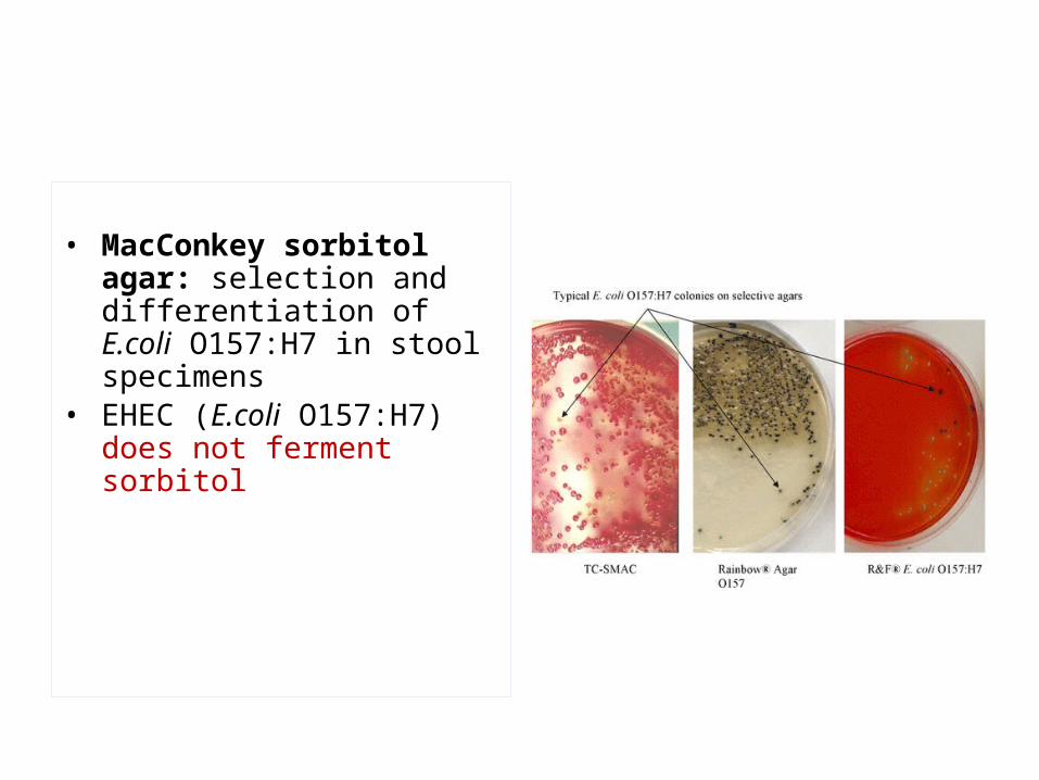
• MacConkey sorbitol agar: selection and differentiation of E.coli O157:H7 in stool specimens
• EHEC (E.coli O157:H7) does not ferment sorbitol

Differential media: contain some nutrient or chemical which allows colonies of bacterial species or type to exhibit certain metabolic or culture characteristics which can be used to distinguish them from other bacteria in the same plate
• MacConkey’s agar: most commonly used differential medium for differentiating lactose fermenting Gram negative rods from non lactose fermenting Gram negative rods; from stool specimens, urine and other specimens. It is also a selective medium which does not allow most Gram positive bacteria to grow.
Pink lactose fermenting colonies
Pale non lactose fermenting colonies
A; Lactose and B; non lactose fermenting colonies

• EMB (eosin methylene blue) agar: differential agar for differentiating lactose fermenting and non lactose fermenting Gram negative rods from urine specimens
• Hecktoen enteric agar: defferential and selective medium for the isolation and differentiation of Salmonella and Shigella from other Gram negative enteric bacilli in stool
Black colonies of Salmonella and pale colonies of Shigella and E.coli


Differentiating Staphylococci from Streptococci
• Gram stain and morphology– Both Gram positive– Staphylococci: clustered cocci
(grape like)– Streptococci: chained cocci
• S. pneumoniae: diplococcus
• Growth– Staph.: large colonies (non-
fastidious), some hemolytic– Strep.: small colonies
(fastidious), many hemolytic ( or )
• Enzyme tests• Catalase test:
– Enzyme catalase breaks down hydrogen peroxide into H2O+O2
– Staphylococci: catalase +– Streptococci: catalase -
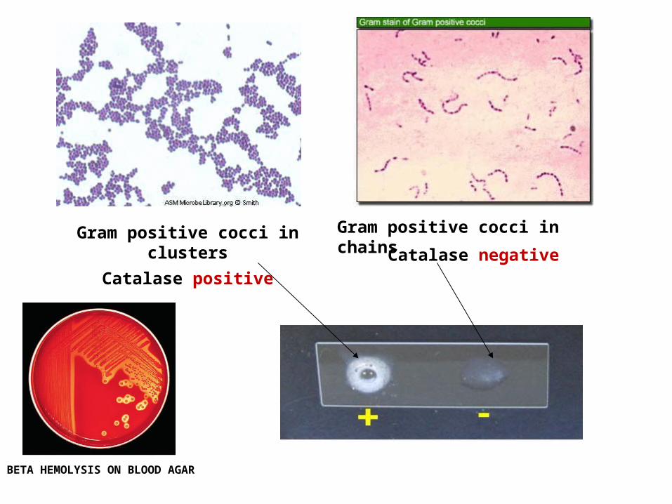
Gram positive cocci in clusters
Catalase positive
Gram positive cocci in chains
Catalase negative
BETA HEMOLYSIS ON BLOOD AGAR

Gram positive cocci in clusters
Staphylococcus colonies grown on Blood agar showing beta hemolysis
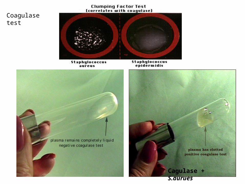
Cagulase + S.aurues
Coagulase test

Streptococci
streptococci Growth on blood agar identification
Streptococcus pyogenesGroup A
Beta hemolysis Sensitive to Bacitracin
Streptococcus agalactiaeGroup B
Beta hemolysis Resistant to Bacitracin
Streptococcus pneumoniae Alpha hemolysis Sensitive to optochin
Viridans streptococci Alpha hemolysis Resistant to optochin
Enterococci Alpha hemolysis or no hemolysis
Resistance to 40% bile

Gram positive cocci in pairs Gram positive cocci in chains

Alpha hemolysis, Streptococcus pneumoniae, S.mutans
Beta hemolysis, Streptococcus pyogenesStreptococcus agalactiae
Gamma hemolysis, Enterococcus spp.,

GROUP A BACITRACIN SENSITIVE
GROUP B BACITRACIN RESISTANT
PYR POSITIVE PYROGLUTAMYL
Beta hemolysis
Group A? group B?
CATALASE negative
Gram positive cocci in chains

Alpha hemolysis, Streptococcus pneumoniae,? S.mutans ? Viridans streptococci

Quellung (capsular typing)
SEROTYPING
Optochin sensitivity
S.pneumoniae
Optochin resistant
S.mitis

Enterococcus
• No hemolysis on blood agar
• 40% bile tolerance
• 6.5% sodium chloride
Gamma hemolysis, Enterococcus spp.


GRAM NEGATIVE COCCI


Growth on Thayer Martin medium
Neisseria gonorrhoea
Neisseria meningitidis
Fermentation of sugars
Oxidase test positive

Gram negative
Straight rods Curved rods
Lactose+ Lactose-
Citrate+ Citrate- H2S+ H2S-
Klebsiella E.coli Salmonella Shigella
Campy blood agar42oC+
Campylobacter
TCBS agarYellow
Vibrio
SIMPLE GROWTH REQUIREMENTS
OXIDASE - OXIDASE +
VIBRIO
PSEUDOMONAS
OXIDASE +

Differentiating rods
Metabolism• Utilization of specific substrates
– Lactose – Citrate
• Production of certain end products
– Fermentation end products – Acid (acetate, propionic acid, butyric
acid etc.)– Acetoin – Alcohol– Amine
• Production of enzymes– Urease
• Specialized tests– Immunological
• O-, H- & K-Ag (serotype)• Agglutination
– Antibiogram pattern– Phage typing
• Pathogenic enteric are Lactose non fermenting- (important distinction from non-pathogenic enteric)
• Citrate slant agar: distinguish two commonly seem Gram negative rods (E.coli vs. Klebsiella)
• H2S: important in distinguishing Salmonella(+) from Shigella(-)
• Urease: differentiate Proteus from othe non lactose fermenters

Gram negative rods with simple growth requirements
Gram negative bacilli with capsule
Klebsiella pneumoniae
E.coli/ Klebsiella/ Salmonella/ Shigella/ Proteus

Gram negative bacteria; A lactose fermenting; (E.coli or Klebsiella)
B. non lactose fermenting colonies (Salmonella or Proteus or Pseudomonas or Shigella on Mac Conkey’s medium

Lactose fermenting Gram negative bacilli
E.coli ??
Klebsiella ??
Indole test
Negative; Klebsiella Positive; E.coli

Indole Test

CITRATE UTILISED
KLEBSIELLACITRATE NOT UTILISED
E.COLI

Identifying Non lactose fermenting colonies B
Oxidase test
Oxidase positive
Pseudomonas spp;
Vibrio cholerae
Oxidase negative
Salmonella spp;
Shigella spp;
Proteus spp;

Proteus spp; swarming and urease positive
Salmonella producing H2S
Oxidase negative: PROTEUS AND SALMONELLA
Urease +ve
swarming

Urease Test
Urea CO2 + NH3 NH4+ + OH-
H2O

Triple Sugar Iron Agar Slants
TSI• Fermentation of
glucose, lactose, and/or sucrose
• Reduction of sulfur to hydrogen sulfide
• Gas formation
Used for Enterobacteriaceae

API test stripAPI system for identification


OXIDASE POSITIVEVIBRIO CHOLEARAE
YELLOW COLONIES on TCBS agar
COMMA SHAPED GRAM NEG BACILLI

CURVED RODS (SEA GULL WING APPEARANCE )
CAMPYLOBACTER
CAMPYLOBACTER COLONIES GROWN AT 420c MICROAEROPHILIC

Gram positive rodsAerobic Gram positive rods
Bacillus anthracis
Nocardia spp.,Corynebacterium diphtheriae
Listeria monocytogenes

Gram positive anaerobic rods
Clostridium spp., with sporesActinomycetes spp.,
Clostridium spp., with spores

Gram negative rods
Anaerobic rods; Bacteroides fragilis

Gram negative cocco bacilli with fastidious growth requirements
Haemophilus influenzae

Culture performed using chocolate agar
S.aureus streak
Growth of H.influenzae
Satellitsm on blood agar (growth factors X and V)

BORDETELLA PERTUSSIS
COLONIES ON REAGAN LOWE

BUFFERED CHARCOAL YEAST EXTRACT AGAR
LEGIONELLA PNEUMOPHILA

BRUCELLA SPP;
Gram negative cocco bacilli

SPIROCHAETES

Silver stained Treponema pallidum,

FUSOBACTERIUM

MYCOBACTERIA ;ACID FAST BACTERIA (ZIEHL NEELSEN STAIN)
COLONIES ON LOWENSTEIN JENSEN MEDIUM