michigan splint and treatment of temporomandibular joint
TRANSCRIPT

http://hrcak.srce.hr/medicina
medicina fluminensis 2013, Vol. 49, No. 2, p. 112-120112
Abstract. Splints, in a broader sense, include various groups of removable intraoral appliances which are used in biomechanical treatment approach and they help establish the neuromus-cular functional balance between different parts of the stomatognathic system. The aim of the paper was to review the literature related to temporomandibular disorder (TMD) treatment with special attention given to clinical importance and the fabrication of the Michigan splint. A clinical case with a 9-year follow-up is presented within the framework of Michigan splint practical use and an evaluation of TMD treatment success until now. Generally, in TMD trea-tment, the principle of palliative medicine is preferred, which means treatment, control and alleviating of temporomandibular pain. The principle of non-invasive and reversible methods of treatment is preferred. The splint achieves a behavioral effect of self-awareness (cognition) about the position, function and parafunction of the mandible as well as a placebo effect.
Key words: magnetic resonance imaging, temporomandibular joint, treatment
Sažetak. Udlage u širem smislu predstavljaju velik broj skupina mobilnih intraoralnih naprava pomoću kojih se provodi biomehanička terapija te uspostavlja neuromuskularna funkcijska harmonija dijelova stomatognatog sustava. Svrha rada je pregled literature vezan uz liječenje temporomandibularnog poremećaja s naglaskom na klinički značaj i način izradbe michigan-ske udlage. U sklopu praktične primjene michiganske udlage i dosadašnje znanstvene evaluac-ije uspjeha liječenja TMP-a opisan je klinički slučaj s 9-godišnjim praćenjem. Općenito, principi palijativne medicine preporučuju se u liječenju TMP-a, što podrazumijeva liječenje i kontrolu temporomandibularnog bola. Prednost se daje neinvazivnim i reverzibilnim metodama liječenja. Udlaga postiže bihevioralni učinak samosvjesnosti (kognicije) o položaju, funkciji i parafunkciji mandibule, te se postiže učinak placeba.
Ključne riječi: liječenje, magnetska rezonancija, temoromandibularni zglob
Adresa za dopisivanje:*Sunčana Simonić-Kocijan, dr. dent. med.Katedra za stomatološku protetiku Medicinski fakultet Sveučilišta u Rijeci Krešimirova 40, 51 000 Rijeka e-mail: [email protected]
1Department of Removable Prosthodontics, School of Dental Medicine, University of Zagreb, Zagreb2Department of Prosthodontics, Department of Dental Medicine School of Medicine, University of Rijeka, Rijeka3Department of Diagnostic and Interventional Radiology, Clinical Hospital Center Sestre milosrdnice, University of Zagreb, Zagreb
Primljeno: 7. 1. 2013. Prihvaćeno: 12. 4. 2013.
Michigan splint and treatment of temporomandibular joint Michiganska udlaga i liječenje temporomandibularnog zgloba
Tomislav Badel1, Sunčana Simonić-Kocijan2*, Vlatka Lajnert2, Nikša Dulčić1, Dijana Zadravec3
Pregledni članak/Review

113http://hrcak.srce.hr/medicina
T. Badel, S. Simonić-Kocijan, V. Lajnert et al.: Michigan splint and treatment of temporomandibular joint
medicina fluminensis 2013, Vol. 49, No. 2, p. 112-120
INTRODUCTION
Temporomandibular disorders (TMDs) have a musculoskeletal origin and are part of orofacial pain problematic. As a form of somatic pain in the stomatognathic system, TMDs imply a disor-der in the masticatory muscles and/or the tem-poromandibular joint (TMJ) with accompanying disturbances (limited mouth opening, noise and/or ear pain) as well as pathologic noise (clicking, crepitations) in the joint1-3.The aim of the paper was to review the literature related to treatment of TMJ with special atten-tion paid to the clinical significance and fabrica-tion of the Michigan splint. A clinical case with a 9-year follow-up is presented within the frame-work of Michigan splint practical use and an eval-uation of TMD treatment success.
TMD DIAGNOSTICS
Diagnostics and differential diagnostics of TMDs are based on a standardized clinical examination. The Research Diagnostic Criteria (RDC)/TMD di-agnostic system has become standard in scientif-ic studies, wherein the clinical term TMDs has been divided into separate diagnoses4,5. Thus, there is a distinction between a muscular disor-der and TMJ disorder: osteoarthritis and anterior disc displacement. However, the generally ac-cepted terminology does not explain all clinical aspects of temporomandibular pain as the most important clinical sign and symptom of the ill-ness6.Apart from the use of nonspecific clinical proce-dures (palpation, auscultation, measuring of ac-tive and passive mandibular mobility), the impor-tance of orthopedic tests is also growing (manual functional analysis by Bumann and Groot Landeweer). This implies a modern, biomedical approach to the illness but also an individual ap-proach to the patient and treatment proce-dures7-10.Direct occlusal analysis is carried out in everyday practice and it provides data on static contacts between teeth in supportive areas as well as on dynamic occlusal relations between the teeth – a type of laterotrusal guidance, hyperbalance and interference contacts. The significance of various
static and dynamic occlusal variables has not been explained in the context of etiopathogene-sis and treatment of TMDs. Although such an ap-proach to TMDs has a strict dental focus, many patients (up to 45 %) have no indications for any kind of dental treatment11,12. On the other hand, the prevalence of temporomandibular pain is rel-atively low (around 5 %) in general population and it is disproportionate with the serious public health issue of untreated teeth and thereby, with non-replaced teeth13-15.
The principle of occlusal therapy is the irreversibility and non-invasiveness in achieving orthopedic stability of TMJ.
MANAGEMENT OF TMDs
Etiopathogenesis of TMDs, as well as of other painful conditions of the musculoskeletal system (such as the public health issue of back pain), has not been completely explained and the treat-ment methods used are primarily those minimal-ly invasive or completely noninvasive16,17. Con-cepts of etiopathogenesis only included dental causes (neuralgia as a part of Costen’s syndrome) but there was also a multifactorial concept and a biopsychosocial concept (apart from the somatic, RDC/TMD includes psychiatric testing of pa-tients). For this reason, TMDs are defined by a concept of nonspecific etiology, similarly to other musculoskeletal disorders in the body. The con-cept of nonspecific etiology gains importance when it has to be applied on individual patients. In such a case, the personalized approach to den-tal medicine/medicine plays an important role and the idiopathic etiology is often mentioned at this stage of direct contact with the patient18,19.Unknown etiology of TMDs and particularly of TMJs does not lessen the importance of radiolog-ical diagnostics. Apart from the panoramic radio-graph as a basic document of identification for each dental patient, there are also noninvasive but rather expensive radiological methods such as magnetic resonance imaging (MRI). Although it is possible to show osteoarthritis of TMJ on im-ages of classical and computerized tomography,

114 http://hrcak.srce.hr/medicina
T. Badel, S. Simonić-Kocijan, V. Lajnert et al.: Michigan splint and treatment of temporomandibular joint
medicina fluminensis 2013, Vol. 49, No. 2, p. 112-120
MRI has been accepted as the gold standard in diagnostics of soft intraarticular structures20,21. Since disc displacement is a common finding, mostly in younger population of TMD patients, MRI was accepted as the gold standard but there is still no agreement on the gold standard in TMDs treatment22,23. The psychological factor can be evident, even in non-characteristic geriatric population of TMD patients, and it can contrib-ute to the general clinical picture as a recurring etiological factor24.Priority is given to noninvasive and reversible treatment methods where the occlusal splint plays a key role in dental, that is, initial occlusal therapy25. The occlusal splint is the most com-mon and efficient treatment procedure of arthro-genic and/or myogenic forms of TMDs and brux-ism. The occlusal stability is established by specific morphology of the splint which is placed on the teeth alignment of one jaw thus serving as an orthopedic means of TMJ stabilization26.The occlusal splint is used as a temporary means of obtaining therapeutic occlusion and as a pre-paratory stage for definite prosthetic treatment27. In treatment with occlusal splints, their biome-chanical concepts of activity, characteristics of position and retention have changed and com-plemented each other. The morphology of the occlusal splint plane has been tested as well as its influence on mandibular position and move-ments and the position and relationship between the intraarticular structures of TMJ. Depending on the indications of use and treatment effects of the occlusal splint, hyperactivity is reduced, that is, the masticatory muscles are relaxed, the con-dyle is therapeutically positioned, which means that it is placed into the centric relation position with the behavioral effects increasing awareness about the position, function and parafunction of the mandible thus achieving placebo effect28,29.
MICHIGAN SPLINT – CHARACTERISTICS AND FABRICATION
Relaxation splints are used in treatment of brux-ism as well as in management of arthrogenic and myogenic temporomandibular pain. The Michi-gan splint by Ramfjord and Ash is an occlusal bite plane stabilization splint with cusped rise and
freedom in centric in a space of 0.5-1.0 mm on the splint plane (Figure 1)30. During occlusal movements, the concept of canine guidance is realized by planes of the splint in the canines re-gion, whereas the interference, hyperbalance and balance contacts between other teeth and splint plane are avoided31.Indications for Michigan splint are as follows: TMDs of arthrogenic and/or myogenic origin, management of nocturnal bruxism and uncon-trolled parafunction during the day, maintaining of centric relations as a precondition to extensive prosthodontic restoration in patients with painful and stiff masticatory muscles or limited mandibu-lar movements, and as a means of differential di-agnostics of TMDs with respect to other ailments with similar symptoms (orofacial and craniocervi-cal pain, tension headache, secondary tinnitus, etc.)32,33.In Michigan splint, centric relation serves as a therapeutic position which stabilizes the mandi-ble in occlusal relations, wherein the habitual mandibular position is often identical to the cen-tric position in the TMJ. Apart from excluding oc-clusal interferences, the relaxation of masticatory muscles is achieved by increasing the occlusal
Figure 1 Antagonists are supported by working cusps of the posterior teeth on the flat occlusal plane of the splint (b, buccal; o, oral)

115http://hrcak.srce.hr/medicina
T. Badel, S. Simonić-Kocijan, V. Lajnert et al.: Michigan splint and treatment of temporomandibular joint
medicina fluminensis 2013, Vol. 49, No. 2, p. 112-120
vertical dimension by the amount of thickness of the occlusal part of the splint. Michigan splint is most often indicated for the maxilla, but esthetic and phonetic reasons can also indicate its place-ment on the mandibular teeth. Splint fabrication is of utmost importance be-cause it has to be made individually and the den-tal technician has to be trained in Michigan splint fabrication. Although the methodology of fabri-cation is limited, apart from the method of di-rectly applying the acrylic composite onto the definitive cast placed into the articulator, the ad-vantage is given to the indirect method, which means that the splint is waxed-up first11,31.The impression of both jaws includes teeth align-ment and surrounding tissues: the palate, mar-ginal gingiva and edentulous spaces in the jaw in cases of partial tooth loss. The limits of the splint are drawn on the cast of the maxilla: vestibularly across the incisal edges of the anterior teeth (2 mm) as well as on the distal teeth in order to achieve splint retention, across the equator of the buccal planes. The palatal border follows the dental arch with the distance of 18-20 mm. Neu-romuscular position of condylar centric relation is achieved by an anterior jig which is obtained by dripping aluminum wax onto the registration wax in the upper central incisors region. A definitive mandibular position in the contact position of centric relation is obtained by aluminum wax in the canine and first molars region (Figure 2)11,34.After mounting the cast of the maxilla into the ar-ticulator (it is recommended to use a semi-ad-justable articulator with a corresponding facial arch), the incisal articulator pin is placed in the ”+2 mm” position (registration wax thickness) prior to mounting of the mandibular cast. This is followed by checking of the space (about 1-2 mm between the cusps of posterior teeth) intended for the splint in order to enable subsequent oc-clusal adjustment and to compensate for splint wear. Prior to modeling of the splint, the custom model bed should be prepared: blocking out the under-cuts, interdental regions and deep fissures by us-ing modeling wax or dental plaster. This helps to avoid difficulties in applying the splint, which can be caused by unwanted changes in acrylic dimen-
sions during polymerization. The use of vacuum-adapted resin sheet wherein the outline of the splint is then cut off the cast along the vestibular and palatal edge is optional. The vacuum-adapt-ed acrylic sheet is waxed-up – a layer of pink wax is softened over a flame and manually molded and immediately adjusted in closing movement made in the articulator so that the incisal pin can contact the guide table in the vertical dimension. The excess wax is removed and the occlusal plane is modeled. This is followed by waxing of slightly concave planes for canine guidance in hard ˝inlay-wax˝. Finally, occlusal contacts are preliminary checked by powder.
Figure 2 Centric relation record obtained from impressions of alu-wax in contact position of centric relation
Figure 3 Vestibular edge (made by putty impression material) of the splint wax up (blue – planes for canine guidance)

116 http://hrcak.srce.hr/medicina
T. Badel, S. Simonić-Kocijan, V. Lajnert et al.: Michigan splint and treatment of temporomandibular joint
medicina fluminensis 2013, Vol. 49, No. 2, p. 112-120
The most complicated stage of laboratory fabri-cation is replacing the wax by acrylic, which be-gins by surrounding the vestibular splint edge with putty impression material. A space for ex-cess acrylic putty is situated dorsally (Figure 3). The wax cast is replicated by dental stone fixator which will precisely copy the splint surface. By re-moving the hardened stone negative from the cast, the entire wax is also removed. The cast should then be isolated by a hard, clear resin sheet as well as the stone mould. The self-curing
transparent acrylic is used (such as Futura Jet®, Schütz Dental). The stone mould is slowly closed in order to squeeze out the excess acrylic mass (Figure 4). The cast with splint is then placed into a pressure chamber (6 bar and temperature 40°C/15min).The polymerized splint is not removed from the casting mould; it is mounted into the articulator for preliminary occlusal adjustment to obtain oc-clusal contacts in centric relations (Figure 5). Af-ter this, the canine guided movements are checked (Figure 6). The splint is then removed from the cast and the final polishing is carried out. Occlusion is also additionally adjusted when the splint is tried in by the patient.
A CLINICAL REPORT
Clicking and pain in the right TMJ appeared 6 months before the 19-year-old female patient visited our clinic, and at the time of her first visit she complained about continuous pain in the joint without clicking. Apart from the preauricu-lar region, she also felt pain in the right ear. She rated the pain with 5.6 on the visual-analogue scale (0 – no pain, 10 – the strongest pain). She had difficulties chewing and limited mouth open-ing (48 mm). Laterotrusal movements were ca-nine guided, 12 mm to the right and 7 mm to the left with pain in the right TMJ. The occlusal status was Angle class I, vertical overlap was 3 mm, hor-izontal was 1.5 mm whereas the incongruity of the medial line amounted to 3 mm. She did not undergo any orthodontic treatment or tooth loss, there were no wear facets and she denied any bruxist experiences. The clinical examination de-termined pain and limited mouth opening on ac-tive movement and under dynamic compression. The right TMJ was also painful under passive compression (bilaminar zone). An isometric ex-amination of the muscles confirmed pain in the right masseter and temporal muscle. The defini-tive diagnosis was confirmed by MRI, including disc displacement without reduction (Figure 7). She was treated by Michigan splint, which she wore regularly for three months at night.Follow-up was carried out by subsequent MRI re-cording after 3 months with the splint placed in the mouth. Clinical check-ups were carried out
The Michigan splint increases occlusal vertical dimensi-on which has to provide comfort to the patient.
Figure 4 Eliminating of excess acrylic is checked by joining the moulage cast in lateral openings (arrows)
Figure 5 Even centric contacts between working cusps and incisal edges on the splint plane
117http://hrcak.srce.hr/medicina
T. Badel, S. Simonić-Kocijan, V. Lajnert et al.: Michigan splint and treatment of temporomandibular joint
medicina fluminensis 2013, Vol. 49, No. 2, p. 112-120
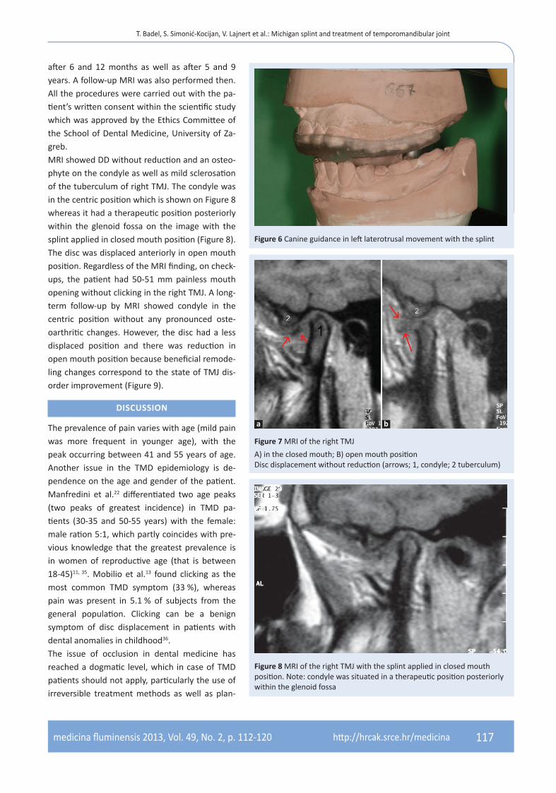
after 6 and 12 months as well as after 5 and 9 years. A follow-up MRI was also performed then. All the procedures were carried out with the pa-tient’s written consent within the scientific study which was approved by the Ethics Committee of the School of Dental Medicine, University of Za-greb. MRI showed DD without reduction and an osteo-phyte on the condyle as well as mild sclerosation of the tuberculum of right TMJ. The condyle was in the centric position which is shown on Figure 8 whereas it had a therapeutic position posteriorly within the glenoid fossa on the image with the splint applied in closed mouth position (Figure 8). The disc was displaced anteriorly in open mouth position. Regardless of the MRI finding, on check-ups, the patient had 50-51 mm painless mouth opening without clicking in the right TMJ. A long-term follow-up by MRI showed condyle in the centric position without any pronounced oste-oarthritic changes. However, the disc had a less displaced position and there was reduction in open mouth position because beneficial remode-ling changes correspond to the state of TMJ dis-order improvement (Figure 9).
DISCUSSION
The prevalence of pain varies with age (mild pain was more frequent in younger age), with the peak occurring between 41 and 55 years of age. Another issue in the TMD epidemiology is de-pendence on the age and gender of the patient. Manfredini et al.22 differentiated two age peaks (two peaks of greatest incidence) in TMD pa-tients (30-35 and 50-55 years) with the female: male ration 5:1, which partly coincides with pre-vious knowledge that the greatest prevalence is in women of reproductive age (that is between 18-45)11, 35. Mobilio et al.13 found clicking as the most common TMD symptom (33 %), whereas pain was present in 5.1 % of subjects from the general population. Clicking can be a benign symptom of disc displacement in patients with dental anomalies in childhood36.The issue of occlusion in dental medicine has reached a dogmatic level, which in case of TMD patients should not apply, particularly the use of irreversible treatment methods as well as plan-
Figure 6 Canine guidance in left laterotrusal movement with the splint
Figure 7 MRI of the right TMJ
A) in the closed mouth; B) open mouth positionDisc displacement without reduction (arrows; 1, condyle; 2 tuberculum)
Figure 8 MRI of the right TMJ with the splint applied in closed mouth position. Note: condyle was situated in a therapeutic position posteriorly within the glenoid fossa
a b

118 http://hrcak.srce.hr/medicina
T. Badel, S. Simonić-Kocijan, V. Lajnert et al.: Michigan splint and treatment of temporomandibular joint
medicina fluminensis 2013, Vol. 49, No. 2, p. 112-120
ning of possible preventive procedures37. Current opinion19 is that TMDs are idiopathic in origin and the correlation with certain etiologic factors can-not be entirely confirmed38. Although MRI is the gold standard in TMJ diagnostics, there is still no gold standard in diagnostics of temporomandibu-lar pain39.The importance of a correct clinical procedure used to determine centric relation is shown in the case of a patient with myalgia whose splint positioned the mandible in a non-physiological bite with shift on the left side28. Ferrario et al.40 noticed that the Michigan splint achieved equilib-rium in the action of temporal and masseter pairs of muscles and that it also reduced electrical ac-tivity of the muscles.An alternative to the traditional splints are those that not require the contribution of a dental lab-oratory, with the so-called ˝Nociceptive Trigemi-nal Inhibition˝ (NTI) being the best known in the treatment of TMDs and bruxism41. However, it has numerous adverse effects, mostly related to changes in occlusion, as well as less efficiency compared to the Michigan splint42. NTI covered the upper incisors only, just like many relaxation splints from the past: the original group of relaxa-tion splints was based on muscular relaxation achieved by elevation of occlusal vertical dimen-sion and by removal of posterior occlusal inter-ferences by covering only the anterior teeth (Hawley retainer, plate by Sved, anterior jig etc.).
Unlike the above mentioned relaxation splints, the Michigan splint (occlusal bite plane stabiliza-tion splint with cuspid rise and freedom in cen-tric) by Ramfjord and Ash is a splint covering all the teeth in the jaw, enabling antagonistic con-tacts on the flat planes according to occlusal con-cepts of freedom in centric position30,43. The new-ly developed Relax splint (Unident), introduced into practice by Nilner et al.44, has proven to be as effective in treatment of myofascial pain as the Michigan splint. On the other hand, the pla-cebo effect of the splint on treatment of TMDs was proven in control groups of patients who wore non-occluding hard palatal oral appliance. Better efficiency of the Michigan splint and of the resilient splint was not proven in treatment of TMDs45,46.Within TMDs treatment modalities, physical ther-apy has shown efficiency in its unique methods as well as in those indicated for other muscu-loskeletal disorders. Namely, the basic principle of improving the function while removing pain is seen in mobilization exercises wherein the pa-tient is directly involved. The basic exercises in-clude performing physiological and accessory movements, such as kinesiotherapy by Schulte47. Physical therapy is an equivalent of the Michigan splint treatment 48. Nonsteroidal anti-inflamma-tory drugs are a complementary treatment in acute pain, and apart from peroral use, they can also be applied topically49,50.Anterior disc displacement is perceived as a de-velopment malpositioning disc form which over time develops from a reducing to a non-reducing disc form. Also, degenerative bone changes had a significant relationship with non-reducing disc displacement51. On the other hand, a two and a half year follow-up of untreated painful disc dis-placement without reduction showed that 42.5 % were asymptomatic52. Apart from removing clini-cal symptoms, the influence of the Michigan splint was also observed in MRI studies, wherein not only the clinical success of the treatment but also the ability to recapture the previously ante-rior displaced disc was evident in 40 % of pa-tients, which proved to be a greater success than the use of anterior repositioning splint53. On the other hand, apart from clinical treatment suc-
Figure 9 A long-term follow-up by MRI showed condyle in the centric position with reducing disc displacement in the right TMJ
A) closed mouth position; B) open mouth position
a b

119http://hrcak.srce.hr/medicina
T. Badel, S. Simonić-Kocijan, V. Lajnert et al.: Michigan splint and treatment of temporomandibular joint
medicina fluminensis 2013, Vol. 49, No. 2, p. 112-120
cess, MRI analysis did not confirm improvement in non-reducing displaced disc54. Hasegawa et al. reported that application of a splint resulted in anterio-inferior condylar movement, and TMJ pain was associated with decreased disc move-ment in response to splint in the mouth55. Biome-chanics and load in the TMJ were never fully ex-plained, in dynamic visualization of TMJ, Gallo56 found disc deformation during condylar-disc complex motion, which should be taken into con-sideration when studying the biomechanical ef-fects of the splint on the intraarticular structures of the joint. In conclusion, depending on the indi-cations of use and achieving of Michigan splint therapeutic effects; there are several ways it par-ticipates in the management of TMDs. The occlu-sal splint has a behavioral effect which increases cognition on mandibular position and function of the stomatognathic system.
LITERATURE
1. Mikić V, Gržić R, Kovačević Pavičić D, Antonić R, Fugošić V. Etiologija temporomandibularnih poremećaja. Me-dicina 2006;42:237-42.
2. Karibe H, Goddard G, McNeill C, Shih ST. Comparison of patients with orofacial pain of different diagnostic cate-gories. Cranio 2011;29:138-43.
3. Kopp S. Screening im kraniomandibulären System. Die Sicht des Zahnarztes/Kieferorthopäden. Manuelle Med 2008;46:381-3.
4. Türp JC, John M, Nilges P, Jürgens J. Schmerzen im Be-reich der Kaumuskulatur und Kiefergelenke. Empfe-hlungen zur standardisierten Diagnostik und Klassifika-tion von Patienten. Schmerz 2000;14:416-28.
5. Lajnert V, Gržić R, Kovačević Pavičić D, Bakarčić D, Badel T, Petričević N. Uporaba DKI/TMP protokola u dijagnos-tici temporomandibularnih poremećaja (TMP-a). Me-dicina 2009;45:56-9.
6. Haley DP, Schiffman EL, Lindgren BR, Anderson Q, An-dreasen K. The relationship between clinical and MRI findings in patients with unilateral temporomandibular joint pain. J Am Dent Assoc 2001;132:476-81.
7. Kordaß B, Fasold A. Manuelle Strukturanalyse. Teil 1: Grundlagen und klinische Untersuchung. ZWR 2012; 212:8-11.
8. Badel T, Krapac L, Kraljević A. The role of physical thera-py in patients with temporomandibular joint disorder. Fiz Rehabil Med 2012;24:21-33.
9. Hoffmann RG, Kotchen JM, Kotchen TA, Cowley T, Das-gupta M, Cowley AW Jr. Temporomandibular disorders and associated clinical comorbidities. Clin J Pain 2011;27:268-74.
10. Schulze W. Therapeutic communication with CMD pa-tients – Part 2. J Craniomandib Funct 2010;2:149-60.
11. Badel T. Temporomandibularni poremećaji i stomatološka protetika. Zagreb: Medicinska naklada, 2007.
12. Badel T, Marotti M, Savić Pavičin I, Bašić-Kes V. Tem-poromandibular disorders and occlusion. Acta Clin Cro-at 2012;51:419-24.
13. Mobilio N, Casetta I, Cesnik E, Catapano S. Prevalence of self-reported symptoms related to temporomandib-ular disorders in an Italian population. J Oral Rehabil 2011;38:884-90.
14. Felton D, Cooper L, Duqum I, Minsley G, Guckes A, Haug S et al. Evidence-based guidelines for the care and maintenance of complete dentures: a publication of the American College of Prosthodontists. J Prosthodont 2011;20 Suppl 1:S1-12.
15. Maixner W. Biopsychological and Genetic Risk Factors for Temporomandibular Joint Disorders and Related Conditions. In: Graven-Nilsen T, Arendt-Nilsen L, Mense S (eds). Fundaments of Muskuloskeletal Pain. Seattle: IASP Press, 2008;263-79.
16. Dym H, Israel H. Diagnosis and treatment of temporo-mandibular disorders. Dent Clin North Am 2012;56:149-61.
17. Houra K, Perović D, Radić A, Bartolek Hamp D, Vukas D, Ledić D. Minimalno invazivne procedure u liječenju križobolje i lumboishijalgije. Medicina Flum 2012;48: 259-70.
18. Manfredini D. Etiopathogenesis of disk displacement of the temporomandibular joint: A review of the mecha-nisms. Indian J Dent Res 2009;20:212-21.
19. Green SC. Concepts of TMD Etiology: Effects on Diagno-sis and Treatment. In: DM, Green CS, Hylander WL (eds). TMDs. An Evidence-Based Approach to Diagnosis and Treatment. Laskin. Chicago: Quintessence, 2006; 219-28.
20. Badel T, Marotti M, Keros J, Kern J, Krolo I. Magnetic Resonance Imaging Study on Temporomandibular Joint Morphology. Coll Anthropol 2009;33:455-60.
21. Moen K, Hellem S, Geitung JT, Skartveit L. A practical approach to interpretation of MRI of the temporoman-dibular joint. Acta Radiol 2010;51:1021-7.
22. Manfredini D, Piccotti F, Ferronato G, Guarda-Nardini L. Age peaks of different RDC/TMD diagnoses in a patient population. J Dent 2010;38:392-9.
23. Syrop SB. Initial management of temporomandibular disorders. Dent Today 2001;21:52-7.
24. Badel T, Kraljević Šimunković S, Marotti M, Kocijan Lovko S, Kern J, Krolo I. Study of temporomandibular joint disorder in older patients by magnetic resonance imaging. Gerodontology 2012;29:e735-41.
25. Palla S. Grundsätze zur Therapie des myoarthropathi-schen Schmerzes. Schmerz 2002;16:373-80.
26. de Leeuw R. Temporomandibular disorders. Guidelines for classification, assessment, and management (4th ed). Chicago: Quintessence, 2008.
27. Badel T, Kraljević S, Pandurić J, Marotti M. Preprosthetic therapy utilizing a temporary occlusal acrylic splint: A case report. Quintessence Int 2004;35:401-5.
28. Badel T, Stražanac J, Marotti M, Krapac L. Treatment of myogenic temporomandibular disorder by occlusal splint and physical therapy: a case report. Acta Stoma-tol Croat 2010;44:202-10.
29. Dylina TJ. A common-sense approach to splint therapy. J Prosthet Dent 2001;86:539-45.
30. Ash MM, Schmieseder J. Schienentherapie. München: Urban & Fischer, 1999.

120 http://hrcak.srce.hr/medicina
T. Badel, S. Simonić-Kocijan, V. Lajnert et al.: Michigan splint and treatment of temporomandibular joint
medicina fluminensis 2013, Vol. 49, No. 2, p. 112-120
31. Badel T, Pandurić J, Kraljević S, Dulčić N. Initial treat-ment of prosthetic patients with a Michigan splint. Acta Stomatol Croat 2003;36:207-10.
32. Badel T, Savić Pavičin I, Podoreški D, Marotti M, Krolo I, Grbeša Đ. Temporomandibular joint development and functional disorders related to clinical otologic symp-tomatology. Acta Clin Croat 2011;50:51-60.
33. Badel T, Pandurić J, Marotti M, Krolo I. Funkcijski pore me-ćaji u žvačnomu sustavu. Med Jadertina 2005; 35:81-6.
34. Türp JC. Vorstellung einer Methode zur Kieferrelati-onbestimmung für die Michigan-Schiene. Quintessenz Zahntech 2011;37:1136-43.
35. Badel T, Keros J, Marotti M, Kern J, Kocijan Lovko S, Rošin Grget K. Therapy of displaced disk of the tem-poromandibular joint in relation to anxiety. Period Biol 2008;110:101-5.
36. Badel T, Lajnert V, Marotti M, Krolo I, Kovačević Pavičić D. Poremećaj čeljusnog zgloba u 12-godišnje bolesnice. Medicina 2008;44:91-7.
37. Carlsson GE. Some dogmas related to prosthodontics, temporomandibular disorders and occlusion. Acta Odontol Scand 2010;68:313-22.
38. Badel T, Marotti M, Krolo I, Kern J, Keros J. Occlusion in patients with temporomandibular joint anterior disk displacement. Acta Clin Croat 2008;47:129-36.
39. Ahmad M, Hollender L, Anderson Q, Kartha K, Ohrbach R, Truelove EL et al. Research diagnostic criteria for temporomandibular disorders (RDC/TMD): develop-ment of image analysis criteria and examiner reliability for image analysis. Oral Surg, Oral Med, Oral Pathol, Oral Radiol, Endod 2009;107:844-60.
40. Ferrario VF, Sforza C, Tartaglia GM, Dellavia C. Immedia-te effect of a stabilization splint on masticatory muscle activity in temporomandibular disorder patients. J Oral Rehabil 2002;29:810-5.
41. Lobbezoo F, van der Zaag J, van Selms MK, Hamburger HL, Naeije M. Principles for the management of brux-ism. J Oral Rehabil 2008;35:509-23.
42. Magnusson T, Adiels AM, Nilsson HL, Helkimo M. Treat-ment effect on signs and symptoms of temporoman-dibular disorders–comparison between stabilisation splint and a new type of splint (NTI). A pilot study. Swed Dent J 2004;28:11-20.
43. Ash MM Jr, Ramfjord SP. Reflections on the Michigan splint and other intraocclusal devices. J Mich Dent As-soc 1998;80:32-5,41-6.
44. Nilner M, Ekberg E, Doepel M, Andersson J, Selovuo K, Le Bell Y. Short-term effectiveness of a prefabricated oc-clusal appliance in patients with myofascial pain. J Oro-fac Pain 2008;22:209-18.
45. Ekberg E, Nilner M. The influence of stabilisation appli-ance therapy and other factors on the treatment out-come in patients with temporomandibular disorders of arthrogeneous origin. Swed Dent J 1999;23:39-47.
46. Nilsson H, Vallon D, Ekberg EC. Long-term efficacy of re-silient appliance therapy in TMD pain patients: a ran-domised, controlled trial. J Oral Rehabil 2011;38:713-21.
47. Schulte W. Die exzentrische Okklusion. Folge-schäden im stomatognathen System. Diagnose, Therapie und Prophylaxe. Berlin: Quintessenz, 1983.
48. Niemelä K, Korpela M, Raustia A, Ylöstalo P, Sipilä K. Ef-ficacy of stabilisation splint treatment on temporoman-dibular disorders. J Oral Rehabil. 2012;39:799-804.
49. Badel T, Rošin-Grget K, Krapac L, Marotti, M. Principi farmakoterapije temporomandibularnih poremećaja. Medicus 2007;16:241-50.
50. Badel T, Krapac L, Savić Pavičin I, Zadravec D, Rosić D, Kocijan Lovko S et al. Fizikalno liječenje i korištenje ke-toprofen gela za poremećaj temporomandibularnog zgloba potvrđenog magnetskom rezonancijom (ab-stract). Reumatizam 2012;59:182-3.
51. Campos MI, Campos PS, Cangussu MC, Guimarães RC, Line SR. Analysis of magnetic resonance imaging char-acteristics and pain in temporomandibular joints with and without degenerative changes of the condyle. Int J Oral Maxillofac Surg 2008;37:529-34.
52. Kurita K, Westesson PL, Yuasa H, Toyama M, Machida J, Ogi N. Natural course of untreated symptomatic tem-poromandibular joint disc displacement without reduc-tion. J Dent Res 1998;77:361-5.
53. Fayed MM, El-Mangoury NH, El-Bokle DN, Belal AI. Oc-clusal splint therapy and magnetic resonance imaging. World J Orthod 2004;5:133-40.
54. Badel T, Marotti M, Kern J, Laškarin M. A quantitative analysis of splint therapy of displaced temporomandib-ular joint disc. Ann Anat 2009;191:280-7.
55. Hasegawa Y, Kakimoto N, Tomita S, Honda K, Tanaka Y, Yagi K et al. Movement of the mandibular condyle and articular disc on placement of an occlusal splint. Oral Surg Oral Med Oral Pathol Oral Radiol Endod 2011;112:640-7.