methyl erythritol 4-phosphate (mep) pathway metabolic
TRANSCRIPT

Methyl erythritol 4-phosphate (MEP) pathway metabolic
regulation
Journal: Natural Product Reports
Manuscript ID: NP-REV-11-2013-070124.R2
Article Type: Review Article
Date Submitted by the Author: 18-Apr-2014
Complete List of Authors: Banerjee, Aparajita; Michigan State University, Biochemistry and Molecular Biology Sharkey, Thomas ; Michigan State University, Dept. Biochem. Mol. Biol.
Natural Product Reports

Natural Products Reports RSCPublishing
ARTICLE
This journal is © The Royal Society of Chemistry 2014 Nat. Prod. Rep., 2014, 00, 1-3 | 1
Cite this: DOI: 10.1039/x0xx00000x
Received 00th January 2012,
Accepted 00th January 2012
DOI: 10.1039/x0xx00000x
www.rsc.org/
Methyl Erythritol 4-phosphate (MEP) Pathway
Metabolic Regulation
A. Banerjee,a and T.D. Sharkey a
The methylerythritol 4-phosphate (MEP) pathway is the recently discovered source of isoprenoid
precursors isopentenyl diphosphate and dimethylallyl diphosphate in most bacteria, some eukaryotic
parasites, and the plastids of plant cells. The precursors lead to the formation of various isoprenoids
having diverse roles in different biological processes. Some isoprenoids have important commercial
uses. Isoprene, which is made in surprising abundance by some trees, plays a significant role in
atmospheric chemistry. The genetic regulation of this pathway has been discussed but information
about metabolic regulation is just now becoming available. This review covers metabolic regulation of
the MEP pathway starting from the inputs of carbon, ATP, and reducing power. A number of different
regulatory mechanisms involving intermediate metabolites and/or enzymes are discussed. Some recent
data indicate that methylerythritol cyclodiphosphate, the fifth intermediate of this pathway, is a key
metabolite. It has been found to play diverse roles in regulation within the pathway as well as
coordinating other biological processes by acting as a stress regulator in bacteria and possibly a
retrograde signal from plastids to nucleus in plants. In this review we focus on the role of the MEP
pathway in photosynthetic leaves during isoprene emission and more generally the metabolic
regulation of the MEP pathway in both plants and bacteria.
Covering through March 1, 2014
1 Introduction
2 Regulation of inputs into the pathway
2.1 Carbon supply
2.2 Input of reducing power
2.3 Input of ATP (CTP)
3 Regulation of DXS
4 Regulation of DXR and CMS by phosphorylation
5 Regulation of and by MEcDP
5.1 A feedforward effect
5.2 Regulation of the biosynthesis and metabolism of MEcDP
5.3 Effect of MEcDP accumulation in other biochemical
processes
5.3.1 Bacteria
5.3.2 Plants
6 Regulation at HDS and HDR
7 Conclusions and perspectives
8 Acknowledgements
9 Notes and references
1 Introduction
Isoprenoids, also known as terpenoids, constitute the largest
group of natural products. They are the most abundant
secondary metabolites present in all living organisms including
both prokaryotes and eukaryotes.1, 2 More than 35,000 different
isoprenoids have been reported so far.3 Some of them include
carotenoids, chlorophylls, plastoquinones, ubiquinones, sterols,
dolichols, cytokinins, brassinosteroids, gibberellic acid, abscisic
acid, and prenylated proteins.2 Some isoprenoids have
significant roles in primary metabolism like photosynthesis,
respiration, and regulation of growth and development.2, 4
Various other biological processes like defence mechanisms of
plants against different biotic and abiotic stresses, attracting
pollinators and seed dispersers for reproductive processes in
plants, intracellular signal transduction, vesicular transport
within the cell, and construction of cellular and organelle
membrane are mediated by different isoprenoids.2-6
In addition to various biological roles, some isoprenoids have
commercial applications as pigments, fragrance and flavours,
drugs, and polymers.7 A large number of natural products used
as therapeutic agents are terpenoids. A wide variety of
therapeutic properties of this group of natural products include
anticancer, antiparasitic, antimicrobial, antiallergenic,
antispasmodic, antihyperglycemic, anti-inflammatory, and
immunomodulatory properties.8 A well-known anticancer drug,
Page 1 of 13 Natural Product Reports

ARTICLE Natural Products Reports
2 | Nat. Prod. Rep. 2014, 00, 1-3 This journal is © The Royal Society of Chemistry 2014
paclitaxel, is a complex diterpenoid obtained from the bark of
Pacific yew.8 A wide variety of monoterpenes and
sequiterpenes contribute to various odours ranging from fruity
and flowery smell to woody and balsamic smell.9 Different
terpenoids like menthol (minty odour), D-carvone (spicy
odour), D-limonene (orange peel odour), citral (lemon peel
odour), and 1,8-cineole (Eucalyptus odour) are extensively used
in flavor and fragrance industries.9 Different terpenoids like β-
carotene, lutein, zeaxanthin, lycopene, phytoene are widely
used as pigments in food industries.10 Rubber, the most
abundant polymer used in various industries, is chemically
composed of linearly arranged polyterpenoids.11
In terms of total production, the most important isoprenoid is
isoprene, the smallest member of isoprenoid family. Isoprene is
emitted by many organisms including bacteria, plants, and
humans. The global annual production of isoprene from plants
is estimated to be 600 Tg (teragrams), which is about one third
of the global non-methane hydrocarbon emission.12 A few
plants, mainly certain species of pine trees (e.g., lodgepole,
ponderosa) in the western part of North America13-16 also
produce the related compound 2-methyl-3-buten-2-ol
(methylbutenol or MBO).17 Atmospheric chemistry is strongly
affected by these hemiterpenes emitted by vegetation. In the
presence of nitric oxide, isoprene catalyses the formation of
ozone, which can cause atmospheric pollution and is
detrimental to both humans and plants.18 There have been many
attempts to build mechanistic models of isoprene emission from
leaves, which should help identify gaps in our understanding
and may better predict isoprene emission under future
conditions19 but it has been difficult to determine the correct
molecular basis for these models. A major limitation is
understanding the metabolic regulation of the MEP pathway in
isoprene-emitting leaves.
Isoprenoids are derived from two isomeric five-carbon units
called isopentenyl diphosphate (IDP) and dimethylallyl
diphosphate (DMADP).20, 21 (These compounds are also known
as pyrophosphates but diphosphate is the preferred term when
pyrophosphate is esterified.) Isoprene itself is made from
DMADP.22, 23 It was known for a long time that IDP (only) is
synthesized by the acetate/mevalonate (MVA) pathway
followed by isomerization by one of two different isomerases
(IDI1 and IDI2).20, 24, 25 Studies involving labelling of
polyprenoids by feeding 13C-labelled precursors indicated that
an alternative pathway exists for the biosynthesis of
isoprenoids.20 In the early 1990s, an alternative pathway, now
known as the 2-C-methyl-D-erythritol 4-phosphate (MEP)
pathway, was discovered in bacteria, that leads to the
biosynthesis of both IDP and DMADP.20, 26, 27 Because the last
step in the pathway makes both IDP and DMADP, bacteria do
Page 2 of 13Natural Product Reports

Natural Products Reports ARTICLE
This journal is © The Royal Society of Chemistry 2014 Nat. Prod. Rep. 2014, 00, 1-3 | 3
not require IDI and some do not have IDI. Subsequent studies
have demonstrated the presence of the MEP pathway in plastids
of green algae and higher plants.28-34 Both the MEP and the
MVA pathway are present in higher plants and are localized in
the chloroplast and cytoplasm respectively.5 However, the MEP
pathway is not present in humans.
The MEP pathway comprises seven enzymatic steps (Fig. 1).18
It starts with the biosynthesis of 1-deoxy-D-xylulose 5-
phosphate (DXP) from pyruvate and D-glyceraldehyde 3-
phosphate (GAP) catalysed by the enzyme 1-deoxy-D-xylulose-
5-phosphate synthase (DXS).27, 35 In the next step, DXP is
converted to MEP by the enzyme 1-deoxy-D-xylulose- 5-
phosphate reductoisomerase (DXR). MEP is then converted to
the cyclic intermediate methylerythritol 2,4-cyclodiphosphate
(MEcDP) through three consecutive enzymatic steps involving
cytidylation (CTP-dependent), phosphorylation (ATP-
dependent), and cyclization. In the sixth step, MEcDP is
converted into hydroxymethylbutenyl diphosphate (HMBDP)
catalyzed by HMBDP synthase (HDS). In the last step,
HMDBP is reduced to IDP and DMADP by HMBDP reductase
(HDR). IDP and DMADP are also isomerized by isopentenyl
diphosphate isomerase (IDI).
The MEP pathway was originally known as non-mevanolate
pathway or Rohmer pathway.27, 35-37 The discovery of the first
step of this pathway involving the formation DXP from
pyruvate and glyceraldehyde-3-phosphate led to the name of
DXP pathway or pyruvate/glyceraldehyde-3-phosphate
pathway.35 DXP is, however, found to be a precursor for the
biosynthesis of thiamin and pyridoxol in certain bacteria.35, 38, 39
The second intermediate MEP, on the other hand, contains the
characteristic branched C5 skeleton for all isoprenoids and is so
far not known to be involved in other biochemical pathways.40
Thus MEP is considered to be the first committed intermediate
of this pathway and the name of this pathway is widely
accepted as MEP pathway.35, 40
The absence of the MEP pathway in humans and its presence in
eubacteria, apicomplexa parasites, and photosynthetic
eukaryotes make it a target for development of new antibiotics,
antiparasitic drugs, and herbicides.21, 41 Various terpenoids with
potential therapeutic activities are available in limited quantity
from natural sources;8 metabolic engineering leading to
improved biosynthetic production of these important terpenoids
has commercial potential. A sesquiterpene antimalarial drug,
artemisinin, comes from Artemisia annua but now has been
engineered in yeast.42, 43 Some isoprenoids including
carotenoids, tocopherols, and antimicrobial drugs are important
targets for biotechnological manipulation.21, 44
The genetic regulation of the MEP pathway has been reviewed
extensively.7, 21, 45 A recent review has discussed the
mechanistic details of the enzymes involved in this pathway.46
A short account of the regulation involved in the metabolomics
of the MEP pathway has also been recently discussed.47 Here
we emphasize discoveries that have been made in the past ten
years regarding the regulation of MEP pathway based on
enzymatic activity and metabolites involved in the pathway.
Insights into the metabolic regulation of the MEP pathway can
be beneficial for the biomedical and biotechnological purposes.
In this review we provide some examples of how improved
understanding of the MEP pathway may improve models of
global isoprene emission.
2 Regulation of inputs into the pathway
Metabolic regulation of the MEP pathway is dictated by the
source of carbon and energetic cofactors. Intensive research has
been carried out in the past few years to understand how the
carbon flux contributes to the regulation of this pathway.
Earlier studies were mainly done by observing the pattern of
labelled isoprene emission after feeding 13CO2 or deuterated
deoxyxylulose, the isotopic composition of isoprene being an
indicator of the carbon source of DMADP and hence the MEP
pathway.48-55 The effect of the availability of carbon, ATP, and
reducing power equivalents on the metabolomics of the MEP
pathway are summarized here.
2.1 Carbon supply
Results from early studies involving the incorporation of 13C-
labelled precursors into terpenoids was inconsistent with
acetate as the starting compound.20, 26, 56 Later studies showed
that the MEP pathway starts with the synthesis of DXP from
GAP and pyruvate catalysed by DXS.27, 38, 39, 57-60 The supply of
GAP and pyruvate for the MEP pathway in bacteria can be
maintained through primary metabolism and so will not be
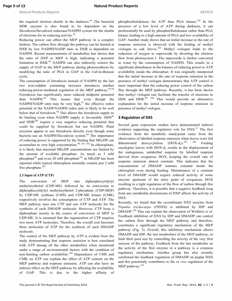
considered here. The source of GAP and pyruvate for the MEP
pathway in chloroplasts of plants is potentially more complex
(Fig. 2).
When 13CO2 is fed to photosynthesizing plant leaves, isoprene
rapidly becomes labelled confirming the close relationship
between isoprene synthesis and the Calvin-Benson cycle.48, 50,
61-63 Sugars transported in the xylem can provide additional
carbon for leaf isoprene biosynthesis through MEP pathway.53,
54 Isoprene does not become completely labelled when 13CO2 is
fed, but, for reasons not yet known, the intermediates of the
Calvin-Benson cycle also do not become fully labelled over
short time frames,64 thus isoprene labelling kinetics may be
fully consistent with all of the carbon for isoprene coming from
the Calvin-Benson cycle.48 However, analysis of the fragments
of isoprene in mass spectrometry studies have been interpreted
to indicate a slightly slower labelling of carbon atoms derived
from pyruvate.50, 65
Page 3 of 13 Natural Product Reports

ARTICLE Natural Products Reports
4 | Nat. Prod. Rep. 2014, 00, 1-3 This journal is © The Royal Society of Chemistry 2014
There are several sources of chloroplastic pyruvate for the MEP
pathway. A small amount of pyruvate is produced by Rubisco
through β-elimination of phosphate from a carbocation
intermediate of the Rubisco reaction.66 The ratio of pyruvate
produced by carboxylation of ribulose bisphosphate is 0.7% at
25 °C. One pyruvate leads to the loss of five carbons as
isoprene. Therefore, Rubisco production of pyruvate could
support carbon loss as isoprene at a rate of 3.5% (0.7% times
five carbons in isoprene) of carbon assimilation and as much as
4.3% if photorespiration, which makes the rate of CO2
assimilation smaller than the rate of carboxylation. Carbon loss
as a result of isoprene emission in excess of 3.5 to 4.3% of
photosynthetically fixed carbon would require pyruvate from
other carbon sources.
Pyruvate cannot be directly synthesized from 3-
phosphoglycerate inside the chloroplast of mesophyll cells
mainly because of the absence of the glycolytic enzymes
phosphoglyceromutase and enolase.67-69 Activity of these
enzymes inside plastids are observed only in the developing
embryos in Arabidopsis.70 A feasible route could be the
transport of phosphoenolpyruvate (PEP) produced by glycolysis
in the cytosol into the chloroplast involving a
phosphoenolpyruvate/phosphate translocator (PPT) followed by
the synthesis of pyruvate from PEP by pyruvate kinase inside
the chloroplast.71, 72 It is known that the chloroplast of
photosynthesizing leaves is dependent on the cytosol for PEP
(but not necessarily pyruvate).73
There is evidence for the presence of plastidic pyruvate kinase
(PKp) in different heterotrophic tissues, e.g. leucoplast pyruvate
kinase has been purified and characterized from developing
castor bean (Ricinus communis) endosperm, Brassica napus
(Rapeseed) suspension cells, and plastidic pyruvate kinase
complex has been purified and characterized from the
developing seeds of Arabidopsis.74-77 Isoenzymes of pyruvate
kinase from green leaves of castor bean and etiolated leaves of
pea plants have been separated by ion filtration chromatography
and one of the isoenzymes is located in the plastid.78
Considering the use of pyruvate in other metabolic pathways
inside the chloroplast (e.g. fatty acid biosynthesis), it is highly
likely that a plastidic pyruvate kinase exists.
Recently, a plastidial sodium-dependent pyruvate transporter,
BASS2, has been discovered.79 It has been observed abundantly
in C4 plant species and in considerable amount in C3/C4
intermediate species. The authors showed that an Arabidopsis
thaliana BASS2 orthologue is mainly observed in developing
leaves and is thought to provide pyruvate for the MEP pathway
in developing leaves.79 Chloroplastic pyruvate obtained from
imported cytosolic PEP is important for the MEP pathway in a
fully expanded leaf when the isoprene emission occurs in its
full capacity.18, 80
The suppression of isoprene emission and DMADP content
under high CO2 concentration has been hypothesized to be due
to the competition for PEP by cytosolic PEP carboxylase over
the transport of PEP from cytosol to chloroplast.81 However,
this hypothesis has been challenged.82, 83 Rasulov et al.
concluded that the variation of isoprene emission with CO2
concentration depends on the regulation of the synthesis of
DMADP by energetic cofactors instead of the carbon
availability. In addition, there is now evidence that CO2-
suppression is eliminated at 30 °C and above.84-87
2.2 Input of reducing power
Several enzymatic steps of the MEP pathway need reducing
power. DXR, the second enzyme of the MEP pathway uses
NADPH for reducing power.88 It is likely that NADPH is
obtained from the photosynthetic electron transport chain in
phototrophic organisms. This helps explain the lack of isoprene
emission in the darkness, when NADPH from photosynthesis is
not available. A post-illumination isoprene burst is often
observed in oak and poplar leaves.89 It has been suggested that
this burst is made possible by NADPH supplied by the
oxidative branch of the pentose phosphate pathway in
darkness90 but other mechanisms are possible. It has also been
suggested that the dark isoprene emission from the aspen leaves
could arise from the pool of phosphorylated intermediates of
the MEP pathway when the required energetics (ATP and
NADPH) are available through the chloroplastic glycolysis or
chlororespiration.91
Both HDS and HDR, the last two enzymes of the MEP
pathway, have [4Fe-4S] clusters and involve double one-
electron transfers in their catalytic reaction mechanism.92 It has
been observed that in presence of light the HDS/GcpE from
Arabidopsis thaliana obtain the required electrons from the
photosynthetic electron transport chain through ferredoxin
whereas the bacterial HDS enzyme requires
flavodoxin/flavodoxin reductase and NADPH as the reducing
system.93, 94 It has however, been suggested that an electron
shuttle is required for plant HDS in darkness and a
ferredoxin/ferredoxin reductase/NADPH system can provide
CO2 3-PGA
GAP RuBP
FBP
F6P
E4P
3-PGA
Shikimate pathway Phenylpropanoids
TPT
Triose phosphate Pi
2-PGA
PEP PEP
Pi PEP
PPT Pyruvate
β-elimina on
(Rubisco
side reac on)
MEP Pathway Isoprenoids
Pyruvate Na-dependent
Pyruvate transport
(developing leaves)
Fa y acid Biosynthesis
PKP
?
Chloroplast
Cytosol
Calvin-Benson Cycle
Page 4 of 13Natural Product Reports
Natural Products Reports ARTICLE
This journal is © The Royal Society of Chemistry 2014 Nat. Prod. Rep. 2014, 00, 1-3 | 5
the required electron shuttle in the darkness.94 The bacterial
HDR enzyme is also found to be dependent on the
flavodoxin/flavodoxin reductase/NADPH system for the shuttle
of electrons for its reducing activity.95
Reducing power can affect the MEP pathway in a complex
fashion. The carbon flow through the pathway can be limited at
DXR by low NADPH/NADP ratio as DXR is dependent on
NADPH. Recent measurements of metabolites has shown that
the ratio of DXP to MEP is high, indicating a potential
limitation at DXR.90 NADPH can also indirectly restrict the
supply of GAP to the MEP pathway during photosynthesis by
modifying the ratio of PGA to GAP in the Calvin-Benson
cycle.
The consumption of ferredoxin instead of NADPH by the last
two iron-sulphur containing enzymes introduces another
reducing-power-mediated regulation of the MEP pathway.92-94
Ferredoxin has significantly more reduced midpoint potential
than NADPH.96 This means that even though the
NADPH/NADP ratio may be very high,97 the effective redox
potential of the NADP/NADPH redox pair is likely to be well
below that of ferredoxin.98 This allows the ferredoxin supply to
be limiting even when NADPH supply is favourable. HDS99
and HDR100 require a very negative reducing potential that
could be supplied by ferredoxin but not NADPH. Plant
enzymes appear to use ferredoxin directly even though some
bacteria use an NADPH/flavodoxin system.94 The importance
of reducing power is supported by the finding that MEcDP can
accumulate to very high concentration.90, 101, 102 In chloroplasts,
it is likely that maximal MEcDP concentrations are limited by
the amount of available phosphate. As much as 3 mM
phosphate90 and even 20 mM phosphate101 in MEcDP has been
reported while typical chloroplasts normally contain just 2 mM
free phosphate.103
2.3 Input of ATP (CTP)
The conversion of MEP into diphosphocytidylyl
methylerythritol (CDP-ME) followed by its conversion to
diphosphocytidylyl methylerythritol 2-phosphate (CDP-MEP)
by CDP-ME synthase (CMS) and CDP-ME kinase (CMK)
respectively involves the consumption of CTP and ATP. The
MEP pathway uses one CTP and one ATP molecule for the
synthesis of each DMADP molecule. However, CTP loses a
diphosphate moiety in the course of conversion of MEP to
CDP-ME. It is assumed that the regeneration of CTP requires
two more ATP molecules. Therefore, the overall cost becomes
three molecules of ATP for the synthesis of each DMADP
molecule.
The control of the MEP pathway by ATP is evident from the
study demonstrating that isoprene emission is best correlated
with ATP among all the other metabolites when monitored
under a range of environmental factors with the condition of
non-limiting carbon availability.104 Dependence of CMS and
CMK on ATP can explain the effect of ATP content on the
MEP pathway and isoprene emission. ATP can also have an
indirect effect on the MEP pathway by affecting the availability
of GAP. This is due to the higher affinity of
phosphoribulokinase for ATP than PGA kinase.105 In the
presence of a low level of ATP during darkness, it can
preferentially be used by phosphoribulokinase rather than PGA
kinase, leading to a high amount of PGA and low availability of
GAP. Another study shows that an initial increase in the rate of
isoprene emission is observed with the feeding of methyl
viologen to oak leaves.106 Methyl viologen leads to the
reduction of oxygen to superoxide by diverting the electron
flow from photosystem I. The superoxide is further converted
to water by the consumption of NADPH. This results in a
significant disturbance in the balance of reducing power to ATP
availability inside the chloroplast. It was originally interpreted
that the initial increase in the rate of isoprene emission in the
presence of methyl viologen demonstrates that ATP control is
more important than the reducing power control of the carbon
flux through the MEP pathway. Recently, it has been shown
that methyl viologen can facilitate the transfer of electrons to
HDS and HDR.99, 100 This would provide an alternative
explanation for the initial increase of isoprene emission in
presence of methyl viologen.
3 Regulation of DXS
Several gene expression studies have demonstrated indirect
evidence supporting the regulatory role for DXS.21 The first
evidence from the metabolic stand-point came from the
observation of labelled isoprene emission when leaves were fed
dideuterated deoxyxylulose (DOX-d2).107, 108 Feeding
eucalyptus leaves with DOX-d2 results in the displacement of
the endogenous, unlabelled isoprene by labelled isoprene
derived from exogenous DOX, keeping the overall rate of
isoprene emission almost constant. This indicates that the
concentration of DMADP remains constant inside the
chloroplast even during feeding. Maintenance of a constant
level of DMADP would require reduced activity of some
enzyme upstream of the entry point of exogenous DOX
resulting in a tight regulation of the flow of carbon through this
pathway. Therefore, it is possible that a negative feedback loop
from any metabolite downstream of DXP affects the activity of
DXS.
Recently, we found that the recombinant DXS enzyme from
Populus trichocarpa (PtDXS) is inhibited by IDP and
DMADP.109 This can explain the observation of Wolfertz et al.
Feedback inhibition of DXS by IDP and DMADP can control
the carbon flow through the MEP pathway and therefore,
constitutes a significant regulatory mechanism of the MEP
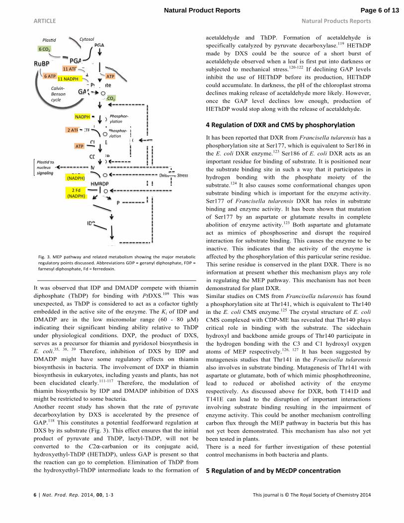
pathway (Fig. 3). Overall, this inhibitory mechanism allows
DMADP and IDP, the last metabolites of the MEP pathway, to
limit their pool size by controlling the activity of the very first
enzyme of the pathway. Feedback from the last metabolite on
the activity of the first enzyme in a pathway is a common
regulatory mechanism. Another group has also recently
confirmed the feedback regulation of DMADP on poplar DXS
and this potentially contributes to the in vivo regulation of the
MEP pathway.110
Page 5 of 13 Natural Product Reports
ARTICLE Natural Products Reports
6 | Nat. Prod. Rep. 2014, 00, 1-3 This journal is © The Royal Society of Chemistry 2014
It was observed that IDP and DMADP compete with thiamin
diphosphate (ThDP) for binding with PtDXS.109 This was
unexpected, as ThDP is considered to act as a cofactor tightly
embedded in the active site of the enzyme. The Ki of IDP and
DMADP are in the low micromolar range (60 - 80 µM)
indicating their significant binding ability relative to ThDP
under physiological conditions. DXP, the product of DXS,
serves as a precursor for thiamin and pyridoxol biosynthesis in
E. coli.35, 38, 39 Therefore, inhibition of DXS by IDP and
DMADP might have some regulatory effects on thiamin
biosynthesis in bacteria. The involvement of DXP in thiamin
biosynthesis in eukaryotes, including yeasts and plants, has not
been elucidated clearly.111-117 Therefore, the modulation of
thiamin biosynthesis by IDP and DMADP inhibition of DXS
might be restricted to some bacteria.
Another recent study has shown that the rate of pyruvate
decarboxylation by DXS is accelerated by the presence of
GAP.118 This constitutes a potential feedforward regulation at
DXS by its substrate (Fig. 3). This effect ensures that the initial
product of pyruvate and ThDP, lactyl-ThDP, will not be
converted to the C2α-carbanion or its conjugate acid,
hydroxyethyl-ThDP (HEThDP), unless GAP is present so that
the reaction can go to completion. Elimination of ThDP from
the hydroxyethyl-ThDP intermediate leads to the formation of
acetaldehyde and ThDP. Formation of acetaldehyde is
specifically catalyzed by pyruvate decarboxylase.119 HEThDP
made by DXS could be the source of a short burst of
acetaldehyde observed when a leaf is first put into darkness or
subjected to mechanical stress.120-122 If declining GAP levels
inhibit the use of HEThDP before its production, HEThDP
could accumulate. In darkness, the pH of the chloroplast stroma
declines making release of acetaldehyde more likely. However,
once the GAP level declines low enough, production of
HEThDP would stop along with the release of acetaldehyde.
4 Regulation of DXR and CMS by phosphorylation
It has been reported that DXR from Francisella tularensis has a
phosphorylation site at Ser177, which is equivalent to Ser186 in
the E. coli DXR enzyme.123 Ser186 of E. coli DXR acts as an
important residue for binding of substrate. It is positioned near
the substrate binding site in such a way that it participates in
hydrogen bonding with the phosphate moiety of the
substrate.124 It also causes some conformational changes upon
substrate binding which is important for the enzyme activity.
Ser177 of Francisella tularensis DXR has roles in substrate
binding and enzyme activity. It has been shown that mutation
of Ser177 by an aspartate or glutamate results in complete
abolition of enzyme activity.123 Both aspartate and glutamate
act as mimics of phosphoserine and disrupt the required
interaction for substrate binding. This causes the enzyme to be
inactive. This indicates that the activity of the enzyme is
affected by the phosphorylation of this particular serine residue.
This serine residue is conserved in the plant DXR. There is no
information at present whether this mechanism plays any role
in regulating the MEP pathway. This mechanism has not been
demonstrated for plant DXR.
Similar studies on CMS from Francisella tularensis has found
a phosphorylation site at Thr141, which is equivalent to Thr140
in the E. coli CMS enzyme.125 The crystal structure of E. coli
CMS complexed with CDP-ME has revealed that Thr140 plays
critical role in binding with the substrate. The sidechain
hydroxyl and backbone amide groups of Thr140 participate in
the hydrogen bonding with the C3 and C1 hydroxyl oxygen
atoms of MEP respectively.126, 127 It has been suggested by
mutagenesis studies that Thr141 in the Francisella tularensis
also involves in substrate binding. Mutagenesis of Thr141 with
aspartate or glutamate, both of which mimic phosphothreonine,
lead to reduced or abolished activity of the enzyme
respectively. As discussed above for DXR, both T141D and
T141E can lead to the disruption of important interactions
involving substrate binding resulting in the impairment of
enzyme activity. This could be another mechanism controlling
carbon flux through the MEP pathway in bacteria but this has
not yet been demonstrated. This mechanism has also not yet
been tested in plants.
There is a need for further investigation of these potential
control mechanisms in both bacteria and plants.
5 Regulation of and by MEcDP concentration
GDP IDP
CDPME
PGA RuBP
6 ATP
11 ATP
6 CO2
GAP
11 NADPH
PGA
PEP
Pyruvate
DXP
MEP
NADPH
2 ATP CTP
ATP
CDPMEP
MEcDP 2 Fd
(NADPH)
IDP DMADP
HMBDP 2 Fd
(NADPH)
-
Phosphor-
yla on -
Plas d to
nucleus
signaling
+
Cytosol Plas d
Calvin-
Benson
cycle
ATP
CO2
+
FDP
-
Oxida ve Stress -
-
Phosphor-
yla on -
Page 6 of 13Natural Product Reports

Natural Products Reports ARTICLE
This journal is © The Royal Society of Chemistry 2014 Nat. Prod. Rep. 2014, 00, 1-3 | 7
Several recent studies have demonstrated that MEcDP is a key
intermediate in the MEP pathway. It has been observed that in
leaves in the presence of light, more MEcDP is accumulated
than all the other MEP pathway intermediates.90 Here we
discuss the various types of regulation inside and outside the
MEP pathway that are coordinated by this metabolite.
5.1 A feedforward effect
MEcDP, the cyclo-diphosphate-containing intermediate of the
MEP pathway, is synthesized by MEcDP synthase (MCS) from
CDP-MEP. The crystal structure of MCS is known from
different organisms.128-131 These structural studies have shown
that a hydrophobic cavity is present along the threefold non-
crystallographic symmetry axis of the enzyme. Evidence
indicates that the cavity is occupied with different isoprenoids
containing a diphosphate moiety like IDP/DMADP, geranyl
diphosphate (GDP), and farnesyl diphosphate (FDP).130, 131
Sequence alignment studies of the MCS enzyme from various
organisms indicate that the motif involved in the formation of
the cavity and the binding of the ligand are well conserved in
the protein family suggesting that the simultaneous
conservation of both the motifs might have evolved due to a
biological function.130 It has been proposed that MCS could be
a significant point of feedback regulation by the downstream
isoprenoids.130, 131
Recent studies have shown that recombinant MCS enzyme
from E. coli is stabilized and activated in the presence of IDP,
DMADP, GDP, and FDP.132 Analysis of the effect of different
MEP pathway metabolites on MCS stability and activation by
the in vitro assays has identified MEP as the most effective
modulator for MCS. It has also been shown by in vitro studies
that the methylerythritol scaffold is essential and sufficient for
the observed effect of activation and enhancement of stability
of MCS by MEP. The 2-C-methylerythritol scaffold is unique
to the MEP pathway. The feedforward activation of MCS by
MEP (Fig. 3) constitutes a regulatory mechanism very specific
to the MEP pathway.
It is also observed that FDP inhibits the E. coli MCS-MEP
complex whereas it activates and stabilizes E. coli MCS
alone.132 It has been speculated that the binding of MEP to
MCS might cause some conformational changes of MCS and
the inhibitory effect of FDP is selective for the MEP-bound
conformation of MCS. The feedback inhibition of MCS-MEP
complex by FDP (Fig. 3) indicates that the downstream
isoprenoids control their biosynthesis by modulating the
activity of a key enzyme involved in the biosynthesis of their
precursor. There is another significance of this feedback
inhibition. It sets a limit on the activated MCS-MEP complex in
the presence of high levels of downstream isoprenoids so that
the carbon flux through the MEP pathway is controlled.
Overall, this observation suggests that MCS plays a key role in
the regulation of the MEP pathway.
5.2 Regulation of the biosynthesis and metabolism of MEcDP
It has been observed that MEcDP accumulates in bacteria under
oxidative stress.133-135 Nitrosative stress (caused by the reactive
species nitric oxide, NO) is also found to be responsible for the
accumulation of MEcDP, to a lesser extent than oxidative
stress, in Corynebacterium ammoniagenes.135 Recently, spinach
leaves were also found to accumulate MEcDP under high light
and high temperature and in the presence of heavy metals like
Cd.101 These external factors can cause oxidative stress in vivo
leading to the accumulation of MEcDP. HDS contains a [4Fe-
4S] cluster susceptible to oxidative stress. Studies have found
that ROS generated under oxidative stress damages the
reconstitution of the [4Fe-4S]-cluster and thus interferes with
the turnover of the holo enzyme.101 It has been suggested that
under oxidative stress, the reconstitution of the apo-HDS with
the [4Fe-4S]-cluster functions as the rate limiting step of the
MEP pathway and thus is a bottleneck in the MEP pathway.
MEcDP has also been found to act as an effective
antioxidant.136 This property of MEcDP allows the repair of the
HDS enzyme to keep it functional by limiting oxidative stress.
This protective ability of MEcDP is not sufficient for the
reconstitution of the holo-enzyme in the presence of inhibitors
like Cd. The accumulation of MEcDP in illuminated leaves
may affect the phosphate balance of the chloroplast. Synthesis
of high levels of MEcDP could potentially act as a sink for
phosphate and disturb the phosphate supply for ATP synthesis.
It has been demonstrated that utilization of phosphate to
maintain the synthesis of high level of MEcDP can cause
phosphate deficiency syndrome in chloroplast.101 It is possible
that the maximal MEcDP concentration is restricted by the
amount of chloroplastic phosphate.
Another interesting observation in this context is the
accumulation of a very high level of MEcDP and block of
isoprene emission from leaves under nitrogen atmosphere (i.e.
CO2- and O2-free air).90, 107 Limited availability of carbon
through the Calvin-Benson cycle to feed the MEP pathway
cannot explain the phenomenon of abolished isoprene emission
from leaves held under nitrogen. This is because replenishing
the carbon supply of the MEP pathway by feeding the leaves
directly with deoxyxylulose in the presence of nitrogen is not
able to restore isoprene emission.107 Accumulation of a high
level of MEcDP under nitrogen atmosphere indicates that the
downstream enzymes may not be functional, causing isoprene
emission to stop. It is likely that under a nitrogen atmosphere
the iron-sulphur complexes of HDS and HDR are disrupted. A
nitrogen atmosphere can possibly lead to some signals that
cause these enzymes to become inactive. The exact
mechanisms by which nitrogen atmosphere disrupts the activity
of these two enzymes in leaves are yet to be determined.
5.3 Effect of MEcDP accumulation in other biochemical
processes
It has been found that MEcDP plays a significant role in
various other biochemical pathways unrelated to isoprenoid
biosynthesis.
5.3.1 BACTERIA
In bacteria, studies showed that oxidative stresses caused by
benzyl viologen or other redox mediators lead to the
Page 7 of 13 Natural Product Reports

ARTICLE Natural Products Reports
8 | Nat. Prod. Rep. 2014, 00, 1-3 This journal is © The Royal Society of Chemistry 2014
accumulation of MEcDP, which has been suggested to play an
important role as an antistressor in bacteria.133, 134, 137 It has also
been observed that MEcDP prevents DNA from falling apart in
the presence of Fenton reagent.138 This is achieved when a
complex is formed between the ferrous ions (present in the
Fenton reagent) and MEcDP resulting in their reduced ability to
form hydroxyl radicals and hydrogen peroxide.138 This suggests
that MEcDP could act as an endogenous stabilizing agent for
bacterial cells subjected to oxidative stress.138
MEcDP is also found to modulate chromatin structure by
disrupting the chlamydial histone-DNA interaction in the
intracellular pathogen Chlamydia trachomatis.139, 140 The
chlamydial developmental cycle alternates between the
extracellular infectious form called the elementary body (EB)
and the intracellular replicative form termed the reticulate body
(RB). These two different forms have characteristic chromatin
structures. The RB form has condensed nucleoid structure
mediated by histone-like DNA binding proteins, Hc1 and
Hc2.139, 140 Within a few hours of infection, the metabolically
inert EB form is transformed into the metabolically active RB
form. Dispersion of the chromatin structure is required for the
differentiation of the EB form into the RB form. It was
suggested that MEcDP disrupts the binding between DNA and
histone-like proteins leading to the release of Hc1and Hc2 from
the DNA causing the dispersion of the chromatin and initiation
of transcription. Thus, MEcDP mediates the decondensation of
the chromatin allowing the differentiation of the EB form to the
RB form. Another example of the role of MEcDP in the
regulation of the bacterial genome activity includes its
resuscitating effect regulating the transition of the non-
culturable form of Mycobacterium smegmatis into the state of
its active growth.141
Recent metabolite profiling studies showed an efflux of
MEcDP from genetically engineered E. coli cells containing the
overexpressed enzymes DXS, IDI, CMS, and MCS.142 It has
been observed that the efflux of MEcDP is accompanied with
the simultaneous reduction of the production of lycopene, a
downstream isoprenoid. It was possible to reduce the efflux of
MEcDP by the overexpression of HDS, which consumes
MEcDP, directing more carbon through the last part of the
MEP pathway, resulting in the increased production of
lycopene. This indicates that the efflux of MEcDP could act as
a limiting step in microbial isoprenoid production. Preliminary
studies indicate the involvement of a fosmidomycin resistance
(fsr) efflux pump143 for the process of exporting MEcDP out of
the cell.142 The active efflux of MEcDP from the engineered
lycopene-producing E. coli cells suggests the possibility of a
potential MEP pathway branch point which diverts the carbon
source of the MEP pathway to another competing pathway.142
This is also supported by the study of restoration of the
complete and active MEP pathway by heterologous expression
of HDS and HDR into Listeria innocua lacking these
enzymes.144 Bioinformatics analysis has shown that L. innocua
has lost the genes for HDS and HDR through evolution while
the rest of the MEP pathway genes are present.144 The ability of
this organism to have an active MEP pathway with the
introduction of the lost enzymes suggests that the rest of the
MEP pathway enzymes, which were already present, are
functional. Evolution has selectively truncated the MEP
pathway in such a way that the existing enzymes could catalyse
the biosynthesis of MEcDP, which can further lead to end
products of the MEP pathway in the presence of HDS and
HDR. This suggests some important yet unidentified
biochemical role for MEcDP.144
5.3.2 PLANTS
In plants, recent studies have demonstrated that in addition to
its role in the bacterial system, MEcDP has a potential role as a
signalling molecule in Arabidopsis. Plastidial MEcDP leads to
a retrograde signal regulating the expression of nuclear-
encoded, stress-responsive genes for plastidial proteins (Fig.
3).145 Hydroperoxide lyase (HPL) is a stress-inducible plastidial
protein in the oxylipin pathway encoded by a nuclear gene. It
has been shown that a mutant ceh1 shows constitutive
expression of HPL. CEH1 encodes for HDS and thus ceh1
mutant is defective in the utilization of MEcDP resulting in its
accumulation. It has also been reported that abiotic stresses
including high light or wounding cause a high level of MEcDP
to build up. These abiotic stresses causing accumulation of
endogenous MEcDP, as well as exogenous MEcDP, lead to the
elevated expression of HPL. This indicates that MEcDP is,
directly or indirectly, a retrograde signalling molecule. It has
been shown that abscisic acid and methyl jasmonate, stress-
responsive hormones of plants, increase the activity of DXS.146
It is tempting to speculate that these stress-responsive hormones
lead to the regulation of HPL by accumulating MEcDP through
the increased activity of the upstream enzyme DXS. The mode
of action of MEcDP in the retrograde signalling is not fully
understood. Considering the involvement of MEcDP in the
nucleoid decondensation in chlamydia, it may be that MEcDP
modulates nuclear gene expression in plants through the
remodelling of the nuclear architecture.139, 140 This mechanistic
model would require the transport of plastid-localized MEcDP
to the nucleus. No information is available for any such
transport of MEcDP in plants but the presence of the fsr efflux
pump in bacteria for moving MEcDP out of the cells142 raises
the possibility of such transporter in plants as well. One such
candidate is the Arabidopsis gene At3g47450.
Accumulation of MEcDP can cause transient effects in isoprene
emission. Upon darkening a leaf, isoprene emission continues
long enough to consume the existing DMADP and IDP but not
MEcDP.90, 147 After about five minutes in the dark the leaf
regains the ability to consume MEcDP but not to make
additional MEcDP. This causes a small post-illumination burst
of isoprene between 5 and 10 min after darkening the leaf (Fig.
4). The very high level of MEcDP that builds up in leaves held
in a nitrogen atmosphere (Section 5.2) is likely responsible for
a large overshoot in isoprene emission when O2 and CO2 are
added back to the air (Fig. 4).
Page 8 of 13Natural Product Reports

Natural Products Reports ARTICLE
This journal is © The Royal Society of Chemistry 2014 Nat. Prod. Rep. 2014, 00, 1-3 | 9
6 Regulation at HDS and HDR
Given the propensity for MEcDP to accumulate in plants and
bacteria it is likely that there is significant regulation of HDS.
However, less is known about HDS regulation than HDR
regulation. It has been shown that nitrosative stress in
Mycobacterium smegmatis causes the accumulation of
HMBDP, the substrate for HDR.135 This suggests that NO
damages the [4Fe-4S]-cluster of HDR resulting in the
dysfunctional enzyme, which leads to the accumulation of
HMBDP. The gene of HDR in E. coli has been found to be
involved in penicillin tolerance through its interaction with
RelA responsible for the synthesis of guanosine 3’,5’-
bispyrophosphate (ppGpp), which acts as a nutritional stress
alarmone.148
It has been shown that a point mutation in E. coli HDR
(LytBG120D) enables it to selectively synthesize DMADP over
IDP.149 This suggests that the structural modification of HDR
can potentially regulate the in vivo concentration of DMADP
and IDP, the end products of the MEP pathway.
It has been seen that engineering an additional HDS gene into
E. coli without increasing the activity of HDR leads to a
reduction in productivity in bacteria engineered to emit
isoprene (A.E. Wiberley, E.L. Singsaas, T.D. Sharkey,
unpublished).150 Chotani et al. found that HMBDP accumulated
in such bacteria and that this was correlated with reduced
isoprene production from engineered bacteria. One explanation
for this is that HMBDP is toxic to cells.
Purified HDR is shown to require a very negative redox
potential, maximal activity was found at -450 mV, much lower
than the midpoint potential of NADPH (-320 mV). The
presumed electron source for this enzyme gives an activity less
than 2% of maximal. Xiao et al. suggest that HDR might be
regulated by modulation of the redox potential of its [4Fe-4S]
cluster.100
It is likely that both HDS and HDR are highly regulated and
this regulation has a strong impact on the carbon flow of the
MEP pathway. These are likely to be the steps where light
regulation of DMADP in plants occurs, but there is no
information on how this occurs. It is also tempting to assume
that the metabolites downstream of MEcDP might have some
toxic effect in the cell. This might lead the carbon flux of the
MEP pathway to be constricted, building up a pool of only
MEcDP under the condition of oxidative stresses.
7 Conclusions and perspectives
The MEP pathway is one of the most important biochemical
pathways for sustaining life on earth. Understanding the
different regulations involved in this pathway is critical for
biological, environmental, as well as commercial purposes.
Mechanisms of genetic regulation of this pathway have started
emerging only in the last decade. We have discussed in this
review several different regulatory mechanisms involved in the
metabolism of this pathway and these are summarized in Fig. 3.
Several questions are still to be answered regarding regulatory
mechanisms of the MEP pathway, especially in plants. The
source of pyruvate for the MEP pathway is not clearly
understood. Understanding the source of pyruvate may explain
the discrepancy in the labelling of the isoprenoids derived from
the MEP pathway. Several studies have indicated that MEcDP
has potential roles in MEP pathway regulation. MEcDP may
connect metabolism in the MEP pathway with other cellular
metabolism, independent of its role in making precursors for
isoprenoids. It has been suggested that MEcDP can act as a
stress sensor and can accordingly coordinate stress responses.
The exact mode of its action in response to the stress signals
has yet to be understood.
It has been suggested that the [4Fe-4S]-cluster containing
enzymes, HDS and HDR, can also contribute to the regulatory
mechanisms of the MEP pathway. The susceptibility of the
[4Fe-4S]-clusters to oxidative stress indicates that the in vivo
redox status can influence the carbon flow of the MEP pathway
through these enzymes. In-depth knowledge of the structural
and functional integrity of these enzymes under various redox-
sensitive conditions would be helpful in understanding their
role in the MEP pathway regulation.
Understanding of the metabolic regulation of the MEP pathway
has emerged in the last decade and currently can be considered
at its nascent stage. Studies so far have demonstrated that
several enzymes and metabolites could have various regulatory
roles in this pathway. However, not much is known regarding
the primary points of regulation and how the overall regulation
of the pathway is finely tuned by both the primary and
secondary points of regulation. Future studies in the field
Page 9 of 13 Natural Product Reports

ARTICLE Natural Products Reports
10 | Nat. Prod. Rep. 2014, 00, 1-3 This journal is © The Royal Society of Chemistry 2014
should be aimed at a complete understanding of the metabolic
regulation of the MEP pathway. This would be useful in
biomedical and biotechnological uses of the MEP pathway and
would also help in finding a mechanistic basis for modelling
isoprene emission.
8 Acknowledgements
This material is based in part on work supported by the
National Science Foundation under Grant No. 0950574. Any
opinions, findings, and conclusions or recommendations
expressed in this material are those of the author(s) and do not
necessarily reflect the views of the National Science
Foundation. The authors have no conflicts of interest to declare.
We thank Professor Caren Freel Meyers, Dr. Sean Weise, and
Chris Harvey for comments on the manuscript.
Notes and references a Department of Biochemistry and Molecular Biology, Michigan State
University, East Lansing, MI, 48824 USA.
1. M. Lohr, J. Schwender and J. E. W. Polle, Plant Sci., 2012, 185–186,
9-22.
2. M. Wanke, K. Skorupinska-Tudek and E. Swiezewska, Acta Biochim.
Pol., 2001, 48, 663-672.
3. W. N. Hunter, J. Biol. Chem., 2007, 282, 21573-21577.
4. M. A. Phillips, P. León, A. Boronat and M. Rodríguez-Concepción,
Trends Plant Sci., 2008, 13, 619-623.
5. A. Hemmerlin, J. F. Hoeffler, O. Meyer, D. Tritsch, I. A. Kagan, C.
Grosdemange-Billiard, M. Rohmer and T. J. Bach, J. Biol.
Chem., 2003, 278, 26666-26676.
6. J. C. Sacchettini and C. D. Poulter, Science, 1997, 277, 1788-1789.
7. M. Rodríguez-Concepción, Phytochem. Rev., 2006, 5, 1-15.
8. P. K. Ajikumar, K. Tyo, S. Carlsen, O. Mucha, T. H. Phon and G.
Stephanopoulos, Mol. Pharm., 2008, 5, 167–190.
9. L. Caputi and E. Aprea, Recent Pat. Food Nutr. Agric., 2011, 3, 9-16.
10. T. Cserháti and E. Forgács, J. Chromatogr. A, 2001, 936, 119-137.
11. D. J. McGarvey and R. Croteau, Plant Cell, 1995, 7, 1015-1026.
12. A. Guenther, T. Karl, P. Harley, C. Wieldinmyer, P. I. Palmer and C.
Geron, Atmos. Chem. Phys., 2006, 6, 3181-3210.
13. P. Harley, V. Fridd-Stroud, J. Greenberg, A. Guenther and P.
Vasconcellos, J. Geophys. Res. Atmos., 1998, 103, 25479-
25486.
14. C. Ferronato, J. J. Orlando and G. S. Tyndall, J. Geophys. Res.
Atmos., 1998, 103, 25579-25586.
15. D. W. Gray, S. R. Breneman, L. A. Topper and T. D. Sharkey, J.
Biol. Chem., 2011, 286, 20582-20590.
16. M. Lerdau and D. Gray, New Phytol., 2003, 157, 199-211.
17. P. D. Goldan, W. C. Kuster, F. C. Fehsenfeld and S. A. Montzka,
Geophys. Res. Lett., 1993, 20, 1039-1042.
18. T. D. Sharkey, A. E. Wiberley and A. R. Donohue, Ann. Bot., 2008,
101, 5-18.
19. R. K. Monson, R. Grote, Ü. Niinemets and J.-P. Schnitzler, New
Phytol., 2012, 195, 541-559.
20. W. Eisenreich, M. Schwarz, A. Cartayrade, D. Arigoni, M. H. Zenk
and A. Bacher, Chem. Biol., 1998, 5, R221-R233.
21. E. Cordoba, M. Salmi and P. León, J. Exp. Bot., 2009, 60, 2933-
2943.
22. G. M. Silver and R. Fall, Plant Physiol., 1991, 97, 1588-1591.
23. G. M. Silver and R. Fall, J. Biol. Chem., 1995, 270, 13010-13016.
24. W. Eisenreich, A. Bacher, D. Arigoni and F. Rohdich, Cell. Mol. Life
Sci., 2004, 61, 1401-1426.
25. A. Nakamura, H. Shimada, T. Masuda, H. Ohta and K. Takamiya,
FEBS Lett., 2001, 506, 61-64.
26. M. Rohmer, M. Knani, P. Simonin, B. Sutter and H. Sahm, Biochem.
J, 1993, 295, 517-524.
27. M. Rohmer, M. Seemann, S. Horbach, S. Bringer-Meyer and H.
Sahm, J. Am. Chem. Soc., 1996, 118, 2564-2566.
28. J. Schwender, M. Seemann, H. K. Lichtenthaler and M. Rohmer,
Biochem. J, 1996, 316, 73-80.
29. W. Eisenreich, B. Menhard, P. J. Hylands and M. H. Zenk, Proc.
Natl. Acad. Sci. U. S. A., 1996, 93, 6431-6436.
30. H. K. Lichtenthaler, M. Rohmer and J. Schwender, Physiol. Plant.,
1997, 101, 643-652.
31. H. K. Lichtenthaler, J. Schwender, A. Disch and M. Rohmer, FEBS
Lett., 1997, 400, 271-274.
32. J. Schwender, J. Zeidler, R. Gröner, C. Müller, M. Focke, S. Braun
and H. K. Lichtenthaler, FEBS Lett., 1997, 414, 129-134.
33. J. G. Zeidler, H. K. Lichtenthaler, H. U. May and F. W.
Lichtenthaler, Z. Naturforsch, 1997, 52c, 15-23.
34. D. Arigoni, S. Sagner, C. Latzel, W. Eisenreich, A. Bacher and M. H.
Zenk, Proc. Natl. Acad. Sci. U. S. A., 1997, 94, 10600-10605.
35. M. Rodríguez-Concepción and A. Boronat, Plant Physiol., 2002, 130,
1079-1089.
36. F. Rohdich, K. Kis, A. Bacher and W. Eisenreich, Curr. Opin. Chem.
Biol., 2001, 5, 535-540.
37. M. Rohmer, Natural Product Reports, 1999, 16, 565-574.
38. G. A. Sprenger, U. Schörken, T. Wiegert, S. Grolle, A. A. D. Graaf,
S. V. Taylor, T. P. Begley, S. Bringer-Meyer and H. Sahm,
Proc. Natl. Acad. Sci. U. S. A., 1997, 94, 12857-12862.
39. L. M. Lois, N. Campos, S. R. Putra, K. Danielsen, M. Rohmer and A.
Boronat, Proc. Natl. Acad. Sci. U. S. A., 1998, 95, 2105-2110.
40. M. Rohmer, Pure Appl. Chem., 2003, 75, 375-388.
41. M. Rodríguez-Concepcion, Curr. Pharm. Des., 2004, 10, 2391-2400.
42. C. J. Paddon, P. J. Westfall, D. J. Pitera, K. Benjamin, K. Fisher, D.
McPhee, M. D. Leavell, A. Tai, A. Main, D. Eng, D. R.
Polichuk, K. H. Teoh, D. W. Reed, T. Treynor, J. Lenihan, M.
Fleck, S. Bajad, G. Dang, D. Dengrove, D. Diola, G. Dorin, K.
W. Ellens, S. Fickes, J. Galazzo, S. P. Gaucher, T. Geistlinger,
R. Henry, M. Hepp, T. Horning, T. Iqbal, H. Jiang, L. Kizer,
B. Lieu, D. Melis, N. Moss, R. Regentin, S. Secrest, H.
Tsuruta, R. Vazquez, L. F. Westblade, L. Xu, M. Yu, Y.
Zhang, L. Zhao, J. Lievense, P. S. Covello, J. D. Keasling, K.
K. Reiling, N. S. Renninger and J. D. Newman, Nature, 2013,
496, 528-532.
43. P. J. Westfall, D. J. Pitera, J. R. Lenihan, D. Eng, F. X. Woolard, R.
Regentin, T. Horning, H. Tsuruta, D. J. Melis, A. Owens, S.
Fickes, D. Diola, K. R. Benjamin, J. D. Keasling, M. D.
Leavell, D. J. McPhee, N. S. Renninger, J. D. Newman and C.
J. Paddon, Proc. Natl. Acad. Sci. U. S. A., 2012, 109, E111-
118.
Page 10 of 13Natural Product Reports

Natural Products Reports ARTICLE
This journal is © The Royal Society of Chemistry 2014 Nat. Prod. Rep. 2014, 00, 1-3 | 11
44. D. DellaPenna and B. J. Posgon, Annu. Rev. Plant Biol., 2006, 57,
711-738.
45. E. Vranova, D. Coman and W. Gruissem, Annu. Rev. Plant Biol.,
2013, 64, 665-700.
46. L. Zhao, W. Chang, Y. Xiao, H. Liu and P. Liu, Annu. Rev. Biochem.,
2013, 82, 497-530.
47. W. Chang, H. Song, H. Liu and P. Liu, Curr. Opin. Chem. Biol.,
2013, 17, 571-579.
48. C. F. Delwiche and T. D. Sharkey, Plant Cell Environ., 1993, 16,
587-591.
49. F. Loreto, P. Ciccioli, A. Cecinato, E. Brancaleoni, M. Frattoni, C.
Fabozzi and D. Tricoli, Plant Physiol., 1996, 110, 1317-1322.
50. T. Karl, R. Fall, T. N. Rosenstiel, P. Prazeller, B. Larsen, G. Seufert
and W. Lindinger, Planta, 2002, 215, 894-905.
51. H. P. Affek and D. Yakir, Plant Physiol., 2003, 131, 1727-1736.
52. F. Loreto, P. Pinelli, E. Brancaleoni and P. Ciccioli, Plant Physiol.,
2004, 135, 1903-1907.
53. J. Kreuzwieser, M. Graus, A. Wisthaler, A. Hansel, H. Rennenberg
and J.-P. Schnitzler, New Phytol., 2002, 156, 171-178.
54. J.-P. Schnitzler, M. Grau, J. Kreuzwieser, U. Heizmann, H.
Rennenberg, A. Wisthaler and A. Hansel, Plant Physiol., 2004,
135, 152-160.
55. F. Kühnemann, M. Wolfertz, S. Arnold, M. Lagemann, A. Popp, G.
Schüler, A. Jux and W. Boland, Appl Phys B, 2002, 75, 397-
403.
56. W. Eisenreich, F. Rohdich and A. Bacher, Trends Plant Sci., 2001, 6,
78-84.
57. F. Bouvier, A. d’Harlingue, C. Suire, R. A. A. Backhaus and B.
Camara, Plant Physiol., 1998, 117, 1423-1431.
58. B. M. Lange, M. R. Wildung, D. Mccaskill and R. Croteau, Proc.
Natl. Acad. Sci. U. S. A., 1998, 95, 2100-2104.
59. B. Miller, T. Heuser and W. Zimmer, FEBS Lett., 1999, 460, 485-
490.
60. T. Kuzuyama, M. Takagi, S. Takahashi and H. Seto, J. Bacteriol.,
2000, 182, 891-897.
61. G. A. Sanadze, G. I. Dzhaiani and I. M. Tevzadze, Soviet Plant
Physiol, 1972, 19, 17-20.
62. R. A. Ferrieri, D. W. Gray, B. A. Babst, M. J. Schueller, D. J.
Schlyer, M. R. Thorpe, C. M. Orians and M. Lerdau, Plant
Cell Environ., 2005, 28, 591-602.
63. F. Brilli, C. Barta, A. Fortunati, M. Lerdau, F. Loreto and M.
Centritto, New Phytol., 2007, 175, 244-254.
64. C. A. Atkins and D. T. Canvin, Can. J. Botany, 1971, 49, 1225-1234.
65. A. M. Trowbridge, D. Asensio, A. S. D. Eller, D. A. Way, M. J.
Wilkinson, J.-P. Schnitzler, R. B. Jackson and R. K. Monson,
PLoS ONE, 2012, 7, e32387.
66. T. J. Andrews and H. J. Kane, J. Biol. Chem., 1991, 266, 9447-9452.
67. M. Stitt and T. A. Rees, Phytochemistry, 1979, 18, 1905-1911.
68. P. Bagge and C. Larsson, Physiol. Plant., 1986, 68, 641-647.
69. V. Prabhakar, T. Löttgert, T. Gigolashvili, K. Bell, U.-I. Flügge and
R. E. Häusler, FEBS Lett., 2009, 583, 983-991.
70. V. M. E. Andriotis, N. J. Kruger, M. J. Pike and A. M. Smith, New
Phytol., 2010, 185, 649-662.
71. K. Fischer, B. Kammerer, M. Gutensohn, B. Arbinger, A. Weber, R.
E. Häusler and U. I. Flügge, Plant Cell, 1997, 9, 453-462.
72. U.-I. Flügge, Annu. Rev. Plant Physiol. Plant Mol. Biol., 1999, 50,
27-45.
73. L. Voll, R. E. Häusler, R. Hecker, A. Weber, G. Weissenböck, G.
Fiene, S. Waffenschmidt and U.-I. Flügge, Plant J., 2003, 36,
301-317.
74. W. C. Plaxton, D. T. Dennis and V. L. Knowles, Plant Physiol.,
1990, 94, 1528-1534.
75. F. B. Negm, F. A. Cornel and W. C. Plaxton, Plant Physiol., 1995,
109, 1461-1469.
76. W. C. Plaxton, C. R. Smith and V. L. Knowles, Arch. Biochem.
Biophys., 2002, 400, 54-62.
77. C. Andre, J. E. Froehlich, M. R. Moll and C. Benning, Plant Cell,
2007, 19, 2006-2022.
78. R. J. Ireland, V. Deluca and D. T. Dennis, Plant Physiol., 1979, 63,
903-907.
79. T. Furumoto, T. Yamaguchi, Y. Ohshima-Ichie, M. Nakamura, Y.
Tsuchida-Iwata, M. Shimamura, J. Ohnishi, S. Hata, U.
Gowik, P. Westhoff, A. Brautigam, A. P. M. Weber and K.
Izui, Nature, 2011, 476, 472-475.
80. A. E. Wiberley, A. R. Linskey, T. G. Falbel and T. D. Sharkey, Plant
Cell Environ., 2005, 28, 898-905.
81. T. N. Rosenstiel, M. J. Potosnak, K. L. Griffin, R. Fall and R. K.
Monson, Nature, 2003, 421, 256-259.
82. B. Rasulov, K. Huve, M. Valbe, A. Laisk and U. Niinemets, Plant
Physiol., 2009, 151, 448-460.
83. Z. Li and T. D. Sharkey, in Biology, Controls and Models of Tree
Volatile Organic Compoud Emissions, eds. Ü. Niinemets and
R. K. Monson, Springer, Dordrecht 2013, pp. 119-151.
84. Z. Sun, K. Huve, V. Vislap and U. Niinemets, J. Exp. Bot., 2013, 64,
5509-5523.
85. B. Rasulov, K. Huve, I. Bichele, A. Laisk and U. Niinemets, Plant
Physiol., 2010, 154, 1558-1570.
86. T. D. Sharkey and R. K. Monson, Plant Cell Environ., 2014,
10.1111/pce.12289.
87. M. J. Potosnak, L. LeStourgeon and O. Nunez, Sci. Total Environ.,
2014, 481, 352-359.
88. P. J. Proteau, Bioorg. Chem., 2004, 32, 483-493.
89. Z. Li, E. A. Ratliff and T. D. Sharkey, Plant Physiol., 2011, 155,
1037-1046.
90. Z. Li and T. D. Sharkey, Plant Cell Environ., 2013, 36, 429-437.
91. B. Rasulov, K. Huve, A. Laisk and U. Niinemets, Plant Physiol.,
2011, 156, 816-831.
92. M. Seemann and M. Rohmer, C. R. Chim, 2007, 10, 748-755.
93. M. Seemann, B. T. S. Bui, M. Wolff, D. Tritsch, N. Campos, A.
Boronat, A. Marquet and M. Rohmer, Angew. Chem. Int. Ed.,
2002, 41, 4337-4339.
94. M. Seemann, B. T. S. Bui, M. Wolff, M. Miginiac-Maslow and M.
Rohmer, FEBS Lett., 2006, 580, 1547-1552.
95. M. Wolff, M. Seemann, B. T. S. Bui, Y. Frapart, D. Tritsch, A. G.
Estrabot, M. Rodríguez-Concepción, A. Boronat, A. Marquet
and M. Rohmer, FEBS Lett., 2003, 541 115-120.
96. R. Cammack, K. K. Rao, C. P. Bargeron, K. G. Hutson, P. W.
Andrew and L. J. Rogers, Biochem. J., 1977, 168, 205-209.
97. B. D. Bennett, E. H. Kimball, M. Gao, R. Osterhout, S. J. V. Dien
and J. D. Rabinowitz, Nat. Chem. Biol., 2009, 5, 593-599.
Page 11 of 13 Natural Product Reports
ARTICLE Natural Products Reports
12 | Nat. Prod. Rep. 2014, 00, 1-3 This journal is © The Royal Society of Chemistry 2014
98. H. Huang, S. Wang, J. Moll and R. K. Thauer, J. Bacteriol., 2012,
194, 3689-3699.
99. W. Xu, N. S. Lees, D. Adedeji, D. Adedeji, J. Wiesner, H. Jomaa, B.
M. Hoffman and E. C. Duin, J. Am. Chem. Soc., 2010, 132,
14509-14520.
100. Y. Xiao, L. Chu, Y. Sanakis and P. Liu, J. Am. Chem. Soc.,
2009, 131, 9931-9933.
101. C. Rivasseau, M. Seemann, A.-M. Boisson, P. Streb, E. Gout,
R. Douce, M. Rohmer and R. Bligny, Plant Cell Environ.,
2009, 32, 82-92.
102. G. Mongélard, M. Seemann, A.-M. Boisson, M. Rohmer, R.
Bligny and C. Rivasseau, Plant Cell Environ., 2011, 34, 1241-
1247.
103. T. D. Sharkey and P. J. Vanderveer, Plant Physiol., 1989, 91,
679-684.
104. F. Loreto and T. D. Sharkey, Planta, 1993, 189, 420-424.
105. A. R. Slabas and D. A. Walker, Biochim. Biophys. Acta, 1976,
430, 154-164.
106. F. Loreto and T. D. Sharkey, Planta, 1990, 182, 523-531.
107. M. Wolfertz, T. D. Sharkey, W. Boland, F. Kühnemann, S.
Yeh and S. E. Weise, Plant Cell Environ., 2003, 26, 1357-
1364.
108. M. Wolfertz, T. D. Sharkey, W. Boland and F. Kühnemann,
Plant Physiol., 2004, 135, 1939-1945.
109. A. Banerjee, Y. Wu, R. Banerjee, Y. Li, H. Yan and T. D.
Sharkey, J. Biol. Chem., 2013, 288, 16926-16936.
110. A. Ghirardo, L. P. Wright, Z. Bi, M. Rosenkranz, P. Pulido, M.
Rodriguez-Concepcion, U. Niinemets, N. Bruggemann, J.
Gershenzon and J. P. Schnitzler, Plant Physiol., 2014.
111. J. H. Julliard and R. Douce, Proc. Natl. Acad. Sci. U. S. A.,
1991, 88, 2042-2045.
112. S. Hohmann and P. A. Meacock, Biochim. Biophys. Acta,
1998, 1385, 201-219.
113. A. Chatterjee, C. T. Jurgenson, F. C. Schroeder, S. E. Ealick
and T. P. Begley, J. Am. Chem. Soc., 2007, 129, 2914-2922.
114. M. Raschke, L. Bürkle, N. Müller, A. Nunes-Nesi, A. R.
Fernie, D. Arigoni, N. Amrhein and T. B. Fitzpatrick, Proc.
Natl. Acad. Sci. U. S. A., 2007, 104, 19637-19642.
115. E. Kowalska and A. Kozik, Cell. Mol. Biol. Lett., 2008, 13,
271-282.
116. C. T. Jurgenson, T. P. Begley and S. E. Ealick, Annu. Rev.
Biochem., 2009, 78, 569-603.
117. A. Goyer, Phytochemistry, 2010, 71, 1615-1624.
118. H. Patel, N. S. Nemeria, L. A. Brammer, C. L. Freel Meyers
and F. Jordan, J. Am. Chem. Soc., 2012, 134, 18374-18379.
119. R. Kluger and K. Tittmann, Chem. Rev., 2008, 108, 1797-
1833.
120. T. Karl, A. J. Curtis, T. N. Rosenstiel, R. K. Monson and R.
Fall, Plant Cell Environ., 2002, 25, 1121-1131.
121. K. Jardine, T. Karl, M. Lerdau, P. Harley, A. Guenther and J.
E. Mak, Plant Biol., 2009, 11, 591-597.
122. F. Brilli, T. M. Ruuskanen, R. Schnitzhofer, M. Müller, M.
Breitenlechner, V. Bittner, G. Wohlfahrt, F. Loreto and A.
Hansel, PLoS ONE, 2011, 6, e20419.
123. S. Jawaid, H. Seidle, W. Zhou, H. Abdirahman, M. Abadeer, J.
H. Hix, M. L. v. Hoek and R. D. Couch, PLoS ONE, 2009, 4,
1-9.
124. A. M. Sweeney, R. Lange, R. P. M. Fernandes, H. Schulz, G.
E. Dale, A. Douangamath, P. J. Proteau and C. Oefner, J. Mol.
Biol., 2005, 345, 115-127.
125. A. Tsang, H. Seidle, S. Jawaid, W. Zhou, C. Smith and R. D.
Couch, PLoS One, 2011, 6, e20884.
126. S. B. Richard, M. E. Bowman, W. Kwiatkowski, I. Kang, C.
Chow, A. M. Lillo, D. E. Cane and J. P. Noel, Nat. Struct.
Biol., 2001, 8, 641-648.
127. S. B. Richard, A. M. Lillo, C. N. Tetzlaff, M. E. Bowman, J. P.
Noel and D. E. Cane, Biochemistry, 2004, 43, 12189-12197.
128. S. B. Richard, J. L. Ferrer, M. E. Bowman, A. M. Lillo, C. N.
Tetzlaff, D. E. Cane and J. P. Noel, J. Biol. Chem., 2002, 277,
8667-8672.
129. L. E. Kemp, C. S. Bond and W. N. Hunter, Proc. Natl. Acad.
Sci. U. S. A., 2002, 99, 6591.
130. S. Ni, H. Robinson, G. C. Marsing, D. E. Bussiered and M. A.
Kennedy, Acta Crystallogr., Sect. D: Biol. Crystallogr., 2004,
60, 1949-1957.
131. L. E. Kemp, M. S. Alphey, C. S. Bond, M. A. J. Ferguson, S.
Hecht, A. Bacher, W. Eisenreich, R. Felix and W. N. Huntera,
Acta Crystallogr., Sect. D: Biol. Crystallogr., 2005 61, 45-52.
132. J. K. Bitok and C. Freel Meyers, ACS Chem. Biol., 2012, 7,
1702-1710.
133. D. Ostrovsky, G. Diomina, E. Lysak, E. Matveeva, O. Ogrel
and S. Trutko, Arch. Microbiol., 1998, 171, 69-72.
134. D. Ostrovsky, I. Shipanova, L. Sibeldina, A. Shashkov, E.
Kharatian, I. Malyarova and G. Tantsyrev, FEBS Lett., 1992
298, 159-161.
135. V. Y. Artsatbanov, G. N. Vostroknutova, M. O. Shleeva, A. V.
Goncharenko, A. I. Zinin, D. N. Ostrovsky and A. S.
Kapreliants, Biochemistry (Moscow), 2012, 77, 363-371.
136. D. N. Ostrovsky, G. R. Dyomina, Y. I. Deryabina, A. V.
Goncharenko, M. Eberl, K. B. Shumaev and A. S. Shashkov,
Appl. Biochem. Microbiol., 2003, 39, 497-502.
137. D. Ostrovsky, E. Kharatian, I. Malarova, I. Shipanova, L.
Sibeldina, A. Shashkov and G. Tantsirev, Biofactors, 1992, 4,
261-264.
138. D. N. Ostrovskiǐ, G. P. Demina, I. Deriabina, A. V.
Goncharenko, M. Eberl, K. B. Shumaev and A. S. Shashkov,
Prikl. Biokhim. Mikrobiol., 2003, 39, 565-570.
139. N. A. Grieshaber, E. R. Fischer, D. J. Mead, C. A. Dooley and
T. Hackstadt, Proc. Natl. Acad. Sci. U. S. A., 2004, 101, 7451-
7456.
140. N. A. Grieshaber, J. B. Sager, C. A. Dooley, S. F. Hayes and
T. Hackstadt, J. Bacteriol., 2006, 188, 5289-5292.
141. A. V. Goncharenko, Y. V. Ershov, E. G. Salina, J. Wiesner, G.
N. Vostroknutova, A. A. Sandanov, A. S. Kaprelyants and D.
N. Ostrovsky, Microbiology, 2007, 76, 147-152.
142. K. Zhou, R. Zou, G. Stephanopoulos and H.-P. Too, Plos One,
2012, 7, e47513.
143. S. Fujisaki, S. Ohnuma, T. Horiuchi, I. Takahashi, S. Tsukui,
Y. Nishimura, T. Nishino, M. Kitabatake and H. Inokuchi,
Gene, 1996, 175, 83-87.
Page 12 of 13Natural Product Reports

Natural Products Reports ARTICLE
This journal is © The Royal Society of Chemistry 2014 Nat. Prod. Rep. 2014, 00, 1-3 | 13
144. M. Begley, P. A. Bron, S. Heuston, P. G. Casey, N. Englert, J.
Wiesner, H. Jomaa, C. G. M. Gahan and C. Hill, Infect.
Immun., 2008, 76, 5392-5401.
145. Y. Xiao, T. Savchenko, E. E. K. Baidoo, W. E. Chehab, D. M.
Hayden, V. Tolstikov, J. A. Corwin, D. J. Kliebenstein, J. D.
Keasling and K. Dehesh, Cell, 2012, 149, 1525-1535.
146. D. Yang, P. Ma, X. Liang, Z. Wei, Z. Liang, Y. Liu and F. Liu,
Physiol. Plant., 2012, 146, 173-183.
147. B. Rasulov, L. Copolovici, A. Laisk and Ü. Niinemets, Plant
Physiol., 2009, 149, 1609-1618.
148. C. E. Gustafson, S. Kaul and E. E. Ishiguro, J. Bacteriol.,
1993, 175, 1203-1205.
149. K.-J. Puan, H. Wang, T. Dairi, T. Kuzuyama and C. T. Morita,
FEBS Lett., 2005, 579, 3802-3806.
150. G. K. Chotani, J. C. Mcauliffe, M. C. Miller, R. E. Muir, D. V.
Vavlline and W. Weyler US 201110014672, 2011, 1-482.
Page 13 of 13 Natural Product Reports