methionine regeneration and aspartate aminotransferase...
TRANSCRIPT

JOURNAL OF BACTERIOLOGY,0021-9193/01/$04.0010 DOI: 10.1128/JB.183.15.4421–4434.2001
Aug. 2001, p. 4421–4434 Vol. 183, No. 15
Copyright © 2001, American Society for Microbiology. All Rights Reserved.
Methionine Regeneration and Aspartate Aminotransferase inParasitic Protozoa
LOUISE C. BERGER, JUDITH WILSON,† PAMELA WOOD,‡ AND BRADLEY J. BERGER*
Department of Biochemistry, University of Dundee, Dundee, United Kingdom DD1 5EH
Received 20 March 2001/Accepted 3 May 2001
Aspartate aminotransferases have been cloned and expressed from Crithidia fasciculata, Trypanosoma bruceibrucei, Giardia intestinalis, and Plasmodium falciparum and have been found to play a role in the final step ofmethionine regeneration from methylthioadenosine. All five enzymes contain sequence motifs consistent withmembership in the Ia subfamily of aminotransferases; the crithidial and giardial enzymes and one trypano-somal enzyme were identified as cytoplasmic aspartate aminotransferases, and the second trypanosomalenzyme was identified as a mitochondrial aspartate aminotransferase. The plasmodial enzyme containedunique sequence substitutions and appears to be highly divergent from the existing members of the Iasubfamily. In addition, the P. falciparum enzyme is the first aminotransferase found to lack the invariantresidue G197 (P. K. Mehta, T. I. Hale, and P. Christen, Eur. J. Biochem. 214:549–561, 1993), a feature sharedby sequences discovered in P. vivax and P. berghei. All five enzymes were able to catalyze aspartate-ketoglu-tarate, tyrosine-ketoglutarate, and amino acid-ketomethiobutyrate aminotransfer reactions. In the latter,glutamate, phenylalanine, tyrosine, tryptophan, and histidine were all found to be effective amino donors. Thecrithidial and trypanosomal cytosolic aminotransferases were also able to catalyze alanine-ketoglutarate andglutamine-ketoglutarate aminotransfer reactions and, in common with the giardial aminotransferase, wereable to catalyze the leucine-ketomethiobutyrate aminotransfer reaction. In all cases, the kinetic constants werebroadly similar, with the exception of that of the plasmodial enzyme, which catalyzed the transamination ofketomethiobutyrate significantly more slowly than aspartate-ketoglutarate aminotransfer. This result obtainedwith the recombinant P. falciparum aminotransferase parallels the results seen for total ketomethiobutyratetransamination in malarial homogenates; activity in the latter was much lower than that in homogenates fromother organisms. Total ketomethiobutyrate transamination in Trichomonas vaginalis and G. intestinalis homog-enates was extensive and involved lysine-ketomethiobutyrate enzyme activity in addition to the aspartateaminotransferase activity. The methionine production in these two species could be inhibited by the amino-oxycompounds canaline and carboxymethoxylamine. Canaline was also found to be an uncompetitive inhibitor ofthe plasmodial aspartate aminotransferase, with a Ki of 27 mM.
The amino acid methionine (Met) is required for a numberof vital cellular functions, including the initiation of proteinsynthesis, the methylation of rRNA and xenobiotics, and thebiosynthesis of cysteine, phospholipids, and polyamines. Thislatter function is particularly important in rapidly growingcells, such as most parasites, bacteria, and cancer cells, whichsynthesize large amounts of polyamines immediately prior toDNA replication (31). The formation of spermidine from pu-trescine and of spermine from spermidine consumes Met (inthe form of decarboxylated S-adenosylmethionine) as a sourceof aminopropyl groups, yielding methylthioadenosine as a by-product. As de novo biosynthesis of Met is energetically ex-pensive (from aspartate, it requires one ATP molecule, twoNADPH molecules, succinyl coenzyme A [CoA], cysteine orH2S, and 5-methyltetrahydrofolate) and many organisms lackthe ability to synthesize the amino acid, Met tends to bepresent in limiting amounts. In order to prevent depletion of
free Met, there exists a unique pathway which regenerates Metfrom methylthioadenosine in seven or eight steps (see refer-ence 19 for a diagram of the pathway and the enzymes in-volved); the final step is the transamination of a-ketomethio-butyrate (KMTB) to yield Met.
The Met regeneration pathway, sometimes referred to as themethylthioadenosine cycle, has been partially characterized fora number of organisms, including rat liver (4, 5, 50), plants(47), yeasts (30), and protozoal parasites (16, 37, 41). However,the complete pathway has only been fully delineated for thegram-negative bacterium Klebsiella pneumoniae, where a seriesof unusual enzymes have been found to be responsible for theproduction of KMTB and tyrosine aminotransferase (TyrAT)has been found to catalyze the final step (14, 19, 34, 44, 49, 50).Aside from our recent studies with K. pneumoniae, very little isknown about the identity of the aminotransferase(s) responsi-ble for Met recycling. In the original studies which discoveredKMTB conversion to Met in rat liver, Backlund et al. (4, 5)demonstrated that Met could be produced in tissue homoge-nates using glutamine or asparagine, but not glutamate oraspartate, as the amino donor. No other amino acids wereexamined as potential amino donors, and no purification of theaminotransferase(s) involved was undertaken. These resultswere consistent with the findings of Cooper and Meister (10),who found that purified rat liver or kidney glutamine amino-
* Corresponding author. Mailing address: Defence Research Estab-lishment Suffield, P.O. Box 4000, Medicine Hat, Alberta, Canada T1A8K6. Phone: (403) 544-4621. Fax: (403) 544-3388. E-mail: [email protected].
† Present address: School of Biological Sciences, University ofManchester, Manchester, United Kingdom M13 9PT.
‡ Present address: Department of Molecular and Cellular Pathol-ogy, University of Dundee, Dundee, United Kingdom DD1 9SY.
4421
on July 23, 2018 by guesthttp://jb.asm
.org/D
ownloaded from
transferase (GlnAT) could use the substrate pairs KMTB-glutamine and hydroxyphenylpyruvate-Met. Again, a widerexamination of KMTB amino donor specificity was not con-ducted. These early studies led to the unwarranted assumptionthat GlnAT was responsible for Met recycling in all organismsand that glutamine and asparagine were the primary aminodonors.
In a previous study of Met regeneration with the trypano-somatids Crithidia fasciculata and Trypanosoma brucei brucei, itwas found that aromatic amino acids were the preferred aminodonors for KMTB and that glutamine and asparagine werevery poor amino donors (6). Subsequent purification of thisactivity from C. fasciculata yielded an aminotransferase thatactively catalyzed KMTB-tryptophan, KMTB-phenylalanine,KMTB-tyrosine, KMTB-glutamate, a-ketoglutarate (KG)–tyrosine, and KG-aspartate aminotransfer (7). Amino acid se-quencing of an internal peptide from this enzyme gave asequence with very high identity to sequences of eukaryoticcytosolic aspartate aminotransferases (AspATs). Purified,commercial pig heart AspAT was found to be a very poorcatalyst of Met production from KMTB (7). A similar studywith K. pneumoniae found that TyrAT (tyrB gene product) wasresponsible for Met regeneration in this organism (19). Again,aromatic amino acids and glutamate were the preferred aminodonors, and the enzyme was found to catalyze the KMTB-tyrosine aminotransfer reaction equally as well as the KG-tyrosine reaction normally associated with TyrAT.
In the present study, we have cloned and expressed the C.fasciculata aminotransferase identified in the previous experi-ments and have examined its substrate specificity and apparentkinetics. In addition, the aminotransferases involved in Metformation in Plasmodium falciparum, Giardia intestinalis, andTrichomonas vaginalis have been studied with cell homoge-nates, and the enzymes have been cloned and expressed fromP. falciparum, G. intestinalis, and T. brucei brucei. The aminoacid sequences, substrate specificities, and kinetic parametersof all of the recombinant enzymes identify them as AspATs.
MATERIALS AND METHODS
Parasites and tissues. C. fasciculata clone HS6 was grown in undefined yeastextract-tryptone medium as described previously (24) and was harvested bycentrifugation for 10 min at 3,500 3 g when the cells were in early to middleexponential growth. P. falciparum clone 3D7 was cultured in A-positive humanerythrocytes grown at pH 7.4 in RPMI 1640 medium (Life Technologies, Paisley,United Kingdom) supplemented (per liter) with 1 g of bicarbonate, 2 g ofglucose, 26 mg of hypoxanthine (Sigma, Poole, United Kingdom), and 5 g ofAlbumax II (Life Technologies). The malaria parasites were allowed to growasynchronously and were harvested by saponin lysis followed by sequentialwashes in phosphate-buffered saline. T. brucei brucei clone S427-117 procyclicswere cultured in SDM-79 medium and were harvested by centrifugation for 10min at 3,500 3 g. G. intestinalis and T. vaginalis were obtained as frozen pelletsof trophozoites. Pig kidney was obtained fresh from the Dundee City Abatoir(Dundee, United Kingdom) and kept on ice before being cut into small piecesand frozen at 220°C.
To make subcellular homogenates for determining aminotransferase activitiesand for subsequent purification, parasite cell pellets or minced pig kidney sam-ples were resuspended in 25 mM potassium phosphate (pH 7.8)–120 mM KCl–1mM dithiothreitol (DTT)–2.5 mM KG (Sigma)–0.2 mM pyridoxal-5-phosphate(PLP; Sigma)–1 mM phenylmethylsulfonyl fluoride–5 mM leupeptin–2 mM pep-statin–0.5 mM Na-p-tosyl-L-lysine chloromethyl ketone (TLCK) and then soni-cated on ice (parasites) or ground with a motorized Ystral (Dottingen, Germany)homogenizer (kidney). The resulting homogenate was then centrifuged at14,000 3 g for 20 min, and the supernatant was dialyzed against two changes of10 mM phosphate buffer–1 mM EDTA–1 mM DTT (buffer A) (pH 7.4).
DNA was isolated from each parasite by resuspending the pelleted cells in anequal volume of 100 mM NaCl–10 mM Tris-HCl (pH 8.0)–25 mM EDTA–0.5%(wt/vol) sodium dodecyl sulfate–0.1 mg of proteinase K (Bioline, London, UnitedKingdom)/ml and incubating the suspension at 37°C for 1 h prior to phenol-chloroform-isoamyl alcohol extraction. The isolated DNA was precipitated in300 mM sodium acetate (pH 5.2)–95% ethanol, vacuum dried, and resuspendedin distilled water. P. falciparum total RNA was isolated from saponin-freedasynchronous parasites by resuspension of a 100-ml cell pellet in 500 ml ofRNAce lysis buffer (Bioline) and subsequent phenol-chloroform extraction asoutlined in the RNAce kit instructions. A 100-ng quantity of total RNA was usedfor reverse transcription at 37°C for 30 min with Moloney murine leukemia virusreverse transcriptase (Promega, Southampton, United Kingdom) and antisenseprimers specific for the 39 end of the plasmodial AspAT gene (see below) or theplasmodial lactate dehydrogenase gene (positive control for expression ). Incu-bations without primers were used to control for any DNA contamination. Afterreverse transcription, the products were diluted fourfold and an aliquot was usedfor PCR with 35 cycles of 95°C for 1 min, 55°C for 1 min, and 72°C for 1 min.
Native aminotransferases. The number of aminotransferases catalyzing Metformation was determined by loading 2 ml of dialyzed supernatant into a 10-cmDEAE-Sepharose FF (Pharmacia, St. Albans, United Kingdom) column equil-ibrated with buffer A (pH 7.8) and eluting the sample with a linear gradient of0 to 250 mM KCl in buffer A. The column was connected to a Biosys (Beckman,High Wycombe, United Kingdom)-biocompatible high-pressure liquid chro-matograph run at 1 ml/min and 4°C, with UV detection at 280 nm. One-milliliterfractions were kept from each run and analyzed for general KMTB-amino acidaminotransfer (KMAT activity) as outlined below. Peaks of activity within agiven run were then reassayed for the amino donor specificity of the KMATreaction as outlined below. Total KMAT activity in T. vaginalis and G. intestinalishomogenates was inhibited by incubating 10 ml of the enzyme preparation asdescribed below for general KMAT activity in the presence of 100 mM or 1.0 mMmalic acid, serine-O-sulfate, canaline, carboxymethoxylamine, or nitrophenylala-nine (all from Sigma). The amount of Met produced was then quantified byhigh-pressure liquid chromatography (HPLC) and compared to that of controlincubations.
Enzyme assays. In order to assay for general KMAT activity, 10 ml of testfraction was added to 100 ml of 100 mM potassium phosphate (pH 7.4)–2 mMADEFGHIKNQRSTWY–1 mM KMTB–50 mM PLP, and the mixture was in-cubated at 37°C for 30 min. At the end of the incubation, samples were frozen at220°C until analyzed further. After thawing of the samples, 20 ml of sample wasmixed with 100 ml of 0.4 M borate (pH 10.5) and then with 20 ml of o-phthal-aldehyde (10 mg/ml)–3-mercaptopropionate (12 ml/ml)–0.4 M borate (pH 10.5).Seven microliters of this mixture was then immediately injected into an ODS-AAcolumn (2.1 by 200 mm; Hewlett-Packard, Stockport, United Kingdom) run ona Beckman HPLC system consisting of a model 126 binary pump, a 166 photo-diode array detector, a 507e autosampler, and System Gold operating software.The column was run at ambient temperature with an initial flow rate of 0.45ml/min and with 2.72 mg of sodium acetate (pH 7.2)/ml–0.018% (vol/vol) trieth-ylamine–0.3% tetrahydrofuran as solvent A and 2.72 mg of sodium acetate (pH7.2)/ml–40% (vol/vol) methanol–40% (vol/vol) acetonitrile as solvent B. Elutionwas accomplished with a linear gradient of 0 to 60% solvent B over 17 min, 60to 100% solvent B over 1 min, and 100% solvent B for 6 min. A flow rate of 0.45ml/min was used over the first 18 min, and a flow rate of 0.8 ml/min was used overthe final 6 min. The o-phthalaldehyde-derivatized amino acids were detected byUV spectrophotometry at 338 nm.
To measure the capacity of a single amino acid to act as an amino donor forKMTB, the reaction mixture consisted of a 2 mM concentration of the test aminoacid in place of ADEFGHIKNQRSTWY. The HPLC assay was also capable ofmeasuring the aminotransfer of any amino acid-keto acid pairing by replacementof ADEFGHIKNQRSTWY with the appropriate amino acid and KMTB withthe desired keto acid. In this manner, AspAT activity was assayed with 2 mMaspartate–1 mM KG or 2 mM glutamate–1 mM oxaloacetate, alanine amino-transferase (AlaAT) activity was assayed with 2 mM alanine–1 mM KG or 2 mMglutamate–1 mM pyruvate, and TyrAT activity was assayed with 2 mM tyrosine–1mM KG.
To determine the kinetic parameters of the recombinant enzymes, samples forHPLC analysis were constructed using 0.5 mM PLP, 10 mM cosubstrate, and 0.1,0.5, 1.0, 2.5, 5.0, or 10 mM substrate. These samples were incubated at 37°C for15 min and were then stored at 220°C until analysis. Following conversion ofHPLC peak areas to nanomoles per minute per milligram of protein, kineticconstants were determined via the Scientist program (MicroMath, Salt LakeCity, Utah) with the Michaelis-Menten equation and nonlinear least-squaresfitting. For inhibition studies, recombinant P. falciparum AspAT was added to1.0, 2.0, or 3.0 mM Tyr, 5.0 mM KMTB, 0.5 mM PLP, and 10 mM potassium
4422 BERGER ET AL. J. BACTERIOL.
on July 23, 2018 by guesthttp://jb.asm
.org/D
ownloaded from
phosphate (pH 7.4) containing 0, 100, 200, 300, 400, or 500 mM canaline (Sigma)before incubation at 37°C for 30 min.
Cloning of aminotransferases. For the C. fasciculata enzyme, selected cytoso-lic AspATs from lower and higher eukaryotes were aligned using the Megalignprogram (DNAStar, Madison, Wis.) and the Clustal algorithm (42). Two regionsof very high conservation and relatively low redundancy were chosen for thedesign of degenerate oligonucleotide primers (59-CTNCACGCNTGCGCNCACAACCCNACNGG-39 [sense] and 59-CGCATSGWSACGATNCGGTCNGCCAT-39 [antisense]). After PCR amplification using 5 mg of C. fasciculata genomicDNA, BioTaq DNA polymerase (Bioline), 1.5 mM MgCl2, and 30 cycles ofdenaturation at 95°C for 1 min, annealing at 60°C for 1.5 min, and extension at72°C for 1.5 min, the anticipated product of approximately 480 bp was isolatedfrom an agarose gel using Qiaex II resin (Qiagen, Crawley, United Kingdom) andligated into PCRScript according to the manufacturer’s instructions (Stratagene,Amsterdam, The Netherlands). The plasmid containing the PCR product wasisolated from cultures of Escherichia coli using a Qiaspin Mini kit (Qiagen), andthe insert was sequenced in both directions by automated, dye-labeled DNAsequencing (ABI, Warrington, United Kingdom) at the Department of Biochem-istry, University of Dundee. The translated amino acid sequence of the PCRproduct was then used in multiple alignments to confirm the identity of the C.fasciculata gene fragment. The nucleotide sequence of the insert was then usedto design exact primers for the amplification of a 380-bp portion of the gene(CfAT primers): 59-ACAGGCGTCGACCCCTCGCACGCGCA-39 (sense) and59-TTCCGCAGCTCCTTATCGCTCAGCA-39 (antisense).
One hundred micrograms of C. fasciculata genomic DNA was digested with0.248 U of Sau3A (Promega) for 30 min at 37°C and then for 10 min at 70°C,treated with alkaline phosphatase (Promega) for 30 min at 37°C and then for 10min at 70°C, and ligated to BamHI (Promega)-digested l-Dash at a 1:0.2 ratio ofarms to insert. After Sau3A digestion, alkaline phosphatase treatment, andligation, a sample was subjected to PCR with the CfAT primers to confirm thepresence of the 380-bp sequence. The ligated DNA was then packaged accordingto the Packagene protocol (Promega), and the resulting library was titered,amplified, and stored at 4°C with 0.3% chloroform in SM broth. A 5-ml aliquotof the amplified library was heated at 99°C for 5 min and then used as thetemplate for PCR with the CfAT primers to confirm the presence of the targetgene in the library. An aliquot (0.1 ml) of the amplified library was then plated,and plaque lifting was performed in duplicate using Hybond NX filters (Amer-sham, St. Albans, United Kingdom). One of the filters was rinsed with 2.0 ml of100 mM NaCl–8 mM MgSO4–50 mM Tris-HCl (pH 7.5)–0.1% (wt/vol) gelatin,and then 20 ml of this liquid was heated to 99°C for 5 min and used as thetemplate for PCR with the CfAT primers to confirm the presence of the targetgene on the plate. The second filter was dried, denatured in 0.2 M NaOH–1.5 MNaCl, and subjected to UV cross-linking. The filter was then probed with afluorescein-labeled oligonucleotide prepared from the PCR product of the CfATprimers and C. fasciculata genomic DNA. Labeling of the probe, hybridization,and detection were performed according to the manufacturer’s instructions forthe Gene Images kit (Amersham). Positive plaques were picked from the agarplate into 1 ml of SM broth, and 5 ml of this liquid was used for PCR with theCfAT primers to confirm the presence of the target gene. A large-scale ampli-fication of a single positive plaque was then performed, and the DNA wasisolated and subjected to nucleotide sequencing in both directions. The completeopen reading frame was amplified from genomic DNA using 59-GACGACGACAAGATGTCTGGATCTCCGACCGA-39 (sense) and 59-GGAACAAGACCCGTTTACACCGTGCGAACCGCCTC-39 (antisense) and cloned into pCALnEK(Stratagene).
The P. falciparum AspAT was identified by similarity searching of the malariagenome project (www.ncbi.nlm.gov/Malaria/blastindex.html) using the BLASTprogram (1) and the translated sequence of the 486-bp fragment of the C.fasciculata AspAT as the query sequence. The complete open reading frame wasamplified from genomic DNA using 59-GACGACGACAAGATGGATAAGTTATTAAGCAGCTTAGA-39 (sense) and 59-GGAACAAGACCCGTTCATATTTGACTTAGCGAAAGACAA-39 (antisense) and cloned into pCALnEK. TheG. intestinalis enzyme was similarly identified by similarity searching of theGiardia genome project (www.mbl.edu/Giardia) (32) and assembly of thenucleotide fragments from high-identity matches using the Seqman program(DNAStar). The complete open reading frame was amplified from genomicDNA using 59-GACGACGACAAGATGTCTGTCTTCTCAGGGTTTCCTG-39(sense) and 59-GGAACAAGACCCGTTTCATTTCTTGAACGGGAGCTTCG-39 (antisense) and cloned into pCALnEK. Two AspATs were identified in T.brucei brucei by similarity searching of the Trypanosoma brucei genome project(www.tigr/org/tdb/mdb/tbdb) and assembly of the nucleotide fragments fromhigh-identity matches using Seqman. The complete open reading frame of themitochondrial AspAT was amplified from genomic DNA using 59-GACGACG
ACAAGATGGGGAAACCGGATCCCA-39 (sense) and 59-GGAACAAGACCCGTTCATTTAGTAACGTTGTGA-39 (antisense), and that of the cytosolicAspAT was amplified using 59-GACGACGACAAGATGTCCAGGCCCTTTAAGGACT-39 (sense) and 59-GGAACAAGACCCGTCTACTTGTTACGCACGTGTCGGACAACATCGTCAATC-39 (antisense). Both trypanosomal AspATswere cloned into pCALnEK.
Amino acid sequences were compared by multiple alignments using ClustalW(42) and Clustal analysis with the PAM250 sequence substitution table (12). Thealigned sequences were then subjected to distance analysis using the ProtDistprogram in the Phylip package (13). The resulting distance matrix was used in theNeighbor program of Phylip for tree generation by the method of neighborjoining (39), and all trees were visualized using the TreeView program (http://taxonomy.zoology.gla.ac.uk/rod/rod.html).
Expression of recombinant enzymes. The pCALnEK constructs were trans-formed into E. coli BL21-CodonPlus cells (Stratagene), which were then grownin Luria-Bertani medium at 37°C until the A600 reached 0.6 to 0.8. Isopropyl-b-D-thiogalactopyranoside (IPTG) was added to a final concentration of 1 mM, andthe cultures were grown for an additional 5 to 7 h at 27°C before the cells wereharvested by centrifugation. The cell pellet was resuspended in 10 mM HEPES(pH 7.8)–150 mM NaCl–1 mM DTT–1 mM imidazole–1 mM magnesium ace-tate–2 mM CaCl2 (buffer B) and sonicated on ice before centrifugation at4,000 3 g for 20 min. The cell supernatant was loaded into a calmodulin-agarose(Stratagene) column (1.0 by 10 cm) equilibrated and washed with buffer B andthen was eluted with 10 mM HEPES (pH 7.8)–1.2 M NaCl–1 mM DTT–3 mMEGTA. Fractions containing the recombinant protein were dialyzed against 10mM HEPES (pH 7.4)–1 mM DTT–1 mM EDTA and concentrated to less than5 ml using a 30-kDa molecular mass cutoff filter.
Nucleotide sequence accession numbers. The sequences reported in this paperhave been submitted to GenBank under accession numbers AF326988,AF326989, AF326990, and AF326991.
RESULTS
C. fasciculata cytoplasmic AspAT. Previously, it had beendemonstrated that C. fasciculata and T. brucei brucei werecapable of generating Met from methylthioadenosine (6) andthat C. fasciculata contained three different aminotransferasescapable of catalyzing the final step in this pathway. An internalpeptide sequence with high identity to the sequence of eukary-otic cytosolic AspAT was obtained from the most active ofthese enzymes (7), and degenerate oligonucleotide primerswere designed for amplification of the middle third of the gene.The resulting 480-bp product was sequenced and, upon trans-lation, was found to have a very high identity to eukaryoticcytosolic AspAT (51% to the chicken cytoplasmic AspAT).The gene fragment contained a unique SalI site, and Southernanalysis of restriction enzyme-digested C. fasciculata DNAyielded two bands (of 4.7 and 1.8 kbp) with SalI and singlebands with other endonucleases when probed with the 480-bpfragment. The AspAT gene therefore appears to exist as asingle gene copy in the parasite genome.
The complete coding sequence for the AspAT was clonedfrom a genomic library and was found to be 1,218 bp long(GenBank accession number AF326988). The gene codes for apolypeptide of 405 amino acids with high similarity to eukary-otic cytosolic AspAT. Figure 1 shows an alignment of thecrithidial AspAT with selected AspATs for clarity. A largeralignment, consisting of the sequences discussed in this paperand the sequences of the currently known members of theaminotransferase Ia subfamily or the members of the amino-transferase I family, is available from B. J. Berger; see refer-ences 20 and 23 for information on the classification of en-zymes within the aminotransferase I family and reference 19for a more recent dendrogram. As expected, the crithidialAspAT shares a relatively low number of completely conserved
VOL. 183, 2001 ASPARTATE AMINOTRANSFERASE AND METHIONINE RECYCLING 4423
on July 23, 2018 by guesthttp://jb.asm
.org/D
ownloaded from

FIG
.1.
Alig
nmen
tof
para
site
Asp
AT
sw
ithse
lect
edeu
kary
otic
enzy
mes
.The
enzy
mes
are
asfo
llow
s:C
fC,C
.fas
cicu
lata
cyto
plas
mic
Asp
AT
;Pf,
P.f
alci
paru
mA
spA
T;G
iC,G
.int
estin
alis
cyto
plas
mic
Asp
AT
;TbC
,T.b
ruce
ibru
ceic
ytop
lasm
icA
spA
T;T
bM,T
.bru
ceib
ruce
imito
chon
dria
lAsp
AT
,HuC
,Hom
osa
pien
scy
topl
asm
icA
spA
T;C
hC,G
allu
sga
llus
cyto
plas
mic
Asp
AT
;Y
eC,S
.cer
evis
iae
cyto
plas
mic
Asp
AT
;HuM
,H.s
apie
nsm
itoch
ondr
ialA
spA
T;C
hM,G
.gal
lus
mito
chon
dria
lAsp
AT
;and
YeM
,S.c
erev
isia
em
itoch
ondr
ialA
spA
T.B
oxes
surr
ound
resi
dues
whi
char
eco
nser
ved
acro
ssal
l11
sequ
ence
s,w
hile
the
unde
rlin
edre
sidu
esin
the
C.f
asci
cula
taen
zym
ere
pres
ent
the
sequ
ence
dete
rmin
edpr
evio
usly
byam
ino
acid
sequ
enci
ngof
apu
rifie
dam
inot
rans
fera
se(7
).T
here
sidu
esm
arke
dw
ithas
teri
sks
are
thos
ere
port
edby
Jens
enan
dG
u(2
3)as
bein
gco
nser
ved
inal
lmem
bers
ofth
eIa
subf
amily
ofam
inot
rans
fera
ses,
whi
leth
ose
mar
ked
with
num
ber
sign
sar
eth
ose
repo
rted
byM
ehta
etal
.(33
)as
bein
gco
nser
ved
inal
lam
inot
rans
fera
sefa
mili
es.
4424 BERGER ET AL. J. BACTERIOL.
on July 23, 2018 by guesthttp://jb.asm
.org/D
ownloaded from
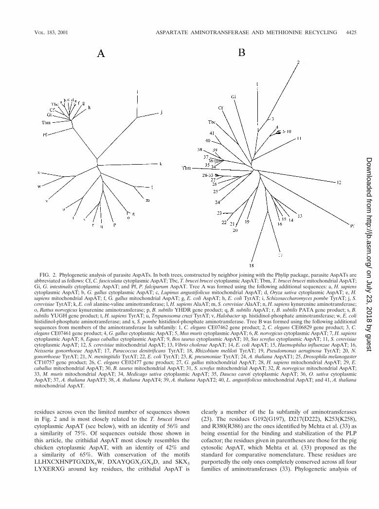
residues across even the limited number of sequences shownin Fig. 2 and is most closely related to the T. brucei bruceicytoplasmic AspAT (see below), with an identity of 56% anda similarity of 75%. Of sequences outside those shown inthis article, the crithidial AspAT most closely resembles thechicken cytoplasmic AspAT, with an identity of 42% anda similarity of 65%. With conservation of the motifsLLHXCXHNPTGXDX5W, DXAYQGX3GX4D, and SKX3
LYXERXG around key residues, the crithidial AspAT is
clearly a member of the Ia subfamily of aminotransferases(23). The residues G192(G197), D217(D222), K253(K258),and R380(R386) are the ones identified by Mehta et al. (33) asbeing essential for the binding and stabilization of the PLPcofactor; the residues given in parentheses are those for the pigcytosolic AspAT, which Mehta et al. (33) proposed as thestandard for comparative nomenclature. These residues arepurportedly the only ones completely conserved across all fourfamilies of aminotransferases (33). Phylogenetic analysis of
FIG. 2. Phylogenetic analysis of parasite AspATs. In both trees, constructed by neighbor joining with the Phylip package, parasite AspATs areabbreviated as follows: Cf, C. fasciculata cytoplasmic AspAT; Tbc, T. brucei brucei cytoplasmic AspAT; Tbm, T. brucei brucei mitochondrial AspAT;Gi, G. intestinalis cytoplasmic AspAT; and Pf, P. falciparum AspAT. Tree A was formed using the following additional sequences: a, H. sapienscytoplasmic AspAT; b, G. gallus cytoplasmic AspAT; c, Lupinus angustifolicus mitochondrial AspAT; d, Oryza sativa cytoplasmic AspAT; e, H.sapiens mitochondrial AspAT; f, G. gallus mitochondrial AspAT; g, E. coli AspAT; h, E. coli TyrAT; i, Schizosaccharomyces pombe TyrAT; j, S.cerevisiae TyrAT; k, E. coli alanine-valine aminotransferase; l, H. sapiens AlaAT; m, S. cerevisiae AlaAT; n, H. sapiens kynurenine aminotransferase;o, Rattus norvegicus kynurenine aminotransferase; p, B. subtilis YHDR gene product; q, B. subtilis AspAT; r, B. subtilis PATA gene product; s, B.subtilis YUGH gene product; t, H. sapiens TyrAT; u, Trypanosoma cruzi TyrAT; v, Halobacter sp. histidinol-phosphate aminotransferase; w, E. colihistidinol-phosphate aminotransferase; and x, S. pombe histidinol-phosphate aminotransferase. Tree B was formed using the following additionalsequences from members of the aminotransferase Ia subfamily: 1, C. elegans CE07462 gene product; 2, C. elegans CE06829 gene product; 3, C.elegans CE07461 gene product; 4, G. gallus cytoplasmic AspAT; 5, Mus muris cytoplasmic AspAT; 6, R. norvegicus cytoplasmic AspAT; 7, H. sapienscytoplasmic AspAT; 8, Equus caballus cytoplasmic AspAT; 9, Bos taurus cytoplasmic AspAT; 10, Sus scrofus cytoplasmic AspAT; 11, S. cerevisiaecytoplasmic AspAT; 12, S. cerevisiae mitochondrial AspAT; 13, Vibrio cholerae AspAT; 14, E. coli AspAT; 15, Haemophilus influenzae AspAT; 16,Neisseria gonorrhoeae AspAT; 17, Paracoccus denitrificans TyrAT; 18, Rhizobium meliloti TyrAT; 19, Pseudomonas aeruginosa TyrAT; 20, N.gonorrhoeae TyrAT; 21, N. meningitidis TyrAT; 22, E. coli TyrAT; 23, K. pneumoniae TyrAT; 24, A. thaliana AspAT1; 25, Drosophila melanogasterCT10757 gene product; 26, C. elegans CE02477 gene product; 27, G. gallus mitochondrial AspAT; 28, H. sapiens mitochondrial AspAT; 29, E.caballus mitochondrial AspAT; 30, B. taurus mitochondrial AspAT; 31, S. scrofus mitochondrial AspAT; 32, R. norvegicus mitochondrial AspAT;33, M. muris mitochondrial AspAT; 34, Medicago sativa cytoplasmic AspAT; 35, Daucus caroti cytoplasmic AspAT; 36, O. sativa cytoplasmicAspAT; 37, A. thaliana AspAT3; 38, A. thaliana AspAT4; 39, A. thaliana AspAT2; 40, L. angustifolicus mitochondrial AspAT; and 41, A. thalianamitochondrial AspAT.
VOL. 183, 2001 ASPARTATE AMINOTRANSFERASE AND METHIONINE RECYCLING 4425
on July 23, 2018 by guesthttp://jb.asm
.org/D
ownloaded from
selected family I aminotransferases demonstrated that the en-zyme clustered within the Ia subfamily, which consists of eu-karyotic AspATs, gram-negative bacterial AspATs, and pro-karyotic TyrATs (Fig. 2A). A similar analysis of the subfamilyIa aminotransferases showed that the crithidial AspAT clus-tered with the other known eukaryotic cytosolic AspATs (Fig.2B).
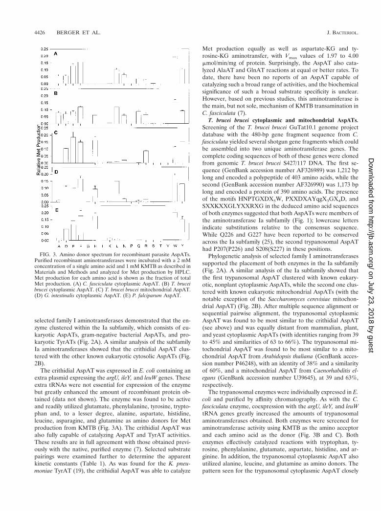
The crithidial AspAT was expressed in E. coli containing anextra plasmid expressing the argU, ileY, and leuW genes. Theseextra tRNAs were not essential for expression of the enzymebut greatly enhanced the amount of recombinant protein ob-tained (data not shown). The enzyme was found to be activeand readily utilized glutamate, phenylalanine, tyrosine, trypto-phan and, to a lesser degree, alanine, aspartate, histidine,leucine, asparagine, and glutamine as amino donors for Metproduction from KMTB (Fig. 3A). The crithidial AspAT wasalso fully capable of catalyzing AspAT and TyrAT activities.These results are in full agreement with those obtained previ-ously with the native, purified enzyme (7). Selected substratepairings were examined further to determine the apparentkinetic constants (Table 1). As was found for the K. pneu-moniae TyrAT (19), the crithidial AspAT was able to catalyze
Met production equally as well as aspartate-KG and ty-rosine-KG aminotransfer, with Vmax values of 1.97 to 4.00mmol/min/mg of protein. Surprisingly, the AspAT also cata-lyzed AlaAT and GlnAT reactions at equal or better rates. Todate, there have been no reports of an AspAT capable ofcatalyzing such a broad range of activities, and the biochemicalsignificance of such a broad substrate specificity is unclear.However, based on previous studies, this aminotransferase isthe main, but not sole, mechanism of KMTB transamination inC. fasciculata (7).
T. brucei brucei cytoplasmic and mitochondrial AspATs.Screening of the T. brucei brucei GuTat10.1 genome projectdatabase with the 480-bp gene fragment sequence from C.fasciculata yielded several shotgun gene fragments which couldbe assembled into two unique aminotransferase genes. Thecomplete coding sequences of both of these genes were clonedfrom genomic T. brucei brucei S427/117 DNA. The first se-quence (GenBank accession number AF326989) was 1,212 bplong and encoded a polypeptide of 403 amino acids, while thesecond (GenBank accession number AF326990) was 1,173 bplong and encoded a protein of 390 amino acids. The presenceof the motifs HNPTGXDX5W, PXXDXAYqgX3GX4D, andSXXKXXGLYXXRXG in the deduced amino acid sequencesof both enzymes suggested that both AspATs were members ofthe aminotransferase Ia subfamily (Fig. 1); lowercase lettersindicate substitutions relative to the consensus sequence.While Q226 and G227 have been reported to be conservedacross the Ia subfamily (25), the second trypanosomal AspAThad P207(P226) and S208(S227) in these positions.
Phylogenetic analysis of selected family I aminotransferasessupported the placement of both enzymes in the Ia subfamily(Fig. 2A). A similar analysis of the Ia subfamily showed thatthe first trypanosomal AspAT clustered with known eukary-otic, nonplant cytoplasmic AspATs, while the second one clus-tered with known eukaryotic mitochondrial AspATs (with thenotable exception of the Saccharomyces cerevisiae mitochon-drial AspAT) (Fig. 2B). After multiple sequence alignment orsequential pairwise alignment, the trypanosomal cytoplasmicAspAT was found to be most similar to the crithidial AspAT(see above) and was equally distant from mammalian, plant,and yeast cytoplasmic AspATs (with identities ranging from 39to 45% and similarities of 63 to 66%). The trypanosomal mi-tochondrial AspAT was found to be most similar to a mito-chondrial AspAT from Arabidopsis thaliana (GenBank acces-sion number P46248), with an identity of 38% and a similarityof 60%, and a mitochondrial AspAT from Caenorhabditis el-egans (GenBank accession number U39645), at 39 and 63%,respectively.
The trypanosomal enzymes were individually expressed in E.coli and purified by affinity chromatography. As with the C.fasciculata enzyme, coexpression with the argU, ileY, and leuWtRNA genes greatly increased the amounts of trypanosomalaminotransferases obtained. Both enzymes were screened foraminotransferase activity using KMTB as the amino acceptorand each amino acid as the donor (Fig. 3B and C). Bothenzymes effectively catalyzed reactions with tryptophan, ty-rosine, phenylalanine, glutamate, aspartate, histidine, and ar-ginine. In addition, the trypanosomal cytoplasmic AspAT alsoutilized alanine, leucine, and glutamine as amino donors. Thepattern seen for the trypanosomal cytoplasmic AspAT closely
FIG. 3. Amino donor spectrum for recombinant parasite AspATs.Purified recombinant aminotransferases were incubated with a 2 mMconcentration of a single amino acid and 1 mM KMTB as described inMaterials and Methods and analyzed for Met production by HPLC.Met production for each amino acid is shown as the fraction of totalMet production. (A) C. fasciculata cytoplasmic AspAT. (B) T. bruceibrucei cytoplasmic AspAT. (C) T. brucei brucei mitochondrial AspAT.(D) G. intestinalis cytoplasmic AspAT. (E) P. falciparum AspAT.
4426 BERGER ET AL. J. BACTERIOL.
on July 23, 2018 by guesthttp://jb.asm
.org/D
ownloaded from
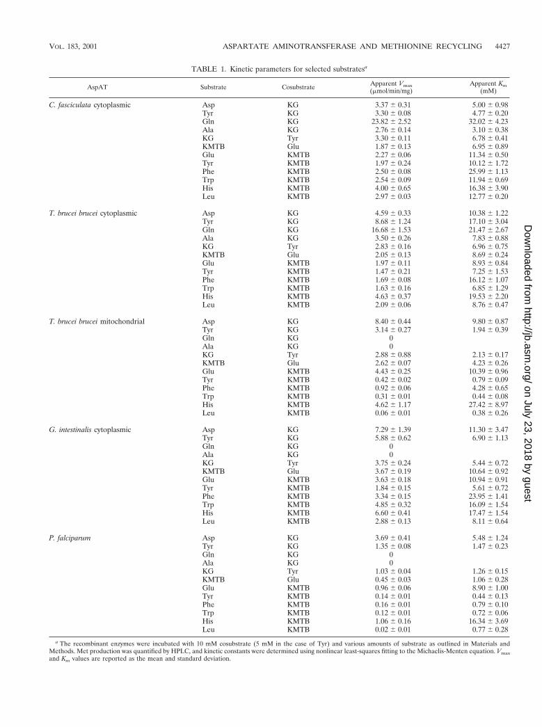
TABLE 1. Kinetic parameters for selected substratesa
AspAT Substrate Cosubstrate Apparent Vmax(mmol/min/mg)
Apparent Km(mM)
C. fasciculata cytoplasmic Asp KG 3.37 6 0.31 5.00 6 0.98Tyr KG 3.30 6 0.08 4.77 6 0.20Gln KG 23.82 6 2.52 32.02 6 4.23Ala KG 2.76 6 0.14 3.10 6 0.38KG Tyr 3.30 6 0.11 6.78 6 0.41KMTB Glu 1.87 6 0.13 6.95 6 0.89Glu KMTB 2.27 6 0.06 11.34 6 0.50Tyr KMTB 1.97 6 0.24 10.12 6 1.72Phe KMTB 2.50 6 0.08 25.99 6 1.13Trp KMTB 2.54 6 0.09 11.94 6 0.69His KMTB 4.00 6 0.65 16.38 6 3.90Leu KMTB 2.97 6 0.03 12.77 6 0.20
T. brucei brucei cytoplasmic Asp KG 4.59 6 0.33 10.38 6 1.22Tyr KG 8.68 6 1.24 17.10 6 3.04Gln KG 16.68 6 1.53 21.47 6 2.67Ala KG 3.50 6 0.26 7.83 6 0.88KG Tyr 2.83 6 0.16 6.96 6 0.75KMTB Glu 2.05 6 0.13 8.69 6 0.24Glu KMTB 1.97 6 0.11 8.93 6 0.84Tyr KMTB 1.47 6 0.21 7.25 6 1.53Phe KMTB 1.69 6 0.08 16.12 6 1.07Trp KMTB 1.63 6 0.16 6.85 6 1.29His KMTB 4.63 6 0.37 19.53 6 2.20Leu KMTB 2.09 6 0.06 8.76 6 0.47
T. brucei brucei mitochondrial Asp KG 8.40 6 0.44 9.80 6 0.87Tyr KG 3.14 6 0.27 1.94 6 0.39Gln KG 0Ala KG 0KG Tyr 2.88 6 0.88 2.13 6 0.17KMTB Glu 2.62 6 0.07 4.23 6 0.26Glu KMTB 4.43 6 0.25 10.39 6 0.96Tyr KMTB 0.42 6 0.02 0.79 6 0.09Phe KMTB 0.92 6 0.06 4.28 6 0.65Trp KMTB 0.31 6 0.01 0.44 6 0.08His KMTB 4.62 6 1.17 27.42 6 8.97Leu KMTB 0.06 6 0.01 0.38 6 0.26
G. intestinalis cytoplasmic Asp KG 7.29 6 1.39 11.30 6 3.47Tyr KG 5.88 6 0.62 6.90 6 1.13Gln KG 0Ala KG 0KG Tyr 3.75 6 0.24 5.44 6 0.72KMTB Glu 3.67 6 0.19 10.64 6 0.92Glu KMTB 3.63 6 0.18 10.94 6 0.91Tyr KMTB 1.84 6 0.15 5.61 6 0.72Phe KMTB 3.34 6 0.15 23.95 6 1.41Trp KMTB 4.85 6 0.32 16.09 6 1.54His KMTB 6.60 6 0.41 17.47 6 1.54Leu KMTB 2.88 6 0.13 8.11 6 0.64
P. falciparum Asp KG 3.69 6 0.41 5.48 6 1.24Tyr KG 1.35 6 0.08 1.47 6 0.23Gln KG 0Ala KG 0KG Tyr 1.03 6 0.04 1.26 6 0.15KMTB Glu 0.45 6 0.03 1.06 6 0.28Glu KMTB 0.96 6 0.06 8.90 6 1.00Tyr KMTB 0.14 6 0.01 0.44 6 0.13Phe KMTB 0.16 6 0.01 0.79 6 0.10Trp KMTB 0.12 6 0.01 0.72 6 0.06His KMTB 1.06 6 0.16 16.34 6 3.69Leu KMTB 0.02 6 0.01 0.77 6 0.28
a The recombinant enzymes were incubated with 10 mM cosubstrate (5 mM in the case of Tyr) and various amounts of substrate as outlined in Materials andMethods. Met production was quantified by HPLC, and kinetic constants were determined using nonlinear least-squares fitting to the Michaelis-Menten equation. Vmaxand Km values are reported as the mean and standard deviation.
VOL. 183, 2001 ASPARTATE AMINOTRANSFERASE AND METHIONINE RECYCLING 4427
on July 23, 2018 by guesthttp://jb.asm
.org/D
ownloaded from
follows that seen previously with homogenates prepared frombloodstream T. brucei brucei (6). In detailed kinetic analyses,the cytoplasmic and mitochondrial AspATs were similar (Ta-ble 1), with the exception that glutamate and the aromaticamino acids were equally as effective as amino donors forKMTB with the cytoplasmic AspAT (Vmax values of 1.63 to1.97 nmol/min/mg of protein), whereas glutamate was 5- to10-fold more effective than the aromatic amino acids with themitochondrial AspAT (4.43 versus 0.31 to 0.92 nmol/min/mg ofprotein). In addition, the cytoplasmic AspAT readily catalyzedGlnAT, AlaAT, and leucine-KMTB aminotransfer reactions,while the mitochondrial AspAT did not catalyze these reac-tions. Both enzymes catalyzed AspAT and TyrAT activities.
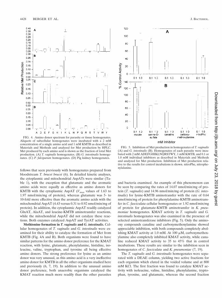
Methionine formation in T. vaginalis and G. intestinalis. Cel-lular homogenates of T. vaginalis and G. intestinalis were ex-amined for their ability to catalyze the formation of Met fromKMTB (Fig. 4A and B). Both organisms were found to havesimilar patterns for the amino donor preference for the KMATreaction, with lysine, glutamate, phenylalanine, histidine, iso-leucine, valine, tryptophan, and tyrosine all being effectiveamino donors. The strong preference for lysine as an aminodonor was very unusual, as this amino acid is a very ineffectiveamino donor for KMTB in all the other organisms studied hereand previously (6, 7, 19). In addition to the unusual aminodonor preference, both anaerobic organisms catalyzed theKMAT reaction much more readily than the other parasites
and bacteria examined. An example of this phenomenon canbe seen by comparing the rates of 14.07 nmol/min/mg of pro-tein (T. vaginalis) and 14.96 nmol/min/mg of protein (G. intes-tinalis) for lysine-KMTB aminotransfer with the rate of 0.64nmol/min/mg of protein for phenylalanine-KMTB aminotrans-fer in C. fasciculata cellular homogenates or 1.92 nmol/min/mgof protein for glutamate-KMTB aminotransfer in K. pneu-moniae homogenates. KMAT activity in T. vaginalis and G.intestinalis homogenates was also examined in the presence ofselected aminotransferase inhibitors (Fig. 5). Only the amino-oxy compounds canaline and carboxymethoxylamine showedappreciable inhibition, with both compounds completely abol-ishing KMAT activity at 1.0 mM. At 100 mM, carboxymethox-ylamine also completely inhibited KMAT activity, while cana-line reduced KMAT activity to 35 to 45% that in controlincubations. These results are similar to the inhibition seen inhomogenates of C. fasciculata and K. pneumoniae (7, 19).
The T. vaginalis and G. intestinalis homogenates were sepa-rated with a DEAE column, yielding two active fractions foreach organism which eluted in the voided volume and at 800mM KCl. The first fraction was found to catalyze KMAT ac-tivity with isoleucine, valine, histidine, phenylalanine, trypto-phan, tyrosine, and glutamate, whereas the second fraction
FIG. 4. Amino donor spectrum for parasite or tissue homogenates.Aliquots of subcellular homogenates were incubated with a 2 mMconcentration of a single amino acid and 1 mM KMTB as described inMaterials and Methods and analyzed for Met production by HPLC.Met produced by each amino acid is shown as the fraction of total Metproduction. (A) T. vaginalis homogenates. (B) G. intestinalis homoge-nates. (C) P. falciparum homogenates. (D) Pig kidney homogenates.
FIG. 5. Inhibition of Met production in homogenates of T. vaginalis(A) and G. intestinalis (B). Homogenates of each parasite were incu-bated with 2 mM ADEFGHIKLNQRSTWY, 1 mM KMTB, and 0.1 or1.0 mM individual inhibitors as described in Materials and Methodsand analyzed for Met production. Inhibition of Met production rela-tive to the results for control incubations is shown. nitroPhe, nitrophe-nylalanine.
4428 BERGER ET AL. J. BACTERIOL.
on July 23, 2018 by guesthttp://jb.asm
.org/D
ownloaded from

used only lysine as an amino donor. With the exception ofisoleucine and valine utilization, the amino donor preferenceof the first fraction closely resembled that of parasite AspATsor bacterial TyrATs (see above and references 6, 7, and 19).
G. intestinalis cytoplasmic AspAT. By similarity searching ofthe G. intestinalis genome project shotgun sequence data, itwas possible to identify the two ends of the cytoplasmicAspAT. The complete gene was cloned and was found to be1,284 bp long and to encode a protein of 427 amino acids(GenBank accession number AF326991). As with the otherparasite AspATs, the presence of the motifs HXCXHNPsGXDX5W, DXAYQGX3GX4D, and SXXKXXGLYXERXGaround the anchor residues identified the enzyme as a memberof the aminotransferase Ia subfamily (Fig. 1). While T196 hasbeen reported to be conserved across the Ia subfamily (23), thegiardial AspAT had S194(S196) in this position. In addition,phylogenetic analysis of the protein sequence placed the en-zyme with other members of the Ia subfamily (Fig. 2A and B),where it clustered with other eukaryotic cytoplasmic AspATs.The G. intestinalis AspAT showed almost equal sequence sim-ilarities to the C. fasciculata AspAT, the T. brucei brucei cyto-plasmic AspAT, human cytoplasmic AspAT (GenBank acces-sion number NM002079), chicken cytoplasmic AspAT(GenBank accession number X15636), and S. cerevisiae cyto-plasmic AspAT (GenBank accession number NC001144), withidentities of 38 to 44% and similarities of 59 to 64%.
The enzyme was expressed in E. coli, where coexpressionwith a vector containing the argU, ileY, and leuW genes wasnecessary to obtain a good yield of active recombinant mate-rial. After purification by affinity chromatography, the enzymewas examined for amino donor specificity in the KMAT reac-tion (Fig. 3D); glutamate, aspartate, phenylalanine, histidine,leucine, tryptophan, and tyrosine were found to be effectivedonors. In this respect, the G. intestinalis AspAT is very similarto the C. fasciculata and T. brucei brucei cytoplasmic AspATs.However, the G. intestinalis enzyme demonstrates reduced uti-lization of alanine, asparagine, glutamine, and arginine asamino donors relative to the two trypanosomal cytoplasmicAspATs. With the exception of an inability to use valine andisoleucine as amino donors, the purified G. intestinalis AspATcatalyzes all the KMAT activity seen in the DEAE voidedfraction isolated from cellular homogenates. Therefore, withregard to global Met regeneration from KMTB, G. intestinalisutilizes AspAT, an aminotransferase with a strict dependenceon lysine as the amino donor, and an additional aminotrans-ferase activity which can utilize valine and isoleucine as aminodonors.
Selected substrate pairs were further examined to character-ize the kinetics of the enzyme (Table 1). The Km and Vmax
values for KMTB, KG, glutamate, tyrosine, phenylalanine,tryptophan, histidine, and leucine were very similar to thoseseen for the C. fasciculata and T. brucei brucei cytoplasmicAspATs, with Km values ranging from 5.44 to 23.95 mM andVmax values of 1.84 to 6.60 mmol/min/mg of protein. Like theother enzymes, the G. intestinalis AspAT catalyzed AspAT andTyrAT reactions but, in contrast to the trypanosomal cytoplas-mic AspATs, did not perform any glutamine-KG or ala-nine-KG aminotransfer. Therefore, while the giardial cytoplas-mic AspAT is very similar to the trypanosomal homologues, ithas a narrower substrate specificity.
Methionine regeneration in P. falciparum. Cellular homog-enates of malarial parasites isolated from human erythrocyteswere examined for their ability to catalyze the KMAT reaction(Fig. 4C). Glutamate, phenylalanine, histidine, isoleucine,leucine, valine, tryptophan, and tyrosine were the only aminoacids capable of acting as amino donors, with the branched-chain amino acids being less active as substrates. The amountof KMAT activity detected in the plasmodial homogenates waslower than that found in other organisms screened to date.When the most active amino donor, glutamate, was used, only0.22 nmol of Met/min/mg of protein could be produced fromKMTB, as opposed to 0.64 nmol/min/mg of protein for phe-nylalanine-KMTB in C. fasciculata homogenates and 1.92nmol/min/mg of protein for glutamate-KMTB in K. pneu-moniae homogenates. The plasmodial homogenates were sep-arated with a DEAE column, where a single active fraction,eluting at 700 mM KCl, was discovered. This fraction was ableto catalyze Met formation from KMTB using the same aminodonors as those used by the original homogenates, suggestingthat only one aminotransferase catalyzes Met regeneration inP. falciparum.
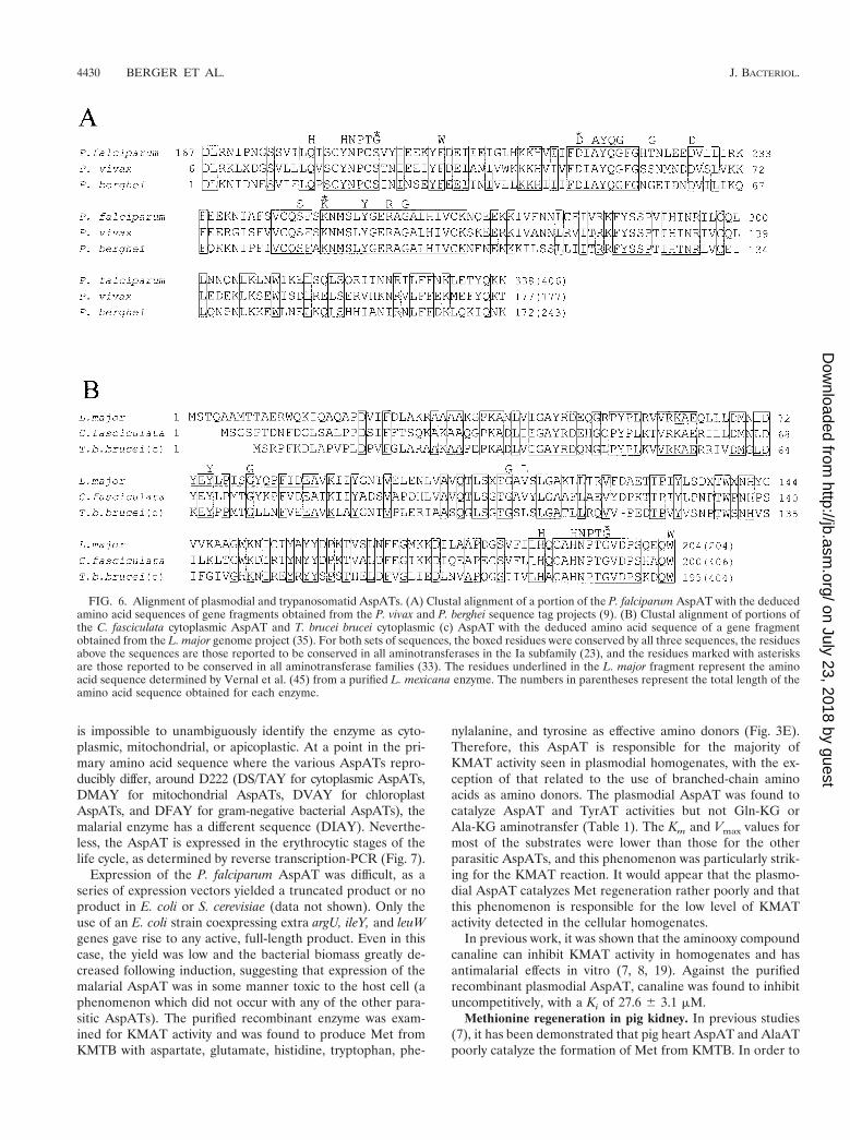
P. falciparum AspAT. The published sequence for P. falcipa-rum chromosome 2 contains one region with significant simi-larity to AspATs (15). In addition, BLAST searching of all thesequence data available for the remaining chromosomesyielded no further AspAT homologues. The putative chromo-some 2 AspAT was cloned, and the published sequence wasconfirmed to be 1,218 bp long and to code for a protein of 405amino acids. The presence of the motifs qX3yNPcsXyX5y,DXAYQGX3tX4D, and SXXKXXsLYXERXG (Fig. 1) aroundthe anchor residues suggests that the plasmodial enzyme is amember of the aminotransferase Ia subfamily. However, it isclear from the number of residues in these motifs shown aslowercase letters that the plasmodial AspAT has a number ofsubstitutions relative to the reported Ia consensus sequence(23). In a most striking variation, it was noted that the normalanchor residue G197, which is postulated to be an essentialrequirement of all aminotransferases (33), is S188(S197) in theP. falciparum AspAT. As the published DNA sequence (Gen-Bank accession number AE001380) and the sequence obtainedin this manuscript are identical, it is highly unlikely thatS188(S197) is due to a sequencing error or a PCR-inducedmutation. In addition, similarity searching of the P. vivax and P.berghei genome surveys (parasite.vetmed.ufl.edu) (9) has un-covered partial sequences for the homologous AspATs (Fig.6A). S188(S197) is present in all three sequences, indicatingthat the postulated requirement of G197 for aminotransferaseactivity is not absolute and that plasmodial (and perhaps api-complexan) AspATs contain unusual sequence variations com-pared to enzymes from other phyla and kingdoms.
Phylogenetic analysis of the plasmodial AspAT and the fam-ily I aminotransferases unambiguously placed the enzyme inthe Ia subfamily (Fig. 2A). However, when only the Ia subfam-ily is examined, the malarial enzyme is the most highly diver-gent sequence available, clustering with neither the eukaryoticcytoplasmic AspATs, the eukaryotic mitochondrial AspATs,the bacterial AspATs, nor the bacterial TyrATs (Fig. 2B). Interms of sequence similarity, the P. falciparum AspAT has only24 to 31% identity and 49 to 55% similarity with any of theother Ia subfamily members. With these terms of reference, it
VOL. 183, 2001 ASPARTATE AMINOTRANSFERASE AND METHIONINE RECYCLING 4429
on July 23, 2018 by guesthttp://jb.asm
.org/D
ownloaded from
is impossible to unambiguously identify the enzyme as cyto-plasmic, mitochondrial, or apicoplastic. At a point in the pri-mary amino acid sequence where the various AspATs repro-ducibly differ, around D222 (DS/TAY for cytoplasmic AspATs,DMAY for mitochondrial AspATs, DVAY for chloroplastAspATs, and DFAY for gram-negative bacterial AspATs), themalarial enzyme has a different sequence (DIAY). Neverthe-less, the AspAT is expressed in the erythrocytic stages of thelife cycle, as determined by reverse transcription-PCR (Fig. 7).
Expression of the P. falciparum AspAT was difficult, as aseries of expression vectors yielded a truncated product or noproduct in E. coli or S. cerevisiae (data not shown). Only theuse of an E. coli strain coexpressing extra argU, ileY, and leuWgenes gave rise to any active, full-length product. Even in thiscase, the yield was low and the bacterial biomass greatly de-creased following induction, suggesting that expression of themalarial AspAT was in some manner toxic to the host cell (aphenomenon which did not occur with any of the other para-sitic AspATs). The purified recombinant enzyme was exam-ined for KMAT activity and was found to produce Met fromKMTB with aspartate, glutamate, histidine, tryptophan, phe-
nylalanine, and tyrosine as effective amino donors (Fig. 3E).Therefore, this AspAT is responsible for the majority ofKMAT activity seen in plasmodial homogenates, with the ex-ception of that related to the use of branched-chain aminoacids as amino donors. The plasmodial AspAT was found tocatalyze AspAT and TyrAT activities but not Gln-KG orAla-KG aminotransfer (Table 1). The Km and Vmax values formost of the substrates were lower than those for the otherparasitic AspATs, and this phenomenon was particularly strik-ing for the KMAT reaction. It would appear that the plasmo-dial AspAT catalyzes Met regeneration rather poorly and thatthis phenomenon is responsible for the low level of KMATactivity detected in the cellular homogenates.
In previous work, it was shown that the aminooxy compoundcanaline can inhibit KMAT activity in homogenates and hasantimalarial effects in vitro (7, 8, 19). Against the purifiedrecombinant plasmodial AspAT, canaline was found to inhibituncompetitively, with a Ki of 27.6 6 3.1 mM.
Methionine regeneration in pig kidney. In previous studies(7), it has been demonstrated that pig heart AspAT and AlaATpoorly catalyze the formation of Met from KMTB. In order to
FIG. 6. Alignment of plasmodial and trypanosomatid AspATs. (A) Clustal alignment of a portion of the P. falciparum AspAT with the deducedamino acid sequences of gene fragments obtained from the P. vivax and P. berghei sequence tag projects (9). (B) Clustal alignment of portions ofthe C. fasciculata cytoplasmic AspAT and T. brucei brucei cytoplasmic (c) AspAT with the deduced amino acid sequence of a gene fragmentobtained from the L. major genome project (35). For both sets of sequences, the boxed residues were conserved by all three sequences, the residuesabove the sequences are those reported to be conserved in all aminotransferases in the Ia subfamily (23), and the residues marked with asterisksare those reported to be conserved in all aminotransferase families (33). The residues underlined in the L. major fragment represent the aminoacid sequence determined by Vernal et al. (45) from a purified L. mexicana enzyme. The numbers in parentheses represent the total length of theamino acid sequence obtained for each enzyme.
4430 BERGER ET AL. J. BACTERIOL.
on July 23, 2018 by guesthttp://jb.asm
.org/D
ownloaded from

better compare mammalian and parasite Met regeneration, pigkidney was chosen as a model. Homogenates of freshly ac-quired tissue were found to readily catalyze the KMAT reac-tion, with glutamate, isoleucine, leucine, and valine being themajor amino donors and aspartate, phenylalanine, histidine,asparagine, glutamine, arginine, tryptophan, and tyrosine alsobeing able to act as amino donors (Fig. 4D). The homogenateswere applied to a DEAE column, and two fractions catalyzingthe KMAT reaction were discovered. The first, eluting in thecolumn voided volume, utilized branched-chain amino acidsand, to a lesser extent, glutamate, aromatic amino acids, andhistidine. The second, eluting at 400 mM KCl, used histidineand, to a lesser extent, asparagine. Neither of these fractionshad AspAT or TyrAT activity.
DISCUSSION
The international situation regarding the chemotherapy ofbacterial and parasitic diseases continues to worsen with thespread of drug-resistant pathogenic strains. In particular, P.falciparum malaria currently threatens half of the world’s pop-ulation, with at least 200 million infections and 2 to 3 milliondeaths per year (48). Resistance to aminoquinolines and anti-folates has become commonplace, and few effective or novelagents are clinically available. In a similar manner, trypanoso-mal diseases are an increasing concern, as evidenced by therecent epidemic of African sleeping sickness in the Congo,Uganda, and southern Sudan (40) and the reports of arsenical-resistant trypanosomiasis and antimony-resistant leishmaniasis(25, 26). Cases of trichomoniasis and giardiasis have become
commonplace, and reports of metronidazole resistance haveoccurred (43). These disease resurgences require the discoveryand development of novel drug targets. In past studies, it hasbeen demonstrated that interference with enzymes involved inthe regeneration of Met from methylthioadenosine can lead tocell death in K. pneumoniae, P. falciparum, and T. brucei (3, 17,36, 41). These studies have focused on the enzymes methyl-thioadenosine phosphorylase and methylthioribose kinase,which convert methylthioadenosine to methylthiophosphori-bose in eukaryotes and prokaryotes, respectively. It has beenshown that the final step of Met regeneration from methylthio-adenosine, the transamination of KMTB to Met, can be inhib-ited in homogenates of C. fasciculata and K. pneumoniae (7,19). In addition, selected inhibitors which are active for thisprocess have a cytocidal effect in vitro against C. fasciculataand P. falciparum (7, 8).
Identity and relationship of aminotransferases catalyzingMet formation. In the present study, we have continued thecharacterization of the final step of Met recycling in parasiticorganisms and have found that AspAT plays an important rolein all of the organisms examined. In C. fasciculata, T. bruceibrucei, and P. falciparum, AspAT represents the major sourceof activity for Met regeneration, and in G. intestinalis, theenzyme is second only to an as-yet-uncharacterized source ofactivity utilizing lysine as the amino donor. The consistency ofthese results across diverse phyla suggests that AspAT is anessential component of Met recycling in lower eukaryotes.However, aside from our work on the same reaction in K.pneumoniae, nothing is known about the exact aminotrans-ferases active in other organisms. In particular, higher mam-mals, fungi, gram-positive bacteria, and archaea have not beenfully examined. Nevertheless, it is clear that different amino-transferases are responsible for Met regeneration in differentgroups of organisms. The activity in K. pneumoniae has beenunequivocally identified as TyrAT, whereas the protozoa ex-amined here rely on AspAT. In mammals, using pig tissues asa model, we have been able to discount AspAT, TyrAT, andAlaAT as being responsible for the activity (7) but have beenunable to fully purify the exact aminotransferase responsible.In studies with purified rat GlnAT (kynurenine aminotransfer-ase), Cooper and Meister have shown that the enzyme activelycatalyzes phenylalanine-KMTB aminotransfer (10). In addi-tion, Davoodi et al. (11) and Hall et al. (18) have found thatpurified and recombinant human and rat branched-chainaminotransferases can catalyze branched-chain amino acid–KMTB aminotransfer. Given the amino donor preference forthe KMAT reaction in pig kidney homogenates, it is entirelypossible that the branched-chain aminotransferase plays alarge role in the reaction in vivo, while a role for the kynure-nine aminotransferase is less clear. Full purification of themammalian KMAT aminotransferase(s) and a broader char-acterization of recombinant mammalian branched-chain andkynurenine aminotransferases is required to completely solvethis problem.
It is interesting that the parasitic protozoa and K. pneu-moniae, while using different types of aminotransferases tocatalyze the KMAT reaction, all use enzymes from the Iasubfamily. However, from the numerous genome projects com-pleted, we have been unable to find any members of the Iasubfamily in gram-positive bacteria or archaea. The AspATs in
FIG. 7. Reverse transcription-PCR of P. falciparum AspAT. RNAisolated from asynchronous P. falciparum was incubated in the pres-ence (lanes E) or absence (lanes C) of reverse transcriptase and thensubjected to PCR with primers specific for the full-length P. falciparumAspAT gene (PfASAT) or the P. falciparum lactate dehydrogenasegene (PfLDH) as a positive control. The products were then analyzedon an agarose gel together with DNA markers (lane M). The lengthsof the markers are (from the gel bottom) 0.5, 0.75, 1.0, 1.5, 2.0, 2.5, 3.0,4.0, 5.0, 6.0, 8.0, and 10.0 kbp. The expected length of AspAT was 1,218bp, and that of lactate dehydrogenase was 951 bp.
VOL. 183, 2001 ASPARTATE AMINOTRANSFERASE AND METHIONINE RECYCLING 4431
on July 23, 2018 by guesthttp://jb.asm
.org/D
ownloaded from

these organisms are members of the If subfamily (see thephylogenic tree in reference 19); thus, they are relatives ofeukaryotic TyrATs and kynurenine aminotransferases.Branched-chain aminotransferases are all members of the IIIfamily of aminotransferases (33). We are currently character-izing KMAT activity in several gram-positive models, with anemphasis on Bacillus subtilis, where there is a strong preferencefor branched-chain amino acids as amino donors (data notshown). As mentioned above, we have also been able to dis-count mammalian AspAT as being responsible for KMATactivity, thus eliminating the only higher eukaryotic membersof the Ia subfamily. Therefore, diverse subfamilies and, poten-tially, different families of aminotransferases may have evolvedspecificity for KMTB.
In terms of substrate specificity, the crithidial and trypano-somal cytosolic AspATs are unusually broad. In addition to theexpected aspartate-KG and tyrosine-KG activities, these en-zymes also catalyzed glutamine-KG and alanine-KG amin-otransfer. In previous studies (19), the K. pneumoniae TyrATwas unable to perform these latter two reactions, and there areno other literature reports of AspATs catalyzing AlaAT orGlnAT activity. When the primary amino acid sequences areexamined, there are no obvious differences between the se-quences for the two kinetoplastid AspATs and the sequencefor the giardial enzyme, which lacks AlaAT and GlnAT activ-ities. Crystallization and structural analyses of the crithidialand/or trypanosomal enzymes may assist in further under-standing the basis for the broad substrate specificity comparedto the known structures for the E. coli, S. cerevisiae, andchicken AspATs (21, 22, 29). As both C. fasciculata and T.brucei brucei cytoplasmic AspATs have this wider range ofactivity, it is likely that the homologous enzyme from othertrypanosomatids would share this feature. Vernal et al. puri-fied from Leishmania mexicana promastigotes an aminotrans-ferase which displayed an unusually broad range of activity andyielded an internal peptide sequence consistent with the se-quence of a cytoplasmic AspAT (45). By similarity searching ofshotgun sequence data from the L. major genome project, wehave been able to identify approximately half of the leishma-nial cytoplasmic AspAT sequence (Fig. 6B). It is clear, fromthe presence of the peptide sequence obtained by Vernal et al.(45), that this cytoplasmic AspAT represents the enzyme pre-viously purified in that study. It is also apparent that the leish-manial sequence has a high identity to the crithidial andtrypanosomal sequences. While we have not examined thebiochemical properties of the leishmanial enzyme, the presentwork suggests that this AspAT should be active in Met regen-eration in Leishmania spp.
Sequence of the plasmodial AspAT. The AspAT from P.falciparum is highly unusual in that it contains sequence sub-stitutions not found in aminotransferases from any of the fourfamilies (33). While, overall, the plasmodial AspAT has suffi-cient homology to subfamily Ia aminotransferases to unequiv-ocally be grouped with these enzymes, the sequence displays avery high level of divergence from cytoplasmic AspATs, mito-chondrial AspATs, plastid AspATs, gram-negative bacterialAspATs, and bacterial TyrATs. The partial sequences ob-tained from P. vivax and P. berghei clearly demonstrate that theunusual AspAT sequence is conserved within the plasmodia.Unfortunately, there is a paucity of aminotransferase se-
quences from nonfungal lower eukaryotes, so it is impossible tostate whether the P. falciparum AspAT is unique to plasmodiaor is indicative of a new subtype of Ia aminotransferases local-ized to apicomplexans or other phyla of protists. As dinoflagel-lates are often hypothesized as representing the phylum closestto the apicomplexans (2), AspAT sequences from these organ-isms would be particularly enlightening. Structural analysis ofthe P. falciparum AspAT would be helpful in determiningwhether the unusual primary sequence is the source for therelatively low activity of this enzyme for KMTB transamina-tion.
KMTB transamination in G. intestinalis and T. vaginalis.The finding that both G. intestinalis and T. vaginalis rely onlysine as a central amino donor for KMTB transamination isunusual. No other organism examined to date makes any use oflysine in this context. In addition, while in the other parasitesand K. pneumoniae aminotransferases other than AspAT orTyrAT clearly play some role in Met regeneration, these ad-ditional enzymes are a minor component of total activity. In G.intestinalis and T. vaginalis, the unknown aminotransferase cat-alyzing the KMTB-lysine reaction appears to be quantitativelymore important than AspAT for transaminating KMTB. In aprevious study, Lowe and Rowe examined aminotransferaseactivities in T. vaginalis (27). Among their discoveries was anaminotransferase which readily catalyzed KG-lysine and phe-nylpyruvate-lysine aminotransfer. This enzyme was partiallypurified and found to copurify with an ornithine aminotrans-ferase activity. While the enzyme was found to utilize orni-thine, lysine, KG, and phenylpyruvate, neither KMTB norother substrates were examined. It is quite possible that thisenzyme is responsible for the KMTB-lysine activity seen in T.vaginalis, but it should be pointed out that Lowe and Rowe(27) also detected lysine-oxaloacetate activity in T. vaginalishomogenates that did not copurify with an ornithine or lysineaminotransferase. In related work, Lowe and Rowe also puri-fied an AspAT from T. vaginalis (28) which had both AspATand TyrAT activities but did not catalyze GlnAT or AlaATreactions. While this enzyme was not examined for KMTBtransamination, the kinetic properties for aspartate-KG andtyrosine-KG suggest that their T. vaginalis AspAT is the ho-mologue of the G. intestinalis AspAT presented here.
Subsequent to the studies outlined in this paper, a reportdemonstrating the lack of any putrescine or spermidine amin-opropyltransferase activity in T. vaginalis was published (51).Clearly, the KMAT activity measured in this organism cannotbe related to Met regeneration from polyamine biosynthesis.The biochemical source of any cellular KMTB utilized in theKMAT reaction in T. vaginalis is presently a mystery. However,several alternatives to polyamine synthesis exist, such as S-adenosylmethionine conversion to methylthioadenosine and1-aminocyclopropane-1-carboxylate (47), the action of aminoacid oxidase on D-methionine, the conversion of hydroxyme-thiobutyrate via an a-hydroxy acid dehydrogenase, and thepossibility of compartmental Met-KMTB cycling within thecell (46). The presence or absence of these alternative sourcesof KMTB has not been investigated.
Inhibition of Met formation. We have shown that the amino-oxy compounds canaline and carboxymethoxylamine can in-hibit total KMAT activity in homogenates of T. vaginalis andG. intestinalis. At 100 mM concentrations, canaline was as po-
4432 BERGER ET AL. J. BACTERIOL.
on July 23, 2018 by guesthttp://jb.asm
.org/D
ownloaded from
tent as in corresponding experiments with C. fasciculata or K.pneumoniae homogenates (7, 19). At present, neither or thesecompounds has been tested by us against T. vaginalis or G.intestinalis in vitro. However, Rowe and Lowe found that car-boxymethoxylamine can completely inhibit T. vaginalis cellgrowth in vitro, albeit at a high concentration (5 mM) (38).
In previous work, canaline was demonstrated to be an ef-fective antimalarial agent in vitro (8). The compound, anaminooxy analogue of ornithine, has been shown here to be anuncompetitive inhibitor of the plasmodial AspAT, with a Ki of27 mM. As Met regeneration in malaria has been shown to benecessary for cellular survival (41) and our present work hasdemonstrated that P. falciparum catalyzes the KMAT reactionmuch less readily than the other protozoa examined, the ma-laria parasite may be uniquely susceptible to interference withthe final step in Met recycling. It should be pointed out, how-ever, that aminooxy inhibitors are not necessarily specific to asingle aminotransferase. We have found that canaline alsoinhibits the plasmodial ornithine aminotransferase (data notshown), suggesting that the compound may exert an effect onmultiple targets and pathways.
ACKNOWLEDGMENTS
We thank Alan H. Fairlamb for helpful discussions during thecourse of these investigations and Graham Coombs (Institute of Bio-medical and Life Sciences, University of Glasgow) for providing theG. intestinalis and T. vaginalis cell pellets.
This work was funded by the Wellcome Trust. The sequences in thisstudy were obtained from preliminary data made available by a num-ber of ongoing sequence projects. Sequencing of the P. falciparumgenome was performed at The Institute for Genomic Research, theNaval Medical Research Center, Stanford University, and the SangerCentre, with financial support provided by the Burroughs WellcomeFund, the Wellcome Trust, the National Institutes of Health (NIAID),and the U.S. Department of Defense Military Infectious DiseasesResearch Program. The giardial genome was sequenced at the MarineBiological Laboratory in Woods Hole, Mass., the University of Texasat El Paso, the University of Arizona at Tucson, and the University ofIllinois at Urbana-Champaign, with funding provided by the NationalInstitutes of Health (NIAID), the LI-COR Biotechnology Division,and the G. Unger Vetlesen Foundation. Sequencing of the T. bruceibrucei genome was performed at The Institute for Genomic Researchand the Sanger Centre and was funded by the National Institutes ofHealth (NIAID), the Wellcome Trust, and Beowulf Genomics. Se-quencing of the L. major genome was performed at the Seattle Bio-medical Research Institute, Fiocruz, EULeish, and the Sanger Centre,with funding from the Burroughs Wellcome Fund, the National Insti-tutes of Health (NIAID), the European Union, Beowulf Genomics,and the World Health Organization (TDR). The P. vivax and P. bergheigenome sequence tag projects were undertaken at the University ofFlorida, with funding from the National Institutes of Health (NIAID).
REFERENCES
1. Altschul, S. F., T. L. Madden, A. A. Schaffer, J. Zhang, Z. Zhang, W. Miller,and D. J. Lipman. 1997. Gapped BLAST and PSI-BLAST: a new generationof protein database search programs. Nucleic Acids Res. 25:3389–3402.
2. Ayala, F. J., A. A. Escalante, A. A. Lal, and S. M. Rich. 1998. Evolutionaryrelationships of human malaria parasites, p. 285–300. In I. Sherman (ed.),Malaria: parasite biology, pathogenesis, and protection. ASM Press, Wash-ington, D.C.
3. Bacchi, C. J., J. R. Sufrin, H. C. Nathan, A. J. Spiess, T. Hannon, J.Garafolo, K. Alecia, L. Katz, and N. Yarlett. 1991. 59-Alkyl-substituted an-alogs of 59-methylthioadenosine as trypanocides. Antimicrob. Agents Che-mother. 35:1904–1906.
4. Backlund, P. S., C. P. Chang, and R. A. Smith. 1982. Identification of2-keto-4-methylthiobutyrate as an intermediate compound in methioninesynthesis from 59-methylthioadenosine. J. Biol. Chem. 257:4196–4202.
5. Backlund, P. S., and R. A. Smith. 1980. Methionine synthesis from 59-methylthioadenosine in rat liver. J. Biol. Chem. 256:1533–1535.
6. Berger, B. J., W. W. Dai, H. Wang, R. E. Stark, and A. Cerami. 1996.Aromatic amino acid transamination and methionine recycling in trypano-somatids. Proc. Natl. Acad. Sci. USA 93:4126–4130.
7. Berger, B. J., W. W. Dai, and J. Wilson. 1998. Methionine formation froma-ketomethiobutyrate in the trypanosomatid Crithidia fasciculata. FEMSMicrobiol. Lett. 165:305–312.
8. Berger, B. J. 2000. Antimalarial activities of aminooxy compounds. Antimi-crob. Agents Chemother. 44:2540–2542.
8a.Brun, R., and M. Schonenberger. 1979. Cultivation and in vitro cloning ofprocyclic culture forms of Trypanosoma brucei in a semi-defined medium.Acta Trop. 36:289–292.
9. Carlton, J. M., and J. B. Dame. 2000. The Plasmodium vivax and P. bergheigene sequence tag projects. Parasitol. Today 16:409.
10. Cooper, A. J. L., and A. Meister. 1972. Isolation and properties of highlypurified glutamine aminotransferase. Biochemistry 11:661–671.
11. Davoodi, J., P. M. Drown, R. K. Bledsoe, R. Wallin, G. D. Reinhart, andS. M. Hutson. 1998. Overexpression and characterization of the humanmitochondrial and cytosolic branched-chain aminotransferases. J. Biol.Chem. 273:4982–4989.
12. Dayhoff, M. O., R. M. Schwartz, and B. C. Orcutt. 1978. A model of evolu-tionary change in proteins, p. 345–352. In M. O. Dayhoff (ed.), Atlas ofprotein sequence and structure. NBRF, Washington, D.C.
13. Felsenstein, J. 1989. PHYLIP—Phylogeny Inference Package (version 3.2).Cladistics 5:164–166.
14. Furfine, E. S., and R. H. Abeles. 1988. Intermediates in the conversion of59-methylthioadenosine to methionine in Klebsiella pneumoniae. J. Biol.Chem. 263:9596–9606.
15. Gardner, M. J., H. Tettelin, D. J. Carucci, L. M. Cummings, L. Aravind,E. V. Koonin, S. Shallom, T. Mason, K. Yu, C. Fujii, J. Pederson, K. Shen,J. Jing, C. Aston, Z. Lai, D. C. Schwartz, M. Pertea, S. Salzberg, L. Zhou,G. G. Sutton, R. Clayton, O. White, H. O. Smith, C. M. Fraser, M. D. Adams,J. C. Venter, and S. L. Hoffman. 1998. Chromosome 2 sequence of thehuman malaria parasite Plasmodium falciparum. Science 282:1126–1132.
16. Ghoda, L. Y., T. M. Savarese, C. H. Northrup, R. E. Parks, J. Garafolo, L.Katz, B. B. Ellenbogen, and C. J. Bacchi. 1988. Substrate specificities of59-deoxy-59methylthioadenosine phosphorylase from Trypanosoma bruceibrucei and mammalian cells. Mol. Biochem. Parasitol. 27:109–118.
17. Gianotti, A. J., P. A. Tower, J. H. Sheley, P. A. Conte, C. Spiro, A. J. Ferro,J. H. Fitchen, and M. K. Riscoe. 1990. Selective killing of Klebsiella pneu-moniae by 5-trifluoromethylthioribose. Chemotherapeutic exploitation ofthe enzyme 5-methylthioribose kinase. J. Biol. Chem. 265:831–837.
18. Hall, T. R., R. Wallin, G. D. Reinhart, and S. M. Hutson. 1993. Branchedchain aminotransferase isoenzymes. Purification and characterization of therat brain isoenzyme. J. Biol. Chem. 268:3092–3098.
19. Heilbronn, J., J. Wilson, and B. J. Berger. 1999. Tyrosine aminotransferasecatalyzes the final step of methionine recycling in Klebsiella pneumoniae. J.Bacteriol. 181:1739–1747.
20. Iraqui, I., S. Vissers, M. Cartiaux, and A. Urrestarazu. 1998. Characterisa-tion of Saccharomyces cerevisiae ARO8 and ARO9 genes encoding aromaticaminotransferase I and II reveals a new aminotransferase subfamily. Mol.Gen. Genet. 257:238–248.
21. Jager, J., R. A. Pauptit, U. Sauder, and J. N. Jansonius. 1994. Three-dimensional structure of a mutant E. coli aspartate aminotransferase withincreased enzymic activity. Protein Eng. 7:605–612.
22. Jeffery, C. J., T. Barry, S. Doonan, G. A. Petsko, and D. Ringe. 1998.Crystallization and preliminary X-ray diffraction analysis of aspartate ami-notransferase from Saccharomyces cerevisiae. Protein Sci. 7:1380–1387.
23. Jensen, R. A., and W. Gu. 1996. Evolutionary recruitment of biochemicallyspecialized subdivisions of family I within the protein superfamily of amin-otransferases. J. Bacteriol. 178:2161–2171.
24. Kidder, G. W., and B. N. Dutta. 1958. The growth and nutrition of Crithidiafasciculata. J. Gen. Microbiol. 18:621–638.
25. Legros, D., C. Fournier, M. Gastellu Etchegorry, F. Maiso, and E. Szumilin.1999. Therapeutic failure of melarsoprol among patients treated for latestage T.b. gambiense human African trypanosomiasis in Uganda. Bull. Soc.Pathol. Exot. 92:171–172.
26. Lira, R., S. Sundar, A. Makharia, R. Kenney, A. Gam, E. Saraiva, and D.Sacks. 1999. Evidence that the high incidence of treatment failures in Indiankala-azar is due to the emergence of antimony-resistant strains of Leishma-nia donovani. J. Infect. Dis. 180:564–567.
27. Lowe, P. N., and A. F. Rowe. 1986. Aminotransferase activities in Trichomo-nas vaginalis. Mol. Biochem. Parasitol. 21:65–74.
28. Lowe, P. N., and A. F. Rowe. 1985. Aspartate:2-oxoglutarate aminotransfer-ase from Trichomonas vaginalis. Biochem. J. 232:689–695.
29. Malashkevich, V. N., B. V. Stokopytor, V. V. Borisov, Z. Dauter, K. S.Wilson, and Y. M. Torchinsky. 1995. Crystal structure of the closed form ofchicken cytosolic aspartate aminotransferase at 1.9 A resolution. J. Mol.Biol. 247:111–124.
30. Marchitto, K. S., and A. J. Ferro. 1985. The metabolism of 5-methylthioad-enosine and 5-methyl-1-phosphate in Saccharomyces cerevisiae. J. Gen. Mi-crobiol. 131:2153–2164.
31. Marton, L. J., and A. E. Pegg. 1995. Polyamines as targets for therapeutic
VOL. 183, 2001 ASPARTATE AMINOTRANSFERASE AND METHIONINE RECYCLING 4433
on July 23, 2018 by guesthttp://jb.asm
.org/D
ownloaded from

intervention. Annu. Rev. Pharmacol. Toxicol. 35:55–91.32. McArthur, A. G., H. G. Morrison, J. E. Nixon, N. Q. Passamaneck, U. Kim,
G. Hinkle, M. K. Crocker, M. E. Holder, R. Farr, C. I. Reich, G. E. Olsen,S. B. Aley, R. D. Adam, F. D. Gillin, and M. L. Sogin. 2000. The Giardiagenome project database. FEMS Microbiol. Lett. 189:271–273.
33. Mehta, P. K., T. I. Hale, and P. Christen. 1993. Aminotransferases: demon-stration of homology and division into evolutionary subgroups. Eur. J. Bio-chem. 214:549–561.
34. Myers, R. W., and R. H. Abeles. 1993. Purification and characterization of anenzyme involved in oxidative carbon-carbon cleavage reactions in the me-thionine salvage pathway of Klebsiella pneumoniae. J. Biol. Chem.268:24785–24791.
35. Myler, P. J., and K. D. Stuart. 2000. Recent developments from the Leish-mania genome project. Curr. Opin. Microbiol. 3:412–416.
36. Riscoe, M. K., A. J. Ferro, and J. H. Fitchen. 1988. Analogs of 5-methylthi-oribose, a novel class of antiprotozoal agents. Antimicrob. Agents Che-mother. 32:1904–1906.
37. Riscoe, M. K., P. A. Tower, D. H. Peyton, A. J. Ferro, and J. H. Fitchen. 1991.Methionine recycling as a target for antiprotozoal drug development, p.450–457. In G. Coombs and M. North (ed.), Biochemical protozoology.Taylor and Francis, London, United Kingdom.
38. Rowe, A. F., and P. N. Lowe. 1986. Modulation of amino acid and 2-oxo acidpools in Trichomonas vaginalis by aspartate aminotransferase. Mol. Bio-chem. Parasitol. 21:17–24.
39. Saitou, N., and M. Nai. 1987. The neighbor-joining method: a new methodfor reconstructing phylogenetic trees. Mol. Biol. Evol. 4:406–425.
40. Smith, D. H., J. Pepin, and A. H. Stich. 1998. Human African trypanosomi-asis: an emerging public health crisis. Br. Med. Bull. 54:341–355.
41. Sufrin, J. R., S. R. Meshnik, A. J. Spiess, J. Garafolo-Hannon, X. Q. Pan,
and C. J. Bacchi. 1995. Methionine recycling pathways and antimalarial drugdesign. Antimicrob. Agents Chemother. 39:2511–2515.
42. Thompson, J. D., D. G. Higgins, and T. J. Gibson. 1994. CLUSTAL W:improving the sensitivity of progressive multiple sequence alignment throughsequence weighting, position-specific gap penalties and weight matrix choice.Nucleic Acids Res. 22:4673–4680.
43. Townson, S. M., P. F. Boreham, P. Upcroft, and J. A. Upcroft. 1994. Resis-tance to the nitroheterocyclic drugs. Acta Trop. 56:173–194.
44. Trackman, P. C., and R. H. Abeles. 1983. Methionine synthesis from 59-S-1-phospho-5-S-methylthioribulose. J. Biol. Chem. 258:6717–6720.
45. Vernal, J., J. J. Cazzulo, and C. Nowicki. 1998. Isolation and partial char-acterization of a broad specificity aminotransferase from Leishmania mexi-cana promastigotes. Mol. Biochem. Parasitol. 96:83–92.
46. Walker, J., and J. Barrett. 1997. Parasite sulphur amino acid metabolism.Int. J. Parasitol. 27:883–897.
47. Wang, S. Y., D. O. Adams, and M. Lieberman. 1982. Recycling of 59-meth-ylthioadenosine-ribose carbon atoms into methionine in tomato tissue inrelation to ethylene production. Plant Physiol. 70:117–121.
48. World Health Organization. 1999. Tropical disease research: progress 1997–1998. World Health Organization, Geneva, Switzerland.
49. Wray, J. W., and R. H. Abeles. 1993. A bacterial enzyme that catalyzesformation of carbon monoxide. J. Biol. Chem. 268:21466–21469.
50. Wray, J. W., and R. H. Abeles. 1995. The methionine salvage pathway inKlebsiella pneumoniae and rat liver. Identification and characterization oftwo novel dioxygenases. J. Biol. Chem. 270:3147–3152.
51. Yarlett, N., M. P. Martinez, B. Goldberg, D. L. Kramer, and C. W. Porter.2000. Dependence of Trichomonas vaginalis upon polyamine backconver-sion. Microbiology 146:2715–2722.
4434 BERGER ET AL. J. BACTERIOL.
on July 23, 2018 by guesthttp://jb.asm
.org/D
ownloaded from