metastatic non-small-cell lung cancer in the setting of
TRANSCRIPT

Case ReportMetastatic Non-Small-Cell Lung Cancer in the Setting ofUnilateral Agenesis of the Left Pulmonary Artery: A CaseReport and Comprehensive Literature Review
John Agzarian ,1 Jakub Kadlec,2 Lori Whitehead,3 and Yaron Shargall1
1McMaster University, Faculty of Health Sciences, Department of Surgery, St. Joseph’s Healthcare Hamilton,T2105-50 Charlton Ave East, Hamilton, ON, Canada L8N 4A62Norfolk and Norwich University Hospital Faculty of Medicine, Department of Surgery, Colney Lane, Norwich NR4 7UY, UK3McMaster University, Faculty of Health Sciences, Department of Medicine, Firestone Institute for Respiratory Health,St. Joseph’s Healthcare Hamilton, T2105-50 Charlton Ave East, Hamilton, ON, Canada L8N 4A6
Correspondence should be addressed to John Agzarian; [email protected]
Received 20 November 2017; Revised 6 March 2018; Accepted 15 January 2019; Published 17 April 2019
Academic Editor: Raffaele Palmirotta
Copyright © 2019 John Agzarian et al. This is an open access article distributed under the Creative Commons Attribution License,which permits unrestricted use, distribution, and reproduction in any medium, provided the original work is properly cited.
Unilateral absence of the pulmonary artery (UAPA) represents a rare condition that is often associated with cardiac congenitalabnormalities but can also be relatively asymptomatic and indolent. There is a lack of consensus regarding the management ofUAPA. However, in the setting of associated complications and ongoing infection, pulmonary resection is advocated. Althoughrare, the association between UAPA and bronchogenic carcinoma has been previously reported in seven published cases. Inthe majority of these, anatomic lung resection (most commonly with pneumonectomy) was curative. We present the firstreported case of ipsilateral metastatic non-small-cell lung cancer- (NSCLC-) associated UAPA in a 47-year-old patient withventilator-dependent hypoxic respiratory failure and bronchorrhea, who had been lost to follow-up for 8 years. Initialinvestigations did not yield evidence of malignancy, and confirmation of metastatic disease was made intraoperatively at thetime of thoracotomy. The findings demonstrated evidence of diffuse metastatic pleural disease with lymphangiticcarcinomatosis and superimposed infection. The patient was palliated and passed away shortly thereafter. In the setting ofUAPA, clinicians should have a high index of suspicion for the possibility of malignancy, and if proven, they should considerearly resection following appropriate staging.
1. Introduction
Unilateral absence of the pulmonary artery (UAPA) is a rarecondition that is twice as common on the right side [1], withan overall prevalence of 1 in 200,000 to 1 in 300,000 [2, 3]. Itwas first described in 1868 [4], with 352 cases reported in theliterature—237 of which were associated with cardiac anom-alies [1, 5]. In the absence of associated cardiac abnormalities,isolated UAPAmight remain relatively indolent with delayeddiagnosis being made later in adulthood due to symptoms ofexertional dyspnea (18-40%), recurrent respiratory infections(37%), pulmonary hypertension (19-44%), and hemoptysis(20%) [6–10]. UAPA is a congenital phenomenon resultingfrom the involution of the proximal sixth aortic arch with a
persistent connection of the intrapulmonary pulmonaryartery (PA) with the distal sixth aortic arch [11]. Collateralblood supply (aortopulmonary collateral vessels) to theaffected ipsilateral lung usually arises from bronchial,intercostal, diaphragmatic, subclavian, and coronary arteries[3, 8, 12]. In the setting of isolated noncardiac-associatedUAPA, treatment is indicated for symptomatic control ofbleeding and/or infection [11]. This is often accomplishedvia pneumonectomy, closure of selected collateral arteries,embolization, or PA anastomosis [13–16]. Treatment is notnecessary in asymptomatic cases, but follow-up should beundertaken at a regular basis [6].
The association between pulmonary artery agenesisand primary lung malignancy has been previously reported
HindawiCase Reports in Oncological MedicineVolume 2019, Article ID 4752835, 4 pageshttps://doi.org/10.1155/2019/4752835
[17–22]. Often diagnosis of UAPA is made secondary tominor symptoms or incidentally [17]. The following is aunique case report of a presentation of UAPA with associ-ated metastatic NSCLC that was nonresectable at the timeof thoracotomy.
2. Case Presentation
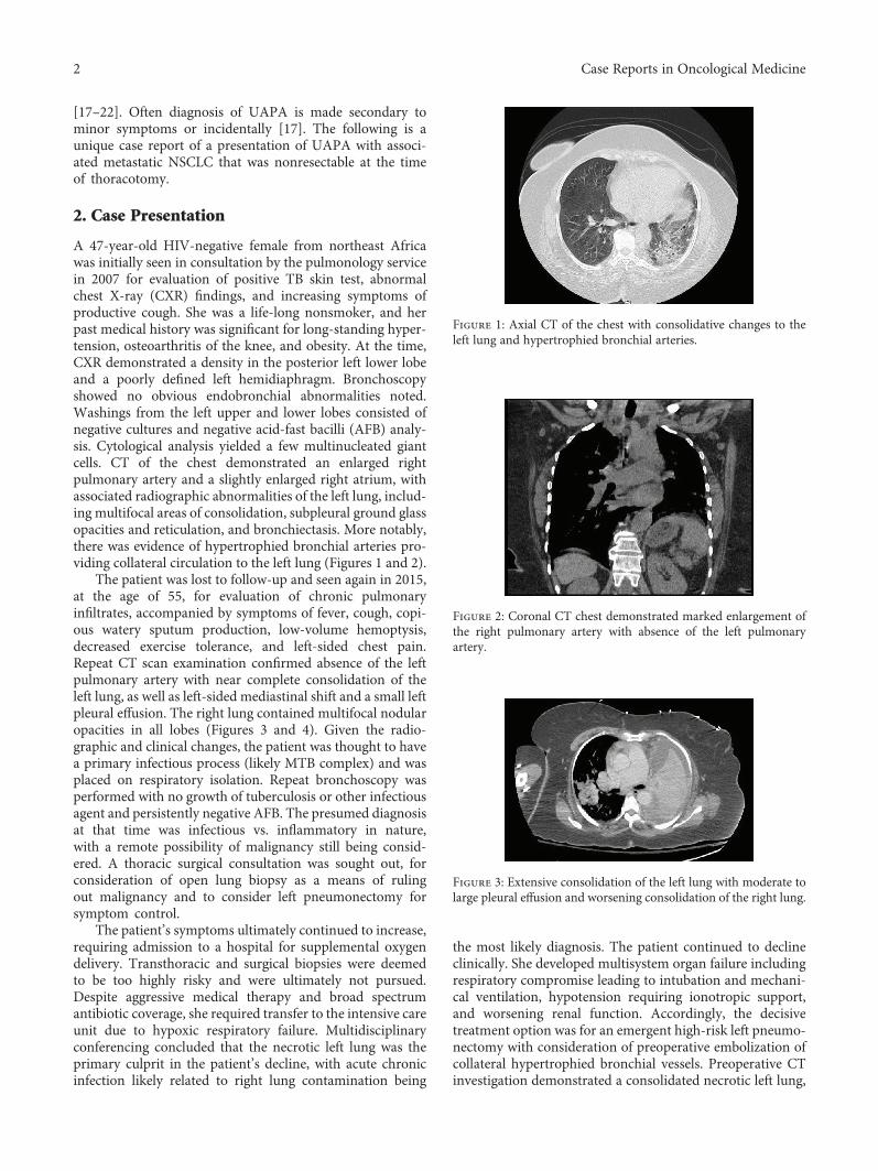
A 47-year-old HIV-negative female from northeast Africawas initially seen in consultation by the pulmonology servicein 2007 for evaluation of positive TB skin test, abnormalchest X-ray (CXR) findings, and increasing symptoms ofproductive cough. She was a life-long nonsmoker, and herpast medical history was significant for long-standing hyper-tension, osteoarthritis of the knee, and obesity. At the time,CXR demonstrated a density in the posterior left lower lobeand a poorly defined left hemidiaphragm. Bronchoscopyshowed no obvious endobronchial abnormalities noted.Washings from the left upper and lower lobes consisted ofnegative cultures and negative acid-fast bacilli (AFB) analy-sis. Cytological analysis yielded a few multinucleated giantcells. CT of the chest demonstrated an enlarged rightpulmonary artery and a slightly enlarged right atrium, withassociated radiographic abnormalities of the left lung, includ-ing multifocal areas of consolidation, subpleural ground glassopacities and reticulation, and bronchiectasis. More notably,there was evidence of hypertrophied bronchial arteries pro-viding collateral circulation to the left lung (Figures 1 and 2).
The patient was lost to follow-up and seen again in 2015,at the age of 55, for evaluation of chronic pulmonaryinfiltrates, accompanied by symptoms of fever, cough, copi-ous watery sputum production, low-volume hemoptysis,decreased exercise tolerance, and left-sided chest pain.Repeat CT scan examination confirmed absence of the leftpulmonary artery with near complete consolidation of theleft lung, as well as left-sided mediastinal shift and a small leftpleural effusion. The right lung contained multifocal nodularopacities in all lobes (Figures 3 and 4). Given the radio-graphic and clinical changes, the patient was thought to havea primary infectious process (likely MTB complex) and wasplaced on respiratory isolation. Repeat bronchoscopy wasperformed with no growth of tuberculosis or other infectiousagent and persistently negative AFB. The presumed diagnosisat that time was infectious vs. inflammatory in nature,with a remote possibility of malignancy still being consid-ered. A thoracic surgical consultation was sought out, forconsideration of open lung biopsy as a means of rulingout malignancy and to consider left pneumonectomy forsymptom control.
The patient’s symptoms ultimately continued to increase,requiring admission to a hospital for supplemental oxygendelivery. Transthoracic and surgical biopsies were deemedto be too highly risky and were ultimately not pursued.Despite aggressive medical therapy and broad spectrumantibiotic coverage, she required transfer to the intensive careunit due to hypoxic respiratory failure. Multidisciplinaryconferencing concluded that the necrotic left lung was theprimary culprit in the patient’s decline, with acute chronicinfection likely related to right lung contamination being
the most likely diagnosis. The patient continued to declineclinically. She developed multisystem organ failure includingrespiratory compromise leading to intubation and mechani-cal ventilation, hypotension requiring ionotropic support,and worsening renal function. Accordingly, the decisivetreatment option was for an emergent high-risk left pneumo-nectomy with consideration of preoperative embolization ofcollateral hypertrophied bronchial vessels. Preoperative CTinvestigation demonstrated a consolidated necrotic left lung,
Figure 1: Axial CT of the chest with consolidative changes to theleft lung and hypertrophied bronchial arteries.
Figure 2: Coronal CT chest demonstrated marked enlargement ofthe right pulmonary artery with absence of the left pulmonaryartery.
Figure 3: Extensive consolidation of the left lung with moderate tolarge pleural effusion and worsening consolidation of the right lung.
2 Case Reports in Oncological Medicine

with moderate left pleural effusion and areas of thicken-ing of the left pleura. On cardiopulmonary testing, echo-cardiogram revealed left-sided ejection fraction of 65-70%,with preserved right ventricular systolic function, withoutany significant tricuspid regurgitation. Quantitative lungventilation-perfusion scanning identified 1% ventilationand perfusion to the left lung, with >90% right-sidedlung perfusion. The patient and her family were quoteda near 50-90% surgical mortality, in the face of imminentdeath without intervention.
The patient was taken to the operating room and placedin the right lateral decubitus position, and an endobronchialblocker was placed through her single-lumen endotrachealtube and positioned in the left mainstem bronchus. A pos-terolateral thoracotomy was performed, after harvesting aserratus anterior muscle flap. Upon entry into the pleuralspace, it was evident that the left lung was cemented to theparietal pleura with dense thick adhesions. Minimal dissec-tion was undertaken in order to identify an area of the pleuralcavity that was free. Five hundred milliliters of serous pleuralfluid was drained. Additionally, there was evidence of multi-ple small pleural nodules dispersed throughout the chest cav-ity and the hilum. Frozen section analysis of these pleuraldeposits confirmed the presence of metastatic adenocarci-noma, confirming the presence of stage IV lung cancer. Assuch, the planned left pneumonectomy was aborted and thechest was closed with an indwelling chest tube.
She was transferred back to the ICU with continuedmechanical ventilation. The working diagnosis at that pointwas diffuse metastatic lung cancer with lymphangitic carci-nomatosis and superimposed infection. After the familywas informed of the operative findings, a palliative approachwas undertaken. Comfort measures were ensured. Mechani-cal ventilation was weaned off as care was being withdrawn,and the patient peacefully passed away.
Pleural fluid analysis identified the presence of malignantcells suggestive of adenocarcinoma. Final pathologic analysisof intraoperative specimens demonstrated pleural tissuecomprised of tall columnar tumor cells with focal necrosis,infiltrated by adenocarcinoma (thyroid transcription factor-1 positive) with predominantly papillary features. Epi-dermal growth factor receptor (EGFR) mutation was notidentified (wild-type allele) by polymerase chain reaction
(PCR) analysis using the EntroGen EGFR Kit Exons 18,19, 20, and 21. In addition, ALK rearrangement was notidentified.
3. Literature Review and Discussion
Several interesting factors contribute to the diagnostic com-plexity of this case. Attempts at invasive testing were madein order to determine the possibility of malignancy. Due topatient delays and worsening clinical status, this ultimatelynever occurred. Similarly, as a consequence of the patient’sdeteriorating critical state, appropriate lung cancer staginginvestigation was not performed, including positron emis-sion topography (PET) scan, invasive mediastinal staging,and cranial imaging. The diagnostic yield of these investiga-tions in the clinical context is uncertain. Overall, the preoper-ative determination of malignancy was challenged by twoclinical features. Firstly, more than three separate broncho-scopic evaluations with lavages and brushings did not dem-onstrate the presence of malignancy—a rare phenomenonin a patient with bronchorrhea and such diffuse disease. Sec-ondly, the protracted course of the presentation (spanningover 8 years) would be unusual for a malignant process.
This case represents the only report in the literature ofdiffuse metastatic lung cancer associated with UAPA andhighlights the challenge of determining a malignant diagno-sis in the setting of consolidated lung secondary to pulmo-nary arterial agenesis. To our knowledge, this is also theonly case of UAPA associated lung cancer in a lifelong non-smoker. To date, there have been 6 reported cases of UAPAwith secondarily associated non-small-cell lung cancer(NSCLC) [17–22]. In all but one case, the primary lungmalignancy was completely resected via pneumonectomy orlobectomy. The association was first reported in 1975 byMancebo and Wanner, based on the presence of metastaticundifferentiated carcinoma in mediastinal lymph nodes asnoted on mediastinoscopy. Accordingly, pulmonary resec-tion was not undertaken [19]. Roman et al. in 1995 andMakdisi et al. in 2015 both reported successful pneumonec-tomy for primary NSCLC with ipsilateral UAPA [18, 20].The latter was the first report of multifocal lung cancer inthe setting of pulmonary artery agenesis. In 2011, Anstadtet al. presented the only case of ipsilateral lobectomy for lungcancer in the context of UAPA [22]. In contrast, Ito et al.reported the only case of postradiotherapy contralateralpulmonary resection (right middle lobectomy) of the normallung in the setting of left-sided UAPA, outlining the possibil-ity of surgical intervention on the nonaffected lung [17].
The causal association between UAPA and lung cancerhas yet to be completely defined. Several plausible explana-tions have been provided. The possibility of structural abnor-malities in a hypoplastic underperfused lung serving as anidus for malignant transformation has been previously sug-gested [21]. More recently, the role of chronic hypoxia as animpetus for DNA damage and the development of lungmalignancy has been highlighted [23]. A hypoxic state atthe core of a hypoperfused lung can lead to the release ofreactive oxygen species (ROS), which in turn induce cellproliferation via the activation of the EGFR pathway [24, 25].
Figure 4: Coronal views with clear complete consolidation of theleft lung, left pleural effusion, and worsening consolidative andnodular changes of the right lung.
3Case Reports in Oncological Medicine

4. Conclusion
Taken together, this case depicts another rare example ofisolated UAPA with associated primary lung malignancy.It represents the seventh reported case in the literatureand is the only presentation associating pulmonary arte-rial agenesis with metastatic lung cancer leading to acuterespiratory failure and death. When faced with the uncom-mon scenario of UAPA, clinicians must actively considerthe possibility of concomitant malignancy with expeditedresection (when appropriate) after complete diagnosticconfirmation and staging.
Conflicts of Interest
The authors declare that they have no conflicts of interest.
References
[1] L. A. Bockeria, O. A. Makhachev, T. K. Khiriev, and M. A.Abramyan, “Congenital isolated unilateral absence of pulmo-nary artery and variants of collateral blood supply of theipsilateral lung,” Interactive CardioVascular and ThoracicSurgery, vol. 12, no. 3, pp. 509-510, 2011.
[2] D. Bouros, P. Pare, P. Panagou, K. Tsintiris, and N. Siafakas,“The varied manifestation of pulmonary artery agenesis inadulthood,” Chest, vol. 108, no. 3, pp. 670–676, 1995.
[3] I. S. Kadir, J. Thekudan, A. Dheodar, M. T. Jones, and K. B.Carroll, “Congenital unilateral pulmonary artery agenesisand aspergilloma,” The Annals of Thoracic Surgery, vol. 74,no. 6, pp. 2169–2171, 2002.
[4] O. Fraentzel, “Ein fall von abnormer communication der aortamit der arteria pulmonalis,” Archiv für Pathologische Anato-mie und Physiologie und für Klinische Medicin, vol. 43, no. 3,pp. 420–426, 1868.
[5] J. O. Finney Jr. and R. N. Finchum, “Congenital unilateralabsence of the left pulmonary artery with right aortic arch arida normal conus,” Southern Medical Journal, vol. 65, no. 9,pp. 1079–1082, 1972.
[6] J. G. Shakibi, H. Rastan, I. Nazarian, M. Paydar, I. Aryanpour,and B. Siassi, “Isolated unilateral absence of the pulmonaryartery: review of the world literature and guidelines for surgicalrepair,” Japanese Heart Journal, vol. 19, no. 3, pp. 439–451, 1978.
[7] R. C. Bahler, P. Carson, E. Traks, A. Levene, and D. Gillespie,“Absent right pulmonary artery: Problems in diagnosis andmanagement,” The American Journal of Medicine, vol. 46,no. 1, pp. 64–71, 1969.
[8] A. D. J. Ten Harkel, N. A. Blom, and J. Ottenkamp, “Isolatedunilateral absence of a pulmonary artery,” Chest, vol. 122,no. 4, pp. 1471–1477, 2002.
[9] N. Griffin, L. Mansfield, K. C. Redmond et al., “Imagingfeatures of isolated unilateral pulmonary artery agenesispresenting in adulthood: a review of four cases,” ClinicalRadiology, vol. 62, no. 3, pp. 238–244, 2007.
[10] W. R. Krall and Y. Ploy-Song-Sang, “Unilateral pulmonaryartery aplasia presenting with chest pain and pleural effusion,”Southern Medical Journal, vol. 73, no. 2, pp. 233–235, 1980.
[11] P. Kruzliak, R. P. Syamasundar, M. Novak, O. Pechanova,and G. Kovacova, “Unilateral absence of pulmonary artery:pathophysiology, symptoms, diagnosis and current treatment,”
Archives of Cardiovascular Diseases, vol. 106, no. 8-9, pp. 448–454, 2013.
[12] K. Gupta, J. J. Livesay, and R. Lufschanowski, “Absent rightpulmonary artery with coronary collaterals supplying theaffected lung,” Circulation, vol. 104, no. 4, pp. E12–E13, 2001.
[13] R. J. Moreno-Cabral, J. McNamara, V. J. Reddy, andP. Caldwell, “Unilateral absent pulmonary artery: surgicalrepair with a new technique,” The Journal of Thoracic andCardiovascular Surgery, vol. 102, no. 3, pp. 463–465, 1991.
[14] C. C. Canver, J. D. Pigott, and R. M. Mentzer, “Neonatalpneumonectomy for isolated unilateral pulmonary arteryagenesis,” The Annals of Thoracic Surgery, vol. 52, no. 2,pp. 294-295, 1991.
[15] N. Sreeram, A. Asante-Korang, and E. Ladusans, “Distal ductalorigin of the right pulmonary artery: prospective diagnosisand primary repair in infancy,” International Journal ofCardiology, vol. 35, no. 2, pp. 272–274, 1992.
[16] T. Ohta, “Congenital absence of the right pulmonary artery—a case report and review,” The Tokai Journal of Experimentaland Clinical Medicine, vol. 8, no. 1, pp. 97–104, 1983.
[17] M. Ito, Y. Yamashita, H. Harada, and K. I. Omori, “Unilateralabsence of the left pulmonary artery accompanied by rightlung cancer,” The Annals of Thoracic Surgery, vol. 90, no. 1,pp. e6–e8, 2010.
[18] J. Roman and S. Jones, “Case report: congenital absence of theleft pulmonary artery accompanied by ipsilateral emphysemaand adenocarcinoma,” The American Journal of the MedicalSciences, vol. 309, no. 3, pp. 188–190, 1995.
[19] A. Mancebo and A. Wanner, “Lung tumor in a patient withcongenital unilateral hypoplasia of the pulmonary artery,”Chest, vol. 68, no. 6, pp. 846-847, 1975.
[20] G. Makdisi, E. S. Edell, J. J. Maleszewski, J. R. Molina, andC. Deschamps, “Pulmonary artery agenesis associated withemphysema and multiple invasive non-small cell lung can-cers,” The Annals of Thoracic Surgery, vol. 99, no. 6,pp. 2192–2195, 2015.
[21] Y. Watanabe, J. Shibuya, M. Handa et al., “Unilateral absence ofthe right pulmonary artery accompanied by right lung cancer,”The Annals of Thoracic Surgery, vol. 100, no. 3, p. 1113, 2015.
[22] M. P. Anstadt, C. J. Wozniak, S. S. K. Kathula, and B. D.Sarodia, “Lobectomy for lung carcinoma with ipsilateral pul-monary artery agenesis,” vol. 183, article A3836, AmericanThoracic Society International Conference Abstracts, 2011,abstract. http://www.atsjournals.org/doi/abs/10.1164/ajrccm-conference.2011.183.1_MeetingAbstracts.A3836.
[23] K. Srujana, S. S. Begum, K. N. Rao, G. S. Devi, A. Jyothy, andM. H. Prasad, “Application of the comet assay for assessmentof oxidative DNA damage in circulating lymphocytes oftetralogy of Fallot patients,” Mutation Research/Fundamentaland Molecular Mechanisms of Mutagenesis, vol. 688, no. 1-2,pp. 62–65, 2010.
[24] H. Lemjabbar-Alaoui, S. S. Sidhu, A.Mengistab,M.Gallup, andC. Basbaum, “TACE/ADAM-17 phosphorylation by PKC-epsilon mediates premalignant changes in tobacco smoke-exposed lung cells,” PLoSOne, vol. 6, no. 3, article e17489, 2011.
[25] A. Franovic, L. Gunaratnam, K. Smith, I. Robert, D. Patten,and S. Lee, “Translational up-regulation of the EGFR by tumorhypoxia provides a nonmutational explanation for its overex-pression in human cancer,” Proceedings of the National Acad-emy of Sciences of the United States of America, vol. 104, no. 32,pp. 13092–13097, 2007.
4 Case Reports in Oncological Medicine

Stem Cells International
Hindawiwww.hindawi.com Volume 2018
Hindawiwww.hindawi.com Volume 2018
MEDIATORSINFLAMMATION
of
EndocrinologyInternational Journal of
Hindawiwww.hindawi.com Volume 2018
Hindawiwww.hindawi.com Volume 2018
Disease Markers
Hindawiwww.hindawi.com Volume 2018
BioMed Research International
OncologyJournal of
Hindawiwww.hindawi.com Volume 2013
Hindawiwww.hindawi.com Volume 2018
Oxidative Medicine and Cellular Longevity
Hindawiwww.hindawi.com Volume 2018
PPAR Research
Hindawi Publishing Corporation http://www.hindawi.com Volume 2013Hindawiwww.hindawi.com
The Scientific World Journal
Volume 2018
Immunology ResearchHindawiwww.hindawi.com Volume 2018
Journal of
ObesityJournal of
Hindawiwww.hindawi.com Volume 2018
Hindawiwww.hindawi.com Volume 2018
Computational and Mathematical Methods in Medicine
Hindawiwww.hindawi.com Volume 2018
Behavioural Neurology
OphthalmologyJournal of
Hindawiwww.hindawi.com Volume 2018
Diabetes ResearchJournal of
Hindawiwww.hindawi.com Volume 2018
Hindawiwww.hindawi.com Volume 2018
Research and TreatmentAIDS
Hindawiwww.hindawi.com Volume 2018
Gastroenterology Research and Practice
Hindawiwww.hindawi.com Volume 2018
Parkinson’s Disease
Evidence-Based Complementary andAlternative Medicine
Volume 2018Hindawiwww.hindawi.com
Submit your manuscripts atwww.hindawi.com