metabolomics and bioactivity guided isolation of secondary...
TRANSCRIPT
ISSN: 2357-0547 (Print) Research Article / JAPR
ISSN: 2357-0539 (Online) Tawfik et al., 2017, 1 (1), 66-74
www.http://aprh.js.iknito.com
66
Metabolomics and Bioactivity Guided Isolation of Secondary Metabolites from the
Endophytic Fungus Chaetomium sp.
Nashwa F. Tawfik1,2, Ahmed F. Tawfike1, Randa Abdou1,3, Grainne Abbott2, Usama R. Abdelmohsen4,
RuAngelie Edrada-Ebel2 and Eman G. Haggag1*
1Department of Pharmacognosy, Faculty of Pharmacy, Helwan University, Cairo, Egypt, 11795.
2Strathclyde Institute of Pharmacy and Biomedical Science, University of Strathclyde, 161 Cathedral street, Glasgow G4
0NR, Scotland, United Kingdom. 3Faculty of Pharmacy Umm Al Qura University, Mekkah, KSA.
4Faculty of Pharmacy, Minia University, Minia, Egypt.
*Corresponding author: Eman G. Haggag 1Department of Pharmacognosy Faculty of Pharmacy, Helwan University, Cairo, 11795, Egypt, Tel.: +201000023022
E-mail address: [email protected]
Submitted on: 18-11-2016; Revised on: 01-12-2016; Accepted on: 03-12-2016
ABSTRACT
Objectives: the aim of this study is to explore the secondary metabolites produced by the endophytic fungus Chaetomium
sp. isolated from Scencio stapeliiformis (E.Phillips) as well as investigate the anticancer and antimicrobial activity of
crude extracts, fractions and pure compounds. Methods: An endophytic fungus (Chaetomium sp.) was isolated from the
arial part of S. stapeliiformis (from Giza, Egypt). DNA sequencing analysis, morphological and chemotaxonomy
investigations were used for taxonomic identification. Metabolomics tools and dereplication studies were employed to
choose the optimum growth medium and conditions that produce the most significant metabolites. The crude extract of the
optimal fungal culture of Chaetomium sp. was then fractionated using flash chromatography and medium pressure liquid
chromatography (MPLC). The structure of the isolated compounds was determined on the basis of 1D, 2D NMR and mass
spectrometry (HR-ESIMS) analysis. Results: The Metabolomics and bioassay-guided isolation afforded five pure
compounds; p-hydroxybenzaldehyde (1), Uracil (2), 3-benzyl-6-isobutyl piperazine-2,5-dione (3), Cyclo (L-Alanin-L-
leucin) (4) and Cyclo-(L-proline-L-leucine) (5). Multivariate data analysis highlighted the most significant metabolites
contributed to the measured bioactivity. All fungal extracts were tested for the anticancer activity but extract of 30 days
liquid culture of Chaetomium showed the most anticancer activity. The pure compounds were tested for their anticancer
and antimicrobial activities. Compounds 3 and 5 exhibited a significant anti-trypanosomal activity while compounds 1, 2
and 5 effectively inhibited the growth of E-coli and Staphylococcus aureus. Conclusion: A combination of metabolomic-
and bioassay-guided protocol can efficiently predict the putative biologically active metabolites during the first stage of
fractionation.
Keywords: Antimicrobial activity, Antitrypanosomal activity, Chaetomium sp., Dereplication, Endophytes, Metabolomics,
Senecio stapeliiformis.
INTRODUCTION
Senecio represents the largest genus of the
family Asteraceae, and has more than 1500 species of
herbs, shrubs, vines and trees1. Senecio species have
been used in folk medicine in the treatment of wounds,
chest pain, cough, fever and runny nose. It was reported
to have a great gastrointestinal protective activity
against ulcers2,3. Moreover, some studies mentioned the
cytotoxic activity of different species of Senecio4.
Chaetomium, an endophytic fungus isolated from S.
stapeliiformis, belongs to Ascomycota of the family
Chaetomiaceae. It is a large genus comprising over 100
species. Several strains of Chaetomium are found in the
ISSN: 2357-0547 (Print) Research Article / JAPR
ISSN: 2357-0539 (Online) Tawfik et al., 2017, 1 (1), 66-74
www.http://aprh.js.iknito.com
67
soil, plants debris5. Endophytic fungi are a highly
diverse group of fungi capable of living symbiotically
inside plant tissue without causing apparent symptoms
of diseases6. Endophytes might be involved in the
biosynthesis of plant products; however, they might
also be the producers themselves of many substances of
potential use to the modern medicine, agriculture and
pharmaceutical industry7,8. An area of major interest to
us is to explore endophytic secondary metabolites as
novel anticancer and antimicrobial agents.
Since sleeping sickness (Human African
Trypanosomiasis "HAT") is an endemic disease in
thirty African countries with the population at risk
being about 60 million, this has driven us to search for a
powerful antitrypanosome of natural source. HAT is a
vector-borne parasitic disease caused by infection with
protozoan parasites belonging to the
genus Trypanosoma which are transmitted to humans
by tsetse fly (Glossina genus) bites9. It has two stages:
the first is the haemolymphatic stage which lasts for one
to three weeks, followed by the chronic stage in which
trypanosomes cross the blood–brain barrier to invade
the central nervous system resulting to chronic
meningo-encephalitis and eventually leads to
encephalopathy10.
Metabolomics is the technological tool
designed to deliver general qualitative and quantitative
profiles of metabolites in organisms exposed to various
conditions. Plants and microorganisms produce many
metabolites with different chemistry and bioactivity
under stress conditions. Metabolomics displays extra
information to figure out these complex relationships
between the endophytes and their host plants which aids
to discover novel bioactive natural components11. The
metabolome is the complete set of small molecules
found in a cell, tissue or organism at a certain point in
time. Dereplication is the process of testing sample
mixtures that are active in screening in order to
recognize the novel compounds from the active
substances that have already been studied.
Dereplication was accomplished by employing
differential expression analysis softwares like MZmine
which involves dictionary of natural products database
(DNP) to aid compound identification11. By using
combinations of analytical, statistical and dereplication
methods, the bioassay-guided isolation route is getting
shorter and rapid dereplication of known activities is
rapidly delivered12.
MATERIALS AND METHODS General instruments
1H-, 13C- and 2D-NMR spectra were recorded
at 25ᵒC in DMSO-d6 on JNM-LA400 NMR
spectrometer, JEOL, Japan and the magnet NMR
AS400 model EUR0034 from Oxford Instruments,
England at Strathclyde Institute of Pharmacy and
Biomedical Science and an AVANCE-III 600
instrument with a 14.1 T Bruker UltraShield magnet
at Chemistry Department, Faculty of science,
Strathclyde University. ESI-HRMS was measured
using FTHRMS-Finnigan LTQ Orbitrap or Exactive
mass spectrometer (Thermo Scientific). HPLC
analysis was carried out using Dionex UltiMate
3000-ThermoScientific Exactive system instrument,
Germany. Crude extracts were initially fractionated
using medium pressure liquid chromatography
(MPLC) from BÜCHI, MPLC instrument was the
Sepacore Purification System with Versaflash
column stand. The Reveleris® Flash Forward system
of Grace Davison Discovery Sciences (Illinois,
United States) was also used for further isolation,
which is characterized of having two detectors, an
evaporative light scattering detector (ELSD) and a
UV detector (wavelength range: 200-500 nm). The
fractions were investigated on normal phase thin
layer chromatography plates (TLC silica gel 60 F254),
reverse phase TLC plates (TLC silica gel 60 RP-18
F254S) and fractionated using preparative TLC plates
(TLC silica gel 60 F254 on 20x20 cm aluminium
sheets) from Merck KGaA, Germany. Spots were
visualized under UV lamp (λ 254 nm and λ 380 nm)
and after spraying with anisaldehyde and heating
chromatograms till colour development.
LC-MS spectra were viewed using Thermo
Xcalibur 2.1 (Thermo Scientific, Germany). To convert
the raw data into separate positive and negative
ionization files, Ms converter software was used. The
files were then imported to the data mining software
MZmine 2.10 forpeak picking, deconvolution,
deisotoping, alignment and formula prediction11. Macro
file with built in databases was written in Excel, used to
combine positive and negative MS files and for further
clean-up of media components13. The databases used
for the identification of compounds were the Dictionary
of Natural Products (DNP) 2015, MestReNova
(MNova) 2.10 by Mestrelab Research, S.L, (Santiago
de Compostela, Spain) was used to process all NMR
data and SIMCA 14(Umetrics AB, Umeå, Sweden) was
used for multivariate data analysis.
For microbiological work, the laminar flow
hood (BioMAT2) was purchased from Medical Air
Technology, UK. The stand incubator (Incu-160S) used
for agar plates was from SciQuip Ltd., Shropshire. The
homogenizer (IKA T18 Basic Ultra-Turrax) and
handheld homogenizer (Ultra-Turrax T8) were obtained
from IKA Labortechnik, Germany.
Plant material
Fresh plant (Senecio stapeliiformis
E.Phillips) was collected from the Orman Botanical
Garden in Giza, Egypt and identified by; Dr. Therese
L. Yousef, senior taxonomist and Engineer Mervat
ISSN: 2357-0547 (Print) Research Article / JAPR
ISSN: 2357-0539 (Online) Tawfik et al., 2017, 1 (1), 66-74
www.http://aprh.js.iknito.com
68
A. Hasan, herbarium curator at Orman Botanical
Garden. Fresh plant materials including all arial parts
of the plant were collected a day before isolation of
fungal strains, kept in zipped plastic bags under 4̊ C
for the isolation work.
Culture media for isolated endophytes
Wickerham liquid medium (yeast extract 3.0
gm, malt extract 3.0 gm, peptone 5.0 gm, glucose 10.0
gm and distilled water to 1L with pH adjusted at 7.4)
and solid medium composed of 100 gm Rice and 100
ml distilled water, were used as culture media for the
isolated endophytes.
Cell lines and culture media for cytotoxic assay
Lung cancer cells (A549), Prostatic cancer
cells (PC3), breast cancer (ZR75), ovarian cancer cells
(A278O) and normal epithelial cells derived from
human prostate (PNT2 cells) were purchased from
ECACC (Sigma-Aldrich, Dorset, UK). A549 and PC3
cells were cultured in Dulbecco’s Modified Eagle’s
Medium (DMEM), while PNT2, ZR75 and A2780 cells
were cultured in RPMI 1640 media; both were
supplemented with 10% (v/v) fetal bovine serum, 2 mM
glutamine and 50 µ g/mL penicillin/streptomycin
solution (all Invitrogen, Paisley, UK). All cells were
maintained in a humidified incubator at 37 ºC in the
presence of 5% CO2. Cells were routinely passaged at
90%–95% confluence.
Isolation of the endophytes
The Arial part of the plant was rinsed with
sterilized distilled water twice. In order to eliminate
surface contaminating microbes, sterilization was
carried out by immersing leaves and stems in 70%
isopropanol (2 min x 2) followed by rinsing again twice
with sterilized distilled water. Using a sterile scalpel a
small segment of leave and stem tissue (1 cm in length)
was cleaned from outer tissue, the inner tissues were
carefully dissected under sterile conditions and placed
on malt agar plate containing antibiotic to suppress
bacterial growth (medium composition: 15 gm agar
(Oxoid), 15 gm malt extract (Oxoid) and
chloramphenicol (Acros organics, purity> 98%) in
distilled water), pH was adjusted to (7.4-7.8) and
incubated at 30ºC. After 3-4 weeks, hyphal tips of the
fungi were removed and transferred to fresh MA
medium. Plates were prepared in duplicates to eliminate
the possibility of contamination. Pure strains were
isolated by repeated inoculation. The purified fungus
was later transferred to the liquid medium for scaling
up14.
Identification of fungal strain The isolated fungal strains were identified
according to molecular biological procedure by DNA
extraction, amplification and sequencing of the ITS
region15. BLAST search of the FASTA sequence was
performed with the option “nr”, including GenBank,
Ref Seq Nucleotides, EMBL, DDBJ and PDB
sequences on the BLAST homepage, (NCBI, Bethesda,
USA) using accession number KC427016.1.
Seven endophytic fungi were isolated from
different parts of S. stapeliiformis identified as
Trichospherical sp., Chaetomium sp., Chaetomium
megalocarpum, Asperagillus sp., Rhizopus sp.,
Ceratobasidium sp and Microascus sp.
Small-scale extraction for screening, metabolomics
profiling and dereplication
A plate of each fungal species was transferred
into 250 ml flask, then macerated with ethyl acetate
(200 ml) overnight followed by homogenization and
filtration. The filtrate was then dried under vacuum.
One mg of each extract was subjected to HRMS
analysis and 8-10 mg for NMR analysis for
metabolomics profiling and dereplication studies. A
sample of 1 mg/mL concentration of each fungal extract
was prepared in duplicate and sent to Strathclyde
Institute for Drug Research SIDR for bioassay
screening against ovarian cancer (A278O), lung cancer
(A549), prostatic cancer (PC3) and breast Cancer
(ZR75) cell lines.
Medium scale fermentation, extraction and isolation Fresh fungal cultures were transferred into
Erlenmeyer flasks (1L each) containing 500 ml of
Wickerham medium for liquid cultures prepared as
stated per materials and methods. The cultures were
then incubated at room temperature in static form for 30
days. Medium scale cultivation was carried out using 20
One-L Erlenmeyer flasks for liquid cultures, then 250
mL EtOAc was added to Erlenmeyer flasks containing
500 ml culture medium and left overnight to stop cell
growth. Culture media and mycelia were then
homogenized in the Ultraturrax for 10 min for cell
destruction, followed by vacuum filtration using a
Buchner funnel. The mycelium residue was discarded
while EtOAc culture filtrates were collected, pooled,
dried under vacuum, suspended in 200 mL H2O and
extracted with EtOAc (3 x 200 mL) using a separating
funnel 14.
Cytotoxic activity Cells were seeded in clear 96 flat-bottomed
plates and allowed to adhere overnight. Thereafter,
metabolite extracts and fractions were added at a final
concentration of 30 µg/mL, while for the pure
compounds at a concentration of 10 mM/mL and
allowed to incubate for 48 hours. Viability was
determined using Alamar Blue® (Thermo Fisher,
Paisley, UK), according to the manufacturer’s
ISSN: 2357-0547 (Print) Research Article / JAPR
ISSN: 2357-0539 (Online) Tawfik et al., 2017, 1 (1), 66-74
www.http://aprh.js.iknito.com
69
instructions and incubated for a further 6 h. The
resulting fluorescence was measured using a Wallac
Victor 2 1420 multi-label counter (Perkin Elmer,
Beaconsfield, UK), in fluorescence mode: excitation
560, emission 590. Vehicle treated control cells (media
with 0.3% DMSO) were considered 100% viable
against which metabolite extract treated cells (at a
concentration of 30 µg/mL, at least n = 2) and the pure
compounds treated cells (at concentration 10 mM/mL)
were compared. All results were confirmed
microscopically16.
Antitrypanosomal activity
Antitrypanosomal activity was tested
following the protocol of Huber and Koella17. Briefly,
104 trypanosomes per ml of the Trypanosoma brucei
brucei strain TC 221 were cultivated in Complete Baltz
Medium. Trypanosomes were tested in 96-well plate
chambers against different concentrations of test
substances at 0.25–50 µM in 1% DMSO to a final
volume of 200 µL. For controls, 1% DMSO as well as
parasites without any test compound were used
simultanously in each plate to show that DMSO did not
perturb the results. The plates were then incubated at 37
°C in an atmosphere of 5% CO2 for 24 h. After addition
of 20 µL of Alamar Blue, the activity was measured
after 48 and 72 h by light absorption using an MR 700
Microplate Reader (Dynatech, Chantilly, United States)
at a wavelength of 550 nm with a reference wavelength
of 650 nm. The IC50 values of the test compound were
quantified by linear interpolation of three independent
measurements.
Anti-microbial activity
The in vitro antimicrobial activity assessment
was carried out using a modified Kirby-Bauer disk
diffusion assay18, 19 against various pathogenic bacterial
strains (Staphylococcus aureus strain
12600, Escherichia coli strain 11775, and Fungi
(Candida albicans strain 7102). Standard discs of
Ampicillin (Antibacterial agent), Amphotericin B
(Antifungal agent) served as positive controls, while a
filter discs impregnated with 10 µL of solvent (DMSO)
was used as a negative control.
RESULTS
Compound 1
Brown sugary substance (8mg), 1H-NMR
(DMSO, 400 MHz) 13C-NMR (DMSO, 100 MHz) data
presented in table 1; ESIHRMS(pos): m/z
121.0296[M+H]+ (calcd. for C7H6O2) Thus compound
3 was assigned in accordance to the reported data 20
as p-hydroxybenzaldehyde.
Compound 2 Colorless needles (9 mg); 1H-NMR (DMSO,
400 MHz) 13C-NMR (DMSO, 100 MHz) data presented
in table 1; ESIHRMS(pos): m/z 113.03 [M+H]+ (calcd.
for C4H4N2O2). Thus compound 3 was assigned in
accordance to the reported data 21 as Uracil.
Compound 3
Colourless needles (9 mg); 1H-NMR (DMSO,
400 MHz) 13C-NMR (DMSO, 100 MHz) data presented
in table 1; ESIHRMS(pos): m/z 261.1598 [M+H]+
(calcd. for C15H20N2O2). Thus compound 3 was
assigned in accordance to the reported data 22 as 3-
benzyl-6-isobutyl piperazine-2,5-dione.
Compound 4
Colourless needles (7mg); 1H-NMR (DMSO,
400 MHz) 13C-NMR (DMSO, 100 MHz) data presented
in table 1; ESIHRMS(pos): m/z 185.1286 [M+H]+
(calcd. for C9H16N2O2). Thus compound 4 was
assigned in accordance to the reported data 21 as
Cyclo(L-Alanin-L-leucine)
Compound 5
White crystals (17.8 mg), 1H-NMR (DMSO,
400 MHz) 13C-NMR (DMSO, 100 MHz) data presented
in table 1; ESIHRMS(pos): m/z 211.1448 [M+H]+
(calcd. for C11H18N2O2). Thus compound 5 was
assigned in accordance to the reported data 23 as
Cyclo-(L-proline-L-leucine).
DISCUSSION
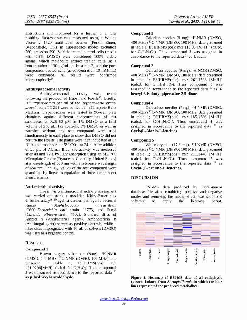
ESI-MS data produced by Excel-macro
database file after combining positive and negative
modes and removing the media effect, was sent to R
software to apply the heatmap script.
Figure 1. Heatmap of ESI-MS data of all endophytic
extracts isolated from S. stapeliiformis in which the blue
lines represented the produced metabolites.
ISSN: 2357-0547 (Print) Research Article / JAPR
ISSN: 2357-0539 (Online) Tawfik et al., 2017, 1 (1), 66-74
www.http://aprh.js.iknito.com
70
Table 1. 1HNMR and 13CNMR data of isolated compounds (1-5)
Atom
No. Compound (1) Compound (2) Compound (3) Compound (4) Compound (5)
δH (m, J in
Hz) ppm
δC δH (m, J in
Hz) ppm
δC δH (m, J
in Hz)
ppm
δC δH (m, J in
Hz) ppm
δC δH (m, J in
Hz) ppm
δC
1 - 164.4 10.83 - 8.09 - 8.10 - - 167.4
2 6.93(d, J=8.68 Hz)
132.7 - 151.1 - 166.7 - 168.8 - -
3 7.77(d, J=8.68
Hz)
116.4 11.03 - 4.17 56.0 3.77(m) 53.1 3.35( 2H,m) 45.0
4 - 129.3 - 164.9 8.13 - 8.12 - 1.92, 2.13(m) 28.0
5 7.77(d, J=8.68
Hz)
116.4 5.45 (d,
J=7.57Hz)
101.0 - 168.1 - 169.3 1.84( 2H, m) 22.7
6 6.93(d, J=8.68 Hz)
132.7 7.39 (dd, J=7.01Hz)
143.1 3.47(m) 52.7 3.86(q,,J=7.01Hz)
50.4 4.21(t) 59.1
7 9.79(s) 191.8 10.83 - 2.83,
3.13 (m)
38.9 1.46, 1.61(m) 42.9 - 170.8
8 - - - - - 136.6 1.82(m) 24.0 8.00 -
9 - - - - 7.27 128.6 0.87(3H,d,J=7Hz)
22.4 4.01 52.9
10 - - - - 7.13 130.0 0.87(3H,d,J=7
Hz)
23.5 1.37, 1.78 (m) 38.0
11 - - - - 7.23 127.2 1.27(3H, d,
J=7.01Hz)
20.1 1.89(m) 24.4
12 - - - - 7.13 130.0 - - 0.88(3H) 22.4
13 - - - - 7.27 128.6 - - 0.86(3H) 23.6
14 - - - - 0.74,
0.09
44.1 - - - -
15 - - - - 1.42 23.3 - - - -
16 - - - - 0.61(3H) 23.5 - - - -
17 - - - - 0.61(3H) 21.8 - - - -
The heatmap of all extracts showed that Chatomium
sp. and Trichospherical sp. fungal extracts were the
richest in metabolites of different mass range as
shown in figure 1. The cytotoxicity assay showed
that Chaetomium sp. was active against A549,
A278O and PC3 cell lines and non-toxic for the
normal cells PNT2A (Figure 2), implying that
Chaetomium Sp. could have a unique chemical and
biological fingerprints.
Chaetomium was then cultivated on small
scale solid and liquid cultures to test the optimum
growth condition producing the highest amount of
interesting metabolites. HRESI-MS data of crude
extracts of both rice (RC) and liquid (LC) culture
media of Chaetomium have been subjected to a
metabolomics workflow which begun with data
mining by MZmine. The heatmap for the processed
ESI-MS data of both RC and LC extracts of
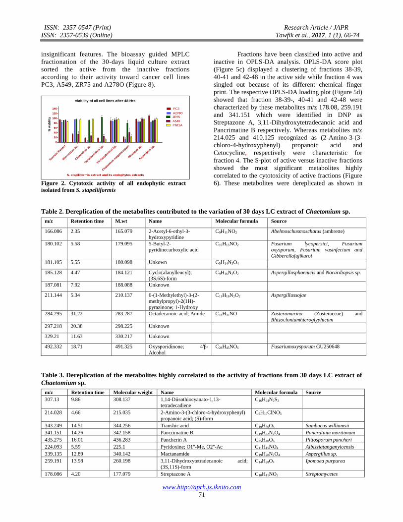
Chaetomium showed more abundancy of metabolites
inthe 30 days LC extract (Figure 3). Moreover,
Multivariate data analysis (MVDA) of different
culture extracts of Chaetomium, performed by
SIMCA-P V.14 software, discriminated 30-days LC
extracts from other fungal extracts as shown in the
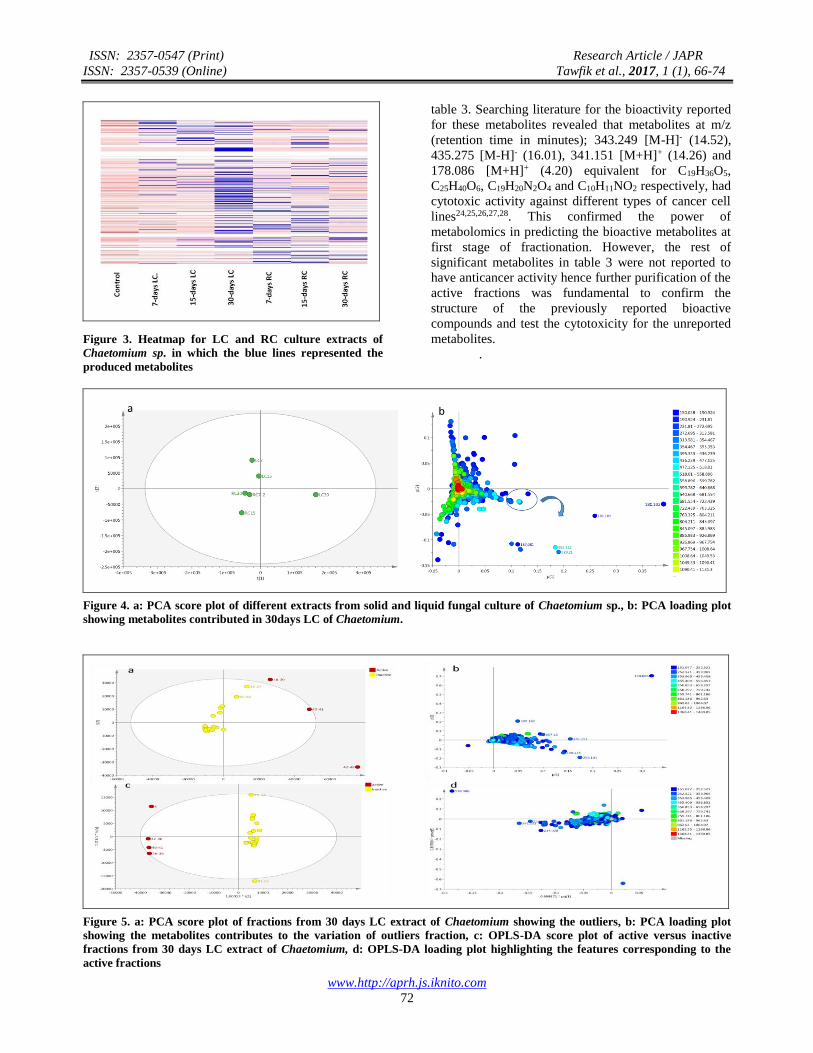
PCA score plot (Figure 4a) which was indicative of
the unique nature of the metabolites produced in LC-
30 extract.PCA loading plot (Figure 4b) illustrated the
metabolites which could be contributed to the variation
of 30 days LC extracts. These metabolites were
dereplicated by searching DNP 2015 as shown in table
2. Most of these metabolites were reported previously
in the literature however metabolites at m/z (retention
time in minutes); 181.105 [M+H]+ (5.55), 187.081 [M-
H]- (7.92), 297.218 [M-H]- (20.38) and 329.210 [M-H]-
(11.63) were not identified in the database. This was
motivating to work further on 30 days LC extract. Since
it was the most active against the selected cancer cell
line and showed no toxicity toward normal cells, 30
days LC was chosen for scale up and further isolation
work.
The thirty-day liquid culture extract of
Chaetomium was subjected to fractionation using
MPLC. The resulted fractions were imported into
SIMCA for MVDA. The PCA score plot (Figure 5a)
showed an outlying of fractions 35-37, 38-39, 40-41,
42-48 and 91-92. The PCA loading plot (Figure 5b)
showed the metabolites corresponding to the outlier
fractions which are further jack-knifed to remove the
ISSN: 2357-0547 (Print) Research Article / JAPR
ISSN: 2357-0539 (Online) Tawfik et al., 2017, 1 (1), 66-74
www.http://aprh.js.iknito.com
71
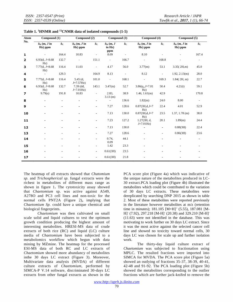
insignificant features. The bioassay guided MPLC
fractionation of the 30-days liquid culture extract
sorted the active from the inactive fractions
according to their activity toward cancer cell lines
PC3, A549, ZR75 and A278O (Figure 8).
viability of all cell lines after 48 Hrs
Scenci
o Ext
ract
Mic
roas
cus
Sp.
Chat
omiu
m S
p.
Cer
atib
asid
ium
Sp.
Trichosp
heric
al S
p.
Chat
omiu
m m
egal
ocarp
um
Rhiz
opus Sp.
Asp
erag
lus
Sp.
0
20
40
60
80
100
120
140 PC3
A278O
ZR75
A549
PNT2A
S. stapiliiformis extract and its endophytes extracts
% v
iab
ilit
y
Figure 2. Cytotoxic activity of all endophytic extract
isolated from S. stapeliiformis
Fractions have been classified into active and
inactive in OPLS-DA analysis. OPLS-DA score plot
(Figure 5c) displayed a clustering of fractions 38-39,
40-41 and 42-48 in the active side while fraction 4 was
singled out because of its different chemical finger
print. The respective OPLS-DA loading plot (Figure 5d)
showed that fraction 38-39-, 40-41 and 42-48 were
characterized by these metabolites m/z 178.08, 259.191
and 341.151 which were identified in DNP as
Streptazone A, 3,11-Dihydroxytetradecanoic acid and
Pancrimatine B respectively. Whereas metabolites m/z
214.025 and 410.125 recognized as (2-Amino-3-(3-
chloro-4-hydroxyphenyl) propanoic acid and
Cetocycline, respectively were characteristic for
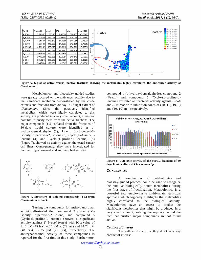
fraction 4. The S-plot of active versus inactive fractions
showed the most significant metabolites highly
correlated to the cytotoxicity of active fractions (Figure
6). These metabolites were dereplicated as shown in
Table 2. Dereplication of the metabolites contributed to the variation of 30 days LC extract of Chaetomium sp.
m/z Retention time M.wt Name Molecular formula Source
166.086 2.35 165.079 2-Acetyl-6-ethyl-3-hydroxypyridine
C9H11NO2 Abelmoschusmoschatus (ambrette)
180.102 5.58 179.095 5-Butyl-2-
pyridinecarboxylic acid
C10H13NO2 Fusarium lycopersici, Fusarium
oxysporum, Fusarium vasinfectum and
Gibberellafujikuroi
181.105 5.55 180.098 Unkown C5H14N3O4
185.128 4.47 184.121 Cyclo(alanylleucyl); (3S,6S)-form
C9H16N2O2 Aspergillusphoenicis and Nocardiopsis sp.
187.081 7.92 188.088 Unknown
211.144 5.34 210.137 6-(1-Methylethyl)-3-(2-
methylpropyl)-2(1H)-
pyrazinone; 1-Hydroxy
C11H18N2O2 Aspergillussojae
284.295 31.22 283.287 Octadecanoic acid; Amide C18H37NO Zosteramarina (Zosteraceae) and
Rhizocloniumhieroglyphicum
297.218 20.38 298.225 Unknown
329.21 11.63 330.217 Unknown
492.332 18.71 491.325 Oxysporidinone; 4'β-
Alcohol
C28H45NO6 Fusariumoxysporum GU250648
Table 3. Dereplication of the metabolites highly correlated to the activity of fractions from 30 days LC extract of
Chaetomium sp.
m/z Retention time Molecular weight Name Molecular formula Source
307.13 9.86 308.137 1,14-Diisothiocyanato-1,13-tetradecadiene
C16H24N2S2
214.028 4.66 215.035 2-Amino-3-(3-chloro-4-hydroxyphenyl)
propanoic acid; (S)-form
C9H10ClNO3
343.249 14.51 344.256 Tianshic acid C19H36O5 Sambucus williamsii
341.151 14.26 342.158 Pancrimatine B C19H22N2O4 Pancratium maritimum
435.275 16.01 436.283 Pancherin A C25H40O6 Pittosporum pancheri
224.093 5.59 225.1 Pyridoxine; O1''-Me, O2''-Ac C11H15NO4 Albizziatanganyicensis
339.135 12.89 340.142 Mactanamide C19H20N2O4 Aspergillus sp.
259.191 13.98 260.198 3,11-Dihydroxytetradecanoic acid;
(3S,11S)-form
C14H28O4 Ipomoea purpurea
178.086 4.20 177.079 Streptazone A C10H11NO2 Streptomycetes
ISSN: 2357-0547 (Print) Research Article / JAPR
ISSN: 2357-0539 (Online) Tawfik et al., 2017, 1 (1), 66-74
www.http://aprh.js.iknito.com
72
Figure 3. Heatmap for LC and RC culture extracts of
Chaetomium sp. in which the blue lines represented the
produced metabolites
table 3. Searching literature for the bioactivity reported
for these metabolites revealed that metabolites at m/z
(retention time in minutes); 343.249 [M-H]- (14.52),
435.275 [M-H]- (16.01), 341.151 [M+H]+ (14.26) and
178.086 [M+H]+ (4.20) equivalent for C19H36O5,
C25H40O6, C19H20N2O4 and C10H11NO2 respectively, had
cytotoxic activity against different types of cancer cell
lines24,25,26,27,28. This confirmed the power of
metabolomics in predicting the bioactive metabolites at
first stage of fractionation. However, the rest of
significant metabolites in table 3 were not reported to
have anticancer activity hence further purification of the
active fractions was fundamental to confirm the
structure of the previously reported bioactive
compounds and test the cytotoxicity for the unreported
metabolites.
.
Figure 4. a: PCA score plot of different extracts from solid and liquid fungal culture of Chaetomium sp., b: PCA loading plot
showing metabolites contributed in 30days LC of Chaetomium.
Figure 5. a: PCA score plot of fractions from 30 days LC extract of Chaetomium showing the outliers, b: PCA loading plot
showing the metabolites contributes to the variation of outliers fraction, c: OPLS-DA score plot of active versus inactive
fractions from 30 days LC extract of Chaetomium, d: OPLS-DA loading plot highlighting the features corresponding to the
active fractions
ISSN: 2357-0547 (Print) Research Article / JAPR
ISSN: 2357-0539 (Online) Tawfik et al., 2017, 1 (1), 66-74
www.http://aprh.js.iknito.com
73
Figure 6. S-plot of active versus inactive fractions showing the metabolites highly correlated the anticancer activity of
Chaetomium.
Metabolomics- and bioactivity guided studies
were greatly focused on the anticancer activity due to
the significant inhibition demonstrated by the crude
extracts and fractions from 30 day LC fungal extract of
Chaetomium. Since the putatively identified
metabolites, which were highly correlated to this
activity, are produced in a very small amount, it was not
possible to purify them from the active fractions. The
major compounds (1-5) isolated from the fractions of
30-days liquid culture were identified as p-
hydroxybenzaldehyde (1), Uracil (2),3-benzyl-6-
isobutyl piperazine-2,5-dione (3), Cyclo(L-Alanin-L-
leucin) (4) and Cyclo-(L-proline-L-leucine) (5)
(Figure 7), showed no activity against the tested cancer
cell lines. Consequently, they were investigated for
their antitrypanosomal and antimicrobial activity
Figure 7. Structure of isolated compounds (1-5) from
Chaetomium extract.
Testing the compounds for antitrypanosomal
activity illustrated that compound 3 (3-benzyl-6-
isobutyl piperazine-2,5-dione) and compound 5
(Cyclo-(L-proline-L-leucine) showed a significant
activity against T. brucei brucei with IC50 value of
5.17 μM (48 hrs), 6.26 μM at (72 hrs) and 14.73 µM
(48 hrs), 17.35 µM (72 hrs), respectively. The
antitrypanosomal activity of these compounds is
reported for the first time in this study. Furthermore,
compound 1 (p-hydroxybenzaldehyde), compound 2
(Uracil) and compound 5 (Cyclo-(L-proline-L-
leucine) exhibited antibacterial activity against E-coli
and S. aureus with inhibition zones of (10, 11), (9, 9)
and (10, 10) mm respectively.
Figure 8. Cytotoxic activity of the MPLC fractions of 30
days liquid culture of Chaetomium Sp.
CONCLUSION
A combination of metabolomic- and
bioassay-guided protocol could be used to recognize
the putative biologically active metabolites during
the first stage of fractionation. Metabolomics is a
powerful tool employing a multivariate statistical
approach which logically highlights the metabolites
highly correlated to the biological activity.
Metabolomics gave an access to predict the
significant metabolites that might be produced in a
very small amount, solving the mystery behind the
fact that purified major compounds are not found
active.
Conflict of Interest
The authors declare that they don’t have any
conflict of interest.
ISSN: 2357-0547 (Print) Research Article / JAPR
ISSN: 2357-0539 (Online) Tawfik et al., 2017, 1 (1), 66-74
www.http://aprh.js.iknito.com
74
REFERENCES
1. Nordestam, B. The Biology and Chemistry of the
Compositae. Academic Press Inc., London, UK,
1977.
2. Lakshmanan, H.; Raman J.; Nanjian R.
Gastroprotective effect of Senecio candicans DC on
experimental ulcer models. J. Ethnopharmacol.
2014, 140, 145-150.
3. York, T.; DeWet, H.; Van Vuuren S. F. Plants used
for treating respiratory infections in rural
maputaland, kwazulu-natal, South Africa. J.
Ethnopharmacol. 2011, 135, 696-710.
4. Rosa, T.; Monica R. T.; Marco B.; Federica M.;
Daiela D.; Micodemo G. P.; Giancarlo S.;
Francesco M. In-vitro cytoxic effect of Senecio
stabianus lactia (Astraceae) on human cancer cell
lines. Nat. Prod. Res. 2009, 23 (18), 1707-1718.
5. Rodriguez, K. S.; Guarro J., Mycologia. Three new
species of Chaetomium from soli. Mycologia 2002,
94, 116-126.
6. Petrini, O. Fungal Endophytes of Tree Leaves. In:
Microbial Ecology of Leaves. John, H.; Andrews, J.
H.; Hirano, S. S.; Eds.; Springer-Verlag, New York,
USA, 1991.
7. Strobel, G. A. Endophytes as sources of bioactive
products. Microbes. Infect. 2009, 5 (6), 535-544.
8. Arnold, A. E. Understanding the diversity of foliar
endophytic fungi: Progress, challenges, and
frontiers. Fungal Biol. 2007, 21, 51-66.
9. Barrett, M. P. The fall and rise of sleeping sickness.
Lancet 1999, 353 (9159), 1113-1114.
10. Enanga, B.; Burchmore R. J.; Stewart M. L.; Barrett
M. P. Sleeping sickness and the brain. Cell Mol.
Life Sci. 2002, 59 (5), 845-858.
11. Tawfike, A. F.; Viegelmann C.; and Ebel R. E.
Metabolomics and dereplication strategies in
natural products. Methods Mol. Biol. 2013, 1055,
227-244.
12. Ebadam, S. S.; Edrada R. E.; Lin W.; Proksch P.
Methods for isolation, purification and structural
elucidation of bioactive secondary metabolites from
marine invertebrates. Nat. Protoc. 2008, 3 (12),
1820-1831.
13. Macintyre, L.; Zhang T.; Viegelmann, C.; Martinez,
I. J.; Cheng, C.; Dowdells, C.; Abdelmohsen, U. R;
Gernert, C; Hentschel, U.; Ebel, R. E. Metabolomic
tools for secondary metabolite discovery from
marine microbial symbionts. Mar. Drugs. 2014, 12
(6), 3416-3448.
14. Debbab, A.; Aly, A. H.; Ebel, E. R.; Mueller, W. E.
G.; Mosaddak, M.; Hakikj, A.; Ebel, R.; Proksch, P.
Bioactive secondary metabolites from the
endophytic fungus Chaetomium sp. isolated from
Saliva officinalis growing in Morocco. Biotech.
Agronomy Soc. Environ. 2009, 13 (2), 229-234.
15. Kjer, J.; Debbab A.; Aly, A. H.; Proksch, P.
Methods for isolation of marine-derived endophytic
fungi and their bioactive secondary products. Nat.
Protoc. 2010, 5 (3), 479-490.
16. Kevin, L. M.; Debra, B.; Guðmundur, Ó. H.; Eva,
K.; Margrét, E. Á.; Louise, C. Y.; David, H. G.;
Ebel, R. E.; Katherine R. D. Using molecular
networking for microbial secondary metabolite
bioprospecting. Metabolites 2016, 6 (2), 1-18.
17. Huber, W.; Koella, J. C. A comparison of three
methods of estimating EC50 in studies of drug
resistance of malaria parasites. Acta. Trop. 1993, 55
(4), 257-261.
18. Bauer, A. W.; Kirby, W. M.; Sherris, J. C.; Turck,
M. Antibiotic susceptibility testing by a
standardized single disk method. Am. J. Clin.
Pathol. 1966, 45 (4), 493-496.
19. Pfaller, A. M.; Burmeister, L.; Bartlett, M. S.;
Rinaldi, M. G. Multicenter evaluation of four
methods of yeast inoculum preparation. J. Clin.
Microbiol. 1988, 26 (8), 1437-1441.
20. Kim, H.; Ralph, J.; Lu, F.; Ralph, S. A.; Boudet, A.
M.; MacKay, J. J.; Sederoff, R. R.; Ito, T.; Kawai,
S.; Ohashi, H.; Higuchi, T. NMR analysis of lignins
in cad-deficient plants. Part 1. Incorporation of
hydroxycinnamaldehydes and hydroxy-
benzaldehydes into lignins. Org. Biomol. Chem.
2003, 1 (2), 268-281.
21. Zhang-Gui, D. J. Y. Z.; Pei-Wen Y.; Ming-Gang,
L.; Rong, H.; Meng-Liang, W. 1H and 13C NMR
assignments of eight nitrogen containing
compounds from Nocardia alba sp. nov (YIM
30243t). Magn. Reson. Chem. 2009, 47, 366-370.
22. Estibaliz, S. F. S.; Jacqueline, J.; Ángel, M.;
Aurelio, O. Diketopiperazines derivatives isolated
from Bacillus thuringiensis and Bacillus
endophyticus, establishment of their configuration
by X-ray and their synthesis. Tetrahedron Lett.
2016, 57, 2604-2607.
23. Xujuan, Y.; Naili W.; Albert Sun-C.; Xinsheng, Y.
A new eudesmane derivative and a new fatty acid
ester from Sambucus williamsii. Chem. Pharm.
Bull. 2006, 54 (5) 676-678.
24. Véronique, É. O. T.; Hadjira, B.; Françoise, G.;
Thierry, S.; Marc, L. Cytotoxic farnesyl glycosides
from Pittosporum pancheri. Phytochemistry 2007,
68 (5), 604-608.
25. Wang, F.; Guan Y. Cytotoxic nor-dammarane
triterpenoids from Dysoxylum hainanense.
Fitoterapia 2012, 83 (1), 13-17.
26. Ibrahim, S. R.; Mohamed, G. A.; Shaala, L. A.;
Youssef, D. T.; El Sayed, K. A. New alkaloids from
Pancratium maritimum. Planta Med. 2013, 79 (15),
1480-1484.
27. Carsten, P. P. K.; Axel Z. Streptazones a, b1, b2, c,
and d: New piperidine alkaloids from
Streptomycetes. J. Nat. Prod. 2000, 63 (9), 1258-
1260.