metabolic cooperation of glucose and glutamine is ... · tachyzoite stage, which is followed by...
TRANSCRIPT

1
Metabolic cooperation of glucose and glutamine is essential for the lytic cycle of obligate intracellular parasite Toxoplasma gondii
Richard Nitzsche1, Vyacheslav Zagoriy2, Richard Lucius1, Nishith Gupta1, 3, * 1Department of Molecular Parasitology, Humboldt University, Berlin; 2metaSysX GmbH, Golm; 3 Parasitology Unit, Max-Planck Institute for Infection Biology, Berlin, Germany *Correspondence: Nishith Gupta, PhD, Department of Molecular Parasitology, Humboldt University, Philippstrasse 13, House 14, Berlin, Germany; Telephone: +49-30-20936404; Fax: +49-30-20936051; E-mail: [email protected] Running title: Plasticity of carbon metabolism in Toxoplasma gondii Keywords: Intracellular parasitism, macromolecule biogenesis, metabolic rewiring, cancer metabolism
ABSTRACT Toxoplasma gondii is a widespread protozoan parasite, infecting nearly all warm-blooded organisms. Asexual reproduction of the parasite within its host cells is achieved by consecutive lytic cycles, which necessitates biogenesis of significant energy and biomass. Here, we show that glucose and glutamine are the two major physiologically important nutrients used for the synthesis of macromolecules (ATP, nucleic acid, proteins and lipids) in T. gondii, and either of them is sufficient to ensure the parasite survival. The parasite can counteract genetic ablation of its glucose transporter by increasing the flux of glutamine-derived carbon through the TCA cycle and by concurrently activating gluconeogenesis, which guarantee a continued biogenesis of ATP and biomass for host-cell invasion and parasite replication, respectively. In accord, a pharmacological inhibition of glutaminolysis or oxidative phosphorylation arrests the lytic cycle of the glycolysis-deficient mutant, which is primarily a consequence of impaired invasion due to depletion of ATP. Unexpectedly however, intracellular parasites continue to proliferate, albeit slower, notwithstanding a simultaneous deprivation of glucose and glutamine. Growth defect in the glycolysis-impaired mutant is caused by a compromised synthesis of lipids, which cannot be counterbalanced by glutamine, but can be restored by acetate. Consistently, supplementation of parasite cultures with
exogenous acetate can amend the lytic cycle of the glucose transport mutant. Such plasticity in the parasite’s carbon flux enables a growth-and-survival trade-off in assorted nutrient milieus, which may underlie the promiscuous survival of T. gondii tachyzoites in diverse host cells. Our results also indicate a convergence of parasite metabolism with cancer cells.
INTRODUCTION Toxoplasma gondii is an opportunistic intracellular pathogen of the phylum Apicomplexa, which inflicts life-threatening acute and chronic infections in humans and animals (1). The parasite causes ocular and cerebral toxoplasmosis and sporadic abortion/stillbirth as a consequence of severe tissue necrosis in individuals with a poor or weakened immunity, e.g. neonates, AIDS and organ-transplantation patients. The lytic cycle of T. gondii starts with the active energy-dependent invasion of host cells by the tachyzoite stage, which is followed by rapid intracellular replication, egress by host-cell lysis and reinvasion of adjacent cells. Upon infection of fibroblast host cells in vitro, one tachyzoite can usually produce 32-64 daughter cells within a nonfusogenic vacuole, which provides a safe niche for the parasite replication and an interface for acquisition of host resources (1). Such an efficient asexual reproduction and concomitant expansion of the parasite vacuole requires a significant nutritional import and biomass synthesis within
http://www.jbc.org/cgi/doi/10.1074/jbc.M114.624619The latest version is at JBC Papers in Press. Published on October 30, 2015 as Manuscript M114.624619
Copyright 2015 by The American Society for Biochemistry and Molecular Biology, Inc.
by guest on April 3, 2020
http://ww
w.jbc.org/
Dow
nloaded from

2
the parasite. Moreover, tachyzoites likely need to adjust metabolism according to variable bioenergetic demands of extracellular and intracellular states, as well as to the nutritional cues in distinct host cells encountered by the parasite during its natural life cycle. Tachyzoites encode all major pathways of central carbon metabolism including glycolysis, TCA cycle, pentose phosphate shunt and gluconeogenesis (2-4). Our earlier work showed that tachyzoites harbor a high-affinity glucose transporter (TgGT1) in the plasma membrane, which allows an efficient glucose import to the parasite interior (5) and a rapid glycolytic flux concurrent with excretion of lactate (6). Surprisingly, while glucose transport is required for an optimal growth of tachyzoites, it is dispensable for the parasite survival as well as virulence (5). The parasite mutant lacking the glucose transporter (Δtggt1) shows a higher uptake of glutamine, which can enable parasite motility and appears to offset the lack of sugar (5). Isotope-resolved metabolomics studies have confirmed catabolism of glucose and glutamine (7), as well as induction of gluconeogenesis upon glucose deprivation (8,9). It has also been shown that T. gondii has repurposed a branched-chain alpha-keto acid dehydrogenase (BCKDH) for connecting glycolysis to the TCA cycle (8). BCKDH converts glucose-derived pyruvate into acetyl-CoA and seems to substitute for pyruvate dehydrogenase (PDH), located in the parasite apicoplast (a non-photosynthetic plastid relict) instead of being in the mitochondrion (3). PDH produces the precursor acetyl-CoA for de novo synthesis of acyl chains via fatty acid synthase 2 pathway (FAS2) in the apicoplast of T. gondii (10). In free-living organisms, the aforementioned four routes of carbon metabolism constitute a metabolic hub to ensure the biomass, energy, and redox demands during cell proliferation as well as differentiation. The scope and extent to which these pathways satisfy the bioenergetic obligations during the intracellular and extracellular stages of T. gondii, is poorly understood, however. In particular, the following queries remain to be resolved, (a) does the Δtggt1 mutant indeed lack a glycolytic
flux, especially when intracellular with access to host-derived metabolites; (b) what underlies such a modest growth defect in the mutant given the extensive and diverse metabolic usage of glucose; (c) how important is glutamine-derived carbon for parasite growth and survival; (d) how is metabolism of the two nutrients balanced to accommodate the biogenesis of macromolecules; (e) what are the actual mechanisms that ensure parasite survival in different milieus? Here, we show the mutual cooperation and importance of glucose, glutamine and acetate metabolism in T. gondii, while emphasizing the impact of parasite’s metabolic potential on its pathogenesis and adaptation.
MATERIAL AND METHODS
Biological reagents and resources Cell culture media and additives were obtained from PAA and Biochrom (Germany). Other chemicals were purchased from Carl Roth, Sigma-Aldrich and Applichem (Germany). Stable isotopes, [U-13C]-D-glucose and [U-13C]-L-glutamine, were acquired from Euriso-top and Campro Scientific (Germany). All radioisotopes, namely [U-14C]-labeled glucose, glutamine, acetate, choline, ethanolamine and serine were procured from Hartmann-Analytic (Germany) and mixture of 35S-cysteine/35S-methionine (22:73) was purchased from PerkinElmer (Germany). Atovaquone, 6-diazo-5-oxo-L-norleucine (DON) and anti-HA antibody were obtained from Sigma-Aldrich. DNA-modifying enzymes were purchased from New England Biolabs. Oligonucleotides and fluorophore-conjugated antibodies (Alexa488, Alexa594) were acquired from Life Technologies. The RHΔku80-hxgprt- (11,12) and RHΔku80-TaTi (13) strains of T. gondii were provided by Vern Carruthers (University of Michigan, USA) and Boris Striepen (University of Georgia, USA). TgGap45 (14), TgSag1 (15) and TgHsp90 (16) antibodies were donated by Dominique Soldati-Favre (University of Geneva, Switzerland), Jean-Francois Dubremetz (University of Montpellier, France) and Sergio Angel (National University of San Martín, Argentina), respectively. The plasmid for tagging and knockout (pTKO) was provided by John Boothroyd (Stanford School of Medicine, USA).
by guest on April 3, 2020
http://ww
w.jbc.org/
Dow
nloaded from

3
Cell culture and preparation of extracellular parasites Tachyzoites of the specified strains were maintained by periodic passage in human foreskin fibroblasts (HFF). Host cells were maintained in Dulbecco´s modified Eagle’s medium (DMEM) supplemented with 10% fetal calf serum (FCS), glucose (4.5 g/l), glutamine (2 mM), non-essential amino acids (required for minimum essential medium), penicillin (100 U/ml), and streptomycin (100 µg/ml) in a humidified incubator (37°C, 5% CO2). Freshly egressed tachyzoites were used to infect confluent monolayers of HFF at a multiplicity of infection (MOI) of 3 and cultured every second day, unless stated otherwise. To prepare extracellular parasites, infected host cells were grown in standard culture medium (MOI, 3; 40-44 h infection), and washed with ice-cold PBS. Parasitized cultures were scraped in ice-cold PBS and extruded through 23G (1x) and 27G (2x) syringes followed by filtering (5 µm) and centrifugation (400g, 10 min, 4°C) to isolate tachyzoites from remaining host cells. Parasites were suspended in a defined medium (20 mM HEPES, 140 mM KCl, 10 mM NaCl, 2.5 mM MgCl2, 0.1 µM CaCl2, 1 mM sodium pyruvate, MEM vitamins, MEM amino acids, nonessential amino acids, pH 7.4, (17)) for metabolic labeling assays. To test the viability, extracellular tachyzoites were mixed with 0.6% fluorescein diacetate and ethidium bromide solution and incubated for 5 min. Cells were visualized for green (viable) and/or red (dead) fluorescence signal. Our isolation procedure typically yielded viable extracellular parasites free of host-cell debris; a minor contribution of host metabolites cannot be entirely excluded, however. Making of Δtggt1 and Δtggt1-TgGT1 strains The deletion of TgGT1 gene was performed in the RHΔku80-TaTi strain, essentially as shown before for the RH hxgprt- strain of T. gondii (5). Briefly, the knockout construct contained 5′-UTR and 3′-UTR of the TgGT1 gene (2.7 kb each) flanking the chloramphenicol resistance cassette in the pTUB8CAT vector. Tachyzoites (107) were transfected with the plasmid (10 µg) and selected with chloramphenicol (10 µg/mL) (18). Individual clones lacking TgGT1 expression (Δtggt1) were isolated by recombination-specific PCR screening of
genomic DNA (5). The newly generated Δtggt1 strain differs from the former mutant with respect to the parental genotype (RHΔku80-TaTi instead of RH-hxgprt-). The RHΔku80-TaTi strain (13) offers two specific advantages, (a) first, the strain is deficient in non-homologous end-joining DNA repair (Δku80) (11,12) and thus more amenable to homologous recombination-mediated genetic manipulation; (b) second, the TaTi (trans-activator trap identified) genotype permits conditional knockouts using the tetracycline-repressible transactivator system (19). These features together enable a comprehensive dissection of carbon metabolism in T. gondii that has not been so feasible otherwise. Complemented (Δtggt1-TgGT1) strain expressing the open reading frame of TgGT1 under the control of the NTP3-5’UTR and 3’UTR was produced by pyrimethamine (1 µM) selection (20). Expression of ACS-HA in tachyzoites A single putative acetyl-CoA synthetase (ACS, TGGT1_266640) was identified in the parasite genome (www.ToxoDB.org) by similarity with ACS sequences from Saccharomyces cerevisiae (YAL054C and YLR153C; www.yeastgenome.org). The open reading frame of TgACS was cloned, sequenced and deposited to the NCBI GenBank database (accession number, KR013277). For C-terminal HA-tagging of the endogenous TgACS protein, we amplified 3’-end of the gene using CTCATCCCACCGGTCACCTGGAGTGGAAATGAAATCGAAGGG (XcmI restriction site) and CTCATCGAATTCCTAAGCGTAATCTGGAACATCGTATGGGTAAGCTTTCGCAAGAGAGCCC (with in-frame HA tag and EcoRI restriction site) as the forward and reverse primers, respectively. The amplicon was cloned into pTKO-HXGPRT vector using XcmI and EcoRI enzymes. Tachyzoites (107) of the RHΔku80-hxgprt- strain were transfected with PstI-linearized construct (10 µg) and parasites expressing TgACS-HA under the regulation of native promoter and TgGra2-3’UTR were drug-selected with mycophenolic acid (25 µg/ml) and xanthine (50 µg/ml), as described elsewhere (21).
by guest on April 3, 2020
http://ww
w.jbc.org/
Dow
nloaded from

4
Immuno-detection of acetyl-CoA synthetase HFF host cells grown on glass coverslips were infected with the parasite strain expressing TgACS-HA. Cells were fixed with 4% paraformaldehyde (10 min) followed by neutralization with 0.1% glycine/PBS (5 min). Samples were permeabilized with 0.2% triton X-100/PBS (20 min), blocked with 2% BSA solution in 0.2% triton X-100/PBS and then stained with mouse αHA (1:1000) and rabbit αTgHsp90 (1:3000) antibodies. They were washed 3 times with 0.2% triton X-100/PBS and incubated with goat anti-mouse Alexa488- and anti-rabbit Alexa594-conjugated secondary antibodies (1:3000) followed by 3x washing with PBS and mounting in DAPI-fluoromount G solution. Images were obtained using a fluorescence microscope (ApoTome, Zeiss, Germany). For immunoblot analysis, extracellular tachyzoites (∼2x10⁷) were washed once with PBS (400g, 10 min, 4°C) and subjected to SDS-PAGE and immunoblotting. Samples were first probed with rabbit anti-HA (1:3000, 2 h) and mouse anti-TgSag1 (1:1500, 2 h) as the primary antibodies, and then with IRDye® 680RD and IRDye® 800CW as secondary antibodies (1:15,000, 1 h). Lytic cycle assays Comparative growths of tachyzoites were determined by plaque assays, which represent successive rounds of lytic cycles. Fresh extracellular tachyzoites (250) were used to infect PBS-washed confluent HFF monolayers in 6-well plates. Infected cells were cultured unperturbed for 7 days (37°C, 5% CO2) in standard medium supplemented with normal FCS, or with dialyzed FCS and additives, as indicated. Samples were fixed with cold methanol (-80°C) and stained with crystal violet for 10 min. Plaques were imaged and analyzed using the ImageJ program (National Institute of Health, USA). The parasite replication was examined by infecting HFF with 105 tachyzoites on glass coverslips placed in 24-well plate. Cells were fixed with 4% paraformaldehyde 24 h post-infection and stained with rabbit αTgGap45 and goat anti-rabbit Alexa594 antibodies. Immunostained images were used to count the parasites replicating in vacuoles.
To assess invasion rates, fresh extracellular tachyzoites were suspended in standard culture medium with dialyzed serum and additives, as indicated in figure legends. Subsequently, HFF cells seeded on glass coverslips were infected with fresh tachyzoites (4x10⁵, 1 h, 37°C, 5% CO2) and fixed with 4% paraformaldehyde for 10 min. Parasitized cells were stained with mouse αTgSag1 antibody (1:1500) before permeabilization and incubated with rabbit αTgGap45 antibody (1:3000) after treatment with 0.2% triton X-100 in PBS. Samples were then treated with goat anti-mouse Alexa488 and anti-rabbit Alexa594 antibodies (1:3000) prior to mounting in DAPI/fluoromount G solution. Immunofluorescence imaging was performed using the ApoTome microscope equipped with AxioVision software (Zeiss, Germany). Intracellular and extracellular parasites were identified by differential staining with TgGap45 only, or with TgSag1 and TgGap45 antibodies, respectively. We also performed synchronized invasion assays following Kafsack et al. (22). Briefly, HFF cells grown on glass clover slips were infected with fresh syringe-released parasites (4x10⁵) either in standard culture medium (control) or high potassium buffer (44.7 mM K2SO4, 10 mM MgSO4, 106 mM sucrose, 5 mM glucose, 20 mM Tris, 3.5 mg BSA/ml, pH 8.2). Parasites were allowed to settle for 30 min before medium was carefully replaced by standard culture medium (0.005 mM potassium) with or without 2 mM glutamine. Invasion assay was performed for 1 hour, and samples were immunostained as described above. Stable isotope labeling of tachyzoites Tachyzoites of the indicated strains were labeled either during extracellular or intracellular stages. Extracellular parasites were prepared by syringe-release and filtering, and washed 2x with ice-cold PBS. Purified parasites (108) were suspended in a defined labeling medium (20 mM HEPES, 140 mM KCl, 10 mM NaCl, 2.5 mM MgCl2, 0.1 µM CaCl2, 1 mM sodium pyruvate, MEM vitamins, MEM amino acids, nonessential amino acids, pH 7.4 (17) supplemented with 5 mM [U-13C]-glucose and 2 mM glutamine, or with 5 mM glucose and 2 mM [U-13C]-glutamine. The reactions were performed in a humidified
by guest on April 3, 2020
http://ww
w.jbc.org/
Dow
nloaded from

5
incubator (4 h, 37°C, 5% CO2). Metabolism was quenched by rapid cooling on ice followed by centrifugation (400g, 10 min, 4°C) and 2x washing of the parasites with ice-cold PBS. The parasite pellet was subjected to metabolite extraction, as described below. For intracellular labeling, parasites replicating in host cells (MOI, 3; 40 h infection) were incubated in standard culture medium containing either 5 mM [U-13C]-glucose and 2 mM glutamine, or 5 mM glucose and 2 mM [U-13C]-glutamine (4 h, 37°C, 5% CO2). Parasites were then purified for metabolite extraction by syringe relase and filtering, as stated above. Metabolite extraction and metabolomics Parasites (1x10⁸) were suspended in ice-cold mixture of chloroform:methanol:water (1:3:1 v/v) and metabolites were extracted for 20 min at 60°C, as described previously (7). After induction of phase separation, the polar phase was dried under vacuum and dry residue was derivatized in two steps, first with 2 µl of 4% solution of methoxyamine in pyridine (90 min, 30°C) followed by addition of 18 µl MSTFA (30 min, 37°C). 3 µl of the derivatized solution were injected onto the column for GC-MS analysis with Pegasus IV instrument (Leco Corporation), as reported elsewhere (23). Data extraction was performed using Leco ChromaTOF software. Metabolites were identified by fragmentation pattern using reference standards from the Golm metabolome database (http://gmd.mpimp-golm.mpg.de) and by matching the retention index to the standard library of metaSysX GmbH. The inclusion of stable isotopes was calculated, essentially as reported elsewhere (24). We identified an m/z peak corresponding to the intact or poorly fragmented derivatized analyte (Fig S1-2). Centroid intensity of the unlabeled peak (M) or intensities of the labeled peaks with isotope inclusion from M+1 to M+n (where n corresponds to the number of carbons in underivatized metabolite) were used to quantify the incorporation of 13C-glucose and 13C-glutamine. The correction for the natural abundance of the stable isotopes present in the unlabeled precursors and in the trimethylsilyl derivatization group was performed assuming that M+1 analyte includes input from the natural isotopes in M (13C with a rate natural occurrence equal to 1.1%, or from
29Si with a rate of natural occurrence equal to 4.7%). Likewise, M+2 analyte includes input from M (30Si with a rate of natural occurrence equal to 3.02%) or from M+1; and M+3 analyte includes input from M+1 or from M+2, and so forth. Inclusion of isotopes was determined by calculating the percentage of the sum of intensities of the labeled peaks to the total intensity of all detected isotopomers for a given metabolite (Table S1). Fractional abundance of 13C atoms as well as of individual isotopomers is shown for all those metabolites that were reproducibly detectable in independent assays (Fig 1-2, S1-6). Lipidomics analysis For lipidomics measurements, the parasite pellet (5x10⁷) was suspended in ice-cold mixture (1.425 ml) of methyl tert-butyl ether and methanol (3:1, v/v). Samples were sonicated in a water-bath (10 min, 4°C) and then incubated on ice for 8 hours with vortex-mixing every hour. Phase separation was achieved by adding 0.542 ml of cold H2O followed by incubation on ice for an additional 2 h and centrifugation (5000g, 5 min). The upper organic phase was collected, dried under vacuum, dissolved in 120 µl of isopropanol and acetonitrile (3:7, v/v) and analyzed by ACQUITY UPLC (C8 column, Waters Inc.) coupled to MS/MS (QExactive Orbitrap, Excalibur suite (Thermo Scientific), as described elsewhere (25). The electrospray ionization (ESI) source was operated under standard conditions, and data were acquired in DDA mode with CID fragmentation of precursor ions at 40 eV. Acyl composition of triacylglycerol species was determined by the [Acyl+NH4] neutral loss pattern of the precursor in positive ionization mode. Areas of chromatographic peaks of the selected lipid species were used to quantify the relative amount of lipids in the indicated parasite strains using Genedata Refiner 7.5 (Table S2). Radiolabeling and isolation of biomass Extracellular parasites (0.5-1x10⁸) were incubated (4 h, 37°C, 5% CO2) in defined labeling medium (17) containing either [U-14C]-glucose (0.5 µCi, 0.1 mM) and 2 mM glutamine, or [U-14C]-glutamine (0.5 µCi, 0.1 mM) and 2 mM glucose, or the precursors of major phospholipids, such as [U-14C]-choline
by guest on April 3, 2020
http://ww
w.jbc.org/
Dow
nloaded from

6
(2 µCi, 50 µM), [U-14C]-serine (2 µCi, 90 µM), [U-14C]-ethanolamine (1 µCi, 25 µM), or [U-14C]-acetate (2 µM, 0.2 mM), or a mix of 35S-cysteine and 35S-methionine (2 µCi, 0.2 mM each). Intracellular parasites (MOI, 3; 40 h infection) were labeled in standard culture medium containing the specified isotopes (4 h, 37°C, 5% CO2), and then syringe-released and filtered to collect host-free parasites. Radiolabeled parasites were washed 3x in cold PBS to remove excess radioactivity and subjected to nucleotide, protein or lipid extractions, as appropriate. Briefly, total RNA was isolated using TRIzol and PureLink Kit (Life Technologies). Total proteins were extracted from the parasite pellets suspended in H2O (1 ml) and trichloroacetic acid (250 µl) followed by 2x washing with ice-cold acetone (15000g, 10 min, 4°C) and drying at 95°C. The eventual protein pellet was dissolved in water. Lipids were isolated by methanol-chloroform extraction, as described by Bligh and Dyer (26). Chloroform phase containing lipids was dried, suspended in 100 µl of chloroform/methanol (9:1) for measuring radioactivity and/or for two-dimensional thin layer chromatography on silica 60 plates in chloroform/methanol/ammonium hydroxide (84.5:45.5:6.5) and chloroform/acetic acid/methanol/water (80:12:9:2). They were visualized by iodine staining and identified by their co-migration with standards. Incorporation of radioactivity was determined by liquid scintillation spectrometry of individually isolated biomass fractions. Quantification of biomass and ATP The parasite RNA was quantified by UV-absorption spectroscopy using the Beer-Lambert law. Total proteins were measured by bicinchoninic acid assay (Pierce) using BSA as the internal standard (27). Phospholipids were scraped off the silica plate and measured by chemical phosphorus assay (28). To determine the cellular ATP, freshly syringe-released parasites were filtered and washed 3x in ice-cold PBS. Parasites pellets (5x10⁷ cells) were suspended in 250 µl of boiling water, cooled on ice and then centrifuged (15000g, 5 min, 4°C) to generate the supernatant. ATP levels in the supernatant were measured using a commercial kit (Promega BacTiter-Glo™).
Data plotting and statistical analyses All assays were performed at least three independent times unless specified otherwise. Graphs were plotted using the GraphPad Prism suite. Error bars signify standard error of the mean. Statistics was done using the student’s t-test or ANOVA and Bonferroni tests (*, p<0.05, **, p<0.01; ***, p<0.001).
RESULTS Genetic ablation of sugar transport in T. gondii impairs the central carbon flux We have previously shown that the Δtggt1 mutant shows a negligible transport of glucose to the parasite interior (5). It is unclear however whether a defective sugar transport translates into a compromised glycolysis and TCA cycle. To answer this, we first generated the Δtggt1 mutant in the RHΔku80-TaTi strain, as reported before for the RH hxgprt- parental strain (5). We then performed labeling of extracellular parasites with 13C-glucose in defined medium supplemented with glutamine and other pertinent nutrients and monitored the inclusion of stable isotopes into primary metabolites of the central carbon metabolism employing GC-MS. As shown elsewhere (7,8), a panel of metabolites of glycolysis, pentose phosphate shunt and TCA cycle, as well as glycerol-3-phosphate (a precursor for lipid synthesis) and glutamate were significantly labeled in the parental strain (Fig 1, S3, S4). Usually, glycolytic and pentose phosphate shunt metabolites showed a higher (60-80%) fraction of 13C pool compared to the TCA cycle and other intermediates, which showed only 20 to 40% labeling with glucose (Fig 1). The Δtggt1 mutant strain displayed an evidently lower inclusion of glucose-derived carbon in most of the metabolites, as shown by fractional abundance of stable isotope in the indicated metabolites (Fig 1, S3) as well as in their respective isotopomers (Fig S4). The results suggested an impaired flux of glucose-derived carbon through glycolysis, pentose phosphate shunt and TCA cycle. As anticipated, ectopic expression of a functional glucose transporter in the complemented strain largely restored the carbon flux through all mentioned pathways. Because intracellular parasites may access the metabolic intermediates generated by host metabolism of glucose, we examined the extent to which glycolysis and TCA cycle are
by guest on April 3, 2020
http://ww
w.jbc.org/
Dow
nloaded from

7
operative in parasites replicating intracellularly. We labeled the parental, mutant and complemented strains replicating within human host cells in cultures containing 13C-glucose as a carbon source (Fig 2, S5, S6). The labeling patterns of the specified metabolites across the strains were similar to those seen in the extracellular parasites. Yet again, the Δtggt1 mutant exhibited a significant decline of sugar flux into all three pathways, and the TgGT1-complemented strain displayed an expected reversal of metabolic phenotype similar to the parental parasites (Fig 2, S5, S6). These data confirmed an impaired glycolysis in the Δtggt1 strain, which in turn reduces the carbon flux via pentose phosphate shunt and TCA cycle. The Δtggt1 strain also offered an effective model to study the role of an isolated TCA cycle. The Δtggt1 mutant shows induction of glutamine metabolism Previously, we have shown that the parasite can utilize host-derived glutamine, and its consumption is enhanced when glucose is not available (5); therefore a substitutive role of glutamine is anticipated. However, it is not known how the carbon fluxes of the two nutrients are balanced in extracellular and intracellular Δtggt1 parasites. To address this question, we labeled the mutant as well as parental and complemented strains with 13C-glutamine in cultures supplemented with glucose followed by GC-MS analysis. All three strains utilized glutamine-derived carbon into the TCA cycle of extracellular (Fig 1, S3, S4) as well as of intracellular (Fig 2, S5, S6) stages under glucose-replete milieus. In contrast to glucose, incorporation of glutamine was more prevalent in the TCA cycle intermediates than glycolysis in the parental strains. Inclusion of glutamine into metabolites of central carbon metabolism was distinctly increased in the Δtggt1 mutant. Unlike the reference strains, the Δtggt1 mutant showed a prominently higher labeling of all detectable analytes of glycolysis and pentose phosphate pathway with glutamine, indicating the activation of gluconeogenesis (Fig 2, S5, S6). Isotope labeling was reverted when glucose import was reinstated, which indicated a co-regulation of sugar and amino acid metabolism.
Glucose and glutamine together accounted for about 60 to 80% of carbon tracer in TCA cycle metabolites (citrate, 2-oxoglutarate, succinate, malate) and glutamate in all three strains even though their relative proportions varied among individual strains (Fig 1-2). In other words, glutamine carbon largely compensated for the deficit of sugar in intracellular Δtggt1 parasites, and catabolism of the amino acid reverted to the parental level when glucose transport was restored by complementation. Consistent with a high metabolic demand imposed by parasite division, labeling of intracellular parasites was usually more prominent than extracellular stage (Table S1). Collectively, the results show a constitutive metabolism of glutamine, and suggest a mutual regulation of glycolysis, TCA cycle and gluconeogenesis in T. gondii. Glutamine is required to establish the infection but not for replication of the Δtggt1 mutant To examine the relative importance of glucose and glutamine for the overall growth of T. gondii, we performed plaque assays (Fig 3, S7). The Δtggt1 mutant displayed an expected 25% growth defect in standard medium when compared to the parental and complemented parasites (5). An early removal of glutamine from cultures (Fig 3B; 0 h sample, sedimented, i.e. natural floating of parasites to the host-cell monolayer) abolished the plaque formation in the mutant (no plaques), while the control strains exhibited normal plaque numbers. Consistently, the number of plaques formed by Δtggt1 mutant was nearly ablated when a known analog inhibitor of glutamine catabolism, 6-diazo-5-oxo-L-norleucine (DON, (29)), was applied while setting up the assay (0 h). Conversely, consistent with glutamine-free cultures, the plaque numbers were largely unaffected in the parental and complemented strains treated with DON (compare 0 h samples, Fig 3B). All three strains formed fairly similar number of large plaques when withdrawal of glutamine or treatment with DON was deferred by 4 hours to enable the initial parasite infection of host cells. Interestingly, the parental and complemented strains grown in glutamine-depleted medium showed only a modest 25% decline in plaque sizes when compared to the control cultures, while the Δtggt1 mutant displayed a somewhat
by guest on April 3, 2020
http://ww
w.jbc.org/
Dow
nloaded from

8
accentuated 45% growth defect (Fig 3C). Likewise, DON treatment exerted either negligible or moderate (40%) effect on the control and Δtggt1 strains, respectively. These data demonstrate that exogenous glutamine is expendable in parasite cultures. They also show a differential dependence of glycolysis-competent and glycolysis-deficient parasites on glutamine catabolism for the lytic cycle. The aforementioned plaque assays also indicated a possible role of glutamine in host-cell invasion by tachyzoites. Given that plaques are formed after multiple rounds of cell lysis and re-invasion, a modest difference in plaque size upon glutamine withdrawal (4 h) can also be interpreted as no subsequent invasion defect in the Δtggt1 strain. In other words, the invasion defect is seen only upon addition of the mutant to the wells in the absence of glutamine (0 h), which may be caused by the loss of parasite viability during the time they naturally float down to host cells. In subsequent cycles, parasites can invade host cells more rapidly, because they have less far to travel. To test the notion, we set up plaque assays, in which parasites were rapidly settled by centrifugal force, thereby reducing the travel time to ≤10 min (0 h, centrifuged; Fig 3B). Indeed, we observed some plaques in the mutant strain cultured in glutamine-free medium; however, their number was still markedly reduced (80%) compared to control strains. Moreover, consistent with other data (Fig 3C), the areas of those few emergent plaques were only moderately affected. These results together suggest a critical requirement of exogenous glutamine for the initial infection event (i.e. host-cell invasion) when glucose import is impaired. Glycolysis or oxidative phosphorylation alone is sufficient to drive the invasion by T. gondii Next, we tested the relative importance of glucose and glutamine catabolism by invasion assays using human fibroblast host cells (Fig 4A-C). Under normal culture conditions (control), all parasite strains invaded with a similar efficiency, which remained unaltered for the parental and complemented strains when glutamine was omitted. The Δtggt1 strain exhibited a nearly complete block of invasion
in glutamine-free culture (Fig 4A). To determine a direct involvement of glutamine in host-cell invasion, we performed synchronized invasion assay, i.e. let parasite settle in high K+ medium before switching to low K+ (high Na+) medium to induce invasion (22). The mutant displayed a nearly complete loss of invasion when supplied with glutamine-free medium at the time of potassium shift, indicating a need of glutamine for host-cell infection. In contrast, a switching to glutamine-supplemented medium exerted only ≈20% decline, which confirmed the viable and infective nature of most mutant parasites (Fig 4B). We also examined invasion rates in the presence of DON (29) and atovaquone (inhibitor of respiratory chain (30)). Neither of the two inhibitors exerted a significant effect on the parental and complemented strain compared to the control samples; in contrast, both of them abolished invasion of the Δtggt1 strain (Fig 4C). The results are consistent with plaque numbers in glutamine-depleted and DON-treated cultures (0 h time-point in Fig 3B). They also resonate well with hitherto known facts that the Δtggt1 mutant is impaired in gliding motility (required for invasion) in minimal medium, which could be restored by glutamine but not by pyruvate (5). Consistently, pyruvate (supplied in medium used for invasion assays) seems unable to counteract the collective absence of glucose and glutamine. These results show a strong dependence of tachyzoites on glutamine to facilitate the invasion and motility in the absence of glucose import. We next measured ATP levels in fresh extracellular parasites incubated with or without glutamine. The steady-state ATP level in the Δtggt1 mutant was nearly half of the parental and complemented strains, which was nonetheless sufficient to sustain the invasion process (Fig 4C). A short (30 min) incubation in glutamine-free medium caused a statistically significant 34% decline in ATP content of the mutant compared to a less prominent decline of 23% in the parental and complemented strains. The viability of all parasites in glutamine-free medium was not affected (not shown). Our invasion and ATP assays on the parental strains (Fig 4) agree with earlier work reporting that extracellular parasites (RH hxgprt-) do not require exogenous carbon
by guest on April 3, 2020
http://ww
w.jbc.org/
Dow
nloaded from
9
source for the maintenance of ATP, gliding motility and invasion (31). The fact that ATP content is reduced but not ablated in the glutamine-free Δtggt1 tachyzoites can be explained by endogenous glutamine, and/or potential usage of alternative energy sources including gamma-aminobutyric acid (7). A decline in energy below a certain threshold however appears to be inadequate for invasion by the Δtggt1 strain (Fig 4). In conclusion, glucose and glutamine can deliver adequate energy for a normal invasion process, and glutamine-dependent ATP synthesis becomes very important when the parasite is unable to import glucose. Glucose and glutamine together facilitate the biogenesis of biomass in T. gondii To investigate the importance of glucose and glutamine for anabolic metabolism, we determined their usage into major biomass components, i.e. nucleotides, proteins and lipids. We incubated extracellular parasites of aforementioned strains with either 14C-glucose or 14C-glutamine in defined medium with both carbon sources and quantified the incorporation of radioactivity into individual biomass fractions (Fig 5, 6A). We detected a robust incorporation of radioactive label from glucose into RNA of the parental and complemented strains, confirming the operation of pentose phosphate shunt in T. gondii (Fig 5A), while incorporation of glutamine was significantly lower. Consistent with its inability to import the sugar (5), utilization of 14C-glucose was negligible in the Δtggt1 mutant. Conversely, 14C-glutamine labeling was similar to that of 14C-glucose in the parental and complemented strains. Total RNA levels were comparable in all three strains (Fig 5B), signifying that even upon impairment of glycolysis, the parasite maintained the necessary carbon flux through pentose phosphate pathway, apparently using glutamine-fueled gluconeogenesis. Neither glucose nor glutamine was incorporated significantly for DNA synthesis, which resonates with the quiescent nature of extracellular parasites (not shown). The radiolabeling of total parasite protein in the parental and complemented strains demonstrated that incorporation of glucose
was about six-fold lower than glutamine (Fig 5C). Labeling of nascent proteins by glucose can be explained by the synthesis of nonessential amino acids (e.g. glutamate) from sugar (Fig 1-2), whereas glutamine is possibly incorporated into proteins directly. A fraction of sugar-labeled proteins may also represent glycosylated proteins present in tachyzoites (32). Protein labeling was either completely ablated (in the case of 14C-glucose) or evidently reduced (if 14C-glutamine was used) in the Δtggt1 mutant. Although absence of protein labeling with radioactive glucose in Δtggt1 strain can be explained by impaired glycolysis, a significant decline in glutamine usage is intriguing. To test whether such a regression was caused by a systemic shutdown of protein synthesis in the mutant due to compromised flow of carbon, we labeled tachyzoites with 35S-methionine and 35S-cysteine (Fig 5D). As anticipated, protein synthesis in intracellular and extracellular parasites corresponded to their metabolically active and quiescent nature, respectively. More importantly, all three strains exhibited a comparable inclusion of radioisotopes into nascent protein pools during extracellular as well as intracellular stages. In accord with a comparable protein synthesis, protein contents of the strains were also equivalent (Fig 5E). The observed decline in glutamine labeling of the Δtggt1 mutant therefore appears to be a consequence of metabolic rewiring aimed at accommodating the energy demands of extracellular parasites. The Δtggt1 mutant displays a defective biogenesis of lipids We next analyzed contributions of glucose and glutamine for lipogenesis in extracellular tachyzoites. Similar to the RNA and protein pools, we observed a considerable labeling of lipids with radioactive glucose in the parental and complemented strains, which indicated influx of sugar-derived carbon (e.g., G3P, fatty acids) for the membrane biogenesis (Fig 6A). Labeling was nearly absent in the Δtggt1 strain, as predicted by glycolytic defect. No obvious inclusion of glutamine into lipids was detected in any of the three strains, which ruled out its possible contribution to lipid synthesis, and signified a lack of counteracting mechanism to sustain lipid biogenesis in the
by guest on April 3, 2020
http://ww
w.jbc.org/
Dow
nloaded from

10
Δtggt1 strain. To further examine the observation, we quantified three major phospholipids by chemical phosphorous assays. Indeed, we found a statistically significant and consistent ≈20% decrease in PtdCho and PtdEtn (Fig 6B, S8). To confirm whether such declines were caused by defective syntheses of indicated lipids in the mutant, we performed metabolic labeling with corresponding head groups (14C-choline, 14C-ethanolamine, 14C-serine), as reported in our earlier work (17). Whereas choline and ethanolamine are used to produce PtdCho and PtdEtn, respectively, serine is adeptly utilized to synthesize both PtdSer and PtdEtn (17,33). Indeed, radiolabeling with two of three major precursors of phospholipid synthesis (choline and serine) was decreased in the Δtggt1 strain (Fig 6C). The mutant also showed declines in predominant species of triacylglycerol (Fig 6D), as determined by lipidomics analyses. A comparative estimate of fatty acids bound to all membrane lipids present in the Δtggt1 strain revealed an apparently specific regression in long-chain fatty acids (26:1, 22:1; Fig 6E). These results together showed an important role of glycolysis in lipogenesis and implied a deficit of glucose-derived acetyl-CoA for fatty acid synthesis in the mutant. Acetate supplementation can amend the phenotypic defects in the Δtggt1 mutant It has been shown previously that tachyzoites can incorporate acetate, particularly into long-chain fatty acids produced by elongase (and/or FAS1?) pathways (34,35). Accordingly, our bioinformatics search identified a putative acetyl-CoA synthetase (TgACS) in the parasite database (www.ToxoDB.org). We performed 3’-genomic tagging of TgACS protein with a C-terminal HA epitope (Fig 7A). Expression of TgACS-HA in the eventual transgenic strain was regulated by its native promoter and TgGra2-3’UTR. Immunoblot analysis revealing a single band of expected size (80 kDa) confirmed the protein integrity and endogenous expression of TgACS in tachyzoites (Fig 7B). Immunofluorescence assays showed a predominantly cytosolic expression of the epitope-tagged protein in intracellular tachyzoites, which co-localized with a known cytosolic marker TgHsp90 ((16), Fig 7C). We next compared the influence of
exogenous acetate on the plaque and replication phenotypes of all strains (Fig 7D-E). Consistent with reduced plaque size in regular (acetate-free) medium, the Δtggt1 strain replicated slower, as discerned by a higher fraction of smaller vacuoles compared to the control strains. Both phenotypic defects were entirely restored in the Δtggt1 mutant cultured with acetate (Fig 7D-E). Labeling of the parasites with radioactive acetate demonstrated a significant incorporation into lipids of all three strains, with intracellular parasites showing an expectedly higher inclusion of isotope (Fig 7F). In agreement with its recovered growth and replication, the Δtggt1 mutant assimilated as much isotope as the parental and complemented strains. These results show that a dysfunction of glycolysis impairs lipogenesis, which compromises the lytic cycle of T. gondii in standard cultures lacking acetate. The data also show a flexible use of available nutrients by the parasite.
DISCUSSION Toxoplasma gondii is an obligate parasite, which can infect and replicate in nearly all nucleated cells of an exceptionally wide range of host organisms. Asexual growth of this parasite requires successive rounds of lytic cycles, which comprise invasion, replication, egress and reinvasion of neighboring host cells. We show that glucose and glutamine together furnish a major fraction of biomass and energy in a co-regulated manner (Fig 8), and such a cooperative metabolism is critical to realize a successful lytic cycle of T. gondii in human cells. The facts that the parasite can endure a dearth of either nutrient and it continues to replicate even without these two major carbon sources reflect an unparalleled metabolic plasticity in T. gondii given its evolutionary adaptation to obligate intracellular parasitism. Furthermore, tachyzoites rewire the central carbon metabolism during extracellular and intracellular stages according to their low and high bioenergetic demands, respectively. Last but not least, the parasite is competent in utilizing acetate when available, which can override the glycolytic defect. Not only do all these features ensure the survival of extracellular and intracellular states, they may also underlie parasite growth in variable milieus encountered in distinct host-cell types.
by guest on April 3, 2020
http://ww
w.jbc.org/
Dow
nloaded from

11
Extracellular parasites strictly depend on glucose and/or glutamine to invade host cells, likely because the extracellular milieu lacks any other substitutive nutrients, which can operate glycolysis and/or TCA cycle adequately enough to facilitate the invasion process. Intriguingly, despite a substantially reduced ATP in the Δtggt1 strain, the parasite invasion was not affected. However, a pharmacological inhibition of oxidative phosphorylation or absence of glutamine severely compromised the process even though ATP pool was not ablated. It appears as though a threshold level of energy is required by extracellular parasites and/or another factor (e.g. redox state) plays a key role in sustaining the process of invasion. Unlike energy, the protein and RNA contents of the Δtggt1 mutant were surprisingly normal, implying a flexible usage of compensatory nutrients during its proliferation phase. Indeed, intracellular parasites exhibited a much greater resilience possibly by exploiting host-derived glycolytic intermediates and amino acids. The mutant parasites showed a residual labeling with 13C-glucose, which can be attributed to glycolytic metabolites and amino acids derived from host cells. The observed metabolic plasticity in response to nutritional cues appears to ensure the lytic cycle with a minimal trade-off in tachyzoite growth. We have previously shown that extracellular parasites can actively assemble phospholipids (17,33) even though they seem unable to synthesize acyl chains (7,34). Here, we show that unlike glucose, glutamine does not adequately support lipogenesis upon impairment of glycolysis, resulting in a wide-spectrum deficit of lipids. It appears as though synthesis of acyl chains becomes limited in the mutant deficient in glucose import. In T. gondii, acetyl-CoA for fatty acid synthesis can be derived via multiple sources, including PDH located in the apicoplast (3), BCKDH in the mitochondrion (8), and ATP-citrate lyase (ACL) and acetyl-CoA synthetase in the cytosol ((36), this work). The latter two proteins are genetically redundant and synthetically lethal for the parasite (36), and therefore appear to be functionally redundant with each other. Although the role of citrate for fatty acid synthesis remains to be explored, acetate is
efficiently utilized by elongase (and FAS1?) located in the parasite ER to produce long acyl chains (34), which resonates with the expression of acetyl-CoA synthetase in tachyzoites and restoration of lipogenesis by acetate in the Δtggt1 mutant. Although acetate is not used in routine cultures, it is present in the human blood and its natural occurrence ranges from 0.05 to 0.18 mM (37,38). Besides, acetate has been shown to be utilized as a carbon and/or energy source in astrocytes and neurons (39-42), which may be relevant to in vivo development and pathogenesis of T. gondii in brain tissue. Future work on acetate metabolism should reveal the physiological importance and mutual regulation of acetyl-coA pools in T. gondii. Over the last decade, a consistent picture of carbon metabolism has emerged from studies on diverse types of proliferating cells (tumors, stem cells, lymphocytes), whose metabolic requirements are different from differentiated (quiescent) cells (43,44). Proliferating cells must continually produce major constituents of biomass (nucleic acids, proteins, membranes). Most quiescent cells, however, are relieved of such liabilities and reduce their carbon flux to promote maintenance and survival. Metabolic demands of differentiated cells are primarily met by glucose, most of which (>90%) enters the TCA cycle as pyruvate, and only a small fraction (<10%) is converted to lactate (45). Most dividing cells instead require a rapid glycolysis along with lactate production and glutamine catabolism. At first glance, such a carbon flux seems rather inefficient for making ATP and the waste of 3 carbons as lactate; however, it confers a much-needed benefit to the dividing cells by routing glycolytic and TCA cycle metabolites for making macromolecules. Moreover, two recent studies (46,47) show that several types of cancer cells fuel biogenesis by avidly consuming acetate. Notably, parasites appear to show a similar metabolic phenotype. We demonstrate that co-usage, quintessence (tangible metabolic benefits) and cooperation of glucose, glutamine and acetate in T. gondii resemble the physiology of tumor cells. Such a metabolic convergence might eventually be exploited to develop common therapeutics targeting the parasite and cancer metabolism.
by guest on April 3, 2020
http://ww
w.jbc.org/
Dow
nloaded from

12
Acknowledgments: We thank Stefan Kempa (Max-Delbrück Center for Molecular Medicine, Berlin) for initial metabolomics work, Emanuel Heitlinger for computational support and Grit Meusel for technical assistance. We also thank Vern Carruthers (University of Michigan, USA), Boris Striepen (University of Georgia, USA), Dominique Soldati-Favre (University of Geneva, Switzerland), Jean-Francois Dubremetz (University of Montpellier, France), Sergio Angel (National University of San Martín, Argentina) and John Boothroyd (Stanford School of Medicine, USA) for sharing biological reagents. The work was funded by two independent grants to NG (GRK1121/A7, GU1100/3-1) from German Research Foundation. Financial support to RN was provided through the indicated grants. Conflict of interests: The authors declare that there are no conflicts of interest pertaining to this work. Author Contributions: RN and VZ designed, performed and analyzed experiments, and wrote the paper. RL contributed analytical tools/reagents. NG conceived, designed and coordinated the study, analyzed experiments, and wrote the paper. All authors reviewed the results and approved the final version of the manuscript.
REFERENCES 1. Dubey, J. P., Lindsay, D. S., and Speer, C. A. (1998) Structures of Toxoplasma gondii
tachyzoites, bradyzoites, and sporozoites and biology and development of tissue cysts. Clinical microbiology reviews 11, 267-299
2. Kissinger, J. C., Gajria, B., Li, L., Paulsen, I. T., and Roos, D. S. (2003) ToxoDB: accessing the Toxoplasma gondii genome. Nucleic acids research 31, 234-236
3. Fleige, T., Fischer, K., Ferguson, D. J., Gross, U., and Bohne, W. (2007) Carbohydrate metabolism in the Toxoplasma gondii apicoplast: localization of three glycolytic isoenzymes, the single pyruvate dehydrogenase complex, and a plastid phosphate translocator. Eukaryotic cell 6, 984-996
4. Fleige, T., Pfaff, N., Gross, U., and Bohne, W. (2008) Localisation of gluconeogenesis and tricarboxylic acid (TCA)-cycle enzymes and first functional analysis of the TCA cycle in Toxoplasma gondii. International journal for parasitology 38, 1121-1132
5. Blume, M., Rodriguez-Contreras, D., Landfear, S., Fleige, T., Soldati-Favre, D., Lucius, R., and Gupta, N. (2009) Host-derived glucose and its transporter in the obligate intracellular pathogen Toxoplasma gondii are dispensable by glutaminolysis. Proceedings of the National Academy of Sciences of the United States of America 106, 12998-13003
6. Ohsaka, A., Yoshikawa, K., and Hagiwara, T. (1982) 1H-NMR spectroscopic study of aerobic glucose metabolism in Toxoplasma gondii harvested from the peritoneal exudate of experimentally infected mice. Physiological chemistry and physics 14, 381-384
7. MacRae, J. I., Sheiner, L., Nahid, A., Tonkin, C., Striepen, B., and McConville, M. J. (2012) Mitochondrial metabolism of glucose and glutamine is required for intracellular growth of Toxoplasma gondii. Cell host & microbe 12, 682-692
8. Oppenheim, R. D., Creek, D. J., Macrae, J. I., Modrzynska, K. K., Pino, P., Limenitakis, J., Polonais, V., Seeber, F., Barrett, M. P., Billker, O., McConville, M. J., and Soldati-Favre, D. (2014) BCKDH: The Missing Link in Apicomplexan Mitochondrial Metabolism Is Required for Full Virulence of Toxoplasma gondii and Plasmodium berghei. PLoS pathogens 10, e1004263
9. Blume, M., Nitzsche, R., Sternberg, U., Gerlic, M., Masters, S. L., Gupta, N., and McConville, M. J. (2015) A Toxoplasma gondii Gluconeogenic Enzyme Contributes to Robust Central Carbon Metabolism and Is Essential for Replication and Virulence. Cell host & microbe 18, 210-220
10. Mazumdar, J., E, H. W., Masek, K., C, A. H., and Striepen, B. (2006) Apicoplast fatty acid synthesis is essential for organelle biogenesis and parasite survival in Toxoplasma gondii. Proceedings of the National Academy of Sciences of the United States of America 103, 13192-13197
11. Fox, B. A., Ristuccia, J. G., Gigley, J. P., and Bzik, D. J. (2009) Efficient gene replacements in Toxoplasma gondii strains deficient for nonhomologous end joining. Eukaryotic cell 8, 520-529
12. Huynh, M. H., and Carruthers, V. B. (2009) Tagging of endogenous genes in a Toxoplasma gondii strain lacking Ku80. Eukaryotic cell 8, 530-539
13. Sheiner, L., Demerly, J. L., Poulsen, N., Beatty, W. L., Lucas, O., Behnke, M. S., White, M. W., and Striepen, B. (2011) A Systematic Screen to Discover and Analyze Apicoplast Proteins Identifies a Conserved and Essential Protein Import Factor. PLoS pathogens 7, e1002392
by guest on April 3, 2020
http://ww
w.jbc.org/
Dow
nloaded from

13
14. Plattner, F., Yarovinsky, F., Romero, S., Didry, D., Carlier, M. F., Sher, A., and Soldati-Favre, D. (2008) Toxoplasma profilin is essential for host cell invasion and TLR11-dependent induction of an interleukin-12 response. Cell host & microbe 3, 77-87
15. Dubremetz, J. F., Rodriguez, C., and Ferreira, E. (1985) Toxoplasma gondii: redistribution of monoclonal antibodies on tachyzoites during host cell invasion. Experimental parasitology 59, 24-32
16. Echeverria, P. C., Figueras, M. J., Vogler, M., Kriehuber, T., de Miguel, N., Deng, B., Dalmasso, M. C., Matthews, D. E., Matrajt, M., Haslbeck, M., Buchner, J., and Angel, S. O. (2010) The Hsp90 co-chaperone p23 of Toxoplasma gondii: Identification, functional analysis and dynamic interactome determination. Molecular and biochemical parasitology 172, 129-140
17. Gupta, N., Zahn, M. M., Coppens, I., Joiner, K. A., and Voelker, D. R. (2005) Selective disruption of phosphatidylcholine metabolism of the intracellular parasite Toxoplasma gondii arrests its growth. The Journal of biological chemistry 280, 16345-16353
18. Kim, K., Soldati, D., and Boothroyd, J. C. (1993) Gene replacement in Toxoplasma gondii with chloramphenicol acetyltransferase as selectable marker. Science (New York, N.Y.) 262, 911-914
19. Meissner, M., Schluter, D., and Soldati, D. (2002) Role of Toxoplasma gondii myosin A in powering parasite gliding and host cell invasion. Science (New York, N.Y.) 298, 837-840
20. Donald, R. G., and Roos, D. S. (1993) Stable molecular transformation of Toxoplasma gondii: a selectable dihydrofolate reductase-thymidylate synthase marker based on drug-resistance mutations in malaria. Proceedings of the National Academy of Sciences of the United States of America 90, 11703-11707
21. Donald, R. G., Carter, D., Ullman, B., and Roos, D. S. (1996) Insertional tagging, cloning, and expression of the Toxoplasma gondii hypoxanthine-xanthine-guanine phosphoribosyltransferase gene. Use as a selectable marker for stable transformation. The Journal of biological chemistry 271, 14010-14019
22. Kafsack, B. F., Beckers, C., and Carruthers, V. B. (2004) Synchronous invasion of host cells by Toxoplasma gondii. Molecular and biochemical parasitology 136, 309-311
23. Lisec, J., Schauer, N., Kopka, J., Willmitzer, L., and Fernie, A. R. (2006) Gas chromatography mass spectrometry-based metabolite profiling in plants. Nature protocols 1, 387-396
24. Jung, J.-Y., and Oh, M.-K. (2015) Isotope labeling pattern study of central carbon metabolites using GC/MS. Journal of Chromatography B 974, 101-108
25. Hummel, J., Segu, S., Li, Y., Irgang, S., Jueppner, J., and Giavalisco, P. (2011) Ultra performance liquid chromatography and high resolution mass spectrometry for the analysis of plant lipids. Frontiers in plant science 2, 54
26. Bligh, E. G., and Dyer, W. J. (1959) A rapid method of total lipid extraction and purification. Canadian journal of biochemistry and physiology 37, 911-917
27. Smith, P. K., Krohn, R. I., Hermanson, G. T., Mallia, A. K., Gartner, F. H., Provenzano, M. D., Fujimoto, E. K., Goeke, N. M., Olson, B. J., and Klenk, D. C. (1985) Measurement of protein using bicinchoninic acid. Analytical biochemistry 150, 76-85
28. Rouser, G., Fkeischer, S., and Yamamoto, A. (1970) Two dimensional then layer chromatographic separation of polar lipids and determination of phospholipids by phosphorus analysis of spots. Lipids 5, 494-496
29. Thangavelu, K., Chong, Q. Y., Low, B. C., and Sivaraman, J. (2014) Structural basis for the active site inhibition mechanism of human kidney-type glutaminase (KGA). Scientific reports 4, 3827
30. Vercesi, A. E., Rodrigues, C. O., Uyemura, S. A., Zhong, L., and Moreno, S. N. (1998) Respiration and oxidative phosphorylation in the apicomplexan parasite Toxoplasma gondii. The Journal of biological chemistry 273, 31040-31047
31. Lin, S. S., Blume, M., von Ahsen, N., Gross, U., and Bohne, W. (2011) Extracellular Toxoplasma gondii tachyzoites do not require carbon source uptake for ATP maintenance, gliding motility and invasion in the first hour of their extracellular life. International journal for parasitology 41, 835-841
32. Luo, Q., Upadhya, R., Zhang, H., Madrid-Aliste, C., Nieves, E., Kim, K., Angeletti, R. H., and Weiss, L. M. (2011) Analysis of the glycoproteome of Toxoplasma gondii using lectin affinity chromatography and tandem mass spectrometry. Microbes and infection / Institut Pasteur 13, 1199-1210
33. Hartmann, A., Hellmund, M., Lucius, R., Voelker, D. R., and Gupta, N. (2014) Phosphatidylethanolamine synthesis in the parasite mitochondrion is required for efficient growth but dispensable for survival of Toxoplasma gondii. The Journal of biological chemistry 289, 6809-6824
by guest on April 3, 2020
http://ww
w.jbc.org/
Dow
nloaded from

14
34. Ramakrishnan, S., Docampo, M. D., Macrae, J. I., Pujol, F. M., Brooks, C. F., van Dooren, G. G., Hiltunen, J. K., Kastaniotis, A. J., McConville, M. J., and Striepen, B. (2012) Apicoplast and endoplasmic reticulum cooperate in fatty acid biosynthesis in apicomplexan parasite Toxoplasma gondii. The Journal of biological chemistry 287, 4957-4971
35. Ramakrishnan, S., Docampo, M. D., MacRae, J. I., Ralton, J. E., Rupasinghe, T., McConville, M. J., and Striepen, B. (2015) The intracellular parasite Toxoplasma gondii depends on the synthesis of long chain and very long-chain unsaturated fatty acids not supplied by the host cell. Molecular microbiology
36. Tymoshenko, S., Oppenheim, R. D., Agren, R., Nielsen, J., Soldati-Favre, D., and Hatzimanikatis, V. (2015) Metabolic Needs and Capabilities of Toxoplasma gondii through Combined Computational and Experimental Analysis. PLoS Computational Biology 11, e1004261
37. Skutches, C. L., Holroyde, C. P., Myers, R. N., Paul, P., and Reichard, G. A. (1979) Plasma acetate turnover and oxidation. The Journal of Clinical Investigation 64, 708-713
38. Tollinger, C. D., Vreman, H. J., and Weiner, M. W. (1979) Measurement of acetate in human blood by gas chromatography: effects of sample preparation, feeding, and various diseases. Clin Chem 25, 1787-1790
39. Dienel, G. A., and Cruz, N. F. (2006) Astrocyte activation in working brain: Energy supplied by minor substrates. Neurochemistry International 48, 586-595
40. Cerdan, S., Kunnecke, B., and Seelig, J. (1990) Cerebral metabolism of [1,2-13C2]acetate as detected by in vivo and in vitro 13C NMR. The Journal of biological chemistry 265, 12916-12926
41. Brand, A., Richter-Landsberg, C., and Leibfritz, D. (1997) Metabolism of acetate in rat brain neurons, astrocytes and cocultures: metabolic interactions between neurons and glia cells, monitored by NMR spectroscopy. Cell Mol Biol (Noisy-le-grand) 43, 645-657
42. Waniewski, R. A., and Martin, D. L. (1998) Preferential utilization of acetate by astrocytes is attributable to transport. J Neurosci 18, 5225-5233
43. Lunt, S. Y., and Vander Heiden, M. G. (2011) Aerobic glycolysis: meeting the metabolic requirements of cell proliferation. Annual review of cell and developmental biology 27, 441-464
44. Wang, T., Marquardt, C., and Foker, J. (1976) Aerobic glycolysis during lymphocyte proliferation. Nature 261, 702-705
45. Vander Heiden, M. G., Cantley, L. C., and Thompson, C. B. (2009) Understanding the Warburg effect: the metabolic requirements of cell proliferation. Science (New York, N.Y.) 324, 1029-1033
46. Comerford, S. A., Huang, Z., Du, X., Wang, Y., Cai, L., Witkiewicz, A. K., Walters, H., Tantawy, M. N., Fu, A., Manning, H. C., Horton, J. D., Hammer, R. E., McKnight, S. L., and Tu, B. P. (2014) Acetate dependence of tumors. Cell 159, 1591-1602
47. Mashimo, T., Pichumani, K., Vemireddy, V., Hatanpaa, K. J., Singh, D. K., Sirasanagandla, S., Nannepaga, S., Piccirillo, S. G., Kovacs, Z., Foong, C., Huang, Z., Barnett, S., Mickey, B. E., DeBerardinis, R. J., Tu, B. P., Maher, E. A., and Bachoo, R. M. (2014) Acetate is a bioenergetic substrate for human glioblastoma and brain metastases. Cell 159, 1603-1614
FOOTNOTES
Data deposition: Sequence of TgACS reported in this work has been submitted to the GenBankTM (accession number, KR013277). Abbreviations: ATP, adenosine triphosphate; ACL, ATP-citrate lyase; ACS, acetyl-CoA synthetase; BCKDH, branched-chain alpha-keto acid dehydrogenase; DON, 6-diazo-5-oxo-L-norleucine; FCS, fetal calf serum; GA3P, glyceraldehyde 3-phosphate; G3P, glycerol-3-phosphate; GC/MS, gas chromatography/mass spectrometry; GT1, glucose transporter 1; HFF, human foreskin fibroblasts; PBS, phosphate-buffered saline; PEP, phosphoenolpyruvate; PDH, pyruvate dehydrogenase; TCA, tricarboxylic acid; UPLC/MS, ultra-performance liquid chromatography/ mass spectrometry
FIGURE LEGENDS Fig 1: Extracellular Δtggt1 strain is deficient in utilizing glucose-derived carbon, whereas glutamine metabolism is constitutively active. Fractional abundance of 13C atoms in the carbon pool of selected metabolites from the indicated parasite strains labeled either with [U-13C]-glucose or [U-13C]-glutamine. Extracellular parasites were incubated with 5 mM [U-13C]-glucose and 2 mM glutamine, or with 2 mM [U-13C]-glutamine and 5 mM glucose for 4 h (37°C, 5% CO2), and then subjected to metabolomics
by guest on April 3, 2020
http://ww
w.jbc.org/
Dow
nloaded from

15
analysis by GC-MS, as described in methods (mean +/- SEM, n=4 assays). Corresponding heat-maps and labeling of individual isotopomers are illustrated in Fig S3 and Fig S4, respectively. Statistical significance was measured separately for each group compared to the parental strain using student’s t-test (*, p<0.05; **, p<0.01; ***, p<0.001). Abbreviation: n.d., (label) not detectable. Fig 2: Glutamine-derived carbon flux is induced in intracellular tachyzoites of the Δtggt1 strain. Fractional abundance of 13C atoms in the carbon pool of select metabolites from the indicated parasite strains incubated with 5 mM [U-13C]-glucose and 2 mM glutamine, or with 2 mM [U-13C]-glutamine and 5 mM glucose. Intracellular parasites were labeled with either of the isotopes for 4 h (37°C, 5% CO2) and subjected to metabolomics analysis by GC-MS, as defined in methods (mean +/- SEM, n=4 assays). Corresponding heat-maps and labeling of individual isotopomers are shown in Fig S5 and Fig S6, respectively. Statistical significance was done separately for each group compared to the parental strain using student’s t-test (*, p<0.05; **, p<0.01; ***, p<0.001). Fig 3: The Δtggt1 mutant can replicate without exogenous glutamine, even though it is vital to establish the parasite infection. (A) Schematized lytic cycle of T. gondii tachyzoites showing the consecutive events of invasion, replication and egress. (B-C) Tachyzoites were cultured with or without glutamine (2 mM) in medium containing 10% dialyzed serum, or treated without or with DON (2 µM) in standard culture medium (37°C, 7d, 5% CO2). Parasites added into the wells were allowed to sediment onto host cells by natural floating (sedimented), or centrifuged at 400g for 10 min directly after adding parasites (centrifuged). DON was added at the time of (0 h) or after (4 h) infection of host-cell monolayers. Plaques were stained by crystal violet and analyzed using the ImageJ suite. Plaque numbers (B) and sizes (C) from 3 assays are shown (mean +/- SEM). Fig S7 shows corresponding representative plaque images. Statistics (student’s t-test) in panel B-C was done with respect to the parental strain grown under control condition (*, p<0.05; **, p<0.01; ***, p<0.001). Abbreviation: n.d., not detectable. Fig 4: Glucose as well as glutamine alone can supply ample energy for host-cell invasion by T. gondii. (A) Invasion efficiency of the indicated parasite strains in standard culture medium supplemented with dialyzed serum (10%), and glutamine (2 mM, if any). Fresh syringe-released extracellular parasites were used to infect human fibroblast cells (37°C, 5% CO2, 60 min) followed by staining with α-TgSag1 and α-TgGap45 antibodies. Glutamine-free samples were incubated without the amino acid for 30 min prior to invasion assay. (B) Synchronized host-cell invasion assay. Fresh tachyzoites were suspended in high K+ buffer and added onto host-cell monolayers. Parasites were allowed to settle for 30 min prior to exchanging buffers (high to low K+). Parasites were stained as described in panel A. (C) Effect of known inhibitors of mitochondrial electron transport (atovaquone, 0.1 µM) and of glutamine catabolism (DON, 2 µM) on invasion rates of different parasite strains. Host cells were washed with ice-cold PBS prior to infection, and fresh medium supplemented with individual inhibitors was added at the time of invasion assay. (C) ATP contents of extracellular parasites following incubation (37°C, 5% CO2, 30 min) in culture medium supplemented with dialyzed FCS, and with or without glutamine (2 mM). Relative declines are shown as percentage. The viability of all parasite strains was nearly 100%, which remained unaltered by glutamine depletion (not shown). Statistical significance (student’s t-test) was measured separately for each group by comparing to the parental strain (panel A, B), or to the ATP levels of individual strains under control conditions (panel C) (*, p<0.05; **, p<0.01; ***, p<0.001). Fig 5: Glucose and glutamine are co-utilized and co-regulated for ribogenesis and protein synthesis. (A) Metabolic labeling of RNA with [U-14C]-glucose or [U-14C]-glutamine in fresh extracellular parasites. Fresh syringe-released parasites were cultured with either of the radioisotopes followed by estimation of radiolabeling in total purified RNA (mean +/- SEM, n=3). RNA yields in all radiolabeled samples were similar (not shown). (B) Steady-state RNA content in extracellular tachyzoites, as quantified by UV-absorption spectroscopy. (C) Radioisotope labeling of nascent proteins in extracellular parasites incubated with [U-14C]-glucose or [U-14C]-glutamine. The parasite proteins were extracted to determine the incorporated radioactivity (mean +/- SEM, n=3). (D) Radiolabeling of total proteins in extracellular
by guest on April 3, 2020
http://ww
w.jbc.org/
Dow
nloaded from

16
and intracellular parasites with a commercial mixture of 35S-methionine and 35S-cysteine (mean +/- SEM, n=3). (E) Protein contents in extracellular parasites, as measured by bicinchoninic acid assay. Statistics was performed separately for each group using student’s t-test (**, p<0.01; ***, p<0.001). Fig 6: A dysfunctional glycolysis is detrimental to the membrane biogenesis. (A) Metabolic labeling of nascent lipids using [U-14C]-glucose or [U-14C]-glutamine in extracellular parasites. Total parasite lipids were prepared to determine the radiolabeling by liquid scintillation counting. (B) Comparative amounts of three major lipids in the depicted strains. Phospholipids extracted from extracellular parasites were resolved by thin layer chromatography, detected by iodine-vapor staining, and quantified by chemical-phosphorous assay (see Fig S8 for TLC images). Abbreviations: PtdCho, phosphatidylcholine, PtdEtn, phosphatidylethanolamine, PtdSer, phosphatidylserine. (C) Radiolabeling of parasite lipids with choline, ethanolamine or serine. Extracellular tachyzoites were incubated with either of the [U-14C]-labeled head groups and radiotracer incorporated into total lipids was measured. The data plotted in panel A-C show mean +/- SEM from three assays. (D) Relative contents of the major triacylglycerol species in the three parasite strains, as monitored by lipidomics analysis. Lipids isolated from fresh extracellular parasites were analyzed by UPLC-MS (mean +/- SEM, n=5). (E) Estimated amounts of acyl chains conjugated to total lipids in specified strains. Intensities on the y-axis denote cumulative sum of all peaks corresponding to a given acyl chain irrespective of the bound lipid (mean +/- SEM, n=5). Lipids bound to the specific acyl chains were quantified from the areas of chromatographic peaks. Statistical significance was measured separately for each group with respect to the parental strain using student’s t-test (*, p<0.05; **, p<0.01; ***, p<0.001). Note that the Δtggt1 mutant in panel D-E did not show any significant difference for individual lipid species; however, a collective reduction across all lipid species is very significant (2way ANOVA) (Panel D, p < 0.0001; Panel E, p = 0.0060). Fig 7: Growth impairment in the Δtggt1 mutant is restored by acetate supplementation. (A) Schematic illustration of epitope-tagging for expressing TgACS-HA under the control of its native promoter and TgGra2-3’UTR. The construct for 3’-HA tagging of the TgACS gene was transfected and drug-selected in the RHΔku80-hxgprt- strain. (B) Immunoblot of transgenic (expressing TgACS-HA) and parental strains using α-HA and α-TgSag1 (loading control) antibodies. (C) Immunofluorescent localization of TgACS-HA protein in intracellular tachyzoites. Parasitized cells (24 h infection) were immunostained with α-HA and α-TgHsp90 (cytosolic marker) antibodies. (D) Plaque growth of the designated parasite strains cultured in standard cell culture medium with or without 2 mM acetate (7d, 37°C, 5% CO2). Plaques were stained with crystal violet prior to ImageJ analysis (mean +/- SEM, n=3). Statistical significance (student’s t-test) was measured separately for each group with respect to the parental strain (***, p<0.001). (E) Replication rates of the three parasite strains cultured in the absence or presence of 2 mM acetate. Parasitized fibroblasts (24 h infection) were immunostained with αTgGap45, and the parasitophorous vacuoles were scored for the parasite numbers (mean +/- SEM, n=3). (F) Radiolabeling of parasite lipids using [U-14C]-acetate. Extracellular or intracellular tachyzoites were incubated with the radioisotope for 4 h (37°C, 5% CO2) followed by scintillation counting of lipid fractions isolated from purified parasites (mean +/- SEM, n=3). Fig 8: Carbon metabolism of T. gondii converges with tumor cells. Proposed model of central carbon metabolism is constructed based on this work, published literature and annotations of select enzymes expressed in the tachyzoite stage (www.ToxoDB.org). Only those metabolites detectable or relevant to this work are shown for simplicity. Glucose and glutamine are co-utilized to satisfy the demands of biomass (proteins, nucleotides, lipids), energy, and reducing equivalents (not depicted). Nucleotides synthesis requires ribose 5-phosphate produced by diversion of glycolytic metabolites to the pentose phosphate shunt. Lipid biogenesis utilizes acetyl-CoA and glycerol-3-phosphate, which are primarily derived from glucose under normal condition. Likewise, protein synthesis needs glucose-derived amino acids. When replicating intracellular, glutamine catabolism enables an efficient biosynthetic use of glucose by replenishing the TCA cycle drained by biogenesis of macromolecules. Glutamine also confers the much-needed pool of nitrogen for nucleotide and protein syntheses. Extracellular parasites can use either of the two nutrients to produce sufficient energy for the host-cell invasion. Glutamine-
by guest on April 3, 2020
http://ww
w.jbc.org/
Dow
nloaded from

17
derived carbon flux (TCA cycle and gluconeogenesis) sustains the parasite survival with a minimal growth defect, when glycolysis is compromised. The parasite can also deploy acetate as a carbon source when available in culture. Carbon metabolism is reprogrammed according to proliferating (intracellular) and non-proliferating (extracellular) demands and in response to the available nutrients. Abbreviations: ACS, Acetyl-CoA synthetase; GA3P, glyceraldehyde 3-phosphate; G3P, glycerol-3-phosphate; PEP, phosphoenolpyruvate.
by guest on April 3, 2020
http://ww
w.jbc.org/
Dow
nloaded from

Fig. 1 Stable isotope labeling of extracellular tachyzoites
Glucose-6-phosphate
0
20
40
60
80
100
***
13C-Glucose 13C-Glutamine
Frac
tion
of13
Car
bon
(%)
3-Phosphoglycerate
0
50
100
*
13C-Glucose 13C-Glutamine
*Fr
actio
n of
13C
arbo
n (%
)
Phosphoenolpyruvate
0
50
100
*****
n.d.13C-Glucose 13C-Glutamine
*****
Frac
tion
of13
Car
bon
(%)
Pyruvate
0
10
20
30
*13C-Glucose 13C-Glutamine
Frac
tion
of13
Car
bon
(%)
Ribose-5-phosphate
0
20
40
60
80
100
*
13C-Glucose 13C-Glutamine
Frac
tion
of13
Car
bon
(%)
Glycerol-3-phosphate
0
5
10
15
13C-Glucose 13C-Glutamine
Frac
tion
of13
Car
bon
(%)
Citrate
0
20
40
60
*
13C-Glucose 13C-Glutamine
Frac
tion
of13
Car
bon
(%)
Glutamate
0
20
40
60
80
***
13C-Glucose 13C-Glutamine
Frac
tion
of13
Car
bon
(%)
2-Oxoglutarate
0
20
40
60
13C-Glucose 13C-Glutamine
Frac
tion
of13
Car
bon
(%)
Succinate
0
20
40
60
**
13C-Glucose 13C-Glutamine
Frac
tion
of13
Car
bon
(%)
Fumarate
0
10
20
30
40
50
**
*
13C-Glucose 13C-Glutamine
Frac
tion
of13
Car
bon
(%)
Malate
0
20
40
60
80
*
13C-Glucose 13C-Glutamine
Frac
tion
of13
Car
bon
(%)
by guest on April 3, 2020
http://ww
w.jbc.org/
Dow
nloaded from

Fig. 2 Stable isotope labeling of intracellular tachyzoites
Glucose-6-phosphate
0
20
40
60
80
13C-Glucose 13C-Glutamine
***
**
Frac
tion
of13
Car
bon
(%)
3-Phosphoglycerate
0
20
40
60
13C-Glucose 13C-Glutamine
**
***
Frac
tion
of13
Car
bon
(%)
Phosphoenolpyruvate
0
20
40
60
80
13C-Glucose 13C-Glutamine
***
***
Frac
tion
of13
Car
bon
(%)
Pyruvate
0
10
20
30
40
50
13C-Glucose 13C-Glutamine
**
***
Frac
tion
of13
Car
bon
(%)
Ribose-5-phosphate
0
20
40
60
80
100
13C-Glucose 13C-Glutamine
***
Frac
tion
of13
Car
bon
(%)
Glycerol-3-phosphate
0
10
20
30
40
13C-Glucose 13C-Glutamine
****
Frac
tion
of13
Car
bon
(%)
Citrate
0
20
40
60
80
13C-Glucose 13C-Glutamine
***
***
Frac
tion
of13
Car
bon
(%)
2-Oxoglutarate
0
20
40
60
80
100
13C-Glucose 13C-Glutamine
***
*
Frac
tion
of13
Car
bon
(%)
Succinate
0
20
40
60
80
13C-Glucose 13C-Glutamine
***
Frac
tion
of13
Car
bon
(%)
Fumarate
0
20
40
60
13C-Glucose 13C-Glutamine
**
Frac
tion
of13
Car
bon
(%)
Malate
0
20
40
60
13C-Glucose 13C-Glutamine
*
Frac
tion
of13
Car
bon
(%)
Glutamate
0
20
40
60
80
100
13C-Glucose 13C-Glutamine
**
**
Frac
tion
of13
Car
bon
(%)
by guest on April 3, 2020
http://ww
w.jbc.org/
Dow
nloaded from

Fig. 3
0
50
100
***
******
***
***
***
*
***
** ***
***
***
******
Control 0 h p.i. 0 h p.i. 4 h p.i. 0 h p.i. 4 h p.i.(sedimented) (centrifuged)
Glutamine-depleted Complete medium + 2 µM DON
n.d.***
Plaq
ue S
ize
(%)
A B
C
0
50
100
Control 0 h p.i. 0 h p.i. 4 h p.i. 0 h p.i. 4 h p.i.(sedimented) (centrifuged)
Glutamine-depleted Complete medium + 2 µM DON
n.d. ***
***
Parental Δtggt1 Δtggt1-TgGT1
***
Plaq
ue N
umbe
r (%
)
by guest on April 3, 2020
http://ww
w.jbc.org/
Dow
nloaded from

Fig. 4 A B C
D
0
20
40
60
80
100 Parental Δtggt1 Δtggt1-TgGT1
*** ***Control Atovaquone DON
Inva
sion
(%)
0
20
40
60
80
100 Parental Δtggt1 Δtggt1-TgGT1
Control (+)Glutamine (-)GlutamineK+-Shift (high →low)
*
***
Inva
sion
(%)
0
20
40
60
80 Parental Δtggt1 Δtggt1-TgGT1
Control (-)Glutamine
**
Inva
sion
(%)
0
1
2
3(-)GlutamineControl
Parental Δtggt1 Δtggt1-TgGT1
23% 23%
34%*
ATP
(pm
ol/1
08 cel
ls)
by guest on April 3, 2020
http://ww
w.jbc.org/
Dow
nloaded from

Fig. 5
0.0
0.5
1.0
1.5
2.0
2.5
Parental Δtggt1 Δtggt1-TgGT1
Tota
l Pro
tein
(mg/
108 c
ells
)
0
50
100
150
200
250Parental Δtggt1 Δtggt1-TgGT1
Intracellular Extracellular[35S]
-Pro
tein
s (p
mol
/108 c
ells
)
0
100
200
300
400Parental Δtggt1 Δtggt1::TgGT1
****
[U14C]-Glucose [U14C]-GlutamineExtracellular tachyzoites
[14C
]-Pr
otei
ns (p
mol
/108 c
ells
)
0
50
100
150
200
250
[U14C]-Glucose [U14C]-Glutamine
Parental Δtggt1 Δtggt1-TgGT1
***
Extracellular tachyzoites
[14C
]-R
NA
(pm
ol/1
08ce
lls)
A B C D E
by guest on April 3, 2020
http://ww
w.jbc.org/
Dow
nloaded from

18:1/
14:0/
14:0
16:0/
16:0/
14:0
18:1/
16:0/
14:0
18:0/
16:0/
14:0
18:1/
18:1/
14:0
18:1/
16:0/
16:0
18:2/
18:1/
16:1
18:1/
18:1/
16:0
20:1/
18:1/
16:0
20:1/
18:1/
18:1
0
1
2
3
Parental Δtggt1-TgGT1Δtggt1
Peak
Inte
nsity
(x
108 )
0
200
400
600ParentalΔtggt1Δtggt1-TgGT1
* **[U14C]-Glucose [U14C]-Glutamine
Extracellular tachyzoites
**
14C
-Lip
ids
(pm
ol/1
08ce
lls)
Fig. 6
0
20
40
60 ParentalΔtggt1Δtggt1-TgGT1
PtdCho PtdEtn PtdSer
**
*
Phos
phol
ipid
s (n
mol
/108 c
ells
)
0
100
200
300
400ParentalΔtggt1Δtggt1-TgGT1
[U14C]-Cho [U14C]-Etn [U14C]-Ser
***
**
Extracellular tachyzoites
14C
-Lip
ids
(pm
ol/1
08 para
site
s)
26:1 22:1 18:1 18:0 16:0 14:00.0000
0.0005
0.0010
0.05
0.10
0.15 Parental Δtggt1 Δtggt1-TgGT1
Fatty acids distribution in lipids
Peak
Inte
nsity
(x10
9 )
A B C D E
by guest on April 3, 2020
http://ww
w.jbc.org/
Dow
nloaded from
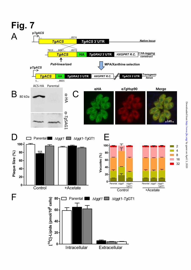
αHA αTgHsp90 Merge
0
20
40
60
80 Parental Δtggt1 Δtggt1-TgGT1
Intracellular Extracellular
[14C
]-Li
pids
(pm
ol/1
08ce
lls)
0
20
40
60
80
100
Parental Δtggt1 Δtggt1-TgGT1
Control +Acetate
***
Plaq
ue S
ize
(%)
0
20
40
60
80
100 2481632
Control +Acetate
Parental Δtggt1 Δtggt1- Parental Δtggt1 Δtggt1-TgGT1 TgGT1
Vacu
oles
(%)
ACS-‐HA Parental
80 kDa
α-‐TgSAG1 α-‐HA
A
B C
D E F
Fig. 7
by guest on April 3, 2020
http://ww
w.jbc.org/
Dow
nloaded from

Fig. 8
ATP produc3on
Respiratory chain
by guest on April 3, 2020
http://ww
w.jbc.org/
Dow
nloaded from

Richard Nitzsche, Vyacheslav Zagoriy, Richard Lucius and Nishith Guptaobligate intracellular parasite Toxoplasma gondii
Metabolic cooperation of glucose and glutamine is essential for the lytic cycle of
published online October 30, 2015J. Biol. Chem.
10.1074/jbc.M114.624619Access the most updated version of this article at doi:
Alerts:
When a correction for this article is posted•
When this article is cited•
to choose from all of JBC's e-mail alertsClick here
Supplemental material:
http://www.jbc.org/content/suppl/2015/10/30/M114.624619.DC1
by guest on April 3, 2020
http://ww
w.jbc.org/
Dow
nloaded from