melastoma malabathricum l. and melastoma...
TRANSCRIPT

PHYTOCHEMICAL AND BIOACTIVITY STUDIES OF
MELASTOMA MALABATHRICUM L. AND MELASTOMA IMBRICATUM WALL.
DENY SUSANTI
UNIVERSITI TEKNOLOGI MALAYSIA

PHYTOCHEMICAL AND BIOACTIVITY STUDIES OF
MELASTOMA MALABATHRICUM L. AND MELASTOMA IMBRICATUM WALL.
DENY SUSANTI
A thesis submitted in fulfilment of the
requirements for the award of the degree of
Doctor of Philosophy
Faculty of Science
Universiti Teknologi Malaysia
DECEMBER 2006

ii
In the memory of my dearest father, Darnis
for being an endless sources of spirit
To my mother, Enimar
for her love, support and ........especially her patience
To my sister and brother, Devy and Dody
for their support and always beside me
To my dearest husband, Muhammad Taher
for his deepest of love, support, inspiration and understanding which have been
essential to the success in the completion of this work
To my beloved sons, Muhammad Ghaisannaufal and Muhammad Luthfirrahman
for their sacrifices, which motivated me to reach my dream. I am very sorry if their
love were ignored for a while
iii
ACKNOWLEDGEMENTS
Bismillahirrahmanirrahim,
This work is the result of many years and the contributions of many people. I
could not have done it without them.
My special thanks are due to my supervisors Prof. Dr. Hasnah M. Sirat for giving
me the opportunity to work on this project and for the patience and support she has
shown me over the years, Assoc. Prof. Dr. Farediah Ahmad for her advice and guidance
and Dr. Rasadah M. Ali for guiding me in this interesting work.
I am highly indebted to The Ministry of Science, Technology and Innovation
Malaysia for financial support in conducting the project. I would like to extend my
thanks to The Chemistry Department technicians, Pn Zahratul Ain, Pn. Mek Zum, Pn.
Mariam, En. Ayob, En. Azmi and En. Fuad. Thanks are also due to Sultanah Zanariah
Library for enabling me to access the literatures and facilities of interlibrary loan. I am
also indebted to Dr. Fadzillah Adibah binti Abdul Majid and Dr. Muhammad Taher from
Bioprocess Department of UTM for facility in cytotoxicity assay, Pn. Mazura Pisar and
Mr. Ong Boen Kean from Forest Reseach Institute of Malaysia for guiding me in the
anti-inflammatory assay.
Many thanks are also due to Pn. Shajarahtunnur Jamil, Syahrizal, Wong Choon
Ling, Ngai Mun Hong and Oh Boon Thai, the lab work was found easier with the
friendship and many useful discussions.
My deepest appreciation goes to my husband, Muhammad Taher, for his love,
patience and understanding. And my sons, Muhammad Ghaisannaufal and Muhammad
Luthfirrahman, for their love and cheering me up during my study. I would also like to
convey my thanks to my mother, sister and brother, for their love, patience and
invaluable support.

v
ABSTRACT
Phytochemical and bioactivity studies of Melastoma malabathricum and M. imbricatum have been investigated. Isolation of the compounds was carried out using several chromatographic techniques. Chemical structures of isolated compounds were identified by spectroscopic methods including UV, IR, NMR (1H, 13C, DEPT, COSY, HMQC and HMBC) and MS. The n-hexane, ethyl acetate (EtOAc) and methanol (MeOH) extracts of the leaves of M. malabathricum yielded three new compounds, namely 2,5,6-trihydroxynaphtoic carbonic acid, methyl-2,5,6-trihydroxynaphtalene carbonate and flavonol glycoside derivative together with the known of auranamide, patriscabratine, α-amyrin, quercetin, quercitrin and kaempferol-3-O-(2”,6”-di-O-p-trans-coumaroyl)-glucoside. The n-hexane extract of the roots gave betulinic acid, serrat-14-en-16-one and 2-(2’-hydroxyvinyl)-1-methyl-4-propoxyphthalate. The EtOAc extract of the flowers yielded three compounds, kaempferol-3-O-β-D-glucoside, kaempferol and naringenin. The MeOH extract of the flowers gave kaempferol-3-O-(2”,6”-di-O-p-trans-coumaroyl)-glucoside and kaempferol-3-O-β-D-glucoside. The EtOAc extract of the fruits afforded betulinic acid, while the n-hexane extract of the stems gave α-amyrin. Phytochemical studies of the EtOAc extract of the leaves of M. imbricatum afforded quercitrin and the MeOH extract gave hyperin and kaempferol-3-O-(2”,6”-di-O-p-trans-coumaroyl)-glucoside. The EtOAc extract of the roots and fruits yielded betulinic acid. The n-hexane of the stems gave α-amyrin. The EtOAc extract of the flowers yielded kaempferol, kaempferol-3-O-β-D-glucoside and quercitrin. Methylation and acetylation of isolated compounds gave the methyl ether and acetyl derivatives, respectively. The pure compounds and crude extracts were subjected to antimicrobial, antioxidant, anti-inflammatory and cytotoxic assays. The MeOH extract of the fruits of M. malabathricum exhibited the strongest inhibition against bacteria, Bacillus subtilis, Streptococcus aureus, Pseudomonas aeruginosa and Escherichia coli with MIC values of 62.5, 62.5, 125.0 and 62.5 µg/mL, respectively, in the antimicrobial assay. The antioxidant assay was carried using FTC and DPPH (UV and ESR spectroscopic) methods. Kaempferol-3-O-(2”,6”-di-O-p-trans-coumaroyl)glucoside, kaempferol-3-O-β-D-glucose, kaempferol, hyperin, quercetin and quercitrin showed strong activities with inhibition more than 90% in the FTC method. Quercetin was found to be the most active as radical scavenger in DPPH-UV and ESR method with IC50 of 0.69 and 0.65 µM, respectively. α-Amyrin and kaempferol-3-O-(2”,6”-di-O-p-trans-coumaroyl)glucoside demonstrated the strongest activities in the anti-inflammatory assay of TPA mouse ear oedema with IC50 of 0.11 and 0.34 mM/ear, respectively. The EtOAc extract of the leaves of M. malabathricum displayed high activity with the inhibition of 94.3%. Kaempferol-3-O-(2”,6”-di-O-p-trans-coumaroyl)glucoside gave an IC50 of 5.6 µM in the PAF anti-inflammatory assay, while the MeOH extract of the leaves of M. imbricatum showed moderate activity with the inhibition of 78.0%. The cytotoxicity study was carried out using MTT assay on MCF7 cell line showed that kaempferol-3-O-(2”,6”-di-O-p-trans-coumaroyl)glucoside and naringenin were found to be active in inhibiting cell proliferation of MCF7 with IC50 of 0.28 and 1.3 µM, respectively.
vi
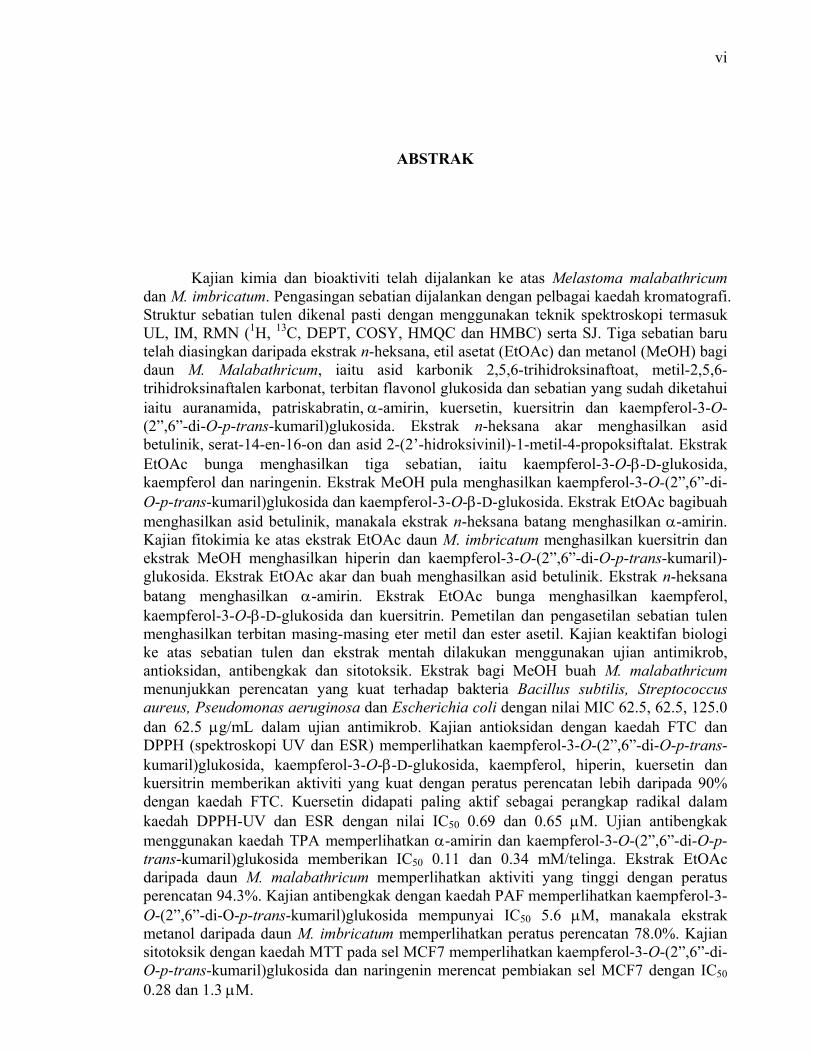
ABSTRAK
Kajian kimia dan bioaktiviti telah dijalankan ke atas Melastoma malabathricum dan M. imbricatum. Pengasingan sebatian dijalankan dengan pelbagai kaedah kromatografi. Struktur sebatian tulen dikenal pasti dengan menggunakan teknik spektroskopi termasuk UL, IM, RMN (1H, 13C, DEPT, COSY, HMQC dan HMBC) serta SJ. Tiga sebatian baru telah diasingkan daripada ekstrak n-heksana, etil asetat (EtOAc) dan metanol (MeOH) bagi daun M. Malabathricum, iaitu asid karbonik 2,5,6-trihidroksinaftoat, metil-2,5,6-trihidroksinaftalen karbonat, terbitan flavonol glukosida dan sebatian yang sudah diketahui iaitu auranamida, patriskabratin, α-amirin, kuersetin, kuersitrin dan kaempferol-3-O-(2”,6”-di-O-p-trans-kumaril)glukosida. Ekstrak n-heksana akar menghasilkan asid betulinik, serat-14-en-16-on dan asid 2-(2’-hidroksivinil)-1-metil-4-propoksiftalat. Ekstrak EtOAc bunga menghasilkan tiga sebatian, iaitu kaempferol-3-O-β-D-glukosida, kaempferol dan naringenin. Ekstrak MeOH pula menghasilkan kaempferol-3-O-(2”,6”-di-O-p-trans-kumaril)glukosida dan kaempferol-3-O-β-D-glukosida. Ekstrak EtOAc bagibuah menghasilkan asid betulinik, manakala ekstrak n-heksana batang menghasilkan α-amirin. Kajian fitokimia ke atas ekstrak EtOAc daun M. imbricatum menghasilkan kuersitrin dan ekstrak MeOH menghasilkan hiperin dan kaempferol-3-O-(2”,6”-di-O-p-trans-kumaril)-glukosida. Ekstrak EtOAc akar dan buah menghasilkan asid betulinik. Ekstrak n-heksana batang menghasilkan α-amirin. Ekstrak EtOAc bunga menghasilkan kaempferol, kaempferol-3-O-β-D-glukosida dan kuersitrin. Pemetilan dan pengasetilan sebatian tulen menghasilkan terbitan masing-masing eter metil dan ester asetil. Kajian keaktifan biologi ke atas sebatian tulen dan ekstrak mentah dilakukan menggunakan ujian antimikrob, antioksidan, antibengkak dan sitotoksik. Ekstrak bagi MeOH buah M. malabathricum menunjukkan perencatan yang kuat terhadap bakteria Bacillus subtilis, Streptococcus aureus, Pseudomonas aeruginosa dan Escherichia coli dengan nilai MIC 62.5, 62.5, 125.0 dan 62.5 µg/mL dalam ujian antimikrob. Kajian antioksidan dengan kaedah FTC dan DPPH (spektroskopi UV dan ESR) memperlihatkan kaempferol-3-O-(2”,6”-di-O-p-trans-kumaril)glukosida, kaempferol-3-O-β-D-glukosida, kaempferol, hiperin, kuersetin dan kuersitrin memberikan aktiviti yang kuat dengan peratus perencatan lebih daripada 90% dengan kaedah FTC. Kuersetin didapati paling aktif sebagai perangkap radikal dalam kaedah DPPH-UV dan ESR dengan nilai IC50 0.69 dan 0.65 µM. Ujian antibengkak menggunakan kaedah TPA memperlihatkan α-amirin dan kaempferol-3-O-(2”,6”-di-O-p-trans-kumaril)glukosida memberikan IC50 0.11 dan 0.34 mM/telinga. Ekstrak EtOAc daripada daun M. malabathricum memperlihatkan aktiviti yang tinggi dengan peratus perencatan 94.3%. Kajian antibengkak dengan kaedah PAF memperlihatkan kaempferol-3-O-(2”,6”-di-O-p-trans-kumaril)glukosida mempunyai IC50 5.6 µM, manakala ekstrak metanol daripada daun M. imbricatum memperlihatkan peratus perencatan 78.0%. Kajian sitotoksik dengan kaedah MTT pada sel MCF7 memperlihatkan kaempferol-3-O-(2”,6”-di-O-p-trans-kumaril)glukosida dan naringenin merencat pembiakan sel MCF7 dengan IC50 0.28 dan 1.3 µM.

vii
TABLE OF CONTENTS
CHAPTER TITLE PAGE
TITLE i
DECLARATION ii
DEDICATION iii
ACKNOWLEDGEMENTS iv
ABSTRACT v
ABSTRAK vi
TABLE OF CONTENTS vii
LIST OF TABLES xiv
LIST OF FIGURES xvi
LIST OF SYMBOLS xix
LIST OF ABBREVIATIONS xx
LIST OF APPENDICES xxiv
1. INTRODUCTION 1
1.1 General Introduction 1
1.2 Drug Discovery 2
1.3 Botany and Distribution of Melastomataceae 4
1.4 Botany and Distribution of Genus Melastoma 5
1.4.1 Melastoma malabathricum L 6
1.4.2 Melastoma imbricatum Wall 7
1.5 Chemical Investigation on Melastomataceae 8
1.6 Chemical Investigation on Melastoma 16
1.7 Bioactivity Investigation on Melastomataceae 21
1.8 Background of the Research 23

viii
1.9 Objective of the Study 24
1.10 Scopes of the Study 24
2. PHYTOCHEMICAL STUDIES ON MELASTOMA SPECIES 26
2.1 The Leaves of Melastoma malabathricum L 26
2.1.1 Auranamide (81) 27
2.1.2 Patriscabratine (82) 31
2.1.3 α-Amyrin (73) 36
2.1.4 2,5,6-Trihydroxynaphtalene carbonic acid (83) 37
2.1.5 Quercitrin (76) 44
2.1.6 Quercetin (7) 46
2.1.7 Kaempferol-3-O-(2”,6”-di-O-p-trans- 48 coumaroyl)glucoside (87)
2.1.8 Quercitrin (76) 51
2.1.9 Methyl 2,5,6-trihydroxynaphtalene carbonate (88) 51
2.1.10 Flavonol glycoside derivative (90) 62
2.2 The Roots of Melastoma malabathricum L 74
2.2.1 2-(2’-Hydroxyvinyl)-1-methyl-4- 75 propoxyphthalate (91)
2.2.2 Serrat-14-en-16-one (92) 76
2.2.3 Betulinic acid (93) 77
2.3 The Stems of Melastoma malabathricum L 78
2.3.1 α-Amyrin (73) 78
2.4 The Flowers of Melastoma malabathricum L 78
2.4.1 Naringenin (94) 79
2.4.2 Kaempferol (8) 81
2.4.3 Kaempferol-3-O-glucoside (11) 82
2.4.4 Kaempferol-3-O-(2”,6”-di-O-p-trans- 83 coumaroyl)glucoside (87)
2.5 The Fruits of Melastoma malabathricum L 84
2.5.1 Betulinic acid (93) 84
2.6 The Leaves of Melastoma imbricatum Wall 85
2.6.1 Quercitrin (76) 85
ix
2.6.2 Kaempferol-3-O-(2”,6”-di-O-p-trans- 86 coumaroyl)glucoside (87)
2.6.3 Hyperin (10) 86
2.7 The Roots of Melastoma imbricatum Wall 88
2.7.1 Betulinic acid (93) 88
2.8 The Stems of Melastoma imbricatum Wall 88
2.8.1 α-Amyrin (73) 89
2.9 The Flowers of Melastoma imbricatum Wall 89
2.9.1 Kaempferol (8) 90
2.9.2 Quercitrin (76) 90
2.9.3 Kaempferol-3-O-glucoside (11) 91
2.10 The Fruits of Melastoma imbricatum Wall 91
2.10.1 Betulinic acid (93) 91
3. BIOACTIVITY STUDIES ON MELASTOMA SPECIES 92
3.1 Antimicrobial 92
3.1.1 Antimicrobial Agent 93
3.1.1.1 Selective Toxicity 93
3.1.1.2 The Spectrum of Activity 93
3.1.1.3 Modes of Action 94
3.1.2 Methods for Evaluating Antimicrobial Drug 95
3.1.3.1 Diffusion Susceptibility Test 95
3.1.3.2 Minimum Inhibition Concentration 96 (MIC) Test
3.1.3.3 Minimum Bactericidal Concentration 96 (MBC) Test
3.1.4 Antimicrobial study on Melastoma species 97
3.2 Antioxidant 101
3.2.1 Mechanism of Antioxidant 103
3.2.2 Electron Spin Resonance (ESR) 104
3.2.3 Antioxidant Activity of Melastoma Species 106
3.2.3.1 Ferric Thiocyanate (FTC) Method 107
3.2.3.2 Free Radical Scavenging Activity 114 (DPPH)
x
3.2.4.3 UV Spectrophotometry Method 115
3.2.4.4 Electron Spin Resonance (ESR) 119 Spectrometry Method
3.2.4.5 Structure-Activity Relationships 122
3.3 Anti-inflammatory 123
3.3.1 Platelet-activating Factor (PAF) 124
3.3.2 Mechanism of Action 125
3.3.3 Anti-inflammatory Activity of Melastoma 125 Species
3.3.3.1 12-O-tetradecanoylphorbol 13-acetate 125 (TPA) Induced Mouse Ear Oedema
3.3.3.2 Platelet Activating Factor (PAF) 129 Receptor Binding Antagonist
3.4 Cytotoxicity 130
3.4.1 Hormones that Affect Cancer 131
3.4.1.1 Endometrial Cancer 131
3.4.1.2 Ovarian Cancer 132
3.4.1.3 Breast Cancer 132
3.4.2 Therapeutic Agents 132
3.4.3 MTT Assay 133
3.4.4 Cytotoxic Effect of Melastoma Species on 134 MCF7 Human Breast Cancer Cell
3 EXPERIMENTAL 137
4.1 Phytochemicals Study 137
4.1.1 General Experimental Procedures 137
4.1.2 Materials 138
4.1.2.1 Chemicals 138
4.1.2.2 Plant Materials 138
4.1.3 Extraction of the Leaves of Melastoma 139 malabathricum L
4.1.3.1 Auranamide (81) 139
4.1.3.2 Patriscabratine (82) 140
4.1.3.3 α-Amyrin (73) 141

xi
4.1.3.4 2,5,6-Trihydroxynaphtalene carbonic 142 acid (83)
4.1.3.5 Quercitrin (76) 143
4.1.3.6 Quercitrin trimethyl ether (84) 144
4.1.3.7 Quercitrin acetate derivative (85) 144
4.1.3.8 Quercetin (7) 145
4.1.3.9 Quercetin tetramethyl ether (86) 146
4.1.3.10 Kaempferol-3-O-(2”,6”-di-O-p- 147 trans-coumaroyl)glucoside (87)
4.1.3.11 Quercitrin (76) 148
4.1.3.12 Methyl 2,5,6-trihydroxy- 148 naphtalene carbonate(88)
4.1.3.13 Derivative of methyl 2,5,6- 148 trihydroxynaphtalene carbonate (89)
4.1.3.14 Flavonol glycoside derivative (90) 149
4.1.3 Extraction of the Roots of Melastoma 150 malabathricum L
4.1.4.1 2-(2’-Hydroxyvinyl)-1-methyl-4- 150 propoxyphthalate (91)
4.1.4.2 Serrat-14-en-16-one (92) 151
4.1.4.3 Betulinic acid (93) 152
4.1.5 Extraction of the Stems of Melastoma 153 malabathricum L
4.1.5.1 α-Amyrin (73) 153
4.1.6 Extraction of the Flower of Melastoma 154 malabathricum L
4.1.6.1 Naringenin (94) 154
4.1.6.2 Kaempferol (8) 155
4.1.6.3 Kaempferol 3-O-glucoside (11) 156
4.1.6.4 Kaempferol-3-O-(2”,6”-di-O-p-trans- 157 coumaroyl)glucoside (87)
4.1.7 Extraction of the Fruits of Melastoma 157 malabathricum L
4.1.7.1 Betulinic acid (93) 157
4.1.8 Extraction of the Leaves of Melastoma 158 imbricatum Wall
4.1.8.1 Quercitrin (76) 158

xii
4.1.8.2 Kaempferol-3-O-(2”,6”-di-O-p-trans- 159 coumaroyl)glucoside (87)
4.1.8.3 Hyperin (10) 159
4.1.9 Extraction of the Roots of Melastoma 160 imbricatum Wall
4.1.9.1 Betulinic acid (93) 161
4.1.10 Extraction of the Stems of Melastoma 161 imbricatum Wall
4.1.10.1 α-Amyrin (73) 161
4.1.11 Extraction of the Flowers of Melastoma 162 imbricatum Wall
4.1.11.1 Kaempferol (8) 163
4.1.11.2 Quercitrin (76) and Kaempferol- 163 3- O-glucoside (11)
4.1.12 Extraction of the Fruits of Melastoma 163 imbricatum Wall
4.1.12.1 Betulinic acid (93) 164
4.1.13 Hydrolysis of the Isolated Compounds 164
4.2 Bioactivity Studies 165
4.2.1 General Instrumentation 165
4.2.2.1 Material 165
4.2.2.1 Chemical 165
4.2.2.2 Animal, Microorganism and Cell 166 Culture
4.2.3 Antimicrobial Assay 166
4.2.3.1 Apparatus 166
4.2.3.2 Preparation of Agar and Broth 167
4.2.3.3 Culturing Microbe 167
4.2.3.4 Preparation of Agar Plate and 167 Mc Farland Solution
4.2.3.5 Sample Preparation 168
4.2.3.6 Disc Diffusion Method 168
4.2.3.7 Minimum Inhibition Concentration 169 (MIC)
4.2.3.8 Minimum Bactericidal Concentration 169 (MBC) and Minimum Fungicidal Concentration (MFC)

xiii
4.2.4 Antioxidant 172
4.2.4.1 Ferric Thiocyanate (FTC) Method 172
4.2.4.2 Free Radical Scavenging Activity 174 (DPPH)
4.2.4.3 UV Spectrophotometry 174 Method
4.2.4.4 Electron Spin Resonance 177 (ESR) Spectroscopy Method
4.2.5 Anti-inflammatory 180
4.2.5.1 12-O-tetradecanoylphorbol 13-acetate 180 (TPA) Induced Mouse Ear Oedema
4.2.5.2 Sample Preparation 180
4.2.5.3 Assay of TPA Induced 181 Inflammation
4.2.5.4 Platelet Activating Factor (PAF) 182 Receptor Binding Antagonist
4.2.5.5 Sample Preparation 182
4.2.5.6 Preparation of Rabbit Platelets 183
4.2.5.7 PAF Receptor Binding 183 Antagonist Assay
4.2.6 Cytotoxic Assay 185
4.2.7 Statistical Analysis 185
5. CONCLUSIONS 186
5.1 Phytochemical Studies 186
5.2 Bioactivity Studies 188
REFERENCES 191
APPENDICES 204

xiv
LIST OF TABLES
TABLE NO. TITLE PAGE
2.1 The 1H, 13C NMR, FGCOSY, HMQC and HMBC 30 data for 81 (500 MHz in CDCl3)
2.2 The 1H, 13C NMR, FGCOSY, HMQC and HMBC 34 data of 82 (500 MHz in CDCl3).
2.3 The 1H, 13C NMR, FGCOSY, HMQC and HMBC 50 data for 87 (600 MHz in Acetone-d6)
2.4 The1H, 13C NMR, HMQC and HMBC data for 88 52 (400 MHz in Acetone-d6) and 89 (300 MHz, CDCl3)
2.5 The 1H and 13C NMR, 1H-1H COSY and HMBC 74 of 90 (500 MHz, CD3OD)
3.1 The antibacterial activity of M. malabathricum 100 and M. imbricatum
3.2 The antifungal activity of M. malabathricum 101 and M. imbricatum
3.3 Comparison of the absorbance values and 112 percent of linoleic acid peroxidation of the extract of M. malabathricum and M. imbricatum as measured by FTC antioxidant assay
3.4 The absorbance values and percent of 113 linoleic acid peroxidation of the flavonoids as measured by FTC antioxidant assay
3.5 The IC50 of the methanol extract from 117 M. malabathricum and M. imbricatum by UV Spectrophotometry method
3.6 The IC50 of the isolated compounds from 118 M. malabathricum and M. imbricatum by UV Spectrophotometry method
3.7 The IC50 of the methanol extract from 120 M. malabathricum and M. imbricatum by ESR spectrometry method

xv
3.8 The IC50 of the isolated compounds from 120 M. malabathricum and M. imbricatum by ESR spectrometry method
3.9 The IC50 and inhibitory effect of the isolated 128 compounds on mice ear oedema
3.10 IC50 and inhibitory effect of the isolated 130 compounds on PAS assay
4.1 Results of minimum inhibition concentration (MIC- 170 Bacteria)
4.2 Results of minimum inhibition concentration (MIC- 171 Fungi)
4.3 Results of minimum bactericidal concentration 171 (MBC)
4.4 Results of minimum fungicidal concentration (MFC) 172
4.5 Results of antioxidant assay for crude extracts from 173 M. malabathricum by FTC method
4.6 Results of antioxidant assay for crude extracts from 173 M. imbricatum by FTC method
4.7 Results of antioxidant for isolated compounds 174 by FTC method
4.8 Results of antioxidant for crude extract from 175 M. malabathricum by UV spectrophotometry
4.9 Results of antioxidant for crude extract from 176 M. imbricatum by UV spectrophotometry
4.10 Results of antioxidant for isolated compounds 177 by UV spectrophotometry
4.11 Results of antioxidant for crude extract from 178 M. malabathricum by ESR spectrometry method
4.12 Results of antioxidant for crude extract from 179 M. imbricatum by ESR spectrometry method
4.13 Results of antioxidant for crude extract from 180 M. malabathricum by ESR spectrometry method
4.14 Result of anti-inflammatory assay by TPA method 182
4.15 Results of anti-inflammatory by PAF assay 184
5.1 Distribution of the phytochemical found in the 187 different part of M. malabathricum and M. imbricatum

xvi
LIST OF FIGURES
FIGURE NO. TITLE PAGE
1.1 Melastoma malabathricum L 7
1.2 Melastoma imbricatum Wall 8
2.1 The infrared spectrum of 2,5,6-trihydroxynaphtalene 38 carbonic acid (83) (KBr Disc)
2.2 The 1H NMR spectrum 2,5,6- trihydroxynaphtalene 39 carbonic acid (83) (500 MHz, in CD3OD)
2.3 The 13C NMR spectrum of 2,5,6-trihydroxy- 40 naphthalene carbonic acid (83) (125 MHz, in CD3OD)
2.4 The DEPT spectrum of 2,5,6-trihydroxy 41 naphthalene carbonic acid (83) (125 MHz, in CD3OD)
2.5 The FGHMQC spectrum of 2,5,6-trihydroxy naphthalene 42 carbonic acid (83) (500 MHz, 125 MHz, in CD3OD)
2.6 The FGHMBC spectrum of 2,5,6-trihydroxynaphtalene 43 carbonic acid (83) (500 MHz, 125 MHz, in CD3OD)
2.7 The infrared spectrum of methyl 2,5,6-trihydroxy- 53 naphthalene carbonate (88) (KBr Disc)
2.8 The 1H NMR spectrum of methyl-2,5,6-trihydroxy- 54 naphthalene carbonate (88) (400 MHz, in Acetone-d6)
2.9 The 13C NMR spectrum of methyl-2,5,6-trihydroxy- 55 naphthalene carbonate (88) (100 MHz, in Acetone-d6)
2.10 The DEPT spectrum of methyl-2,5,6-trihydroxy- 56 naphthalene carbonate (88) (100 MHz, in Acetone-d6)
2.11 The FGHMQC spectrum of 2,5,6-trihydroxy- 57 naphthalene carbonate (88) (600 MHz, 150 MHz, in Acetone-d6)
2.12 The FGHMBC spectrum of methyl-2,5,6-trihydroxy- 58 naphthalene carbonate (88) (600 MHz, 150 MHz, in Acetone-d6)

xvii
2.13 The infrared spectrum of methyl-2,5,6-trimethoxy- 59 naphthalene carbonate (89) (KBr Disc)
2.14 The 1H NMR spectrum of methyl-2,5,6-trimethoxy- 60 naphthalene carbonate (89) (300 MHz, CDCl3)
2.15 The 13C NMR spectrum of methyl-2,5,6-trimethoxy- 61 naphthalene carbonate (89) (75 MHz, in CDCl3)
2.16 The ultraviolet spectrum of flavonol glycoside 64 derivative (90)
2.17 The 1H NMR spectrum of flavonol glycoside 65 derivative (90) (500 MHz, in CD3OD)
2.18 The FGCOSY spectrum of flavonol glycoside 66 derivative (90) (500 MHz, in CD3OD)
2.19 The 13C NMR spectrum of flavonol glycoside 67 derivative (90) (125 MHz, in CD3OD)
2.20 The DEPT spectrum of flavonol glycoside 68 derivative (90) (125 MHz, in CD3OD)
2.21 The FGHMQC spectrum of flavonol glycoside 69 derivative (90) (500 MHz, 125 MHz, in CD3OD)
2.22 The expansion of FGHMQC spectrum of 70 flavonol glycoside derivative (90)
(500 MHz, 125 MHz, in CD3OD)
2.23 The FGHMBC spectrum of flavonol glycoside 71 derivative (90) (500 MHz, 125 MHz, in CD3OD)
2.24 The expansion of the FGHMBC spectrum of 72 flavonol glycoside derivative (90)
(500 MHz, 125 MHz, in CD3OD)
2.25 The infrared spectrum of flavonol glycoside 73 derivative (90) (KBr Disc)
3.1 Antioxidants in the defense system against free 105 radical induced oxidative damage
3.2 Antioxidant activity of M. malabathricum extract 109 as measured by the FTC method
3.3 Antioxidant activity of M. imbricatum extract as 110 measured by the FTC method
3.4 Antioxidant activity of some flavonoids from 111 M. malabathricum and M. imbricatum as measured by the FTC method
3.5 C6-C3-C6 Configurations of flavonoid 112
3.6 Reaction of DPPH with an antioxidant compound 115
3.7 Scavenging activity of selected compounds by 121 ESR spectrometry

xviii
3.8 Mast cell mediator and inflammation 126
3.9 Mechanism of the cleavage of MTT 134
3.10 Morphology of human breast cancer (MCF 7) 136

xix
LIST OF SYMBOLS
[α] 24D - Specific rotation
δ - Chemical shift
ε - Molar extinction coefficient 13C - Carbon-13
°C - degree of Celsius 1H - Proton
J - Coupling constant in Hertz
λ - Wavelength
ν - Wavenumber

xx
LIST OF ABBREVIATIONS
Abs. - absorbance
Ac2O - acetic acid anhydride
AlCl3 - aluminium chloride
BHT - Butylated Hydroxy Toluene
br - broad
CC - Column Chromatography
CDCl3 - deuterated chloroform
CD3OD - deuterated methanol
CHCl3 - chloroform
CIMS - Chemical Ionization Mass Spectrometry
COSY - Correlation Spectroscopy
cm-1 - reciprocal centimeter (wavenumber) 13C NMR - carbon-13 Nuclear Magnetic Resonance
d - doublet
dd - double doublet
dt - double triplet
dec. - decomposed
DEPT - Distortionless Enhancement Polarization Transfer
DMEM - Dulbecco’s Modified Eagle Medium
DMSO-d6 - deuterated dimethyl sulphoxide
DNA - deoxyribonucleic acid
DPPH - 2,2-diphenyl-1-picrylhydrazyl
EIMS - Electron Impact Mass Spectrometry
EPRT - estrogens/progesterone replacement therapy
xxi
EtOAc - ethyl acetate
ESR - Electron Spin Resonance
EPR - Electronic Paramagnetic Resonance
FABMS - Fast Atom Bombardment Mass Spectrometry
FTC - Ferric Thiocyanate
g - gram
GC - Gas Chromatography
GSH - Glutathione peroxide
H3BO3 - boric acid
HCl - hydrochloric acid
HEPES - N-[2-Hydroxyethyl]piperazine-N’-[2-ethanesulfonic acid]
HHDP - hexahydroxydiphenoyl 1H NMR - Proton Nuclear Magnetic Resonance
HMBC - Heteronuclear Multiple-Bond Connectivity
HMQC - Heteronuclear Multiple Quantum Coherence
HRT - estrogens replacement therapy
Hz - Hertz
IC50 - concentration that inhibits a response by 50% relative to a
negative control
IgE - immunoglobulin E
IR - Infrared
J - coupling constant
KBr - potassium bromide
L - liter
m - multiplet
M - molar concentration
M+ - molecular ion
MAO - monoamine oxidase
max - maximum
MBC - Minimum Bactericidal Concentration
MCF7 - Human Breast Cancer cell line

xxii
MeOH - methanol
MFC - Minimum Fungicidal Concentration
MgSO4 - magnesium sulphate
µg - microgram
MHz - megahertz
MIC - Minimum Inhibition Concentration
mL - milliliter
mM - millimolar
µM - micromolar
mp. - melting point
MS - Mass Spectra
MTT - 3-(4,5-Dimethylthiazol-2-yl)-2,5-diphenyltetrazolium bromide
m/z - mass to charge ratio
NA - Nutrient Agar
NB - Nutrient Broth
NaOAc - sodium acetate
NaOMe - sodium methoxide
nm - nanometer
PAF - platelet activating factor
PBS - phosphate buffer saline
PGI1 - prostaglandin I1
PGE2 - prostaglandin E2
PGF2 - prostaglandin F2
PGH - prostaglandin H
PHGPX - phospholipids hydroperoxide glutathione peroxidase
PKC - protein kinase C
% - percent
ppm - parts per million
PTLC - Preparative Thin Layer Chromatography
q - quartet
rel. int. - relative intensity

xxiii
Rf - retention factor
s - singlet
SD - standard deviation
sept - septet
SOD - superoxide dismustase
t - triplet
TLC - Thin Layer Chromatography
TPA - 12-O-Tetradecanoylphorbol-13-acetate
UV - Ultraviolet
VLC - Vacuum Liquid Chromatography

xxiv
LIST OF APPENDICES
APPENDIX TITLE PAGE
1. The infrared spectrum of auranamide (81) (KBr Disc) 204
2. The 1H NMR spectrum of auranamide (81) 205 (500 MHz, in CDCl3)
3. The FGCOSY spectrum of auranamide (81) 206 (500 MHz, CDCl3)
4. The expansion of the FGCOSY spectrum of auranamide (81) 207 (500 MHz, CDCl3)
5. The 13C NMR spectrum of auranamide (81) 208 (125 MHz, in CDCl3)
6. The FABMS spectrum of auranamide (81) 209
7. The DEPT spectrum of auranamide (81) 210 (125 MHz, in CDCl3)
8. The FGHMQC spectrum of auranamide (81) 211 (500 MHz, 125 MHz, in CDCl3)
9. The expansion of the FGHMQC spectrum of auranamide 212 (81) (500 MHz, 125 MHz, in CDCl3)
10. The FGHMBC spectrum of auranamide (81) 213 (500 MHz, 125 MHz, in CDCl3)
11. The expansion of the FGHMBC spectrum of 214 auranamide (81) (500 MHz, 125 MHz, in CDCl3)
12. The infrared spectrum of patriscabratine (82) (KBr Disc) 215
13. The 1H NMR spectrum of patriscabratine (82) 216 (500 MHz, in CDCl3)
14. The 13C NMR spectrum of patriscabratine (82) 217 (125 MHz, in CDCl3)
15. The EIMS spectrum of patriscabratine (82) 218
16. The DEPT spectrum of patriscabratine (82) 219 (125 MHz, in CDCl3)
17. The FGCOSY spectrum of patriscabratine (82) 220 (500 MHz, in CDCl3)

xxv
18. The expansion of the FGCOSY spectrum of 221 patriscabratine (82) (500 MHz, in CDCl3)
19. The HMQC spectrum of patriscabratine (82) 222 (500 MHz, 125 MHz, in CDCl3)
20. The expansion of the HMQC spectrum of 223 patriscabratine (82) (500 MHz, 125 MHz, in CDCl3)
21. The FGHMBC spectrum of patriscabratine (82) 224 (500 MHz, 125 MHz, in CDCl3)
22. The expansion of the FGHMBC spectrum of 225 patriscabratine (82) (500 MHz, 125 MHz, in CDCl3)
23. The infrared spectrum of α-amyrin (73) (KBr Disc) 226
24. The 1H NMR spectrum of α-amyrin (73) 227 (500 MHz, in DMSO-d6)
25. The mass spectrum of α-amyrin (73) 228
26. The 13C NMR spectrum of α- amyrin (73) 229 (125 MHz, in DMSO-d6)
27. The DEPT spectrum of α-amyrin (73) 230 (125 MHz, in DMSO-d6)
28. The infrared spectrum of quercitrin (76) (KBr Disc) 231
29. The ultraviolet spectra of quercitrin (76) 232
30. The 1H NMR spectrum of quercitrin (76) 233 (500 MHz, in CD3OD)
31. The FGCOSY spectrum of quercitrin (76) 234 (500 MHz, in CD3OD)
32. The 13C NMR spectrum of quercitrin (76) 235 (125 MHz, in CD3OD)
33. The DEPT spectrum of quercitrin (76) (125 MHz, in CD3OD) 236
34. The FGHMQC spectrum of quercitrin (76) 237 (500 MHz, 125 MHz, in CD3OD)
35. The infrared spectrum of quercitrin trimethyl ether (84) 238 (KBr Disc)
36. The 1H NMR spectrum of quercitrin trimethyl ether (84) 239 (300 MHz, in CDCl3)
37. The 13C spectrum of quercitrin trimethyl ether (84) 240 (75 MHz, in CDCl3)
38. The DEPT spectrum of quercitrin trimethyl ether (84) 241 (75 MHz, in CDCl3)
39. The infrared spectrum of quercitrin acetate derivative (85) 242 (KBr Disc)

xxvi
40. The 1H NMR spectrum of quercitrin acetate 243 derivative (85) (300 MHz, in Acetone-d6)
41. The ultraviolet spectra of quercetin (7) 244
42. The infrared spectrum of quercetin (7) (KBr Disc) 245
43. The 1H NMR spectrum of quercetin (7) 246 (500 MHz, in Acetone-d6)
44. The 13C NMR spectrum of quercetin (7) 247 (125 MHz, in Acetone-d6)
45. The mass spectrum of quercetin (7) 248
46. The infrared spectrum of quercetin tetramethyl ether (86) 249 (KBr Disc)
47. The 1H NMR spectrum of quercetin tetramethyl 250 ether (86) (300 MHz, in CDCl3)
48. The infrared spectrum of kaempferol-3-O-(2”,6”- 251 di-O-p-trans-coumaroyl)glucoside (87) (KBr Disc)
49. The ultraviolet spectra of kaempferol-3-O-(2”,6”- 252 di-O-p-trans-coumaroyl)glucoside (87)
50. The 1H NMR spectrum of kaempferol-3-O-(2”,6”- 253 di-O-p-trans-coumaroyl)glucoside (87) (600 MHz, in Acetone-d6)
51. The 13C NMR spectrum of kaempferol-3-O-(2”,6”- 254 di-O-p-trans-coumaroyl)glucoside (87) (150 MHz, in Acetone-d6)
52. The FGCOSY spectrum of kaempferol-3-O-(2”,6”- 255 di-O-p-trans-coumaroyl)glucoside (87) (500 MHz, in Acetone-d6)
53. The FGHMQC spectrum of kaempferol-3-O-(2”,6”- 256 di-O-p-trans-coumaroyl)glucoside (87) (500 MHz, 150 MHz, in Acetone-d6)
54. The mass spectrum of kaempferol-3-O-(2”,6”-di-O-p- 257 trans-coumaroyl)glucoside (87)
55. The FGHMBC spectrum of kaempferol-3-O-(2”,6”- 258 di-O-p-trans-coumaroyl)glucoside (87) (500 MHz, 150 MHz, in Acetone-d6)
56. The 1H NMR spectrum of 2-(2’-hydroxyvinyl) 259 methyl-4-propoxyphthalate (91) (300 MHz, in CDCl3)
57. The GC spectrum of 2-(2’-hydroxyvinyl) 260 methyl-4-propoxyphthalate (91)
58. The mass spectrum of 2-(2’-hydroxyvinyl) 261 methyl-4-propoxyphthalate (91)
xxvii
59. The infrared spectrum of serrat-14-en-16-one (92) 262 (KBr Disc)
60. The 1H NMR spectrum of serrat-14-en-16-one (92) 263 (400 MHz, in CDCl3)
61. The 13C NMR spectrum of serrat-14-en-16-one (92) 264 (100 MHz, in CDCl3)
62. The DEPT Spectrum of serrat-14-en-16-one (92) 265 (100 MHz, in CDCl3)
63. The infrared spectrum of betulinic acid (93) (KBr Disc) 266
64. The 1H NMR spectrum of betulinic acid (93) 267 (500 MHz, in DMSO-d6)
65. The 13C NMR spectrum of betulinic acid (93) 268 (125 MHz, in DMSO-d6)
66. The DEPT spectrum of betulinic acid (93) 269 (125 MHz, in DMSO-d6)
67. The mass spectrum of betulinic acid (93) 270
68. The ultraviolet spectra of naringenin (94) 271
69. The infrared spectrum of naringenin (94) (KBr Disc) 272
70. The 1H NMR spectrum of naringenin (94) 273 (300 MHz, in Acetone-d6)
71. The COSY spectrum of naringenin (94) 274 (300 MHz, in Acetone-d6)
72. The 13C NMR spectrum of naringenin (94) 275 (75 MHz, in Acetone-d6)
73. The mass spectrum of naringenin (94) 276
74. The DEPT spectrum of naringenin (94) 277 (75 MHz, in Acetone-d6)
75. The mass spectrum of kaempferol (8) 278
76. The ultraviolet spectra of kaempferol (8) 279
77. The infrared spectrum of kaempferol (8) (KBr Disc) 280
78. The 1H NMR spectrum of kaempferol (8) 281 (300 MHz, in Acetone-d6)
79. The COSY spectrum of kaempferol (8) 282 (300 MHz, in Acetone-d6)
80. The 13C NMR spectrum of kaempferol (8) 283 (75 MHz, in Acetone-d6)
81. The DEPT spectrum of kaempferol (8) 284 (75 MHz, in Acetone-d6)
82. The ultraviolet spectra of kaempferol 3-O-glucoside (11) 285

xxviii
83. The infrared spectrum of kaempferol 3-O-glucoside (11) 286 (KBr Disc)
84. The 1H NMR spectrum of kaempferol 3-O-glucoside (11) 287 (300 MHz, in CD3OD)
85. The COSY spectrum of kaempferol 3-O-glucoside (11) 288 (300 MHz, in CD3OD)
86. The 13C NMR spectrum of kaempferol 3-O-glucoside 289 (11) (75 MHz, in CD3OD)
87. The DEPT spectrum of kaempferol 3-O-glucoside (11) 290 (75 MHz, in CD3OD)
88. The mass spectrum of kaempferol 3-O-glucoside (11) 291
89. The ultraviolet spectra hyperin (10) 292
90. The infrared spectrum of hyperin (10) (KBr Disc) 293
91. The 1H NMR spectrum of hyperin (10) 294 (600 MHz, in CD3OD)
92. The 13C NMR spectrum of hyperin (10) 295 (150 MHz, in CD3OD)
93. The mass spectrum of hyperin (10) 296

CHAPTER I
INTRODUCTION
1.1 General Introduction
Bioactive natural products have an enormous economic importance as
specialty chemicals. They can be used as drugs, lead compounds, biological or
pharmacological tools, feed stock products (raw materials for the production of
drugs) and nutraceuticals. They are found in herbs, dietary supplements, spices and
foods. Some of them are important flavours, fragrances, dyes and cosmetic, and
others are used as insecticides, antifeedants, pesticides and antirodenticides [1].
Plants have been the source of medicinal agents for thousands of years, and
impressive numbers of modern drugs have been isolated from natural sources, many
based on their uses in traditional medicines. In fact, natural products once served as
the source of all drugs. Since more than 80% of the world’s population use plants as
their primary source of medicinal agents, it is not surprising to find that in many
countries of the world there is a well-established system of traditional medicine,
whose remedies are still being compiled. In some instances, the Chinese and the
Ayurvedic systems have documented the remedies of their traditional medicines and
these documents are commercially available [2, 3].

2
1.2 Drug Discovery
A few drug discovery programmes based on plants has restarted in the late
1980’s when pharmacologists, physicians and chemists started reinvestigating the
active ingredients in plants used for medicinal purposes. The more recent advances
in chromatographic and spectroscopic technical equipments have decreased the time
taken to identify and determine the molecular structures of the active compounds in
these plants. This has resulted in an increase in the number of natural products being
isolated and identified every year, although very few have been developed as drugs.
However, due to the novel structures of some of these active compounds, they might
serve as templates or lead compounds for drugs [4].
Different approaches to drug discovery using higher plants can be
distinguished: random selection followed by chemical screening, random selection
followed by one or more biological assays, follow up of biological activity reports,
follow up of ethnomedical (traditional medicines) uses of plants. The latter approach
includes plants used in organized traditional medical system; herbalism and folklore;
the use of database. The objective is the targeted isolation of new bioactive plant
product, i. e. lead substances with novel structure and novel mechanisms of action
[1].
Among the 45 drugs of known structures derived from the tropical rain forest
species, including those that are of major important used in therapy, none is currently
produced through synthesis. For example, the anticancer vinblastine (1) and
vincristine (2) from Catharanthus roseus L. G. Don (Apocynaceae) in which (1) has
been used as part of chemotherapy combination for the treatment of acute
lymphoblastic leukemia and testicular teratoma since 1979’s. The antiplasmodial
artemisinin (3) from Artemisia annua L. (Compositae) as well as the anti malarial
quinine (4) isolated from the bark of Cinchona spp (Rubiaceae). Ginkgolide A (5)
was isolated from the root barks and leaves of Ginkgo biloba (Ginkgoaceae) have
been used from the natural sources. There is evidence that ginkgolides are able to
competitively inhibit the specific platelet aggregating factors to cell membranes. This
type of activity could explain the use of these plants in the treatment of inflammation
and for respiratory and panic disorder [4, 5].

3
OO
H
H
O
OOH
H
H
HOH
O
O
N
NHO HH3CO
H
H
OO
O
O O
H
H
NH
N
OH
H3COOC
NR
N
H
COOCH3
OCOCH3OH
H
NHO
OHO
O
HO
O
O
O OH
O
H
O
OOO
The anti cancer drug, taxol (6) was isolated from the bark of yew trees as a
result of NCI’s systematic effort in collecting and screening plants for antitumor
activity [4, 6]. Taxol has now been approved to be used for the treatment of ovarian
and breast cancers, as well as small cell and non small cell lung cancer which include
neck and head cancers [4, 7].
(1) R = CH3 (2) R = CHO
(3) (4) (5)
(6)
4
The commercial value of drug derived from higher plants should not be
underestimated. For example, in 1980 American consumers paid about USD 8 billion
for prescription of drugs derived solely from higher plants. From 1959 to 1980, drugs
derived from higher plants represented a constant 25% of all new and refilled
prescriptions dispensed from community pharmacies in the United State [8].
Phytochemical investigation will provide data on the chemical constituents of
the plant concerned. However, many new natural compounds were isolated,
characterised and published without any biological testing and their useful biological
activities then remain unknown for a long time. The significance of the
phytochemical work is greatly increased if the fine chemicals isolated from the plant
possess certain biological activities, particularly if the activity is in line with the
development of medicinal agents [9].
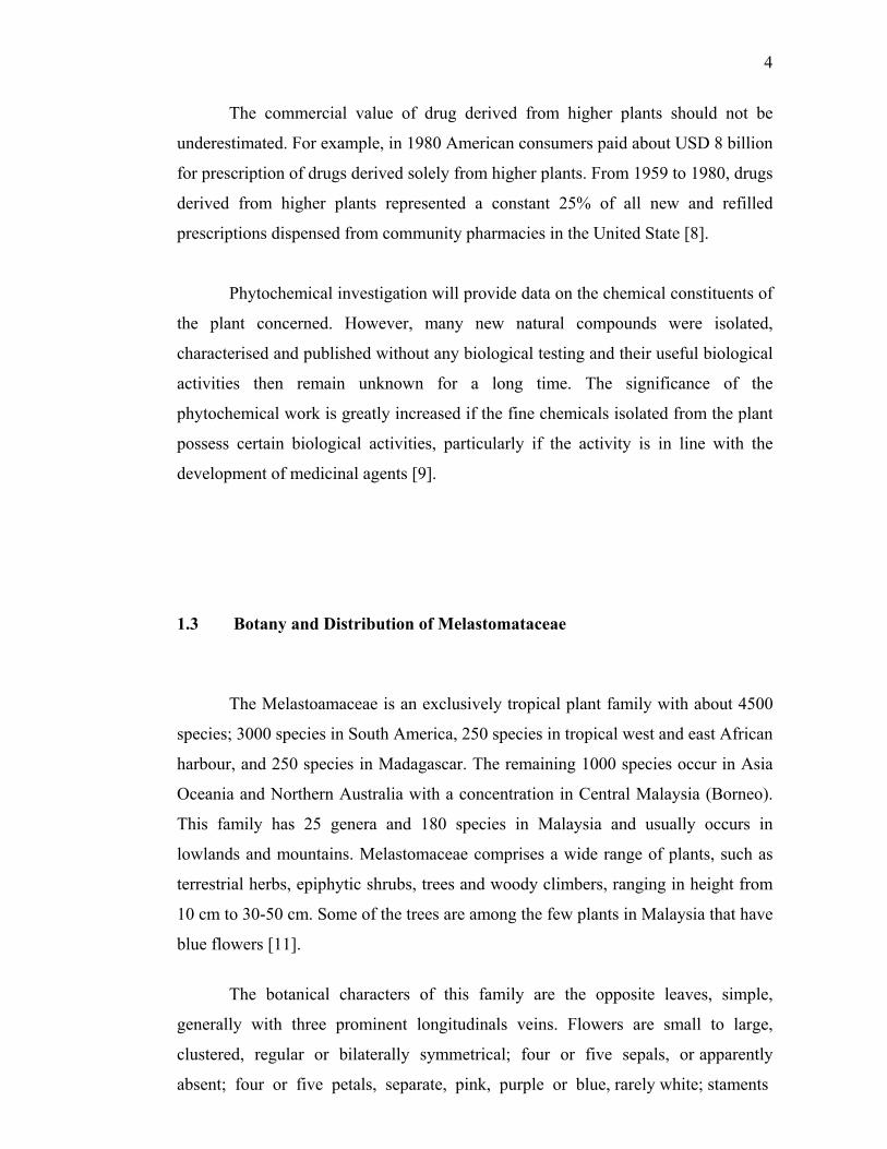
1.3 Botany and Distribution of Melastomataceae
The Melastoamaceae is an exclusively tropical plant family with about 4500
species; 3000 species in South America, 250 species in tropical west and east African
harbour, and 250 species in Madagascar. The remaining 1000 species occur in Asia
Oceania and Northern Australia with a concentration in Central Malaysia (Borneo).
This family has 25 genera and 180 species in Malaysia and usually occurs in
lowlands and mountains. Melastomaceae comprises a wide range of plants, such as
terrestrial herbs, epiphytic shrubs, trees and woody climbers, ranging in height from
10 cm to 30-50 cm. Some of the trees are among the few plants in Malaysia that have
blue flowers [11].
The botanical characters of this family are the opposite leaves, simple,
generally with three prominent longitudinals veins. Flowers are small to large,
clustered, regular or bilaterally symmetrical; four or five sepals, or apparently
absent; four or five petals, separate, pink, purple or blue, rarely white; staments

5
twice as many as the petals, eight or ten, with rather thick, pink or blue stalks (rarely
white) and large yellow, pink or blue anthers; ovary inferior. The fruit is a berry with
many small seeds or with one large seed; in other cases capsular and opening, with
dry or pulpy contents. The family is close to the Myrtales (Myrtaceae) and differs
chiefly in the absence of oil-glands, so that the tissues are not aromatic [11].
The family of Melastomaceae comprises of several genera, for example
Melastoma, Huberia, Lavoisiera, Microlicia, Trembleya, Memycelon, Dissostis,
Tibouchina, Heterocentron and Osbeckia.
1.4 Botany and Distribution of Genus Melastoma
Botanic classification of Melastoma [10]:
Sub-division : Angiospermae
Class : Dicotyledoneae
Sub-class : Archichlamideae
Order : Myrtiflorae
Family : Melastomataceae
Genus : Melastoma
The genus of Melastoma is categorized as shrubs and rarely small trees,
found from the Mascarene Island to Pacific. Most of the species of this genus are
known as ‘senduduk’ and some of them can be distinguished, e.g. M. decemfidum,
Roxb. and M. parkense, Ridl. as ‘senduduk gajah’ and M. imbricatum, Wall as
‘senduduk rimba’ [12]
Plants of Melastoma if repeatedly burnt or cut, and left undisturbed, will
grow as small trees 12-13 ft. high, occasionally even up to 20 ft., and they may be
found in the forest at the edge of the stream, on landslip or in old clearing, and they
are evergreen and flowering throughout the year.
6
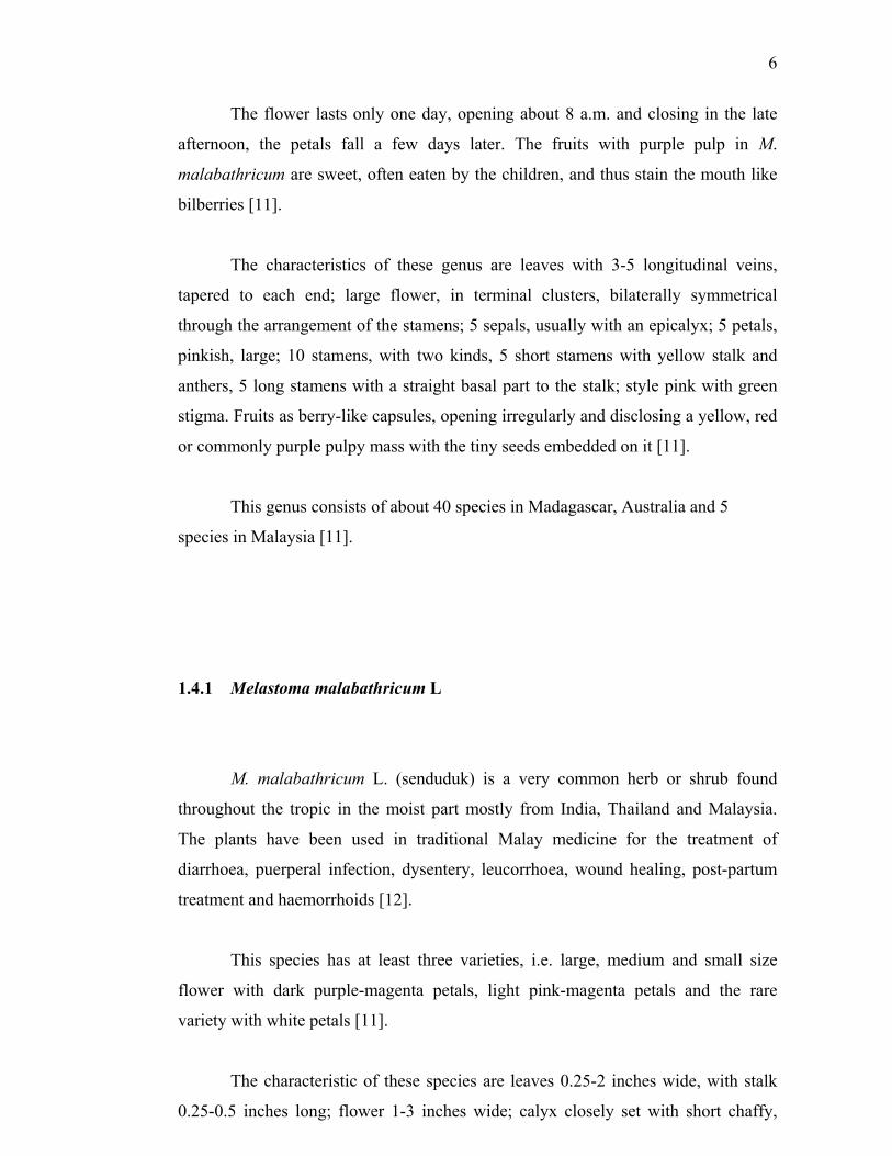
The flower lasts only one day, opening about 8 a.m. and closing in the late
afternoon, the petals fall a few days later. The fruits with purple pulp in M.
malabathricum are sweet, often eaten by the children, and thus stain the mouth like
bilberries [11].
The characteristics of these genus are leaves with 3-5 longitudinal veins,
tapered to each end; large flower, in terminal clusters, bilaterally symmetrical
through the arrangement of the stamens; 5 sepals, usually with an epicalyx; 5 petals,
pinkish, large; 10 stamens, with two kinds, 5 short stamens with yellow stalk and
anthers, 5 long stamens with a straight basal part to the stalk; style pink with green
stigma. Fruits as berry-like capsules, opening irregularly and disclosing a yellow, red
or commonly purple pulpy mass with the tiny seeds embedded on it [11].
This genus consists of about 40 species in Madagascar, Australia and 5
species in Malaysia [11].
1.4.1 Melastoma malabathricum L
M. malabathricum L. (senduduk) is a very common herb or shrub found
throughout the tropic in the moist part mostly from India, Thailand and Malaysia.
The plants have been used in traditional Malay medicine for the treatment of
diarrhoea, puerperal infection, dysentery, leucorrhoea, wound healing, post-partum
treatment and haemorrhoids [12].
This species has at least three varieties, i.e. large, medium and small size
flower with dark purple-magenta petals, light pink-magenta petals and the rare
variety with white petals [11].
The characteristic of these species are leaves 0.25-2 inches wide, with stalk
0.25-0.5 inches long; flower 1-3 inches wide; calyx closely set with short chaffy,

7
silky or silvery scale. This species spread in Madagascar, India to Australia and very
common throughout Malaysia in the lowland and mountain forests, chiefly in open
places and ever flowering [11].
Figure 1.1 Melastoma malabathricum L
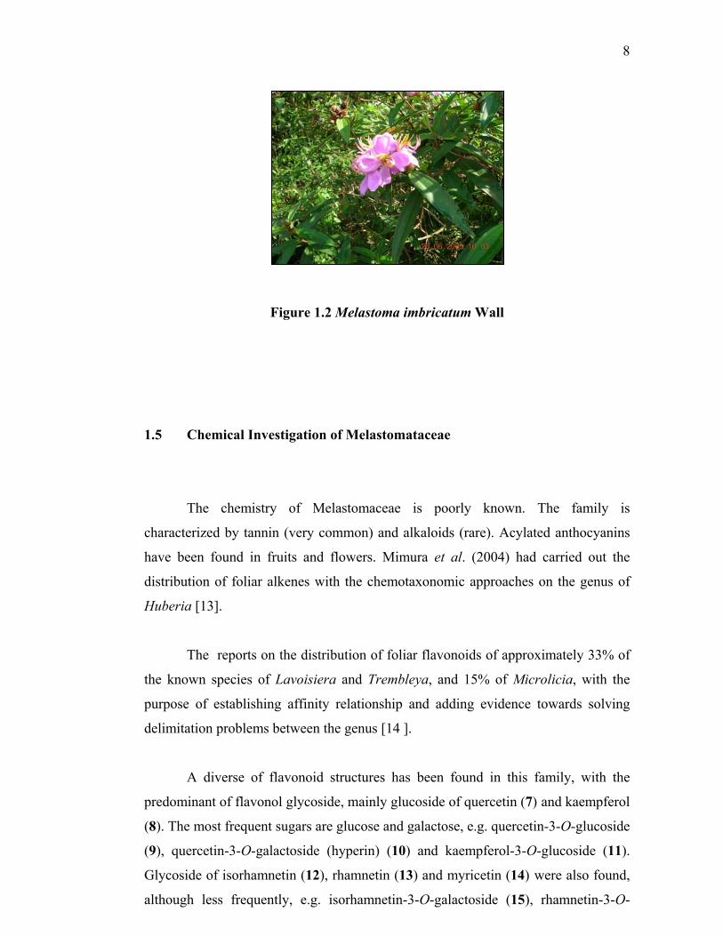
1.4.2 Melastoma imbricatum Wall
The characteristics of this species are the leaves 2-4 inches wide with stalk
0.25-2 inches long, flower 1-2 inches wide, hardly stalked, set in dense cluster; calyx
set with tiny scales. Occasionally spread in the mountain and scarce in the lowlands
[11].
8
Figure 1.2 Melastoma imbricatum Wall
1.5 Chemical Investigation of Melastomataceae
The chemistry of Melastomaceae is poorly known. The family is
characterized by tannin (very common) and alkaloids (rare). Acylated anthocyanins
have been found in fruits and flowers. Mimura et al. (2004) had carried out the
distribution of foliar alkenes with the chemotaxonomic approaches on the genus of
Huberia [13].
The reports on the distribution of foliar flavonoids of approximately 33% of
the known species of Lavoisiera and Trembleya, and 15% of Microlicia, with the
purpose of establishing affinity relationship and adding evidence towards solving
delimitation problems between the genus [14 ].
A diverse of flavonoid structures has been found in this family, with the
predominant of flavonol glycoside, mainly glucoside of quercetin (7) and kaempferol
(8). The most frequent sugars are glucose and galactose, e.g. quercetin-3-O-glucoside
(9), quercetin-3-O-galactoside (hyperin) (10) and kaempferol-3-O-glucoside (11).
Glycoside of isorhamnetin (12), rhamnetin (13) and myricetin (14) were also found,
although less frequently, e.g. isorhamnetin-3-O-galactoside (15), rhamnetin-3-O-

9
OHO
OH OOR
OHOH OHO
OH O
OH
OR
OR1O
OOR4
OR2OR3
OH
OHO
OH OOR
OHOH
OH
ORO
OH O
OH
OR2O
OH O
OR1OH
glucoside (16) and myricetin-3-O-galactose (17). Derivatives of apigenin (18),
luteolin (19) and chrysoeriol (20) were among the flavones found in the
Melastomaceae family e.g. apigenin-7-O-glucoside (21), luteolin-7-O-glucoside (22)
and chrysoeriol-7-O-glucoside (23) [14].
(19) R1 = H, R2 = H (20) R1= CH3, R2 = H (22) R1 = H, R2 = Glc (23) R1 = CH3, R2 = Glc
(18) R = H (21) R = Glc
(7) R = H (9) R = Glc (10) R = Gal
(8) R = H (11) R = Glc
(12) R1, R3 = H, R2 = CH3, R4 = H (13) R1 = CH3, R2, R3 = H, R4 = H (15) R1, R3 = H, R2 = CH3, R4 = Gal(16) R1 = CH3, R2, R3 = H, R4 = Glc
(14) R = H (17) R = Gal
10
OHO
OH
HOO
HOOH
HCO O
COO OO O
O CO
OH
OH
OHCO CO
OH
OHOHOHHO
HO
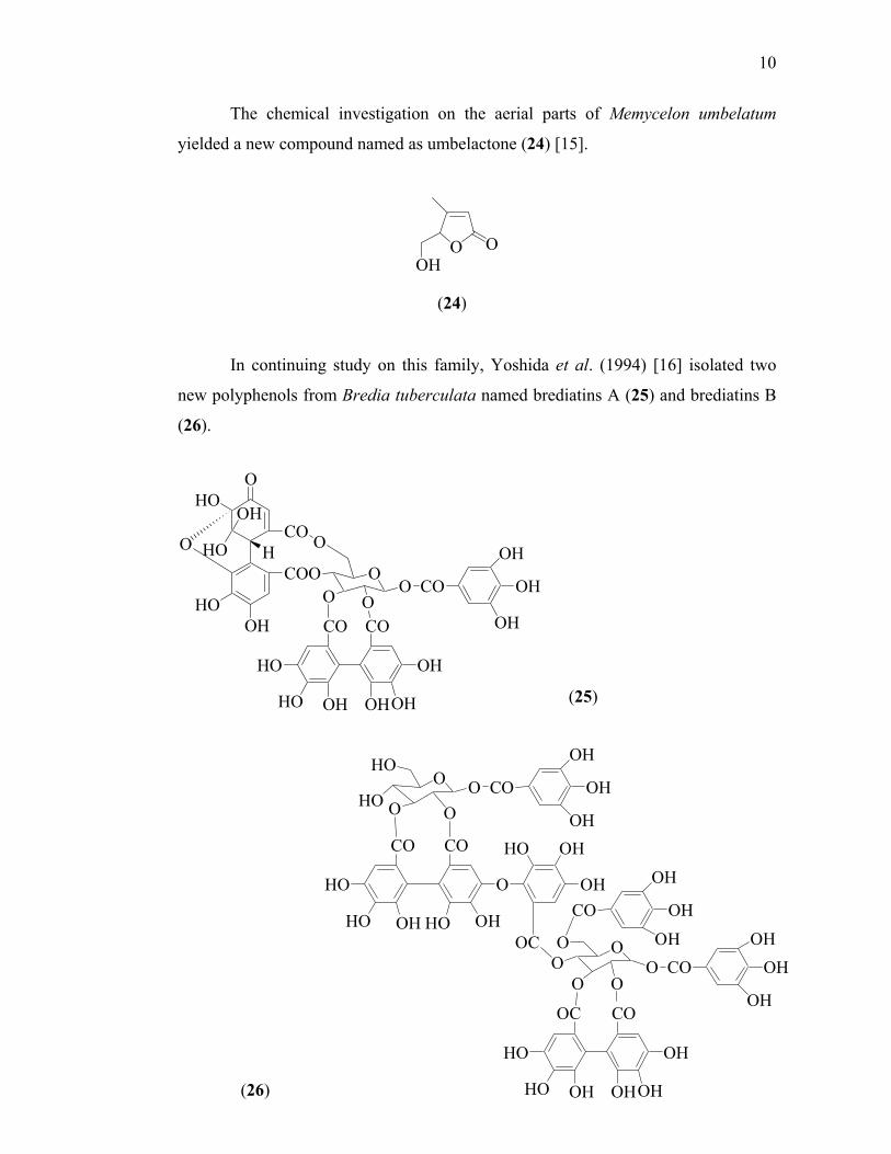
The chemical investigation on the aerial parts of Memycelon umbelatum
yielded a new compound named as umbelactone (24) [15].
In continuing study on this family, Yoshida et al. (1994) [16] isolated two
new polyphenols from Bredia tuberculata named brediatins A (25) and brediatins B
(26).
OOH
O
(24)
(25)
(26)
O
CO CO
HO
HO OH HO OH
HO OH
OH
O
OHO
HOO
OCO
OH
OH
OH
OO
O
OOC
CO
OH
OH
OH
OO CO
OHOH
OHOC CO
OH
OHOHOHHO
HO

11
O
OHHO
HO
HO
HO
HO
HOOH
OOO
O
OHO
CO
OH
OH
OH
CO
HO
HO OH
OC
OOO
O
OO
CO
OC
CO
OC
CO
OH
OHOH
HO
HO OHHO OH
O
HO OH
OH
OO
O
OO CO
OCCO
OH
OH
OHOH
OH
OHHO
HO OHHO OH
OH
CO
HO OH
OH
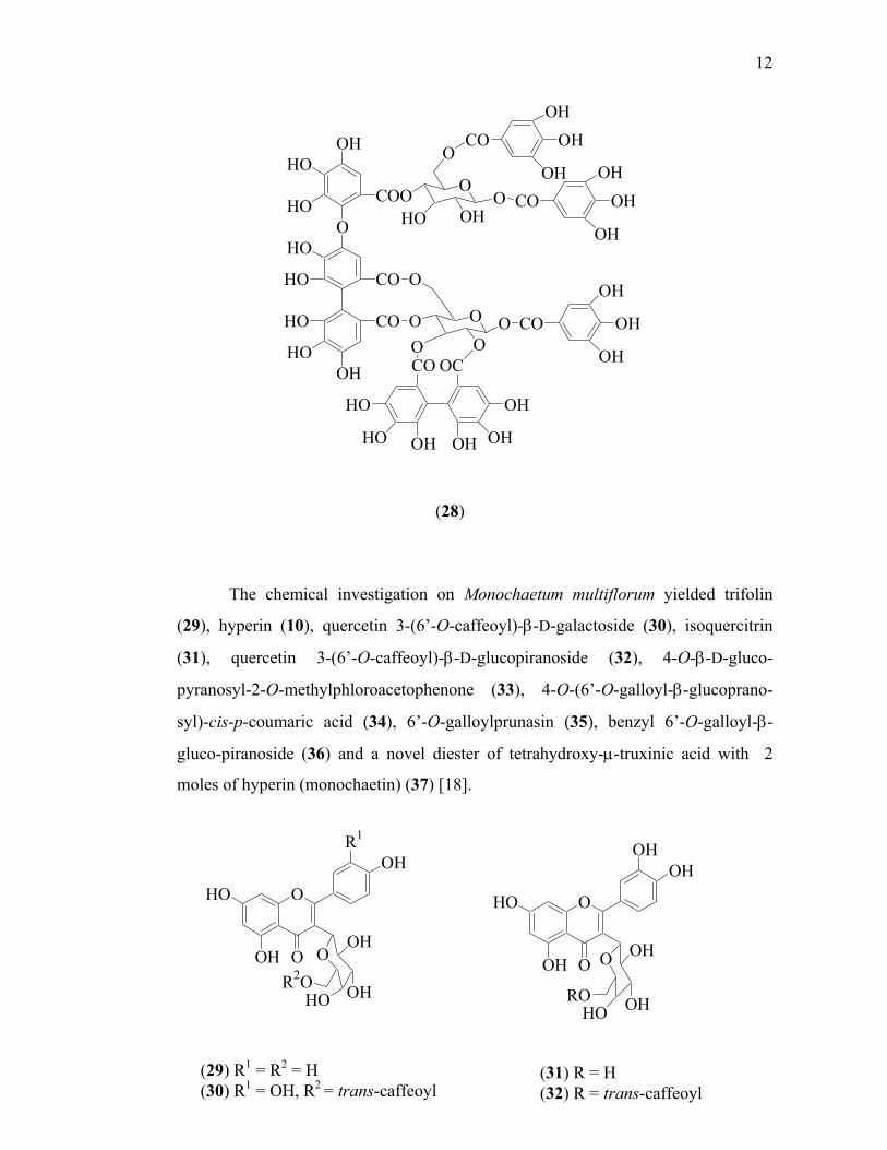
The chromatographic survey of the tannins in this family [17] revealed that
Tibouchina multiflora is rich in tannins, particularly in oligomeric hydrolysable
tannins. Two new oligomeric hydrolysable tannins named nobotanins O (27) and
nobotanins P (28) were isolated from the leaf extract of T. multiflora.
(27)
12
OHO
OH O
OHOH
O OH
ROHO OH
The chemical investigation on Monochaetum multiflorum yielded trifolin
(29), hyperin (10), quercetin 3-(6’-O-caffeoyl)-β-D-galactoside (30), isoquercitrin
(31), quercetin 3-(6’-O-caffeoyl)-β-D-glucopiranoside (32), 4-O-β-D-gluco-
pyranosyl-2-O-methylphloroacetophenone (33), 4-O-(6’-O-galloyl-β-glucoprano-
syl)-cis-p-coumaric acid (34), 6’-O-galloylprunasin (35), benzyl 6’-O-galloyl-β-
gluco-piranoside (36) and a novel diester of tetrahydroxy-µ-truxinic acid with 2
moles of hyperin (monochaetin) (37) [18].
OHO
OH O
R1
OH
OOH
R2O OHHO
(29) R1 = R2 = H (30) R1 = OH, R2 = trans-caffeoyl
(31) R = H (32) R = trans-caffeoyl
(28)
O
OHHO
HO
HO
HO
HO
HOOH
COO
CO
CO
O
O
OOHHO
CO
COOH
OH
OH
OHOH
OH
O
O O
O OO CO
OH
OH
OHCO OC
HO
HO OH OH OH
OH

13
COOHOOHOHO
O
OHO
HO
HO
HO
OO
O
OHO
HO
HOHO
HO HO OH H CN OO
HOHO
O
OHOHO
HOHO
OHO
OH O
OHOH
O OHO OHOH
O O
H
HO OHOOO
OHHOHO
O
O
O
HO
OHOH
OH
OH
OH
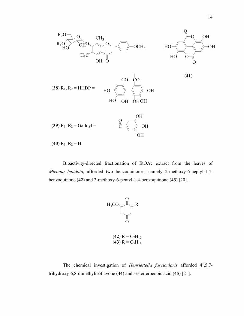
Bioassay-guided fractionation on secreted aspartic protease (SAP) on
Candida albicans of the ethanol extract of twigs and leaves of Miconia myriantha
yielded mattucinol-7-O-[4”,6”-O-(S)-hexahydroxydiphenoyl]-β-D-glucopyranoside
(38), mattucinol-7-O-[4”,6”-di-O-galloyl]-β-D-glucopyranoside (39), mattucinol-7-
O-β-D-glucopyranoside (40) and ellagic acid (41) [19].
OCH3
OOH
OO
OHHOHO
OH
(33) (34)
(35) (36)
(37)
14
O
O
O
O
OH
OHHO
HO
OH
OH
OH
CO
OCH3
OHH3C
O
O
OCH3
OR2O
R1OHO OH
O
O
RH3CO
Bioactivity-directed fractionation of EtOAc extract from the leaves of
Miconia lepidota, afforded two benzoquinones, namely 2-methoxy-6-heptyl-1,4-
benzoquinone (42) and 2-methoxy-6-pentyl-1,4-benzoquinone (43) [20].
The chemical investigation of Henriettella fascicularis afforded 4’,5,7-
trihydroxy-6,8-dimethylisoflavone (44) and sesterterpenoic acid (45) [21].
(38) R1, R2 = HHDP =
(41)
(42) R = C7H15 (43) R = C5H11
CO CO
OH
OHOHOHHO
HO
(39) R1, R2 = Galloyl =
(40) R1, R2 = H

15
OCH3
HO
H3COH O OH
COOH
HHO
HO
OH
OCH3
R1O
H3COH O
OR2
HO
HOHOOC
COOH
HO
COOH
CH3H3C
CH3
H3CH
HOCH3
H
The ethanol extract of Miconia pilgeriana yielded a triterpene compound,
which was characterized as arjunolic acid (46) [22].
Bioactivity-guided fractionation of the ethanol extract of Miconia trailii,
yielded miconioside A (47), miconioside B (48), matteucinol (49), bartogenic acid
(50), arjunolic acid (46) and myrianthic acid (51) [23].
(44) (45)
(46)
(47) R1 = α-L-Ara(1 6)-β-D-Glc, R2 =CH3 (48) R1 = β-D-Api(1 6)-β-D-Glc, R2 = H (49) R1 = H, R2 = H (50)
16
COOHR1
R2O
HO
HOCH2OH
HO
COOH
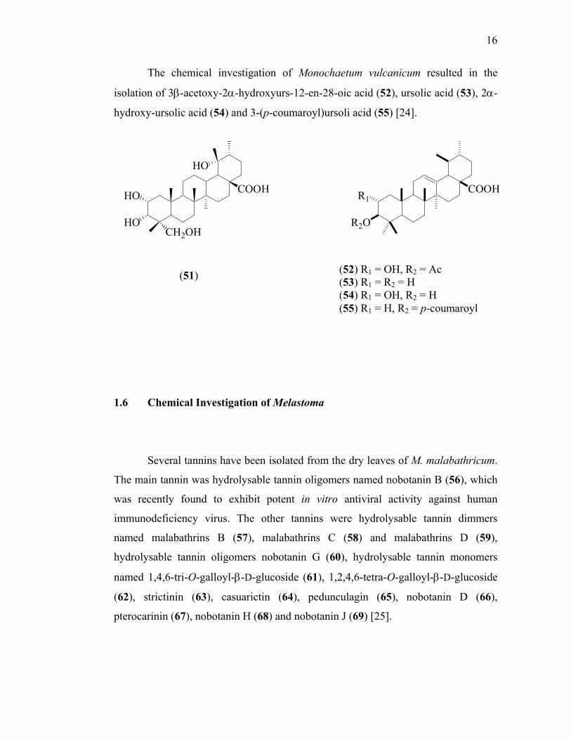
The chemical investigation of Monochaetum vulcanicum resulted in the
isolation of 3β-acetoxy-2α-hydroxyurs-12-en-28-oic acid (52), ursolic acid (53), 2α-
hydroxy-ursolic acid (54) and 3-(p-coumaroyl)ursoli acid (55) [24].
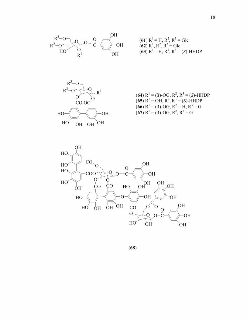
1.6 Chemical Investigation of Melastoma
Several tannins have been isolated from the dry leaves of M. malabathricum.
The main tannin was hydrolysable tannin oligomers named nobotanin B (56), which
was recently found to exhibit potent in vitro antiviral activity against human
immunodeficiency virus. The other tannins were hydrolysable tannin dimmers
named malabathrins B (57), malabathrins C (58) and malabathrins D (59),
hydrolysable tannin oligomers nobotanin G (60), hydrolysable tannin monomers
named 1,4,6-tri-O-galloyl-β-D-glucoside (61), 1,2,4,6-tetra-O-galloyl-β-D-glucoside
(62), strictinin (63), casuarictin (64), pedunculagin (65), nobotanin D (66),
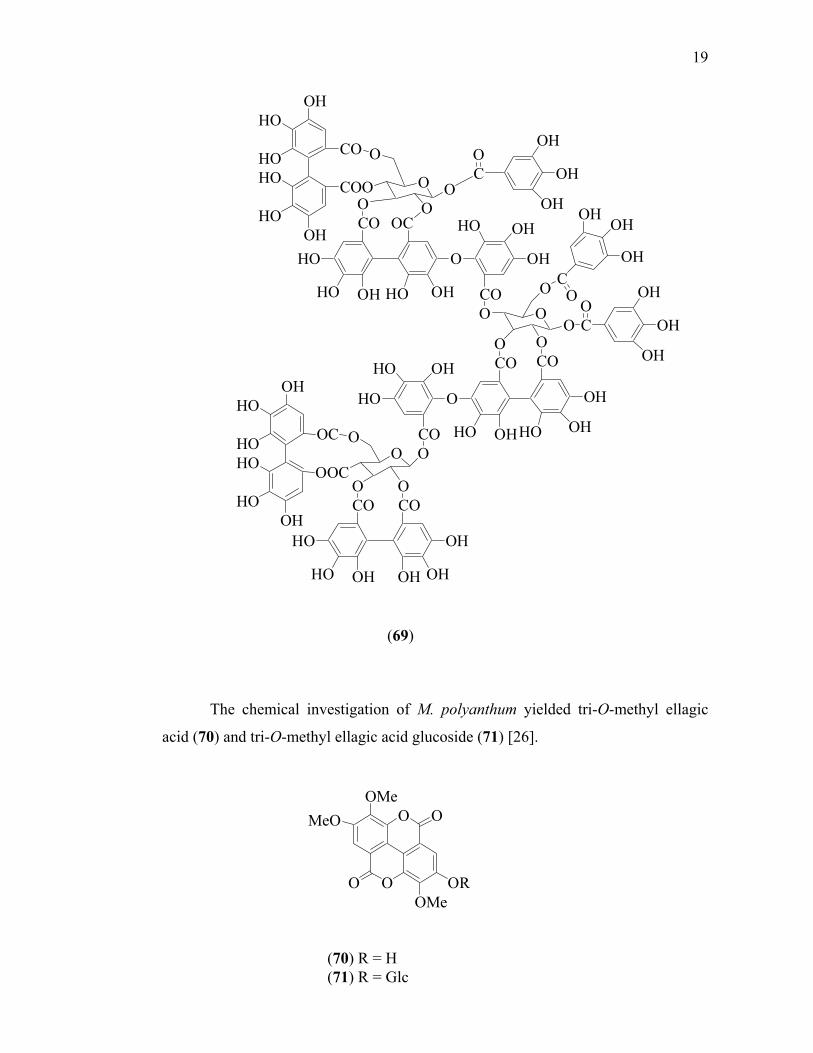
pterocarinin (67), nobotanin H (68) and nobotanin J (69) [25].
(52) R1 = OH, R2 = Ac (53) R1 = R2 = H (54) R1 = OH, R2 = H (55) R1 = H, R2 = p-coumaroyl
(51)

17
CO CO
OH
OHOHOHHO
HO
OH
OH
OH
CO
OHHO
HOHO
HOOH
CO
COO
OO
OO
OCO CO
OHO
HO OHHO OH
HO OH
OH
COO O
O
CH2O
OOCOCO
O
OHHO OH
OH
OHHO
HO
COOR OH
OH
OH
OH
CO
OH
OH
OH
CO
CO
OH
OHOH
O
CO O O
O
O
CO
R5
OCO
HO
HO OHHO OH
OH
HO OH
OH
CO COO O
OO
O
R3
R4
HO
HO OHOH OH
R1
R2
(56) R1, R2 = (β)−OG, R3, R4 = (S)-HHDP, R5 = G (57) R1 = (β)−OG, R2 = OH, R3, R4 = (S)-HHDP, R5 = G (58) R1 = OH, R2 = (β)−OG, R3, R4 = (S)-HHDP, R5 = G
(59) R = CH3 (60) R = H
G = HHDP =
18
OOHO O
OOR3
R2
R1
OH
OH
OH
CO
OH
OH
CO
COO OO
OOCO
OCO
HO
HO OH OH OH
O
HO OH
OH
COO OHO OH
O
O OH
OH
OH
CO
OH
OH
OH
CO
CO
OHOH
OH
HOHO
HO
HO
CO OC
HO
HO OH OH OH
OH
O O
OR1
OO
R3
R2
(61) R1 = H, R2, R3 = Glc (62) R1, R2, R3 = Glc (63) R1 = H, R2, R3 = (S)-HHDP
(64) R1 = (β)-OG, R2, R3 = (S)-HHDP (65) R1 = OH, R2, R3 = (S)-HHDP (66) R1 = (β)-OG, R2 = H, R3 = G (67) R1 = (β)-OG, R2, R3 = G
(68)
19
OH
OH
CO
COO
O
OO O
O
CO OC
HO
HO OH HO OH
O
HO OH
OH
COO O
O OO
O
CO CO
OH
OHHOOHHO
O
HO OH
HO
COOO
O
OOCO OCO CO
HO
HO OH OH OH
OH
OC
OH
OH
OH
OH
OH
CO
OH
OH
OH
CO
CO
OHOH
OH
HOHO
HO
HO
HO
HOHO
HO
The chemical investigation of M. polyanthum yielded tri-O-methyl ellagic
acid (70) and tri-O-methyl ellagic acid glucoside (71) [26].
(69)
O
O
OMeMeO
OMeORO
O
(70) R = H (71) R = Glc

20
OOHOHO
HOHO
O
OHOH
OOH
HO
O
O
OHOH
OHCH3
The polyphenols named strictinin (62), casuarictin (63) and nobotanin B (56)
have been reported from M. normale [16]. While, M. malabathricum with dark
puple-magenta petals contains β-sitosterol (72), α-amyrin (73), uvaol (74), sitosterol
3-O-β-D-glucopiranoside (75), quercetin (7), quercitrin (76) and rutin (77) [27].
HO HO HO
OH
(72) (73) (74)
(76)
(77)
O
OO
HO
OH
OHOH
OOH
OHOH
O
OOH
OHOHCH3
(75)

21
1.7 Bioactivity Investigation on Melastomataceae
Monoamine oxidase type B (MAO-B) activity and free radical scavenger are
elevated in certain neurological disease. Four natural flavonoids, quercitrin (76),
rutin (77), quercetin (7) and isoquercitrin (quercetin-3-O-glucoside) (31), isolated for
the first time from the leaves of M. candidum, were found to inhibit the MAO-B.
These four potent compounds, also exhibited hydroxyl radical scavenging activity.
These important properties may be use for preventing some neurodegenerative
disease [28].
The methanol extract of M. malabathricum L. exhibited attractive antiviral
and cytotoxic activities on murine cell lines. The biological activities of M.
malabathricum could be attributed to the hydrolysable tannin [29].
Dissotis brazae Cogn. was tested for in vitro antiplasmodium activity against
chloroquin-resistant (ENT36). The IC50 was found to be ≤ 10µg/mL [30].
The methanol extract of various Sumatran plants were tested in vivo for
antinematodal activity against Bursaphelenchus xylophilus. In this screening, the root
extract of M. malabathricum showed strong activity [31].
Mattucinol-7-O-[4”,6”-O-(S)- hexahydroxydiphenoyl]-β-D-glucopyranoside
(38) which was isolated from Miconia myriantha exhibited inhibitory effect against
SAP with IC50 of 8.4 µM [22].
Bioactivity-directed fractionation of the leaves of Miconia lepidota in the in
vitro antitumor cytotoxicity assay with Madison Lung Carcinoma (M109) murine
cell line, showed that 2-methoxy-6-heptyl-1,4-benzoquinone (42) and 2-methoxy-6-
pentyl-1,4-benzoquinone (43) were potential anticancer agent [20].
The esterogen receptor (ER) competitive binding experiments revealed higher
affinity of 4’,5,7-trihydroxy-6,8-dimethylisoflavone (44) for ERβ than for

22
OHHO
HOHO
HOOH
O
OO
OHO
H O
O HOH
O
HHO
OHOH
OHOH
OHOH
O
OHOHHO
O
OH
O
OH
HO
OH
OH
OH
OH
OH
OHHO
ERα, isolated from Henriettella fascicularis. In Ishikawa cells, when alkaline
phosphatase was induced by treatment with estradiol, 4’,5,7-trihydroxy-6,8-
dimethylisoflavone (44) mediated a decrease in activity, suggestive of an
antiestrogenic effect [21].
Fatty Acid Synthase (FAS) has been identified as a potential antifungal
target. Bioactivity-guided fractionation on Miconia pilgeriana showed that arjunolic
acid (46) isolated from this plant gave moderate activity against FAS (IC50 27.5
mg/mL) [22].
The antinociceptive effect of the ethanolic extract of M. malabathricum using
acetic acid-induced abdominal writhing test and hot-plate test in mice has been done
by Sulaiman et al. It was demonstrated that the extract (30-300 mg/kg, i.p.) strongly
and dose-dependently inhibited the acetic acid-induced writhing test with an ED50 of
100 mg/kg i.p., suggesting that, the ethanolic extract of M. malabathricum is a
potentially antinociceptive agent that acts at both peripheral and central levels of
nerves [32].
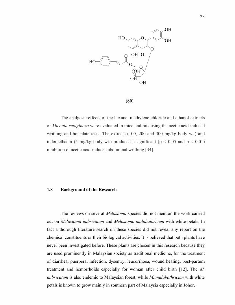
Three active compounds, castalagin (78), procyanidin B-2 (79) and
helichrysoside (80), which were isolated from the leaves of M. candidum possess the
ability to lower blood pressure through a decrease of sympathetic tone as well as due
to direct vasodilatation in SHRs (spontaneously hypertensive rat) [33].
(78) (79)
23
OHO
OH
OH
OH
OO
O
OH
OHOH
O
OHO
The analgesic effects of the hexane, methylene chloride and ethanol extracts
of Miconia rubiginosa were evaluated in mice and rats using the acetic acid-induced
writhing and hot plate tests. The extracts (100, 200 and 300 mg/kg body wt.) and
indomethacin (5 mg/kg body wt.) produced a significant (p < 0.05 and p < 0.01)
inhibition of acetic acid-induced abdominal writhing [34].
1.8 Background of the Research
The reviews on several Melastoma species did not mention the work carried
out on Melastoma imbricatum and Melastoma malabathricum with white petals. In
fact a thorough literature search on these species did not reveal any report on the
chemical constituents or their biological activities. It is believed that both plants have
never been investigated before. These plants are chosen in this research because they
are used prominently in Malaysian society as traditional medicine, for the treatment
of diarrhea, puerperal infection, dysentry, leucorrhoea, wound healing, post-partum
treatment and hemorrhoids especially for woman after child birth [12]. The M.
imbricatum is also endemic to Malaysian forest, while M. malabathricum with white
petals is known to grow mainly in southern part of Malaysia especially in Johor.
(80)

24
1.9 Objectives of the Study
The objectives of this study are to investigate the chemical constituent of two
Malaysian traditional medicinal plants of Melastomataceae i.e. M. malabathricum L.
with white petals and M. imbricatum and to screen the biological activities
(antimicrobial, antioxidant, anti-inflammatory and cytotoxicity) of the crude extracts
and the pure isolated compounds.
1.10 Scopes of the Study
In natural products research, there are two main approaches mostly conducted
by many researchers including chemical investigation and bioactivity testing. Thus,
these two major approaches were also carried out in investigating the Melastomaceae
plants.
The first approach was the extraction, isolation and characterization of the
chemical components from the whole parts of the plants. The extraction was carried
out by successive soxhlet extraction using organic solvents. Isolation procedure was
done by chromatographic technique such as vacuum liquid chromatography and
gravity column chromatography on silica gel as well as sephadex LH-20.
Characterizations of the isolated compounds were carried out by means of physical
and chemical properties such as melting point, optical rotation and chemical reaction.
The structures were elucidated using spectroscopic methods including ultraviolet,
infrared, nuclear magnetic resonance spectroscopies and mass spectrometry.
The second approach was bioactivity screening on the extracts and pure
compounds. The bioactivity assays conducted were antibacterial, antifungal, anti-
inflammatory, antioxidant and cytotoxicity. Antibacterial activity was tested using
disc diffusion methods with four strains of bacteria i.e. Staphylococcus aureus,

25
Bacillus subtilis, Pseudomonas aeruginosa and Escherichia coli. Antifungal activity
was tested against Aspergillus flavus, Aspergillus fumigatus, Aspergillus niger,
Microsporum gypseum, Trichophyton mentagophytes, Trichophyton rubrum,
Cryptococcus neoformans, and Candida albicans. Anti-inflammatory activity was
carried out using 12-O-tetradecanoylphorbol-13-acetate (TPA) induced inflammation
and platelet activating factor receptor binding antagonist on mouse ear oedema and
rabbit platelet, respectively. Antioxidant assay was carried out using lipid
peroxidation and radical scavenging analyzed with ultraviolet and electron spin
resonance, respectively. Cytotoxic activity was conducted using cell culture of
human breast cancer cells (MCF7).

REFERENCES
1. Pieter, L. and Vlietinck, A. J. (2005). Bioguidcd Isolation o f Pharmacologically
Active Plant Components, Still a Valuable Strategy for the Finding o f New Lead
Com pounds. J. Elhnopharmacol. 100(1): 57-60.
2. Cordell, G. A. (1995). Changing Strategies in Natural Product Chemistry.
Phytochemistry. 40(6): 1585-1612.
3. Balandarin, M. F., Kinghom, A. D. and Farnsworth, N. R. (1993). Plant-Derived
Natural Products in Drug Discovery and Development. In: Kinghom, A.D. and
Farnsworth, N.R. Human Medicinal Agents from Plants. ACS Symposium Series
534: W ashington. 2-12.
4. Sim m onds, M. S. J. and Grayer, R. J. (1999). Drug Discovery and Development.
In: W alton, N.J and Brown. D.E. Chemical from Plants Perspectives on the Plant
Secondary Products. Imperial College Press: London. 215-245.
5. Farnsworth, N. R., Akerele, O., Bingel, A. S., Soejarto, D. D. and Guo, Z.
(1985). M edicinal Plants in Therapy. Bull. IV. H. O. 63: 965-981.
6. Cragg, G. M., Boyd, M. R., Cardellina, J. H., Grever, M. R., Schepartz, S. A.,
Snader, K.M. and Suffness, M. (1993). In: Kinghom , A.D. and . Farnsworth, N.R
Human Medicinal Agents from Plants. ACS Symposium Series 534:
W ashington. 80-95.
7. Kingston, D. G. I. (1993). Taxol, an Exciting Anticancer Drug from Taxus
brevifolia. In: Kinghom . A.D. and Farnsworth, N.R. Human Medicinal Agents
from Plants ACS Symposium Series 534: Washington. 138-148.

192
8. Balandrin, M. F., Klocke, J. A.. Wurtele, E. S. and Bollinger, W. H. (1985).
Natural Plant Chemicals: Sources o f Industrial and Medicinal Material. Science.
228:1154-1160.
9. Meyer, B. N „ Ferrifni. N. R.. Putnam, J. E„ Jacobsen, L. B., Nichols, D. E. and
M cLaughlin, J. L. (1982). Brine Shrimp: A Convenient General Bioassay for
Active Plant Constituents. Planta Med. 45(1). 31-34.
10. Com er, E. J. H. (1965). Wayside Trees o f Malaya. Malayan Nature Society:
Kuala Lumpur.Vol. I .
11. W hitmore, T. C. (1972). Tree Flora o f Malaya. Vol. I. Longman: Kuala
Lumpur.
12. Burkill, I. H. (1966). A Dictionary o f the Economic Products o f Malay
Peninsula. Ministry o f Agriculture and Co-Operatives: Kuala Lumpur.
13. M imura, M. R. M., Salatino. A., Salatino. M. L. F. (2004). Distribution o f
flavonoids and the taxonomy o f Huheria (Mclastomataceae). Biochem Syst
Ecol 32(1): 27-34.
14. Bomfim-Patricio, M. C., Salatino, A., Martins, A. B., Wurdack, J. J. and
Salatino, M. L. F. (2001). Flavonoids o f Lavoisiera, Microlicia, and Trembleya
(M elastom ataceae) and their taxonomic meaning. Biochem Syst. Ecol. 29(7):
71 1-726.
15. Agarwal, S. K. and Rastogi, R. P. (1978). Umbeiactone (4-hydroxymethyl-3-
methyl-but-2-ene-4,1 -olide) New Constituent o f Memycelon umbelatum.
Phytochemistry. 17(9): 1663-1664.
16. Yoshida, T. Arioka, H., Fujita, T.. Xin, M. C. and Okuda, T. (1994). Monomeric
and dimeric Hydrolysable Tannins from two Melastomataceous species.
Phytochemistry. 37(3): 863-866.

193
17. Yoshida, T., Amakura, Y„ Yokura, N ., Ito, H., Isaza, J. H., Ramirez, S , Pelaez,
D. P. and Renner. S. S. (1999). Oligomeric hydrolysable tannins from
Tibouchina multiflora. Phytochemistry. 52(8): 1661-1666.
18. Isaza, J. H., Ito, H.. and Yoshida, T. (2001). A Flavonol glycoside-lignan ester
and accom panying acylated glucosides from Monochaetum multiflorum.
Phytochemistry. 58(2): 321-327.
19. Cong Li, X.. Jacob, M. R., Pasco, D. S., ElSohly, H. N., Nimrod, A. C., Walker,
L. A. and Clark. A. M. (2001). Phenolic Compounds from Miconia myriantha
Inhibiting Candida Asparlic Protease../. Nat. Prod. 64(10): 1282-1285.
20. Gunatilaka, A. A. L., Berger, J. M., Evans, R., Miller, J. S., Wisse, J. H.,
Nedderm ann, K. M., Bursuker, 1. and Kingston, D. G. I. (2001). Isolation,
Synthesis and Structure-Activitv Relationships o f Bioactive Benzoquinones from
Miconia lepidota from Suriname Rainforest../. Nat. Prod. 64(1): 2-5.
21. Calderon A. I., Tcrrcaux, C., Schenk, K., Pattison, P., Burdette, J. E., Pezzuto, J.
M., Gupta, M. P. and Hostettmann, K. (2002). Isolation and Structure
Elucidation o f an Isoflavone and a Seslertcrpcnoic Acid from Henriettella
fascicularis. J. Nat. Prod. 65(12): 1749-1753.
22. Cong Li, X., Joshi, A. S., ElSohly, H. N., Khan, S. I., Jacob, M. R., Zhang, Z.,
Khan, 1. A., Ferreira, D.. Walker, L. A., Broedel, S. E., Raulli, R. E. and Cihlar,
R. L. (2002). Fatty Acid Synthase Inhibitors from Plants: Isolation, Structure
Elucidation and SAR Studies. / Nat. Prod. 65(12): 1909-1914.
23. Zhang, Z., ElSohly, H. N., Cong Li, X., Khan, S. I., Broedel, S. E., Raulli, R. E.,
Cihlar, R. L. and Walker, L. A. (2003). Flavanone Glycoside from Miconia
trailii. J. Nat. Prod. 66(1): 39-41.

194
24. Chaturvedula, V. S. P., Gao, Z.. Jones, S. H., Feng, X., Hecht, S. M. and
Kingston D. G. I. (2004). A New Ursane Triterpene from Monochaetum
vulcamcum (hat Inhibits DNA Polymerase p Lyase../. Nat. Prod. 67(5): 899-901.
25. Yoshida, T., Nakata. F.. Hosotani, K.. Nitta, A. and Okuda, T. (1992). Dimeric
Hydrolysable Tannins from Melasloma inalabalhricum. Phytochemistry. 31(8):
2829-2833.
26. Liu, S. R. (1986). Chemical Constituents o f Melasloma polyanthum. Zhong Yao
TongBao. I 1(12): 42-43.
27. Nuresti, S., Hack. S. H. and Asari. A. (2003). Chemical Components o f
Melasloma malahathricum. ACGC Chem. Res. Commun. 16: 28-33.
28. M?i, H. L., Rong. D. L . Lee. Y. S., Ling. L. Y„ Kun, Y. Y. and Wen, C. H.
(2001). M onoamine Oxidase B and Free Radical Scavenging Activities o f
Natural Flavonoids in Melasloma candidum D. Don. J. Agric. Food Chem.
49(1 I): 5551-5555.
29. Lohezic-Le Dcvehat. F. Bakhtiar, A.. Bczivin, C., Amoros, M and Boustie, J.
(2002). Antiviral and Cytotoxic o f Some Indonesian Plants. Filoterapia. 73(5):
400-405.
30. Om ulokoli, E., Khan, B. and C'hhabra, S. C. (1997). Antiplasmodial Activity o f
Four Kenyan Medicinal Plants../. Fthnopharmacol. 56(2): 133-137.
31. Alen, Y„ Nakajima, S., Nitoda, T., Baba, N., Kanzaki, H. and Kawazu, K.
(2000). Antinematodal Activity o f Some Tropical Rainforest Plants against
Pinewood Nematode, Bursaphelenchus xylophylus. Z. Naturforsch. 55(3-4): 259-
299.
32. Sulaiman, M. R., Somchit, M. N„ Israf, D. A., Ahmad, Z. and Moin, S. (2004).
Antinociceptive Effect o f Melasloma Malahathricum Ethanolic Extract in Mice.
Filoterapia. 75(7-8): 667-672.

195
33. Tang Cheng, J., Lin Hsu, F. and Fen Chen, H. (1993). Antihypertensive
Principles from the Leaves o f Melastoma candidum. Planla Med. 59: 405-407
34. Spessoto, M. A., Ferreira, D. S., Crotti, A. E. M„ Silva, M. L. A. And Cunha, W.
R. (2003). Evaluation o f the Analgesic Activity o f Extract o f Miconia rubiginosa
(M elastom aceae). Phytomedicine. 10: 606-609.
35. Banerji, A. and Ray, R. (1981). Auranamide, a New Phenylalanine Derivative
Isolated from Piper aurantiacum Wall. Indian J. Chem. 20B: 597-598
36. Gu, Z. B., Yang, J. G., Liu, W. Y., Li, T. Z., Qiu, Y. and Zhang, W. D. (2002). A
New A lkaloid from Patrinia scabra. Chin. Chem. Lett. 13(10): 957-958.
37. M ahato, S. B. and Kundu A. P. (1994). I3C NMR Spectra o f Pentacyclic
Triterpenoids-A Compilation and some Salient Feature. Phytochemistry. 37(6):
1517-1575.
38. Silverstein, R. M., Bassler, G. C. and Morril, T. C. (1991). Spectrometric
Identification o f Organic Compounds. John Willey and Sons Inc.: New York.
39. W illiam, D. H. and Fleming, I. (1995). Spectroscopic Methods in Organic
Chemistry. 5lh Ed. The McGraw-Hill Companies: London.
40. Ishiguro, K., Nagata, S., Fukumoto, H., Yamaki, M., Takagi, S. and Isoi, K.
(1991). A Flavonol Rhamnoside from Hypericum japonicum. Phytochemistry.
30(9): 3152-3154.
41. Mabry, T. J., Markham, K. R. and Thomas, M. B. (1970). The Systematic
Identification o f Flavonoids. Springer-Verlag: Berlin.
42. M arkham, K. R. (1982). Techniques o f Flavonoid Identification. Academic Press
Inc.: London.

196
43. Beck, P.O.D., Dijoux, M.G., Cartier, G. and Mariote, A.M. (1998). Quercitrin 3’-
Sulphate from Leaves o f Leea guinensis. Phytochemistry. 47(6): 1171-1173.
44. Slowing, K., Sollhuber, M., Carretero, E. and Villar, A. (1994). Flavonoid
Glycoside from Eugenia jambos. Phytochemistry. 37(1): 255-258.
45. Ohm ura, W, Ohara, S., Hashida, K.., Aoyama, M. and Doi, S. (2002).
Hydrotherm olysis o f Flavonoids in Relation to Steaming o f Japanese Larch
Wood. Holzforschung. 56(5): 493-497.
46. Skaltsa, H., Verykokidou, E., Harvala, C., Karabourniotis, G. and Manetas, Y.
(1994). UV-B Protective Potential and Flavonoid Content o f Leaf Hairs o f
Quercus ilex. Phytochemistry. 37(4): 987-990.
47. Tom as-Barberan, F. A., Gil, M. L, Ferreres, F. and Tomas-Lorente, F. (1992).
Flavonoid p-Coum aroyl and 8-Hydroxyflavone Allosylglucoside in Some
Labiatae. Phytochemistry. 31(9): 3097-3102.
48. Harbom e, J. B., Mabry, T. J. and Mabry, H. (1975). The Flavonoids I. Academic
Press: New York.
49. Fiorini, C., David, B., Fouraste, I. ami Vercauteren, J. (1998). Acylated
Kaempferol Glycoside from Laurus nobilis Leaves. Phytochemistry. 47(5): 821-
825.
50. Tanaka, R., Tsujimoto, K. and Matsunaga, S. (1999). Two Serratane Triterpenes
from the Stem Bark o f Pice a jezoensis var. hondoensis. Phytochemistry. 52(6):
1581-1585.
51. Ikuta, A. and Itokawa, H. (1988). Triterpenoid o f Paeonia japonica Callus
Tissue. Phytochemistry. 27(9): 2813-2815.

197
52. Siddiqui, S., Hafeez, F., Begum, S. and Siddiqui, B. S. (1988). Oleanderol, A
New Pentacyclic Triterpene from the Leaves o f Nerium oleander. J. Wat. Prod. 51(2): 229-233
53. Kuo, Y. H., Lee. S. H. and Lai, J. S. (2000). Constituents o f the Whole Herb o f
Clinoponium laxiflorum. J. Chin. Chem. Soc. 47: 241-246.
54. Bohm, B. A. (1998). Introduction to Flavonoids. Harwood Academic Publishers:
Netherlands.
55. Corticchiato. M .f Bernardini, A., Costa, J., Bayet, C., Saunois, A. and Voirin, B.
(1995). Free Flavonoids Aglycon from Thymus Herba Barona and its
M onoterpenoid Chemotype. Phytochemistry. 40(1): 115-120.
56. Budavari, S. (2001). The Merck Index. An Encyclopaedia o f Chemicals. Drugs,
and Biological. 13th ed. Merck & Co. Inc.: New Jersey.
57. Hadizadeh, F., Khalili, N., Hosseinzadeh, H. and Khair-Aldine, R. (2003).
Kaempferol from Saffron Petals. Iranian J. Pharm. Res: 251-252.
58. Hamzah, A. S. and Lajis, N. (1998). Chemical Constituents o f Hedyotis
herbacea. ASEAN Review o f Biodivers, y and Environmental Conservation
(ARBEC). Article II. May 1998: 1-6.
59. Iwashina, T., M atsumoto, S., Nishida, M. and Nakaike, T. (1995). New and Rare
Flavonol Glucoside from Asplenium trichomanes-ramosum as Stable
Chem otaxonom ic Markers. Biochem. Syst. Ecol. 23(3): 283-290.
60. Nasser, A. and Singab, B. (1998). Acylated Flavonol from Ammi Majus L.
Phytochemistry. 49 (7): 2177-2180.
61. Lu, Y. and Foo, L. Y. (1997). Identification and Quantification o f Major
Polyphenols in Apple Pomace. Food Chem. 59(2): 187-194.
198
62. Rios, J.L. and Recio, M.C. (2005). Medicinal Plants and Antimicrobial Activity.
J. Ethnopharmacol. 100(1-2): 80-84
63. Vanden Berghe, D. A. and Vlietinck, A. J. (1991). Screening M ethods for
Antibacterial and Antiviral agents from Higher Plants. In: K. Hostettmann (Ed.).
Methods in Plant Biochemistry. Academic Press: London. 47-69
64. Black, J. G. (1999). Microbiology, Principles and Exploration. 4°’ Ed. John
W iley and Sons Inc.: New York.
65. Nester, W. N „ Anderson, D. G., Robert. C. V., Pearsall, N. N„ Nester, M. T.,
Hurley, D. (2004). Microbiology: A Human Perspective. 4lh ed. McGraw Hill
C om panies Inc.: America.
66. Hardy, S. P. (2002). Human Microbiolog}’. Taylor and Francis: London.
67. Bauman, R. W. (2004). Microbiology. Pearson Benjamin Cumming: San
Francisco.
68. W illiams, R. A. D., Lambert, P. A. and Singleton, P. (1996). Antimicrobial Drug
Action. A Medical Perspective Book. Bios Scientific Publisher: Oxford.
69. W illiamson, G., Rhodes, M. J. C. and Parr, A. J. (1999). Disease Prevention and
Plant Dietary Substance. In: Walton, N.J. and Brown, D.E. (eds). Chemical from
Plants Perspectives on the Plant Secondary Products. Imperial College Press:
London. 251-276.
70. Noguchi, N. and Niki, E. (1998). Chemistry o f Active Oxigen Species and
Antioxidant. In: Papas, A.M. (ed). Antioxidant Status, Diet. Nutrition and
Health. CRC Press: Washington. 3-20.

71. N iki, E. (1993). Antioxidant Defense in Eukariotic Cells: An Overview. In: Poli,
G., Albino, E. and Dianzam, M.U.(eds). Free radicals: From Basic Science to
Medicine (Molecular and Cell Biology Update) Birkhauser Verlag: Basel. 365-
373.
72. W ertz, J. E. and Bolton, J. R. (1972). Electron Spin Resonance. Elementary
Theory and Practical Applications. McGraw-Hill Book Company: New York.
73. Noda, Y„ Kohno, M„ Mori, A. and Packer, L. (1999). Automated Electron Spin
Resonance Free Radical Detector Assays for Antioxidant Activity in Natural
Extract. In. Abclson, J.N. and Simon, M.l. (eds). Methods in Enzymology.
Oxidants and Antioxidants Part A. Academic Press: New York. 29-34.
74. Shi, H. (2001). Introducing Natural Antioxidants. In: Pokomy, J. Yanishlieva, N.
and Gordon, M. (eds). Antioxidant in Food Practical Application. CRC Press:
W ashington. 147-158.
75. M iddleton Jr. E., Kandaswami, C. and Thcoharides, T. C. (2000). The Effect o f
Plant Flavonoids on Mamalian Cell: Implication for Inflammation, Heart Disease
and Cancer. Pharm Rev. 52: 673-751.
76. Hall, C. (2001). Sources o f Natural Antioxidants; Oilseeds, Nuts, Cereals,
Legumes, Animal Products and Microbial Sources. In: Pokomy, J. Yanishlieva,
N . and Gordon, M. (eds). Antioxidant in Food Practical Application. CRC Press:
W ashington. 159-209.
77. Cakir, A., Mavi, A., Yildirim, A., Duru, M. E., Harmandar, M. and Kazaz, C.
(2003). Isolation and Characterization o f Antioxidant Phenolic Compounds from
the Aerial Parts o f Hypericum hissopifolium L. by Activity-Guided Fractionation.
J. Ethnopharmacol 87(1): 73-83.
199

200
78. Heijnen, C. G. M., Haenen, G. R. M. M., Van Acker, F. A. A., Van Der Vijgh,
W. J. F. and Bast, A. (2000). Flavonoids as Peroxynitrite Scavengers: The Role
o f the Hydroxyl Groups. Toxicolog\> in Vitro. 15(1): 3-6.
79. Heim, K. E., Tagliaferro, A. R. and Bobilya D. J. (2002). Flavonoid
Antioxidants: Chemistry, Metabolism and Structure-Activity Relationship. ./.
Nutr. Biochem. 13(10): 572-584.
80. Evans, C. A. R., Miller, N. J. and Paganga, G. (1995). Structure-Antioxidant
Activity Relationships o f Flavonoids and Phenolic Acid. Free Radic. Biol. Med.
20(7): 933-956.
81. Yamaguchi, T., Hitoshi, T., Matoba, T., Tero, J. (1998). HPLC method for
evaluation o f the free radical-scavenging activity o f food by using 1,1-diphenyl-
2-picrrylhydrazyl. Biosci. Biotech. Biochem. 62: 1201-1204
82. Bondet, V., W illiams, W. B. and Berset, C. (1997).. Kinetic and Mechanism o f
Antioxidant Activity using the DPPH’ Free Radical Method. Lebensm. Wiss. U.
Technol. 30: 609-615.
83. Yu, L. (2001). Free Radical Scavenging Properties o f Conjugated Linoleic
Acids. J. Agric. Food Chem. 49(7): 345'’.-3456.
84. Cuvelier, M. E„ Richard, H. and Berset, C. (1992). Comparison o f Antioxidative
Activity o f Some Acid-phenols: Structure-activity Relationship. Biosci. Biotech.
Biochem. 56(2): 324-330.
85. Dugas Jr., A. J., Castaneda-Acosta, J., Bonin, G. C., Price, K. L., Fischer, N. H.,
W inston, G. W. (2000). Evaluation o f the Total Peroxyl Radical-Scavenging
Capacity o f Flavonoids: Structure-Activity Relationships. J. Nat. Prod. 63(2):
327-331.

201
86. Gallin, J. I., Goldstein, I. M. and Snyderman, R. (1992). Overview. In: Gallin,
J.I., Goldstein, I.M. and Snyderman, R. Inflammation Basic Principles and
Clinical Correlates. 2nd ed. Raven Press: New York. 1-4
87. Sell, S., Berkomer, I. and Max. E. E. (1996). Immunology, Immunopatology and
Immunity. 5th ed. Appleton and Lange, Stamford: Connecticut.
88. Hanahan, D. J. (1986). Platelet Activating Factor: A Biologically Active
Phosphoglyceride. Ann. Rev. Biochem. 55: 483-509.
89. Braquet. P, Touqui, L., Shen, T. Y. and Vargaftig, B. B. (1987). Perspectives in
Platelet-activating Factor Research. Pharmacol. Rev. 39(2): 97-145.
90. Braquet, P. and Plcszczynski, M. R. (1987). Platelet-activating Factor and
Cellular Immune responses. Immunology Today. 8(11): 345-352.
91. Goldring, W. P. D„ Alexander, S. P. H., Kendall, D. A. and Pattenden, G.
(2005). Novel Phomaclin Analogue as PAF Receptor Ligands. Bioorg. Med.
Chem. Lett. 15: 3263-3266.
92. Cam pbell, W. B. (1990). Lipid-derived Autocoid: Eicosanoids and Platelet-
activatin Factor. In: Rail, T.W., Nies, A.S, and Taylor, P. (Eds). Goodman and
Gilman’s the Pharmacological Basis o f Therapeutics. 8,h ed. Pergamon Press:
New York. 600-604
93. Lloret, S. and Moreno, J. J. (1995). Effects o f an Anti-inflammatory Peptide
(Antiflammin 2) on Cell Influx, Eicosanoid Biosynthesis and Oedema Formation
by Arachidonic Acid and Tetradecanoyl Phorbol Dermal Application. Biochem.
Pharmacol. 50(3): 347-353.
94. Jeenapongsa, R., Yoovathaworn, K„ Sriwatanakul, K. M., Pongprayoon, U. and
Sriwatanakul, K. (2003). Anti-inflammatory Activity o f (£)-l-(3 ,4-
dimethoxyphenyl)butadiene from Zingiber cassumunar Roxb. J.
Ethnopharmacol. 87(2-3): 143-148.

202
95. Parker, J., Daniel, L. W. and Waite, M. (1987). Evidence o f Protein Kinase C
Involvement in Phorbol Diesler-stimulated Arachidonic Acid Release and
Prostaglandin Synthesis../. Biol. Chem. 262(11): 5385-5393.
96. M ontrucchio, G., Alloatti, G. and Camussi, G. (2000). Role o f Platelet Activating
Factor in Cardiovascular Pathophysiology. Physiol. Rev. 80(4): 1669-1699.
97. Jantan, I., Kang, Y. H., Suh, D. Y. and Han, B. H. (1996). Inhibitory Effects of
M alaysian Medicinal Plants on the Platelet Activating Factor (PAF) Receptor
Binding. Nal. Prod. Sci. 2(2): 86-89.
98. Albanes, D. and Hartman, T. (1998). Antioxidants and Cancer: Evidence from
Human Observational Studies and Intervention Trials. In: Papas, A.M. (ed).
Antioxidant Status, Diet, Nutrition and Health. CRC Press: Washington. 497-
544.
99. Lewis, W. H. and Lewis, M. P. F. E. (2003). Medical Botany. Plants Affecting
Human Health. 2nd cd. John Wiley and Sons Inc.: New Jersey.
100. M osmann, T. (1983). Rapid Colorimetric Assay for Cellular Growth and
Survival: Application to Proliferation and Cytotoxic Assays. J. Immunol.
Methods. 65(1-2): 55-63.
101. Roche. (2003). Cell Proliferation Kit I (MTT). Roche Applied Science:
Germany. Cat. No. 1 465 007.
102. Rodgers, E. H. and Grant, M. H. (1998). The Effect o fth e Flavonoids, Quercetin,
Myricetin and Epicatcchin on the Growth and Enzyme Activities of MCF7
Human Breast Cancer Cell. Chemico-Biological Interactions. 116: 213-228.
103. Zavala, M. A., Perez, G. S. and Perez G. R. M. (1997). Antimicrobial Screening
o f Some Medicinal Plants. Phytother. Res. 11(5): 368-371.

203
103 Arias, M. E., Gomez, J. D.. Cudmani, N. M., Vattuone, M. A. And Isla, M. I.
(2004). Antibacterial Activity o f Ethanolic and Aqueous Extract o f Acacia
aroma Gill. Ex Hook et Arn. Life Sci. 75(2): 191-202.
104 Kikuzaki, H. and Nakatani N. (1993). Antioxidant Effect o f Some Ginger
Constituents. / Food Sci. 58(6): 1407-1410.
105 Tagashira, M. and Ohtake. Y. (1998) A New Antioxidative 1,3-Benzodioxole
from Melissa officinalis. Planla Med. 64(6): 555-558.
106 Ohtani, II., Gotoh. N., Tanaka. J.. Higa, T„ Gyamfi. M.A. and Aniya, Y. (2000).
Thonningianins A and B. New Antioxidants from the African Medicinal Herb
Thonningia sanguinca. J. Nat. Prod. 63(5): 676-679.
107 Nik M usa'adah. M.and Rasadah. M.A. (2000). Inhibitor Effect o f Solanum
torvum on TPA-lnduccd Mouse Ear O edem a../. Trop. Forest Prod. 6: 222-224.
108 Valone, F.H.. Coles. E.. Reinhold, V.R and Goctzl, E.J. (1982). Specific Binding
o f Phospholipid Platelet-Activating factor by Human Platelet. J. Immunol.
129(4): 1637-1641.
109 Yang, 11. K., Sun. I). Y. and Han, B. H. H995). Isolation and Characterization of
Platelet Activating Factor Receptor Binding Antagonist from Biota orientalis.
Planta Med. 61(1): 37-40.