medical applications of epidermal stem...
TRANSCRIPT
Medical applications of epidermalstem cells
Gaelle Lapouge and Cedric Blanpain, IRIBHM, Universite Libre deBruxelles, 808 route de Lennik, 1070 Brussels, Belgium
Table of Contents1. Introduction . . . . . . . . . . . . . . . . . . . . . . . . . . . . . . . . . . . . . . . . . . . . . . . . . . . . . . . . . . . . . . . . . . . . . . . . . . . . . . . 12. The skin stem cells . . . . . . . . . . . . . . . . . . . . . . . . . . . . . . . . . . . . . . . . . . . . . . . . . . . . . . . . . . . . . . . . . . . . . . . . . 2
2.1. The different cell lineages of the skin epidermis . . . . . . . . . . . . . . . . . . . . . . . . . . . . . . . . . . . . . . . . . . 22.2. stem cell niche in skin . . . . . . . . . . . . . . . . . . . . . . . . . . . . . . . . . . . . . . . . . . . . . . . . . . . . . . . . . . . . . . . 3
3. Skin stem cells for the treatment of third-degree burns . . . . . . . . . . . . . . . . . . . . . . . . . . . . . . . . . . . . . . . . . . . 44. Cellular therapy for inherited genetic skin diseases . . . . . . . . . . . . . . . . . . . . . . . . . . . . . . . . . . . . . . . . . . . . . . 55. Reprogramming of skin stem cells into pluripotent stem cells . . . . . . . . . . . . . . . . . . . . . . . . . . . . . . . . . . . . . 56. References . . . . . . . . . . . . . . . . . . . . . . . . . . . . . . . . . . . . . . . . . . . . . . . . . . . . . . . . . . . . . . . . . . . . . . . . . . . . . . . . 7
Abstract
The skin epidermis, like many other epithelia, continues to self-renew throughout the life of the animalsdue to the presence of adult stem cells that provide new cells to replace the damaged or dead cells. Followingwounding, the skin is able to regenerate itself to some degree. However, when the wound is too extensive, suchas in third-degree burns or in some skin genetic diseases, the skin cannot repair itself properly without medicalinterventions. The purpose of this chapter is to highlight the recent medical progresses that have been developedto regenerate the skin using stem cell technologies.
1. Introduction
The skin is the largest organ of our body. The main function of the skin is to act as a waterproof and mechanicalbarrier. Beside these critical roles in the regulation of water balance and in the protection against microorganisminfection, the skin plays important role in the thermoregulation and in the sensory perception of the animal’s surroundingenvironment. In addition to these physiological functions, the skin plays also a major role in the social and reproductivebehavior by providing important information concerning the gender, the age, and the social status of an individual(Blanpain and Fuchs, 2006).
The skin epidermis, like many other epithelia, continues to self-renew throughout animals’ life due to thepresence of adult stem cells (SC) that provide new cells to replace the damaged or dead cells. The skin is also capable
*Edited by Leslie Silberstein. Last revised September 18, 2008. Published November 15, 2008. This chapter should be cited as: Gaelle, L. andCedric, B., Medical applications of epidermal stem cells (November 15, 2008), StemBook, ed. The Stem Cell Research Community, StemBook,doi/10.3824/stembook.1.27.1, http://www.stembook.org.
Copyright: C© 2008 Lapouge Gaelle and Blanpain Cedric. This is an open-access article distributed under the terms of the Creative CommonsAttribution License, which permits unrestricted use, distribution, and reproduction in any medium, provided the original work is properly cited.
To whom correspondence should be addressed. E-mail: [email protected]
1
stembook.org
Medical applications of epidermal stem cells
SG
IFE
Bu
DP
Der
ORS
IRS
DP
HS precursors
HS
Mx
Mx
CD34K14DAPI
AE13Ki67
DAPIβ4
HF
HG
HFHFHFHF
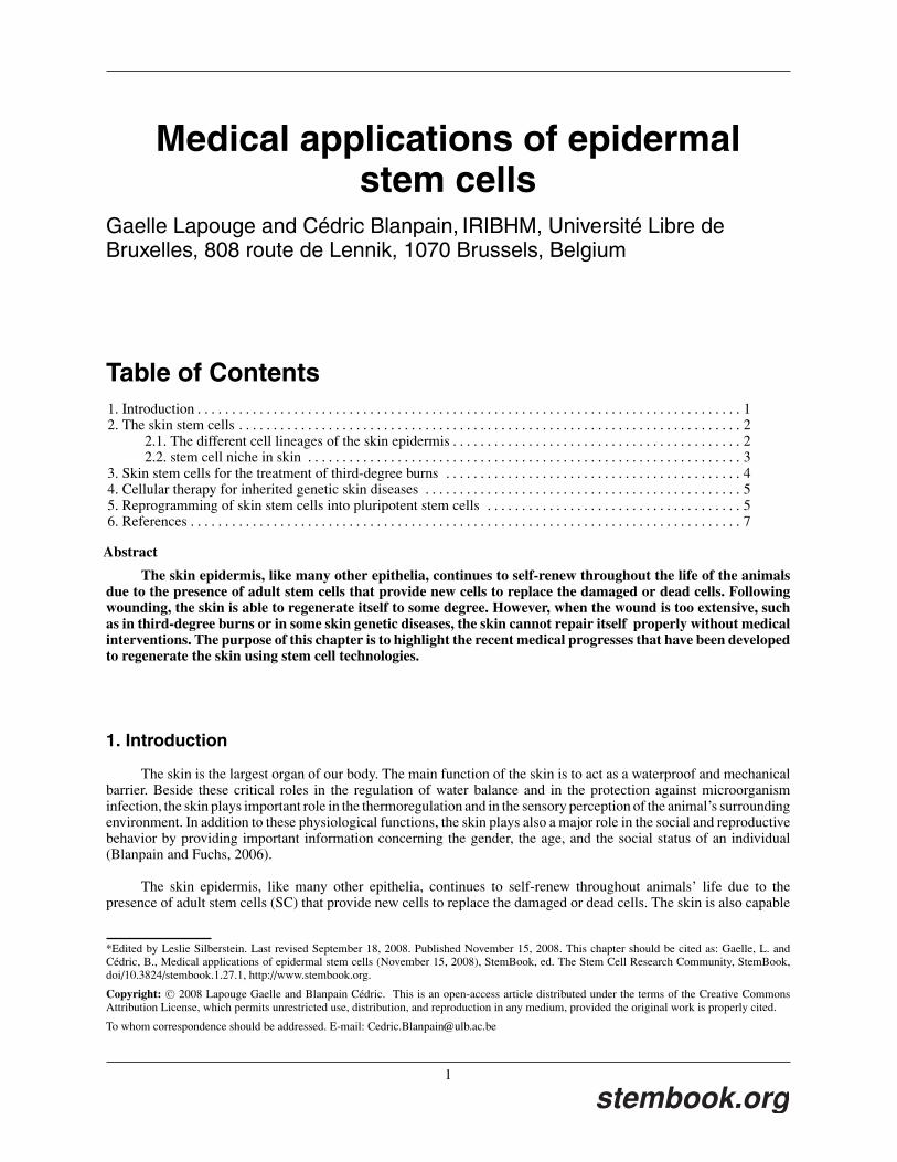
Figure 1. Hair follicle and inter-follicular epidermis morphogenesis. The hair follicle growth is stimulated by the dermal papilla (DP) signals, activatingthe bulge stem cells (Bu) to form the distinct lineages of the hair follicle (HF). Several concentric layers composed the HF. The external and basal layer, calledouter root sheath (ORS) maintains contact with the basal layer of the inter-follicular epidermis (IFE). At the base of the HF, the matrix cells (Mx) representthe highly proliferative cells, which differentiate in concentric rings that give rise to the Hair shaft (HS) and its channel the inner root sheath (IRS) and thecompanion layer. The HF contains also the sebaceous gland (SG), which produces the waterproof sebum. In the left panel, skin was co-stained for the basalcells using the anti-keratin14 antibody (red) and for the bulge SC using the anti–CD34 antibody (green). In the right panel, skin was co-stained for the basalcells using the anti-β4 antibody (white), for the Mx cells using the anti–KI67 antibody (red) and for the HS precursors using the anti-AE13 antibody (green).Nuclei were marked using DAPI staining. Abbreviations used: Bu: bulge stem cells, Der: dermis, DP: dermal papilla, IFE: inter-follicular epidermis, IRS:inner root sheath, HF: hair follicle, HG: hair germ, HS: hair shaft, IRS: inner root sheath, Mx: matrix cells, ORS: outer root sheath, SG: sebaceous gland.
of extensive regeneration following wounding, which implicates the activation of many cell types including epidermalSC, inflammatory and endothelial cells. However, when the wound is too extensive, the skin cannot regenerate by itselfwithout therapeutic intervention. We discuss in this chapter the recent progress that has been made in the identificationof SC from the skin epidermis, the role played by epidermal SC during homeostasis and wound healing, the therapeuticuse of skin stem cells to treat patients suffering from extensive burns or hereditary skin diseases. Finally, we willdiscuss how cellular reprogramming of skin stem cells can be envisaged as an alternative source of cells for treatingnon-skin diseases.
2. The skin stem cells
2.1. The different cell lineages of the skin epidermis
The skin epidermis is composed by the juxtaposition of pilo-sebaceous units containing one hair follicle and itssebaceous gland, which are surrounded by the interfollicular epidermis (IFE; see Figure 1).
The IFE is a stratified squamous epithelium constituted by different layers of cells. The innermost layer, calledthe basal layer, is strongly attached to its underlying dermis and contains mitotically active progenitor cells that divideand give rise to the differentiated suprabasal cells. The first suprabasal layer corresponds to the spinous layer, in whichcells strengthen their cytoskeleton and intercellular connections to provide better resistance to mechanical stress. Theoutermost granular layer produces biochemical components of the skin barrier itself. Finally, the stratum corneumcorresponds to dead-enucleated cells that are continuously sloughed from the skin surface (Blanpain and Fuchs, 2006;Candi et al., 2005). The IFE continuously self-renews throughout life to replace the cells that are constantly sloughedoff from the skin surface. In mice and humans, it takes about 3 to 4 weeks to replace all the cells of the IFE.
The hair follicle (HF) is organized into different concentric layers of cells. In contrast to the continuous renewalof the IFE, HF alternates cycles of growth and degeneration. At the end of morphogenesis, when HF reache theirfinal size, matrix cells located at the base of the HF cease to proliferate, and undergo apoptosis, which induces thedegeneration of the lower two-thirds of the HF. After this degenerative stage (catagen phase), HF stem cells (SC)located at the base of the remaining follicle called the bulge region, enter in a quiescent stage (telogen phase). At thestart of the first hair cycle, quiescent bulge SC are stimulated by signals coming from the underlying mesenchyme, theyexit from the SC niche, and proliferate actively to provide the cells that will re-form the new hair (anagen phase). Themature hair follicle contains seven concentric layers of differentiated cells: the outer root sheath, which is contiguousto the basal layer of the IFE, the companion layer, 3 layers of cells forming the inner root sheath, which forms thechannel of the hair and 3 layers of cells forming the hair shaft. The matrix cells located at the base of the hair folliclerepresent the transient-amplifying cells of the HF, which differentiate into the inner root sheath and the hair shaft cells(Blanpain and Fuchs, 2006).
2
stembook.org

Medical applications of epidermal stem cells
The skin epidermis also contains two types of glands: the sebaceous glands (SG), which are holocrine-secretingglands attached to each HF and produce the waterproof sebum, and the sweat glands that contribute to the regulationof the body temperature.
2.2. stem cell niche in skin
For decades, stem cells of the hair follicle were though to reside in the highly proliferative matrix cellularcompartments (Kligman, 1959). In the early nineties, Cotsarelis and colleagues demonstrated that prolonged admin-istration of nucleotide analogs (pulse) followed by a chase period, results in the presence of Label Retaining Cells inthe bulge region, suggesting that bulge cells are more quiescent than the rest of the epidermal cells (Cotsarelis et al.,1990). To determine the clonogenic potential of bulge cells, Barrandon and colleagues, dissected the skin epidermisinto different fragments and cultured the cells originating from these different epidermal regions in vitro. Bulge cellsgive rise to more highly proliferative colonies, known as holoclones, than the epidermal cells coming from otherregions, suggesting that bulge cells, although more quiescent in vivo, present a much greater proliferative potentialduring in vitro culture (Barrandon and Green, 1987; Rochat et al., 1994). When bulge cells were transplanted ontoimmunodeficient mice, bulge cells can differentiate into all cell lineages of the skin epidermis including HF, IFE andSG, demonstrating that bulge region contains stem cells with different potential lineages (Oshima et al., 2001). Clonalanalysis of bulge cells demonstrate that the progenies of one single bulge SC can reform all the epidermal lineages ofthe skin epidermis, demonstrating that bulge SC are truly multipotent (Blanpain et al., 2004; Claudinot et al., 2005).Fate mapping studies also indicate that bulge cells are multipotent SC, having the capacities to give rise to all cellsof the hair follicle, sebaceous gland and interfollicular epidermis (Ito et al., 2007; Morris et al., 2004; Tumbar et al.,2004). Interestingly, these studies also demonstrate that during adult homeostasis, there is a little if any contribution ofbulge SC to the maintenance of IFE, suggesting that unipotent progenitors ensure the renewal of IFE (Levy et al., 2005;Morris et al., 2004). However, upon wounding, bulge SC are activated, and migrate rapidly toward the skin lesionsand participate actively in the repair of epidermis (Ito et al., 2007; Levy et al., 2007; Nowak et al., 2008; Taylor et al.,2000). After the completion of epidermal repair, the flux of bulge cells stops, and the bulge cells that had migrated inthe IFE will progressively disappear overtime (Ito et al., 2007).
Different techniques have been developed to isolate and characterize the molecular properties of bulge SCs.Tumbar and colleagues developed an ingenious method, which allows to perform pulse chase experiments using a fusionprotein between Histone-2B and the green fluorescent protein (H2B-GFP), allowing identification and isolation ofliving label retaining bulge cells (Tumbar et al., 2004). Cotsarelis and colleagues developed transgenic mice expressingthe GFP under a bulge specific transgene allowing the isolation of bulge SC (Morris et al., 2004). Combination ofα6-integrin and CD34 monoclonal antibodies can also be used to purify specifically bulge SC during all stages of haircycle, which greatly facilitate isolation and characterization of bulge SC (Blanpain et al., 2004; Trempus et al., 2003).Gene expression profiling of bulge SC isolated using these three different approaches yield very similar results anduncover a list of genes specifically expressed in bulge SC irrespective of the stage of hair cycle or the isolation method(Blanpain et al., 2004; Morris et al., 2004; Tumbar et al., 2004). This list of genes provides valuable information tounderstand the unique features of multipotent bulge SC and how bulge cells actively participate in the formation oftheir own niche. Also these microarray studies allow the identification of multiple and important regulators of bulgeSC functions such as Wnt/βcatenin pathways, Lhx2 and NFATc1 transcription factors (Horsley et al., 2008; Lowryet al., 2005; Rhee et al., 2006).
Lineage tracing experiments demonstrated that the IFE is organized into discrete units of proliferation, knownas epidermal proliferative units (EPU). EPU can be observed in lineage tracing experiments as long term IFE labeledclones forming stacks of cells extending from basal cells to the top cornified cells (Ghazizadeh and Taichman,2001; Kolodka et al., 1998; Mackenzie, 1997; Ro and Rannala, 2005). In addition to the bulge and IFE SC, thereis accumulating evidence that other types of epidermal progenitors participate in the homeostasis of other epidermalcompartments such as the SG and the infundibulum, the portion of the epidermis that connects the HF to the IFE.Transplantation experiments have suggested the existence of unipotent sebaceous lineage progenitors (Ghazizadehand Taichman, 2001). Recent lineage tracing experiments using Blimp1-Cre identified rare cells located at the juncturebetween HF and SG that give rise to the entire SG (Horsley et al., 2006). These findings suggest that like the IFE,SG homeostasis may be maintained by the presence of unipotent progenitors. Recently, another population of SCwas identified in a region located above the bulge but below the SG, within the upper isthmus (UI; Nijhof et al.,2006). These cells were identified by their expression of MTS24 (Nijhof et al., 2006), a cell surface marker, or usinga combination of different monoclonal antibodies (Jensen et al., 2008). Moreover, transplanted UI cells reformed allthree epidermal lineages (Jensen et al., 2008), suggesting that UI SC may even be multipotent SC (Jensen et al.,
3
stembook.org
Medical applications of epidermal stem cells
Keratinocyte culture Epidermal sheet Grafted sheet Histology of the graft
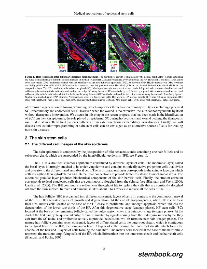
Figure 2. Cultured epithelial autograft in burn treatment. Keratinocytes from a severely burnt patient were isolated from a biopsy of unaffected skin andkeratinocytes SC colonies, called holoclones, were cultivated in vitro for 2 or 3 weeks to form sheets of epidermal cells. These epidermal sheets were graftedonto the patient burnt skin following Barrandon and Green’s procedure. The graft skin is well differentiated and similar to non-grafted skin except the lackof hair follicle and sebaceous gland. Reprint by permission from Lippincott, Williams and Wilkins; Ronfard et al., Transplantation 2000).
2008; Nijhof et al., 2006). Further studies are needed to better characterize the respective contributions of these newlyidentified epidermal SC to the overall skin epidermis homeostasis.
3. Skin stem cells for the treatment of third-degree burns
Major skin injuries, resulting from extensive burns, infection or trauma, cannot repair alone and require medicalinterventions to heal properly. Wound healing is organized into different steps. The first step is the release of solublemediators from the degranulated platelets and from injured blood vessels, leading to the formation of a blood clot.Few minutes after injury, the invasion of neutrophils, monocytes and leukocytes promotes migration, proliferation andsurvival of various cell types including keratinocyte SC, leading to the beginning of the re-epithelialization process. Inparallel to the reparation of the epidermis, the injured dermis is also repaired by the recruitment and proliferation offibroblasts producing extracellular matrix and keratinocyte growth promoting factors. While wound repair was thoughto require influx of inflammatory cells, mice deficient for PU.1, which lack neutrophils and macrophages, repair skinwound as well as wild-type mice (Martin et al., 2003). These results challenge the current view that inflammatory cellsare required for orchestrating the different steps of skin repair. Clearly more studies are needed to better understandthe respective contribution of the various types of cells involved during wound healing.
In USA, the skin loss from thermal injuries represents about 1 million person each year, whereas the skin lossfrom trauma and chronic ulcerations from diabetes or from venous ulcers represents 2 millions and 600.000 patientsrespectively (Clark et al., 2007). These numbers reveal the importance of this clinical problem and the need for furtherresearch in the treatment of skin injuries. The autologous skin grafting, consisting in the removal of a piece of skin fromunaffected tissue and its transplantation to the wound area, is the most viable and aesthetic technique for the treatmentof extensive skin injuries. Nevertheless, this approach has important limitations, such as only a limited fraction of theskin can be repaired by this method and it creates additional injuries at the donor sites. For these reasons, researcherslook for alternative methods to treat severe skin injuries. In the eighties, Green and colleagues discovered that humankeratinocyte SC could be propagated in vitro when culture on fibroblast feeder cells (Green et al., 1979; Rheinwaldand Green, 1975, 1977). Cultured under these conditions, human keratinocyte SC have an enormous proliferationpotential, and only few cells can regenerate sufficient keratinocytes to cover the all-human skin surface. These culturedhuman keratinocyte SC can differentiate and reform a functional skin barrier that can be transplanted into patientssuffering from severe burnt injuries (see Figure 2).
The human keratinocyte SC are obtained from aseptic skin biopsy (Gallico et al., 1984; Pellegrini et al., 1999;Ronfard et al., 2000) dissociated with trypsin to obtain single cells. These cells are then cultivated on irradiated 3T3fibroblasts feeders that create, through the secretion of extracellular matrix and growth factors, an artificial nicheparticularly suitable for the proliferation of epidermal SC. The keratinocytes are kept in culture until the formation of astratified epithelium that can be removed to cover the wound area, while the leftover cells can be frozen for future use(Green et al., 1979). This method is extremely powerful and allows the transplantation of a large piece of autologousskin starting from the removal of only a very small piece of unaffected skin. The first limitation of this technique isthe time required to grow confluent epithelial sheets in vitro, during which the patient is particularly susceptible toinfections. To prevent wound contraction and to cover the burns wound during the period of cell expansion, cadaver“decellularized” skin allografts are used. The second limitation is the huge cost of this treatment, which estimates at13.000$ per 1% total body surface, suggesting a cost of at least 500.000$ for the majority of patients that are severelyinjured (Clark et al., 2007).
4
stembook.org
Medical applications of epidermal stem cells
The grafted skin obtained by this method does not contain hair follicle and sweat glands (see Figure 2). Theabsence of hair follicle and sweat glands in third-degree burns does not result only from the destruction of multipotenthair follicle SC but also from the destruction of its underlying instructive dermis. Hair follicle morphogenesis andregeneration are critically dependent on the interaction between epidermal SC and specialized cells from the underlyingmesenchyme, called the dermal papilla (DP) cells (Jahoda et al., 1984). To achieve HF regeneration in grafted epidermalsheets coming from cultured epidermal SC, DP cells will need to be transplanted together with epidermal cells inorder to stimulate epidermal SC to adopt hair follicle fate. Recently progress has been accomplished to this end by thepurification and genetic profiling of DP cells (Rendl et al., 2005). Using this approach, Rendl and colleagues identifiedBMP-6 as one of the critical factors expressed by DP cells to maintain its hair follicle inducing activity in vitro and invivo, providing new hopes for the development of novel methods allowing the expansion and the differentiation of DPlike cells in vitro (Rendl et al., 2008).
4. Cellular therapy for inherited genetic skin diseases
There are many theoretical advantages to use skin SC for cellular therapy in human. Skin is a highly accessiblesource of SC, which have an extraordinary capacity of cellular expansion in culture, and that can be stably geneticallymodified (Shi et al., 2006).
There are many different genetic diseases affecting the skin epidermis. Among the most devastating genetic skindiseases are the epidermolysis bullosa, which is characterized by an extreme fragility of the skin. There are thousandsof patients around the world presenting these genetic diseases and suffering from repeating blistering lesions when theskin is submitted to a minor trauma. The blisters are painful and require long time to heal. In addition, possibly due topermanent state of wound healing that stimulates SC proliferation, these patients are at high risk of developing skincancer (Eady, 2001).
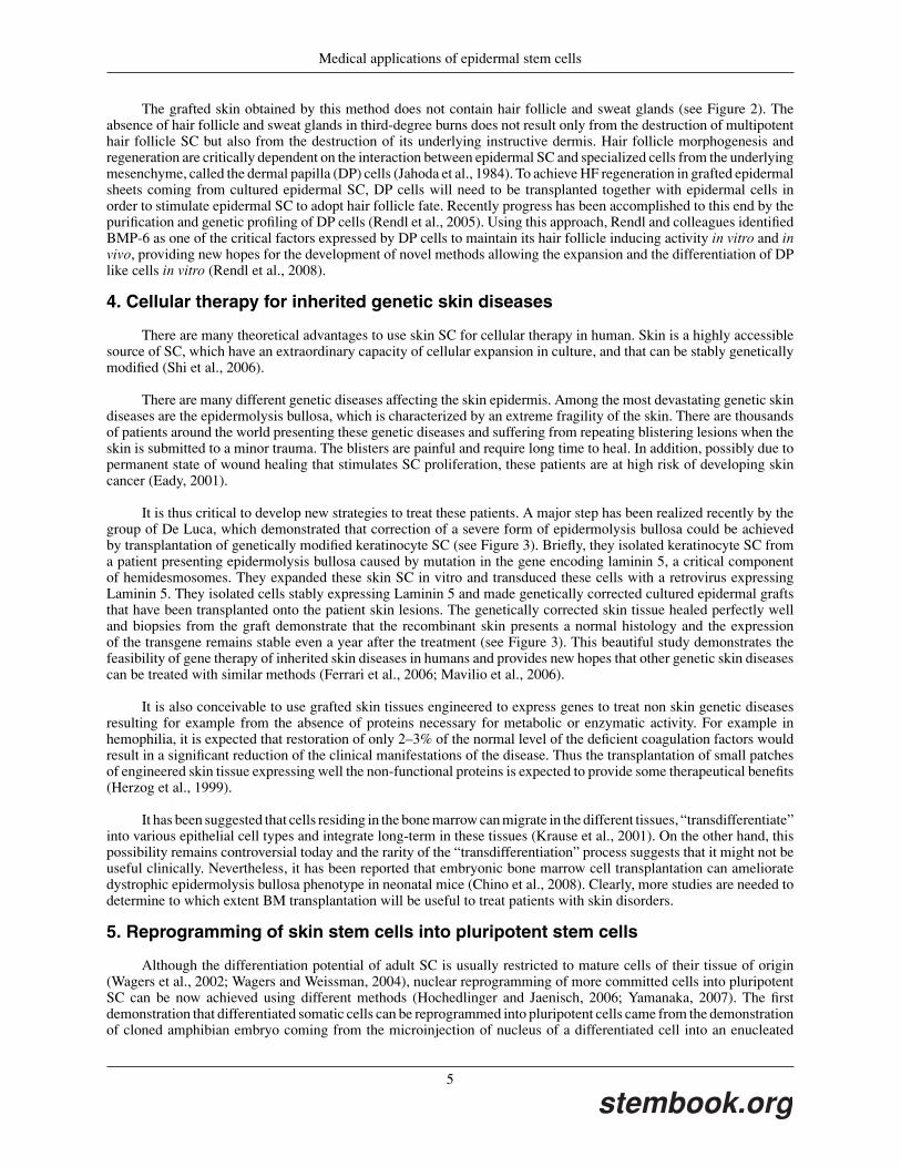
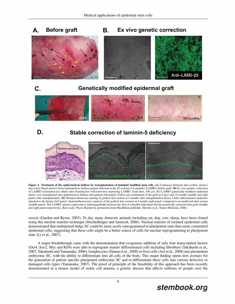
It is thus critical to develop new strategies to treat these patients. A major step has been realized recently by thegroup of De Luca, which demonstrated that correction of a severe form of epidermolysis bullosa could be achievedby transplantation of genetically modified keratinocyte SC (see Figure 3). Briefly, they isolated keratinocyte SC froma patient presenting epidermolysis bullosa caused by mutation in the gene encoding laminin 5, a critical componentof hemidesmosomes. They expanded these skin SC in vitro and transduced these cells with a retrovirus expressingLaminin 5. They isolated cells stably expressing Laminin 5 and made genetically corrected cultured epidermal graftsthat have been transplanted onto the patient skin lesions. The genetically corrected skin tissue healed perfectly welland biopsies from the graft demonstrate that the recombinant skin presents a normal histology and the expressionof the transgene remains stable even a year after the treatment (see Figure 3). This beautiful study demonstrates thefeasibility of gene therapy of inherited skin diseases in humans and provides new hopes that other genetic skin diseasescan be treated with similar methods (Ferrari et al., 2006; Mavilio et al., 2006).
It is also conceivable to use grafted skin tissues engineered to express genes to treat non skin genetic diseasesresulting for example from the absence of proteins necessary for metabolic or enzymatic activity. For example inhemophilia, it is expected that restoration of only 2–3% of the normal level of the deficient coagulation factors wouldresult in a significant reduction of the clinical manifestations of the disease. Thus the transplantation of small patchesof engineered skin tissue expressing well the non-functional proteins is expected to provide some therapeutical benefits(Herzog et al., 1999).
It has been suggested that cells residing in the bone marrow can migrate in the different tissues, “transdifferentiate”into various epithelial cell types and integrate long-term in these tissues (Krause et al., 2001). On the other hand, thispossibility remains controversial today and the rarity of the “transdifferentiation” process suggests that it might not beuseful clinically. Nevertheless, it has been reported that embryonic bone marrow cell transplantation can amelioratedystrophic epidermolysis bullosa phenotype in neonatal mice (Chino et al., 2008). Clearly, more studies are needed todetermine to which extent BM transplantation will be useful to treat patients with skin disorders.
5. Reprogramming of skin stem cells into pluripotent stem cells
Although the differentiation potential of adult SC is usually restricted to mature cells of their tissue of origin(Wagers et al., 2002; Wagers and Weissman, 2004), nuclear reprogramming of more committed cells into pluripotentSC can be now achieved using different methods (Hochedlinger and Jaenisch, 2006; Yamanaka, 2007). The firstdemonstration that differentiated somatic cells can be reprogrammed into pluripotent cells came from the demonstrationof cloned amphibian embryo coming from the microinjection of nucleus of a differentiated cell into an enucleated
5
stembook.org
Medical applications of epidermal stem cells
Before graft Ex vivo genetic correction
Genetically modified epidermal graft
Stable correction of laminin-5 deficiency
A. B.
C.
D.
Figure 3. Treatment of the epidermolysis bullosa by transplantation of laminin5 modified stem cells. (A) Continous blistered skin (yellow arrows)and crusts (black arrows) from epidermolysis bullosa patient deficient in the β3 isoform of Laminin5 (LAMB3) before graft. (B) Ex vivo genetic correctionof LAMB3 in keratinocyte culture after transduction with retrovirus expressing LAMB3. Scale bars, 100 µm. (C) LAMB3 genetically modified epidermalsheets were transplanted onto epidermolysis bullosa skin patient (left panel). Follow-up examination of the graft at 8 days and 10 months (middle and rightpanel) after transplantation. (D) Hematoxylin/eosin staining of grafted skin section at 4 months after transplantation shows a fully differentiated epidermisattached to the dermis (left panel). Immunofluorescence analysis of the grafted skin sections at 4 month (right panel) compared to an unaffected skin section(middle panel). The LAMB3 (green) expression is indistinguishable between the skin of a healthy individual and the genetically corrected skin graft (middleand right panel respectively). Bars scale, 50µm. Reprint by permission from MacMilian publisher; Mavilio et al., Nature Medicine, 2006).
oocyte (Gurdon and Byrne, 2003). To day, many domestic animals including cat, dog, cow, sheep, have been clonedusing this nuclear transfer technique (Hochedlinger and Jaenisch, 2006). Nuclear transfer of isolated epidermal cellsdemonstrated that multipotent bulge SC could be more easily reprogrammed to pluripotent state than more committedepidermal cells, suggesting that these cells might be a better source of cells for nuclear reprogramming to pluripotentstate (Li et al., 2007).
A major breakthrough came with the demonstration that exogenous addition of only four transcription factors(Oct4, Sox2, Myc and Klf4) were able to reprogram mature differentiated cells including fibroblast (Takahashi et al.,2007; Takahashi and Yamanaka, 2006), lymphocytes (Hanna et al., 2008) or liver cells (Aoi et al., 2008) into pluripotentembryonic SC, with the ability to differentiate into all cells of the body. This major finding opens new avenues forthe generation of patient specific pluripotent embryonic SC and re-differentiate these cells into various defective ordamaged cells types (Yamanaka, 2007). The proof of principle of the feasibility of this approach has been recentlydemonstrated in a mouse model of sickle cell anemia, a genetic disease that affects millions of people over the
6
stembook.org
Medical applications of epidermal stem cells
world. Jaenisch and colleagues took fibroblasts from a mouse model of sickle cell anemia, reprogrammed these cellsinto pluripotent SC, corrected the genetic deficiency by homologous recombination, and redirected these pluripotentcells toward the hematopoietic lineages, and transplanted these engineered cells to a lethally irradiated mice (Hannaet al., 2007). Treated animals presented many features demonstrating clinical improvement of the disease. The higherefficiency of bulge SC to be reprogrammed to pluripotent state by nuclear transfer (Li et al., 2007), together with theirenormous expansion potential in vitro (Barrandon, 2007), suggest that bulge cells or other keratinocyte SC could bethe ideal source of cells for nuclear reprogramming into pluripotent embryonic stem cells and the re-differentiationof these cells toward the cell types such as neurons, cardiac cells, hepatocytes, or pancreas cells that are defective ormissing in human diseases.
6. References
Aoi, T., Yae, K., Nakagawa, M., Ichisaka, T., Okita, K., Takahashi, K., Chiba, T., and Yamanaka, S. (2008). Generationof pluripotent stem cells from adult mouse liver and stomach cells. Science 321, 699–702.
Barrandon, Y. (2007). Genetic manipulation of skin stem cells: success, hope, and challenges ahead. Mol Ther 15,443–444.
Barrandon, Y., and Green, H. (1987). Three clonal types of keratinocyte with different capacities for multiplication.Proc Natl Acad Sci U S A 84, 2302–2306.
Blanpain, C., and Fuchs, E. (2006). Epidermal stem cells of the skin. Annu Rev Cell Dev Biol 22, 339–373.
Blanpain, C., Horsley, V., and Fuchs, E. (2007). Epithelial stem cells: turning over new leaves. Cell 128, 445–458.
Blanpain, C., Lowry, W.E., Geoghegan, A., Polak, L., and Fuchs, E. (2004). Self-renewal, multipotency, and theexistence of two cell populations within an epithelial stem cell niche. Cell 118, 635–648.
Candi, E., Schmidt, R., and Melino, G. (2005). The cornified envelope: a model of cell death in the skin. Nat Rev MolCell Biol 6, 328–340.
Chino, T., Tamai, K., Yamazaki, T., Otsuru, S., Kikuchi, Y., Nimura, K., Endo, M., Nagai, M., Uitto, J., Kitajima,Y., et al. (2008). Bone marrow cell transfer into fetal circulation can ameliorate genetic skin diseases by providingfibroblasts to the skin and inducing immune tolerance. Am J Pathol 173, 803–814.
Clark, R.A., Ghosh, K., and Tonnesen, M.G. (2007). Tissue engineering for cutaneous wounds. J Invest Dermatol 127,1018–1029.
Claudinot, S., Nicolas, M., Oshima, H., Rochat, A., and Barrandon, Y. (2005). Long-term renewal of hair folliclesfrom clonogenic multipotent stem cells. Proc Natl Acad Sci U S A 102, 14677–14682.
Cotsarelis, G., Sun, T.T., and Lavker, R.M. (1990). Label-retaining cells reside in the bulge area of pilosebaceous unit:implications for follicular stem cells, hair cycle, and skin carcinogenesis. Cell 61, 1329–1337.
Eady, R.A. (2001). Epidermolysis bullosa: scientific advances and therapeutic challenges. J Dermatol 28, 638–640.
Ferrari, S., Pellegrini, G., Matsui, T., Mavilio, F., and De Luca, M. (2006). Gene therapy in combination with tissueengineering to treat epidermolysis bullosa. Expert Opin Biol Ther 6, 367–378.
Gallico, G.G., 3rd, O’Connor, N.E., Compton, C.C., Kehinde, O., and Green, H. (1984). Permanent coverage of largeburn wounds with autologous cultured human epithelium. N Engl J Med 311, 448–451.
Ghazizadeh, S., and Taichman, L.B. (2001). Multiple classes of stem cells in cutaneous epithelium: a lineage analysisof adult mouse skin. EMBO J 20, 1215–1222.
Green, H., Kehinde, O., and Thomas, J. (1979). Growth of cultured human epidermal cells into multiple epitheliasuitable for grafting. Proc Natl Acad Sci U S A 76, 5665–5668.
7
stembook.org
Medical applications of epidermal stem cells
Gurdon, J.B., and Byrne, J.A. (2003). The first half-century of nuclear transplantation. Proc Natl Acad Sci U S A 100,8048–8052.
Hanna, J., Markoulaki, S., Schorderet, P., Carey, B.W., Beard, C., Wernig, M., Creyghton, M.P., Steine, E.J., Cas-sady, J.P., Foreman, R., et al. (2008). Direct reprogramming of terminally differentiated mature B lymphocytes topluripotency. Cell 133, 250–264.
Hanna, J., Wernig, M., Markoulaki, S., Sun, C.W., Meissner, A., Cassady, J.P., Beard, C., Brambrink, T., Wu, L.C.,Townes, T.M., et al. (2007). Treatment of sickle cell anemia mouse model with iPS cells generated from autologousskin. Science 318, 1920–1923.
Herzog, R.W., Yang, E.Y., Couto, L.B., Hagstrom, J.N., Elwell, D., Fields, P.A., Burton, M., Bellinger, D.A., Read,M.S., Brinkhous, K.M., et al. (1999). Long-term correction of canine hemophilia B by gene transfer of blood coagulationfactor IX mediated by adeno-associated viral vector. Nat Med 5, 56–63.
Hochedlinger, K., and Jaenisch, R. (2006). Nuclear reprogramming and pluripotency. Nature 441, 1061–1067.
Horsley, V., Aliprantis, A.O., Polak, L., Glimcher, L.H., and Fuchs, E. (2008). NFATc1 balances quiescence andproliferation of skin stem cells. Cell 132, 299–310.
Horsley, V., O’Carroll, D., Tooze, R., Ohinata, Y., Saitou, M., Obukhanych, T., Nussenzweig, M., Tarakhovsky, A.,and Fuchs, E. (2006). Blimp1 defines a progenitor population that governs cellular input to the sebaceous gland. Cell126, 597–609.
Ito, M., Yang, Z., Andl, T., Cui, C., Kim, N., Millar, S.E., and Cotsarelis, G. (2007). Wnt-dependent de novo hairfollicle regeneration in adult mouse skin after wounding. Nature 447, 316–320.
Jahoda, C., Horne, K., and Oliver, R. (1984). Induction of hair growth by implantation of cultured dermal papilla cells.Nature 311, 560–562.
Jensen, U.B., Yan, X., Triel, C., Woo, S.H., Christensen, R., and Owens, D.M. (2008). A distinct population ofclonogenic and multipotent murine follicular keratinocytes residing in the upper isthmus. J Cell Sci 121, 609–617.
Kligman, A.M. (1959). The human hair cycle. J Invest Dermatol 33, 307–316.
Kolodka, T.M., Garlick, J.A., and Taichman, L.B. (1998). Evidence for keratinocyte stem cells in vitro: long termengraftment and persistence of transgene expression from retrovirus-transduced keratinocytes. Proc Natl Acad SciU S A 95, 4356–4361.
Krause, D.S., Theise, N.D., Collector, M.I., Henegariu, O., Hwang, S., Gardner, R., Neutzel, S., and Sharkis, S.J.(2001). Multi-organ, multi-lineage engraftment by a single bone marrow-derived stem cell. Cell 105, 369–377.
Levy, V., Lindon, C., Harfe, B.D., and Morgan, B.A. (2005). Distinct stem cell populations regenerate the follicle andinterfollicular epidermis. Dev Cell 9, 855–861.
Levy, V., Lindon, C., Zheng, Y., Harfe, B.D., and Morgan, B.A. (2007). Epidermal stem cells arise from the hairfollicle after wounding. FASEB J 21, 1358–1366.
Li, J., Greco, V., Guasch, G., Fuchs, E., and Mombaerts, P. (2007). Mice cloned from skin cells. Proc Natl Acad SciU S A 104, 2738–2743.
Lowry, W.E., Blanpain, C., Nowak, J.A., Guasch, G., Lewis, L., and Fuchs, E. (2005). Defining the impact ofbeta-catenin/Tcf transactivation on epithelial stem cells. Genes Dev 19, 1596–1611.
Mackenzie, I.C. (1997). Retroviral transduction of murine epidermal stem cells demonstrates clonal units of epidermalstructure. J Invest Dermatol 109, 377–383.
8
stembook.org
Medical applications of epidermal stem cells
Martin, P., D’Souza, D., Martin, J., Grose, R., Cooper, L., Maki, R., and McKercher, S.R. (2003). Wound healing inthe PU.1 null mouse–tissue repair is not dependent on inflammatory cells. Curr Biol 13, 1122–1128.
Mavilio, F., Pellegrini, G., Ferrari, S., Di Nunzio, F., Di Iorio, E., Recchia, A., Maruggi, G., Ferrari, G., Provasi, E.,Bonini, C., et al. (2006). Correction of junctional epidermolysis bullosa by transplantation of genetically modifiedepidermal stem cells. Nat Med 12, 1397–1402.
Morris, R.J., Liu, Y., Marles, L., Yang, Z., Trempus, C., Li, S., Lin, J.S., Sawicki, J.A., and Cotsarelis, G. (2004).Capturing and profiling adult hair follicle stem cells. Nat Biotechnol 22, 411–417.
Nijhof, J.G., Braun, K.M., Giangreco, A., van Pelt, C., Kawamoto, H., Boyd, R.L., Willemze, R., Mullenders, L.H.,Watt, F.M., de Gruijl, F.R., et al. (2006). The cell-surface marker MTS24 identifies a novel population of follicularkeratinocytes with characteristics of progenitor cells. Development 133, 3027–3037.
Nowak, J.A., Polak, L., Pasolli, H.A., and Fuchs, E. (2008). Hair follicle stem cells are specified and function in earlyskin morphogenesis. Cell Stem Cell 3, 33–43.
Oshima, H., Rochat, A., Kedzia, C., Kobayashi, K., and Barrandon, Y. (2001). Morphogenesis and renewal of hairfollicles from adult multipotent stem cells. Cell 104, 233–245.
Pellegrini, G., Ranno, R., Stracuzzi, G., Bondanza, S., Guerra, L., Zambruno, G., Micali, G., and De Luca, M. (1999).The control of epidermal stem cells (holoclones) in the treatment of massive full-thickness burns with autologouskeratinocytes cultured on fibrin. Transplantation 68, 868–879.
Rendl, M., Lewis, L., and Fuchs, E. (2005). Molecular dissection of mesenchymal-epithelial interactions in the hairfollicle. PLoS Biol 3, e331.
Rendl, M., Polak, L., and Fuchs, E. (2008). BMP signaling in dermal papilla cells is required for their hair follicle-inductive properties. Genes Dev 22, 543–557.
Rhee, H., Polak, L., and Fuchs, E. (2006). Lhx2 maintains stem cell character in hair follicles. Science 312, 1946–1949.
Rheinwald, J.G., and Green, H. (1975). Serial cultivation of strains of human epidermal keratinocytes: the formationof keratinizing colonies from single cells. Cell 6, 331–343.
Rheinwald, J.G., and Green, H. (1977). Epidermal growth factor and the multiplication of cultured human epidermalkeratinocytes. Nature 265, 421–424.
Ro, S., and Rannala, B. (2005). Evidence from the stop-EGFP mouse supports a niche-sharing model of epidermalproliferative units. Exp Dermatol 14, 838–843.
Rochat, A., Kobayashi, K., and Barrandon, Y. (1994). Location of stem cells of human hair follicles by clonal analysis.Cell 76, 1063–1073.
Ronfard, V., Rives, J.M., Neveux, Y., Carsin, H., and Barrandon, Y. (2000). Long-term regeneration of human epidermison third degree burns transplanted with autologous cultured epithelium grown on a fibrin matrix. Transplantation 70,1588–1598.
Shi, C., Zhu, Y., Su, Y., and Cheng, T. (2006). Stem cells and their applications in skin-cell therapy. Trends Biotechnol24, 48–52.
Takahashi, K., Tanabe, K., Ohnuki, M., Narita, M., Ichisaka, T., and Tomoda, K. S. Y. (2007). Induction of pluripotentstem cells from adult human fibroblasts by defined factors. Cell 131, 861–872.
Takahashi, K., and Yamanaka, S. (2006). Induction of pluripotent stem cells from mouse embryonic and adult fibroblastcultures by defined factors. Cell 126, 663–676.
9
stembook.org
Medical applications of epidermal stem cells
Taylor, G., Lehrer, M.S., Jensen, P.J., Sun, T.T., and Lavker, R.M. (2000). Involvement of follicular stem cells informing not only the follicle but also the epidermis. Cell 102, 451–461.
Trempus, C.S., Morris, R.J., Bortner, C.D., Cotsarelis, G., Faircloth, R.S., Reece, J.M., and Tennant, R.W. (2003).Enrichment for living murine keratinocytes from the hair follicle bulge with the cell surface marker CD34. J InvestDermatol 120, 501–511.
Tumbar, T., Guasch, G., Greco, V., Blanpain, C., Lowry, W.E., Rendl, M., and Fuchs, E. (2004). Defining the epithelialstem cell niche in skin. Science 303, 359–363.
Wagers, A.J., Sherwood, R.I., Christensen, J.L., and Weissman, I.L. (2002). Little evidence for developmental plasticityof adult hematopoietic stem cells. Science 297, 2256–2259.
Wagers, A.J., and Weissman, I.L. (2004). Plasticity of adult stem cells. Cell 116, 639–648.
Yamanaka, S. (2007). Strategies and new developments in the generation of patient-specific pluripotent stem cells.Cell Stem Cell 1, 39–49.
10
stembook.org