medicago marina (l.) seed: unravelling mechanisms ... · ... the ovule differentiates into one or...
TRANSCRIPT

UNIVERSITA’ DEGLI STUDI DEL MOLISE
FACOLTÀ DI SCIENZE MATEMATICHE, FISICHE E NATURALI
Dipartimento di Scienze e Tecnologie per l’Ambiente e il Territorio
Tesi presentata per il conseguimento del Dottorato di Ricerca in
AMBIENTE E TERRITORIO (XXIV CICLO) S.S.D. BIO/01
Medicago marina (L.) seed: unravelling mechanisms
controlling germination and dormancy
Tutor/ Relatore Coordinatore
Chiar.ma Prof.ssa Chiar.mo Prof.
G. Stefania Scippa Claudio Caprari
Candidato
Elisa Petrollini
Anno Accademico 2011
- 1 -
Contents
General introduction…………………………………………………………………………3
1. Development and structure of seed……………………………………………………3
1.2. Legumes seed development and features…………………………………………8
2. Seed dormancy……………………………………………………………………….15
3. Seed germination……………………………………………………………………..21
4. Hormones involved in the control of seed dormancy and germination………………23
4.1. Molecular factors and hormonal cross-talk in regulating seed dormancy and
germination…………………………………………………………………………...26
References……………………………………………………………………………………33
Objectives……………………………………………………………………………………64
Chapter I. Dormancy of Medicago marina (L.) seed……………………………………..67
Chapter II. Dormancy of Medicago marina (L.) seed: the role of the micropylar
endosperm…………………………………………………………………………………...78
1. Introduction………………………………………………………………………….78
2. Materials and Methods………………………………………………………………83
2.1 Seed material and germination test………………………………………………83
2.2 Puncture force meseaurement……………………………………………………84
2.3 Reverse transcriptase Polymerase Chain Reaction (RT-PCR) analysis………….86
2.3.1 RNA extraction…………………………………………………………..86
2.3.2 cDNA synthesis and RT-PCR……………………………………………86
2.4 Proteomic Analysis……………………………………………………………….87
2.4.1 Protein extraction…………………………………………………………87
2.4.2 Two dimensional electrophoresis…………………………………………88
- 2 -
2.4.3 PDQuest analysis………………………………………………………….88
2.5 Statistical analysis and identification of specific markers...………………………89
3. Results…………………………………………………………………………………90
3.1 Puncture force measurement………………………………………………............90
3.2 Germination test…………………………………………………………………...93
3.3 Reverse transcriptase Polymerase Chain Reaction (RT-PCR) analysis…………...94
3.4 Proteomic analysis…………………………………………………………………95
3.5 Identification of specific markers………………………………………………….98
3.6 Markers expression pattern………………………………………………………100
4. Discussion and conclusions..………………...………………………………………..106
References……………………………………………………………………………116
Supplementary material………………………………………………………………127
- 3 -
General introduction
1. Development and structure of seed
Seeds are the typical propagation units of the flowering plants. They represent the delivery
system for the transfer of genetic materials from one generation to the next, trough the sexual
reproduction in vascular plant. The seed-producing organisms of the plant kingdom belong to the
division Spermatophyta, further classified into 2 sub-divisions: Gymnospermae (gymnosperms)
and Angiospermae (angiosperms). The angiosperms are divided into 2 classes:
Monocotyledoneae and Dicotyledoneae (Cantino et al., 2007, Linkies et al., 2010).
In angiosperm and gymnosperms seeds develop from ovules, which can be defined as
unfertilized, immature seed precursors (Gasser et al, 1998, Finch-Savage & Leubner-Metzger,
2006; Frohlich & Chase, 2007).
During its development, the ovule differentiates into one or two covering layers that envelop
the nucellus. In the gymnosperms, the nucellus is enveloped by one covering layer, while in the
angiosperms, it is enveloped by two covering layers, called integuments (Linkies et al, 2010).
Then, during the seed growth, the external covering layer develop into the testa (seed coat),
which, in mature seeds is usually constituted by a layer of dead cells, while the inner integument,
that develop into the tegmen, is formed by alive cells (Debeaujon et al., 2000; Windsor et al.,
2000; Haughna & Chaudhuryb, 2005).
Within the nucellus, a megaspore develops into a haploid megagametophyte (female
gametophyte), that in most gymnosperm species is multicellular, and usually several archegonia
develop within the megagametophyte, forming only one egg in each archegonium (Floyd &
Friedman, 2000; Baroux et al., 2002). On the contrary, the angiosperm megagametophyte, that in
its mature state is also called embryo sac, is seven-celled and eight-nucleate (Polygonum-type)
(Floyd & Friedman, 2000; Baroux et al., 2002; Friedman & Williams, 2004; Berger et al., 2008;
Friedman & Ryderson, 2009). Occurrence, the mature megagametophyte is four-celled and four-
nucleate, also called Nuphar/Schisandra-type, because can be found in the basal angiosperms,
namely Nymphaeales and Austrobaileyales (Floyd & Friedman, 2000; Baroux et al., 2002;
Friedman & Williams, 2004; Berger et al., 2008; Friedman & Ryderson, 2009).
After pollination, in all angiosperms and most gymnosperms a pollen tube is formed through
which the sperm can reaches and fertilizes the egg cell, leading to development of the diploid
embryo. The micropylar synergid cells help attract the pollen tube to the female gametophyte.

- 4 -
The egg cell, that is the progenitor of the embryo, lies adjacent to the synergid cells, while the
large central cell, the progenitor of the endosperm, contains two polar nuclei, that fuse together
forming a diploid central cell nucleus, at the time of fertilization (Higashiyama et al., 2001).
Angiosperms seed development begins upon double fertilization (Dumas & Rogowsky,
2008). In contrast of gymnosperm, the hallmark of angiosperm reproduction is the double
fertilization mechanism, in which two haploid sperm nuclei are transported trough the pollen
tube. Indeed, the double fertilization is characterized by two fertilization events. The first event
involves the egg fertilization and occurs when one haploid sperm is transported to the ovule
through the pollen tube, leading to the development of diploid embryo. The second fertilization
event occurs when an other haploid sperm nucleus fuses with the central cell, which develops
into the endosperm (Floyd & Friedman, 2000; Baroux et al., 2002; Friedman & Williams, 2004;
Berger et al., 2008; Dumas & Rogowsky, 2008, Friedman et al., 2008; Friedman & Ryderson,
2009). The resulting embryo and endosperm are genetically identical except for ploidy level, as
the embryo is diploid and the endosperm is triploid: since the central cell of most angiosperm
species has either one (Nuphar/Schisandra-type) or two nuclei (Polygonum-type), the fertilized
endosperm is either diploid or triploid (Gehring et al., 2004).
From this point onwards, seed development is characterized by the rapid development and
growth of the endosperm and the embryo, until seed maturation.
Embryonic development proceeds within the protective maternal tissue of the ovule (which
becomes the seed coat surrounding the developing embryo and endosperm) and, in higher plants,
the embryogenesis can be divided conceptually into three distinct phases, partially overlapping
(West & Harada, 1993; Dumas & Rogowsky, 2008). The first phase is characterized by
morphogenesis events, during which the polar axis of the plant body is defined with the
specification of the shoot and root apices, and the embryonic tissue and organ systems are
formed. Generally, in many angiosperms, the initial division of the zygote is transverse and
asymmetric and generates a small, chalazally oriented apical cell and a large basal cell
(Mansfield & Briarty, 1990). From the apical cell originates the bulk of the embryo, including
the cotyledons, shoot apex, and hypocotyls. First, the apical cell of embryo undergoes two
longitudinal divisions to produce a four-celled embryo (Mansfield & Briarty, 1990, West &
Harada, 1993), and then, a transverse division follows to produce two layers of cells in an octant
stage embryo. The next division of the octant stage embryo is periclinal or parallel to the embryo
surface and leads to the formation of first histologically detectable tissue, the protoderm, which
is the precursor of the epidermis. The globular stage embryo is established following the
delineation of the protoderm, and during this phase, the embryo increases in size and cell number
- 5 -
by anticlinal cell divisions of the protoderm and by longitudinal and, later, transverse divisions
of interior cells (Mansfield & Briarty, 1990, West & Harada, 1993).
A part of the root apex and the suspensor originate from the basal cell, which again,
undergoes a series of transverse divisions, resulting in the formation of the hypophysis and the
suspensor (West & Harada, 1993). In many plants, the hypophysis, the uppermost derivative of
the basal cell, serves as the precursor to the root cortex initials and the central region of the root
cap (Mansfield & Briarty, 1990), while the suspensor is an ephemeral embryonic structure,
formed by a various number of cell layers depending of plant species (Marsden & Meinke, 1985;
Mansfield & Briarty, 1990). During embryogenesis, this structure functions as conduit for
nutrients from the endosperm and for growth factors from the maternal tissue to the embryo
(Yeung & Meinke, 1993).
During the transition from the globular to the heart stage, embryo undergoes a significant
transformation of its morphology: trough the cell divisions that occur at specific regions of the
lateral margins of the globular stage embryo, the two lobes of the cotyledons emergence. Then,
the shift in embryo symmetry from radial, at the globular stage, to bilateral, at the heart stage,
represents the initial delineation of the cotyledons and axis. Following their formation, the
cotyledons and axis elongate rapidly as a result of cell division and cell expansion, and at this
stage of embryogenesis are also visible other tissues and structures such as the ground meristem
and the procambium, which is the precursor of the vascular tissue (Mansfield & Briarty, 1990).
Thus, the cells that will form the root apex and the shoot apex can be distinguished from this
stage of embryogenesis. After the heart stage, morphogenesis of embryos is interrupted and the
second phase of embryogenesis takes place.
The second phase is characterized by a period of embryo maturation during which initiates the
storage accumulation of reserve macromolecules: proteins, lipids and carbohydrates. These
reserves are particularly prevalent in the embryonic cotyledons of plants that do not store
substantial reserves in their endosperm, otherwise these reserves accumulates in developing
endosperm and they are responsible for a rapid increase in embryo mass and size (Mansfield &
Briarty, 1992). Therefore, during this second phase, also the endosperm developments and it
grows much more rapidly than the embryo, initially through nuclear divisions and then by
cellularization of each nucleus. In detail, during nuclear division, endosperm nuclei continue to
divide without cytokinesis to create a syncytium of nuclei (Brown et al., 1999; Dumas &
Rogowsky, 2008). Each nucleus is surrounded by dense cytoplasm and organelles that compose
nuclear cytoplasmic domains (Brown et al., 1999). Then, after cellularization of each nucleus,
three distinct domains are formed: the micropylar endosperm, the peripheral endosperm and the
- 6 -
chalazal endosperm (Boisnard-Lorig et al., 2001, Baroux et al., 2002; Olsen, 2004; Dumas &
Rogowsky, 2008; Friedman & Ryderson, 2009).
The growth of the seed is coupled with the growth of the endosperm that constitutes the
major contribution to the volume of the mature seed (Sundaresan, 2005).
However, when seed maturation is completed, the relative abundance of the endosperm can
differ considerably. In some Brassicaceae species and many other dicots (like as many legume
seeds), the endosperm is finally consumed, being replaced by the growing embryo, which then
constitutes most of the mature seed (Forbis et al., 2002; Sundaresan, 2005, Finch-Savage and
Leubner-Metzger, 2006). In other species, belonging for examples to Solanaceae family, the
endosperm is not lost and constitutes a large portion of the mature seed. In some other cases the
endosperm is just partially lost and is present as a thin layer, often formed by a single cell layer,
as happens for Lactuca sativa or Arabidopsis thaliana seeds (Pritchard et al., 2002; Liu et al.,
2005) and Lepidium sativum seeds (Müller et al., 2006). If, after seed maturation, the endosperm
persists as the primary food storage tissue, seeds are described as endospermic; on the contrary,
if the cotyledons become the site of food storage because the endosperm is consumed during
embryo development and is absent, or exists only as a very thin layer of tissue in mature seeds,
these species are designated as non-endospermic (Lopes & Larkins, 1993). When the second
phase is completed, the embryo is differentiated and exhibits developmental polarity that is
divided into the radicle (embryonic root) and the shoot with the cotyledons (only one in
Monocotyledoneae, and two in Dicotyledoneae), that represent the embryonic leaves (Linkies et
al., 2010).
The third stages of embryogenesis are concerned primarily with preparing the embryo for
developmental arrest and germination (Kermode, 1990; Galau et al., 1991; Thomas, 1993).
Indeed, during this final stage of embryogenesis, embryo acquires the desiccation tolerance and
eventually enters a period of metabolic quiescence (Kermode, 1990). The mature embryo, if not
affected by dormancy, remains quiescent until it encounters conditions appropriate for
germination.
Fertilization also initiates changes in maternal tissues. The ovary develops into a fruit and the
ovule integuments differentiate to form the protective seed coats, including testa and tegmen
(Moïse et al., 2005). Before fertilization, cells of the integuments are relatively undifferentiated,
then, after fertilization, during seed coats development, the undifferentiated cell layers can
differentiate into specialized cell types. However, some of these cell layers not undergo any
significant differentiation remaining parenchymatous, and often, are crushed upon the
completion of seed maturity; other cells undergo a slight thickening of the cell wall, becoming
collenchymatous. In addition, some cell layers can undergo an extensive secondary thickening of
- 7 -
various parts of the cell walls, becoming sclerotic: if these cells also elongates in the radial plane,
these layers are called palisade layers (Moïse et al., 2005).
Although the seed coats of different species vary greatly in structure and composition, they
undergo similar phases of development in relation to the embryo and endosperm. For example,
in legume seed development the seed coat and endosperm develop first, followed by the
development of the embryo, maturation of the seed coat, and maturation of the embryo (Weber et
al., 2005). The coordination of these events is governed by communication among the tissues of
the seed organs, such as communication between the seed coat and endosperm reported in
Arabidopsis seeds (Weijers et al., 2003).
Seed coats accomplish multiple tasks in the mature seed. They can contribute to overall seed
morphology thanks to the accumulation of large quantities of mucilage or pigments into some
cell layers. During the early embryo development and differentiation that is controlled by the
maternal tissues, seed coats are able to transmit growth signals to the embryo. For example, the
cell wall invertases of legumes seed coats play a central role in the maternal control of seed
development, facilitating the sucrose assimilation and accumulation by increasing the sucrose
gradient (Weber et al., 1995). The increasing of sucrose concentration into seed coats, and its
further transport to the embryo, promotes embryo growth by enhancing cell division (Borisjuk et
al., 1998). Seed coats separate one generation of plants from the next and ensure the survival of
the offspring, giving the resistance to biotic and abiotic stress (Moïse et al., 2005). Again, strong
impermeable seed coats protect the embryos during quiescence or dormancy and maintain an
environment around the embryo that is conducive for these conditions (Bewley, 1997). By
governing seed dormancy and germination the seed coat plays an important role in determining
the optimal environmental conditions for the viability and growth of the next plant generation,
and during germination, they provide components that contribute to biotic and abiotic stress
resistance (Bewley, 1997). Also, in a recent work by Weber et al., (2005), has been shown that
the seed coat can contribute to the direct nutrient supply to the embryo during seed development.
For example, some of their specialized cell types, called transfer cells, facilitate the transfer of
nutrients within the seed (Wobus & Weber, 1999; Thompson et al., 2001; Weber et al., 2005).
Moreover, seeds coat are able to integrate a variety of other signaling pathways that involve
phytohormones (Bewley, 1997), hypoxia (Rolletschek et al., 2002) and carbon dioxide recycling
(Furbank et al., 2004), that finally, converge in seed development (Gibson, 2004).
Thus, seeds consist of three genetically distinct components: seed coats, whit protective
function for the embryo, embryo and the endosperm, that stores starch, lipids and proteins and
acts as a source of nourishment during germination and early seedling development (Lopes &
Larkins, 1993).

- 8 -
While gymnosperms seeds are naked because they are not enclosed by an ovary, and usually
the embryo is enveloped by two covering layers, a typical angiosperm seed is covered, as it is
enclosed inside the ovary. A mature ovary, called fruit, contains one or more mature seeds and
both seeds and fruits can be the dispersal units of angiosperms (Linkies et al., 2010).
1.2 Legumes seed development and features
Legumes represent the third largest and most diverse family (Leguminosae or Fabaceae) of
flowering plants, with approximately 20,000 species classified (Doyle & Luckow, 2003).
Traditionally, the legume family comprises three subfamilies (Caesalpinioideae, Mimosoideae
and Papilionoideae) and the largest, Papilionoideae, contains most of the model species, in
which different aspects of plant biology, including seed and embryo development, have been
studied (Goldberg et al., 1989; Johnson et al., 1994; Coste et al., 2001; Weterings et al., 2001;
VandenBosch & Stacey, 2003; Gepts et al., 2005; Gonzales et al., 2005, Weber et al., 2005; Zhu
et al., 2005). The most common legumes used as models are peanut (Arachis hypogaea), Lotus
(Lotus japonicus), Medicago (Medicago truncatula), soybean (Glycine max), common bean
(Phaseolus vulgaris), pea (Pisum sativum), and broad bean (Vicia faba). Many legumes, such as
soybean and peanut, are food crops of major economic importance for human nutrition and
animal feed, because of their high nutritional value (Duranti & Gius, 1997; Graham & Vance,
2003).
One of the most advantages of legumes is that they produce a large range of seed sizes. For
example, some legume seeds are giants and are excellent models for developmental studies
because the manipulation of seeds and embryos at early stages of development resulting
facilitated thanks to legume large size (Weterings et al., 2001). A second important advantage of
legumes is that their embryos show a wide range of morphological forms. The variety in size and
shape of legume seeds and embryos makes them excellent models for comparative
morphological studies (Le et al., 2007).
Seed development in legume is highly related to nutrient metabolism and its transport and the
phases of seed development are well established in many legume species (Weber et al., 2005),
for example in Phaseolus vulgaris (Coelho & Benedito, 2008), Lotus japonicus (Dam et al,
2009); Medicago truncatula (Gallardo et al; 2003). Cross talk among various pathways must
play a major role in the control of seed development and provide mechanisms for
communication among the various seed organs, which undergo coordinated development
(Olszewski et al., 2002; Gibson, 2004; Weber et al., 2005).
- 9 -
A typical mature legume seed consist mainly of the embryo. Seed development proceeds
through three distinct phases: histodifferentiation (embryogenesis), seed filling, and desiccation
(Weber et al., 2005).
Initially, legume seed development is characterized by a relative slow mass accumulation
during histodifferentiation; the maturation phase is followed by a continuous and fast increase of
dry matter, until reaching the maximum content of dry matter at physiological maturity. Seed
dehydration is characteristic of the third phase, together with biological mechanisms that leads to
embryo desiccation resistance and germination viability. Sugars and nitrogen are signals
regulating seed development (Wobus & Weber, 1999) and metabolic exchange are intermediated
by seed coats (Borisjuk et al., 2003). Also, many hormones play fundamental roles during seed
development (Koornneef et al., 2002; Ali-Rachedi et al., 2004).
During the first phase, growth of the embryo is characterized by cell division and
differentiation that will generate the embryo tissue and the endosperm. Thus, histodifferentiation
can be divided into two phases, where cell division is confined to endosperm and seed coat in the
first phase and to the embryo in the second phase (Wang & Hedley, 1993). The endosperm first
develops into a syncytium and then cellularizes, while, cell division of the zygote (fertilized egg
cell) initiates when a small portion of the endosperm is already formed and also, the polarity
lines are established in the embryo sac, in preparation for future division and growth (Wang &
Hedley, 1993; Coelho & Benedito, 2008). The embryo, which develops in the interior of the
ovule from the zygote, initially assumes a claviform or cylindrical shape, then, its distal portion
becomes the active site of cellular division, increasing in volume, assuming a spherical shape. At
this level it is possible distinguishes the body of embryo and the suspensor. In the following
events, the embryo changes its symmetry: from the spherical shape with radial symmetry it
develops acquiring bilateral symmetry. At this level, the suspensor is hereby degenerated by
programmed cell death events (Lombardi et al., 2007). The cotyledons also are defined at this
level, initially like small protrusions, which, after a series of cell divisions and expansion,
acquire their characteristic aspect similar to leaves, while the terminal portion of the axis below
the cotyledons differentiate into root meristem, forming the hypocotyls-radicle axis (Wang &
Hedley, 1993; Coelho & Benedito, 2008). At the advanced developmental stage of the embryo,
all structures that formed it can be easily identified: the apical meristem, both cotyledons (in the
case of dicots, or just one in the opposite case of monocots), the hypocotyls located below the
cotyledons, the radical meristems and the embryonic root
Whereas the embryo and the endosperm are developing, also the teguments go through visible
modification, especially increasing in thickness, and the funiculus usually suffer abscission,
leaving a scar that represents the hilum (Wang & Hedley, 1993; Coelho & Benedito, 2008).

- 10 -
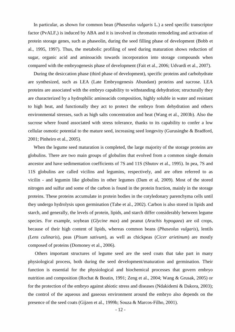
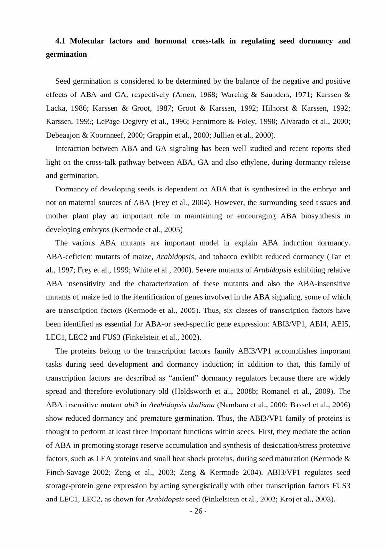
Schematic picture of embryo morphogenesis and seed development events of soybean, used
as a common model legume, is represented in figure 1.
Figure 1 Schematic representation of soybean seed development. Embryo morphologies and developmental
events; a: axis; c: cotyledon; ep: embryo proper; s: suspensor (Brandon et al., 2007).
The mitotic activity of the seed in this first phase is controlled by hormones, environmental
factors, and carbon and nitrogen supply (Egli et al., 1989; Munier-Jolain & Ney, 1998; Ozga et
al., 2002). In the recent literature it has been demonstrated that cytokinins, auxin and gibberellins
are involved in early embryogenesis. Cytokinins are present in the beginning of seed
development, regulating growth trough cell division and sugar metabolism, increasing at rapid
speed after fecundation and diminishing as the seed develops. As extensively studied during pea
seed development (Quesnelle & Emery, 2007), cytokinins reach a peak of concentration during
the heart-shape stage of embryo developmement. Gibberellins are associated whit cell expansion
and, in association whit the auxins, whit driving reserve synthesis. The expression of genes
involved in the last step of active gibberellins synthesis (GA-oxidases) was localized in different
tissues and a at different stages of embryogenesis during seed development of Phaseolus
coccineus: between the late globular and heart stages, in the embryo epidermal cells, in the
endosperm during the transition from globular to heart stage, and during cotyledons and inner
tegument development (Solfanelli et al., 2005). Auxins are synthesized from the aminoacid
tryptophan, and in seeds it is present since the initial phases of development and it’s responsible
of compound assimilation from the mother plant. It has been shown that the auxins are important
in controlling seed development of common bean: in fact, a functional enzyme involved in
- 11 -
auxins conjugation, during the auxins homeostatis control, was defected during the rapid growth
period of seed development in common beans (Walz et al., 2002; Walz et al., 2008).
During the seed-filling phase (second phase of development), the endosperm contents are
hydrolyzed and transported to the embryo. Seed nitrogen accumulation and protein composition
depend on both symbiotic N2 fixation and nitrogen from the soil, while carbon storage derives
mainly from recent photosynthate rather than from remobilized sources (Domoney et al., 2006).
Proteins involved in cell division that were abundant during early stages of seed development,
undergoes a decreases of their level, before the accumulation of the major storage proteins
during seed filling (Gallardo et al., 2003).
Thus, during seed-filling the embryo stops growing, mitosis is less intense, teguments tissue
differentiate, storage compound starts to accumulate, and seed develops tolerance to desiccation
(third phase of development) (Gutierrez et al., 2007).
In general, at the end of the third phase of development, the maximum accumulation of dry
matter in the seed is observed, as well as the rupture of trophic connection with the mother plant,
representing the physiological maturity. At this level, although the metabolism decreases
drastically, the embryo remains viable thanks to the high concentration of abscissic acid that
guaranties dry matter flow and enzyme activity acting on anabolic process (Pammenter &
Berjak, 1999). This metabolic profiling indicates that the preparation for germination starts
during seed desiccation (Fait et al., 2006). At this stage gibberellins and brassinosteroid synthesis
are locally active and exert important functions during maturation (Radchuck et al., 2006), while
the high accumulation of abscissic acid is essential for embryo tolerance to desiccation (Buitink
et al., 2006).
The accumulation of reserve compounds in the seeds serves to feed the embryo during
development and guaranties the completion of germination and the early seedling development.
Among the main seed reserve compounds are carbohydrates, proteins, lipids and phytic acid
(phytin). The main reserve substances derives from carbon fixation by leaf mesophyll cells into
sucrose that is transported via phloem from the mother plant to the seed cells and later
incorporated into the seed trough seed coats (Weber et al., 1997; Patrick & Offler, 2001). Then,
during the maturation phase, when all trophic connection with the mother plant is interrupted, the
sucrose arrive to the seed trough a ventral vascular system localized along the pod, in a region
delimited around the hilum, which forms a vascular net around the seed teguments (Borisjuk et
al., 2003). Besides the sucrose, also other simple molecules are transported to the developing
seed such as aminoacids, especially asparagine and glutamine, and mineral ions (Golombek et
al., 2001). Seed maturation genes are controlled by specific regulatory transcription factor that in
legumes may be regulated by sugar (Tsukagoshi et al., 2007) and by abscissic acid (ABA).
- 12 -
In particular, as shown for common bean (Phaseolus vulgaris L.) a seed specific transcriptor
factor (PvALF,) is induced by ABA and it is involved in chromatin remodeling and activation of
protein storage genes, such as phaseolin, during the seed filling phase of development (Bobb et
al., 1995, 1997). Thus, the metabolic profiling of seed during maturation shows reduction of
sugar, organic acid and aminoacids towards incorporation into storage compounds when
compared with the embryogenesis phase of development (Fait et al., 2006; Udvardi et al., 2007).
During the desiccation phase (third phase of development), specific proteins and carbohydrate
are synthesized, such as LEA (Late Embryogenesis Abundant) proteins and sucrose. LEA
proteins are associated with the embryo capability to withstanding dehydration; structurally they
are characterized by a hydrophilic aminoacids composition, highly soluble in water and resistant
to high heat, and functionally they act to protect the embryo from dehydration and others
environmental stresses, such as high salts concentration and heat (Wang et al., 2003b). Also the
sucrose where found associated with stress tolerance, thanks to its capability to confer a low
cellular osmotic potential to the mature seed, increasing seed longevity (Gurusinghe & Bradford,
2001; Pinheiro et al., 2005).
When the legume seed maturation is completed, the large majority of the storage proteins are
globulins. There are two main groups of globulins that evolved from a common single domain
ancestor and have sedimentation coefficients of 7S and 11S (Shutov et al., 1995). In pea, 7S and
11S globulins are called vicilins and legumins, respectively, and are often referred to as
vicilin - and legumin like globulins in other legumes (Dam et al, 2009). Most of the stored
nitrogen and sulfur and some of the carbon is found in the protein fraction, mainly in the storage
proteins. These proteins accumulate in protein bodies in the cotyledonary parenchyma cells until
they undergo hydrolysis upon germination (Tabe et al., 2002). Carbon is also stored in lipids and
starch, and generally, the levels of protein, lipids, and starch differ considerably between legume
species. For example, soybean (Glycine max) and peanut (Arachis hypogaea) are oil crops,
because of their high content of lipids, whereas common beans (Phaseolus vulgaris), lentils
(Lens culinaris), peas (Pisum sativum), as well as chickpeas (Cicer arietinum) are mostly
composed of proteins (Domoney et al., 2006).
Others important structures of legume seed are the seed coats that take part in many
physiological process, both during the seed development/maturation and germination. Their
function is essential for the physiological and biochemical processes that govern embryo
nutrition and composition (Rochat & Boutin, 1991; Zeng et al., 2004; Wang & Grusak, 2005) or
for the protection of the embryo against abiotic stress and diseases (Ndakidemi & Dakora, 2003);
the control of the aqueous and gaseous environment around the embryo also depends on the
presence of the seed coats (Gijzen et al., 1999b; Souza & Marcos-Filho, 2001).
- 13 -
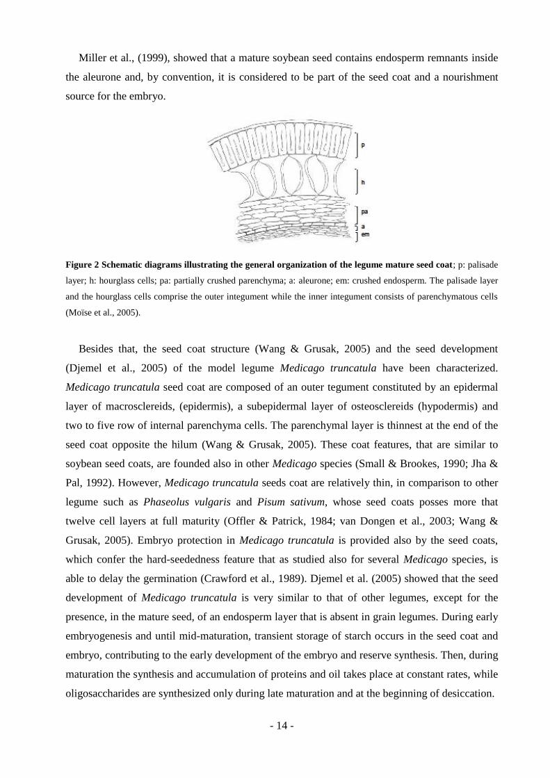
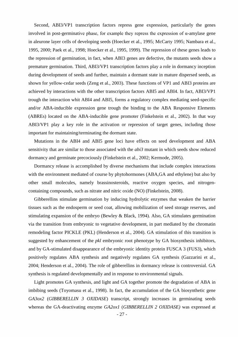
The seed coats of legumes are relatively large and complex and generally, the mature seed
coat is structurally characterized by an outer integument which consists of a single layer of
palisade cells (macrosclereids) and hourglass cell (osteosclereids), and an inner integument
consisting of parenchymatous cells (Miller et al., 1999) (Fig. 2).
The outermost layer of the legume seed coat is the cuticle, which is variable in thickness and
represents the first barrier to imbibitions. As suggested for soybean seeds (Ragus, 1987; Souza &
Marcos-Filho, 2001), the cuticle is formed by two layers of waxy deposits, one very stable and
the other environmentally labile. The epidermis (macrosclereids) is located below the cuticle and
originates from the outer cell layer of the outer integument (Zeng et al., 2004). It is composed of
a layer of thick-walled cells, called palisade cell. In general, only one palisade layer is present,
except under the hilum, where two layers can be present (Souza and Marcos-Filho, 2001). It has
been proposed that the cell walls of the palisade layer contribute to the mechanical strength of
the seed coat (Algan & Büyükkartal, 2000).
Below the epidermis is located the hypodermis (osteosclereids), which is formed from a
single layer of cells. Because of their shape, the cells that formed the osteosclereids are called
hourglass cell. They arise from the outer-cell layer of the inner integument (Zeng et al., 2004)
and are usually larger than adjacent cell layers and are separated by wide intercellular spaces
(Souza and Marcos-Filho, 2001). Often, hourglass cells contain numerous starch grains during
embryogenesis: this indicates that the seed coat could synthesize nutrients, for the developing
embryo. Besides that, the hourglass cells can serve as a reserve of proteins, for the developing
embryo, as shown for soybean seed coat (Gillikin & Graham, 1991; Gijzen et al., 1993). In
addition, like palisade cells, hourglass cell walls, play a role in the mechanical strength of the
seed coat (Algan & Büyükkartal, 2000; Wang & Grusak, 2005).
Adjacent to the hourglass cells is the interior parenchyma, formed by 6-8 layers of
thin-walled, tangentially elongated parenchyma cells (Moïse et al., 2005), uniformly distributed
throughout the seed coat. It has been proposed, that an important function of the parenchyma
cells layers is to deliver nutrients to the embryo, thanks to the presence of a vascular systems,
which are responsible for transporting the nutrients from the maternal organs to the embryo
(Algan & Büyükkartal, 2000; van Dongen et al., 2003). Then, in mature seed coats, the interior
parenchyma is often crushed or partially crushed (Miller et al., 1999) as the embryo expands.
The endosperm cell layers close to the embryo degenerate and eventually appears as
compressed wall materials at seed maturity. However, the outermost endosperm layer remains
intact and differentiates into the aleurone layer (Miller et al., 1999), in which, during
germination, occurs the enzymatic mobilization of seed reserves, such as carbohydrates (Ma et
al., 2004b).
- 14 -
Miller et al., (1999), showed that a mature soybean seed contains endosperm remnants inside
the aleurone and, by convention, it is considered to be part of the seed coat and a nourishment
source for the embryo.
Figure 2 Schematic diagrams illustrating the general organization of the legume mature seed coat; p: palisade
layer; h: hourglass cells; pa: partially crushed parenchyma; a: aleurone; em: crushed endosperm. The palisade layer
and the hourglass cells comprise the outer integument while the inner integument consists of parenchymatous cells
(Moïse et al., 2005).
Besides that, the seed coat structure (Wang & Grusak, 2005) and the seed development
(Djemel et al., 2005) of the model legume Medicago truncatula have been characterized.
Medicago truncatula seed coat are composed of an outer tegument constituted by an epidermal
layer of macrosclereids, (epidermis), a subepidermal layer of osteosclereids (hypodermis) and
two to five row of internal parenchyma cells. The parenchymal layer is thinnest at the end of the
seed coat opposite the hilum (Wang & Grusak, 2005). These coat features, that are similar to
soybean seed coats, are founded also in other Medicago species (Small & Brookes, 1990; Jha &
Pal, 1992). However, Medicago truncatula seeds coat are relatively thin, in comparison to other
legume such as Phaseolus vulgaris and Pisum sativum, whose seed coats posses more that
twelve cell layers at full maturity (Offler & Patrick, 1984; van Dongen et al., 2003; Wang &
Grusak, 2005). Embryo protection in Medicago truncatula is provided also by the seed coats,
which confer the hard-seededness feature that as studied also for several Medicago species, is
able to delay the germination (Crawford et al., 1989). Djemel et al. (2005) showed that the seed
development of Medicago truncatula is very similar to that of other legumes, except for the
presence, in the mature seed, of an endosperm layer that is absent in grain legumes. During early
embryogenesis and until mid-maturation, transient storage of starch occurs in the seed coat and
embryo, contributing to the early development of the embryo and reserve synthesis. Then, during
maturation the synthesis and accumulation of proteins and oil takes place at constant rates, while
oligosaccharides are synthesized only during late maturation and at the beginning of desiccation.

- 15 -
The composition in the major class of storage compounds of Medicago truncatula seeds is
similar to other grain legumes, although the major difference resides in the nature of their carbon
storage, which in M. truncatula is represented by triacylglycerides, and in other grain legumes by
starch (Djemel et al., 2005).
2. Seed dormancy
Seed dormancy is described as the inability of an intact viable seed to complete germination
under favorable conditions (Hilhorst, 1995; Bewley, 1997; Li & Foley, 1997). This block to
germination has evolved differently across species through adaptation to the environment, so that
germination occurs when conditions for establishing a new plant generation are appropriate
(Hilhorst, 1995; Vleeshouwers et al, 1995; Bewley, 1997; Li & Foley, 1997; Baskin & Baskin,
2004; Fenner & Thompson, 2005). A more sophisticated definition of dormancy has been
proposed by Baskin & Baskin (2004): “A dormant seed is one that does not have the capacity to
germinate in a specified period of time under any combination of normal physical environmental
factors (temperature, light/dark, etc.) that are otherwise favorable for its germination, i.e. after
the seed becomes non-dormant”. On the other hand, a non-dormant seed has the capacity to
germinate over the widest range of normal physical environmental factors (temperature,
light/dark, etc.). A non-dormant seed does not germinate if a certain combination of physical
environmental factors (temperature, light/dark, etc.) is absent. In that case seed enters in a state
of quiescence, also called enforced dormancy by Harper (1957, 1977) and pseudodormancy by
Hilhorst & Karssen (1992), Koornneef & Karssen (1994) and Karssen (1995). A quiescent seed
germinates when the appropriate set of environmental conditions, required for radicle
emergence, is restored (Baskin & Baskin, 2004).
Thus, dormancy is a mechanism of the plant to adapt germination timing to environmental
conditions, to prevent germination when the conditions for seedling establishment and plant
growth are unfavorable. Dormancy can have different origins and several schemes for its
classification have been published (Harper, 1957, 1977; Nikolaeva, 1977, 1999; Lang et al.,
1985, 1987; Lang 1987).
On the basis of the classification system provided by Nikolaeva (1977, 1999, 2004), that
devised dormancy types studying both morphological and physiological properties of the seed,
Baskin & Baskin (1998, 2004) have proposed a complete classification system which includes
five classes of seed dormancy: physiological (PD), morphological (MD), morphophysiological
(MPD), physical (PY) and combinational (PY + PD).
- 16 -
Physiological dormancy (PD) is the most abundant form and is found in seeds of
gymnosperms and all major angiosperm clades.
Physiological dormancy (PD) is established and regulated by the phytohormone abscissic acid
(ABA). It is generally accepted that the mechanisms of seed dormancy and germination involve
the plant growth regulators abscisic acid and gibberellins (GA). It has been proposed a hormone-
balance model, in which ABA (inhibitor) and GA (promoter) simultaneously and
antagonistically regulate the onset, maintenance and termination of dormancy (Amen, 1968;
Wareing & Saunders, 1971). ABA, produced by the embryo, induces dormancy during seed
development, when its concentration within the developing embryo increases respect to the GA
concentration, and GA promotes germination upon dormancy overcoming, when, on the
contrary, the levels of GA increases (Karssen & Lacka, 1986; Karssen & Groot, 1987; Groot &
Karssen, 1992; Hilhorst & Karssen, 1992; Karssen, 1995; LePage-Degivry et al., 1996;
Fennimore & Foley, 1998). More recently, evidences has been presented for the involvement of
both ABA and GA in dormancy-break in seeds of Fagus sylvatica (Nicolás et al., 1996; Lorenzo
et al., 2002), Arabidopsis (Debeaujon & Koornneef, 2000), potato (Alvarado et al., 2000) and
Nicotiana plumbaginifolia (Grappin et al., 2000; Jullien et al., 2000). All these models for
control of dormancy and germination show antagonistic interactions of ABA and GA by
decreasing and increasing, respectively, embryo growth potential. In addition to ABA and GA, a
third plant hormone, ethylene, is involved in the regulation of seed dormancy and germination.
Ethylene breaks dormancy and stimulates germination in the seeds of many species (Kępczyński
& Kępczyńska, 1997; Matilla, 2000), apparently by decreasing the sensitivity of the seed to
endogenous ABA. Thus, ethylene may promote germination by interfering with the action of
ABA (Beaudoin et al., 2000; Ghassemian et al., 2000, Matilla & Matilla-Vázquez, 2008). At the
molecular level, several studies have shown that specific ABA-responsive mRNAs and heat-
stable proteins are present in embryos of dormant seeds. Amounts of dormancy-associated
transcripts remained high in embryos of dormant seeds and completely disappeared during
germination (Morris et al., 1991; Goldmark et al., 1992; Dyer, 1993; Li & Foley, 1994, 1995,
1996, 1997; Johnson et al., 1995; Holdsworth et al., 1999). Thus, the continuous presence of
specific mRNAs and proteins seems to be required to maintain dormancy, that consequently it is
regulated at the level of gene expression (Bewely, 1997; Li & Foley, 1997; Holdsworth et al.,
1999; Garello et al., 2000; Koornneef et al., 2002).
Physiological dormancy can be divided into three levels: deep, intermediate and non-deep
(Baskin & Baskin, 2004). In deep physiological dormancy embryos excised from the seeds do
not grow or produce abnormal seedlings. Also, GA treatment does not break their dormancy, and

- 17 -
several months of cold or warm stratification are required before germination can takes place
(Baskin & Baskin, 2004; Baskin et al., 2005).
Embryo excised from seed affected by intermediate physical dormancy produce normal
seedlings; GA can promotes germination in some species, although seeds require two or three
months of cold stratification or a dry storage period for dormancy break (Baskin & Baskin,
2004).
The non-deep physiological dormancy is the most common type of seed physiological
dormancy (Baskin & Baskin, 2004). Embryos excised from these seeds produce normal
seedlings; GA treatment promotes germination and, depending on species, dormancy can also be
broken by scarification, after-ripening in dry storage, and cold or warm stratification. Also, based
on patterns of change in physiological responses to temperature, it’s possible to distinguish five
types of non-deep physiological dormancy. Most seeds belong to type one or two, in which the
temperature range at which seed germination can occur, increases gradually during the
progression of non-deep dormancy release from low to higher (Type 1), or from high to lower
temperature (Type 2). Only few seed have Type 3, seeds with Type 4 or 5 appear to be even
more uncommon than those with Type 3 (Baskin & Baskin, 2004).
In seeds affected by morphological dormancy (MD), the embryo is underdeveloped in term of
size, but differentiated (cotyledons and hypocotyls-radicle can be distinguished) (Baskin and
Baskin, 1998, 2004). These embryos are not physiologically dormant, and does not require a
dormancy-breaking pretreatment in order to overcoming dormancy, but need time to grow to full
size and then germinate. Thus, the dormancy period is just the time elapsed between incubation
of fresh seeds and radical emergence; consequently, under appropriate conditions, embryos begin
to elongate within a period of a few days to some weeks, and then completes the germination.
Besides that, several studies show that the morphological dormancy is also the most primitive
dormancy class. The thick endosperm layer, that characterizes most of the angiosperms seeds
affected by this type of dormancy, is able to inhibit the germination of the embryo. Thus,
morphologically dormant seeds germination timing is regulated by the time the embryo requires
to elongate and, finally to protrude the surrounding tissues, including the endosperm. Because of
that, was observed a general trend of reducing endosperm abundance during the higher plant
evolution, that caused a gradually decrease in morphological dormancy and, at the same time, the
appearance of physiological dormancy (Finch-Savage & Leubner-Metzger, 2006; Linkies et al.,
2010). On the basis of these remarks, it has been proposed that the morphological dormancy is
the ancestral type of dormancy among seed plants (Baskin & Baskin, 1998; Forbis et al., 2002;
Linkies et al., 2010).

- 18 -
Seeds with morphophysiological dormancy (MPD) have an underdeveloped embryo with a
physiological component of dormancy. Thus, in order to germinate, these seeds require a
dormancy-breaking pretreatments, such as a defined combination of warm and/or cold
stratification otherwise, is some cases, GA application. In seeds with morphophysiological
dormancy, the embryo growth and therefore, the radicle emergence requires a considerably
longer period of time than in seeds with morphological dormancy. There are eight known levels
of morphophysiological dormancy, based on the protocol for seed dormancy break and
germination (Baskin & Baskin, 2004).
In seeds whit physical dormancy (PY), prevention of water uptake causes the seed to remain
dormant until some factors, such as, in nature, high temperatures, fire, drying, freezing/thawing
or the passage through the digestive tracts of animals, render the covering layers permeable to
the water (Baskin & Baskin, 1998; Baskin et al., 2000). Once the permeability to the water is
restored, seeds can germinate over a wide range of temperatures in both light and darkness and,
generally, coat of seeds cannot revert to impermeability (Baskin & Baskin, 1998; Baskin et al.,
2000). Thus, physical dormancy is associated with the main mechanical layers of the seed coat
which in most hardseeded species are represented by one or more water-impermeable layers of
palisade cells (Baskin et al., 2000, Baskin & Baskin, 2004). This kind of dormancy can be
removed under both natural and artificial conditions, that allow the formation of an opening,
called “water gap”, in a specialized anatomical structure on the seed coat, through which water
moves to the embryo (Baskin et al., 2000). In Leguminosae seeds, the water-impermeable layers
become permeable after the lens (strophiole) is disrupted. In most legume seeds, stress, such as
heating, causes disruption of the thin-wall cells of the lens, or disruption occurs via a pop-up lens
(Baskin & Baskin, 1998; Baskin et al., 2000).
However, Morrison et al., (1998) have presented evidence that, in some taxa of Fabaceae,
dormancy break by heating may occur through the disruption of the seed coat in a region other
than the lens. In particular, these authors showed that in some legumes, an area on the seed coat,
in addition to lens region, was disrupted by dry-heating pretreatment. Seed disrupted only at the
lens generally had a thinner testa, thicker palisade layer, a thinner mesophyll layer and became
permeable only at the lens (Morrison et al., 1998).
Mechanical or chemical scarification can promote germination in seeds with physical
dormancy (Baskin & Baskin, 2004).
- 19 -
Combinational dormancy (PY + PD) refers to a condition in which seeds coat is water
impermeable and, in addition, the embryo is physiologically dormant. Generally, the
physiological component appears to be at the non-deep level, and embryos will come out of
dormancy in dry storage or in the field within a few weeks after maturity, even while the seed
coat remains impermeable to water (Baskin & Baskin, 1998, 2004). However, embryos in such
genera as Cercis (Fabaceae) and Ceanothus (Rhamnaceae) are more deeply dormant and
therefore, require a few weeks of cold stratification: after physical dormancy is broken and seeds
imbibe water, the germination can be completed (Baskin & Baskin, 2004).
Moreover, as proposed for some species, seeds have a delaying mechanism to prevent
germination when moisture is not sufficient, during late summer and early autumn, for seedling
establishment and growth (Thanos & Georghiou, 1988; Thanos et al., 1992; Abeles, 1986;
Gallardo et al., 1991; Yoshioka et al., 1998). The suppression of germination at supraoptimal
high temperatures is often called thermoinhibition or thermodormancy (Reynolds & Thompson,
1971; Abeles, 1986; Gallardo et al., 1991; Yoshioka et al., 1998). It has been shown that seed
responsiveness to temperature is closely related to the level of dormancy in soil-buried seeds of
winter and summer annuals (Baskin and Baskin, 1998). Thus, the change in seed sensitivity to
temperature plays an ecologically important role in the detection of the appropriate seasonal
timing for germination (Baskin and Baskin, 1998; Yoshioka et al., 1998, 2003).
Thermodormancy condition can be overcome by various phytohormones, such as ethylene, as
showed for thermoinhibited Cicer arietinum seeds (Gallardo, 1991), in which small quantities of
this phytohormone are produced when seeds started to germinate. Other studies, performed on
Lactuca sativa seeds (Abeles, 1986; Dutta & Bradford, 1994; Yoshioka et al., 1998),
demonstrate that thermoinhibition can also be prevented by removing the endosperm or
weakening it. These authors suggest that the effect of supraoptimal high temperatures inhibit the
ability of the embryo to develop sufficient force to penetrate the barrier endosperm, and when
the endosperm is removed or punctured the embryo germinates readily.
Phytohormones abscisic acid (ABA) and giberellic acid (GA), that are well known to be
involved in germination control, are also important in establishing and removing
thermodormancy condition. In seeds of lettuce and chickweed the reduction of ABA content and
de novo ABA biosynthesis is required for thermoinhibition (Yoshioka et al., 1998, 2003), while,
although it is not completely clarified, the application of exogenous GA can suppress
thermoinhibition on several plant species (Madakadze et al., 1993; Dutta et al., 1994; Carter &
Stevens, 1998; Gonai et al., 2004; Tho et al., 2008).

- 20 -
In a more recent work by Tho et al., (2008) carried out on thermodormant Arabidopsis
thaliana seeds, it has been demonstrated that ABA synthesis de novo, after the start of
imbibitions, is essential for germination inhibition at supraoptimal temperature in the light.
These authors reported genetic evidence that shows that ABA has a critical function in
thermoinhibition of Arabidopsis seeds and that a subset of ABA signaling components, including
ABI1, ABI3 and ABI2, are required for germination inhibition at high temperature. The presence
of these components suggests that seeds have a specific mechanism to modulate ABA content in
response to high temperature and use a specific ABA-signaling pathway for germination
inhibition (Tamura et al., 2006; Tho et al, 2008). ABA synthesized in the endosperm is also
involved in thermoinhibition of the seeds (Lefebvre et al., 2006; Tho et al, 2008).
Alleviation of thermoinhibition by exogenous GA3 in lettuce and Arabidopsis seeds suggests
that suppression of active GA synthesis at high temperature, through the action of ABA is
required for thermoinhibition (Gonai et al., 2004; Tho et al., 2008). Thus, as been proposed from
these authors, ABA are able to suppresses the expression of GA biosynthesis genes, GA3ox1,
GA3ox2, GA20ox2, and GA20ox3, at high temperature in the light, avoiding the completion of
germination. Therefore, ABA may inhibit GA action by suppressing GA biosynthesis and also
by suppressing GA signaling (Gonai et al., 2004; Tho et al., 2008).
Thus, dormancy condition can be consider as a complex seed trait to regulate germination
timing and, also the endosperm tissue, can contribute to dormancy and germination timing
(Finch-Savage & Leubner-Metzger, 2006). The role of endosperm in regulating germination is
described in the second chapter of this thesis.
When, under natural or artificial conditions, dormancy is released germination can takes
place.
- 21 -
3. Seed germination
Germination is the process that start whit the imbibition of a mature seed (uptake of water by
the quiescent dry seed or non-dormant seed) and terminates whit the the elongation of the
embryonic axis (Bewley & Black, 1994; Bewley, 1997). The visible sign that germination is
complete is the penetration of the covering layers around the embryo by the radicle. All the other
events that take place after the radicle protrusion, including the mobilization of the major storage
reserves, are associated with growth of the seedling, and because of that, are part of post-
germination process (Bewley & Black, 1994; Bewley, 1997).
Thus, the first event occurring during germination is the imbibition and resumption of
metabolism by dry mature seed.
Water uptake in germinating seed is triphasic process, with a rapid initial uptake during the
first phase (imbibition phase), followed by a plateau phase (phase II). A further increase in water
uptake occurs only after germination is completed, during radicle protrusion (Schopfer & Plachy,
1984; Bewley, 1997; Manz et al., 2005).
During the phase I, the influx of water into the cells of dry seeds causes a temporary structural
perturbation to membranes, which rapidly discharge solutes and low molecular weight
metabolites into the surrounding imbibition solution. This is symptomatic of a transition of the
membrane phospholipid components from the gel phase, achieved during maturation drying, to
the hydrated liquid-crystalline state (Crowe & Crowe, 1992). Then, within a short time of
rehydration, the membranes return to their more stable configuration, and the solute leakage is
reduced. These desiccation and rehydration events can induce damage to membranes and
organelles, thus repair mechanisms are activated during the imbibitions phase. It has been
demonstrated that, the amount of phospholipid with membrane-stabilizing properties, such as
N-acetylphosphatidylethanolamine, increases during the imbibition of dry seed: these molecules
may be involved in maintaining or enhancing membrane integrity (Sandoval et al., 1995).
Upon imbibition, the quiescent dry seed rapidly resumes metabolic activity. The structures
and enzymes necessary for this initial resumption of metabolic activity are present, partially
intact within the dry seed, having survived the desiccation phase. After the water uptake,
turnover or replacement of components occurs until the full metabolic status is achieved
(Bewley, 1997). One of the first changes upon imbibition is the resumption of respiratory
activity, detecting within few minutes. After an initial steep increase in oxygen consumption, the
rate declines until the radicle penetrates the surrounding structures, when, another increase of
respiratory activity occurs (Botha et al., 1992; Bewley and Black, 1994). The glycolytic and
oxidative pentose phosphate pathways both resume during phase I, and the Kreb's cycle enzymes
- 22 -
become activated (Salon et al., 1988). Tissues of the mature dry seed contain poorly
differentiated mitochondria as a consequence of maturation drying. Nevertheless, these
mitochondria are able to provide adequate amounts of ATP to support metabolism for several
hours after imbibition (Ehrenshaft & Brambl, 1990; Attucci et al., 1991).
During germination of embryos, there appear to be two distinct patterns of mitochondrial
development. These patterns, which are particularly obvious in cotyledons, depend on the nature
of the stored reserves. In starch-storing seeds, repair and activation of preexisting organelles
predominate, whereas oil-storing seeds typically produce new mitochondria (Morohashi &
Bewley, 1980; Morohashi, 1986; Ehrenshaft & Brambl, 1990).
Although polysomes are absent, all of the components necessary for the resumption of protein
synthesis upon imbibitions are present within the cells of mature dry embryos. In fact, within
minutes of rehydration, polysomal protein-synthesizing complexes were assembled and the
synthesis of necessary proteins can be started (Dommes & Van der Walle, 1990). Preformed
mRNAs are also present within the dry embryo and some of these are residual messages
associated with previous developmental processes that may be used transiently during early
germination (Comai & Harada, 1990; Lane, 1991). Messages encoding proteins that are
important during seed maturation and drying, such as Late Embryogenesis Abundant (LEA)
proteins, are likely to be degraded rapidly upon imbibition (Jiang & Kermode, 1994; Han et al.,
1996). Conversely, those encoding proteins required during early germination, for example
ribosomal protein messages, are replaced by identical messages at later times, when protein
synthesis becomes more dependent on the new transcripts (Beltrán-Peňa et al., 1995). New
mRNAs are transcribed as germination proceeds. The majority of these are likely to encode
proteins essential for the support of normal cellular metabolism (Bewley & Marcus, 1990), like
the enzymes for to the mobilization and conversion of the major stored reserves.
Radicle extension, through the covering layers surrounding the embryo, is the event that
terminates germination. This extension may or may not be accompanied and by cell division
(Bewley, 1997), but cell elongation is essential for radicle extension and protrusion (Barroco et
al., 2005; Kucera et al., 2005). Two distinct phases of DNA synthesis occur in the radicle cells
after imbibitions. The first takes place soon after imbibition and probably involves the repair of
DNA damaged during maturation drying and rehydration, as well as the synthesis of
mitochondrial DNA. The second phase of DNA synthesis is mainly associated with
postgerminative cell division (Zlatnova et al., 1987; Osborne & Boubriak, 1994).
The extension of the radicle is a turgor-driven process (Cosgrove, 1997), and, besides that, it
requires the activation of many enzymes that allow the radical elongation, such as xyloglucan
endotransglycosylase (XET), (Wu et al., 1994), or expansin (McQueen-Mason & Cosgrove,

- 23 -
1995; Cosgrove, 1997). Xyloglucan-endotransglycosylase enhances cell wall loosening trough
the cleavage and rejoining of xyloglucan molecules that tether adjacent cellulose microfibrils,
permitting expansion by microfibril separation. Conversely, expansin allows the radical cell
expansion thanks to its ability to disrupt the hydrogen bonds between cell wall polymers, like as
matrix polysaccharides and cellulose microfibrils (Wu et al., 1994; McQueen-Mason &
Cosgrove, 1995; Cosgrove, 1997).
Moreover, there is a severe constraint on radicle cell growth imposed by the surrounding
structures, as the external covering layers or the endosperm. In particular, the endosperm acts as
a mechanical barrier to the germination of seeds in several angiosperm clades. A decline in this
mechanical resistance of the micropylar endosperm (the endosperm layer covering the radicle
tip) appears to be a prerequisite for radicle protrusion during seed germination and is called
endosperm weakening (Ni & Bradford, 1993, Hilhorst, 1995; Bewley, 1997; Leubner-Metzger,
2003; Sanchez & Mella, 2004; Kucera et al., 2005; Finch-Savage & Leubner-Metzeger, 2006).
The endosperm weakening mechanism, that can be promoted by gibberellic acid and, at least in
part, inhibited by ABA, is part of the seed germination process of endospermic species, like
Arabidopsis thaliana and Lepidium sativum (Brassicaceae) or Nicotiana tabacum (Solanaceae).
For these species the germination consists of two visible steps: testa rupture and endosperm
rupture. Testa rupture takes place after a certain time of imbibition and then, endosperm rupture
event occurs after several hours or even days, depending on the species (Leubner-Metzger, 2003;
Liu et al., 2005; Muller et al., 2006).
Germination is regulated by several environmental factors, such as temperature, light, oxygen,
moisture, and nutrients. Further, hormones play a key role in seed dormancy and germination.
Generally, ABA is a negative regulator of seed germination, while GA, cytokinins, ethylene
and brassinosteroids promote germination (Kucera et al., 2005). ABA and GA and their
antagonism during germination are extensively described in literature, but the mechanism of
interaction between other hormones during germination needs further clarification.
4. Hormones involved in the control of seed dormancy and germination
The two most important hormones involved in the control of seed dormancy and germination
are, respectively, Abscissic Acid (ABA) and Gibberellin Acid (GA). ABA is a positive regulator
of dormancy and a negative regulator of germination, while, in contrast to ABA, GA regulates
negatively seed dormancy and positively the germination (Koornneef, 2002; Peng & Harberd,
2002; Leubner-Metzger, 2003; Kermode, 2005; Kucera et al., 2005; Finkelstein et al., 2008;
Holdsworth et al., 2008b). ABA is also a key regulator of seed development, and adaptive
- 24 -
responses to abiotic stresses (Zeevaart & Creelman, 1988). Endogenous ABA content is a
determinant of these physiological processes, and ABA-deficient mutants exhibit reduced seed
dormancy and reduced drought tolerance (McCarty, 1995). ABA is synthesized from carotenoids
through the indirect pathway (Zeevaart & Creelman, 1988; Nambara & Marion-Poll, 2005) and
the first compound formed is zeaxanthin. Afterwards, zeaxanthin is converted to all-trans-
violaxanthin by two-step epoxidation catalyzed by zeaxanthin epoxidase (ZEP) in plastids
(Marin et al., 1996; Thompson et al., 2000a; Agrawal et al., 2001; Audran et al., 2001). Then,
9-cis-epoxycarotenoid dioxygenase (NCED) catalyzes oxidative cleavage of the 9-cis isomer of
violaxanthin or neoxanthin, producing produces a C15 product called xanthoxin and a C25
metabolite (Tan et al., 1997; Burbidge et al., 1999; Qin & Zeevaart, 1999; Chernys & Zeevaart,
2000; Iuchi et al., 2000, 2001). NCED is considered to be a key regulatory enzyme in ABA
biosynthesis (Tan et al., 1997; Qin and Zeevaart, 1999, 2002; Thompson et al., 2000b; Iuchi et
al., 2001). NCED enzymes are encoded by a multigene family (Tan et al., 1997; Burbidge et al.,
1999; Qin & Zeevaart, 1999; Chernys & Zeevaart, 2000; Iuchi et al., 2000, 2001) and each
member of the NCED family plays a unique regulatory role in specific environmental responses
and developmental processes, ad also in dormancy establishment (Lefebvre et al., 2006).
The reactions following production of xanthoxin occur in the cytosol. Xanthoxin is converted
to abscisic aldehyde by dehydrogenase/reductase (SDR) (Cheng et al., 2002), and finally, the
abscisic aldehyde is oxidized to ABA by abscisic aldehyde oxidase (Seo et al., 2000a).
On the other hand, the inactivation of ABA is obtained through hydroxylation and
conjugation reactions (Zeevaart & Creelman, 1988; Nambara & Marion-Poll, 2005). However,
among these pathways, the ABA hydroxylation pathway is shown to be the regulatory step in a
variety of physiological processes, and has a major role in the rapid decrease in ABA content
during seed germination (Kushiro et al., 2004; Saito et al., 2004).
As mentioned before, GA is a positive regulator of germination and it is also essential for
stem elongation and flowering. Within the plant cells, GA responses are regulated by the
modulation of GA levels and by altering the ability of cells to respond to the hormone (Richards
et al., 2001). Biologically active GAs, such as GA1 and GA4, are tetracyclic diterpenoids
synthesized from geranylgeranyl diphosphate. The biosynthesis pathway of biologically active
GA is divided into three stages (Hedden & Kamiya, 1997; Olszewski et al., 2002). Briefly, first,
geranylgeranyl diphosphate is cyclized to entkaurene in plastids, then ent-kaurene is oxidized to
GA12 by microsomal cytochrome P450 monooxygenases, and, finally, GA12 is converted into
active GAs in cytosol by two 2-oxoglutarate-dependent dioxygenases, GA 20-oxidase and
GA 3-oxidase (Hedden & Kamiya, 1997; Olszewski et al., 2002).

- 25 -
Conversely, bioactive GAs are deactivated by a 2-oxoglutarate-dependent dioxygenase,
GA 2-oxidase, which also catabolizes immediate precursors of active GAs (Hedden & Kamiya,
1997; Olszewski et al., 2002). Also, additional deactivation mechanisms by a cytochrome P450
monooxygenase and methyltransferases were reported (Zhu et al., 2006; Varbanova et al., 2007).
Environmental signals regulate GA level through modulation of the late steps of GA
biosynthesis and catabolism (Olszewski et al., 2002). Light signals mediated by phytochromes
are critical environmental determinants for photoblastic seed germination (Shinomura et al.,
1996; Toyomasu et al., 1998; Yamaguchi et al., 1998), while also the exposure to low
temperature can promotes germination trough GA signaling pathway, as shown for Arabidopsis
seeds, in which the expression of GA 20-oxidase genes, GA20ox1 and GA20ox2, and GA3ox1
was up-regulated by low temperature in darkness, while the germination of Arabidopsis GA3ox1
mutant seeds are not stimulated by low temperature (Yamauchi et al., 2004). Genetic studies
have identified several GA-signaling components in Arabidopsis, some of which play a role in
GA-induced seed germination, as shown by their loss-of-function mutant phenotypes (Olszewski
et al., 2002). The DELLA subfamily of GRAS proteins and SPINDLY (SPY), a Ser/ThrO-
linkedN-acetylglucosamine transferase, acts as negative regulators of GA pathways, inhibiting
all the process, including germination, that depend on GA signaling (Jacobsen & Olszewski,
1993; Jacobsen et al., 1996; Lee et al., 2002; Wen & Chang, 2002).
SPY is thought to increase the activity of DELLA proteins by N-acetylglucosamine
modification (Silverstone et al., 2007) and, also acts as a positive regulator of cytokinin signaling
(Greenboim-Wainberg et al., 2005) and as a negative regulator of brassinosteroid biosynthesis
(Shimada et al., 2006). One of the five DELLA protein genes isolated in Arabidopsis, RGL2,
plays a major role on seed germination (Lee et al., 2002; Tyler et al., 2004), and has been
documented that others DELLA proteins GAI, RGA, and RGL1 enhance the function of RGL2
(Cao et al., 2005).
Besides the antagonistic action of GA and ABA in the control of dormancy and germination,
ethylene (ET) is also known to promote the germination of non-dormant seeds (Matilla &
Matilla-Vàzquez, 2008) and an ABA-ethylene antagonism has been shown for the germination
of several species, such as Arabidopsis thaliana and Lepidium sativum (Linkies et al., 2009).
- 26 -
4.1 Molecular factors and hormonal cross-talk in regulating seed dormancy and
germination
Seed germination is considered to be determined by the balance of the negative and positive
effects of ABA and GA, respectively (Amen, 1968; Wareing & Saunders, 1971; Karssen &
Lacka, 1986; Karssen & Groot, 1987; Groot & Karssen, 1992; Hilhorst & Karssen, 1992;
Karssen, 1995; LePage-Degivry et al., 1996; Fennimore & Foley, 1998; Alvarado et al., 2000;
Debeaujon & Koornneef, 2000; Grappin et al., 2000; Jullien et al., 2000).
Interaction between ABA and GA signaling has been well studied and recent reports shed
light on the cross-talk pathway between ABA, GA and also ethylene, during dormancy release
and germination.
Dormancy of developing seeds is dependent on ABA that is synthesized in the embryo and
not on maternal sources of ABA (Frey et al., 2004). However, the surrounding seed tissues and
mother plant play an important role in maintaining or encouraging ABA biosynthesis in
developing embryos (Kermode et al., 2005)
The various ABA mutants are important model in explain ABA induction dormancy.
ABA-deficient mutants of maize, Arabidopsis, and tobacco exhibit reduced dormancy (Tan et
al., 1997; Frey et al., 1999; White et al., 2000). Severe mutants of Arabidopsis exhibiting relative
ABA insensitivity and the characterization of these mutants and also the ABA-insensitive
mutants of maize led to the identification of genes involved in the ABA signaling, some of which
are transcription factors (Kermode et al., 2005). Thus, six classes of transcription factors have
been identified as essential for ABA-or seed-specific gene expression: ABI3/VP1, ABI4, ABI5,
LEC1, LEC2 and FUS3 (Finkelstein et al., 2002).
The proteins belong to the transcription factors family ABI3/VP1 accomplishes important
tasks during seed development and dormancy induction; in addition to that, this family of
transcription factors are described as “ancient” dormancy regulators because there are widely
spread and therefore evolutionary old (Holdsworth et al., 2008b; Romanel et al., 2009). The
ABA insensitive mutant abi3 in Arabidopsis thaliana (Nambara et al., 2000; Bassel et al., 2006)
show reduced dormancy and premature germination. Thus, the ABI3/VP1 family of proteins is
thought to perform at least three important functions within seeds. First, they mediate the action
of ABA in promoting storage reserve accumulation and synthesis of desiccation/stress protective
factors, such as LEA proteins and small heat shock proteins, during seed maturation (Kermode &
Finch-Savage 2002; Zeng et al., 2003; Zeng & Kermode 2004). ABI3/VP1 regulates seed
storage-protein gene expression by acting synergistically with other transcription factors FUS3
and LEC1, LEC2, as shown for Arabidopsis seed (Finkelstein et al., 2002; Kroj et al., 2003).
- 27 -
Second, ABI3/VP1 transcription factors repress gene expression, particularly the genes
involved in post-germinative phase, for example they repress the expression of α-amylase gene
in aleurone layer cells of developing seeds (Hoecker et al., 1995; McCarty 1995; Nambara et al.,
1995, 2000; Paek et al., 1998; Hoecker et al., 1995, 1999). The repression of these genes leads to
the repression of germination, in fact, when ABI3 genes are defective, the mutants seeds show a
premature germination. Third, ABI3/VP1 transcription factors play a role in dormancy inception
during development of seeds and further, maintain a dormant state in mature dispersed seeds, as
shown for yellow-cedar seeds (Zeng et al., 2003). These functions of VP1 and ABI3 proteins are
achieved by interactions with the other transcription factors ABI5 and ABI4. In fact, ABI3/VP1
trough the interaction whit ABI4 and ABI5, forms a regulatory complex mediating seed-specific
and/or ABA-inducible expression gene trough the binding to the ABA Responsive Elements
(ABREs) located on the ABA-inducible gene promoter (Finkelstein et al., 2002). In that way
ABI3/VP1 play a key role in the activation or repression of target genes, including those
important for maintaining/terminating the dormant state.
Mutations in the ABI4 and ABI5 gene loci have effects on seed development and ABA
sensitivity that are similar to those associated with the abi3 mutant in which seeds show reduced
dormancy and germinate precociously (Finkelstein et al., 2002; Kermode, 2005).
Dormancy release is accomplished by diverse mechanisms that include complex interactions
with the environment mediated of course by phytohormones (ABA,GA and ethylene) but also by
other small molecules, namely brassinosteroids, reactive oxygen species, and nitrogen-
containing compounds, such as nitrate and nitric oxide (NO) (Finkelstein, 2008).
Gibberellins stimulate germination by inducing hydrolytic enzymes that weaken the barrier
tissues such as the endosperm or seed coat, allowing mobilization of seed storage reserves, and
stimulating expansion of the embryo (Bewley & Black, 1994). Also, GA stimulates germination
via the transition from embryonic to vegetative development, in part mediated by the chromatin
remodeling factor PICKLE (PKL) (Henderson et al., 2004). GA stimulation of this transition is
suggested by enhancement of the pkl embryonic root phenotype by GA biosynthesis inhibitors,
and by GA-stimulated disappearance of the embryonic identity protein FUSCA 3 (FUS3), which
positively regulates ABA synthesis and negatively regulates GA synthesis (Gazzarini et al.,
2004; Henderson et al., 2004). The role of gibberellins in dormancy release is controversial. GA
synthesis is regulated developmentally and in response to environmental signals.
Light promotes GA synthesis, and light and GA together promote the degradation of ABA in
imbibing seeds (Toyomasu et al., 1998). In fact, the accumulation of the GA biosynthetic gene
GA3ox2 (GIBBERELLIN 3 OXIDASE) transcript, strongly increases in germinating seeds
whereas the GA-deactivating enzyme GA2ox1 (GIBBERELLIN 2 OXIDASE) was expressed at

- 28 -
the highest levels in dormant seeds (Finch-Savage et al., 2007). Changes in expression of GA
biosynthetic genes in response to light and cold are regulated by two basic helix-loop-helix
(bHLH) transcription factors, SPATULA (SPT) and PHYTOCHROME-INTERACTING
FACTOR 3-LIKE 5 (PIL5) (Oh et al., 2004; Oh et al., 2006; Penfield et al., 2005). SPT inhibits
expression of two GA3ox genes (GA3ox1 and GA3ox2) during seed imbibitions in the cold, and
mutation in gene that encodes this transcription factor generates spt mutants. Spt mutants show
reduced dormancy more pronounced in the light, suggesting a SPT-mediated cross talk between
light and cold in regulation of dormancy. On the other hand, PIL5 prevents seed germination in
the dark at low temperature by repressing expression of GA3ox1 and GA3ox2 genes, whereas it
induces expression of the GA2ox2 catabolic gene during imbibition in the dark (Oh et al., 2006;
Penfield et al., 2005). When PIL5 protein disappears, in the light, due to phytochrome-stimulated
degradation by the 26S proteasome, the germination can be completed, thanks to the
accumulation of bioactive GA (Oh et al., 2006). PIL5 also repress GA responses by stimulating
expression of the DELLA repressors of GA response, REPRESSOR OF GA1-3 (RGA) and GA-
INSENSITIVE (GAI ), through direct binding to their promoters (Oh et al., 2007). This suggests
that also DELLA proteins are involved in the control of seed dormancy, mediating the signaling
of GA.
The DELLA family is defined by an N-terminal DELLA domain required for GA regulation
and a C-terminal GRAS [GAI (GA-INSENSITIVE), RGA (REPRESSOR OF GA 1-3), and
SCARECROW] domain required for function of these putative transcription factors (Peng et al.,
1999; Pysh et al., 1999). Mutations in the DELLA domain result in gain-of-function GA
signaling phenotypes resembling GA deficiency, whereas loss-of-function mutations in the
GRAS domain typically result in an enhanced or constitutive GA response (Thomas et al., 2005).
DELLA proteins are negative regulators of a variety of GA responses (Sun et al., 2004) and
generally, when DELLA proteins are degraded, GA becomes active and explains its effect,
including the stimulation of germination (Ariizumi et al., 2007; Tyler et al., 2004). GA relieves
DELLA repression of seed germination through proteolysis of the DELLA protein by the 26S
proteasome, triggered via polyubiquitination reaction by the SCF SLY1/GID2
E3 ubiquitin ligase
complex (Ariizumi et al., 2007). Upon binding to the GA receptor GDI (GIBBERELLIN
INSENSITIVE DWARF 1) the complex of GA and the receptor binds to DELLA factors that act
as negative regulators in the GA signalling pathway. Then, thanks to the binding of GA-GID1-
DELLA, DELLA protein is recognized by the F-box proteins SLY1 and GA INSENSITIVE
DWARF 2 (GID2) that are F-box subunits of an SCF E3 ubiquitin ligase complex. These F-box
proteins target ubiquitinates DELLA protein for degradation by the 26S proteasome, which in
turn leads to a de-repression of the GA signalling pathway (Dill et al., 2004; Fu et al., 2004;
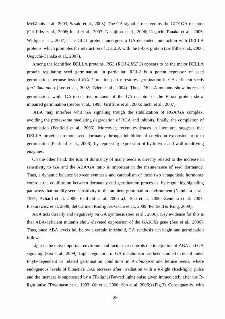
- 29 -
McGinnis et al., 2003; Sasaki et al., 2003). The GA signal is received by the GID1GA receptor
(Griffiths et al., 2006; Iuchi et al., 2007; Nakajima et al., 2006; Ueguchi-Tanaka et al., 2005;
Willige et al., 2007). The GID1 protein undergoes a GA-dependent interaction with DELLA
proteins, which promotes the interaction of DELLA with the F-box protein (Griffiths et al., 2006;
Ueguchi-Tanaka et al., 2007).
Among the identified DELLA proteins, RGL (RGA-LIKE 2) appears to be the major DELLA
protein regulating seed germination. In particular, RGL2 is a potent repressor of seed
germination, because loss of RGL2 function partly restores germination in GA-deficient seeds
(ga1-3mutants) (Lee et al., 2002; Tyler et al., 2004). Thus, DELLA-mutants show increased
germination, while GA-insensitive mutants of the GA-receptor or the F-box protein show
impaired germination (Steber et al., 1998; Griffiths et al., 2006; Iuchi et al., 2007).
ABA may interfere whit GA signaling trough the stabilization of RGA/GA complex,
avoiding the proteasome mediating degradation of RGA and inhibits, finally, the completion of
germination (Penfield et al., 2006). Moreover, recent evidences in literature, suggests that
DELLA proteins promote seed dormancy through inhibition of cotyledon expansion prior to
germination (Penfield et al., 2006), by repressing expression of hydrolytic and wall-modifying
enzymes.
On the other hand, the loss of dormancy of many seeds is directly related to the increase in
sensitivity to GA and the ABA/GA ratio is important in the maintenance of seed dormancy.
Thus, a dynamic balance between synthesis and catabolism of these two antagonistic hormones
controls the equilibrium between dormancy and germination processes, by regulating signaling
pathways that modify seed sensitivity to the ambient germination environment (Nambara et al.,
1991; Achard et al. 2006; Penfield et al. 2006 a,b; Seo et al. 2006; Zentella et al. 2007;
Piskurewicz et al. 2008; del Carmen Rodríguez-Gacio et al., 2009; Penfield & King, 2009).
ABA acts directly and negatively on GA synthesis (Seo et al., 2006). Key evidence for this is
that ABA-deficient mutants show elevated expression of the GA3OXs gene (Seo et al., 2006).
Thus, once ABA levels fall below a certain threshold, GA synthesis can begin and germination
follows.
Light is the most important environmental factor that controls the integration of ABA and GA
signaling (Seo et al., 2009). Light-regulation of GA metabolism has been studied in detail under
PhyB-dependent or related germination conditions in Arabidopsis and lettuce seeds, where
endogenous levels of bioactive GAs increase after irradiation with a R-light (Red-light) pulse
and the increase is suppressed by a FR-light (Far-red light) pulse given immediately after the R-
light pulse (Toyomasu et al. 1993; Oh et al. 2006; Seo et al. 2006;) (Fig.3). Consequently, with
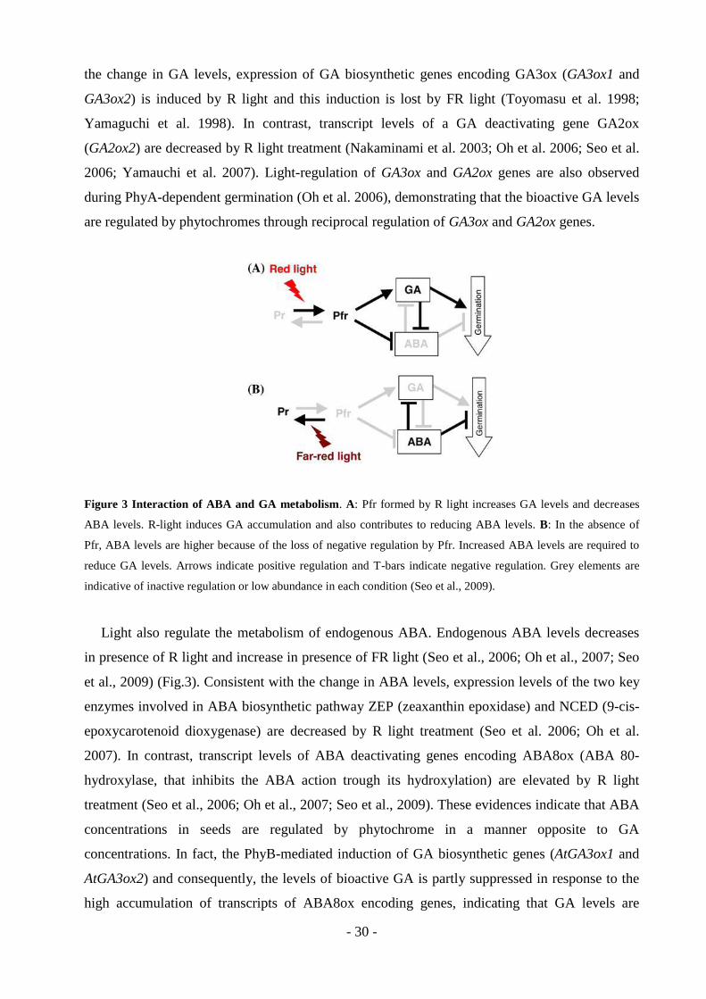
- 30 -
the change in GA levels, expression of GA biosynthetic genes encoding GA3ox (GA3ox1 and
GA3ox2) is induced by R light and this induction is lost by FR light (Toyomasu et al. 1998;
Yamaguchi et al. 1998). In contrast, transcript levels of a GA deactivating gene GA2ox
(GA2ox2) are decreased by R light treatment (Nakaminami et al. 2003; Oh et al. 2006; Seo et al.
2006; Yamauchi et al. 2007). Light-regulation of GA3ox and GA2ox genes are also observed
during PhyA-dependent germination (Oh et al. 2006), demonstrating that the bioactive GA levels
are regulated by phytochromes through reciprocal regulation of GA3ox and GA2ox genes.
Figure 3 Interaction of ABA and GA metabolism. A: Pfr formed by R light increases GA levels and decreases
ABA levels. R-light induces GA accumulation and also contributes to reducing ABA levels. B: In the absence of
Pfr, ABA levels are higher because of the loss of negative regulation by Pfr. Increased ABA levels are required to
reduce GA levels. Arrows indicate positive regulation and T-bars indicate negative regulation. Grey elements are
indicative of inactive regulation or low abundance in each condition (Seo et al., 2009).
Light also regulate the metabolism of endogenous ABA. Endogenous ABA levels decreases
in presence of R light and increase in presence of FR light (Seo et al., 2006; Oh et al., 2007; Seo
et al., 2009) (Fig.3). Consistent with the change in ABA levels, expression levels of the two key
enzymes involved in ABA biosynthetic pathway ZEP (zeaxanthin epoxidase) and NCED (9-cis-
epoxycarotenoid dioxygenase) are decreased by R light treatment (Seo et al. 2006; Oh et al.
2007). In contrast, transcript levels of ABA deactivating genes encoding ABA8ox (ABA 80-
hydroxylase, that inhibits the ABA action trough its hydroxylation) are elevated by R light
treatment (Seo et al., 2006; Oh et al., 2007; Seo et al., 2009). These evidences indicate that ABA
concentrations in seeds are regulated by phytochrome in a manner opposite to GA
concentrations. In fact, the PhyB-mediated induction of GA biosynthetic genes (AtGA3ox1 and
AtGA3ox2) and consequently, the levels of bioactive GA is partly suppressed in response to the
high accumulation of transcripts of ABA8ox encoding genes, indicating that GA levels are
- 31 -
negatively regulated by ABA during seed germination (Seo et al., 2006). There is also evidence
that GA negatively regulates endogenous ABA concentrations. In Arabidopsis, higher ABA
levels, activation of ABA biosynthetic genes and suppression of an ABA deactivating gene are
observed in the GA deficient mutant ga1 in comparison with wild type (Oh et al., 2007).
Moreover, Zentella et al., (2007) reported that XERICO, one of the early GA and DELLA-
responsive genes, regulated ABA levels in seeds. XERICO encodes a protein containing a
RING-H2 type zinc finger motif. Over-expression of this gene causes accumulation of ABA
through an increased ABA biosynthesis. Conversely, DELLA proteins repress the expression of
XERICO by directly associating with its promoter (Zentella et al., 2007). Thus, also the
repression of XERICO by DELLA proteins is an important mechanism by which GA regulates
ABA metabolism, in a mutual negative regulation manner.
Recently, Penfield & King (2009), proposed a new model to explain the integration of the
signaling underlining these two hormones. They suggest that the stabilization of ABI3 by ABA
and the destabilization (or the stimulation of GID-binding and deactivation) of DELLAs by GA
are central processes in hormone balance. In this model, they assumes that germination is
promoted by a complex containing two proteins PIFs (PHYTOCHROME-INTERACTING
FACTORS), and PRRs (PSEUDORESPONSE REGULATOR), that can interact with each other,
and each with one of either ABI3 or DELLA. In a dormant seed, the concentration of the
germination-promoting PIFs-PRRs complex is low because PIFs remains bound to DELLA and
PRRs to ABI3. In a germinating seed, free DELLA levels are low (because DELLAs are either
turned over or are prevented from binding PIFs by GIDs), as are those of ABI3. This allows the
formation of PIFs- PRRs complex and subsequent germination, thanks to the degradation of
DELLA protein via proteasome 26S.
Besides GAs, ethylene (ET) is also known to promote the germination of non-dormant seeds
of several species (Matilla & Matilla-Vàzquez, 2008). This hormonal involvement is
demonstrated by the use of inhibitors of ethylene synthesis and action, for example, 1-
methylcyclopropene, 2,5-norbornadiene and silver thiosulfate. When non-dormant seeds are
subjected to the application of these inhibitors, they are incapable to complete the germination
(Gallardo et al., 1994; Nascimento et al., 2004; Kozarewa et al., 2006). Different possibilities can
explain the action of ethylene in the germination process, including the promotion of radial-cell
expansion, the increase of the water potential in the embryonic axis, and the increase in seed
respiration (Kucera et al., 2005). Recently, Achard et al., (2003) reported that part of the growth-
regulatory action of ethylene is mediated via its effects on the DELLA proteins, which repress
growth in response to ethylene, auxin and GAs signals.
- 32 -
Also, thermoinhibition (or thermodormancy) can be overcome by ethylene, as shown for
lettuce seeds (Nascimento, 2003) and chick-pea (Gallardo et al., 1994; Gallardo et al., 1996;
Matilla, 2000). As proposed by these authors, ethylene is needed for the germination of
thermosensitive lettuce genotypes at high temperatures, while the germination of chick-pea
under optimal conditions depends on ethylene production by the embryonic axis.
Moreover, ethylene promotes dormancy breaking and germination trough the interaction whit
ABA signalling. Linkies et al., (2009) demonstrated that ethylene biosynthesis in germinating
L.sativum and A. thaliana seeds, promotes endosperm rupture by promoting endosperm
weakening, counteracting the ABA inhibition of this process. Furthermore, in different works,
Beaudoin et al., (2000), Ghassemian et al., (2000), Chibani et al. (2006) reported that seeds whit
mutations in receptors of ethylene signal (such as ethylene resistant 1 (etr) 1 receptor and
ethylene insensitive (ein) 2/enhanced response to aba (era) 3 mutants) display increased
dormancy correlated with increased sensitivity to ABA and increased ABA biosynthesis. In
comparison with wild-type, these seeds showed increased dormancy due to ABA
overaccumulation.
Thus, the balance between plant hormone signaling pathway controls the induction and the
release of seed dormancy regulating finally, the germination timing.

- 33 -
References
Abeles, F.B. (1986) Role of ethylene in Lactuca sativa cv. ‘Grand Rapids’ seed germination.
Plant Physiology, 81, 780-787.
Achard, P., Cheng, H., De Grauwe, L., Decat, J., Schoutteten, H., Moritz, T., Van Der Straeten,
D., Peng, J. & Harberd, N. P. (2006) Integration of plant responses to environmentally activated
phytohormonal signals. Science, 311, 91-94.
Achard, P., Vriezen, W.H., Van der Straeten, D., Harberd, N.P. (2003) Ethylene regulates
Arabidopsis development via the modulation of DELLA protein growth repressor function. The
Plant Cell, 15, 2816-2825.
Agrawal, G.K., Yamazaki, M., Kobayashi, M., Hirochika, R., Miyao, A., Hirochika, H. (2001)
Screening of the rice viviparous mutants generated by endogenous retrotransposon Tos17
insertion. Tagging of a zeaxanthin epoxidase gene and a novel OsTATC gene. Plant Physiology,
125, 1248-1257.
Algan, G. & Büyükkartal, H.N.B. (2000) Ultrastructure of seed coat development in the natural
tetraploid Trifolium pratense L. Journal of Agronomy and Crop Science, 184, 205-213.
Ali-Rachedi, S., Bouinot, D., Wagner, M.H., Bonnet, M., Sotta, B., Grappin, P., Jullien, M.
(2004) Changes in endogenous abscisic acid levels during dormancy release and maintance of
mature seeds: studies with the Cape Verde Island ecotype, the dormant model of Arabidopsis
Thaliana. Planta, 219, 479-488.
Alvarado, V., Hiroyaki, H., Bradford, K.J. (2000) Expression of endo-β-mannanase and
SNF-related protein kinase genes in true potato seeds in relation to dormancy, gibberellin and
abscisic acid. In: Dormancy in plants: From whole plant behavior to cellular control.
(Eds: Viémont, J.D. & Crabbé, J.) Wallingford, CABI Publishing, 347-364.
Amen, R.D. (1968) A model of seed dormancy. The Botanical Review, 34, 1-31.
Ariizumi, T. & Steber, C.M. (2007) Seed germination of GA-insensitive sleepy1 mutants does
not require RGL2 protein disappearance in Arabidopsis. The Plant Cell, 19, 791-804.
- 34 -
Attucci, S., Carde, J.P., Raymond, P., Saint Gès, V., Spiteri, A., Pradet, A. (1991) Oxidative
phosphorylation by mitochondria extracted from dry sunflower seeds. Plant Physiology, 95,
390-398.
Audran, C., Liotenberg, S., Gonneau, M., North, H., Frey, A., Tap-Waksman, K., Vartanian, N.,
Marion-Poll, A. (2001) Localisation and expression of zeaxanthin epoxidase mRNA in
Arabidopsis in response to drought stress and during seed development. Australian Journal of
Plant Physiology, 28, 1161-1173.
Baroux, C., Spillane, C., Grossniklaus, U. (2002) Evolutionary origins of the endosperm in
flowering plants. Genome Biology, 3, 1026.1-1026.5.
Barroco, R.M., Van Poucke, K., Bergervoet, J.H.W., De Veylder, L., Groot, S.P.C., Inze, D,
Engler, G. (2005) The role of the cell cycle machinery in resumption of postembryonic
development. Plant Physiology, 137, 127-140.
Baskin, C.C. & Baskin, J.M. (1998) Seeds - ecology, biogeography, and evolution of dormancy
and germination. San Diego, CA, USA: Academic Press.
Baskin, J.M. & Baskin, C.C. (2004) A classification system for seed dormancy. Seed Science
Research, 14, 1-16.
Baskin, J.M., Baskin, C.C., Li, X. (2000) Taxonomy, ecology, and evolution of physical
dormancy in seeds. Plant Species Biology, 15, 139-152.
Baskin, C.C., Baskin, J.M., Yoshinaga, A., Thompson, K. (2005) Germination of drupelets in
multi-seeded drupes of the shrub Leptecophylla tameiameiae (Ericaceae) from Hawaii: a case for
deep physiological dormancy broken by high temperatures. Seed Science Research, 15, 349-356.
Bassel, G.W., Mullen, R.T., Bewley, J.D. (2006) ABI3 expression ceases following, but not
during germination of tomato and Arabidopsis seeds. Journal of Experimental Botany, 57,
1291-1297.
Beaudoin, N., Serizet, C., Gosti, F., Giraudat, J. (2000) Interactions between abscisic acid and
ethylene signaling cascades. The Plant Cell, 12, 1103-1115.
- 35 -
Beltrán-Peňa, E., Ortiz-López, A., Sanchez de Jiménez, E. (1995) Synthesis of ribosomal
proteins from stored mRNAs early in seed germination. Plant Molecular Biology, 28, 327-336.
Berger, F., Hamamura, Y., Ingouff, M., Higashiyama, T. (2008) Double fertilization-caught in
the act. Trends in Plant Science, 13: 437-443.
Bewley, J. D. (1997) Seed germination and dormancy. The Plant Cell, 9, 1055-1066.
Bewley, J.D. & Black, M. (1994) Seeds: Physiology of development and germination. New
York: Plenum Press.
Bewley, J.D. & Marcus, A. (1990) Gene expression in seed development and germination.
Progress in Nucleic Acid Research and Molecular Biology, 38, 165-193.
Bobb, A.J., Chern, M.S., Bustos, M.M., (1995) PvAlf, an embryo specific acidic transcriptional
activator enhances gene expression from phaseolin and phytohemagglutinin promoters. The
Plant Journal, 8, 331-343.
Bobb, A.J., Eiben, H.G., Bustos, M.M., (1997) Conserved RY-repeats mediate trans-activation
of seed-specific promoters by the developmental regulator PvALF. Nucleic Acid Research, 25,
641-647.
Boisnard-Lorig, C., Colon-Carmona, A., Bauch, M., Hodge, S., Doerner, P., Bancharel, E.,
Duman, C., Haseloff, J., Berger, F. (2001) Dynamic analyses of the expression of the
HISTONE::YFP fusion protein in Arabidopsis show that syncytial endosperm is divided in
mitotic domains. The Plant Cell, 13, 495-509.
Borisjuk, L., Rolletschek, H., Wobus, U., Weber, H. (2003) Differentiation of legume cotyledons
are related to metabolic gradients and assimilate transport into seed. Journal of Experimental
Botany, 54, 503-512.
Borisjuk, L., Walenta, S., Weber, H., Mueller-Klieser, W., Wobus, U. (1998) High resolution
histographical mapping of glucose concentrations in developing cotyledons of Vicia faba in
relation to mitotic activity and storage processes: glucose as a possible developmental trigger.
The Plant Journal, 15, 583-591.

- 36 -
Botha, F.C., Potgieter, G.P., Botha, A.M. (1992) Respiratory metabolism and gene expression
during seed germination. Journal of Plant Growth Regulation, 11, 211-224.
Brown, R.C., Lemmon, B.E., Nguyen, H., Olsen, O.A. (1999) Development of endosperm in
Arabidopsis thaliana. Sexual Plant Reproduction, 12, 32-42.
Buitink, J., Leger, J.J., Guisle, I., Vu, B.L., Wuillème, S., Lamirault, G., Le Bars, A., Le Meur,
N., Becker, A., Küster, H., Leprince, O. (2006) Transcriptome profiling uncovers metabolic and
regulatory processes occurring during the transition from desiccation-sensitive to
desiccation- tolerant stages in Medicago truncatula seeds. The Plant Journal, 47, 735-750.
Burbidge, A., Grieve, T.M., Jackson, A., Thompson, A., McCarty, D.R., Taylor, I.B. (1999)
Characterization of the ABA-deficient tomato mutant notabilis and its relationship with maize
Vp14. The Plant Journal, 17, 427-431.
Cantino, P.D., Doyle, J.A., Graham, S.W., Judd, W.S., Olmstead, R.G., Soltis, D.E., Soltis, P.S.,
Donoghue, M.J. (2007) Towards a phylogenetic nomenclature of Tracheophyta. Taxon, 56,
822-846.
Cao, D., Hussain, A., Cheng, H., Peng, J. (2005) Loss of function of four DELLA genes leads to
light- and gibberellin-independent seed germination in Arabidopsis. Planta, 223, 105-113.
Carter, A.K. & Stevens, R. (1998) Using ethephon and GA3 to overcome thermoinhibition in
“Jalapeno M” pepper seed. HortScience, 33, 1026-1027.
Cheng, W.H., Endo, A., Zhou, L., Penney, J., Chen, H.C., Arroyo, A., Leon, P., Nambara, E.,
Asami, T., Seo, M. (2002) A unique short-chain dehydrogenase/reductase in Arabidopsis glucose
signaling and abscisic acid biosynthesis and functions. The Plant Cell, 14, 2723-2743.
Chernys, J.T. & Zeevaart, J.A. (2000) Characterization of the 9-cis-epoxycarotenoid
dioxygenase gene family and the regulation of abscisic acid biosynthesis in avocado. Plant
Physiology, 124, 343-353.
Chibani, K., Ali-Rachedi, S., Job, C., Job, D., Jullien, M., Grappin, P. (2006) Proteomic analysis
of seed dormancy in Arabidopsis. Plant Physioogy, 142, 1493-1510.
- 37 -
Coelho, C.M.M. & Benedito, V.A. (2008) Seed development and reserve compound
accumulation in common bean (Phaseolus vulgaris L.). Seed Science and Biotechnology, 2,
42-52.
Comai, L. & Harada, J.J. (1990) Transcriptional activities in dry seed nuclei indicate the timing
of the transition from embryogeny to germination. Proceedings of the National Academy of
Science, USA, 87, 2671-2674.
Cosgrove, D.J. (1997) Relaxation in a high-stress environment: the molecular bases of extensible
cell walls and cell enlargement. The Plant Cell, 9, 1031-1041.
Coste, F., Ney, B., Crozat, Y. (2001) Seed development and seed physiological quality of field
grown beans (Phaseolus vulgaris L.). Seed Science and Technology, 29, 121-136.
Crawford, E.J., Lake, A.W.H., Boyce, K.G. (1989) Breeding annual Medicago species for
semiarid condition in southern Australia. Advances in Agronomy, 42, 399-437.
Crowe, J.H. & Crowe, L.M. (1992). Membrane integrity in anhydrobiotic organisms: toward a
mechanism for stabilizing dry seeds. In: Water and Life (Eds: Somero, G.N, Osmond, C.B.,
Bolis, C.L.) Berlin: Springer-Verlag, 87-103.
Dam, S., Laursen, B.S., Ørnfelt, J.H., Jochimsen, B., Stærfeldt, H.H, Friis, C., Nielsen, K.,
Goffard, N., Besenbacher, S., Krusell, L., Sato, S., Tabata, S., Thøgersen, I.B, Enghild, J.J,
Stougaard, J. (2009) The Proteome of seed development in the model legume Lotus japonicus.
Plant Physiology, 149, 1325–1340.
del Carmen Rodríguez-Gacio, M., Matilla-Vázquez, M.A., Matilla, A.J. (2009) Seed dormancy
and ABA signaling. Plant Signaling & Behavior, 4 (11), 1035-1048;
Debeaujon, I., Léon-Kloosterziel, K.M., Koornneef, M. (2000) Influence of the testa on seed
dormancy, germination, and longevity in Arabidopsis. Plant Physiology, 122, 403-413.
Dill, A., Thomas, S.G., Hu, J., Steber, C.M., Sun, T.P. (2004) The Arabidopsis F-box protein
SLEEPY1 targets gibberellin signaling repressors for gibberellin-induced degradation. The Plant
Cell, 16, 1392-1405.
- 38 -
Djemel, N., Guedon, D., Lechevalier, A., Salon, C., Miquel, M., Prosperi, J.M., Rochat, C.,
Boutin, J.P. (2005) Development and composition of the seeds of nine genotypes of the
Medicago truncatula species complex. Plant Physiology and Biochemistry, 43, 557-566.
Domoney, C., Duc, G., Ellis, T.H., Ferrandiz, C., Firnhaber, C., Gallardo, K., Hofer, J., Kopka,
J., Kuster, H., Madueno, F. (2006) Genetic and genomic analysis of legume flowers and seeds.
Current Opinion in Plant Biology, 9, 133-141.
Dommes, J. & Van de Walle, C. (1990) Polysome formation and incorporation of new ribosomes
into polysomes during germination of the embryonic axis of maize. Physiologia Plantarum, 79,
289-296.
Doyle, J.J. & Luckow, M.A. (2003) The rest of the iceberg. Legume diversity and evolution in a
phylogenetic context. Plant Physiology, 131, 900-910.
Dyer, W.E. (1993) Dormancy-associated embryonic mRNAs and proteins in imbibing Avena
fatua caryopses. Physiologia Plantarum, 88, 201-211.
Dumas, C. & Rogowsky, P. (2008) Fertilization and early seed formation. Comptes Rendus
Biologies, 331, 715-725.
Duranti, M. & Gius, C. (1997) Legume seeds: protein content and nutritional value. Field Crops
Research, 53, 31–45.
Dutta, S. & Bradford, K.J. (1994) Water relations of lettuce seed thermoinhibition. II. Ethylene
and endosperm effects on base water potential. Seed Science Research, 4, 11-18.
Egli, D.B., Ramseur, E.L., Zhen-wen, Y., Sullivan, C.H. (1989) Source-sink alterations affect the
number of cells in soybean cotyledons. Crop Science, 29, 732-735.
Ehrenshaft, M. & Brambl, R. (1990) Respiration and mitochondrial biogenesis in germinating
embryos of maize. Plant Physiology, 93, 295-304.

- 39 -
Fait, A., Angelovici, R., Less, H., Ohad, I., Urbanczyk-Wochaiak, E., Fernie, A.R., Galili, G.
(2006) Arabidopsis seed development and germination is associated with temporally distinct
metabolic switches. Plant Physiology, 142, 839-854.
Fenner, M. & Thompson, K. (2005) The ecology of seeds. Cambridge, UK: Cambridge
University Press.
Fennimore, S.A. & Foley, M.E. (1998) Genetic and physiological evidence for the role of
gibberellic acid in the germination of dormant Avena fatua seeds. Journal of Experimental
Botany, 49, 89-94.
Finch-Savage, W.E., Cadman, C.S., Toorop, P.E., Lynn, J.R., Hilhorst, H.W. (2007) Seed
dormancy release in Arabidopsis Cvi by dry after-ripening, low temperature, nitrate and light
shows common quantitative patterns of gene expression directed by environmentally specific
sensing. The Plant Journal, 51, 60-78.
Finch-Savage, W.E. & Leubner-Metzger, G. (2006) Seed dormancy and the control of
germination. New Phytologist, 171, 501-523.
Finkelstein, R.R, Gampala, S.S.L., Rock, C.D. (2002) Abscisic acid signaling in seeds and
seedlings. The Plant Cell, Supplement, S15-S45.
Finkelstein, R.R, Reeves, W., Ariizumi, T., Steber, C. (2008) Molecular aspects of seed
dormancy. Annual Review of Plant Biology, 59, 387-415.
Floyd, S.K. & Friedman, W.E. (2000) Evolution of endosperm developmental patterns among
basal flowering plants. International Journal of Plant Sciences, 161, 57-81.
Forbis, T.A., Floyd, S.K., deQueiroz, A. (2002) The evolution of embryo size in angiosperms
and other seed plants: implication for the evolution of seed dormancy. Evolution, 56, 2112-2125.
Frey, A., Audran, C., Marin, E., Sotta, B., Marion-Poll, A. (1999) Engineering seed dormancy by
modification of zeaxanthin epoxidase gene expression. Plant Molecular Biology, 39, 1267-1274.
- 40 -
Frey, A., Godin, B., Bonnet, M., Sotta, B., Marion-Poll, A. (2004) Maternal synthesis of abscisic
acid control seed development and yield in Nicotiana plumbaginafolia. Planta, 218, 958-964.
Friedman, W.E., Madrid, E.N., Wiliams, J.H. (2008) Origin of the fittest and survival of the
fittest: relating female gametophyte development to endosperm genetics. International Journal
of Plant Sciences, 169, 79-92.
Friedman, W.E. & Ryderson, K.C. (2009) Reconstructing the ancestral female gametophyte of
angiosperms: insights from Amborella and other ancient lines of flowering plants. American
Journal of Botany, 96, 129-143.
Friedman, W.E. & Williams, J.H. (2004) Developmental evolution of the sexual process in
ancient flowering plant lineages. The Plant Cell, 16, 119-132.
Frohlich, M.W. & Chase, M.W. (2007) After a dozen years of progress the origin of angiosperms
is still a great mystery. Nature, 450, 1184-1189.
Fu, X., Richards, D.E., Fleck, B., Xie, D., Burton, N., Harberd, N.P. (2004) The Arabidopsis
mutant sleepy1gar2−1
protein promotes plant growth by increasing the affinity of the SCFSLY1
E3
ubiquitin ligase for DELLA protein substrates. The Plant Cell, 16, 1406-1418.
Furbank, R. T., White, R., Palta, J. A., Turner, N.C. (2004) Internal recycling of respiratory CO2
in pods of chickpea (Cicer arietinum L.): the role of pod walls, seed coat, and embryo. Journal
of Experimental Botany, 55, 1687-1696.
Galau, G.A., Jakobsen, K.S., Hughes, D.W. (1991) The controls of late dicot embryogenesis and
early germination. Physiologia Plantarum, 81, 280-288.
Gallardo, K., Le Signor, C., Vandekerckhove, J., Thompson, R.D., Burstin, J. (2003) Proteomics
of Medicago truncatula seed development establishes the time frame of diverse metabolic
processes related to reserve accumulation. Plant Physiology, 133, 664-682.
Gallardo, M., Delgado, M.D., Sánchez-Calle, I.M., Matilla, A.J. (1991) Ethylene production and
1-aminocyclopropane-1-carboxylic acid conjugation in thermo inhibited Cicer arietinum L.
seeds. Plant Physiology, 97, 122-127.
- 41 -
Gallardo, M., Gallardo, M.E., Matilla, A.J., Muňoz de Rueda, P., Sánchez-Calle, I.M. (1994)
Inhibition of polyamine synthesis by cyclohexylamine stimulates the ethylene pathway and
accelerates the germination of Cicer arietinum seeds. Physiologia Plantarum, 91, 9-16.
Gallardo, M., Muňoz de Rueda, P., Matilla, A., Sánchez-Calle, I.M. (1994) The relationships
between ethylene production and germination of Cicer arietinum seeds. Biologia Plantarum, 36,
201-207.
Gallardo, M., Sánchez-Calle, I.M., Muňoz de Rueda, P., Matilla, A.J (1996) Alleviation of
thermoinhibition in chick-pea seeds by putrescine involves the ethylene pathway. Australian
Journal of Plant Physiology, 23, 479-487.
Garello, G., Barthe, P., Bonelli, M., Bianco-Trinchant, J., Bianco, J., Le Page-Degivry, M.T.
(2000) Abscisic acid-regulated responses of dormant and non-dormant embryos of Helianthus
annuus: role of ABA-inducible proteins. Plant Physiology and Biochemistry, 38, 473-482.
Gasser, C.S., Broadhvest, J., Hauser, B.A. (1998) Genetic analysis of ovule development. Annual
Review of Plant Physiology and Plant Molecular Biology, 49, 1-24.
Gazzarrini, S., Tsuchiya, Y., Lumba, S., Okamoto, M., McCourt, P. (2004) The transcription
factor FUSCA3 controls developmental timing in Arabidopsis through the hormones gibberellins
and abscisic acid. Developmental Cell, 7, 373-385.
Gehring, M., Choi, Y., Fischer, R.L. (2004) Imprinting and seed development. The Plant Cell,
16, 203-213.
Gepts, P., Beavis, W.D., Brummer, E.C., Shoemaker, R.C., Stalker, H.T., Weeden, N.F., Young,
N.D. (2005) Legumes as a model plant family. Genomics for food and feed report of the
Cross-Legume Advances Through Genomics Conference. Plant Physiology, 137, 1228-1235.
Ghassemian, M., Nambara, E., Cutler, S., Kawaide, H., Kamiya, Y., McCourt, P. (2000)
Regulation of abscisic acid signaling by the ethylene response pathway in Arabidopsis. The
Plant Cell, 12, 1117-1126.
- 42 -
Gibson, S. I. (2004) Sugar and phytohormone response pathways: navigating a signaling
network. Journal of Experimental Botany, 55, 253-264.
Gijzen, M., Miller, S.S., Bowman, L.A., Batchelor, A.K., Boutilier, K., Miki, B.L.A. (1999a)
Localization of peroxidase mRNAs in soybean seeds by in situ hybridization. Plant Molecular
Biology, 41, 57-63.
Gillikin, J.W. & Graham, J.S. (1991) Purification and developmental analysis of the major
anionic peroxidase from the seed coat of Glycine max. Plant Physiology, 96, 214-220.
Goldberg, R.B., Barker, S.J., Perez-Grau, L. (1989) Regulation of gene expression during plant
embryogenesis. Cell, 56, 149-160.
Goldmark, P.J., Curry, J., Morris, C.F., Walker-Simmons, M.K. (1992) Cloning and expression
of an embryo-specific mRNA up-regulated in hydrated dormant seeds. Plant Molecular Biology,
19, 433-441.
Golombek, S., Rolletschek, H., Wobus, U., Weber, H. (2001) Control of storage protein
accumulation during legume seed development. Journal of Plant Physiology, 158, 457-464.
Gonai, T., Kawahara, S., Tougou, M., Satoh, S., Hashiba, T., Hirai, N., Kawaide, H., Kamiya,
Y., Yoshioka, T. (2004) Abscisic acid in the thermoinhibition of lettuce seed germination and
enhancement of its catabolism by gibberellin. Journal of Experimental Botany, 55, 111-118.
Gonzales, M.D., Archuleta, E., Farmer, A., Gajendran, K., Grant, D., Shoemaker, R., Beavis,
W.D., Waugh, M.E. (2005) The Legume Information System (LIS): an integrated information
resource for comparative legume biology. Nucleic Acids Research, 33, 660-665.
Graham, P.H. & Vance, C.P. (2003) Legumes: importance and constraints to greater use. Plant
Physiology, 131, 872-877.
Groot, S.P.C. & Karssen, C.M. (1992) Dormancy and germination of abscisic acid-deficient
tomato seeds. Studies with the sitiens mutant. Plant Physiology, 99, 952-958.
- 43 -
Grappin, P., Bouinot, D., Sotta, B., Miginiac, E., Jullien, M. (2000) Control of seed dormancy in
Nicotiana plumbaginifolia: post imbibition abscisic acid synthesis imposes dormancy
maintenance. Planta, 210, 279-285.
Greenboim-Wainberg, Y., Maymon, I., Borochov, R., Alvarez, J., Olszewski, N., Ori, N., Eshed,
Y., Weiss, D. (2005) Cross talk between gibberellin and cytokinin: the Arabidopsis GA response
inhibitor SPINDLY plays a positive role in cytokinin signaling. The Plant Cell, 17, 92-102.
Griffiths, J., Murase, K., Rieu, I., Zentella, R., Zhang, Z.L., Powers, S.J, Gong, F., Phillips, A.L.,
Hedden, P., Sun, T., Thomas, S.G. (2006) Genetic characterization and functional analysis of the
GID1 gibberellin receptors in Arabidopsis. The Plant Cell, 18, 3399-3414.
Gurusinghe, S. & Bradford, K.J. (2001) Galactosyl-sucrose oligosaccharides and potential
longevity of primed seeds. Seed Science Research, 11, 121-133.
Gutierrez, L., Van Wuytswiakel, O., Castelain, M., Bellini, C. (2007) Combined networks
regulating seed maturation. Trends in Plant Science, 12, 294-300.
Han, B., Hughes, D.W., Galau, G.A., Bewley, J.D., Kermode, A.R. (1996). Changes in late
embryogenesis abundant (LEA) messenger RNAs and dehydrins during maturation and
premature drying of Ricinus communis L. seeds. Planta, 201, 27-35.
Harper, J.L. (1957) The ecological significance of dormancy and its importance in weed control.
Proceedings of the international congress on crop protection (Hamburg), 4, 415-420.
Harper, J.L. (1977) Population biology of plants. London, Academic Press.
Haughna, G., Chaudhuryb, A. (2005) Genetic analysis of seed coat development in Arabidopsis.
Trends in Plant Science, 10, 472-477.
Hedden, P. & Kamiya, Y. (1997) Gibberellin biosynthesis: enzymes, genes and their regulation.
Annual Review of Plant Physiology and Plant Molecular Biology, 48, 431-460.

- 44 -
Henderson, J.T., Li, H.C., Rider, S.D., Mordhorst, A.P., Romero-Severson, J., Cheng, J.C,
Robey, J., Sung, Z.R., de Vries, S.C., Ogas, J. (2004) PICKLE acts throughout the plant to
repress expression of embryonic traits and may play a role in gibberellin-dependent responses.
Plant Physiology, 134, 995-1005.
Higashiyama, T., Yabe, S., Sasaki, N., Nishimura, Y., Miyagishima, S., Kuroiwa, H., Kuroiwa,
T. (2001) Pollen tube attraction by the synergid cell. Science, 293, 1480-1483.
Hilhorst, H.W.M. (1995) A critical update on seed dormancy. I. Primary dormancy. Seed Science
Research, 5: 61-73.
Hilhorst, H.W.M. & Downie, B. (1995) Primary dormancy in tomato (Lycopersicon esculentum
cv. Moneymaker): studies with the sitiens mutant. Journal of Experimental Botany, 47, 89-97.
Hilhorst, H.W.M. & Karssen, C.M. (1992) Seed dormancy and germination: the role of abscisic
acid and gibberellins and the importance of hormone mutants. Plant Growth Regulation, 11,
225-238.
Holdsworth, M.J., Finch-Savage, W.E., Grappin, P., Job, D. (2008b) Post-genomics dissection of
seed dormancy and germination. Trends in Plant Science, 13, 7-13.
Holdsworth, M.J, Kurup, S., McKibbin, R. (1999) Molecular and genetic mechanisms regulating
the transition from embryo development to germination. Trends in Plant Science, 4, 275-280.
Iuchi, S., Kobayashi, M., Taji, T., Naramoto, M., Seki, M., Kato, T., Tabata, S., Kakubari, Y.,
Yamaguchi-Shinozaki, K., Shinozaki, K. (2001) Regulation of drought tolerance by gene
manipulation of 9-cis epoxycarotenoid dioxygenase, a key enzyme in abscisic acid biosynthesis
in Arabidopsis. The Plant Journal, 27, 325-333.
Iuchi, S., Kobayashi, M., Yamaguchi-Shinozaki, K., Shinozaki, K. (2000) A stress-inducible
gene for 9-cis-epoxycarotenoid dioxygenase involved in abscisic acid biosynthesis under water
stress in drought-tolerant cowpea. Plant Physiology, 123, 553-562.
- 45 -
Iuchi, S., Suzuki, H., Kim, Y.C., Iuchi, A., Kuromori, T., Ueguchi-Tanaka, M., Asami, T.,
Yamaguchi, I., Matsuoka, M., Kobayashi, M., Nakajima, M. (2007) Multiple loss-of-function of
Arabidopsis gibberellin receptor AtGID1s completely shuts down a gibberellin signal. The Plant
Journal, 50, 958-966.
Jacobsen, S.E., Binkowski, K.A., Olszewski, N.E. (1996) SPINDLY, a tetratricopeptide repeat
protein involved in gibberellin signal transduction in Arabidopsis. Proceedings of the National
Academy of Science, USA, 93, 9292-9296.
Jacobsen, S.E. & Olszewski, N.E. (1993) Mutations at the SPINDLY locus of Arabidopsis alter
gibberellin signal transduction. The Plant Cell, 5, 887-896.
Jha, S.S. & Pal, A. (1992) Seed structure in Medicago L. Flora, 187, 209-218.
Jiang, L. & Kermode, A.R. (1994) Role of desiccation in the termination of expression of genes
for storage proteins. Seed Science Research, 4, 149-173.
Johnson, R.R., Cranston, H.J., Chaverra, M.E., Dyer, W.E. (1995) Characterization of cDNA
clones for differentially expressed genes in embryos of dormant and nondormant Avena fatua L.
caryopses. Plant Molecular Biology, 28, 113-122.
Johnson, S., Liu, C.M., Hedley, C.L., Wang, T.L. (1994) An analysis of seed development in
Pisum sativum XVIII. The isolation of mutants defective in embryo development. Journal of
Experimental Botany, 45, 1503-1511.
Jullien, M., Bouinot, D., Ali-Rachedi, S., Sotta, B., Grappin, P. (2000) Abscisic acid control of
dormancy expression in Nicotiana plumbaginifolia and Arabidopsis thaliana. In: Dormancy in
plants: From whole plant behavior to cellular control. (Eds: Viémont, J.D. & Crabbé, J.)
Wallingford, CABI Publishing, 195-210.
Kermode, A.R. (1990) Regulatory mechanisms involved in the transition from seed development
to germination. Critical Review in Plant Sciences, 81, 280-288.
Kermode, A.R. (2005) Role of the abscissic acid in seed dormancy. Journal of Plant Growth
Regulation, 24, 319-344.
- 46 -
Kermode, A.R. & Finch-Savage, W. (2002) Desiccation sensitivity in orthodox and recalcitrant
seeds in relation to development. In: Desiccation and plant survival (Eds: Black, M. & Pritchard,
H.) Oxford, UK: CABI, 149-184.
Karssen, C.M. (1995) Hormonal regulation of seed development, dormancy, and germination
studied by genetic control. In: Seed development and germination (Eds: Kigel, J. & Galili, G.)
New York, Marcel Dekker, 333-350.
Karssen, C.M. & Groot, S.P.C. (1987) The hormonebalance theory of dormancy evaluated. In:
British plant growth regulator group monograph 15 – Growth regulators and seeds (Ed:
Pinfield, N.J. & Black, M.) Bristol, UK, British Plant Growth Regulator Group, 17-30.
Karssen, C.M. & Lacka, E. (1986) A revision of the hormone balance theory of seed dormancy:
studies on gibberellin and/or abscisic acid-deficient mutants of Arabidopsis thaliana. In: Plant
growth substances 1985 (Ed: Bopp, M.) Berlin, Springer-Verlag, 315-323.
Kępczyński, J. & Kępczyńska, E. (1997) Ethylene in seed dormancy and germination.
Physiologia Plantarum, 101, 720-726.
Koornneff, M., Bentsink, L., Hilhorst, H.W.M. (2002) Seed dormancy and germination. Current
Opinion in Plant Biology, 5, 33-36.
Koornneef, M. & Karssen, C.M. (1994) Seed dormancy and germination. In: Arabidopsis (Eds:
Myerowitz, E.M., Somerville, C.R.) Plainview, NY, Cold Spring Harbor Press, 313-334.
Kozarewa, I., Cantliffe, D.J., Nagata, R.T., Stoffella, P.J. (2006) High maturation temperature of
lettuce seeds during development increased ethylene production and germination at elevated
temperatures. Journal of the American Society of Horticultural Science, 131, 564-570.
Kroj, T., Savino, G., Valon, C., Giraudat, J., Parcy, F. (2003) Regulation of storage protein gene
expression in Arabidopsis. Development, 130, 6065-6073.
Kucera, B., Cohn, M.A., Leubner-Metzger, G. (2005) Plant hormone interactions during seed
dormancy release and germination. Seed Science Research, 15, 281-307.
- 47 -
Kushiro, T., Okamoto, M., Nakabayashi, K., Yamagishi, K., Kitamura, S., Asami, T., Hirai, N.,
Koshiba, T., Kamiya, Y., Nambara, E. (2004) The Arabidopsis cytochrome P450 CYP707A
encodes ABA 8’-hydroxylases: key enzymes in ABA catabolism. The EMBO Journal, 23,
1647-1656.
Lane, B.G. (1991) Cellular desiccation and hydration: Developmentally regulated proteins, and
the maturation and germination of seed embryos. The FASEB Journal, 5, 2893-2901.
Lang, G.A. (1987) Dormancy: a new universal terminology. HortScience, 22, 817-820.
Lang, G.A., Early, J.D., Arroyave, N.J., Darnell, R.L., Martin, G.C., Stutte, G.W. (1985)
Dormancy: toward a reduced, universal terminology. HortScience, 20, 809-812.
Le, B.H., Wagmaister, J.A., Kawashima, T., Bui, A.Q., Harada, J.J., Goldberg, R.B. (2007)
Using genomics to study legume seed development. Plant Physiology, 144, 562-574.
Lee, S., Cheng, H., King, K.E., Wang, W., He, Y., Hussain, A., Lo, J., Harberd, N.P., Peng, J.
(2002) Gibberellin regulates Arabidopsis seed germination via RGL2, a GAI/RGA-like gene
whose expression is up-regulated following imbibition. Genes & Development, 16, 646-658.
Lefebvre, V., North, H., Frey, A., Sotta, B., Seo, M., Okamoto, M., Nambara, E., Marion-Poll,
A. (2006) Functional analysis of Arabidopsis NCED6 and NCED9 genes indicates that ABA
synthesized in the endosperm is involved in the induction of seed dormancy. The Plant Journal,
45, 309-319.
LePage-Degivry, M.T., Bianco, J., Barthe, P. Garello, G. (1996) Changes in hormone sensitivity
in relation to onset and breaking of sunflower embryo dormancy. In: Plant dormancy:
Physiology, biochemistry and molecular biology (Ed: Lang, G.A.) Wallingford, CAB
International, 221-231.
Leubner-Metzger, G. (2003) Functions and regulation of β-1,3-glucanase during seed
germination, dormancy release and after-ripening. Seed Science Research, 13, 17-34.
Li, B. & Foley, M.E. (1994) Differential polypeptide patterns in imbibed dormant and
after-ripened Avena fatua embryos. Journal of Experimental Botany, 45, 275-279.

- 48 -
Li, B. & Foley, M.E. (1995) Cloning and characterization of differentially expressed genes in
imbibed dormant and afterripened Avena fatua embryos. Plant Molecular Biology, 29, 823-831.
Li, B. & Foley, M. (1996) Transcriptional and posttranscriptional regulation of
dormancy-associated gene expression by afterripening in wild oat. Plant Physiology, 110,
1267-1273.
Li, B. & Foley, M.E. (1997) Genetic and molecular control of seed dormancy. Trends in Plant
Science, 2, 384-389.
Linkies, A., Graeber, K., Knight, C., Leubner-Metzger, G. (2010) The evolution of seed. New
Phytologist, 186, 817-831.
Linkies, A., Müller, K., Morris, K., Turečková, V., Wenk, M., Cadman, C.S.C., Corbineau, F.,
Strnad, M., Lynn, J.R., Finch-Savage, W.E., Leubner-Metzger, G. (2009) Ethylene interacts with
Abscisic Acid to regulate endosperm rupture during germination: A comparative approach using
Lepidium sativum and Arabidopsis thaliana. The Plant Cell, 21, 3803-3822.
Liu, P.P., Koizuka, N., Homrichhausen, T.M., Hewitt, J.R., Martin, R.C., Nonogaki, H. (2005)
Large-scale screening of Arabidopsis enhancer-trap lines for seed germination-associated genes.
The Plant Journal, 41, 936-944.
Lombardi, L., Ceccarelli, N., Piccairelli, P., Lorenzi, R. (2007) DNA degradation during
programmed cell death in Phaseolus coccineus suspensor. Plant Physiology and Biochemistry,
45, 221-227.
Lopes, M.A. & Larkins, B.A. (1993) Endosperm origin, development, and function. The Plant
Cell, 5, 1383-1399.
Lorenzo, O., Nicolás, C., Nicolás, G., Rodriquez, D. (2002) GA3-induced expression of a new
functional AAA-ATPase (FsA1) is correlated with the onset of germination in Fagus sylvatica L.
seeds. Plant and Cell Physiology, 43, 27-34.
Ma, F., Peterson, C.A., Gijzen, M. (2004b) Reassessment of the pits and antipits in soybean
seeds. Canadian Journal of Botany, 82, 654-662.
- 49 -
Madakadze, R., Chirco, E.M., Khan, A.A. (1993) Seed germination of three flower species
following matriconditioning under various environments. Journal of the American Society for
Horticultural Science, 118, 330-334.
Mansfield, S.G. & Briarty, L.G. (1990) Early embryogenesis in Arabidopsis thaliana. II. The
developing embryo. Canadian Journal of Botany, 69, 461-476.
Mansfield, S.G. & Briarty, L.G. (1992) Cotyledon cell development in Arabidopsis thaliana
during reserve deposition. Canadian Journal of Botany, 70, 151-164.
Manz, B., Müller, K., Kucera, B., Volke, F., Leubner-Metzger, G. (2005) Water uptake and
distribution in germinating tobacco seeds investigated in vivo by nuclear magnetic resonance
imaging. Plant Physiology, 138, 1538-1551.
Marin, E., Nussaume, L., Quesada, A., Gonneau, M., Sotta, B., Hugueney, P., Frey, A.,
Marion-Poll, A. (1996) Molecular identification of zeaxanthin epoxidase of Nicotiana
plumbaginifolia, a gene involved in abscisic acid biosynthesis and corresponding to the ABA
locus of Arabidopsis thaliana. The EMBO Journal, 15, 2331-2342.
Marsden, M.P.F. & Meinke, D.W. (1985) Abnormal development of the suspensor in an
embryo-lethal mutant of Arabidopsis thaliana. American Journal of Botany, 72, 1801-1812.
Matilla, A.J. (2000) Ethylene in seed formation and germination. Seed Science Research, 10,
111-126.
Matilla, A.J & Matilla-Vázquez, M.A. (2008) Involvment of ethylene in seed physiology. Plant
Science, 175, 87-97.
McCarty, D.R. (1995) Genetic control and integration of maturation and germination pathways
in seed development. Annual Review of Plant Physiology and Plant Molecular Biology, 46,
71-93.
McGinnis, K.M., Thomas, S. G., Soule, J.D., Strader, L.C., Zale, J.M., Sun, T., Steber, C.M.
(2003) The Arabidopsis SLEEPY1 gene encodes a putative F-box subunit of an SCF E3 ubiquitin
ligase. The Plant Cell, 15, 1120-1130.
- 50 -
McQueen-Mason, S. & Cosgrove, D.J. (1995) Expansin mode of action on cell walls. Analysis
of wall hydrolysis, stress relaxation, and binding. Plant Physiology, 107, 87-100.
Miller, S.S. Bowman, L.A., Gijzen, M., Miki, B.L.A. (1999) Early development of the seed coat
of soybean (Glycine max). Annals of Botany, 84, 297-304.
Moïse, J.A., Han, S., Gudynaitę-Savitch, L., Johnson, D.A., Miki, B.L.A. (2005) Seed coats:
structure, development, composition, and biotechnology. In Vitro Cellular & Developmental
Biology-Plant, 41, 620-644.
Morohashi, Y. (1986) Patterns of mitochondrial development in reserve tissue of germinated
seeds: a survey. Physiologia Plantarum, 66, 653-658.
Morohashi, Y., & Bewley, J.D. (1980) Development of mitochondrial activities in pea
cotyledons during and following germination of the axis. Plant Physiology, 66, 70-73.
Morris, C.F., Anderberg, R.J., Goldmark, P.J., Walker-Simmons, M.K. (1991) Molecular cloning
and expression of abscisic acid-responsive genes in embryos of dormant wheat seeds. Plant
Physiology, 95, 814–821.
Morrison, D.A., McClay, K., Porter, C., Rish, S. (1998) The role of the lens in controlling
heat-induced breakdown of testa-imposed dormancy in native Australian legumes. Annals of
Botany, 82, 35–40.
Müller, K., Tintelnot, S., Leubner-Metzger, G. (2006) Endosperm-limited Brassicaceae seed
germination: Abscisic Acid inhibits embryo-induced endosperm weakening of Lepidium sativum
(cress) and endosperm rupture of cress and Arabidopsis thaliana. Plant and Cell Physiology, 47,
864-877.
Munier-Jolain, N. & Ney, B. (1998) Seed growth rate in grain legumes. II. Seed growth rate
depends on cotyledon cell number. Journal of Experimental Botany, 49, 1971-1976.
- 51 -
Nakajima, M., Shimada, A., Takashi, Y., Kim, Y.C., Park, S.H., Ueguchi-Tanaka, M., Suzuki,
H., Katoh, E., Iuchi, S., Kobayashi, M., Maeda, T., Matsuoka, M., Yamaguchi, I. (2006)
Identification and characterization of Arabidopsis gibberellin receptors. The Plant Journal, 46,
880-889.
Nakaminami, K., Sawada, Y., Suzuki, M., Kenmoku, H., Kawaide, H., Mitsuhashi, W., Sassa,
T., Inoue, Y., Kamiya, Y., Toyomasu, T. (2003) Deactivation of gibberellin by 2-oxidation
during germination of photoblastic lettuce seeds. Bioscience Biotechnology and Biochemistry,
67, 1551-1558.
Nambara, E., Akazawa, T., McCourt, P. (1991) Effects of the gibberellin biosynthetic inhibitor
uniconazol on mutants of Arabidopsis. Plant Physiology, 97, 736-738.
Nambara, E., Hayama, R., Tsuchiya, Y., Nishimura, M., Kawaide, H., Kamiya, Y., Naito, S.
(2000) The role of ABI3 and FUS3 loci in Arabidopsis thaliana on phase transition from late
embryo development to germination. Developmental Biology, 220, 412-423.
Nambara, E. & Marion-Poll, A. (2005) Abscisic acid biosynthesis and catabolism. Annual
Review of Plant Biology, 56, 165-185.
Nascimento, W.M. (2003) Ethylene and lettuce seed germination. Scientia Agricola, 60,
601-606.
Nascimento, W.M., Cantliffe, D.J., Huber, D.J. (2004) Ethylene evolution and
endo-β-mannanase activity during lettuce seed germination at high temperature, Scientia
Agricola, 61, 156-163.
Ndakidemi, P.A. & Dakora, F.D. (2003) Legume seed flavonoids and nitrogenous metabolites as
signals and protectants in early seedling development. Functional Plant Biology, 30, 729-745.
Ni, B.R. & Bradford, K.J. (1993) Germination and dormancy of abscisic acid-deficient and
gibberellin-deficient mutant tomato (Lycopersicon esculentum) seeds - Sensitivity of germination
to abscisic acid, gibberellin, and water potential. Plant Physiology, 101, 607-617.
- 52 -
Nicolás, C., Nicolás, G., Rodriquez, D. (1996) Antagonistic effects of abscisic acid and
gibberellic acid on the breaking of dormancy of Fagus sylvatica seeds. Physiologia Plantarum,
96, 244-250.
Nikolaeva, M.G. (1977) Factors controlling the seed dormancy pattern. In: The physiology and
biochemistry of seed dormancy and germination (Ed: Khan, A.A.) Amsterdam, North-Holland,
51–74.
Nikolaeva, M.G. (1999) Patterns of seed dormancy and germination as related to plant
phylogeny and ecological and geographical conditions of their habitats. Russian Journal of Plant
Physiology, 46, 369-373.
Nikolaeva, M.G. (2004) On criteria to use in studies of seed evolution. Seed Science Research,
14, 315-320.
Oh, E., Kim, J., Park, E., Kim, J.I., Kang, C., Choi, G. (2004) PIL5, a phytochrome-interacting
basic helix-loop-helix protein, is a key negative regulator of seed germination in Arabidopsis
thaliana. The Plant Cell, 16, 3045-3058.
Oh, E., Yamaguchi, S., Hu, J., Yusuke, J., Jung, B., Paik, I., Lee, H.S., Sun, T., Kamiya, Y.,
Choi, G. (2007) PIL5, a phytochrome-interacting bHLH protein, regulates gibberellin
responsiveness by binding directly to the GAI and RGA promoters in Arabidopsis seeds. The
Plant Cell, 19, 1192-1208.
Oh, E., Yamaguchi, S., Kamiya, Y., Bae, G., Chung, W.I., Choi, G. (2006) Light activates the
degradation of PIL5 protein to promote seed germination through gibberellin in Arabidopsis. The
Plant Journal, 47, 124-139.
Offler, C.E. & Patrick, J.W. (1984) Cellular structure, plasma membrane surface, and
plasmodesmatal frequencies of seed coats of Phaseolus vulgaris L. in relation to photosynthate
transfer. Australian Journal of Plant Physiology, 11, 79-99.
Olsen, O.A. (2004) Nuclear endosperm development in cereals and Arabidopsis thaliana. The
Plant Cell, 16, 214-227.

- 53 -
Olszewski, N., Sun, T., Gubler, F. (2002) Gibberellin signaling: biosynthesis, catabolism, and
response pathways. The Plant Cell, 14, 61-80.
Osborne, D.J. & Boubriak, I.I. (1994) DNA and desiccation tolerance. Seed Science Research, 4,
175-185.
Ozga, J.A., van Huizen, R., Reinecke, D.M. (2002) Hormone and seed-specific regulation of pea
fruit growth. Plant Physiology, 128, 1379-1389.
Pammenter, N.W. & Berjak, P.A. (1999) A review of recalcitrant seed physiology in relation to
desiccation-tolerance mechanisms. Seed Science Research, 9, 132-37.
Patrick, J.W. & Offler, C.E. (2001) Compartmentation of transport and transfer events in
developing seeds. Journal of Experimental Botany, 52, 551-564.
Penfield, S., Gilday, A.D., Halliday, K.J., Graham, I.A. (2006a) DELLA-mediated cotyledon
expansion breaks coat-imposed seed dormancy. Current Biology, 16, 2366-2370.
Penfield, S. & King, J. (2009) Towards a systems biology approach to understanding seed
dormancy and germination. Proceeding of the Royal Society: Biological Science, 276,
3561-3569.
Penfield, S., Li, Y., Gilday, A.D., Graham, S., Graham, I.A. (2006b) Arabidopsis ABA
INSENSITIVE4 regulates lipid mobilization in the embryo and reveals repression of seed
germination by the endosperm. The Plant Cell , 18, 1887-1899.
Penfield, S., Josse, E.M., Kannangara, R., Gilday, A.D., Halliday, K.J., Graham, I.A. (2005)
Cold and light control seed germination through the bHLH transcription factor SPATULA.
Current Biology, 15, 1998-2006.
Peng, J. & Harbered, N.P. (2002) The role of GA-mediated signaling in the control of seed
germination. Current Opinion in Plant Biology, 5, 376-381.

- 54 -
Pinheiro, C., Rodrigues, A.P., De Carvalho, I.S., Chaves, M.M., Ricardo, C.P. (2005) Sugar
metabolism in developing lupin seeds is affected by a short-term water deficit. Journal of
Experimental Botany, 56, 2705-2712.
Peng, J., Richards, D.E., Hartley, N.M., Murphy, G.P., Devos, K.M., Flintham, J.E., Beales, J.,
Fish, L.J., Worland, A.J., Pelica, F., Sudhakar, D., Christou, P., Snape, J.W, Gale, M.D.,
Harberd, N.P. (1999) “Green revolution” genes encode mutant gibberellin response modulators.
Nature, 400, 256-261.
Piskurewicz, U., Jikumaru, Y., Kinoshita, N., Nambara, E., Kamiya, Y., Lopez-Molina, L.
(2008) The gibberellic acid signaling repressor RGL2 inhibits Arabidopsis seed germination by
stimulating abscisic acid synthesis and ABI5 activity. The Plant Cell, 20, 2729-2745.
Pritchard, S.L., Charlton, W.L., Baker, A., Graham, I.A. (2002) Germination and storage reserve
mobilization are regulated independently in Arabidopsis. The Plant Journal, 31, 639-647.
Pysh, L.D., Wysocka-Diller, J.W., Camilleri, C., Bouchez, D., Benfey, P.N. (1999) The GRAS
gene family in Arabidopsis: sequence characterization and basic expression analysis of the
SCARECROW-LIKE genes. The Plant Journal, 18, 111-119.
Qin, X. & Zeevaart, J.A. (1999) The 9-cis-epoxycarotenoid cleavage reaction is the key
regulatory step of abscisic acid biosynthesis in water-stressed bean. Proceedings of the National
Academy of Science, USA, 96, 15354-15361.
Qin, X. & Zeevaart, J.A. (2002) Overexpression of a 9-cis-epoxycarotenoid dioxygenase gene in
Nicotiana plumbaginifolia increases abscisic acid and phaseic acid levels and enhances drought
tolerance. Plant Physiology, 128, 544-551.
Quesnelle, P.E., & Emery, R.J.N. (2007) Cis-cytokininis that predominate in Pisum sativum
during early embryogenesis will accelerate embryo growth in vitro. Canadian Journal of Botany,
85, 91-103.

- 55 -
Radchuck, R., Radchuck, V., Weschke, W., Borisjuk, L., Weber, H. (2006) Repressing the
expression of the SUCROSE NONFERMENTING-1-RELATED PROTEIN KINASE gene in
pea embryo causes pleiotropic defects of maturation similar to an abscisic acid-insensitive
phenotype. Plant Physiology, 140, 263-278.
Ragus, L.N. (1987) Role of water absorbing capacity in soybean germination and seedling
vigour. Seed Science and Technology, 15, 285-296.
Reynolds, T., Thompson, P. (1971) Characteristics of the high temperature inhibition of
germination of lettuce (Lactuca sativa). Physiologia Plantarum, 24, 544-547.
Richards, D.E., King, K.E., Ait-Ali, T., Harberd, N.P. (2001) How gibberellins regulates plant
growth and development: a molecular genetic analysis of gibberellin signaling. Annual Review of
Plant Physiology and Plant Molecular Biology, 52, 67-88.
Rochat, C. & Boutin, J.P. (1991) Metabolism of phloem-borne amino acids in maternal tissues of
fruit of nodulated or nitrate-fed pea plants (Pisum sativum L.). Journal of Experimental Botany,
42, 207-214.
Rolletschek, H., Borisjuk, L., Koschorreck, M., Wobus, U., Weber, H. (2002) Legume embryos
develop in a hypoxic environment. Journal of Experimental Botany, 53, 1099-1107.
Romanel, E.A., Schrago, C.G., Counago, R.M., Russo, C.A., Alves-Ferreira, M. (2009)
Evolution of the B3 DNA binding superfamily: new insights into REM family gene
diversification. PLoS One, 4, 1-18.
Saito, S., Hirai, N., Matsumoto, C., Ohigashi, H., Ohta, D., Sakata, K., Mizutani, M. (2004)
Arabidopsis CYP707As encode (+)-abscisic acid 8’-hydroxylase, a key enzyme in the oxidative
catabolism of abscisic acid. Plant Physiology, 134, 1439-1449.
Salon, C., Raymond, P., Pradet, A. (1988) Quantification of carbon fluxes through the
tricarboxylic acid cycle in early germinating lettuce embryos. Journal of Biological Chemistry,
263, 12278-12287.
- 56 -
Sanchez, R.A., Mella, R.A. (2004) The exit from dormancy and the induction of germination:
Physiological and molecular aspects. In: Handbook of seed physiology: applications to
agriculture (Eds: Benech-Arnold, R.L. & Sanchez, R.A.) New York, NY, USA: Food Product
Press and the Haworth Reference Press, 221-243.
Sandoval, J.A., Huang, ZH., Garrett, D.C., Gage, D.A., Chapman, K.D. (1995).
N-Acylphosphatidylethanolamine in dry and imbibing cotton seeds. Amounts, molecular species,
and enzymatic synthesis. Plant Physiology, 109, 269-275.
Sasaki, A., Itoh, H., Gomi, K., Ueguchi-Tanaka, M., Ishiyama, K., Kobayashi, M., Jeong, D.H.,
An, G., Kitano, H., Ashikari, M., Matsuoka, M. (2003) Accumulation of phosphorylated
repressor for gibberellin signaling in an F-box mutant. Science, 299, 1896-1898.
Schopfer, P. & Plachy, C. (1984) Control of seed germination by abscisic acid. II. Effect on
embryo water uptake in Brassica napus L. Plant Physiology, 76, 155-160.
Seo, M., Hanada, A., Kuwahara, A., Endo, A., Okamoto, M., Yamauchi, Y., North, H., Marion
Poll, A., Sun, T., Koshiba, T., Kamiya, Y., Yamaguchi, S., Nambara, E. (2006) Regulation of
hormone metabolism in Arabidopsis seeds: phytochrome regulation of abscisic acid metabolism
and abscisic acid regulation of gibberellins metabolism. The Plant Journal, 48, 354-366.
Seo, M., Koiwai, H., Akaba, S., Komano, T., Oritani, T., Kamiya, Y., Koshiba, T. (2000a)
Abscisic aldehyde oxidase in leaves of Arabidopsis thaliana. The Plant Journal, 23, 481-488.
Seo, M., Nambara, E., Choi, G., Yamaguchi, S. (2009) Interaction of light and hormone signals
in germinating seeds. Plant Molecular Biology, 69, 463-472.
Shimada, A., Ueguchi-Tanaka, M., Sakamoto, T., Fujioka, S., Takatsuto, S., Yoshida, S.,
Sazuka, T., Ashikari, M., Matsuoka, M. (2006) The rice SPINDLY gene functions as a negative
regulator of gibberellin signaling by controlling the suppressive function of the DELLA protein,
SLR1, and modulating brassinosteroid synthesis. The Plant Journal, 48, 390-402.
Shinomura, T., Nagatani, A., Hanzawa, H., Kubota, M., Watanabe, M., Furuya, M. (1996)
Action spectra for phytochrome A- and B-specific photoinduction of seed germination in
Arabidopsis thaliana. Proceedings of the National Academy of Science, USA, 93, 8129-8133.
- 57 -
Shutov, A.D., Kakhovskaya, I.A., Braun, H., Baumlein, H., Muntz, K. (1995) Legumin-like and
vicilin-like seed storage proteins: evidence for a common single-domain ancestral gene. Journal
of Molecular Evolution, 41, 1057-1069.
Silverstone, A.L., Tseng, T.S., Swain, S.M., Dill, A., Jeong, S.Y., Olszewski, N.E., Sun, T.
(2007) Functional analysis of SPINDLY in gibberellin signaling in Arabidopsis. Plant
Physiology, 143, 987-1000.
Small, E. & Brookes, B. (1990) Circumscription of the genus Medicago (Leguminosae) by seed
characters. Canadian Journal of Botany, 68, 613-629.
Solfanelli, C., Ceron, F., Paolicchi, F., Giorgetti, L., Geri, C., Ceccarelli N., Kamiya, Y.,
Picciarelli, P. (2005) Expression of two genes encoding gibberellin 2- and 3-oxidases in
developing seeds of Phaseolus coccineus. Plant and Cell Physiology, 46, 1116-1124.
Souza, F.H.D.D. & Marcos-Filho, J. (2001) The seed coat as a modulator of seed-environment
relationships in Fabaceae. Revista Brasileira de Botânica, 24, 365-375.
Steber, C.M., Cooney, S.E., McCourt, P. (1998) Isolation of the GA-response mutant sly1 as a
suppressor of ABI1-1 in Arabidopsis thaliana. Genetics, 149, 509-521.
Sun, T.P. & Gubler F. (2004) Molecular mechanism of gibberellin signaling in plants. Annual
Review of Plant Biology, 55, 197-223.
Sundaresan, V. (2005) Control of seed size in plants. Proceedings of the National Academy of
Sciences, USA, 102, 17887-17888.
Tabe, L., Hagan, N., Higgins, T.J. (2002) Plasticity of seed protein composition in response to
nitrogen and sulfur availability. Current Opinion in Plant Biology, 5, 212-217.
Tamura, N., Yoshida, T., Tanaka, A., Sasaki, R., Bando, A., Toh, S., Lepiniec, L., Kawakami, N.
(2006) Isolation and characterization of high temperature-resistant germination mutants of
Arabidopsis thaliana. Plant and Cell Physiology, 47, 1081-1094.

- 58 -
Tan, B.C., Schwartz, S.H., Zeevaart, J.A., McCarty, D.R. (1997) Genetic control of abscisic acid
biosynthesis in maize. Proceeding of the National Academy of Science, USA, 94, 12235-12240.
Thanos, C.A. & Georghiou, K. (1988) Ecophysiology of fire-stimulated seed germination in
Cistus incanus ssp. creticus (L.) Hey wood and C. salvifolius L. Plant Cell & Environment, 11,
841-849.
Thanos, C.A., Georgidou, K., Kadis, C., Panfazi, C. (1992) Cistaceae: a plant family with hard
seeds. Israel Journal of Botany, 41, 251-263.
Toh, S., Imamura, A., Watanabe, A., Nakabayashi, K., Okamoto, M., Jikumaru, Y., Hanada, A.,
Aso, Y., Ishiyama, K., Tamura, N., Iuchi, S., Kobayashi, M., Yamaguchi, S., Kamiya, Y.,
Nambara, E., Kawakami, N. (2008) High temperature induced abscisic acid biosynthesis and its
role in the inhibition of gibberellin action in Arabidopsis seeds. Plant Physiology, 146,
1368-1385.
Thomas, T.L. (1993) Gene expression during plant embryogenesis and germination: an
overview. The Plant Cell, 5, 1401-1410.
Thomas, S.G. Rieu, I., Steber, C.M. (2005) Gibberellin metabolism and signaling. Vitamins &
Hormones, 72, 289-338.
Thompson, R. D., Hueros, G., Becker, H.A., Maitz, M. (2001) Development and functions of
seed transfer cells. Plant Science, 160, 775-783.
Thompson, A.J., Jackson, A.C., Parker, R.A., Morpeth, D.R., Burbidge, A., Taylor, I.B. (2000a)
Abscisic acid biosynthesis in tomato: regulation of zeaxanthin epoxidase and
9-cis-epoxycarotenoid dioxygenase mRNAs by light/dark cycles, water stress and abscisic acid.
Plant Molecular Biology, 42, 833-845.
Thompson, A.J., Jackson, A.C., Symonds, R.C., Mulholland, B.J., Dadswell, A.R., Blake, P.S.,
Burbidge, A., Taylor, I.B. (2000b) Ectopic expression of a tomato 9-cis-epoxycarotenoid
dioxygenase gene causes over-production of abscisic acid. The Plant Journal, 23, 363-374.

- 59 -
Toyomasu, T., Kawaide, H., Mitsuhashi, W., Inoue, Y., Kamiya, Y. (1998) Phytochrome
regulates gibberellin biosynthesis during germination of photoblastic lettuce seeds. Plant
Physiology, 118, 1517-1523.
Toyomasu, T., Tsuji, H., Yamane, H., Nakayama, M., Yamaguchi, I., Murofushi, N., Takahashi,
N., Inoue, Y. (1993) Light effects on endogenous levels of gibberellins in photoblastic lettuce
seeds. Journal of Plant Growth Regulation, 12, 85-90.
Tyler, L., Thomas, S.G., Hu, J., Dill, A., Alonso, J.M., Ecker, J.R., Sun, T. (2004) DELLA
proteins and gibberellin-regulated seed germination and floral development in Arabidopsis. Plant
Physiology, 135, 1008-1019.
Tsukagoshi, H., Morikami, A., Nakamura, K., (2007) Two B3 domain transcriptional repressor
prevent sugar-inducible expression of seed maturation genes in Arabidopsis seedlings.
Proceeding of the National Academy of Science, USA, 104, 2543-2547.
Udvardi, M.K., Kakar, K., Wandrey, M., Montanari, O., Murray, J., Andrjankaja, A., Zhang,
J.Y., Benedito, V., Hofer, J.M., Chueng, F., Town, C.D. (2007) Legume transcription factors:
global regulators of plant development and response to the environment. Plant Physiology, 144,
538-549.
Ueguchi-Tanaka, M., Ashikari, M., Nakajima, M., Itoh, H., Katoh, E., Kobayashi, M., Chow, Y.,
Hsing, Y.C., Kitano, H., Yamaguchi, I., Matsuoka, M. (2005) GIBBERELLIN
INSENSITIVEDWARF1 encodes a soluble receptor for gibberellin. Nature, 437, 693-698.
Ueguchi-Tanaka, M., Nakajima, M., Katoh, E., Ohmiya, H., Asano, K., Saji, S., Hongyu,X.,
Ashikari, M., Kitano, H., Yamaguchi, I., Matsuoka, M. (2007) Molecular interactions of a
soluble gibberellin receptor, GID1, with a rice DELLA protein, SLR1, and gibberellin. The Plant
Cell, 19, 2140-2155.
VandenBosch, K.A., Stacey, G. (2003) Summaries of legume genomics projects from around the
globe. Community resources for crops and models. Plant Physiology, 131, 840-865.
- 60 -
van Dongen, J.T., Ammerlaan, A.M.H., Wouterlood, M., van Aelst, A.C., Borstlap, A.C. (2003)
Structure of the developing pea seed coat and the postphloem transport pathway of nutrients.
Annals of Botany, 9, 729-737.
Varbanova, M., Yamaguchi, S., Yang, Y., McKelvey, K., Hanada, A., Borochov, R., Yu, F.,
Jikumaru, Y., Ross, J., Cortes, D., Ma, C.J., Noel, J.P., Mander, L., Shulaev, V., Kamiya, Y.,
Rodermel, S., Weiss, D., Pichersky, E. (2007) Methylation of gibberellins by Arabidopsis
GAMT1 and GAMT2. The Plant Cell, 19, 32-45.
Vleeshouwers, L.M., Bouwmeester, H.J., Karssen, C.M. (1995) Redefining seed dormancy: An
attempt to integrate physiology and ecology. Journal of Ecology, 83, 1031-1037.
Walz, A., Park, S., Slovin, J.P., Ludwig-Müller, J., Momonoki, Y.S., Cohen, J.D. (2002) A gene
encoding a protein modified by the phytohormone indoleacetic acid. Proceedings of the National
Academy of Science, USA, 99, 1718-1723.
Walz, A., Seidel, C., Rusak, G., Park, S., Cohen, J.D., Ludwig-Müller, J. (2008) Heterologous
expression of IAP1, a seed protein from been modified by indole-3-acetic acid, in Arabidopsis
thaliana and Medicago truncatula. Planta, 227, 1047-1061.
Wang, T.L., Hedley, C.L. (1993) Genetic and developmental analysis of the seed. In: Genetics,
Molecular Biology and Biotechnology (Eds: Casey, R. & Davies, D.R.). CAB International,
UK/John Innes Institute, Norwich, UK, 83-120.
Wang, H.L. & Grusak, M.A. (2005) Structure and development of Medicago truncatula pod wall
and seed coat. Annals of Botany, 95, 737-747.
Wang, W.X., Vinocur, B., Altman, A. (2003b) Plant response to drought, salinity, and extreme
temperatures: towards genetic engineering for stress tolerance. Planta, 218, 1-14.
Wareing, P.F. & Saunders, P.F. (1971) Hormones and dormancy. Annual Review of Plant
Physiology, 22, 261-288.
- 61 -
Weber, H., Borisjuk, L., Heim, U., Buchner, P., Wobus, U. (1995) Seed coat associated
invertases of fava bean control both unloading and storage functions: cloning of cDNAs and cell
type-specific expression. The Plant Cell, 7, 1835-1846.
Weber, H., Borisjuk, L., Wobus, U. (1997) Sugar import and metabolism during seed
development. Trends in Plant Science, 2, 169-174.
Weber, H., Borisjuk, L., Wobus, U. (2005) Molecular physiology of legume seed development.
Annual Review of Plant Biology, 56, 253-279.
Weijers, D., van Hamburg, J.P., van Rijn, E., Hooykaas, P. J. J., Offringa, R. (2003) Diphtheria
toxin-mediated cell ablation reveals interregional communication during Arabidopsis seed
development. Plant Physiology, 133, 1882–1892.
Welbaum, G.E., Muthui, W.J., Wilson, J.H., Grayson, R.L., Fell, R.D. (1995) Weakening of
muskmelon perisperm envelope tissue during germination. Journal of Experimental Botany, 46,
391-400.
Wen, C.K. & Chang, C. (2002) Arabidopsis RGL1 encodes a negative regulator of gibberellin
responses. The Plant Cell, 14, 87-100.
West, M.A.L. & Harada, J.J. (1993) Embryogenesis in higher plants: an overview. The Plant
Cell, 5, 1361-1369.
Weterings, K., Apuya, N.R., Bi, Y., Fischer, R.L., Harada, J.J., Goldberg, R.B. (2001) Regional
localization of suspensor mRNAs during early embryo development. The Plant Cell, 13,
2409-2425.
White, C.N., Proebsting, W.M., Hedden, P., Rivin, C.J. (2000) Gibberellins and seed
development in maize. I. Evidence that gibberellins/abscisic acid balance governs germination
versus maturation pathways. Plant Physiology, 122, 1081-1088.
Willige, B.C., Ghosh, S., Nill, C., Zourelidou, M., Dohmann, E.M., Maier, A., Schwechheimer,
C. (2007) The DELLA domain of GA INSENSITIVE mediates the interaction with the GA
INSENSITIVE DWARF1A gibberellin receptor of Arabidopsis. The Plant Cell, 19, 1209-1220.
- 62 -
Windsor, J.B., Symonds, V.V., Mendenhall, J., Lloyd, A.M. (2000) Arabidopsis seed coat
development: morphological differentiation of the outer integument. The Plant Journal, 22,
483-493.
Wobus, U. & Weber, H. (1999) Sugars as signal molecules in plant seed development. The
Journal of Biological Chemistry, 380, 937-944.
Wu, Y., Spollen, W.G., Sharp, R.E., Hetherington, P.R., Fry, S.C. (1994). Root growth
maintenance at low water potentials. Increased activity of xyloglucan endotransglycosylase and
its possible regulation by abscisic acid. Plant Physiology, 106, 607-615.
Yamauchi, Y., Ogawa, M., Kuwahara, A., Hanada, A., Kamiya, Y., Yamaguchi, S. (2004)
Activation of gibberellin biosynthesis and response pathways bylow temperature during
imbibition of Arabidopsis thaliana seeds. The Plant Cell, 16, 367-378.
Yamaguchi, S., Smith, M.W., Brown, R.G., Kamiya, Y., Sun, T. (1998) Phytochrome regulation
and differential expression of gibberellins 3β-hydroxylase genes in germinating Arabidopsis
seeds. The Plant Cell, 10, 2115-2126.
Yamauchi, Y., Takeda-Kamiya, N., Hanada, A., Ogawa, M., Kuwahara, A., Seo, M., Kamiya,
Y., Yamaguchi, S. (2007) Contribution of gibberellin deactivation by AtGA2ox2 to the
suppression of germination of dark-imbibed Arabidopsis thaliana seeds. Plant and Cell
Physiology, 48, 555-561.
Yeung, E.C. & Meinke, D.W. (1993) Embryogenesis in angiosperms: development of the
suspensor. The Plant Cell, 5, 1371-1381.
Yoshioka, T., Endo, T., Satoh, S., (1998) Restoration of seed germination at supraoptimal
temperatures by fluridone, an inhibitor of abscisic acid biosynthesis. Plant and Cell Physiology,
39, 307-312.
Yoshioka, T., Gonai, T., Kawahara, S., Satoh, S., Hashiba, T. (2003) The regulation of the
thermoinhibition of seed germination in winter annual plants by abscisic acid. In: The Biology of
Seeds: Recent Research Advances (Eds: Nicolàs, G, Bradford, K.J., Côme, D., Pritchard, H.W.)
CABI Publishing, Oxon, 217-223.

- 63 -
Zeevaart, J.A.D. & Creelman, R.A. (1988) Metabolism and physiology of abscisic acid. Annual
Review of Plant Physiology and Plant Molecular Biology, 39, 439-473.
Zeng, C.L., Wang, J.B., Liu, A.L., Wu, X.M. (2004) Seed coat microsculpturing changes during
seed development in diploid and amphidiploid Brassica species. Annals of Botany, 93, 555-566.
Zeng, Y. & Kermode, A.R. (2004) A gymnosperm ABI3 gene functions in a severe abscisic acid-
insensitive mutant of Arabidopsis (abi3-6) to restore the wild-type phenotype and demonstrates a
strong synergistic effect with sugar in the inhibition of post-germinative growth. Plant Molecular
Biology, 56, 731-746.
Zeng, Y., Raimondi, N., Kermode, A.R. (2003) Role of an ABI3 homologue in dormancy
maintenance of yellow-cedar seeds and in the activation of storage protein and Em gene
promoters. Plant Molecular Biology, 51, 39-49.
Zhu, H., Choi, H.K., Cook, D.R., Shoemaker, R.C. (2005) Bridging model and crop legumes
through comparative genomics. Plant Physiology, 137, 1189-1196.
Zhu, Y., Nomura, T., Xu, Y., Zhang, Y., Peng, Y., Mao, B., Hanada, A., Zhou, H., Wang, R., Li,
P, Zhu, X., Mander, L.N., Kamiya, Y., Yamaguchi, S., He, Z. (2006) ELONGATED
UPPERMOST INTERNODE encodes a cytochrome P450 monooxygenase that epoxidizes
gibberellins in a novel deactivation reaction in rice. The Plant Cell, 18, 442-456.
Zlatanova, J.S., Ivanov, P.V., Stoilov, L.M., Chimshirova, K.V., Stanchev, B.S. (1987) DNA
repair precedes replicative synthesis during early germination of maize. Plant Molecular
Biology, 10, 139-144.

- 64 -
Objectives
The general aim of this thesis is to understand the mechanisms involved in seed dormancy
and germination which are part of the life cycle of higher plant. Seeds are the vital dispersal unit
of the angiosperms and gymnosperms and the evolution of the seed structure and its proprieties
have ensured the propagation of the flowering plants in a wide variety of habitats. The evolution
of dormancy trait has considerably contributed to the dominance of the angiosperms in the
terrestrial ecosystems, since seed dormancy, regulating the germination time, is considered as an
ecological mechanism that allows the adaption of the plant to environmental changes and
specialized life habitats (Baskin & Baskin, 2004; Linkies et al., 2010), like the dunes.
Because of that, seeds of Medicago marina, a dune plant species, distributed along the whole
coasts of Italian peninsula, were chosen to analyze the complex and unknown mechanisms
underlying dormancy and germination of this species, adapted to living in a hardly stressed
environmental condition.
Results of these studies are reported in two main chapters: the first is focused on the
dormancy habit of these seeds and on their ability to tolerate stress conditions; the second reports
data related with the specific involvement of micropylar endosperm and the role of hormones as
abscissic acid (ABA), gibberellic acid (GAs) and ethylene in seed dormancy. Moreover by using
a proteomic approach integrated with biostatistic analyses, key factors controlling dormancy and
germination are investigated.
Chapter I: Dormancy of Medicago marina (L.) seed
Results obtained during the first stage of PhD program are published in a paper by Scippa et al.,
(2011). Seeds are first subjected to three different pretreatments (hand mechanical scarification,
freezing at -20°C and heating at 100°C) in order to break dormancy condition and then to the
germination test. Further, the effects of these treatments in dormancy overcoming and
germination were analyzed by a combination of physiologic (abscissic acid assay) and proteomic
approaches (study of proteome and phosphoproteome maps and immunoblotting analysis). The
data reported in this paper showed that Medicago marina seeds adopt a dormancy strategy based
on hard seed coats that prevent water uptake and germination, known as hardseededness, which
is a common strategy present also in seed of other legumes. The three pretreatments used to
overcome dormancy condition yielded different results on germination percentage and rate,
indicating that dormancy is regulated by other complex mechanisms beside seed coat, such as

- 65 -
thermoinhibition, a delaying mechanisms of germination adopted by different species when,
during late summer or early autumn, the environmental condition are not favorable for seedling
establishment and growth. Also, the three pretreatments are perceived by the seeds as an
environmental stress condition that evoke an increase of abscissic acid level and of the
expression of proteins that protect important cellular component, like chromatin and the cell
membranes. Thus, abscissic acid regulates Medicago marina seed dormancy/germination and
seed response to environmental stress through different signaling pathways. Finally, the analysis
of the proteome maps revealed the presence of 46 differentially expressed spots among treated
and untreated seed, 14 of which were subsequently identified and collocated in four different
functional classes. Some of these identified spots resulted also phosporylated indicating, besides
the altered protein pattern, the presence of post-translational modifications in seeds subjected to
various types of treatment.
Chapter II: Dormancy of Medicago marina (L.) seed: the role of the micropylar endosperm
Starting from the results obtained during the first period of PhD program, the research
proceeded in further investigations of the role of the endosperm, and also of the micropylar
endosperm, in dormancy establishment and in regulation of the germination timing.
In order to accomplish this aim, the weakening of the endosperm was analyzed trough the
puncture force technique in seeds subjected to mechanical scarification and abscissic acid (ABA)
treatment, along a 22 hours time course of imbibition. In addition to that, the effect of abscissic
acid and gibberellic acid (GAs) treatments on germination rate and speed, were analyzed by
performing germination test. Results obtained from the puncture force measurements and
germination test for scarified seeds and ABA treated scarified seeds, revealed that the
physiological phenomena of endosperm weakening occurs in Medicago marina seeds before the
germination and that the ABA is able to delay the weakening and also the germination of treated
seeds. Then, the using of proteomic analysis coupled whit the multivariate statistical analysis
allowed the individuation of 95 spots differentially and significant expressed among the scarified
seeds (treated and not treated whit ABA) collected along the 22 hours time course, scarified
germinated seeds not subjected to the ABA treatment and scarified not germinated seeds
subjected to the ABA treatment. In addition to that, the role of ABA, GAs and ethylene in seed
dormancy and germination, was investigated by RT-PCR approach trough the evaluation of the
expression of genes encoding the key enzymes involved in the final step of these hormones
biosynthesis.

- 66 -
Preliminary results, reported in a paper in preparation, suggest that, also the endosperm, and
in particular the micropylar endosperm, can contribute to the establishment of Medicago marina
physical dormancy, that once again, it can be removed by mechanical scarification. Moreover,
abscisic acid seems to be strongly involved in regulation of germination timing, by its action on
micropylar endosperm weakening.

- 67 -
Chapter I
Dormancy of Medicago marina (L.) seed
All the experiments carried out whit the purpose to investigate the dormancy features in
Medicago marina seeds and the obtained results, are reported in the following paper.
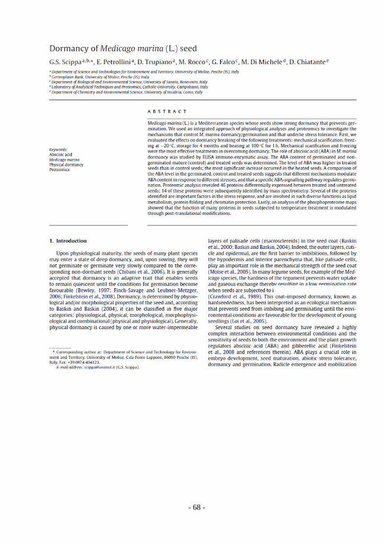
- 68 -
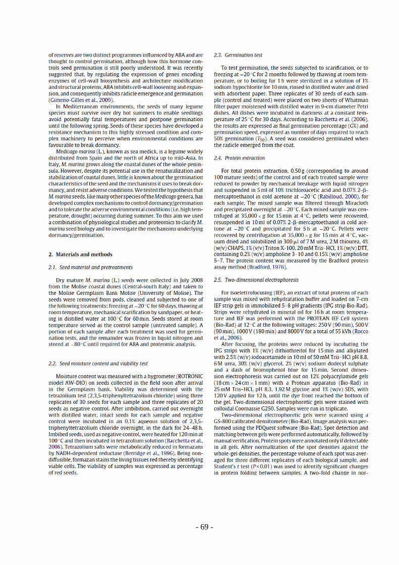
- 69 -
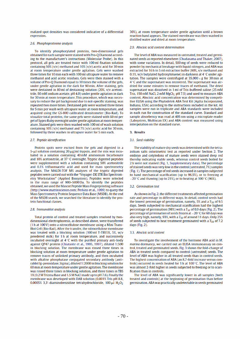
- 70 -
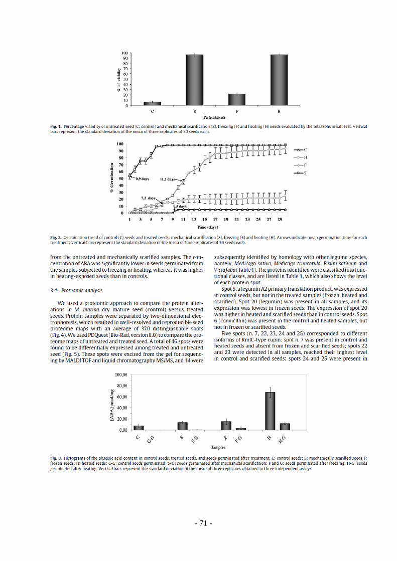
- 71 -
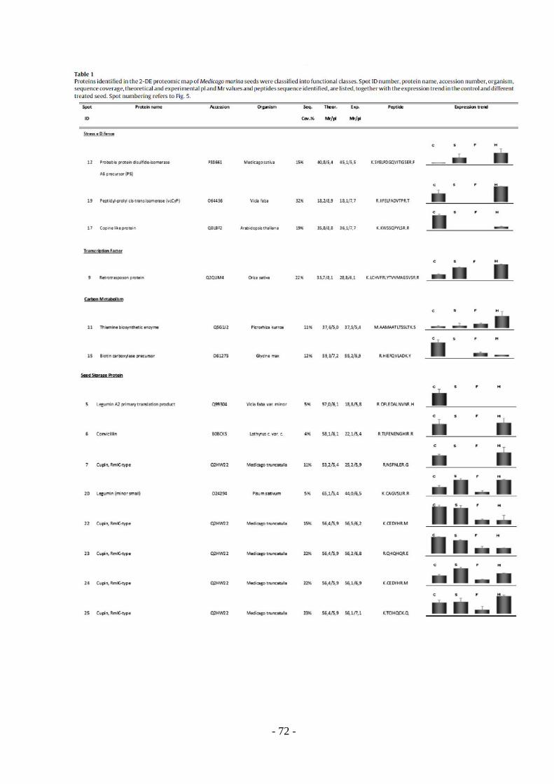
- 72 -
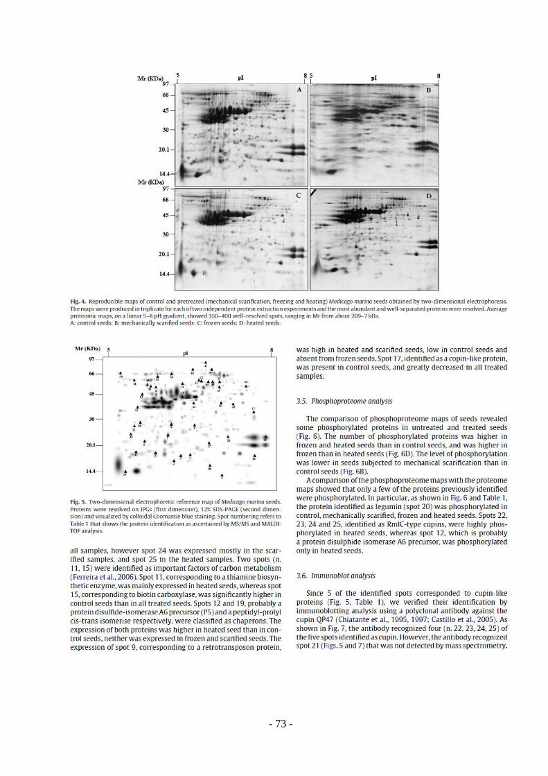
- 73 -

- 74 -
- 75 -

- 76 -

- 77 -
- 78 -
Chapter II
Dormancy of Medicago marina (L.) seed: the role of the micropylar endosperm
1. Introduction
The endosperm is an important tissue not only as a source of nutrients for the embryo during
seed development and maturation, but also as an integrator of seed growth signals. As
demonstrated by Berger et al., (2006), reciprocal signalling between the embryo, the endosperm
and seed integuments appears to coordinate the seed growth and development. In particular, in
this work the authors highlight the role of the endosperm in mediating maternal signals that are
based on memory of the transcriptional states of imprinted genes, which is decisive for
harmonious seed growth (Berger et al., 2006).
When seed maturation is completed, depending on the species, the endosperm may be fully or
partially lost (Linkies et al., 2010). However, in most angiosperm species which preserve an
endosperm layer in their mature seeds, the endosperm can be involved in the control of
germination. In mature seeds, the endosperm and in particular the one between the root embryo
and micropyle, namely micropylar endosperm is able to regulate the timing of germination, by
being a physical barrier for the radicle protrusion (Ni & Bradford, 1993; Bewley, 1997; Toorop
et al., 2000; Leubner - Metzger, 2003; Finch-Savage & Leubner-Metzeger, 2006).
However, because the germination of mature seed can be completed, it is needed that two
events occur simultaneously: 1) the radicle starts to elongate, trying to overcome the endosperm
and 2) the covering layers around the embryo and the micropylar endosperm gets softer,
allowing the radicle expansion (Ni & Bradford, 1993). The process making the endosperm to
become soft during germination is called endosperm weakening (Ni & Bradford, 1993). This
process occurs during germination, in between testa and endosperm rupture, as showed for
Lepidium sativum seed, where it seems to be regulated trough a large number of enzymes and
hydroxyl radicals (Muller et al., 2006).
Endosperm weakening requires the action of cell-wall modifying enzymes such as
endo-β-mannanase, expansins (Sitrit et al., 1999; Chen & Bradford, 2000; Nonogaki et al., 2000;
Chen et al., 2002, Iglesias-Fernández et al., 2011), β-1,3-glucanases (β-Glu) (Leubner-Metzger,
2003) and polygalacturonases (PGs) González-Carranza et al., (2007).
Many authors (Nonogaki & Morohashi, 1996; Chen & Bradford, 2000, Nonogaki et al., 2000)
showed that endo-β-mannanase and expansins are specifically expressed in tomato endosperm
- 79 -
cup during seed germination. In fact, the wall of endosperm cells are rich in mannanase
(galactomannans, glucomannans), hemicellulosic polysaccharides that confer a strong
mechanical resistance for the radicle protrusion in germinating seeds. The endo-β-mannanase
(MAN) are hydrolytic enzymes which catalyze the cleavage of β1→4 bonds in the mannan-
polymer (Iglesias-Fernández et al., 2011) and their role in endosperm weakening has been
widely demonstrated. Nonogaki et al., (2000) showed that in the endosperm cap of germinating
tomato seeds prior radicle emergence, there is an endo-β-mannanase isoform different from the
one found in the lateral endosperm after radicle emergence. Furthermore, the paper demonstrated
that germinative and post-germinative mannanases are encoded by different genes and in
particular, the mannanase cDNA isolated from a cDNA library prepared from imbibed seeds,
termed LeMAN2, encodes for the endo-β-mannanase specifically expressed in the endosperm cap
of tomato seeds prior to radicle emergence. In a recent work, Iglesias-Fernández et al., (2011)
investigated the role of the eight MAN genes in the A. thaliana genome in different organs of this
plant and in the germinating seeds. Using Real-time quantitative PCR assays and in situ
hybridization analyses, the authors demonstrated that four of these genes (AtMAN7, AtMAN6,
AtMAN2 and AtMAN5) were expressed in germinating seeds and that their transcript
accumulation was restricted to the micropylar endosperm and to the radicle, disappearing
immediately after radicle protrusion.
From these results the authors pointed to a possible cooperation between embryo and
endosperm MAN activities, prior to radicle emergence in A. thaliana seeds, suggesting the
important role of AtMAN7, AtMAN5 and specially AtMAN6 genes and their products for the
germination by facilitating the weakening of the micropylar endosperm through the hydrolysis of
the mannan-rich cell walls.
Leubner-Metzger (2003) demonstrated that β-1,3-glucanases (βGlu), which are able to
catalyse hydrolytic cleavage of the 1,3-β-D-glucosidic linkages in β-1,3-glucanas, present in the
cell wall, are expressed in some Solanaceae species such as tobacco and tomato. In particular β-
Glu 1 has been shown to play a key role in tobacco seed germination. β-Glu 1 is induced during
tobacco seed germination after testa rupture and just prior to endosperm rupture, and it is
exclusively localized in the micropylar endosperm. Moreover, the hormonal interactions during
endosperm weakening regulate the expression of cell wall modifying enzymes. In several works
(Leubner, 2003; Kucera et al., 2005, Müller et al., 2006; Gimeno-Gilles, 2009; Linkies et al.,
2009) it has been demonstrated that the endosperm weakening is promoted by gibberellins and
inhibited by abscissic acid, which regulate the weakening by their antagonistic effect on the
endosperm.
- 80 -
Endosperm rupture involves the light/gibberellins pathway. Different class of hydrolases and
class I β-1-3-glucanase (βGlu I) are induced by the light/gibberellins pathway in the micropylar
endosperm, allowing endosperm rupture and radicle protrusion (Leubner, 2003; Kucera et al.,
2005). However it has been shown that abscissic acid is able to repress the expression of
endosperm-specific βGlu I and other class of enzymes involved in cell wall loosening and
expansion, such as xyloglucan endotransglucosylase, and, consequently the endosperm rupture
(Leubner, 2003; Kucera et al., 2005; Gimeno-Gilles, 2009). In tobacco seeds, abscissic acid has
been shown to inhibit β-Glu 1 induction, delaying endosperm rupture in a dose-dependent
manner (Leubner-Metzger, 2003). Moreover, in this work is also reported that the endosperm
rupture kinetic depends on a critical threshold concentration of β-Glu 1, while the kinetic of testa
rupture is not affected by the β-Glu 1 accumulation, indicating the specific action of this enzyme
in promoting micropylar endosperm rupture (Leubner-Metzger, 2003).
Light/gibberellins pathway can counteracts the abscissic acid effect by promoting its
degradation (Leubner, 2003; Kucera et al., 2005), and also, ethylene counteracts ABA effect,
antagonizing the ABA inhibition and promoting endosperm rupture (Linkies et al., 2009). In
particular, ethylene action, in counteracting ABA effect, is due to specific transcription factor
responsive to ethylene signal, called Ethylene Responsive Element Binding Proteins (EREBPs),
that mediate hormonal regulation of cell wall modifying enzymes, such as the βGlu I (Leubner,
2003; Kucera et al., 2005).
The action of other cell wall loosening enzymes in endosperm rupture, as polygalacturonases,
is reported in a work by González-Carranza et al. (2007) which analysed the endosperm cap of
Arabidopsis thaliana. Polygalacturonases (PGs) play an important role in the process of cell
separation, causing the breakdown of adhesion between neighbouring cells (Rose et al., 2003).
The A. thaliana genome contains 69 genes that could be classified as putative genes that encode
for PGs proteins and these genes can be grouped into multiple clades. From the analysis of five
of these members located in two separate clades, using reporter fusion constructs and reverse
transcription-PCR, the authors demonstrated that, despite these PGs exhibit high sequence
similarity, they have distinct patterns of spatial and temporal expression, and sites of expression
include also the aleurone and cells surrounding the micropylar endosperm in a germinating seed
(González-Carranza et al., 2007). The gene At2g43860 that encodes for a polygalacturonases
expressed also in the abscission zones and pistils of tomato flowers (Hong et al., 2000), is
specifically expressed in the micropylar endosperm region of A. thaliana seed, indicating a key
role for cell wall-degrading enzymes, including PGs, in seed germination (González-Carranza et
al., 2007). Sitrit et al., (1999), and subsequently Chen et al., (2002), on the basis of several
studies performed on germinating tomato seeds, showed that the transcripts of expansin and gene
- 81 -
encoding for xiloglucan endotransglycosylase (XET), that modify xyloglucans, major
components of primary cell walls in dicots, accumulate precisely in the endosperm region
adjacent to the expanding radicle. These results additionally support the theory that a large
number of cell wall-degrading enzymes are involved in micropylar endosperm weakening during
seed germination. The expression of the weakening enzymes in endosperm cap allows cell wall
loosening and cell separation that results in the endosperm rupture and, finally, in the radicle
emergence.
In addition to the activities of the hydrolytic enzymes in the recent years, the role of hydroxyl
radicals in cell wall loosening has been investigated. Preliminary experiments by Miller (1986)
and Fry (1998) suggested that OH-, produced in the apoplastic space of plant tissues, plays an
important physiological role in cell-wall loosening process underlying cell expansion. In fact,
they showed that two cell-wall polysaccharides, pectin and xyloglucan, can be broken down in
vitro by OH-. Supporting these data, Chen & Schopfer (1999), found that plant peroxidase can
catalyse the production of OH- from O2 in the presence of a NADH, in the apoplastic space of
germinating seeds. In two independent studies Fry et al. (2001), and Schopfer (2001) proposed
the non-enzymatic degradation of cell wall polymers by hydroxyl radical (OH-). The experiments
reported in this work, carried out on excised embryo hypocotyl segments of different plant
species, such as Zea mays (L.), Cucumis sativus (L.), Helianthus annus (L.), Glycine max (L.),
Pinus sylvestris (L.), supported the hypothesis proposing OH- as the wall loosening agent
responsible for elongation growth, illustrating that the wall-loosening reaction appears to be
equally effective in growing organs of each of these species, and also, the independence of OH-
mediated wall loosening from species-specific differences in chemical wall properties (Schopfer,
2001). Recently, Muller et al., (2009), showed that OH- radicals play a role in endosperm
weakening and rupture of Lepidium sativum, and that their production increase in the endosperm
cap and in radicle of imbibed seeds. Furthermore, it has been also show that the increase of OH-
radicals is promoted by GA and ethylene and inhibited by ABA (Graeber et al., 2010).
Due to its involvement in regulating seed germination, endosperm can also contribute to
dormancy. In fact, as shown in Arabidopsis thaliana and in some Solanaceae species (Bewley,
1997; Koornneef, 2002), such as tomato and tobacco, the micropylar endosperm can contribute
to dormancy by inhibiting the protrusion of the growing radicle through the covering layers of
the embryo. Bethke et al., (2007) found that the only micropylar endosperm is enough to confer
coat dormancy in A. thaliana. As described by Windsor et al., (2000) and Debeaujon et al.,
(2000), the seed coats of Arabidopsis consist of an external layer, called testa, which consist of
dead cells, and an internal single cell layer of living aleurone cells, which represent the sole
endosperm tissue. As demonstrated by Bethke et al., (2007), one function of the Arabidopsis
- 82 -
aleurone layer is to maintain the dormancy of imbibed seeds, as removal of the Arabidopsis testa
did not render seeds nondormant for up to 28 days, but removal of the aleurone layer or damage
to the aleurone layer resulted in embryo growth, overcoming seed dormancy.
The studies carried out during the first phase of the PhD program and reported in the paper
Scippa et al., (2011), showed that Medicago marina seed is characterized by a strong dormancy.
Furthermore, based on the results obtained in the germination test of differently treated seed and
in the hormone assay, it has been proposed that M. marina seed has a coat dormancy which
seems to be finely regulated by complex molecular mechanisms. Since a large number of
evidences point out the role of endosperm in the control of seed dormancy, the role of the
endosperm in Medicago marina seeds dormancy has been investigated.
In particular, the weakening of endosperm was quantified trough a biomechanical approach
using the puncture force test in seeds subjected to mechanical scarification and abscissic acid
(ABA) treatment, along a 22 hours time course of imbibition. In addition to that, the effect of
abscissic acid and gibberellic acid treatments on scarified seeds germination rate and speed, were
analyzed by performing germination test, while RT-PCR analyses was used to evaluate the gene
expression of key enzymes involved in abscissic acid, gibberellic acid and ethylene biosynthesis,
namely NCED5 (9-cis-epoxycarotenoid dioxygenase), GA3ox (gibberellin 3-beta-hydroxylase)
GA20ox (gibberellins 20 3-beta-hydroxylase) and ACO (1-aminocyclopropane-1-carboxylic acid
oxidase). Then, through a proteomic approach coupled with stastical analysis, was studied the
protein pattern expressed during the weakening of the endosperm and the effect of ABA on this
pattern, during the time course of imbibition.
- 83 -
2. Materials and methods
2.1 Seed material and germination test
Dry mature M. marina (L.) seeds were collected in July 2010 from the Molise coastal dunes
(Central-south Italy) and taken to the Molise Germplasm Bank (University of Molise). The seeds
were removed from pods and cleaned. Then, seeds were sterilized in a solution of 1% sodium
hypochlorite for 10 min, rinsed in distilled water, dried with adsorbent paper and subjected to
mechanical scarification by sandpaper in order to promote the germination. A portion of
scarified seeds were used for the germination test, while the remainder were subjected to
imbibition along a 22 hours time course and used for the puncture force measurements, RT-PCR
and proteomic analysis. Imbibition were performed placing scarified seeds on two sheets of
Whatman filter paper moistened with 4 ml of sterilized distilled water with 0,2% (v/v) Plant
Preservative Mixture (Plant Cell Technology Inc.), in 9-cm diameter Petri dishes with or without
the addition of 100 μM (+/-) of Abscissic Acid (Sigma). All dishes were sealed with parafilm an
incubated in darkness at a constant temperature of 25°C and, during the time course, harvested
every two hours starting from 6-8 h after imbibition. After harvesting a portion of seeds were
frozen in liquid nitrogen and stored at -80 °C until required for proteomic analysis and RT-PCR,
while an other portion were immediately used for the puncture force measurement. Time course
of imbibition was repeated in three independent replicates.
Germination was tested in presence or absence of abscissic acid (ABA) and gibberellic acid
(GA3) (Sigma) to evaluate possible differences in terms of germination rate and speed, due to
hormonal treatments. At this purpose, three biological replicates of 60 scarified seeds were
placed on two sheets of Whatman filter paper moistened with 4 ml of sterilized distilled water
with 0,2% (v/v) Plant Preservative Mixture (Plant Cell Technology Inc.) in 9-cm diameter Petri
dishes. Then, an other portion of scarified seeds were incubated in the same condition, but whit
the addition of 100 μM (+/-) ABA (Sigma), 5 μM GA3 (Sigma), 10 μM GA3 and 100 μM (+/-)
ABA plus 5 μM GA3, individually, to test the germination. For each hormonal treatment were
used three biological replicates of 60 seeds. All dishes were sealed with parafilm and incubated
in darkness at a constant temperature of 25 °C for 7 days. According to Bacchetta et al. (2006),
the results are expressed as final germination percentage (G%) and germination speed, expressed
as number of hours required to reach 50% germination (T50). A seed was considered germinated
when the radicle emerged from the coat.
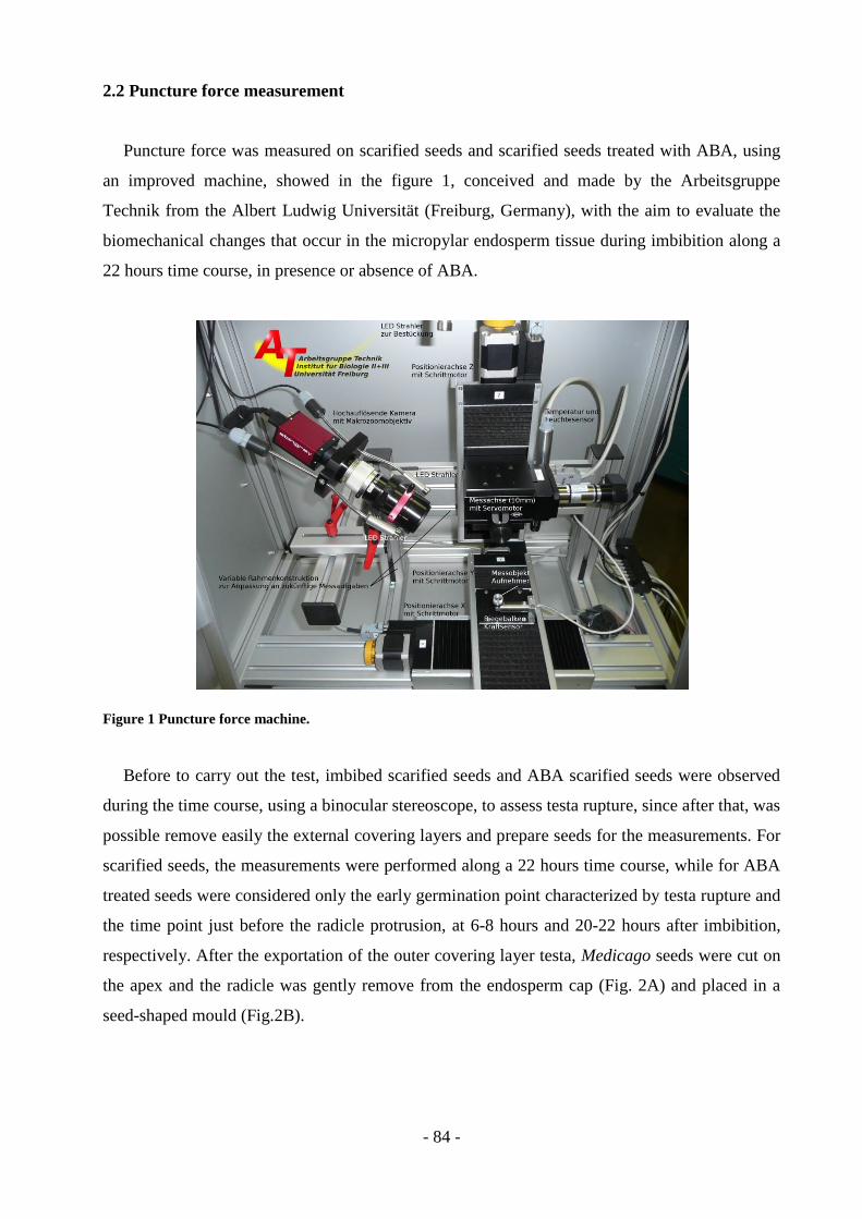
- 84 -
2.2 Puncture force measurement
Puncture force was measured on scarified seeds and scarified seeds treated with ABA, using
an improved machine, showed in the figure 1, conceived and made by the Arbeitsgruppe
Technik from the Albert Ludwig Universität (Freiburg, Germany), with the aim to evaluate the
biomechanical changes that occur in the micropylar endosperm tissue during imbibition along a
22 hours time course, in presence or absence of ABA.
Figure 1 Puncture force machine.
Before to carry out the test, imbibed scarified seeds and ABA scarified seeds were observed
during the time course, using a binocular stereoscope, to assess testa rupture, since after that, was
possible remove easily the external covering layers and prepare seeds for the measurements. For
scarified seeds, the measurements were performed along a 22 hours time course, while for ABA
treated seeds were considered only the early germination point characterized by testa rupture and
the time point just before the radicle protrusion, at 6-8 hours and 20-22 hours after imbibition,
respectively. After the exportation of the outer covering layer testa, Medicago seeds were cut on
the apex and the radicle was gently remove from the endosperm cap (Fig. 2A) and placed in a
seed-shaped mould (Fig.2B).

- 85 -
1
4
5
2
3
A B
A metal probe was slowly lowered into the individual empty endosperm cap at a constant
speed of 250 nm and the puncture force was measured (Fig.3).
Figure 2 A: Medicago marina seed prepared for the endosperm weakening measurement. 1. Cut on the apex of
seed to remove the radicle; 2. Portion of seed without the external covering layer testa; 3. External covering layer
testa; 4. Empty micropylar endosperm cap; 5. Radicle removed from the micropylar endosperm. B. Seed shaped-
mould.
Figure 3 Quantification of endosperm weakening by direct biomechanical measurements. The radicle was
removed from the mycropylar endosperm cup and the seed was placed in the seed shaped mould. A metal probe was
lowered into the empty endosperm cap until it ruptures. During the measurement, the force needed to rupture was
recorded in mN and plotted as a peak on the computer monitor (indicated by the red arrow in the figure).

- 86 -
For each time point were taken 30 measurements, using three biological replicates of 60
imbibed seeds. During the test the machine recorded the force that was needed to break the
endosperm and the corresponding results were expressed in mN. These values provide an
estimation of the mechanical resistance of the endosperm when the radicle starts to elongate,
during germination.
2.3 Reverse transcriptase Polymerase Chain Reaction (RT-PCR) analysis
For the RT-PCR analysis were used imbibed scarified seeds not ABA (100 μM) treated and
imbibed scarified seeds ABA (100 μM) treated collected at 13h after imbibition. Also, other four
sample were considered, namely dry mature seeds (used as control sample), scarified seeds,
scarified seeds ABA (100 μM) treated and not germinated, scarified seeds not ABA treated (100
μM) and germinated.
2.3.1 RNA extraction
RNA was extracted from approximately 0.07 g of seeds using a Qiagen RNeasy Mini kit
protocol (Qiagen, Valencia, CA). Seeds were ground with a mortar and pestle to a fine powder in
liquid nitrogen, lysis buffer was added to the samples and the RNA was extracted according whit
the instructions included in the kit. RNA concentrations were measured at 260 nm using a
spectrophotometer (Bio-Rad, Hercules, CA, USA) and RNA integrity was checked on 1.5%
(w/v) agarose Tris-acetate EDTA ethidium bromide gels.
2.3.2 cDNA synthesis and RT-PCR
cDNA was synthesized using 1.0 µg of total RNA, poly(A) oligonucleotide primer and
Superscript III reverse transcriptase (Invitrogen Co., Carlsbad, CA, USA). Standard PCR
conditions and gene specific primers were used.
In particular the following primers were used: for GA3ox, primers GA3ox Forw
(ACCCAAATGCTTCAAATTAATAG) and GA3ox Rev
(TCACAGTGTTTGGCATAATCT) (Di Giacomo et al., 2008); for GA20ox, primers GA20ox
Forw (GAGGGGAACATTGTGGTTATGC) and GA20ox Rev
(GTCTTGTTGTTGTTCACCACTGCC) (Aubert et al., 2006); for NCED5, primers NCED5
Forw (GGACGGTTTTATGCATGATCCT) and NCED5 Rev
(TGGCAAAGTCATGCATCATGAT) (Planchet et al., 2011).
- 87 -
The following gene-specific primers pair, used for ACO, was designed: primers ACO Forw
(AGAGCACTATAGGAAATGCA) and ACO Rev (TTGACAACGATGGAGTGG).
18S rRNA cDNA fragment was used as a loading control using the same RT reaction and the
following primers: 18S Forw (CCAGGTCCAGACATAGTAAG) and 18S Rev
(GTACAAAGGGCAGGGACGTA) (Pislariu & Dickstein, 2007).
All PCR reactions were performed in 25 µl volumes using Taq recombinant polymerase
(Invitrogen Co., Carlsbad, CA, USA) according to the manufacturer’s protocol and cDNA
obtained previously was used as template in all reactions.
Reaction conditions for thermal cycling were 94 °C for 4 min, 30 cycles of 94 °C for 45 s,
50°C for 45 s, 72 °C for 50 s and 72 °C for 4 min for GA3ox, GA20ox, NCED5 and ACO. For
18S, reaction condition for thermal cycling were 94 °C for 4 min, 28 cycles of 94 °C for 45 s,
52°C for 45 s, 72 °C for 50 s and 72 °C for 4 min. Two independent biological replicates were
run for each sample, each with two technical replications. Results were analyzed using Image J
1.41o software (Wayne Rasbanb National Institute of Health, USA; http//rsb.info.nih.gov/ij). To
account for small differences in RNA loadings data was normalized to 18S gene expression.
2.4 Proteomic analysis
For the proteomic analysis were used imbibed scarified seeds not ABA (100 μM) treated and
imbibed scarified seeds ABA (100 μM) treated collected at 6h, 8h, 13h, 18h, 20h, and 22h after
imbibition. Also, other four sample were considered, namely dry mature seeds (used as control
sample), scarified seeds, scarified seeds ABA (100 μM) treated and not germinated, scarified
seeds not ABA treated (100 μM) and germinated.
2.4.1 Protein extraction
For total protein extraction, 0.50 g (corresponding to around 100 mature seeds) of each seeds
samples were reduced to powder by mechanical breakage with liquid nitrogen and suspended in
5 ml of 10% trichloroacetic acid and 0.07% 2-β-mercaptoethanol in cold acetone at -20°C
(Rabilloud, 2000), individually. The mixed sample was filtered through Miracloth and
precipitated overnight at -20°C. Each mixed sample was centrifuged at 35,000xg for 15 min at
4°C, pellets were recovered, resuspended in 10 ml of 0.07% 2-β-mercaptoethanol in cold acetone
at -20°C and precipitated for 5 h at -20°C. Pellets were recovered by centrifugation at 35,000×g
for 15 min at 4°C, vacuum dried and solubilised in 300 ml of 7M urea, 2M thiourea, 4% (w/v)
CHAPS, 1% (v/v) Triton X-100, 20mMTris–HCl, 1% (w/v) DTT, containing 0.2% (w/v)
- 88 -
ampholine 3-10 and 0.15% (w/v) ampholine 5-7. The protein content was measured by the
Bradford protein assay method (Bradford, 1976), using the BioRad (Bio-Rad, Hercules, CA,
USA) protein assay and BSA as standard.
2.4.2 Two-dimensional electrophoresis
For isoelettrofocusing (IEF), an extract of total proteins of each sample was mixed with
rehydratation buffer and loaded on 7-cm IEF strip gels in immobilized 5–8 pH gradients (IPG
strip Bio-Rad). Strips were rehydrated in mineral oil for 16 hours, at room temperature and IEF
was performed in PROTEAN IEF Cell system (Bio-Rad) at 250 V for 1 h, 500 V for 1 h, 4000 V
for 10.000 “volthours” and 500 V for 99:00 h. After focusing, the proteins were reduced by
incubating the IPG strips with 1% (w/v) dithiothreitol for 20 min and alkylated with 2.5% (w/v)
iodoacetamide in 10 ml of 50mM Tris-HCl pH 8.8, 6M urea, 30% (w/v) glycerol, 2% (w/v)
sodium dodecyl sulphate for 20 min. Second dimension electrophoresis was carried out on 12%
polyacrylamide gels with a Mini-Protean apparatus (Bio-Rad) in 25mM Tris-HCl, pH 8.3, 1.92M
glycine and 1% (w/v) SDS, applying a voltage of 100 V until the dye front reached the bottom of
the gel. Two-dimensional electrophoretic gels were stained with colloidal Coomassie G250 o/n
and then washed several times in distilled water to remove the excess dye.
2.4.3 PDQuest analysis
Two-dimensional electrophoretic gels were acquired using a GS-800 calibrated densitometer
(Bio-Rad), and gels analysis was performed using PDQuest 2-D Analysis Software (Bio-Rad), to
identify differentially expressed proteins. The intensities of the differential expressed protein
were obtained from three independent experiment of each biological sample, followed by
comparison automatically executed by the software (and then, manually verified), which make a
quantitative measurement to obtain statistical information on variations in the protein levels.
Protein spots were annotated only if detectable in all gels. After normalization of the spot
densities against the whole-gel densities, the percentage volume of each spot was averaged for
the three different replicates of each biological sample and statistical Student‘s t test analysis at
significance level p < 0.01 was chosen to find out significant changes in protein folding between
samples. A two-fold change in normalized spot densities was considered indicative of a
differential expression.
- 89 -
2.5 Statistical analysis and identification of specific markers
On the basis of the results obtained from PD-Quest analysis, protein that showed a
differentially expressed pattern, were also analyzed trough uni - and multivariate analysis to
identify the proteins able to discriminate significantly the different samples. The protein
variation among and within the different samples was obtained through the analysis of variance
(ANOVA). ANOVA was computed at several different levels: first it was computed between the
control seeds (dry mature seeds) and scarified seeds, second it was computed between control
seeds and germinated seeds not ABA (100 μM) treated, third it was computed between scarified
seeds and scarified germinated seeds not ABA (100 μM) treated, fourth it was computed
between scarified germinated seeds not ABA (100 μM) treated and not germinated scarified
seeds ABA (100 μM) treated, to identify spots that are specific markers of a physiological state
(germination - dormancy) and/or of a treatment (ABA - scarification). Moreover, ANOVA was
computed between the scarified imbibed seeds samples along the 22 hours time course, treated
and not treated whit ABA (100 μM), to highlight the proteins with significant expression pattern
among these samples, during the weakening of the micropylar endosperm and in relation to the
ABA treatment. Subsequently, the cluster analysis, using the Ward’s method as linkage, was
performed among proteins resulted discriminating by ANOVA (P≤ 0.05) to group all significant
protein in relation to their quantitative relationship.
These variable protein were manual excised from the gels and analyzed by mass spectrometry
technologies.

- 90 -
3. Results
3.1 Puncture force measurement
To define when testa rupture, which characterizes the early germination event, takes place,
and to verify the involvement of ABA, imbibed scarified seeds (control) and imbibed scarified
seeds treated with ABA (100 µM) were kept at the constant temperature of 25°C and observed
during a time course, using a binocular stereoscope. The observation taken during a 24 h time
course showed that both ABA treated and untreated seeds undergo to testa rupture after 6-8
hours of imbibition (Fig. 4B). However, the micropylar endosperm rupture, assessed trough the
radicle emergence, occurred between 22 and 24 hours after imbibition only in ABA untreated
scarified seeds.
For this reason the time point at 22 hours after imbibition was took as the final time point just
before the completion of germination and consequently, it was considered as the last imbibition
time useful for the puncture force measurement. After this time point, because of the radicle
emergence, it was not possible measure the micropylar endosperm resistance.
Figure 4 Testa rupture of imbibed scarified seeds. A: scarified seed before testa rupture; B and C: testa rupture of
scarified not ABA treated seeds; B: testa rupture after 6- 8h hours of imbibition; C: testa rupture after 20-22 hours of
imbibition.
A B C

- 91 -
The mechanical resistance of the micropylar endosperm was used as a measure of endosperm
weakening and quantified by puncture-force method (Müller et al., 2006). Puncture force
measurements, performed on scarified seeds during imbibition, showed a constant decrease of
micropylar endosperm resistance. As shown in figure 5 the highest endosperm cap puncture
force was measured after 6-8 h of imbibition, when testa rupture, characterizing the early
germination event, occurred (Fig. 4B).
Puncture force values decrease quickly after 6-8 h of imbibition (97,55 ± 8,47 mN), and then
slowly between 10 h and 18 h of imbibition, where the puncture force resulted equal to 90,48 ±
7,27 mN and 71,94 ± 7,27 mN, respectively. A new significant decrease of the puncture force
that reached the value of 52,31 ± 5,01 mN, was visible only after 20 h of imbibition. The lowest
puncture force (42,67 ± 4,55 mN) was necessary to puncture the endosperm at 20-22 hours of
imbibition (Fig. 5).
Figure 5 Modification of the micropylar endosperm resistance during imbibition of scarified seeds measured
by the puncture force method. For each time point, the histograms represent the mean values of at least 30
individual endosperm cap measurements and the vertical bars represent the standard error.
0
30
60
90
120
150
6-8 h 8-10 h 10-12 h 12-14 h 14-16 h 16-18h 18-20 h 20-22 h
Pu
nct
ure
fo
rce
(mN
)
Time of imbibition
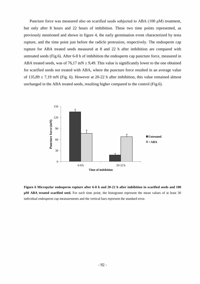
- 92 -
Puncture force was measured also on scarified seeds subjected to ABA (100 μM) treatment,
but only after 8 hours and 22 hours of imbibition. These two time points represented, as
previously mentioned and shown in figure 4, the early germination event characterized by testa
rupture, and the time point just before the radicle protrusion, respectively. The endosperm cap
rupture for ABA treated seeds measured at 8 and 22 h after imbibition are compared with
untreated seeds (Fig.6). After 6-8 h of imbibition the endosperm cap puncture force, measured in
ABA treated seeds, was of 76,17 mN ± 9,49. This value is significantly lower to the one obtained
for scarified seeds not treated with ABA, where the puncture force resulted in an average value
of 135,89 ± 7,19 mN (Fig. 6). However at 20-22 h after imbibition, this value remained almost
unchanged in the ABA treated seeds, resulting higher compared to the control (Fig.6).
Figure 6 Micropylar endosperm rupture after 6-8 h and 20-22 h after imbibition in scarified seeds and 100
µM ABA treated scarified seed. For each time point, the histograms represent the mean values of at least 30
individual endosperm cup measurements and the vertical bars represent the standard error.
0
30
60
90
120
150
6-8 h 20-22 h
Pu
nctu
re f
orce (m
N)
Time of imbibition
Untreated
+ ABA
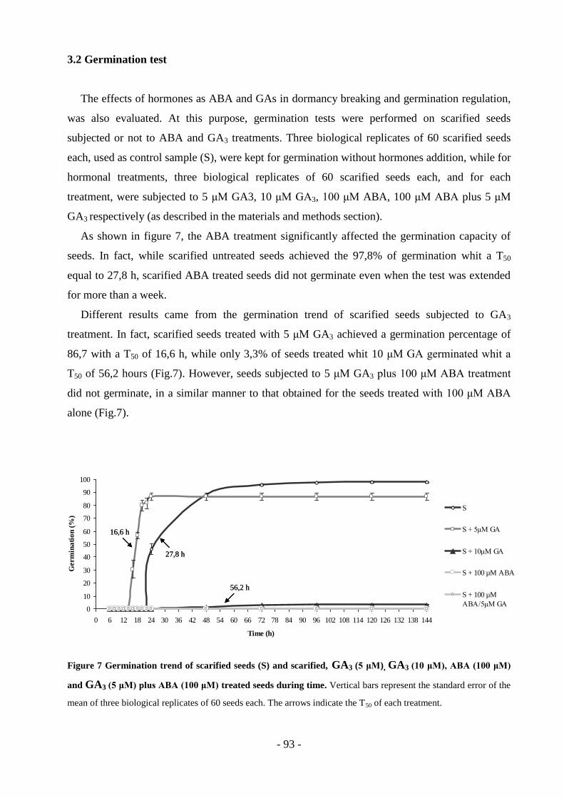
- 93 -
3.2 Germination test
The effects of hormones as ABA and GAs in dormancy breaking and germination regulation,
was also evaluated. At this purpose, germination tests were performed on scarified seeds
subjected or not to ABA and GA3 treatments. Three biological replicates of 60 scarified seeds
each, used as control sample (S), were kept for germination without hormones addition, while for
hormonal treatments, three biological replicates of 60 scarified seeds each, and for each
treatment, were subjected to 5 μM GA3, 10 μM GA3, 100 μM ABA, 100 μM ABA plus 5 μM
GA3 respectively (as described in the materials and methods section).
As shown in figure 7, the ABA treatment significantly affected the germination capacity of
seeds. In fact, while scarified untreated seeds achieved the 97,8% of germination whit a T50
equal to 27,8 h, scarified ABA treated seeds did not germinate even when the test was extended
for more than a week.
Different results came from the germination trend of scarified seeds subjected to GA3
treatment. In fact, scarified seeds treated with 5 μM GA3 achieved a germination percentage of
86,7 with a T50 of 16,6 h, while only 3,3% of seeds treated whit 10 μM GA germinated whit a
T50 of 56,2 hours (Fig.7). However, seeds subjected to 5 μM GA3 plus 100 μM ABA treatment
did not germinate, in a similar manner to that obtained for the seeds treated with 100 μM ABA
alone (Fig.7).
Figure 7 Germination trend of scarified seeds (S) and scarified, GA3 (5 μM), GA3 (10 μM), ABA (100 μM)
and GA3 (5 μM) plus ABA (100 μM) treated seeds during time. Vertical bars represent the standard error of the
mean of three biological replicates of 60 seeds each. The arrows indicate the T50 of each treatment.
0
10
20
30
40
50
60
70
80
90
100
0 6 12 18 24 30 36 42 48 54 60 66 72 78 84 90 96 102 108 114 120 126 132 138 144
Time (h)
Germ
ina
tio
n (
%)
S
S + 5μM GA
S + 10μM GA
S + 100 µM ABA
S + 100 µM
ABA/5μM GA
27,8 h
16,6 h
56,2 h
0
10
20
30
40
50
60
70
80
90
100
0 6 12 18 24 30 36 42 48 54 60 66 72 78 84 90 96 102 108 114 120 126 132 138 144
Time (h)
Germ
ina
tio
n (
%)
S
S + 5μM GA
S + 10μM GA
S + 100 µM ABA
S + 100 µM
ABA/5μM GA
27,8 h 27,8 h
16,6 h 16,6 h
56,2 h 56,2 h
- 94 -
3.3 Reverse transcriptase Polymerase Chain Reaction (RT-PCR) analysis
RT-PCR analysis was used to evaluate the gene expression of key enzymes involved in
abscissic acid (NCED5), gibberellic acid (GA3ox, GA20ox) and ethylene (ACO) biosynthesis to
investigate the role of these hormones in dormancy breaking and germination. In particular, the
expression pattern of GA3ox, GA20ox, NCED5 and ACO genes in dry mature seeds (C), scarified
seeds (S), scarified seeds subjected or not to ABA (100 µM) treatment collected at 13 h after
imbibition (13h + ABA; 13h - ABA), scarified germinated seeds not ABA treated (G - ABA),
scarified not germinated seeds ABA treated (NG + ABA), were analysed by reverse
transcriptase-PCR using gene specific primers and optimised PCR conditions, as described in
materials and methods section.
GA3ox and ACO genes showed a similar expression pattern as both appeared to be expressed
in scarified seeds not treated with ABA collected at 13 h after imbibition and in scarified
germinated seeds, while they were undetectable in all the other samples (Fig. 8). However,
transcripts of GA3ox gene seemed to increase during germination showing almost a 0,8 fold
induction in germinated seeds, while ACO transcripts remained constant in both imbibed (13 h -
ABA) and germinated seeds (Fig.8).
GA20ox gene was preferentially expressed in germinated seeds, resulting undetectable in the
remaining samples; NCED5 resulted to be induced by the scarification and up-regulated in
scarified seeds collected at 13 h after imbibition not subjected to ABA treatment. In fact, as
reported in figure 8, NCED5 showed approximately 1,4 fold induction for both, compared with
control seeds and germinated seeds not ABA treated. Furthermore, it resulted that NCED5
transcript was not detected in ABA treated samples (scarified seeds ABA treated collected after
13 h of imbibition and scarified not germinated seeds ABA treated).

- 95 -
Figure 8 NCED5, GA3ox, GA20ox and ACO genes transcriptional profiling. Histograms represent the
quantification of specific PCR products normalised with the control 18S rRNA. Error bars indicate the standard
deviation for three biological replicates.
3.4 Proteomic analysis
For proteomic analysis, proteome maps obtained from dry mature seeds (used as control
sample), scarified seeds, scarified germinated seeds not ABA (100 μM) treated, scarified not
germinated seeds ABA (100 μM) treated and scarified seeds ABA (100 μM) treated and not
ABA treated, collected along a 22 h time course, were compared to analyse the protein pattern in
M. marina dry mature seed during the endosperm weakening in preparation to germination, after
the dormancy release, and to identify proteins regulated by ABA.
Total protein extracts from each sample were resolved by 2-DE, as described in materials and
methods section, and proteins detected by blue comassie.
As reported in figure 9, proteomic maps, resolved on a 5-8 pH gradient, showed a high level
of reproducibility with an average of 450-500 distinguishable spots that reached Mr values
between about 104 and 15 Da.
Software assisted proteome analysis was carried out by matching all the replicate gels with
the dry mature seed (control) used as reference.
The comparison of relative spot densities between the different proteomic maps revealed a
total of 351 spots differentially expressed among the dry mature seeds, scarified seeds,
0
0,2
0,4
0,6
0,8
1
1,2
1,4
1,6
C S 13 h
(-ABA)
13 h
(+ABA)
G -ABA NG +ABA
Fo
ld in
du
cti
on
NCED5
GA3ox
GA20ox
ACO
- 96 -
germinated seeds, not germinated seeds and among scarified ABA treated and not treated seeds
along the 22 hours time course, while the remaining spots showed a constant expression.
The 351 spots resulted differentially expressed by the Student‘s t test (P≤ 0.01), were
subjected to Analysis of Variance (ANOVA) and further grouped according to their similar
expression profile by cluster analysis.
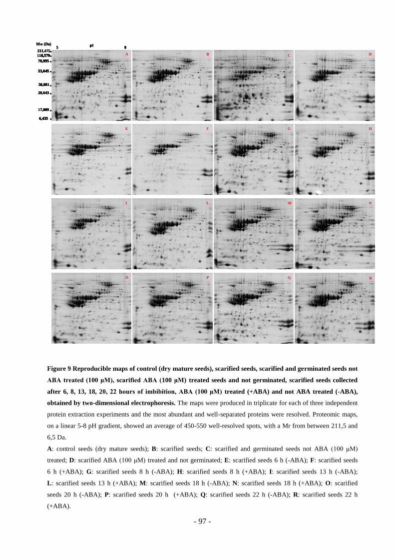
- 97 -
Figure 9 Reproducible maps of control (dry mature seeds), scarified seeds, scarified and germinated seeds not
ABA treated (100 μM), scarified ABA (100 μM) treated seeds and not germinated, scarified seeds collected
after 6, 8, 13, 18, 20, 22 hours of imbibition, ABA (100 μM) treated (+ABA) and not ABA treated (-ABA),
obtained by two-dimensional electrophoresis. The maps were produced in triplicate for each of three independent
protein extraction experiments and the most abundant and well-separated proteins were resolved. Proteomic maps,
on a linear 5-8 pH gradient, showed an average of 450-550 well-resolved spots, with a Mr from between 211,5 and
6,5 Da.
A: control seeds (dry mature seeds); B: scarified seeds; C: scarified and germinated seeds not ABA (100 μM)
treated; D: scarified ABA (100 μM) treated and not germinated; E: scarified seeds 6 h (-ABA); F: scarified seeds
6 h (+ABA); G: scarified seeds 8 h (-ABA); H: scarified seeds 8 h (+ABA); I: scarified seeds 13 h (-ABA);
L: scarified seeds 13 h (+ABA); M: scarified seeds 18 h (-ABA); N: scarified seeds 18 h (+ABA); O: scarified
seeds 20 h (-ABA); P: scarified seeds 20 h (+ABA); Q: scarified seeds 22 h (-ABA); R: scarified seeds 22 h
(+ABA).
5Mw (Da) pI
8
6,435
17,809
118,579
36,881
53,045
78,995
211,475
28,643
A B C D
E
O
ML
P
F G
I
H
N
Q R
5Mw (Da) pI
8
6,435
17,809
118,579
36,881
53,045
78,995
211,475
28,643
A B C D
E
O
ML
P
F G
I
H
N
Q R
5Mw (Da) pI
8
6,435
17,809
118,579
36,881
53,045
78,995
211,475
28,643
A
5Mw (Da) pI
8
6,435
17,809
118,579
36,881
53,045
78,995
211,475
28,643
5Mw (Da) pI
8
6,4356,435
17,80917,809
118,579118,579
36,88136,881
53,04553,045
78,99578,995
211,475211,475
28,64328,643
A BB CC DD
EE
OO
MMLL
PP
FF GG
II
HH
NN
QQ R
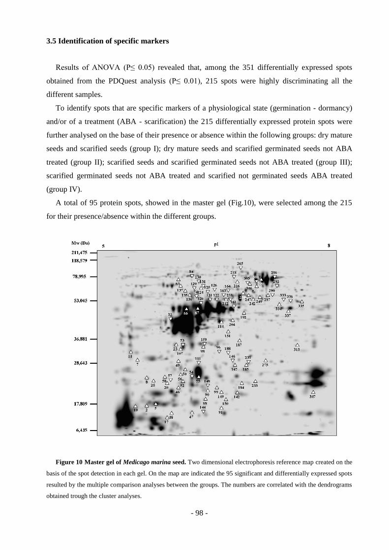
- 98 -
3.5 Identification of specific markers
Results of ANOVA (P≤ 0.05) revealed that, among the 351 differentially expressed spots
obtained from the PDQuest analysis (P≤ 0.01), 215 spots were highly discriminating all the
different samples.
To identify spots that are specific markers of a physiological state (germination - dormancy)
and/or of a treatment (ABA - scarification) the 215 differentially expressed protein spots were
further analysed on the base of their presence or absence within the following groups: dry mature
seeds and scarified seeds (group I); dry mature seeds and scarified germinated seeds not ABA
treated (group II); scarified seeds and scarified germinated seeds not ABA treated (group III);
scarified germinated seeds not ABA treated and scarified not germinated seeds ABA treated
(group IV).
A total of 95 protein spots, showed in the master gel (Fig.10), were selected among the 215
for their presence/absence within the different groups.
Figure 10 Master gel of Medicago marina seed. Two dimensional electrophoresis reference map created on the
basis of the spot detection in each gel. On the map are indicated the 95 significant and differentially expressed spots
resulted by the multiple comparison analyses between the groups. The numbers are correlated with the dendrograms
obtained trough the cluster analyses.

- 99 -
However, while no protein spots were found to discriminate dry mature from scarified seeds
(group I), within the other groups the following results were obtained:
65 spots discriminated samples within group II; 66 spots discriminated samples with the group
III while 64 samples within group IV.
Furthermore, the comparison of the 95 spots expression between different groups generated
the data represented in the Venn diagram (Fig.11). In particular, while five and six spots are
specifically expressed in group II and group III respectively, 60 were present in both (Fig. 11A);
twelve and seventeen spots characterize group II-III and IV respectively, wile remaining 35 were
commonly expressed in all the three groups (Fig. 11B).
A complete list of these specific spot-markers is reported in table 1 in the supplementary
material section.
Figure 11 Venn diagrams of the expressed proteins between and within the four groups. A: sixty proteins were
present in both the second and third group, while a few proteins discriminated in only one of these groups. B: thirty-
five proteins were expressed in the second, third and fourth group, twelve discriminated only the second and the
third group, while seventeen were expressed only in the fourth group. II group: dry mature seed and germinated
seed; III group: scarified seed and scarified germinated seed not ABA treated; IV group: scarified germinated seed
not ABA treated and scarified not germinated seed ABA treated. The first group (scarified seed and dry mature
seed) is not represented in graph because no proteins discriminated within the group.
- 100 -
3.6 Markers expression pattern
The expression profile of the 95 spots in control (C), scarified (S), germinated (G - ABA) and
not germinated plus ABA (NG + ABA) was organized by cluster analysis.
Results reported in figure 12 shown that the three samples: C, S and NG + ABA are very
similar, and all significantly differ from the germinated sample (G - ABA).
In fact, protein spots discriminated, for their qualitative or quantitative different expression,
the samples as following:
- sixty-eight spots discriminated, for their high expression, germinated seeds (G - ABA)
from control (C), scarified (S) and not germinated seeds (NG + ABA);
- eleven spots discriminated control (C) and scarified seeds (S) from germinated seeds (G -
ABA) and not germinated seeds (NG + ABA), for their high expression in C and S and
their low expression in the other two samples;
- three spots discriminated control (C), scarified (S) and germinated seeds (G - ABA) from
not germinated seeds for their low expression in not germinated seeds (NG + ABA);
- two spots discriminated for their high expression, not germinated samples (NG + ABA)
from the other three samples (C, S, NG + ABA);
- two spots discriminated, for their high expression samples C, S and NG + ABA from
sample G - ABA.

- 101 -
Figure 12 Cluster analysis. The protein spots resulted differentially expressed from the ANOVA analysis between
and within the dry mature seeds (C), scarified seeds (S), scarified and germinated seeds not ABA treated (G - ABA
after 27,8 h) and ABA treated scarified and not germinated seeds (NG + ABA after 7 days), were grouped by cluster
analysis on the basis of their low or high quantity. Two different clusters (I and II) were generated, in turn, divided
into several sub-clusters (III - VIII). The down regulated proteins are indicated by the red colour, while the up
regulated proteins are indicated by the green colour.
- 102 -
To further investigate the effect of ABA treatment on these 95 spots, their expression was
evaluated in all the samples (dry mature seeds, scarified seeds, germinated and not germinated
seeds, scarified imbibed seeds along the 22 hours time course in presence or absence of ABA),
respect to the ABA treatment. Twenty-two spots were found to be altered by the ABA treatment
along the entire time course (spot n. 32, 57, 66, 67, 114, 126, 131, 149, 150, 151, 159, 161, 164,
188, 204, 218, 231, 247, 313, 333, 336). In particular, according with their profile during time,
these spots can be grouped in spots (n. 32, 57, 66, 67, 151, 188, 218) up regulated after the ABA
treatment (Fig. 13); spots (n. 114, 126, 131, 149, 150, 159, 161, 164,) down regulated by the
ABA treatment (Fig. 14), and spots (n. 204, 231, 247, 313, 333, 336, 347) showing an
heterogeneous expression profile during the time course (Fig.15).
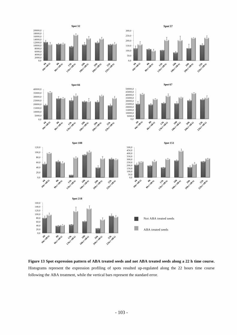
- 103 -
Figure 13 Spot expression pattern of ABA treated seeds and not ABA treated seeds along a 22 h time course.
Histograms represent the expression profiling of spots resulted up-regulated along the 22 hours time course
following the ABA treatment, while the vertical bars represent the standard error.
0,0
5000,0
10000,0
15000,0
20000,0
25000,0
30000,0
35000,0
40000,0
Spot 66
0,0
2000,0
4000,0
6000,0
8000,0
10000,0
12000,0
14000,0
16000,0
18000,0
20000,0
Spot 32
0,0
5000,0
10000,0
15000,0
20000,0
25000,0
30000,0
35000,0
40000,0
45000,0
50000,0
Spot 67
0,0
50,0
100,0
150,0
200,0
250,0
300,0
Spot 57
0,0
20,0
40,0
60,0
80,0
100,0
120,0
Spot 188
0,0
20,0
40,0
60,0
80,0
100,0
120,0
140,0
160,0
Spot 218
0,0
50,0
100,0
150,0
200,0
250,0
300,0
350,0
400,0
450,0
500,0
Spot 151
Not ABA treated seeds
ABA treated seeds
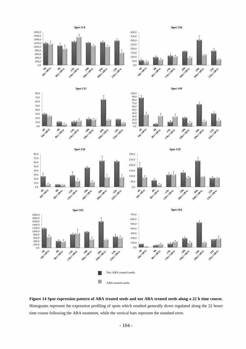
- 104 -
Figure 14 Spot expression pattern of ABA treated seeds and not ABA treated seeds along a 22 h time course.
Histograms represent the expression profiling of spots which resulted generally down regulated along the 22 hours
time course following the ABA treatment, while the vertical bars represent the standard error.
0,0
200,0
400,0
600,0
800,0
1000,0
1200,0
1400,0
1600,0
1800,0
Spot 114
0,0
50,0
100,0
150,0
200,0
250,0
300,0
350,0
400,0
Spot 126
0,0
10,0
20,0
30,0
40,0
50,0
60,0
70,0
80,0
Spot 131
0,0
10,0
20,0
30,0
40,0
50,0
60,0
70,0
80,0
90,0
100,0
Spot 149
0,0
50,0
100,0
150,0
200,0
250,0
300,0
Spot 159
0,0
200,0
400,0
600,0
800,0
1000,0
1200,0
1400,0
1600,0
1800,0
2000,0
Spot 161
0,0
10,0
20,0
30,0
40,0
50,0
60,0
70,0
80,0
Spot 150
0,0
100,0
200,0
300,0
400,0
500,0
600,0
700,0
Spot 164
Not ABA treated seeds
ABA treated seeds
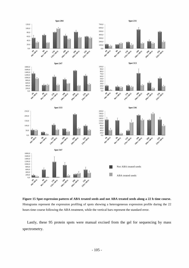
- 105 -
Figure 15 Spot expression pattern of ABA treated seeds and not ABA treated seeds along a 22 h time course.
Histograms represent the expression profiling of spots showing a heterogeneous expression profile during the 22
hours time course following the ABA treatment, while the vertical bars represent the standard error.
Lastly, these 95 protein spots were manual excised from the gel for sequencing by mass
spectrometry.
0,0
50,0
100,0
150,0
200,0
250,0
Spot 333
0,0
10,0
20,0
30,0
40,0
50,0
60,0
70,0
80,0
90,0
100,0
Spot 313
0,0
20,0
40,0
60,0
80,0
100,0
120,0
140,0
160,0
180,0
200,0
Spot 336
0,0
200,0
400,0
600,0
800,0
1000,0
1200,0
1400,0
1600,0
1800,0
Spot 247
0,0
100,0
200,0
300,0
400,0
500,0
600,0
700,0
Spot 231
0,0
200,0
400,0
600,0
800,0
1000,0
1200,0
1400,0
1600,0
1800,0
Spot 347
0,0
20,0
40,0
60,0
80,0
100,0
120,0
Spot 204
Not ABA treated seeds
ABA treated seeds
- 106 -
4. Discussion and conclusions
Results obtained in the first phase of the PhD program and published in the paper Scippa et
al., (2011), showed that Medicago marina seeds, like in other legumes, have adopted a dormancy
strategy based on a hard seed coat which may be also regulated by other complex mechanisms.
As widely reported, the micropylar endosperm contributes to coat dormancy. In fact,
micropylar endosperm can act as a barrier for radicle protrusion and is able, in that way, to
regulate the timing of germination (Ni & Bradford, 1993; Bewley, 1997; Toorop et al., 2000;
Leubner-Metzger, 2003; Finch-Savage & Leubner-Metzeger, 2006). Several works, conducted
on different seeds species, such as lettuce (Ikuma & Thiamann, 1963), tomato (Chen &
Bradford, 2000; Wu et al., 2001, Leubner-Metzeger, 2003) tobacco (Leubner-Metzeger, 2003)
and coffee (Da Silva et al., 2004) report that the weakening of the micropylar endosperm is a
prerequisite for the completion of germination, which is visible when the radicle protrudes the
external covering layers surrounding the embryo. Furthermore, it has been reported that the
micropylar endosperm weakening requires the involvement of many hydrolytic enzymes and is
finely regulated by plant hormones (Leubner-Metzger, 2003; Finch-Savage & Leubner-
Metzeger, 2006). Since the micropylar endosperm can represents an obstacle for the radicle
protrusion, it is possible to quantify directly the weakening of the micropylar endosperm by
measuring puncture force (Müller et al., 2006).
In this work, puncture force measurements were performed on scarified imbibed seeds
collected along a 22 hours time course to verify the involvement of micropylar endosperm in M.
marina dormancy. Results obtained showed a constant decrease of micropylar endosperm
resistance, starting from 6-8 h after imbibition (testa rupture), when the highest endosperm cap
puncture force (135,89 ± 7,19 mN) was obtained, until the time point at 20-22 h after imbibition
(time point just before the radicle protrusion), when only an average force of 42,67 ± 4,55 mN
was enough to puncture the endosperm.
In between these two time points, with the exception for 10 h (97,55 ± 8,47 mN) and 20 h
after imbibition (52,31 ± 5,01 mN), in which the corresponded puncture force values quickly
decreased, the micropylar endosperm resistance decreased slowly in a linear manner in all points
included between 12 and 18 hours.
Thus, the micropylar endosperm puncture force seems to decrease in two significant drop
phases: the first occurred after 10 and the second occurred after 20-22 hours from the beginning
of imbibition, (Fig.5). This second reduction coincided whit the radicle protrusion. In fact, 22 h
after imbibition seed radicle was able to generate enough force to protrude the endosperm and
- 107 -
complete the germination within the next 2 hours. Indeed, scarified seeds completed the
germination in 24 h with a germination rate of almost 98% (Fig. 7).
As stated before, a large number of evidences demonstrated that the endosperm weakening is
a common phenomena of many plant species, that precedes the radicle protrusion, and occurs
when, during seed imbibition, the water uptake starts (Müller et al., 2006; Linkies et al., 2009,
Graber et al., 2010, Morris et al., 2011).
The plant species Lepidium sativum has been widely used as a model species to investigate
the role of endosperm weakening (Müller et al., 2006; Linkies et al., 2009, Graber et al., 2010,
Morris et al., 2011). The completion of germination in Lepidium seed, measured trough the
radicle emergence, is regulated by two opposing forces, the growth potential of the radicle, and
the resistance to this growth from the seed covering layers (testa and endosperm cap) (Müller et
al., 2006; Morris et al., 2011). After testa rupture, the strength of the endosperm progressively
decreases toward germination completion. Furthermore, in Lepidium sativum seeds it has been
observed that the endosperm weakening occurs after 8 h of imbibition on medium or in water
without hormonal addition and its progression results in the occurrence of endosperm rupture
and germination completion after 18 h of imbibition (Müller et al., 2006; Linkies et al., 2009,
Graber et al., 2010, Morris et al., 2011).
By the puncture force measurements, two related work Müller et al., (2006) and Linkies et al.,
(2009), demonstrated that in Lepidium seeds, the mechanical resistance of the micropylar
endosperm decreases during imbibition, starting from the testa rupture that occurring 8 hours
after imbibition. Then, this mechanical resistance continued to decrease until 18 h after
imbibition, when the radicle protruded the endosperm within the next 2-4 h.
The weakening of the endosperm was quantified also in tomato seeds (Chen & Bradford,
2000; Wu et al., 2001), in Coffea Arabica (Da Silva et al., 2004) and Solanum lycocarpum (Pinto
et al., 2007). In two different works, Chen & Bradford (2000) and Wu et al., (2001),
demonstrated that the physical weakening of tomato cap tissue occurs during seeds imbibition in
water, starting within the 12 h, and reaches the minimum value after 36 h of imbibition, when
seeds complete the germination. Additionally, in water-imbibed Coffea arabica seeds (Da Silva
et al., 2004), the puncture force required to break the micropylar endosperm, strongly decrease
just before the completion of germination, that in these seeds occurs after 9 days of imbibition. A
similar behavior was demonstrated by Pinto et al., (2007) for Solanum lycocarpum seed imbibed
in water. Data emerged in the present work for M. marina seeds, in which the micropylar
endosperm mechanical resistance decreases along a 22 h of imbibition time course allowing the
radicle to protrude and complete the germination process, provide additional evidence of the role
of endosperm in seed dormancy. However, the two step drops of endosperm resistance may
- 108 -
indicate the involvement of different mechanisms in the control of dormancy/germination during
time, and data supporting this suggestion seem to derive from the analysis of the effect of ABA
on endosperm weakening.
Several studies show that hormones like ABA and GAs are involved in the control of
endosperm weakening in germinating seeds (Leubner-Metzger, 2003; Kucera et al., 2005; Finch-
Savage & Leubner-Metzeger, 2006).
In the present paper, the effect of 100μM ABA treatment on micropylar endosperm
weakening of scarified imbibed seeds was evaluated for the time point characterized by the testa
rupture, (6-8 hours of imbibition), and the time point just before the radicle protrusion, (22 hours
of imbibition). The endosperm cap puncture force measured at 6-8 h after imbibition resulted in
an average force of 76,17 mN ± 9,49, 1,8-fold lower if compared whit the average value
registered in control seeds (135,89 ± 7,19 mN) at the same time. Instead, after 22 h of imbibition
while only an average force of 42,67 ± 4,55 mN was enough to protrude the endosperm of
scarified imbibed seeds, a force of 67,36 ± 7,69 mN was needed to puncture the endosperm of
scarified imbibed seeds treated with ABA. Thus a 1,5-fold increase of the puncture force value
was evident in micropylar endosperm following the ABA addition. Based on these values, it
seems that ABA may have different effects on endosperm during time. In fact although at the
beginning of imbibitions (6-8 h), the ABA seems to increase the endosperm weakening, later it
seems to counteract the further rupture which is the prerequisite for germination. Indeed, the
higher value of puncture force compared to the control, measured at this time point seems to be
related to the germination trend of scarified seeds treated with ABA. The treatment with 100µM
of ABA determined a significant delay of germination, as demonstrated from the inability of
these seeds to germinate, even when the test was extended for more than a week (Fig. 7). The
increase of force required to puncture the endosperm cap after 22 h of imbibition and the
germination trend shown by the ABA treated seeds, may suggest that, while ABA treatment had
no visible effect on testa rupture, it was able to delay the endosperm rupture through an increase
of the micropylar endosperm resistance that, consequently, delayed or completely inhibited the
germination. In a recent work, Gimeno-Gilles et al., (2009) showed that the germination pattern
and radicle elongation of scarified Medicago truncatula seeds, were significantly affected by the
addition of ABA (100 µM). In this work is reported that ABA treatment inhibited the
germination rate of scarified seeds by about 50% and blocked the radicle elongation. More
recently, Bolingue et al., (2010) reported that non dormant Medicago truncatula seeds imbibed
in a solution containing ABA underwent to testa breaking but not to endosperm rupture,
indicating the involvement of ABA in the endosperm weakening but not in testa rupture.

- 109 -
Additional evidences are reported in the literature on the effect of the ABA in delaying
endosperm weakening and germination. As for example the works carried out by Muller et al.,
(2006) in Lepidium sativum and Arabidopsis thaliana seed, and by Graber et al., (2010) in two
different accessions of Lepidium sativum seeds. In these works, seeds germinated in medium
containing ABA showed a delay in endosperm weakening and consequently in germination in a
dose dependent manner, if compared with seed geminated in medium without ABA. Morris et
al., (2011) demonstrated again, that the addition of ABA delayed completion of endosperm
weakening in Lepidium sativum seed, shifting its onset to more than 30 h of imbibition and its
completion to 96-120 h; while, without ABA addition, the endosperm weakening of L. sativum
seed occurred within 18 hours, indicating that ABA strongly affects the timing of this process.
Also, the effect of the ABA in delaying micropylar endosperm rupture and germination was
demonstrated in tomato (Chen & Bradford, 2000; Wu et al., 2001), and in coffee (Da Silva et al.,
2004).
As for other plants, in M. marina seed the decrease of the micropylar endosperm resistance
leads seeds toward the germination, indicating that the weakening of the micropylar endosperm
is required to allow the germination to be completed. Thus, it is conceivable that also in M.
marina seed the physiological phenomena of endosperm weakening takes place when, during
imbibition, the mechanical opposition of the micropylar endosperm is progressively lost,
contextually to the radicle elongation in preparation to germination (Ni & Bradford, 1993). Like
other species as L. sativum, A. thaliana and M. truncatula, endosperm weakening was affected
by ABA treatment that again, delayed or inhibited the germination, trough the increase of the
micropylar endosperm resistance occurring, in M. marina seeds, after 22 h of imbibition just
before the radicle emergence.
To better assess the role of hormones in M. marina dormancy, scarified seeds were subjected
to different GA3 and GA3 plus ABA treatments. In literature, is well documented that mechanical
or chemical scarification improve the germination of species affected by “hardseededness”, like
other species of Medicago genus (Patanè & Bradford, 1993; Patanè, & Gresta, 2006; Bolingue et
al., 2010), but little is known about the combined action of GA application and mechanical
scarification on dormant legume seeds. In the present work emerged that the GA3 treatment can
act synergically with the mechanical scarification enhancing the speed of germination, but in a
dose-dependent manner. In fact, scarified seeds treated whit 5 μM of GA3 showed a decrease in
T50 value of about 11 hours, respect to scarified seeds not subjected to GA treatment used as
control.
Different works documented the action of GA3 in enhancing germination on different plant
species as soybean (Wang et al., 1996, Zhang et al., 1997) and corn (Wang et al., 1996).
- 110 -
Additionally, in a similar manner to that obtained for Medicago marina seeds, Commander et
al., (2009) demonstrated that not only the germination, but also the speed of germination of
Anthocercis littorea, Diplolaena grandiflora, Solanum orbiculatum are promoted by the addition
of GA3.
On the other hand, the application of 10 µM of GA3 on scarified seeds, reduced the
germination percentage (only the 3,3% of seeds germinated) and increase the time of 50%
germination of about 28 hours more in comparison with control seeds, establishing a slowing
down germination pattern. This latter result suggests that the application of gibberellic acid in
combination whit mechanical scarification, could act in a dose dependent-manner on
germination speed and rate, and that the concentration of 10 µM, not only inhibited the
germination but also significantly delayed the germination speed.
Moreover, the simultaneous application of GA3 and ABA on scarified seeds had no effects in
restoring germination, as these seeds, like 100 µM ABA treated seeds, failed to germinate.
The literature reports many evidences indicating that the key of seed germination and
dormancy regulation is the antagonistic action of the three hormones abscisic acid, gibberellins
and ethylene (Hedden & Phillips, 2000; Wang et al., 2002; Kucera et al., 2005; Seo et al., 2006;
Sun, 2008; Cheng et al., 2009; Seo et al., 2009). Several studies have demonstrated that ABA
catabolism and GAs biosynthesis are required for dormancy breaking and for the completion of
germination (Ogawa et al., 2003; Kushiro et al., 2004; Saito et al., 2004; Okamoto et al., 2006).
Because of this, we can assume that seeds receiving concomitant ABA and GA3 treatment, failed
to germinate for the antagonistic effects of these two hormones in the molecular pathway
underling the germination process. With the aim to elucidate the action of these hormones in M.
marina seed dormancy and germination, RT-PCR analysis on genes encoding the key enzymes
involved in the final step of ABA, GA3 and ethylene biosynthesis was carried out in dry mature
seeds, scarified seeds, scarified seeds subjected or not to 100 µM ABA treatment collected at 13
h after imbibition, scarified germinated seeds ABA untreated and scarified not germinated seeds
ABA treated.
RT-PCR analysis revealed that the expression pattern of GA3ox, GA20ox, NCED5 and ACO
encoding respectively gibberellin 3-beta-hydroxylase, gibberellins 20 3-beta-hydroxylase, 9-cis-
epoxycarotenoid dioxygenase and 1-aminocyclopropane-1-carboxylic acid oxidase enzymes
were all influenced by ABA treatment, showing very interesting results.
The pattern of expression of all analysed genes was very similar in dry mature seed (control),
in seed treated with ABA after 13 h of imbibition and in non-germinate ABA treated seeds
(NG+ABA). In fact, in all the three samples the level of the analysed gene transcripts was too
low to be detected.
- 111 -
Scarified seeds showed a very high level of NCED5 transcript, which was 1,2 fold higher
compared to the control, while all the other gene transcripts remained undetectable.
In seed imbibed for 13 h, in absence of ABA, a very high level of NCED5 together with
GA3ox and ACO transcripts were detected; while in germinated seed not treated with ABA the
level of NCED5 transcript was very low, the level of GA3ox, GA20ox and ACO was rather high
compared to the other samples.
A variety of studies demonstrated that NCED genes family are strongly up regulated during
seed development and dormancy (Tan et al., 2003; Lefebvre et al., 2006; Millar et al., 2006) and
in the pathways controlling stress induced ABA biosynthesis, like stress related to drought (Qin
& Zeevart, 1999, 2002; Iuchi et al, 2000; Tan et al, 2003, Wan & Li, 2006) and high temperature
(Toh et al., 2008), or biotic stress (Fan et al., 2009).
As described in the general introduction of this thesis, 9-cis-violaxanthin and 9-cis-neoxanthin
are oxidized by 9-cis epoxycarotenoid dioxygenase (NCED) to generate the intermediate product
xanthoxin and this step is considered to be the rate-limiting step in ABA biosynthesis (Xiong &
Zhu, 2003). Thus, NCED not only is an important gene involved in the biosynthesis, but it also
regulates the level of ABA. In Arabidopsis, Lefebvre et al., (2006) described nine NCED genes,
five of which (AtNCED2, 3, 5, 6, and 9) are thought to play a role in ABA production. Using
RT-PCR, they showed that in Arabidopsis, AtNCED5, together with AtNCED6 and AtNCED9
were detected only in seed tissue and were required for ABA biosynthesis during seed
development. While AtNCED6 and AtNCED9 transcripts were detected at early and mid-
maturation stages, AtNCED5 expression was observed only at later stages. Results obtained in
this thesis show that while NCED5 is expressed at very low level in dry mature M. marina seeds,
it is significantly induced in seeds subjected to scarification and in seed after 13 h of imbibition
in absence of ABA treatment. Many works documented the expression of NCED gene family
members also in response to abiotic stress (Qin & Zeevart, 1999, 2002; Iuchi et al, 2000, 2001;
Tan et al, 2003, Ruggiero et al., 2004, Wan & Li, 2006) but just few work describe the specific
expression of NCED5 in response to abiotic stress like high temperature exposure (Toh et al.,
2008) or drought stress (Frey et al., 2011). In my previous work it has been suggested the
scarification pretreatment, used to remove seed dormancy, can also be perceived as a stress
condition from M. marina seeds (Scippa et al., 2011). Despite the fact that there is no available
information in the recent literature about the induction of NCED5 in seed subjected to
mechanical or chemical scarification, we can assume that, like drought or high temperature
exposure, mechanical scarification could induce the expression of NCED5 in response to this
specific stress condition. This result is also confirmed by the level of ABA measured in seeds
subjected to scarification (Scippa et al., 2011).
- 112 -
Using qRT-PCR, Millar et al., (2006) showed that in non dormant seeds of Arabidopsis and
barley the expression of NCED5 together with NCED9 increased during the first 6 h of
imbibition in water but declined after this time, in preparation to the germination. These authors
demonstrated that the modulation of NCED gene during seed imbibitions is an important factor
able to determine if seed will germinate or will remain dormant. In fact, in Arabidopsis, NCED
genes were induced soon after imbibition, during the first 3-6 h in non dormant seeds, remaining
constant within the first 12 h from the onset of imbibition. After this time the expression of
NCED gene decreased, leading to a rapid decline of ABA level into the seed, culminating in the
completion of germination after 48-72 h. Similar results were obtained for barley seeds (Millar et
al., 2006). The involvement of NCED5 gene in seed dormancy establishment has been further
proved by Frey et al., (2011) in Arabidopsis, trough the phenotypic analysis of triple mutant
seeds. The increase of NCED5 here obtained in M. marina seeds after 13h of imbibition may be,
as reported in Arabidopsis and other species, linked to the role of ABA in determining if seed
will germinate or will remain dormant.
Scarified seeds imbibed for 13 h in absence of ABA, are also characterized by an elevated
amount of GA3ox and ACO, while GA20ox is not detectable. Instead, germinated ABA untreated
seeds showed low level of NCED5 and high GA3ox, together with GA20ox and ACO transcripts
levels. Liu et al., (2010) reported that during the imbibition in water of Arabidopsis seeds, the
expression of three GA20ox and two GA3ox genes was significantly induced in the initial hours
of imbibition. While transcription levels of GA20ox genes increased rapidly during the first 6 h,
and then decreasing to a lower level, GA3ox genes displayed a delayed initiation, reaching their
maximum at 12 h of imbibition with a peak at 24 h of imbibition. In Pisum sativum, Ayele et al.,
(2006) showed that following seed imbibition in water GA20ox transcripts levels decreased,
while GA3ox levels and consequently the amount of bioactive GA1 increased. Based on these
results it has been suggested that the GA20 serves as substrate for in situ 3β-hydroxylation into
bioactive GA1 required, for the storage mobilization during the germination (Ayele et al., 2006).
Since similar results were obtained for M. marina seeds, we can assume that, like pisum, during
the imbibition GA20 was used for de novo synthesis of bioactive GA needed for the storage
mobilization during the germination. This situation may result in the depletion of GA20ox
transcripts and in a constant expression of GA3ox gene during the imbibition until the
completion of germination. The increase of GA20ox gene measured further in germinated M.
marina seed may be explained with its involvement in radicle protrusion and subsequent growth
of seedling. In fact, as observed in germinated Pisum sativum (Ayele et al., 2006) and
Arabidopsis thaliana seeds (Ogawa et al., 2003), transcripts abundance of GA20ox increased
markedly in embryonic-axis when the initial radicle protrusion was visibile, remaining high also
- 113 -
when germination was completed. These authors suggested that the large increase in amount of
GA20ox transcripts serves to synthesize and maintain growth-active GA in the embryo axis for
the rapid expansion of radicle. Moreover, after germination, the high levels of GA20ox gene
serve for the growth of young seedlings.
Like GA3ox gene, ACO gene resulted expressed during imbibition remaining constant up to
the radicle protrusion. Petruzzelli et al., (1995) showed that the action of ethylene was needed
only during the early phases of the germination process in Pisum sativum, assuming that the
germination in Pisum was dependent on ethylene. Subsequent studies on germinating Pisum
sativum seeds (Petruzzelli et al., 1999; 2000) have shown that ACO gene induction and ethylene
biosynthesis and responsiveness were confined to the embryonic axis, demonstrating that ACO
enzyme activity is mainly localized to the radicle during seed germination. In addition to that,
these authors demonstrated that maximal ACO gene induction was achieved after radicle
emergence and was correlated with maximal ethylene amount (Petruzzelli et al., 1999; 2000).
Similar findings were obtained for Cicer arietinum seeds (Gómez-Jiménez et al., 1998; Gómez-
Jiménez et al., 2001). Thus, on the basis of the expression pattern obtained for ACO gene in M.
marina seeds, it can be proposed that like in Pisum, ethylene, acting together with gibberellins,
plays a pivotal role in allowing the completion of germination process.
Results reported in this thesis showed that none of these genes resulted expressed when seed
were subjected to the ABA treatment.
It is well known that seed germination is regulated by dormancy and different environmental
factors such as light, oxygen, temperature. Moreover, it is also thought that the key to this
regulation is the balance of the negative and positive effects due to abscisic acid, gibberellins
(Hedden & Phillips, 2000; Seo et al., 2006; Sun, 2008; Seo et al., 2009). Studies on numerous
mutants have demonstrated that ABA catabolism and GA biosynthesis are required for seed
germination (Ogawa et al., 2003; Kushiro et al., 2004; Saito et al., 2004; Okamoto et al., 2006).
Moreover, Seo et al., (2006) indicated that endogenous ABA suppressed GA biosynthesis in
developing Arabidopsis thaliana seeds, through the suppression of GA20ox and GA3ox genes.
Recently, Toh et al. (2008) showed that high temperature can stimulate ABA biosynthesis,
which in turn repress GA biosynthesis and signaling during Arabidopsis seeds germination. In
addition to the action of endogenous ABA, the application of exogenous ABA on germinating
seeds may regulate the expression of gibberellins biosynthetic genes (Zentella et al., 2007; Liu et
al., 2010).
Zentella et al., (2007) showed that the transcript levels of GA20ox in Arabidopsis were
significantly reduced by application of exogenous ABA, and in a more recent study, Liu et al.,
(2010) reported that also the expression of GA3ox gene was significantly inhibited by exogenous

- 114 -
application of 0,5 µM of ABA in Arabidopsis seeds. Similar results were obtained here for M.
marina in which the application of 100 µM of ABA completely inhibited the expression of GA
biosynthetic genes in all the seeds subjected to ABA treatment. This finding suggests that
exogenous ABA was able to inhibit the biosynthesis of bioactive GA, trough the suppression of
GA20ox and GA3ox genes. Also NCED5 gene transcripts were not detectable in presence of
ABA treatment. Because the NCED gene product has been suggested to catalyze the rate-
limiting step in the ABA biosynthesis pathway (Tan et al., 1997; Qin & Zeevaart, 1999; Taylor
et al., 2000; Thompson et al., 2000b, Xiong & Zhu, 2003), whether or not the expression of this
gene is regulated also by the application of exogenous ABA is a very important information
regarding the capacity of ABA to auto-regulate its own biosynthesis. Results obtained for M.
marina seeds were in agreement with those found for tomato seeds and cowpea. In tomato, it
was found that the NCED gene was not induced by the application of exogenous ABA
(Thompson et al., 2000a), and similarly, in cowpea, ABA was unable to activate NCED family
genes (Iuchi et al., 2000). These observations would support the hypothesis that exogenous ABA
may regulate the biosynthesis of endogenous ABA trough the suppression of NCED genes.
Finally, the application of exogenous ABA on M. marina seeds suppressed also the
expression of ACO gene. Several studies indicate a role for ethylene in promoting seed
germination, mainly through the negative regulation of ABA action (Ghassemian et al., 2000;
Cheng et al., 2009). However, some evidence suggests ethylene influences germination by the
accumulation of ACO transcripts in seeds of numerous species (Petruzzelli et al., 2000;
Chiwocha et al., 2005; Matilla et al., 2005; Hermann et al., 2007; Iglesias-Fernandez & Matilla,
2009). Linkies et al., (2009) showed that ACO transcripts accumulate in the embryo and
endosperm of germinated Arabidopsis thaliana and Lepidium sativum wild-type seeds. For both
species, this accumulation was inhibited following the application of 10µM of ABA on wild-type
seeds, in a similar manner as occurred for M. marina seeds treated with 100 µM of ABA.
Taken together, these findings generally demonstrated the presence of an antagonistic
interaction between ABA, GAs and ethylene and a synergy between GAs and ethylene in
controlling seed germination. Medicago marina seed germination was inhibited by the
application of exogenous ABA which is able to act on the repression of GAs and ethylene
biosynthetic genes, avoiding, in that way, the accumulation of these two hormones into the
seeds. As stated before, GAs and ethylene regulate the expression of cell wall modifying
enzymes, counteracting the ABA inhibitory effect and promoting endosperm weakening
(Leubner-Metzger, 2003; Kucera et al., 2005, Müller et al., 2006; Gimeno-Gilles, 2009; Linkies
et al., 2009). Exogenous ABA, used for hormonal treatments in this work, trough the repression
- 115 -
of GA3ox, GA20ox and ACO genes, may prevent the accumulation of cell wall loosening
enzymes regulated by GAs and ethylene, leading to a final block of germination.
In conclusion, results emerged from this work suggest that the micropylar endosperm can
contribute to the establishment of M. marina physical dormancy, that once again, it can be
removed by mechanical scarification. Moreover, abscisic acid seems to be strongly involved in
regulation of germination timing, and its action may be on micropylar endosperm weakening and
on the expression of GA and ethylene biosynthetic genes.
In this work, the preliminary results of a proteomic analysis, focused on the identification of
molecular factors controlling dormancy, germination and ABA response are also presented. In
particular using of 2D analysis coupled whit the multivariate statistical analysis were identified
95 spots that are markers of specific physiological state (dormancy-germination) or of a specific
treatment (ABA-scarification). From these analyses emerged that a large number of spots (sixty-
eight) were strongly up regulated during germination and strongly down regulated in control,
scarified seeds and not germinated seeds treated with ABA. Thus, the three samples control,
scarified seeds and not germinated seeds subjected to ABA treatment, showed a very similar
proteins expression profile, indicating the involvement of ABA in the molecular pathways
allowing dormancy induction and repression of germination. Indeed, in non germinated seeds
ABA treated, it appears that exogenous ABA influences all those spots resulting strongly up
regulated in germinated seeds, making their expression similar to that found in dormant mature
seeds. On the basis of these information, it can be assumed that these proteins represent specific
markers of different physiological states and might be expressed in seeds when dormancy is
overcome in order to complete germination.
The sequencing by mass spectrometry and the identification of these spots is actually in
progress to understand the role of the specific protein markers of dry mature seeds (dormant
seeds), scarified seeds, germinated and not germinated seeds and ABA treatment, and to unravel
their role in dormancy breaking and during the weakening of the micropylar endosperm.
- 116 -
References
Aubert, G., Morin, J., Jacquin, F., Loridon, K., Quillet, M.C., Petit, A., Rameau, C., Lejeune-
Hénaut, I., Huguet, T., Burstin, J. (2006) Functional mapping in pea, as an aid to the candidate
gene selection and for investigating synteny with the model legume Medicago truncatula.
Theoretical and Applied Genetics, 112, 1024-1041.
Ayele, B.T., Ozga, J.A., Kurepin, L.V., Reinecke, D.M. (2006) Developmental and embryo axis
regulation of gibberellin biosynthesis during germination and young seedling growth of pea.
Plant Physiology, 142, 1267-1281.
Bacchetta, G., Belletti, P., Brullo, S., Cagelli, L., Carasso, V., Casas, J.L., Cervelli, C., Escribà,
M.C., Fenu, G., Gorian, F., Güemes, J., Mattana, E., Nepi, M., Pacini, E., Pavone, P., Piotto, B.,
Pontecorvo, C., Prada, A., Venora, G., Vietto, L., Virevaire., M. (2006) Trattamento del
germoplasma prima della conservazione. In: Manuale per la raccolta, studio, conservazione e
gestione ex situ del germoplasma (ed. G. Bacchetta) Agenzia per la protezione dell’ambiente e
per i servizi tecnici, 37, 65-79.
Berger, F., Grini, P.E., Schnittger, A. (2006) Endosperm: an integrator of seed growth and
development. Current Opinion in Plant Biology, 9, 664 - 670.
Bethke, P.C., Libourel, I.G.L., Aoyama, N., Chung, Y.Y., Still, D.W., Jones, R.L. (2007) The
Arabidopsis aleurone layer responds to nitric oxide, gibberellin, and abscisic acid and is
sufficient and necessary for seed dormancy. Plant Physiology, 143, 1173-1188.
Bewley, J.D. (1997) Seed germination and dormancy. The Plant Cell, 9, 1055-1066.
Bolingue, W., Ly Vu, B., Leprince, O., Buitink, J. (2010) Characterization of dormancy
behaviour in seeds of the model legume Medicago truncatula. Seed Science Research, 20,
97-107.
Bradford, M.M. (1976) A rapid and sensitive method for the quantitation of microrganism
quantities of protein utilizing the principle of protein-dye binding. Annual Review of
Biochemistry, 72, 248-254.
- 117 -
Chen, F. & Bradford, K.J. (2000) Expression of an expansin is associated with endosperm
weakening during tomato seed germination. Plant Physiology, 124, 1265-1274.
Chen, F., Nonogaki, H., Bradford, K.J. (2002) A gibberellin-regulated xyloglucan
endotransglycosylase gene is expressed in the endosperm cap during tomato seed germination.
Journal of Experimental Botany, 53, 215-223.
Chen, S.X. & Schopfer, P. (1999) Hydroxyl-radical production in physiological reactions. A
novel function of peroxidase. European Journal of Biochemistry, 260, 726-735.
Cheng, W.H., Chiang, M.H., Hwang, S.G., Lin, P.C. (2009) Antagonism between abscisic acid
and ethylene in Arabidopsis acts in parallel with the reciprocal regulation of their metabolism
and signaling pathways. Plant Molecular Biology, 71, 61-80.
Chiwocha, S.D.S., Cutler, A.J., Abrams, S.R., Ambrose, S.J., Yang, J., Ross, A.R.S., Kermode,
A.R. (2005) The etr1-2 mutation in Arabidopsis thaliana affects the abscisic acid, auxin,
cytokinin and gibberellin metabolic pathways during maintenance of seed dormancy, moist-
chilling and germination. The Plant Journal, 42, 35-48.
Commander, L.E., Merritt, D.J., Rokich, D.P., Dixon, K.W. (2009) Seed biology of Australian
arid zone species: germination of 18 species used for rehabilitation. Journal of Arid
Environments, 73, 617-625.
Da Silva, E.A.A., Toorop, P.E., van Aelst A.C., Hilhorst, H.W.M. (2004) Abscisic acid controls
embryo growth potential and endosperm cap weakening during coffee (Coffea arabica cv. Rubi)
seed germination. Planta, 220, 251-261.
Debeaujon, I., Léon-Kloosterziel, K., Koornneef M. (2000) Influence of the testa on seed
dormancy, germination and longevity in Arabidopsis. Plant Physiology, 122, 403-413.
Di Giacomo, E., Sestili, F., Iannelli, M.A., Testone, G., Mariotti, D., Frugis, G. (2008)
Characterization of KNOX genes in Medicago truncatula. Plant Molecular Biology, 67, 135-150.
- 118 -
Djemel, N., Guedon, D., Lechevalier, A., Salon, C., Miquel, M., Prosperi, J.M., Rochat, C.,
Boutin, J.P. (2005) Development and composition of the seeds of nine genotypes of the
Medicago truncatula species complex. Plant Physiology and Biochemistry 43, 557-566.
Fan, J., Hill, L., Crooks, C., Doerner, P., Lamb, C (2009) Abscisic acid has a key role in
modulating diverse plant-pathogen interactions. Plant Physiology, 150, 1750-1761.
Finch-Savage, W.E. & Leubner-Metzger, G. (2006) Seed dormancy and the control of
germination. New Phytologist, 171, 501-523.
Frey, A., Effroy, D., Lefebvre, V., Seo, M., Perreau, F., Berger, A., Sechet, J., To, A., North,
H.M., Marion-Poll, A. (2011) Epoxycarotenoid cleavage by NCED5 fine-tunes ABA
accumulation and affects seed dormancy and drought tolerance with other NCED family
members. The Plant Journal, doi: 10.1111/j.1365- 313X.2011.04887.x.
Fry, S.C. (1998) Oxidative scission of plant cell wall polysaccharides by ascorbate induced
hydroxyl radicals. Biochemical Journal, 332, 507-515.
Fry, S.C., Dumville, J.C., Miller, J.G. (2001) Fingerprinting of polysaccharides attacked by
hydroxyl radicals in vitro and in the cell walls of ripening pear fruit. Biochemical Journal, 357,
729-737.
Ghassemian, M., Nambara, E., Cutler, S., Kawaide, H., Kamiya, Y., McCourt, P. (2000)
Regulation of abscisic acid signaling by the ethylene response pathway in Arabidopsis. The
Plant Cell, 12, 1117-1126.
Gimeno-Gilles, C., Lelièvre, E., Viau, L., Malik-Ghulam, M., Ricoult, C., Niebel, A., Nathalie
Leduc, N., Limami A.M. (2009) ABA-mediated inhibition of germination is related to the
inhibition of genes encoding cell-wall biosynthetic and architecture: modifying enzymes and
structural proteins in Medicago truncatula embryo Axis. Molecular Plant, 2, 108-119.
Gómez-Jiménez, M.C., Garcia-Olivares, E., Matilla, A.J. (2001)
1-Aminocyclopropane-1-carboxylate oxidase from embryonic axes of germinating chick-pea
(Cicer arietinum L.) seeds: cellular immunolocalization and alterations in its expression by
indole-3-acetic acid, abscisic acid and spermine. Seed Science Research, 11, 243-253.
- 119 -
Gómez-Jiménez, M.C., Matilla, A.J., Garrido, D. (1998) Isolation and characterization of a
cDNA encoding an ACC oxidase from Cicer arietinum and its expression during embryogenesis
and seed germination. Australian Journal of Plant Physiology, 25, 765-773.
González-Carranza, Z.H., Elliott, K.A., Roberts, J.A. (2007) Expression of polygalacturonases
and evidence to support their role during cell separation processes in Arabidopsis thaliana.
Journal of Experimental Botany, 58, 3719-3730.
Graeber, K., Linkies, A., Muller, K., Wunchova, A., Rott, A., Leubner-Metzger, G. (2010)
Cross-species approaches to seed dormancy and germination: conservation and biodiversity of
ABA-regulated mechanisms and the Brassicaceae DOG1 genes. Plant Molecular Biology, 73,
67-87.
Hedden, P. & Phillips, A.L. (2000) Gibberellin metabolism: new insights revealed by the genes.
Trends in Plant Science, 5, 523-530.
Hermann, K., Meinhard, J., Dobrev, P., Linkies, A., Pesek, B., Heß, B., Machackova, I., Fischer,
U., Leubner-Metzger, G. (2007) 1-Aminocyclopropane-1-carboxylic acid and abscisic acid
during the germination of sugar beet (Beta vulgaris L.) - A comparative study of fruits and seeds.
Journal of Experimental Botany, 58, 3047-3060.
Hong, S.B., Sexton, R., Tucker, M.L. (2000) Analysis of gene promoters for two tomato
polygalacturonases expressed in abscission zones and the stigma. Plant Physiology, 123,
869-880.
Ikuma, H. & Thimann, K.V. (1963) The role of the seed-coats in germination of photosensitive
lettuce seeds. Plant and Cell Physiology, 4, 169-185.
Iglesias-Fernandez, R. & Matilla, A. (2009) After-ripening alters the gene expression pattern of
oxidases involved in the ethylene and gibberellin pathways during the early imbibition of
Sisymbrium officinale L. seeds. Journal of Experimental Botany, 60, 1645-1661.
Iglesias-Fernández, R., Rodríguez-Gacio, M.C., Barrero-Sicilia, C., Carbonero, P., Matilla, A.
(2011) Three endo-mannanase genes expressed in the micropylar endosperm and in the radicle
influence germination of Arabidopsis thaliana seeds. Planta, 233, 25-36.
- 120 -
Iuchi, S., Kobayashi, M., Yamaguchi-Shinozaki, K., Shinozaki, K. (2000) A stress-inducible
gene for 9-cis-epoxycarotenoid dioxygenase involved in abscisic acid biosynthesis under water
stress in drought-tolerant cowpea. Plant Physiology, 123, 553-562.
Iuchi, S., Kobayashi, M., Taji, T., Naramoto, M., Seki, M., Kato, T., Tabata, S., Kakubari, Y.,
Yamaguchi-Shinozaki, K., Shinozaki, K. (2001) Regulation of drought tolerance by gene
manipulation of 9-cis-epoxycarotenoid dioxygenase, a key enzyme in abscisic acid biosynthesis
in Arabidopsis. The Plant Journal, 27, 325-333.
Koornneef, M., Bentsink, L., Hilhorst, H. (2002) Seed dormancy and germination. Current
Opinion in Plant Biology, 5, 33-36.
Kucera, B., Cohn, M.A., Leubner-Metzger, G. (2005) Plant hormone interactions during seed
dormancy release and germination. Seed Science Research, 15, 281-307.
Kushiro, T., Okamoto, M., Nakabayashi, K., Yamagishi, K., Kitamura, S., Asami, T., Hirai, N.,
Koshiba, T., Kamiya, Y., Nambara, E. (2004) The Arabidopsis cytochrome P450 CYP707A
encodes ABA 8’-hydroxylases: key enzymes in ABA catabolism. EMBO Journal, 23,
1647-1656.
Lefebvre, V., North, H., Frey, A., Sotta, B., Seo, M., Okamoto, M., Nambara, E., Marion-Poll,
A. (2006) Functional analysis of Arabidopsis NCED6 and NCED9 genes indicates that ABA
synthesized in the endosperm is involved in the induction of seed dormancy. The Plant Journal,
45, 309-319.
Leubner-Metzger, G. (2003) Hormonal and molecular events during seed dormancy release and
germination. In: The Biology of Seeds: Recent Research Advances. (Eds: Nicolas, G., Bradford,
K.J., Come, D., Pritchard, H.) CABI Publishing, Wallingford, UK.
Linkies, A., Graeber., K., Knight C., Leubner - Metzger, G. (2010) The evolution of seeds. New
phytologist, 186, 817-831.

- 121 -
Linkies, A., Müller, K., Morris, K., Turečkovà,V., Cadman, C.S.C., Corbineau, F., Strnad, M.,
Lynn, J.R., Finch-Savage, W.E., Leubner-Metzger, G. (2009) Ethylene interacts with abscisic
acid to regulate endosperm rupture during germination; a comparative approach using Lepidium
sativum (cress) and Arabidopsis thaliana. The Plant Cell, 21, 3803-3822.
Liu, Y., Ye, N., Liu, R., Chen, M., Zhang, J. (2010) H2O2 mediates the regulation of ABA
catabolism and GA biosynthesis in Arabidopsis seed dormancy and germination. Journal of
Experimental Botany, doi:10.1093/jxb/erq125.
Matilla, A., Gallardo, M., Puga-Hermida, M.I. (2005) Structural, physiological and molecular
aspects of heterogeneity in seeds: a review. Seed Science Research, 15, 63-76.
Millar, A.A., Jacobsen, J.V., Ross, J.J., Helliwell, C.A., Poole, A.T., Scofield, G., Reid, J.B.,
Gubler, F. (2006) Seed dormancy and ABA metabolism in Arabidopsis and barley: the role of
ABA 8’-hydroxylase. The Plant Journal, 45, 942-954.
Miller, A.R. (1986) Oxidation of cell wall polysaccharides by hydrogen peroxide: a potential
mechanism for cell wall breakdown in plants. Biochemical and Biophysical Research
Communications, 141, 238-244.
Morris, K., Linkies, A., Müller, K., Oracz, K., Wang, X., Lynn, J.R., Leubner-Metzger, G.,
Finch-Savage, W.E. (2011) Regulation of seed germination in the close Arabidopsis relative
Lepidium sativum: a global tissue-specific transcript analysis. Plant Physiology, 155, 1851-1870.
Müller, K., Linkies, A., Vreeburg, R.A.M., Fry, S.C., Krieger-Liszkay, A., Leubner-Metzger, G.
(2009) In vivo cell wall loosening by hydroxyl radicals during cress (Lepidium sativum L.) seed
germination and elongation growth. Plant Physiology, 150, 1855-1865.
Müller, K., Tintelnot, S., Leubner-Metzger, G. (2006) Endosperm-limited Brassicaceae seed
germination: Abscisic Acid inhibits embryo-induced endosperm weakening of Lepidium sativum
(cress) and endosperm rupture of cress and Arabidopsis thaliana. Plant and Cell Physiology, 47,
864-877.

- 122 -
Ni, B.R., Bradford, K.J. (1993) Germination and dormancy of abscisic acid-deficient and
gibberellin-deficient mutant tomato (Lycopersicon esculentum) seeds. Sensitivity of germination
to abscisic acid, gibberellin, and water potential. Plant Physiology 101, 607-617.
Nonogaki, H., Gee, O.H., Bradford, K.J. (2000) A germination-specific endo β-mannanase gene
is expressed in the micropylar endosperm cap of tomato seeds. Plant Physiology, 123,
1235-1246.
Ogawa, M., Hanada, A., Yamauchi, Y., Kuwahara, A., Kamiya, Y., Yamaguchi, S. (2003)
Gibberellin biosynthesis and response during Arabidopsis seed germination. The Plant Cell, 15,
1591-1604.
Okamoto, M., Kuwahara, A., Seo, M., Kushiro, T., Asami, T., Hirai, N., Kamiya, Y., Koshiba,
T., Nambara, E. (2006) CYP707A1 and CYP707A2, which encode abscisic acid
8’-hydroxylases, are indispensable for proper control of seed dormancy and germination in
Arabidopsis. Plant Physiology, 141, 97-107.
Patanè, C., Bradford, K.J. (1993) Seed dormancy in two Medicago species typical of natural
pastures of Ragusa Plateau. Rivista di Agronomia, 4, 412- 418.
Patanè, C. & Gresta, F. (2006) Germination of Astragalus hamosus and Medicago orbicularis as
affected by seed-coat dormancy breaking techniques. Journal of Arid Environments, 67,
165-173.
Petruzzelli, L., Coraggio, I., Leubner-Metzger, G. (2000) Ethylene promotes ethylene
biosynthesis during pea seed germination by positive feedback regulation
of 1-aminocyclopropane-1-carboxylic acid oxidase. Planta, 211, 144-149.
Petruzzelli, L., Harren, F., Perrone, C., Reuss, J. (1995) On the role of ethylene in seed
germination and early root growth of Pisum sativum. Journal of Plant Physiology, 145, 83-86.
Petruzzelli, L., Kunz, C., Waldvogel, R., Meins, F., Leubner-Metzger G. (1999) Distinct
ethylene-and tissue-specific regulation of β-1,3-glucanases and chitinases during pea seed
germination. Planta, 209, 195-201.
- 123 -
Pinto, L.V.A., Da Silva, E.A.A., Davide, A.C., Mendez De Jesus, V.A., Toorop E.P., Hilhorst,
H.W.M. (2007) Mechanism and control of Solanum lycocarpum seed germination. Annals of
Botany, 100, 1175-1187.
Pislariu, C.I. & Dickstein, R. (2007) An IRE-like AGC kinase gene, MtIRE, has unique
expression in the invasion zone of developing root nodules in Medicago truncatula. Plant
Physiology, 144, 682-694.
Planchet, E., Rannou, O., Ricoult, C., Boutet-Mercey, S., Maia-Grondard, A., Limami, A.M.
(2011) Nitrogen metabolism responses to water deficit act through both abscisic acid
(ABA)-dependent and independent pathways in Medicago truncatula during post-germination.
Journal of Experimental Botany, 62, 605-615.
Qin, X. & Zeevaart, J.A.D. (1999) The 9-cis-epoxycarotenoid cleavage reaction is the key
regulatory step of abscisic acid biosynthesis in waterstressed bean. The Proceedings of the
National Academy of Science, USA, 96, 15354-15361.
Qin, X. & Zeevaart, J.A.D. (2002) Overexpression of a 9-cis-epoxycarotenoid dioxygenase gene
in Nicotiana plumbaginifolia increases abscisic acid and phaseic acid levels and enhances
drought tolerance. Plant Physiology, 128, 544-551.
Rabilloud, T., (2000) Proteome Research: two-dimensional gel electrophoresis and identification
methods, principles and practice. Springer Verlag, Berlin Heidelberg, DE, 109-110.
Rose, J.K.C., Catala, C., González-Carranza, Z.H., Roberts, J.A. (2003) Cell wall disassembly.
Annual Plant Reviews, 8, 264–324.
Ruggiero, B., Koiwa, H., Manabe, Y., Quist, T.M., Inan, G., Saccardo, F., Joly, R.J., Hasegawa,
P.M., Bressan, R.A., Maggio, A. (2004) Uncoupling the effects of abscisic acid on plant growth
and water relations. Analysis of sto1/nced3, an abscisic acid-deficient but salt stress-tolerant
mutant in Arabidopsis. Plant Physiology, 136, 3134-3147.
Saito, S., Hirai, N., Matsumoto, C., Ohigashi, H., Ohta, D., Sakata, K., Mizutani, M. (2004)
Arabidopsis CYP707As encode (+)-abscisic acid 8’-hydroxylase, a key enzyme in the oxidative
catabolism of abscisic acid. Plant Physiology, 134, 1439-1449.

- 124 -
Scippa, G.S., Petrollini, E., Trupiano, D., Rocco, M., Falco, G., Di Michele, M., Chiatante, D.
(2011) Dormancy of Medicago marina (L.) seed. Environmental and Experimental Botany, 72,
320-329.
Schopfer P, Plachy C, Frahry G. (2001) Release of reactive oxygen intermediates (superoxide
radicals, hydrogen peroxide, hydroxyl radicals) and peroxidase in germinating radish (Raphanus
sativus L.) seeds controlled by light, gibberellin and abscisic acid. Plant Physiology, 125,
1591-1602.
Seo, M., Hanada, A., Kuwahara, A., Endo, A., Okamoto, M., Yamauchi, Y., North, H., Marion-
Poll, A., Sun, T.P., Koshiba, T., Kamiya, Y., Yamaguchi, S., Nambara, E. (2006) Regulation of
hormone metabolism in Arabidopsis seeds: phytochrome regulation of abscisic acid metabolism
and abscisic acid regulation of gibberellin metabolism. The Plant Journal, 48, 354-366.
Seo, M., Nambara, E., Choi, G., Yamaguchi, S. (2009) Interaction of light and hormone signals
in germinating seeds. Plant Molecular Biology, 69, 463-472.
Sitrit, Y., Hadfield, K.A., Bennett, A.B., Bradford, K.J, Downie, B. (1999) Expression of a
polygalacturonase associated with tomato seed germination. Plant Physiology, 121, 419-428.
Sun, T.P. (2008) Gibberellin metabolism, perception and signaling pathways in Arabidopsis. The
Arabidopsis Book, 6, doi: 10.1199/tab.0103.
Tan, B.C., Joseph, L.M., Deng, W.T., Liu, L., Li, Q.B., Cline, K., McCarty, D.R. (2003)
Molecular characterization of the Arabidopsis 9-cis-epoxycarotenoid dioxygenase gene family.
The Plant Journal, 35, 44-56.
Tan, B.C., Schwartz, S.H., Zeevaart, J.A.D., McCarty, D.R. (1997) Genetic control of abscisic
acid biosynthesis in maize. The Proceedings of the National Academy of Science, USA, 94,
12235-12240.
Taylor, I.B., Burbidage, A., Thompson, A.J. (2000) Control of abscisic acid synthesis. Journal of
Experimental Botany, 51, 1563-1574.
- 125 -
Thompson, A.J., Jackson, A.C., Parker, R.A., Morpeth, D.R., Burbidge, A., Taylor, I.B. (2000a)
Abscisic acid biosynthesis in tomato: regulation of zeaxanthin epoxidase and 9-cis-
epoxycarotenoid dioxygenase mRNAs by light/dark cycles, water stress and abscisic acid. Plant
Molecular Biology, 42, 833-845.
Thompson, A.J., Jackson, A.C., Symonds, R.C., Mulholland, B.J., Dadswell, A.R., Blake, P.S.,
Burbidge, A., Taylor, I.B. (2000b) Ectopic expression of a tomato 9-cis-epoxycarotenoid
dioxygenase gene causes over-production of abscisic acid. The Plant Journal, 23, 363-374.
Toh, S., Imamura, A., Watanabe, A., Nakabayashi, K., Okamoto, M., Jikumaru, Y., Hanada, A.,
Aso, Y., Ishiyama, K., Tamura, N., Iuchi, S., Kobayashi, M., Yamaguchi, S., Kamiya, Y.,
Nambara, E., Kawakami, N. (2008) High temperature-induced abscisic acid biosynthesis and its
role in the inhibition of gibberellin action in Arabidopsis seeds. Plant Physiology, 146,
1368-1385.
Toorop, P.E., van Aelst, A.C., Hilhorst, H.W.M. (2000) The second step of the biphasic
endosperm cap weakening that mediates tomato (Lycopersicon esculentum) seed germination is
under control of ABA. Journal of Experimental Botany, 51, 1371-1379.
Xiong, L. & Zhu, J.K. (2003) Regulation of abscisic acid biosynthesis. Plant Physiology, 133,
29-36.
Wan, X-R. & Li, L. (2006) Regulation of ABA level and water-stress tolerance of Arabidopsis
by ectopic expression of a peanut 9-cis-epoxycarotenoid dioxygenase gene. Biochemical and
Biophysical Research Communications, 347, 1030-1038.
Wang, K.L.C., Li, H., Ecker, J.R. (2002) Ethylene biosynthesis and signaling networks. The
Plant Cell, S131-S151.
Wang, Q., Zhang, F., Smith, D.L. (1996) Application of GA3 and kinetin to improve corn and
soybean seedling emergence at low temperature. Environmental and Experimental Botany, 36,
377-383.

- 126 -
Windsor, J.B., Symonds, V.V., Mendenhall, J., Lloyd, A.M. (2000) Arabidopsis seed coat
development: morphological differentiation of the outer integument. The Plant Journal, 22, 483-
493.
Wu, C.T., Leubner-Metzger, G., Meins, F., Bradford, K.J. (2001) Class I β1,3-glucanase and
chitinase are expressed in the micropylar endosperm of tomato seeds prior to radicle emergence.
Plant Physiology, 126, 1299-1313.
Zhang, F., Pan, B., Smith, D.L. (1997) Application of gibberellic acid to the surface of soybean
seed (Glycine max (L.) Merr.) and symbiotic nodulation, plant development, final grain and
protein yield under short season conditions. Plant and Soil, 188, 329-335.
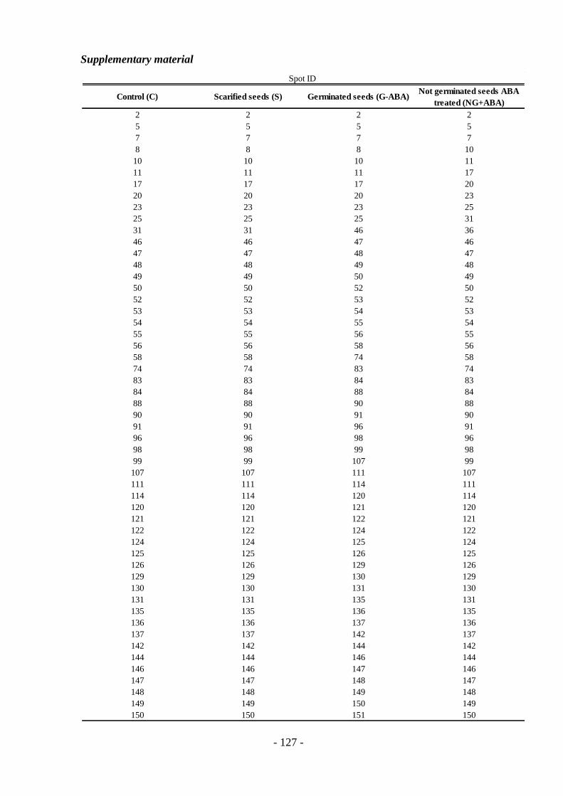
- 127 -
Supplementary material
Control (C) Scarified seeds (S) Germinated seeds (G-ABA) Not germinated seeds ABA
treated (NG+ABA)
2 2 2 2
5 5 5 5
7 7 7 7
8 8 8 10
10 10 10 11
11 11 11 17
17 17 17 20
20 20 20 23
23 23 23 25
25 25 25 31
31 31 46 36
46 46 47 46
47 47 48 47
48 48 49 48
49 49 50 49
50 50 52 50
52 52 53 52
53 53 54 53
54 54 55 54
55 55 56 55
56 56 58 56
58 58 74 58
74 74 83 74
83 83 84 83
84 84 88 84
88 88 90 88
90 90 91 90
91 91 96 91
96 96 98 96
98 98 99 98
99 99 107 99
107 107 111 107
111 111 114 111
114 114 120 114
120 120 121 120
121 121 122 121
122 122 124 122
124 124 125 124
125 125 126 125
126 126 129 126
129 129 130 129
130 130 131 130
131 131 135 131
135 135 136 135
136 136 137 136
137 137 142 137
142 142 144 142
144 144 146 144
146 146 147 146
147 147 148 147
148 148 149 148
149 149 150 149
150 150 151 150
Spot ID

- 128 -
Table 1 In the table are listed spots found in control seeds (C), scarified seeds (S), germinated seeds not ABA
treated (G-ABA) and in not germinated seeds ABA treated (NG+ABA).
Control (C) Scarified seeds (S) Germinated seeds (G-ABA) Not germinated seeds ABA
treated (NG+ABA)
163 163 185 163
164 164 164 185
184 184 184 164
185 185 187 181
187 187 188 184
188 188 192 187
192 192 211 188
211 211 212 192
212 212 216 204
216 216 231 211
231 231 233 212
233 233 242 216
242 242 247 231
244 244 249 233
247 247 254 242
249 249 265 244
254 254 275 247
265 265 287 249
275 275 290 254
287 287 292 265
290 290 296 275
292 292 299 287
296 296 300 290
299 299 301 292
300 300 305 296
301 301 306 299
305 305 313 300
306 306 333 301
313 313 334 302
333 333 335 305
334 334 336 306
335 335 337 313
336 336 326
337 337 333
334
335
336
337
343
Spot ID

- 129 -
Acknowledgements
Many thanks to:
my supervisor Prof.ssa G. Stefania Scippa for supporting me during my PhD program;
my colleagues Manuela, Gaby, Dalila, Miriam, Vincenzo, Paolo for their help and friendship;
Dr. Gerhard Leubner for hosting me in his laboratory in Freiburg.
Finally, I want to express my infinite gratitude to my mother and father, to my brother,
Giuliano, Manuela and Margitta.