med. forum, vol. 25, no.1 january, 2014med. forum, vol. 25, no.1 january, 2014 issn 1029-385-x...
TRANSCRIPT

Electro
nic Cop
y
Electro
nic Cop
y
Electro
nic Cop
y
Electro
nic Cop
y
Med. Forum, Vol. 25, No.1 January, 2014 ISSN 1029-385-X
Recognized by PMDC CONTENTS Recognized by HEC
Editorial
1. Mortality may be Reduced after Hip or Knee Replacement – A New Study 1
Mohsin Masud Jan
Original Articles
2. Hepatoprotective Effects of Curcuma Longa against Carbon Tetrachloride Induced Liver Injury
in Rats 2-5
1. Zaheer Ahmed Memon 2. Muhammad Bachal Pandhiani 3. Aftab Ahmed Shaikh
4. Haji Khan Khoharo
3. Proportion of Urinary Symptoms in Pre and Post-Menopausal Women with Uterovaginal
Prolapse 6-9
1. Samina 2. Deeba Kalim 3. Amjad Zahoor 4. Israr Ahmed Akhund 5. Muhammad Ishaq
4. A Randomized Controlled Trial on Prevention of Postpartum Haemorrhage with Sublingual
Misoprostol or Oxytocin 10-12
1. Kulsoom Bhatti 2. Tahmina Mahar 3. Rubeena Hafeez 4. Shoaib-u-Nisa
5. Causes and Management Out Come of Peritonitis 13-17
1. Muhammad Ali Sohail 2. Rafique Ahmed Sahito 3. Feroze Ahmed Mahar
6. Beneficial Effect and Safety of 5% Permethrin Cream in Scabies Patients 18-21
1. Farah Asad 2. Fatima Rizvi 3. Jawed Iqbal
7. Alterations in Mitochondria of Kidney Tubules by Different Doses of Diclofenac Sodium in
Rabbits 22-26
1. Talat Yasmeen 2. Irfan Ashraf Siddiqui 3. Zaheer Amjad 4. Amir Ali Shoro 5. Talat Mirza
8. Efficacy of Platelet Rich Plasma Application in Comparison to Conventional Dressing Therapy
in Partial Thickness Burn Wound 27-30
1. Ehmer-Al-Ibran 2. Masood Hussain Rao 3. Maria Khan 4. Farrukh Hasan 5. Raaziyah Abdul Khaliq
6. Syeda Zehra
9. Prediction of Large Esophageal Varices in Patients with Decompensated Cirrhosis by Child-
Pugh Score, in Medical Unit-II, Chandka Medical College Hospital, Larkana 31-35
1. Muhammad Aslam Soomro 2. Imdad Ali Ansari 3. Ghulam Yasin Abro 4. Sayed Aftab Ahmed
Shah 5. Sayed Shafiq Rahman Shah
10. Cognition-Enhancing Effect of Oral Therapeutic Doses of Methylphenidate in Rats 36-39
1. Nausheen Alam 2.Rahila Najam
11. EPI Status under One Year Children and their Mothers attending Paediatric Department Sheikh
Zayed Medical College & Hospital, Rahim Yar Khan 40-43
1. Jamal Anwer 2. M.I.Babar 3. Kifayat Niazi 4. Zahid Mahmood
12. Functional Outcome of Cemented Versus Uncemented Hemiarthroplasty for Intracapsular Hip
Fractures 44-48
1. Manqoosh ur Rehman 2. Muhammad Imran 3. Tanveer Ahmad Kang
13. Efficacy of Intra Articular Injections in Different Grades of Osteoarthritis of Knee 49-52
1. Muhammad Imran 2. Manqoosh ur Rehman 3. Tanveer Ahmad Kang
14. Effect of Thiamine on Glycemic Control in Induced Diabetic Rat Model 53-56
1. Muhammad Bachal Pandhiani 2. Maria Kazi 3. Saif-ur-Rehman 4. Navaid Kazmi 5. Salman Ahmad
Farsi Kazi 6. Haji Khan Khoharo
15. Pentoxifylline Protects against Carbon Tetrachloride Induced Liver Injury in Adult Male Wistar
Rat Model 57-61
1. Maria Kazi 2. Muhammad Bachal Pandhiani 3. Salman Ahmad Farsi Kazi 4. Saif-ur-Rehman
5. Haji Kjan Khoharo
16. Validity of Pleural Fluid Protein in Differentiating Tuberculouse from Malignant Pleural
Effusion 62-65
1. Ghulam Nabi Khokhar 2. Muhammad Ashfaq 3. Ikram Ullah Khan 4. Muhammad Ishaq
5. Israr Ahmed
Electro
nic Cop
y
Med. Forum, Vol. 25, No.1 January, 2014 ISSN 1029-385-X
17. To Assess the In vitro Genotoxicity of Metformin and Aspartame alone & In Combination 66-70
1. Amna Nazar 2. Rafique Ahmed 3. Sajida Bano 4. Muhammad Ashraf 5. Imran Altaf
6. Aqeel Javeed
18. Study of Type 2 Diabetes Mellitus and its Correalation with Blood Groups 71-75
1. Shamsul Arfin Qasmi 2. Aftab Ahmed Khan 3. Hasan Askari 4. Rabiyah Pirzada
19. The Effects of Monosodium Glutamate on the Histology of Fallopian Tube in Female Rats 76-79
1. Aftab Ahmed Shaikh 2. Muhammad Bachal Pandhiani 3. Zaheer Ahmed Memon
4. Haji Khan Khoharo
20. Incidence of Malignant Lymphomas in Balochistan 80-84
1. Zahid Mehmood 2. M.Hanif Mengal 3. Haroon ur Rasheed 4. Hafiz Khush Naseeb Ahmed
21. Adjuvant Therapy for Old Age Glioblastoma Patients 85-89
Salman A. Jaffery
22. Comparative Study on Neem Leaf Extract and Nimolicin (NC) on Gastric Mucosa of Albino
Rats 90-94
1. Qamar Aziz Siddiqui 2. Syed Ijaz Hussain Zaidi 3. Asghar Hussain 4. Muhammad Omar Shamim
5. Nazar Hussain 6. Syed Naimul Hassan Naqvi 7. Mohammed Arshad 8. Rajput Muhammad Tariq
9. Mazhar Hijazi
Subject Index
23. Subjects Index January to December 2013 95-103
Azhar Masud Bhatti
Author Index
24. Authors Index January to December 2013 104-107
Azhar Masud Bhatti
Electro
nic Cop
y
Med. Forum, Vol. 25, No.1 January, 2014 1
Editorial Mortality may be Reduced after
Hip or Knee Replacement – A New Study Mohsin Masud Jan
Editor
The risk of death from hip or knee replacement surgery
as dropped substantially in recent years. Dutch
researchers found that since the early 1990s, death rates
have fallen by almost two-thirds among Danish adults
having the procedures. The length of patients’ hospital
stays also dropped — from more than two weeks, on
average, to about one week.
The study did not dig into the reasons for the
improvements, but it’s likely that changes in post-
surgical care have had a big impact, said lead researcher
Arief Lalmohamed, of the Utrecht Institute of
Pharmaceutical Sciences in the Netherlands.
Those changes, he said, include new blood-thinning
medications that help prevent patients from developing
potentially dangerous blood clots after surgery. Clots
can, in some cases, lead to a heart attack, stroke or
pulmonary embolism. In the United States, more than 1
million people have a hip or knee replacement each
year, according to the U.S. National Institutes of
Health. The surgery often is prompted by severe wear
and tear on the joints due to arthritis.
The findings, reported recently in the Journal Arthritis
& Rheumatology, are based on data from only one
country. But Lalmohamed said he would expect to see a
similar pattern in other countries that made the same
changes in medical care over the years. Dr. Richard
Iorio, chief of adult reconstruction at NYU Langone
Medical Center in New York City, agreed that the trend
would be similar in the United States. Iorio, who was
not involved in the study, named a number of advances
that have been made over the years to make joint-
replacement surgery safer and better. Changes in the
procedures and anesthesia techniques have been key,
Iorio said. And patients start physical rehab much faster
than they did in years past. “We get people out of bed
and moving on the first day after — or the day of —
surgery,” Lorio said. That mobility is important, he
said, because it lowers patients’ risk of developing
blood clots. Iorio said doctors have also gotten better at
managing chronic health conditions that many patients
have. That, in turn, lowers the risk of complications.
For the study, Lalmohamed’s team turned to Denmark’s
system of national health registries. The researchers
found information on more than 112,000 people who
had a hip or knee replacement between 1989 and 2007.
Overall, the rate of death in the two months after
surgery fell over time, from about 3.4 percent each year
between 1989 and 1991 to 1.4 percent per year between
2003 and 2007, Lalmohamed said. Deaths from heart
attack, stroke and pneumonia all dropped, despite the
fact that heart and lung disease was more common in
patients who had surgery in recent years.
Lalmohamed said there’s still a need for similar studies
in other countries. But he also said candidates for joint
replacements can be reassured by his team’s findings.
Lorio agreed, “Clearly, patients can take heart,” he said.
“This operation is safer than it was 20 years ago, and
it’s very effective.”
However, some surgeons and hospitals are better than
others. In general, surgeons and centers with the most
experience in hip and knee replacements have better
results than those who do fewer procedures.
And, of course, each patient is different, Lorio said. An
individual’s overall health — rather than age alone —
is vital. But, he added, it’s also possible to manage
some of the health issues that can increase the risks of
joint-replacement surgery. Patients can quit smoking or
lose excess weight, for example.
Electro
nic Cop
y

Med. Forum, Vol. 25, No. 1 January, 2014 2
Hepatoprotective Effects of
Curcuma Longa against Carbon Tetrachloride
Induced Liver Injury in Rats 1. Zaheer Ahmed Memon 2. Muhammad Bachal Pandhiani 3. Aftab Ahmed Shaikh
4. Haji Khan Khoharo 1. Asstt. Prof. of Anatomy, Faculty of Medicine and Allied Medical Sciences, Isra University, Hyderabad
2. Asstt. Prof. of Anatomy, Al-Tibri Medical College, Karachi 3. Asstt. Prof. of Pharmacology, Faculty of Medicine
and Allied Medical Sciences, Isra University, Hyderabad 4. Asstt. Prof. of Physiology/Medicine, Faculty of
Medicine and Allied Medical Sciences, Isra University, Hyderabad
ABSTRACT
Objective: To investigate the protective effect of Curcuma longa (CL) against carbon tetrachloride (CCl4) induced
liver injury in adult male Wistar rat model.
Study Design: Experimental/Analytical study
Place and Duration of Study: Animal House, Isra University Hyderabad from March to December 2013.
Subjects and Methods: Forty five adult male Wistar rats were divided into three groups; Group 1. controls received
0.9% isotonic saline, Group 2. received CCl4 orally (1.9mg/kg) mixed in olive oil, and Group 3. received the
CCl4+CL (250mg/kg) Blood samples were collected for liver biochemical assays. The animals were sacrificed, liver
tissue, after fixation in 4% formaldehyde, was embedded in paraffin. Tissue sections of 5μ thickness were subjected
to haematoxylin and eosin staining and were assessed by light microscopy. The data was analyzed on SPSS 21.0
using one-way ANOVA, Fischer`s LSD and Chi-square tests. A p-value of ≤ 0.05 was taken statistically significant.
Results: The liver biochemical and histological findings reveal statistically significant differences among the
controls, CCl4 and CCl4+CL groups (p=0.0001). Liver enzymes and histology were deranged significantly in CCl4
group compared to controls and CCl4 +CL group (p=0.0001). The CCl4+CL group showed less elevation of liver
enzymes and derangement in liver histology compared to CCl4 group (p=0.001). The histological findings of
congestion, inflammatory cell infiltrate, vacuolar degeneration and necrosis are found prominent in CCl4 group.
Conclusion: The Curcuma longa protects against oxidative damages caused by carbon tetrachloride induced liver
injury in rat model.
Key Words: Curcuma longa, Carbon Tetrachloride, Liver injury.
INTRODUCTION
Curcuma longa (CL) is a rhizomatous perennial herb
that belongs to the family Zingiberaceae, native to
South Asia and is commonly known as turmeric.1 In
Sindhi, it is commonly known as “Hade”. The turmeric
plant is a popular ingredient for preparing culinary
dishes. In addition, it is used as herbal remedy due to
the prevalent belief that the plant has medical
properties. In folk medicine, the rhizome juice from C.
longa is used in the treatment of many diseases such as
anthelmintic, asthma, gonorrhea and urinary, and its
essential oil is used in the treatment of carminative,
stomachic and tonic.2 In traditional medicine, several
plants and herbs have been used experimentally to treat
liver disorders, including liver cirrhosis. 3,4
C. longa
possesses antioxidant5, anti-tumor
6, antimicrobial
7, anti-
inflammatory8, wound healing
9, and gastroprotective
activities.10
Carbon tetrachloride (CCl4) is a hepatotoxic compound.
The CCl4 has been has been used extensively in
laboratory animals for induction of liver injury,
elucidate the underlying mechanism of liver injury and
hepatoprotective effects of various therapeutic agents.11
One of the postulated mechanism of CCl4 induced liver
injury is the formation of ROS. The ROS disrupts the
hepatocyte at cell membrane level through the lipid
peroxidation11, 12
causing anatomical disruption of liver
architecture and physiological disturbances.13
The
hepatocyte injury causes leakage of cytoplasmic and
mitochondrial enzymes in the blood streams.14
The cytoplasm and mitochondrial enzymes of
hepatocytes are clinically used as markers of liver
injury, and for monitoring and treating the liver
diseases. The liver enzymes which appear in the blood
as a result of liver injury include; alanine transaminase
(ALT), aspartate transaminase (AST), alkaline
phophatase (ALP) and lactate dehyderogenase (LDH)
are important enzymes that are often employed in
assessing liver injury.11,15
The previous studies have shown that the aqueous
extract of C. longa has hepatoprotective activity against
carbon tetrachloride toxicity19
. The present study aims
to investigate the possible hepatoprotective effects of
the ethanolic extract of Curcuma longa rhizomes
against Carbon tetrachloride (CCl4)-induced
hepatotoxicity in adult male Wistar rat model.
Original Article Anatomy
Electro
nic Cop
y
Med. Forum, Vol. 25, No. 1 January, 2014 3
MATERIALS AND METHODS
The present original study was conducted at the animal
house of Isra University from March to December
2013. Adult male Wistar rats of 250-300 grams were
included while female rats and rats weighing <250
grams or >300 grams were excluded from the study
protocol. The animal’s house is well equipped with
essential facilities like an optimal room temperature
with 55-60% humidity and exposure to 12 hour light-
dark cycles. The fresh alfalfa and clean water are
provided freely. The rats were divided into three
groups;
Group 1. Control Group (n=15) Rats received 0.9%
isotonic saline orally on alternate day for three
successive weeks and served as control group,
Group 2. Carbon tetrachloride Group (n=15) Rats
were given CCl4 orally mixed in olive oil on alternate
day for three successive weeks and
Group 3. Experimental Group (n=15) Rats received
Curcuma longa (250 mg/kg) and CCl4 on alternate days
for three successive weeks
Experimental Details: The CL was purchased from
Medical store of Isra University Hospital. The Curcuma
longa was administered in a dose of 250 mg/kg orally.16
Carbon tetrachloride was purchased from scientific
drug store at Hyderabad City. The CCl4 dissolved in
olive oil as vehicle (1:1 Ratio) at a dose level of 1.9
ml/kg orally on alternate day for three successive weeks
and sacrificed at the end of their respective period of
time.15
The animals were sacrificed using standard
method as described by Nayak et al. (2006)17
In order
to examine the liver tissue, the liver of the sacrificed
animals was removed promptly and preserved in
formaldehyde.
Blood sampling: The blood samples were collected
from tail at twenty four hours of experimental period.
Sera were separated by centrifugation at 300xs for ten
minutes. Serum samples were used to estimate liver
enzymes.
Biochemical assay: Liver enzyme assays were
determined for alanine transaminase (ALT), aspartate
transaminase (AST), alkaline phophatase (ALP) and
lactate dehyderogenase (LDH) using commercially
available diagnostic kits.
Histological studies: After fixation in 4%
formaldehyde, samples were embedded in paraffin.
Sections of 5μ thickness were subjected to
haematoxylin and eosin Hepatic morphology was
assessed by light microscopy. A total of five sections
for each liver tissue sample were observed under light
microscope.
In H & E staining, damaged hepatocytes graded as 0=
normal, += mild damage (swollen and pale cytoplasm),
++= moderate damage (vacuolated cytoplasm), +++=
severe damage and ++++= very severe damage
(pyknotic nucleus and eosinophil cytoplasm).18
The data was analyzed on SPSS version 21.0 (IBM
corporation). The continous variables were presented as
mean±SD using one-way ANOVA and Fischer`s LSD
test. Chi-square test was used for categorical variables.
A p-value of ≤ 0.5 was taken statistically significant.
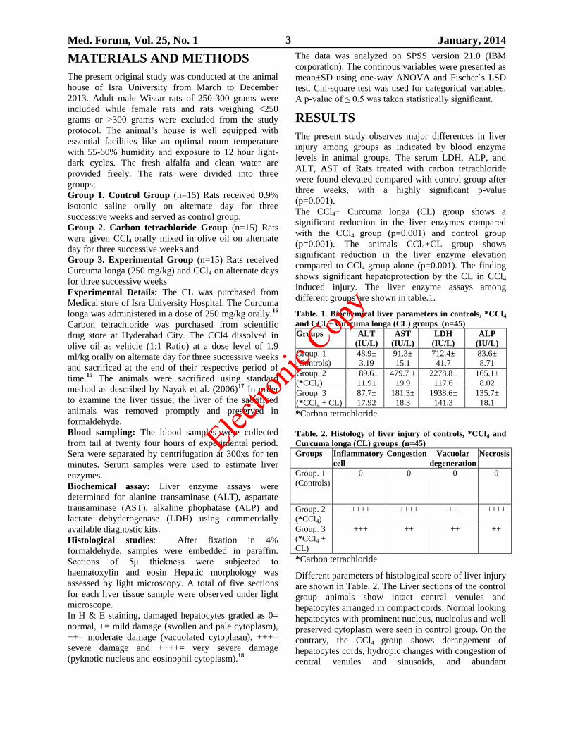
RESULTS
The present study observes major differences in liver
injury among groups as indicated by blood enzyme
levels in animal groups. The serum LDH, ALP, and
ALT, AST of Rats treated with carbon tetrachloride
were found elevated compared with control group after
three weeks, with a highly significant p-value
(p=0.001).
The CCl4+ Curcuma longa (CL) group shows a
significant reduction in the liver enzymes compared
with the CCl4 group (p=0.001) and control group
(p=0.001). The animals CCl4+CL group shows
significant reduction in the liver enzyme elevation
compared to CCl4 group alone (p=0.001). The finding
shows significant hepatoprotection by the CL in CCl4
induced injury. The liver enzyme assays among
different groups are shown in table.1.
Table. 1. Biochemical liver parameters in controls, *CCl4
and CCl4+ Curcuma longa (CL) groups (n=45) Groups ALT
(IU/L)
AST
(IU/L)
LDH
(IU/L)
ALP
(IU/L)
Group. 1
(Controls)
48.9±
3.19
91.3±
15.1
712.4±
41.7
83.6±
8.71
Group. 2
(*CCl4)
189.6±
11.91
479.7 ±
19.9
2278.8±
117.6
165.1±
8.02
Group. 3
(*CCl4 + CL)
87.7±
17.92
181.3±
18.3
1938.6±
141.3
135.7±
18.1
*Carbon tetrachloride
Table. 2. Histology of liver injury of controls, *CCl4 and
Curcuma longa (CL) groups (n=45) Groups Inflammatory
cell
Congestion Vacuolar
degeneration
Necrosis
Group. 1
(Controls)
0 0 0 0
Group. 2
(*CCl4)
++++
++++
+++ ++++
Group. 3
(*CCl4 +
CL)
+++ ++
++ ++
*Carbon tetrachloride
Different parameters of histological score of liver injury
are shown in Table. 2. The Liver sections of the control
group animals show intact central venules and
hepatocytes arranged in compact cords. Normal looking
hepatocytes with prominent nucleus, nucleolus and well
preserved cytoplasm were seen in control group. On the
contrary, the CCl4 group shows derangement of
hepatocytes cords, hydropic changes with congestion of
central venules and sinusoids, and abundant
Electro
nic Cop
y
Med. Forum, Vol. 25, No. 1 January, 2014 4
inflammatory cell infiltration. The centrilobular
hepatocytes show hydropic changes and necrosis, while
midzonal and peripheral hepatocytes show vacuolar
degeneration and fatty changes in CCl4 group. In
CCl4+CL animals, liver tissue sections reveal less
significant derangement of hepatocytes cords,
hepatocytes damage and necrosis was limited compared
with CCl4 group.
DISCUSSION
The present study is an original research work, which
investigates the effect of Curcuma longa (CL) on
carbon tetrachloride (CCl4) induced liver injury in adult
male Wistar rats. The Null hypothesis is rejected
because the study observes hepatoprotective effects of
CL as evidenced by biochemical and histological
marker of liver injury.
Curcumin, the most common antioxidant constituent of
Curcuma longa rhizome extract, was reported to
enhance apoptosis of damaged hepatocytes which might
be the protective mechanism whereby curcumin down-
regulated inflammatory effects and fibrogenesis of the
liver. 19
The present study shows liver damage caused by the
carbon tetrachloride as indicated by serum levels of
liver enzymes compared to control group in rat model.
The carbon tetrachloride induced liver injury with
release of liver enzymes is comparable finding to
reported previously by Hurkkeri et al.20
The Hurkkeri et
al.20
reported elevated hepatocyte enzyme of liver as a
consequence of CCl4 induced liver injury in animal
model. The release of large quantities of cytoplasmic
and mitochondrial enzymes of liver is a clinical
indicator of hepatocyte cell membrane damage and
rupture sufficient to produces change in enzyme levels
in blood.21
The ethanolic extract of C. longa rhizomes showed a
significant hepatoprotective effect when orally
administrated in doses of 250 mg/kg. The main
constituents of CL extract are the flavonoid curcumin
and various volatile oils, including tumerone, atlantone,
and zingiberene.1 The hepatoprotective effects of
turmeric and curcumin might be due to direct
antioxidant and free radical scavenging mechanisms, as
well as the ability to indirectly augment glutathione
levels, thereby aiding in hepatic detoxification.22
The
volatile oils and curcumin of C. longa exhibit potent
anti-inflammatory effects.23
The present study shows that the damage of liver
caused by CCl4 is evident by the rise in serum enzymes
levels beside the histological changes in liver tissue.
Administration of CCl4 significantly increases the
serum levels of liver enzymes; LDH, ALP, ALT and
AST, which are indices of hepatocyte damage and
leakage of enzymes from cells. 24,25
The histological examination of present research study
correlates in parallel to disturbance in biochemical
markers of liver injury. The histology of liver tissue
shows disruption of liver architecture, hepatocytes,
hepatic lobules and arrangement of hepatocytes in
cords. The hepatocytes show findings of cellular injury
with marked cytoplasmic vacuolization. The injured
hepatocytes show pyknotic nuclei with lymphocyte
infiltrations. The pyknotic nuclei are a sign of severe
cellular injury caused by a toxin like carbon
tetrachloride. The histological and biochemical findings
of present study are comparable to those mentioned
previously.26,27
The carbon tetrachloride is metabolized
to free radical during its metabolism and detoxification
in smooth endoplasmic reticulum by the cytochrome
P450.28
The findings of present study are highly
comparable to a recent study of Salma et al.1 The Salma
et al1 reported hepatoprotective effects of CL in
thiacetamide induced liver cirrhosis in rat models. The
present study concludes that the Curcuma longa
decreases the carbon tetrachloride induced oxidative
stress and liver damage.
CONCLUSION
The Curcuma longa protects against oxidative damages
caused by carbon tetrachloride induced liver injury in
rat model. The Curcuma longa may be used as an
effective protector against chemical induced liver
damages; however, further studies are warranted.
REFERENCES
1. Salma SM, Abdulla MA, AlRashdi AS, Ismail S,
Alkiyumi SS, Gulbabapour S. Hepatoprotective
effect of ethanolic extract of Curcuma long on
thiacetamide induced liver cirrhosis in rats. BMC
Compl Alt Med 2013;13:56.
2. Phansawan B, Poungbangpho S. Antioxidant
capacities of Pueraria mirifica, Stevia rebaudiana
Bertoni, Curcuma longa Linn. Andrographis
paniculata (Burm. f.) Nees. and Cassia alata Linn.
for the development of dietary supplement.
Kasetsart J 2007;41(3):407–413.
3. Alshawsh MA, Abdulla MA, Ismail S, Amin ZA.
Hepatoprotective Effects of Orthosiphon stamineus
Extract on Thioacetamide-Induced Liver Cirrhosis
in Rats. Evid Based Complement Alternat Med
2011;2011:1–6.
4. Kadir FA, Othman F, Abdulla MA, Hussan F,
Hassandarvish P. Effect of Tinospora crispa on
thioacetamide-induced liver cirrhosis in rats. Indian
J Pharmacol 2011;43(1):64.
5. Maizura M, Aminah A, Wan Aida W. Total
phenolic content and antioxidant activity of kesum
(Polygonum minus), ginger (Zingiber officinale)
and turmeric (Curcuma longa) extract. Int Food
Res J 2011;18:526–531.
6. Kunnumakkara AB, Guha S, Krishnan S,
Diagaradjane P, Gelovani J, Aggarwal BB.
Curcumin Potentiates Antitumor Activity of
Electro
nic Cop
y
Med. Forum, Vol. 25, No. 1 January, 2014 5
Gemcitabine in an Orthotopic Model of Pancreatic
Cancer through Suppression of Proliferation,
Angiogenesis, and Inhibition of Nuclear Factor-
κB–Regulated Gene Products. Cancer Res 2007;
67(8):3853.
7. Kim KJ, Yu HH, Cha JD, Seo SJ, Choi NY, You
YO. Antibacterial activity of Curcuma longa L.
against methicillin‐ resistant Staphylococcus
aureus. Phytother Res 2005;19(7):599–604.
8. Kohli K, Ali J, Ansari M, Raheman Z. Curcumin: a
natural anti-inflammatory agent. Indian J
Pharmacol 2005;37(3):141–147.
9. Panchatcharam M, Miriyala S, Gayathri VS,
Suguna L. Curcumin improves wound healing by
modulating collagen and decreasing reactive
oxygen species. Mol Cell Biochem 2006;
290(1):87–96.
10. Miriyala S, Panchatcharam M, Rengarajulu P.
Cardioprotective effects of curcumin. The
molecular targets and therapeutic uses of curcumin
in health and disease 2007;595:359–377.
11. Obi FO, Omogbal LA, Orlafo OS, Ovat OD. Effect
of a Short Time Post Carbon Tetrachloride
Treatment Interval on Rat Plasma Enzyme Levels
and Percentage Mortality. J Applied Sci Environ
Mgt 2001;5:5-8.
12. Muriel P, Albo N, Perez-Alvarez VM. Kupffer
cells inhibition prevents hepatic lipid peroxidation
and damage induced by carbon tetrachloride. Comp
Biochem Physiol C Toxicol Pharmacol 2001;130:
219-26.
13. Rasha SA, Ashraf AA and Aly R. Carbon
tetrachloride-induced liver disease in rats: the
potential effect of supplement oils with vitamins E
and C on the nutritional status. German Med Sci
2009;7:1612-3174.
14. Rost DA, Welker DA, Welker J, Millonig G,
Berger I, Autschbach F, et al. Liver-homing of
purified glucose oxidase: A novel in vivo model of
physiological hepatic oxidative stress (H2O2). J
Hepatol 2007;46: 482-91.
15. Essawy AE, Abdel-Moneim AM, Khayyat LI and
Elzergy AA. Nigella sativa seeds protect against
hepatotoxicity and dyslipidemia induced by carbon
tetrachloride in mice. J Appl Pharm Sci 2012;2
(10):021-5.
16. Movssaghi S, Sharifi ZN, Mohammadzadeh F,
Soleimani M. Pentoxifylline protects the rat liver
against fibrosis and apoptosis induced by acute
administration of 3,4-Methyleneoxy methampheta-
mine (MDMA or Ecstasy). Ir J Basic Med Sci
2013; 16: 922-27.
17. Nayak S, Nalabothu P, Sandiford S, Bhogadi V,
Adogwa A. Evaluation of wound healing activity
of Allamanda cathartica L. and Laurus nobilis. L.
Extracts on rats. BMC Compl Alt Med 2006; 6: 12.
18. Murat-Bilgin H, Atmaca M, Deniz-Obay B,
Ozekinci S, Taşdemir E, Ketani A. Protective
effects of coumarin and coumarin derivatives
against carbon tetrachloride-induced acute
hepatotoxicity in rats. Exp Toxicol Pathol. 2011;
63(4): 325-30.
19. Wang ME, Chen YC, Chen IS, Hsieh SC, Chen SS,
Chiu CH. Curcumin protects against
thioacetamide-induced hepatic fibrosis by
attenuating the inflammatory response and
inducing apoptosis of damagedhepatocytes. J Nutr
Biochem 2012;11:120-8.
20. Hurkkeri VI, Jaiparkash B, Lavhale RV, Karadi
RV, Kuppast IJ. Hepatoprotective activity of
Anthus Excelsa Roxb leaf extract on experimental
liver damage in rats. J Pharmacogn 2002;11:
120-28.
21. Shaarawy SM, Tohamy AA, Elgendy SM
Elmageed ZY, Bahnasy A, Mohamed MS, et al.
Protective effects of garlic and silymarin on
NDEA-induced rats hepatotoxicity. Int J Biol Sci
2009;5(6):549-57.
22. Girish C, Koner BC, Jayanthi S, Ramachandra Rao
K, Rajesh B, Pradhan SC.Hepatoprotective activity
of picroliv, curcumin and ellagic acid compared to
silymarin on paracetamol induced liver toxicity in
mice. Fundam Clin Pharmacol 2009;23(6):735–45.
23. Organization WH. WHO monographs on selected
medicinal plants-Vol. 1. Geneva: World Health
Organization 2002.
24. Rajesh M, Latha M. Preliminary evaluation of anti-
hepatotoxic activity of Kamilari, a polyherbal
formulation. J Ethnopharmacol 2004; 91: 99-104.
25. Bashandy S, Al-Wasel S. Carbon tetrachloride-
induced hepatotoxicity and nephrotoxicity in rats:
Protective role of vitamin C. J Pharm Toxico 2001;
16(30): 283-92.
26. Trivedi P, Mowat A. Carbon tetrachloride-induced
hepatic fibrosis and cirrhosis in the developing rat:
an experimental model of cirrhosis in childhood.
Br J Exp Pathol 1983; 64(1): 25–33.
27. Berman E, House D, Allis J, Simmons J.
Hepatotoxic interactions of ethanol with allyl
alcohol or carbon tetrachloride in rats. J Toxicol
Environ Health 1992; 37(1): 161–76.
28. Brandao C, Ferreira H, Piovesana H, Polimeno N,
Ferraz J, de-Nucci G. Developmental model of
liver cirrhosis in rabbits. Clin Exp Pharma Physiol
2000; 27: (12): 987-90.
Address for Corresponding Author:
Dr. Haji Khan Khoharo ,
Assistant Professor of Physiology/Medicine
Faculty of Medicine and Allied Medical Sciences
Isra University, Hyderabad.
Cell No.: 0331-2662500.
Electro
nic Cop
y
Med. Forum, Vol. 25, No. 1 January, 2014
6
Proportion of Urinary Symptoms
in Pre and Post-Menopausal Women with
Uterovaginal Prolapse 1. Samina 2. Deeba Kalim 3. Amjad Zahoor 4. Israr Ahmed Akhund 5. Muhammad Ishaq
1. Asstt. Prof. of Obst. & Gynae 2. Asstt. Prof. of Obst. & Gynae 3. Assoc. Prof. of Pediatrics 4. Prof. of Physiology
5. Prof. of Surgery, Jinnah Medical College, Peshawar
ABSTRACT
Objective: To compare the proportion of urinary symptoms in pre and postmenopausal women with uterovaginal
prolapse.
Study Design: Comparative Study.
Place and Duration of Study: The study was carried out at the Department of Obstetrics & Gynecology Unit II,
Dow University of Health Sciences and Civil Hospital Karachi from July 2005 to January 2006.
Materials and Methods: Sixty consecutive patients (30 premenopausal and 30 postmenopausal) were included in
the study through structured Proforma from the out patient ward or emergency. Informed consent was obtained. A
detailed history and related examinations and investigations were done. These include urine DR, Urine C/S and
Urodynamic like Cystometry in selected patients.
Results: In this study the difference of urinary symptoms in pre and postmenopausal women of Uterovaginal
prolapse were statistically found insignificant like frequency of urine (26.7% versus 33.3%), Urgency (20% vs
26.7%), Nocturia (26.7% vs 13.3%), Dysuria (40% vs 26.7%), Voiding problems (40% vs 46.7%), Urge
incontinence (40%vs 20%). Stress incontinence was slightly higher in postmenopausal group than premenopausal
(53.3%vs 46.7%) but this difference was found insignificant, while parity status between these two groups had
significant difference like parity 2-5 was higher in pre-menopause group than postmenopause (66.7% vs 36.7%) and
parity 6-10 was higher in postmenopause group than pre-menopause (63.3% vs 26.7%).
Conclusion: Significant difference in parity was found between pre and post-menopausal women with uterovaginal
prolapse but the difference of urinary symptoms in pre and postmenopausal group was found significant.
Uterovaginal prolapse associated with different urinary symptoms especially incontinence and voiding problems.
These urinary symptoms effect over quality of life of women. This warrants greater attention for Gynecological
health needs in our country by safe family planning practices.
Key Word: Uterovaginal Prolapse, Pre–Menopausal, Post-Menopausal
INTRODUCTION
Uterovaginal prolapse is the descent of vagina and
uterus due to the loss of integrity of structures that
support the contents of female pelvis1. The exact
incidence of uterovaginal prolapse is difficult to
determine. It has been estimated that a 50% of parous
women lose pelvic floor support, resulting in prolapse
and about 10-20% of them seek medical care2. A WHO
multicentre collaborative study carried out in Pakistan
identified at least 22% women with uterovaginal
prolapse who attended health care facility3. The chance
of a woman having a prolapse increases with age.
Therefore the incidence of prolapse will rise as life
expectancy increases4. Uterovaginal prolapse is usually
the result of childbirth, menopause (due to deficiency of
estrogen), increase intra abdominal pressure, injury to
sacral nerves S1-S4 or diabetic neuropathy. Congenital
weakness of pelvic support causes prolapse in young
nulliparous women, prolapse of vagina may occur after
hysterectomy5. Prolapse is often asymptomatic. The
usual presenting symptoms in patients with
uterovaginal prolapse are something coming down of
vagina, lump in vagina and backache which is
aggravated by standing and eased by lying down6.
Prolapse is frequently associated with urinary
complains like increase frequency, urgency, nocturia,
hesitancy, urinary incontinence or incomplete
emptying. In severe cases of incomplete bladder
emptying, retention of urine may occur. Voiding
dysfunction may result in increase frequency of urinary
tract infections and occasionally, overflow
incontinence. Due to the kinking of the urethra, stress
incontinence and even intrinsic sphincter deficiency
may be over looked7. The world wide prevalence of
urinary symptoms in association with prolapse varies
ranging from 5 to 39%. In Pakistan, hospital based
study on urinary incontinence reported the frequency of
urinary incontinence as 20.5%. According to one study
conducted by community health centre of Agha khan
University Hospital (AKUH) from November
1stto30th, 2002 stress incontinence was the highest
reported complaint (38.4%) followed by burning
(34.4%), frequency (26%), painful micturition (20.4%),
urge incontinence (18.8%), incomplete empting of
bladder (14.4%) and poor stream (8.4%) 8.
Original Article Uterovaginal Prolapse
Electro
nic Cop
y

Med. Forum, Vol. 25, No. 1 January, 2014
7
Urogynecology as a subspecialty has not been
introduced in Pakistan and we believe the extent of
significant lower urinary tract symptoms has been
hugely underestimated 9. The severity of these
symptoms is more in postmenopausal women10
. The
rationale for conducting this study is based on the
hypothesis that the proportion of urinary symptoms will
be significantly different in the pre and postmenopausal
women with uterovaginal prolapse. The aim of our
study is to compare the proportion of urinary symptoms
in pre and postmenopausal women with uterovaginal
prolapse.
MATERIALS AND METHODS
The study was carried out in the department of
Obstetrics & Gynecology unit II, Dow University of
Health Sciences and Civil Hospital Karachi, Pakistan.
This was a cross sectional comparative study from July
2005 to January 2006. Sixty consecutive patients (30
premenopausal and 30 postmenopausal) were included
in the study through structured Proforma by purposive
sampling technique from the outpatient, ward or
emergency. Informed consent was obtained. The
inclusion criteria were patients with all degree of
uterovaginal prolapse, both pre and postmenopausal
and married and unmarried women. Exclusion criteria
were women who were pregnant with uterovaginal
prolapse, patients with diagnosed renal pathology,
recurrent urinary tract infection, and medical disorder
such as diabetes mellitus or sclerosis. A detailed history
and related examination and investigations were done.
These include urine D/R, Urine C/S and urodynamic
like Cystometry in selected patients. Frequency was
defined as the passage of urine every 2 hrs or more than
seven times a day. Nocturia was defined as interruption
of sleep more than once each night of need to micturate.
Urgency was a strong sudden desire to void while
Dysuria defined as urethral pain during micturition.
Retention of urine means failure to empty the bladder
totally, bladder pressure being unable to overcome
urethral resistance. Voiding problems include
hesitancy, a poor stream, straining to void, incomplete
bladder emptying and also frequency, urgency and
dysuria. Stress incontinence was defined as the
involuntary loss of urine when the intravesical pressure
exceeds the maximum urethral closing pressure. While
urge incontinence was defined as urinary leakage
associated with the sensation of urgency. Data analysis
was performed through SPSS Version 10. Age was
presented by Mean standard deviation and its
histogram was also presented in premenopause and
postmenopause group because Means were not
comparable. Frequencies and percentages were
computed to present all categorical variables including
.menstrual status, parity status, urinary complains and
symptoms. Chi-square test was applied to compare
parity status, urinary symptoms including frequent
urine passing, urgent urine passing, dysuria urine
passing, incontinence and retention urine passing while
Fisher’s exact test was applied to compare nocturia
urine passing, retention urine passing between two
groups. Statistical significance was taken at P<0.05.
RESULTS
Average age of premenopause group was computed
(35.57±5.04 years, range=25-40) and postmenopause
group was (58.77+_9.97 years, range 40-80). There
were only two patients were either nulliparous or single
parous and both these patients were found in
premenopause group. Parity 2-5 was significantly
higher in premenopause than postmenopause group
(66.7%vs36.7%), while parity 6-10 was significantly
higher in postmenopause group than premenopause
group (63.3%vs26.7%).This data revealed a significant
difference (X2=9.09, P=0.011) of parity status between
two groups. (Table1). A symptom of frequent urine
passing was reported by 8 (26.7%) patients of
premenopause group and 10(33.3%) patients of
postmenopause group. There was insignificant
difference(X2=0.317, P=0.573) of proportion of
frequency of urine between two groups. Urgency in
passing urine was reported by 6 (20%) patients of
premenopause group and 8(26.7%) patients of
postmenopause group and had insignificant
difference(x2=0.373, P=0.542) between two groups
(fig 1).8(26.7%) patients of pre and 4(13.3%) patients
of postmenopause group complained nocturia.
Table No. 1: Comparison of Parity Status between
Pre & Most Menopausal Patients
Parity
Status
Pre-
Menopause
(N=30)
Post-
Menopause
(N=30)
Total
Para 0 – 1 2 (6.7%) 0 (0%) 2
Para 2 – 5 20 (66.7%) 11 (36.7%) 31
Para 6 – 10 8 (26.7%) 19 (63.3%)* 27 *Significantly high proportion (X2 = 9.09, p=0.011) at p <0.05
Table No. 2: Comparison of Incontinence Symptom
between two Groups
Incontinence Pre-
Menopause
(N=30)
Post-
Menopause
(N=30)
Total
Urge
Yes
No
12 (40%)
18 (60%)
06 (20%)
24(80%)
X2 =
2.86
P= 0.091
Stress
Yes
No
14 (46.7%)
16 (53.3%)
16 (53.3%)
14 (46.7%)
X2 =
0.27
P= 0.606
An insignificant difference (P=0.333) of proportion was
observed in 4 (13.3%) of pre and 6 (20%) patients of
postmenopause group. Statistically insignificant
difference (P=0.731) of proportion was observed
Electro
nic Cop
y

Med. Forum, Vol. 25, No. 1 January, 2014
8
between two groups (fig 6).Greater number of patients
of premenopause than postmenopause group were
found with dysuria (40%vs 26.7%, P=0.273), However
this difference of proportion was insignificant
statistically (fig7).Urge type incontinence was reported
by 12(40%) patients of pre and 6(20%) patients of
postmenopause group, however this difference of
proportion was insignificant (P=0.091). Stress
incontinence was slightly higher in postmenopause than
premenopause group (53.35vs.46.7%,P=0.606) but this
difference was insignificant(table2).Proportion of
voiding problem in premenopause group was 40% and
postmenopause group was 46.7%, this difference of
proportion was statistically insignificant(x2=
0.271,P=0.602) (Table 2).
DISCUSSION
Women with uterovaginal prolapse may present with a
plethora of lower urinary tract symptoms including
urine frequency, urgency, nocturia, hesitancy,
incomplete emptying and incontinence. These
symptoms may or may not be related to the
uterovaginal prolapse, but with careful clinical and
urodynamic investigations it is usually possible to
determine the underlying pathophysiology11
. There is
study conducted by weber AM, Waltar MD Schova LR,
Mitchinson A, they found that women with prolapse
were older than those without prolapse.(mean age 58.2
vs 49.2 years respectively)12
. In this study we used a
comparison group of pre and postmenopausal women
with uterovaginal prolapse to determine the frequency
and proportion of associated urinary symptoms. In this
study we found that the average age of premenopausal
group was 35.57yrs(range 25-40yrs) while the average
age of postmenopausal women was 58.77 (range 40-
61yrs). A multi country collaborative study in Pakistan
found that women with multiparty were more likely to
have prolapse1. In our study only two patients in
premenopause group were either nulliparous or single
parous. Other patients in premenopause group had
parity 2-5 which was significantly higher than
postmenopausal group (66.7%vs, 36.7%), while parity
6-10 was significantly higher in postmenopause group
than premenopause (63.3%vs,26.7%). So the parity
status between these two groups had significant
difference (x2=9.09, P=0.001). As this study was
carried out on the basis of hypothesis that there is a
likely to be significant difference in urinary symptoms
between pre and postmenopausal group with
uterovaginal prolapse, the result of our study was found
against this hypothesis that differences were
insignificant. In our study urge incontinence was
reported by 40% patients of premenopause and 20%
patients of postmenopause group, however this
difference of proportion was insignificant (P=0.091)
while stress incontinence was slightly higher in
postmenopause group (53.3%vs.46.7%, P=0.606) but
this difference was insignificant and was not support
the hypothesis on which our study based. (Risk factors
for urge and stress incontinence were different). The
major predictors of urge incontinence were increasing
age, UTI and diabetes. Other large population based
studies have reported age as an important risk factor for
urge incontinence13
. According to one study the major
predictors of stress urinary incontinence were white
race, higher basal metabolic index and higher waist to
hip ratio14
. In our study the urinary frequency, urgency
and voiding difficulties were found more in
postmenopausal group as compare to premenopausal
group (33.3%vs26.7%, 26.7%vs20% and 46.7%vs 40%
respectively) but statistically these differences were
insignificant. There is one study conducted by Bun gay
et al who reported that frequency and urgency are 20%
and 15% respectively and these figures increases only
slightly with age15
. In our study greater number of
patients of premenopause group than postmenopause
were found with dysuria (40%vs 26.7%, P=0.273) but
this difference is insignificant. According to survey
conducted in Chinese population in Hong Kong the
prevalence of dysuria reported by 166 of 819 women
(20%). According to one study conducted by Schatzl
and colleagues in 2000 only 3.1% of women younger
than 30 experienced nocturia greater than twice per
night, where as 26.7% of those aged 60 and older did.
But in our study group nocturia found to be higher in
premenopause group than postmenopause (26.7%vs
13.3%). However this difference was found to be
insignificant (P=0.333).Similarly retention of urine was
observed higher in postmenopause group (20%) than
premenopause (13.3%) but again this difference was
found insignificant. Despite thorough evaluation, the
source of voiding dysfunction will not be discovered in
many women with lower urinary tract symptoms. Age
alone correlates with its development; regardless of
existing risk factor. This may be caused by a number of
processes such as occult Supratentorial central nervous
system lesion, neurogenic or myogenic dysfunction at
the level of the bladder and changes in extracellular
matrix composition at the level of the bladder and
urethra16
.
CONCLUSION
Uterovaginal prolapse is associated with different
urinary symptoms commonly incontinence and voiding
problems. These symptoms are usually affected with
age and parity. In our study significant difference in
parity was found between pre and postmenopausal
group but the difference of proportion of urinary
symptoms was found insignificant. All urinary
symptoms need to be investigated before performing
any vaginal surgery to exclude coexistent pathology
such as detrusor instability. These urinary symptoms
effect over quality of life of women. This warrants
greater attention for gynecological health needs in our
Electro
nic Cop
y

Med. Forum, Vol. 25, No. 1 January, 2014
9
country by safe family planning practices, strengthened
with health education of women for delayed age at
marriage, to reduce the risk of uterovaginal prolapse
and associated urinary symptoms. Further studies are
required in the community to study the natural history
of the development of urinary symptoms and the
relationship to prolapse.
REFERENCES
1. Mehboob R, Ahmad N. Uncomplicated pathology
at vaginal hysterectomy for genital prolapse. Pak J
Med Res 2002;41(4):142- 4.
2. Thakar R, Stanton S. Management of genital
prolapse. BMJ 2003;324:1258-62.
3. Bhurt AW, Fikree FF, Bhurt AM, Bozdar NM,
Bhurt A, Bhurt R. Uterogenital prolapse in a rural
community of Sindh, Pakistan. J Coll Physician
& Surg Pak 2001;11:42-6.
4. Olsen AL, Smith VJ, Bergstrom JO, Colling JE,
Clark AL. Epidemology of Surgically managed
pelvic organ prolapse and urinary incontinence.
Obstet Gynaecol 1997;89:501-6.
5. Tayyab S, Hussain N, Incidence and surgical
management of genital prolapse in periurban
based teaching hospital. Medical channel 1999;5
(3):9-12.
6. Jones DL. Uterovagin al displacements, damage
and prolapse. In: Oates J, Abraham S, editors.
Fundamentals of obstetrics and gynaecology.
Edinburgh: Elsevier Mosby;2005.p.311-6.
7. Adam RA. Enterocele and massive vaginal
eversion. e medicine 2002;1-18. Available at
website www.emedicine.com.
8. Andrades M, Paul R, Ambreen A, Dodani S,
Dhanani RH, Qidwai W. Distribution of lower
urinary tract symptoms in adult women. J Coll
Physician Surg Pak 2004;14:132-5.
9. Rizvi RM, Nazim MH. Frequency of urinary
symptoms in women attending gynaecology clinics
at the AKUH, Pakistan. JPMA 2005;55(11):
489 – 492.
10. Versi E, Harvey M, Cardoz L, Urogenital prolapse
and atrophy at menopause: a prevalence study.
Int Urogynaecol J 2001;12:107-10.
11. Balmforth J. Assessment of lower urinary tract
function. In: Lussely DM, Baker PN, editors,
obstetrics and gynaecology. London: Arnold;
2004.p.619-33.
12. Tayrac RD, Chebalier N, Chauveaud lambling A,
Gervaise A, Fernandoze H. Rsk of urge and stress
in continence at long term follow up after vaginal
hysterectomy. Am J of Obstet and Gynacol 2004;
191:90-4.
13. Barranger E, Fritel X, Pigne A. Abdominal
sacrohysteropexy in young women with
uterovaginal prolapse: Long term follow-up. Am J
Obst and Gynaecol 2003;189:1245-50.
14. Jones DL. The urinary tract and its relationship to
gynaecology. In: Oats J, Abraham S. Fundamental
of obstetrics and gynaecology, Edinburgh: Elsevier
Mosby;2005.p.317-22.
15. Dolan LM, Hilton P. Deciding on the appropriate
surgery for stress incontinence. In: Bonnar J,
editors. Recent advances in obstetrics and
Gynaecology 23rd
ed. London: Royal society of
medicine press limited;2005.p.133-45.
16. Charlton RG, Morley AR, chambers P, Gillepsie
JL. Focal changes in nerve, musle and connective
tissue in normal and unstable human bladder. B J
Urol Int 1999;84 (9):953-60.
Address for Corresponding Author:
Prof. Dr. Muhammad Ishaq Chairman & Founder
Jinnah Medical College
Warsak Road Peshawar
Cell: +92-333-9152060
Email: [email protected]
Electro
nic Cop
y

Med. Forum, Vol. 25, No. 1 January, 2014 10
A Randomized Controlled
Trial on Prevention of Postpartum Haemorrhage with
Sublingual Misoprostol or Oxytocin 1. Kulsoom Bhatti 2. Tahmina Mahar 3. Rubeena Hafeez 4. Shoaib-u-Nisa
1. Asstt. Prof., 2, 3, 4. Senior Registrars, Department of Obstetrics & Gynecology,
Ghulam Muhammad Mahar Medical College Sukkur.
ABSTRACT
Introduction: Failure of the uterus to contract adequately after child birth (a tonicity) is the most common cause of
postpartum hemorrhage and misoprostol produces a rapid peak concentration, and is more effective than oral
administration.
Objective: We compared the postpartum blood loss with 400µg sublingual misoprostol and after standard care
using 10 iu intramuscular oxytocin.
Study Design: Randomized controlled trial.
Place and Duration of Study: This study was conducted in a Department of Obstetrics and Gynecology Ghulam
Muhammad Mahar Medical College Teaching Hospital Khairpur Sindh during 2011.
Materials and Methods: 60 women for each group were assigned to receive the study medications with in 1 minute
of clamping and cutting the cord. Chi-square and studentt-test were used to test categorical and continuous
outcomes. Mean postpartum blood loss and PPH (>500ml), >10% pre- to postpartum decline in hemoglobin and
reported side effects.
Results: The mean estimated blood loss with sublingual misoprostol was 200± 125ml (n=60) and 360±136ml with
oxytocin (n=60) P-value ≤ 0.001.The incidence of PPH was 3.3% with misoprostol and 6.6% with oxytocin group.
No women lost >1000 ml of blood. Hemoglobin decline of > 10% observed that 11.6% and 45.0% in women after
receiving misoprostol and oxytocin (P≤ 0.001). Side effects were significantly greater in the misoprostol group than
in the oxytocin group.
Conclusion: In this trial we found sublingual misoprostol more effective than intramuscular oxytocinin reducing
PPH, with only transient side effects.
Key Words: Hemoglobin, misoprostol, oxytocin, Postpartum hemorrhage, sublingual.
INTRODUCTION
Postpartum hemorrhage (PPH), defined as a blood loss
of > 500 ml after delivery, is the most common
complication of the 3rd
stage of labor and is the leading
cause of maternal death in Africa and Asia.1
Although most women experiencing a blood loss of
500ml do not require additional treatment,2PPH can
also exacerbate anemia and its consequences.3
The usefulness of misoprostol a synthetic prostaglandin
E1 analog marketed for the prevention and/or treatment
of peptic ulcer, also used in the active management of
third stage of labor (AMTSL) in developing countries
was first reported by EL-Rafaey.4 Because of its
uterotonic effects, misoprostol has been demonstrated
to be effective for both the prevention and treatment of
PPH.5 With its ease of administration and storage, there
has been increasing evaluation and promotion of
misoprostol in developing countries.6
Compared with other routes of administration,
sublingual and oral misoprostol have the shortest time
to reach peak concentration (20 minutes), which is
approximately one-third of the time of the vaginal
route. Sublingual misoprostol also has the highest peak
concentration and greatest bioavailability, 400µg
administered sublingually approaches nearly twice the
peak concentration of oral administration.7 The
avoidance of first-pass metabolism via the liver
achieves a higher peak concentration by sublingual
administration than by oral administration. Sublingual
misoprostol more suitable than other routes of
administration for clinical applications requiring a rapid
onset of action, such as that required for the prevention
of PPH.8
Oxytocin, long considered the gold standard of
uterotonics, remains an efficient uterotonic , with a
slightly more rapid onset of action, but has a shorter
half-life than misoprostol.9
A study was conducted in India showed women
receiving 600 and 400µg sublingual misoprostol had
lower mean blood loss (96 and 26ml) than those
receiving oxytocin (126ml).10
Our study’s aim was to compare the effectiveness of a
relatively low dose, 400µg of sublingual misoprostol
with the standard care of 10 iu I/M oxytocin on
measured postpartum blood loss.
MATERIALS AND METHODS
Women with a singleton pregnancy at >37 weeks of
gestation, with cephalic presentation, anticipating a
Original Article Postpartum Haemorrhage
Electro
nic Cop
y

Med. Forum, Vol. 25, No. 1 January, 2014 11
normal spontaneous vaginal delivery and with
hemoglobin > 10 g/dl at the time of presentation, who
were admitted to the labor room of Obstetric
Department of Ghulam Muhammad Mahar Medical
College Teaching Hospital Khairpur Sindh during year
of 2011 were included in the study.
Women with postdates pregnancy, medical disorders,
instrumental deliveries, multiple gestation and still
births were excluded from the study.
Subjectswere assigned to treatment with 1:1 ratio using
simple randomization.
During one year of study period 120 women was
included in the study. Sixty women for each group. One
group received 2 tablets (400µg) misoprostol sublingual
and other group received 10 iu I/M oxytocin within 1
minute of clamping and cutting the cord. Clinicians
continued to monitor the patients.
As part of the standard of care, controlled card traction
and uterine massage were provided to both groups.
Visual assessment of blood loss was measured for 2
hours after delivery, women’s vital sign and side effects
(nausea, vomiting, diarrhea, abdominal pain and fever)
were monitored for 6 hours after delivery. A blood
sample for hemoglobin and hematocrit estimation was
obtained between 12 and 48 hours after delivery.
The primary outcomes are mean blood loss and
postpartum hemorrhage. Secondary outcomes include
side effects and > 10% postpartum decline in
hemoglobin, which is directly associated with blood
loss.
SPSS version 15 was used for data analysis. Student t-
test and Chi-square test were used for analysis of the
study results.
RESULTS
A total of 120 women were eligible during the study
period. Of these, 60 were randomly assigned to the
misoprostol group and 60 were randomly for the
oxytocin group.
The characteristics of the study subjects and the birth
weights of the newborns were comparable (Table: 1).
On average all women were aged <25 years and
delivered between 38-39 weeks of gestation.
Women receiving oxytocin were slightly younger (22±
3.0 years old) and more had first pregnancy (53.3%)
compared with womenreceiving misoprostol (23±3.2
years old and 55% were parity more than one). Un
booked women were equally more in both groups of
study population. More women in the misoprostol
group received antenatal iron supplementation (41.6%)
compared with those receiving oxytocin (33.3%),
P=0.06. The average birth weight of newborn in both
groups was nearly identical (Table: 1).
Regarding complications and blood loss in third stage
of labor, estimated mean blood loss was 200±125 ml in
women receiving misoprostol compared with 360ml
±136 ml in women receiving 10 iu intramuscular
oxytocin (P=<0.001) shown in (Table: 2). Only
2(3.33%) of women experienced PPH in misoprostol
group, compared with 5(8.33%) in women who
receiving oxytocin (P=0.002). There was no blood loss
of >1000ml or maternal death occurred in both study
groups (Table: 2).
Table No.1: Characteristics of study population
variable Misoprostol
N=60 %
Oxytocin
N=60 %
-Age 23±3.2 22±3.0
-Parity
-Nuliparous
-Multiparous<5
27 45%
33 55%
32 53.3%
28 46.6%
-Gestation at
delivery
38.6 weeks 38.5 weeks
-Received
antenatal care
20 33.3% 18 30%
-Un booked
women
40 66.6% 42 70%
-Received
antenatal iron
25 41.6% 20 33.3%
-Duration of Ist
stage labor
9-10 hours 8-9 hours
-Duration of 2nd
stage labor
20-45 minutes 35-50 minutes
-Episiotomy 32 53.3% 30 50.0%
-Birth weight (g) 2.6-2.9 kg 2.8-3.1 kg
Table No. 2 Blood loss, Hb changes and side effects Variables Misoprostol
N:60
Oxytocin
N:60
P-value
Mean blood
loss (ml)
200±125ml 360±136ml < 0.001
PPH 2(3.33%) 5(8.33%) <0.002
Hb decline
>10%
7(11.6%) 27(45%) <0.001
Duration of
3rd stage
10-22
minutes
10-25
minutes
<0.05
Nausea 4(6.66%) - -
Vomiting 3(5.0%) 1(1.6%) <0.02
Shivering 32(53.3%) 3(5.0%) <0.001
Fever (>38,c) 2(3.33%) - -
Additional
uterotonic
1(1.6%) 3(5.0%) <0.02
Blood
transfusion
1(1.6%) 1(1.6%) <0.98
Maternal
death
- - -
Women receiving misoprostol and oxytocin had a
similar duration of third stage of labor.
More than 10% decline of postpartum hemoglobin in
women receiving misoprostol and oxytocin, 11.6% and
45% respectively.
Minor type of side effects like nausea, vomiting and
fever was more seen in misoprostol group as compared
to oxytocin group. Shivering was more seen in
misoprostol group 32(53.3%) as compared to 3(5.6%)
in oxytocin receiving women. In more cases additional
utero-tonic required in oxytocin group 3(5.0%) as
Electro
nic Cop
y

Med. Forum, Vol. 25, No. 1 January, 2014 12
compared to 1(1.6%) in misoprostol group. One woman
in each group required a blood transfusion.
DISCUSSION
The WHO recommends oxytocin as the preferred
treatment for managing PPH due to uterine atony but
oxytocin has a slower onset of action,11
or in situation
where skilled health worker are not able to provide
AMTSL,WHO recommends either oxytocin or
misoprostol for prevention of PPH.12
In 2011, the WHO added misoprostol (600µg) orally to
its Model list of essential Medicines for the prevention
of PPH.
This study found 400µg sublingual misoprostol, with
more rapid bioavailability than oral misoprostol, to be
significantly superior to 10 iu intramuscular oxytocin in
reducing mean postpartum blood loss. The Cochrane
review found virtually no difference in mean blood loss
in its comparisons of sublingual misoprostol and
injectable uterotonics.13
Women receiving oxytocin required additional
uterotonics than sublingual misoprostol for PPH
treatment in our study, consistent with Vimalaet al.14
The incidence of PPH in this study is higher in those
receiving oxytocin I/M (8.33%) where as those
receiving sublingual misoprostol had a PPH rate
(3.33%), it is correlate with study of Vimalaet al.14
Consistent with studies comparing 600µg oral
misoprostol or 400µg sublingual misoprostol with
injectable oxytocin, the incidence of side effects in
women receiving 600µg oral misoprostol or 400µg
sublingual misoprostol is similar and may be
attributable to the group and sustained bioavailability of
sublingual misoprostol.13,14
In those studies, more
women receiving misoprostol experienced side effects,
although, as in this study, transient shivering has often
been the side effects with substantially greater
incidence than that associated with oxytocin 32(53.3%),
3(5.6%) women who experienced shivering and other
side effects in misoprostol group cure with simple
supportive measures, all side effects, including fever,
were short lived and required no medical interventions.
CONCLUSION
The transient and self-resolving nature of the side
effects associated with misoprostol and the
effectiveness and ease of the administration of
sublingual misoprostol particularly useful in buzzy and
low resource setting labor room.
This trial found sublingual misoprostol more effective
than intramuscular oxytocin in reducing PPH, with only
minorand transient side effects.
REFERENCES
1. Khan KS, Wojdyla D, Say L, Gulmezogy AV, Van Look PF. WHO analysis of causes of maternal death: a systematic review. Lancet 2006;367: 1066-74.
2. Slon NL, Durocher J, Aldrich T, et al. What measured blood loss tells us about postpartum bleeding: a systematic review? BJ Obstet Gynecol 2010;117:788-80.
3. Brab in BJ, Hakim M, Pelletire D. An analysis of anemia and pregnancy-related maternal mortality. J Nutr 2001;131:604-14.
4. EL-Refaey H, O,Brien P, Morafa W. et al.
Misoprostol for the third stage of labor. Lancet 1996;347:1257.
5. Winikoff B, Dabash R, Duocher J, et al. Treatment of postpartum hemorrhage with sublingual misoprostol versus oxytocin in women not exposed to oxytocin during labour: a double-blind, randomized, non-inferiority trial. Lancet 2010;16: 375.
6. Walley RL, Wilson JB, Crane JM, et al. A double-blind placebo controlled randomized trial of misoprostol and oxytocin in the management of the third stage of labor. BJOG 2000;107:111-5.
7. Tang Os, Ho PC. The pharmacokinetics and different regimens of misoprostol in early first-trimester medical abortion. Contraception 2006; 74:26-30.
8. Tang Osschweer H Seyberth HW. Pharma-cokinetics of different routes of administration of misoprostol. Hum Repord 2002; 17: 332-336s.
9. Paker KL, Schimmer BP. Pituitary hormones and their hypothalamic releasing hormones. In: Brunton LL, LazoJs, Parker KL, editors. Goodman and Gilman: The pharmacologic basis of therapeutics 11
th ed. New York: McGraw- Hill;
2006.p.1489-1510. 10. Singh G, Radhakrishnan G, Guleria K. Comparison
of sublingual misoprostol, intravenous oxytocin and intravenous methylergometrine in active management of third stage of labor. Int J Gynecol Obstet 2009;107:130-4.
11. Gulmezoglu AM, Villar J, Ngoc NTN, et al. WHO multicenter recognized trial of misoprostol in the management of third stage of labor. Lancet 2001; 358: 689-95.
12. The WHO is updating its guidelines on the prevention and treatment of PPH in March 2012.
13. Gulmezoglu AM, Forna F, Villar J, Hofmeyr GJ. Prostaglandins for preventing postpartum hemorrhage, Cochrane Database Syst Rev 2007;3.
14. Vimala N, Nittal S, Kumar S. Sublingual misoprostol versus oxytocin infusion to reduce blood loss at cesarean section. Int J Gynecol Obstet 2006;106-10.
Electro
nic Cop
y

Med. Forum, Vol. 25, No. 1 January, 2014 13
Electro
nic Cop
y

Med. Forum, Vol. 25, No. 1 January, 2014
13
Causes and Management Out Come
of Peritonitis 1. Muhammad Ali Sohail 2. Rafique Ahmed Sahito 3. Feroze Ahmed Mahar
1. Assoc. Prof. of Urology 2. Assoc. Prof. of Surgery 3. PGR of Urology,
Peoples University of Medical and Health Science for Women, Nawabshah, SAB
ABSTRACT
Objective: This retrospective study was conducted to study the different causes of peritonitis and to determine the
surgical out come.
Study Design: Retrospective study
Place and Duration of Study: This study was carried out at the Department of Surgery Peoples Medical College
Hospital Nawabshah from 1st January 2001 to 31st December 2001.
Materials and Methods: In this study of 51 patients of peritonitis admitted, out of them, 16 cases of typhoid ileal
perforation, 8 cases of perforated duodenal Ulcer, 7 cases of tuberculosis of those 2 cases were of jujenal perforation
and 5 cases of ileal perforation, 4 cases of perforated appendix, 4 cases of ruptured liver abscess, 2 cases of
perforated neoplasms of those 1case each with multiple ileal/ jujenal perforation due to lymphoma ,malignant
caecal perforation, gastric perforation, jujenal perforation obstructive/strangulated, infective caecal perforation each,
2 cases of post operative peritonitis, 3 cases trauma, 2 cases of blunt abdominal trauma each developed peritonitis
due to ileal and jejunal perforation, 1 case of gunshot injury causing peritonitis due to colon perforation, 2 cases of
ruptured ovarian cyst associated with appendicitis.
Results: In our study, male to female ratio is 4:1. Maximum frequency was observed in 20-30 years age, whereas
most of the patients ranged between 13 to 40 years.
Conclusion: Surgical outcome of the peritonitis resulted poor in those cases who came late with hugely
contaminated peritoneal cavity when operated. They developed post operative complications i.e. wound infection,
septicemia, fecal fistula & longer hospital stays. 22 patient’s outcome was poor and out of them 8 patients expired.
The mean hospital stay was 23.0+17.7 days and the range was 67(3-110 days).
Key Words: Peritonitis, Causes & Management
INTRODUCTION
Generalized peritonitis is one of the most common
emergencies in general surgery. It is an inflammation of
peritoneum, can be aseptic or septic, bacterial or viral,
primary or secondary, can be acute or chronic1,2
.
Intestinal perforations are a common cause of
generalized peritonitis. It is often severe because of
fecal contamination and over whelming sepsis resulting
in high morbidity and mortality2,3,4,5,7,8,9,10
, even after
treatment with all conventional means2,3
. A better
survival can be achieved by early diagnosis, fluid
resuscitation, appropriate parental antibiotics, and
prompt surgical intervention with thorough peritoneal
lavage. All are often valuable adjuncts on reducing
mortality and morbidity from these common
conditions2,5,6
.
MATERIALS AND METHODS
This retrospective study was carried out in one year at
Department of surgery Peoples Medical College
Hospital Nawabshah from 1st January 2001 to 31st
December 2001, a total of 51 cases with peritonitis
were treated. All the cases had clinically established
generalized peritonitis confirmed by clinical
examination, radiological , ultrasonography, X ray
abdomen erect/supine and operative findings, in order
to assess the underlying etiology, a detailed history was
taken in each case inquiring especially about the mode
of onset and original site of pain before becoming
generalized, duration, prodromal symptoms, associated
symptoms and past-history. A comprehensive general
physical examination and systemic examination were
performed in order to assess the association of co-
existing abnormalities and complications. Emphasis
was laid on symptoms and signs of abdominal pain,
pyrexia, guarding, rigidity, distension, generalized and
rebound abdominal tenderness, absent bowel sounds,
free fluid and obliteration of hepatic dullness.Complete
blood count (CBC), erythrocyte sedimentation rate,
random blood glucose level, blood urea nitrogeon
(BUN), serum electrolytes and urine analysis and cross
match blood with grouping were performed in all
cases.
X-ray chest, (P.A), view was also performed in all the
patients and in those suspected of suffering from
tuberculosis or chest complications suggested by
history and physical examination. An electrocardiogram
(ECG) was also obtained in all the cases of above 40
years of age.
On arrival, all the patients were resuscitated,
nasogastric suction was commenced, urinary catheter
was inserted and broad spectrum parenteral antibiotic.
The anemic patients were given blood transfusion.
Original Article Peritonitis
Electro
nic Cop
y

Med. Forum, Vol. 25, No. 1 January, 2014
14
Patients in septicemia state were given appropriate
circulatory support as required in addition to
intravenous fluids and antibiotics in high doses. The
antibiotics used were Metronidazole, Gentamycin,
Ampicillin and third generation cephalosporins and in
patients with impaired renal function, replacing
gentamycin and additionally ciprofloxacin in infusion
form in typhoid enteric perforation. The antibiotics
were continued routinely for 7-10 days. Patients
suffering from intestinal tuberculosis confirmed by
AFB staining, operative findings and histopathology
were registered for antituberculous therapy for 9
months orally. Patients suspected of peptic ulcer
perforation on history and physical and examination,
were administered H2- receptor blockers. All the
operations were performed as emergency under general
anaesthesia after resuscitation and clinical evaluation.
Abdominal cavity explored through a grid iron /
midline incisions, the quality and quantity of intra-
abdonimal exudates were measured. Pus and intestinal
contents were removed. Then abdominal cavity washed
A definitive diagnosis was made during the operation.
Samples of pus for culture were obtained in cases of
obscure etiology/surgical finding and were not routinely
taken in the presence of fecal peritonitis. Biopsy
specimens comprising of excised tissues/resected
segments and mesenteric lymph nodes were taken for
histopathology wherever applicable. Outcome of
surgical management was determined by intra operative
complications, post-operative complications i.e. wound
infection, septicemia and fecal fistulas.
On the basis of operative findings, patients were
classified into 5 etiological categories as follows (graph
2& 3):
1. Intrinsic diseases of gastrointestinal tract: This
category was used to classify all inflammatory,
ischemic and neoplastic diseases of gastrointestinal
tract from stomach to rectum, leading to
perforation and intrinsic contamination of
peritoneal cavity.
2. Hepatobiliary category: Cases of peritonitis from
gangrenous perforating cholecystitis and ruptured
liver abscess were classified under this category.
3. Genitourinary category: This category was
exclusively used to classify cases of peritonitis
resulting from urologic or female genital origin.
4. Post-operative category: Cases operated for
abdominal problems other than peritonitis and
developing peritonitis post-operatively due to
dehiscence of surgical repair/anastomosis were
classified under this category.
5. Traumatic category: This category was used to
classify all those cases developing peritonitis as a
result of contamination caused by blunt and
penetrating abdominal trauma.
Data collection is done by Questionnaire filled at
the time of admission and postoperatively Data
analysis was done on SPSS Version 11.0.
Inclusion Criteria: Patients above 12 years age of
either sex. All cases proved as peritonitis based on
history, examination, clinical features, X-ray abdomen,
ultrasound and C.T scan (if required) appearances
included in the study.
Exclusion Criteria: All other causes of acute
abdominal conditions without peritonitis like Gastritis,
Renal Colic, Pancreatitis, and Gastroenteritis.
RESULTS
A total of 51 cases treated ,out of them, there were 40
males and 11 females ranging from 13 years to 80 years
in age (mean age = 31.2 + 13.6 years). They were
classified into 5 etiological categories on the basis of
operative findings (Table 10) as follows:
a. Intrinsic Diseases of Gastrointestinal Tract: 40 cases (32 males and 8 females) were included in this category. There were 16 cases of typhoid ileal perforation alone accounting for 31.4% of the total. Besides these, there were 4 appendicular perforations (7.8%) with generalized peritonitis. 7 cases of tuberculosis out of 7. 2 cases were of jejunal perforation and 5 (9.8%) of ileal perforation. 2 cases of perforated neoplasms 1 (2%) of those with multiple perforations in ileam and jejunam due to lymphoma other was 2% of caecal perforation due to carcinoma rectum 8 cases (15.7%) duodenal ulcer perforations, 1 case 2% of gastric ulcer perforation, 1 case 2% of jejunal perforation due to intestinal obstruction caused by strangulated incisional hernia, 1 case 2%of caecal perforation due to amoebic colitis.
b. Hepatobilliary: Total 4 (7.8%) cases of ruptured liver abscess leading to generalized peritonitis included in the study.
c. Post operative: 2 Cases 3.9% were included both were of jejunal perforation who developed peritonitis few days post operatively.
d. Traumatic: 3 cases (5.8%)of trauma out of those 2 cases were of blunt abdominal trauma each developed peritonitis due to ileal and jejunal perforation 1 case (2%) of gunshot injury causing peritonitis due to perforation in colon.
e. Genitourinary: Only 2 (3.9%) case of Ruptured ovarian cyst associated with appendicitis who developed generalized peritonitis .
Sign & Symptoms: Acute generalized pain was present in all cases. 90.19% with fever, 66.66% with vomiting. Constipation 68.62%. 15.68% complained of coffee ground vomiting (duodenal perforation), and 1 case of typhoid ileal perforation attributed to stress ulceration. 9.80% patients reported Melina and 1.96% fresh bleeding per rectum. On examination, all had generalized abdominal tenderness, while rebound
Electro
nic Cop
y

Med. Forum, Vol. 25, No. 1 January, 2014
15
tenderness was (90.19%) of cases. Abdominal rigidity (82.35%), dehydration (86.27%), abdominal distention (84.31%), absent gut sound (66.66%). Shifting dullness was elicited in (64.70%) of cases and jaundice in (1.96%) of cases.
Management Outcome: The patients of poor outcome developed post operative complications like wound infection, septicemia and faecal fistula &longer hospital stay, 8 (15.6%) patients were expired out of 22(43.4%). Patients of good outcome got smooth recovery with hospital stay was shorter, few of them developed mild wound infection which was cured. The mean hospital was 23.0+17.7 and the range was 67(3-110 days) (Table No.3).
Investigations: In all causes of perforation like Typhoid ileal perforation, Appendicular Perforation, Perforated Neoplasm, Perforated intestinal obstruction Upright abdominal X-rays showed gas under the diaphragm. Abdominal ultrasonography revealed the presence of free fluid in the peritoneal cavity. Serum revealed electrolytes imbalance
Perforated Neoplasm: Abdominal X-rays showed pneumo-peritoneum and ground glass abdomen with dilated loops of bowel and fluid in peritoneal cavity in these cases. Histopathology reports revealed small T cell lymphoma and Carcinoma rectum. Para colic nodes were positive for malignant also.
Table No.1: Hospital Stay (four groups)
Days Cases Percentage
Up to 10 days 13 25.5%
Up to 11-20 days 17 33.3%
Up to 21-30 days 12 23.5%
More than 30 days 9 17.6%
Mean Stay was 23.0 + 17.7 and Range was 107 (3-110)
Table No.2: Biopsy
Frequency Percent
CGI TB 7 13.7
Appendices 6 11.8
SC Lymphoma 1 2.0
Widal + 14 27.5
CA Rectum 1 2.0
Nil 21 41.2
Amoebic Perforation 1 2.0
Total 51 100.0
Table No.3: Post Operative Complications
Frequency Percent
WI+SEP+FF 10 19.6
WI 20 39.2
WI+SEP 5 9.8
SEP 2 3.9
Nil 14 27.5
Total 51 100
WI- WOUND INFECTION
FF- FAECAL FISTULA
SEP-SEPTECEMIA
Table No.4: Outcome
Outcome Frequency Percentage
Good 29 56.6%
Poor 22 43.5%
Total 51 100%
Table No.5: Etiological Classification on the basis of
Operative Finding
Category Total %age
I. Intrinsic
Gastrointestinal tract
A. Typhoid ileal perforation 16 31.4%
B. Perorated appendix 4 7.8%
C. (i) perforated ileocaecal
tuberculosis
(ii) jejunal perforation
(tuberculosis)
5
2
9.8%
3.9%
D. Perforated duodenal ulcer 8 15.7%
E. Perforated intestinal
obstruction
1 2%
F. Perforated neoplasm’s
(i) Caecal perforation
(ii) Multiple perforations
(ileum, jejunum)
1
1
2%
2%
G. Caecal perforation
(amoebic)
1 2%
H. Gastric ulcer perforation 1 2%
II. Hepatobiliary
Ruptured liver abscess 4 7.8%
III. Post-Operative
Jejunal Perforation 2 3.9%
IV. Traumatic
A. ASC. Trans. Colon Perf.
(Gunshot)
1 2%
B. Ilieal Perf. (Blunt ABD.
Trauma)
1 2%
C. Jujenal Perforation (Blunt
ABD. Trauma)
1 2%
V. Genitourinary
1. Ruptured Ovarian Cyst +
Appendicitis
2 3.9%
Total 51 100%
DISCUSSION
Acute generalized peritonitis is a challenging surgical
condition that requires prompt attention and appropriate
surgical treatment. Despite advances in surgical
techniques, good antimicrobial therapy and intensive
care support, it carries high morbidity and mortality
while its management remains difficult and complex 11
.
Peritonitis, if not treated promptly, can lead to
multisystem organ failure and death 12,13
. Worldwide
there is a predominance of males presenting with this
life-threatening disease 14,15
our series also shows a
similar trend, with a male to female ratio of 4:1.
Maximum frequency was observed in the 20-30 years
Electro
nic Cop
y

Med. Forum, Vol. 25, No. 1 January, 2014
16
age group (52.94%) whereas most of the patients
ranged between 13 to 40 years in age (84.31%).
Many etiological factors have been implicated in the
pathogenesis of this generalized intra-abdominal sepsis.
The objective of this research exercise is to study the
different causes of generalize peritonitis (secondary
peritonitis) and to determine the surgical outcome
Abdominal pain was the most common symptom and
tenderness the commonest sign observed in 100 percent
cases. Rebound tenderness was noted in 90% cases
attributable to a possible masking effect of altered
conscious level in toxaemia. Findings of pneumo-
peritoneum in 56.9 percent patients and elicitation of
shifting dullness in (64.70%) is comparable with similar
studies. Coffee ground vomiting in (15.68%) patients
reflects the diverse nature of clinical presentation that
can be observed in generalized peritonitis. These
included patients with typhoid heal perforation and 8
cases with duodenal perforation and was attributed to
stressful effects of toxemia on stomach in cases of
typhoid leading to stress ulceration. Similarly, Melina
was observed in 9.80% cases, 8 of those 9 cases had a
perforated duodenal ulcer and 1 had typhoid ileal
perforation.
Investigations irrespective of etiology revealed
anaemia, varying degrees of electrolyte imbalance,
raised blood urea nitrogen. Metabolic acidosis &
predominance of polymorph nuclear leukocytes in the
differential count which was not markedly elevated and
even below normal in some cases. Cause of this
functional neutropenia has been described to be a
massive shift of leukocytes along with the
inflammatory exudates into the peritoneal cavity so that
their number falls in circulating blood usually observed
in the reactive toxic stage of peritonitis with multiple
organ failure. Delayed treatment associated with other
factors such as malnourishment and impaired immunity
are the major reasons for high mortality and morbidity.
Kaur N et al., in their study also attribute delay seeking
surgical treatment as an important cause for high
morbidity 16
.
Almost (31.4%) of the cases were due to
typhoid ileal perforation alone and the second most
common cause of peritonitis was perforated duodenal
ulcer and than ileocaecal tuberculosis perforation and
perforated appendicitis. Quereshi AM 17
& Dorairajan
LN 11
who report majority of perforations involving
distal gastrointestinal tract such as ileum. Chaterjee H
too reported typhoid as the commonest cause of
perforations in two separate studies 18,19
.
Similarly, 4 cases of ruptured liver abscess and 2 cases
of perforated neoplasms were observed. No case of
colonic diverticular perforation was observed which
was fairly common cause of generalized peritonitis in
the West .
Incidence of post-operative peritonitis was also very
low and only 2 cases developed peritonitis post-
operatively. In our study perforated appendix 7.8% and
peptic ulcer 15.7 %. Dandpat MC studied 340 cases of
gastrointestinal perforations and found that 22(6.4%)
patients developed secondary peritonitis secondary to
perforated appendix 20
. Primary intestinal tuberculosis
is uncommon in the west 21
but is still common in
developing countries like Pakistan 22
. In our study
perforated tuberculosis ileocaecal perforation was 9.8
% and tuberculosis jejunal perforation 3.9%.
Blunt abdominal trauma ignored earlier by the patients
was seen to be associated with serious intra-abdominal
injuries leading to peritoneal contamination and
generalized peritonitis. Those patients sought medical
help upon development of abdominal pain and
distention 24 hours to 5 days (average 2 days) after
sustaining abdominal trauma. Intestinal obstruction
leading to peritonitis was observed in 2 cases.
Perforation was found in both cases.2 cases of
malignancy were found in our study one involved the
large bowel 2%, while one showed involvement of
small bowel 2% .
This etiological pattern observed reflected the effects of
a poor socioeconomic background and lack of hygienic
conditions. Ignorance and delay in seeking medical
advice was observed leading to typhoid ileal perforation
whereas incomplete and inadequate anti tuberculous
treatment was seen in cases of open pulmonary
tuberculosis leading to gastrointestinal complications.
While long history of epigastric pain found in patients
of perforated duodenal ulcer which developed due to in
appropriate treatment of acid peptic disease.
The patients of poor outcome developed post operative
complications & longer hospital stay, 8 (15.6%)
patients were expired out of 22(43.4%). postoperative
complication in secondary peritonitis reported by
Jhobta RS 23
are respiratory tract infections (28%),
wound infection (25%), septicaemia (18%) and
electrolyte imbalance (17%). Kim et al. 24
in their study
report mortality rate of 9.9%. Patients of good outcome
got smooth recovery with hospital stay was shorter, few
of them developed mild wound infection which was
cured. The mean hospital was 23.0+17.7 and the range
was 67(3-110 days)
CONCLUSION
The diagnosis of acute generalized peritonitis was
largely clinical and the radiological evidence of
perforation was conclusive in only 57.9 percent of the
cases. Majority of cases belonged to the intrinsic
diseases of gastrointestinal category with the following
etiological distribution; typhoid ileal perforation 31.4%
, appendicular perforation 7.8%, perforated intestinal
tuberculosis 13.7%, perforated duodenal ulcer 15.7%,
perforated intestinal obstruction 2%, perforated
carcinoma 3.9%. Ruptured ovarian cyst 3.9%, Ruptured
liver abscess 7%.Majority of cases had faecal peritonitis
Electro
nic Cop
y

Med. Forum, Vol. 25, No. 1 January, 2014
17
and rest of the cases had supperative peritonitis.
Surgical outcome of the peritonitis resulted poor in
those cases who came late & they developed post
operative complications with longer hospital stay.
Surgical outcome of patients whose arrival was early
and there was less contamination of peritoneum,
outcome was good with smooth recovery and there
hospital stay was shorter.
REFERENCES
1. Richard H. Turnage. Abdominal wall, umbilicus,
peritoneum, mesenteries omentum and
retroperitoneum. In: Sabiston DC, JR eds.
Textbook of Surgery. The biological basis of
modern surgical practice. 17th
ed. Philadelphia:
WB Saunders Co; 2004: 1137-97.
2. Miyano G, Okazaki T, Kato Y, Marusasa T,
Takahashi T, Lane GJ, et al. Open versus
laparoscopic treatment for pan-peritonitis
secondary to perforated appendicitis in children: a
prospective analysis. J Laparoendosc Adv Surg
Tech A 2010;20(7):655-7.
3. Adesunkanmi ARK, Badmus TA, Ogundoyin O.
Causes and determinants of outcome of intestinal
perforations in a semiurban African Community.
Ann Coll Surg HK 2003;7:116-23.
4. Wani RA, Parry FQ, Bhat NA, Wani MA, Bhat
TH, Farzana F. Non traumatic terminal ileal
perforation. World J Emerh Surg 2006; 1:7.
5. Aziz M, Qadir A, Aziz M, Faizullah. Prognostic
factors in typhoid perforation. J Coll Physicians
Surg Pak 2005;15:704-7.
6. White SI, Frenkiel B, Martin PJ. A ten-year audit
of perforated sigmoid diverticulitis: highlighting
the outcomes of laparoscopic lavage. Dis Colon
Rectum 2010;53(11):1537-41.
7. Haider W, Abdul Majid, Khanum A, Bhutta A. The
prognostic factors in typhoid ileal erforations. Pak
Postgrad Med J 2002;13:1-3.
8. Trenti L, Biondo S, Golda T, Monica M, Kreisler
E, Fraccalvieri D, et al. Generalized peritonitis
due to perforated diverticulitis: Hartmann's
procedure or primary anastomosis?Int J Colorectal
Dis 2010;15.
9. Miller SV, Vinnik IuS, Tepliakova OV. The results
of the practical use of the destructive peritonitis'
standartized treatment. Khirurgiia (Mosk) 2012;
(5):24-30.
10. Qureshi JN, Qureshi ZA, Ahuja P, Memon A.
Gunshot perforation of gut and associated injuries.
J Surg Pak 2001;6:21-3.
11. Dorairajan LN, Gupta S, Deo SV, Chumber S,
Sharma L: Peritonitis in India-a decade's
experience. Trop Gastroenterol 1995;16(1):33-38.
12. Gupta S, Kaushik R: Peritonitis--the Eastern
experience. World J Emerg Surg 2006;1:13.
13. Eid HO, Hefny AF, Joshi S, Abu-Zidan FM: Non-
traumatic perforation of the small bowel. Afr
Health Sci 2008;8(1):36-39.
14. Jhobta RS, Attri AK, Kaushik R, Sharma R, Jhobta
A. Spectrum of perforation peritonitis in India-
review of 504 consecutive cases. World J Emerg
Surg 2006;1:26.
15. Wani RA, Parray FQ, Bhat NA, Wani MA, Bhat
TH, Farzana F. Nontraumatic terminal ileal
perforation. World J Emerg Surg 2006;1:7.
16. Kaur N, Gupta MK, Minocha VR: Early enteral
feeding by nasoenteric tubes in patients with
perforation peritonitis. World J Surg 2005;
29(8):1023-1027.
17. Qureshi AM, Zafar A, Saeed K, Quddus A.
Predictive power of Mannheim Peritonitis Index. J
Coll Physicians Surg Pak 2005;15(11):693-696.
18. Chatterjee H, Jagdish S, Pai D, Satish N, Jayadev
D, Reddy PS. Changing trends in outcome of
typhoid ileal perforations over three decades in
Pondicherry. Trop Gastroenterol 2001;22(3):
155-158.
19. Chatterjee H, Pai D, Jagdish S, Satish N, Jayadev
D, Srikanthreddy P. Pattern of nontyphoid ileal
perforation over three decades in Pondicherry.
Trop Gastroenterol 2003, 24(3):144-147
20. Dandapat MC, Mukherjee LM, Mishra SB,
Howlader PC. Gastrointestinal perforations. Ind J
Surg 1991;53:189-193.
21. Michalopoulos A, Papadopoulos VN, Panidis S,
Papavramidis TS, Chiotis A, Basdanis G. Cecal
obstruction due to primary intestinal tuberculosis: a
case series. J Med Case Reports 2011;5:128.
22. Jamal S, Khan Z, Ahmed I, Shabbir S, Khaliq
T. Presentation and Outcome of Abdominal
Tuberculosis in a Tertiary Care Unit. Ann Pak Inst
Med Sci 2011;7(1):33-36.
23. Langell JT, Mulvihill SJ: Gastrointestinal
perforation and the acute abdomen. Med Clin
North Am 2008;92(3):599-625.
24. Kim JP, Oh SK, Jarrett F: Management of ileal
perforation due to typhoid fever.Ann Surg 1975;
181(1):88-91.
Address for Corresponding Author:
Dr. Muhammad Ali Suhail
Email;[email protected]
Cell No.: 03002654334
Electro
nic Cop
y

Med. Forum, Vol. 25, No. 1 January, 2014 18
Beneficial Effect and Safety of
5% Permethrin Cream in Scabies Patients 1. Farah Asad 2. Fatima Rizvi 3. Jawed Iqbal
1,2. Asstt. Profs. of Pharmacology & Therapeutic, JM&DC, Karachi
3. M.Phil Student, Dept. of Pathology, BMSI, JPMC, Karachi
ABSTRACT
Objective: To evaluate the efficacy and safety of 5% permethrin in scabies patients
Background: Scabies is a contagious, itchy ectoparasitic infection. It is a common public health problem with an
estimated global prevalence of 300 to 400 million cases. They increases day by day.5%permethrin is one of the
effective treatment of scabies, it is highly effective, well tolerated, poorly absorbed and rapidly metabolized by skin.
Study Design: Open label clinical trial
Place and Duration of Study: This study conducted in Basic Medical Sciences Institute with collaboration of
Dermatology Department from ___.
Materials and Methods: This study sample involved 65 clinically diagnosed scabies patients one was lost from
follow up, the duration of study was 90 days. They were treated with 5% permethrin cream, clinical data was
collected by using questionnaire. Patients were divided into three age groups and given two applications of
permethrin cream on first and 15 day. Follow up was done on Day 3, Day 15, Day 30 and Day 90
Results: It was found that efficacy and safety of permethrin cream in scabies patients was highly significant which
clinically improved the symptoms of patients.
Conclusion: Scabies patients should advised 5% Permethrin cream with conventional therapy which improves
symptoms significantly following proper local.
Key Words: Scabies, 5% Permethrin Cream, Efficacy And Safety.
INTRODUCTION
Scabies is a common ectoparasitic infection caused by
the mite sarcoptes scabiei variety hominis, an arthrobe
of order Acarina.1
The world wide prevalence has been estimated at about
300 million cases yearly, although it is common disease
of children but occur in both sexes, in all ethnic groups
and at all socioeconomic levels.2 A scabies infestation
symptom includes rash and intense pruritus that is often
worse at night. The lesion begins as tiny erythematous
papules.3 The diagnostic signs of scabies are the
burrows4.
Permethrin is a synthetic pyrethroid and considered as a
gold standard of topical scabiecides.5 It acts on voltage
dependent sodium channels by extended channel
opening causing increased sodium current,
depolarization is prolonged leading repetitive filling of
nerve.6 It is well tolerated, poorly absorbed through
skin and rapidly metabolized by skin esterase. Local
skin irritation, such as pruritus, burning sensation or
tingling has occasionally been reported but all are short
duration7,8
.
MATERIALS AND METHODS
This was open label clinical trial approved by ethical
Committee of Jinnah Postgraduate Medical Centre,
Karachi conducted in Basic Medical Sciences Institute
with collaboration of Dermatology Department. Cases
diagnosed by consultant dermatologist were enrolled to
participate in trial. Detailed questionnaire was
completed and inform written consent was taken from
patients and their relatives. Patients willing to
participate were screened by applying the inclusion and
exclusion criteria. Patients were divided into 3 groups
according to age. Inclusion criteria are night itching,
diagnosed cases of scabies either gender, age above 5
years and below 70 years, demonstration of burrows or
presence of scabies lesion at the classical sites, history
of similar illness in the family. Three or more criteria
mentioned above made the patient eligible to include
the study. Exclusion criteria are pregnant women,
lactating women, crusted scabies, patient who had
received treatment during last 1 month, diabetic and
patients with hepatic impairment or with
dermatological, cardiovascular and neurological
diseases. Eligible subjects were assigned to apply 5%
permethrin cream over night (over 14 hours) and then
repeated the application on day 15.The duration of
study was 90 days with 5 follow up visits. Total 65
scabies patients enrolled in this trial and divided into 2
groups which were further sub-divided according to age
into 3 groups. Clinical efficacy assessed by appearance
of new skin lesion and pruritus which was evaluate by
visual analogue scales, it has both static and dynamic
component. Dynamic component was scored on a scale
0-6.
Statistical Analysis: Data were analyzed using SPSS
software Ver. 11.0, mean±SEM.
Original Article Permethrin in Scabies
Electro
nic Cop
y

Med. Forum, Vol. 25, No. 1 January, 2014 19
RESULTS
Table No.1: Visual Analogue Scale In Scabies
Treated Patients on Day-0 to Day-90 of Permethrin
Group
Follow up available
Permethrin Cream
Group (n=65)
(n=64)
Visual analogue
scale (VAS) Mean ± SEM
Day – 0 6.00 ± 0.00
Day – 30 3.67 ± 0.08
Day – 15 2.33 ± 0.10
Day – 30 0.94 ± 0.15
Day – 60 0.48 ± 0.18
Day – 90 0.87 ± 0.26**
Visual analogue scale (VAS) according to age
groups
5-25 years
Day – 0 6.00 ± 0.00
Day – 3 3.46 ± 0.15
Day – 15 2.08 ± 0.18
Day – 30 0.71 ± 0.24
Day – 60 0.21 ± 0.21
Day – 90 1.00 ± 0.47**
26-45 years
Day – 0 6.00 ± 0.00
Day – 3 3.75 ± 0.09
Day – 15 2.35 ± 0.15
Day – 30 1.00 ± 0.24
Day – 60 0.55 ± 0.34
Day – 90 0.75 ± 0.41**
Day – 0 6.00 ± 0.00
46-70 years
Day – 3 3.85 ± 0.15
Day – 15 2.60 ± 0.18
Day – 30 1.15 ± 0.30
Day – 60 0.70 ± 0.38
Day – 90 0.80 ± 0.44**
**=Highly significant.
Total mean VAS scoring for pruritus in all scabies
patients at day 0 was 6.00±0.00, at day 3 the mean VAS
scoring was decreased to 3.67±0.08, at day 15 the mean
VAS scoring was improved to 2.33±0.10, at day 30 the
mean VAS scoring was decreased to 0.94±0.15, at day
60 the mean VAS scoring was 0.48±0.18 and at day 90
the mean VAS scoring was increased to 0.84±0.26 this
increased may be due to recurrence in patients. When
day 0 compared with day 90 the outcome was highly
significant. According to age groups in all ages the
baseline mean VAS scoring of scabies patients
compared with the outcome was highly significant, as
depicted in table 1. The Total percentage of new lesion
in scabies patients at day 15 was 1.6%(1), at day 30 the
percentage of new lesion was 9.4%(6), at day 60 the
percentage was changed to10.9% (7 ) and at day 90 the
percentage was increased to 15.6% (10) which was non
significant. In all age groups the appearance of new
lesion was also non-significant. As depicted in table 2.
Table No.2: Appearance of new lesion in scabies
treated patients permethrin group
Permethrin Cream Group
(n=65)
Follow-up available n = 64 P-value
Appearance of new lesion
Day – 3 -
0.052
Day – 15 1 (1.6%)
Day – 30 6 (9.4%)
Day – 60 7 (10.9%)
Day – 90 10 (15.6%)
Appearance of new lesion according to age groups
5-25 years
(n=24)
Day – 3 -
0.265
Day – 15 -
Day – 30 1 (1.6%)
Day – 60 1 (1.6%)
Day – 90 4 (6.2%)
26-45
years
(n=20)
Day – 3 -
0.741
Day – 15 1 (1.6%)
Day – 30 3 (4.7%)
Day – 60 3 (4.7%)
Day – 90 3 (4.7%)
46-70
years
(n=20)
Day – 3 -
1.000
Day – 15 -
Day – 30 2 (3.1%)
Day – 60 3 (4.7%)
Day – 90 3 (4.7%)
Figure No.1: Cure rate in scabies treated patients on
day-30 to day-90 permethrin group
Cure rate of 64 (98.5%) Permethrin treated scabies
patients at day 30 was 92.1% (59 patients), at day 60
the percentages was changed to 89.1% (57 patients) and
at day 90 the percentage was decreased to 84.4% (54
patients). This reduction may be due to recurrence
when day 0 compared to day 90 the cure rate was
highly significant and same in all age groups. As
depicted in fig 1. The safety profile clinically was
Electro
nic Cop
y

Med. Forum, Vol. 25, No. 1 January, 2014 20
assessed by complaint of adverse effects in treated
patients during the drug study period. The major
adverse effects were not observed except mild burning,
the percentage was 1.6 % (1), as depicted in figure 2.
Figure No.2: Clinically safety profile assessed by
adverse effects in scabies treated patients
DISCUSSION
Scabies is widely spread in our community and
conventional scabiecidal therapies are difficult to
instinct and hard to implant. This often results in higher
failure rates because of non-compliance or
reinfestation.9 The reported incidence of scabies in
Karachi is 22.7% which is more than other infection
and shown alarming prevalence indicating a lack of
awareness about this common skin problem.10 Absolute
confirmation can be made by the discovery of burrows
and microscopical examination.11,12 Treatment of
scabies is as important as making a correct diagnosis.
Patient should be properly instructed about the method
of using scabiecide.13
The cure rate in this study was 84.4% and only one
patient gave complain of mild burning. The results of
our study are in accordance with other study that
compared the 5% Permethrin with Ivermectin and
found better results with permethrin cream.14
In present
study cure rate was assessed by the presence of new
lesions. The participants who did not have any new
lesion were considered as cured. Every participant was
asked about pruritus which was quoted as
0/25/50/75/100% on visual analogue scale (VAS) at
day 0, the pruritus was considered as 100%. Our results
matched with other study who declared that permethrin
act against all emerging stages of mites.15
In the present
study these participants who showed new lesions and
gave complain of pruritus that had developed resistance
and recurrence because of poor compliance16
. Improper
application of medicine was may be the main factor of
drug resistance in study, to decrease the drug resistance
of scabies patients advised the repeated prescription of
scabeicidal medicines. This highly contagious nature of
disease requires that all other household members be
treated simultaneously whether they have no
symptoms18
. This study shows that permethrin was safe
in adults, children and pregnant and lactating women19
.
For achieving good control, increased awareness and
education, hygiene improvement and massive treatment
campaign should be integrated20.
CONCLUSION
5% Permethrin cream is effective and safe, but advice
with conventional therapy.
REFERENCES
1. Downs AMR, Harvey I, Kennedy CTC. The
epidemiology of head lice and scabies in the UK.
Epidemiol Infect 1999;122:471-477.
2. Walton SF, Currie BJ. Problems in diagnosing
scabies, a Global disease in Human and animal
populations. Clin Microbiol Rev 2007;20(2):
268-279.
3. Khan I, Yasmin R. Ivermectin in the treatment of
scabies. J Pak Assoc Mermatologists 2007;17:
78-83.
4. Wendel K, Rompalo A. Scabies and pediculosis
pubis: An update of treatment regimens and
general review. CID 2002; 35(Suppl 2):S147-S151.
5. Ahmed EF, Abdol-Wawad MM, Moustafa YM.
Oral invermectin versus topical permethrin in the
treatment of uncomplicated human scabies. Egypt J
Dermatol 2001; 21:53-58.
6. Chansirpornchai P, Chansirpornchai N. Effects of
permethrin at a prophylactic dose for ectoparasite
infection on cholinesterase activity in dogs. Thai J
Vet Med 2009;39(4):343-347.
7. Schultz MW, Gomez M, Hansen RC, Mills J,
Menter A, Rodgers H, et al. Comparative study of
5% permethrin cream and 1% lindane lotion for the
treatment of scabies. Arch Dermatol 1990; 126(2):
167-170.
8. Albakri U, Goldman RD. Permethrin for scabies in
children. Child Health Update 2010; 54:100529.
9. Akhtar SJ, Mann MA, Iqbal J, Kapadia N.
Treatment of scabies simplified. J Pak Assoc
Dermatologists 2007;17:240-249.
10. Rathi SK, Rathi HSK, Lakhani H and Hansotia
MF. Awareness about scabies among general
medical practitioners (GPs) of Karachi, Pakistan. J
Pak Med Assoc 2001; 51:370-375.
11. Rook’s Mites (Acari) In: Burns T, Breathnach S,
Cox N, Griffiths C, editors Rook’s Textbook of
Dermatology. 7th
ed. Wiley-Blackwell; 2004.p.
33.37-33.48.
12. Burns T, Breathnach S, Cox N, Griffiths C, editors.
Rook’s Morphology and biology of the scabies
mite. Rook’s Textbook of Dermatology. 7th
ed.
Wiley-Blackwell; 2010.p.38.36-38.47.
Electro
nic Cop
y

Med. Forum, Vol. 25, No. 1 January, 2014 21
13. Karthikeyan K. Treatment of scabies: newer
perspectives. Postgrad Med J 2005; 81:7-11.
14. Usha V, Nair TVG. A comparative study of oral
ivermectin and topical permethrin cream in the
treatment of scabies. J Am Acad Dermatol 2000;
42:236-240.
15. Kenawi MZ, Morsy TA, Abdalla KF, el Hady HM.
Treatment of human scabies by sulfur and
permethrin. J Egypt Soc Parasitol 1993;23(3):
691-696.
16. Raza N, Qadir SNR, Agha H. Risk factors for
scabies among male soldiers in Pakistan: case
control study. Eastern Mediterranean J 2009; 15(5):
1105-1110.
17. Rizvi SDA, Uddin R, Syed TM. Impact of
demonstration of extracted sarcoptes scabiei Pak
Armed Forces Med J 2009; 2:1-5.
18. Banez JA, Nazareno RC, Medel RB. Clinical trial
on the effectiveness of Gliricidia sepium
(Kakawati) in treating patients with scabies in the
Antipolo CBHP. Phil J Microbiol Infect Dis 1999;
28(4):147-153.
19. Hamm H, Beiteke U, Hoger PH, Seitz CS, Thaci
D, Sunderkotter C. Treatment of scabies with 5%
permethrin cream: results of a German multicenter
study. J Der Deutschen Dermatologischen
Gassellschaft 2006; 4(5):407-413.
20. Ursani NM, Baloch GH. Scabies epidemic at
Tando Muhammad Khan, Sindh. J pak Assoc
Dermatologists 2009; 19:86-89.
Address for Corresponding Author:
Dr. Farah Asad,
Assistant Prof.
Department of Pharmacology and Therapeutic,
Jinnah Medical and Dental College, Karachi.
Electro
nic Cop
y

Med. Forum, Vol. 25, No. 1 January, 2014 22
Alterations in Mitochondria of
Kidney Tubules by Different Doses of Diclofenac
Sodium in Rabbits 1. Talat Yasmeen 2. Irfan Ashraf Siddiqui 3. Zaheer Amjad 4. Amir Ali Shoro 5. Talat Mirza
1. Prof. of Anatomy, JSMU, Karachi 2. Asstt. Prof. of Anatomy, JSMU, Karachi 3. Senior Demonstrator Anatomy,
DUHS, Karachi 4. Prof. of Anatomy, LNMC, Karachi 5. Prof. of Pathology, DUHS, Karachi
ABSTRACT
Introduction: Diclofenac sodium, a Non-Steroidal anti-inflammatory agent (NSAIDs), is being prescribed since
many decades for the treatment of rheumatic diseases as well as for the relief of pain and fever. It is the inhibitor of
enzyme cyclooxygenase. Unfortunately its use is often accompanied by gastro-intestinal renal and hepatic side
effects. Renal dysfunction is characterized from acute renal failure to chronic injury. We herein report the damaging
effect of diclofenac sodium on the ultrastructure of PCT of rabbit kidney by increasing the doses above the
recommended.
Study Design: Experimental Study
Place and Duration of Study: This study was conducted at the Institute of Basic Medical Sciences (IBMS), Dow
University of Health Sciences from March 2009 to March 2010.
Material and methods: In this study 88 male albino rats were selected, they were divided into 4 groups group A
received normal saline 2 ml/kg, group B diclofenac sodium 2mg/kg body weight group C 4 mg/kg and group D 6
mg/kg for two weeks. At the end of experiment animals were sacrificed, dissected, kidneys were identified, fixed in
4% gluterldehyde than 1% osmium tetraoxide and passed through graded alcohols, infiltrated and embedded in
resins. Semithin 3~4 µm sections were stained with toluidine, ultrathin (1µm) with uranyle acetate. Tissue sections
were observed under transmission electron microscope. Tissue changes were graded as 0, +, ++ and +++, no change,
mild moderate and severe changes respectively. The results were then analyzed statistically.
Results: There were non-significant changes in the cell organelles of PCT in group A and B, while significant
changes were observed in group C and highly significant in group D.
Conclusion: Diclofenac sodium has damaging effect on the mitochondria of PCT cells far before the light
microscopic changes. So its use should be restricted only in very painful conditions. Secondly in case of prolong
treatment follow up with regular renal function test should be carried out.
Key Words: Diclofenac sodium, kidney, ultrastructure.
INTRODUCTION
NSAIDs (Non-Steroidal Anti-Inflammatory Drugs) are
very commonly prescribed drugs. During the past thirty
years there has been a substantial increase in the
number of NSAIDs, but their availability varies
markedly between
countries1.
These drugs when
grouped by generic categories they are the most widely
prescribed of all medications2. As large population is
exposed to these compounds their adverse effects are
also rapidly expanding.
Diclofenac sodium, a commonly prescribed analgesic
also belongs to the group non-steroidal anti-
inflammatory drugs (NSAID). It is a non-selective
COX inhibitor3 and probably one of the most common
non-steroidal compounds with analgesic, anti-
inflammatory, anti-rheumatic and antipyretic properties
with inhibitory effect on prostaglandin synthesis4.
Its potency is substantially greater than that of
indomethacin, naproxen and several other agents5.
Chief clinical application of Diclofenac sodium is an
anti-inflammatory agent in the treatment of
musculoskeletal disorders such as rheumatoid arthritis,
osteoarthritis, ankylosing spondylitis, and in
dysmenorrhoea. It is also found effective for the control
of postoperative pain when used preoperatively6, pre-
operative rectal diclofenac is found to delay the onset of
post-operative pain and is adequate as an analgesic for
early post-operative period7.It has also been used in
neonates to close the ductus arteriosus when it has
remained patent8.
The gastrointestinal toxicities are well known, while
kidney is also an important site for untoward effects. In
fact these agents have been known to produce a wide
array of untoward renal effects that may be divided into
several distinct nephrologic syndromes, including acute
renal failure9, chronic renal injury, nephrotic
syndrome10
, interstitial nephritis, abnormalities of water
metabolism, and abnormalities of sodium and
potassium homeostasis. Renal papillary necrosis and
nephritic syndrome have been reported in many patients
taking Diclofenac sodium9. Experimental studies have
also shown damage to kidney tubules at ultrastructural
level11
. Hepatotoxicity though uncommon but when
occurs it is lethal.
Original Article Anatomy
Electro
nic Cop
y

Med. Forum, Vol. 25, No. 1 January, 2014 23
MATERIALS AND METHODS
For this experimental study 88 male rabbits were taken
from the Institute of Basic Medical Sciences (IBMS),
Dow University of Health Sciences. They were about
three months of age and 900-1000 gm. in weight. They
were divided into four groups A, B, C, and D each
comprising of 22 animals.
The animals were weighed on single pan triple beam
balance on day-1 and day-14 of the experimental
period. Group A served as control receiving normal
saline 2ml intraperitoneally/daily, group B received
diclofenac sodium (injection Voren) in the dose of 2
mg/kg body weight daily, group C received diclofenac
sodium in the dose of 4mg/kg body weight daily
(double the therapeutic dose), and the group D received
diclofenac sodium 6 mg/kg/daily (three times the
therapeutic dose), given intraperitoneally. These
injections were given for 14 days. On day 15 the
animals were sacrificed by a blow on head.
The kidneys were dissected out and divided into small
pieces with the help of a sharp knife, and were fixed in
4% gluterldehyde for electron microscopy12
.
Tissue Processing for Electron Microscopy
All the tissue processing was done under a fume hood.
Pieces of kidney were then chopped to 1 mm3
pieces
again transferred to the same fixative for further 24
hours for proper infiltration of the fixative.
a) The tissue pieces were washed with Sorensen
phosphate buffer for ten minutes then post-fixed in
osmium tetra oxide at 4 ºC for one hour. They were
dehydrated in alcohols and infiltrated with half
resin and half acetone at 37 ºC for one hour and
embedded in full resin.
b) Semi-thin sections 3~4 µm were cut and stained
with toluidine blue for general orientation of the
tissue. After areas of interest were identified thin
sections 1 µm (100 nm) stained with uranyle
acetate and lead citrate were examined under
transmission electron microscope (TEM Leica,
Germany).
c) Tissue changes were graded as 0 no change, +, ++,
and +++ as mild, moderate and sever changes
respectively. The results were then analyzed
statistically. P value less than 0.05 was taken as
statistically significant. SPSS version 17 was used
or the analysis of data.
RESULTS
The gross examination of the kidney in the group A, B,
C, and D showed normal appearance. The kidney in
group A appeared oval in shape, dark red in colour, soft
in consistency with smooth shiny surface having
delicate fibrous capsule that stripped off with ease. The
kidneys of animals in groups B, C, and D were
appeared normal, somewhat oval in shape with smooth
looking contour, extra soft in consistency and capsule
stripped with ease. The cut surface showed normal
thickness of the cortex.
Electron Microscopic Observation: The kidney
sections from animals of all four groups which were
processed for electron microscopy and were mounted
on grids; were examined under E/M. The following
observations were recorded.
Group A: In group A animals the ultrastructure of
kidney tubules revealed normal morphology. The
plasmalemma at the microvillar brush border showed
normal hazy appearance. In the cytoplasm the cell
organelles appeared normal.
The mitochondria were elongated and rod shape, they
were scattered all over the cell but gathered more near
the base of the cell. Here they were arranged parallel to
the long axis of cell in the basal infolding of
plasmalemma. Both the inner and outer mitochondrial
membranes were distinctly visible with densities in
matrix; while most of the cristae were arranged
transversely (Fig-1). The mean value of changes in this
group was 0.14 + 0.34 (Table-1).
Table A.1: Changes in Mitochondria of PCTs
Animal No Group A Group B Group C Group D
1 0 0 0 1
2 0 0 0 0
3 1 0 1 2
4 0 0 0 0
5 0 0 2 3
6 0 0 0 0
7 0 1 0 2
8 0 0 0 0
9 0 0 0 1
10 0 0 1 0
11 0 0 0 0
12 1 0 0 1
13 0 0 0 0
14 0 0 2 3
15 0 1 0 1
16 0 0 0 1
17 0 0 2 0
18 1 0 0 0
19 0 0 0 0
20 0 1 0 1
21 0 0 0 2
22 0 0 2 0
Mean 0.14 0.14 0.45 0.82
Std Dev 0.34 0.34 0.78 0.98
Table A.2: Post Hoc Tests – Changes in
Mitochondria of PCTs – Multiple Comparisons
Group B Group C Group D
Group A 1.000 0.695 0.033
Group B 0.695 0.033
Group C 0.615
Electro
nic Cop
y
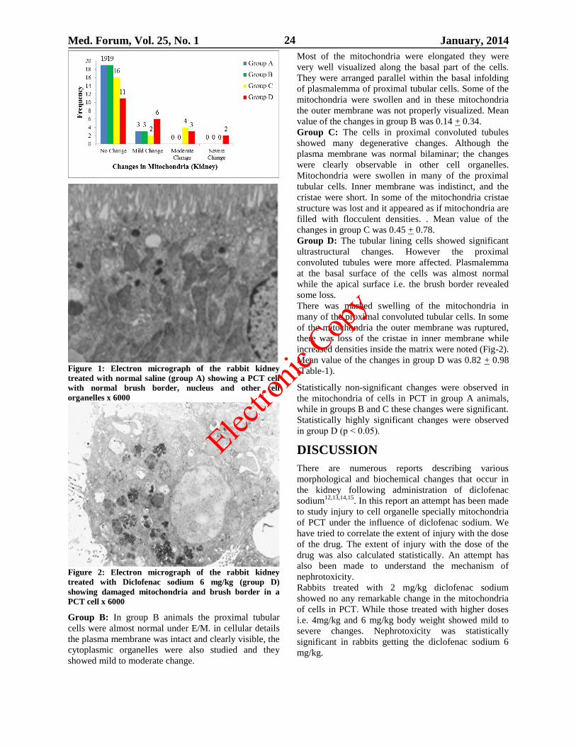
Med. Forum, Vol. 25, No. 1 January, 2014 24
Figure 1: Electron micrograph of the rabbit kidney
treated with normal saline (group A) showing a PCT cell
with normal brush border, nucleus and other cell
organelles x 6000
Figure 2: Electron micrograph of the rabbit kidney
treated with Diclofenac sodium 6 mg/kg (group D)
showing damaged mitochondria and brush border in a
PCT cell x 6000
Group B: In group B animals the proximal tubular
cells were almost normal under E/M. in cellular details
the plasma membrane was intact and clearly visible, the
cytoplasmic organelles were also studied and they
showed mild to moderate change.
Most of the mitochondria were elongated they were
very well visualized along the basal part of the cells.
They were arranged parallel within the basal infolding
of plasmalemma of proximal tubular cells. Some of the
mitochondria were swollen and in these mitochondria
the outer membrane was not properly visualized. Mean
value of the changes in group B was 0.14 + 0.34.
Group C: The cells in proximal convoluted tubules
showed many degenerative changes. Although the
plasma membrane was normal bilaminar; the changes
were clearly observable in other cell organelles.
Mitochondria were swollen in many of the proximal
tubular cells. Inner membrane was indistinct, and the
cristae were short. In some of the mitochondria cristae
structure was lost and it appeared as if mitochondria are
filled with flocculent densities. . Mean value of the
changes in group C was 0.45 + 0.78.
Group D: The tubular lining cells showed significant
ultrastructural changes. However the proximal
convoluted tubules were more affected. Plasmalemma
at the basal surface of the cells was almost normal
while the apical surface i.e. the brush border revealed
some loss.
There was marked swelling of the mitochondria in
many of the proximal convoluted tubular cells. In some
of the mitochondria the outer membrane was ruptured,
there was loss of the cristae in inner membrane while
increased densities inside the matrix were noted (Fig-2).
Mean value of the changes in group D was 0.82 + 0.98
(Table-1).
Statistically non-significant changes were observed in
the mitochondria of cells in PCT in group A animals,
while in groups B and C these changes were significant.
Statistically highly significant changes were observed
in group D (p ˂ 0.05).
DISCUSSION
There are numerous reports describing various
morphological and biochemical changes that occur in
the kidney following administration of diclofenac
sodium12,13,14,15
. In this report an attempt has been made
to study injury to cell organelle specially mitochondria
of PCT under the influence of diclofenac sodium. We
have tried to correlate the extent of injury with the dose
of the drug. The extent of injury with the dose of the
drug was also calculated statistically. An attempt has
also been made to understand the mechanism of
nephrotoxicity.
Rabbits treated with 2 mg/kg diclofenac sodium
showed no any remarkable change in the mitochondria
of cells in PCT. While those treated with higher doses
i.e. 4mg/kg and 6 mg/kg body weight showed mild to
severe changes. Nephrotoxicity was statistically
significant in rabbits getting the diclofenac sodium 6
mg/kg.
Electro
nic Cop
y
Med. Forum, Vol. 25, No. 1 January, 2014 25
The swelling and rupture of mitochondrial membrane is
due to a phenomenon called mitochondrial permeability
transition (MPT). In this phenomenon the permeability
of mitochondrial membrane is increased to solutes of
molecular weight less than 1500 Da. This MPT is
formed at the contact sites between the inner and outer
mitochondrial membrane. It results in depolarization
and there is equilibrium of solutes between cytoplasm
and mitochondrial matrix. This ultimately leads to the
rupture of mitochondrial membranes.
Many researchers have identified the ability of
diclofenac sodium to induce apoptosis and it was found
that mitochondria play an important role in this
mechanism16,17
. At therapeutic dose it causes swelling
of mitochondria but as the dose is increased there is
rupture of mitochondrial membrane. The changes in
this study are comparable with that found by Kretz
Romel and Boelsterli18
. They suggested that changes in
mitochondria are dose dependent.
The E/M observations are essentially in agreement with
those of Hickey EJ19
also who proved that Diclofenac
targets the mitochondria of renal proximal tubules. But
he proposes the mechanism in which there is oxidative
stress and massive DNA fragmentation.
The present results may have both toxicological and
bioenergetic implications. First they show the potential
of diclofenac sodium for toxicity in renal PCT
mitochondria and secondly this drug is potential
inducer of MMPT prevention. In this regard studies are
necessary to evaluate the sensitivity to MMPT
following diclofenac sodium exposure in vivo20
.
CONCLUSION
Diclofenac sodium has damaging effect on the
mitochondria of PCT cells far before the light
microscopic changes. So its use should be restricted
only in very painful conditions. Secondly in case of
prolong treatment follow up with regular renal function
test should be carried out.
REFERENCES
1. Hayat MA. “Principles and techniques of electron
microscopy” USA: Cambridge; 2000.p.124-129
2. Purcell P, Henry D, Melville G. Diclofenac
hepatitis. Gut 1991;32:1381-1385.
3. Aydin G, Gokeimen A, Oncu M, Cieck E, Karahan
N, Gokalp O. Histopathological changes in liver
and renal tissues induced by different doses of
Diclofenac sodium in rats. Turk J Vet Sci 2003;
(27): 1131-1140.
4. Katzung BG. Basic and clinical pharmacology,
10th ed. Connecticut; Appleton and Lange: 2007.p.
580.
5. Lippincott’s illustrated reviews: Pharmacology. 3rd
ed. Philadelphia: Lippincott’s Williams and
Wilkins; 2006.p.503.
6. Wuolijki E, Oikarinen VJ, Ylipaavalniemi P,
Hampf G, Tolynen M. Effective postoperative pain
control by preoperative injection of diclofenac. Eur
J Clin Pharmacol 1987;32:249-252.
7. Alijanpour E, et al. Suppository diclofenac instead
of intravenous meperidine for postoperative
management. Caspaian J Intern Med 2012;3(3):
506-507.
8. Clyman RI, Hardy P, Waleh N, Chen YQ, Mauray
F, Fouran JC, et al. Cyclooxygenase-2 plays a
significant role in regulating the tone of foetal lamb
ductus arteriosus. Am Physiol 1999;276:R913-
R921.
9. Irit K, Roxana C, Bella E, Miriam D. Acute renal
failure, associated with non- steroidal anti-
inflammatory drugs in healthy children. Paediatric
Nephrol 2005;20(9):1295-1298.
10. Yinnon AM, Mores JS, Slotki IN. Nephrotic
syndrome associated with Diclofenac sodium. Br
Med J (Clin Res Ed) 1987;295:556.
11. Kusuhara H, Matsuyuki H, Matsuura M, Imayoshi
T, Okumoto T, Matsui H. Induction of apoptotic
DNA fragmentation by nonsteroidal anti-
inflammatory drugs in cultured rat gastric mucosal
cells. Eur J Pharmacol 1998;360(2-3):273-280.
12. Ng LE, Halliwell B, Wong KP. Nephrotoxic cell
death by diclofenac and meloxicam. Biochemical
and biophysical research communications 2008;
(369):873-877.
13. Ng LE, Vincent AS, Halliwell B, Wong KP.
Action of Diclofenac on kidney mitochondria and
cells. Biochem Biophys Res Commun 2006;
348(2):494-500.
14. Dixit MP, Nguyen C, Carson T, Guedes B, Naznin
MD, Bell MJ, Wang Y. non-steroidal anti-
inflammatory drugs-associated acute interstitial
nephritis with granular tubular basement membrane
deposits. Paed Nephrol 2008; (23):145-148.
15. Meteyer CU, Rideout BA, Gilbert M, Shivaprasad
H, Land Oaks JL. pathology and proposed
pathophysiology of diclofenac poisoning in free-
living and experimentally exposed oriental white-
backed vultures (Gyps bengalensis). J Wild Life
Disease 2005;41(4):705-716.
16. Gómez-Lechón MJ, Ponsoda X, O'Connor E,
Donato T, Castell JV, Jover R. Diclofenac induces
apoptosis in hepatocytes by alteration of
mitochondrial function and generation of ROS.
Biochem Pharmacol 2003;66(11):2155-2167.
17. Masubuchi Y, Nakayama S, Horie T. Role of
mitochondrial permeability transition in
Diclofenac-induced hepatocyte injury in rats.
Hepatol 2002;35:544-551.
18. Kretz-Rommel A, Boelsterli UA. Diclofenac
covalent protein binding is dependent on acyl
Electro
nic Cop
y
Med. Forum, Vol. 25, No. 1 January, 2014 26
glucuronide formation and is inversely related to
acute cell injury in cultured rat hepatocytes.
Toxicol. Appl. Pharmacol 1993;120:155–161.
19. Hickey EJ, Raje RR, Reid VE, Gross SM, Ray SD.
Diclofenac induced in vivo nephrotoxicity may
involve oxidative stress-mediated massive genomic
DNA fragmentation and apoptotic cell death. Free
Radic Biol Med 2001;31(2):139-152.
20. Uyemura SA, Santos AC, Mingatto FE, Jordani
MC, Curti C. Diclofenac sodium and mefenamic
acid: Potent inducers of the membrane
permeability transition in renal cortex
mitochondria. Arch Biochem Biophys 1997;
342(2):231-235.
Address for Corresponding Author:
Prof. Dr. Talat Mirza
Professor of Pathology
Dow International Medical College (DUHS),
Karachi.
E-Mail: [email protected]
Electro
nic Cop
y

Med. Forum, Vol. 25, No. 1 January, 2014 27
Efficacy of Platelet Rich Plasma
Application in Comparison to Conventional
Dressing Therapy in Partial Thickness Burn Wound 1. Ehmer-Al-Ibran 2. Masood Hussain Rao 3. Maria Khan 4. Farrukh Hasan
5. Raaziyah Abdul Khaliq 6. Syeda Zehra 1. Asstt. Prof. of Burns, DUHS, Karachi 2. Principal Research Officer, PMRC Research Centre, DMC, Karachi
3. PG Student, DUHS, Karachi 4. Medical Director, Vice President of South Asian Association of Transfusion
Medicine 5,6. PG Students, DUHS, Karachi
ABSTRACT
Objective: To determine the efficacy of Platelet Rich Plasma application in comparison to conventional dressing
therapy in partial thickness burn wound
Study Design: Comparative analytical study
Place and Duration of Study: This study was carried out at Department of Burns, Civil Hospital Karachi from
March 2011 to January 2013.
Materials and Methods: A comparative analytical study was conducted to determine the efficacy of Platelet Rich
Plasma application in comparison to conventional dressing therapy in partial thickness burn wound at Department of
Burns, Civil Hospital Karachi. All the admitted patients of either sex having age between 20-40 years, victims of
fire and scald burn, having partial thickness burn with 10-30% of TBSA involved. Patients were divided into two
groups on random basis. In group “A”, Platelet Rich Plasma (PRP) application was given with three day gap
between two applications until full recovery of the wound. While in group “B” conventional dressing therapy was
adopted till full recovery of wound.
Results: All 30 patients of group A, selected for PRP application, were recovered maximally within 18 days (6
therapies with a gap of 3 days). Whereas in other 30 cases, of group B selected for conventional dressing therapy, it
took minimum 21 days or more for complete recovery. Hence recovery was found slow in conventional dressing
therapy as compared to PRP and it is statistically significant at P,0.05.
Conclusion: Platelet-rich plasma application in non-healing deep partial and full thickness burn wound accelerate
the wound healing as compared to conventional dressing therapy and is very effective in preparing healthy beds for
grafting and provides 100% graft take.Now it is up to the Burns surgeon to select it for rapid results to save time and
cost with availability of more beds in Burn Centre.
Key Words: Platelet Rich plasma, Burns, Conventional dressing, wound healing.
INTRODUCTION
Burn injuries occur as a result of fire and hot water or
liquid (scald burn). It may lead to extensive damage to
skin surface and also its depth, depending on the
intensity of heat delivered to the skin and time of
contact. As a result of damage to the skin surface, the
protective function gets compromised and results in the
exposure of the sub-epithelial skin layers. It depends
upon the depth of injury which immediately comes in
contact with the external environment specially the
micro-organisms like bacteria, fungi, yeast, virus etc.
that cause infection on the burnt surface, initially
confined to the wound surface but as pathological
events progress deeper down they may invade the
circulation (bacteremia). If not stopped, further progress
leads to septicemia, septic shock and finally death of
the patient. In order to avoid these complications, early
healing of wound and coverage of damaged skin
surface in deeper injuries is necessary to decrease the
chances of above mentioned life threatening
consequences.
Platelet Rich Plasma has attained a significant
importance in facilitating wound healing in many fields
of surgery. According to many health workers
platelets1,2
and natural sources of growth factors 3,4,5
play a fundamental role in homeostasis and healing as
well6.
They also release chemotactic factors7,8
and
induce proliferation of fibroblasts and endothelial cells
for neo-angiogenesis hence they have
a significant
effect on minimizing wound infection which is very
effective in wound healing9
in superficial burn wounds
and also equally important in providing early well
vascularized healthy granulation tissue for resurfacing
deep wounds by grafting.
We have all the studies from abroad and no work has
been done in Pakistan so far. After studying the results
obtained from work done by Marquez-de-Aracena10
,
Kazakook11
and Knighton,
12 etc, we want to apply PRP
on burn wounds to get a comparative analysis in partial
thickness burn wound.
Original Article Dressing on
Burn Wound
Electro
nic Cop
y
Med. Forum, Vol. 25, No. 1 January, 2014 28
MATERIALS AND METHODS
This comparative analytical study was carried out from
March 2011 to January 2013 at Burns Centre, Civil
Hospital, Karachi. We have selected 60 patients
suffering from fire and scald burn resulting in partial
thickness burn. We excluded electrical, chemical and
fire burn of superficial and deep thickness. We have
also excluded smokers, diabetics, hypertensive and
patients with other cardiac problems and also pregnant
women.
We have divided these patients into two groups. Group
A was treated by PRP while Group B was treated by
conventional dressing therapy.
Platelet concentrate was achieved by extracting
platelets from one pint of whole blood. Donor should be
healthy, free of communicable blood-borne disease,
ABO compatible and should have normal values of
complete blood picture on lab test evaluation. Platelet is
extracted by CRYOFUGE (centrifuge machine) made
by JOVAN (France). The extracted platelets are
suspended in 50-100ml plasma in transfusion bag
having 3-5 times the concentration as compared to
whole blood.
Before each application of PRP in Group A, the patient
was shifted to Operation Theater. The wound was
carefully examined and if any slough or dead tissue was
found that was removed by desloughing or
debridement. All procedures are done under strict
aseptic conditions. If slough or dead tissue is not found
then the wound is washed with NaCl. Before
application, the platelet extract received from the blood
bank in the transfusion bag is mixed with CaCl2 which
is present in a separate syringe provided by the blood
bank. As a result of this mixing, the platelets
degranulate and start releasing growth factors. The
activated PRP is then applied over the wound surface
and within 10 minutes the wound is covered with sterile
dressing. It is left for 3 days because the activity of the
platelet borne factors is supposed to be maximum
during this period. So we plan next application after 3
days.
In Group B, thirty patients were selected for giving
treatment with conventional dressing therapy. Dressing
is done in Operation Theater under sterile conditions.
The wound was examined, and washed with normal
saline. The dead and devitalized tissues are removed
and non-adhesive dressing is done by 1% silver
sulphadiazine and then dressing is done. Initially the
dressing change depends upon the soakage, the
condition wound and the half life of the topical anti
bacterial cream.
These procedures were adopted till the completion of
wound healing. During this period, the wound was
observed for the presence of infection, time of infection
eradication from wound, changes in pain perception,
total time of wound healing and time of hospital stay
during treatment.
RESULTS
According to the analysis of the data, we have selected
60 patients suffering from fire and scald burn resulting
in partial thickness burn. These patients were divided
into two groups through systematic random sampling.
Out of these, 29 (48.3%) patients who were suffering
with 10-19%, only 5 (17.0%) required debridement,
where as in 31 patients with 20-30 % of burn,
22(71.0%) required debridement which was done. Over
all 27 (45.0%) patients requiring debridement (Table 1)
All 60 patients selected for this study were divided in
two equal groups. Group A was selected for PRP
application and its effects on wound healing were
observed and Group B was treated by conventional
dressing therapy. All selected patients had partial
thickness burn wounds of fire and scald burns,
categorized on the basis of history and clinical
examination before application of PRP (Group A) and
dressing (Group B).
Table No.1: Patients with % of burn requiring
debridement
S.
No
No. of
Patients
% of
Burn
Debride-
ment
not done
Debride-
ment
done
Total
1. 29 10-
19
24 5 29
2. 31 20-
30
9 22 31
Total: 60 33 27 60
Table No.2: Epithelization time of burn wound and
no of PRP applications
No of
patients
PRP applications
with a 3 day gap
Total no of days of
therapy
16 Three (3) 9
9 Four (4) 12
5 Six (6) 18
Total :
30
Maximum 6
therapy
Maximum 18 days
for full recovery.
Table No.3: Epithelization of burn wound with
conventional dressing method
No. of patients Duration of dressing
16 21-30 days
11 31-40 days
3 >40 days
Total : 30
PRP application was given in Group A (30 patients) at a
gap of three days between two consecutive applications
to achieve maximum effect of PRP which lasts for three
days. 16 out of 30 (53.4%) showed epithelization after
3 applications with a 3 day gap in between every
application i.e. 9-day therapy. 9 out of 30 (30%)
Electro
nic Cop
y

Med. Forum, Vol. 25, No. 1 January, 2014 29
showed epithelization after 4 applications, 3 days apart
i.e. 12 days therapy and 5 out of 30 (16.7%) showed
proper epithelization after 6 applications i.e. maximum
18 days required for complete healing. (Table 2).
In conventional dressing method Group II dressing was
done by 1% silver sulphadiazine and wound was
covered by Gamgee dressing, 16 out of 30 (53.3%)
patients took 21-30 days, 11 out of 30 (36.7%) took 31-
40 days , 3 out of 30(10.0%) patients took more than 40
days to show proper epithelization (Table 3)
The results of these two groups were compared to get
an idea regarding the efficacy of PRP in rapid wound
healing and it was found that Platelet Rich Plasma
application in comparison to conventional dressing
therapy in partial thickness burn wound which is
statistically significant at P<0.05 (Table 4)
Table No.4: Comparison of epithelization of burn
wound treated with PRP and with conventional
dressing method
No. of days No. of patients
fully recovered
with PRP
No. of patients
recovered with
conventional
dressing method
<10 16 (53.3%) 0
11-20 14 (46.7%) 0
21-30 __ 16 (53.3%)
31-40 __ 11 (36.7%)
>40 __ 3 (10.0%)
DISCUSSION
Fire and scald burn are common in burn cases and rapid recovery is the main issue in partial thickness burn wounds. We have designed a study to compare the efficacy of PRP with conventional dressing methods in partial thickness burn wound. Partial thickness burn wound specially in fire and scald burn have an advantage of healing spontaneously nearly within 3 weeks, provided all protocols of sterilization are followed, otherwise if partial thickness burn wound gets infected, the depth of the wound may extend deep down and the partial thickness burn may turn into deep partial thickness wound where the healing takes a longer time and most of the healing occurs by formation of fibrous tissue which ends up in hypertrophic scar, keloid or contracture formation. As far as effect of PRP on burn wound is concerned, few studies are present so far, Marquez-de-Aracena et al (2007)
10 applied PRP on ten
Ocular burn patients and got very good results regarding fast and excellent healing. According to him, he got very good corneal healing after application in eye suffering from chemical burn involving the cornea. He also observed minimal post burn scarring after sub-conjunctival injection of PRP. According to Kazakos K et al (2008)
11 it is very
effective in partial thickness burn wounds. No study was found on PRP application in Burn patients of higher % of body surface area involvement.
Many workers used it in deep wounds as Ganco et al3
showed faster epithelization of chronic lower extremity ulcers where he found appropriate results. Knighton DR et al
12 also showed successful treatment of non-healing
wounds after application of PRP. In our study, the comparison clearly showed that the conventional dressing method takes twice longer time as in Group B the average healing time was 21 days to >40 days as compared to Group A where the average healing time was 9 days to maximum 18 days which definitely affects the duration of hospital stay. The other advantage of PRP application in Group A as opposed to Group B is the decrease in pain, rapid control of infection and hence faster epithelization. A broad majority of workers used PRP on small area of burn wounds so mostly they used autogenous PRP in their studies like Martinez Zapata
13 was in favor of
autogenous PRP, but in our study we had to apply PRP on a larger % of body surface area so it was not possible to use autogenous PRP. Other factors also contributed to make it impossible such as the patients admitted in our hospital belong to very low socio-economic class and their health status is also very low at the time of admission and after admission, G.I. upset due to exposure to micro-organisms, heavy antibiotic therapy, systemic infection, intolerance to food intake, non-availability to food supplements leading to low caloric intake lead to a decreased hemoglobin making it impossible to use autogenous PRP. The quantity of PRP required for single application for burn wound between 10-30% of body surface area needs 2 pints of blood for extraction of platelets. We have to consult blood bank and use donated blood therefore we cannot avoid chances of allergic reactions so this is a drawback of not using autogenous PRP. But its advantages over conventional dressing such as decrease in duration of healing time, decrease in pain, infection and hospital stay are more convincing for its use in partial thickness burn wounds.
CONCLUSION
According to our study results, Platelet-rich plasma
application in non-healing deep partial and full
thickness burn wound accelerate the wound healing as
compared to conventional dressing therapy and is very
effective in preparing healthy beds for grafting and
provides 100% graft take. Now it is up to the Burns
surgeon to select it for rapid results to save time and
cost with availability of more beds in Burn Centre or to
use the conventional dressing therapy according to the
condition of the patient.
REFERENCES
1. Everts PA, Brown C, Hoffman JJ, Schonberger JP,
Box HA, Van Zundert A, et al. Platelet-rich plasma
and platelet gel: a review. J Extra Corpor Technol
2006;38(2):174-87.
Electro
nic Cop
y

Med. Forum, Vol. 25, No. 1 January, 2014 30
2. Henderson JL, Cupp-CL-Ross EV, Shick PC,
Keefe MA, Wester DC, et al. The effects of
autologous platelet gel on wound healing. Ear Nose
Throat J 2003; 82(8): 598-602.
3. Ganio C, Tenewitz FE, Wilson RC, Moyles BG.
The treatment of chronic non healing wounds using
autologous platelet-derived growth factors. J Foot
Ankle Surg 1993; 32(3):263-8.
4. Sammartino G, Tia M, Marenzi G, Di Lauro AE,
D’Agostino E, Claudio PP. Use of autologous
Platelet Rich Plasma (PRP) in periodontal defect
treatment after extraction of impacted mandibular
third molars. J Oral Maxillofac Surg 2005; 63:
766-70.
5. Cervelli V, Gentile P, Grimaldi M. Regenerative
surgery: use of fat grafting combined with platelet-
rich plasma for chronic lower-extremity ulcers.
Aesthetic Plast Surg 2009; ISBN/ISSN 1432-
5241(Electronic): doi:10.1007/s00266-008-9302-z.
6. Eppley BL, Woodell JE, Higgins J. Platelet
quantification and growth factor analysis from
platelet-rich plasma: implications for wound
healing. Plast Reconstr Surg 2004;114(6):
147e-59e.
7. Pierce G, Mustoe TA, Altrock BW, Deuel TF,
Thomason A. Role of platelet-derived growth
factor in wound healing. J Cell Biochem 1991;45:
319-26.
8. Eppley BL, Pietrzak WS, Blanton M. Platelet-rich
plasma: a review of biology and applications in
plastic surgery. Plast Reconstr Surg 2006; 118
(6):147e-59e.
9. Pietrzak WS, Eppley BL. Platelet rich plasma:
biology and new technology. J Craniofac Surg
2005; 16(6):1043-54.
10. Marquez-de-Aracena R Montrero-de-espinosa I,
Munoz M, Pereira G. Subconjunctival application
of plasma platelet concentrate in the treatment of
ocularburns. Preliminary results. Arch Soc Esp
Oftalmol 2007; 82(8):475-81
11. Kazakos K, Lyras DN, Verettas D, Tilkeridis K,
Tryfonidis M. the use of autologous PRP gel as an
aid in the management of acute trauma wounds.
Injury 2008; ISBN/ISSN 1879-0267 (Electronic);
doi:10.1016/j.injury.2008.05.002.
12. Knighton DR, Ciresi KF, Fiegel VD, Austin LL,
Butler ET. Classification and treatment with
autologous platelet derived wound healing factors
(PDWHF). Ann Surg 1986; 204(3):322-30
13. Martinez-Zapata MJ, Marti-Carvajal A, Sola I,
Bolibar I, Angelo Exposito J, Rodriguez L, et al.
Efficacy and safety of the use of autologous plasma
rich in platelets for tissue regeneration: a
systematic review. Transfusion 2009; 49(1):44-56.
Address for Corresponding Author:
Dr.Ehmer-Al-Ibran,
Assistant Professor, Dept. of Burns,
Dow University of Health Sciences,
Civil Hospital, Karachi,
Mobile: 0333-2262820
Email: [email protected]
Electro
nic Cop
y
Med. Forum, Vol. 25, No. 1 January, 2014 31
Prediction of Large Esophageal
Varices in Patients with Decompensated Cirrhosis by
Child-Pugh Score, in Medical Unit-II, Chandka
Medical College Hospital, Larkana 1. Muhammad Aslam Soomro 2. Imdad Ali Ansari 3. Ghulam Yasin Abro
4. Sayed Aftab Ahmed Shah 5. Sayed Shafiq Rahman Shah 1. Senior Registrar of Medicine 2. Asstt. Prof. of Medicine 3. Senior Registrar of Medicine 4. Prof. of Medicine
5. Senior Registrar of Medicine, Chandka Medical College & Hospital Larkana.
ABSTRACT
Objective: To assess prediction of large esophageal varices in patients with decompensated cirrhosis by Child-Pugh
score, in Medical Unit-II, Chandka Medical College Hospital Larkana.
Study Design: Cross sectional study
Setting and Duration of Study: This study was carried out at Medical Unit II, Chandka Medical College &
Hospital, Larkana from November 2011 to November 2012.
Materials and Methods: In this study 88 consecutive cirrhotic patients with ascites (those Patients who fulfilled the
inclusion criteria) were included; blood samples for Serum bilirubin, serum albumin, and INR ratio were sent to
single laboratory. Then, child Pugh score were assigned to each patient on the basis of clinical and to laboratory
parameter. The ultrasound of abdomen was carried out for size of liver and spleen, portal vein diameter, and
quantification of ascits. Eligible patients were subjected for upper gastrointestinal endoscopy for the presence of
esophageal varices and their grading. The data was analyzed using SPSS version 19.
Results: The mean age of enrolled patients was 43.19±7.1 years. Of 88 patients, 69 (78.4%) were male and 19
(21.6%) were female. Child Pugh class relation to number of patients were; 52, 20, and 16 in class A, B, and C
respectively. While Child Pugh class relation to frequency of esophageal varices were; 6, 11 and 14 in class A, B,
and C respectively. Frequency of grading of esophageal varices was; 4, 13, and 14 in grade one, two, and three
respectively. Distribution of large esophageal varices (LEVX) in relation to child Pugh class is one in class A, two in
class B and 11 in class C.
Conclusions: It is concluded from this study that; as the child pugh score advances, the number and size of
esophageal varices increases, and chance of absence of varices decreases.
Key Words: Esophageal varices, Child Pugh score, Grades of varices
INTRODUCTION
Cirrhosis is an end result of various hepatic diseases
resulting to fibrosis with replacement of normal hepatic
architecture in to nodular form leading to opening of
Porto-systemic shunts. Some patients with cirrhosis are
completely asymptomatic and have a reasonably normal
life expectancy. Others have symptoms of end-stage
liver disease and have a limited chance for survival.
Chronic liver disease and cirrhosis result in about
35,000 deaths each year in the United States. Cirrhosis
is the ninth leading cause of death in the United States
and is responsible for 1.2% of all US deaths. Many
patients die from the disease in their fifth or sixth
decade of life. Cirrhosis was the 10th leading cause of
death for men and the 12th for women in the United
States in 2001, killing about 27,000 people each year.1
26% of hepatitis B and 15% of hepatitis C leads to liver
cirrhosis worldwide.2
Many patients of liver cirrhosis
ultimately develop complications; amongst them portal
hypertension is one of the major complication
manifested as esophageal varices.2
The portal vein carries approximately 1500 ml/min of
blood from the small and large bowel, spleen, and the
stomach to the liver. Obstruction of portal venous flow,
whatever the etiology, results in a rise in portal venous
pressure. The response to increased venous pressure is
the development of a collateral circulation diverting the
obstructed blood flow to the systemic veins. These
Porto - systemic collaterals form by the opening and
dilatation of preexisting vascular channels connecting
the portal venous system and the superior and inferior
vena cava.
In Western countries, alcoholic and viral cirrhosis are
the leading causes of portal hypertension and
esophageal varices. Thirty percent of patients with
compensated cirrhosis and 60-70% of patients with
decompensate cirrhosis have gastro esophageal varices
at presentation. The de novo rate of development of
esophageal varices in patients with chronic liver
diseases is approximately 8% per year for the first 2
years and 30% by the sixth year. The risk of bleeding
from esophageal varices is 30% in the first year after
identification. Bleeding from esophageal varices
Original Article Esophageal Varices
Electro
nic Cop
y

Med. Forum, Vol. 25, No. 1 January, 2014 32
accounts for approximately 10% of episodes of upper
GI bleeding.
At least two thirds of cirrhotic patients develop
esophageal varices during their lifetime. Severe upper
gastrointestinal (UGI) bleeding as a complication of
portal hypertension develops in about 30%-40% of
cirrhotic, 3
the reported mortality from first episode of
variceal bleeding in western studies ranges from 17% to
57% as compared to 5-10% mortality reported in our
population. 4
Upper GI endoscopy is gold standard investigation for
diagnosis of esophageal varices5. But because of
various limitations (not available commonly at primary
and secondary care centers, lack of experts and cost of
procedure), and not all patients of cirrhosis are
developing esophageal varices, so it cannot be
performed in every patients. Therefore an alternate,
easy, less costly, non invasive way to identify the high
risk patients for large esophageal varices has to be
sorted out. So this study may help us to detect those
high risk patients of liver cirrhosis.
MATERIALS AND METHODS
This Cross-sectional study was conducted in medial
unit II, Chandka Medical College & Hospital Larkana,
over a period of one year, from November 2011 to
October 2012.
After the approval from Ethical Review Committee of
Chandka medical college hospital Larkana, Patients
were selected from medical unit II indoor department,
those Patients who fulfilled the inclusion criteria
(Cirrhotic patients with ascites, aged >12 years, of
either sex, without past history of upper and lower GI
bleeding) were enrolled in this study. A verbal and
written consent was obtained from all the patients after
having fully explained the purpose and protocol of the
study by researcher.
After taking history and clinical examination, blood
samples for Serum bilirubin, serum albumin, and INR
ratio were sent to laboratory. Then, on the basis of
clinical and lab parameter child Pugh scoring were
assigned. The ultrasound of abdomen was carried out
for size of liver and spleen, portal vein diameter, and
quantification of ascits, by an expert having at least 5
years experience.
Upper gastrointestinal endoscopy of eligible patients
was performed after explaining the procedure and
getting written consent from patient. After
premedication with intravenous injection of 2 mg
Midazolam and Xylocaine 1% spray as a topical
anesthetic, procedure was done with Fiberoptic
Endoscope by expert endoscopist and esophageal
varices were graded on the basis of de-Franchis
classification, i.e. Grade1 (small size); small straight
varices, not occupying the lumen. Grade 2 (medium
size); enlarged tortuous varices occupying less than one
third of the lumen. Grade 3 (large size) ; large coil-
shaped varices occupying more than one third of the
lumen.
Following patients were excluded from our study.
Patients who were taking medications for prophylaxis
of variceal bleed. Patients who had previously
underwent scelerotherapy or band ligation. Patients
with hepatocellular carcinoma, previous portasystemic
anastomosis or portal vein thrombosis. Patients with
medical contraindications to upper gastrointestinal
endoscopy like shock, atlanto-axial sublaxation, any
coagulation disorder or not willing for endoscopy.
Patients of age less than 12 years.
Data Analysis: All the data were entered and analyzed
using SPSS version 19.0. Frequency and percentages
was computed for categorical variables like gender,
child Pugh classification and classification of
esophageal varices. Mean ± S.D. was computed for
quantitative variables age. Child Pugh scoring was
correlated with grading of esophageal varices and their
signification was assessed by chi-square test.
RESULTS
In this study 88 patients were enrolled, male were 69
(78.4%) and female were 19 (21.6%), with male to
female ratio were 3.6:1.Mean age of enrolled
participants was 49 + 15.538.
Out of 88 study population 52 (29.1%) patients were
found in Child Pugh class A, 20 (22.7%) in class B, and
16 (18.2%) in class C. Table: 1
Table No.1: Salient Features of Study Population
Gender Male.
69
(74.41%)
Female.
19
(21.59%)
Male to
Female Ratio
(3.6:1)
Child Pugh Class (Frequency & Percentage)
Class A
Class B
Class C
52 (29.1%)
20 (22.7%)
16 (18.2%)
Esophaeal Varices (Frequency & Percentage)
Present
Absent
31 (35.2%)
57 (64.8%)
Grading Of Esophageal Varices (Frequency &
Percentage)
Grade 1
(small)
Grade 2
(medium)
Grade 3
(large)
4 (4.5%)
13 (14.8%)
14 (15.9%)
Esophageal varices were present in 31(35.2%) out of 88
study population, Grade one (GI) esophageal varices
were found in 4 (12.90%), Grade two (GII) in
13(41.94%) and Grade three (GIII) in 14 (45.16%)
patients.
Correlation of Child Pugh class with esophageal
varices and their grade shows that, in Child Pugh class
Electro
nic Cop
y

Med. Forum, Vol. 25, No. 1 January, 2014 33
A out of 52 patients; oesophageal varices were present
in 6 (11.5%) patients, and out of 6 patients 3(50%) were
in grade one, 2 (33.3%) were in grade two, and
1(16.7%) was in grade three. Table: 2
In Child Pugh class B out of 20 patients; 11(55%) had
esophageal varices, and out of 11 patients 1(9.1%) had
grade one, 8 (72.7%) had grade two, and 2 (18.1%) had
grade three esophageal varices. Table: 2
Table No. 2: Child Pugh * Grades of esophageal varices (EV) Cross tabulation
Grades of Esophageal Varices
Total No Varices Grade 1 Grade 2 Grade 3
Child Pugh Class A Count 46 3 2 1 52
% within Child Pugh 88.5% 5.8% 3.8% 1.9% 100.0%
% within Grades 80.7% 75.0% 15.4% 7.1% 59.1%
% of Total 52.3% 3.4% 2.3% 1.1% 59.1%
Class B Count 9 1 8 2 20
% within Child Pugh 45.0% 5.0% 40.0% 10.0% 100.0%
% within Grades 15.8% 25.0% 61.5% 14.3% 22.7%
% of Total 10.2% 1.1% 9.1% 2.3% 22.7%
Class C Count 2 0 3 11 16
% within Child Pugh 12.5% .0% 18.8% 68.8% 100.0%
% within Grades 3.5% .0% 23.1% 78.6% 18.2%
% of Total 2.3% .0% 3.4% 12.5% 18.2%
Total Count 57 4 13 14 88
% within Child Pugh 64.8% 4.5% 14.8% 15.9% 100.0%
% within Grades 100.0% 100.0% 100.0% 100.0% 100.0%
% of Total 64.8% 4.5% 14.8% 15.9% 100.0%
Chi-square = 61.273, df=6, p= < 0.05
While in Child Pugh class C out of 16; esophageal
varices were found in 14 patients, among them grade
one was not found in any patient, grade two in 3
(21.4%) patients, while grade three in 11(78.6%)
patients. Table: 2
It means that; as the child pugh class advances (A to C)
the number and size of esophageal varices increases (in
Child Pugh class A 50% in grade one, in class B 72% in
grade two and in class C 78% in grade three) it was
statistically significant (p= < 0.05). Table: 2
DISCUSSION
Cirrhosis is the most advanced form of liver disease and
variceal hemorrhage is one of its lethal complications.
Over half of the patients with cirrhosis develop varices.
The risk of bleeding once varices formed is 20% to
35% within 2 years,5 and the reported mortality rate
from first episode of variceal bleeding is 17% to 57%,
and of those who survive the initial episode of bleeding
and who do not receive active treatment, the risk of
recurrent bleeding is approximately 66% and usually
occurs within 6 months of the initial bleeding
episode.6,7
Because cirrhotic patients with large esophageal varices
are at a high risk for bleeding, an overt hemorrhage
from the gastric mucosa occurred in 60% of patients
with severe PHG. It has been documented that empiric
beta-blocker therapy for the primary prophylaxis of
variceal hemorrhage is a cost-effective measure, and it
decreases the incidence of bleeding and an effect on
bleeding related mortality. The use of screening
endoscopy to guide therapy adds significant cost with
only marginal increase in effectiveness. This added cost
to screening endoscopy is presumably due only to the
large number of unplanned endoscopies; where the
endoscopic findings do not significantly alter the
treatment plan, hence identification of non invasive
parameters that can accurately predict esophageal
varices and help to identifying patients at greatest risk
is important to improve the yield and cost- effectiveness
of endoscopic screening.8
In this study esophageal varices has been related to
child-pugh categories, to identify the large esophageal
varices, in order to do endoscopy in selective patients
and or to start the oral beta blockers in those who are
not willing or fit for endoscopy.
In this study esophageal varices observed in 31 (35.2%)
patients. Of 31 cases of varices, 12.90% were of Grade
I, 41.94% in Grade II and 45.16% in Grade III. While
some of local and European studies suggest higher
frequencies of esophageal varices observed in cirrhotic
patients, like 91.7% by Said HEE et al,9 70% by Gill
ML et al,10
70.6% by Prihartini J et al,11
70.7% by Hong
WD et al,12
76.9% by Stojanov DB et al,13
and 80 % by
Kaji BC et al.14
On other hand few studies suggest little bit lower
frequencies, 29% by Almani SH et al,15
50% by
thompoulos KC et al,16
and two studies show 51% by
Carles P et al17
and Madhotra et al, 18
55% by
schwarzenberger E et al,19
58.4% by Sethar GH et al,20
and 61% by Giannini E et al.21
In all above studies
other non invasive parameters were used for the
Electro
nic Cop
y
Med. Forum, Vol. 25, No. 1 January, 2014 34
detection of esophageal varices. In our study large
esophageal varices were present in 45.16 % of cases. In
comparision to other studies large oesophageal varices
were observed were, 30% 10
, 33%,16
34% 14
and 46% by
Sharma SK et al,22
50% Barrera F et al,23
and 65%. 13
Study by Madhotra,18
shows distribution of esophageal
variecs to child pugh category A, 35%; B, 60%; C, 69%
and large varices were 29% in category A, 24 % in
category B and C, while in our study frequency of large
esophageal varices were, 16.66% in class A, 18.18% in
class B, and 92.85% in class C, which correlates with
study by Said HEE,9 they also observed increasing
frequency of esophageal varices with advancement of
child-pugh class (50% in A, 93.5% in B and 100% in
C). In our study frequency and size of esophageal
varices is correlated rather than frequency alone.
CONCLUSION
In this study we conclude that as the Child Pugh score
advances the frequency of esophageal varices increases
especially the large varices. So we recommend that
those patients in which upper gastrointestinal
endoscopy is not feasible of any reason, and having
history of upper gastrointestinal bleeding, with Child
Pugh class “C” then beta blocker drugs shuld be given
for prophylaxis if there are no contraindications.
REFERENCES
1. Anderson RN, Smith BL. Deaths: leading causes
for 2001. National vital statistics reports: from the
Centers for Disease Control and Prevention,
National Center for Health Statistics, National
Vital Statistics System. 2003;52(9):56–8.
2. Groszmann RJ. Hyperdynamic circulation of liver
disease 40 years later: pathophysiology and clinical
consequences. Hepatol 1994;20(5):1359-63.
3. Jensen DM. Endoscopic screening for varices in
cirrhosis: findings, implications, and outcomes.
Gastroenterol 2002;122:1620-30.
4. Farooqi RJ, Farooqi JI, Rehman M, Ahmad H,
Ahmad F, Gul S. Outcome after injection
sclerotherapy for esophageal variceal bleeding in
patients with liver cirrhosis and COPD.
5. Garcia-Tsao G. Spontaneous bacterial
peritonitis. Gastroenterol Clin North Am 1992;
21(1):257-75.
6. Gines P, Arroyo V, Quintero E. Comparison of
paracentesis and diuretics in the treatment of
cirrhotics with tense ascites. Results of a
randomized study. Gastroenterol 1987;93(2):
234-41.
7. Ochs A, Rossle M, Haag K. The transjugular
intrahepatic portosystemic stent-shunt procedure
for refractory ascites. N Engl J Med 1995;
332(18):1192-7.
8. Garrison RN, Cryer HM, Howard DA.
Clarification of risk factors for abdominal
operations in patients with hepatic cirrhosis. Ann
Surg 1984;199(6):648-55.
9. Said HEE, Elsayed EY, Ameen A, Elal HA.
Cytopenia as predictor of oesophageal varices in
patients with liver cirrhosis.http:/www.sciencepub.
net/report 7/7/2010:2 (7) 17-23.
10. Gill ML, Atiq M, Sattar S, Khokhar N. Non-
endoscopic parameters for the identification of
esophageal varices in patients with chronic
hepatitis. JPMA 54:575;2004.
11. Prihartini J, Lesmana LA, Manan C, Gani RA.
Detection of esophageal varices in liver cirrhosis
using non invasive parameters. Acta Med Indones J
Intern Med 2005; 37;(3):126-131.
12. Hong WD, Zhu QH, Huang ZM, Chen XR, Jiang
ZC, Xu SH, Jin K. Predictor of esophageal varices
in patients with HBV- related cirrhosis: a
retrospective study. BMC Gastroenterol 2009;
9(11):1-7.
13. Stojanov DB, Nagorni A, Bjelakovic G, Milanovic
J, Stojanov D. Predictive factors of bleeding from
esophageal varices in patients with liver cirrhosis
and portal hypertension. Med and Biol 2006;13(3):
164-167.
14. Kaji BC, Bhavsar YR, Patel N, Garg A, Kevadiya
H, Mansuris Z. A Study of Clinical Profile of 50
Patients with Portal Hypertension and to Evaluate
Role of Noninvasive Predictor of Esophageal
Varices. Ind J Clin Prac 2012;22(9):454-457.
15. Almani SA, Memon AS, Memon AI, Shah MI,
Rahpoto MQ, Solangi R. Cirrhosis of liver:
etiological factors, complications and prognosis.
JLUMHS 2008;61-66.
16. Thomopulos KC, Karatza L, Mimidis KP,
Iconomou G, Nikolopoulou VN. Non invasive
predictors of the presence of large esophageal
varices in patients with cirrhosis. Digestive Liver
Dis 2003;35(7): 473-8.
17. Carles P, Desmorat H, Vinel JP, Caucanas JP,
Ravaud A, Gerin P, et al. Incidence of large
oesophageal varices in patientswith cirrhosis:
application to prophylaxis of first bleeding. Gut
1990;31:1298-1302.
18. Madhotra, Ravi, Mulchy, Hugh E, Wilner I, Ira,
Reubn, Adrian. Prediction of esophageal varices in
patients of cirrhosis. J Clin Gastroenterol 2002;
34(1): 81-85.
19. Schwarzenberger E, Meyer T, Golla V, Sadala NP,
Min AD. Utilization of platelet count spleen
diameter ratio in predicting the presence of
esophageal varices in patients with cirrhosis. J Clin
Gastroenterol 2010;44 (2):146- 50.
20. Sethar GH, Ahmed R, Rathi SK, Shaikh NA.
Platelet count/splenic size ratio: a parameter to
predict the presence of esophageal varices in
Electro
nic Cop
y

Med. Forum, Vol. 25, No. 1 January, 2014 35
cirrhotics. J Coll Physicians Surg Pak 2006;16(3):
183-6.
21. Giannini E, Botta F, Borro P, Malfatti F, Fumagalli
A, Testa F, et al. Relationship between
thrombopoietin serum levels and liver function in
patients with chronic liver disease related to
hepatitis C virus infection. Am J Gastrornterol
2003;98 (11):2516-20.
22. Sharma SK, Aggarwal R. Prediction of large
esophageal varices in patients with cirrhosis of the
liver using clinical, laboratory and imaging
parameters. J Gastroenterol Hepatol 2007;22 (11):
1909-15.
23. Barrera F, Riguelme A, Soza A, Contreras A,
Barrios O, Viviani P, et al. Platelet count/spleen
diameter ratio for non-invasive prediction of high
risk esophageal varices in cirrhotic patients. Ann
Hepatol 2009;8(4):325-30.
Address for Corresponding Author:
Dr. Muhammad Aslam Soomro
Senior Registrar Department of Medicine
Chandka Medical College & Hospital Larkana
E-mail: [email protected]
Cell #: 0333-7543724
Electro
nic Cop
y
Med. Forum, Vol. 25, No. 1 January, 2014 36
Cognition-Enhancing Effect of Oral
Therapeutic Doses of Methylphenidate in Rats 1. Nausheen Alam 2.Rahila Najam
1. Asstt. Prof. of Pharmacology, Faculty of Pharmacy, Federal Urdu University, Karachi
2. Prof. of Pharmacology, Faculty of Pharmacy, University of Karachi.
ABSTRACT
Objective: To determine the effects of oral therapeutic doses of methylphenidate, on memory. It was thought that
long term use of methylphenidate possibly may lead to tolerance in the ability of the drug elicit enhancement of
learning and memory. A dose-dependent effect may therefore help to extend the therapeutic use of the drug for
better clinical response.
Study Design: Experimental / Analytic study
Place and Duration of Study: This study was conducted in the Department of Pharmacology, Faculty of pharmacy,
University of Karachi, Karachi for a period of period of 4 weeks.
Materials and Methods: Twenty-four male Albino Wister rats (weighing 180-220g) were randomly assigned to
four groups, one control and 3 test groups. The experimental protocol was designed to administer methylphenidate
orally two times daily for 4 weeks. Four groups were: (i) Saline (1.0 ml/kg/ day), (ii) Methylphenidate (2mg/kg/day)
(iii) Methylphenidate (5mg/kg/day) (iv) Methylphenidate (8mg/kg/day) treated groups. Behavioral activity of rats
i.e. performance of rats in passive avoidance test was monitored weekly. The experiment was performed in a
balanced design to avoid the order effect.
Results: In the present study methylphenidate treated rats exhibited an increased in Passive Avoidance learning as
there was increased in the latency time to reach the punished compartment as compared to control rats. This memory
improvement effect of methylphenidate on PA was dose dependent in 1st week as the rats treated with 8 mg/kg/day
took greater time to reach the dark box than 5mg/kg/day and 2mg/kg/day, but in 2nd
, 3rd
and 4th
week it was seen that
rats treated with fore mention doses took same time but greater than 1st week to enter the punishable compartment
Conclusion: It can be concluded that high dose produce greater cognitive effect in short term treatment than low
and moderate doses, however in long-term treatment all the doses produce similar improvement in memory without
tolerance in cognition
Key Words: methylphenidate, oral dose, cognition, passive avoidance test.
INTRODUCTION
Cognition enhancement has received much attention in
recent scientific literatures due to our aging society and
the increasing prevalence of Alzheimer’s disease.
However, the healthy young population also engages in
drug use to enhance memory. Methylphenidate a
medication effective to enhance attention1,2,3
, and
cognition4 in ADHD patients, as well as in healthy
subjects5,6,7,8
.This has raised concern regarding the
ethical and safety aspects of potential cognition-
enhancing drugs9,10,11
. These issues aside, it is important
to know if the drug does actually have cognition-
enhancing effect after long-term administration.
Methylphenidate blocks the dopamine transporter and
the noradrenaline transporter12,13,14,15
, thus increasing
the extracellular concentrations of these
catecholamines. The attention-improving characteristic
of methylphenidate has been attributed to the
amplification of dopamine release in the central
nervous system16
. Dopamine (DA) modulates cognitive
performance in part via its regulation of the prefrontal
cortex through dopamine D1 and D2 receptors17
.
Methylphenidate (MPH) increase DA signaling in the
brain and are used in the treatment of attention deficit
hyperactivity disorder (ADHD) and other
neuropsychiatric disorders to enhance attention and
cognition18,19,20,21
.
Purpose of our study was to monitor the effects of oral
therapeutic doses of methylphenidate, on memory
function in rats. It was thought that long term use of
methylphenidate possibly may lead to tolerance in the
ability of the drug elicit enhancement of learning and
memory. A dose-dependent effect may therefore help to
extend the therapeutic use of the drug for better clinical
response.
MATERIALS AND METHODS
2.1 Test systems used (Animals):
Locally bred Albino Wister rats (weighing 180-200g)
were housed individually under 12 h light and dark
cycles (light on at 06:00h) and controlled room
temperature c) with free access to tap water and
cubes of standard rodent diet at least 7 days before the
start of experiment so that they could become familiar
to the environment. They were accustomed to various
handling procedures to nullify stress effects. All
experiments were performed according to the protocols
approved by the local animal care committee.
Original Article Pharmacology
Electro
nic Cop
y

Med. Forum, Vol. 25, No. 1 January, 2014 37
2.2 Behavioral assessment
Passive Avoidance Test: Passive avoidance paradigm
consists of two compartments as an illuminated ‘safe’
and a dark 'punishable' one. Both compartments were
connected with a door that enable free crossing from
one compartment to another. Both compartments had a
grid floor. The diameter of rods was 5 mm with 0.5 cm
distance between the rods.
In the training session, rat was placed in an illuminated
box. Once the rats prompted by their instinct stepped its
four paws into the dark compartment, rats received 1.5
mA foot shock through the grid floor to its paws for 5
seconds. After receiving the foot shock, it immediately
came back to illuminated safe compartment. During the
test period (24 hour later), rats were placed in the bright
compartment again for a maximum of 5minutes. The
step- through latency that indicates the time elapsed
before the rat entered the dark compartment was
recorded in the test session as described earlier [22].
Passive avoidance test of all drug treated and control
animals were performed weekly in a balance design to
avoid order effect.
2.3. Drugs: Methylphenidate HCl was obtained from
local medical store and prepared in 0.9% NaCl (saline).
Drug was administered in a volume of 1 ml/kg of body
weight by per oral route twice a day to the treated
animals and control animals were treated with saline
(0.9%) at the dose of 1 ml/kg per oral twice a day.
2.4. Experimental protocol: Twenty-four male Albino
Wister rats (weighing 180-220g) were randomly
assigned to four groups, one control and 3 test groups,
each containing six animals. The experimental protocol
was designed to administer methylphenidate orally two
times daily for 4 weeks.
Four groups were: (i) Saline (1.0 ml/kg/ day), (ii)
Methylphenidate (2mg/kg/day) (iii) Methylphenidate
(5mg/kg/day) (iv) Methylphenidate (8mg/kg/day)
treated groups.
Behavioral activity of rats i.e. performance of rats in
passive avoidance test was monitored weekly. The
experiment was performed in a balanced design to
avoid the order effect.
2.5. Statistical analysis: Results are represented as
mean ±S.D. Statistical analysis of passive avoidance
test was performed by two-way analysis of variance
(ANOVA) repeated measure design. Post hoc
comparison of groups was performed by Newman-Keul
test. Values of p<0.05 and p<0.01 were considered as
significant.
RESULTS
Dose related effect of methylphenidate on
performance of rats in passive avoidance test.
Fig. shows the effects of methylphenidate doses on the
performance of rats in PA test. Two-way ANOVA
repeated measure revealed a significant dose (F=200.4,
df=3,20, P<0.01), week (F=32.9, df=3,20, P<0.01)
effect and also a significant interaction between the two
factors (F=10.97, df=9,60, P<0.01).
Post hoc comparison by Newman-keuls test showed
that retention of Passive Avoidance was improved in
test rats than in controls as low dose (2 mg/kg/day),
moderate dose (5 mg/kg/day) and high dose
(8mg/kg/day) of methylphenidate significantly (P<0.01)
increased latency time in all 4 weeks compared to
similar week saline treated rats and in 2nd
,3rd
and 4th
week from 1st week values.
Figure No.1: Passive Avoidance
DISCUSSION
Methylphenidate has been shown to potentiate the
cognitive effect and is the main medication prescribed
for attention deficit hyperactivity disorder23
to improve
memory24
attention and concentration25,26,27
, yet there is
increasing evidence that they do not promote learning27
,
which can be due to tolerance to the cognitive effect of
the drug.
In the current study we chose passive avoidance test to
measure effects in memory function following
methylphenidate administration at three different doses.
Study showed methylphenidate treated rats exhibited an
increased in Passive Avoidance learning as there was
increased in the latency time to reach the punished
compartment as compared to control rats. This memory
improvement effect of methylphenidate on PA was
dose dependent in 1st week as the rats treated with 8
mg/kg/day took greater time to reach the dark box than
5mg/kg/day dose and the rats treated with 5mg/kg/day
day took greater time to reach the dark box than
2mg/kg/day dose, but in 2nd
, 3rd
and 4th
week it was
seen that rats treated with fore mention doses took same
time to enter the dark punishable compartment which
showed that high dose produce greater cognitive effect
in short term treatment however in long-term treatment
all the doses produce similar cognitive effect.
Central dopaminergic systems play a critical role in the
regulation cognitive behavior. Memory improvement
effect of methylphenidate in the present study is due to
increased DA levels as there is an overwhelming
evidence for a critical role of dopamine in
Electro
nic Cop
y
Med. Forum, Vol. 25, No. 1 January, 2014 38
cognition28,29
. Cognitive symptoms have been
associated with dopamine disregulation in numerous
diseases including schizophrenia30
, depression31
, drug
addiction32
and Parkinson disease33
. Support for a role
of dopamine in cognition also comes from studies of
dopaminergic drug in normal subject34
Methylphenidate blocks the dopamine
transporter12,13,14,15
thus increasing the extracellular
concentrations of these dopamine. The memory
improvement effect of methylphenidate has been
attributed to the amplification of dopamine release in
the central nervous system16
. Dopamine (DA)
modulates cognitive performance in part via its
regulation of the prefrontal cortex through dopamine
D1 and D2 receptors17
. Methylphenidate (MPH)
increases DA signaling in the brain and is used in the
treatment of attention deficit hyperactivity disorder
(ADHD) and other neuropsychiatric disorders to
enhance attention and cognition 18,19,20,21
CONCLUSION
In summary this study provides the documentation of
significant relationship between oral therapeutic doses
of methylphenidate and cognation .Our results suggest
that initially cognitive enhancing effect is dose
dependent, but later on cognitive improvement response
in low, moderate and high doses of methylphenidate
became similar after long term use.
REFERENCES
1. Solanto MV, Wender EH, Bartell SS. Effects of
methylphenidate and behavioral contingencies on
sustained attention in attention-deficit hyperactivity
disorder: a test of the reward dysfunction
hypothesis. J Child Adolesc Psychopharmacol
1997;7:123–136.
2. Hawk LW, Jr, Yartz AR, Pelham WE, Lock TM.
The effects of methylphenidate on prepulse
inhibition during attended and ignored prestimuli
among boys with attention-deficit hyperactivity
disorder. Psychopharmacol 2003;165:118–127.
3. Overtoom CCE, Verbaten MN, Kemner C,
Kenemans JL, Van Engeland H, Buitelaar JK, et al.
Effects of methylphenidate, desipramine, and l-
dopa on attention and inhibition in children with
attention deficit hyperactivity disorder. Behav
Brain Res 2003;145:7–15.
4. Mehta MA, Goodyer IM, Sahakian BJ.
Methylphenidate improves working memory and
set-shifting in AD/HD: relationships to baseline
memory capacity. J Child Psychol Psychiat
2004;45:293–305.
5. Elliott R, Sahakian BJ, Matthews K, Bannerjea A,
Rimmer J, Robbins TW. Effects of
methylphenidate on spatial working memory and
planning in healthy young adults.
Psychopharmacol 1997;131:196–206.
6. Mehta MA, Owen AM, Sahakian BJ, Mavaddat N,
Pickard JD, Robbins TW. Methylphenidate
enhances working memory by modulating discrete
frontal and parietal lobe regions in the human
brain. J Neurosci 2000;20:RC65.
7. Pietras CJ, Cherek DR, Lane SD, Tcheremissine
OV, Steinberg JL. Effects of methylphenidate on
impulsive choice in adult humans.
Psychopharmacol 2003;170:390–398.
8. Volkow ND, Wang G-J, Fowler JS, Telang F,
Maynard L, Logan J, et al. Evidence that
methylphenidate enhances the saliency of a
mathematical task by increasing dopamine in the
human brain. Am J Psychiat 2004;161:1173–1180.
9. Greely H, Sahakian B, Harris J. Towards
responsible use of cognitive-enhancing drugs by
the healthy. Nature 2008;11:702–705.
10. Larriviere D, Williams MA, Rizzo M, et al.
Responding to requests from adult patients for
neuroenhancements: guidance of the Ethics, Law
and Humanities Committee. Neurol 2009;73(17):
1406–1412.
11. Sahakian B, Morein-Zamir S. Professor’s little
helper. Nature 2007;450(7173):1157–1159.
12. Ferris RM, Tang FLM. Comparison of the effects
of the isomers of amphetamine, methylphenidate
and deoxypipradrol on the uptake of l-
[3H]norepinephrine and [
3H]dopamine by synaptic
vesicles from rat whole brain, striatum and
hypothalamus. J Pharmacol Exp Ther 1979;210:
422–428.
13. Ritz MC, Lamb RJ, Goldberg SR, Kuhar MJ.
Cocaine receptors on dopamine transporters are
related to self-administration of cocaine. Science
1987;237:1219–1223.
14. Kollins SH, MacDonald EK, Rush CR. Assessing
the abuse potential of methylphenidate in
nonhuman and human subjects: a review.
Pharmacol Biochem Behav 2001;68:611–627.
15. Barrett SP, Darredeau C, Bordy LE, Pihl RO.
Characteristics of methylphenidate misuse in a
university student sample. Can J Psychiat 2005;
50:457–461.
16. Volkow ND, Wang G-J, Fowler JS, Logan J, Jayne
M, Franceschi D, et al. Nonhedonic food
motivation in humans involves dopamine in the
dorsal striatum and methylphenidate amplifies this
effect. Synapse 2002;44:175–180.
17. Goldman-Rakic P. The cortical dopamine system:
role in memory and cognition. Adv
Pharmacol 1998;42:707–711.
18. Ackerman P, Dykman R, Holcomb P, McCray D.
Methylphenidate effects on cognitive style and
reaction time in four groups of children. Psychiat
Res1982;7:199–213.
Electro
nic Cop
y

Med. Forum, Vol. 25, No. 1 January, 2014 39
19. Camp-Bruno J, Herting R. Cognitive effects of
milacemide and methylphenidate in healthy young
adults. Psychopharmacol (Berl)1994;115:46–52.
20. Clatworthy P, Lewis S, Brichard L, Hong Y,
Izquierdo D, Clark L, et al. Dopamine release in
dissociable striatal subregions predicts the different
effects of oral methylphenidate on reversal learning
and spatial working memory. J Neurosci 2009;
29:4690–4696.
21. Izquierdo I, Bevilaqua L, Rossato J, Lima R,
Medina J, Cammarota M. Age-dependent and age-
independent human memory persistence is
enhanced by delayed posttraining methylphenidate
administration. Proc Natl Acad Sci USA 2008;
105:19504–19507.
22. Khaliq S, Haider S, Ahmed SP, Perveen T, Haleem
DJ. Relationship of brain tryptophan and serotonin
in improving cognitive performance in rats. Pak J
Pharm Sci 2006;19(1):11-5.
23. Spencer T, Biederman J, Wilens T, Doyle R,
Surman C, Prince J, et al. A large, double-blind,
randomized clinical trial ofmethylphenidate in the
treatment of adults with attention-deficit/
hyperactivity disorder. Biological Psychiatr 2005;
57(5):456-463.
24. Gonzalez-Garrido AA, Barrios FA, de la Serna-
Tuya JM, Cocula-León H, Gómez-Velázquez FR:
(Methylphenidate and short-term memory in young
females with attention deficit hyperactivity
disorder. A study using functional magnetic
resonance imaging). Rev Neurol 2009;48(10):
509-14.
25. Pietrzak RH, Mollica CM, Maruff P and Snyder PJ.
Cognitive effects of immediate-release
methylphenidate in children with attention-deficit/
hyperactivity disorder Neuroscience &
Biobehavioral. Reviews 2006;30(8):1225-1245.
26. Bedard AC, Jain U, Hogg-Johnson S and Tannock
R. Effects of methylphenidate on working memory
components: influence of measurement. J Child
Psychol and Psychiatr 2007;48:872–880.
27. Advokat CD, Guidry D, Martino L. Licit and Illicit
Use of Medications for Attention-Deficit
Hyperactivity Disorder in Undergraduate College
Students. J Am Coll Health 2008;56(6):601-606.
28. Wilkerson A, Levin ED. Ventral hippocampal
dopamine D1 and D2 systems and spatial working
memory in rats. Neuroscience 1999;89(3):743-9.
29. Liao RM, Lai WS, Lin JY. The role
ofcatecholamines in retention performance of a
partiallybaited radial eight arm maze for rats. Chin
J Physiol 2002;45(4): 177-85.
30. Knable MB, Weinberger DR. Dopamine, the
prefrontal cortex and schizophrenia. J
Psychopharmacol 1997;11:123-131.
31. Jimerson DC. Role of dopamine mechanisms in
affective disorders. Psychopharmacol 1987;
505-511.
32. Wise RA. Neurobiology of addiction. Curr Opin
Neurobiol 1996;6:243-251.
33. Gotham AM, Brown RG, Marsden CP. Frontal
cognitive function in patients with Parkinsons
diseaseon and off levodopa. Brain 1988;111:
299-321.
34. Luciana M, Collins PF, Depue RA. Opposing role
of dopamine and serotonin in the modulation of
human spatial working memory functions. Cereb
Cortex 1998;8:218-226.
Address for Corresponding Author:
Dr. Nausheen Alam,
Asstt. Prof. of Pharmacology,
Faculty of Pharmacy,
Federal Urdu University, Karachi
Elec
tronic
Copy

Med. Forum, Vol. 25, No. 1 January, 2014 40
EPI Status under One Year
Children and their Mothers attending Paediatric
Department Sheikh Zayed Medical College &
Hospital, Rahim Yar Khan 1. Jamal Anwer 2. M.I.Babar 3. Kifayat Niazi 4. Zahid Mahmood
1. Asstt. Prof. of Paediatrics 2. Asstt. Prof. of Paediatrics 3 & 4. PG Students of Paediatrics,
Sheikh Zayed Medical College & Hospital, Rahim Yar khan
ABSTRACT
Objective: To determine the EPI status in children <1 year of age and the status of Tetanus immunization in their
mothers attending paediatric department. This will help us to determine the facts and figures in our population and to
construct a plan to improve the vaccination status.
Study Design: Cross-sectional analytical study
Place and Duration of Study: This study was conducted at the Department of Paediatrics, Sheikh Zayed Medical
College & Hospital, Rahim Yar khan from April 2011 to July 2011.
Materials and Methods: In the study 492 consecutive children whether admitted in the ward or visited in Paediatric
OPD and their mothers were inquired about EPI status in the child and the status of Tetanus immunization in their
mothers, strictly following the inclusion and exclusion criteria.
Results: Out of 492 patients, we have found three groups. Group 1 includes those children who were unvaccinated.
Group 2 includes those children who were incompletely vaccinated. Group 3 include those who were completely
vaccinated according to EPI schedule. Similarly three groups were found in mothers. Group 1 includes those who
were unvaccinated. Group 2 includes incompletely vaccinated ladies. Group 3 include those who were completely
vaccinated according to EPI schedule.
Conclusion: In our study, we found that EPI status is very poor in our low socioeconomic and illiterate population.
Those people living in peripheries and remote areas are particularly not properly following the EPI programme.
Key Words: EPI (Expanded Program on Immunization), Immunization, Vaccines, Tetanus, Tetanus Toxoid(TT)
INTRODUCTION
The Expanded Program on Immunization is a World
Health Organization program with the goal to
make vaccines available to all children throughout the
world.
For evaluation community-based surveys are applied
using a modified cluster sampling survey method
developed by the World Health Organization. Vaccine
coverage is evaluated using a two-stage sampling
approach in which 30 clusters and seven children within
each cluster are selected. Health care workers with no
or limited background in statistics and sampling are
able to carry out data collection with minimal training 1.
Such a survey implementation provides a way to get
information from areas where there is no reliable data
source. It is also used to validate reported vaccine
coverage (for example, from administrative reports) and
is expected to estimate vaccine coverage within 10
percent. Surveys or questionnaires, though frequently
considered inaccurate due to self-reporting, can provide
more detailed information than administrative reports
alone2. A survey in Thailand has found effective
expended programme of immunization3.
Expanded Programme on Immunization (EPI) in
Pakistan: The Expanded Programme on Immunization
(EPI) was launched in Pakistan in 1976 by WHO and
UNICEF to protect children by immunizing them
against childhood tuberculosis, poliomyelitis,
diphtheria, pertussis, tetanus and measles.
The programme also vaccinates pregnant women with
tetanus toxoid vaccine to protect the new born from
neonatal tetanus. With support from the Government of
Pakistan and development partners, the programme has
added hepatitis B (Hep B) and Hemophiles
influenza type B (Hib) vaccinations to its childhood
immunization schedule. In 2002, the programme
introduced hepatitis B vaccine with support from the
Global Alliance for Vaccine and Immunization (now
called GAVI). In 2006, a tetravalent combination
vaccine was introduced replacing separate diphtheria,
tetanus and pertussis and hepatitis B vaccines.
This was later switched in 2008 to the pentavalent
(DPT, Hep B, Hib) vaccine with the addition of the new
Hib vaccine. Now a child needs only five visits during
the first year and one visit during the second year of
his/her life to complete the vaccination with four
antigens against eight dreadful diseases.
Programme Development: From mid 2012
pneumococcal conjugate vaccine was added in
immunization programme. This new vaccine will
protect children from pneumonia and meningitis due to
Original Article EPI Status
Electro
nic Cop
y

Med. Forum, Vol. 25, No. 1 January, 2014 41
pneumococcal infection. The programme also plans to
introduce Rota virus vaccine, which will prevent
diarrhea due to rotavirus. The new vaccines may
together avert 17% of childhood mortality in Pakistan
and thus help in achieving Millennium Development
Goal 4 on reducing child mortality.
Safer Demmunization: The programme has adopted
new technology to make immunization safer and more
clients compliant. Since 2002, to prevent the risk of
blood borne diseases, the programme has been using
auto-disable syringes for all immunization injections
and safety boxes for proper disposal of sharps waste4.
Programme objectives: The aim is to reduce mortality
and morbidity resulting from the eight EPI target
diseases by immunizing children aged from 0 to 11
months and pregnant women.
The specific objectives of the programme are
interruption of polio virus by 2012, elimination of neo-
natal tetanus by 2015, elimination of measles by 2015,
reduction of diphtheria, pertussis and childhood
tuberculosis to a minimum level so that they do become
a public health problem, control of other diseases by
introducing new vaccines in EPI as and when they
become available, using EPI as a spearhead for
promoting other primary health care activities,
integrating EPI into primary health care.
Current situation: Despite significant efforts by the
government and its partners, Pakistan’s immunization
indicators have yet to reach the expected benchmarks.
The key goals of polio eradication, and measles and
neo-natal tetanus elimination, have not been achieved.
People coming from remote areas have poor knowledge
and facilities of vaccination 5,6
. A study published by
Furqan Kurshid found poor vaccination coverage of
mothers during pregnancy with tetanus toxoid and
infant after birth 7.
Routine vaccination coverage is still suboptimal for
achieving the desired goals. Poor routine coverage is
also evidenced by the occurrence of numerous
outbreaks of measles, pertussis and diphtheria in
different parts of the country. A study conducted by
Shaikh S. “Immunization status and reasons for low
vaccination in children, attending O.P.D. at Liaquat
University Hospital” found poor knowledge of
vaccination in community 8.
MATERIALS AND METHODS
We have included the children who were below one
year age. Data collected by taking interview from
mothers, whether the child was admitted in Pediatric
unit due to some illness or visited paediatric OPD. The
Performa was filled strictly following the rules. Their
mothers were asked about the EPI status of the child
i.e.; BCG, polio, pentavalent (DPT, HiB, Hep B) and
measles 1. Child's mother was interviewed by using a
structured pre-tested questionnaire, regarding the EPI
coverage of her child, her own T.T. coverage and other
demographic and potential risk factors for low
vaccination coverage. EPI cards were checked where
ever available and if not, the subjects were inquired
verbally and BCG scars were checked. We gathered
information on characteristics such as basic
demographics, socio-economic status, reproductive
history, health services utilization, immunization
coverage of mother and child and reasons for non-
compliance with the EPI schedule.
The children who were below one year of age admitted
in Pediatric unit due to some illness or visited paediatric
OPD and their mothers were included in the study.
The children above one year of age were excluded from
the study.
EPI Schedule For Children
Age Injection or Drops
Immediate
after birth BCG , Polio (birth)
6 weeks OPV-1, Pentavalent – Pneumococcal – 1
10 weeks OPV-2, Pentavalent – Pneumococcal – 2
14 weeks OPV-3, Pentavalent – Pneumococcal – 3
9 months Measles – 1
15 months Measles – 2
Tetanus Vaccination Schedule for Pregnant Women
Sr.
No.
Schedule Vaccination
1 During Pregnancy TT-1
2 After one month of 1st dose of
vaccination
TT-2
3 After six month of 2nd
dose of
vaccination
TT-3
4 After one year of 3rd
dose of
vaccination
TT-4
5 After one year of 4th
dose of
vaccination
TT-5
RESULTS
492 children of age less than one year, who were
admitted in pediatric unit due to some ailment or visited
Paediatric OPD and their mothers, were interviewed.
The mean age of the infants was 5.4 +_3.5 months. We
have found three groups. Group 1 includes those
children who were unvaccinated. Group 2 includes
those children who were incompletely vaccinated.
Group 3 include those who were completely vaccinated
according to EPI schedule.
Group 1 Unvaccinated : 92(18.69%)
Group 2 Incompletely vaccinated : 177(35.97%)
Group 3 Completely vaccinated : 223(45.32%)
Out of 492, twenty one percent had received no
vaccination. Sixty two percent of mothers had received
one to four doses of T.T. Seventeen percent had
Electro
nic Cop
y
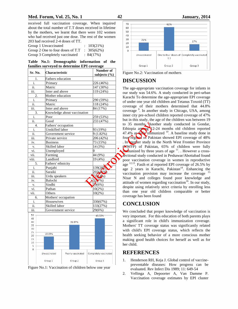
Med. Forum, Vol. 25, No. 1 January, 2014 42
received full vaccination coverage. When inquired
about the total number of T.T doses received in lifetime
by the mothers, we learnt that there were 102 women
who had received just one dose. The rest of the women
203 had received 2-4 doses of TT.
Group 1 Unvaccinated : 103(21%)
Group 2 One to four doses of T.T : 305(62%)
Group 3 Completely vaccinated : 84(17%)
Table No.1: Demographic information of the
families surveyed to determine EPI coverage
Sr. No. Characteristic Number of
subjects (%)
1. Fathers education
i. Primary 226 (46%)
ii. Matric 147 (30%)
iii. Inter and above 119 (24%)
2. Mother education
i. Primary 290 (59%)
ii. Matric 118 (24%)
iii. Inter and above 84 (17%)
3. Knowledge about vaccination
i. Poor 259 (53%)
ii. Good 233 (47%)
4. Fathers' occupation
i. Unskilled labor 93 (19%)
ii. Government service 9 (1.82%)
iii. Private service 206 (42%)
iv. Business 73 (15%)
v. Skilled labor 14 (3%)
vi. Unemployed 0
vii. Farming 44 (9%)
viii. Landlord 19 (4%)
5. Fathers' ethnicity
i. Punjabi 139(28%)
ii. Saraiki 202(41%)
iii. Urdu speakers 71(14%)
iv. Balochi 32(7%)
v. Sindhi 29(6%)
vi. Pathan 10(2%)
vii. Others 10(2%)
6. Mothers' occupation
i. Housewives 330(67%)
ii. Skilled labor 133(27%)
iii. Government service 29(6%)
Figure No.1: Vaccination of children below one year
Figure No.2: Vaccination of mothers
DISCUSSION
The age-appropriate vaccination coverage for infants in
our study was 54.6%. A study conducted in peri-urban
Karachi To determine the age-appropriate EPI coverage
of under one year old children and Tetanus Toxoid (TT)
coverage of their mothers determined that 44.8%
coverage 9. In another study in Chicago, USA, among
inner city pre-school children reported coverage of 47%
but in this study, the age of the children was between 19
to 35 months. Another study conducted in Gondar,
Ethiopia among 12-24 months old children reported
47.4% as fully immunized 10
. A baseline study done in
four regions of Pakistan showed EPI coverage of 48%
. In another study in the North West Frontier Province
(NWFP) of Pakistan, 65% of children were fully
immunized by three years of age 11
. . However a cross-
sectional study conducted in Peshawar/Abottabad found
poor vaccination coverage in women in reproductive
age 12,13
. Fasih et al reported EPI coverage of 26.5% by
age 2 years in Karachi, Pakistan14
. Enhancing the
vaccination provision may increase the coverage 15
Nisar N and colleges found poor knowledge and
attitude of women regarding vaccination16
. In our study,
despite using relatively strict criteria by enrolling less
than one year old children comparable or better
coverage has been found
CONCLUSION
We concluded that proper knowledge of vaccination is
very important. For this education of both parents plays
a significant role in child's immunization coverage.
Mothers' TT coverage status was significantly related
with child's EPI coverage status, which reflects the
health seeking behavior of a more conscious mother
making good health choices for herself as well as for
her child.
REFERENCES
1. Henderson RH, Keja J. Global control of vaccine-
preventable diseases: How progress can be
evaluated. Rev Infect Dis 1989; 11: 649-54
2. Vellinga A, Depoorter A, Van Damme P.
Vaccination coverage estimates by EPI cluster
Electro
nic Cop
y

Med. Forum, Vol. 25, No. 1 January, 2014 43
sampling survey of children (18-24months) in
Flanders, Belgium. Acta Paediatr 2002;91:
599-603.
3. Bhunbhu T. Expanded Programme on
Immunization in Thailand. Rev Infect Dis 1989;
11:514-7.
4. Tarin E, Khalil M, Mustafa T, Alvi ZM, Sy ITS,
Thomson SJ, et al. Impact of community based
intervention on immunization coverage against
vaccine preventable diseases in Pakistan.
Pakistan J Health 1999;36:53-6.
5. Anjum Q, Omair A, Inam SNB, Ahmad Y,
Usman Y, Shaikh S. Improving Vaccination
Status of children under five through Health
Education. J Pak Mod Assoc 2004;54:610-13.
6. Rahman M, Islam MA, Mahalanabis D. Mothers'
knowledge about vaccine preventable diseases
and immunization coverage in a population with
a high rate of illiteracy. J Trop Pediatr 1995; 41:
376-8.
7. Hashmi FK, Islam M, et al. Vaccination coverage
of mothers during pregnancy with tetanus toxoid
and infant after birth. Pak J Pharm 2011;24(2):
35-39.
8. Shaikh S. Immunization status and reasons for
low vaccination in children, attending O.P.D. at
Liaquat University Hospital. Pak Ped J 2003; 27:
81-6.
9. Siddiqi N, Khan A, Nisar N. Assessment of EPI
(Expanded program of immunization) vaccine
coverage in a peri-urban area. JPMA 2007; 57:
391.
10. Gedlu E, Tesemma T. Immunization coverage
and identification of problems associated with
vaccination delivery in Gondar, northwest
Ethiopia. East Afr Mod J 1997; 74: 239-41.
11. Ahmad N, Akhtar T, Roghani MT, Byas HM,
Abroad M. Immunization coverage in three
districts of North West Frontier Province
(NWFP). J Pak Mod Assoc 1999;49: 301-5.
12. Afridi NY, Hatcher J, Mahmud S, Nanan D.
Coverage and factors associated with tetanus
toxoid vaccination status among females of
reproductive age in Peshawar. J Coll Physicians
Surg Pak 2005;15: 391-5.
13. Naeem, Khan ZI, et al. Coverage and factors
associated with Tetanus toxoid Vaccination
among married women of reproductive age. A
cross sectional study in Peshawar J Ayub Med
Col l Abbotabad 2010;22(3).
14. Fasih Z, Hussain E, Ali Z. Risk Factors for
complete un-immunization & under
immunization of children under 2 years of age in
third world countries. J Pak Pediatr Assoc 2000;
24:145-52.
15. Mansuri FA, Baig LA. Assessment of
immunization services in perspective of both the
recipients and the providers: A reflection from
focus group discussions. J Ayub Med Coll
Abbotabad 2003; 15:14-8.
16. Nisar N, Mirza M, Qadri MH, Knowledge,
Attitude, and practices of mothers regarding
immunization of one year old child at Mawatch
Goth, Kemari Town Karachi Pak J Med Sci
2010;26(1):103-6.
Address for Corresponding Author:
Dr. Jamal Anwer,
Asstt. Prof. of Paediatrics,
Sheikh Zayed Medical College & Hospital,
Rahim Yar khan
Elec
tronic
Copy

Med. Forum, Vol. 25, No. 1 January, 2014 44
Functional Outcome of
Cemented Versus Uncemented Hemiarthroplasty
for Intracapsular Hip Fractures 1. Manqoosh ur Rehman 2. Muhammad Imran 3. Tanveer Ahmad Kang
1, 2 & 3. Asstt. Profs. of Orthopaedics, Multan Medical & Dental College,
Ibn e Siena Hospital & Research Institute Multan
ABSTRACT
Objectives: To determine the functional outcome of cemented versus uncementedhemiarthroplasty in displaced
intracapsular fractures of the hip.
Study Design: Randomized control trial.
Place and Duration of Study:This study was carried out at the Orthopaedics Department, Shaikh Zayed Hospital
Lahore and Ibn e Siena Hospital & Research Institute Multan from August 2010 to August 2013.
Materials and Methods: 110 patients with hip fractures fulfilling the criteria were included,55 patients in each
group were randomized. Patients in group A were having cemented hemiarthroplasty and in group B were having
uncementedhemiarthroplasty respectively. After surgery all patients were mobilized as soon as they were able. All
patients were reviewed at 12 weeks follow up using a pain scale of one to six and a mobility scale of zero to nine.
Results: In group A, the preoperative mean pain score was 5.91±0.29 and postoperative mean residual pain score at
12 weeks was 2.73±0.45. In group B, the preoperative mean pain score was 5.91±0.29 and postoperative mean
residual pain score at 12 weeks was 3.00±0.64. P value 0f 0.000 was significant in the favor of cemented
hemiarthroplasty.
In group A, the preoperative (before fracture) mean mobility score was 7.20±0.75 and postoperative mean reduction
in mobility score at 12 weeks was 2.80±0.76. In group B, the preoperative (before fracture) mean mobility score was
7.20±0.75 and postoperative mean reduction in mobility score at 12 weeks was 3.20±0.76. P value 0f 0.000 was
significant in the favor of cemented hemiarthroplasty.
Conclusion: The use of cemented hemiarthroplasty lead to less pain in the hip and improved return of mobility as
compared to an uncemented prosthesis.
Key Words: Hip Fractures, Hip Prosthesis, Femoral Neck Fractures
INTRODUCTION
Hip fractures are relatively common injuries in elderly
people. The incidence of hip fractures is increasing as
the general life expectancy of the population has
increased significantly during the past few decades.
More than 280,000 hip fractures occur in the United
States every year and this incidence is expected to
double by 2050.1 These fractures are associated with
substantial morbidity and mortality.2
These fractures in elderly individuals occur mostly due
to moderate or minimal trauma.3,4
The goal of treating
hip fractures is to return patients to their pre-fracture
levels of function without long-term disability. The
advantages of prosthetic replacement allow immediate
weight bearing to return elderly patients to the activity
and help avoid complications of recumbency and
inactivity.5,6
Displaced intracapsular hip fractures in elderly
individuals are commonly treated by hemiarthrolpasty.
The two most common types of hemiarthroplasty used
for treatment of a displaced intracapsular hip fracture
are the uncemented Austin-Moore hemiarthroplasty and
the cemented Thompson hemiarthroplasty.It is thought
that cementing the prosthesis provide more secure
fixation and may result in less residual pain and better
functions.7
Most of the time uncementedhemiarthroplasty is
preferred in our setup but it has been found in limited
international literature that cemented hemiarthroplasty
with Thompson prosthesis led to less pain, improved
mobility and reduced hospital stay compared to an
uncemented hemiarthroplasty with Austin-Moore
prosthesis with no increase in mortality related to use of
cement.8,9,10,11,12
Austin Moore and Thompson Prosthesis
Original Article Intracapsular Hip Fractures
Electro
nic Cop
y

Med. Forum, Vol. 25, No. 1 January, 2014 45
The continued use of a mixture of uncemented and
cemented prosthesis reflects uncertainty as to the
relative advantages and disadvantages of using bone
cement.13,14
Keeping in view the limited evidence of
improved functional outcome with cemented over
uncemented prosthesis in worldwide literature, and
limited or perhaps no study regarding this have been
found in our setup, we have decided to conduct study
on functional outcome of cemented versus uncemented
hemiarthroplasty for intracapsular hip fractures.
MATERIALS AND METHODS
We undertook a randomized controlled trial of 110
patients with a displaced intracapsular fracture of the
hip to determine the functional outcome of cemented
versus uncemented hemiarthroplasty. The study was
carried out at the Orthopaedics Department, Shaikh
Zayed Hospital Lahore and Ibn e Siena Hospital &
Research Institute Multan. The duration of study was 3
year from August 2010 to August 2013.
After approval from the hospital ethical committee, 110
patients with hip fractures fulfilling the criteria
admitting through outpatient and emergency
department were included. Written informed consent,
demographic information, history and examination
were taken. 55 patients in each group were randomized
by the opening of a sealed opaque numbered envelope,
prepared by a person independent of the study,
containing detail of the procedure to be undertaken.
Patients in group A were having cemented
hemiarthroplasty and in group B will be having
uncementedhemiarthroplasty respectively. All
operations were performed or supervised by the same
orthopaedic surgeon and by a standard lateral approach.
Same Brand of Austin Moore and Thompson prosthesis
with cement was used in all patients. Standard
techniques were used for cement when femur has been
prepared by reaming and saline irrigation. All patients
received perioperative antibiotic prophylaxis and 14
days of low molecular weight heparin as thrombo-
embolic prophylaxis. After surgery all patients were
mobilized as soon as they were able, with no restriction
on hip movements or weight bearing.
All patients were reviewed at four, eight and twelve
weeks in outpatient department after the surgery. For
the follow up assessment, pain was assessed using a
pain scale of one to six. All patients’ pre and
postoperative mobility was assessed using a mobility
scale of zero to nine. All assessments were recorded on
especially designed proforma.
Inclusion Criteria:
1. Displaced intracapsular hip fracture (Garden type
III and IV)
2. Patient age > 60
3. Both genders
Exclusion Criteria:
1. Patients with pathological hip fractures
2. Previous treatment to same hip for a fracture
3. Patient with significant arthritis of the hip assessed
radiologically
All the collected information were entered into SPSS
version 17 for the analysis of data. The quantitative
variables like age, residual pain score and mobility
score were presented as mean and standard deviation.
The qualitative variables like gender were presented as
frequency and percentage. Mobility score was
calculated preoperatively (i-e before fracture) and at 12
weeks post-operatively to calculate mean reduction in
mobility score.
Pain Scale (1 – 6)
No pain 1
Occasional and slight pain 2
Pain when starting walking but getting
better with occasional analgesia 3
No pain at rest, pain with activities, frequent
mild analgesia 4
Constant but bearable pain, stronger
analgesia used occasionally 5
Constant pain with frequent strong
analgesia 6
Mobility Scale (0 – 9)
Could they get about the house?
Was the patient able to get out of the house?
Could they do their shopping?
Without any difficulty 3
On their own with an aid 2
Only with someone else help 1
Not at all, bed or chair bound 0
Variables of interest such as residual pain and reduction
in mobility score in the two groups were compared
using t-test taking p value ≤ 0.05 as significant.
RESULTS
110 patients were divided into two groups i.e. A and B.
In group A cemented hemiarthroplastyandin group B
uncementedhemiarthroplasty was done. The mean age
of the patients in group Awas 68.44±6.73 year andin
group B was 71.24±8.73 year. (Table 1)
In group A, 35 (63.6%) patients were male and 20
(36.4%) patients were female. In group B, 29 (52.7%)
patients were male and 26 (47.3%) patients were
female. (Table 1)
In group A, thepreoperative mean pain score was
5.91±0.29and postoperative mean residual pain score at
12 weeks of follow up was 2.73±0.45. In group B, the
preoperative mean pain score was 5.91±0.29 and
postoperative mean residual pain score at 12 weeks of
follow up was 3.00±0.64. P value 0f 0.000 was
Electro
nic Cop
y
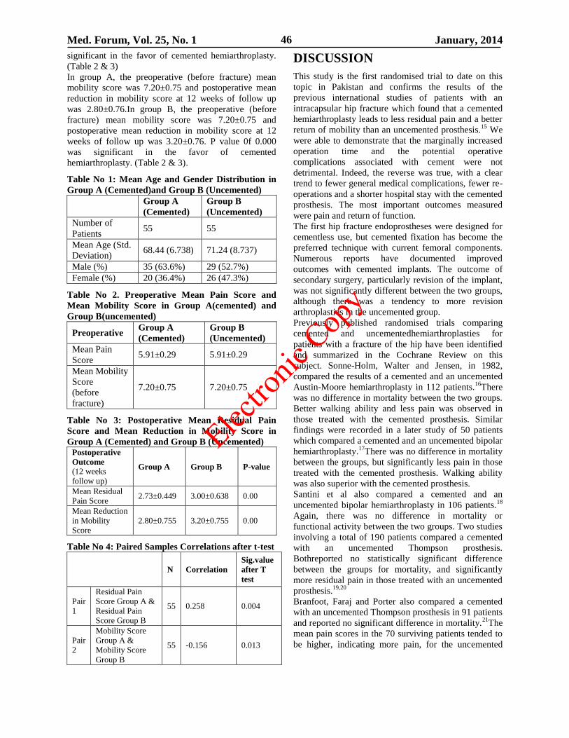
Med. Forum, Vol. 25, No. 1 January, 2014 46
significant in the favor of cemented hemiarthroplasty.
(Table 2 & 3)
In group A, the preoperative (before fracture) mean
mobility score was 7.20±0.75 and postoperative mean
reduction in mobility score at 12 weeks of follow up
was 2.80±0.76.In group B, the preoperative (before
fracture) mean mobility score was 7.20±0.75 and
postoperative mean reduction in mobility score at 12
weeks of follow up was 3.20±0.76. P value 0f 0.000
was significant in the favor of cemented
hemiarthroplasty. (Table 2 & 3).
Table No 1: Mean Age and Gender Distribution in
Group A (Cemented)and Group B (Uncemented)
Group A
(Cemented)
Group B
(Uncemented)
Number of
Patients 55 55
Mean Age (Std.
Deviation) 68.44 (6.738) 71.24 (8.737)
Male (%) 35 (63.6%) 29 (52.7%)
Female (%) 20 (36.4%) 26 (47.3%)
Table No 2. Preoperative Mean Pain Score and
Mean Mobility Score in Group A(cemented) and
Group B(uncemented)
Preoperative Group A
(Cemented)
Group B
(Uncemented)
Mean Pain
Score 5.91±0.29 5.91±0.29
Mean Mobility
Score
(before
fracture)
7.20±0.75 7.20±0.75
Table No 3: Postoperative Mean Residual Pain
Score and Mean Reduction in Mobility Score in
Group A (Cemented) and Group B (Uncemented)
Postoperative
Outcome
(12 weeks
follow up)
Group A Group B P-value
Mean Residual
Pain Score 2.73±0.449 3.00±0.638 0.00
Mean Reduction
in Mobility
Score
2.80±0.755 3.20±0.755 0.00
Table No 4: Paired Samples Correlations after t-test
N Correlation
Sig.value
after T
test
Pair
1
Residual Pain
Score Group A &
Residual Pain
Score Group B
55 0.258 0.004
Pair
2
Mobility Score
Group A &
Mobility Score
Group B
55 -0.156 0.013
DISCUSSION
This study is the first randomised trial to date on this
topic in Pakistan and confirms the results of the
previous international studies of patients with an
intracapsular hip fracture which found that a cemented
hemiarthroplasty leads to less residual pain and a better
return of mobility than an uncemented prosthesis.15
We
were able to demonstrate that the marginally increased
operation time and the potential operative
complications associated with cement were not
detrimental. Indeed, the reverse was true, with a clear
trend to fewer general medical complications, fewer re-
operations and a shorter hospital stay with the cemented
prosthesis. The most important outcomes measured
were pain and return of function.
The first hip fracture endoprostheses were designed for
cementless use, but cemented fixation has become the
preferred technique with current femoral components.
Numerous reports have documented improved
outcomes with cemented implants. The outcome of
secondary surgery, particularly revision of the implant,
was not significantly different between the two groups,
although there was a tendency to more revision
arthroplasties in the uncemented group.
Previously published randomised trials comparing
cemented and uncementedhemiarthroplasties for
patients with a fracture of the hip have been identified
and summarized in the Cochrane Review on this
subject. Sonne-Holm, Walter and Jensen, in 1982,
compared the results of a cemented and an uncemented
Austin-Moore hemiarthroplasty in 112 patients.16
There
was no difference in mortality between the two groups.
Better walking ability and less pain was observed in
those treated with the cemented prosthesis. Similar
findings were recorded in a later study of 50 patients
which compared a cemented and an uncemented bipolar
hemiarthroplasty.17
There was no difference in mortality
between the groups, but significantly less pain in those
treated with the cemented prosthesis. Walking ability
was also superior with the cemented prosthesis.
Santini et al also compared a cemented and an
uncemented bipolar hemiarthroplasty in 106 patients.18
Again, there was no difference in mortality or
functional activity between the two groups. Two studies
involving a total of 190 patients compared a cemented
with an uncemented Thompson prosthesis.
Bothreported no statistically significant difference
between the groups for mortality, and significantly
more residual pain in those treated with an uncemented
prosthesis.19,20
Branfoot, Faraj and Porter also compared a cemented
with an uncemented Thompson prosthesis in 91 patients
and reported no significant difference in mortality.21
The
mean pain scores in the 70 surviving patients tended to
be higher, indicating more pain, for the uncemented
Electro
nic Cop
y

Med. Forum, Vol. 25, No. 1 January, 2014 47
prosthesis, although the results were not statistically
significant.
We chose the two prostheses used in this study as they
are currently the most commonly used in the practice. It
is possible that a modern uncemented prosthesis,
perhaps with hydroxyapatite coating may produce
superior outcomes to the uncemented Austin-Moore
prosthesis which we used, but this remains to be proved
in a randomized controlled trial. The only study that has
compared an uncemented Austin-Moore with a
hydroxyapatite-coated Furlong prosthesis in 84 patients
was too small to make any definite conclusions on any
difference between the two implants.22
In summary, this study found that a cemented
Thompson hemiarthroplasty led to less pain in the hip,
improved return of mobility and a reduced hospital stay
compared to an uncemented Austin-Moore prosthesis.
There was no increase in complications or mortality
related to the use of cement.
CONCLUSION
Our study found that a cemented Thompson
hemiarthroplasty led to less pain in the hip and
improved return of mobility compared to an
uncemented Austin-Moore prosthesis. There was no
increase in complications or mortality related to the use
of cement. In conjunction with previous studies which
have also reported improved outcomes for a cemented
rather than an uncementedhemiarthroplasty, we suggest
that when a hemiarthroplasty is used for a fracture of
the hip it should be cemented in place.
REFERENCES
1. Leighton RK. Fractures of the neck of femur. In:
Bucholz RW, Heckman JD, Court-Brown CM,
editors. Rockwood and green’s fractures in adults.
Philadelphia: Lippincott;2006.p.1753.
2. Lavelle DG. Fractures and dislocations of the hip.
In: Canale ST, Beaty JH, editors. Campbell’s
operative orthopaedics. Philadelphia: Mosby; 2008.
p. 3237.
3. Stevens JA, Olson S. Reducing falls and resulting
hip fractures among older women. MMWR
Recomm Rep 2000;49:3-12.
4. Mamji F, Hasan JA, Sabri S. Risk factors for
osteoporosis in post-menopausal women with hip
fractures. J Surg Pak 2010;15:82-6.
5. Hayat K, Ashfaq K, Tago IA, Bhatti A, Minhas S,
Mehboob G. Factors related to development of
traumatic osteonecrosis of femoral head. J Pak
Orthop Assoc 2008;20:108-13.
6. Shafee M, Asghar M, Tariq MA. Pauwel`s
osteotomy and oseosynthesis; Its use in patients
with nonunion of femoral neck fractures. Prof Med
J 2007;14:89-97.
7. Parker MI, Pryor G, Gurusamy K. Cemented
versus uncementedhemiarthroplasty for
intracapsular hip fractures: A randomised
controlled trial in 400 patients. J Bone Joint Surg
Br 2010;92:116-22.
8. Figved W, Opland V, Frihagen F, Jervidalo T,
Madsen JE, Nordsletten L. Cemented versus
uncementedhemiarthroplasty for displaced femoral
neck fractures. Clin Orthop Relat Res 2009;467:
2426-35.
9. Vochteloo AJ, Niesten D, Riedijk R, Rijnberg WJ,
Bolder SB, Koëter S, et al. Cemented versus non-
cemented hemiarthroplasty of the hip as a
treatment for a displaced femoral neck fracture:
Design of a randomised controlled trial. BMC
Musculoskeletal Disorders 2009;10:56.
10. Khan RJ, MacDowell A, Crossman P, Datta A,
Jallali N, Arch BN, Keene GS. Cemented or
uncementedhemiarthroplasty for displaced
intracapsular femoral neck fractures. Int Orthop
2002;26:229–32.
11. Khan RJ, MacDowell A, Crossman P, Keene GS.
Cemented or uncementedhemiarthroplasty for
displaced intracapsular fractures of the hip: A
systematic review. Injury 2002;33:13-7.
12. Foster AP, Thompson NW, Wong J, Charlwood
AP. Periprosthetic femoral fractures--a comparison
between cemented and uncemented
hemiarthroplasties. Injury 2005;36:424-9.
13. Lennox IA, McLauchlan J. Comparing the
mortality and morbidity of cemented and
uncementedhemiarthroplasties. Injury 1993;24:
185–6.
14. Singh GK, Deshmukh RG. Uncementedaustin-
moore and cemented thompson unipolar
hemiarthroplasty for displaced fracture neck of
femur--comparison of complications and patient
satisfaction. Injury 2006;37:169-74.
15. Parker MJ, Gurusamy K. Arthroplasties (with and
without bone cement) for proximal femoral
fractures in adults. (Cochrane Review). The
Cochrane Library, Chichester: Wiley; 2006.
16. Sonne-Holm S, Walter S, Jensen JS. Moore hemi-
arthroplasty with and without bone cement in
femoral neck fractures: a clinical controlled trial.
Acta Orthop Scand 1982;53:953-6.
17. Dorr LD, Glousman R, Sew Hoy AL, Vanis R,
Chandler R. Treatment of femoral neck fractures
with total hip replacement versus cemented and
noncementedhemiarthroplasty. J Arthroplasty
1986;1:210-18.
18. Santini S, Rebeccato A, Bolgan I, Turi G. Hip
fractures in elderly patients treated with bipolar
hemiarthroplasty: comparison between cemented
and cementless implants. J Orthop Traumatol
2005;6:80-7.
Electro
nic Cop
y
Med. Forum, Vol. 25, No. 1 January, 2014 48
19. Anderson GH, Dias JJ, Hoskinson J, Harper WM.
A randomized study of the use of bone cement with
thompson’s prosthesis in the treatment of
intracapsular fractures of the femoral neck. J Bone
Joint Surg 1992;74:132-3.
20. Emery RJH, Broughton NS, Desal K, Bulstrode
CJK, Thomas TL. Bipolar hemiarthroplasty for
subcapital fracture of the femoral neck: a
prospective randomized trial of cemented
Thompson and uncemented Moore stems. J Bone
Joint Surg 1991;73:322-4.
21. Branfoot T, Faraj AA, Porter P. Cemented versus
uncementedthompson’s prosthesis: A randomised
prospective functional outcome study. Injury
2000;31:280-1.
22. Livesley PJ, Srivastiva VM, Needoff M, Prince
HG, Moulton AM. Use of a hydroxyapatite-coated
hemiarthroplasty in the management of subcapital
fracture of the femur. Injury 1993;24:236-40.
Address for Corresponding Author:
Dr. Manqoosh ur Rehman
Assistant Professor of Orthopaedics
Multan Medical & Dental College
Ibn e Siena Hospital & Research Institute Multan
Electro
nic Cop
y

Med. Forum, Vol. 25, No. 1 January, 2014 49
Efficacy of Intra Articular
Injections in Different Grades of
Osteoarthritis of Knee 1. Muhammad Imran 2. Manqoosh ur Rehman 3. Tanveer Ahmad Kang
1, 2 & 3. Asstt. Profs. of Orthopaedics, Multan Medical & Dental College,
Ibn e Siena Hospital & Research Institute Multan
ABSTRACT
Objectives: To assess the efficacy of intra articular injections in different grades of osteoarthritis of knee.
Study Design: Descriptive study
Place and Duration of Study: This study was conducted at the Department of Orthopedics Railway Hospital
Rawalpindi and Ibn-e-Seina Hospital & Research Institute Multan started from June 2008 to June 2012.
Materials and Methods: This study included 90 patients of either sex having mild, moderate and severe grades of
osteoarthritis of knee based on radiographic findings. A study proforma was designed in which bio-statics and
results of the study were recorded. Five intra-articular injections of hyaluronic acid were given on weekly basis and
patients were assessed for pain and functional activity at intervals of 0, 6 and 12 weeks by KSKS, WOMAC and
VAS scoring system. These patients were selected consecutively.
Results: In this study patients with mild and moderate grades of osteoarthritis of knee showed improvement but
patients with severe grades of osteoarthritis either showed no response or less improvement.
Conclusion: Intra-articular injection of hyaluronic acid is an effective modality in early stages of osteoarthritis
of knee.
Key Words: Osteoarthritis, Hyaluronic acid, Knee
INTRODUCTION
Osteoarthritis is the most common joint disorder in the
elderly.1 The prevalence of osteoarthritis ranges from
1% below the age of 30 years to over 50% in peoples
above the age of 60.2 In osteoarthritis of knee there is
progressive cartilage destruction which results in sub-
articular cysts and osteophyte formation. There is also
sclerosis of the surrounding bone and capsular fibrosis.
Patient with osteoarthritis of knee usually presents with
pain in one or both joints, stiffness, intermittent
swelling and loss of function.
Although knee arthroplasties are performed in sufficient
number in Pakistan but still this group represents only a
small number of patients with knee osteoarthritis. Most
patients avoid surgeries due to their high cost by using a
variety of non-operative treatments like oral analgesics,
nonsteroidal anti-inflammatory drugs, exercise and
physiotherapy, weight relieving braces or different
types of intra articular injections.3
Hyaluronic acid is a critical component of normal
synovial fluid and an important contributor to joint
homeostasis.4 In osteoarthritis, both the concentration
and the molecular weight of intra-articular endogenous
hyaluronic acid is decreased, which reduces the
viscoelasticity of the synovial fluid.5,6
Therefore, the
original rationale for intra articular injection of
hyaluronic acid was to restore the viscoelasticity of
synovial fluid.7,8
In addition, it has been found that
injected hyaluronic acid augments the flow of synovial
fluid, normalize the synthesis and inhibit the
degradation of endogenous hyaluronic acid, and relieve
joint pain.9, 10
Clinical trials have demonstrated the safety and efficacy
of hyaluranic acid injections for the treatment of knee
osteoarthritis.11
But studies in our setup are
inconclusive regarding the best responders with respect
to age, grade of osteoarthritis, level of symptoms and
level of physical activity. This study was undertaken to
access the efficacy of intra articular hyaluronic acid in
different grades of osteoarthritis of knee defined
radiologically.
MATERIALS AND METHODS
This multicentered, prospective and observational study
included those patients who satisfied the inclusion
criteria. Patients were offered enrolment until planned
number of patients were reached. Patients were enrolled
between June 2008 to June 2012.
Inclusion criteria was age above 30 years, radiological
evidence of symptomatic osteoarthritis of knee and
failure of other non operative modalities like NSAIDS,
physical therapy and knee braces.
Exclusion criteria was pregnant and lactating mothers,
radiographic evidence of chondrocalcinosis or if
physical examination demonstrated insufficiency of
ligaments of the knee and neuropathic joints. The study
was approved by the ethical committee and patients
were explained about the study. Follow up of the
patients was ensured.
Data including sex, age, height, weight and site of
involvement was recorded. Weight bearing
Original Article Osteoarthritis
Electro
nic Cop
y

Med. Forum, Vol. 25, No. 1 January, 2014 50
anteroposterior and lateral radiographs of the involved
knee were obtained. Findings on the initial radiographs
were graded by the radiologist as mild, moderate and
severe arthritis. Mild arthritis was defined as minimal
loss of joint space and osteophyte formation. Moderate
arthritis was defined as up to 75% loss of joint space,
subchondral changes and more noticeable osteophyte
formation. Severe arthritis was defined as total loss of
joint space and more severe changes. During grading all
the three compartments including medial and lateral
tibiofemoral and patellofemoral were graded and worse
grade was recorded.
The efficacy assessment included Knee Society Knee
Score (KSKS), Western Ontario and Mcmaster
Universities Osteoarthritis Index (WOMAC) score and
Visual Analogue Scale (VAS) score for pain at rest and
following walking and stepping activities.12,13
With KSKS clinical rating scale a score of < 55 point
indicate a poor result, 55-64 fair result, 65-79 good
result and 80-100 excellent result. WOMAC was used
as self-administered questionnaire. With WOMAC a
lower number represent a better result ranging from
zero (Best) to 96 (Poor). Visual Analogue Scale was
also administered to the patient ranging from 0-100mm
with lower number representing less pain and higher
number representing more pain.
All the three instruments were used to assess the
patients during the time of enrollment, at 6 weeks and
then at 12 weeks. One of the medical officer who was
skilled to use the study instruments was appointed to
assess the patients.
Hyaluronic acid injections (Hyalgan) were administered
as a course of five weekly injections. All injections
were given in the similar manner with the patient lying
supine and the knee joint approached through
superolateral pouch under the superolateral edge of
patella. Ethylchloride spray was used immediately prior
to the injection for the patient comfort. A 22 gauge
needle was used to inject the prefilled 2ml syringe. In
case of the knee effusion 18 gauge needle was used for
the aspiration and then the needle was left in place for
the injection. Patients was encouraged to avoid
strenuous activity for a day following the intra articular
injection.
The results obtained from KSKS, WOMAC, VAS were
tested separately. Two-way Anova (Analysis of
Variance) technique and F-Test were used.
RESULTS
This study included 100 patients having symptomatic
osteoarthritis of the knee. 10 patients left the follow up.
Mean age of the patients was 50.45±0.90 years with
range of 35-75 years. Majority of the patients were
above 40 years of age. There were 68 (75.5%) patients
who were 41-60 years of age. There were 16 (17.7%)
patients who were less than or equal to 40 years of age
and 6 (6.6%) patients were above 60 years of age. Mean
weight of the patients was 80.30±0.75 kg. (Table No.1)
There were 52 (57.8%) male and 38 (42.2%) female
patients. 20 (22.2%) patients had low socioeconomic
status, 55 (61.1%) had middle and 15 (16.6%) had high
socioeconomic status. In 38 (42.2%) patients left side
was involved while in 44 (48.9%) patients right side
was involved and in 8 (8.9%) patients both sides were
involved. 30 (33.3%) patients had mild, 30 (33.3%)
patients had moderate and 30 (33.3%) patients had
severe grade of osteoarthritis.
Table No.1: Characteristics of patients with OA Knee
(n=90)
No. of
Patients (%)
No. of
Patients (%)
No. of
Patients (%)
Age (yrs) ≤ 40 = 16
(17.7%)
41-60 = 68
(75.5%)
≥ 60 = 6
(6.6%)
Socioeconomic
Status
Low = 20
(22.2%)
Middle = 55
(61.1%)
High =15
(16.6%)
Side
Involvement
Left = 38
(42.2%)
Right = 44
(48.9%)
Both = 8
(8.9%)
Arthritis Grade Mild = 30
(33.3%)
Moderate =
30 (33.3%)
Severe =
30 (33.3%)
Table No.2: Knee Society Knee Score (KSKS) in Patients
with OA Knee (n=90)
Mild Moderate Severe All
KSKS-a
N=30
Mean=62.400
S.D=4.739
N=30
Mean=54.933
S.D=2.434
N=30
Mean=37.600
S.D=4.280
N=90
Mean=51.644
S.D=11.151
KSKS-b
N=30
Mean=87.900
S.D=4.831
N=30
Mean=70.600
S.D=6.806
N=30
Mean=42.500
S.D=3.655
N=90
Mean=67.000
S.D=19.519
KSKS-c
N=30
Mean=90.900
S.D=3.872
N=30
Mean=65.567
S.D=10.516
N=30
Mean=40.267
S.D=3.483
N=90
Mean=67.444
S.D=22.006
All
N=90
Mean=80.400
S.D=13.607
N=90
Mean=65.567
S.D=10.516
N=90
Mean=40.122
S.D=4.282
N=270
Mean=62.030
S.D=19.534
Table No.3: WOMAC in Patients with OA Knee (n=90)
Mild Moderate Severe All
WAM
AC-a
N=30
Mean=45.467
S.D=2.825
N=30
Mean=51.967
S.D=2.173
N=30
Mean=61.000
S.D=3.806
N=90
Mean=52.811
S.D=7.063
WAM
AC-b
N=30
Mean=22.167
S.D=5.032
N=30
Mean=37.033
S.D=6.636
N=30
Mean=55.067
S.D=3.868
N=90
Mean=38.089
S.D=14.507
WAM
AC-c
N=30
Mean=17.133
S.D=6.213
N=30
Mean=36.467
S.D=10.095
N=30
Mean=58.167
S.D=5.167
N=90
Mean=37.256
S.D=18.400
All
N=90
Mean=28.256
S.D=13.322
N=90
Mean=41.822
S.D=10.059
N=90
Mean=58.078
S.D=4.922
N=270
Mean=42.719
S.D=15.792
Using KSKS, mild grades showed mean score of 62.40
at the start and after 12 weeks mean score was 90.90.
Moderate grades showed mean score of 54.93 at the
start and after 12 week mean score was 65.56. Severe
grades showed mean score of 37.60 at the start and after
12 week mean score was 40.26.
Using WOMAC, mild grades showed mean score of
45.46 at the start and after 12 weeks mean score was
Electro
nic Cop
y

Med. Forum, Vol. 25, No. 1 January, 2014 51
17.13. Moderate grades showed mean score of 51.96 at
the start and after 12 weeks mean score was 36.46.
Sever grades showed mean score of 61.00 at the start
and after 12 week mean score was 58.16.
Table No.4: Visual Analogue Scale (VAS) in Patients with
OA Knee (n=90)
Mild Moderate Severe All
VAS-
a
N=30
Mean=56.000
S.D=4.433
N=30
Mean=68.167
S.D=2.780
N=30
Mean=78.333
S.D=3.304
N=90,
Mean=67.500
0
S.D=9.837
VAS-
b
N=30
Mean=26.500
S.D=8.610
N=30
Mean=48.000
S.D=8.367
N=30
Mean=70.687
S.D=5.231
N=90
Mean=48.456
S.D=19.493
VAS-
c
N=30
Mean=20.000
S.D=8.610
N=30
Mean=45.467
S.D=15.204
N=30
Mean=74.500
S.D=6.867
N=90
Mean=46.656
S.D=24.823
All
N=30
Mean=34.167
S.D=17.178
N=90
Mean=53.878
S.D=14.317
N=90
Mean=74.567
S.D=6.102
N=270
Mean=54.204
S.D=21.233
Similarly using Visual Analogue Scale, mild grades
showed mean score of 56.00 at the start and after 12
weeks mean score was 20.00. Moderate grades showed
mean score of 68.16 at the start and after 12 weeks
mean score was 45.46. Severe grades showed mean
score of 78.33 at the start and after 12 weeks mean
score was 74.50.
These results showed the improvement more in mild
and moderate grades of osteoarthritis of knee than in
severe grades of osteoarthritis of knee.
DISCUSSION
The present study showed the treatment effects of
hyaluronic acid injection in different grades of
osteoarthritis of knee. Although intra-articular
injections have shown improvement in all grades but
effects are more in mild and moderate grades as
compared to sever grades in our population as well.
No radiological grading system has undergone
interobserver and intraobserver reliability testing, this
should be considered a limitation for any study that
draws interference between radiological grades and
outcome.
The meta-analysis by Wang CT, et al showed
signification improvement in pain and functional
outcome except those who had severe osteoarthritis.14
Similarly a prospective randomized study by Petrela RJ
and Petrela M showed patient satisfaction in terms of
pain relief and functional improvement by hyaluronic
acid.15
In this study 10 patients were slipped during the course
of treatment as it require five weeks to complete so it is
concluded that the course should be of shorter duration.
As new variants of hyaluronic acid are discovered,
single intra-articular injection with 6ml Hylan G-F 20
can replace routine method of once a week for five
weeks.16
The effect of hyaluronic acid at molecular
level has also been established.17,18
CONCLUSION
Intra-articular injection of hyaluronic acid is an
effective modality in early stages of osteoarthritis of
knee.
REFERENCES
1. Felson DT, Zhang Y. An update on the
epidemiology of knee and hip osteoarthritis with a
view to prevention. Arthritis Rheum 1998;41:
1343-55.
2. Felson DT, Naimark A, Anderson J, Kazis L,
Castelli W, Meenan RF. The prevalence of knee
osteoarthritis in the elderly. Arthritis &
Rheumatism 1987;80:914-8.
3. Watterson JR, Esdaile JM. Viscosupplementation:
therapeutic mechanisms and clinical potential in
osteoarthritis of the knee. J Am Acad Orthop Surg
2000;8:277-84.
4. Pendleton A, Arden N, Dougados M, Doherty M,
Bannwarth B, Bijlsma JW, et al. EULAR
recommendations for the management of knee
osteoarthritis: report of a task force of the Standing
Committee for International Clinical Studies
Including Therapeutic Trials (ESCISIT). Ann
Rheum Dis 2000;59:936-44.
5. Laurent TC, Fraser JR. Hyaluronan. FASEB J
1992;6:2397-404.
6. Lee YT, Shao HJ, Wang JH, Liu HC , Hou SM,
Young TH. Hyaluronic acid modulates gene
expression of connective tissue growth factor
(CTGF), transforming growth factor –beta1 (TGF
beta1), and vascular endothelial growth factor
(VEGF) in human fibroblast-like synovial cells
from advanced stage osteoarthritis in vitro. J
Orthop Res 2010;28:492-6.
7. Balazs EA, Watson D, Duff IF, Roseman S.
Hyaluronic acid in synovial fluid. I. Molecular
parameters of hyaluronic acid in normal and
arthritis human fluids. Arthritis Rheum
1967;10:357-76.
8. Evanich JD, Evanich CJ, Wright MB, Rydlewicz
JA. Efficacy of intraarticular hyaluronic acid
injections in knee osteoarthritis. Clin Orthop Relat
Res 2001;390:173-81.
9. Peyron JG. A new approach to the treatment of
osteoarthritis: viscosupplementation. Osteoarthritis
Cartilage 1993;1:85-7.
10. Rydell N, Balazs EA. Effect of intra-articular
injection of hyaluronic acid on the clinical
symptoms of osteoarthritis and on granulation
tissue formation. Clin Orthop 1971;80:25-32.
11. Foti C, Cisari C, Carda S, Giordan N, Rocco A,
Frizziero A, et al. A prospective observational
study of the clinical efficacy and safety of intra-
articular sodium hyaluronate in synovial joints with
Electro
nic Cop
y

Med. Forum, Vol. 25, No. 1 January, 2014 52
osteoarthritis. Eur J Phys Rehabuil Med 2011;
47:407-15.
12. Turajane T, Tanavaree A, Labpiboonpong V,
Maungsiri S. Outcomes of intra-articular injection
of sodium hyaluronate for the treatment of
osteoarthritis of the knee. J Med Assoc Thai
2007;90:1845-52.
13. Bellamy N. Pain assessment in osteoarthritis
experience with the WOMAC osteoarthritis index.
Semin Arthritits Rheumatrol 1988;15:1833-40.
14. Wang CT, Lin J, Chang CJ, Lin YT, Hou SM.
Therapeutic effects of hyaluronic acid on
osteoarthritis of the knee. A meta-analysis of
randomized control trials. JBJS 2004;86:538-45.
15. Petrella RJ, Petrella M. A prospective, randomized,
double blind, placebo controlled study to evaluate
the efficacy of intraarticular hyaluronic acid for
osteoarthritis of the knee. J Rehumatol 2006;
33:951-6.
16. Abate M, Pulcii, D, Di Lorio A, Schiavone C.
Viscosupplementation with intra articular
hyaluronic acid for treatment of osteoarthirits In
the elderly. Curr Pharm 2010;16:631-40.
17. Phiphobmongkol V, Sudhasaneya V. The effective-
ness and safety of intra-articular injection of
sodium hyaluronate (500-730 kDa) in the treatment
of patients with painful knee osteoarthritis. J Med
Assoc Thai 2009;92:1287-94.
18. Kolarz G, Kotz R, Hochmayer I. Long-term
benefits and repeated treatment cycles of intra-
articular sodium hyaluronate (Hyalgan) in patients
with osteoarthritis of the knee. Semin Arthritis
Rheum 2003;32:310-9.
Address for Corresponding Author:
Dr. Muhammad Imran
Assistant Professor of Orthopaedics
Multan Medical & Dental College
Ibn e Siena Hospital & Research Institute Multan
Electro
nic Cop
y

Med. Forum, Vol. 25, No. 1 January, 2014 53
Effect of Thiamine on
Glycemic Control in Induced Diabetic Rat Model 1. Muhammad Bachal Pandhiani 2. Maria Kazi 3. Saif-ur-Rehman 4. Navaid Kazmi
5. Salman Ahmad Farsi Kazi 6. Haji Khan Khoharo 1. Asstt. Prof. of Anatomy, Al-Tibri Medical College, Karachi 2. Asstt. Prof. of Biochemistry, Al-Tibri Medical
College, Karachi 3, 4. Asstt. Profs. of Physiology, Faculty of Medicine and Allied Medical Sciences,
Isra University, Hyderabad 5. Asstt. Prof. of Forensic Medicine, Faculty of Medicine and Allied Medical Sciences,
Isra University, Hyderabad 6. Asstt. Prof./Consultant Physician, Dept. of Physiology/Medicine, Faculty of Medicine
and Allied Medical Sciences, Isra University, Hyderabad
ABSTRACT
Objective: To evaluate the effect of high dose thiamine on postprandial blood glucose (PPBG) and hemoglobin
A1C levels in induced diabetic albino rat model.
Study Design: Experimental/Analytical study
Place and Duration of Study: Animal House, Isra University Hyderabad from March to October 2013.
Materials and Methods: Forty adult albino rats were divided into four groups; Group I. Controls receiving normal
diet, Group II. Controls receiving thiamine fortified diet, Group III. Diabetics receiving normal diet and Group IV.
Diabetics receiving thiamine fortified diet. Diabetes mellitus was induced using Streptozocin. Thiamine was given at
a dose of 1.6 g/kg body weight. Venous blood samples were drawn from animal’s tail with a small bore cannula
before and after 12 weeks of experimentation. The PPBG levels and Glycosylated HbA (HbA1c) were measured.
The data was converted into database and analyzed on SPSS version 21.0 by ANOVA and Tukey-Kramer`s test. A
p-value of ≤ 0.05 was taken statistically significant.
Results: The PPBG and HbA1c levels were found statistically significant in groups I vs. III (p=0.0001), I vs. IV
(p=0.0001), II vs. III (p=0.0001), II vs. IV, (p=0.001) and III vs. IV (p=0.024) at 12th
week of experimentation. The
study shows significant reduction in the PPBG and HbA1c levels of rats taking thiamine compared to controls
(p=0.001). Significant differences in HbA1c levels were observed between control groups I and II vs. experimental
groups III and IV (p=0.024) (p=0.0001) respectively. A highly significant difference in HbA1c was observed in rats
given thiamine fortified diet compared to those not given (p=0.001).
Conclusion: The thiamine improves glucose metabolism in induced diabetic rat models, hence it is concluded that
the thiamine may be given along with anti- diabetic drugs to overcome defects of glucose metabolism.
Key words: Thiamine, Glycemic control, HbA1c, Diabetic rats
INTRODUCTION
The number of diabetic population older than twenty is
estimated to rise from 285 million in 2010 to 439
million in 2030 as reported by International Diabetes
Federation (IDF).1 The current rise in diabetic
population has put Pakistan at sixth position in the
world.2 According to an estimate, 15% of Pakistani
population is diagnosed of having diabetes mellitus
(DM) and millions more are unaware and undiagnosed
of having DM.3,4
According to the estimates of
Pakistan National Diabetes Survey (PNDS), for each
diagnosed case of DM, there are approximately two
cases of undiagnosed DM and three cases of impaired
glucose tolerance who remain unaware of it.5,6
The DM
is characterized by chronic hyperglycemia which in
long term causes damage in the target organs like
kidney, nerves, eyes, heart and blood vessels.7,8
The
DM is a multifactorial metabolic disorder, mainly
characterized by abnormal glucose metabolism which is
responsible for most of its symptoms and
complications. But the thiamine deficiency is also
linked with the pathogenesis and complicaitons of DM
as has been reported in many studies.9, 10,11
The thiamine
is known as vitamin B1. Thiamine forms an
indispensable co-enzyme and participates in several
steps of glucose and intermediary metabolisms. In
human body it plays role in glucose metabolism by
forming co-enzymes essential for functioning of
enzymes of glycolysis ad citric acid cycle. The DM has
been characterized by thiamine deficiency due to
amplified glucose metabolism.11
Thiamine deficiency
causes disturbed glucose metabolism, and accelerates
DM complications through various mechanisms.11
The
various mechanisms which have been postulated
causing diabetic complications include the activation of
polyol pathway, formation of advanced glycation end
products (AGES), activation of protein kinase C (PKC)
and increased flux through the hexosamine biosynthetic
pathway (HBP).11
These metabolic abnormalities are
triggered through increased formation and elevated
concentration of triose-phosphate intermediates of
glycolysis.12,13
Triose-phosphate can be suppressed by
the activation of reductive pentose phosphate pathway
(PPP) by high-dose thiamine therapy that would
increase transketolase (TK) activity and stimulate the
Original Article Thiamine on Glycemic Control
Electro
nic Cop
y
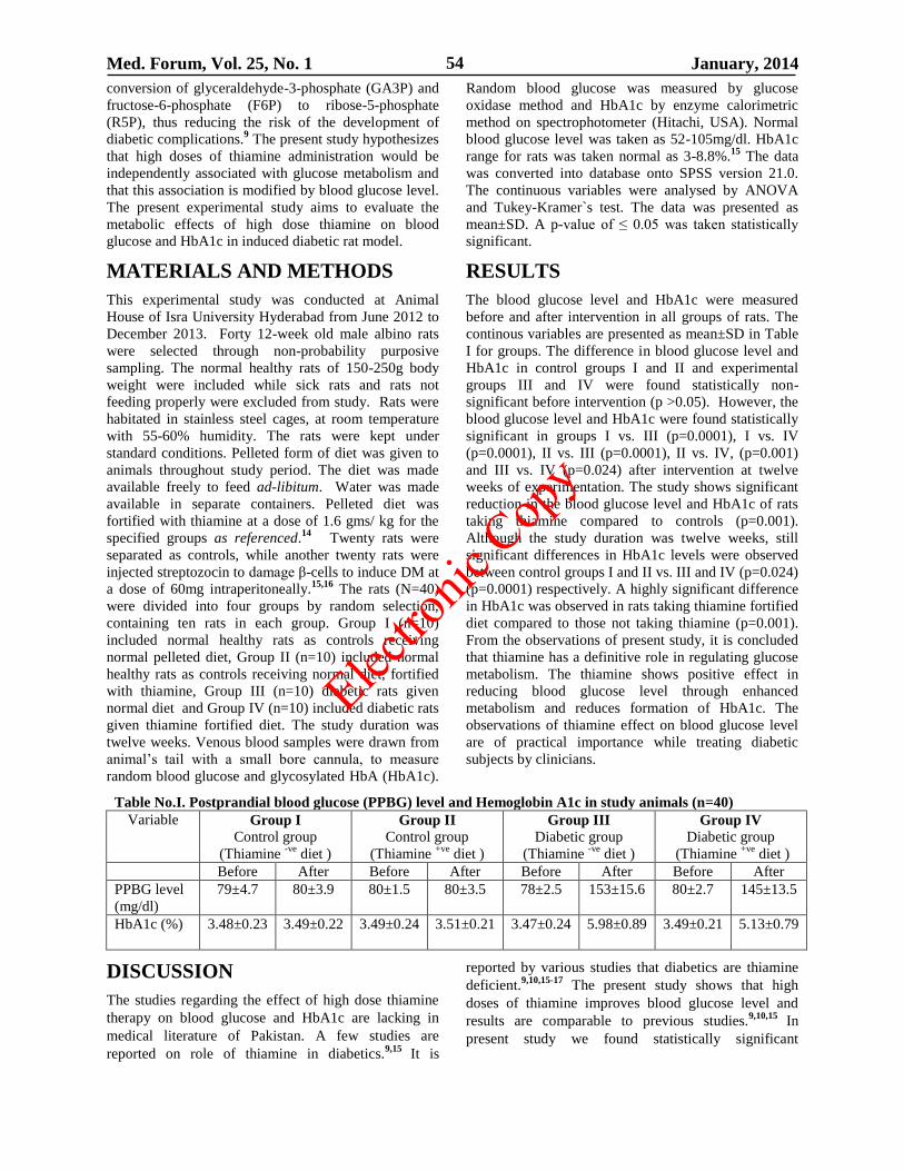
Med. Forum, Vol. 25, No. 1 January, 2014 54
conversion of glyceraldehyde-3-phosphate (GA3P) and
fructose-6-phosphate (F6P) to ribose-5-phosphate
(R5P), thus reducing the risk of the development of
diabetic complications.9 The present study hypothesizes
that high doses of thiamine administration would be
independently associated with glucose metabolism and
that this association is modified by blood glucose level.
The present experimental study aims to evaluate the
metabolic effects of high dose thiamine on blood
glucose and HbA1c in induced diabetic rat model.
MATERIALS AND METHODS
This experimental study was conducted at Animal
House of Isra University Hyderabad from June 2012 to
December 2013. Forty 12-week old male albino rats
were selected through non-probability purposive
sampling. The normal healthy rats of 150-250g body
weight were included while sick rats and rats not
feeding properly were excluded from study. Rats were
habitated in stainless steel cages, at room temperature
with 55-60% humidity. The rats were kept under
standard conditions. Pelleted form of diet was given to
animals throughout study period. The diet was made
available freely to feed ad-libitum. Water was made
available in separate containers. Pelleted diet was
fortified with thiamine at a dose of 1.6 gms/ kg for the
specified groups as referenced.14
Twenty rats were
separated as controls, while another twenty rats were
injected streptozocin to damage β-cells to induce DM at
a dose of 60mg intraperitoneally.15,16
The rats (N=40)
were divided into four groups by random selection,
containing ten rats in each group. Group I (n=10)
included normal healthy rats as controls receiving
normal pelleted diet, Group II (n=10) included normal
healthy rats as controls receiving normal diet, fortified
with thiamine, Group III (n=10) diabetic rats given
normal diet and Group IV (n=10) included diabetic rats
given thiamine fortified diet. The study duration was
twelve weeks. Venous blood samples were drawn from
animal’s tail with a small bore cannula, to measure
random blood glucose and glycosylated HbA (HbA1c).
Random blood glucose was measured by glucose
oxidase method and HbA1c by enzyme calorimetric
method on spectrophotometer (Hitachi, USA). Normal
blood glucose level was taken as 52-105mg/dl. HbA1c
range for rats was taken normal as 3-8.8%.15
The data
was converted into database onto SPSS version 21.0.
The continuous variables were analysed by ANOVA
and Tukey-Kramer`s test. The data was presented as
mean±SD. A p-value of ≤ 0.05 was taken statistically
significant.
RESULTS
The blood glucose level and HbA1c were measured
before and after intervention in all groups of rats. The
continous variables are presented as mean±SD in Table
I for groups. The difference in blood glucose level and
HbA1c in control groups I and II and experimental
groups III and IV were found statistically non-
significant before intervention (p >0.05). However, the
blood glucose level and HbA1c were found statistically
significant in groups I vs. III (p=0.0001), I vs. IV
(p=0.0001), II vs. III (p=0.0001), II vs. IV, (p=0.001)
and III vs. IV (p=0.024) after intervention at twelve
weeks of experimentation. The study shows significant
reduction in the blood glucose level and HbA1c of rats
taking thiamine compared to controls (p=0.001).
Although the study duration was twelve weeks, still
significant differences in HbA1c levels were observed
between control groups I and II vs. III and IV (p=0.024)
(p=0.0001) respectively. A highly significant difference
in HbA1c was observed in rats taking thiamine fortified
diet compared to those not taking thiamine (p=0.001).
From the observations of present study, it is concluded
that thiamine has a definitive role in regulating glucose
metabolism. The thiamine shows positive effect in
reducing blood glucose level through enhanced
metabolism and reduces formation of HbA1c. The
observations of thiamine effect on blood glucose level
are of practical importance while treating diabetic
subjects by clinicians.
Table No.I. Postprandial blood glucose (PPBG) level and Hemoglobin A1c in study animals (n=40)
Variable Group I
Control group
(Thiamine -ve
diet )
Group II
Control group
(Thiamine +ve
diet )
Group III
Diabetic group
(Thiamine -ve
diet )
Group IV
Diabetic group
(Thiamine +ve
diet )
Before After Before After Before After Before After
PPBG level
(mg/dl)
79±4.7 80±3.9 80±1.5 80±3.5 78±2.5 153±15.6 80±2.7 145±13.5
HbA1c (%) 3.48±0.23 3.49±0.22 3.49±0.24 3.51±0.21 3.47±0.24 5.98±0.89 3.49±0.21 5.13±0.79
DISCUSSION
The studies regarding the effect of high dose thiamine
therapy on blood glucose and HbA1c are lacking in
medical literature of Pakistan. A few studies are
reported on role of thiamine in diabetics.9,15
It is
reported by various studies that diabetics are thiamine
deficient.9,10,15-17
The present study shows that high
doses of thiamine improves blood glucose level and
results are comparable to previous studies.9,10,15
In
present study we found statistically significant
Electro
nic Cop
y
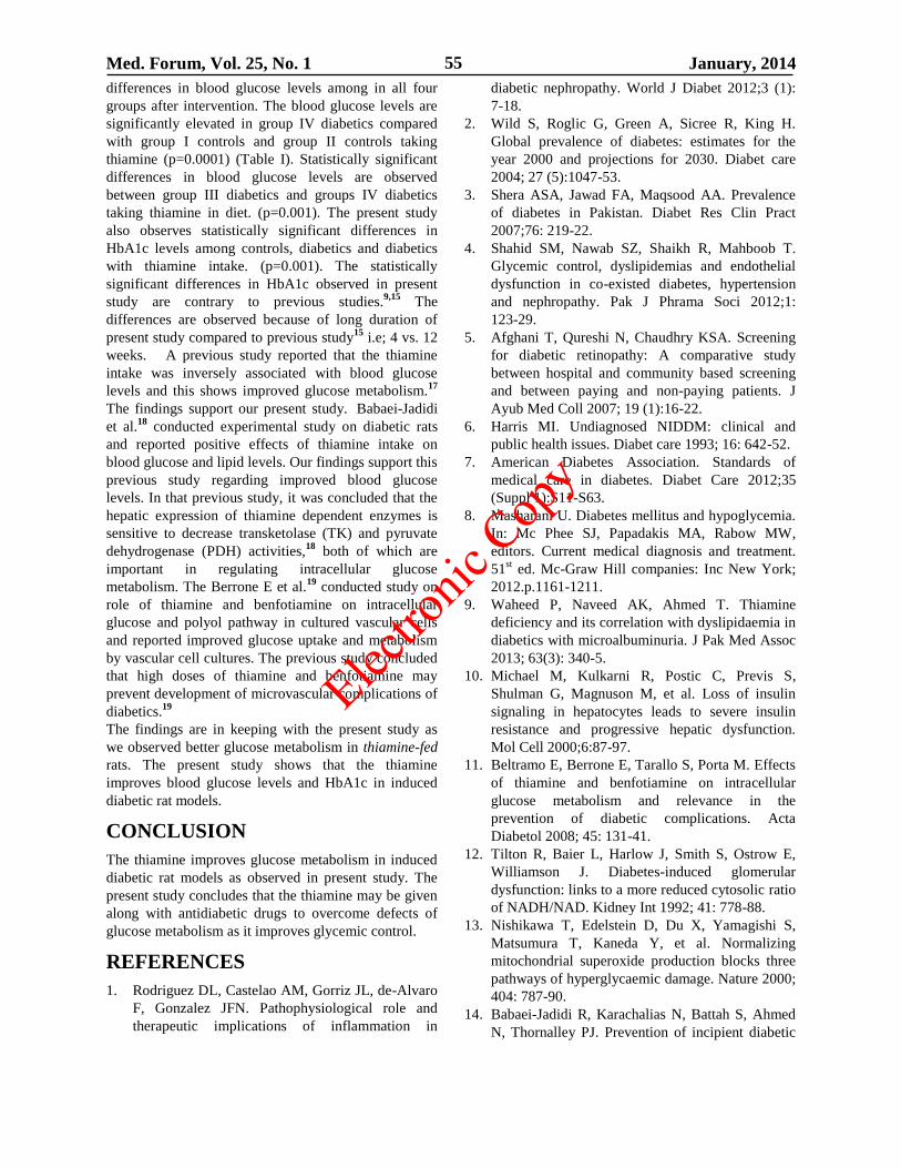
Med. Forum, Vol. 25, No. 1 January, 2014 55
differences in blood glucose levels among in all four
groups after intervention. The blood glucose levels are
significantly elevated in group IV diabetics compared
with group I controls and group II controls taking
thiamine (p=0.0001) (Table I). Statistically significant
differences in blood glucose levels are observed
between group III diabetics and groups IV diabetics
taking thiamine in diet. (p=0.001). The present study
also observes statistically significant differences in
HbA1c levels among controls, diabetics and diabetics
with thiamine intake. (p=0.001). The statistically
significant differences in HbA1c observed in present
study are contrary to previous studies.9,15
The
differences are observed because of long duration of
present study compared to previous study15
i.e; 4 vs. 12
weeks. A previous study reported that the thiamine
intake was inversely associated with blood glucose
levels and this shows improved glucose metabolism.17
The findings support our present study.
Babaei-Jadidi
et al.18
conducted experimental study on diabetic rats
and reported positive effects of thiamine intake on
blood glucose and lipid levels. Our findings support this
previous study regarding improved blood glucose
levels. In that previous study, it was concluded that the
hepatic expression of thiamine dependent enzymes is
sensitive to decrease transketolase (TK) and pyruvate
dehydrogenase (PDH) activities,18
both of which are
important in regulating intracellular glucose
metabolism. The Berrone E et al.19
conducted study on
role of thiamine and benfotiamine on intracellular
glucose and polyol pathway in cultured vascular cells
and reported improved glucose uptake and metabolism
by vascular cell cultures. The previous study concluded
that high doses of thiamine and benfotiamine may
prevent development of microvascular complications of
diabetics.19
The findings are in keeping with the present study as
we observed better glucose metabolism in thiamine-fed
rats. The present study shows that the thiamine
improves blood glucose levels and HbA1c in induced
diabetic rat models.
CONCLUSION
The thiamine improves glucose metabolism in induced
diabetic rat models as observed in present study. The
present study concludes that the thiamine may be given
along with antidiabetic drugs to overcome defects of
glucose metabolism as it improves glycemic control.
REFERENCES
1. Rodriguez DL, Castelao AM, Gorriz JL, de-Alvaro
F, Gonzalez JFN. Pathophysiological role and
therapeutic implications of inflammation in
diabetic nephropathy. World J Diabet 2012;3 (1):
7-18.
2. Wild S, Roglic G, Green A, Sicree R, King H.
Global prevalence of diabetes: estimates for the
year 2000 and projections for 2030. Diabet care
2004; 27 (5):1047-53.
3. Shera ASA, Jawad FA, Maqsood AA. Prevalence
of diabetes in Pakistan. Diabet Res Clin Pract
2007;76: 219-22.
4. Shahid SM, Nawab SZ, Shaikh R, Mahboob T.
Glycemic control, dyslipidemias and endothelial
dysfunction in co-existed diabetes, hypertension
and nephropathy. Pak J Phrama Soci 2012;1:
123-29.
5. Afghani T, Qureshi N, Chaudhry KSA. Screening
for diabetic retinopathy: A comparative study
between hospital and community based screening
and between paying and non-paying patients. J
Ayub Med Coll 2007; 19 (1):16-22.
6. Harris MI. Undiagnosed NIDDM: clinical and
public health issues. Diabet care 1993; 16: 642-52.
7. American Diabetes Association. Standards of
medical care in diabetes. Diabet Care 2012;35
(Suppl 1):S11-S63.
8. Masharani U. Diabetes mellitus and hypoglycemia.
In: Mc Phee SJ, Papadakis MA, Rabow MW,
editors. Current medical diagnosis and treatment.
51st ed. Mc-Graw Hill companies: Inc New York;
2012.p.1161-1211.
9. Waheed P, Naveed AK, Ahmed T. Thiamine
deficiency and its correlation with dyslipidaemia in
diabetics with microalbuminuria. J Pak Med Assoc
2013; 63(3): 340-5.
10. Michael M, Kulkarni R, Postic C, Previs S,
Shulman G, Magnuson M, et al. Loss of insulin
signaling in hepatocytes leads to severe insulin
resistance and progressive hepatic dysfunction.
Mol Cell 2000;6:87-97.
11. Beltramo E, Berrone E, Tarallo S, Porta M. Effects
of thiamine and benfotiamine on intracellular
glucose metabolism and relevance in the
prevention of diabetic complications. Acta
Diabetol 2008; 45: 131-41.
12. Tilton R, Baier L, Harlow J, Smith S, Ostrow E,
Williamson J. Diabetes-induced glomerular
dysfunction: links to a more reduced cytosolic ratio
of NADH/NAD. Kidney Int 1992; 41: 778-88.
13. Nishikawa T, Edelstein D, Du X, Yamagishi S,
Matsumura T, Kaneda Y, et al. Normalizing
mitochondrial superoxide production blocks three
pathways of hyperglycaemic damage. Nature 2000;
404: 787-90.
14. Babaei-Jadidi R, Karachalias N, Battah S, Ahmed
N, Thornalley PJ. Prevention of incipient diabetic
Electro
nic Cop
y

Med. Forum, Vol. 25, No. 1 January, 2014 56
nephropathy by high dose thiamine and
Benfotiamine. Diabetes 2003; 52:2110-20.
15. Qamar T, Fatima K, Saleem S. Effect of Thiamine
on Glycemic Control in Diabetic Rats. Ann Pak
Inst Med Sci 2011; 7(4): 213-6. rticle
16. Page GLJ, Laight D, Cummings MH. Thiamine
deficiency in diabetes mellitus and the impact of
thiamine replacement on glucose metabolism and
vascular disease. Int J Clin Prac 2011;65:684-90.
17. Rabbani N, Thornalley P. Emerging role of
thiamine therapy for prevention and treatment of
early-stage diabetic nephropathy. Diabet Obes
Metab 2011; 13(7): 577-83.
18. Babaei-Jadidi R, Karachalias N, Kupich C, Ahmed
N, Thornalley PJ. High dose thiamine therapy
counters dyslipidemias in streptozotocin-induced
diabetic rats. Diabetol 2004; 47: 2235-46.
19. Berrone E, Beltramo E, Solimine C, Ape AU, Porta
M. Regulation of Intracellular Glucose and Polyol
Pathway by Thiamine and Benfotiamine in
Vascular Cells Cultured in High Glucose. J Biol
Chem 2006; 281: 9307-13.
Address for Corresponding Author:
Dr. Haji Khan Khoharo, Assistant Professor/Consultant Physician,
Department of Physiology/Medicine
Faculty of Medicine and Allied Medical Sciences, Isra
University, Hyderabad.
Tel: 0331-2662500
Electro
nic Cop
y

Med. Forum, Vol. 25, No. 1 January, 2014 57
Pentoxifylline Protects against
Carbon Tetrachloride Induced Liver Injury in Adult
Male Wistar Rat Model
1. Maria Kazi 2. Muhammad Bachal Pandhiani 3. Salman Ahmad Farsi Kazi
4. Saif-ur-Rehman 5. Haji Kjan Khoharo
1. Asstt. Prof. of Biochemistry, Al-Tibri Medical College, Karachi 2. Asstt. Prof. of Anatomy, Al-Tibri Medical
College, Karachi 3. Asstt. Prof. of Forensic Medicine, Faculty of Medicine and Allied Medical Sciences, Isra
University, Hyderabad 4. Asstt. Prof. of Physiology, Faculty of Medicine and Allied Medical Sciences, Isra
University, Hyderabad 5. Asstt. Prof./Consultant Physician, Dept. of Physiology/Medicine, Faculty of Medicine and
Allied Medical Sciences, Isra University, Hyderabad
ABSTRACT
Objective: To investigate the protective effect of Pentoxifylline (PTX) in carbon tetrachloride (CCl4) induced
liver injury in adult male Wistar rat model.
Study design: Experimental/Analytical study
Place and Duration: Animal House, Isra University Hyderabad from May to December 2012.
Subjects and Methods: Forty five adult male Wistar rats were divided into three groups; Group A. controls
received 0.9% isotonic saline, Group B. received CCl4 orally (1.9mg/kg) mixed in olive oil, and Group C. received
the PTX+ CCl4. Blood samples were collected for liver biochemical assays. The animals were sacrificed, liver
tissue, after fixation in 4% formaldehyde, was embedded in paraffin. Tissue sections of 5μ thickness were subjected
to haematoxylin and eosin staining and were assessed by light microscopy. The data was analyzed on SPSS 21.0
using one-way ANOVA, Tukey-Cramer and Chi-square tests. A p-value of ≤ 0.05 was taken statistically significant.
Results: The liver biochemical and histological findings reveal statistically significant differences among the
controls, CCl4 and PTX+ CCl4 groups (p=0.0001). Liver enzymes and histology was deranged significantly in CCl4
group compared to controls and PTX+ CCl4 group (p=0.0001). The CCl4+PTX group shows less elevation of liver
enzymes and derangement in liver histology when compared to CCl4 group (p=0.001). The histological findings of
congestion, inflammatory cell infiltrate, vacuolar degeneration and necrosis are found prominent in CCl4 group.
Conclusion: The Pentoxifylline protects against oxidative damages caused by carbon tetrachloride induced liver
injury in rat model.
Key words: Carbon tetrachloride, Liver injury, Pentoxifylline
INTRODUCTION
Liver is the largest gland of human body. The
parenchyma cells of liver are known as the hepatocyte,
which performs biochemical and metabolic functions.1
The oxygen is a precursor of a number of free radicals
which are collectively known as the reactive oxygen
species (ROS). The ROS are implicated in the
pathogenesis of most liver diseases both inflammatory
and non-inflammatory like ischemia/reperfusion injury,
chronic viral hepatitis, alcoholic hepatitis, non-
alcoholic fatty liver disease (NAFLD), non-alcoholic
steatohepatitis (NASH) and cholestasis.2
The liver is a
metabolically active organ, and protects against drug
toxicity for two reasons 1). As it is interposed between
porto-systemic circulation i.e. between absorption of
intestinal contents containing toxins. 2). Hence
preferred site of toxin metabolism and elimination
through intestine. Drug induced liver injury poses body
systems to possible injury if toxins access to systemic
circulation.3,4
Carbon tetrachloride (CCl4) is a hepatotoxic compound.
The CCl4 has been has been used extensively in
laboratory animals for induction of liver injury,
elucidate the underlying mechanism of liver injury and
hepatoprotective effects of various therapeutic agents.5
One of the postulated mechanism of CCl4 induced liver
injury is the formation of ROS. The ROS disrupts the
hepatocyte at cell membrane level through the lipid
peroxidation5,6
causing anatomical disruption of liver
architecture and physiological disturbances.7
The
hepatocyte injury causes leakage of cytoplasmic and
mitochondrial enzymes in the blood streams.8
The cytoplasmic and mitochondrial enzymes are
clinically used as markers of liver injury, and for
monitoring and treating the liver diseases also. The
liver enzymes which appear in the blood as a result of
liver injury include; alanine transaminase (ALT),
aspartate transaminase (AST), alkaline phophatase
(ALP) and lactate dehyderogenase (LDH) are important
enzymes that are often employed in assessing liver
injury.1,5
The Pentoxifylline (PTX) is a well known methyl
xanthine. The PTX is a non-specific inhibitor of
phosphodiesterase-4 type. The PTX is used clinically in
peripheral arterial disease with intermittent
Original Article Pentoxifylline on Liver
Electro
nic Cop
y

Med. Forum, Vol. 25, No. 1 January, 2014 58
claudication. The mechanism underlying its therapeutic
effects seems to be related to improvement of
microcirculation, tissue perfusion and cell functions.
The PTX is reported to possess anti-inflammatory and
hepatoprotective effects against chemical-induced liver
injury like alcohol.4,9,10
The present study aims to investigate the possible
protective effects of Pentoxifylline (PTX) against
Carbon tetrachloride (CCl4)-induced hepatotoxicity in
experimental animals.
MATERIALS AND METHODS
An experimental study was conducted at the animal
house of Isra University on rabbit model over a period
of one year, from January to December 2012. Adult
male Wistar rats of 250-300 grams were included in the
study. Female rats, weight <250 grams or >300 grams
were excluded from the study. Animals were housed in
animal house at an optimal room temperature with 55-
60% humidity and exposed to 12 hour light-dark cycles.
The chaw like fresh alfalfa and clean water are
provided freely. The rats were divided into three
groups;
Group A. Control Group (n=15) Rats received 0.9%
isotonic saline orally on alternate day for three
successive weeks and served as control group,
Group B. Carbon tetrachloride Group (n=15) Rats
were given CCl4 orally mixed in olive oil on alternate
day for three successive weeks and
Group C. Experimental Group (n=15) Rats received
PTX (200 mg/kg intraperitoneally) and CCl4 on
alternate days for three successive weeks
Experimental Details: The PTX was purchased from
Medical store of Isra University Hospital. The PTX was
administered in a dose of 200 mg/kg intraperitoneally.4
Carbon tetrachloride was purchased from scientific
drug store at Hyderabad City. The CCl4 dissolved in
olive oil as vehicle (1:1 Ratio) at a dose level of 1.9
ml/kg orally on alternate day for three successive weeks
and sacrificed at the end of their respective period of
time.1
Sacrifice of animals: The animals were sacrificed
using standard method as described by Nayak et al.
(2006)11
In order to examine the liver tissue, the liver of
the sacrificed animals was removed promptly and
preserved in formaldehyde.
Blood sample Processing: The blood samples were
collected from peripheral veins at twenty four hours of
experimental period. Sera were separated by
centrifugation at 300xs for ten minutes. Serum samples
were used to determine liver enzymes.
Liver enzyme assay: Liver enzyme assays were
determined for alanine transaminase (ALT), aspartate
transaminase (AST), alkaline phophatase (ALP) and
lactate dehyderogenase (LDH) using commercially
available diagnostic kits.
Histological studies: After fixation in 4%
formaldehyde, samples were embedded in paraffin.
Sections of 5μ thickness were subjected to
haematoxylin and eosin Hepatic morphology was
assessed by light microscopy. A total of five sections
for each liver tissue sample were observed under light
microscope.
In H & E staining, damaged hepatocytes graded as 0=
normal, += mild damage (swollen and pale cytoplasm),
++= moderate damage (vacuolated cytoplasm), +++=
severe damage and ++++= very severe damage
(pyknotic nucleus and eosinophil cytoplasm).12
The data was analyzed on SPSS version 21.0 (IBM
corporation). The continous variables were presented as
mean±SD using one-way ANOVA and Tukey-Cramer
test for multiple comparisons. Chi-square test was used
for categorical variables. A p-value of ≤ 0.5 was taken
statistically significant.
RESULTS
The present study observes major differences in liver
injury between and among groups as indicated by blood
enzyme levels in different animal groups. The LDH,
ALP, and ALT, AST in serum of Rats treated with
carbon tetrachloride were found elevated compared
with control group after three weeks, with a highly
significant p-value for multiple comparisons (p=0.001).
The CCl4+PTX group shows a significant reduction in
the liver enzymes compared with the CCl4 group
(p=0.001) and control group (p=0.001). The animals
CCl4+PTX group shows significant reduction in the
liver enzyme elevation compared to CCl4 group alone
(p=0.001). The finding shows significant hepato
protection by the PTX in CCl4 induced injury. The liver
enzyme assays among different groups are shown in
table.1.
Table No. 1: Liver enzyme levels in controls, *CCl4 and
CCl4+ Pentoxifylline groups Groups LDH
(IU/L)
ALP
(IU/L)
ALT
(IU/L)
AST
(IU/L)
Group. A
(Controls)
711.5±
51.7
93.6±
8.91
48.9±
3.19
91.2±
16.81
Group. B
(CCl4)
2778.8±
139.6
167.1±
8.02
189.6±
11.91
499.7 ±
21.9
Group. C
(CCl4 +
PTX)
2138.6±
153.3
136.7±
18.14
87.7±
17.92
171.3±
19.3
*Carbon tetrachloride
Different parameters of histological score of liver injury
are shown in Table. 2. The Liver sections of the control
group animals show intact central venules and
hepatocytes arranged in compact cords. Normal looking
hepatocytes with prominent nucleus, nucleolus and well
preserved cytoplasm are seen in control group
(Figure.1). On the contrary, the CCl4 group shows
derangement of hepatocytes cords, hydropic changes
Electro
nic Cop
y
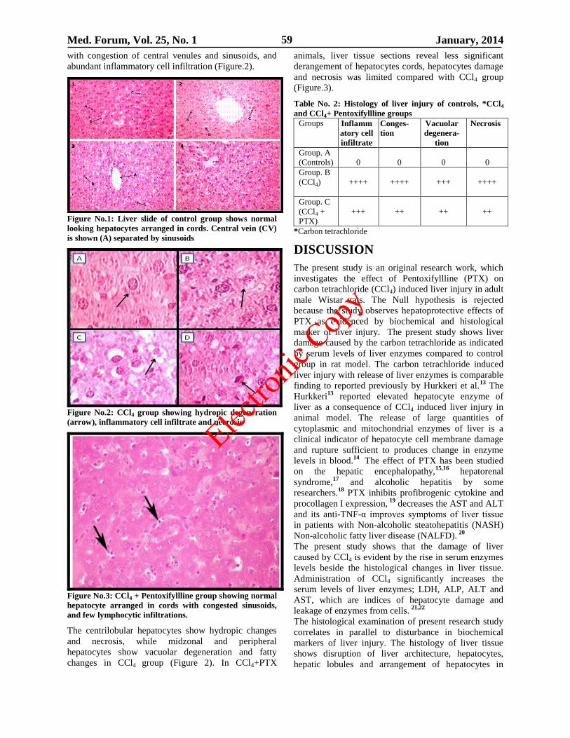
Med. Forum, Vol. 25, No. 1 January, 2014 59
with congestion of central venules and sinusoids, and
abundant inflammatory cell infiltration (Figure.2).
Figure No.1: Liver slide of control group shows normal
looking hepatocytes arranged in cords. Central vein (CV)
is shown (A) separated by sinusoids
Figure No.2: CCl4 group showing hydropic degeneration
(arrow), inflammatory cell infiltrate and necrosis
Figure No.3: CCl4 + Pentoxifyllline group showing normal
hepatocyte arranged in cords with congested sinusoids,
and few lymphocytic infiltrations.
The centrilobular hepatocytes show hydropic changes
and necrosis, while midzonal and peripheral
hepatocytes show vacuolar degeneration and fatty
changes in CCl4 group (Figure 2). In CCl4+PTX
animals, liver tissue sections reveal less significant
derangement of hepatocytes cords, hepatocytes damage
and necrosis was limited compared with CCl4 group
(Figure.3).
Table No. 2: Histology of liver injury of controls, *CCl4
and CCl4+ Pentoxifyllline groups
Groups Inflamm
atory cell
infiltrate
Conges-
tion
Vacuolar
degenera-
tion
Necrosis
Group. A
(Controls)
0
0
0
0
Group. B
(CCl4)
++++
++++
+++
++++
Group. C
(CCl4 +
PTX)
+++
++
++
++
*Carbon tetrachloride
DISCUSSION
The present study is an original research work, which
investigates the effect of Pentoxifyllline (PTX) on
carbon tetrachloride (CCl4) induced liver injury in adult
male Wistar rats. The Null hypothesis is rejected
because the study observes hepatoprotective effects of
PTX as evidenced by biochemical and histological
marker of liver injury. The present study shows liver
damage caused by the carbon tetrachloride as indicated
by serum levels of liver enzymes compared to control
group in rat model. The carbon tetrachloride induced
liver injury with release of liver enzymes is comparable
finding to reported previously by Hurkkeri et al.13
The
Hurkkeri13
reported elevated hepatocyte enzyme of
liver as a consequence of CCl4 induced liver injury in
animal model. The release of large quantities of
cytoplasmic and mitochondrial enzymes of liver is a
clinical indicator of hepatocyte cell membrane damage
and rupture sufficient to produces change in enzyme
levels in blood.14
The effect of PTX has been studied
on the hepatic encephalopathy,15,16
hepatorenal
syndrome,17
and alcoholic hepatitis by some
researchers.18
PTX inhibits profibrogenic cytokine and
procollagen I expression, 19
decreases the AST and ALT
and its anti-TNF-α improves symptoms of liver tissue
in patients with Non-alcoholic steatohepatitis (NASH)
Non-alcoholic fatty liver disease (NALFD). 20
The present study shows that the damage of liver
caused by CCl4 is evident by the rise in serum enzymes
levels beside the histological changes in liver tissue.
Administration of CCl4 significantly increases the
serum levels of liver enzymes; LDH, ALP, ALT and
AST, which are indices of hepatocyte damage and
leakage of enzymes from cells. 21,22
The histological examination of present research study
correlates in parallel to disturbance in biochemical
markers of liver injury. The histology of liver tissue
shows disruption of liver architecture, hepatocytes,
hepatic lobules and arrangement of hepatocytes in
Electro
nic Cop
y

Med. Forum, Vol. 25, No. 1 January, 2014 60
cords. The hepatocytes show findings of cellular injury
with marked cytoplasmic vacuolization. The injured
hepatocytes show pyknotic nuclei with lymphocyte
infiltrations. The pyknotic nuclei are a sign of severe
cellular injury caused by a toxin like carbon
tetrachloride. The histological and biochemical findings
of present study are comparable to those mentioned
previously.23-,25
The carbon tetrachloride is metabolized
to free radical during its metabolism and detoxification
in smooth endoplasmic reticulum by the cytochrome
P450.26
The Movassaghi, et al 4
(2013) studied
hepatoprotective effects of PTX on ecstasy induced
liver damage and reported effects of PTX. The PTX
reduced liver injury in this previous study by reducing
apoptosis and fibrosis caused by ecstasy in rat liver
model. The present study concludes that the PTX
decreases the carbon tetrachloride induced liver
damage.
CONCLUSION
The Pentoxifylline protects against oxidative damages
caused by carbon tetrachloride induced liver injury in
rat model. The Pentoxifyllline may be used as an
effective protector against chemical induced liver
damages; however, further studies are warranted.
REFERENCES
1. Essawy AE, Abdel-Moneim AM, Khayyat LI,
Elzergy AA. Nigella sativa seeds protect against
hepatotoxicity and dyslipidemia induced by carbon
tetrachloride in mice. J Appl Pharm Sci 2012;2
(10):021-5.
2. Wafay H, El-Saeed G, El-Toukhy S, Youness E,
Ellaithy N, Agaibi M, Eldaly S. Potential effect of
garlic oil and silymarin on carbon tetrachloride
induced liver injury. Austr J Basic Appl Sci 2012;
6 (3):409-14.
3. Russmann S, Kullak-Ublick GA, Grattagliano I.
Current concepts of mechanisms in drug-induced
hepatotoxicity. Curr Med Chem 2009; 16:3041-53.
4. Movssaghi S, Sharifi ZN, Mohammadzadeh F,
Soleimani M. Pentoxifylline protects the rat liver
against fibrosis and apoptosis induced by acute
administration of 3,4-Methyleneoxy methamphe-
tamine (MDMA or Ecstasy). Ir J Basic Med Sci
2013; 16: 922-27.
5. Obi FO, Omogbal LA, Orlafo OS, Ovat OD. Effect
of a Short Time Post Carbon Tetrachloride
Treatment Interval on Rat Plasma Enzyme Levels
and Percentage Mortality. J Applied Sci Environ
Mgt 2001; 5: 5-8.
6. Muriel P, Albo N, Perez-Alvarez VM. Kupffer
cells inhibition prevents hepatic lipid peroxidation
and damage induced by carbon tetrachloride. Comp
Biochem Physiol C Toxicol Pharmacol 2001;130:
219-26.
7. Rasha SA, Ashraf AA, Aly R. Carbon
tetrachloride-induced liver disease in rats: the
potential effect of supplement oils with vitamins E
and C on the nutritional status. German Med Sci
2009; 7: 1612-3174.
8. Rost DA, Welker DA, Welker J, Millonig G,
Berger I, Autschbach F, et al. Liver-homing of
purified glucose oxidase: A novel in vivo model of
physiological hepatic oxidative stress (H2O2). J
Hepatol 2007; 46: 482-91.
9. Seiffge D. Pentoxifylline: its influence on
interaction of blood cells with the vessel wall.
Atherosclerosis 1997;131:27–28.
10. Windmeier C, Gressner AM. Pharmacological
aspects of pentoxifylline with emphasis on its
inhibitory actions on hepatic fibrogenesis. Gen
Pharmacol 1997; 29:181–196.
11. Nayak S, Nalabothu P, Sandiford S, Bhogadi V,
Adogwa A. Evaluation of wound healing activity
of Allamanda cathartica L. and Laurus nobilis. L.
Extracts on rats. BMC Compl Alt Med 2006; 6: 12.
12. Murat-Bilgin H, Atmaca M, Deniz-Obay B,
Ozekinci S, Taşdemir E, Ketani A. Protective
effects of coumarin and coumarin derivatives
against carbon tetrachloride-induced acute
hepatotoxicity in rats. Exp Toxicol Pathol 2011;
63(4): 325-30.
13. Hurkkeri VI, Jaiparkash B, Lavhale RV, Karadi
RV, Kuppast IJ. Hepatoprotective activity of
Anthus Excelsa Roxb leaf extract on experimental
liver damage in rats. J Pharmacogn 2002;11:
120-28.
14. Shaarawy SM, Tohamy AA, Elgendy SM
Elmageed ZY, Bahnasy A, Mohamed MS, et al.
Protective effects of garlic and silymarin on
NDEA-induced rats hepatotoxicity. Int J Biol Sci
2009; 5(6): 549-57.
15. Assimakopoulos SF, Thomopoulos KC,
Labropoulou-Karatza C. Pentoxifylline: a first line
treatment option for severe alcoholic hepatitis and
hepatorenal syndrome? World J Gastroenterol
2009;15: 3194-3195.
16. Amini M, Runyon BA. Alcoholic hepatitis 2010: a
clinician's guide to diagnosis and therapy. World J
Gastroenterol 2010 21; 16:4905-4912.
17. McHutchison JG, Runyon BA, Draguesku JO,
Cominelli F. Person JL, Castracane J.
Pentoxifylline may prevent renal impairment
(hepatorenal syndrome) in severe acute alcoholic
hepatitis. Hepatol 1991; 14:96.
18. Akriviadis E, Botla R, Briggs W, Han S, Reynolds
T, Shakil O. Pentoxifylline improves short-term
survival in severe acute alcoholic hepatitis: a
double-blind, placebo-controlled trial.
Gastroenterol 2000; 119:1637-48
19. Raetsch C, Jia JD, Boigk G, Bauer M, Hahn EG,
Riecken EO, et al. Pentoxifylline downregulates
Electro
nic Cop
y
Med. Forum, Vol. 25, No. 1 January, 2014 61
profibrogenic cytokines and procollagen I
expression in rat secondary biliary fibrosis. Gut
2002; 50:241-7.
20. Li W, Zheng L, Sheng C, Cheng X, Qing L, Qu S.
Systematic review on the treatment of
pentoxifylline in patients with non-alcoholic fatty
liver disease. Lipids Health Dis 2011; 10:49.
21. Rajesh M and Latha M. Preliminary evaluation of
anti-hepatotoxic activity of Kamilari, a polyherbal
formulation. J Ethnopharmacol 2004; 91: 99-104.
22. Bashandy S, and Al-Wasel S. Carbon tetrachloride-
induced hepatotoxicity and nephrotoxicity in rats:
Protective role of vitamin C. J Pharm Toxico 2001;
16(30): 283-92.
23. Trivedi P, Mowat A. Carbon tetrachloride-induced
hepatic fibrosis and cirrhosis in the developing rat:
an experimental model of cirrhosis in childhood.
Br J Exp Pathol 1983; 64(1): 25–33.
24. Berman E, House D, Allis J, Simmons J.
Hepatotoxic interactions of ethanol with allyl
alcohol or carbon tetrachloride in rats. J Toxicol
Environ Health 1992;37(1): 161–76.
25. Brandao C, Ferreira H, Piovesana H, Polimeno N,
Ferraz J, de-Nucci G. Developmental model of
liver cirrhosis in rabbits. Clin Exp Pharma Physiol
2000;27:(12): 987-90.
26. Wang P, Kaneko T, Tsukada H, Nakano M,
Nakajima T, Sato A. Time courses of hepatic
injuries by chloroform and by carbon tetrachloride
comparison of biochemical and histopathological
changes. Arch Toxicol 1997; 71: 638-45.
Address for Corresponding Author:
Dr. Haji Khan Khoharo, Assistant Professor/Consultant Physician,
Department of Physiology/Medicine
Faculty of Medicine and Allied Medical Sciences, Isra
University, Hyderabad.
Tel: 0331-2662500
Electro
nic Cop
y

Med. Forum, Vol. 25, No. 1 January, 2014 62
Validity of Pleural Fluid Protein
in Differentiating Tuberculouse from Malignant
Pleural Effusion 1. Ghulam Nabi Khokhar 2. Muhammad Ashfaq 3. Ikram Ullah Khan
4. Muhammad Ishaq 5. Israr Ahmed 1. Prof. of Pharmacology 2. Assoc. Prof. of ENT 3. Asstt. Prof. of Pharmacology 4. Prof. of Surgery
5. Prof. of Physiology, Jinnah Medical College, Peshawar
ABSTRACT
Objective: Validity of pleural fluid protein in differentiating tuberculouse from malignant pleural effusion keeping
histopathology as gold standard.
Study Design: Cross sectional study.
Place and Duration: This study was conducted in the Pulmonology department post graduate medical institute,
Lady Reading Hospital Peshawar, Khyber Pakhtunkhwa (KPK) Pakistan from March 2009 to March 2010.
Materials and Methods: One hundred and seventy nine patients having clinical suspicion of pulmonary
tuberculouse and malignancy and fulfilling the inclusion criteria were subjected to Abrams needle biopsy, plural
tissue was examined by histopathology. Biopsy in order to know the significant difference of pleural fluid protein
level between tuberculous and malignant pleural effusion, histopathology finding and protein concentration were
determined their frequency and percentage.
Results: Among total number of 179 patients one hundred and fourteen (63.69%) were male and sixty five (36.32%)
were female. The age limit from 15-80 years, the result shows that 60.9% were tuberculous and 39.9% were
malignant pleuraleffusion, among these malignant 20 (11.2%) showed primary and 50 (27.9%) secondary
malignancy. Tuberculous PE was more common in younger age group while malignant PE in older age group, 32
number of patients falling in category A, 59 in category B, and 88 in category C.A protein level in belonging to
category C, there was statistically significant difference between tuberculous and malignant PE, tuberculous PE
have high concentration of protein than malignant PE, The category “A” have malignant PE.
Conclusion: Plural fluid total protein level determination and differentiating is a valuable tool in reaching to the
diagnosis of suspectedtuberculouse from malignant pleural effusion provided it is used in addition to the adequate
clinical scenario.
Key Words: Tuberculouse pleural effusion, malignant pleural effusion, Exudates, pleural fluid protein.
INTRODUCTION
Pleural effusion (PE) is a common clinical problem
both in developed and developing countries.(1)
The
etiological investigation of a pleural effusion is to
determine whether the effusion is a transudate or
exudates. Transudates reflect the presence of systemic
disease with repercussion.On the mechanism of pleural
fluid production and production and reabsorption.(2)
In
contrast, exudates reflect, the presence of primary
pleural disease and require etiological investigation.(3)
So in case with trandatePE, the diagnosis is usually
made without any difficulty but executive PE, require
careful differential diagnosis that include tuberculouse
(TB) and metastatic cancers, which are often found to
be the cause in a large number of patients.(4)(5)
Disease
in any organ can cause execute PE through a variety of
mechanisms including, malignancy, immunologic
response, lymphatic abnormality and non-infections
inflammation.(6)
Tuberculouse and malignancy are the
most common causes of exudative PE in our country.(7)
The gold standard for diagnosis of pleural tuberculouse
is theidentification of mycobacterium tuberculouse in
pleural fluid or tissue(8)
however in clinical practice this
identification is problematic of the low identification
rate of the bacillus (less than 30% in pleural fluid and
approximately 50% in the pleura) and the slow growth
of mycobacterium in culture (about 60 days). (9)
The
diagnosis of neoplastic pleural effusion is made based
on the presence of malignant cells in the pleural fluid or
tissue. The positivity rate of the cytological
examination ranges from 40 to 87% higher than that
obtained with a needle biopsy which ranges from 35 -
65 %. (13)
Several tests for the diagnosis of tuberculous
in pleural effusion have been used as tuberculouse
identification such as Adenosine deaminase, interferon,
Lysozyme, the polymerase chain reaction. (11)
and
specific C antibodies.(12)
However these test need
specific measure and expensive equipment that are not
available in most laboratories particularly in developing
countries, similarly in developed countries various new
parameters like pleural viscosity, C- reactive protein,
carcinoembryonic antigen, interleukin, interferon,
vascular endothelial growth factor, tumor necrosis
factor and pleural fluid T-cells are used for the
determination of tuberculous and malignancy.(13)
In our
Original Article Pharmacology
Electro
nic Cop
y
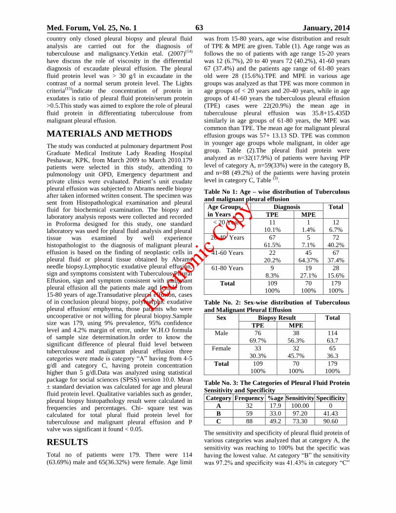
Med. Forum, Vol. 25, No. 1 January, 2014 63
country only closed pleural biopsy and pleural fluid
analysis are carried out for the diagnosis of
tuberculouse and malignancy.Yetkin etal. (2007)(14)
have discuss the role of viscosity in the differential
diagnosis of excaudate pleural effusion. The pleural
fluid protein level was > 30 g/l in excaudate in the
contrast of a normal serum protein level. The Lights
criteria(15)
indicate the concentration of protein in
exudates is ratio of pleural fluid protein/serum protein
>0.5.This study was aimed to explore the role of pleural
fluid protein in differentiating tuberculouse from
malignant pleural effusion.
MATERIALS AND METHODS
The study was conducted at pulmonary department Post Graduate Medical Institute Lady Reading Hospital Peshawar, KPK, from March 2009 to March 2010.179 patients were selected in this study, attending to pulmonology unit OPD, Emergency department and private clinics were evaluated. Patient’s unit exudate pleural effusion was subjected to Abrams needle biopsy after taken informed written consent. The specimen was sent from Histopathological examination and pleural fluid for biochemical examination. The biopsy and laboratory analysis reposts were collected and recorded in Proforma designed for this study, one standard laboratory was used for plural fluid analysis and pleural tissue was examined by well experience histopathologist to the diagnosis of malignant pleural effusion is based on the finding of neoplastic cells in pleural fluid or pleural tissue obtained by Abrams needle biopsy.Lymphocytic exudative pleural effusion, sign and symptoms consistent with Tuberculous Pleural Effusion, sign and symptom consistent with malignant pleural effusion all the patients male and female from 15-80 years of age.Transudative pleural effusion, cases of in conclusion pleural biopsy, polymorphic exudative pleural effusion/ emphyema, those patients who were uncooperative or not willing for pleural biopsy.Sample size was 179, using 9% prevalence, 95% confidence level and 4.2% margin of error, under W.H.O formula of sample size determination.In order to know the significant difference of pleural fluid level between tuberculouse and malignant pleural effusion three categories were made is category “A” having from 4-5 g/dl and category C, having protein concentration higher than 5 g/dl.Data was analyzed using statistical package for social sciences (SPSS) version 10.0. Mean ± standard deviation was calculated for age and pleural fluid protein level. Qualitative variables such as gender, pleural biopsy histopathology result were calculated in frequencies and percentages. Chi- square test was calculated for total plural fluid protein level for tuberculouse and malignant pleural effusion and P valve was significant it found < 0.05.
RESULTS
Total no of patients were 179. There were 114
(63.69%) male and 65(36.32%) were female. Age limit
was from 15-80 years, age wise distribution and result
of TPE & MPE are given. Table (1). Age range was as
follows the no of patients with age range 15-20 years
was 12 (6.7%), 20 to 40 years 72 (40.2%), 41-60 years
67 (37.4%) and the patients age range of 61-80 years
old were 28 (15.6%).TPE and MPE in various age
groups was analyzed as that TPE was more common in
age groups of < 20 years and 20-40 years, while in age
groups of 41-60 years the tuberculous pleural effusion
(TPE) cases were 22(20.9%) the mean age in
tuberculouse pleural effusion was 35.8+15.435D
similarly in age groups of 61-80 years, the MPE was
common than TPE. The mean age for malignant pleural
effusion groups was 57+ 13.13 SD. TPE was common
in younger age groups whole malignant, in older age
group. Table (2).The pleural fluid protein were
analyzed as n=32(17.9%) of patients were having PfP
level of category A, n=59(33%) were in the category B,
and n=88 (49.2%) of the patients were having protein
level in category C, Table (3)
.
Table No 1: Age – wise distribution of Tuberculous
and malignant pleural effusion Age Groups
in Years
Diagnosis Total
TPE MPE
< 20 Years 11
10.1%
1
1.4%
12
6.7%
20-40 Years 67
61.5%
5
7.1%
72
40.2%
41-60 Years 22
20.2%
45
64.37%
67
37.4%
61-80 Years 9
8.3%
19
27.1%
28
15.6%
Total 109
100%
70
100%
179
100%
Table No. 2: Sex-wise distribution of Tuberculous
and Malignant Pleural Effusion
Sex Biopsy Result Total
TPE MPE
Male 76
69.7%
38
56.3%
114
63.7
Female 33
30.3%
32
45.7%
65
36.3
Total 109
100%
70
100%
179
100%
Table No. 3: The Categories of Pleural Fluid Protein
Sensitivity and Specificity Category Frequency %age Sensitivity Specificity
A 32 17.9 100.00 0
B 59 33.0 97.20 41.43
C 88 49.2 73.30 90.60
The sensitivity and specificity of pleural fluid protein of
various categories was analyzed that at category A, the
sensitivity was reaching to 100% but the specific was
having the lowest value. At category “B” the sensitivity
was 97.2% and specificity was 41.43% in category “C”
Electro
nic Cop
y

Med. Forum, Vol. 25, No. 1 January, 2014 64
the sensitivity was the lowest of three having value
73.39% while the specificity was highest reaching to
90.6%.the positive (PPV) and negative predictive
(NPV) values were analyzed for the three categories of
plural fluid protein as that PPV was lowest for category
C having value of 2.7 while NPV was 58.57 for
category B, the PPV was 26.6 while the NPV was
having value of 52.87. The PPV was highest for
category “A” reading to the value 73.4 similarly the
PPV was also highest in this category reading to the
value of 88.57.
DISCUSSION
The distinction between tuberculous and malignant
pleural effusion poses a diagnostic challenge to the
physician. This is mainly due to the large proportion of
cases in which no confirmatory diagnosis of pleural
tuberculouse is achieved by microbiological methods,
and the sensitivity of cytological studies for malignancy
is inadequate(16)
. Tuberculouse and malignancy are the
most important and commonest causes of lymphocytic
exudative pleural effusion. But the ratio is different in
developed and developing countries. In areas were the
tuberculouse is not prevent MPE is more common than
TPE. In a study, conducted in Spain (2003) on 392
patients 73% cases were MPE and 27% were
TPE.(17)
Our study also showed that fact that TPE was
commoner than MPE, accounting for 60.9% of total
classes. Rest of cases (39.1 %) was malignant PE. Age
has also been an important complementary variable
while deciding about tuberculous or malignant PE. In
our study the TPE was commoner in younger age group
than MPE, Which was more in older age group. The
mean age for TPE was 35.8 + 15.43 SD while 57 +
13.13 SF was the mean age for MPE. Our findings were
comparable with the international studies.Porcel JM et
al (2003) reported in their study, the mean age of 30
years (range 22-40) in TPE while the mean age was
68(58-76) years in MPE cases.(18)
In our study we
showed that pleural fluid protein level was higher in
TPE than MPE and the difference was statistically
significant at > 5g/dl (category “C”) at < 4g/dl(category
“A”). The MPE was commoner than TPE and the
difference was significant. These finding were
consistent with international data.Porcel- Perez JM et al
(2004) reported that 73% of TPE were having pleural
fluid protein level > 5g/dl.This value was the cutoff
point for considering tuberculous etiology of the
lymphocytic educative PE(14)
. In another Spanish study
conducted on 105 patients of TPE, 57% of patients
showed plural fluid protein level above
5g/dl.(19)
Antonangelo et al (2007) reported higher
protein level in TPE than MPE. The protein level was
5.3+0.8g/dl in tuberculous PE while
4.2+1 was the level in MPE. The difference was
statically significant.(23)
Along with other laboratory
parameters, protein level was utilized for discrimination
between tuberculous and malignant PE. The same
findings were found in the study conducted by Liam et
al (2000) on patients having tuberculous or malignant
PE (20)
. Melo et al proposed 4.5g/dl as cutoff value for
diagnostic presumption of TPE (21).
Porcel JM et al
(2003) reported protein level of 5.4g/dl in tuberculous
while the level was 4.2g/dl in malignant pleural
effusion. The difference was statistically significant.
(18)The study recommended two scoring models for
differentiation between tuberculous and malignant PE.
It also revealed that in areas where ADA facility is
unavailable, plural fluid total protein level can be used
for differentiation between tuberculous and malignant
PE as used in one of the scoring model lacking ADA.
The pleural fluid protein showed sensitivity of 77% and
specificity of 80%. These results are comparable with
those of our study which showed 73.30% sensitivity
and 90.60% specificity at pleural fluid protein level of
> 5g/dl.
CONCLUSION
Plural fluid total protein level determination and
differentiating is a valuable tool in reaching to the
diagnosis of suspectedtuberculouse from malignant
pleural effusion provided it is used in addition to the
adequate clinical scenario.
REFERENCES
1. Magsi JA khan SU, Awan SR. Pleural biopsy in the
diagnosis of lymphatic educative pleural effusion.
Ann king Edward Med Coll 2005;11:572-4.
2. Maskell NA, Butland JA. BTS Guidelines for the
investigation of unilateral pleural effusion in
adults. Thorax 2003;58:8-17.
3. Story DD, Dines DE, Coles DT. Pleural effusion: a
diagnosis dilemma. Am J med Assoc 1976; 236:
2183-6.
4. Gannels JJ, Perplexing plural effusion. Chest 1978;
47:390-3.
5. Kesmiri M, Hashemzadeh M. use of cholesterol in
differentiating of educative and trasudative pleural
effusion. Med Iran 1997;2:187-9.
6. Sahn SA. Plural anatomy, physiology and
diagnostic producers. In: Baum GL, editors,
Textbook of pulmonary diseases. 6th
ed.
Philadelphia: Lippincott Raven;1998.p.255-65.
7. Anwar R, FarooqiJi causes of lymphocytic
exudative pleural effusion as revealed by
percutaneous pleural biopsy: experience from
Peshawar. Pak J Med Sci 2005;21:39-43.
8. Gopi A, Madahavan SM, Sharma SK, Diagnosis
and treatment of tuberculous pleural effusion in
2006. Chest 2007;131:880-9.
9. Valdes L, Pose A, San-jose E, Martinez – Vazquez
JM. The positive pleural effusion. A retrospective
study of cytopathologic diagnosis with autopsy
confirmation. Actacytol 1992;36:329-32.
Electro
nic Cop
y

Med. Forum, Vol. 25, No. 1 January, 2014 65
10. Villena V, Rebollo MJ Aguado JM, Galan A,
Encuentra AL, palenqye E. polymerase chain
reaction for the diagnosis of plural tuberculous in
immunocompromised and immunocompetent
patients. Clin infect Dis 1998;26:212-4.
11. Caminero JA, de-Castro T, Lafarga B, Daiz F,
Cabrera P. Diagnosis of plural tuberculouse by
detection of specific IgG anti-antigen 60 in serum
and plural fluid. Respiration 1993;60:58-62.
12. Hashimoto K. T-Helper type 1/T- Helper type 2
balance in malignant pleural effusions compared to
tuberculous effusion. Chest 2005;128:4030-5.
13. Chakrabarti B, Ryland I, sheard J, Chritopher J,
Earis EJ. The role of Abrams percutaneous pleural
biopsy in the investigation of exudative pleural
effusion. Chest 2006;129:1549-55.
14. Antonangelo L, varges FS, Seisceno M,
Bomboarda S, Teixera L, Sales RK. Clinical and
laboratory parameters in the differential diagnosis
of plural effusion secondary to tuberculouseor
cancer. Clinics 2007;62:585-90.
15. Yetkin O, Tek I, Yethkin F, Numanoglun N. Role
of plural viscosity in the differential diagnosis of
exudative pleural effusion. Respirol 2007;12:
267-71.
16. Light RW, MacGreg MI, luchsinger PC, ball WC.
Pleural effusion: the diagnostic separation of
transudtes pleural effusions. Chest 1997;111:
970-80.
17. Porcel JM, Vives M. Differentiating tuberculous
from malignant pleural effusion: A scoring model.
Med Scimonit 2003;9:175-80.
18. Chierakul N, Kanitsap A, Chaiprasert A,
Virivataveekul R. A simple Reactive protein
measurement for the differentiation between
tuberculous and malignant pleural effusion.
Respirol 2004;9:66-9.
19. Haro M, Ruiz –Manzano J, Gallego M, Abad J,
Manterola JM, Morera J. Pleural tuberculous:
analysis of 105 cases. Enfermifect Microbol Clin
1996;14:285-9.
20. Liam CK, Lim Wong CM. Differences in plural
fluid characteristic, white cell count, biochemistry
of tuberculous and malignant plural effusions. Med
Malaysia 2000;55:21-8.
21. Melo AF, Santos ML. Diagnosistco Da tuberculous
pleural pelaada, Isolada our combinadaaoutras
variaveis inclusive emhivpostivos. Folha Med
Address for Corresponding Author:
Prof. Dr. Muhammad Ishaq Chairman & Founder
Jinnah Medical College
Warsak Road Peshawar
Cell: +92-333-9152060
Email: [email protected] : [email protected]
Electro
nic Cop
y
Med. Forum, Vol. 25, No. 1 January, 2014 66
To Assess the In vitro Genotoxicity
of Metformin and Aspartame alone & In Combination 1. Amna Nazar 2. Rafique Ahmed 3. Sajida Bano 4. Muhammad Ashraf 5. Imran Altaf
6. Aqeel Javeed 1. Senior Lecturer of Pharmacology, HM&DC, Gujrat 2. Prof. of Anatomy, HM&DC, Gujrat 3. Prof. of
Biochemistry, HM&DC, Gujrat 4. Prof. & Chairman of Pharmacology, UVAS, Lahore 5. Asstt. Prof. of
Microbiology, UVAS, Lahore 6. Asstt. Prof. of Pharmacology, UVAS, Lahore
ABSTRACT
Objective: Metformin is a known oral antidiabetic agent belonging to the class of biguanides, widely prescribed for
the treatment of type 2 Diabetes Mellitus (DM). In this study the genotoxic potential of metformin was studied alone
and in combination with an artificial sweetener aspartame as most of the diabetic patients utilized this low calorie
sweetener to reduce their sugar consumption per day. Many complaints regarding its potential to cause DNA
damage have been submitted to FDA
Study Design: Experimental study
Place and Duration of Study: This study was carried out at the Department of Pharmacology and Toxicology,
University of Veterinary and Animal Sciences Lahore from 1st Jan 2011 to Dec 2011.
Materials and Methods: Peripheral Blood Lymphocytes were exposed to various concentrations of metformin,
aspartame and their combination. DNA damage was checked by comet assay. The data was analyzed by using
Analysis of Variance (ANOVA) by Statistical Package of Social Sciences SPSS
Results: Exhibited dose dependant rise in comet tail lengths. Moreover the data advocates that tail lengths of
lymphocytes after exposure to aspartame were high as compared to metformin. When lymphocytes were exposed to
combination of aspartame and metformin and DNA damage was checked by comet assay, the results were
significantly different (p<0.05) as compared to metformin and aspartame alone.
Conclusion: It can be concluded from the present study that aspartame is posing great genotoxic threat to the cells
as compared to metformin and the combination is even more toxic to DNA, so the drug regime of diabetic patient
must be closely monitored. There is further need of more studies in this regard.
Key Words: Metformin, Aspartame, Diabetes Mellitus, Comet Assay. DNA damage.
INTRODUCTION
Among the antidiabetic agents, metformin
(1-(diaminomethylidene)-3,3-dimethyl-guanidine) is
reported to have several properties, such as it reduces
formation of Advance glycation endproducts(AGE),
helps to regain the reduced levels of glutathione in DM
and increases the antioxidant defense1. A recent study
demonstrates that metformin increases AMPK
activation in MIN6 cells responsible for insulin
production and primary rat beta cells in dose dependant
manner. Prolonged exposure of metformin (>24h)
resulted in a gradual rise in apoptotic beta-cells similar
to the effect which was documented for AMPK-
activator AICAR2.
AMPK is an enzyme that plays a role in cellular energy
homeostasis and its persistent activation has recently
been concerned with apoptosis of beta cells of pancreas
and in cell death of islets of langerhans3.
Activation of AMPK by metformin might be mediated
by the mitochondria-derived RNS in vivo in C57BL6
mice4. There are certain studies supporting that
metformin stimulate cell death in p53-deficient
HCT116 cells in condition of limited glucose
availability. So metformin can be beneficial to be used
in patients with p53 deficient tumors which have
become resistant to chemotherapy5.
The antidiabetic drug has also been reported to cause a
decline in the occurrence of mammary carcinomas in
hamsters when administered in therapeutic doses. Most
probably it would have damaged the genetic material of
the carcinoma cells to reduce chances of cancer
prevalence in mammary cells of hamster6. Certain
reports support combination therapy of metformin and
doxorubicin which kills both cancer stem cells and
non–stem cancer cells in culture, and cause reduction in
tumor mass and delays relapse more efficiently than
metformin of doxorubicin can do alone7.
Onaran et al. (2006) suggested that therapeutic
concentrations of metformin could not check DNA
damage caused by oxidative stimulus in cultured human
lymphocytes regardless of its anti oxidant
characteristic8.
The use of artificial sweetener aspartame is increasing
day by day by both diabetic as well as non diabetic
population as a sugar substitute. Aspartame is mainly
composed of phenylalanine (50%), aspartic acid (40%)
and methanol (10%). In this study different doses of
aspartame was administered to check the cellular
effects of aspartame on brain and it was revealed that
Original Article Pharmacology
Electro
nic Cop
y

Med. Forum, Vol. 25, No. 1 January, 2014 67
consistent use of aspartame might cause various mental
disorders due to damage to the brain cells 9.
The effect of an artificial sweetener Aspartame was
studied to investigate oxidative stress and brain
monoamines in normal conditions and also after
administration of lipopolysaccharides intraperitoneally
in mice. The results revealed that aspartame alone and
in combination with any mild systemic inflammatory
response enhance inflammation and oxidative stress in
brain cells10
.
MATERIALS AND METHODS
Chemicals: Tested chemicals i.e., Aspartame and
Metformin were provided by Popular Laboratories,
Lahore Pakistan. Dimethyl Sulfoxide (DMSO) was
purchased from Merck Pakistan. All other chemicals
and media used were of analytical grade.
Comet Assay: Cavity slides were layered with 1%
Normal melting Agarose and placed in refrigerator for
2-12 hours. Peripheral blood lymphocytes were
separated by using Lymphocyte separating medium.
3ml of 1:1 ratio of peripheral blood with PBS was
layered onto 4ml of Lymphocyte separating medium
and after centrifuging lymphocyte suspension was made
in RPMI-164011
. Now the lymphocytes were exposed to
various concentrations of test chemicals i.e. metformin,
aspartame and combination doses of aspartame and
metformin. The exposed cells were mixed 1% low
melting point agarose and layered onto the precoated
slides. After solidification 3rd
layer of Agarose 1% was
layered onto the same slide and allowed to solidify.
Subsequently the slides were dipped in the lysing
solution (pH 10) consisting of high salt concentration
and detergent for 2 hours. After that slides were
exposed to alkaline buffer (pH > 13) consisting of 1mM
EDTA and 300mM NAOH for DNA unwinding for 20
minutes. The cells were subjected to electrophoresis for
20 minutes at 25 volts and 300mA. Slides were washed
with 0.4M TRIS buffer/ Neutralizing buffer (pH 7.5) 3
times and then stained with Ethidium bromide12
.
PBS was used as negative control and DMSO 20% was
used as positive control13
.
Microscopic Analysis: The slides were visualized by
using Fluorescent Microscope at 40X objective. The
DNA damage was quantified by measuring tail lengths
of 25 consecutive cells and quantification was done in
triplicate manner. Four damage categories were
established according to the method described by
Valencia-Quintana and coworkers (Valencia-Quintana
et al. 2012).
Class 0= Undamaged cells
Class 1= Tail length <Head Diameter
Class 2= Tail Length > Head Diameter BUT Tail
Length <Double of Head Diameter
Class 3= Tail Length > Double of Head Diameter
The damage to the DNA was expressed as %age
fragmentation and ultimately finding the DNA damage
index of each concentration according to the following
formula.
Damage Index= No. of cells in Class.1+ (2×No. of
cells in Class.2) + (3×No. of cells in Class.3)
Fragmentation %= No. of cells in Class.1 + No. of
cells in Class.2 + No. of cells in Class.3 ×100
Total Number of cells under observation
Statistical Analysis: The data was analyzed by using
Analysis of Variance (ANOVA) by Statistical Package
of Social Sciences SPSS for windows (version 16;
SPSS Inc; Chicago IL; USA) and Tukey’s test was
applied. The value of p<0.05 was considered
significant.
RESULTS
The results of comet assay on lymphocyte sampled
from peripheral blood treated with different
concentrations of metformin are presented in Table 1.
Treatment with different doses of metformin induced a
significant increase in DNA damage at 200, 250, 300,
350 and 400µg/ml as these values were significantly
higher (p < 0.01) than that found in the negative
control. It is noted that percentage of fragmentation of
DNA and damage index increases as the concentration
of metformin increases reaching a maximum value at
400µg/ml. The effect observed with 10, 20, 80, 100 and
150µg/ml do not show significant differences (p < 0.05)
when compared with the negative control.
Lymphocytes were exposed to 10 different
concentrations of aspartame presented in the Table 2.
The treatment with various doses of aspartame showed
same order of genotoxicity as that of metformin.
Aspartame caused significant rise (p < 0.01) in DNA
damage at 500, 1000, 2000, 4000, 8000 µg/ml as
compared to the DNA damage caused by negative
control. The percentage fragmentation of DNA and
Damage Index becomes high when the exposed
concentration of aspartame increases and the maximum
DNA damage index is found at 8000 µg/ml. However
the results with lower doses i.e. 12.5, 25, 50, 100 and
250µg/ml did not show any significant rise (p < 0.05) in
the DNA damaging potential as compared to negative
control.
When the cells were exposed to the combination of
aspartame and metformin, the results were significantly
high (p < 0.05) as compared to metformin and
aspartame individually. Here again dose dependant rise
in genotoxicity is observed in manner similar to the
individual drugs. We can see a significant rise (p <
0.01) in DNA damage index at 500:200, 1000:250,
2000:300, 4000:350, 8000:400 µg/ml as compared to
the negative control (Table 3). Percentage
fragmentation and DNA damage index increases as the
concentration of aspartame and metformin increases
with maximum DNA damage index obtained at
8000:400 µg/ml which has highly significant difference
(p < 0.001) with negative control.
Electro
nic Cop
y
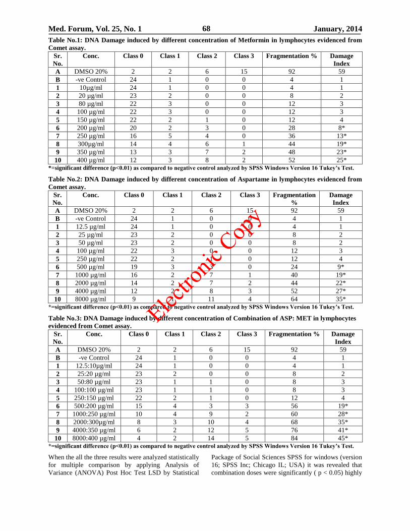
Med. Forum, Vol. 25, No. 1 January, 2014 68
Table No.1: DNA Damage induced by different concentration of Metformin in lymphocytes evidenced from
Comet assay.
Sr.
No.
Conc. Class 0 Class 1 Class 2 Class 3 Fragmentation % Damage
Index
A DMSO 20% 2 2 6 15 92 59
B -ve Control 24 1 0 0 4 1
1 10µg/ml 24 1 0 0 4 1
2 20 µg/ml 23 2 0 0 8 2
3 80 µg/ml 22 3 0 0 12 3
4 100 µg/ml 22 3 0 0 12 3
5 150 µg/ml 22 2 1 0 12 4
6 200 µg/ml 20 2 3 0 28 8*
7 250 µg/ml 16 5 4 0 36 13*
8 300µg/ml 14 4 6 1 44 19*
9 350 µg/ml 13 3 7 2 48 23*
10 400 µg/ml 12 3 8 2 52 25*
*=significant difference (p<0.01) as compared to negative control analyzed by SPSS Windows Version 16 Tukey’s Test.
Table No.2: DNA Damage induced by different concentration of Aspartame in lymphocytes evidenced from
Comet assay.
Sr.
No.
Conc. Class 0 Class 1 Class 2 Class 3 Fragmentation
%
Damage
Index
A DMSO 20% 2 2 6 15 92 59
B -ve Control 24 1 0 0 4 1
1 12.5 µg/ml 24 1 0 0 4 1
2 25 µg/ml 23 2 0 0 8 2
3 50 µg/ml 23 2 0 0 8 2
4 100 µg/ml 22 3 0 0 12 3
5 250 µg/ml 22 2 1 0 12 4
6 500 µg/ml 19 3 3 0 24 9*
7 1000 µg/ml 16 2 7 1 40 19*
8 2000 µg/ml 14 2 7 2 44 22*
9 4000 µg/ml 12 2 8 3 52 27*
10 8000 µg/ml 9 1 11 4 64 35*
*=significant difference (p<0.01) as compared to negative control analyzed by SPSS Windows Version 16 Tukey’s Test.
Table No.3: DNA Damage induced by different concentration of Combination of ASP: MET in lymphocytes
evidenced from Comet assay.
Sr.
No.
Conc. Class 0 Class 1 Class 2 Class 3 Fragmentation % Damage
Index
A DMSO 20% 2 2 6 15 92 59
B -ve Control 24 1 0 0 4 1
1 12.5:10µg/ml 24 1 0 0 4 1
2 25:20 µg/ml 23 2 0 0 8 2
3 50:80 µg/ml 23 1 1 0 8 3
4 100:100 µg/ml 23 1 1 0 8 3
5 250:150 µg/ml 22 2 1 0 12 4
6 500:200 µg/ml 15 4 3 3 56 19*
7 1000:250 µg/ml 10 4 9 2 60 28*
8 2000:300µg/ml 8 3 10 4 68 35*
9 4000:350 µg/ml 6 2 12 5 76 41*
10 8000:400 µg/ml 4 2 14 5 84 45*
*=significant difference (p<0.01) as compared to negative control analyzed by SPSS Windows Version 16 Tukey’s Test.
When the all the three results were analyzed statistically
for multiple comparison by applying Analysis of
Variance (ANOVA) Post Hoc Test LSD by Statistical
Package of Social Sciences SPSS for windows (version
16; SPSS Inc; Chicago IL; USA) it was revealed that
combination doses were significantly ( p < 0.05) highly
Electro
nic Cop
y

Med. Forum, Vol. 25, No. 1 January, 2014 69
genotoxic as compared to metformin. However the
results of combination doses were not significantly (p <
0.05) different as compared to aspartame individually.
DISCUSSION
The prime objective of therapy for type 2 diabetes mellitus is to obtain and sustain good glycemic control, and to reduce the risk of secondary complications associated with DM. Metformin is considered a gold standard treatment of DM along with certain interventions in lifestyle of patients. Metformin also have efficacy against polycystic ovary syndrome and various neoplasms
14.
The antidiabetic drug has been reported to cause a decline in the occurrence of mammary carcinomas in hamsters when administered in therapeutic doses. Most probably it would have damaged the genetic material of the carcinoma cells to reduce chances of cancer prevalence in mammary cells of hamster
6. The results
of comet assay on various concentrations of metformin exhibited dose dependent rise in its potential to damage DNA. Percentage fragmentation of DNA and damage index increases as the concentration of metformin increases reaching a maximum value at 400µg/ml. The results of this study are in accordance with the fact that prolonged exposure of metformin may become a potential threat of genotoxicity. A study was conducted to check the genotoxic potential of metformin in Chinese Hamster Ovary cells and DNA damage was checked by Chromosomal Aberration Assay and single cell gel alkaline electrophoresis/comet assay. Results showed significant damage to the DNA by metformin
15.
However data is available which supports that metformin is neither cytotoxic nor genotoxic when various concentrations were administered in vivo condition to diabetic and non diabetic rats
16. Another
study was conducted to evaluate the prevention of DNA damage in human lymphocytes by metformin. Cells were exposed to various concentrations of metformin and subsequently they were incubated with cumene hydroperoxide a known DNA damaging substance. The results were evaluated by comet assay revealing that metformin is unable to prevent DNA damage by any chemical causing oxidative stress to the cells
8.
Metformin kills cancer stem cells in genetically different type of breast cancer so combination therapy of metformin and doxorubicin is used to reduce tumor mass and delays relapse more efficiently that metformin and doxorubicin can do alone
7.
The underlying mechanism responsible for DNA damage by metformin may lie in the fact that metformin may inhibit mitochondrial respiration cells and ultimately there is rapid rise in the superoxide level because of inhibition of cellular respiration due to insult to the mitochondria
17. According to Warburg theory of
cancer, the key reason for development of tumor is an inadequate cellular respiration due to insult to mitochondria
18.
The use of sweetening agent by diabetic individuals is common. A survey of our diabetic clinic population showed that 65% regularly use these products
19. It is
widely used by the diabetic population for the sweet taste avoiding high calories through sugar consumption
20. The results of comet assay revealed that
aspartame exhibited significant potential of genotoxicity when different concentrations of the sweetener were exposed to the lymphocytes. Damage Index of DNA revealed that aspartame was causing more damage to the DNA of peripheral blood lymphocytes as compared to metformin when compared with the negative control. The results of comet assay are in agreement with a study exhibiting significantly high (p < 0.01) carcinogenic potential on prolonged use, demonstrating that these artificial sweeteners are not entirely safe although they are FDA approved
21.
Results also state that there is dose dependant rise in the DNA damaging potential of aspartame endorsing the statement that aspartame can cause chromosomal aberrations at all concentrations (500, 1000 and 2000 µg/ml) and treatment periods, in human lymphocytes, in a dose dependant manner
22. The underlying
mechanism of DNA damage caused by aspartame may lie in the fact that prolong exposure of aspartame may result in detectable amount of methanol in blood
23. As
aspartame consists of 3 components aspartic acid, phenylalanine and methanol which is most dangerous of all
24. Various studies revealed the data that
aspartame is responsible for increased frequency of lymphomas and leukemias and is also responsible in significant rise in prevalence of transitional cell carcinomas of renal pelvis at doses approximated with the ADI of aspartame for humans. These results indicate that aspartame proves to be the multipotential carcinogen greatly affecting the quality of life
25.
As far as results of the combination doses are concerned they cause severe damage to DNA when exposed to the lymphocytes and cells were quantified using comet assay. %age fragmentation and DNA damage index were significantly high (p < 0.05) as compared to individual results of metformin and aspartame. The data advocates the same threshold level of genotoxicity but the intensity of the genotoxic effect was relatively high in case of combination as compared to the individual drugs. The additive effect of both aspartame and metformin on DNA damage may be accredited to the fact that aspartame is responsible for increased oxidative stress to the cells
10. Along with that
metformin is associated with production of reactive nitrogen species from mitochondria
4 which ultimately
hinder the cellular respiration leading to cell damage.
CONCLUSION
It can be concluded from the present study that aspartame is posing great genotoxic threat to the cells as compared to metformin. When combination of aspartame and metformin were exposed to the peripheral blood lymphocytes the results showed significantly high (p < 0.05) genotoxicity as compared to metformin and aspartame individually. Most of the diabetic patients utilize aspartame as an artificial sweetener along with their daily regimen of antidiabetic drug metformin. So caution must be taken while using
Electro
nic Cop
y

Med. Forum, Vol. 25, No. 1 January, 2014 70
both these products together as it may cause significant damage to the cells of body.
REFERENCES
1. Arnalich F, Hernanz A, Lopez-Maderuelo D, De la Fuente M, Arnalich FM, Andres-Mateos E, Fernandez-Capitan C, Montiel C. Intracellular glutathione deficiency is associated with enhanced nuclear factor kappa B activation in older non-insulin dependent diabetic patients. Free Radic Res 2001;35(6): 873–884.
2. Kefas BA, Cai Y, Kerckhofs K, Ling Z, Marten G, Heimberg H, Pipeleers D, Van de Casteele M. Metformin induced stimulation of AMP-activated protein kinase in beta-cells impairs their glucose responsiveness and can lead to apoptosis. Biochem Pharmacol 2004; 68: 409-416.
3. Riboulet-Chavey A, Diraison F, Siew LK, Wong FS, Rutter GA. Inhibition of AMP activated protein kinase protects pancreatic ß-cells from cytokine-mediated apoptosis and CD8 T-Cell-Induced cytotoxicity. Am Diab Assoc 2008;57(2): 415-23.
4. Zou MH, Kirkpatrick SS, Davis BJ, Nelson JS, Wiles WG, Schlattner U, Neumann D, Brownlee M, Freeman MB, Goldman MH. Activation of the AMP-activated protein kinase by the anti-diabetic drug metformin in vivo. Role of mitochondrial reactive nitrogen species. J Biol Chem 2004; 279(42): 43940-43951.
5. Buzzai M, Jones RG, Amaravadi RK, Lum JJ, DeBerardinis RJ, Zhao F, et al. Systemic treatment with the antidiabetic drug metformin selectively impairs p53-deficient tumor cell growth. Cancer Res 2007;67(14): 6745-6752.
6. Anisimov VN, Berstein LM, Egormin PA, Piskunova TS, Popovich IG, Zabezhinski MA, et al. Effect of metformin on life span and on the development of spontaneous mammary tumors in HER-2/neu transgenic mice. Exp Gerontol 2005; 40(8-9): 685–693.
7. Hirsch HA, Iliopoulos D, Tsichlis PN, Struhl K. Metformin selectively targets cancer stem cells and acts together with chemotherapy to block tumor growth and prolong remission. Cancer Res 2009;69: 7507-7511.
8. Onaran I, Guven GS, Ozdas SB, Kanigur G, Vehid S. Metformin does not prevent DNA damage in lymphocytes despite its antioxidant properties against cumene hydroperoxide-induced oxidative stress. Mutat Res 2006; 611: 1-8.
9. Humphries P, Pretorius E, Naude H. Direct and indirect cellular effects of aspartame on the brain. Eur J Clin Nutr 2008;62(4):451-462.
10. Abdel-Salam OM, Salem NA, Hussein JS. Effect of aspartame on oxidative stress and monoamine neurotransmitter levels in lipopolysaccharide-treated mice. Neurotox Res 2012; 21(3): 245-55.
11. Dhawan A, Bajpayee M, Pandey AK, Parmar D. 2009. Protocol for the single cell gel Electrophoresis / comet assay for rapid Genotoxicity assessment. ITRC.
12. Tice RR, Agurell E, Anderson D, Burlinson B,
Hartmann A, Kobayashi H, et al. The single cell gel/comet assay: Guidelines for in vitro and in vivo genetic toxicology testing. Environ Mol Mutagen 2000;35(3): 206--221.
13. Valencia-Quintana R,1Gómez-Arroyo S, Marian Waliszewski S, Sánchez-Alarcón J, Gómez- Olivares JL, Flores-Márquez AR, et al. Evaluation of the genotoxic potential of dimethyl sulfoxide (DMSO) in meristematic cells of the root of Vicia faba. Toxicol Environ Health Sci 2012;4(3): 154-160.
14. Marić A. Metformin – more than ‘gold standard’ in the treatment of type 2 diabetes mellitus. Diabetol Croat 2010;39-3.
15. Amador RR, Longo JP, Lacava ZG, Dórea JG, Almeida Santos Mde F. Metformin (dimethyl-biguanide) induced DNA damage in mammalian cells. Genet Mol Biol 2011;35(1): 153-158.
16. Attia SM, Helal GK, Alhaider AA. Assessment of genomic instability in normal and diabetic rats treated with metformin. Chem Biol Interact 3009; 180: 296-304.
17. González-Barroso MM, Anedda A, Gallardo-Vara E, Redondo-Horcajo M, Rodríguez-Sánchez L, Rial E. Fatty acids revert the inhibition of respiration caused by the antidiabetic drug metformin to facilitate their mitochondrial β-oxidation. Biochim Biophys Acta 2012; 1817(10): 1768-75.
18. Verschoor ML, Ungard R, Harbottle A, Jakupciak JP, Parr RL, Singh G. Mitochondria and Cancer: Past, Present, and Future. Bio Med Res Int 2013; 1-10.
19. Coulston AM, Hollenbeck CB, Donner CC, Williams R, Chiou YM, Reaven GM. Metabolic effects of added dietary sucrose in individuals with non-insulin dependent diabetes mellitus (NIDDM). Metabolism 1985; 34: 962-6.
20. Nehrling JK, Kobe P, McLane MP, Olson RE, Kamath S, Horwitz DL. Aspartame use by persons with diabetes. Diab Care 1985;8(41): 5-7.
21. Mukherjee A, Chakrabarti J. In vivo cytogenetic studies on mice exposed to Acesulfame K-a non-nutritive sweetener. Food Chem Toxicol 1997;35: 1177-1179.
22. Rencüzoǧullari E, Tüylü BA, Topaktaş M, İla HB, Kayraldız A, Arslan M, Diler SB. Genotoxicity of aspartame. Drug Chem Toxicol 2004;27(3): 257-268.
23. Iyyaswamy A, Rathinasamy S. Effect of chronic exposure to aspartame on oxidative stress in the brain of albino rats. J Biosci 2012; 37(4): 679-88.
24. Lydon C. Could there be evils lurking in aspartame consumption? Oxygen Magazine, retrieved October 30, 2008 fromhttp://www. aspartame. com/lydon.htm
25. Soffritti M, Belpoggi F, Esposti DD, Lambertini L, Tibaldi E, Rigano A. First experimental demonstration of the multipotential carcinogenic effects of aspartame administered in the feed to sprague-dawley rats. Environ Health Perspect 2006;114(3): 379-85.
Electro
nic Cop
y

Med. Forum, Vol. 25, No. 1 January, 2014 71
Study of Type 2 Diabetes Mellitus
and its Correalation with Blood Groups 1. Shamsul Arfin Qasmi 2. Aftab Ahmed Khan 3. Hasan Askari 4. Rabiyah Pirzada 1. Prof. of Pathology, Mohammad Bin Qasim M&DC, Karachi 2. Asstt. Prof. of Dental Materials Science,
Mohammad Bin Qasim M&DC, Karachi 3. Lecturer of Biochemistry, Mohammad Bin Qasim M&DC, Karachi
4. Sindh Medical College (DUHS). Karachi
ABSTRACT
Objectives: To ascertain any relation between T2 Diabetes Mellitus with Blood Groups of the patients coming in
the OPD.
Study Design: Experimental / Analytical study
Place and Duration of Study: This study was carried out at Med Path Laboratories and Diagnostic Center
Gulshan-e-Iqbal Karachi & KESC Medical center Karachi from August 2012 to December 2012.
Materials and Methods: The present study deals with the distribution of the ABO blood types in patients with
diabetes mellitus. 500 samples were collected from Med Path Laborites and Diagnostic Center KESC Medical
Center.
Results: The study shows blood group B was prevalent at a high percentage among patients with DM type 2.
Blood group O+ were significantly higher among the male diabetics and blood group B+ among female diabetics.
People with middle-aged group were seen to be more affected with type 2 diabetes mellitus.
Conclusion: It is highly recommended that this study may proceed further and the affiliation of T2DM can be
screened at molecular level to find out the exact mechanism of action for susceptibility to these particular Blood
Groups.
Key words: Diabetes Mellitus, Correalation, Blood Groups
INTRODUCTION
Diabetes Mellitus (DM) is a set of related diseases in
which the body cannot regulate the amount of sugar
(specifically, glucose) in the blood. The blood delivers
glucose to provide the body with energy to perform all
of a person's daily activities. The liver converts the
food a person eats into glucose. In diabetes, glucose in
the blood cannot move efficiently into cells, so blood
glucose levels remain high. This not only starves all
the cells that need the glucose for fuel, but also harms
certain organs and tissues exposed to the high glucose
levels.1
Classification of Diabetes Mellitus: Diabetes Type 1
Patients produce no insulin at all. Diabetes Type 2 –
Patients don't produce enough insulin, or insulin is not
working properly. Gestational Diabetes - develops
diabetes just during your pregnancy.
Maturity onset diabetes of the young (Monogenic
Diabetes) (MODY)-Refers to any of several hereditary
forms of diabetes caused by mutations in an autosomal
dominant gene disrupting insulin production.
Type 1 diabetes (T1D): The body stops producing
insulin or produces too little insulin to regulate blood
glucose level.
Type 1 diabetes involves about 10% of all people with
diabetes. Type 1 diabetes is typically diagnosed during
childhood or adolescence. It used to be referred to as
juvenile-onset diabetes or insulin-dependent diabetes
mellitus.2
Causes of Type 1 diabetes: Type 1 diabetes can occur
in an older individual due to destruction of the pancreas
by alcohol, disease, or removal by surgery. It also
results from progressive failure of the pancreatic beta
cells, the only cell type that produces significant
amounts of insulin.
Type 2 diabetes (T2D): Although the pancreas still
secretes insulin, the body of someone with type 2
diabetes is partially or completely unable to use this
insulin. This is sometimes referred to as insulin
resistance. The pancreas tries to overcome this
resistance by secreting more and more insulin.
Causes of Type 2 Diabetes: Type 2 diabetes is thought
to be caused by a combination of genetic and
environmental factors.
Genetic causes — Many people with type 2 diabetes
have a family member with either type 2 diabetes or
other medical problems associated with diabetes, such
as high cholesterol levels, high blood pressure, or
obesity. Environmental conditions — Environmental
factors such as eating habits and physical activity are,
combined with genetic causes, affect the risk of
developing type 2 diabetes.3
Gestational Diabetes: A small number (about 3 to 5
percent) of pregnant women develop diabetes during
pregnancy, called "gestational diabetes."
Maturity onset diabetes of the young: MODY is a
rare form of diabetes which is different from both Type
1 and Type 2 diabetes, and runs strongly in families.
MODY is caused by a mutation (or change) in a
single gene.
Original Article Diabetes Mellitus
Electro
nic Cop
y

Med. Forum, Vol. 25, No. 1 January, 2014 72
It was reported that DM type 2 is the most common
type, accounting for 90-95% of all diabetic cases.(4) In
1998 it was estimated that there were almost140
million people with diabetes and the predictions by
Hilary King indicate that this figure would rise up
to 300 million by the year 2025.5
Blood Groups: Blood groups are created by molecules
present on the surface of red blood cells (and often on
other cells as well). The major human blood group
system is ABO. The blood group of a person depends
upon the presence or absence of two genes, A and B.
The ABO blood groups were the first to be discovered
(in 1900) by Karl Landsteiner and are the most
important in assuring safe blood transfusions. The Rh
blood group system is the second most significant
system for blood grouping.
Rh factor refers to Rh D antigen only. Determination of
Rh factor along with ABO is essential for defining the
Rh +ve or Rh -ve status of the individual. Around 85%
of the human population is Rh +ve while 15% is
Rh -ve. 6
The ABO & Rh systems are the most significant blood
group systems from the clinical point of view.7
Many researchers have made attempts to determine the
significance of particular ABO phenotypes for
susceptibility to disease. The relationship between the
ABO/rhesus (Rh) blood groups and various diseases has
generated a great deal of interest8. Certain diseases
show a strong association with the ABO/Rh blood
groups, notably peptic ulcer and gastric cancer9.
Individuals with blood group O have been found to
be at a higher risk of contracting cholera than those
with other blood groups. The ratio of this risk of group
O to group A individuals has been reported as 1.35:1.
Oral candidiasis shows a higher incidence of group O
over other ABO groups10
. Small pox virus has been
found to carry an A antigen-like structure, so that
individuals who possess a naturally occurring anti-A
(group O and B individuals) are thought to have an
increased resistance to the infection.11
This could be attributed to the inability of the
immune system of group A individuals to recognize the
A like antigen of the tumor cells as foreign and cannot
destroy them, but group O and B individuals do have a
naturally Occurring anti- A that are most likely to
destroy tumor cells.12
It has been reported that group O individuals are
proportionately more prone to bleeding than individuals
of the other blood groups; and group an individuals with
highest incidence of thrombosis than in any other
group. 13
The blood groups of diabetics have been extensively
studied since McConnell’s suggestion in 1955 of an
increased frequency of blood group A among these
patients.14
In Copenhagen, an excess of blood group O
was found in male diabetics.15
No diseases are known to result from the lack of
expression of ABO blood group antigens, but the
susceptibility to a number of diseases has been
interrelated to a person’s ABO phenotype.16
Such
correlations remain conflicting and include the
observation that gastric cancer is more common in
group A individuals, whereas gastric and duodenal
ulcers occur more commonly among blood group O
individuals.17
In the present study, an attempt has been
made to investigate any association with the ABO blood
types and diabetes mellitus type 2.
MATERIALS AND METHODS
The present study was an epidemiological study. A total
no of 500 blood samples from patients with Diabetes
mellitus (Type 2) were collected from the Med Path
Laboratory. Since both gender differentiations are not
known to exist in the ABO blood type system, the
samples collected from both males and females were
pooled for the analyses.
Materials:
BSL 2 lab
Gloves
Lab coat
Blood typing tiles
500 diabetic patients Blood samples
Synthetic anti-Rh (D) serum
Synthetic anti-A serum
Synthetic anti-B serum
Justors (50 ul)
Mixing sticks (blue, yellow, and white)
Method:
Blood Sample Collection: For the ABO blood types,
standard serological procedures were followed using the
anti-A, anti-B and anti-D antisera. A blood sample drawn
from a vein in the arm or a finger-prick
Abo Test:
1. Using the justor, place a drop of the blood sample in
each demo created area of the blood typing tile.
Replace the tip of the jester.
2. Add a drop of synthetic anti-A (blue) to the well
labeled A.
3. Add a drop of synthetic anti-B serum (yellow) to the
well labeled B.
4. Add a drop of synthetic anti-Rh serum (clear) to the
well labeled Rh.
5. Using a different color mixing stick for each well
(blue for anti-A, yellow for anti-B, white for anti-Rh),
gently stir the synthetic blood and anti-serum drops
for 30 seconds. Remember to discard each mixing
stick in danger box after a single use to avoid
contamination of your samples.
6. Carefully examine the thin films of liquid mixture. If
a film remains uniform in appearance, there is no
agglutination. If the sample appears granular,
agglutination has occurred. Determine the blood type
Electro
nic Cop
y

Med. Forum, Vol. 25, No. 1 January, 2014 73
of the sample. A positive agglutination reaction
indicates the blood type.
7. Record the results in the data table.
8. Thoroughly rinse the blood typing tile with a
disinfectant, and then repeat steps.
9. The data were entered in Microsoft excel for analysis.
Further analysis was carried out using instant graph-
pad.
RESULTS
In Table 1 the distribution of the ABO blood types in
patients with diabetes mellitus is shown including both
male and female.
Table No.1: Correlation of blood groups
Blood Group Frequency Percentage
A+ 124 24.80%
A- 9 1.80%
B+ 150 30.00%
B- 6 1.20%
AB+ 63 12.60%
AB- 2 0.40%
O+ 131 26.20%
O- 15 3.00%
Total 500 100.00%
Chart: Blood group frequency
Table No.2A: Correlation of blood groups in Male
Blood Group Frequency Percentage
A+ 71 24.83%
A- 7 2.45%
B+ 66 23.08%
B- 3 1.05%
AB+ 41 14.34%
AB- 2 0.70%
O+ 87 30.42%
O- 9 3.15%
Total 286 100.00%
In our study, the results showed association between ABO
blood groups and DM type 2. B+, O+, A+ blood group
showed higher incidence of type 2 diabetes mellitus than
groups which are Rh-ve signifying the higher incidence in
patients which refers to Rh+ve group. However,
significant association was found between DM type 2 and
blood groups A (P 24.80%) and O (P 26.20%) The
frequency of B blood group (30.00%) was high among
patients with DM type 2. AB blood groups showed little
association with DM (P 12.60% ) ,which implied that
AB blood group patients have less chances of DM
type 2. (Table 1)
In Table 2: A and 2: B the distribution of ABO blood
groups between genders with diabetes mellitus is shown.
Figure No.2A: Frequency
Table No.2B: Correlation of blood groups in Female
Blood Group Frequency Percentage
A+ 53 24.77%
A- 2 0.93%
B+ 84 39.25%
B- 3 1.40%
AB+ 22 10.28%
AB- 0 0.00%
O+ 44 20.56%
O- 6 2.80%
Total 214 100.00%
Figure No.2b: Frequency
The distribution of ABO/Rh blood groups between the
genders for diabetics is shown in Table II. Blood groups
O+ were more dominant in the diabetic group among men
(30.42%) than women (20.56%); the type 2 DM in B+
Electro
nic Cop
y

Med. Forum, Vol. 25, No. 1 January, 2014 74
blood groups were more common in women (39.25%)
than men (23.08%). (Table 2)
Table No.3A: Correlation of blood group by Age in
Male
Blood Group 20 – 40 41 - 60 61 - 80
A+ 18 36 17
A- 3 4 0
B+ 11 36 19
B- 1 0 2
AB+ 8 21 12
AB- 0 2 0
O+ 27 47 13
O- 0 4 5
Total 68 150 68
Figure No.3A:
Table No.3B: Correlation of blood group by Age in
Female
Blood Group 20 – 40 41 - 60 61 - 80
A+ 15 29 9
A- 0 1 1
B+ 22 50 12
B- 1 2 0
AB+ 7 12 3
AB- 0 0 0
O+ 16 22 6
O- 1 4 1
Total 62 120 32
Figure No.3B:
In Table 3: A and 3: B the distribution of diabetic’s blood
types by age between the genders is shown.
A distinction is also drawn between diabetes in the young
and in the middle-aged and in old respectively. It was
observed that in middle-aged (40-60) people were more
affected by DM type 2 having different blood groups.
(Table 3)
DISCUSSION
It was long suggested that the ABO blood group system
had evolved under a positive selection pressure in both
humans and other primates.18
This implies that certain
ABO groups provide a selected vulnerability to individuals
possessing a particular ABO blood group. Diabetes
mellitus was observed to occur in all the ABO/Rhesus
blood groups. However it was observed that the patients
with type 2 diabetes mellitus were at rise in Rh+ve cases
of which are A, B and O. This study clearly reveals the
importance of blood group in different group of patients
based on gender, age and Rh groups. This study suggests
that person with Rh+ve are more prone to develop type 2
diabetes mellitus than the person with Rh -ve group.
In this study it was also found out that the following
groups were more susceptible to develop type 2 diabetes
mellitus in sequence of higher percentage.
Blood
group Percentage
Blood
group Percentage
B+ 30.00% B- 1.20%
O+ 26.20% O- 3.00%
A+ 24.80% A- 1.80%
AB+ 12.60% AB- 0.40%
This comparison certainly reflects that the person having
B+ blood group is more likely to develop type 2 diabetes
mellitus than person having blood group B.
The association of type 2 DM in male and female is
somewhat equal however, in male the incidence was
higher in O+ group and female shows high incidence in
blood group B+.
This was also evident with regard to age which were
divided in to three groups, 20-40, 41-60, 61-80 showing
higher incidence in age groups of 41-60 with equal
distribution of type 2 DM O+ and B+ blood groups which
are more likely to develop type 2 DM in their age group.
The study of Qureshi and Bhatti demonstrated that DM
type 2 and ABO blood groups are interrelated; they found
that among 70 patients with DM, blood group B was
more common and represented 35.71%.19
It is interesting
to note that our study did show a higher percentage
of blood group B (30.00% ) in the diabetic group, but
this failed to achieve statistical significance, and results
are in agreement with Qureshi and Bhatti. In addition,
there were reports from Italy and Trinidad showing an
increased frequency of blood group B among
diabetics.20
Electro
nic Cop
y

Med. Forum, Vol. 25, No. 1 January, 2014 75
CONCLUSION
It is concluded that the persons having these blood groups
who are more susceptible to develop T2DM should take
due care with reference to increased incidence of
developing DM and adopt healthy life style in every
respect keeping in view the danger zone in which they are
living and the information they have with respect to their
own blood group. It is highly recommended that this study
may proceed further and the affiliation of T2DM can be
screened at molecular level to find out the exact
mechanism of action for susceptibility to these particular
Blood Groups.
REFERENCES
1. American Diabetes Association (2008). Diabetes
Basics. Available online: http://www.diabetes.
org/diabetes-basics.
2. Van Dam RM, et al. Dietary patterns and risk for type
2 diabetes mellitus in U.S. men. Annals of Int Med
2002;136(3):201–209.
3. Belmokhtar F, Belmokhtar R. Department of Ecology
and Environment, Université Abou Bakr Belkaïd de
Tlemcen, 2009.
4. King H, Aubert RE, Herman W. Global burden of
diabetes 1995 2025. Diabetes Care 1998;21:1414 31.
5. Harris M, Hadden WC, Knowler WC, Bennett PH.
Prevalence of diabetes and impaired glucose tolerance
and plasma glucose levels in U.S. population aged 20
74. 2010.
6. Zaoui S, Fingold J, Meguenni K, Chabane Sari D.
ABO and rhesus blood groups system in Tlemcen
population, West Algerian. Biologie et Santé 2007;
7(1):62-71.
7. Raj K. The Distribution of the ABO Blood Types in
Patients with Diabetes Mellitus (Department of Sports
Medicine and Physiotherapy, Guru Nanak Dev
University, Amritsar, Punjab, India). Anthropologist
2008;10(2):129-132.
8. Kamil M, Nagi HA, Al-Jamal, Yusoff NM. Advanced
Medical and Dental Institute, University Sains
Malaysia, Penang, Malaysia; 2009.
9. Buford-Mason AP, Weber JC, Willoughby IM. Oral
Carriage of Candida albicans, ABO blood group and
secretor status in healthy subjects. J Med Mycol
26(1):49-56. Diabetes 1987;36:523 34.
10. Issitt PD, Anstee DJ. Applied Blood Group Serology.
Fourth Edition Montgomery. Sci Publi 1999;218-246.
11. McConnell R, Pyke D, Roberts JAF. Blood groups in
diabetes mellitus. Br Med J 2006;772 6.
12. Koley S. The distribution of the ABO blood types in
patients with diabetes mellitus. Anthropologist 2008;
10:129 32.
13. Der ABO Blugruppen, Acta. Gene. Et Statistica
Medica 2010;10:267-294.
14. O’huigen C, Sato A, Klein J. Evidence for
convergent evaluation of A and B blood group
antigens in primates. Hum. Genet 1997;101:141-148.
15. Qureshi M, Bhatti R. Frequency of ABO blood
groups among the diabetes mellitus type 2 patients. J
Coll Physician Surg Pak 2003;8:453-5.
16. Tedeschi G, Cavazzuti F. Contributo casistico allo
studio dei rapporti tra diabete mellito e gruppi
sanguigni. ABO & Rh Prog Med (Napoli) 2010;15:
76 82.
17. Arid I, Bentall H, Roberts J. A relationship between
cancer of stomach and the ABO blood groups. Br
Med J 1953;1:799 801.
18. Andersen J, Lauritzen E. Blood groups and diabetes
mellitus. Diabetes 1960;9:20-4.
19. Vogel von F, Pettenkofor HJ, Helmbold W. Uber Die
Populations genetics, 1960.
20. Henry MU, Poon-King T. Blood groups and diabetes.
West Ind Med J 1961;10:156-60.
Address for Corresponding Author:
Dr. Shamsul Arfin Qasmi,
Professor & HOD, Dept of Pathology,
Mohammad Bin Qasim Medical & Dental College,
Bin Qasim Town. Karachi
e-mail: [email protected]
Electro
nic Cop
y
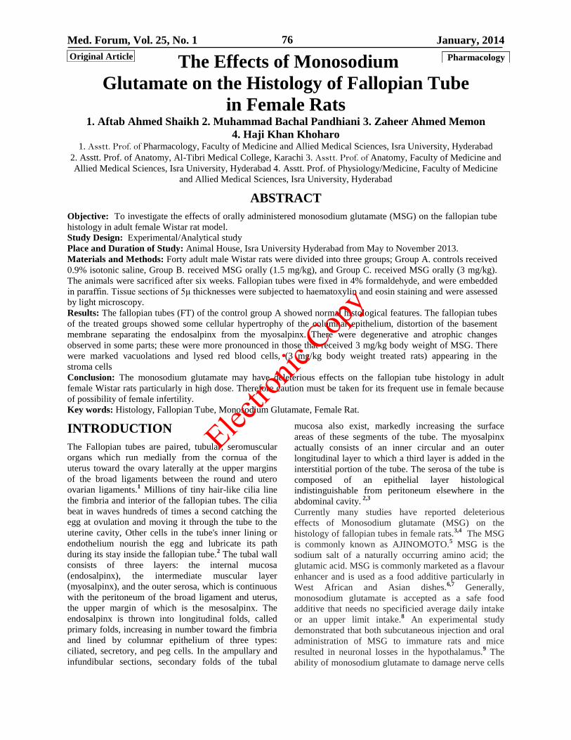
Med. Forum, Vol. 25, No. 1 January, 2014 76
The Effects of Monosodium
Glutamate on the Histology of Fallopian Tube
in Female Rats 1. Aftab Ahmed Shaikh 2. Muhammad Bachal Pandhiani 3. Zaheer Ahmed Memon
4. Haji Khan Khoharo 1. Asstt. Prof. of Pharmacology, Faculty of Medicine and Allied Medical Sciences, Isra University, Hyderabad
2. Asstt. Prof. of Anatomy, Al-Tibri Medical College, Karachi 3. Asstt. Prof. of Anatomy, Faculty of Medicine and
Allied Medical Sciences, Isra University, Hyderabad 4. Asstt. Prof. of Physiology/Medicine, Faculty of Medicine
and Allied Medical Sciences, Isra University, Hyderabad
ABSTRACT
Objective: To investigate the effects of orally administered monosodium glutamate (MSG) on the fallopian tube
histology in adult female Wistar rat model.
Study Design: Experimental/Analytical study
Place and Duration of Study: Animal House, Isra University Hyderabad from May to November 2013.
Materials and Methods: Forty adult male Wistar rats were divided into three groups; Group A. controls received
0.9% isotonic saline, Group B. received MSG orally (1.5 mg/kg), and Group C. received MSG orally (3 mg/kg).
The animals were sacrificed after six weeks. Fallopian tubes were fixed in 4% formaldehyde, and were embedded
in paraffin. Tissue sections of 5μ thicknesses were subjected to haematoxylin and eosin staining and were assessed
by light microscopy.
Results: The fallopian tubes (FT) of the control group A showed normal histological features. The fallopian tubes
of the treated groups showed some cellular hypertrophy of the columnar epithelium, distortion of the basement
membrane separating the endosalpinx from the myosalpinx. There were degenerative and atrophic changes
observed in some parts; these were more pronounced in those that received 3 mg/kg body weight of MSG. There
were marked vacuolations and lysed red blood cells, (3 mg/kg body weight treated rats) appearing in the
stroma cells
Conclusion: The monosodium glutamate may have deleterious effects on the fallopian tube histology in adult
female Wistar rats particularly in high dose. Therefore caution must be taken for its frequent use in female because
of possibility of female infertility.
Key words: Histology, Fallopian Tube, Monosodium Glutamate, Female Rat.
INTRODUCTION
The Fallopian tubes are paired, tubular, seromuscular
organs which run medially from the cornua of the
uterus toward the ovary laterally at the upper margins
of the broad ligaments between the round and utero
ovarian ligaments.1 Millions of tiny hair-like cilia line
the fimbria and interior of the fallopian tubes. The cilia
beat in waves hundreds of times a second catching the
egg at ovulation and moving it through the tube to the
uterine cavity, Other cells in the tube's inner lining or
endothelium nourish the egg and lubricate its path
during its stay inside the fallopian tube.2 The tubal wall
consists of three layers: the internal mucosa
(endosalpinx), the intermediate muscular layer
(myosalpinx), and the outer serosa, which is continuous
with the peritoneum of the broad ligament and uterus,
the upper margin of which is the mesosalpinx. The
endosalpinx is thrown into longitudinal folds, called
primary folds, increasing in number toward the fimbria
and lined by columnar epithelium of three types:
ciliated, secretory, and peg cells. In the ampullary and
infundibular sections, secondary folds of the tubal
mucosa also exist, markedly increasing the surface
areas of these segments of the tube. The myosalpinx
actually consists of an inner circular and an outer
longitudinal layer to which a third layer is added in the
interstitial portion of the tube. The serosa of the tube is
composed of an epithelial layer histological
indistinguishable from peritoneum elsewhere in the
abdominal cavity. 2,3
Currently many studies have reported deleterious
effects of Monosodium glutamate (MSG) on the
histology of fallopian tubes in female rats.3,4
The MSG
is commonly known as AJINOMOTO.5 MSG is the
sodium salt of a naturally occurring amino acid; the
glutamic acid. MSG is commonly marketed as a flavour
enhancer and is used as a food additive particularly in
West African and Asian dishes.6,7
Generally,
monosodium glutamate is accepted as a safe food
additive that needs no specificied average daily intake
or an upper limit intake.8 An experimental study
demonstrated that both subcutaneous injection and oral
administration of MSG to immature rats and mice
resulted in neuronal losses in the hypothalamus.9 The
ability of monosodium glutamate to damage nerve cells
Original Article Pharmacology
Electro
nic Cop
y

Med. Forum, Vol. 25, No. 1 January, 2014 77
of the hypothalamus is a pointer to the fact that it may
alter the neural control of reproductive hormone
secretion via the hypothalamic-pituitary-gonadal
regulatory axis. The effects of such toxicants on male
reproduction may be anatomical or only functional,
depending on whether they produce structural changes
in the reproductive system, or merely affect the
functions of the reproductive organs.10
In the last few years, fear had increased due to the
adverse reactions and toxicity of MSG, with few and
limited literature regarding the histological studies of
the damage in fallopian tubes of animals treated with
MSG. Therefore, the present study aimed to investigate
some probable histological effects of MSG on the
fallopian tube histology in adult female Wistar rats.
MATERIALS AND METHODS
The present experimental study included forty young
adult male Wistar rats at animal house of Isra
University from May to November 2013. Adult female
Wistar rats of 250-300 grams were included, while
male rats weighing <250 grams or >300 grams were
excluded from the study.
Animals: The Animals were housed in animal
house at an optimal room temperature with 55-60%
humidity and exposed to 12 hour light-dark cycles.
The chaw like fresh alfalfa and clean water are
provided freely. The rats were divided into three
groups;
o Group A. Control Group (n=10) Rats
received 0.9% isotonic saline orally on
alternate day for three successive weeks and
served as control group,
o Group B. (n=10) Experimental Rats were
given 1 mg/kg of monosodium glutamate
orally.
o Group C. (n=10) Experimental Rats were
given 3mg/kg of monosodium glutamate
orally.
Chemical: The chemical used was monosodium
glutamate (C5H9NO4-Na+). The MSG was
purchased from the open market of Hyderabad
under the license of Ajinomoto co.INC. Tokyo,
Japan. A stock solution was prepared by dissolving
30 and 60 g of MSG crystals in 100 ml of distilled
water. The dose schedule was so adjusted that the
amount of MSG administration per animal was as
per their respective weight. The MSG doses were
given for six weeks. The applied doses were
selected according to as referenced.11
Fallopian tube histology: At the end of the
experimental period, the animals were sacrificed by
cervical dislocation and the abdominal cavity was
opened up to expose the fallopian tube which were
quickly dissected out, and fixed in 10% formal
saline for routine histological techniques. The
tissues were dehydrated in an ascending grade of
alcohol (ethanol), cleared in xylene and embedded
in paraffin wax. Serial sections of 3–4 lm thick
were obtained using a rotator microtome. The
deparaffinized sections were stained routinely with
hematoxylin and eosin. Sections of fallopian tube
were examined by light microscope.
Photomicrographs of the desired sections were
obtained for microscopic details..
RESULTS
The present study was conducted to evaluate the effects
of monosodium glutamate (MSG) on the histology of
testis in rat model. The MSG was given in different
doses in the experimental group animals as mentioned
in methodology. The control group revealed normal
histology. The fallopian tubes (FT) of the control group
A showed normal histological features, illustrating a
well defined tubal wall which consists of three layers:
the internal mucosa (endosalpinx), the intermediate
muscular layer (myosalpinx), and the outer serosa
(Fig. 1).
The experimental groups (Groups B and C) were
studied separately for the microscopic structure of
fallopian tubes. The major derangements were observed
in the fallopian tubes of high dose MSG treated rats (3
mg/kg body weight). The histological details of
experimental rats are shown in figure 2-3.
The fallopian tubes of the treated groups showed some
cellular hypertrophy of the columnar epithelium,
distortion of the basement membrane separating the
endosalpinx from the myosalpinx. There were
degenerative and atrophic changes observed in some
parts; these were more pronounced in those that
received 3 mg/kg body weight of MSG. There were
marked vacuolations and lysed red blood cells, (3
mg/kg body weight treated rats) appearing in the stroma
cells (Figs. 2 and 3).
Figure No. 1: Section of testis from rats of control group
(Group A) rat showing semniferous tubules (T) and
interstitial spaces (N) showing normal Leydig cells (H & E
stains, × 400).
Electro
nic Cop
y
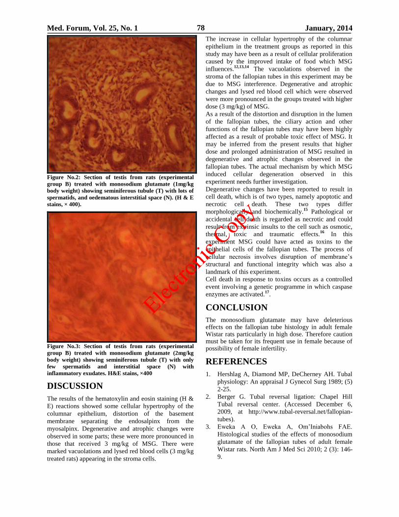
Med. Forum, Vol. 25, No. 1 January, 2014 78
Figure No.2: Section of testis from rats (experimental
group B) treated with monosodium glutamate (1mg/kg
body weight) showing seminiferous tubule (T) with lots of
spermatids, and oedematous interstitial space (N). (H & E
stains, × 400).
Figure No.3: Section of testis from rats (experimental
group B) treated with monosodium glutamate (2mg/kg
body weight) showing seminiferous tubule (T) with only
few spermatids and interstitial space (N) with
inflammatory exudates. H&E stains, ×400
DISCUSSION
The results of the hematoxylin and eosin staining (H &
E) reactions showed some cellular hypertrophy of the
columnar epithelium, distortion of the basement
membrane separating the endosalpinx from the
myosalpinx. Degenerative and atrophic changes were
observed in some parts; these were more pronounced in
those that received 3 mg/kg of MSG. There were
marked vacuolations and lysed red blood cells (3 mg/kg
treated rats) appearing in the stroma cells.
The increase in cellular hypertrophy of the columnar
epithelium in the treatment groups as reported in this
study may have been as a result of cellular proliferation
caused by the improved intake of food which MSG
influences.12,13,14
The vacuolations observed in the
stroma of the fallopian tubes in this experiment may be
due to MSG interference. Degenerative and atrophic
changes and lysed red blood cell which were observed
were more pronounced in the groups treated with higher
dose (3 mg/kg) of MSG.
As a result of the distortion and disruption in the lumen
of the fallopian tubes, the ciliary action and other
functions of the fallopian tubes may have been highly
affected as a result of probable toxic effect of MSG. It
may be inferred from the present results that higher
dose and prolonged administration of MSG resulted in
degenerative and atrophic changes observed in the
fallopian tubes. The actual mechanism by which MSG
induced cellular degeneration observed in this
experiment needs further investigation.
Degenerative changes have been reported to result in
cell death, which is of two types, namely apoptotic and
necrotic cell death. These two types differ
morphologically and biochemically.15
Pathological or
accidental cell death is regarded as necrotic and could
result from extrinsic insults to the cell such as osmotic,
thermal, toxic and traumatic effects.16
In this
experiment MSG could have acted as toxins to the
epithelial cells of the fallopian tubes. The process of
cellular necrosis involves disruption of membrane’s
structural and functional integrity which was also a
landmark of this experiment.
Cell death in response to toxins occurs as a controlled
event involving a genetic programme in which caspase
enzymes are activated.17
.
CONCLUSION
The monosodium glutamate may have deleterious effects on the fallopian tube histology in adult female Wistar rats particularly in high dose. Therefore caution must be taken for its frequent use in female because of possibility of female infertility.
REFERENCES
1. Hershlag A, Diamond MP, DeCherney AH. Tubal
physiology: An appraisal J Gynecol Surg 1989; (5)
2-25.
2. Berger G. Tubal reversal ligation: Chapel Hill
Tubal reversal center. (Accessed December 6,
2009, at http://www.tubal-reversal.net/fallopian-
tubes).
3. Eweka A O, Eweka A, Om’Iniabohs FAE.
Histological studies of the effects of monosodium
glutamate of the fallopian tubes of adult female
Wistar rats. North Am J Med Sci 2010; 2 (3): 146-
9.
Electro
nic Cop
y

Med. Forum, Vol. 25, No. 1 January, 2014 79
4. Berger G. Tubal reversal ligation: Chapel Hill
Tubal reversal center. (Accessed December 6,
2009, at http://www.tubal-reversal.net/fallopian-
tubes).
5. Eweka O. Histological studies of the effects of
monosodium glutamate on the kidney of adult
Wistar rats. Internet J Health 2007; 6:2.
6. Igwebuike UM, Ochiogu IS, Ihedinihu BC, Ikokide
JE, Idika IK. The effects of oral administration of
monosodium glutamate (MSG) on the testicular
morphology and cauda epididymal sperm reserves
of young and adult male rats. Veterinarski Arhiv
2011; 81 (4): 525-34.
7. Farombi EO, Onyema OO. Monosodium
glutamate-induced oxidative damage and
genotoxicity in the rat: modulatory role of vitamin
C, vitamin E and guercetin. Human Experimental
Toxicol 2006; 125: 251-259.
8. Samuels S. The toxicity/safety of MSG: a study in
suppression of information. Account Res 1999;
6(4):259–310.
9. Burde RM, Schainker MB, Kayes J. Acute effect of
oral and subcutaneous administration of
monosodium glutamate on the arcuate nucleus of
the hypothalamus in mice and rats. Nature 1971;
233, 58-60.
10. Witorsch RJ. Reproductive toxicology, 2nd ed.
Raven press, new york.1995.
11. Tyl R, Friedman M. Effects of acrylamide on
rodents reproductive performance. Reprod Toxicol
2003; 17(1):1–13.
12. Rogers PP, Blundell JE. Umani and appetite.
Effects of monosodium glutamate on hunger and
food intake in human subjects. Physiol Behav1990;
48(6): 801-4.
13. Iwase M, Yamamoto M, Iino K, IchikawaK,
Shinohara N, Yoshinari F. Obesity induced by
neonatal monosodium glutamate treatment in
spontaneously hypertensive rats: an animal model
of multiple risk factors'. Hypertens Res 1998; 43:
62-8.
14. Gobatto CA, Mello MA, Souza CT, Ribeiro IA.
The monosodium glutamate (MSG) obese rat as a
model for the study of exercise in obesity. Res
Commun Mol Pathol Pharmacol 2002; (2): 116-28.
15. Wyllie AH. Glucocorticoid-induced thymocyte
apoptosis is associated with endogenous
endonuclease activation. Nature, London, 1980;
284: 555-6.
16. Farber JL, Chein KR, Mittnacht S. The
pathogenesis of irreversible cell injury in ischemia.
Am J Pathol 1981; 102:271-81.
17. Waters CM, Walkinshaw G, Moser B, Mitchell IJ.
Death of neurons in the neonatal rodent globus
pallidus occurs as a mechanism of apoptosis.
Neuroscience 1994; 63: 881-894.
Address for Corresponding Author:
Dr. Haji Khan Khoharo,
Assistant Professor of Physiology/Medicine
Faculty of Medicine and Allied Medical Sciences
Isra University, Hyderabad.
Tel: 0331-2662500
Electro
nic Cop
y

Med. Forum, Vol. 25, No. 1 January, 2014 80
Incidence of Malignant
Lymphomas in Balochistan 1. Zahid Mehmood 2. M.Hanif Mengal 3. Haroon ur Rasheed
4. Hafiz Khush Naseeb Ahmed 1. Asstt. Prof., Dept. of Radiotherapy & Oncology, BMCH, Quetta 2. Asstt. Prof., Haematology Section, BMCH,
Quetta 3. Ex- Asstt. Prof., Dept. of Radiotherapy & Oncology, BMCH, Quetta 4. Director CENAR, Quetta.
ABSTRACT
Background: Traditionally lymphomas are classified into Hodgkin’s disease (HD) and Non Hodgkin’s lymphoma
(NHL) depending upon histo-pathological evidence on biopsy taken from an enlarged lymph node. Delayed
diagnosis in lymphoma deteriorates the health eminence resulting in poor outcome.
Objectives: The aim of the study is to estimate the incidence and clinical presentation of malignant lymphomas in
Balochistan.
Study Design: Prospective Study
Place and Duration of Study: This study was carried in the Department of Radiotherapy & Oncology, Bolan
Medical Complex Hospital, Quetta from June, 2006 to May, 2012
Materials and Methods: A total of 263 newly diagnosed patients of both types of lymphoma from different parts of
Balochistan were registered in Bolan Medical Complex Hospital in Department of Radiotherapy & Oncology.
Followed by histopathology, WHO classification and Ann Arbor staging was done to assign subtype and extent of
disease.
Results: It was found that the incidence of Non Hodgkin’s lymphoma a (64.7%) was greater than Hodgkin’s disease
(35.2%) and both present bimodal distribution in age. Male patients dominate female patients in both cases (2.5:1).
Lymph-adenopathy of cervical region was primary site in 44 % of cases while 27.6 % were extra nodal.
Histopathology shows 57.4 % mixed cellularity variant in Hodgkin’s lymphoma and 25.5% diffuse B cell pattern in
Non Hodgkin’s lymphoma. Ann Arbor staging reveals that 3.4% cases present with stage I and 64.5% show
stage IV.
Conclusion: To conclude Non Hodgkin’s lymphoma is two times more frequent than Hodgkin’s lymphoma with
greater male contribution. Due to delayed diagnosis resulted in late stage presentation ,health awareness is needed
for physicians and general population for availability of patients at a due time for management.
Key Words: Hodgkin’s lymphoma, Non Hodgkin’s lymphoma, Lymph-adenopathy.
INTRODUCTION
Lymphomas are group of disorders caused by
malignant lymphocytes that accumulates in lymph
nodes and cause the characteristic feature of lymph-
adenopaty. Occasionally, they may spill over in blood
or infiltrate organs outside the lymphoid tissue [1]
.
Lymphatic organs play an important part in the immune
system, having a considerable overlap with the
lymphoid system. Lymphoid tissue is found in many
organs, particularly the lymph nodes, and in the
lymphoid follicles associated with the digestive system.
Lymphoid tissues contain lymphocytes, but they also
contain other types of cells for support [2].
The system
also includes all the structures dedicated to the
circulation and production of lymphocytes, which
includes the spleen, thymus, bone marrow, and the
lymphoid tissue associated with the digestive system[3]
.
The Primary lymphoid tissues generate two major types
of cells, B lymphocytes and T lymphocytes. Lymphoma
is a group of cancers that affect these lymphocytes. It is
malignant transformation of either B cell or T cell or
their subtypes, these abnormal lymphocytes may travel
from one lymph node to another and some time to
remote organs via lymphatic system. Care should be
taken to assign the primary site of origin as it
strappingly affects treatment modality. The primary site
may be nodal when malignant cells originate from
lymph nodes and is extra nodal when develop from the
organs other than lymphatic system such as
gastrointestinal tract, hypochondriam, Para-nasal
sinuses, central nervous system . Lymphoma may
invade the nearby organs involving extra nodal sites
and vice versa [4]
.
Traditionally lymphomas are classified into Hodgkin’s
disease (HD) and Non Hodgkin’s lymphoma (NHL)
depending upon histo-pathological evidence on biopsy
taken from an enlarged lymph node. Hodgkin’s
lymphoma develops from a specific abnormal B
lymphocyte lineage and has characteristics Read
Sternberg cells having large, abundant cytoplasm,
double or multiple nuclei with its distinctive clinical
features [5]
. Since there are so many different types of
lymphomas, its classification is very complicated [6].
Many of these subtypes look similar but they are
functionally quite different and respond to different
types of therapies with different probability of cure [7]
.
Original Article Malignant Lymphomas
Electro
nic Cop
y

Med. Forum, Vol. 25, No. 1 January, 2014 81
Diffuse large B cell Lymphoma is most common and is
potentially curable while mantle cell lymphoma is
unique subtypes B cell lymphoma and is potentially
incurable [4,8,9]
. Due to the varied clinical picture, many
patients are misdiagnosed and treated for diseases like
tuberculosis[10].
Sometimes, Benign disorders including
ordinary infections, sebaceous cysts and other non-
neoplastic conditions may be interpreted as malignant
lymphoma and unnecessarily subjected to surgery
and/or chemotherapy [11].
The first sign of lymphoma is
painless enlarged lymph nodes accompanying fever for
more than 3 days. Usually patient presents with low
grade intermittent fever, unexplained weight loss and
drenching night sweats. Confirmed diagnosis is
established on the basis of histopathology findings
followed by fine needle aspiration or biopsy of relevant
site. Histopathology clearly marks the morphology of
cells, their subtypes and resemblance to the normal
cells. Clinically Non Hodgkin’s lymphoma is dividing
into low grade, intermediate and high grade. High grade
lymphoma has cells and multiply rapidly hence
aggressive in nature while low grade lymphoma cells
look much similar to normal cells, multiply slowly and
are indolent. Lymphomas are also categorized on the
basis of tumor burden for appropriate treatment. The
Ann arbor staging system is the most popular system
for classifying lymphoma in different stages on the
basis of number of tumor sites involved (nodal and
extra nodal), location, and the presence or absence of B
symptoms [5]
.
The aim of this study is to evaluate the incidence of
malignant lymphomas and its extent at the time of
presentation and to promote health awareness in
population.
MATERIALS AND METHODS
In this prospective study, 263 newly diagnosed patients
of both types of lymphoma from different parts of
Balochistan, were registered in Bolan Medical Complex
Hospital, Quetta, in Department of Radiotherapy &
Oncology, from June, 2006 to May 2012. The study
includes patients of all ages and sexes. Exclusion
criteria from the study were the patients having chronic
lymphocytic leukemia and patients with viral hepatitis.
Informed consent was obtained from individual patients
for collecting demographic and disease data on pre-
designed questionnaire. Initial laboratory evaluation
included complete blood count, erythrocyte
sedimentation rate, serum electrolytes and urine
analysis. Further investigation includes biochemical
tests like renal function test, liver function test, total
proteins and blood urea, serum creatinin, specific tests
such as lactate dehydrogenate and B2 micro globulin
also performed. Final diagnosis was established by fine
needle aspiration and excision biopsy of enlarged
lymph node. Some cases were diagnosed by bone
marrow biopsy. Histopathology confirms the specific
subtype and grade of lymphoma. With the help of
radiology imaging such as ultra sonogram, magnetic
resonance imaging, computer tomography scan etc.
staging and extent of disease assigned by using
American Joint Commission on Cancer (AJCC) staging
manual and Surveillance Epidemiology End Result
(SEER) summary stage, respectively.
RESULTS
Two hundred and sixty nine diagnosed cases of lymphomas were included in this prospective study, out of which 169(71%) cases belonged to NHL with male to female ratio 1.5:1 while 94 (29%) cases were diagnosed having HD with male to female ratio 2:1. Demographics (Table.1) showed that in HL group 56 (60%) were males and 38 (40.4%) patients were females. The patients were divided in two groups, age<40 years consist of 62 (65.9%) patients and age ≥ 40 years consist of 32 (34%) patients. In NHL group, out of 169 (64.9%) patients ,32 (34%) patients were ≥ 40 years. In NHL group, out of 169(64.2) patients, the 123 (172.7) patients were male and 46 (27.2) patients were female. Graphical representation of frequency of age exhibited bimodal distribution, where the first peak appears between 20-29 years, both HD and NHL whereas, in NHL the second larger peak stuck between 50-59 years. On general examination, lymph-adenopathy was the commonest finding and 100% patients presented with enlarged lymph node of any site. The most frequent primary sites were cervical lymph nodes (41%). It was also observed that incidence of malignant site was also very high (28%), so was the abnormal lymphocyte growth outside the lymphatic system (Fig.1). All extra nodal cases were Non Hodgkin lymphomas. Accompanied with lympha-denopathy, patient usually complained low grade fever, fatigue, weight loss and abdominal fullness. Physical and clinical findings clearly demonstrated the situation of patient at the time of presentation and it was obvious that most of the victims were suffering from B symptoms (Table.1). Fine needle aspiration cytology established the initial diagnosis and surgical resection of lymph node finally confirmed the subtype and cell surface marker by histopathology and immuno-histochemistry analysis. Among HD, rate of occurrence of mixed cellularity was highest with 77.5%, and only 18 cases of nodular sclerosis (22.5%) were indentified. In NHL, diffuse large B cell pattern was widespread with 45% cases whereas B cell Non Hodgkin’s lymphoma exhibited by 23 patients (13%),seventeen patients each of follicular lymphoma and small lymphocytic lymphoma (SLL) ,7 % each were reported (Table.2). Other less common variant included anaplastic large cell lymphoma, lymphoblastic lymphoma and large cell lymphoma. In some cases, histopathology mentioned WHO grade but most of the cases were without grading so cell nature became ambiguous and was difficult to assess whether it was aggressive or indolent. Further work up, including computerized tomography (CT) scan,
Electro
nic Cop
y

Med. Forum, Vol. 25, No. 1 January, 2014 82
magnetic resonance imaging (MRI), bone marrow biopsy, was performed to formulate the staging and extent of disease. Eight (8.5%) cases of HL presented with stage I while 12 (12.7%) in stage II and 16 (17%) patients were in stage III and 58 (61.7%) patients were in stage IV, respectively. Stage IV had higher frequency as 61.7% cases were identified with stage IV. One the other, in NHL, 6 (3.5%) cases were stage I disease whereas 19(11.2%) and 45(26.6%) cases presented with stage II and III respectively. Stage IV was dominant in NHL with 109 (64.4%) cases. The comparison between different stages of HL and NHL showed that stage III was dominant in HL and stage IV in NHL. Stage I lied side by side in both types (Fig.2), according to SEER summary stage, stage I and II represent local and regional disease respectively while stage III and IV symbolized for distant metastasis..
Table No. 1: Characteristics of 263 patients
Presenting with(HL 94 cases and NHL 169 cases).
HL NHL
Characteristics No (%) No (%)
Sex
Male 56 60 123 72.7
Female 38 40.4 46 27.2
Age
≤4 yr 62 65.9 61 36
≥4 yr 32 34 108 63.9
Ann Arbor Stage
I 08 8.5 06 3.5
II 12 12.7 19 11.2
III 42 24.8 45 26.6
IV 32 34 109 64.4
Presentation
Nodal 68 72.3 128 75.7
Extra nodal 26 27.6 41 24.2
Symptoms
Fever 78 82.9 134 79.2
Weight loss 69 73.4 112 66.2
Lymphomatous involvement
Spleen 72 76.5 104 61.5
Bone Marrow 31 32.9 42 24.8
Liver 48 51 71 42.0
GIT 26 27.6 36 21.3
CNS 02 2.1 03 1.7
Pleural effusion 08 8.5 11 6.5
Asities 10 10.6 16 9.4
LDH
1. Normal 30 31.9 42 24.8
2. Abnormal 58 61.7 113 66.8
3. Not known 06 3.5 12 7.1
Extranodal involvement
1 Site 36 21.3 45 26.6
2 or > 2 sites 51 30.1 109 64.4
Not known 7 4.1 13 7.6
Table No. 2: Different types of NHL cases
Type WHO classification Cases %age
HL Mixed callularity
Nodulr sclerosis
Nodular lymphocyte
Predominanat
Lymphocyte rich
54
24
10
06
57.4
25.5
10.6
6.3
NHL Diffuse large B cell
Lymphoma
85 37.8
Small Lymphocytic
Lymphoma
Folicular Lymphoma
Mantle cell Lymphoma
Anaplastic large T-cell
Lymphoma
Lymhoblastic Lymphoma
Peripheral T-Cell
Lymphoma
40
15
13
10
8
5
17.2
8.8
7.6
5.9
4.7
2.9
41.00%
3.40%
15%
12%
28%
Cervical LN
Abd LN
Axillary LN
Ing LN
Figure No.1: Distribution of Malignant lymph nodes
in NHL.
0
20
40
60
80
I II III IV
HL
NHL
Figure No.2: Staging in HL and NHL.
DISCUSSION
Lymphoma is the most common form of hematological
malignancy in the developed world. With addition of
life style, malignant lymphoma is increasing in
developing countries. Malignant lymphomas accounts
for 5.3% and 55.6% of all blood cancers. According to
the National Institute of Child Health, lymphomas
account for about five percent of all cases of cancer in
the United States, and Hodgkin’s lymphoma in
Electro
nic Cop
y

Med. Forum, Vol. 25, No. 1 January, 2014 83
particular accounts for less than one percent of all cases
of cancer in the United States.
The patients present in the study belong to different
geographic and ethnic groups and show evidence of
different kinds of lymphoma. The present study shows
the incidence of NHL greater then HL with
approximate 2.5:1. This frequency is also affected by
age as the study shows that people who develop HL are
between 30-40 years of age. On the contrary, victims of
NHL are young as well as aged people. Moreover,
males are more susceptible then female. This pattern is
validated by the previous study but no reason was yet
established. Age distribution demonstrate bimodal
pattern in NHL where first peak lies 30-40 years and
second peak involve 41-70 years. HL shows the same
pattern with one peak at 3-40 years and second at 41-49
years. These findings are consistent with previous
studies [12,13]
. The process of aging which contribute to
health deterioration may explain this type of
distribution. Lymphoma occurs when genes associated
with programmed cell death (apoptosis) are irregular
and the lymphocytes apoptosis response is interrupted.
Consequently, the lymphocytes do not die but rather
continue to proliferate and circulate causing disease and
possible death. The clinical presentation of NHL and
HL is also very typical. Lymph-adenopathy is a
common sign of beginning of malignant disorders [14].
All HL patients present with single chain of cervical
lymph node with no disseminate involvement of other
lymph nodes but rather directly engross bone marrow.
On the contrary, NHL cases present with cervical, intra
abdominal and extra nodal sites as well. Most of the
cases present with more than one nodal site diffuse
extra nodal association and in 20% cases extra nodal
sites are primary sites involving lymph nodes as
secondary sites. Ascites, pleural effusion and focal
defect in spleen and GIT involvement are also frequent.
These all findings effect the staging and extent of
disease. In our country the trend to go for regular check
up is very low and usually self medication prevent
people to go for proper to go for examination, so most
of the patients are diagnosed when they develop
metastasis in more than one secondary site. Hence 45%
under study were diagnosed with stage III and IV in HL
and NHL respectively where prognosis is very poor and
rate of survival is low because in late age the immunity
continues to decrease and body rapidly consequences to
advance stage. As in advance stage, response to
treatment is poor and chances of survival decline so it
burdens health budget.
According to WHO, morphological diagnosis of NHL
relies on cytological details, although the development
of new technologies has helped to define several NHL
tend to be sclerotic and diagnosis is possible only with
excisional biopsy [15,16]
. Fine needle aspiration cytology
(FNAC) though minimally invasive, produces
suboptimal material and reveals scanty neoplastic cells.
Presence of lymphoid cell in FNAC are usually
considered to be associated with the diagnosis of
lymphoma However there are other types of lymphoid
infiltrates that may be misleading e.g. granulomatous
infiltrates like tuberculosis, lymphoid infiltrates in extra
nodal site, and neoplasm containing lymphocytes [17].
Recent advances attempting at increasing the specificity
of FNAC by combining it with immune flowcytometry
(IFC) and immune-histochemistry (IHC), have proven
unsuccessful for certain lymphomas and excisional
biopsy is still generally recommended[18].
Histopathology, cytology and immuno-histochemistry
analysis set the morphological variations in lymphoma.
Our study reveals most common variant lymphoma
Diffuse Large B Cell Lymphoma (DLBCL) (38%),
since DLBCL is heterogeneous, CNS prophylaxis by
adding ritoxumab in chemotherapy is promising [19,20]
,
while mixed cellularity is dominant entity in HL rather
than Nodular sclerosis. An important finding is that
12% cases of NHL and 10% cases of HL have no
further specification of type and designated as not
otherwise specified.
Stage III and IV are dominating in patients diagnosed
with HL and NHL, respectively. This delayed diagnosis
reveals unawareness of the importance of regular
medical check up in general public . Patients presenting
with late stage diagnosis too have poor prognosis that
may have economic and social impacts by increasing
the burden on health care budget and their families.
Health awareness, both for physicians and general
population, is required on priority basis.
CONCLUSION
To conclude Non Hodgkin’s lymphoma is two times
more frequent than Hodgkin’s lymphoma with greater
male contribution. Due to delayed diagnosis resulted in
late stage presentation ,health awareness is needed for
physicians and general population for availability of
patients at a due time for management.
REFERENCES
1. Haffbrand AV, Moss PA. Hodgkin’s lymphoma.
Essentials of Haematology. 6th
ed. Wiley Blackwell
Science: London; 2011.p.246-52.
2. Roger W, Williams PL. Angiology. Gray’s
anatomy. Illustrated by Richard E.M. Longman.
Moore. 40th
ed. London;2008.p.588-785.
3. Tak W, Mak ME, Saunders ME. Primer to the
immune response. Academic Press 2008, 28-34.
4. Abramson JS, Ship MA. Advances in the biology
and therapy of diffuse large B-cell lymphoma;
moving toward a molecularly targeted approach.
Blood 2005;106:1164- 74.
5. Armitage JO. Staging Non-Hodgin lymphoma.
Clin Cancer J 2005;55:368-376.
6. Jafe ES, Pittaluga S. The pathologic basis for the
classification of Non Hodgkin’s Lymphoma.
Electro
nic Cop
y

Med. Forum, Vol. 25, No. 1 January, 2014 84
Haematology. Basic principles and practice. 4th ed.
Elsevier: Philadelphia; 2005.p.1369-1379.
7. Gribben JG, Lacase AS. Clinical manifestation,
staging and treatment of Non Hodgkin’s
Lymphoma. Haematology. Basic principles and
practice. 4th
ed. Elsevier: Philadelphia;2005.p.
1397-1419.
8. Ghobrial IM, Mccormick DJ, Kanfmann SH,
Leontovich AA. Proteomic analysis of Mantle cell
lymphoma by protein microarray. Blood 2005;1
(105):3722-30.
9. WU G, Keating A. Biomarkers of potential
prognostic significance in diffuse large B-cell
lymphoma. Cancer 2006;106:247-57.
10. Al-Mobeireek AF, Araffah M, Siddiqui N. An
African male with cough, hemoptysis, weight loss
and hypercalemia: TB or no TB?, Eur Ruepir J
2007:20:1060-63.
11. Phelan E, Lang E, Gormley P, Lang J. Kickuchi-
Fujimoto disease; a report of 3 cases. Ear Nose
Throat J 2007;86:412-13.
12. Horner MJ, Ries LAG, Krapcho M, Neyman N.
Neyman N. Seer Cancer Statistics Review 1975-
2006, National Cancer Institute. Bethesda 2007.
13. Yahalom J, Straus D. Hodgkin’s Lymphoma. In
cancer management: A multidisciplinary approach.
11th
ed. CMP Medical Album. Lawrence;
2008.p.697-17.
14. Olu-eddo NN, Ohanaka CE. Perreiphral
lymphadenopathy in Nigerian adults. JPMA 2006;
56:405-408.
15. Song JY, Cheong HJ, Kee SY, Lee J. Disease
spectrum of cervical lymphadenitis: analysis based
on ultrasound guided core needle gum biopsy. J
Infect 2007;55:310-16.
16. Mayall F, Darlington A, Harrison B. Fine needle
aspiration cytology in the diagnosis of uncommon
types of lymphoma. J Clin Pathol 2003;821-25.
17. Rosen ST, Winter JN Gordon LI. Non Hodgkin’s
Lymphoma. In Cancer management: A multidiscip:
approach. 11th
ed. CMP Med. Lawrance KS;
2008.p.719-74.
18. Frederick JW, Mauch PM. American Joint
committee on cancer, American cancer society.
AJCC staging manual. 6th
ed. 2002.
19. Dunleavy K, Pittaluga S, Maeda LS. Dose adjested
EPOCH- Ritoximab therapy in primary
Mediastinal B-cell lymphoma. N Eng J Med 2013;
368:1408-16.
20. Mori J, Takashashi Y, Tsubokura M. CNS
Prophylaxis in Diffuse Large B Cell Lymphoma.
Lancet 2012;379:1486-87.
Address for Corresponding Author:
Dr. Zahid Mahmood,
Assistant Professor,
Dept. of Radiotherapy & Oncology,
Bolan Medical Complex Hospital, Quetta.
Electro
nic Cop
y

Med. Forum, Vol. 25, No. 1 January, 2014 85
Adjuvant Therapy for Old Age
Glioblastoma Patients Salman A. Jaffery
Assoc. Prof. of Neurosurgery, Women Medical College, Abbottabad.
ABSTRACT
Objective: Since the advent of Temozolomide (TMZ), optimum management for elderly patients with newly
diagnosed Glioblastoma (GBM) is still elusive. The object of this study was to clarify outcomes of present
management.
Study Design: Long term prospective study
Place and Duration of Study: This study was carried out on patients who were treated at the Aseer Central
Hospital Abha KSA, Frontier Medical College Abbottabad, Women Medical College Abbottabad and those operated
privately between August 2001 and August 2013.
Material and Methods: This is a long term study of 41 consecutive cases involving patients aged 55 years or more
with newly diagnosed GBM. The patients' median age was 61 years (range 55-87 years). Twenty nine patients
underwent resection and rest underwent biopsy. Patients with deep-seated lesions and multifocal lesions (12
patients= 29.26%) were preferably biopsied than gross total resection. Eighteen patients (43.90%) were treated with
chemotherapy (mostly TMZ) with radiation therapy (RT) and Six (14.63%) with RT alone. Three patients (7.31%)
received only palliative care after surgery.
Results: New neurological deficits developed in 5 patients (12.19%). Postoperative hemorrhage occurred in 8
patients (19.51%), all of whom underwent biopsy. Chemotherapy complications occurred in 19.51% (Advanced
hematological complications in 14.63%).
The overall median values for progression-free survival and overall survival were 4.5 and 6 months respectively.
Younger age, single lesion resection and adjuvant treatment were associated with better overall survival. Only
adjuvant treatment was significantly associated with prolonged progression-free survival. With combined therapy
containing resection, RT, and chemotherapy, the median progression-free survival and overall survival were 7.5 and
11 months, respectively.
Conclusions: The prognosis for GBM worsens with increasing age in elderly patients. When high risk factors are
present, resection with adjuvant treatment are associated with prolonged survival but are with associated risks.
Advanced age alone should not preclude optimal resection followed by adjuvant radio-chemotherapy.
Key Words: Glioblastoma, Old Patients, Adjuvant Therapy, Overall Survival, Progression Free Survival.
INTRODUCTION
Glioblastoma (GBM) is the second most common
primary brain tumor after meningioma, accounting for
17.6% of all CNS tumors, and it is by far the most
common malignant primary brain tumor.1 The
prognosis of GBM remains dismal with median
survival of approximately 1 year despite advances in
surgery, radiation and chemotherapy.2 Current standard
treatment consists of maximal safe resection followed
by RT and concomitant and adjuvant chemotherapy
with TMZ(3) (47), which results in a median overall
survival of approximately 15 months or longer in
appropriately selected patients.4
Advanced age has been reported as the most significant
unfavorable prognostic factor for patients with GBM.6
GBM peaks in incidence at 5-74 of age and affects
more than 10 per 100,000 people older than 65. (23) In
the Pakistani setting an age of 55 is considered to be
old, relating to socio-economic standards and health
awareness level. Older patients with GBM tend to be
offered less aggressive treatment, such as biopsy or
even palliative care, for fear of their possible
intolerance of standard treatment.
However, the best treatment regimen for these patients
is yet to be fully determined. Stupp et al3 proved the
efficacy of TMZ in patients with newly diagnosed
GBM, but did not target individuals over the age of 70
years. Therefore, GBM therapy in this population is a
difficult decision making resulting in the paucity of
evidence. In an attempt to shed light on this situation,
we analyzed the outcome in our series of elderly
patients with newly diagnosed GBM treated at the Asir
central Hospital Abha Saudi Arabia and in Pakistani
settings.
MATERIALS AND METHODS
Patients aged 55 or older who underwent surgery in the
Department of Neurosurgery Assir Central Hospital
Abha, Saudi Arabia, Frontier Medical College
Abbottabad, Women Medical College Abbottabad and
those operated privately between 2001 and 2013
leading to a new diagnosis of GBM were recruited in
this study. Patients with progression from previously
diagnosed lower grade glioma were excluded. A total of
Original Article Glioblastoma
Electro
nic Cop
y
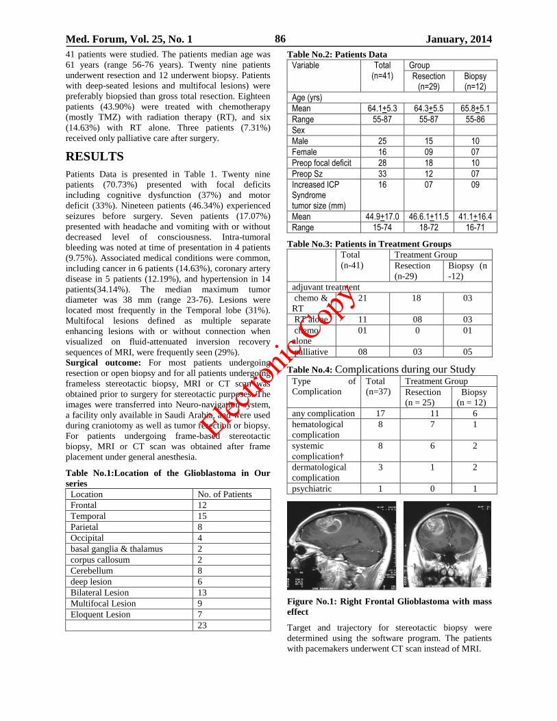
Med. Forum, Vol. 25, No. 1 January, 2014 86
41 patients were studied. The patients median age was
61 years (range 56-76 years). Twenty nine patients
underwent resection and 12 underwent biopsy. Patients
with deep-seated lesions and multifocal lesions) were
preferably biopsied than gross total resection. Eighteen
patients (43.90%) were treated with chemotherapy
(mostly TMZ) with radiation therapy (RT), and six
(14.63%) with RT alone. Three patients (7.31%)
received only palliative care after surgery.
RESULTS
Patients Data is presented in Table 1. Twenty nine
patients (70.73%) presented with focal deficits
including cognitive dysfunction (37%) and motor
deficit (33%). Nineteen patients (46.34%) experienced
seizures before surgery. Seven patients (17.07%)
presented with headache and vomiting with or without
decreased level of consciousness. Intra-tumoral
bleeding was noted at time of presentation in 4 patients
(9.75%). Associated medical conditions were common,
including cancer in 6 patients (14.63%), coronary artery
disease in 5 patients (12.19%), and hypertension in 14
patients(34.14%). The median maximum tumor
diameter was 38 mm (range 23-76). Lesions were
located most frequently in the Temporal lobe (31%).
Multifocal lesions defined as multiple separate
enhancing lesions with or without connection when
visualized on fluid-attenuated inversion recovery
sequences of MRI, were frequently seen (29%).
Surgical outcome: For most patients undergoing
resection or open biopsy and for all patients undergoing
frameless stereotactic biopsy, MRI or CT scan was
obtained prior to surgery for stereotactic purposes. The
images were transferred into Neuro-navigation system,
a facility only available in Saudi Arabia, and were used
during craniotomy as well as tumor resection or biopsy.
For patients undergoing frame-based stereotactic
biopsy, MRI or CT scan was obtained after frame
placement under general anesthesia.
Table No.1:Location of the Glioblastoma in Our
series
Location No. of Patients
Frontal 12
Temporal 15
Parietal 8
Occipital 4
basal ganglia & thalamus 2
corpus callosum 2
Cerebellum 8
deep lesion 6
Bilateral Lesion 13
Multifocal Lesion 9
Eloquent Lesion 7
23
Table No.2: Patients Data
Variable Total (n=41)
Group
Resection (n=29)
Biopsy (n=12)
Age (yrs)
Mean 64.1+5.3 64.3+5.5 65.8+5.1
Range 55-87 55-87 55-86
Sex
Male 25 15 10
Female 16 09 07
Preop focal deficit 28 18 10
Preop Sz 33 12 07
Increased ICP Syndrome tumor size (mm)
16 07 09
Mean 44.9+17.0 46.6.1+11.5 41.1+16.4
Range 15-74 18-72 16-71
Table No.3: Patients in Treatment Groups
Total
(n-41)
Treatment Group
Resection
(n-29)
Biopsy (n
-12)
adjuvant treatment
chemo &
RT
21 18 03
RT alone 11 08 03
chemo
alone
01 0 01
palliative 08 03 05
Table No.4: Complications during our Study
Type of
Complication
Total
(n=37)
Treatment Group
Resection
(n = 25)
Biopsy
(n = 12)
any complication 17 11 6
hematological
complication
8 7 1
systemic
complication†
8 6 2
dermatological
complication
3 1 2
psychiatric 1 0 1
Figure No.1: Right Frontal Glioblastoma with mass
effect
Target and trajectory for stereotactic biopsy were
determined using the software program. The patients
with pacemakers underwent CT scan instead of MRI.
Electro
nic Cop
y

Med. Forum, Vol. 25, No. 1 January, 2014 87
Twenty nine (70.73%) patients underwent resection and
12 patients (29.27%) underwent biopsy. The resection
group included 5 patients who had previously
undergoing biopsy for diagnostic purposes prior to
resection. There were no statistically significant
differences between the biopsy and resection groups
regarding age, sex, morbidities, or tumor size.
Resection group presented more frequently with
increased ICP and had a higher dexamethazone dose
than the biopsy group.
Lesions were located more often in the temporal lobe in
the resection group and less often in the parietal lobe,
the basal ganglia, and the corpus callosum (Deep
Lesion) in the biopsy group.
Surgical complications were divided into 3 groups as
suggested by the Glioma Outcome Project . The overall
complication rate was 24.8%. Neurological
complications were seen in 17 patients (41.46%).
The neurological complication rate did not differ
significantly between the resection group and the
biopsy group. Regional complications affected 13
patients (31.70%) including hemorrhage in 6 patients
(14.63%). All hemorrhagic events occurred in the
biopsy group. Systemic complications occurred in 7
patients (17.07%), including thrombo-embolic events in
4 patients (9.7%).
The perioperative mortality (death within 30 days of
surgery) rate was 9.7% (3 patients undergoing biopsy
and one from resection group).
Outcomes for Adjuvant Treatment: Five patients
(12.08%) received palliative care only without any
active tumor treatment. All remaining received RT,
with a completion rate of 100%. The median radiation
dose delivered was 51.0 Gy (range 10.0-68.0 Gy). More
patients in the biopsy group received only palliative
care postoperatively than the resection group of patients
with hematological complications (myelosuppression),
2 required dose reduction and 3 needed to discontinue
chemotherapy. One of those patients receiving TMZ
required platelets. Systemic complications occurred in 6
patients Frontal ,Temporal ,corpus callosum Eloquent
Lesion and included deep venous thrombosis (3
patients) and pulmonary embolism (1 patient),
complications included rash, stomatitis, and depression.
Among 64 patients who received RT, 8 patients
(19.5%) were not able to complete the prescribed
treatment. Five patients experienced clinical
deterioration during RT.
Significant differences were noted between patients
receiving palliative care and patients receiving adjuvant
treatment Patients receiving RT alone were older than
those receiving RT and chemotherapy, were more likely
to have presented with increased ICP (39% vs 15%),
and had a higher mean Dexamethazone dose.
Progression Free Survival (PFs) and overall survival
Median PFS, calculate from date of surgery, was 4.5
months. Factors significantly associated with poor PFS
included deep lesion, multifocal lesions biopsy only,
new persistent postoperative focal deficit, and palliative
care (that is lack of adjuvant treatment) The median
PFS for patients undergoing adjuvant treatment was 5.5
months, whereas that for patients who had palliative
care only was 0.5 months. Patients receiving
chemotherapy along with RT had achieved significantly
longer PFS compared with those with RT alone.
The median Overall Survival differed strikingly among
the age groups: 1. Months for patients younger than 70
years, but merely 4.5 months for patients aged over 70
years. Favorable survival effects of resection and
adjuvant treatment in this study mainly result from
relatively younger patients (< 65 years) among our
elderly patient adjuvant treatment lived substantially
longer if they had undergone resection compared with
biopsy (median OS 11.5 months vs 6.5 months). With
maximal safe resection followed by the combination of
RT and chemotherapy, which is the standard treatment
for unselected GBM patients, the median PFS and OS
among our elderly patients with GBM were 8 months
and 12.5 months, respectively. When limited to patients
who were 70 years of age or older, they were 6 months
and 10.0 months, respectively.
DISCUSSION
Gross total (safe) surgical resection followed by
adjuvant treatment including RT and chemotherapy
with TMZ has become the standard of care for patients
with newly diagnosed GBM since the landmark study
from Stupp and colleagues3,21
.
But patients older than 70 years were excluded from
this study which is relatively common.8,17
In clinical
practice, physicians and surgeons are frequently
reluctant to offer aggressive treatment to elderly
patients because of concerns that it may not be tolerated
due to advanced age, co-morbidities and an underlying
propensity to complications. Therefore, although the
age-adjusted incidence of GBM is greatest among
patients older than 65 years, the best treatment strategy
for these patients is not as well defined as in younger
patients, the very reason this study was carried out.
Impact of Resection versus Biopsy on Survival in
Elderly Patients have been performed evaluating the
impact of the prospective studies9,25 have been more
definitive, demonstrating an association Sanai et al,10,14
and between resection and survival specially in younger
patient impact of extent of resection (determined with
or without computer-assisted volumetric assessment)
on survival and concluded that the majority supported
extent of resection as a prognostic factor for prolonged
survival. Some authors ha reported increased extent of
GBM resection and prolonged PFS in patients
undergoing fluorescence-guided surgery using 5-
aminolevulnic acid (5-ALA) in a prospective,
randomized controlled trial"12,20
Older patients are often
believed to take longer to recover from an aggressive
Electro
nic Cop
y

Med. Forum, Vol. 25, No. 1 January, 2014 88
surgery and carry a higher risk of peri-operative
complications. Thus, biopsy is often resection for these
patients.13,1
A retrospective study from the independent
predictor of survival and was associated with favorable
OS among patients along with age and performance
status.14,13
However, this benefit was only evident for
patients who went on to receive adjuvant treatment
(median survival for resection vs biopsy: 11.5 vs 6.5
months).
Surgical Complications in Elderly Patients: High
complication rates in elderly patients like new
neurological deficits are associated with decreased
survival, not just decreased quality of life."15,16
Our
overall complication rate of 24.8% for elderly patients
compares favorably to other authors and true for
patients undergoing resection in our series. The overall
complication rate in these patients was 18.9%. By
comparison, patients with newly diagnosed malignant
glioma undergoing first craniotomy in the Glioma
Outcome project had an overall complication rate of
24.2% including 8.1% permanent neurological
worsening, 10% regional complications, 9.2% systemic
complications, and 1.5% mortality.
Similarly, Sawaya et al reported their experience at MD
Anderson Cancer Center with 400 craniotomies for
intra-axial tumors (206 gliomas, 194 metastases). The
overall complication rate was 32% (including 8.5%
permanent neurological worsening, 7% regional
complications, 7.8% systemic complications and 1.7%
mortality) those undergoing resection (30.8% vs.
18.9%) although the difference did not reach statistical
significance. complication rates for stereotactic biopsy
in our series of elderly patients with GBM are higher
than those reported in large unselected mortality rates
range from 0.6% to 2.8%.3,9,16,25
Impact of Adjuvant Treatment on Survival in
Elderly Patients: Radiation therapy has an established
role in GBM therapy. RT has a significant survival
benefit and appears to hold true for elderly
patients.13,14,15,17
Superiority of an abbreviated RT
course (40 Gy in 15 fractions over 3 weeks) in elderly
patients has been successfully proved by a prospective
randomized clinical trial by Roa and colleagues.18
Like RT, chemotherapy has an established role in GBM
treatment. For many years nitrosoureas such as BCNU
or lomustine (CCNU) were the mainstay of
chemotherapy for GBM patients and 2 meta-analyses
consistently confirmed their efficacy for longer
survival.19,10
However, the benefits of nitrosoureas for
old patients was questioned (similar to RT) especially
given the higher incidence chemotherapy-related
neurotoxcity and myelosuppression in this population.
The changed the standard of care for patients with
newly paper by Stupp et al changed the standard of care
for the patients with newly diagnosed GBM by
demonstrating increased survival for patients receiving
RT with concurrent TMZ followed by adjuvant TMZ
compared with RT alone (median survival 14.6 months
vs 12.1 months), excluding 70 plus patients. Some
recent papers have reported favorable survival for
elderly patients with GBM receiving RT and TMZ,
such as 10.6 months with the Stupp regimen for 2
patients aged 70 or older or 11 months with adjuvant
RT and reduced dose of TMZ.9,21
Adjuvant Treatment Toxicity in Elderly Patients:
Adjuvant treatment including RT and TMZ was
feasible for elderly patients and was allocated with a
modest complication rate.4,5,6,9,11,12,24,25
Most of the major complications did not seem to be
directly associated with RT except for an infarct of the
ipsilateral corona radiata 2 years after RT for a
temporal tumor. In addition to resection and adjuvant
treatment, other factors reported to favorably influence
survival in unselected GBM patients include young age,
good performance status, good mental status, seizure
presentation, frontal lesion, superficial 3.1780.11)
Younger single lesion, and the absence of postoperative
complications age has consistently been one of the
strongest favorable prognostic factors across many
studies. It is interesting that our data suggest that this
holds true even among the elderly population (age-65
years). Several hypotheses have been proposed to
explain the poor clinical outcome of GBM in older
patients, including the presence of comorbidities,
resistance to cancer therapy, genetic aberrations,
different histology, neurodegeneration and age
discrimination.2,4,19, 22,23,26
CONCLUSION
This study demonstrates that aggressive treatment with
resection and adjuvant treatment is associated with
significantly longer survival for patients 65 years of age
or older with newly diagnosed GBM. This finding must
be interpreted with caution given that this study not
randomized and patient biopsy and/or palliative care
were significantly different (had tumors in deeper, more
eloquent locations poorer performance status, increased
dexamethasone dose) from those receiving resection
and or adjuvant treatment. Nevertheless, resection and
adjuvant treatment remained significant prognostic
factors in a multivariate analysis and were generally
well tolerated in this group of elderly patients.
Although advanced age is an independent unfavorable
factor, this alone should not disqualify patients from
being treated with optimal tumor resection followed by.
REFERENCES
1. Central Brain Tumor Registry of the United States: Statistical Report: Primary Brain and Central Nervous System Tumors Diagnosed in the United States in 2004–2005 [Accessed October 22, 2012]
Electro
nic Cop
y

Med. Forum, Vol. 25, No. 1 January, 2014 89
2. Radiotherapy plus concomitant and adjuvant temozolomide for glioblastoma. N Engl J Med 2005;352:987–996.
3. Stupp R, Mason WP, van den Bent MJ, Weller M, Fisher B, Taphoorn MJ, et al. Radiotherapy plus concomitant and adjuvant temozolomide for glioblastoma. N Engl J Med 2005;352:987–996.
4. Grossman SA, Ye X, Piantadosi S, Desideri S, Nabors LB, Rosenfeld M, et al.: Survival of patients with newly diagnosed glioblastoma treated with radiation and temozolomide in research studies in the United States. Clin Cancer Res 2010;16:2443–2449.
5. Buckner JC. Factors influencing survival in high-grade gliomas. Semin Oncol 2003;30:6(Suppl): 1910–14.
6. Tilgner J, Herr M, Ostertag C, Volk B. Validation of intraoperative diagnoses using smear preparations from stereotactic brain biopsies: intraoperative versus final diagnosis—influence of clinical factors. Neurosurg 2005;56:257–265.
7. Chang SM, Parney IF, McDermott M, Barker FG, Schmidt MH, Huang W, et al. Perioperative complications and neurological outcomes of first and second craniotomies among patients enrolled in the Glioma Outcome Project. J Neurosurg 2003;98:1175–1181.
8. Mirimanoff RO, Gorlia T, Mason W, Van den Bent MJ, Kortmann RD, Fisher B, et al.:Radiotherapy and temozolomide for newly diagnosed glioblastoma: recursive partitioning analysis of the EORTC 26981/22981-NCIC CE3 phase III randomized trial. J Clin Oncol 2006;24:2563–2569.
9. Devaux BC, O'Fallon JR, Kelly PJ: Resection, biopsy, and survival in malignant glial neoplasms. A retrospective study of clinical parameters, therapy and outcome. J Neurosurg 1993;78: 767–775.
10. Lacroix M, Abi-Said D, Fourney DR, Gokaslan ZL, Shi W, DeMonte F, et al. A multivariate analysis of 416 patients with glioblastoma multiforme: prognosis, extent of resection, and survival. J Neurosurg 2001;95:190–198.
11. Sanai N, Berger MS: Glioma extent of resection and its impact on patient outcome. Neurosurg 2008;62:753–764.
12. Stummer W, Pichlmeier U, Meinel T, Wiestler OD,Zanella F, Reulen HJ: Fluorescence-guided surgery with 5-aminolevulinic acid for resection of malignant glioma: a randomised controlled multicentre phase III trial. Lancet Oncol 2006; 7:392–401.
13. Barnholtz Sloan JS, Williams VL, Maldonado JL, Shahani D, Stockwell HG, Chamberlain M, et al. Patterns of care and outcomes among elderly individuals with primary malignant astrocytoma. J Neurosurg 2008;108:642–648.
14. Keime-Guibert F, Chinot O, Taillandier L, Cartalat-Carel S, Frenay M, Kantor G, et al.
Radiotherapy for glioblastoma in the elderly. N Engl J Med 2007;356:1527–1535.
15. McGirt MJ, Mukherjee D, Chaichana KL, Than KD, Weingart JD, Quinones-Hinojosa A.Association of surgically acquired motor and language deficits on overall survival after resection of glioblastoma multiforme. Neurosurg 2009;65:463–470.
16. Bernstein M, Parrent AG. Complications of CT-guided stereotactic biopsy of intra-axial brain lesions. J Neurosurg 1994;81:165–168.
17. Laperriere N, Zuraw L, Cairncross G.Radiotherapy for newly diagnosed malignant glioma in adults: a systematic review. Radiother Oncol 2002;64: 259–273.
18. Roa W, Brasher PM, Bauman G, Anthes M, BrueraE, Chan A, et al. Abbreviated course of radiation therapy in older patients with glioblastoma multiforme: a prospective randomized clinical trial. J Clin Oncol 2004;22:1583–1588.
19. Fine HA, Dear KB, Loeffler JS, Black PM, Canellos GP. Meta-analysis of radiation therapy with and without adjuvant chemotherapy for malignant gliomas in adults. Cancer 1993; 71:2585–2597.
20. Brandes AA, Vastola F, Basso U, Berti F, Pinna G,Rotilio A, et al. A prospective study on glioblastoma in the elderly. Cancer 2003;97:657–662.
21. Combs SE, Wagner J, Bischof M, Welzel T, Wagner F, Debus J, et al. Postoperative treatment of primary glioblastoma multiforme with radiation and concomitant temozolomide in elderly patients. Int J Radiat Oncol Biol Phys 2008; 70:987–992.
22. Kelly PJ, Hunt C: The limited value of cytoreductive surgery in elderly patients with malignant gliomas. Neurosurg1994;34:62–67.
23. Rosenblum ML, Gerosa M, Dougherty DV, Reese C, Barger GR, Davis RL, et al. Age-related chemosensitivity of stem cells from human malignant brain tumours. Lancet 1982;1:885–887.
24. Brandes AA, Monfardini S: The treatment of elderly patients with high-grade gliomas.Semin Oncol 2003;30(6 Suppl):1958–62.
25. Fiorica F, Berretta M, Colosimo C, Stefanelli A, Ursino S, Zanet E, et al.: Glioblastoma in elderly patients: safety and efficacy of adjuvant radiotherapy with concomitant temozolo-mide. Arch Gerontol Geriatr 2010;51:31–35
26. Batchelor TT, Betensky RA, Esposito JM, Pham LD, Dorfman MV, Piscatelli N, et al. Age-dependent prognostic effects of genetic alterations in glioblastoma. Clin Cancer Res 2004;10: 228–233.
Address for Corresponding Author:
Dr. Salman A. Jaffery
Cell #: +92 301 7516514
Electro
nic Cop
y

Med. Forum, Vol. 25, No. 1 January, 2014 90
Comparative Study on Neem Leaf
Extract and Nimolicin (NC) on Gastric
Mucosa of Albino Rats 1. Qamar Aziz Siddiqui 2. Syed Ijaz Hussain Zaidi 3. Asghar Hussain 4. Muhammad Omar
Shamim 5. Nazar Hussain 6. Syed Naimul Hassan Naqvi 7. Mohammed Arshad
8. Rajput Muhammad Tariq 9. Mazhar Hijazi 1. Prof. of Physiology, IM&DC, Sialkot 2. Asstt. Prof. of Pharmacology, Al Tibri Medical College, Karachi 3. Asstt.
Prof. of Physiology, Quetta Institute of Medical Sciences, CMH, Quetta 4. Asstt. Prof. of Physiology, IM&DC,
Sialkot 5. Prof. of Pathology, BMU, Karachi 6. Prof. of Pharmacology, BIPS, Karachi 7. Asstt. Prof. of
Pharmaceutics, Baqai Institute of Pharmacy, Karachi 8. M.AH, Qadri Biological Research Centre, Karachi
University, Karachi 9. Prof. of Anatomy, BMU, Karachi
ABSTRACT
Objective: Compare the anti ulcer effect of Methanolic Neem (Azadirachta indica, A Juss, Meliaceae) Leaf Extract
(NLE) and Neem compound Nimolicin on gastric mucosa of albino rats.
Azadiradione also called Nimolicine coded as NC has been studied for its anti insect effect but anti ulcer effect has
never been studied.
Study Design: Experimental study
Place and Duration of Study: This study was carried out at the Pharmacy and Physiology Department of Baqai
Medical College for duration of two years.
Materials and Methods: Gastric ulcers in albino rats were induced in group-1 (check group) by a single oral dose
of 1 ml 100% ethanol. After 24 hours the treatment was started. Group-2 was treated with oral administration of pea
nut oil 1ml/day for 5 days (control of the treated group). Group-3 was treated with NLE (1ml/day for 5 days) and
Group-4 with NC 1% (1ml/day for 5 days). The healing effects of neem were compared to oral administration of
anti ulcer drugs ranitidine (50mg/kg daily for 5 days) and omeperazole (2.5 mg/kg daily for 10 days).
Histopathology of the stomach was performed to confirm the presence or healing of ulcers. Ulcers were scored and
indexed on the basis of histopathology.
Results: Ranitidine had the highest ulcer inhibition of 84%. NLE proved to be better than omeperazole by showing
an ulcer inhibition of 82 % compared to 73% ulcer inhibition of omeperazole. NC showed least anti ulcer activity
with an ulcer inhibition of only 69%. HPLC was performed to show the tissue concentration of NC, omeparazole
and ranitidine showing their retention time, area and concentration compared to their controls.
Conclusion: It is concluded that NLE proved to be better anti ulcer agent as compared to NC and can be used as an
anti ulcer drug after clinical trials.
Key Words: Gastric ulcer, neem (Azadirachta indica, A Juss, Meliaceae) leaf extract, azadiradione, nimolicin,
ranitidine, omeperazole, ulcer inhibition.
INTRODUCTION
Neem (Azadirachta indica A. Juss) has been known for
its medical values for the last 2000 years. Even today
neem has been the centre of attraction especially for
workers of medical field. All parts of neem are
important particularly the leaves which is “storehouse
of organic compounds”1. The leaves are easily available
through out the year and preparation of extracts is easy.
Thus they have been used extensively for medicinal
applications2.
More than 300 different compounds of neem have been
isolated from different parts of tree about one third are
tetranortriterpinoids (Liminoids).3 Nimolicine coded as
NC is a known neem compound. The neem fruit is
divided into two parts. Fleshy outer part pericarp and
mesocarp and inner part seed coat and kernel. The
active ingredient of seed coat and kernel is Azadirachtin
(AZ).4 NC (Azadiradione) was isolated from fresh fruit
coatings (after separation of seeds from fruits). Thus in
the present study the anti ulcer effects of neem leaf
extract were studied and compared with NC. The potent
anti ulcer and anti secretory effect of neem leaf extract
has been attributed to a glycoside.5 Plant glycosides are
known to inhibit chloride transport in gastric juice
reducing gastric acidity.6 The mechanism of action of
NLE in healing of gastric ulcers is possibly because of
its antioxidant effect which is independent of acid
suppression.7 This effect may be similar to the effects
of omeperazole providing gastroprotection due to its
antioxidant and anti apoptotic role.5
This study shows a comparison of anti ulcer effects of
methanolic extract of neem leaves with neem
compound NC showing the healing effects by
histopathology.
Original Article Pharmacology
Electro
nic Cop
y

Med. Forum, Vol. 25, No. 1 January, 2014 91
MATERIALS AND METHODS
Wistar strain of rats were purchased from HEJ Institute
of Chemistry, Karachi University and kept under
optimum conditions in the animal house of Baqai
Medical University. The animals were acclimatized and
had free access to food and tap water ad libitum.
Principals of laboratory animal care (NIH publication
no.82-83, revised 1985) guidelines were followed. The
animal experimentation approval was obtained from
University Animal Ethical committee. NLE and NC
were obtained from HEJ Institute of Chemistry,
Karachi.
A total of 60 albino rats weighing 180Gm to 200Gm
were divided into six groups. In each group 5 male and
5 female rats were included. Group-1 was the check
group which was given 1 ml of 100% ethanol orally and
sacrificed after 24 hrs to check for ulcers. Macroscopic
and microscopic examination for the confirmation of
ulcers was done. Once it was established that oral
administration of ethanol caused gastric ulceration in
rats, the next part of the study was undertaken. All the
other groups were first given oral ethanol for ulcer
production and then treated orally after 24 hours
accordingly and then sacrificed. Group no 2 (control of
the treated group) was treated with 1 ml of peanut oil
daily for 5-7 days. Group-3 (test group) was treated
with 1ml NLE daily for 5 days. The rats of fourth group
(test group) were treated with 1% NC daily for 5 days.
The fifth group (comparison group) of rats was treated
with ranitidine (purity 50%) 50mg/Kg daily for 5 days.
The rats of the 6th
group were treated with Omeperazole
(purity 83.33%) 2.5 mg/kg daily for 10 days. After
completion of the respective treatment rats of Group-2
to Group-6 were sacrificed and stomach was incised.
The gastric ulcer was examined in all the rats by a hand
lens (power 10). The gastric tissues were then
processed for histopathological examination by staining
procedure of8. Gastric ulcers were scored on the basis
of histopathology and indexed as Ulcers = 3, Heavy
infiltration of polymorpho nuclear (PMN) cells = 2,
Mild infiltration of (PMN) cells = 1, no ulcers = 0. The
ulcer index and ulcer inhibition was determined.9,10
The
experiments were carried out in the Pharmacology
Department of the Baqai Institute of Pharmaceutical
Sciences and were repeated three times. The total
duration of the study was three years.
High Performance Liquid Chromatography
(HPLC): HPLC was performed to demonstrate the
concentration of drugs in tissues. This was then
compared with the standard solution prepared. One Gm
of gastric tissue was homogenized in a homogenizer
(OSK 9258) at 500RPM and centrifuged at 3500RPM
(Labofuge-200 Heraeus). Soxhalation of the
supernatant fluid was done. Sorption was done by
passing the solution through Alluminium Oxide and
Silica11
. Pure methanol was used as a solvent for the
mobile phase with a flow rate of 1 ml/min. A UV
detector was used at a wavelength of 214 nm, pressure
200 kg/cm2 and absorbance 0.08 with chart speed
2.5mm/min. Standard samples were prepared and run
for comparison. The samples were filtered by Swiney
syringe a micro filter pore diameter of 0.42 nm
(Millipore Corporation Bedford MA01730). 20µL of
the sample were injected by a special 25µL
chromatographic syringe via the injector pore. HPLC
apparatus Shimadzu (Japan) SPD-10A UV
spectrophotometer with Merck reverse phase column
(RP-C18, 25cm X 0.46mm) was used. The
chromatographic data were processed with Class LC-10
software (Shimadzu, Japan) and CBM (communication
bus model) to the monitor. Peaks were recorded by
software programme Real Time Analysis. The peaks
were compared on the basis of retention time (RT) with
the standard peaks. The area of each peak was noted to
quantify the different compound residues in the
samples.
RESULTS
In normal healthy rats ulcers were induced by giving
100% ethanol in the check group. This group had the
highest ulcer score as shown in table-1 seen on naked
eye and later confirmed on microscopic examination
(Fig.2). There was no ulcer inhibition because no
treatment was given to this group. The remaining 5
groups were treated with pea nut oil, neem,
omeperazole or ranitidine after the induction of ulcers.
The comparison of anti ulcer effects of neem with anti
ulcer drugs on gastric tissue of rats have been shown in
table-1 and figures 1-7.
Table No.1: Comparison of ulcer inhibition of NLE, NC, Omeperazole and Ranitidine with the control.
Ethanol
(Check Group)
n=17
PNO (Control
Group)
n=17
NC (Test
Group) n=7
NLE
(Test Group)
n=9
OMP (Known
Antiulcer Group)
n=6
Ranitidine
(Known Antiulcer
Group) n=7
Ulcer Score 31 31 4 3 3 2
Ulcer Index 1.82 1.82 0.57 0.33 0.5 0.29
Ulcer
Inhibition
Nil Nil 69% 82% 73% 84%
Electro
nic Cop
y

Med. Forum, Vol. 25, No. 1 January, 2014 92
Table No.2: Shows concentration of NC,
omeperazole and ranitidine in gastric tissue
compared to the control by HPLC (Schimadzu)
NC Ranitidine Omeprazole
Gastric
tissue Conc
(µg/20 µl)
3.37 3.62 7.11
Control
Conc.
(µg/20 µl)
7.08 14.64 9.22
Figure No. 1: H & E stained, 5 micron thick paraffin section
of stomach in an adult female rat untreated showing normal
gastric mucosa. (Photomicrograph H&Ex10)
Figure No.2: H & E stained, 5 micron thick paraffin section
of stomach in an adult female rat given oral Ethanol 100%
showing massive erosions (A). The section reveals ulceration
of mucous membrane with infiltration of the base with
neutrophils (B & C) and dilatation and congestion of blood
vessels. (Photomicrograph H&E x 10)
The normal gastric tissue Fig.1 has been compared with
ulcerated gastric epithelium in Fig.2. Treated gastric
epithelium has been shown in Fig.3-7. Pea nut oil used
as control of treatment group has not caused a healing
effect and the persistence of ulcers has been
demonstrated in Fig.3. The healing effect of neem has
been shown in Fig.4 & Fig.7 and this gastric tissue can
be compared to the normal gastric mucosa fig.1 and
healing effect of ranitidine and omeperazole Fig 6 &
Fig 7. Table-1 shows that ranitidine had the highest
ulcer inhibition of 84%. The ulcer inhibition of NLE
was better than omeperazole by showing an ulcer
inhibition of 82% compared to 73% of omeperazole.
NC showed least anti ulcer activity with an ulcer
inhibition of only 69% (Table-1). The ulcer score of the
check group and control is the same. There was no
ulcer inhibition with pea nut oil which served as the
control of treated group. Tissue concentration of NC,
ranitidine and omeperazole on HPLC has been shown
in table -2 compared to their controls.
Figure No.3: H & E stained 5 micron thick paraffin section of
stomach in an adult female rat treated with Pea nut oil for 5
days. The section shows the presence of chronic inflammatory
cells showing a non healing effect of PNO. Ulcers are seen. A
& B. (Photomicrograph H&E x 10)
Figure No.5: Photomicrograph of H & E stained, 5 micron
thick paraffin section of stomach in an adult female rat treated
with neem compound nimolicin (NC). The section reveals
Electro
nic Cop
y

Med. Forum, Vol. 25, No. 1 January, 2014 93
mild infiltration of mucosa by PMN cells, lymphocytes and
occasional plasma cells. No ulcers visible. A & B. (H & E X 10).
Figure No.5: Photomicrograph of H & E stained, 5 micron
thick paraffin section of stomach in an adult female rat treated
with neem compound nimolicin (NC). The section reveals
mild infiltration of mucosa by PMN cells, lymphocytes and
occasional plasma cells. No ulcers visible. A & B. (H & E X 10).
Figure No.6: H & E stained, 5 micron thick paraffin section
of stomach in an adult female rat treated with ranitidine The
section shows mild focal infiltration by chronic inflammatory
cells (A). No ulcers seen (B). (Photomicrograph H & E x 10).
Figure No.7: H & E stained, 5 micron thick paraffin section
of stomach from an adult female rat treated with omeperazole.
The section shows mild focal infiltration by chronic
inflammatory cells. No ulcers seen A & B. (Photomicrograph
H & E x 10).
DISCUSSION
In the present study ulcer was induced by oral
administration of ethanol which is in contrast with the
findings of12
who used mercaptomethylimidazole
compound for the production of ulcer which is less
potent in ulcer induction than ethanol. The anti ulcer
effect of neem has been demonstrated in the present
study by the induction of ulcer first, followed by
treatment and confirmation by histopathology which is
in contrast with the findings of13
who studied the ulcer
protective effect by pretreatment followed by ulcer
induction without any histological evidence.
The anti ulcer effects of methanolic extract of NLE in
the present study has been reported which has been
shown to decrease ulcer index, which is similar with the
findings of14
who used aqueous extract of NLE and
bark extract of neem respectively.
Hence the present study demonstrates that NLE is more
effective as an anti ulcer agent compared to NC
possibly because of its cumulative effect. Resistance is
difficult to develop with extracts compared to isolated
compounds. Clinical trials may further help in
supporting our results.
CONCLUSION
It is concluded that NLE proved to be better anti ulcer
agent as compared to NC and can be used as an anti
ulcer drug after clinical trials.
Acknowledgements: We gratefully acknowledge the
contribution of Professor Dr Beena Siddiqui of HEJ
institute of Chemistry, Karachi University for providing
us neem leaf extract and neem compound NC. We are
also highly indebted to Madam Zehra Yaqeen, Dr
Nudrat Fatima and Dr Lubna of the PCSIR laboratories
Karachi for their cooperation and support during the
project. We are very thankful to Dr Mirza MF Subhan,
Assistant Professor, Department of Physiology,
University of Kingdom of Bahrain for his expert
guidance in editing this article. A special word of
thanks to the department of Baqai Institute of
Information Technology specially Syed Rizwan Nisar
for their cooperation in computer skills in formatting
and processing for publication of this article. We also
are greatly indebted to Professor Dr Arshad Azmi of
Karachi University for his support during HPLC. Partial
financial support from HEC project no. 20-
1127/R&D/2008 is gratefully acknowledged.
REFERENCES
1. Subapriya R, Nagini S. Medicinal properties of
Neem Leaves: A Review. Curr Med Chem
AntiCancer Agents 2005;5(2):1-8.
2. Brahmachari G. Neem–An Omnipotent Plant: A
Retrospection. Chem Bio Chem 2004; 5: 408- 421.
Electro
nic Cop
y

Med. Forum, Vol. 25, No. 1 January, 2014 94
3. Siddiqui BS, Kashif SA, Tariq SA, Naqvi SNH,
Tariq RM. Variation of Major Liminoids in
Azadirachta indica fruits at different ripening
stages and toxicity against Aedes aegypti. Natural
Product Communications 2009;4(4): 473-476.
4. Lavie D, Levie E C, Jain M K. Tetrahedron 1971;
27: 3927-3939.
5. Biswas K, Chattopadhyay I, Ranajit K, Banerjee,
Bandyopadhyay U. Biological activities and
medicinal properties of Neem (Azadirachta indica).
Current Sci 2002;82(11): 1336-1345.
6. Machen TF, Forte JG. Gastric secretion. In:
Guebischil G, Tasteson DC, Using HH, editors.
Handbook of Transport Organs. Berlin: Springer;
1979.p.693-747.
7. Saddiqui QA, Feisal S, Naqvi SNH. The effect of
neem (Azadirachta indica) leaf extract and neem
compound Nimolicine on gastric acidity. Med
Forum 2011;22(3):30-34.
8. Bancroft JD, Stevens A. Theory and Practice of
Histological Techniques. 3rd
ed. Edinburgh
(London): Churchil Living Stone;1990.p.42-
43,107-110,
186-189.
9. Bandyopadhyay U, Biswas K, Sengupta A,
Moitra P, Dutta P, Sarkar D, et al. Clinical studies
on the effect of Neem (Azadirachta indica) bark
extract on gastric secretion and gastroduodenal
ulcer. Life Sci2004.75(24): 2867-2878.
10. Nijar VCO, Adesonwo JK, Raji Y. Methyl
Angolensate: The antiulcer agent of the stem bark
of Entandrophragma angolense. Planta Medica
1995; 61: 91-92.
11. Holden AV, Marsdon M. Single stage clean up of
animal tissue extract for Organochlorine’s residue
analysis. J Chromatograph 1969;44: 481.
12. Bandyopadhyay U, Biswas K, Chatterjee R,
Bandyopadhyay D, Chattopadhyay I, Ganguly CK,
et al. Gastroprotective effect of Neem (Azadirachta
indica) bark extract: Possible involvement of H+-
K+-ATPase inhibition and scavenging of hydroxyl
radical. Life Sci 2002;71; 2845-2865.
13. Yoshikawa M, Youtana P, Matsuda H, Morikawa
T, Yasunobu A. Protective effects of Polygoidal on
gastric mucosal lesions induced by necrotizing
agents in rats and the possible mechanisms of
action. Biol Pharm Bull 2003;26(5): 651-657.
14. Garg GP, Nigam SK, Ogle CW. The gastric
antiulcer effects of the leaves of the neem tree.
Planta Med 1993;59(3):215-217.
Address for Corresponding Author:
Dr. Qamer Aziz Siddiqui,
Professor & Head Department of Physiology,
Islam Medical & Dental College, Pasroor Road,
Sialkot, Pakistan.
Cell No.: 092-3333145734
Residence No.:092-21-34657855
Tel: 092-52-3610001
Fax: 092-52-3610009
email: [email protected]
Electro
nic Cop
y

Med. Forum, Vol. 25, No. 1 January, 2014 95
Subjects Index January to December 2013 Azhar Masud Bhatti
Editor in Chief
Vol. 24, No. 1, January, 2013
ANATOMY Page No.
Morphological Changes after Chronic Heat Exposure
on Zona Fasiculata of Adrenal Gland of Albino Rats
(Sarwat S, et al) 2
Morphological Study of Native Foramina Transversaria
in the Cervical Spine (Maqbool A, et al) 33
BIO CHEMISTRY
Effect of Concept Maps on Knowledge Retention in
Dental Students (Khan I, et al) 7
SURGERY
Procedure for Prolapsed Haemorrhoids versus
Excisional Haemorrhoidectomy - A Systematic Review
and Meta-Analysis (Shahid ZA, et al) 12
Acute Abdomen, Causes and Outcome - Experience at
DHQ Hospital Abbottabad (Saeed A, et al) 38
Determinants of Upper Limb Amputations in a Tertiary
Care Center, Pakistan (Soomro N, et al) 60
MEDICINE
Incidence of Thyroid IMA Artery in our Population
(Ullah S, et al) 17
Vitamin D Levels in OPD Patients in Teaching
Hospital, Sukkur (Soomro AA, et al) 26
Experience of Acute Systemic Lupus Erythematosus in
Indoor and Outdoor Patients (Ansari IA, et al) 29
Effects of Combined Interferon Alpha and Ribavirin
Therapy on Thyroid Functions in Patients
with Chronic Hepatitis C, Attending Hepatitis Centre
Ghulam Mohammad Medical College Hospital, Sukkur
(Phulpoto JA, et al) 44
GYNAE & OBS
Frequency, Risk Factors and Complications in
Pregnancies Associated with Placenta Praevia (Hafeez
R, et al) 21
PATHOLOGY
Histopathological spectrum of Gastric biopsies in a
tertiary care hospital (Hasan SM, et al) 41
Histopathological Study of 209 Cases of Early
Abortions (Hashim Y, et al) 55
OPERATIVE DENTISTRY Page No.
Evaluation of Post Operative Pain in Single and Multi
Visit Endodontic Therapy with Rotary Instruments
(Shaikh AG, et al) 49
Determination of Number of Apical Foramina in Mesial
Root of Mandibular First Molar - Ex Vivo (Dall AQ,
et al) 70
COMMUNITY MEDICINE
Effects L-Arginine on the Gastric Mucosal Cell count
when given with Ibuprofen in Albino Rats under light
Microscope (Kumar S, et al) 64
Vol. 24, No. 2, February, 2013
PHYSICAL MEDICINE
Diabetes Mellitus and Limb loss: An analysis from a Tertiary Care Center (Soomro N, et al) 2
ANATOMY
Immunohistoichemical Determination of Effects of Withania Somnifera Root Extract on Noise Stress Induced Adenohypophysis in Albino Rats (Naqvi, et al) 6
NEUROLOGY
Frequency of Demyelinating Pattern on Electrophysiological Study in Patients with Diabetic Peripheral Polyneuropathy (Iqbal J, et al) 10
GYNAE & OBS
Histopathological findings of abdominal hysterectomies in patients presenting with heavy menstrual bleeding (HMB) (Jaffar Y, et al) 14
Adolescent Menorrhagia: Causes and Evaluation (Bokhari NA, et al) 22
Role of Foleys Catheter for Pre-Induction Ripening of Cervix (Bhatti K, et al) 39
Histomorphological Features of Placentae in Pregnancies Complicated with Intrauterine Growth Restriction (Rehman MZ, et al) 42
A Randomized Clinical Trial of 200 Patients of Oral Vs Vaginal Misoprostol in Second Trimester Pregnancy Termination (Usmani I, et al) 55
MEDICINE
Outcome with and without Aspiration of the Amoebic Liver Abscess at Civil Hospital, Karachi (Alam MT, et al) 17
Occupational Exposure to Blood and Body Fluids amongst Health Care Workers in a Teaching Hospital of the Sukkur, Sindh (Phulpoto JA, et al) 26
The IT IT Study (The Incubation Time in Tetanus Study) (Khan BA, et al) 52
PHARMACOLOGY
Studies on Effectiveness of DOTs and weekly three times Treatment of Sputum Positive Tuberculosis Patients (Bhurgri GR, et al) 30
ANTHROPOLOGY
Role of Traditional Birth-practices causing Maternal and Neonatal Mortality in Faisalabad. An
Subject Index
Electro
nic Cop
y

Med. Forum, Vol. 25, No. 1 January, 2014 96
Anthropological Approach of Household Decision Making (Ahmed A, et al) 35
ENT
Outcome of Silver Nitrate Cauterization in Controlling Anterior Epistaxis in Children (Khan MI, et al) 48
COMMUNITY MEDICINE
Knowledge and Practices of Basic Life Support amongst the Ambulatory Staff in Karachi (Mehtab K, et al) 58
BIOCHEMISTERY
Lipid profile in Females of Reproductive Age Group Using Injectable Contraceptives (Progestogenonly) (Faryal U, et al) 61
Association of BMI with Elevated Values of CK-MB and cTnT in Acute Myocardial Infarction (Rashid S, et al) 64
Effects of Ibuprofen on Gastric Mucosa of Albino Rats and the Protective Role of L-Arginine.A Morphological Study Under Light Microscope (Kumar S, et al) 68
NEUROSURGERY
Surgical Management of Missile Trauma to Thoraco -Lumbar Spine (Jafferi SA) 74
NEUROLOGY
A study of Chronic Inflammatory Demyelinating Polyneuropathy (CIDP) in Diabetics: Clinical Features, Laboratory findings and Response to Treatment (Javed MA, et al) 79
CARDIAC SURGERY
Right Coronary Artery Aneurysm (Saleem M) 83
Vol. 24, No. 3, March, 2013
GYNAE & OBS
Frequency of Perinatal Mortality at Bolan Medical
Complex Hospital Quetta (Usmani I, et al 2
Eclapsia: Still A Major Cause For Adverse Maternal
and Perinatal Outcome (Bhatti K, et al) 15
ANATOMY
Effects of Diclofenac Sodium on Renal Parenchyma
and Prevention by Vitamin-E in Adult Albino Rats
(Rathore MI, et al) 6
Depletion of Body and Cerebellar Weights after
Chronic Lithium Carbonate Ingestion in Albino Rats
(Kohari T, et al) 47
A study of Gross Anatomical Observations of Stomach
and Changes in Body Weight of Albino Rats after
Simultaneous Administration of Ibuprofen and
L-Arginine (Kumar S, et al) 55
COMMUNITY MEDICINE
Morbidity Pattern and Health Education for School age
Children in Karachi (Kolachi HB, et al) 11
Colored Text Enhances Learning Power of Medical
Students “A New Concept of Learning” (Mehtab K,
et al) 60
BIOCHEMISTRY
Isolation of E – Coli in UTI cases with malnutrition in
children of distant rural area of Sukkur, Sindh (Laghari
AH, et al) 18
MEDICINE
The Frequency Causative Organisms and Their
Sensitivity to the Commonly used Antibacterial
Agents in Cases of Urinary Tract Infections (Alam MT,
et al) 22
Glycemic control in type 2 Diabetes mellitus and its
association with duration of Diabetes & Glycosylated
Hemoglobin (Memon A, et al) 28
Hypertension in Working and Non working Women
Attending OPD of A Tertiary Care Private Sector
Hospital in Karachi (Shafi S, et al) 51
MEDICAL EDUCATION
Teaching Anatomy at Undergraduate Level -- A Survey
amongst Students Comparing use of Powerpoint and
Blackboard (Ali S, et al) 32
NEUROLOGY
Clinical Features, Role of Mobile Plasmapharesis Unit
and Outcome in Patients with Acute Inflammatory
Demyelinating Polyradiculoneuropathy (Guillain-Barré
Syndrome) (Javed MA, et al) 38
DENTISTRY
Gingivitis & Dental Caries Status among the School
Going Children of Jamshoro City (Banglani MA,
et al) 43
MAXILLOFACIAL SURGERY
Management of Mandibular Fracture in Paediatric Patients at L.U.H Hyderabad (Hassan SG, et al) 63
Vol. 24, No. 4, April, 2013
ORTHOPAEDIC
Effectiveness of Local Corticosteroids in Carpal Tunnel
Syndrome (Jan R, et al) 2
PATHOLOGY
Screening for Dengue Virus Infection at GMMMC
Hospital, Sukkur (Qureshi KA, et al) 6
The Significance of C-Reactive Protein in Adult Obese
and Centrally Obese Group (Azhar S, et al)19
MAXILLOFACIAL SURGERY
Risk Factors in Spreading Odontogenic Infection (Ali
S, et al) 9
Maxillofacial Fractures in Hyderabad City: A 1-year
Study of 448 Patients (Punjabi SK, et al) 58
Public Awareness Assessment: regarding Qualified and
Unqualified Dental Practitioners. What difference does
it makes in Treatment Outcome (Mirza A, et al) 62
Comparison of Analgesic Effects of Oral
Carbamezapine with Pregablin in Idiopathic Trigeminal
Neuralgia (Hassan SG, et al) 66
Electro
nic Cop
y

Med. Forum, Vol. 25, No. 1 January, 2014 97
DENTISTRY
Single Arch Treatment in Class II Div 1 Malocclusion
(Moazzam M, et al) 14
Reasons of Tooth Extraction at L.U.M.H.S Civil
Hospital Hyderabad Sindh (Shahzad M, et al) 51
MEDICINE
Methylglyoxal Level in Type-2 Diabetes Mellitus
Patients: A Prospective, Randomized and Case
Control Study at Tertiary Care Hospitals, Hyderabad,
Pakistan (Qureshi MA, et al) 22
Frequency of Culture Positive Tuberculosis in
Exudative Pericardial Effusion (Alam MT, et al) 38
Role of Pregablin in Neuropathic Pain (Alvi H,
et al) 54
Efficacy of Telbivudine in the Treatment of
Chronic Hepatitis B Infection in Population of
Peshawar, Pakistan (Meher T, et al) 73
ANATOMY
Histomorphological Patterns of Testicular Biopsies
in Azoospermic Infertile Males From a Tertiary Care
Unit of Pakistan (Qazi S, et al) 26
Histological Changes in Human Gallbladder Mucosa
due to Cholelithiasis (Saqib A, et al) 34
GYNAE & OBS
Manual Vacum Aspiration: A Safe Alternative for
Surgical Management of Early Pregnancy Loss (Bhatti K,
et al) 31
Placenta Previa Major Degree; Obstetrical Risk Factors
and Maternal Outcome (Sarfraz N, et al) 47
PATHOLOGY
Miscellaneous Liver Diseases in Infancy & Childhood
in Several Hospitals (Fatima I, et al) 42
SURGERY
Surgery as a Preferred Modality of Treatment for the
Patients of Chronic Anal Fissure with Sentinel Piles
(Qadeer A, et al) 70
Vol. 24, No. 5, May, 2013
PATHOLOGY
Spectrum of Gall Bladder Diseases Diagnosed at Dow Diagnostic Reference and Research Laboratory, Dow University of Health Sciences (Butt SA, et al) 2
Study of Changing Pattern of Bacterial Isolates in Neonatal Sepsis (Sami F, et al) 17
Disease Trends in Appendix in Relation to Age, Gender and Month of the Year (Saeed N, et al) 26
Frequency of Endometrial Hyperplasia and Endometrial Cancer in Women from Different Hospital of Karachi (Fatima IF, et al) 37
Prevalence of Cutaneous Leishmaniasis in urban and semi-urban areas of Karachi (Soomro AA, et al) 55
ORTHOPAEDIC SURGERY
Incidence and Subsequent Mortality after Hip Fracture in Sindh and KPK Population (Hussain A, et al) 6
Postoperative Complications and Mortality in Elderly Patients with Acute Hip Fracture (Vohra S, et al) 9
BIOCHEMISTRY
Methylglyoxal Level In Type 2 Diabetes with Acute Myocardial Infarction and its Association with Systemic Hypertension (Soomro N, et al) 12
Evaluation of Efficacy & Biochemical effects of Pharmaceutical Optimized Amlodipine 5mg (F-5) with Essential Hypertension (Asnad, et al) 64
GYNAE & OBS
Maternal and Perinatal Outcome in Women With Threatened Miscarriage in the First Trimester (Maqsood SM, et al) 21
DENTISTRY
Prevalence of Dental Fluorosis among School Going Students of District Khairpur (Memon AB, et al) 32
ANATOMY
A comparative Study of Evaluating Healing Effect of Nigella Sativa on Experimentally Induced Skin Wounds in Rabbit Model (Memon S, et al) 41
FORENSIC MEDICINE
Pakistani Society and Autopsy Practices (Jawaad I, et al) 45
Hanging: The Commonest Cause of Suicide in All the Fatal Compressive Traumas to the Neck in Lahore (Iqbal M, et al) 78
SURGERY
Effect of Obesity on Outcome of Ventral Hernia Mesh Repair (Shaikh B, et al) 51
Incidence of Carcinama in Gall Stone Disease and its Effect on People (Mirani SH, et al) 64
Removal of Mesh; a Prerequisite to Resolve the Infection of Inguinal Hernia Repair (Qadeer A, et al) 82
MEDICINE
Orthostatic Hypertension in Patients with Type 2 Diabetes (Shafi S, et al) 60
Cutaneous Manifestations in Diabetes Mellitus Patients “Ignored Issue Dangerous Outcome” (Mehtab K, et al) 68
PHYSIOLOGY
Incidence of ABO Blood Groups in Patients with Stroke (Akhund IA, et al) 71
MAXILLOFACIAL SURGERY
Analysis of Jaw Lesions at Liaquat University Hospital Hyderabad (Shahzad M, et al) 85
Patterns of Orbital Fracture and Association with Other Mid Face Fractures: A Retrospective Study (Hassan SG, et al 89
Electro
nic Cop
y
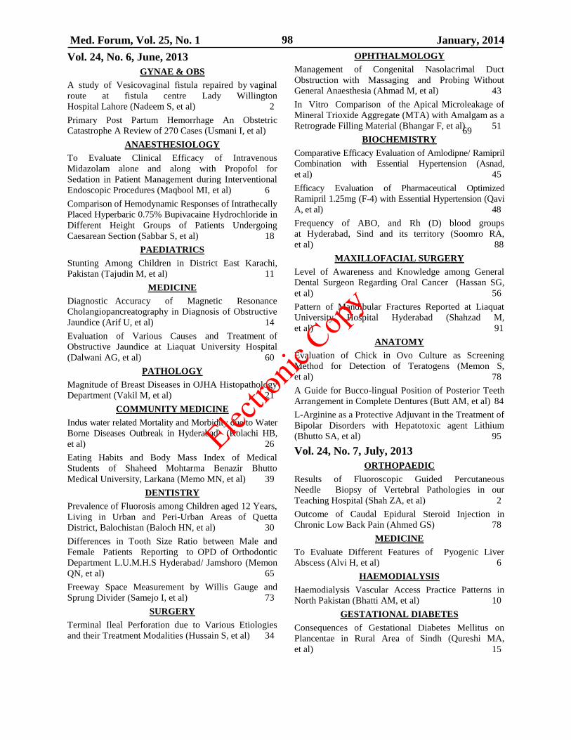
Med. Forum, Vol. 25, No. 1 January, 2014 98
Vol. 24, No. 6, June, 2013
GYNAE & OBS
A study of Vesicovaginal fistula repaired by vaginal
route at fistula centre Lady Willington
Hospital Lahore (Nadeem S, et al) 2
Primary Post Partum Hemorrhage An Obstetric
Catastrophe A Review of 270 Cases (Usmani I, et al) 69
ANAESTHESIOLOGY
To Evaluate Clinical Efficacy of Intravenous
Midazolam alone and along with Propofol for
Sedation in Patient Management during Interventional
Endoscopic Procedures (Maqbool MI, et al) 6
Comparison of Hemodynamic Responses of Intrathecally
Placed Hyperbaric 0.75% Bupivacaine Hydrochloride in
Different Height Groups of Patients Undergoing
Caesarean Section (Sabbar S, et al) 18
PAEDIATRICS
Stunting Among Children in District East Karachi,
Pakistan (Tajudin M, et al) 11
MEDICINE
Diagnostic Accuracy of Magnetic Resonance
Cholangiopancreatography in Diagnosis of Obstructive
Jaundice (Arif U, et al) 14
Evaluation of Various Causes and Treatment of
Obstructive Jaundice at Liaquat University Hospital
(Dalwani AG, et al) 60
PATHOLOGY
Magnitude of Breast Diseases in OJHA Histopathology
Department (Vakil M, et al) 21
COMMUNITY MEDICINE
Indus water related Mortality and Morbidity due to Water
Borne Diseases Outbreak in Hyderabad (Kolachi HB,
et al) 26
Eating Habits and Body Mass Index of Medical
Students of Shaheed Mohtarma Benazir Bhutto
Medical University, Larkana (Memo MN, et al) 39
DENTISTRY
Prevalence of Fluorosis among Children aged 12 Years,
Living in Urban and Peri-Urban Areas of Quetta
District, Balochistan (Baloch HN, et al) 30
Differences in Tooth Size Ratio between Male and
Female Patients Reporting to OPD of Orthodontic
Department L.U.M.H.S Hyderabad/ Jamshoro (Memon
QN, et al) 65
Freeway Space Measurement by Willis Gauge and
Sprung Divider (Samejo I, et al) 73
SURGERY
Terminal Ileal Perforation due to Various Etiologies
and their Treatment Modalities (Hussain S, et al) 34
OPHTHALMOLOGY
Management of Congenital Nasolacrimal Duct
Obstruction with Massaging and Probing Without
General Anaesthesia (Ahmad M, et al) 43
In Vitro Comparison of the Apical Microleakage of
Mineral Trioxide Aggregate (MTA) with Amalgam as a
Retrograde Filling Material (Bhangar F, et al) 51
BIOCHEMISTRY
Comparative Efficacy Evaluation of Amlodipne/ Ramipril
Combination with Essential Hypertension (Asnad,
et al) 45
Efficacy Evaluation of Pharmaceutical Optimized
Ramipril 1.25mg (F-4) with Essential Hypertension (Qavi
A, et al) 48
Frequency of ABO, and Rh (D) blood groups
at Hyderabad, Sind and its territory (Soomro RA,
et al) 88
MAXILLOFACIAL SURGERY
Level of Awareness and Knowledge among General
Dental Surgeon Regarding Oral Cancer (Hassan SG,
et al) 56
Pattern of Mandibular Fractures Reported at Liaquat
University Hospital Hyderabad (Shahzad M,
et al) 91
ANATOMY
Evaluation of Chick in Ovo Culture as Screening
Method for Detection of Teratogens (Memon S,
et al) 78
A Guide for Bucco-lingual Position of Posterior Teeth
Arrangement in Complete Dentures (Butt AM, et al) 84
L-Arginine as a Protective Adjuvant in the Treatment of
Bipolar Disorders with Hepatotoxic agent Lithium
(Bhutto SA, et al) 95
Vol. 24, No. 7, July, 2013
ORTHOPAEDIC
Results of Fluoroscopic Guided Percutaneous
Needle Biopsy of Vertebral Pathologies in our
Teaching Hospital (Shah ZA, et al) 2
Outcome of Caudal Epidural Steroid Injection in
Chronic Low Back Pain (Ahmed GS) 78
MEDICINE
To Evaluate Different Features of Pyogenic Liver
Abscess (Alvi H, et al) 6
HAEMODIALYSIS
Haemodialysis Vascular Access Practice Patterns in
North Pakistan (Bhatti AM, et al) 10
GESTATIONAL DIABETES
Consequences of Gestational Diabetes Mellitus on
Plancentae in Rural Area of Sindh (Qureshi MA,
et al) 15
Electro
nic Cop
y

Med. Forum, Vol. 25, No. 1 January, 2014 99
FORENSIC MEDICINE
Determine the Prevalence and pattern of killings in
Quetta, Balochistan (Rehman S, et al) 20
Third Decade of Life: The Most Vulnerable Age in
Homicidal Fire-Arms Deaths in Lahore (Jawad I,
et al) 51
DIABETIC FOOT ULCER
Management of Diabetic Foot Ulcers (Latif A,
et al) 23
PATHOLOGY
A Review of Uterine Cancers (Zafar MN, et al) 27
Detection of Beta-Lactamase and Extended Spectrum
Beta-Lactamase from Bacteria Causing Neonatal Sepsis
(Sami F, et al) 85
GYNAE & OBS
Comparison of Efficacy of Cervical Foleys Catheter
and Prostaglandin E2 for Induction of Labour (Bhatti
K, et al) 31
Internal Podalic Version Still Has a Role in the
Management of Transverse Lie (Gul K, et al) 59
MEDICINE/HIV
Radiographic Patterns in HIV Associated Pulmonary
Tuberculosis (Siddiqui MH, et al) 35
ANESTHESIA
To Compare the Efficacy of Rocuronium Bromide and
Succinylcholine in Elective Surgery for Endotracheal
Tube (Iqbal M, et al) 38
ANATOMY
Protective Effect of Vitamin E (Α Tocopherol Acetate)
on Gentamicin-induced Nephrotoxicity in Adult Albino
Rats (Rathore MI, et al) 42
Effect of Nigella sativa extract oil on granulation
tissue in cutaneous wound: An experimental study in a
Rabbit Model (Shahani MY, et al) 72
Ethanol induced developmental defects can be
prevented with supplementation of vitamin E (Memon
S, et al) 89
A Morphological Study of the Effects of X-Rays on
the Melanocytes of the Skin, Under Light Microscope
(Memon FM, et al) 99
COMMUNITY MEDICINE
Body Mass Index (BMI) of International Medical
Student in OJHA Campus Karachi, Pakistan (Maqsood
SM, et al) 47
Maternal Age & Parity as Risk Factors for Preterm
Births & Low Birth Weight among New-bornes
Delivered In Outskirts of District Mirpurkhas (Memon
KN, et al) 68
Psychological Effects among Internally Displaced
Persons (IDPS) Residing in two Districts of Sindh
(Rehman AT, et al) 82
Neonatal Sepsis - its Determinants & Outcome among
Newborns admitted in Neonatal Intensive Care Unit
(NICU), Liaquat University Hospital Hyderabad
(Siddique M, et al) 94
ORTHODONTICS
Comparison of Cephalometric Norms of Pleasing
Faces Reporting to OPD of Orthodontic Department
L.U.M.H.S Jamshoro / Hyderabad (Memon QN,
et al) 55
PHYSIOLOGY
Comparison of Effects of Short Term (12 Weeks) Low,
Moderate and High Intensity Exercises on Lipids and
Lipoproteins Levels in Young Hyperlipidemic Adults
(Khan GM, et al) 63
DENTISTRY
Dental Caries Status among Children aged 12-14
Years in Urban and Rural Areas of Lasbella District,
Balochistan (Khail AAK, et al) 103
Vol. 24, No. 8, August, 2013
GYNAE & OBS
Role of PH and Leukocyte Count in Prediction of
Preterm Labour (Masood H, et al) 2
Feto-maternal Outcomes in Women with Congenital
Heart Disease (Shaikh MM, et al) 5
Obstetrics Determinants of Cerebral Palsy in Children
from Birth to Five Years (Soomro N, et al) 47
Threatened Miscarriage as a Predictor of Obstetric
Outcome (Waheed S, et al) 51
ORTHOPAEDIC
Cannulated Screw Fixation of Fracture Neck of Femur
(Mehmood SQ, et al) 10
MEDICINE
Diagnostic Yield of Lymphocyte Rich Exudative
Pleural Effusion (Khan BA, et al) 13
Sensitivity Pattern of Causative Microorganisms in
Septicemia (Bashir B, et al) 16
Various Clinical Presentations of Falciparum Malaria
and Outcome: A study of 100 Patients (Fazel PA,
et al) 35
Incidence of Hepatitis A, B & C with Relation to Age,
Sex, Occupation, Socio-Economic Status, & Urban or
Rural Area of Hazara Division & District Sialkot (Haq
QM, et al) 43
Management of Hypertension and Diabetes Mellitus
at Primary Health Care Settings in Pakistan;
Clinician’s Perspective (Saleem S, et al) 98
BIOCHEMISTRY
To Study Biochemical Effects of Optimized Product
Ramipril 1.25mg Tablet with Essential Hypertension
(Latif A, et al) 20
Electro
nic Cop
y
Med. Forum, Vol. 25, No. 1 January, 2014 100
Biochemical Evaluation of Amlodipne / Ramipril in
Combination with Essential Hypertension (Asnad,
et al) 76
PATHOLOGY
Estrogen and Progesterone Receptor Status and Her-
2/Neu Over expression in Breast Carcinoma with
Relation to Age (Shaikh TA, et al) 24
Role of Amaltas and Dandasa in Controlling Biofilm
Formation of Streptococcus Mutans and Pseudomonas
(Faraz N, et al) 39
Pancytopenia: A Clinico-Haematologic Analysis of
100 Cases At Tertiary Care Hospital (Memon F,
et al) 80
SURGERY
Incidence of Inguinal Hernia with relation to Age, Sex,
Occupation, Socio-Economic Status, Dietary Habits &
Urban or Rural Area of District Sialkot (Sheikh TH,
et al) 27
Incidence of Tuberculosis with Relation to Age, Sex,
Occupation, Socio-Economic Status & Urban or Rural
Area of Hazara Division & District Sialkot (Ahmad N,
et al) 55
Adult Intussusception –Temple of Surprise 7 Year
Experience in Sialkot (Hussain S, et al) 71
FORENSIC MEDICINE
Chest: The Most Targeted Area in Homicidal Firearms
Fatalities in Lahore (Khokhar JI, et al) 31
ZOOLOGY
Study of Histological Changes in Liver , Lungs and
Testes of Dermally Nicotine Treated Albino Mice
(Latif AA, et al) 59
FIRE BURN
To Compare the Rate of Mortality among Fire Burn
Inhalational and Non Inhalational injuries in admitted
Patients at Burns Centre Civil Hospital Karachi (Ibran
E, et al) 64
REHABILITATION MEDICINE
Causes of Hand Impairment (Aasi MH, et al) 67
ANATOMY
Histochemical and Serological Parameters of
Hepatic Lithium Toxicity Ameliorated by L-Arginine
Supplementation (Makhija PM, et al) 84
PULMONOLOGY
Diagnostic yield of Fine Needle Aspiration Cytology
and Biopsy (FNAC/FNAB) in Thoracic Mass Lesions
(Mushtaq MA, et al) 90
COMMUNITY MEDICINE
Frequency of Hypertension in Male and Female
Medical students of Public Sector University of
Jamshoro in Hilly Region (Memon MN, et al) 93
Vol. 24, No. 9, September, 2013
ANATOMY
Comparative Study of Hepatic Serum Enzymes in
Albino Rats after Treatment with Tamoxifen
and Taurine (Ali A, et al) 2
NEUROSURGERY
Surgical Outcome of Thoracic and Thoraco-lumbar
Injuries of the Spine: A Case For Posterior
Instrumentation (Jaffery SA) 7
MEDICINE
Prevalence of Metabolic Syndrome in patients present
with Unstable Angina (Baloch AA, et al) 13
Serum Prolactin: A diagnostic marker in Fertile
Female Suffering from Rheumatoid Arthritis (Iqbal T,
et al) 25
GYNAE & OBS
Efficacy of Umbilical Artery Doppler in Detection and
Management of Intrauterine Growth Retardation
(Parveen R, et al) 18
PATHOLOGY
Significance of Platelet Count and D-Dimers Level in
Carcinoma Prostate (Shaikh SS, et al) 22
SURGERY
Altered lipid profile in patients with gallstones admitted
for cholecystectomy (Moosa FA, et al) 29
Evaluation of Risk Factors for Perforation in Acute
Appendicitis (Javed M, et al) 79
PHYSIOLOGY
Measurement of Plasma Fibrinogen Level in Healthy
Smokers to Predict the Risk of Cardio-vascular
Disease (Aslam N, et al) 32
LITHOTRIPSY
A prospective study of Extracorporeal Shock Wave
Lithotripsy (ESWL) Induced Hypertension, in Lahore
Division, Pakistan (Qadir MI, et al) 37
OSTEOPOROSIS
Cortical Changes in Orthopantomogram (OPG) of
Human Mandible as Early Detector of Osteoporosis
(Rehman DS, et al) 41
RADIOLOGY
Disk Degeneration in Adolescence and Young Adults:
A Cross-Sectional Magnetic Resonance Imaging
Study (Sattar A, et al) 45
ORTHOPAEDICS
Effectiveness of Platelete Rich Plasma (PRP) Injections
in Patients with Plantar Fasciitis (Jan R, et al) 49
CARDIOLOGY
Early Assessment of Outcome by ST Segment
Elevation after Acute Myocardial Infarction (Haq QM,
et al) 53
Electro
nic Cop
y

Med. Forum, Vol. 25, No. 1 January, 2014 101
DIABETES MELLITUS
To Correlate the Serum Levels of Adiponectin with
Fasting and Random Blood Sugars in Diabetes Mellitus
and Healthy Individuals (Ghori HN, et al) 57
OPERATIVE DENTISTRY
Assessment of Microleakage of Three Restorative
Materials in Class V Cavities. An in Vitro Study (Naz
F, et al) 60
COPD
Bronchoalveolar lavage fluid (BALF) differential
leucocyte count in Patients with Chronic Obstructive
Pulmonary Diseases (Rashid MN, et al) 65
COMMUNITY MEDICINE
Self Medication Practices among Residents of
Slum Areas of Qasimabad, Hyderabad (Memon KN,
et al) 70
BURN
To Evaluate the Presence and Spread of Environmental
Pathogens and their Sensitability. An Epidemiological
Survey of the Microbiological Monitoring at Burns
Ward Civil Hospital Karachi (Ibran E, et al) 74
NEUROSURGERY
Translation and Validation Study of the First Modified
Pakistani Version of the Neck Disability Index and its
Revalidation (Ahmed M, et al) 82
PULMONARY TB
Radiographic Patterns in HIV Associated Pulmonary
Tuberculosis (Siddiqui MH, et al) 87
Vol. 24, No. 10, October, 2013
DYSPEPSIA
To See the Synergistic Effect of PPI and Itopride in
Functional Dyspepsia (Alvi H, et al) 2
PRE AND POST MENOPAUSAL WOMEN
Study to Compare Serum Beta Crosslaps Levels
Between Premenopausal and Postmenopausal
Women (Nihal S, et al) 6
ANATOMY
Morphological Study of Superior Articular Facets of
Atlas Vertebrae (Hameed O, et al) 11
Cytoprotection of Heat – Induced Splenic Tissue by
Cyanocobalamin in Albino Rats (Hafeez A, et al) 67
Anti-stress Role of Cyanocobalamin in Heat- induced
Weight Loss in Albino Rats (Kumar A, et al) 91
BACTERIA RESISTANCE
Incidence of Bacteria Resistance to Antibiotics in
Hospitals (Ijaz A, et al) 16
DENTAL CARIES
Socio-economic Gradient & its Impact on Dental Caries
among School Children of Primary and Early Mixed
Dentition (Baig QA, et al) 20
SAFE INJECTION PRACTICES
Assessment of the Knowledge and Factors Affecting safe
Injection Practices among Health Care Providers in
Public and Private Sector Health Facilities of Tehsil Gujar
khan Rawalpindi Pakistan (Arshad A, et al) 25
BACTERIAL VAGINOSIS
Wet Mount Microscopy A Better Diagnostic Tool for
Bacterial Vaginosis Screening than Gram Staining
(Rizvi S, et al) 30
DIABETES MELLITUS
Burden of Diabetes Mellitus in the Families of Medical
Students of Lower Sindh (Ilyas M, et al) 35
PATHOLOGY
Sensitivity and Specificity of Phenotypic and Genotypic
tests for Detection of Methicillin-Resistant Staphy
lococcus aureus in Clinical Isolates (Taj Y, et al) 38
HYPOGLYCAEMIC EFFECT OF DRUGS
To Compare the Hypoglycaemic Effect of Sitagliptin /
Metformin Combination versus Glimiperide in Type II
Diabetes Patients during Ramadan (Abid R, et al) 43
PRETERM LABOUR
Increased Risk of Preterm Labour in Younger Pregnant
Females (Javed M, et al) 47
IODINE STATUS
Evaluation of Impact of Dietary Pattern on Iodine and
Thyroid Status of Adolescent Girls in Lahore (Imdad S,
et al) 49
CAESAREAN SECTION
Frequency and indications of Caesarean section at
Department of Obstetrics and Gynaecology Unit-III,
Civil Hospital Quetta (Jaffar Y, et al) 55
IMPAIRED GLUCOSE
Frequency of Impaired Glucose Tolerance in Patients of
Essential Hypertension in Tertiary Care Hospital,
Larkana (Soomro MA, et al) 58
HEPATITIS B
Prevalence of Hepatitis-D Virus Infection in Hepatitis
B Surface Antigen-Positive Children in Southern
Punjab (Ahmad M, et al) 64
ORTHOPAEDIC
External Fixation: Daily Versus Weekly Pin Site Care
(Jan R, et al) 72
RADIATION AND GROWTH HORMONE
Radioprotective Effect of Growth Hormone
(Somatotrophin) on Irradiated Long Bones of Young
Albino Rats (Panwhar AL, et al) 77
GYNAE & OBS
Three Monthly Intravenous Injections of Ibandronate
in the Treatment of Postmenopausal Osteoporosis
(Akhtar S, et al) 82
Electro
nic Cop
y

Med. Forum, Vol. 25, No. 1 January, 2014 102
HOMICIDAL FIREARMS
Predominance of Homicidal Fire-Arms Deaths in
Medico-Legal Autopsies in Lahore (Khokhar JI,
et al) 86
SUBDURAL EMPYEMA
Subdural Empyema: How Effective is The Burr Hole
Drainage? (Jaffery SA) 95
PROSTATE LESIONS
Spectrum of Prostate Lesions: A Decade Experience
(Sheikh NA, et al) 100
ISCHEMIC STROKE
Integrated Care of Modifiable Risk Factors in Patients
with Ischemic Stroke can Improve the Reproductivity
of State Rehman B, et al) 103
Vol. 24, No. 11, November, 2013
PHARMACOLOGY
Prevention of Insulin Induced Airway Hyper-Reactivity
with Montelukast on Isolated Tracheal Muscle of
Guinea Pig (Sharif M, et al) 2
JUNES FRACTURE
Conservative Management of Proximal Fifth Metatarsal
Fractures (Jones fracture) (Jan R, et al) 6
APPENDICITIS
Incidence of Appendicitis with relation to Age, Sex,
Occupation, Socio-Economic Status, Dietary Habits &
Urban or Rural Area of District Sialkot (Sheikh TH, et
al) 10
HEPATITIS A
Frequency of hepatitis A in patients with acute hepatitis
in Abbottabad (Haq QM, et al) 13
BACKACHE
To Determine the Frequency of Chronic Back ache
in the Nursing Staff of a Tertiary Care Hospital (Jatoi S,
et al) 16
INFERIOR WALL M1
To Know the Frequency of Right Ventricular
Infarction in Patients Presenting with Inferior
Wall Myocardial Infarction (Alam MT, et al) 20
ABDOMINAL TB
Various Presentation of Abdominal Tuberculois in
Surgical Patients (Jawed M, et al) 25
CHEMICAL BURN
Frequency of Chemical Burn and Mortality Rate in
Admitted Patients at a Tertiary Care Hospital of
Karachi (Ibran E, et al) 30
DRONE ATTACKS
Increasing Trend of Homicidal Drone Attacks in
Pakistan (Hanif M, et al) 33
MAXILLOFACIAL SURGERY
Dry Socket following Extraction of Permanent Teeth. A
Study (Shahzad M, et al) 37
MAXILLOFACIAL SURGERY
Pattern, Etiology and Risk Factors for Tooth Extraction
in Adult Population of Larkana City (Sheikh Q,
et al) 41
NOISE STRESS
The Effects of Acute and Chronic Noise Stress on the
Body Weight of Male Albino Rats (Iftekhar SM,
et al) 45
ACUTE MI
Predictors of In-Hospital Mortality in Acute Myocardial
Infarction (Rathore JA) 49
VIOLENCE WITH WOMEN
Increasing Trend of Domestic Violence with women in
Pakistan (Bhatti A, et al) 54
PRETERM LABOUR
Increased Risk of Preterm Birth in Anemic Pregnants
(Javed M, et al) 58
LIPID PROFILE DURING PREGNANCY
Evaluation of Serum Lipid Profile During The
Trimesters of Pregnancy (Kumar A, et al) 60
MAXILLOFACIAL SURGERY
Maxillofacial Trauma and Associated Brain Injuries
(Islam T, et al) 64
LYMPH NODE BIOPSY
A 4-Year Study of Lymph Node Biopsy in the Patients
Belonging to the Labour Class (Noorani SM,
et al) 67
SACROILIAC JOINT SYNDROME
Sacroiliac Joint Syndrome in the Differential Diagnosis
of Low Back Pain and Sciatica (Jaffery SA) 71
RESPIRATORY DISEASES WITH CEMENT
Pattern of Respiratory Diseases & Associated Socio-
Demographic Factors among Workers of Zeal Pak
Cement Factory, site area, Hyderabad (Shaikh K,
et al) 76
ACUTE POISONING
Current Trends of Acute Poisoning Reporting at Private
Medical Center of Rural Sindh (Khoharo HK,
et al) 80
CARPAL TUNNEL SYNDROME
Carpal Tunnel Syndrome in Pregnancy (Akhtar S,
et al) 84
BIOCHEMICAL EFFECTS OF DRUGS WITH
HYPERTENSION
Evaluation of Efficacy & Biochemical Effects of
Amlodipine / Atorvastatin with Essential Hypertension
(Asnad, et al) 88
Electro
nic Cop
y
Med. Forum, Vol. 25, No. 1 January, 2014 103
OSTEOARTHRITIS
Efficacy of Piroxicam in Osteoarthritis Khokhar GN, et
al) 91
OPHTHALMOLOGY
Comparison of the Effect of Ringer's Lactate and
Balanced Salt Solution on Postoperative Endothelial
Cell Count after Phacoemulcification? (Hussain SA,
et al) 94
Vol. 24, No. 12, December, 2013
ERB’S PALSY
Birth related Erb’s Palsy: Obstetrical correlates and
Outcome (Jan R, et al) 2
FETAL BIOMETRY
Fetal Biometry in Correlation with Ultrasonographic
Appearance and Measurement of Epiphyseal
Ossification Centres of Fetal Peripheral Long Bones for
Assessment of Gestation age in the third Trimester
(Channar MI, et al) 6
CHILD PUGH CLASS
Frequency of Different Child Pugh Class
Presentation in Patients of Chronic Hepatitis-C and
diabetes mellitus based on HBA1c (Shah MU, et al) 11
BETA THALASSAEMIA
Evaluation of Patterns of Beta Thalassaemia in
Children with Microcytic Hypochromic Anemia (Khoja
S, et al) 15
ACUTE APPENDICITIS
Pattern of Microbes Associate to Acute Appendicitis at
Liaquat University Hospital (Yousif S, et al) 20
DIAGNOSIS BRONCHOSCOPY
The Role of Fiberoptic Bronchoscopy in the Diagnosis
of Unsuspected Pulmonary Disease Presenting with
Hemoptysis (Ashfaq M, et al) 24
WISDOM TOOTH
Wisdom Tooth Removal: Are We Following any
Guidelines in Pakistan (Hassan SG, et al) 27
OTITIS MEDIA
Prevalence of Bacteria and Their Sensitivity to
Various Antibiotics in Chronic Suppurative
Otitis Media (Nisa Z, et al) 31
MEDICAL EDUCATION
Students’ Perspective of an Innovative Assessment
Strategy of Anatomy (Zareen N, et al) 34
FATTY DIET
In-House Prepared High Fat Diet (HFD) can Cause an
Increase in Body Weights of Male Albino Wistar Rats
in a Short Period of 2-3 Weeks (Mubeen S, et al) 38
TARGET KILLING
Increasing Tendency of “Target Killing” in Pakistan
(Hanif F, et al) 42
GIT TUMOUR
Pattern of Gastrointestinal (GIT) Cancer in CENAR,
Quetta Balochistan Pakistan: 10 Year Review (2001-
2010) (Manzoor H, et al) 46
PULMONARY NOCARDIOSIS
Pulmonary Infections caused by Nocardia Species
in Patients with Chronic Obstructive Pulmonary
Diseases (Rashid MN, et al) 52
ENZYMES IN CHRONIC DIABETIC
Correlation of Advanced Glycation End Products
(AGEs) with the Level of Alanine Amino Transferase
(ALT) and Aspartate Amino Transferase (AST)
Enzymes in Chronic Diabetic Patients (Malik RM,
et al) 57
INGROWN TOE NAIL
Early Stage Ingrown Toe Nail & role of conservative
treatment- An experience at Sukkur (Shah AA, et al) 61
FETAL BIOMETRY
Fetal Biometry in the Third Trimester; Local
Perspective (Suhag AH, et al) 64
ASSESSMENT OF GESTATION AGE
Ultrasonographic Appearance and Measurement of
Epiphyseal Ossification Centres of Fetal Peripheral
Long Bones for Assessment of Gestation Age (Suhail
S, et al) 69
VESICOVAGINAL FISTULA
Simultaneous Repair for Complicated/Complex
Vesicovaginal Fistula (Suhail MA, et al) 74
LIVER ABSCESS
Frequency of Clinical Manifestations, in Cases of
Liver Abscess in Hospital Based Study (Shah
SK, et al) 78
LOWER BACK PAIN
Effectiveness of Core Stabilization Exercises versus
Mckenzie’s Exercises in Chronic Lower Back Pain
(Ali S, et al) 82
HYPERCHOLESTEROLEMIA
Significance of Hypercholesterolemia in Hypothyroid
Patients (Qavi A, et al) 86
RELIGION & DEPRESSION
Relationship Between Religious Orientation and
Depression (Akram H, et al) 89
BRONCHOGENIC CARCINOMA
Frequency of Bronchogenic Carcinoma in Smoker
Patients with Hemoptysis Undergoing Fiberoptic
Bronchoscopy (Muhammad S, et al) 92
VIT. D MENOPAUSAL
Prevalence of Vitamin D Inadequacy among
Postmenopausal Women (Akhtar S, et al) 96
Electro
nic Cop
y
Med. Forum, Vol. 25, No. 1 January, 2014 104
Authors Index January to December 2013 Azhar Masud Bhatti
Editor in Chief
Vol. 24, No. 1, January, 2013
Names Page No.
1. Bhatti AM 1
2. Sarwat S, Rafique M, Ahmed R 2
3. Khan I, Qadir F, Fareed A, Ain Q 7
4. Shahid ZA, Qureshi KH, Zulfiqar L 12
5. Ullah S, Nizami SM, Anjum I 17
6. Hafeez R, Memon SR, Bhatti K 21
7. Soomro AA, Soomro ZA, Channa SM,
Chand H, Jamro BD 26
8. Ansari IA, Shah SAH, Lal S, Yasin G 29
9. Maqbool A, Hameed O, Athar Z, Rafi A 33
10. Saeed A, Mehmood SM, Awan AN 38
11. Hasan SM, Siddiqui KJ, Hasan SM,
Khan TN, Zaidi SAH 41
12. Phulpoto JA, Bhatti ZA 44
13. Shaikh AG, Kalhoro FA, Dall AQ 49
14. Hashim Y, Rizvi S, Muhammad D,
Aftab R. Hasan M 55
15. Soomro N, Khan M, Ahmed SI, Hanafi M,
Anwar K, Minhas MA, Siddiqui KY 60
16. Kumar S, Kolachi GM, Usman G 64
17. Dall AQ, Siyal ZH, Butt m 70
Vol. 24, No. 2, February, 2013
Names Page No.
1. Bhatti AM 1
2. Soomro N, Khan M, Ahmed SI 2
3. Naqvi A, Shoro AA, Farooq AD 6
4. Iqbal J, Anjum MS, Rashid A, Tahir GA,
Hussain T, Hameed A 10
5. Jaffar Y, Ehsan N, Nosheen 14
6. Alam MT, Ahmed S, Salekeen MS,
Siddiqui AS, Nasir S 17
7. Bokhari NA, Irshad S, Akhtar S,
Neelam S 22
8. Phulpoto JA, Bhatti ZA 26
9. Bhurgri GR, Samon NA, Baig F 30
10. Ahmed A, Chaudhry AG, Ali N,
Hussain S, Afzal MI, Irshad MK 35
11. Bhatti K, Hafeez R, Shaikh F 39
12. Rehman MZ, Habib S, Ullah S, Ullah E,
Malik ZI 42
13. Khan MI, Khan W, Ashraf M,
Muhammad 48
14. Khan BA, Ahmad A, Ahad A 52
15. Usmani I 55
16. Mehtab K, Kumar S, Ahmed R,
Ahmed W, Bhura S 58
17. Faryal U, Rashid S, Fayyaz I 61
18. Rashid S, Faryal U, Fayyaz I 64
19. Kumar S, Kolachi GM, Mehtab K 68
20. Jafferi SA 74
21. Javed MA, Ahmad F, Gill AH 79
22. Saleem M 83
Vol. 24, No. 3, March, 2013
Names Page No.
1. Jan MM 1
2. Usmani I, Kamal Z, Ashraf T 2
3. Rathore MI, Sultana K, Bhurgri GR,
Qazi MA 6
4. Kolachi HB, Tajuddin M, Maqsood SM,
Shaikh MA 11
5. Bhatti K, Lashari AA, Shaikh F,
Hafeez R 15
6. Laghari AH, Samoo AH, Qureshi KA,
Shaikh GS, Shah AM 18
7. Alam MT, Faraz A, Ahmed S, Siddiqui
AS, Siddiqui SS, Nasir S 22
8. Memon A, Laghari M, Talpur AH,
Shah Q, Khoharo HK 28
9. Ali S, Niazi S, Tariq NA, Hameed W,
Tareen, Qamar K 32
10. Javed MA, Furqan A, Ali S 38
11. Banglani MA, Punjabi SK, Khoso NA 43
12. Kohari T, Rizvi MIH 47
13. Shafi S, Rao MH, Rehman A 51
14. Kumar S, Mehab K, Kolachi GM,
Rajput AR 55
15. Mehtab K, Nisar N, Kumar S,
Nihal S, Zaheer S, Aman H 60
16. Hassan SG, Shahzad M, Butt AM,
Shams S, Ali N 63
Vol. 24, No. 4, April, 2013
Names Page No.
1. Jan MM 1
2. Jan R, Askar Z, Iqbal J 2
3. Qureshi KA, Laghari AH, Samoo AH 6
4. Ali S, Hassan Q, Channar KA, Kumar S,
Memon AB, Dahri WM 9
5. Moazzam M, Hameed W, Khan RMS,
Khan AS, Rahber I, Masood MO 14
6. Azhar S, Shafiq M, Farrukh A 19
7. Qureshi MA, Soomro N, Talpur GM 22
8. Qazi S, Mazhar A, Sarwat S 26
9. Bhatti K, Shaikh D, Hafeez R 31
10. Saqib A, Fatima S, Bhugri AN,
Ansari AK 34
Author’s Index
Electro
nic Cop
y

Med. Forum, Vol. 25, No. 1 January, 2014 105
11. Alam MT, Lashari MN, Ahmed S,
Siddiqui AS, Aslam M 38
12. Fatima I, Sheikh NA, Khan MKH 42
13. Sarfraz N, Anjum S, Masood MS 47
14. Shahzad M, Hassan SG, Arain GH,
Shams S 51
15. Alvi H, Arain MA 54
16. Punjabi SK, Hassan Q, Nisa Z, Ali S 58
17. Mirza A, Hassan SG, Shahzad M,
Arain GH 62
18. Hassan SG, Dall AD, Shahzad M,
Shams S, Baig MA, Ali N 66
19. Qadeer A, Usmani MS, Kumar S,
Rajput AR 70
20. Meher T, Iman N, Maheshwary N,
Ishtiaq S, Siddiqi A 73
Vol. 24, No. 5, April, 2013
Names Page No.
1. Jan MM 1
2. Butt SA, Raja TM, Shaikh NA 2
3. Hussain A, Vohra S, Abid S 6
4. Vohra S, Hussain A, Ali R 9
5. Soomro N, Qureshi MA, Talpur GM 12
6. Sami F, Hashim Y, Barakzai R,
Aftab R, Zafar N 17
7. Maqsood SM, Tajuddin M, Matloob A 21
8. Saeed N, Mustansar T, Sheikh NA,
Khanzada MS, Rasool S, Mirza T 26
9. Memon AB, Channar KA, Kalhoro FA,
Mirani SA 32
10. Fatima I, Taj Y, Khan MKH, Kamal M,
Fraz TR 37
11. Memon S, Shahani SB, Shahani MY,
Bano U 41
12. Jawaad I, Mughal MI, Ehsan N 45
13. Shaikh B, Shaikh SA, Shah AA,
Sohu KM, Soomro SH 51
14. Soomro AA, Taj Y, Mirjat KA,
Parveen A, Ali SW 55
15. Shafi S, Rao MH, Rehman S 60
16. Mirani SH, Sohu KM, Shah AA 64
17. Mehtab K, Salat MY, Shaikh S, Chughtai
S, Abid S, Khalid S, Hameed H 68
18. Akhund IA, Abid S, Vohra MS 71
19. Asnad, Vohra MS, Abid S 74
20. Iqbal M, Khokhar JI, Jawad I, Nadir S 78
21. Qadeer A, Noonari SM, Kumar S 82
22. Shahzad M, Hassan SG, Abbas Z,
Shams S, Aijaz P, Kumar M 85 23. Hassan SG, Israr M, Shahzad M,
Shams S, Aijaz P 89
Vol. 24, No. 6, June, 2013
Names Page No.
1. Jan MM 1
2. Shahzad N, Malik A, Hanif A 2
3. Maqbool MS, Draz MU, Shahani AS,
Iqbal M 6
4. Tajuddin M, Maqsood SM, Kolachi HB,
Khan RTY, Kamdar HA 11
5. Arif U, Shah ZA, Khan MNB 14
6. Sabbar S, Siddiqui MH, Bashir B, Turab
SM, Majid U, Salam J, Baloch AA 18
7. Vakil M, Mustanser T, Shaikh NA,
Rasool S, Mirza T, Ali A 21
8. Kolachi HB, Memon MN, Siddiqui MI 26
9. Baloch HN, Mengal N, Khail AAK,
Kurd SA, Alizai KA 30
10. Hussain S, Sajjad A, Farooq A 34
11. Memon MN, Memon AH, Abro A 39
12. Ahmad M, Fida R, Khattak NN,
Akhund IA 43
13. Asnad, Nasir N, Ali R, Ishaq M 45
14. Qavi A, Asnad, Hussain A, Ishaq M 48
15. Bhangar F, Kumar N, Channar KA,
Ali SA, Inayat N 51
16. Hassan SG, Shahzad M, Hassan Q,
Shams S, Ijaz P 56
17. Dalwani AG, Shaikh AR, Devanand 60
18. Memon QN, Alizai KA 65
19. Usmani I, Bakhsh FM 69
20. Samejo I, Dall AQ, Channar KA,
Shaikh IA 73
21. Memon S, Bano U, Shahani MY,
Shahani SB 78
22. Butt AM, Punjabi SK, Channar KA 84
23. Soomro RA, Shaikh TA, Memon MN,
Qazi N, Pathan NA, Abro A 88
24. Shahzad M, Hassan SG, Nisa Z,
Shams S, Aijaz P, Habib G 91
25. Bhutto SA, Makhija PM, Chang AH,
Naqvi A 95
Vol. 24, No. 7, July, 2013
Names Page No.
1. Hameed N 1
2. Shah ZA, Arif U, Bilal A 2
3. Alvi H, Arain MA 6
4. Bhatti AM, Mansoor J, Ahsin S, Salim B,
Humayun J, Anwar I, Mir M, Aslam M,
Mallick T 10
5. Qureshi MA, Bhurgri GR, Shah MR 15
6. Rehman S, Khan Z, Agha SA, Khetran
AK, Baloch MR 20
7. Latif A, Ansar A, Waheed S, Hamid A 23
8. Zafar MN, Barakzai R, Hashim Y,
Muhammad D, Rizvi S, Akram S 27
9. Bhatti K, Mahar T, Hafeez R 31
10. Siddiqui MH, Majid U, Salam J, Bashir
B, Turab SM, Baloch AA, Qasim A 35
11. Iqbal M, Turab SM, Siddiqui MH, Salam
J, Majid U, Baloch AA, Bashir B 38
12. Rathore MI, Khooharo Y, Ali SMM 42
Electro
nic Cop
y
Med. Forum, Vol. 25, No. 1 January, 2014 106
13. Maqsood SM, Tajuddin M, Rahman AA,
Kolachi HB, Shaikh MA 47
14. Jawad I, Khokhar JI, Iqbal M 51
15. Memon QN, Alizai KA, Ahmed S 55
16. Gul K, Bibi S, Gul Z, Gul P 59
17. Khan GM, Memon AS, Khan D 63
18. Memon KN, Memon S 68
19. Shahani MY, Memon S, Shahani SB,
Bano U, Arain SN 72
20. Ahmed GS 78
21. Rahman AA, Akhtar P, Siddiqui MI 82
22. Sami F, Rizvi S, Barakzai R, Hashim Y,
Farrukh, Shah T 85
23. Memon S, Shahani MY, Bano U,
Shahani SB 89
24. Siddique M, Memon KN, Sulehri MA 94
25. Memon FM, Kolachi GM, Kumar S 99
26. Khail AAK, Baloch HN, Hammad J,
Kurd SA, Baloch MR 103
Vol. 24, No. 8, August, 2013
Names Page No.
1. Hameed N 1
2. Masood H, Ashraf S, Siddique H,
Masood M, Masood MS 2
3. Shaikh MM, Bano K, Mujtaba S 5
4. Mehmood SQ, Ahmed S, Duha M,
Latifs R 10
5. Khan BA, Ahmad A, Aziz MI 13
6. Bashir B, Baloch AA, Fazel PA,
Siddiqui MH, Salam J, Majid U,
Masroor M, Turab SM 16
7. Latif A, Asnad, Karim A, Ishaq M,
Akhund IA 20
8. Shaikh TA, Memon F, Pathan NA,
Soomro RA 24
9. Sheikh TH, Azeem K, khan MA,
Hamid A, Sabir M, Hamid M 27
10. Khokhar JI, Nadir S, Iqbal M 31
11. Fazel PA, Bashir B, Baloch AA,
Siddiqui MH, Tahir M, Turab SM,
Salam J, Majid U 35
12. Faraz N, Abbas S, Murad R, Sehrish 39
13. Haq QM, Ahmad N, Ijaz A,
Hamid A, Sabir M, Hamid M 43
14. Soomro N, Khan M, Ahmed SI 47
15. Waheed S, Ansar A, Meraj B 51
16. Ahmad N, Haq QM, Ijaz A, Hamid A,
Sabir M, Hamid M 55
17. Latif AA, Mansha M, Sharif S, Saeed S,
Gill T, Saif K, Farasat T 59
18. Ibran E, Rao MH, Ali S, Raza A,
khanum A, Malik R 64
19. Aasi MH, Malik TM, Malik NI 67
20. Hussain S, Din IH, Farooq A 71
21. Asnad, Muhammad S, Fida R, Ishaq M,
Akhund IA 76
22. Memon F, Shaikh TA, Memon JH,
Soomro RA 80
23. Bhutto SA, Shoro AA, Makhija PM,
Arif M, Chang AH, Mangi MM 84
24. Mushtaq MA, Ayaz S, Khan SA 90
25. Memon MN, Siddiqui MI,
Soomro RA, Abro A, Kolachi HB 93
26. Saleem S, Qaiser N, Tahir A,
Mahmood H 98
Vol. 24, No. 9, September, 2013
Names Page No.
1. Jan MM 1
2. Ali A, Tasneem K, Naikzada, Inayat Q 2
3. Jaffery SA 7
4. Baloch AA, Bashir B, Siddiqui MH,
Qasim A, Majid U, Salam J, Turab SM,
Masroor M 13
5. Parveen R, Khakwani M, Masood MS 18
6. Shaikh SS, Saqib A, Memon RA,
Shaikh NS, Shaikh SS, Shaikh KR 22
7. Iqbal T, Zuberi NA, Niger S 25
8. Moosa FA, Bakhtiar N, Khalique A,
Jawaid M 29
9. Aslam N, Khalid M, Qureshi H,
Khan K 32
10. Qadir MI, Mobeen A, khan MM,
Iqbal A 37
11. Rehman DS. Rafique M, Hussain M 41
12. Sattar A, Shahzad A, Anjum S, Jalal LA 45
13. Jan R, Akhtar S, Khan MS, Karam F,
Iqbal J 49
14. Haq QM, Ahmed N, Ali J, Ijaz A,
Hamid A, Sabir M, Hamid M 53
15. Ghori HN, Akram SF, Ashraf F,
Masood SH 57
16. Naz F, Amin M, Ahmed A,
Rahman M 60
17. Rashid MN, Memon MA, Ismail MO
Noman B 65
18. Memon KN, Memon S, Shaikh K 70
19. Ibran E, Hassan M, Farooq Z,
Rao MH, Khan M 74
20. Javed M, Shaima A, Mubeen A 79
21. Ahmed M, Jaffery SA 82
22. Siddiqui MH, Baloch AA, Bashir B,
Salam J, Qasim A, Masroor M,
Javed A, Wahab S, Mehmood A 87
Vol. 24, No. 10, October, 2013
Names Page No.
1. Jan MM 1
2. Alvi H, Arian MA 2
3. Nihal S, Masood SH, Memon AS,
Zuberi NA, Fatima SS 6
4. Hameed O, Athar Z, Maqbool A 11
Electro
nic Cop
y
Med. Forum, Vol. 25, No. 1 January, 2014 107
5. Ijaz A, Haq QM, Ahmad N, Hamid A,
Sabir M, Hamid M 16
6. Baig QA, Afaq A, Nasir M, Qureshi M 20
7. Arshad A, Sikander S, Arshad T,
Habib H, Asghar MI, Rana SM 25
8. Rizvi S, Sami F, Hashim Y, Zafar MN,
Shah T, Muhammad D 30
9. Siddiqui MI, Shaikh TA, Soomro RA,
Memon MN, Kolachi HB, Nizamani
WM, Abro S 35
10. Taj Y, Soomro AA, Ali SW,
Parveen A, Kazmi SU 38
11. Abid R, Zahid M 43
12. Javed M, Mubeen A, Javed N 47
13. Imdad S, Muzaffar R, Shoukat MS 49
14. Jaffar Y, Sikander N 55
15. Soomro MA, Abro GY, Ansari IA,
Shah SAA, Shah SSR 58
16. Ahmad M, Irshad H, Khan ZA 64
17. Hafeez A, Kumar A, Jokhio AL 67
18. Jan R, Akhtar S, Khan MA 72
19. Panwhar AL, Kumar H, Gohar N,
Ansari AG 77
20. Akhtar S, Jan R 82
21. Khokhar JI, Iqbal M, Nadar S 86
22. Kumar A, Hafeez A, Ansari AG 91
23. Jaffery SA 95
24. Sheikh NA, Butt SA, Raja TM,
Zaheer S 100
25. Rehman B, Qidwai A, Ahmed S,
Hussain T 103
Vol. 24, No. 11, November, 2013
Names Page No.
1. Jan MM 1
2. Sharif M, Khan BT, Sharif A 2
3. Jan R, Akhtar S, Askar G 6
4. Sheikh TH, Azeem K, Khan MA,
Hamid A, Sabir M, Hamid M 10
5. Haq QM, Ahmed N, Ejaz A 13
6. Jatoi S, Alam MT, Nisa Q, Arvind,
Naila, Nasir S 16
7. Alam MT, Lashari MN, Murtaza M,
Moin F, Sadiq M, salkeen S 20
8. Jawed M, Khan MLZ, Qureshi MA,
Shaikh U, Akhater N 25
9. Ibran E, Rao MH, Khan M, Yasin I 30
10. Hanif M, Ahmad A, Masood R, Din SZ,
Hamid A, Najeeb 33
11. Shahzad M, Hassan SG, Sheikh Q,
Shams S. Aijaz P 37
12. Sheikh Q, Hassan SG, Shahzad M,
Shams S, Aijaz P 41
13. Iftehkar SM, Ilyas A, Mubeen S 45
14. Rathore JA 49
15. Bhatti A, Din SZ. Hamid A, Hanif F 54
16. Javed M, Mubeen A, Javed N 58
17. Kumar A, Baloch S, Shahani RA, Lata 60
18. Islam T, Lakhani MJ, Mehdi H,
Zubairi A 64
19. Noonari SM, Qadeer A, Soomro AQ,
Sajjad K 67
20. Jaffery SA 71
21. Shaikh K, Memon MA, Memon KN,
Sarah B, Ahmadani R 75
22. Khoharo HK, Kazi SAF, Rehman S,
Shah Q 80
23. Akhtar S, Jan R 84
24. Asnad, Ansari AL, Fida R, Ishaq M,
Akund IA 88
25. Khokhar GN, Khalid M, Ansari AL,
Alvi IA, Akhund IA 91
26. Hussain SA, Imran M 94
Vol. 23, No. 12, December, 2012
Names Page No.
1. Jan MM 1
2. Jan R, Akhtar S 2
3. Channar MI, Lakho AR, Suhag AH 6
4. Shah MU, Shah K, Zarkoon AK 11
5. Khoja S, Fahim A, Ahmed N, Azmi MA,
Kazi SA 15
6. Yousif S, Ahuja P, Bashir J,
Arain MA 20
7. Ashfaq M, Ullah I, Zahoor A, Ishaq M,
Akhund IA 24
8. Hassan SG, Shahzad M, Mirza D,
Shams S, Aijaz P 27
9. Nisa Z, Hassan Q, Hassan SG, Shams S 31
10. Zareen N 34
11. Mubeen S, Waggan IA, Nigar S 38
12. Hanif F, Din SZ, Bhatti MA, Hamid A 42
13. Manzoor H, Shuja J, Mahmood Z,
Ahmad HK, Mustafa KA 46
14. Rashid MN, Memon MA, Shakoor S,
Noman B 52
15. Malik RM, Bano S, Ahmed R 57
16. Shah AA, Sohu KM, Mirani SH,
Shaikh B 61
17. Suhag AH, Channar MU, Lakho AR 64
18. Suhail S, Channar MI, Shaikh AH,
Lakho AR, Suhag AH 69
19. Suhail MA, Shaikh AR, Kashif F 74
20. Shah SK, Kakar AK, Zarkoon AK 78
21. Ali S, Ali SM, Memon KN 82
22. Qavi A, Khokhar GN, Zahoor A,
Akhund IA, Ishaq M 86
23. Akram H, Bano S, Ahmed R 89
24. Muhammad S, Ullah I, Ashfaq M,
Ishaq M, Akhund IA 92
25. Akhtar S, Jan R 98
Electro
nic Cop
y

Electro
nic Cop
y