mechanisms of metabolic dysfunction in cancer-associated
TRANSCRIPT

REVIEW
Mechanisms of metabolic dysfunctionin cancer-associated cachexiaMichele Petruzzelli1 and Erwin F. Wagner2
1Department of Oncology, The Medical Research Council Cancer Unit, University of Cambridge, Addenbrooke’s Hospital,Cambridge CB2 0QQ, United Kingdon; 2Genes, Development, and Disease Group, Cancer Cell Biology Programme, CentroNacional de Investigaciones Oncológicas, Madrid 28029, Spain
Metabolic dysfunction contributes to the clinical deterio-ration observed in advanced cancer patients and is charac-terized by weight loss, skeletal muscle wasting, andatrophy of the adipose tissue. This systemic syndrome,termed cancer-associated cachexia (CAC), is a majorcause of morbidity and mortality. While once attributedsolely to decreased food intake, the present descriptionof cancer cachexia is a disorder of multiorgan energy im-balance. Here we review the molecules and pathways re-sponsible for metabolic dysfunction in CAC and theideas that led to the current understanding.
Human cancers develop as a localized focus of uncon-trolled cell growth and subsequently progress to a system-ic disease (Fig. 1). Cancer research primarily focuses onthe agents, events, and genetic alterations underlying tu-mor initiation, progression, and metastasis. However,the vastmajority of end-stage cancer patients suffers a sys-temic illness defined as cachexia, a widespread but poorlyunderstood condition (Lok 2015). The “most time hon-ored symptom of cancer” (Editors 1929), cachexia is theprototype image that comes to mind when thinking ofcancer. The loss of appetite, energy, and vigor; the languidand unsmiling face; the sallow and anemic aspect; and theskinny and wasted complexion are all too familiar to phy-sicians treating cancer patients. Despite the obvious clin-ical picture, a formal definition of the diagnostic criteriahas only recently been reached (Fearon et al. 2011). Thecurrent consensus for diagnosis is the unintentional lossof total bodyweight or skeletalmusclemass. Importantly,cancer-associated cachexia (CAC) is a complex metabolicdisorder with profound changes in energy balance, whichmight be already irreversible at the time of obvious bodyweight loss. While cachexia itself is often rapidly progres-sive, marking the irreversible decline in health and sur-vival, the time when cachexia appears in the clinicalhistory of the cancer patients is, at present, mostly unpre-
dictable. The severity of CAC is often unrelated to tumorsize or stage, with small tumors commonly leading tosevere wasting, as is the case, for example, for pancreaticand lung tumors. In contrast, widely disseminated cancersmay cause death without any evidence of CAC. The rea-sons for this paradoxical lack of correlation between tu-mor burden and the degree of cancer cachexia are, atpresent, elusive. Furthermore, CAC often results in fewercompleted cycles of chemotherapy with higher compli-cation rates. Therefore, a better characterization of themetabolic changes in the organism affected by cancer isurgently needed in order to recognize the early eventsof CAC and improve its prognosis. In this review, we dis-cuss the conceptual advances that shaped the currentunderstanding of the systemic metabolic maladaptationto cancer.
Research milestones in cancer cachexia
For centuries, the concept that a local malignant growthcould be responsible for systemic effects has been underdebate (E.F.B. 1909). Rather than a specific disease, thewasting associated with cancer was attributed to nonspe-cific pathological complications of the tumor, such asanorexia, hemorrhage, infection, or ulceration of the neo-plastic tissue (Willis 1948). In contrast, those in favor ofsystemic alterations produced by the tumor on the hostconsidered cachexia the result of either direct secretionby the tumor of some substances active in distant organsor uptake by the tumor of components from the blood thatare essential for the correct functioning of distant organs(Greenstein 1947; Donovan 1954). Evidence supportingone hypothesis or the other was slim, and the contentionwas disputed on the basis of small clinical case series andanecdotal post-mortem findings (Donovan 1954).During the past decades, a vast body of investigation has
reshaped our understanding of CAC (Fig. 2). Experimentalwork with animal models of cancer rather than
[Keywords: cancer-associated cachexia (CAC); metabolic failure; whiteadipose tissue (WAT) browning; skeletal muscle atrophy]Corresponding authors: [email protected], [email protected] is online at http://www.genesdev.org/cgi/doi/10.1101/gad.276733.115.
© 2016 Petruzzelli and Wagner This article is distributed exclusively byCold Spring Harbor Laboratory Press for the first six months after the full-issue publication date (see http://genesdev.cshlp.org/site/misc/terms.xhtml). After six months, it is available under a Creative Commons Li-cense (Attribution-NonCommercial 4.0 International), as described athttp://creativecommons.org/licenses/by-nc/4.0/.
GENES & DEVELOPMENT 30:489–501 Published by Cold Spring Harbor Laboratory Press; ISSN 0890-9369/16; www.genesdev.org 489
Cold Spring Harbor Laboratory Press on February 4, 2022 - Published by genesdev.cshlp.orgDownloaded from
observations in the clinical setting led to the recognitionof CAC as a legitimate entity independent of the effectsof anorexia or mechanical interference of the tumorwith the surrounding tissues. When tumor-bearing ratswere force-fed a high-fat diet, weight loss was prevented.However, the development of anemia and the enlarge-ment of the adrenal glands were not affected, thus show-ing the existence of systemic manifestations of cancerindependent of nutritional intake (Begg and Dickinson1951). Following the kinetics of tissue loss in tumor-bear-ingmice, it was noticed that adipose tissuewastingwas anearly event, occurring at a time when the tumor was bare-ly palpable (Costa andHolland 1962). Surprisingly, fat losscould also be induced by nonviable tumor preparations,indicating that soluble components of tumor extractscan induce cancer cachexia (Costa and Holland 1962).The causes for the systemic effects associated with cancerweremore sophisticated than just reduced food intake andneeded to be sought in the complex relationship betweenthe host and the tumor. Analogies between systemic re-sponses to infectious agents and cancers were noted, in-cluding fever, leukocytosis, and increased serum levels
of acute phase response proteins (Rosenthal and Franklin1975). The first evidence that inflammatory mediators—namely, cytokines—were involved in the process of pro-tein breakdown in isolated skeletalmuscle and a potentialrole for interleukin-1 (IL-1) in muscle degradation duringfever was published in 1983 (Baracos et al. 1983). Particu-lar attention was received by the somewhat paradoxicalincrease in serum lipid levels despite the obvious loss ofbody weight in severely sick patients. It was found thatsuch hypertriglyceridemia could be induced experimen-tally in animals by either injection of infective agents ortransplantation of tumors (Rouzer and Cerami 1980;Kawakami and Cerami 1981). Hypertriglyceridemia wasthe result of lipoprotein lipase (LPL) inhibition and couldbe reproduced by injecting animals with conditioned me-dium from inflammatory cells incubated with endotoxin(Kawakami and Cerami 1981).
In 1985, Bruce Beutler in Anthony Cerami’s group(Cerami et al. 1985) provided definitive proof that circulat-ing mediators could cause cachexia, showing that culturemedium from endotoxin-activated macrophages causedbody weight loss when injected into mice. The moleculein the conditioned medium causing cachexia was purifiedand termed “cachectin” (Beutler et al. 1985). Subse-quent determination of the complete primary structureof cachectin revealed its identity with tumor necrosis fac-tor-α (TNFα) (Fransen et al. 1985; Pennica et al. 1985). Itshould be noticed that these early preparations of condi-tionedmedium containedmultiplemacrophage products,and it is therefore erroneous to attribute all of the cachex-iogenic action to the effect of TNFα alone. Furthermore,TNFα causes systemic shock and the release of other cyto-kines, further confounding the attribution of the observedphenotype to a single identifiable factor. However, despitethe technical limitations, these early studies contributedto a conceptual evolution in the field of cachexia research,and the wasting syndrome was finally regarded as the re-sult of the host response to the tumor. Clinical evidenceconfirmed the conclusions of preclinical investigationsshowing that intravenous hyperalimentation could not al-leviate cachexia in cancer patients (Evans et al. 1985). Re-markably, the focus of research had gradually shifted fromthe nature of the invasive agent (infection or cancer) to thequality of the response elicited in the organism. The im-mune systemwasthe likely source of allmediators respon-sible for the systemic changes, and a variety of cytokinesjoined TNFα in the ability to cause systemic alterations(Beutler and Cerami 1986). However, despite the clearrole played by cytokines in experimental cachexia, theirinvolvement in human disease was less obvious, and clin-ical translation yielded ambiguous results (Balkwill et al.1987; Socher et al. 1988). Perhaps as a consequence of thedisappointment generated by the lack of clinical benefitsfrom basic findings, the pace of research in the field of can-cer cachexia progressively reduced.An additional explana-tion could be the rapid progress of molecular biology ofcancer in themid-1980s. That was the timewhen the firstoncogenes were discovered, and the simplistic view of re-ducing cancer to a single base mutation was occupyingthe entire scene (Weinberg 2014). The war on cancer
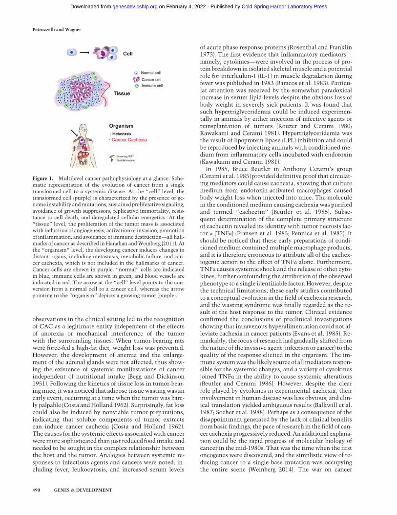
Figure 1. Multilevel cancer pathophysiology at a glance. Sche-matic representation of the evolution of cancer from a singletransformed cell to a systemic disease. At the “cell” level, thetransformed cell (purple) is characterized by the presence of ge-nome instability andmutations, sustained proliferative signaling,avoidance of growth suppressors, replicative immortality, resis-tance to cell death, and deregulated cellular energetics. At the“tissue” level, the proliferation of the tumor mass is associatedwith induction of angiogenesis, activation of invasion, promotionof inflammation, and avoidance of immune destruction—all hall-marks of cancer as described in Hanahan andWeinberg (2011). Atthe “organism” level, the developing cancer induces changes indistant organs, including metastasis, metabolic failure, and can-cer cachexia, which is not included in the hallmarks of cancer.Cancer cells are shown in purple, “normal” cells are indicatedin blue, immune cells are shown in green, and blood vessels areindicated in red. The arrow at the “cell” level points to the con-version from a normal cell to a cancer cell, whereas the arrowpointing to the “organism” depicts a growing tumor (purple).
Petruzzelli and Wagner
490 GENES & DEVELOPMENT
Cold Spring Harbor Laboratory Press on February 4, 2022 - Published by genesdev.cshlp.orgDownloaded from

seemed to be close to a favorable ending, and the focus ofresearch zoomed back to the tumor itself rather than theresponse that the tumor ignites in the organism.
Cancer cachexia is an energy balance disorder linkedto inflammation
Weight loss is acardinal signof cachexiaand represents themain independentpredictorofmortality in cancerpatients(Fearon et al. 2012). The mechanisms for weight loss incancer are multiple, including decreased nutrient intake,systemic metabolic dysfunction, and increased energy ex-penditure. Inflammation represents a common denomina-tor in the pathophysiology of energy imbalance duringcachexia. In mice, the peritoneal injection of cancer cellsexpressing TNFα has been shown to cause weight lossand cachexia. In contrast, mice injected with the samecells without TNFα do not lose body weight (Oliff et al.1987). Similar results have been obtained with IL-6 in pre-clinicalmodels (Strassmann et al. 1992). Both host- and tu-mor-derived cytokines cooperate in a complex way withthe tumor microenvironment to sustain tumor growthandcachexia (Cahlin et al. 2000). In support of a role of can-cer-derived cytokines, it has been shown recently that ex-pression of the cytokine TNF-related weak inducer ofapoptosis (TWEAK) by cancer cells causes cachexia, andthe effect is similar in wild-type and TWEAK-deficientmice (Johnston et al. 2015).As the cancer persists, it is assumed that ongoing local
inflammationmay reach a threshold when cytokines spillinto the circulation, thus transforming the cancer diseasefrom a localized tumor to a systemic impairment. Unfor-tunately, such a simplistic view does not stand present ex-perimental validation. The levels of serum cytokines donot correlatewith the appearance of cachexia in cancer pa-tients (Fearon et al. 2012). Furthermore, treatments withantibodies targeting a single cytokine have failed so farto prevent or significantly ameliorate the wasting syn-drome (Penna et al. 2010; Fearon et al. 2013). Very recentdata emphasize the multifactorial etiology of CAC, show-ing now that a combination of cytokines and/or additionalmediators is responsible for the cachectic phenotype(Schaefer et al. 2016).
Despite the absence of a simplistic threshold modellinking cytokine levels to cachexia development, a richbody of evidence supports their causal role in the meta-bolic dysfunction observed in CAC. Mechanistically,cytokines were shown to increase the metabolic ratethrough activation of thermogenesis, inhibit adipocyteand skeletal myocyte differentiation, and reduce foodintake (Guttridge et al. 2000; Li et al. 2002; Ruan et al.2002; Arruda et al. 2010). However, weight loss in cancerpatients cannot be attributed solely to decreased food in-take, since dietary supplements fail to reverse cachexia(Bruera and Sweeney 2002). In contrast, a recent study inmice expressing high levels of the proinflammatory cyto-kine IL-18 suggests that high caloric feeding in the contextof metabolic dysfunction may exacerbate weight loss andcause fatal cachexia (Murphy et al. 2016). In the context ofcancer, metabolic dysfunction is caused by deregulatedcarbohydrate and lipid metabolism.
Altered carbohydrate metabolism in cancer cachexia
Carbohydrate intolerance in cancer patients has long beennoted (Rohdenburg et al. 1919). While fasting blood sugarconcentration between control and cancer groups did notdiffer significantly, intravenous glucose tolerance testsshowed significantly decreased disappearance of glucosein cancer patients (Bishop and Marks 1959). In the firsthalf of the last century, Cori and Cori (1925) comparedglucose levels in the venous blood from the tumor-bearingarm and the unaffected arm of a patient with a sarcoma onthe forearm. Glucose levels from the tumor-bearing armwere reduced, thus confirming in vivo the increased rateof tumor glycolysis (Cori and Cori 1925). Since tumor tis-sue takes up glucose, the decreased disappearance of glu-cose observed in the tolerance test must be sought inmetabolic alterations in the host tissues associated withcancer development. Either increased hepatic glucose pro-duction or a decrease in peripheral utilization could ac-count for the reduced glucose tolerance observed incancer patients. Despite decreased hepatic glycogenstores, endogenous glucose production is increased in ca-chectic patients due to increased hepatic glucose recy-cling via lactate, a phenomenon termed the Cori cycle
1951 1962 1983 1985 1993 2001 2011 2015
Injection of non-viable
tumor preparations causes
fat atrophy in mice
Cachectin/TNF-α is the first identified
mediator of cachexia
Myostatin causes
skeletal muscle atrophy
Anamorelin increases lean body mass in a Phase III clinical trial
International consensus
on the diagnostic criteria
of cachexia
Role of the ubiquitin
pathway in skeletal
muscle atrophy
IL1 causes protein
breakdown in isolated
skeletal muscle
Systemic manifestation
of cancer despite forced-
feeding in rats
Figure 2. Timeline of discoveries in cancer cachex-ia. In 1951, the first systemicmanifestation of cancerwas described in rats. In 1962, it was observed thatinjection of tumor preparations in mice was suffi-cient to induce fat atrophy. In 1983 and 1985, thefirst candidate molecules were identified. Seminalpublications in 1993 and 2001 described a role forthe ubiquitin pathway and myostatin in skeletalmuscle atrophy. It was not until some years agothat an international consensus on the diagnosticcriteria of CAC was reached. Promising resultshave been reported in late 2015 from the first phaseIII clinical trial targeting CAC with the ghrelin re-ceptor agonist anamorelin (https://www.iaslc.org/news/results-phase-iii-trials-anamorelin-advanced-non-small-cell-lung-cancer-patients-cachexia).
Cancer cachexia and metabolic dysfunction
GENES & DEVELOPMENT 491
Cold Spring Harbor Laboratory Press on February 4, 2022 - Published by genesdev.cshlp.orgDownloaded from

(Holroyde et al. 1984). Apart from these studies, clinicalinvestigations on glucose metabolism in cachectic pa-tients are noticeably thin. While one study suggests thatglucose intolerance may worsen with the developmentof cachexia (Jasani et al. 1978), other studies found thatglucose intolerance did not correlate with body weightloss (Yoshikawa et al. 2001; Agustsson et al. 2011). Veryrecently, elegant genetic studies in the fruit fly Droso-philahave identified an important role of insulin signalingin inducing a cachexia-like systemic wasting followingtransplantation ofDrosophila tumors (Figueroa-Clarevegaand Bilder 2015; Kwon et al. 2015). Both studies have iden-tified a tumor-secreted factor, ImpL2/IGFBP (an insulin-binding protein and antagonist of insulin/insulin-likegrowth factor [IGF] signaling), that is responsible for thewasting phenotypes in organs distant from the transplant-ed tumors (Wagner and Petruzzelli 2015).
Role of lipids, burning fat, and white adiposetissue (WAT) browning
Besides changes in carbohydrate metabolism, the han-dling of lipids between tissues is severely impaired in can-cer patients. The deposition of triglycerides (TGs) incytoplasmic lipid droplets represents the most efficientform to store lipids inWAT andmany other cell types. Al-ready in 1848, the French physiologist Claude Bernarddiscovered that TGs, commonly called fat, are digestedin the gut before they can be absorbed. The hydrolysis ofTGs, designated lipolysis, generates glycerol and fatty ac-ids (FAs). The enzymes mediating intracellular lipolysisinclude adipose TG lipase (ATGL) and the hormone-sensi-tive lipase (HSL), while LPL is responsible for the hydroly-sis of plasma TGs of lipoproteins in the vascular system(Young and Zechner 2013). FA uptake and TG synthesisdecline in WAT in murine cancer models, whereas, inhuman CAC, it is associated with normal lipid synthesisbut elevated lipolysis in WAT. This suggests that lipid ca-tabolism is more relevant than inhibition of lipid synthe-sis for the loss of WAT in CAC (Dahlman et al. 2010).These findingswere corroborated in an elegant study dem-onstrating that WAT lipolysis in cancer patients is in-creased due to elevated enzyme activities of ATGL andHSL (Das et al. 2011). Importantly, genetic deletion ofAtgl in mice prevented increased lipolysis and the reduc-tion of WAT and skeletal muscle mass in certain modelsof CAC. Similar results were also observed, although toa lesser extent, when HSL was inactivated (Das et al.2011). Lipolysis in CAC is induced bymany serum factorssecreted by tumor or host cells, including hormones suchas glucocorticoids and catecholamines; cytokines likeTNFα, IL-1β, IL-6, prostaglandins; and a zinc–glycopro-tein, ZAG, also called lipid-mobilizing factor (Tisdale2010). How functional lipolysis impacts the developmentof cancer cachexia is the focus of ongoing investigations inseveral laboratories (for review, see Tsoli et al. 2015).
While quantitative changes in WAT content duringcancer cachexia have long been recognized, only recentlya qualitative change in the morphology and function of
white adipocytes has been described. During the progres-sion of cancer cachexia in preclinical models, WAT cellsgradually convert to brown adipose tissue (BAT)-like cells,also called “beige” cells, in a process termed “browning”(Kir et al. 2014; Petruzzelli et al. 2014). Beige cells arecharacterized by high mitochondrial content and in-creased expression of uncoupling protein 1 (UCP1), whichis responsible for uncoupling the use of mitochondrialelectron transport from ATP synthesis to thermogenesis(Nedergaard and Cannon 2014). The phenomenon ofbrowning was initially described as an adaptive responseto prolonged exposure to cold environments (Cousinet al. 1992). When exposed to cold temperatures, mice de-ficient in the ability to activate thermogenesis rapidlylose core body temperature and are more susceptibleto cold-induced damage (Nguyen et al. 2011). The induc-tion of browning in humans was initially hypothesizedon the basis of increased fluorodeoxyglucose (FDG) up-take in WAT depots using positron emission tomography(PET) (Nedergaard et al. 2007) and later confirmed at thehistological level (Cypess et al. 2009; vanMarkenLichten-belt et al. 2009; Virtanen et al. 2009). Recent investiga-tions have shown that the role of browning is notlimited to cold acclimatization. In preclinical models ofdiet-induced obesity, browning promotes systemic energyexpenditure, which results in body weight loss andimproved insulin sensitivity. The protection conferredby browning against high-fat diet-induced obesity sug-gests pharmacological enhancement of browning as apromising therapeutic strategy for metabolic disordersdue to excess of nutrients (Feldmann et al. 2009; Yone-shiro et al. 2013). While the effect of browning is identicalin both obesity and cancer, the metabolic result is the op-posite. Increased lipid mobilization and energy expendi-ture are favorable in obesity while being deleterious incancer (Fig. 3). In fact, different from obesity and the met-abolic syndrome, browning in the context of cancer exac-erbates the metabolic dysfunction, enhancing energydissipation and contributing to the progression of CAC(Kir et al. 2014; Petruzzelli et al. 2014). Browning in can-cer-bearing mice is a systemic event manifested in multi-ple WAT depots. It precedes the onset of skeletal muscleatrophy and determines a hypermetabolic state character-ized by high resting energy expenditure. Notably, brow-ning is not restricted to one experimental model and isnot associated with one specific cancer type, since itwas documented in complementary model systems, in-cluding genetically engineered mouse models (GEMMs),carcinogen-induced cancers, syngeneic transplants of mu-rine cancer cells, and xenogeneic transplants of humancancer tissue (Petruzzelli et al. 2014). Recently, browningof WAT has been shown to take part in the pathogenesisof hypermetabolism commonly observed in other morbidconditions, like post-burn injury, severe adrenergic stress,and kidney failure (Kir et al. 2015; Patsouris et al. 2015;Sidossis et al. 2015). Treatment of mice with a syntheticthyroid hormone receptor agonist induces adaptivethermogenesis in subcutaneous WAT, thus suggesting arole for WAT browning also in hyperthyroidism (Linet al. 2015).
Petruzzelli and Wagner
492 GENES & DEVELOPMENT
Cold Spring Harbor Laboratory Press on February 4, 2022 - Published by genesdev.cshlp.orgDownloaded from

Tumors can directly activate thermogenesis in beigecells through the secretion of parathyroid-related peptide(PTHrP), which has been identified in the supernatantsfrom amurine lung carcinoma cell line and shown to dras-tically induce the expression ofUCP1 (Kir et al. 2014). Atthe molecular level, transformation of white adipocytesinto beige cells requires the function of the transcriptionalcoregulator PRDM16. Interestingly, fat-specific Prdm16-deficient mice challenged in a model of cancer cachexiashowed a significant reduction of browning, thermogenicactivity, and WAT atrophy. Importantly, injection of ca-chectic xenotransplant mice with a neutralizing antibodyspecific for PTHrP was beneficial, reducing the intensityof cancer cachexia and skeletal muscle atrophy. In lungand colorectal cancer patients, higher plasma PTHrP con-centrations are associated with increased energy expendi-ture and enhanced lean tissue wasting, thus confirmingthe therapeutic potential of inhibiting PTHrP in humancancer (Kir et al. 2014). While treatment of cachecticmice with a PTHrP antibody ameliorated the severity ofcachexia, it did not inhibit it completely, thus suggestingthat other tumor-derived or host-derived molecules col-
laboratewith PTHrP in the induction of browning and sys-temic wasting.Next to direct activation of browning through tumor-
derived PTHrP, systemic inflammation and activation ofthe β-adrenergic pathway represent complementarymechanisms involved in the pathogenesis of browningduring CAC (Petruzzelli et al. 2014). Plasma levels of IL-6 are increased in cachectic mice, and genetic blockadeof IL-6 by stable incorporation of a shRNA led to a drasticreduction of the severity of cancer cachexia in a xenogene-ic cancer model. In addition, IL-6 receptor (IL-6-R) knock-out mice implanted with melanoma cells displayedreduced browning when compared with control mice, fur-ther corroborating the role of IL-6 in the activation of thethermogenic program in white adipocytes (Petruzzelliet al. 2014). While the direct induction of UCP1 expres-sion by incubation of adipocytes in the presence of recom-binant IL-6 is modest, indirect mechanisms are likely toenhance IL-6-induced browning, such as alternative acti-vation of macrophages (Mauer et al. 2014). These cellshave been shown to sustain adaptive thermogenesis bymeans of enhanced recruitment of β-adrenergic fibers(Nguyen et al. 2011). Indeed, macrophages infiltrate theWATof cachecticmice and expressmarkers of alternativeactivation. The link between the immune system andadipose tissue biology is further supported by recent in-vestigations showing WAT browning following micro-biota depletion (Suarez-Zamorano et al. 2015; Yeoh andVijay-Kumar 2015). Interestingly, colonization of the in-testine by different strains of bacteria has been shown tomodulate disease severity and cachexia development inmousemodels (Schieber et al. 2015). A role for therapeuticagents targeting intestinal function inCAC remains large-ly speculative at present (Klein et al. 2013). Whetherinhibition of browning may indirectly affect tumor me-tabolism is not known. Systemic alterations of themetab-olism in the host are predicted to affect local metabolicpathways of cancer cells, although more experimentaldata are needed.In addition to browning, many studies using murine
cancer models have demonstrated that lipolysis inducesthe activation of interscapular BAT during cancer cachex-ia, further contributing to energy uncoupling in mito-chondria with the subsequent worsening of the negativeenergy balance (Tsoli and Robertson 2012). BAT has akey role in thermogenesis and energy balance and there-fore may well participate in energy expenditure in cancerpatients. BAT has been shown to be present in adult hu-mans, and a role for BAT in CAC is possible but is by nomeans definitively proven (Bauwens et al. 2014).
Muscle wasting in cancer cachexia
CAC is characterized by muscle atrophy, which severelyimpairs the patient’s mobility because of fatigue andweakness (Cohen et al. 2015). Early labeling experimentshave shown that different mechanisms cause skeletalmuscle atrophy in different conditions. Increased myofi-brillar degradation was observed in skeletal muscle
White Adipose Tissue (WAT) Beige adipose tissue
Cel
l
White Adipocyte Beige Adipocyte
Tiss
ue
msi nagrO
Obesity: beneficial Cancer: detrimental
UCP1
Inflammation (IL-6)β-adrenergic stimulation
Parathyroid-hormone-related protein (PTHrP)
Energy storage Production of heat
Lipolysis
WAT browning
Decreased lipogenesis
Mitochondrial content
Browning of WAT
Figure 3. Mechanisms and consequences of WAT browning incancer cachexia. At the “cell” level, beige adipocytes are inducedin WAT by a combination of signaling pathways, including β-ad-renergic stimulation, inflammation mediated by IL-6, and thepresence of parathyroid-related peptide (PTHrP); as a result,UCP1 levels andmitochondrial content are increased. At the “tis-sue” level, CAC is associated with the appearance of islets ofbeige adipocytes in WAT, surrounded by white adipocytes of re-duced size due to ongoing lipolysis. WAT browning and lipolysisresult in decreased energy storage and increased production ofheat. In the context of obesity,WATbrowning is beneficial, whilein cancer patients, it is detrimental.
Cancer cachexia and metabolic dysfunction
GENES & DEVELOPMENT 493
Cold Spring Harbor Laboratory Press on February 4, 2022 - Published by genesdev.cshlp.orgDownloaded from

atrophy caused by denervation,while a combination of de-creased synthesis and increased degradationwas responsi-ble for cortisone-induced muscle atrophy (Goldberg andGoodman 1969). In animal models, glucocorticoids didnot cause skeletal muscle atrophy at physiological con-centrations but only at increased concentrations underpathological conditions (Tomas et al. 1979). Therefore,increased adrenal activity and glucocorticoid levels incancer patients were hypothesized as likely to be respon-sible for skeletal muscle-wasting during cancer cachexia.However, adrenalectomy did not prevent skeletal muscleatrophy in tumor-bearing animals, thus arguing against arole for adrenal hyperfunction in muscle atrophy duringexperimental CAC (Svaninger et al. 1987). Conversely,microscopic examination of skeletal muscle from cancercachexia patients did not show evidence of degenerationof muscular or intramuscular nerve bundles, thus exclud-ing also a role for denervation (Marin and Denny-Brown1962). The factors responsible for skeletal muscle atrophyin CAC remained elusive until the important role of cyto-kines was finally identified.
Administration of TNFα or IL-1 in mice was found tocause loss of skeletal musclemass similar to whatwas ob-served in cachectic cancer patients (Fong et al. 1989).However, while treatment of tumor-bearing rats withanti-cytokine immunoglobulins reduced skeletal muscleatrophy, the protection against systemic wasting wasonly partial (Costelli et al. 1993). Evidence accumulatedpointing to the idea that, in cachexia, the synergistic ac-tion of multiple cytokines and other mediators was re-sponsible for skeletal muscle atrophy and likely most ofthe other components of the wasting syndrome (Argilesand Lopez-Soriano 1999). At themolecular level, the ubiq-uitin-dependent proteasome pathway (UPP) was identi-fied as one important mechanism underlying musclebreakdown in pathologic states, such as prolonged fastingand metabolic acidosis (Wing and Goldberg 1993; Mitchet al. 1994). Similarly, activation of the UPP was observedin preclinical models of cancer cachexia (Temparis et al.1994; Baracos et al. 1995). At the genetic level, deletionof muscle-specific E3 ligases Atrogin-1/MAFbx or Murf1(muscle RING finger protein 1) protected skeletal muscleagainst experimental atrophy (Bodine et al. 2001). In con-trast, muscle-specific activation of NF-kB caused skeletalmuscle wasting (Cai et al. 2004). In vitro studies haveshown a role for TNFα in the activation of NF-kB, whichresults in inhibition of myocyte differentiation (Guttridgeet al. 2000). In addition, cytokines cause a reduction inmyofibrillar protein by decreasing the expression of nucle-ar transcription factor MyoD and through activation ofUPP (Acharyya et al. 2004). A large body of evidence im-plicates the FOXO family of transcription factors as keymediators of skeletal muscle atrophy during CAC aswell as during fasting and other pathological conditions(Egerman and Glass 2014; Cohen et al. 2015). The catabol-ic effects of FOXO transcription factors are mediated byinduction of the atrophy-related ubiquitin ligase Atro-gin-1/MAFbx (Sandri et al. 2004) and Murf1 (Zhao et al.2007; Cohen et al. 2009). A third E3 ligase, Mul1, hasbeen shown to be involved in the reduction of oxidative
capacity in cachectic muscles by controlling mitochon-drial protein degradation (Lokireddy et al. 2012).
Compelling evidence shows that the atrophy-relatedgenes, also called atrogenes, are directly responsible forskeletal muscle atrophy due to conditions different fromCAC, such as denervation, diabetes, or renal failure.Therefore, this points to a concept that a common tran-scriptional program underlies the loss of skeletal musclemass independently of the triggering factor (Lecker et al.2004; Sandri et al. 2006). Skeletal muscle activation ofatrogenes in experimental cachexia may also be the resultof cross-talk mechanisms between distant organs. As pre-viously noticed, genetic inhibition of lipolysis amelio-rates skeletal muscle atrophy in mouse models of CAC(Das et al. 2011). Lipolysis determines an elevated fluxof FAs from adipose tissue, and increased FA uptake inthe skeletal muscle leads to ceramide synthesis, reducedmTOR activity, and Atrogin and Murf expression (Cor-coran et al. 2007; De Larichaudy et al. 2012). In this regard,intramyocellar lipid droplets have been described in skel-etal muscle of cancer patients, and its overall content wasassociated with the extent of body weight loss (Stephenset al. 2011).
While there is considerable experimental evidence forthe contribution of atrophy-related UPP in preclinicalmodels, its direct role in human disease and humanCAC in particular is, at present, controversial. Conflictingevidence comes from studies that have measured the ex-pression levels of the different UPP components in cancercachexia patients. Arguing against a direct role, individualcomponents of the UPP were actually found to be un-changed or even down-regulated in cancer patients withsuppression of both anabolic and catabolic processes, in-dicative of reduced muscle turnover that was restored tonormal levels following tumor resection (Stephens et al.2010; Gallagher et al. 2012). On the contrary, differentstudies reported an increase in the expression levels ofproteasome subunits in skeletal muscle of cancer patientswith weight loss (Williams et al. 1999; Khal et al. 2005).Besides overexpression of the ubiquitin gene, direct mea-surement of the proteasome proteolytic activity showedenhancement in skeletal muscles of patients with gastriccancer when compared with noncancer surgical controlsand was associated with advanced tumor stage and poornutritional status (Bossola et al. 2003). These conflictingdata comparing animal models and patients with CACmay be due to differences in timing of examination ofthe skeletal muscle. The analysis in rodents was per-formed during or at the end of rapid skeletal muscle wast-ing, while, in cachectic humans, it was performed in thefinal stage that follows the period of dramatic wasting. Ithas been shown that changes in expression levels of atro-genes are maximal during the periods of rapid changes inskeletal muscle mass, while further weight loss is associ-ated with reduced gene expression (Khal et al. 2005). Mul-tiple time points during skeletal muscle atrophy inhuman cachexia must be measured before conclusionscan be drawn. Additional mechanisms of muscle atrophyin cachexia have been suggested, including activation ofthe JAK/STAT3 pathway (Bonetto et al. 2012; Shum and
Petruzzelli and Wagner
494 GENES & DEVELOPMENT
Cold Spring Harbor Laboratory Press on February 4, 2022 - Published by genesdev.cshlp.orgDownloaded from

Polly 2012), induction of apoptosis (He et al. 2014), mito-chondrial dysfunction (White et al. 2011), and the directeffect of cancer chemotherapy (Le Bricon et al. 1995).Besides the factors responsible for skeletal muscle atro-
phy, studies on factors relevant for muscle hypertrophyhave also provided important insights into the mecha-nisms underlying muscle wasting in CAC. Insulin is themain anabolic factor opposing the catabolic effects of glu-cocorticoids, and the absence of insulin in rats contributesto skeletal muscle atrophy (Price et al. 1996). At the mo-lecular level, IGF-1 activates insulin receptor substrate1, which signals through PI3K–AKT to induce protein syn-thesis by activating mTOR (Rommel et al. 2001). Skeletalmuscle hypertrophy is also observed in the presence of in-activating mutations in Myostatin (Schuelke et al. 2004),while forced expression of Myostatin causes muscle atro-phy in adult mice (Zimmers et al. 2002). The musclehypertrophy observed inmyostatin-deficientmice is abol-ished after inhibition of bone morphogenetic protein(BMP) signaling, which results in up-regulation of themuscle ubiquitin ligase of the SCF complex in atrophy-1(MUSA1) (Sartori et al. 2013). Myostatin and Activin aremembers of the transforming growth factor β (TGFβ) fam-ily that were shown to be involved in skeletal muscleatrophy by binding to theMyostatin/Activin type II recep-tor B (ActRIIB) (Benny Klimek et al. 2010). Interestingly,expression of a dominant-negative ActRIIB in transgenicmice results in skeletal muscle hypertrophy (Lee andMcPherron 2001). Furthermore, expression of Myostatinis increased upon inflammatory signaling, whereas itinhibits myoblast differentiation and increases Foxoactivation and the expression of ubiquitin ligases (Sartoriet al. 2009; Trendelenburg et al. 2009). A recently identi-fied PGC1α isoform, Pgc1α4, has been shown to be highlyexpressed in exercised muscle and was able to preventskeletal muscle atrophy by repressing Myostatin activity.Notably, mice with skeletal muscle expression of Pgc1α4were protected from CAC (Ruas et al. 2012). As anadditional mechanism for skeletal muscle dysfunctionin cancer, TGFβ release from bone metastasis has beendemonstrated to lower intracellular calcium signalingand reduce the force of muscle contraction (Waninget al. 2015).From a therapeutic perspective, recent clinical trials
have provided proof of principle that it is possible to pro-mote skeletal muscle anabolism in cancer patients. Ahigh-protein diet supplemented with leucine has beenshown to increase muscle fractional synthetic rate in asmall randomized trial in cancer patients (Deutz et al.2011). However, it has been reported that leucine supple-mentation increases pancreatic cancer growth in mice,a mechanism mediated by activation of mTOR (Liuet al. 2014). The landmark study by Zhou et al. (2010)has shown that pharmacological blockade of ActRIIB inmouse models of CAC ameliorates skeletal muscle atro-phy and prevents atrophy of cardiac muscle. Importantly,ActRIIB blockade significantly prolonged survival even inthe absence of direct effects on tumor growth and cyto-kine secretion. At present, it is not clear whether Myosta-tin inhibition may also ameliorate skeletal muscle
atrophy by direct stimulation of stem cell proliferation.Protection against skeletal muscle atrophy and regrowthof skeletal muscle myocytes are observed after ActRIIBblockade, although a causative role of Myostatin inhibi-tion has yet to be proven. Impaired regenerative capacityof myogenic cells has been recently described in CAC, aprocess mediated by NF-kB-dependent expression of theself-renewing factorPax7 (He et al. 2013). Furthermore, in-hibition of ActRIIB by a humanized monoclonal antibodyhas been shown to increase skeletal muscle mass and pre-vent glucocorticoid-induced atrophy in mice (Lach-Trifi-lieff et al. 2014). The beneficial effects of inhibiting theMyostatin/Activin pathway is not limited to amelioratingskeletal muscle atrophy but was also shown to improveother pathological conditions in preclinical models, suchas insulin resistance and systemic inflammation. Thetranslational potential of Myostatin/Activin antagonismis currently being evaluated in multiple clinical settings(Han et al. 2013; Cohen et al. 2015). Implementation ofthese findings in clinical practice is anticipated to poten-tially ameliorate the prognosis of cancer patients.
The role of the liver in cancer cachexia
Next to WAT and skeletal muscle, the liver is of primaryimportance in the control of systemic metabolism. How-ever, the nature and extent of liver damage during CAChas received little attention. Similarly, the contributionof the liver to the metabolic dysfunction observed in ca-chexia is currently poorly characterized. Enhanced liverinflammation during CAC is suggested by increased infil-tration of macrophages in the livers of pancreatic cancerpatients with cachexia when compared with pancreaticcancer patients without cachexia (Martignoni et al.2009). Activated macrophages in the liver parenchymamay provide a local source of IL-6 production, which stim-ulates the synthesis of hepatic acute-phase protein (Cas-tell et al. 1989). Preclinical investigations have shownthat hepatic oxidative phosphorylation is reduced in arat model of peritoneal carcinosis, concomitant with in-creased energy wasting and production of reactive oxygenspecies (Dumas et al. 2010). Furthermore, clinical investi-gations have shown that hepatic gluconeogenesis is in-creased in cancer patients (Yoshikawa et al. 1999). Last,hepatic steatosis has been documented in CAC patients(Teli et al. 1995). At the molecular level, hepatic gene ex-pression of the transcription factor TGFβ1-stimulatedclone 22 D4 (TSC22D4) is increased in experimentalcachexia and correlates with the degree of systemic wast-ing (Jones et al. 2013). Gene expression levels of the nucle-ar receptor cofactor receptor-interacting protein 140(RIP140) are also increased in CAC and may contributeto liver steatosis by preventing the release of TG stores(Berriel Diaz et al. 2008).
Endocrine routes to cancer cachexia
Activation of neuroendocrine responses plays amajor rolein CAC (Fearon et al. 2012). The role of the hypothalamus
Cancer cachexia and metabolic dysfunction
GENES & DEVELOPMENT 495
Cold Spring Harbor Laboratory Press on February 4, 2022 - Published by genesdev.cshlp.orgDownloaded from

in cachexia has been the focus of intense investigationsdue to the critical function that this gland has in the cen-tral control of food intake and appetite. Cytokine ex-pression in the brain is very low under physiologicalconditions but is strongly induced in response to peripher-al inflammation and is prominent in the hypothalamus(Grossberg et al. 2009). Feed-forwardmechanisms amplifyand maintain the inflammatory response in the brainthrough local production of cytokines and neurotransmit-ters. Neurons in the arcuate nucleus of the hypothalamusand in the nucleus tractus solitarus of the brainstem com-pose the central melanocortin system (Fan et al. 1997),and inhibition of this neural network has been demon-strated to alleviate the severity of CAC in preclinicalmod-els (Wisse et al. 2001; Cheung et al. 2005). For instance,treatment with the gastrointestinal hormone ghrelininhibits the central melanocortin system and reduces hy-pothalamic inflammation, resulting inweight gain and in-creased lean body mass in tumor-implanted rats (DeBoeret al. 2007). The central mechanisms responsible for cyto-kine-induced body weight loss include reduction of foodintake and increased metabolic rate through activationof thermogenesis (Li et al. 2002; Arruda et al. 2010). Lessis known on the role of the hypothalamus in the pathogen-esis of endocrine dysregulation observed in an organismaffected by cancer, although a potential contribution ofthe hypothalamic–pituitary–adrenal axis is suspected.Chronically elevated IL-6 levels in the brain have beenshown to activate the hypothalamic–pituitary–adrenalaxis and cause adrenal gland hyperplasia (Raber et al.1997). Furthermore, increased serum levels of hormonesfrom the cortical adrenal gland (cortisol) andmedulla (nor-epinephrine and epinephrine) have been reported in pa-tients with cachexia associated with chronic heartfailure, a condition termed cardiac cachexia (Anker andCoats 1999).
Conclusions and perspectives
Our understanding of CAC has changed dramatically overthe past three decades (Fig. 4). It suffices to look at the dif-ferences between reviews published a few decades ago torealize the conceptual leap forward. In 1977, “cancer ag-gression” had a minor metabolic component for cachexiadevelopment, which was caused solely by the cancer tis-
sue (Costa 1977). The present description envisionsCAC as a complex and multifactorial syndrome resultingfrom the interaction and mutual effects of the tumor andhost tissues (Fearon et al. 2012). Experiments in animalmodels proved instrumental in revealing the mechanismsby which the tumor perturbs host homeostasis—mecha-nisms that reach far beyond reduction of food intake or lo-cal damage at the site of tumor growth. Studies inpreclinical GEMMs helped to define the molecular mech-anisms involved in key manifestations of systemic wast-ing. During the past years, many scientists thought thatthe basic pathological events had been characterized andthat the responsible factors had been enumerated. Thewealth of knowledge generated on the molecular mecha-nisms underlying CAC pathophysiology has paved theway to novel therapeutic approaches, and new candidatemolecules hold promise for clinical use (Table 1). Howev-er, to date, the therapeutic application of basic discoverieshas proven elusive (Lok 2015), and current therapeuticmanagement of cachectic patients is palliative, based onappetite improvement and best supportive care (Maet al. 2014).
We now know that prevention and treatment of CACneeds to be multifactorial, as targeting single mediatorshas repeatedly failed. To increase the chances of success,treatment has to start early in the clinical history of can-cer patients, before obvious evidence of metabolic dys-function. A phase of adapting and adjusting must occurbetween the tumor and the host in the “unaffected”weight-stable cancer patient. Characterization of theseearly events at multiple organ levels is essential for un-derstanding the pathophysiology of the host–tumor inter-action, including the neuroendocrine axis (Lainscak et al.2008). Similarly limited is our knowledge of themetaboliccross-talk between the tumor and the host, which isthe starting point for understanding the progressionfrom a local malignant growth to a systemic disease.The characterization of these events requires a new levelof “systemic approaches” to design the right experimentsfor a scientific field that has historically been studyingone phenotype or organ and one tumor at the time. Futureinvestigations focusing more on the “whole” and lesson the “parts” will go beyond the dichotomy tumor–or-ganism and provide the conceptual framework to devisenew therapeutic strategies for treating the organism in ad-dition to just killing the tumor. Such holistic approaches
CANCER
CACHEXIA
Adrenalhypertrophy
Liverinflammation
WATatrophy
WATbrowning
Muscle atrophy
Heartatrophy
Gut barrierdisfunction Brain/
Endocrinedysfunction
Cytokines and tumor/host-derived factors
Figure 4. Conceptual evolution of the understanding ofcancer cachexia. The scheme depicts the way we envi-sionmultifactorial cancer cachexia in 2015, involving re-ciprocal compounding interactions between the tumorand the organism, which result in inflammatory andmetabolic changes distant from the pathological sitesof tumor growth. This way is very different from the uni-directional way that “cancer aggression” was viewed de-cades ago (Costa 1977).
Petruzzelli and Wagner
496 GENES & DEVELOPMENT
Cold Spring Harbor Laboratory Press on February 4, 2022 - Published by genesdev.cshlp.orgDownloaded from

will likely lead to a better understanding of the metabolicdysfunction in cancer cachexia for the benefit of cancerpatients.
Acknowledgments
We are very grateful to Dr. Doug Hanahan, Dr. Anna Hupalow-ska, Dr. Graham Robertson, Dr. Martina Schweiger, Dr. MartaShahbazi, Dr. Rudolf Zechner, and our laboratory colleaguesDr. Latifa Bakiri, Dr. Oezge Uluckan, and Dr. Sebastian Hasen-fuss for critical reading and comments on our manuscript. E.F.W. is supported by a grant from the SpanishMinistry of Economy(BFU2012-40230) and a European Research Council AdvancedGrant (ERC FCK/2008/37).
References
Acharyya S, Ladner KJ, Nelsen LL, Damrauer J, Reiser PJ, SwoapS, Guttridge DC. 2004. Cancer cachexia is regulated by selec-tive targeting of skeletal muscle gene products. J Clin Invest114: 370–378.
Agustsson T, D’Souza MA, Nowak G, Isaksson B. 2011. Mecha-nisms for skeletal muscle insulin resistance in patients withpancreatic ductal adenocarcinoma. Nutrition 27: 796–801.
Anker SD, CoatsAJ. 1999. Cardiac cachexia: a syndromewith im-paired survival and immune and neuroendocrine activation.Chest 115: 836–847.
Argiles JM, Lopez-Soriano FJ. 1999. The role of cytokines in can-cer cachexia. Med Res Rev 19: 223–248.
Arruda AP, Milanski M, Romanatto T, Solon C, Coope A, Alber-ici LC, Festuccia WT, Hirabara SM, Ropelle E, Curi R, et al.2010.Hypothalamic actions of tumor necrosis factor α providethe thermogenic core for the wastage syndrome in cachexia.Endocrinology 151: 683–694.
Balkwill F, Osborne R, Burke F, Naylor S, Talbot D, Durbin H,Tavernier J, Fiers W. 1987. Evidence for tumour necrosis fac-tor/cachectin production in cancer. Lancet 2: 1229–1232.
Baracos V, Rodemann HP, Dinarello CA, Goldberg AL. 1983.Stimulation of muscle protein degradation and prostaglandinE2 release by leukocytic pyrogen (interleukin-1). A mecha-nism for the increased degradation of muscle proteins duringfever. N Engl J Med 308: 553–558.
Baracos VE, DeVivo C, Hoyle DH, Goldberg AL. 1995. Activationof the ATP-ubiquitin-proteasome pathway in skeletal muscle
of cachectic rats bearing a hepatoma. Am J Physiol 268:E996–E1006.
BauwensM,Wierts R, van Royen B, Bucerius J, BackesW, Motta-ghy F, Brans B. 2014. Molecular imaging of brown adipose tis-sue in health and disease. Eur J Nucl Med Mol Imaging 41:776–791.
Bayliss TJ, Smith JT, Schuster M, Dragnev KH, Rigas JR. 2011. Ahumanized anti-IL-6 antibody (ALD518) in non-small celllung cancer. Expert Opin Biol Ther 11: 1663–1668.
Begg RW, Dickinson TE. 1951. Systemic effects of tumors inforce-fed rats. Cancer Res 11: 409–412.
Benny Klimek ME, Aydogdu T, Link MJ, Pons M, Koniaris LG,Zimmers TA. 2010. Acute inhibition ofmyostatin-family pro-teins preserves skeletal muscle in mouse models of cancer ca-chexia. Biochem Biophys Res Commun 391: 1548–1554.
Berriel Diaz M, Krones-Herzig A, Metzger D, Ziegler A, Vegio-poulos A, Klingenspor M, Muller-Decker K, Herzig S. 2008.Nuclear receptor cofactor receptor interacting protein 140controls hepatic triglyceride metabolism during wasting inmice. Hepatology 48: 782–791.
Beutler B, Cerami A. 1986. Cachectin and tumour necrosis factoras two sides of the same biological coin.Nature 320: 584–588.
Beutler B, Mahoney J, Le Trang N, Pekala P, Cerami A. 1985. Pu-rification of cachectin, a lipoprotein lipase-suppressing hor-mone secreted by endotoxin-induced RAW 264.7 cells. J ExpMed 161: 984–995.
Bishop JS, Marks PA. 1959. Studies on carbohydrate metabolismin patients with neoplastic disease. II. Response to insulin ad-ministration. J Clin Invest 38: 668–672.
Bodine SC, Latres E, Baumhueter S, Lai VK, Nunez L, Clarke BA,PoueymirouWT, Panaro FJ, Na E, Dharmarajan K, et al. 2001.Identification of ubiquitin ligases required for skeletal muscleatrophy. Science 294: 1704–1708.
Bonetto A, Aydogdu T, Jin X, Zhang Z, Zhan R, Puzis L, KoniarisLG, Zimmers TA. 2012. JAK/STAT3 pathway inhibitionblocks skeletal muscle wasting downstream of IL-6 and in ex-perimental cancer cachexia. Am J Physiol Endocrinol Metab303: E410–E421.
BossolaM,Muscaritoli M, Costelli P, GriecoG, Bonelli G, PacelliF, Rossi Fanelli F, Doglietto GB, Baccino FM. 2003. Increasedmuscle proteasome activity correlateswith disease severity ingastric cancer patients. Ann Surg 237: 384–389.
Bruera E, Sweeney C. 2002. Palliative care models: internationalperspective. J Palliat Med 5: 319–327.
Table 1. Novel pharmacological agents and potential future treatment strategies for CAC
Agent Mechanism of action Physiological effects References
Anamorelin Ghrelin receptor agonist Appetite-enhancing and anabolic activity Garcia et al. 2015Bimagrumab Anti-ActRII monoclonal antibody Prevent skeletal muscle atrophy Lach-Trifilieff et al. 2014Clazakizumab Anti-IL-6 monoclonal antibody Anti-inflammatory activity Bayliss et al. 2011Enobosarm Selective androgen receptor modulator Anabolic activity Dobs et al. 2013IP-1510 IL-1 receptor antagonist Anti-inflammatory activity Paspaliaris et al. 2011MABp1 Anti-IL-1α monoclonal antibody Anti-inflammatory and anti-neoplastic activity Hong et al. 2014REGN1033 Myostatin antagonizing antibody Prevents skeletal muscle atrophy Ebner et al. 2014
A selective set of drugs are listed: Anamorelin holds great promise, since, in two phase III clinical trials, a favorable clinical responseprofile was obtained in patients with cachexia–anorexia and non-small-cell lung (NSCL) cancer. Bimagrumab is under study in phaseII clinical trials for sarcopenia and sporadic inclusion body myositis. Clazakizumab has been investigated in a phase II clinical trial inpatients with NSCL cancer. Enobosarm has been investigated in phase II/III clinical trials in patients with muscle wasting related tocancer and is currently under investigation in phase II clinical trials in patients with breast cancer. IP-1510 has been investigated inphase I/II clinical trials in patients with advanced cancer. MABp1 is under study in a phase III clinical trial in patients with advancedcolorectal cancer. REGN1033 is under study in a phase II clinical trial for sarcopenia.
Cancer cachexia and metabolic dysfunction
GENES & DEVELOPMENT 497
Cold Spring Harbor Laboratory Press on February 4, 2022 - Published by genesdev.cshlp.orgDownloaded from
CahlinC, Korner A, AxelssonH,WangW, LundholmK, SvanbergE. 2000. Experimental cancer cachexia: the role of host-de-rived cytokines interleukin (IL)-6, IL-12, interferon-γ, and tu-mor necrosis factor α evaluated in gene knockout, tumor-bearingmice onC57 Bl background and eicosanoid-dependentcachexia. Cancer Res 60: 5488–5493.
Cai D, Frantz JD, Tawa NE Jr, Melendez PA, Oh BC, Lidov HG,Hasselgren PO, Frontera WR, Lee J, Glass DJ, et al. 2004.IKKβ/NF-κB activation causes severe muscle wasting inmice. Cell 119: 285–298.
Castell JV, Gomez-LechonMJ, DavidM,Andus T, Geiger T, Trul-lenque R, Fabra R, Heinrich PC. 1989. Interleukin-6 is thema-jor regulator of acute phase protein synthesis in adult humanhepatocytes. FEBS Lett 242: 237–239.
Cerami A, Ikeda Y, Le Trang N, Hotez PJ, Beutler B. 1985.Weightloss associated with an endotoxin-induced mediator fromperitoneal macrophages: the role of cachectin (tumor necrosisfactor). Immunol Lett 11: 173–177.
Cheung W, Yu PX, Little BM, Cone RD, Marks DL, Mak RH.2005. Role of leptin and melanocortin signaling in uremia-as-sociated cachexia. J Clin Invest 115: 1659–1665.
Cohen S, Brault JJ, Gygi SP, Glass DJ, Valenzuela DM, Gartner C,Latres E, Goldberg AL. 2009. During muscle atrophy, thick,but not thin, filament components are degraded by MuRF1-dependent ubiquitylation. J Cell Biol 185: 1083–1095.
Cohen S, Nathan JA, Goldberg AL. 2015. Muscle wasting in dis-ease: molecular mechanisms and promising therapies. NatRev Drug Discov 14: 58–74.
Corcoran MP, Lamon-Fava S, Fielding RA. 2007. Skeletal musclelipid deposition and insulin resistance: effect of dietary fattyacids and exercise. Am J Clin Nutr 85: 662–677.
Cori CF, Cori GT. 1925. The carbohydratemetabolism of tumors:II. Changes in the sugar, lactic acid, and CO2-combining pow-er of blood passing through a tumor. J Biol Chem 65: 397–405.
Costa G. 1977. Cachexia, the metabolic component of neoplasticdiseases. Cancer Res 37: 2327–2335.
Costa G, Holland JF. 1962. Effects of Krebs-2 carcinoma on thelipide metabolism of male Swiss mice. Cancer Res 22:1081–1083.
Costelli P, CarboN, Tessitore L, Bagby GJ, Lopez-Soriano FJ, Arg-iles JM, Baccino FM. 1993. Tumor necrosis factor-α mediateschanges in tissue protein turnover in a rat cancer cachexiamodel. J Clin Invest 92: 2783–2789.
Cousin B, Cinti S,MorroniM, Raimbault S, Ricquier D, PenicaudL, Casteilla L. 1992. Occurrence of brown adipocytes in ratwhite adipose tissue: molecular and morphological character-ization. J Cell Sci 103: 931–942.
Cypess AM, Lehman S, Williams G, Tal I, Rodman D, GoldfineAB, Kuo FC, Palmer EL, Tseng YH, Doria A, et al. 2009. Iden-tification and importance of brown adipose tissue in adult hu-mans. N Engl J Med 360: 1509–1517.
Dahlman I, Mejhert N, Linder K, Agustsson T, Mutch DM,Kulyte A, Isaksson B, Permert J, Petrovic N, Nedergaard J,et al. 2010. Adipose tissue pathways involved in weight lossof cancer cachexia. Br J Cancer 102: 1541–1548.
Das SK, Eder S, Schauer S, Diwoky C, Temmel H, Guertl B, Gor-kiewicz G, Tamilarasan KP, Kumari P, TraunerM, et al. 2011.Adipose triglyceride lipase contributes to cancer-associatedcachexia. Science 333: 233–238.
DeBoerMD, Zhu XX, Levasseur P,MeguidMM, Suzuki S, Inui A,Taylor JE, Halem HA, Dong JZ, Datta R, et al. 2007. Ghrelintreatment causes increased food intake and retention of leanbody mass in a rat model of cancer cachexia. Endocrinology148: 3004–3012.
De Larichaudy J, Zufferli A, Serra F, Isidori AM, Naro F, DessalleK, Desgeorges M, Piraud M, Cheillan D, Vidal H, et al. 2012.TNF-α- and tumor-induced skeletal muscle atrophy involvessphingolipid metabolism. Skelet Muscle 2: 2.
DeutzNE, Safar A, Schutzler S,MemelinkR, FerrandoA, SpencerH, van Helvoort A, Wolfe RR. 2011. Muscle protein synthesisin cancer patients can be stimulatedwith a specially formulat-ed medical food. Clin Nutr 30: 759–768.
Dobs AS, Boccia RV, Croot CC, Gabrail NY, Dalton JT, HancockML, Johnston MA, Steiner MS. 2013. Effects of enobosarm onmuscle wasting and physical function in patients with cancer:a double-blind, randomised controlled phase 2 trial. LancetOncol 14: 335–345.
DonovanH. 1954.Malignant cachexia. ProcR SocMed 47: 27–31.Dumas JF, Goupille C, Julienne CM, Pinault M, Chevalier S,
Bougnoux P, Servais S, Couet C. 2010. Efficiency of oxidativephosphorylation in liver mitochondria is decreased in a ratmodel of peritoneal carcinosis. J Hepatol 54: 320–327.
Ebner N, Steinbeck L, Doehner W, Anker SD, von Haehling S.2014. Highlights from the 7th cachexia conference: musclewasting pathophysiological detection and novel treatmentstrategies. J Cachexia Sarcopenia Muscle 5: 27–34.
Editors. 1929. Cachexia in cancer. N Engl J Med 201: 956–957.E.F.B. 1909. The natural history of cancer, with special reference
to its causation and prevention lectures on the pathology ofcancer. Nature 79: 391–392.
Egerman MA, Glass DJ. 2014. Signaling pathways controllingskeletal muscle mass. Crit Rev Biochem Mol Biol 49: 59–68.
EvansWK, Makuch R, Clamon GH, Feld R, Weiner RS, Moran E,BlumR, Shepherd FA, Jeejeebhoy KN,DeWysWD. 1985. Lim-ited impact of total parenteral nutrition on nutritional statusduring treatment for small cell lung cancer. Cancer Res 45:3347–3353.
FanW, Boston BA, Kesterson RA, Hruby VJ, Cone RD. 1997. Roleof melanocortinergic neurons in feeding and the agouti obesi-ty syndrome. Nature 385: 165–168.
FearonK, Strasser F, Anker SD, Bosaeus I, Bruera E, Fainsinger RL,Jatoi A, Loprinzi C, MacDonald N, Mantovani G, et al. 2011.Definition and classification of cancer cachexia: an interna-tional consensus. Lancet Oncol 12: 489–495.
Fearon KC, Glass DJ, Guttridge DC. 2012. Cancer cachexia: me-diators, signaling, and metabolic pathways. Cell Metab 16:153–166.
Fearon K, Arends J, Baracos V. 2013. Understanding the mecha-nisms and treatment options in cancer cachexia. Nat RevClin Oncol 10: 90–99.
Feldmann HM, Golozoubova V, Cannon B, Nedergaard J. 2009.UCP1 ablation induces obesity and abolishes diet-inducedthermogenesis in mice exempt from thermal stress by livingat thermoneutrality. Cell Metab 9: 203–209.
Figueroa-Clarevega A, Bilder D. 2015. Malignant Drosophila tu-mors interrupt insulin signaling to induce cachexia-like wast-ing. Dev Cell 33: 47–55.
Fong Y, Moldawer LL, Marano M, Wei H, Barber A, Manogue K,Tracey KJ, Kuo G, Fischman DA, Cerami A, et al. 1989.Cachectin/TNF or IL-1 α induces cachexiawith redistributionof body proteins. Am J Physiol 256: R659–R665.
Fransen L, Muller R, Marmenout A, Tavernier J, Van der HeydenJ, Kawashima E, Chollet A, Tizard R, VanHeuverswynH, VanVliet A, et al. 1985. Molecular cloning of mouse tumour ne-crosis factor cDNA and its eukaryotic expression.Nucleic Ac-ids Res 13: 4417–4429.
Gallagher IJ, StephensNA,MacDonald AJ, Skipworth RJ, Husi H,Greig CA, Ross JA, Timmons JA, Fearon KC. 2012. Suppres-sion of skeletal muscle turnover in cancer cachexia: evidence
Petruzzelli and Wagner
498 GENES & DEVELOPMENT
Cold Spring Harbor Laboratory Press on February 4, 2022 - Published by genesdev.cshlp.orgDownloaded from

from the transcriptome in sequential humanmuscle biopsies.Clin Cancer Res 18: 2817–2827.
Garcia JM, Boccia RV, Graham CD, Yan Y, Duus EM, Allen S,Friend J. 2015. Anamorelin for patients with cancer cachexia:an integrated analysis of two phase 2, randomised, placebo-controlled, double-blind trials. Lancet Oncol 16: 108–116.
Goldberg AL, Goodman HM. 1969. Relationship between corti-sone and muscle work in determining muscle size. J Physiol200: 667–675.
Greenstein JP. 1947. Biochemistry of Cancer. Academic Press,Inc., New York.
Grossberg AJ, Scarlett JM, Zhu X, Bowe DD, Batra AK, Braun TP,Marks DL. 2009. Arcuate nucleus proopiomelanocortin neu-ronsmediate the acute anorectic actions of leukemia inhibito-ry factor via gp130. Endocrinology 151: 606–616.
Guttridge DC, Mayo MW, Madrid LV, Wang CY, Baldwin AS Jr.2000. NF-κB-induced loss of MyoD messenger RNA: possiblerole in muscle decay and cachexia. Science 289: 2363–2366.
HanHQ, Zhou X,MitchWE, Goldberg AL. 2013.Myostatin/acti-vin pathway antagonism: molecular basis and therapeutic po-tential. Int J Biochem Cell Biol 45: 2333–2347.
Hanahan D, Weinberg RA. 2011. Hallmarks of cancer: the nextgeneration. Cell 144: 646–674.
He WA, Berardi E, Cardillo VM, Acharyya S, Aulino P, Thomas-Ahner J, Wang J, Bloomston M, Muscarella P, Nau P, et al.2013. NF-κB-mediated Pax7 dysregulation in the muscle mi-croenvironment promotes cancer cachexia. J Clin Invest123: 4821–4835.
HeWA, Calore F, Londhe P, Canella A, Guttridge DC, Croce CM.2014.Microvesicles containingmiRNAs promotemuscle celldeath in cancer cachexia via TLR7. Proc Natl Acad Sci 111:4525–4529.
Holroyde CP, Skutches CL, Boden G, Reichard GA. 1984. Glu-cose metabolism in cachectic patients with colorectal cancer.Cancer Res 44: 5910–5913.
Hong DS, Hui D, Bruera E, Janku F, Naing A, Falchook GS, Piha-Paul S, Wheler JJ, Fu S, Tsimberidou AM, et al. 2014. MABp1,a first-in-class true human antibody targeting interleukin-1αin refractory cancers: an open-label, phase 1 dose-escalationand expansion study. Lancet Oncol 15: 656–666.
Jasani B, Donaldson LJ, Ratcliffe JG, Sokhi GS. 1978. Mechanismof impaired glucose tolerance in patients with neoplasia. Br JCancer 38: 287–292.
JohnstonAJ,Murphy KT, Jenkinson L, LaineD, Emmrich K, FaouP, Weston R, Jayatilleke KM, Schloegel J, Talbo G, et al. 2015.Targeting of Fn14 prevents cancer-induced cachexia and pro-longs survival. Cell 162: 1365–1378.
Jones A, Friedrich K, Rohm M, Schafer M, Algire C, Kulozik P,Seibert O, Muller-Decker K, Sijmonsma T, Strzoda D, et al.2013. TSC22D4 is a molecular output of hepatic wasting me-tabolism. EMBO Mol Med 5: 294–308.
Kawakami M, Cerami A. 1981. Studies of endotoxin-induceddecrease in lipoprotein lipase activity. J Exp Med 154:631–639.
Khal J, Hine AV, Fearon KC, Dejong CH, Tisdale MJ. 2005. In-creased expression of proteasome subunits in skeletal muscleof cancer patientswithweight loss. Int J BiochemCell Biol 37:2196–2206.
Kir S, White JP, Kleiner S, Kazak L, Cohen P, Baracos VE, Spiegel-man BM. 2014. Tumour-derived PTH-related protein triggersadipose tissue browning and cancer cachexia. Nature 513:100–104.
Kir S, Komaba H, Garcia AP, Economopoulos KP, Liu W, LanskeB, Hodin RA, Spiegelman BM. 2015. PTH/PTHrP receptorme-
diates cachexia in models of kidney failure and cancer. CellMetab doi: 10.1016/j.cmet.2015.11.003.
Klein GL, Petschow BW, Shaw AL, Weaver E. 2013. Gut barrierdysfunction and microbial translocation in cancer cachexia:a new therapeutic target. Curr Opin Support Palliat Care 7:361–367.
Kwon Y, Song W, Droujinine IA, Hu Y, Asara JM, Perrimon N.2015. Systemic organwasting induced by localized expressionof the secreted insulin/IGF antagonist ImpL2. Dev Cell 33:36–46.
Lach-Trifilieff E, Minetti GC, Sheppard K, Ibebunjo C, Feige JN,Hartmann S, Brachat S, Rivet H, Koelbing C, Morvan F,et al. 2014. An antibody blocking activin type II receptors in-duces strong skeletal muscle hypertrophy and protects fromatrophy. Mol Cell Biol 34: 606–618.
Lainscak M, Filippatos GS, Gheorghiade M, Fonarow GC, AnkerSD. 2008. Cachexia: common, deadly, with an urgent need forprecise definition and new therapies. Am J Cardiol 101:8E–10E.
Le Bricon T,Gugins S, Cynober L, Baracos VE. 1995.Negative im-pact of cancer chemotherapy on protein metabolism inhealthy and tumor-bearing rats. Metabolism 44: 1340–1348.
Lecker SH, Jagoe RT, Gilbert A, Gomes M, Baracos V, Bailey J,Price SR, Mitch WE, Goldberg AL. 2004. Multiple types ofskeletal muscle atrophy involve a common program of chang-es in gene expression. FASEB J 18: 39–51.
Lee SJ, McPherron AC. 2001. Regulation of myostatin activityand muscle growth. Proc Natl Acad Sci 98: 9306–9311.
Li G, Klein RL, Matheny M, King MA, Meyer EM, Scarpace PJ.2002. Induction of uncoupling protein 1 by central interleu-kin-6 gene delivery is dependent on sympathetic innervationof brown adipose tissue and underlies one mechanism ofbody weight reduction in rats. Neuroscience 115: 879–889.
Lin JZ, Martagon AJ, Cimini SL, Gonzalez DD, Tinkey DW, BiterA, Baxter JD, Webb P, Gustafsson JA, Hartig SM, et al. 2015.pharmacological activation of thyroid hormone receptors elic-its a functional conversion of white to brown fat. Cell Rep 13:1528–1537.
Liu KA, Lashinger LM, Rasmussen AJ, Hursting SD. 2014. Leu-cine supplementation differentially enhances pancreatic can-cer growth in lean and overweight mice. Cancer Metab 2: 6.
Lok C. 2015. Cachexia: the last illness. Nature 528: 182–183.Lokireddy S, Wijesoma IW, Teng S, Bonala S, Gluckman PD,
McFarlane C, SharmaM, Kambadur R. 2012. The ubiquitin li-gase Mul1 induces mitophagy in skeletal muscle in responseto muscle-wasting stimuli. Cell Metab 16: 613–624.
Ma JD, Heavey SF, Revta C, Roeland EJ. 2014. Novel investiga-tional biologics for the treatment of cancer cachexia. ExpertOpin Biol Ther 14: 1113–1120.
Marin OS, Denny-BrownD. 1962. Changes in skeletal muscle as-sociated with cachexia. Am J Pathol 41: 23–39.
Martignoni ME, Dimitriu C, Bachmann J, Krakowski-Rosen H,Ketterer K, Kinscherf R, Friess H. 2009. Liver macrophagescontribute to pancreatic cancer-related cachexia. Oncol Rep21: 363–369.
Mauer J, Chaurasia B, Goldau J, Vogt MC, Ruud J, Nguyen KD,Theurich S, Hausen AC, Schmitz J, Bronneke HS, et al.2014. Signaling by IL-6 promotes alternative activation ofmacrophages to limit endotoxemia and obesity-associated re-sistance to insulin. Nat Immunol 15: 423–430.
Mitch WE, Medina R, Grieber S, May RC, England BK, Price SR,Bailey JL, Goldberg AL. 1994. Metabolic acidosis stimulatesmuscle protein degradation by activating the adenosine tri-phosphate-dependent pathway involving ubiquitin and pro-teasomes. J Clin Invest 93: 2127–2133.
Cancer cachexia and metabolic dysfunction
GENES & DEVELOPMENT 499
Cold Spring Harbor Laboratory Press on February 4, 2022 - Published by genesdev.cshlp.orgDownloaded from
Murphy AJ, Kraakman MJ, Kammoun HL, Dragoljevic D, LeeMK, Lawlor KE, Wentworth JM, Vasanthakumar A, GerlicM, Whitehead LW, et al. 2016. IL-18 production from theNLRP1 inflammasome prevents obesity and metabolic syn-drome. Cell Metab 23: 155–164.
Nedergaard J, Cannon B. 2014. The browning of white adipose tis-sue: some burning issues. Cell Metab 20: 396–407.
Nedergaard J, Bengtsson T, Cannon B. 2007. Unexpected evi-dence for active brown adipose tissue in adult humans. Am JPhysiol Endocrinol Metab 293: E444–E452.
Nguyen KD, Qiu Y, Cui X, Goh YP, Mwangi J, David T, Mukun-dan L, Brombacher F, Locksley RM, Chawla A. 2011. Alterna-tively activated macrophages produce catecholamines tosustain adaptive thermogenesis. Nature 480: 104–108.
Oliff A, Defeo-Jones D, Boyer M, Martinez D, Kiefer D, VuocoloG, Wolfe A, Socher SH. 1987. Tumors secreting humanTNF/cachectin induce cachexia in mice. Cell 50: 555–563.
Paspaliaris V, Langan B, DeAndrea R,Wood J, Tsouvelekas A, De-mosthenes B. 2011. Phase I/II study of IP-1510 a novel inter-leukin-1 receptor antagonist in the management of cancer-related cachexia. J Cachexia Sarcopenia Muscle 2: 209–261.
Patsouris D, Qi P, Abdullahi A, Stanojcic M, Chen P, Parousis A,Amini-Nik S, Jeschke MG. 2015. Burn induces browning ofthe subcutaneous white adipose tissue in mice and humans.Cell Rep 13: 1538–1544.
Penna F, Minero VG, Costamagna D, Bonelli G, Baccino FM,Costelli P. 2010. Anti-cytokine strategies for the treatmentof cancer-related anorexia and cachexia. Expert Opin BiolTher 10: 1241–1250.
PennicaD,Hayflick JS, BringmanTS, PalladinoMA,GoeddelDV.1985. Cloning and expression in Escherichia coli of the cDNAfor murine tumor necrosis factor. Proc Natl Acad Sci 82:6060–6064.
PetruzzelliM, SchweigerM, Schreiber R, Campos-Olivas R, TsoliM, Allen J, Swarbrick M, Rose-John S, Rincon M, RobertsonG, et al. 2014. A switch from white to brown fat increases en-ergy expenditure in cancer-associated cachexia. Cell Metab20: 433–447.
Price SR, Bailey JL, Wang X, Jurkovitz C, England BK, Ding X,Phillips LS,MitchWE. 1996.Musclewasting in insulinopenicrats results from activation of the ATP-dependent, ubiquitin-proteasome proteolytic pathway by a mechanism includinggene transcription. J Clin Invest 98: 1703–1708.
Raber J, O’Shea RD, Bloom FE, Campbell IL. 1997. Modulation ofhypothalamic-pituitary-adrenal function by transgenic ex-pression of interleukin-6 in the CNS of mice. J Neurosci 17:9473–9480.
RohdenburgGL, BernhardA, Krehbiel O. 1919. Sugar tolerance incancer. J Am Med Assoc 72: 1528–1530.
Rommel C, Bodine SC, Clarke BA, Rossman R, Nunez L, StittTN, Yancopoulos GD, Glass DJ. 2001. Mediation of IGF-1-in-duced skeletal myotube hypertrophy by PI(3)K/Akt/mTORand PI(3)K/Akt/GSK3 pathways. Nat Cell Biol 3: 1009–1013.
Rosenthal CJ, Franklin EC. 1975. Variation with age and diseaseof an amyloid A protein-related serum component. J Clin In-vest 55: 746–753.
Rouzer CA, Cerami A. 1980. Hypertriglyceridemia associatedwith Trypanosoma brucei brucei infection in rabbits: role ofdefective triglyceride removal. Mol Biochem Parasitol 2:31–38.
Ruan H, Hacohen N, Golub TR, Van Parijs L, Lodish HF. 2002.Tumor necrosis factor-α suppresses adipocyte-specific genesand activates expression of preadipocyte genes in 3T3-L1 adi-pocytes: nuclear factor-κB activation by TNF-α is obligatory.Diabetes 51: 1319–1336.
Ruas JL, White JP, Rao RR, Kleiner S, Brannan KT, Harrison BC,Greene NP, Wu J, Estall JL, Irving BA, et al. 2012. A PGC-1αisoform induced by resistance training regulates skeletal mus-cle hypertrophy. Cell 151: 1319–1331.
Sandri M, Sandri C, Gilbert A, Skurk C, Calabria E, Picard A,Walsh K, Schiaffino S, Lecker SH, Goldberg AL. 2004. Foxotranscription factors induce the atrophy-related ubiquitin li-gase atrogin-1 and cause skeletal muscle atrophy. Cell 117:399–412.
Sandri M, Lin J, Handschin C, Yang W, Arany ZP, Lecker SH,Goldberg AL, Spiegelman BM. 2006. PGC-1α protects skeletalmuscle from atrophy by suppressing FoxO3 action and atro-phy-specific gene transcription. Proc Natl Acad Sci 103:16260–16265.
Sartori R,MilanG, PatronM,Mammucari C, BlaauwB, AbrahamR, Sandri M. 2009. Smad2 and 3 transcription factors controlmuscle mass in adulthood. Am J Physiol Cell Physiol 296:C1248–C1257.
Sartori R, Schirwis E, Blaauw B, Bortolanza S, Zhao J, Enzo E,Stantzou A, Mouisel E, Toniolo L, Ferry A, et al. 2013. BMPsignaling controls muscle mass. Nat Genet 45: 1309–1318.
Schaefer M, Oeing CU, Rohm M, Baysal-Temel E, Lehmann LH,Bauer R, Volz HC, Boutros M, Sohn D, Sticht C, et al. 2016.Ataxin-10 is part of a cachexokine cocktail triggering cardiacmetabolic dysfunction in cancer cachexia. Mol Metab doi:10.1016/j.molmet.2015.11.004.
Schieber AM, Lee YM, Chang MW, Leblanc M, Collins B,Downes M, Evans RM, Ayres JS. 2015. Disease tolerance me-diated by microbiome E. coli involves inflammasome andIGF-1 signaling. Science 350: 558–563.
Schuelke M, Wagner KR, Stolz LE, Hubner C, Riebel T, KomenW, Braun T, Tobin JF, Lee SJ. 2004. Myostatin mutation asso-ciated with gross muscle hypertrophy in a child.N Engl J Med350: 2682–2688.
ShumAM, Polly P. 2012. Cancer cachexia: molecular targets andpathways for diagnosis and drug intervention. Endocr MetabImmune Disord Drug Targets 12: 247–259.
Sidossis LS, Porter C, Saraf MK, Borsheim E, Radhakrishnan RS,Chao T, Ali A, Chondronikola M, Mlcak R, Finnerty CC,et al. 2015. Browning of subcutaneous white adipose tissuein humans after severe adrenergic stress. Cell Metab 22:219–227.
Socher SH, Martinez D, Craig JB, Kuhn JG, Oliff A. 1988. Tumornecrosis factor not detectable in patients with clinical cancercachexia. J Natl Cancer Inst 80: 595–598.
StephensNA, Gallagher IJ, Rooyackers O, Skipworth RJ, Tan BH,Marstrand T, Ross JA, Guttridge DC, Lundell L, Fearon KC,et al. 2010. Using transcriptomics to identify and validate nov-el biomarkers of human skeletal muscle cancer cachexia. Ge-nome Med 2: 1.
Stephens NA, Skipworth RJ, Macdonald AJ, Greig CA, Ross JA,Fearon KC. 2011. Intramyocellular lipid droplets increasewith progression of cachexia in cancer patients. J CachexiaSarcopenia Muscle 2: 111–117.
Strassmann G, Fong M, Kenney JS, Jacob CO. 1992. Evidence forthe involvement of interleukin 6 in experimental cancer ca-chexia. J Clin Invest 89: 1681–1684.
Suarez-Zamorano N, Fabbiano S, Chevalier C, Stojanovic O,Colin DJ, Stevanovic A, Veyrat-Durebex C, Tarallo V, RigoD, Germain S, et al. 2015. Microbiota depletion promotesbrowning of white adipose tissue and reduces obesity. NatMed 21: 1497–1501.
Svaninger G, Gelin J, Lundholm K. 1987. Tumor–host wastingnot explained by adrenal hyperfunction in tumor-bearing ani-mals. J Natl Cancer Inst 79: 1135–1141.
Petruzzelli and Wagner
500 GENES & DEVELOPMENT
Cold Spring Harbor Laboratory Press on February 4, 2022 - Published by genesdev.cshlp.orgDownloaded from

Teli MR, James OF, Burt AD, Bennett MK, Day CP. 1995. Thenatural history of nonalcoholic fatty liver: a follow-up study.Hepatology 22: 1714–1719.
Temparis S, Asensi M, Taillandier D, Aurousseau E, Larbaud D,Obled A, Bechet D, Ferrara M, Estrela JM, Attaix D. 1994. In-creasedATP-ubiquitin-dependent proteolysis in skeletalmus-cles of tumor-bearing rats. Cancer Res 54: 5568–5573.
Tisdale MJ. 2010. Are tumoral factors responsible for host tissuewasting in cancer cachexia? Future Oncol 6: 503–513.
Tomas FM,Munro HN, Young VR. 1979. Effect of glucocorticoidadministration on the rate of muscle protein breakdown invivo in rats, as measured by urinary excretion of N τ-methyl-histidine. Biochem J 178: 139–146.
Trendelenburg AU, Meyer A, Rohner D, Boyle J, Hatakeyama S,Glass DJ. 2009. Myostatin reduces Akt/TORC1/p70S6K sig-naling, inhibiting myoblast differentiation and myotubesize. Am J Physiol Cell Physiol 296: C1258–C1270.
Tsoli M, Robertson G. 2012. Cancer cachexia: malignant inflam-mation, tumorkines, and metabolic mayhem. Trends Endo-crinol Metab 24: 174–183.
TsoliM, SwarbrickMM, RobertsonGR. 2015. Lipolytic and ther-mogenic depletion of adipose tissue in cancer cachexia. SeminCell Dev Biol doi: 10.1016/j.semcdb.2015.10.039.
van Marken Lichtenbelt WD, Vanhommerig JW, Smulders NM,Drossaerts JM, Kemerink GJ, Bouvy ND, Schrauwen P, TeuleGJ. 2009. Cold-activated brown adipose tissue in healthymen.N Engl J Med 360: 1500–1508.
Virtanen KA, Lidell ME, Orava J, Heglind M, Westergren R,Niemi T, Taittonen M, Laine J, Savisto NJ, Enerback S, et al.2009. Functional brown adipose tissue in healthy adults. NEngl J Med 360: 1518–1525.
Wagner EF, Petruzzelli M. 2015. Cancer metabolism: a waste ofinsulin interference. Nature 521: 430–431.
Waning DL, Mohammad KS, Reiken S, Xie W, Andersson DC,John S, Chiechi A, Wright LE, Umanskaya A, Niewolna M,et al. 2015. Excess TGF-βmediates muscleweakness associat-ed with bone metastases in mice. Nat Med 21: 1262–1271.
Weinberg RA. 2014. Coming full circle-from endless complexityto simplicity and back again. Cell 157: 267–271.
White JP, Baltgalvis KA, PuppaMJ, Sato S, Baynes JW, Carson JA.2011.Muscle oxidative capacity during IL-6-dependent cancercachexia. Am J Physiol Regul Integr Comp Physiol 300:R201–R211.
Williams A, Sun X, Fischer JE, Hasselgren PO. 1999. The expres-sion of genes in the ubiquitin-proteasome proteolytic pathwayis increased in skeletal muscle from patients with cancer. Sur-gery 126: 744–749.
Willis RA. 1948. Pathology of Tumors. C.V.Mosby Co., St. Louis,MO.
Wing SS, Goldberg AL. 1993. Glucocorticoids activate the ATP-ubiquitin-dependent proteolytic system in skeletal muscleduring fasting. Am J Physiol 264: E668–E676.
Wisse BE, Frayo RS, Schwartz MW, Cummings DE. 2001. Rever-sal of cancer anorexia by blockade of central melanocortin re-ceptors in rats. Endocrinology 142: 3292–3301.
Yeoh BS, Vijay-Kumar M. 2015. Debugging the host browns thefat. Nat Med 21: 1390–1391.
Yoneshiro T, Aita S, Matsushita M, Kayahara T, Kameya T,Kawai Y, Iwanaga T, Saito M. 2013. Recruited brown adiposetissue as an antiobesity agent in humans. J Clin Invest 123:3404–3408.
Yoshikawa T, Noguchi Y, Doi C, Makino T, Okamoto T, Matsu-motoA. 1999. Insulin resistancewas connectedwith the alter-ations of substrate utilization in patients with cancer.CancerLett 141: 93–98.
Yoshikawa T, Noguchi Y, Doi C, Makino T, Nomura K. 2001. In-sulin resistance in patients with cancer: relationships with tu-mor site, tumor stage, body-weight loss, acute-phase response,and energy expenditure. Nutrition 17: 590–593.
Young SG, Zechner R. 2013. Biochemistry and pathophysiologyof intravascular and intracellular lipolysis. Genes Dev 27:459–484.
Zhao J, Brault JJ, Schild A, Cao P, Sandri M, Schiaffino S, LeckerSH, Goldberg AL. 2007. FoxO3 coordinately activatesprotein degradation by the autophagic/lysosomal and protea-somal pathways in atrophying muscle cells. Cell Metab 6:472–483.
Zhou X, Wang JL, Lu J, Song Y, Kwak KS, Jiao Q, Rosenfeld R,Chen Q, Boone T, Simonet WS, et al. 2010. Reversal of cancercachexia and muscle wasting by ActRIIB antagonism leads toprolonged survival. Cell 142: 531–543.
Zimmers TA, Davies MV, Koniaris LG, Haynes P, Esquela AF,Tomkinson KN, McPherron AC, Wolfman NM, Lee SJ.2002. Induction of cachexia in mice by systemically adminis-tered myostatin. Science 296: 1486–1488.
Cancer cachexia and metabolic dysfunction
GENES & DEVELOPMENT 501
Cold Spring Harbor Laboratory Press on February 4, 2022 - Published by genesdev.cshlp.orgDownloaded from

10.1101/gad.276733.115Access the most recent version at doi: 30:2016, Genes Dev.
Michele Petruzzelli and Erwin F. Wagner Mechanisms of metabolic dysfunction in cancer-associated cachexia
References
http://genesdev.cshlp.org/content/30/5/489.full.html#ref-list-1
This article cites 147 articles, 29 of which can be accessed free at:
License
Commons Creative
.http://creativecommons.org/licenses/by-nc/4.0/at Creative Commons License (Attribution-NonCommercial 4.0 International), as described
). After six months, it is available under ahttp://genesdev.cshlp.org/site/misc/terms.xhtmlsix months after the full-issue publication date (see This article is distributed exclusively by Cold Spring Harbor Laboratory Press for the first
ServiceEmail Alerting
click here.right corner of the article or
Receive free email alerts when new articles cite this article - sign up in the box at the top
© 2016 Petruzzelli and Wagner; Published by Cold Spring Harbor Laboratory Press
Cold Spring Harbor Laboratory Press on February 4, 2022 - Published by genesdev.cshlp.orgDownloaded from