mechanisms driving acentric chromosome transmission
TRANSCRIPT

REVIEW
Mechanisms driving acentric chromosome transmission
Brandt Warecki & William Sullivan
Received: 2 June 2020 /Revised: 16 July 2020 /Accepted: 19 July 2020# Springer Nature B.V. 2020
Abstract The kinetochore-microtubule association is acore, conserved event that drives chromosome transmis-sion during mitosis. Failure to establish this associationon even a single chromosome results in aneuploidyleading to cell death or the development of cancer.However, although many chromosomes lacking centro-meres, termed acentrics, fail to segregate, studies in anumber of systems reveal robust alternative mecha-nisms that can drive segregation and successful pole-ward transport of acentrics. In contrast to the canonicalmechanism that relies on end-on microtubule attach-ments to kinetochores, mechanisms of acentric trans-mission largely fall into three categories: direct attach-ments to other chromosomes, kinetochore-independentlateral attachments to microtubules, and long-range teth-er-based attachments. Here, we review these “non-ca-nonical”methods of acentric chromosome transmission.Just as the discovery and exploration of cell cyclecheckpoints provided insight into both the origins ofcancer and new therapies, identifying mechanisms andstructures specifically involved in acentric segregationmay have a significant impact on basic and appliedcancer research.
Keywords Acentric . chromosome fragment . mitosis .
microtubules . doubleminutes . genome stability
AbbreviationsAPC/C Anaphase-promoting complexPtK cells Potorous tridactylus cellsUFBs Ultrafine DNA bridgesCHMP4C Charged multivesicular body
protein 4CESCRT-III Endosomal sorting complexes
required for transport-III
Kinetochore-microtubule interactions drivepoleward chromosome transmission
In order to produce genetically identical daughter cellsfollowing mitosis, a cell must first duplicate its genomeand then equally partition genetic material to its daugh-ter cells through chromatid segregation. To accomplishthis, replicated chromosomes condense and align on themetaphase plate. Sister chromatids, held together by thecohesin ring complex, biorient through the formation ofstable kinetochore-microtubule connections (Tanaka2005). The spindle assembly checkpoint ensures thatchromosomes establish microtubule-kinetochore associ-ations prior to exiting metaphase. Anaphase entry isachieved by CDK1 activation of the anaphase-promoting complex (APC/C), resulting in cyclin degra-dation and proteolytic severing of cohesion, followed by
Chromosome Reshttps://doi.org/10.1007/s10577-020-09636-z
Responsible Editor: Conly Rieder
B. Warecki :W. Sullivan (*)Department of Molecular, Cell, and Developmental Biology,University of California, Santa Cruz, Santa Cruz, CA, USAe-mail: [email protected]

separation and poleward segregation of sister chroma-tids (Rahal and Amon 2008).
The critical role of the kinetochore in driving pole-ward chromosome movement is highlighted by elegantexperiments in which either the kinetochore is ablated orthe chromosome arms are severed. Elimination of thekinetochore prevents poleward transport, while severingof the chromosome arms does not disrupt polewardtransport of the kinetochore-bearing chromosome frag-ment (McNeill and Berns 1981; Uretz et al. 1954). Inaddition, mutations of the conserved kinetochore proteinNdc80 cease chromosome segregation in multiple or-ganisms (for review, see Tooley and Stukenberg 2011).Chemical detachment of kinetochores from the chromo-somes yields kinetochore fragments that still move pole-ward (Brinkley et al. 1988), demonstrating that kineto-chores are sufficient for mediating the poleward move-ment of chromosomes. Kinetochore-mediated transportrequires the formation of a mitotic spindle. The spindleis composed of three classes of microtubules: K-fibers,astral microtubules, and interpolar microtubules(Merdes and Cleveland 1997). K-fibers provide themajor force driving chromosome segregation as theyemanate from the spindle pole and connect directly tothe kinetochore. During the initial stages of chromo-some segregation, microtubule depolymerization at thekinetochore drives poleward motion (Inoué and Salmon1995; Asbury 2017). Fluorescent labeling of microtu-bule segments reveals K-fibers also undergo polewardflux during late anaphase through enhanced depolymer-ization at the poles (Mitchison 1989). The minus end-directed motor protein dynein is associated with kineto-chores (Bader and Vaughan 2010), although whether itprovides a force driving chromosome segregation re-mains unclear.
The functional significance of kinetochore-microtubule attachments is underlined by the fact thatchromosomes lacking centromeres, and therefore unableto build kinetochores, often fail to segregate in mitosis,resulting in the formation of harmful micronuclei(Fenech et al. 2011). DNA contained inside a micronu-cleus is lost during subsequent rounds of cell division oris reincorporated in the nucleus after having undergone adramatic DNA damage event termed chromothripsis(Crasta et al. 2012; Zhang et al. 2015; Ly et al. 2017).Micronucleus loss leads to aneuploidy, often resulting incell death. Chromothripsis can produce mutants in tumorsuppressor genes and may be an initiating event in thedevelopment of cancer (Stephens et al. 2011).
The obvious conclusion from these observations isthat kinetochores are essential for chromosome trans-mission. However, careful observations in multiple in-sect species, yeast, mammalian, and plant cells revealexamples of successful transmission of chromosomeslacking a centromere/kinetochore (see Table 1). Thestudy of these chromosomes, known as acentrics, pro-vides insights into kinetochore-independent mecha-nisms driving the behavior of intact chromosomes, in-cluding their congression and segregation (for review,see Fuge 1990;Maiato et al. 2017). Here, wewill reviewpast and recent studies of acentric transmission thatprovide insights into the underlying mechanisms.
Acentric chromosomes arise through a varietyof mechanisms
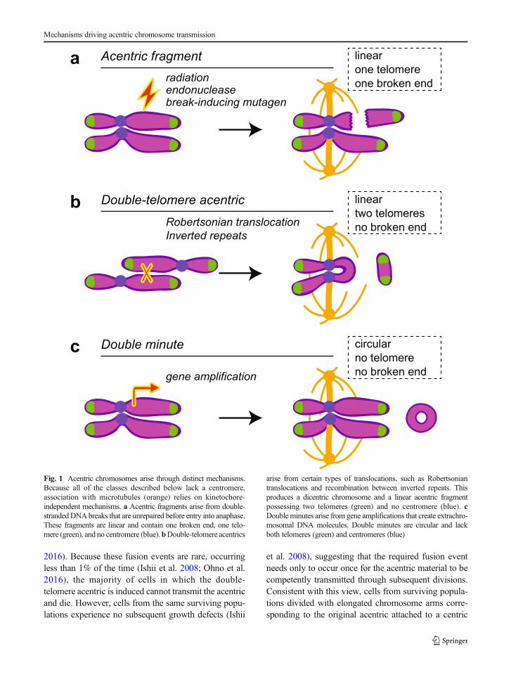
Perhaps the most common means of acentric formationoccurs when a cell enters and exits metaphase with animproperly or unrepaired double-stranded DNA break(Fenech et al. 2011). The unrepaired break generatestwo chromosome fragments: one containing a telomere,a centromere, and a broken end, and the other containingonly a telomere and a broken end (Fig. 1a). Lacking itscentromere, the acentric fragment cannot establish akinetochore and attach to the mitotic spindle throughcanonical methods.
Acentrics are also generated through translocations,such as Robertsonian translocations. Robertsoniantranslocations result from translocations between acro-centric chromosomes (Fig. 1b). When both breakpointsare in the short arms, the products are a short acentricchromosome and a metacentric chromosome bearingtwo centromeres, one of which often becomesinactivated (Morin et al. 2017). Similarly, recombina-tion between inverted repeats on two sister chromatidsresults in (1) a comparable acentric chromosome con-taining two telomeres but no centromere and (2) adicentric chromosome (Titen and Golic 2008) (Fig.1b). Lacking a kinetochore, these acentrics do not formcanonical attachments with microtubules.
Finally, acentrics also arise through gene amplifica-tion. Commonly found in cancer cells, these small,circular acentric chromosomes bearing oncogenes arereferred to as double minutes (Cox et al. 1965; Cowell1982) (Fig. 1c). Displaced from their regulatory ele-ments, the genes on these extrachromosomal elementsundergo increased transcription and may provide a
B. Warecki, W. Sullivan

fitness advantage for the cancer cell (Pauletti et al.1990). As a consequence of being circular, double mi-nutes lack telomeres and a centromere (Wahl 1989).
Acentrics resulting from double-stranded breaks con-tain an unrepaired broken end, while acentrics formedfrom translocation/recombination events and dou-ble minutes do not have broken ends. Similarly,acentrics from double-stranded breaks and fromtranslocation/recombination events are linear andcontain telomeres, while acentrics from double mi-nutes are circular and lack telomeres. These differ-ences influence the behavior and segregationmechan i sms of acen t r i c s dur ing mi tos i s .Therefore, we will subsequently distinguish thesetypes of acentrics as “acentric fragments,” (Fig.1a) “double-telomere acentrics,” (Fig. 1b) or “dou-ble minutes” (Fig. 1c).
Acentric chromosome transmission occursin a diversity of eukaryotes
Fixed studies of dividing grasshopper neuronal stemcells suggested early evidence of successful mitotictransmission of acentric chromosomes (Carlson1938a). Cells were irradiated to generate acentric frag-ments and then fixed throughout mitosis. In metaphasecells, the acentrics were observed at the edge of themetaphase plate clearly separated from the main massof centric chromosomes. In early anaphase cells, sisteracentrics appeared to separate at the same time as un-damaged chromosomes but had not moved polewardand instead remained at the edge of the metaphase plate.However, in late anaphase, acentrics appeared to havebegun to move poleward. In telophase cells, some acen-trics were interpreted as reincorporating into daughternuclei while others had formed micronuclei (Carlson1938a).
Since this initial observation, studies in a diversity ofspecies including yeast, insects, mammals, and plantshave documented acentric chromosome transmission(Table 1). For example, in Saccharomyces pombe,endonuclease-mediated excision of a centromere createsacentrics analogous to double-telomere acentrics. Whilemost cells died after removal of the centromere, presum-ably due to failed transmission of the acentric, somecolonies survived and grew, indicating the maintenanceof these acentrics through division (Ishii et al. 2008). Inhuman cancer cell lines under selective pressure,
advantageous double minutes were maintained at a highcopy number over an extended period of time (Paulettiet al. 1990). Furthermore, acentric fragments were ob-served to move off the metaphase plate and migratepoleward in Scadoxus multiflorus (previously knownas Haemanthus katherinae) cells (Bajer 1958; forreview, see Östergren et al. 1960). Collectively, thesestudies demonstrate the occurrence of successful trans-mission of acentrics to daughter cells through division.
Current evidence suggests that the efficiency of acen-tric chromosome transmission varies widely among spe-cies. For example, successful double-telomere acentrictransmission in Saccharomyces pombe occurs in < 1%of divisions (Ishii et al. 2008) while successful acentricfragment transmission in Drosophila melanogaster oc-curs at a very high frequency of > 80% (Royou et al.2010). The successful acentric transmission rates forother species appear to lie within this spectrum.Successful transmission of double minutes in hu-man cells may occur at a more moderate frequen-cy, as double minute transmission was estimated tooccur in ~ 30% of anaphase cells studied (Kandaet al. 1998). Interestingly, analysis of fixed prima-ry human lymphocytes revealed that even though~ 12.5% of anaphase cells had divided with acen-trics, only 5% of interphase cells had micronuclei(Falck et al. 2002), suggesting that successfulacentric transmission might occur roughly 60% ofthe time. However, estimates of acentric transmis-sion based on the comparisons between frequen-cies of fixed anaphase cells with acentrics and offixed interphase cells with micronuclei are indirect,and could potentially exclude other possible out-comes for cells dividing with acentrics (Udroiuand Sgura 2020).
Nevertheless, the existence of systems with highrates of successful acentric transmission that havebeen directly observed through live imaging hasenabled precise characterization of the mechanismsinvolved in poleward acentric movement. In gen-eral, these studies reveal three distinct strategiesfor successful acentric transmission: (1) direct as-sociation with centromere-containing chromosomes, (2)non-canonical association with microtubules, and (3)long-range tether-based interactions with centromere-containing chromosomes. Described in the next sectionsare our current understandings of the cellular mecha-nisms involved in each of these modes of acentrictransmission.
Mechanisms driving acentric chromosome transmission

Acentric transmission through direct associationwith normal centric chromosomes
In theory, the most straightforward way for an acentricto be transported to a daughter cell would be to connectwith a centromere-containing chromosome. A connect-ed acentric could then “ride” poleward as the centricchromosome is pulled by the mitotic spindle to the pole.No dedicated force would be required to act on theacentric, as acentric segregation would be dependentupon the kinetochore of the centric chromosome towhich it was attached. In fact, acentrics forming func-tional connections with centric chromosomes duringmitosis have been observed in multiple species. Forexample, studies in fission yeast have demonstrated thatdouble-telomere acentrics form end-to-end fusions withcentromere-containing chromosomes and are transmit-ted through multiple cell cycles (Ishii et al. 2008; Ohnoet al. 2016). In human cells, double minutes also attachto segregating chromosomes (Kanda et al. 1998).
Double minutes cluster together, and clusters stick tochromosome arms (Kanda et al. 1998). Thus, whencentromere-containing chromosomes segregate pole-ward, double minutes are also transported to daughtercells (Kanda et al. 1998).
Acentric chromosome attachment to normal centricchromosomes can occur through homologous recombi-nation, protein scaffolds, and possibly other mecha-nisms such as DNA catenation. For example, double-telomere acentrics attach to centric chromosomes inSaccharomyces pombe via non-canonical homologousrecombination between sub-telomeric sequences on theacentric and centric chromosomes (Ohno et al. 2016).This recombination restores a centromere to the formeracentric, allowing for the genetic material on the acen-tric to be transported poleward. This interchromosomalrecombination occurs between the acentric and either ofthe other two centric chromosomes (Ishii et al. 2008).Fusion results in loss of telomeric sequences from bothchromosomes involved (Ishii et al. 2008; Ohno et al.
Table 1 Summary of examples of acentric segregation poleward.Examples of proposed instances of acentric transmission. Examplesare sorted based on the type of acentric they most resemble, the cell
type the acentrics were observed in, and the potential mechanism ofacentric segregation (segregation mode left blank if currentlyunknown)
Acentric category Organism Potential segregationmode
References
“Acentricfragment”
Drosophila melanogaster Long-range tethers +microtubules
Royou et al. 2005; Royou et al. 2010;Kotadia et al. 2012; Derive et al. 2015;Karg et al. 2015; Bretscher and Fox 2016;Karg et al. 2017; Montembault et al. 2017; Wareckiand Sullivan 2018;Warecki et al. 2020; Landmann et al.2020
Potorous tridactylus Long-range tethers Humphrey and Brinkley 1969; Liang et al. 1993;Ono et al. 2017
Nephrotoma suturalis Long-range tethers +microtubules
LaFountain Jr et al. 2001, 2002a, b
Chortophaga viridifasciata Carlson 1938a; Carlson 1938b
Saccharomyces cerevisiae Direct association withother chromosomes
Malkova et al. 1996; Galgoczy and Toczyski 2001;Melo et al. 2001; Kaye et al. 2004
Scadoxus multiflorus Microtubules Bajer 1958; Bajer 1964; Bajer et al. 1987; Bajer andVantard 1988; Khodjakov et al. 1996
Cyclops strenuus Stich 1953
“Double-telomereacentric”
Saccharomyces pombe Direct association withother chromosomes
Ishii et al. 2008; Ohno et al. 2016
Drosophila melanogaster Titen and Golic 2008
Pales ferruginea Microtubules Dietz 1972; Fuge 1975;
“Double minuteacentric”
Homo sapiens Direct association withother chromosomes
Kanda et al. 1998; Kanda et al. 2001a
Amaranthus palmeri Direct association withother chromosomes
Koo et al. 2018
B. Warecki, W. Sullivan
2016). Because these fusion events are rare, occurringless than 1% of the time (Ishii et al. 2008; Ohno et al.2016), the majority of cells in which the double-telomere acentric is induced cannot transmit the acentricand die. However, cells from the same surviving popu-lations experience no subsequent growth defects (Ishii
et al. 2008), suggesting that the required fusion eventneeds only to occur once for the acentric material to becompetently transmitted through subsequent divisions.Consistent with this view, cells from surviving popula-tions divided with elongated chromosome arms corre-sponding to the original acentric attached to a centric
a Acentric fragmentone telomereone broken end
linear
radiationendonucleasebreak-inducing mutagen
b Double-telomere acentrictwo telomeresno broken end
linear
Robertsonian translocationInverted repeats
c Double minuteno telomereno broken end
circular
gene amplification
Fig. 1 Acentric chromosomes arise through distinct mechanisms.Because all of the classes described below lack a centromere,association with microtubules (orange) relies on kinetochore-independent mechanisms. a Acentric fragments arise from double-strandedDNAbreaks that are unrepaired before entry into anaphase.These fragments are linear and contain one broken end, one telo-mere (green), and no centromere (blue).bDouble-telomere acentrics
arise from certain types of translocations, such as Robertsoniantranslocations and recombination between inverted repeats. Thisproduces a dicentric chromosome and a linear acentric fragmentpossessing two telomeres (green) and no centromere (blue). cDouble minutes arise from gene amplifications that create extrachro-mosomal DNA molecules. Double minutes are circular and lackboth telomeres (green) and centromeres (blue)
Mechanisms driving acentric chromosome transmission

chromosome (Ishii et al. 2008). Therefore, althoughhomologous recombination between an acentric andcentric chromosome might occur rarely, the result is ahighly stable DNA molecule that can effectively bepassed on through many divisions (Ohno et al. 2016)(Fig. 2a).
In contrast, connections between double minuteacentrics and centric chromosomes may depend on pro-tein scaffolds rather than homologous recombination. Inmitotic cultured human cells, double minutes closelyassociate with each other and the arms of segregatingcentric chromosomes (Kanda et al. 1998; 2001a). Thekinetochores of the centric chromosomes are then re-sponsible for driving the poleward segregation of thedouble minutes. These associations persist through cy-tokinesis (Kanda et al. 1998), allowing double minutesto be successfully transmitted to daughters. The natureof the connection between double minutes and chromo-some arms is unclear, although their behavior in mitosissuggests they possess an inherent “stickiness.” It hasbeen proposed that this stickiness is derived from scaf-folding proteins associated with multiple copies of rep-lication origins on double minutes (Kanda et al. 2001a).These scaffolding proteins might then mediate the con-nection between the double minutes and the arms ofnormal chromosomes. Interestingly, episomes of theEpstein-Barr virus, which are structurally similar todouble minutes, likewise attach to mitotic chromosomearms, an association mediated by the viral EBNA-1protein (Kanda et al. 2001b). In general, several otherviral genomes utilize a similar strategy to segregateduring mitosis (Feeney and Parish 2009). The distribu-tion of double minutes to daughter cells is unequal(Kanda et al. 1998), which suggests the possibility thatsome divisions fail to segregate double minutes to bothdaughters. However, this strategy of acentric transmis-sion is nevertheless successful, perhaps because unlikewith other acentric types, double minutes are oftenpresent in high numbers within a cell (Kanda et al.1998). In addition, double minute attachment to chro-mosomes is relatively common (Kanda et al. 1998).Thus, in contrast to connections formed through homol-ogous recombination, acentric-centric connectionsbased on protein scaffolding occur more frequently butare less permanent, requiring re-establishment everydivision cycle (Fig. 2b).
Another possibility is that acentrics could potentiallyremain connected to chromosomes through DNA cate-nation. Interestingly, catenation tightly links sister
acentric fragments to one another in metaphase Alliumcepa cells (Giménez-Abián et al. 2002). TopoisomeraseII activity is required to resolve these catenations andallow sister acentric fragments to separate from oneanother (Giménez-Abián et al. 2002). After separation,acentric fragments do not appear to segregate and in-stead form micronuclei (Giménez-Abián et al. 2002).Nevertheless, it is possible to imagine a situation inwhich a double-stranded break occurring in G2 couldresult in one intact sister chromatid and one sister chro-matid broken into a centric and an acentric fragment.Unresolved catenation could then hold the acentric frag-ment close to the intact sister chromatid’s arm. Duringanaphase, the acentric would be transported polewardwith the intact sister chromatid, driven by the intactsister chromatid’s kinetochore.
Acentric transmission through direct associationwith microtubules
Early in mitosis, acentrics often move outward from thepoles either towards the metaphase plate or the cellperiphery. Studies indicate this movement is primarilydriven by microtubules. Localization of acentric frag-ments to the edge of the metaphase plate was firstobserved in grasshopper neuroblasts (Carlson 1938a).In these cells, acentric fragments were indistinguishablefrom centric chromosomes until metaphase when theybecame positioned on the periphery of the metaphaseplate (Carlson 1938a). Subsequent observations in di-verse cell types revealed similar patterns of antipolaracentric fragment movement. These includeprometaphase II of crane fly spermatocytes(LaFountain Jr et al. 2002a), prometaphase Potoroustridactylus (PtK) cells (Humphrey and Brinkley 1969;Liang et al. 1993; Khodjakov and Rieder 1996), humancells (Barisic et al. 2014), Scadoxus multiflorus cells(Bajer 1958; Bajer and Östergren 1963), Taricha gran-ulosa cells (Rieder et al. 1986), and Drosophilaneuroblasts (Royou et al. 2010; Karg et al. 2017).Antipolar localization to either the periphery or themetaphase plate is also observed for double minutes(Kanda et al. 2001a) and double-telomere acentrics(Fuge 1975). It is worth noting that in the early stagesof division, in some systems, acentrics also move pole-ward before moving back towards the metaphase plate(Stich 1953; Bajer 1958; Khodjakov et al. 1996;LaFountain Jr et al. 2001, 2002a). Experiments in
B. Warecki, W. Sullivan

aFirst division
First division Second division
First division Second divisionb
Cell death due toaneuploidyM
ost
Rar
eM
ost
“All-or-Nothing” strategy
“Shotgun” strategy
Fig. 2 Acentric transmission through association with normalchromosomes. a The “all-or-nothing” strategy: this is exemplifiedin instances in which rare recombination events occur betweensubtelomeric sequences on both the double-telomere acentric andcentromere-containing chromosome. Although a rare event, thisrecombinant chromosome results in stable incorporation of theacentric fragment into an intact centromere-containing chromo-some and stable mitotic transmission through multiple generations
(Ishii et al. 2008; Ohno et al. 2016). b The “shotgun” strategy: thisis exemplified by the segregation behavior of double minutechromosomes. Double minutes readily, but randomly, associatewith the arms of normal chromosomes during mitosis. However,the association is temporary and must be re-established each timethe cell divides (Kanda et al. 1998). This can result in unequalsegregation or loss of some double minutes
Mechanisms driving acentric chromosome transmission
Nephrotoma suturalis spermatocytes indicate that insome systems this initial acentric fragment movementpoleward is mediated by microtubule flux (LaFountainJr et al. 2001).
The simplest explanation for early, antipolar move-ment is through the plus-ends of growing microtubulescontacting and exerting a force on the acentric. Known aspolar ejection forces, they arise from the combined actionof polymerizing microtubules and chromokinesins andpush chromosome arms towards the metaphase plate innormal mitosis (Brouhard and Hunt 2005). Acentricchromosome fragments usually align at the outer edgeof the metaphase plate encompassed by microtubules(Karg et al. 2017). Drug inhibition of microtubule fluxor depletion of key chromokinesins result in failure ofacentrics to properly localize to the periphery (Ault et al.1991; Kanda et al. 2001a; Barisic et al. 2014; Karg et al.2017). These data suggest that polar ejection forces arethe key determinant for localizing acentrics to the pe-riphery of the metaphase plate prior to anaphase.
After anaphase onset, in some systems, acentricsgenerally move poleward. Acentric segregation is oftendelayed relative to segregation of the normal centricchromosomes (Carlson 1938a; Fuge 1975; Khodjakovet al. 1996; Kanda et al. 2001a; LaFountain Jr et al.2001, 2002a; Royou et al. 2010; Bretscher and Fox2016). Studies in a variety of cell types demonstrate thatpoleward transport of acentrics relies on microtubules.Drug inhibition of microtubule flux in Nephrotomasuturalis spermatocytes slows poleward acentric frag-ment movement (LaFountain Jr et al. 2001). In lateanaphase of Drosophila neuroblasts, microtubule bun-dles encompass poleward-moving acentric fragments(Karg et al. 2017). Laser ablation of these microtubulesresults in a cessation of acentric poleward movement(Karg et al. 2017). As acentrics are initially positioned atthe outer edge of the metaphase plate, it is likely that themicrotubules responsible for mediating acentric transportare primarily interpolar microtubules (Karg et al. 2017).This conclusion is supported by the fact that mutantsspecifically disrupting interpolar microtubules disruptpoleward migration of acentrics (Karg et al. 2017).
Acentrics exhibit extensive lateral associations withmicrotubules, as opposed to the canonical end-on kinet-ochore attachments observed in normal chromosomes.In Drosophila neuroblasts, robust microtubule bundleslaterally encompass acentric fragments (Karg et al.2017). Similar lateral microtubule bundles are also as-sociated with double-telomere acentrics in Pales
ferruginea (Fuge 1975) and acentric fragments inScadoxus multiflorus (Bajer and Vantard 1988). Theseassociations are reminiscent of the normal meiotic pole-ward chromosome segregation in Caenorhabditiselegans oocytes (Dumont et al. 2010). As with acentrics,in C. elegans oocytes, kinetochores are not required forchromosome segregation (Dumont et al. 2010;Muscat et al. 2015). In these meiotic divisions,lateral associations with microtubule bundles in-stead drive chromosome orientation and transportpoleward (Wignall and Villeneuve 2009; Dumontet al. 2010; Muscat et al. 2015).
While lateral microtubule-acentric associations havebeen observed numerous times, the mechanisms bywhich these associations direct poleward force on acen-trics remain unclear, although motor proteins are likelyinvolved. In C elegans female meiosis, lateralmicrotubule-associated chromosomes rely on the minusend-directed motor protein dynein to propel their pole-ward segregation (Muscat et al. 2015). However, howdynein would localize to an acentric chromosome re-mains unclear. Studies in Drosophila neuroblasts revealaccurate acentric fragment segregation requires thechromokinesin KIF4A/Klp3A (Karg et al. 2017).However, how a plus end-directed motor contributesto acentric movement poleward towards the minus endsof microtubules remains to be determined. BecauseKlp3A depletion in these cells results in diminishedarrays of interpolar microtubules (Karg et al. 2017), asdescribed above, it is likely that Klp3A’s effect onacentric segregation is indirect. Since KIF4A/Klp3A isneeded for proper microtubule flux (Wandke et al.2012), one possibility is that poleward acentric fragmenttransport requires microtubule flux. It is also possiblethat microtubule sliding during anaphase B transmitsforce from the elongating spindle to the acentrics viathe interpolar microtubules. Additionally, an unknownchromokinesin could associate with the acentric anddrive it poleward.
While both acentric fragments and double-telomere acentrics rely to some degree on attach-ments to microtubules for their poleward move-ment (Fuge 1975; Bajer and Vantard 1988;LaFountain Jr et al. 2001; Karg et al. 2017),double minutes seemingly do not. This conclusionis based on the fact that drug inhibition of micro-tubule flux disrupts the initial peripheral localiza-tion of double minutes but not their subsequentpoleward movement (Kanda et al. 2001a).
B. Warecki, W. Sullivan

Acentric transmission through long-range DNAtether/thread-based associations
Acentric fragments are also capable of connecting tocentric chromosomes through long-range connections(LaFountain Jr et al. 2002b; Royou et al. 2010;Bretscher and Fox 2016; Ono et al. 2017). Known asDNA tethers, they are thought to connect the brokenends of the centric and acentric fragments (Royou et al.2010). For example, in Drosophila larval neuroblasts,DNA tethers contain histones and a number of associ-ated proteins, including the chromosome passengercomplex components Aurora B kinase and INCENP;the cell cycle kinases BubR1, Bub3, and Polo; and theAPC/C cofactor Cdc20 (Royou et al. 2010; Derive et al.2015). The tethers are extremely efficient at promotingmitotic acentric fragment transmission. Despite induc-tion of large acentric fragments in ~ 80% of larval cells,the larvae develop to adults, exhibiting no reduction inviability (Royou et al. 2010). Live analysis reveals that,although delayed, sister acentrics separate, move pole-ward, and are incorporated into daughter telophase nu-clei (Royou et al. 2010). Functional studies reveal thatthe DNA tether and its associated proteins are essentialfor the proper mitotic segregation of the acentrics.Reductions in BubR1 or Polo activity result in abnormalpositioning of the acentric on the metaphase plate andultimately failed acentric segregation (Royou et al.2010). Although the mechanism for how these proteinscontribute to acentric transport remains unclear, interac-tions between BubR1 kinase and Bub3 lead to localizedinhibition of APC/C near the tether and the acentric(Derive et al. 2015), suggesting that delayed acentricsegregation may be functionally important for polewardmovement.
As the tether links the acentric fragment to its centricpartner, the tether may provide the force driving acentricpoleward transport. However, studies revealing acentricfragments often move poleward with their telomeresleading would argue against a connection to the brokenend of the acentric fragment providing this polewardpulling force (Karg et al. 2017). Additionally, it ispossible that acentric fragment segregation is dependentupon the kinetochore of the centric chromosome towhich the tether connects. As discussed above, micro-tubules provide a major transporting force for theseacentric fragments (Karg et al. 2017). While the role ofthe tether in generating/transmitting force on/to theacentric fragment is unclear, it may be that the tether is
required to maintain the acentric in the vicinity of themetaphase spindle, enabling the acentric to associatewith interpolar microtubules during anaphase. In addi-tion, as described below, the tether provides a distinct,essential role facilitating incorporation of the late-segregating acentric into the telophase nucleus (Karget al. 2015; Warecki and Sullivan 2018).
Efficient transmission of acentrics has also been ob-served in the polyploid Drosophila papillary cells(Bretscher and Fox 2016). Acentric fragments, eithergenerated naturally or through X-irradiation, exhibit adelayed but successful poleward migration and incorpo-ration into daughter nuclei (Bretscher and Fox 2016). Incontrast to neuroblasts, in papillary cells, DNA tethersare not observed and proper segregation does not rely onBubR1 (Bretscher and Fox 2016). Instead, acentrictransmission relies on FANC2D, FANC1, and Bloomhelicase activity (Bretscher and Fox 2016). Previousstudies demonstrated that these proteins are associatedwith ultrafine DNA bridges (UFBs) (Liu et al. 2014).UFBs contain DNA, but in contrast to DNA tethers, donot contain histones or stain with DAPI (Liu et al. 2014).UFBs connect centromeres, telomeres, or fragile sites ofseparating sister chromatids (Liu et al. 2014). UFBsmayarise due to unresolved catenations or due to incompletereplication (Liu et al. 2014) or from entanglementscaused by homologous recombination (Chan and West2018). It is thought that FANC2D, FANC1, and Bloomhelicase resolve UFBs as sister chromatids separate (Liuet al. 2014). Therefore, it is possible that similar con-nections provide a link between acentric fragments andcentric chromosomes in Drosophila papillary cells.Whether this UFB-like link drives acentric polewardtransport or, like the DNA tethers, provides an alterna-tive role remains to be determined.
Long-range tethers and threads are present in diversecell types. For example, in Nephrotoma suturalis sper-matocytes, acentric fragments are connected to centricchromosomes through telomere-telomere tethers(LaFountain Jr et al. 2002b). Similar connections areproposed to occur between acentric fragments and cen-tric chromosomes in PtK2 cells (Ono et al. 2017). Themakeup of these tethers is unknown. In addition, fragilesites that result in lagging chromosome sections con-nected to main nuclei bear remarkable resemblance toacentric fragments and tethers. For example, in severalgenera of bluegrass, fragile sites result in lagging chro-mosome sections in anaphase that lack centromeres butremain connected to centric chromosomes by a thin
Mechanisms driving acentric chromosome transmission

DNA thread (Rocha et al. 2017a; Rocha et al. 2017b).Aurora B and INCENP-coated DNA threads are alsoobserved connecting spatially distant chromosomes dur-ing early meiosis in Drosophila oocytes (Hughes et al.2009; Hughes and Hawley 2014). These threads arecomposed of heterochromatin and must be resolved byTopoisomerase II activity for proper division, suggest-ing they result from catenation (Hughes et al. 2009;Hughes and Hawley 2014).
It is tempting to speculate that the tethers connectingacentric fragments and centric chromatin must originatefrom the broken ends of the acentric and centric frag-ments. However a number of recent observations sug-gest tethers/threads form a variety of connections. Forexample, in Drosophila neuroblasts, the DNA bindingprotein barrier-to-autointegration factor, BAF, whichlocalizes to acentric-centric tethers, is also observedlocalizing to tethers between sister acentrics segregatingto opposing daughters (Warecki et al. 2020).Furthermore, in crane fly spermatocytes undergoingmeiosis, acentric fragments generated by cutting atrailing chromosome arm travel across the equator ofthe cell to the opposing daughter cell (LaFountain Jret al. 2002b). Laser ablation studies indicate this move-ment is mediated by a cytologically undetectable tetherbetween the telomeres of segregating sister chromatids(LaFountain Jr et al. 2002b). Analogous experimentshave shown a similar telomere-telomere tether exists inPtK2 cells as well (Ono et al. 2017). Laser severing ofchromosome arms in anaphase resulted in some acentricfragments that regularly moved to the opposing pole. Asecond laser cut between the telomeres of the acentricfragment and its sister chromatid halted the acentricmovement (Ono et al. 2017). Taken together, theseexamples indicate that tethers may not only originatefrom the broken ends of acentric fragments but alsofrom their telomeres.
Cellular adaptations facilitating acentrictransmission
Acentric incorporation into telophase nuclei has beenobserved in many cell types (Carlson 1938a; Liang et al.1993; Kanda et al. 2001a; LaFountain Jr et al. 2002a;Royou et al. 2010). Acentric segregation is often asso-ciated with striking temporal and spatial modificationsduring anaphase, telophase, and cytokinesis. These in-clude delays in and/or modification of cytokinesis and
nuclear envelope reassembly (Kotadia et al. 2012; Karget al. 2015; Montembault et al. 2017). Acentrics are alsoassociated with spindle elongation, cell elongation, andexpansion of the myosin-based contractile ring (Kotadiaet al. 2012; Montembault et al. 2017). These modifica-tions during the final stage of the cell cycle likely serveto promote proper segregation and incorporation ofacentrics into daughter nuclei, preserving genome integ-rity (Fig. 3).
Late-segregating acentrics risk blocking theingressing cleavage furrow, resulting in furrow regres-sion and aneuploidy. Consequently, it has been pro-posed that mammalian and yeast cells have evolved amechanism known as the abscission checkpoint thatdelays cytokinesis until lagging chromatin has clearedthe midzone (for review, see Petsalaki and Zachos2019). According to this model, in the abscission check-point, Aurora B kinase phosphorylates chargedmultivesicular body protein 4C (CHMP4C), a key com-ponent of the endosomal sorting complexes required fortransport (ESCRT)-III complex (Carlton et al. 2012).CHMP4C phosphorylation results in its sequestrationin the center of the midbody in a complex with ANCHRandVps4, another ESCRT-III component (Petsalaki andZachos 2019). Although still unclear, it is possible thatthe formation of this complex at the midbody preventsVps4 from performing membrane remodeling events atthe ingression sites that are required for cytokinesiscompletion (Petsalaki and Zachos 2019). Further re-search on the proposed checkpoint is required to defineits mechanism and the nature of the cytokinesis delay.
Drosophila neuroblasts rely on the alternative strate-gy of spindle and cell elongation for clearing late-segregating acentric fragments from the cleavage plane(Kotadia et al. 2012). It is likely that chromatin remain-ing on the metaphase plate during late anaphase pro-vides signals that drive these adaptations. Support forthis idea comes from the observation that in the presenceof lagging acentrics, myosin and likely the cleavagefurrow are both broadened, and there is an increasedflow of myosin from the cleavage furrow to the corticesof daughter cells (Montembault et al. 2017). Spindleelongation, broadening of the cleavage furrow, andenriched myosin at cell cortices require RhoGEF activ-ity (Kotadia et al. 2012; Montembault et al. 2017),which localizes to the overlapping midzone microtu-bules via the centralspindlin motor complex (Somersand Saint 2003). Delayed nuclear sequestration ofRhoGEF in divisions with acentrics is important for
B. Warecki, W. Sullivan

driving these adaptations (Montembault et al. 2017). Inaddition, because the lagging acentric fragments inDrosophila neuroblasts are encompassed by microtu-bules, it is possible that these overlapping microtubulesresult in an increased sequestering of RhoGEF at themidzone during late anaphase, although this remains tobe demonstrated.
While spindle and cell elongation facilitate clearingof late-segregating acentrics from the cleavage plane,additional adaptations are required to ensure they arriveat the poles prior to the completion of nuclear envelope
assembly. Otherwise, acentrics would be “locked out”and form highly mutagenic micronuclei (Fenech et al.2011). In general, nuclear envelope assembly beginsfirst on the poleward-facing sides of daughter nucleibefore completion on the midzone-facing sides of thenuclei (Gerlich et al. 2001). This would provide a smallamount of extra time for acentrics to enter daughternuclei. In addition, midzone-localized Aurora B kinaseinhibits nuclear envelope reassembly on late-segregating acentrics until they move away from themidzone (Afonso et al. 2014) (Fig. 4a). In Drosophila
a
b
Tem
pora
l ada
ptat
ions
Spat
ial a
dapa
tatio
ns
+ Delayed nuclear envelope reassembly+ Delayed cytokinesis
+ Channel formation+ Cell elongation
Fig. 3 Cellular adaptations that facilitate successful mitotic trans-mission of acentrics. a Temporal adaptations: global delays innuclear envelope reassembly (dark green) provide more time foracentrics to rejoin daughter nuclei at the poles (Montembault et al.2017). In addition, delayed cytokinesis provides enough time forlagging chromatin, including late-segregating acentric fragments,to clear the metaphase plate before cleavage furrow ingression
(Petsalaki and Zachos 2019). b Spatial adaptations: highly specificchannels in the nascent nuclear envelope (dark green) form toprovide a passageway for acentrics to enter daughter nuclei(Karg et al. 2015). In addition, cell elongation provides enoughspace for late-segregating acentrics to clear the site of cleavagefurrow ingression (Kotadia et al. 2012; Montembault et al. 2017)
Mechanisms driving acentric chromosome transmission
neuroblasts, incorporation into daughter nuclei of thelate-segregating acentric fragments is also accomplishedby both global and highly localized delays innuclear envelope reassembly (Karg et al. 2015;Montembault et al. 2017). For example, in re-sponse to lagging chromatin, nuclear envelope re-assembly is globally delayed on daughter nuclei(Montembault et al. 2017).
However, a global delay is not always sufficient toprovide time for late-segregating acentrics to reachdaughter nuclei before completion of nuclear envelopeassembly (Warecki et al. 2020). In these cases, theacentric and its associated DNA tether induce formationof localized channels in the envelopes of the newlyformed daughter nuclei that permit acentric passageand incorporation into daughter nuclei (Karg et al.2015) (Fig. 4b). These channels form through all layersof the nuclear envelope (Warecki et al. 2020) and aredependent upon the activity of a pool of Aurora B kinaselocalized to the tether connecting acentrics todaughter nuclei (Karg et al. 2015; Warecki andSullivan 2018). Channel formation is not due toa physical blockage of nuclear envelope reassem-bly by the tether, as inhibition of Aurora B activ-ity disrupts channel formation without affecting thetether (Karg et al. 2015). The final step of acentricfragment incorporation requires fusion of the nu-clear membrane on acentrics to the nuclear mem-brane on daughter nuclei and is thought to providethe final driving force to allow acentrics to enterdaughter nuclei (Warecki et al. 2020).
The acentric fragment is associated with nuclearmembrane but absent of nuclear lamin and nuclear porecomplexes (Afonso et al. 2014; Karg et al. 2015;Warecki et al. 2020). This inhibition is likely due tothe pools of Aurora B localizing at the midzone and tothe acentric. The spatial inhibition of nuclear envelopeassembly has led to the proposal of an Aurora B–mediated checkpoint surveilling the anaphase-to-telophase transition (Afonso et al. 2014; Afonso et al.2019). It has also been argued that the spindle mediatesthis inhibition as opposed to Aurora B (Liu et al. 2018)(Fig. 4a). In human cancer cell lines, lagging wholechromosomes associate with nuclear membrane butnot with nuclear lamin or nuclear pore complexes (Liuet al. 2018). In this case, the spindle is proposed tomediate this inhibition (Liu et al. 2018). More researchis required to differentiate between these two models.As segregating acentric fragments are encompassed by
microtubules (Karg et al. 2017), it would be worthwhileto determine what role these microtubules play in theexclusion of certain nuclear envelope components fromthe late-segregating acentric (Fig. 4).
It is interesting to note that these cellular adaptationsmay produce synergistic interactions. For example, thedelays in nuclear envelope reassembly that allowacentric entry into nuclei are also believed to beresponsible for the delayed nuclear sequestration ofRhoGEF that leads to a broadening of the cleavagefurrow (Montembault et al. 2017). In addition, theabscission checkpoint is also activated when nucle-ar pore complex assembly is defective (Mackay et al.2010), as it is on lagging chromatin (Afonso et al. 2014;Karg et al. 2015; Liu et al. 2018) and locally on the mainnuclei at the site of channels (Karg et al. 2015; Wareckiet al. 2020).
Unexplored issues in acentric transmission
Acentric transmission studies have primarily focused oncongression and poleward transport. As described be-low, much remains unknown concerning the mecha-nisms driving these events. In addition, there are anumber of other unique aspects of the mitotic behaviorof acentrics that remain unexplored and are likely toprovide insight into core mechanisms of chromosometransmission.
Spatial and temporal separation of sister acentrics
Acentrics are often physically separated fromcentromere-containing chromosomes on the metaphaseplate. In addition, acentric segregation is often consid-erably delayed relative to their normal centric chromo-somes. This delay may be a direct consequence of thephysical separation. For example, the signals that releasecohesin may be much weaker at the edge of the meta-phase plate. Once cohesin is removed, catenated DNAremains binding sister chromosomes. Kinetochore mi-crotubules may provide force that efficiently resolvesthese catenations in conjunction with Topoisomerase IIactivity (Holm et al. 1985; Baxter et al. 2011). Could theseverely delayed separation of sister acentrics observedin some systems result from delays in resolving catenat-ed sister DNA? In some instances, it appears thatkinetochore-mediated forces are not required to resolvecatenations between sister acentrics in a timely manner
B. Warecki, W. Sullivan

(Giménez-Abián et al. 2002; Paliulis and Nicklas 2004).Nevertheless, it will be of great interest to determineboth cohesin and DNA decatenation dynamics on se-verely delayed-separating acentrics relative to neighbor-ing centric chromosomes. For acentric fragments, whichresult from damaged chromosomes, their physical andtemporal separation from the normal chromosome com-plement may be adaptive, preventing inappropriate in-teractions between the two and preserving genomicstability. Might acentric fragments, which possess abroken end, be treated differently than other acentrictypes that have no breaks? Double minutes are alsopushed to the periphery of metaphase cells (Kandaet al. 2001a). This suggests that physical separation ofacentrics from centric chromosomes during metaphasemay be a more general phenomenon, though more re-search is needed.
Accuracy of sister acentric segregation
Although acentric segregation is often delayed, acentricseparation and partitioning to daughter cells can besurprisingly accurate in some systems. For example,Drosophila neuroblasts and papillary cells that dividewith acentric fragments often produce euploid daughters(Royou et al. 2010; Bretscher and Fox 2016). Withoutkinetochores, it is unclear how sister acentric fragmentsare guided correctly to opposing poles. In Drosophilaneuroblasts, each acentric is connected to its centricpartner via a DNA tether (Royou et al. 2010). Whilelateral microtubule interactions are required for pole-ward transport of acentric fragments (Karg et al.2017), perhaps the tether is important for connectingsister acentrics with their proper nuclei to ensure anequal partitioning of acentrics to each daughter cell.
b
Teth
er-m
edia
ted
inhi
bitio
n
a
Mid
zone
-med
iate
d in
hibi
tion
Aurora B gradientSpindle midzone
Tether-based Aurora BMicrotubule bundles
Fig. 4 Local midzone and tether-localized Aurora B signalingdelays nuclear envelope reassembly in the presence of an acentric.a Midzone-mediated inhibition: recruitment of lamin and nuclearpore complexes is inhibited by an Aurora B gradient (blue) ema-nating from the midzone (Afonso et al. 2014). Inhibition may alsobe mediated by the high concentrations of spindle microtubules(orange) at the midzone (Liu et al. 2018). Low inhibitory Aurora Band microtubule concentrations at the poles facilitate recruitmentof lamin and nuclear pore complexes and completion of nuclearenvelope reassembly. b Tether-mediated inhibition: InDrosophilaneuroblasts, a pool of Aurora B (blue) highly localized to the
severely-delayed acentric fragment and its associated DNA tether(Royou et al. 2010) is responsible for localized inhibition ofnuclear envelope assembly (Karg et al. 2015; Warecki andSullivan 2018). The daughter nuclei, which are free of Aurora B,can recruit lamin and nuclear pore complexes. Acentrics andtethers, which are coated with Aurora B, cannot. Importantly,tether-based Aurora B activity explains the formation of channelsin the nuclear envelope of daughter nuclei, which are far from themidzone. Late-segregating acentrics are surrounded by microtu-bule arrays (Karg et al. 2017). The contribution of these arrays toinhibition is currently unknown
Mechanisms driving acentric chromosome transmission

Additionally, as the tether provides a connection be-tween the acentric fragment and a centric chromosome,perhaps acentric transmission inDrosophila neuroblastsis so successful partly due to canonical kinetochore-based mechanisms acting on the connected centric chro-mosome. Acentric segregation does not always occur soaccurately though. For example, partitioning of doubleminutes in human cells and acentric fragments inChortophaga viridifasciata, Scadoxus multiflorus, andSaccharomyces cerevisiae appears to be more random(Kanda et al. 1998; Carlson1938a; Khodjakov et al.1996; Kaye et al. 2004). The mechanisms that allowfor accurate acentric partitioning in some cases but notothers are yet to be identified.
Kinetochore-independent poleward transport forces
Much remains unknown about the forces driving acen-trics poleward in most systems. For acentric fragmentsconnected to centric chromosomes through long-rangeDNA tethers, it is possible that the tether provides apulling force, although this seems unlikely. Analysis ofacentric-microtubule interactions seems to suggest thatinterpolar microtubules are the ones important for acen-tric poleward transport (Fuge 1975; Karg et al. 2017).Given the close lateral association between acentricsand microtubules (Fuge 1975; Karg et al. 2017), it islikely that microtubule motor proteins drive the pole-ward transport of acentrics, although which ones remainunidentified. Although the chromokinesin Klp3a is re-quired for proper acentric segregation in Drosophilaneuroblasts, its action appears to be indirect, insteadbeing required to form the overlap interpolar microtu-bules on which the acentric travels (Karg et al. 2017).Minus end-directed dynein plays a role in the polewardsegregation of holocentric but kinetochore-less chromo-somes inC. elegans oocytes (Muscat et al. 2015). Giventhis, might dynein provide the poleward force drivingsome acentric transport? If so, it is unclear how dyneinwould localize to an acentric chromosome.
Origin, composition, and function of the DNA tetherand its relationship to UFBs
The origin, composition, and function of the DNA tetherconnecting acentric fragments to centric chromosomesand its relationship to UFBs are still unknown. When inthe cell cycle the tethers form, if they form after S-phase,or whether they are composed of existing DNA or result
from unscheduled DNA replication remains to be deter-mined. Potential insight into this comes from studies inLolium species, where fragile sites result in laggingacentric chromosome sections that are connected tocentric chromosomes through DNA threads (Rochaet al. 2017a; Rocha et al. 2017b). This tether is com-posed of the genetic material of the fragile site (Rochaet al. 2017a). Unlike UFBs (Chan et al. 2007; Ke et al.2011), tethers in Drosophila neuroblasts contain his-tones (Royou et al. 2010), suggesting a fundamentaldifference between the two structures. Cell cycle kinasesand chromosome passenger proteins associate with thetether (Royou et al. 2010; Derive et al. 2015), and it islikely that additional proteins remain to be identified. Asdescribed above, insight has been gained on the functionof some of these proteins, but much remains unknown.In addition, it would be interesting if poleward-movingacentrics with seemingly no connection to centric chro-mosomes were in fact connected to centric chromo-somes by difficult-to-observe UFBs. If so, the presenceof connections to centric chromosomes may be thedefining feature that differentiates the small class ofacentrics capable of segregation from the majority ofacentrics that do not move poleward.
Fate of acentric fragments successfully incorporatedinto daughter nuclei
The fate of acentric fragments once successfully incor-porated into telophase nuclei has not been directly in-vestigated. The best outcome is that they re-associatewith their centric counterpart and are subsequentlyrepaired. Alternatively, continued failure to repair thedouble-stranded break that generated the acentric frag-ment may result in apoptosis (Brodsky et al. 2004; Titenand Golic 2008; Nowsheen and Yang 2012). The worst-case scenario would be for the acentric fragment to beunrepaired through the next cell cycle, fail to enter intothe daughter nucleus, form a micronucleus, and undergochromothripsis (Zhang et al. 2015).
Studying acentric segregation to identify forces actingon centric chromosomes
Studying acentric segregation reveals forces acting oncentric chromosomes that might otherwise be difficult todetect due to the presence of kinetochore-mediatedforces. For example, the telomere-telomere tetheringobserved between acentric fragments and chromosomes
B. Warecki, W. Sullivan

segregating to the opposite pole (LaFountain Jr et al.2002b; Ono et al. 2017) has been suggested to occurbetween intact centric chromosomes as well (for review,see Paliulis and Forer 2018). In these cases, the tethersare proposed to provide resistance to the poleward mo-tion of the segregating chromosomes. Additionally, thelateral connections observed between acentrics andinterpolar microtubules (Fuge 1975; Karg et al. 2017)may also occur between microtubules and the arms ofcentric chromosomes (for review see Fuge 1990). Theseconnections likely provide a kinetochore-independentpoleward force on centric chromosomes. With this inmind, as acentric transmission is further explored, themechanisms and forces revealed likely influence thedynamics of centric chromosome arms as well.
Acknowledgments We would like to thank Alexey Khodjakov,Travis Karg, Anna Russo, and Hannah Vicars for their criticalreadings of the manuscript.
Author’s contributions WS conceived this review. BW andWS assessed the published literature and wrote and revised themanuscript.
Funding information This work was funded by a NationalInstitutes of Health grant NIHRO1GM120321 awarded toW.S.Data availabilityNot applicableCode availabilityNot applicable
Compliance with ethical standards
Competing interests The authors declare that they have nocompeting interests.
Ethics approval Not applicable
Consent to participate Not applicable
Consent for publication Not applicable
References
Afonso O,Matos I, Pereira AJ, Aguiar P, LampsonMA,Maiato H(2014) Feedback control of chromosome separation by amidzone Aurora B gradient. Science 345:332–336.https://doi.org/10.1126/science.1251121
Afonso O, Castellani CM, Cheeseman LP, Ferreira JG, Orr B,Ferreira LT, Chambers JJ, Morais-de-Sá E, Maresca TJ,Maiato H (2019) Spatiotemporal control of mitotic exit dur-ing anaphase by an aurora B-Cdk1 crosstalk. eLife 8:e47646.https://doi.org/10.7554/eLife.47646
Asbury CL (2017) Anaphase a: disassembling microtubules movechromosomes toward spindle poles. Biology (Basel) 6:E15.https://doi.org/10.3390/biology6010015
Ault JG, DeMarco AJ, Salmon ED, Rieder CL (1991) Studies onthe ejection properties of asters: astral microtubule turnoverinfluences the oscillatory behavior and positioning of mono-oriented chromosomes. J Cell Sci 99:701–710
Bader JR, Vaughan KT (2010) Dynein at the kinetochore: timing,interactions and functions. Semin Cell Dev Biol 21:269–275.https://doi.org/10.1016/j.semcdb.2009.12.015
Bajer A (1958) Cine-micrographic studies on chromosome move-ments in beta-irradiate cells. Chromosoma 9:319–331
Bajer A (1964) Cine-micrographic studies on dicentric chromo-somes. Chromosoma 15:630–651. https://doi.org/10.1007/bf00319996
Bajer A, Östergren G (1963) Observation on transverse move-ments within the phragmoplast. Hereditas (Lund) 50:179–195
Bajer A, Vantard M (1988) Microtubule dynamics determinechromosome lagging and transport of acentric fragments.Mutat Res 201:271–281. https://doi.org/10.1016/0027-5107(88)90016-4
Bajer A, Vantard M, Molè-Bajer J (1987) Multiple mitotic trans-ports expressed by chromosome and particle movement.Fortschr Zool 34:171–186
Barisic M, Aguiar P, Geley S, Maiato H (2014) Kinetochoremotors drive congression of peripheral polar chromosomesby overcoming random arm-ejection forces. Nat Cell Biol 16:1249–1256. https://doi.org/10.1038/ncb3060
Baxter J, Sen N, López Martínez V, Monturus de Carandini ME,Schvartzman JB, Diffley JFX, Aragón L (2011) Positivesupercoiling of mitotic DNA drives decatenation by topo-isomerase II in eukaryotes. Science 331:1328–1332.https://doi.org/10.1126/science/1201538
Bretscher HS, Fox DT (2016) Proliferation of double-strandbreak-resistant polyploid cells requires DrosophilaFANCD2. Dev Cell 37:444–457. https://doi.org/10.1016/j.devcel.2016.05.004
Brinkley BR, Zinkowski RP, Mollon WL, Davis FM, PisegnaMA, PerhouseM, Rao PN (1988)Movement and segregationof kinetochores experimentally detached from mammalianchromosomes. Nature 336:251–254. https://doi.org/10.1038/336251a0
Brodsky MH, Weinert BT, Tsang G, Rong YS, McGinnis NM,Golic KG, Rio DC, Rubin GM (2004) Drosophilamelongaster MNK/Chk2 and p53 regulate multiple DNArepair and apoptotic pathways following DNA damage.https://doi.org/10.1128/mcb.24.3.1219-1231.2004
Brouhard GJ, Hunt AJ (2005) Microtubule movements on thearms of mitotic chromosomes: polar ejection forces quanti-fied in vitro. Proc Natl Acad Sci U S A 102:13903–13908.https://doi.org/10.1073/pnas.0506017102
Carlson JG (1938a) Mitotic behavior of induced chromosomalfragments lacking spindle attachments in the neuroblasts ofthe grasshopper. Proc Natl Acad Sci U S A 24:500–507.https://doi.org/10.1073/pnas.24.11.500
Carlson JG (1938b) Some effects of X-radiation on the neuroblastchromosomes of the grasshopper, Chortophaga viridifasciata.Genetics 23:596–609
Carlton JG, Caballe A, Agromayor M, Kloc M, Martin-Serrano J(2012) ESCRT-III governs the Aurora B-mediated abscissioncheckpoint through CHMP4C. Science 336:220–225.https://doi.org/10.1126/science.1217180
Mechanisms driving acentric chromosome transmission

Chan YW, West SC (2018) A new class of ultrafine anaphasebridges generated by homologous recombination. Cell Cycle1 7 : 2 1 0 1 – 2 1 0 9 . h t t p s : / / d o i . o r g / 1 0 . 1 0 8 0/15384101.2018.1515555
Chan KL, North PS, Hickson ID (2007) BLM is required forfaithful chromosome segregation and its localization definesa class of ultrafine anaphase bridges. EMBO J 26:3397–3409. https://doi.org/10.1038/sj.emboj.7601777
Cowell JK (1982) Double minutes and homogeneously stainingregions: gene amplification in mammalian cells. Annu RevGenet 16:21–59. https://doi.org/10.1146/annurev.ge.16.120182.000321
CoxD, Yuncken C, Spriggs AI (1965)Minute chromatin bodies inmalignant tumours of childhood. Lancet 286:55–58.https://doi.org/10.1016/s0140-6736(65)0-131-5
Crasta K, Ganem NJ, Dagher R, Lantermann AB, Ivanova EV,Pan Y, Nezi L, Protopopov A, Chowdhury D, Pellman D(2012) DNA breaks and chromosome pulverization fromerrors in mitosis. Nature 482:53–58. https://doi.org/10.1038/nature10802
Derive N, Landmann C, Montembault E, Claverie MC, Pierre-Elies P, Goutte-Gattat D, FounounouN,McCusker D, RoyouA (2015) Bub3-BubR1-dependent sequestration ofCdc20Fizzy at DNA breaks facilitates the correct segregationof broken chromosomes. J Cell Biol 211:517–532.https://doi.org/10.1083/jcb.201504059
Dietz R (1972) Anaphase behavior of inversions in living craneflyspermatocytes. Chromosom Today 3:70–85
Dumont J, Oegema K, Desai A (2010) A kinetochore-independentmechanism drives anaphase chromosome separation duringacentrosomal meiosis. Nat Cell Biol 12:894–901. https://doi.org/10.1038/ncb2093
Falck GC, Catalán J, Norppa H (2002) Nature of anaphase lag-gards and micronuclei in female cytokinesis-blocked lym-phocytes. Mutagenesis 17:111–117. https://doi.org/10.1093/mutage/17.2.111
Feeney KM, Parish JL (2009) Targeting mitotic chromosomes: aconserved mechanism to ensure viral genome persistence.Proc Biol Sci 276:1535–1544. https://doi.org/10.1098/rspb.2008.1642
Fenech M, Kirsch-Volders M, Natarajan AT, Surralles J, CrottJW, Parry J, Norppa H, Eastmond DA, Tucker JD, Thomas P(2011) Molecular mechanisms of micronucleus, nucleoplas-mic bridge and nuclear bud formation in mammalian andhuman cells. Mutagenesis 26:125–132. https://doi.org/10.1093/mutage/geq052
Fuge H (1975) Anaphase transport of akinetochoric fragments intipulid spermatocytes. Electron microscopic observations onfragment-spindle interactions. Chromosoma 52:149–158.https://doi.org/10.1007/bf00326264
Fuge H (1990) Non-kinetochore transport phenomena,microtubule-chromosome associations, and force transmis-sion in nuclear division. Protoplasma 158:1–9. https://doi.org/10.1007/BF01323267
Galgoczy DJ, Toczyski DP (2001) Checkpoint adaptation pre-cedes spontaneous and damage-induced genomic instabilityin yeast. Mol Cell Biol 21:1710–1718. https://doi.org/10.1128/MCB.21.5.1710-1718.2001
Gerlich D, Beaudouin J, Gebhard M, Ellenberg J, Eils R (2001)Four-dimensional imaging and quantitative reconstruction to
analyse complex spatiotemporal processes in live cells. NatCell Biol 3:852–855. https://doi.org/10.1038/ncb0901-852
Giménez-Abián JF, Clark DJ, Giménez-Martín G,WeingartnerM,Giménez-Abián MI, Carballo JA, Díaz de la Espina SM,Bögre L, De la Torre C (2002) DNA catenations that linksister chromatids until the onset of anaphase are maintainedby a checkpoint mechanism. Eur J Cell Biol 81:9–16.https://doi.org/10.1078/0171-9335-00226
Holm C, Goto T, Wang JC, Botstein D (1985) DNA topoisomer-ase II is required at the time of mitosis in yeast. Cell 41:553–563. https://doi.org/10.1016/s0092-8674(85)80028-3
Hughes SE, Hawley RS (2014) Topoisomerase II is required forthe proper separation of heterochromatic regions duringDrosophila melanogaster female meiosis. PLoS Genet 23:e1004650. https://doi.org/10.1371/journal.pgen.1004650
Hughes SE, Gilliland WD, Cotitta JL, Takeo S, Collins KA,Hawley RS (2009) Heterochromatic threads connect oscillat-ing chromosomes during prometaphase I in Drosophila oo-cytes. PLoS Genet 5:e1000348. https://doi.org/10.1371/journal.pgen.1000348
Humphrey RM, Brinkley BR (1969) Ultrastructural studies ofradiation-induced chromosome damage. J Cell Biol 42:745–753. https://doi.org/10.1083/jcb.42.3.745
Inoué S, Salmon ED (1995) Force generation by microtubuleassembly/disassembly in mitosis and related movements.Mol Biol Cell 6:1619–1640. https://doi.org/10.1091/mbc.6.12.1619
Ishii K, OgiyamaY, Chikashige Y, Soejima S,Masuda F, KakumaT, Hiraoka Y, Takahashi K (2008) Heterochromatin integrityaffects chromosome reorganization after centromere dys-function. Science 321:1088–1091. https://doi.org/10.1126/science.1158699
Kanda T, Sullivan KF, Wahl GM (1998) Histone-GFP fusionprotein enables sensitive analysis of chromosome dynamicsin living mammalian cells. Curr Biol 8:377–385. https://doi.org/10.1016/s0960-9882(98)70156-3
Kanda T, Otter M, Wahl GM (2001a) Mitotic segregation of viraland cellular acentric extrachromosomal molecules by chro-mosome tethering. J Cell Sci 114:49–58
Kanda T, Otter M, Wahl GM (2001b) Coupling of mitotic chro-mosome tethering and replication competence in epstein-barrvirus-based plasmids. Mol Cell Biol 21:3576–3588.https://doi.org/10.1128/MCB.21.10.3576-3588.2001
Karg T, Warecki B, Sullivan W (2015) Aurora B-mediated local-ized delays in nuclear envelope formation facilitate inclusionof late-segregating chromosome fragments. Mol Biol Cell26:2227–2241. https://doi.org/10.1091/mbc.E15-01-0026
Karg T, Elting MW, Vicars H, Dumont S, Sullivan W (2017) Thechromokinesin Klp3a and microtubules facilitate acentricchromosome segregation. J Cell Biol 216:1597–1608.https://doi.org/10.1083/jcb.201604079
Kaye JA, Melo JA, Cheung SK, Vaze MB, Haber JE, ToczyskiDP (2004) DNA breaks promote genomic instability byimpeding proper chromosome segregation. Curr Biol 14:2096–2106. https://doi.org/10.1016/j.cub.2004.10.051
KeY, Huh JW,Warrington R, Li B,Wu N, LengM, Zhang J, BallHL, Li B, Yu H (2011) PICH and BLM limit histone asso-ciation with anaphase centromeric DNA threads and promotetheir resolution. EMBO J 30:3309–3321. https://doi.org/10.1038/emboj.2011.226
B. Warecki, W. Sullivan
Khodjakov A, Rieder CL (1996) Kinetochores moving away fromtheir associated pole do not exert a significant pushing forceon the chromosome. J Cell Biol 135:315–327. https://doi.org/10.1083/jcb.135.2.315
Khodjakov A, Cole RW, Bajer AS, Rieder CL (1996) The forcefor poleward chromosome motion in Haemanthus cells actsalong the length of the chromosome during metaphase butonly at the kinetochore during anaphase. J Cell Biol 132:1093–1104. https://doi.org/10.1083/jcb.132.6.1093
Koo DH, Molin WT, Saski CA, Jiang J, Putta K, Jugulam M,Friebe B, Gill BS (2018) Extrachromosomal circular DNA-based amplification and transmission of herbicide resistancein crop weed Amaranthus palmeri. Proc Natl Acad Sci U S A115:3332–3337. https://doi.org/10.1073/pnas.1719354115
Kotadia S, Montembault E, Sullivan W, Royou A (2012) Cellelongation is an adaptive response for clearing long chroma-tid arms from the cleavage plane. J Cell Biol 199:745–753.https://doi.org/10.1083/jcb.201208041
LaFountain JR Jr, Oldenbourg R, Cole RW, Rieder CL (2001)Microtubule flux mediates poleward motion of acentric chro-mosome fragments during meiosis in insect spermatocytes.Mol Biol Cell 12:4054–4065. https://doi.org/10.1091/mbc.12.12.4054
LaFountain JR Jr, Cole RW, Rieder CL (2002a) Polar ejectionforces are operative in crane-fly spermatocytes, but theiraction is limited to the spindle periphery. Cell MotilCytoskeleton 5:16–26. https://doi.org/10.1002/cm.10011
LaFountain JR Jr, Cole RW, Rieder CL (2002b) Partner telomeresduring anaphase in crane-fly spermatocytes are connected byan elastic tether that exerts a backward force and resistspoleward motion. J Cell Sci 115:1541–1549
Landmann C, Pierre-Elies P, Goutte-Gattat D, Montembault E,Claverie M-C, Royou A (2020) The Mre11-Rad50-Nbs1complex mediates the robust recruitment of Polo to DNAlesions during mitosis in. Journal of Cell Science 133 (13):jcs244442
Leimbacher P-A, Jones SE, A-MK. Shorrocks, de Marco ZompitM, Day M, Blaauwendraad J, Bundschuh D, Bonham S,Fischer R, Fink D, Kessler BM, Oliver AW, Pearl LH,Blackford AN, Stucki M (2019) MDC1 Interacts withTOPBP1 to Maintain Chromosomal Stability duringMitosis. Molecular Cell 74 (3):571–583.e8
Liang H, Wright WH, Cheng S, He W, Berns MW (1993)Micromanipulation of chromosomes in PTK2 cells usinglaser microsurgery (optical scalpel) in combination withlaser-induced optical force (optical tweezers). Exp Cell Res204:110–120. https://doi.org/10.1006/excr.1993.1015
Liu Y, Nielsen CF, Yao Q, Hickson ID (2014) The origins andprocessing of ultrafine anaphase DNA bridges. Curr OpinGenet Dev 26:1–5. https://doi.org/10.1016/j.ge.2014.03.003
Liu S, Kwon M, Mannino M, Yang N, Renda F, Khodjakov A,Pellman D (2018) Nuclear envelope assembly defects linkmitotic errors to chromothripsis. Nature 561:551–555.https://doi.org/10.1038/s41586-018-0534-z
Ly P, Teitz LS, Kim DH, Shoshani O, Skaletsky H, Fachinetti D,Page DC, Cleveland DW (2017) Selective Y centromereinactivation triggers chromosome shattering in micronucleiand repair by non-homologous end joining. Nat Cell Biol 19:68–75. https://doi.org/10.1038/ncb3450
Mackay DR, Makise M, Ullman KS (2010) Defects in nuclearpore assembly Lead to activation of an Aurora B-mediated
abscission checkpoint. J Cell Biol 191:923–931. https://doi.org/10.1083/jcb.201007124
Maiato H, Gomes AM, Sousa F, Barisic M (2017) Mechanisms ofchromosome congression during mitosis. Biology (Basel) 6:E13. https://doi.org/10.3390/biology6010013
Malkova A, Ivanov EL, Haber JE (1996) Double-strand breakrepair in the absence of RAD51 in yeast: a possible role forbreak-induced DNA replication. Proc Natl Acad Sci U S A93:7131–7136. https://doi.org/10.1073/pnas.93.14.7131
McNeill PA, Berns MW (1981) Chromosome behavior after lasermicroirradiation of a single kinetochore in mitotic PtK2 cells.J Cell Biol 88:543–553. https://doi.org/10.1083/jcb.88.3.543
Melo JA, Cohen J, Toczyski DP (2001) Two checkpoint com-plexes are independently recruited to sites of DNA damagein vivo. Genes Dev 15:2809–2821. https://doi.org/10.1101/gad.903501
Merdes A, Cleveland DW (1997) Pathways of spindle pole for-mation: different mechanisms; conserved components. J CellBiol 138:953–956. https://doi.org/10.1083/jcb.138.5.953
Mitchison TJ (1989) Polewards microtubule flux in the mitoticspindle: evidence from photoactivation of fluorescence. JCel l Biol 109:637–652. ht tps:/ /doi.org/10.1083/jcb.109.2.637
Montembault E, Claverie MC, Bouit L, Landmann C, Jenkins J,Tsankova A, Cabernard C, Royou A (2017) Myosin effluxpromotes cell elongation to coordinate chromosome segrega-tion with cell cleavage. Nat Commun 8:326. https://doi.org/10.1038/s41467-017-00337-6
Morin SJ, Eccles J, Iturriaga A, Zimmerman RS (2017)Translocations, inversions and other chromosome rearrange-ments. Fertil Steril 107:19–26. https://doi.org/10.1016/j.fertnstert.2016.10.013
Muscat CC, Torre-Santiago KM, Tran MV, Powers JA, WignallSM (2015) Kinetochore-independent chromosome segrega-tion driven by lateral microtubule bundles. eLife 4:e06462.https://doi.org/10.7554/eLife.06462
Nowsheen S, Yang ES (2012) The intersection between DNAdamage response and cell death pathways. Exp Oncol 34:243–254
Ohno Y, Ogiyama Y, Kubota Y, Kubo T, Ishii K (2016) Acentricchromosome ends are prone to fusion with functional chro-mosome ends through a homology-directed rearrangement.Nucleic Acids Res 44:232–244. https://doi.org/10.1093/nar/gkv997
Ono M, Preece D, Duquette ML, Forer A, Berns MW (2017)Mitotic tethers connect sister chromosomes and transmit“cross-polar” force during anaphase A of mitosis in PtK2cells. Biomed Opt Express 8:4310–4315. https://doi.org/10.1364/BOE.8.004310
Östergren G, Molè-Bajer J, Bajer A (1960) An interpretation oftransport phenomena at mitosis. Ann N Y Acad Sci 90:381–408. https://doi.org/10.1111/j.1749-6632.1960.tb23258.x
Paliulis LV, Forer A (2018) A review of “tethers”: elastic connec-tions between separating partner chromosomes in anaphase.Protoplasma 255:733–740. https://doi.org/10.1007/s00709-017-1201-1
Paliulis LV, Nicklas RB (2004) Micromanipulation of chromo-somes reveals that cohesion release during cell division isgradual and does not require tension. Curr Biol 14:2124–2129. https://doi.org/10.1016/j.cub.2004.11.052
Mechanisms driving acentric chromosome transmission

Pauletti G, Lai E, Attardi G (1990) Early appearance and long-term-persistence of the submicroscopic extrachromosomalelements (amplisomes) containing the amplified DHFRgenes in human cell lines. Proc Natl Acad Sci U S A 87:2955–2959. https://doi.org/10.1073/pnas.87.8.2955
Petsalaki E, Zachos G (2019) Building bridges between chromo-somes: novel insights into the abscission checkpoint. CellMol Life Sci 76:4291–4307. https://doi.org/10.1007/s00018-019-03224-z
Rahal R, Amon A (2008) Mitotic CDKs control the metaphase-anaphase transition and trigger spindle elongation. GenesDev 22:1534–1548. https://doi.org/10.1101/gad.1638308
Rieder CL, Davison EA, Jensen LC, Cassimeris L, Salmon ED(1986) Oscillatory movements of monooriented chromo-somes and their position relative to the spindle pole resultfrom the ejection properties of the aster and half-spindle. JCel l Biol 103:581–591. ht tps:/ /doi.org/10.1083/jcb.103.2.581
Rocha LC, Jankowska M, Fuchs J, Mittelmann A, Techio VH,Houben A (2017a) Decondensation of chromosomal 45SrDNA sites in Lolium and Festuca genotypes does not resultin karyotype instability. Protoplasma 254:285–292.https://doi.org/10.1007/s00709-016-0942-6
Rocha LC, Silva GA, Bustamante FO, Silveira RA,Mittlemann A,Techi VH (2017b) Dynamics of 45S rDNA sites in the cellcycle: fragile sites and chromosomal stability in Lolium andFestuca. Genet Mol Res 16. https://doi.org/10.4328/gmr16019156
RoyouA,Macias H, SullivanW (2005) TheDrosophila Grp/Chk1DNA damage checkpoint controls entry into anaphase. CurrBiol 15:334–339. https://doi.org/10.1016/j.cub.2005.02.026
Royou A, Gagou ME, Karess R, Sullivan W (2010) BubR1- andPolo-coated DNA tethers facilitate poleward segregation ofacentric chromatids. Cell 140:235–245. https://doi.org/10.1016/j.cell.2009.12.043
Somers WG, Saint R (2003) A RhoGEF and Rho family GTPase-activating protein complex link the contractile ring to corticalmicrotubules at the onset of cytokinesis. Dev Cell 4:29–39.https://doi.org/10.1016/s1534-5807(02)00402-1
Stephens PJ, Greenman CD, Fu B, Yan G, Bignell GR, Mudie LJ,Pleasance ED, Lau KW, Beare D, Stebbings LA et al (2011)Massive genomic rearrangement acquired in a single cata-strophic event during cancer development. Cell 144:27–40.https://doi.org/10.1016/j.cell.2010.11.055
Stich H (1953) Stoffe und Ströungen in der Spindel von Cyclopsstrenuous. Chromosoma 6:199–236. https://doi.org/10.1007/BF01259940
Tanaka TU (2005) Chromosome bi-orientation on the mitoticspindle. Philos Trans R Soc Lond Ser B Biol Sci 360:581–589. https://doi.org/10.1098/rstb.2004.1612
Titen SW, Golic KG (2008) Telomere loss provokes multiplepathways to apoptosis and produces genomic instability inDrosophila melanogaster. Genetics 180:1821–1831.https://doi.org/10.1534/genetics.108.093625
Tooley J, Stukenberg PT (2011) The Ndc80 complex: integratingthe kinetochore’s many movements. Chromosom Res 19:377–391. https://doi.org/10.1007/s10577-010-9180-5
Udroiu I, Sgura A (2020) Quantitative relationships between acen-tric fragments and micronuclei: new models and implicationsfor curve fitting. Int J Radiat Biol 96:197–205. https://doi.org/10.1080/09553002.2020.1683638
Uretz RB, Bloom W, Zirkle RE (1954) Irradiation of parts ofindividual cells. II Effects of an ultraviolet microbeam fo-cused on parts of chromosomes. Science 120:197–199.https://doi.org/10.1126/science.120.3110.197
Wahl GM (1989) The importance of circular DNA in mammaliangene amplification. Cancer Res 49:1333–1340
Wandke C, Barisic M, Sigl R, Rauch V, Wolf F, Amaro AC, TanCH, Pereira AJ, Kutay U, Maiato H, Meraldi P, Geley S(2012) Human chromokinesins promote chromosomecongression and spindle microtubule dynamics during mito-sis. J Cell Biol 198:847–863. https://doi.org/10.1083/jcb.201110060
Warecki B, SullivanW (2018)Micronuclei formation is preventedby Aurora B-mediated exclusion of HP1a from late-segregating chromatin in Drosophila. Genetics 210:171–187. https://doi.org/10.1534/genetics.118.301031
Warecki B, Ling X, Bast I, Sullivan W (2020) ESCRT-III-mediated membrane fusion drives chromosome fragmentsthrough nuclear envelope channels. J Cell Biol 219:e201905091. https://doi.org/10.1083/jcb.201905091
Wignall SM, Villeneuve AM (2009) Lateral microtubule bundlespromote chromosome alignment during acentrosomal oocytemeiosis. Nat Cell Biol 11:839–844. https://doi.org/10.1038/ncb1891
Zhang CZ, Spektor A, Cornils H, Francis JM, Jackson EK, Liu S,Meyerson M, Pellman D (2015) Chromothripsis from DNAdamage in micronuclei. Nature. 522:179–184. https://doi.org/10.1038/nature14493
Publisher’s note Springer Nature remains neutral with regard tojurisdictional claims in published maps and institutionalaffiliations.
B. Warecki, W. Sullivan