mechanism of tongue protraction in microhylid...
TRANSCRIPT

Early morphological studies of the feeding system in frogsidentified several morphologically distinct tongue types(Magimel-Pelonnier, 1924; Regal and Gans, 1976; Gans andGorniak, 1982a,b; Trueb and Gans, 1983), indicating thatseveral mechanisms of tongue protraction might be foundwithin anurans. Subsequent functional studies identified threebasic mechanisms of tongue protraction: mechanical pulling,inertial elongation and hydrostatic elongation. The first twomechanisms are widespread among frogs (Nishikawa, 1997,1999), but the third, hydrostatic elongation, has been foundonly in the monogeneric family Hemisotidae (Ritter andNishikawa, 1995; Nishikawa et al., 1999).
Mechanical pulling is the primitive mechanism of tongueprotraction in frogs. This mechanism is found in allarchaeobatrachians (Nishikawa, 1997; Nishikawa andCannatella, 1991; Nishikawa and Roth, 1991), as well as insome meso- and neobatrachian frogs (Deban and Nishikawa,1992; O’Reilly and Nishikawa, 1995; Nishikawa, 2000).Mechanical pulling is characterized by a modestly protrusibletongue (less than 70% of jaw length), which is protracted bycontraction of the m. genioglossus. As the m. genioglossusshortens, the tongue bunches at the front of the jaws and isextended beyond the mandibular symphysis. Unless a prey
item is extremely close to a mechanical-pulling frog, themodest extent of tongue protraction requires forward bodymovement (lunging) in concert with tongue protraction in orderfor the tongue to come in contact with the prey (Deban andNishikawa, 1992; Valdez and Nishikawa, 1996).
Inertial elongation is a derived mechanism of tongueprotraction among anurans and has evolved at least seventimes independently (Nishikawa, 2000). With respect tosheer numbers of species, it is probably the most prevalentmechanism of tongue protraction among living frogs(Nishikawa, 1997, 2000; Nishikawa and Gans, 1995). Inertialelongation is accomplished by tightly coordinated tongue andjaw movements that flip the tongue over the mandibles andextend it well beyond its resting length (Nishikawa, 1992, 2000;Nishikawa and Gans, 1996). The tongue is protracted very fastand is delivered to the target with minimal body movement,allowing the animal to remain relatively cryptic during feedingbouts (Gray, 1997). However, possibly because the tonguemovements are ballistic, the frogs are apparently unable tochange the trajectory during protraction and have little or noability to laterally aim the tongue independent of the head.
The third known mechanism of tongue protraction inanurans is hydrostatic elongation. In contrast to the other two
21The Journal of Experimental Biology 207, 21-31Published by The Company of Biologists 2004doi:10.1242/jeb.00715
High-speed videography and muscle denervationexperiments were used to elucidate the mechanism oftongue protraction in the microhylid frog Phrynomantisbifasciatus. Unlike most frogs, Phrynomantishas theability to protract the tongue through a lateral arc of over200° in the frontal plane. Thus, the tongue can be aimedside to side, independently of head and jaw movements.Denervation experiments demonstrate that the m.genioglossus complex controls lateral tongue aiming witha hydrostatic mechanism. After unilateral denervation ofthe m. genioglossus complex, the tongue can only beprotracted towards the denervated (inactive) side and therange through which the tongue can be aimed is reducedby 75%. Histological sections of the tongue reveal acompartment of perpendicularly arranged muscle fibers,
the m. genioglossus dorsoventralis. This compartment,in conjunction with the surrounding connective tissue,generates hydrostatic pressure that powers tonguemovements in Phrynomantis. A survey of aiming abilitiesin 17 additional species of microhylid frogs, representing atotal of 12 genera and six subfamilies, indicates thathydrostatic tongues are found throughout this family.Among frogs, this mechanism of tongue protraction waspreviously known only in Hemisus and may represent asynapomorphy of Hemisusand Microhylidae.
Key words: tongue protraction, m. genioglossus dorsoventralis,muscular hydrostat, Microhylidae, frog, Phrynomantis bifasciatus,feeding, prey capture.
Summary
Introduction
Mechanism of tongue protraction in microhylid frogs
Jay J. Meyers1,*, James C. O’Reilly2, Jenna A. Monroy1 and Kiisa C. Nishikawa11Physiology and Functional Morphology Group, Department of Biological Sciences, Northern Arizona University,Flagstaff, AZ 86011-5640, USAand 2Department of Biology, University of Miami, Coral Gables, FL 33124-0421,
USA*Author for correspondence (e-mail: [email protected])
Accepted 23 September 2003
22
mechanisms of tongue protraction, muscular hydrostaticelongation has been described only once, in the African pig-nosed frog Hemisus sudanensis(Ritter and Nishikawa, 1995;Nishikawa et al., 1999). Although this mechanism is similar toinertial elongation in that the tongue is rotated forward overthe mandibular symphysis, it differs in that the tongue can beaimed laterally and in elevation relative to the head (Ritter andNishikawa, 1995). Initially, it was suggested that Hemisususeda hydraulic protraction mechanism (Ritter and Nishikawa,1995). However, a more detailed study of its tonguemorphology suggests a muscular hydrostatic mechanism. InHemisus, the tongue has a separate compartment of dorso-ventrally arranged muscle fibers that are surrounded byconnective tissue. The connective tissue fibers are arranged torestrict lateral expansion, so that shortening of the dorso-ventral fibers results in elongation of the tongue (Nishikawa etal., 1999).
Molecular and morphological data suggest that Hemisusisclosely related to frogs of the family Microhylidae (Wu, 1994;Emerson et al., 2000; Haas, 2003). It is therefore interesting thatobservations of feeding behavior from representatives of severalgenera within this family indicate that they have a similar tongueprotraction mechanism to that seen in Hemisus(Meyers et al.,1996; Monroy and Nishikawa, 2000). When capturing prey,microhylids are capable of aiming the tongue independently ofhead movements. The tongue can be protracted to either the leftor right side, allowing them to effectively capture preypositioned over 90° from the midline of the head. Thus, theirbehavior suggests that microhylids may have a muscularhydrostatic tongue protraction mechanism similar to that seen inHemisus. However, morphological work by Emerson (1976)suggested another possible explanation. She noted thatmicrohylids possess accessory slips of the m. intermandibularisthat may be involved in bending the mandible at thementomeckelian joint during tongue protraction, allowing thetongue to deviate from a straight trajectory.
Here, we examine the mechanism of tongue protraction inmicrohylids using high-speed videography and muscledenervation techniques. Although we examined 17 species ofmicrohylids, our studies focused on one species in particular:the South African snake-necked frog Phrynomantisbifasciatus. The goals of this study were: (1) to determinewhether tongue aiming is widespread among microhylids and(2) to elucidate the mechanism(s) that microhylids use to aimtheir tongue independently of the lower jaw. Our resultsindicate that all microhylids are capable of lateral tonguemovements and that they share a muscular hydrostaticmechanism of tongue protraction with Hemisus.
Materials and methodsMost of the animals used in these experiments were obtained
from commercial animal dealers, but several individuals alsovolunteered animals (see Acknowledgements). A total of 20Phrynomantis bifasciatus(Smith 1847), ranging in snout ventlength from 35·mm to 52·mm, were used. Animals were housed
individually at room temperature in plastic shoe boxes with asubstrate of damp paper towels. To examine tongue aimingability, the frogs were fed either fruit flies or locally collectedtermites while being filmed with a high-speed video system.The animals then received one of two muscle denervationtreatments. After the treatment, they were filmed again to testfor an effect of the treatment on their ability to aim the tongue.Differences in aiming before and after treatments werequantified by measuring the angle of the tongue during preycapture events. Although we concentrated our effort on preycapture behavior of Phrynomantis, we also examined thisbehavior in 17 additional species of microhylid frogs. Forcomparison with species with an inertial elongation mechanism,we also examined aiming ability in Bufo woodhousii(Woodhouse toad) and Rana pipiens (leopard toad).
High-speed videography
Animals were videotaped with a high-speed video camera(model 660; Display Technologies) with synchronizedstroboscopic illumination and a Panasonic AG-6300 videocassette recorder. Feeding sequences were filmed at either120·fields·s–1 or 180·fields·s–1 at room temperature (20–24°C).The frogs were placed on a damp paper towel facing thecamera. The camera was elevated above the animal and tiltedto an angle of 45°. During prey capture, the lower jaw rotatesdownward to an angle of approximately 45°, resulting in aperpendicular view of the tongue during protraction. Animalswere filmed in several planes, including horizontal and directlyoverhead, but we found that filming at 45° provided thegreatest detail about tongue trajectory and angle.
To initiate tongue aiming, forceps were used to placeindividual termites around the head. Placement of the termitesranged from directly in front of the animal to positions nearthe feet and on the forearms. Although the frogs often turn theirheads during prey capture, by positioning the termites on thelateral parts of the body we were able to elicit extremes oftongue aiming.
Quantification of aiming
Tongue angle was measured as the maximum angle betweenthe midline of the head (determined by drawing a line downthe long axis of the body so that it was placed midway betweenthe eyes and the nares) and the midline of the protractedtongue. One potential problem with measuring tongue anglesin this manner is that the angle of the tongue relative to thehead will be distorted as the camera angle deviates fromperpendicular. To address this concern, we measured knownangles drawn on paper with the camera placed at 20°, 45°and90° (dorsal view). When the camera is placed directlyoverhead, the angles measured are identical with those drawnon the paper. When the camera angle is at 20°, there is up toa 5° increase in our measurement and at 45°there is a 10°increase. Most of this occurs when the tongue is 45° to eitherside of the midline. As the tongue angle approaches either themidline or 90°, the actual angle and the measured angle differby no more than 2°. When Phrynomantisreaches peak tongue
J. J. Meyers and others

23Tongue protraction in microhylid frogs
protraction, the lower jaw is at an angle of approximately 45°to the horizon, and with the camera positioned at 45° to thehorizon we get a perpendicular view of the tongue. Thus, themagnitude of the error in these measurements is always lessthan 10°and in most cases much less than 10°.
In order to examine tongue aiming ability, we divided thenormal aiming range of Phrynomantis bifasciatusinto fivequadrants relative to the head (see Fig.·1). Since, in additionto aiming the tongue laterally, the head can also be rotated inthe direction of the prey, all measurements were taken relativeto the midline of the head. This prevents us from confoundingthe effects of tongue aiming relative to the lower jaw and headturning. Tongue angle was measured for at least three feedingattempts in each quadrant. The maximum range of 105° wasthe greatest angle observed in Phrynomantis. Left and rightsides were denoted as negative and positive, respectively, toavoid confusion of tongue trajectory after muscle denervation(i.e. after right unilateral genioglossus denervation the tongueis protracted to +45°, even when attempting to capture preyplaced at –45°).
Morphology
Two preserved individuals of P. bifasciatus were sectionedto examine the arrangement of tongue and hyobranchialmuscles. Histological sections of the lower jaw and tonguewere made in the transverse and sagittal planes. Specimenswere decalcified, embedded in paraffin and sectioned seriallyat 10·µm. Sections were stained using Milligan’s Trichromestain (Humason, 1979). The presence of m. intermandibularisaccessory slips was confirmed through gross dissection of twoindividuals. Photos taken of the dissection with a Nikon
Coolpix camera were used to make drawings of the m.intermandibularis musculature. To determine which branchesof the hypoglossal nerve innervated the muscles of the lowerjaw, we cleared and stained the peripheral nerves of oneindividual (Fig.·2; Nishikawa, 1987).
Muscle denervation
Animals that received muscle denervation treatments werefirst anesthetized in 7% ethanol. For most anurans, tricainemethanosulfonate (MS222) is sufficient to anesthetize theanimal within 30·min. However, using MS222 it took severalhours to fully anesthetizePhrynomantis bifasciatus. Using 7%ethanol, the animals could be anesthetized in approximately30·min. We determined that the animals were under surgicalanesthesia when tactile stimulation elicited no response.
Once anesthetized, frogs were placed on the stage of adissecting microscope. Except for the lower jaw, the entireanimal was covered with damp paper towels to preventdehydration. A small incision was made in the skin abovewhere the nerve branch of interest was located. The
C
–105° 105°
–45° 45°
–5° 5°
DB
EA
0°
Fig.·1. Tongue aiming ability was quantified by having individuals ofPhrynomantis bifasciatusaim into five quadrants: (A) left –46° to–105°, (B) left –6° to –45°, (C) 0° to 5° to either side, (D) right 6° to45°, (E) right 46° to 105°. The quadrant is essentially a bib, with themidline of the head designating 0°. As the head of the animal turns,the quadrant follows this movement so that a line drawn down themidline of the head would always be located at 0°. Fig.·2. Ventral view of the buccal region of a cleared and stained
specimen of Phrynomantis bifasciatus. Left and right sides are nearlyidentical. Major cranial nerves are labeled on the left side and ramiof the nerves that innervate the tongue and hyobranchial musculatureare labeled on the right side. Branches of the trigeminal nerve (V)innervate the m. submentalis (1) and the m. intermandibularis (2).Branches of the hypoglossal nerve (XII) innervate the m.genioglossus dorsoventralis, longitudinalis and transversalis (3) andthe m. hyoglossus (4). The glossopharyngeal nerve (IX) is dorsal tothe hypoglossal nerve and innervates other hyobranchial musculatureand the tongue pad. Numbers 1 and 3 are located at the approximatesites of nerve transection for denervation of the m. intermandibularisand m. genioglossus lateralis and dorsoventralis, respectively.
24
surrounding musculature and blood vessels were teased apartto expose the nerve. To minimize damage to individual musclefibers, muscles were always teased apart parallel to their longaxis. A 1–2·mm section of the nerve was removed and then theincision was closed using Nexaband veterinary surgicaladhesive. Post-surgery feeding attempts were made as soon asthe animals recovered from anesthesia. To confirm thesurgeries before regeneration of the nerve, animals wereeuthanized within three weeks of the surgery date. Animalswere over-anesthetized in 10% ethanol and then fixed in 10%formalin and stored in 70% ethanol.
Two different surgical treatments were performed:denervation of the m. genioglossus and denervation of the m.intermandibularis. The ramus mandibularis of the trigeminalnerve innervates both the m. intermandibularis (posteriorly)and the m. submentalis (anteriorly); it was transected distal tothe innervation of the m. intermandibularis. In the secondtreatment, the hypoglossal nerve branch innervating the m.genioglossus dorsoventralis and longitudinalis was unilaterallytransected. Although this nerve also innervates the m.hyoglossus and m. geniohyoideus, we transected the nervedistal to these branches (Fig.·2).
In both treatments, the animals were anesthetized, anincision was made in the skin of the buccal floor, and theintermandibular muscles were teased apart to expose theunderlying nerves. The difference between treatments is thatin the m. genioglossus treatment, the m. geniohyoideus wasalso teased apart to expose the ramus hypoglossus of thehypoglossal nerve. Because there was no effect of m.intermandibularis denervation on feeding kinematics ortongue aiming, it is unlikely that the observed effects ofm. genioglossus denervation were due to treatment alone.Previous studies of other species support this conclusion, asthey revealed no effect of this procedure in sham surgeries inwhich the hypoglossal nerve was exposed but not transected(Deban and Nishikawa, 1992; Ritter and Nishikawa, 1995).
Statistical analysis
Statistical analysis was accomplished using Statview
software on a G3 Power Macintosh computer. We performedan analysis of variance (ANOVA) to determine the effect ofunilateral genioglossus denervation on normal tongueprotraction. This analysis allowed us to compare feedingattempts before and after denervation when the prey ispresented directly in front of the animal. In addition, in oneindividual we were able to record post-denervation feedingsequences in all of the aiming quadrants. For this individual, at-test was used to examine the effect on aiming in eachquadrant after m. genioglossus denervation.
ResultsTongue morphology
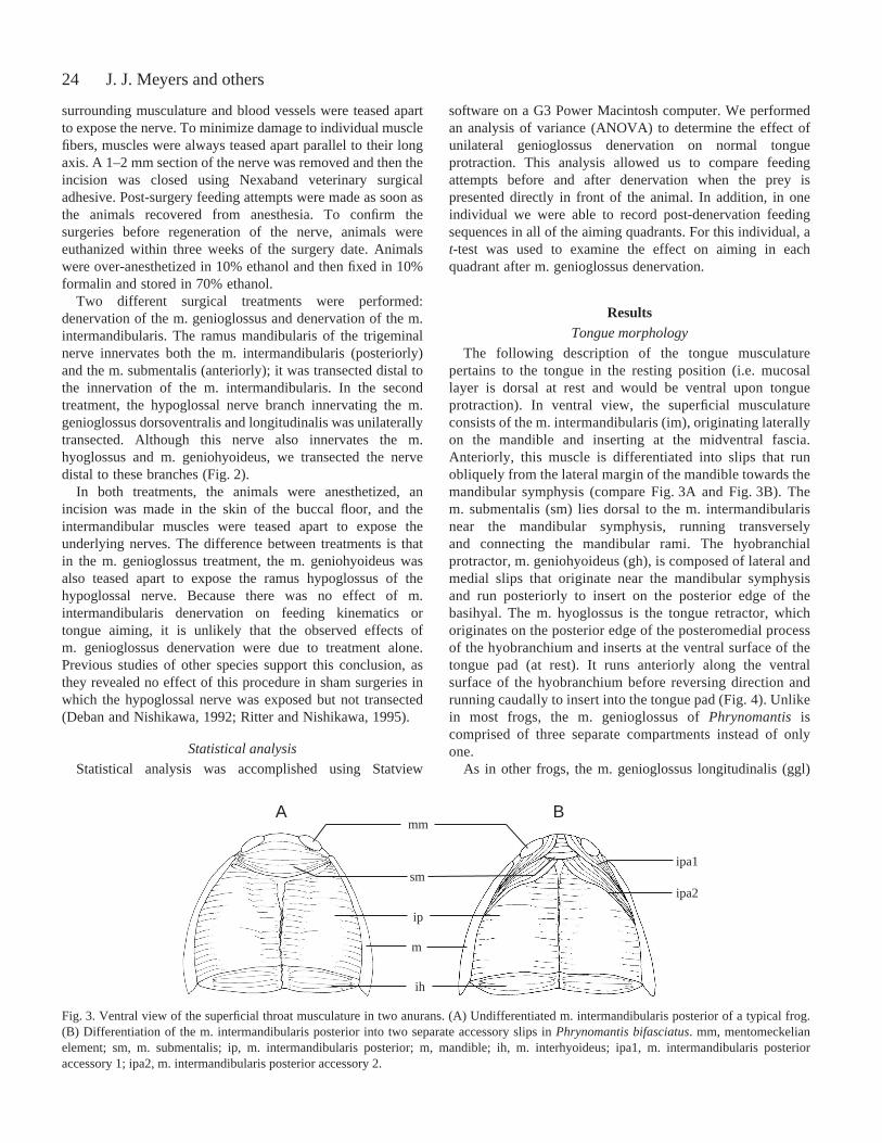
The following description of the tongue musculaturepertains to the tongue in the resting position (i.e. mucosallayer is dorsal at rest and would be ventral upon tongueprotraction). In ventral view, the superficial musculatureconsists of the m. intermandibularis (im), originating laterallyon the mandible and inserting at the midventral fascia.Anteriorly, this muscle is differentiated into slips that runobliquely from the lateral margin of the mandible towards themandibular symphysis (compare Fig.·3A and Fig.·3B). Them. submentalis (sm) lies dorsal to the m. intermandibularisnear the mandibular symphysis, running transverselyand connecting the mandibular rami. The hyobranchialprotractor, m. geniohyoideus (gh), is composed of lateral andmedial slips that originate near the mandibular symphysisand run posteriorly to insert on the posterior edge of thebasihyal. The m. hyoglossus is the tongue retractor, whichoriginates on the posterior edge of the posteromedial processof the hyobranchium and inserts at the ventral surface of thetongue pad (at rest). It runs anteriorly along the ventralsurface of the hyobranchium before reversing direction andrunning caudally to insert into the tongue pad (Fig.·4). Unlikein most frogs, the m. genioglossus of Phrynomantisiscomprised of three separate compartments instead of onlyone.
As in other frogs, the m. genioglossus longitudinalis (ggl)
J. J. Meyers and others
A Bmm
smipa1
ipa2
ip
ih
m
Fig.·3. Ventral view of the superficial throat musculature in two anurans. (A) Undifferentiated m. intermandibularis posterior of a typical frog.(B) Differentiation of the m. intermandibularis posterior into two separate accessory slips in Phrynomantis bifasciatus. mm, mentomeckelianelement; sm, m. submentalis; ip, m. intermandibularis posterior; m, mandible; ih, m. interhyoideus; ipa1, m. intermandibularis posterioraccessory 1; ipa2, m. intermandibularis posterior accessory 2.

25Tongue protraction in microhylid frogs
originates at the mandibular symphysis. It is attached to themandible by a thin band of fascia and runs postero-dorsallyalong the ventral surface of the mucosa. However, unlike inother frogs, it does not spread extensively into the tongue pador interdigitate with fibers of the m. hyoglossus. In addition tothe ggl, there is an m. genioglossus dorsoventralis (ggdv) thatlies ventral to the ggl and shares a similar origin and connectivetissue attachment. Near its origin, the ggdv fibers runposteriorly into the tongue tip, but as they proceed posteriorlythey turn and are directed ventrad, inserting into a thick layerof surrounding connective tissue. Finally, there is an additionalintrinsic muscle, the m. genioglossus transversalis (ggt), whichoriginates laterally from connective tissue and runstransversely beneath the ggdv (Fig.·4).
Tongue aiming
The ability to aim the tongue is well developed inPhrynomantis, although there appears to be individual
variation in the propensity to aim the tongue. A comparison ofaiming ability in the different quadrants revealed that allindividuals were able to aim in each quadrant. In addition tothe tongue being protruded at an angle, the head may also bemoved towards the prey item when the tongue is aimedlaterally about the head. This strategy increases the range overwhich the frogs are able to capture prey and effectively allowsthem to capture prey items off the forearms. Although theanimals are able to capture prey items over a wide range,qualitative observations of prey capture suggest that preycapture success decreases at extreme angles.
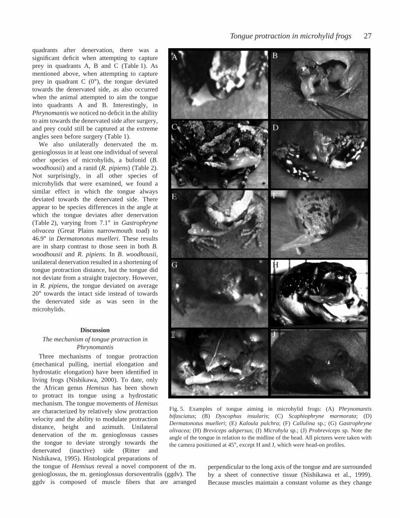
Although individuals of Phrynomantisexhibit little variationin aiming ability (Table·1), there are clear species differenceswithin the Microhylidae. All microhylid species examinedwere able to aim the tongue independent of head movements.However, the maximum tongue angles measured for thespecies varied greatly, with the most extreme angles (>100°)measured in Phrynomantisand Dermatonotus muelleri
Fig.·4. Sagittal section of the tongue of Phrynomantis bifasciatus. (A) Note that the fibers of the m. genioglossus dorsoventralis are directedlongitudinally and then dorso-ventrally. (B) Magnified view of the m. genioglossus dorsoventralis. Single fibers run in both the longitudinal andvertical planes. d, dentary; gh, m. geniohyoideus; ggdv, m. genioglossus dorsoventralis; ggl, genioglossus longitudinalis; h, hyobranchium; hg,m. hyoglossus; im, m. intermandibularis; ggt, m. genioglossus transversalis; m, mucosal layer. Scale bar, 1 mm.
Table 1. Mean ±S.D. of tongue angles in each quadrant for six individuals of Phrynomerus bifasciatus, together with the resultsof a t-test examining the effect on aiming after right unilateral M. genioglossus denervation on one individual
Quadrant A Quadrant B Quadrant C Quadrant D Quadrant E Individual (–105°to –46°) (–45°to –6°) (–5° to +5°) (+6° to +45°) (+46° to +105°)
Normal1 –57.0±10.9 –33.8±8.5 0.4±0.5 +32.2±8.4 +62.0±14.82 –51.2±3.0 –32.8±8.9 0.8±0.8 +32.4±6.1 +57.8±4.33 –60.0±1.4 –33.0±3.4 2.0±2.1 +37.3±4.6 +67.5±20.04 –60.2±18.2 –36.4±6.9 0.2±0.4 +32.6±3.5 +84.2±21.35 –52.2±5.6 –30.4±8.2 1.0±1.4 +35.4±8.1 +51.0±5.56 –55.3±11.4 –30.5±9.5 0.8±1.3 +36.8±5.1 +62.3±6.3
After right unilateral M. genioglossus denervation3 +30.0±16.5* +38.0±9.4* +34.6±4.0* +38.6±5.9 +64.3±19.0
*Aiming was significantly (P<0.05) affected in quadrants A, B and C, in which the tongue consistently deviated towards the intact side(animal’s right side).
26
(Mullers’ termite frog; Table·2; Fig.·5). The most extremetongue angles were measured from animals showing thegreatest propensity to aim. Hence, we may not have elicitedmaximum aiming attempts in some species. Unlikemicrohylids, Rana pipiensand Bufo woodhousiiexhibited littleor no ability to aim the tongue (Table·2). Although R. pipienswas able to aim the tongue up to 5°, this is substantially lessthan in all the microhylids examined.
Effect of m. intermandibularis denervation
To determine whether mandibular bending plays a role intongue aiming, we transected the ramus of the trigeminal nerveinnervating the m. intermandibularis. After bilateral transectionof the m. intermandibularis, the tongue is still able to protractnormally, and prey capture sequences are qualitatively similarto sequences recorded before denervation (compare Fig.·6A andFig.·6B). In addition, feeding attempts after surgery revealed nodeficits in the ability to aim to the extreme angles seen beforesurgery. One individual consistently aimed more than 58°, withone attempt at 90°. Although we did not test for differences, it
appeared that prey capture success rate did not differ fromnormal feeding sequences.
Effect of m. genioglossus denervation
Unilateral denervation of the m. genioglossus in P.bifasciatusresulted in deficits in the ability to both aim thetongue and to capture prey. When the nerve branch innervatingthe m. genioglossus dorsoventralis and m. genioglossuslongitudinalis is transected on the right side, the tongue isflipped out of the mouth and bends towards the denervated side(right side) upon protraction. Even when the frog attempts tofeed on prey placed directly in front of it (0°), its tonguedeviates towards the denervated side (ANOVA, F=262.2,P=0.0001; compare Fig.·6A and Fig.·6C). Prior to denervation,the tongue deviated only 3.4±2.4°. However, after denervation,the tongue is protracted at a mean angle of 43±9.4° towardsthe denervated side. Regardless of which side of the m.genioglossus is denervated, animals are never able to aim thetongue towards the active side after unilateral denervation.
In the one individual that attempted to aim in all the
J. J. Meyers and others
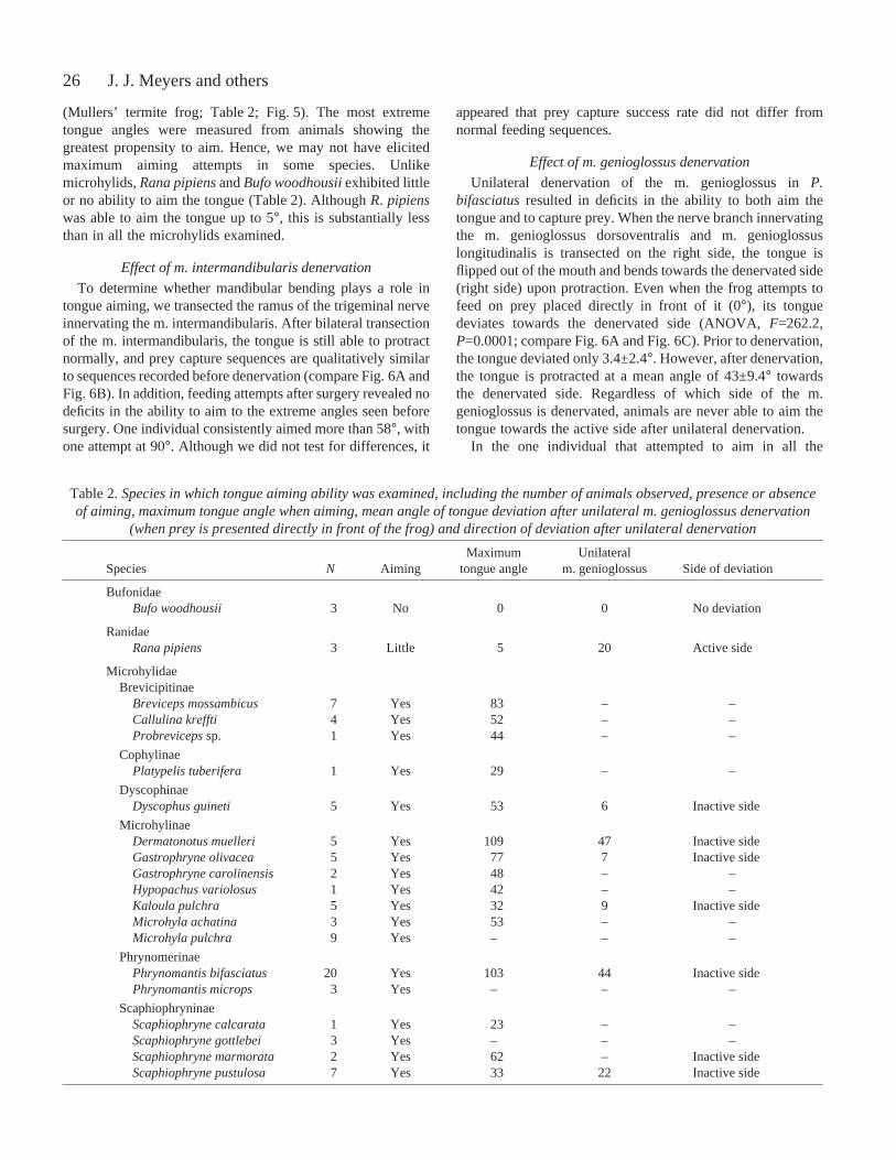
Table 2. Species in which tongue aiming ability was examined, including the number of animals observed, presence or absenceof aiming, maximum tongue angle when aiming, mean angle of tongue deviation after unilateral m. genioglossus denervation
(when prey is presented directly in front of the frog) and direction of deviation after unilateral denervation
Maximum Unilateral Species N Aiming tongue angle m. genioglossus Side of deviation
BufonidaeBufo woodhousii 3 No 0 0 No deviation
RanidaeRana pipiens 3 Little 5 20 Active side
MicrohylidaeBrevicipitinae
Breviceps mossambicus 7 Yes 83 – –Callulina kreffti 4 Yes 52 – –Probrevicepssp. 1 Yes 44 – –
CophylinaePlatypelis tuberifera 1 Yes 29 – –
DyscophinaeDyscophus guineti 5 Yes 53 6 Inactive side
MicrohylinaeDermatonotus muelleri 5 Yes 109 47 Inactive sideGastrophryne olivacea 5 Yes 77 7 Inactive sideGastrophryne carolinensis 2 Yes 48 – –Hypopachus variolosus 1 Yes 42 – –Kaloula pulchra 5 Yes 32 9 Inactive sideMicrohyla achatina 3 Yes 53 – –Microhyla pulchra 9 Yes – – –
PhrynomerinaePhrynomantis bifasciatus 20 Yes 103 44 Inactive sidePhrynomantis microps 3 Yes – – –
ScaphiophryninaeScaphiophryne calcarata 1 Yes 23 – –Scaphiophryne gottlebei 3 Yes – – –Scaphiophryne marmorata 2 Yes 62 – Inactive sideScaphiophryne pustulosa 7 Yes 33 22 Inactive side
27Tongue protraction in microhylid frogs
quadrants after denervation, there was asignificant deficit when attempting to captureprey in quadrants A, B and C (Table·1). Asmentioned above, when attempting to captureprey in quadrant C (0°), the tongue deviatedtowards the denervated side, as also occurredwhen the animal attempted to aim the tongueinto quadrants A and B. Interestingly, inPhrynomantis we noticed no deficit in the abilityto aim towards the denervated side after surgery,and prey could still be captured at the extremeangles seen before surgery (Table·1).
We also unilaterally denervated the m.genioglossus in at least one individual of severalother species of microhylids, a bufonid (B.woodhousii) and a ranid (R. pipiens) (Table·2).Not surprisingly, in all other species ofmicrohylids that were examined, we found asimilar effect in which the tongue alwaysdeviated towards the denervated side. Thereappear to be species differences in the angle atwhich the tongue deviates after denervation(Table·2), varying from 7.1° in Gastrophryneolivacea (Great Plains narrowmouth toad) to46.9° in Dermatonotus muelleri. These resultsare in sharp contrast to those seen in both B.woodhousiiand R. pipiens. In B. woodhousii,unilateral denervation resulted in a shortening oftongue protraction distance, but the tongue didnot deviate from a straight trajectory. However,in R. pipiens, the tongue deviated on average20° towards the intact side instead of towardsthe denervated side as was seen in themicrohylids.
DiscussionThe mechanism of tongue protraction in
Phrynomantis
Three mechanisms of tongue protraction(mechanical pulling, inertial elongation andhydrostatic elongation) have been identified inliving frogs (Nishikawa, 2000). To date, onlythe African genus Hemisushas been shownto protract its tongue using a hydrostaticmechanism. The tongue movements of Hemisusare characterized by relatively slow protractionvelocity and the ability to modulate protractiondistance, height and azimuth. Unilateraldenervation of the m. genioglossus causesthe tongue to deviate strongly towards thedenervated (inactive) side (Ritter andNishikawa, 1995). Histological preparations ofthe tongue of Hemisus reveal a novel component of the m.genioglossus, the m. genioglossus dorsoventralis (ggdv). Theggdv is composed of muscle fibers that are arranged
perpendicular to the long axis of the tongue and are surroundedby a sheet of connective tissue (Nishikawa et al., 1999).Because muscles maintain a constant volume as they change
Fig.·5. Examples of tongue aiming in microhylid frogs: (A) Phrynomantisbifasciatus; (B) Dyscophus insularis; (C) Scaphiophryne marmorata; (D)Dermatonotus muelleri; (E) Kaloula pulchra; (F) Callulina sp.; (G) Gastrophryneolivacea; (H) Breviceps adspersus; (I) Microhylasp.; (J) Probrevicepssp. Note theangle of the tongue in relation to the midline of the head. All pictures were taken withthe camera positioned at 45°, except H and J, which were head-on profiles.
28
shape (Kier and Smith, 1985), shortening of the dorso-ventralfibers must be directed into either lateral expansion, forwardelongation of the tongue or both. When the fibers of ggdv arerecruited in Hemisus, the thick sheet of connective tissuesurrounding this muscle resists lateral expansion and causestongue elongation. It is thought that asymmetrical recruitmentof the m. genioglossus pushes the tongue towards the side withrelatively lower recruitment.
Morphological evidence suggests that Phrynomantisalsoutilizes hydrostatic elongation for tongue protraction. Thetongue of Phrynomantis is anatomically similar to that ofHemisus in that both a longitudinal and a dorso-ventralcomponent of the genioglossus muscle are present. However,the tongue of Phrynomantisdiffers in several respects fromthat of Hemisus. First, the m. genioglossus longitudinalis andm. genioglossus dorsoventralis both have a connective tissue
J. J. Meyers and others
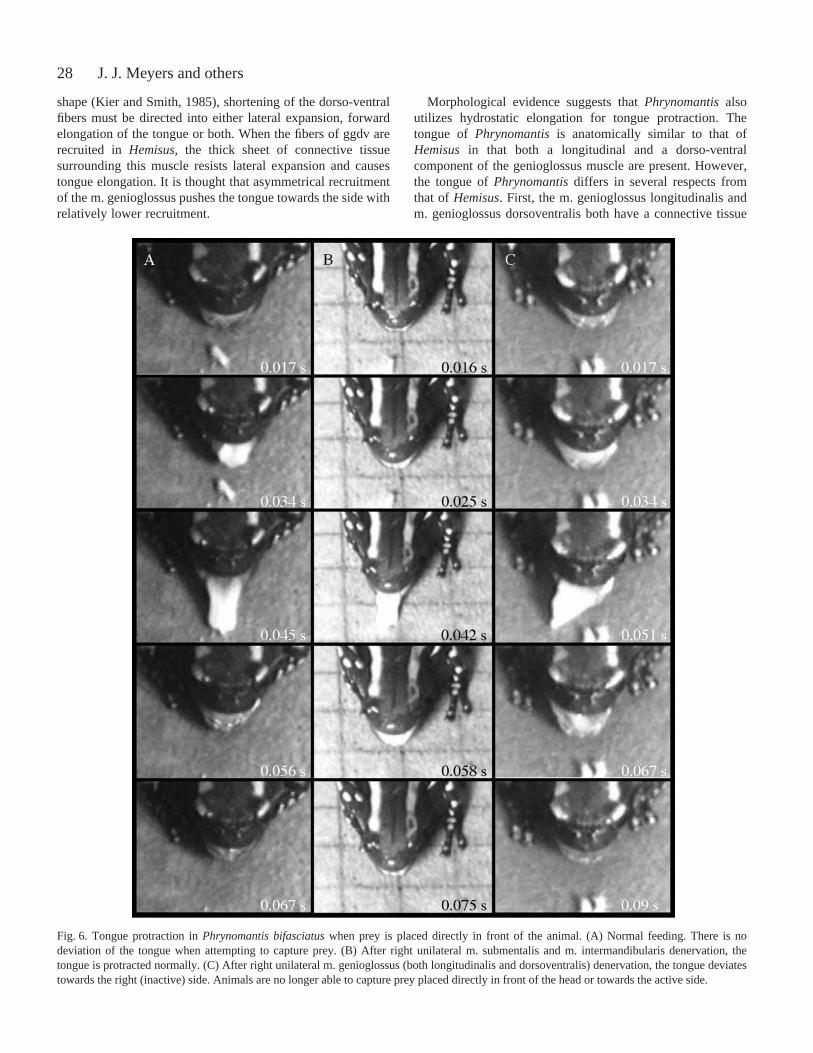
Fig.·6. Tongue protraction in Phrynomantis bifasciatuswhen prey is placed directly in front of the animal. (A) Normal feeding. There is nodeviation of the tongue when attempting to capture prey. (B) After right unilateral m. submentalis and m. intermandibularis denervation, thetongue is protracted normally. (C) After right unilateral m. genioglossus (both longitudinalis and dorsoventralis) denervation, the tongue deviatestowards the right (inactive) side. Animals are no longer able to capture prey placed directly in front of the head or towards the active side.

29Tongue protraction in microhylid frogs
origin on the mandible. Second, in Phrynomantis, individualfibers of the m. genioglossus dorsoventralis are directedlongitudinally and dorso-ventrally (Fig.·4), whereas inHemisusthey are only in the dorso-ventral plane (compareFig.·4 in the present study with fig.·3 in the study by Nishikawaet al., 1999). The functional significance of this arrangementin Phrynomantisremains unclear, since it seems that a strictlydorso-ventrally arranged compartment would be the mosteffective in lengthening the tongue.
Denervation experiments in Phrynomantis also support amuscular hydrostatic mechanism. After unilateral denervationof the entire m. genioglossus (gg) complex, the tongue isprotruded towards the denervated (inactive) side when animalsattempt to feed on prey lying directly in front of them. This isthe expected result if a hydrostatic pushing mechanism is beingutilized in tongue protraction. If inertial elongation were atplay, the tongue would either show no lateral deviation (as inB. woodhousii) or would be pulled to the active side (as in R.pipiens). In combination with the anatomical data, deviation tothe inactive side after unilateral denervation supports the useof a muscular hydrostatic mechanism of tongue protraction inPhrynomantis.
The mechanism of tongue aiming in Phrynomantis
At least three different mechanisms could be used bymicrohylids to aim their tongues laterally independent of headmovements, including: (1) rotating the base of the tongue bycontraction of accessory slips of the m. intermandibularis; (2)pulling the tongue to the side by differential contraction oflongitudinal fibers of the m. genioglossus; or (3) pushing thetongue to one side with hydrostatic pressure generated bydorso-ventral fibers of the m. genioglossus (as previouslydescribed in Hemisus; Nishikawa et al., 1999). We willdescribe each potential mechanism in turn and discuss theevidence for and against its playing a role in laterally directedtongue protraction in microhylids.
In the plesiomorphic m. intermandibularis of frogs, all of thefibers are arranged in parallel, forming a sheet that runslaterally from the mid-ventral line to the mandibles and therod-shaped mentomeckelian bones. By contrast, the m.intermandibularis of some frogs can include one or moreaccessory slips with fibers that run from the mandibles to thefascia covering the submentalis, which in turn lies directlybelow the base of the tongue pad (e.g. Trewavas, 1933; Liem,1970; Tyler, 1971; Emerson, 1976). Accessory slips of the m.intermandibularis are well developed in many microhylids (seefigs·2–5 in Emerson, 1976). Emerson (1976) suggested thatthese accessory slips of the m. intermandibularis might play arole in tongue protraction but did not specifically speculate ona potential role in lateral tongue movement. We hypothesizedthat differential activation of the right or left accessory slipsmight pull the mentomeckelian bones and tilt or rotate the baseof the tongue. Tilting or rotation of the tongue base couldplausibly contribute to lateral movements. Our results do notsupport this functional hypothesis, because when the m.intermandibularis complex was denervated unilaterally, there
was no measurable deficit in the range of lateral protraction toeither side. These results unambiguously indicate that thecomplex anatomy of the m. intermandibularis and unusuallyshaped mentomeckelian bones in microhylids are notfunctionally related to the ability to protract the tonguelaterally.
The second potential mechanism would involve the m.genioglossus pulling the tongue to one side as it is protracted.The fibers of the ggl originate on the mandible tips and runcaudally along the dorsal surface of the resting tongue pad.Asymmetrical recruitment of these fibers could pull the tongueeither to the right or left side (as in R. pipiens), the tonguedeviating towards the side showing greater activation. If thismechanism were present in microhylids, we would expect thatunilateral denervation of the m. genioglossus would cause thetongue to bend towards the intact (active) side. Although wewere unable to denervate the ggl and ggdv individually inPhrynomantis, unilateral denervation of both muscles causedthe tongue to deviate towards the inactive side. While it ispossible that the ggl, when acting alone, pulls the tongue as itdoes in R. pipiens, the overriding effects of ggdv suggest thatthe ggl is not determining tongue trajectory.
The third potential mechanism would involve using thehydrostatic pressure generated in the ggdv to aim the tongueto one side or the other during protraction. Because thepressure generated in the ggdv pushes the tongue out of themouth, differential activation of the ggdv would bend thetongue towards the less active side. This mechanism isconsistent with the results of the denervation experiments.Unilateral gg denervation invariably reduced the range ofmotion of the tongue to part of the range on the denervated(inactive) side.
Although the results of unilateral gg denervation inPhrynomantisare similar to those in Hemisus, some importantdifferences should be noted. The tongue of Hemisusinitiallymoves directly forward after unilateral gg denervation suchthat prey can still be captured directly in front of an individualas long as it is not far from the mouth (Ritter and Nishikawa,1995). By contrast, after unilateral gg denervation, thetrajectory of the tongue of Phrynomantisis initially to thedenervated side. Thus, food directly in front of the animalcannot be captured. If Hemisusmisses prey directly anterior tothe head after unilateral gg denervation, the tongue tip followsa semi-circular trajectory and eventually runs into the side ofthe head, close to 180° off course (Ritter and Nishikawa, 1995).By contrast, the tongue of Phrynomantistravels in a relativelystraight line and lands at approximately 45° off course(Fig.·6C). Ritter and Nishikawa (1995) did not determine ifHemisuscould capture prey positioned to either side of thehead after unilateral gg denervation. Our results indicate thatPhrynomantiscan still accurately aim the tongue through alimited range on the denervated side.
The results of the unilateral gg denervation experimentsuggest that both sides of the ggdv are active during protractionregardless of where the tongue is aimed. If prey are presentedon the intact (active) side, the tongue is still protruded when
30
attempting to feed. If the ggdv were stimulated unilaterallyduring aiming, then we would expect no tongue protrusionwhen attempting to aim towards the active side after unilateralgg denervation. This hypothesis could be further explored byrecording bilateral muscle activity from both ggl and ggdv orby selectively denervating each muscle compartment.
The evolution of tongue protraction in microhylids
Previous authors have noted internal compartments ofthe gg from gross dissection in several other microhylids,including Callulops stictogaster(irumbofoie callulops frog;Burton, 1983), Brevicepssp., Cophixalus ornatus(ornaterainforest frog) and Austrochaperina robusta(chirping landfrog; Horton, 1982). The fact that all microhylids surveyed todate can aim the tongue laterally and react similarly tounilateral gg denervation suggests that the ggdv muscleand hydrostatic elongation are common features of allmicrohylids. Although the ggdvs of Hemisus and microhylidsdiffer, in that the former has no mandibular origin, they mayrepresent a morphocline in the development of an internalgenioglossus compartment. The similarities in morphologyand the fact that Hemisus consistently falls out nearMicrohylidae in recent phylogenetic hypotheses (Ford andCannatella, 1993; Wu, 1994; Emerson et al., 2000; Hass,2003) suggest that the ggdv may be a derived character ofHemisus and Microhylidae (Nishikawa et al., 1999; Emersonet al., 2000). Intrinsic tongue muscles have also been reportedin the tongue of Rhinophrynus dorsalis (Mexican burrowingtoad). However, these fibers are thought to be derived fromthe m. hyoglossus and only play a role in changing tongueshape, with tongue protraction being powered mainly byhyobranchial movements (Trueb and Gans, 1983).
The results of the present study reveal considerable variationin aiming prowess among microhylids. This variation inperformance suggests that important morphological variationin the tongue musculature among microhylids awaitsdescription. Variation in the arrangement of connective tissueand collagen fibers in the tongue may also play an importantrole in elongation and aiming. The orientation of theconnective tissue fibers determines the direction of shapechange (Kier and Smith, 1985) and may also influence theextent of tongue elongation (Zepnewski and Nishikawa, 2000).Fiber angles less than 54°44′ tend to inhibit elongation,whereas those greater than 54°44′ facilitate elongation. InHemisus(Nishikawa et al., 1999), collagen fibers surroundingthe m. genioglossus are oriented at an angle of nearly 80°,resisting lateral expansion and facilitating elongation.Although we did not measure fiber angles in Phrynomantis, wewould predict them to be greater than 54°44′. Connective tissueorientation might also explain species differences in tonguedeviation angles after unilateral transection. Initially, wesuspected that post-transection tongue angle could be predictedby the maximum aiming angle observed during normalfeeding, so that species with the largest aiming angles alsoexhibited the largest deviations after transection. However,this is not necessarily the case. For example, Gastrophryne
olivaceaaims to a slightly greater extent than Scaphiophrynemarmorata(marbled rain frog) but has a mean deviation anglesubstantially less than that of S. marmorata (Table·2). Theunderlying mechanism for these differences in behaviorremains unclear. It is likely that a combination ofmorphological characteristics, including connective tissue andmuscle fiber orientation, are important, and detailed anatomicalstudies are needed.
In summary, it appears that microhylid frogs protract thetongue using a muscular hydrostatic mechanism. Thismechanism was previously known only in Hemisus. Whileprotraction is probably accomplished by recruiting the lateraland dorso-ventral portions of the m. genioglossus, lateraldisplacement is due to the ggdv. Due to the orientation of thesurrounding connective tissue, muscle contraction of the ggdvresults in lateral rather than longitudinal displacement. Thismechanism of protraction increases the range of possiblemovement relative to that of tongues protracted by mechanicalpulling or inertial elongation. Feeding behavior ofPhrynomantis and 17 other species of microhylids,representing six subfamilies, suggests that this generalmechanism is used by all microhylids. The presence of anintrinsic component of the m. genioglossus, a hydrostaticelongation protraction mechanism and lateral tongue aimingmay be synapomorphies of Microhylidae and Hemisus.
Stephen Deban made the original observation thatPhrynomantiswas capable of aiming its tongue independentlyof the lower jaw. Sheng-Hai Wu and David Cannatella helpedin the identification of specimens. David Cannatella andRonald Nussbaum kindly provided some of the specimensused in this study. Louis Porras (Zooherp Inc.) and RobMacInnes (Glades Herp Inc.) helped us acquire manyspecimens. Mark Mandica created Fig.·1. Christian Jaeger andStephen Deban helped record feeding sequences. AnthonyHerrel, Jen Glass, Kurt Schwenk and an anonymous reviewerprovided helpful comments on an earlier version of thismanuscript. This study was supported by grant numbers NSFIBN–0215438, NSF IBN-0240349 and NIH R25-GM56931 toK.C.N.
ReferencesBurton, T. C. (1983). The musculature of the Papuan frog Phrynomantis
stictogaster (Anura, Microhylidae). J. Morph. 175, 307-324.Deban, S. M. and Nishikawa, K. C. (1992). The kinematics of prey capture
and the mechanism of tongue protraction in the green tree frog Hyla cinerea.J. Exp. Biol. 170, 235-256.
Emerson, S. B. (1976). A preliminary report on the superficial throatmusculature of the Microhylidae and its possible role in tongue action.Copeia3, 546-551.
Emerson, S. B., Richards, C., Drewes, R. C. and Kjer, K. M. (2000). Onthe relationships among ranoid frogs: a review of the evidence.Herpetologica56, 209-230.
Ford, L. S. and Cannatella, D. C. (1993). The major clades of frogs. Herp.Monogr. 7, 94-117.
Gans, C. and Gorniak, G. C. (1982a). How does the toad flip its tongue?Test of two hypotheses. Science216, 1135-1137.
Gans, C. and Gorniak, G. C. (1982b). Functional morphology of lingualprotrusion in marine toads (Bufo marinus). Am. J. Anat. 163, 195-222.
Gray, L. A. (1997). Tongue morphology, feeding behavior and feeding
J. J. Meyers and others

31Tongue protraction in microhylid frogs
ecology in anurans. Ph.D. Dissertation. Northern Arizona University,Flagstaff, AZ, USA.
Haas, A. (2003). Phylogeny of frogs as inferred from primarily larvalcharacters (Amphibia: Anura). Cladistics19, 23-89.
Horton, P. (1982). Diversity and systematic significance of anuran tonguemusculature. Copeia3, 595-602.
Humason, G. L. (1979). Animal Tissue Techniques.4th edition. SanFrancisco: W. H. Freeman and Co.
Kier, W. M. and Smith, K. K. (1985). Tongue, tentacles and trunks: thebiomechanics of movement in muscular-hydrostats. Zool. J. Linn. Soc.83,307-324.
Liem, S. S. (1970). The morphology, systematics, and evolution of Old Worldtreefrogs (Rhacophoridae and Hyperoliidae). Fieldiana Zool. 57, 1-145.
Magimel-Pelonnier, O. (1924). La langue des Amphibiens. These.Facultedes Sciences, Université de Paris, France. A. Saugnac and E. Provillard,Bordeaux.
Meyers, J. J., O’Reilly, J. C. and Nishikawa, K. C. (1996). Tongue aimingin the microhylid frog Phrynomerus bifasciatus. Am. Zool. 36, 81A.
Monroy, J. A. and Nishikawa, K. C. (2000). Aiming during prey capture inmicrohylid frogs. Am. Zool.40, 1135A.
Nishikawa, K. C. (1987). Staining amphibian peripheral nerves with SudanBlack B: progressive vs. regressive methods. Copeia2, 489-491.
Nishikawa, K. C. (1992). The role of hypoglossal sensory feedback duringfeeding in the marine toad, Bufo marinus. J. Exp. Biol.264, 245-252.
Nishikawa, K. C. (1997). Emergence of novel functions during brainevolution. Bioscience47, 341-354.
Nishikawa, K. C. (1999). Neuromuscular control of prey capture in frogs.Phil. Trans. R. Soc. Lond.354, 941-954.
Nishikawa, K. C. (2000). Feeding in frogs. In Feeding in TetrapodVertebrates: Form, Function, Phylogeny(ed. K. Schwenk), pp. 117-141.London: Academic Press.
Nishikawa, K. C. and Cannatella, D. C. (1991). Kinematics of prey capturein the tailed frog Ascaphus truei(Anura: Ascaphidae). Zool. J. Linn. Soc.103, 289-307.
Nishikawa, K. C. and Gans, G. (1996). Mechanisms of tongue protractionand narial closure in the marine toad Bufo marinus. J. Exp. Biol.199, 2511-2529.
Nishikawa, K. C. and Roth, G. (1991). The mechanism of tongue protractionduring prey capture in the frog Discoglossus pictus. J. Exp. Biol.159, 217-234.
Nishikawa, K. C., Kier, W. M. and Smith, K. K. (1999). Morphology andmechanics of tongue movement in the African pig-nosed frog Hemisusmarmoratum: a muscular hydrostatic model. J. Exp. Biol.202, 771-780.
O’Reilly, S. R. and Nishikawa, K. C. (1995). Mechanism of tongueprotraction during prey capture in the spadefoot toad Spea mutiplicata(Anura: Pelobatidae). J. Exp. Zool. 273, 282-296.
Regal, P. J. and Gans, C. (1976). Functional aspects of the evolution of frogtongues. Evolution30, 718-734.
Ritter, D. and Nishikawa, K. C. (1995). The kinematics and mechanism ofprey capture in the African pig-nosed frog (Hemisus marmoratum):description of a radically divergent anuran tongue. J. Exp. Biol.198, 2025-2040.
Trewavas, E. (1933). The hyoid and larynx of the Anura. Trans. R. Phil. Soc.Lond.222, 401-527.
Trueb, L. and Gans, C. (1983). Feeding specializations of the Mexicanburrowing toad, Rhinophrynus dorsalis(Anura: Rhinophrynidae).J. Zool.Lond.199, 198-208.
Tyler, M. J. (1971). Observations of anuran myo-integumental attachmentsassociated with the vocal sac apparatus.J. Nat. Hist.5, 225-231.
Valdez, C. M. and Nishikawa, K. C. (1996). Sensory modulation andbehavioral choice during feeding in the Australian frog, Cyclorananovaehollandiae. J. Comp. Physiol. A 180, 187-202.
Wu, S. (1994). Phylogenetic relationships, higher classification, and historicalbiogeography of the microhyloid frogs (Lissamphibia: Anura:Brevicipitidae and Microhylidae. Ph.D. Dissertation. University ofMichigan, Ann Arbor, MI, USA.
Zepnewski, E. and Nishikawa, K. C. (2000). Connective tissue in ballistictongues. Am. Zool. 40, 1271A.