mechanism of hic-5/ara55 action, a novel stromal …
TRANSCRIPT
MECHANISM OF HIC-5/ARA55 ACTION, A NOVEL STROMAL-SPECIFIC NUCLEAR RECEPTOR COACTIVATOR
by
Marjet Danteel Heitzer
M.S., Wright State University, 2000
Submitted to the Graduate Faculty of
the School of Medicine in partial fulfillment
of the requirements for the degree of
Doctor of Philosophy
University of Pittsburgh
2005
UNIVERSITY OF PITTSBURGH
FACULTY OF THE SCHOOL OF MEDICINE
This dissertation was presented
by
Marjet Danteel Heitzer
It was defended on
September 26, 2005 and approved by
Dr. Anthony Zeleznik Committee Chair Department of Cell Biology and Molecular Physiology Dr. William Walker Department of Cell Biology and Molecular Physiology Dr. Mark Nichols Department of Pharmacology Dr. Alan Wells Department of Pathology Dr. Donald B. DeFranco Dissertation Advisor Department of Pharmacology
ii

Copyright by Marjet Danteel Heitzer
2005
iii
Donald B. DeFranco
MECHANISM OF HIC-5/ARA55 ACTION, A NOVEL STROMAL-SPECIFIC NUCLEAR
RECEPTOR COACTIVATOR
Marjet Danteel Heitzer, PhD
University of Pittsburgh, 2005
Hydrogen peroxide inducible clone-5/Androgen Receptor Activator 55 (Hic-5/ARA55) is a
group III LIM domain protein that functions at focal adhesion complexes as well as in the
nucleus as a nuclear receptor coactivator. Because the interaction of the androgen receptor (AR)
with Hic-5/ARA55 results in enhanced androgen-induced transcription, we analyzed Hic-
5/ARA55 expression in prostate tissue sections from normal human donors and prostate cancer
patients. In each sample, Hic-5/ARA55 expression was confined to the stromal compartment of
the prostate. Furthermore, in a human prostate stromal cell line (i.e. WPMY-1 cells) Hic-
5/ARA55 was localized both at focal adhesion complexes and within the soluble cytoplasmic
compartment. The ability of Hic-5/ARA55 to shuttle between the nuclear and cytoplasmic
compartments within WPMY-1 cells was revealed upon inhibition of nuclear export with
leptomycin B (LMB). siRNA ablation experiments established endogenous Hic-5/ARA55 as a
coactivator for both viral and endogenous cellular AR-regulated genes. Furthermore, chromatin
immunoprecipitation (ChIP) analysis showed androgen-dependent recruitment of Hic-/ARA55 to
the promoter of the stromal androgen-responsive KGF gene. Using the A1-2 derivative of T47D
iv
breast cancer cells, we examined the mechanism by which Hic-5/ARA55 potentiates nuclear
receptor transactivation. Hic-5/ARA55 was found to be an important component of
glucocorticoid receptor (GR)-coactivator complexes in A1-2 cells since ablation of Hic-
5/ARA55 expression by RNA interference-mediated silencing reduced GR transactivation. As
shown by ChIP assays, Hic-5/ARA55 is recruited to glucocorticoid-responsive promoters of the
MMTV, c-fos, and p21 genes in response to glucocorticoid treatment. Results from sequential
ChIP assays established that Hic-5/ARA55 associates with the corepressor, NCoR, in the
absence of glucocorticoids. However, upon glucocorticoid stimulation, Hic-5/ARA55 interacts
with GR-coactivator containing complexes at these promoters. Ablation of Hic-5/ARA55
expression resulted in reduction of both TIF-2 and p300 recruitment to glucocorticoid-responsive
promoters. These data provide the first demonstration of a stromal-specific AR coactivator that
has an impact on an androgen regulated growth factor that is essential for stromal/epithelial cell
communication in the prostate. Furthermore, these results suggest that Hic-5/ARA55 is required
for optimal GR-mediated gene expression possibly by providing a scaffold that organizes or
stabilizes coactivator complexes at some hormone-responsive promoters.
v
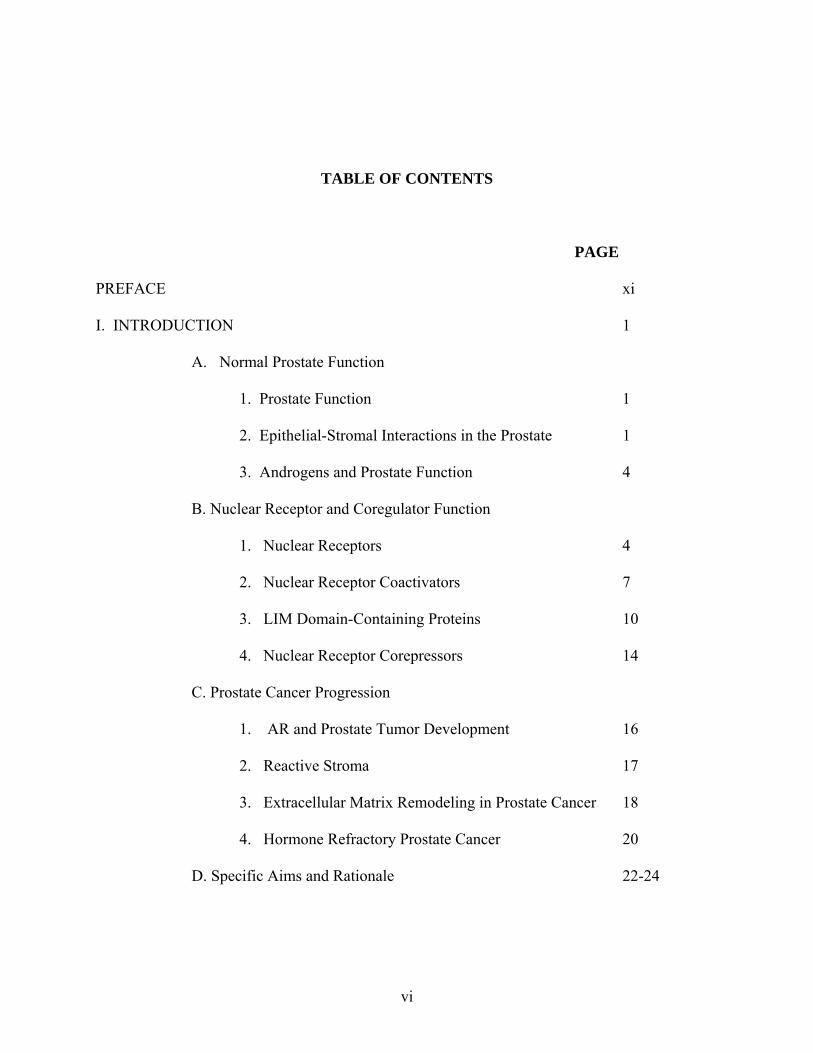
TABLE OF CONTENTS
PAGE
PREFACE xi
I. INTRODUCTION 1
A. Normal Prostate Function
1. Prostate Function 1
2. Epithelial-Stromal Interactions in the Prostate 1
3. Androgens and Prostate Function 4
B. Nuclear Receptor and Coregulator Function
1. Nuclear Receptors 4
2. Nuclear Receptor Coactivators 7
3. LIM Domain-Containing Proteins 10
4. Nuclear Receptor Corepressors 14
C. Prostate Cancer Progression
1. AR and Prostate Tumor Development 16
2. Reactive Stroma 17
3. Extracellular Matrix Remodeling in Prostate Cancer 18
4. Hormone Refractory Prostate Cancer 20
D. Specific Aims and Rationale 22-24
vi
II. MATERIALS AND METHODS
A. Antibodies, Plasmids, and Reagents 26
B. Cell Culture and Transient Transfection 26
C. Extracellular Matrix Application to Tissue Culture Plates 27
D. Western Blot Analysis 27
E. RNAi 28
F. Luciferase Assays 29
G. Chromatin Immunoprecipitation 29
H. Reverse Transcriptase PCR 32
I. Immunofluorescence 33
J. Immunohistochemistry 33
K. Statistics 34
III. DETERMINING THE MECHANISM BY WHICH HIC-5/ARA55 FUNCTIONS AS A
COACTIVATOR FOR NUCLEAR RECEPTORS
A. Hypothesis 35
B. Introduction 35
C. Results 37
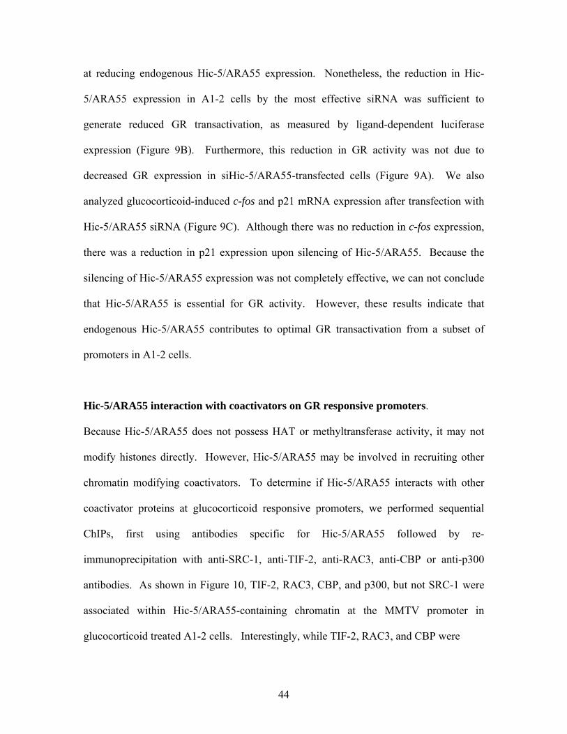
D. Discussion 49
IV. DETERMINING HIC-5/ARA55 EXPRESSION AND FUNCTION IN THE PROSTATE
A. Hypothesis 52
B. Introduction 52
C. Results 54
D. Discussion 63
vii
V. DETERMINING THE EFFECTS OF EXTRACELLULAR MATRIX (ECM) SIGNALING
ON GR-MEDIATED TRANSCRIPTION IN A1-2 CELLS
A. Hypothesis 67
B. Introduction 67
C. Results 70
D. Discussion 79
VI. DISCUSSION
A. Hic-5/ARA55 Coregulator Interactions 82
B. Coactivators and Prostate Cancer 85
C. Reactive Stromal Transition 87
D. Hic-5/ARA55 and Development 88
E. Hic-5/ARA55 and Cell Growth 90
REFERENCES 92
viii
LIST OF FIGURES
PAGE
Figure 1: Stromal-epithelial interactions in the prostate. 3
Figure 2: Modular Structure of the Nuclear Receptor Family 7
of Transcription Factors
Figure 3: Coregulator Exchange on Nuclear Receptor-Regulated Genes 15
Figure 4: Transition of Normal Epithelial-Stromal Homeostasis to Carcinoma- 19
Reactive Stroma Interactions in PCa
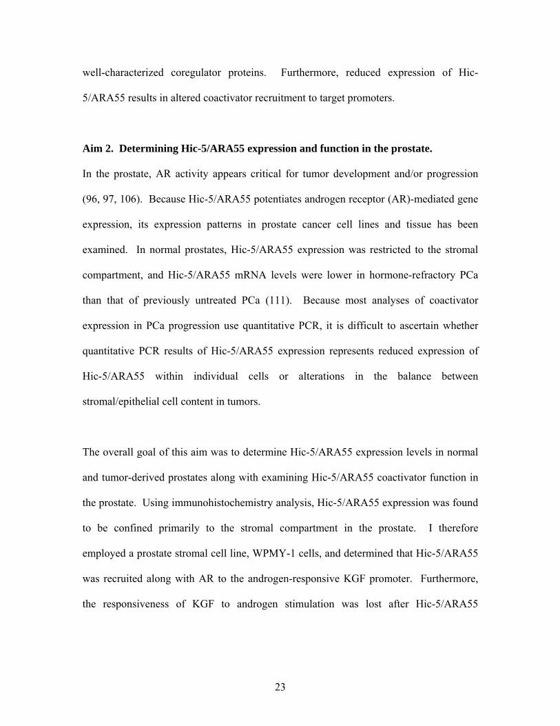
Figure 5: Chromatin Immunoprecipitation (ChIP) Assay 25
Figure 6: Binding of Hic-5/ARA55 to the MMTV Promoter 38
Figure 7: Co-occupancy of GR and Hic-5/ARA55 on the MMTV promoter 39
Figure 8: Association of GR and Hic-5/ARA55 with the Chromatin of 41
Endogenous Promoters in vivo
Figure 9: Effects of RNAi-mediated silencing of Hic-5/ARA55 on 43
GR Transactivation
Figure 10: Co-occupancy of Hic-5/ARA55 and Coregulators on the 45-46
MMTV and c-fos Promoters in vivo
Figure 11: Coactivator Recruitment on the MMTV Promoter Following 48
Ablation of Hic-5/ARA55
Figure 12: Hic-5/ARA55 Expression in the Human Prostate 55
Figure 13: Expression and Nucleocytoplasmic Shuttling of Hic-5/ARA55 57
ix
in the WMPY-1 Prostate Stromal Cell Line
Figure 14: Androgen-Induced KGF Expression in WPMY-1 cells 59
Figure 15: Association of AR and Hic-5/ARA55 with the Chromatin of 60
the KGF promoter in vivo
Figure 16: Effects of RNAi-mediated silencing of Hic-5/ARA55 on AR 62-63
Transactivation
Figure 17: Effects of Extracellular Matrices on GR-mediated Transcription 71
Figure 18: Effects of Fibronectin on Various Concentrations of Dex 72
Figure 19: Effects of Matrigel and Fibronectin on GR and Hic-5/ARA55 74
Expression
Figure 20: Effects of Fibronectin on Nucleocytoplasmic Shuttling of GR 75
Figure 21: Effects of Cell Signaling Inhibitors on Fibronectin-Enhanced 77
GR Activity
Figure 22: Effects of Fibronectin on GR and Hic-5/ARA55 Promoter 78
Recruitment in Response to Glucocorticoids
x

PREFACE
I would first like to thank my Lord for providing strength and patience as well as many
extraordinary people whose culmination of inspiring words and deeds without which this thesis
would not exist.
I would like to thank my well-respected advisor Donald B. DeFranco for his guidance these past
years. Although I joined Don’s lab rather late in my graduate career, Don has taught me many
valuable lessons in scientific writing as well as data interpretation. I would also like to
acknowledge my thesis committee members, Drs. Anthony Zeleznik,
William Walker, Mark Nichols, and Alan Wells. My committee provided many ideas and
reagents that were particularly useful in the completion of my thesis.
I would also like to thank laboratory members, past and present, not only for their assistance at
the bench but also their friendships, which mean the most. In particular, I would like to thank
Rosalba Escamilla-Hernandez and Patricia Cano-Sanchez who were both instrumental in my
training in various laboratory techniques as well as dear friends. In the DeFranco lab, I would
like to thank Marcia Lewis and Xinjia Wang for assisting me in many experiments as well as
providing many lovely conversations and tasty recipes.
In addition, I would like to thank my family and friends for their support. My parents, Pamela F.
Heitzer and James M. Heitzer, taught me the value of education and that everything is possible,
xi
the impossible just takes a little longer. My siblings, Aaron C. Heitzer, Tammy L. Burkett, and
Tracy G. Walborn, have continued to encourage me, and I thank them all very much for
supporting me even at the expense of time spent together. Finally, I want to thank my G. Mbella
Ekema for moving to Pittsburgh to continue our life together. Mbella, through the multitude of
your talents, you have taught me that the only restraints that impede us are those we place on
ourselves. You have exemplified your love and support for my dreams by not settling for second
best in your endeavors. I look forward to our fully post-graduate life with much anticipation.
xii
CHAPTER 1 INTRODUCTION
Prostate Function
The prostate gland secretes the main protein components found in seminal fluid.
Structurally, the prostate is composed of both a glandular epithelium and a fibromuscular
stroma, which are separated by a basement membrane. The epithelial compartment is
composed of two types of epithelium, basal and secretory. Basal epithelial cells secrete
proteins found in the basement membrane, and some may constitute secretory epithelial
stem cells (4, 5). The secretory epithelial compartment is responsible for production and
secretion of enzymes, such as human prostate acid phosphatase (hPAP) and prostate
specific antigen (PSA), which are found in seminal fluid. The stromal compartment is
composed of smooth muscle cells, fibroblasts, endothelial cells, nerve cells, and
lymphocytes. The stroma secretes growth factors and cytokines responsible for normal
growth and development of the epithelial compartment (6).
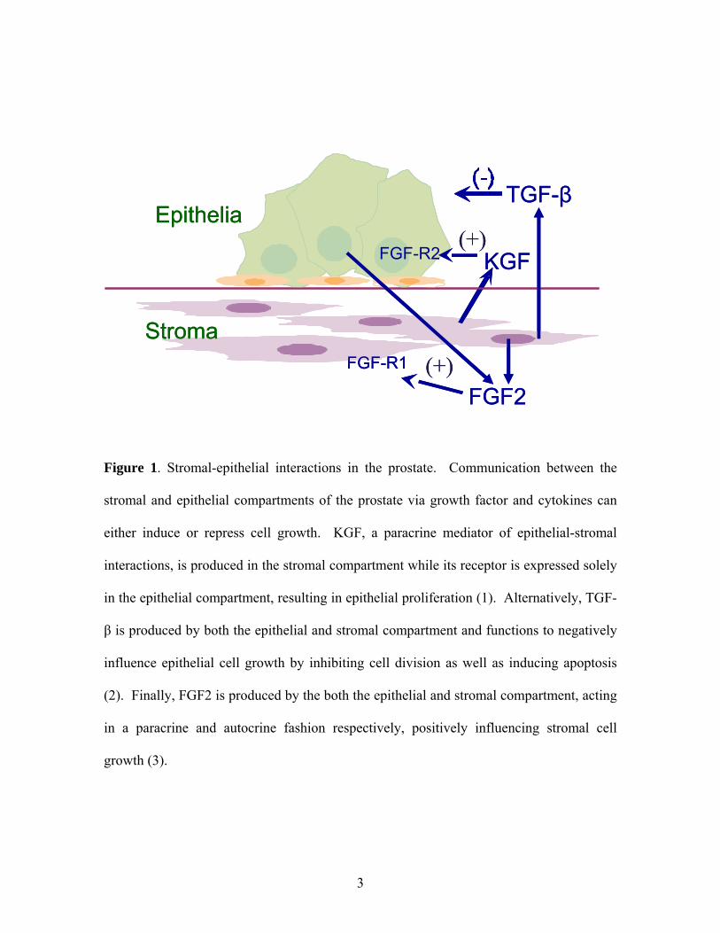
Epithelial-Stromal Interactions in the Prostate
Communication between the epithelial and stromal compartments of the prostate,
mediated by growth factors and cytokines, is crucial for the maintenance of prostate
growth and function (Figure 1, 7, 8). Examples of such growth factors in the prostate
include transforming growth factor beta (TGF-β), transforming growth factor alpha
(TGF-α), epidermal growth factor (EGF), keratinocyte growth factor (KGF), fibroblast
1
growth factor-2 (FGF-2), and insulin-like growth factor (IGF) (9, 10). Most of these
growth factors enhance both growth and development of the prostate, although TGF-β
may negatively affect prostate growth (2).
During prostate development, the urogenital sinus mesenchyme (UGM) stimulates
epithelial differentiation, ductal branching, and proliferation (11). Conversely, signals
derived from the urogenital epithelium (UGE) promote mesenchymal differentiation (12).
Along with epithelial-stromal communication, androgen signaling through AR is crucial
for prostate development (13). Reconstitution experiments with isolated stromal and
epithelial cells performed by Cunha and Lang illustrated the necessity for functional AR
in the UGM for the development of a functional prostate (14). In these studies, prostate
tissue was derived from either normal or testicular feminized (TFM) mice that have a
loss-of-function mutation in the AR gene. Whereas combining TFM UGE and normal
UGM produced normal prostate organ formation in the presence of androgen, analogous
tissue reconstitutions with normal UGE and TFM UGM did not support prostate
organogenesis. This correlates with AR expression patterning seen in the developing
prostate. Specifically, before and during prostatic bud formation, AR expression is
restricted to the UGM (13). These data suggest that AR controls the expression of a
soluble factor in the UGM that is necessary for proper epithelial differentiation and thus
prostate development.
2
Stroma
FGF-R2 KGF
FGF2FGF-R1
EpitheliaTGF-β
(+)
(+)Stroma
FGF-R2 KGF
FGF2FGF-R1
EpitheliaTGF-β
(+)
(+)
Figure 1. Stromal-epithelial interactions in the prostate. Communication between the
stromal and epithelial compartments of the prostate via growth factor and cytokines can
either induce or repress cell growth. KGF, a paracrine mediator of epithelial-stromal
interactions, is produced in the stromal compartment while its receptor is expressed solely
in the epithelial compartment, resulting in epithelial proliferation (1). Alternatively, TGF-
β is produced by both the epithelial and stromal compartment and functions to negatively
influence epithelial cell growth by inhibiting cell division as well as inducing apoptosis
(2). Finally, FGF2 is produced by the both the epithelial and stromal compartment, acting
in a paracrine and autocrine fashion respectively, positively influencing stromal cell
growth (3).
3
Androgens and Prostate Function
Circulating androgens derived primarily from the testes also contribute to normal growth
and development of the prostate gland (15). Testicular Leydig cells secrete testosterone
(T) that is converted in the prostate to dihydrotestosterone (DHT) by 5α-reductase (16).
Androgen action in both stromal and epithelial compartments of the prostate is mediated
through AR (5). In the epithelial compartment, androgens are required for both
proliferative and secretory functions of the prostate (12). For example, androgens induce
the expression of prostate-specific secretory proteins, including human prostate acid
phosphatase (hPAP), prostate specific antigen (PSA), and prostate-specific kallikrein
(hK2) (17, 18). In the stromal compartment, AR regulates the expression of various
growth factors such as KGF (FGF-7), a member of the fibroblast growth factor family
(14, 19-21). KGF is exclusively expressed in the stromal compartment of the prostate
while its receptor, a spliced variant of the FGF type 2 receptor (FGFR2IIIb), is expressed
solely in the epithelial compartment (3). These characteristics suggest that KGF is a
paracrine mediator of signaling from the stroma to the epithelial compartment that can
influence epithelial cell growth (1, 22).
Nuclear Receptors
The nuclear receptors comprise a large family of ligand-dependent transcription factors
that regulate target gene expression through a variety of mechanisms. Receptors for
steroid hormones include estrogen receptor (ER), androgen receptor (AR), progesterone
4
receptor (PR), and glucocorticoid receptor (GR). In the absence of hormone, steroid
receptors are associated with heat shock proteins and immunophilins (23). Once bound
by ligand, steroid receptors undergo a conformational change, permitting homo-
dimerization and translocation into the nucleus. Upon entering the nucleus, steroid
receptors induce gene expression by binding hormone response elements (HREs) located
in the promoter regions of target genes. Alternatively, nonsteroidal nuclear receptors
including thyroid hormone receptor (TR), vitamin D receptor (VDR), retinoic acid
receptor (RAR), and peroxisome proliferators activated receptor (PPAR) are nuclear
proteins regardless of the presence or absence of ligand. In the absence of ligand, these
receptors form heterodimers with retinoid X receptor (RXR), binding HREs in
association with a repressor complex and preventing target gene expression. Upon ligand
binding, the repressor complex disassociates and gene expression ensues (24). Along
with steroid and nonsteroidal nuclear receptors, orphan receptors such as constitutive
androstane receptor (CAR), liver X receptor (LXR), pregnane X receptor (PXR),
farnesoid X receptor (FXR) and hepatocyte nuclear factor (HNF) are also part of the
nuclear receptor family of transcription factors. Orphan receptors were first grouped
together as proteins that resemble nuclear receptors and most of their physiological
ligands were not known. However, in recent years, a diverse range of endogenous and
xenobiotic lipophilic compounds have been identified as ligands for some of the orphan
receptors. For example, bile acids activate FXR-mediated gene expression while LXR is
activated by cholesterol (reviewed (25).
5
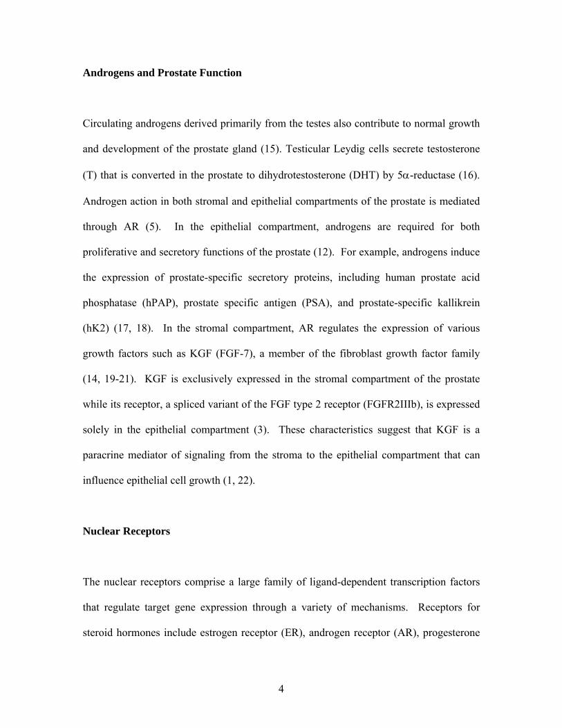
Structurally, nuclear receptors are modular proteins composed of an amino terminal
domain (NTD), a central DNA binding domain (DBD), and a carboxyl terminal ligand
binding domain (LBD) (Figure 2). Both the DBD and LBD are highly conserved while
the NTD is more variable. Both the NTD and LDB contain activation function domains,
AF-1 and AF-2 respectively, which potently activate transcription of nuclear receptor
target genes when expressed. However, AF-1 activates nuclear receptor-mediated gene
expression independent of ligand whereas AF-2 activation requires ligand (26).
Once in the nucleus, ligand-bound nuclear receptors are recruited to target gene
promoters either through direct binding to HREs or association with other promoter-
bound transcription factors to positively or negatively affect gene expression (27). For
example, prostate specific antigen (PSA) and phosphoenolpyruvate carboxykinase
(PEPCK) are both positively regulated by AR and GR, respectively (28, 29).
Alternatively, GR interacts with chromatin-associated NF-κB as well as AP-1
transcription factors, repressing their transcriptional activity in a ligand dependent
manner, termed transrepression. Initially, it was hypothesized that GR binds NF-κΒ,
occluding other protein interactions or recruiting histone deacetylase (HDAC) complexes,
thus preventing NF-ΚB-mediated gene expression. Additionally, in A549 cells, GR
inhibits NF-κB-mediated gene expression by preventing phosphorylation of serine 2 of
the C terminal domain of pol II, thereby preventing pol II-mediated transcription (30).
Along with binding other transcription factors, ligand-bound nuclear receptors promote
the recruitment of coactivator complexes that stimulate gene expression by remodeling
6
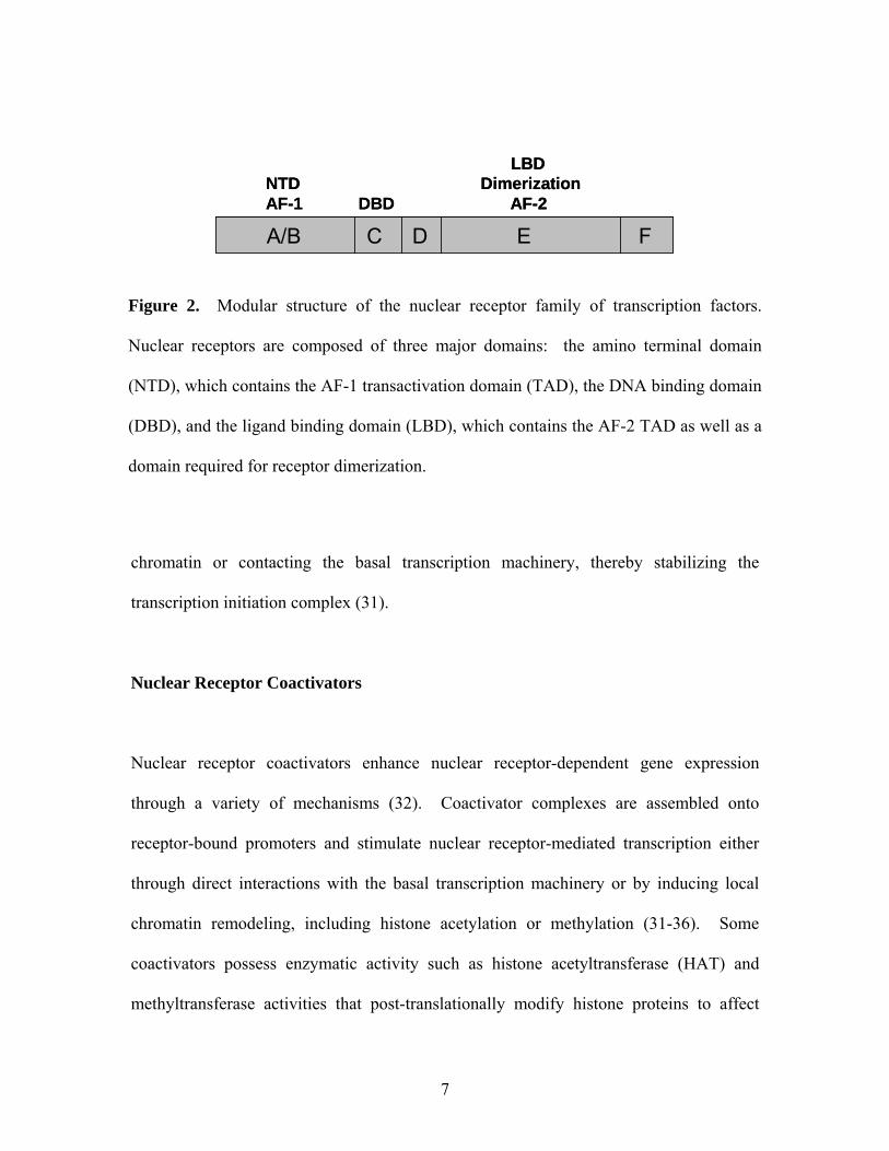
A/B C D E F
LBDNTD DimerizationAF-1 DBD AF-2
A/B C D E F A/B C D E F
LBDNTD DimerizationAF-1 DBD AF-2
Figure 2. Modular structure of the nuclear receptor family of transcription factors.
Nuclear receptors are composed of three major domains: the amino terminal domain
(NTD), which contains the AF-1 transactivation domain (TAD), the DNA binding domain
(DBD), and the ligand binding domain (LBD), which contains the AF-2 TAD as well as a
domain required for receptor dimerization.
chromatin or contacting the basal transcription machinery, thereby stabilizing the
transcription initiation complex (31).
Nuclear Receptor Coactivators
Nuclear receptor coactivators enhance nuclear receptor-dependent gene expression
through a variety of mechanisms (32). Coactivator complexes are assembled onto
receptor-bound promoters and stimulate nuclear receptor-mediated transcription either
through direct interactions with the basal transcription machinery or by inducing local
chromatin remodeling, including histone acetylation or methylation (31-36). Some
coactivators possess enzymatic activity such as histone acetyltransferase (HAT) and
methyltransferase activities that post-translationally modify histone proteins to affect
7
chromatin structure, while others that lack such activities function to recruit chromatin
modifying enzymes to active promoters.
Once a potential coactivator has been isolated, it must fulfill three main criteria to be
defined as a “classical” nuclear receptor coactivator. For example, coactivators must
enhance nuclear receptor-mediated gene expression without altering the basal expression
of a target gene. Furthermore, coactivators must reverse squelching effects observed by
the addition of other, competing nuclear receptors. Finally, coactivators must contain
intrinsic activation domains, capable of activating target gene expression when fused to a
DNA binding domain. Although these criteria were originally used to differentiate
coactivators from other nuclear receptor-interacting proteins, this definition may be too
limiting as it ignores other potentially novel contributions of nuclear receptor interacting
proteins to transactivation.
Most coactivators interact with the ligand binding domain of a variety of nuclear
receptors including GR and AR via nuclear receptor (NR) boxes such as LXXLL motifs
(37, 38). However, differences in binding affinities between nuclear receptors and their
coactivators may play a role in determining the specificity of hormonal responses. For
example, AR can bind glucocorticoid receptor interacting protein (GRIP/TIF-2/SRC-2)
with a higher affinity then steroid receptor coactivator-1 (SRC-1) (39). Although both
AR and GR interact with similar coactivators, it is postulated that unique coactivator
complexes may be responsible for specific cellular and gene responses to each hormone.
Furthermore, each nuclear receptor associates with specific coactivator complexes at the
8
same promoter (40). For example, using ChIP analysis of the mouse mammary tumor
virus (MMTV) promoter, which contains a common hormone response element capable
of binding AR, GR, or the progesterone receptor (PR), Li et al. isolated a specific
coactivator complex associated with PR that differed from the coactivator complex that
bound to GR (14). Specifically, SRC-1, RAC3, CBP, and p300 interacted with PR
whereas TIF-2, RAC3, pCAF, and p300 interacted with GR. Additionally, these
alternative coactivator complexes resulted in alternative posttranslational modifications
of histones. Histone 4 is acetylated at lysine 5 after progesterone stimulation, and histone
3 is phosphorylated at serine 10 as well as acetylated at lysine 14 upon glucocorticoid
treatment (40).
While many coactivators are likely to be redundant in their effects on nuclear receptor
function in cultured cell lines, recent in vivo studies suggest that different physiological
outcomes may result from variations in coactivator expression (35-37). For example,
TIF-2 and RAC3 null mice display reduced reproductive capability (41, 42). TIF-2 null
male mice have defective spermiogenesis and display testicular degeneration, whereas
females display poor placental development, resulting in embryonic growth retardation
(41). RAC3 -/- mice have decreased mammary gland growth, ovulatory capacity, and
litter size (42). Alternatively, whereas SRC-1 null mice are fertile, they exhibit partial
hormone resistance (43). Specifically, SRC-1 -/- mice have decreased growth and
development of the uterus, mammary gland, prostate, and testes in response to steroid
hormones (43).
9
Recently, many coactivators have been identified and classified into sub-families
consisting of similar members. For example, the p160 family of coactivators, designated
nuclear receptor coactivators (SRCs) consists of SRC-1 (or NcoA1), SRC-2 (or TIF-2,
GRIP1), and SRC-3 (or p/CIP, RAC3, ACTR, or AIB1) (24-30). These coactivators
share conserved sequence regions termed nuclear receptor interaction domains that
permit interactions with a broad range of nuclear receptors. Other coactivators that are
distinct from the p160 family are components of large complexes, such as the vitamin D
interacting proteins (DRIPs) and thyroid receptor associated proteins (TRAPs) (31, 32).
Finally, many other proteins have been identified as androgen receptor activators (ARAs)
that may utilize unique mechanisms to impact receptor transactivation that remain largely
undefined (33, 34). For example, ARA70, an AR-specific coactivator, may play a role in
uncovering the agonist activity of certain anti-androgens to activate AR activity (44).
Furthermore, a LIM domain containing protein that associates with focal adhesions,
hydrogen peroxide-inducible clone-5 (Hic-5/ARA55) was identified as a nuclear receptor
coactivator (45, 46).
LIM Domain-Containing Proteins
LIM domains are cysteine-rich motifs that may provide critical surfaces for protein-
protein interactions (47). LIM domain proteins are grouped into three broad families
based on their subcellular localization and function (48). Group I LIM proteins such as
LIM only 2 (LMO) are nuclear proteins that influence gene expression by interacting
with transcription factors (49). Group II LIM proteins are localized primarily in the
10
cytoplasm and function in the organization of the actin cytoskeleton (50). Finally, group
III LIM proteins were initially found to localize to the cytoplasm often associated with
focal adhesion complexes, but recently, our lab as well as others have revealed distinct
pools of group III LIM domain proteins including Hic-5/ARA55, paxillin, and Trip6 in
the nucleus (51-53).
A variety of LIM domain-containing proteins have been described that either positively
or negatively influence gene expression (54). For example, thyroid interacting protein
partner (Trip6), a group III LIM domain protein, is able to both enhance and repress AP-1
and NF-κΒ-regulated promoters by assembling different complexes in response to
differential cellular signals (52). Upon TPA or TNF-α treatment, Trip6 is recruited to the
collagen I or interleukin-8 (ColI or IL-8) promoters, enhancing AP-1 and NF-κB-
mediated gene expression respectively. However, upon glucocorticoid treatment, Trip6
represses AP-1 and NF-κB action by tethering GR to specific promoters (52). This
indicates that specific signaling events can determine if Trip6 activates or represses
specific gene expression. Additionally, Four and a half LIM-only protein (FHL2) is also
a LIM-domain containing protein that can both enhance or repress gene expression.
FHL2 was initially identified as an AR-specific coactivator whose expression in the
prostate overlaps that of AR and was later reported to influence β-catenin-mediated gene
expression (55, 56). The mechanism by which FHL2 coactivates β-catenin-mediated
transcription is due in part to a physical interaction with CBP/p300, resulting in increased
acetylation of β-catenin by CBP/p300 (55). Acetylation of β-catenin may enhance its
interaction with TCF4, thereby increasing β-catenin-mediated gene expression (57).
11
Interestingly, FHL2 represses β-catenin-mediated gene expression in muscle cells,
resulting in myogenic differentiation (58).
As mentioned previously, group III LIM domain containing proteins, including Hic-
5/ARA55, are predominantly localized at focal adhesions. However, some group III LIM
domain proteins such as zyxin, lipoma preferred partner (LPP), Trip6, paxillin, and Hic-
5/ARA55 can also be found in the nucleus (45, 51, 52, 59, 60). In fact, distinct pools of
Hic-5/ARA55 were found in the nucleus associated with the nuclear matrix (45).
Although many LIM domain containing proteins have been detected in the cytoplasm and
at focal adhesions and their nuclear export sequences have been identified, the precise
signals that induce their translocation are mostly unknown (reviewed in (54). For
example, both LPP and Trip6 are detected in the nucleus after LMB treatment, indicating
residual shuttling between the compartments (60, 61). However, FHL2 translocates to
the nucleus in response to activation of Rho, a small GTPase, while mechanical forces
applied to vascular smooth muscle cells lead to nuclear accumulation of zyxin (62, 63).
Furthermore, it has been reported that nuclear accumulation of Hic-5/ARA55 occurs in
response to oxidants such as H2O2 (64). However, in A1-2 cells, increased nuclear
receptor-mediated gene expression in the presence of both ligand and H2O2 not only
resulted in reduced nuclear receptor-mediated gene expression (unpublished results).
Subsequent analysis of GR and Hic-5/ARA55 promoter recruitment to the MMTV
promoter in the presence of dexamethasone (Dex) and H2O2 by ChIP analysis revealed
less GR and Hic-5/ARA55 promoter localization in the presence of H2O2 (unpublished
results).
12
In addition to nuclear receptors, Hic-5/ARA55 functions as a coactivator for Sp1,
enhancing p21 expression (65). In this case, Hic-5/ARA55, forced into the nucleus with
an added heterologous nuclear localization signal (NLS), influences p21 expression by
interacting directly with Smad3 and indirectly with Sp1 and p300 (65). Although a
direct interaction between Sp1 and Hic-5/ARA55 has not been detected, Hic-5/ARA55
has been found to associate with Smad3. Also, a LIM 4 deletion mutant of Hic-5/ARA55
interfered with the coactivation properties of p300, suggesting a functional interaction
between these coactivators.
Along with AR and GR, Hic-5/ARA55 serves as a coactivator for PPARγ (66).
Specifically, Hic-5/ARA55 enhances PPARγ-mediated induction of genes such as keratin
20, a cytokeratin that is expressed in mature intestinal epithelium as well as
uroepithelium, L-FABP, a fatty acid binding protein found in differentiated intestinal
epithelium, and KLF, a zinc finger transcription factor that is also expressed in intestinal
epithelial cells (67-69). Furthermore, siRNA ablation of Hic-5/ARA55 expression
reduced the expression of PPARγ-regulated genes. Finally, a role for Hic-5/ARA55 in
epithelial differentiation induced by PPARγ was reported. Forced expression of Hic-
5/ARA55 in preadipocytes not only inhibited adipocyte differentiation but also induced
epithelial gene expression in these mesenchymal cells (66).
Hic-5/ARA55 was isolated by yeast two hybrid analyses as a nuclear receptor
coactivator, specifically binding the τ2 transactivation domain of GR located in its hinge
13
region (45). However, the Hic-5/ARA55 interacting domain for AR has only been
broadly localized to its ligand binding domain (46). Although the association of Hic-
5/ARA55 with AR and GR has been well established, the nature of their interaction and
specifically in which compartment it occurs and how it affects nuclear-receptor-mediated
gene expression is not clear. Furthermore, redistribution of Hic-5/ARA55 to the nucleus
in response to hormone treatment has not been detected (unpublished results).
Nuclear Receptor Corepressors
Along with coactivators, another set of nuclear receptor interacting proteins has recently
been identified termed corepressors, including both nuclear receptor corepressor (NCoR)
and silencing mediator or retinoid and thyroid receptors (SMRT) (70, 71). Corepressors
repress nuclear receptor-mediated gene expression in the absence of ligand or presence of
antagonist by interacting with histone deacetylases (HDACs) that in turn modify
chromatin by removing acetyl groups on histone tails thereby promoting a closed
chromatin structure, repressing transcription (24).
Individually, coactivators induce and corepressors repress nuclear receptor-mediated
gene expression (Figure 3). However, most cells express a combination of both
corepressors and coactivators that also interact with each other. For example, RAC3
interacts with NCoR, modifying thyroid hormone receptor (TR) regulated transcription
(72). An equilibrium model hypothesizes that it is not the absolute amount of coregulator
expression but the ratio of corepressors versus coactivators that determines the extent of
14
nuclear receptor-mediated gene expression (73). For example, overexpression of SMRT
antagonizes TIF-2 coactivation of GR-mediated gene expression (74). Because
coactivators and corepressors, collectively referred to as coregulators, alter nuclear
receptor-mediated gene expression in endocrine target tissues, their activity and
expression along with various nuclear receptors, including AR and ER, in prostate as well
as breast cancer has been an area of intense investigation.
SWI/SNFCBP
NRNR
p160 p300
Target Genes ON
HDAC
NCoRSMRT
Target Genes OFF
+ Hormone
- Hormone
SWI/SNFCBP
NRNR
p160 p300
Target Genes ON
SWI/SNFCBP
NRNR
p160 p300
Target Genes ON
HDAC
NCoRSMRT
Target Genes OFF
HDAC
NCoRSMRT
Target Genes OFF
+ Hormone
- Hormone
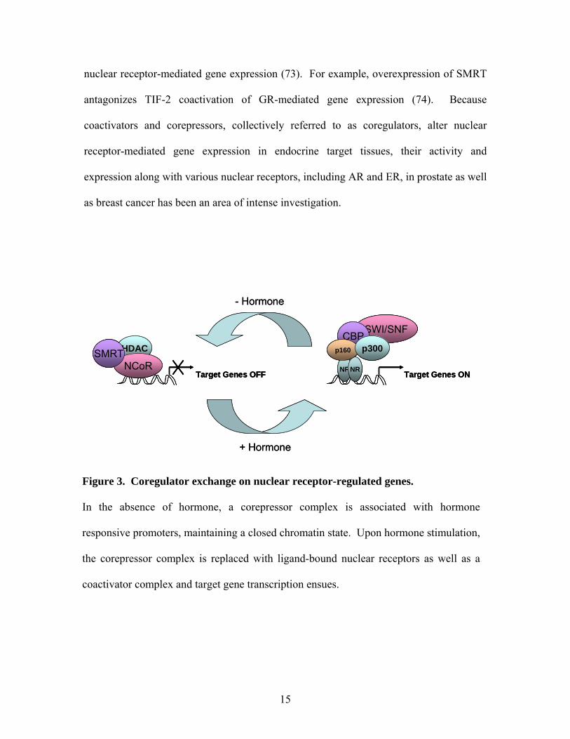
Figure 3. Coregulator exchange on nuclear receptor-regulated genes.
In the absence of hormone, a corepressor complex is associated with hormone
responsive promoters, maintaining a closed chromatin state. Upon hormone stimulation,
the corepressor complex is replaced with ligand-bound nuclear receptors as well as a
coactivator complex and target gene transcription ensues.
15
AR and Prostate Tumor Development
Changes in androgen function are linked to the development and progression of prostate
cancer (PCa). In fact, androgen-regulated gene products, such as PSA, are used as a
marker of tumor progression and responsiveness to therapy (75, 76). As previously
mentioned PSA is produced by the epithelial cells and secreted into the seminal fluid.
Normally, the basement membrane acts as a barrier to block PSA from escaping into
systemic circulation. However, disruption of the basement membrane in PCa, results in a
significant increase in serum PSA.
Due to the androgenic dependence of prostate growth and survival, surgical and chemical
castration has been used for more than sixty years as treatment for PCa. Huggins et al.
reported that castration significantly reduced levels of prostate acid phosphatase, an
androgen-regulated gene product, also used as a tumor marker in patients with PCa,
indicating the androgenic dependence of these tumors (77). Since then, androgen
ablation therapy including, orchiectomy and flutamide or bicalutamide anti-androgen
treatment, which interfere with androgen binding to AR, has been the primary treatment
for metastatic PCa (78, 79). Androgen ablation therapy leads to temporary remission of
most PCa, but most tumors eventually regress into an androgen independent or hormone
refractory tissue, capable of metastasis (80).
16
Reactive Stroma
Although the epithelia compartment is the source of tumor cells, growth factor and
cytokine secretion from both compartments of the prostate is altered in tumor
development. In fact, development of an altered stromal microenvironment, termed
reactive stroma, carcinoma associated fibroblasts (CAF) or tumor stroma, enhances tumor
cell survival, cellular proliferation, and migration (Figure 4, 81, 82). Reactive stroma is
characterized by the presence of undifferentiated myofibroblast cells that express both
smooth muscle cell and fibroblasts markers (83). Myofibroblasts are commonly found at
areas undergoing tissue remodeling, producing ECM such as collagen I and fibronectin as
well as proteases, including matrix metalloproteinases (MMPs) (84-86). Although the
process by which normal stroma converts to reactive stroma phenotype is unknown,
differential epithelial-stromal interactions during carcinogenesis may result in the
inability of neoplastic epithelium to maintain a differentiated stromal compartment (87).
Furthermore, TGF-β may be involved in the progression to a reactive stroma phenotype
by inducing myofibroblast formation (83).
Data indicate that normal stroma cannot support tumor growth. Using the Dunning rat
prostate andenocarcinoma model, Hayashi and Cunha grafted fragments of tumorigenic
tissue into normal mesenchyme located under the renal capsule (11). After one month,
not only did the tumor tissue’s growth rate decrease, but the cells also adopted the normal
morphology of tall columnar secretory epithelium. Alternatively, reactive stroma can
17
induce initiated, non-tumorigenic epithelial cells to form prostate tumors whereas normal
stromal cells failed to promote tumor formation (88).
To date, the exact mechanism by which reactive stroma influences the development of
prostate tumors is unclear. However, data indicate altered gene expression of growth
factors such as KGF, FGF-2, TGF-β, and IL-6, matrix metalloproteinases (MMPs), and
extracellular matrix in reactive stromal cells (22, 89-92). The differential gene
expression pattern induced by the reactive stroma may confer resistance to cell cycle
inhibitors or metastatic properties to neighboring cells.
Extracellular Matrix Remodeling in PCa
Along with changes in reactive stromal-associated cells, alterations in the composition of
the ECM associated with prostate tumors occur also. For example, myofibroblasts
produce ECM such as collagen I and fibronectin, resulting in increased expression of
these ECM components in reactive stromal microenvironment (83, 85, 86, 93). In
addition to differential ECM production, myofibroblasts also synthesize various
proteinases that aid in ECM remodeling within reactive stroma (84).
ECM remodeling in PCa may disrupt cell adhesion as well as promote cellular invasion.
For example, addition of fibronectin, which mediates cell adhesion, induced invasion of
DU145 prostate cancer cells through basement membranes (94). Also, a MMP inhibitor,
18
A-177430, reduced tumor growth, angiogenesis, and metastasis, indicating that ECM
remodeling may be necessary for tumor progression (95).
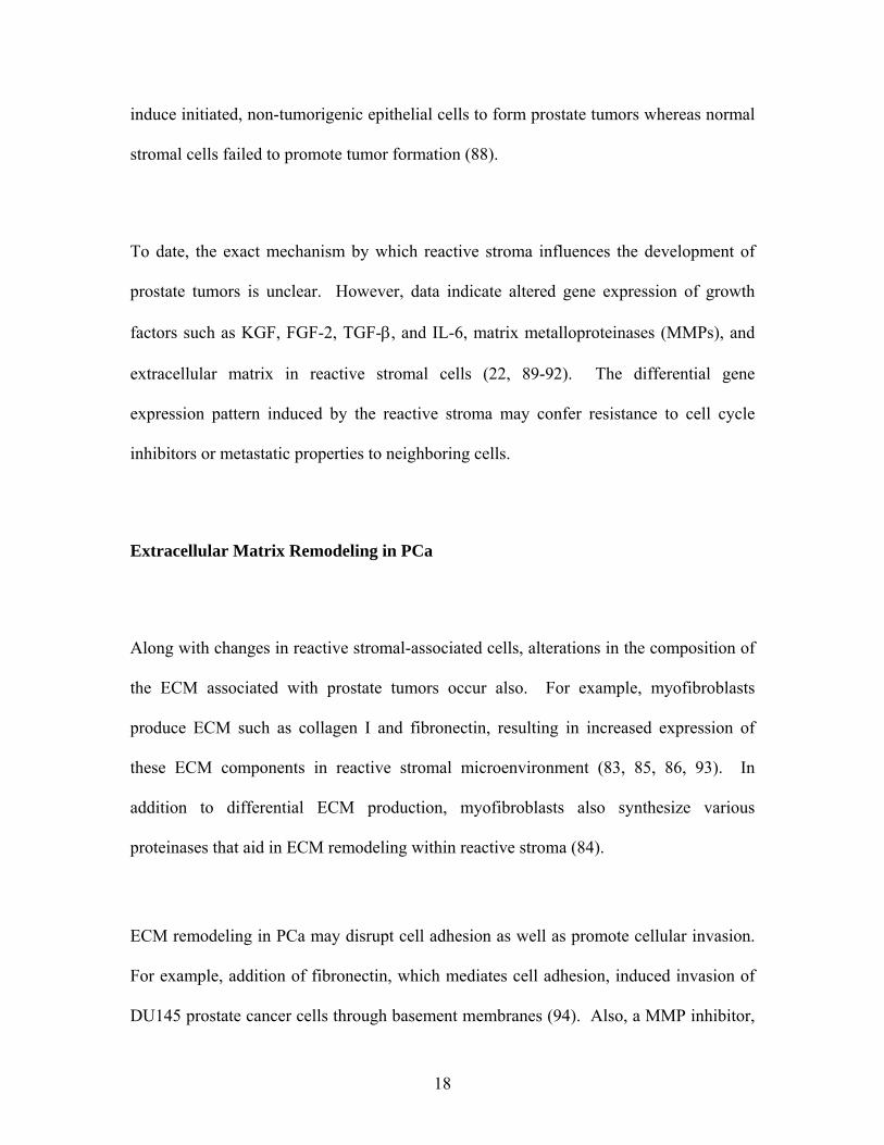
BasementMembrane
Myofibroblasts
TAMCarcinoma Cells
BasementMembrane
Myofibroblasts
TAMCarcinoma Cells
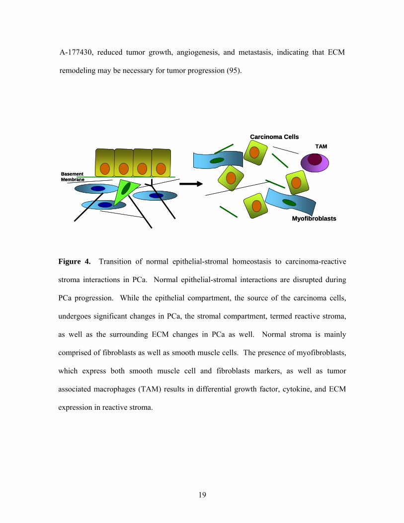
Figure 4. Transition of normal epithelial-stromal homeostasis to carcinoma-reactive
stroma interactions in PCa. Normal epithelial-stromal interactions are disrupted during
PCa progression. While the epithelial compartment, the source of the carcinoma cells,
undergoes significant changes in PCa, the stromal compartment, termed reactive stroma,
as well as the surrounding ECM changes in PCa as well. Normal stroma is mainly
comprised of fibroblasts as well as smooth muscle cells. The presence of myofibroblasts,
which express both smooth muscle cell and fibroblasts markers, as well as tumor
associated macrophages (TAM) results in differential growth factor, cytokine, and ECM
expression in reactive stroma.
19
Hormone Refractory Prostate Cancer
Despite the initial remission of most PCa upon androgen ablation therapy, most tumors
eventually regress into an androgen independent or hormone refractory tissue, capable of
metastasis (80). The mechanism by which PCa develops into an androgen independent
state is not known. However, AR expression is maintained in androgen independent
tissue and may play a role in continued prostate survival during the androgen-
independent state (96). For example, disrupting AR activity by addition of an AR
binding antibody or an AR mRNA hammerhead ribozyme effectively inhibited
proliferation of androgen-independent prostate cancer cells (97).
Although the mechanisms by which AR is activated post androgen ablation therapy in
androgen independent prostate cancer is unclear, several theories have emerged (1). The
prostate may develop a capacity to increase its sensitivity to low levels of androgen
through amplification of AR gene expression, increased AR stability, or enhanced AR
nuclear localization (98, 99). For example, elevated AR expression may be necessary for
PCa progression to an androgen-independent state (100). Furthermore, increased AR
expression correlates with a higher probability of recurrence (101). (2) In some cases, the
AR is mutated within its LBD, resulting in its activation by other steroid hormones, such
as estrogen, progesterone, and glucocorticoids (102). However, the frequency of this
mutation in patients is rather low (103). (3) Altered secretion of autocrine or paracrine
factors may account for prostate cellular proliferation and AR activity in the absence of
androgens. Altered growth factor secretion has not only been implicated in the reactive
20
stroma phenotype and subsequent tumor support, but also has been reported to increase
AR activity in a ligand independent manner, promoting androgen independent prostate
cancer growth. For example, IGF-1, EGF, KGF and IL-6 treatment of LNCaP cells
induced PSA expression by AR in the absence of androgen (104, 105). (4) Altered
expression of coactivators may enhance ligand-independent AR activity or increase
androgen responsiveness in androgen independent prostate cancer. Furthermore,
transient overexpression of TIF-2 in CV-1 cells enhanced AR-mediated MMTV reporter
activity in response to estradiol as well as progesterone (106).
AIB 1 (amplified in breast 1)/ACTR/SRC-3/RAC3 is a nuclear receptor coactivator
whose expression is amplified in 10% of primary breast tumor biopsies (107). Many
investigators have subsequently examined whether altered expression, activity, or
mutation of other nuclear receptor coactivators are associated with tumors of different
stages (107-109). Analysis of coactivator expression in prostate cancer has yielded
variable results. For example, Gregory et al. report increased SRC-1 and TIF-2
expression in prostate tumors whereas Fujimoto et al. report similar SRC-1 expression in
benign, intermediate, and high grade cancers (106, 110). Interestingly, Hic-5/ARA55
expression was reduced in tumor as compared to normal tissue and Hic-5/ARA55 mRNA
levels were lower in hormone-refractory PCa than that of previously untreated PCa
(111,112)). However, the effect of decreased Hic-5/ARA55 expression on AR-mediated
gene expression as well as PCa progression is not known.
21
SPECIFIC AIMS AND RATIONALE
Aim 1. Determining the mechanism by which Hic-5/ARA55 functions as a
coactivator for nuclear receptors.
Because Hic-5/ARA55 does not possess an obvious catalytic domain that is responsible
for its coactivation properties, its mechanism of coregulator function has remained
largely undefined. At focal adhesion complexes, Hic-5/ARA55 serves as an adapter
protein, coordinating multiple protein-protein interactions. Therefore, it may serve as an
adaptor molecule, either recruiting or stabilizing promoter-specific protein complexes by
interacting with multiple coactivator proteins, coordinating their association with nuclear
receptors at hormone responsive promoters.
The overall goal of this aim was to determine if Hic-5/ARA55 interacts with nuclear
receptors as well as other coregulators in a chromatin context. Interaction studies
preformed thus far have used yeast two hybrid analyses and in vitro pull down assays (45,
46). Here, we attempted to isolate GR and Hic-5/ARA55 in association with a known
GR-responsive promoter, the mouse mammary tumor virus (MMTV) as well as
endogenous p21 and c-fos gene promoters, using chromatin immunoprecipitation (ChIP)
analysis (Figure 5). Furthermore, the requirement of Hic-5/ARA55 for GR activity and
promoter recruitment of coactivators was analyzed using RNA interference (RNAi)
analysis. The results from this aim indicate that Hic-5/ARA55 is associated to hormone-
responsive promoters in the absence and presence of glucocorticoids along with other
22
well-characterized coregulator proteins. Furthermore, reduced expression of Hic-
5/ARA55 results in altered coactivator recruitment to target promoters.
Aim 2. Determining Hic-5/ARA55 expression and function in the prostate.
In the prostate, AR activity appears critical for tumor development and/or progression
(96, 97, 106). Because Hic-5/ARA55 potentiates androgen receptor (AR)-mediated gene
expression, its expression patterns in prostate cancer cell lines and tissue has been
examined. In normal prostates, Hic-5/ARA55 expression was restricted to the stromal
compartment, and Hic-5/ARA55 mRNA levels were lower in hormone-refractory PCa
than that of previously untreated PCa (111). Because most analyses of coactivator
expression in PCa progression use quantitative PCR, it is difficult to ascertain whether
quantitative PCR results of Hic-5/ARA55 expression represents reduced expression of
Hic-5/ARA55 within individual cells or alterations in the balance between
stromal/epithelial cell content in tumors.
The overall goal of this aim was to determine Hic-5/ARA55 expression levels in normal
and tumor-derived prostates along with examining Hic-5/ARA55 coactivator function in
the prostate. Using immunohistochemistry analysis, Hic-5/ARA55 expression was found
to be confined primarily to the stromal compartment in the prostate. I therefore
employed a prostate stromal cell line, WPMY-1 cells, and determined that Hic-5/ARA55
was recruited along with AR to the androgen-responsive KGF promoter. Furthermore,
the responsiveness of KGF to androgen stimulation was lost after Hic-5/ARA55
23
expression was partially silenced using RNAi. These results indicate that Hic-5/ARA55
serves as a stromal-specific AR coactivator in the prostate.
Aim 3. Determining the effects of extracellular matrix signaling on GR-mediated
transcription.
Integrins, a family of membrane receptors located at focal adhesion complexes, bind
specific ECM components, activating intracellular signaling cascades that influence cell
migration, proliferation, and apoptosis. Furthermore, ECM also influences nuclear
receptor activity, resulting in differential cell growth and/or gene expression. Malignant
mammary cells grown on a laminin-rich basement membrane displayed greater ERα
activity and express higher levels of ERα (6). Although cross-talk between ER and ECM
signaling has been reported, the mechanism by which ECM influences nuclear receptor-
mediated gene expression is unclear (6).
Because Hic-5/ARA55 modulates focal adhesion complex signaling as well as
functioning as a nuclear receptor coactivator, I analyzed the role of Hic-5/ARA55 in
mediating GR responses to various ECM. In this aim, I first determined the effects of
ECM signaling on GR-mediated gene expression. Matrigel decreased GR activity by
decreasing GR protein levels. Alternatively, fibronectin enhanced GR-mediated gene
expression without effecting GR and Hic-5/ARA55 expression, subcellular localization,
and DNA binding. These results indicate that ECM does influence GR activity although
the exact mechanisms by which those signals are transmitted are unclear.
24
Hormone TreatmentCrosslinking Immunoprecipitation Purify IC and
Reverse X-link
Protease DigestionAnd Purify DNA
PCR with promoter-specific primers
Control Dex
IgG
GR
Input
Hormone TreatmentCrosslinking Immunoprecipitation Purify IC and
Reverse X-link
Protease DigestionAnd Purify DNA
PCR with promoter-specific primers
Control Dex
IgG
GR
Input
Figure 5. Chromatin Immunoprecipitation (ChIP) Assay. After the cells are
stimulated for 1 hour with hormone to allow nuclear receptor translocation to the
nucleus and promoter recruitment, the cells are crosslinked with formaldehyde to
preserve nuclear receptor-DNA interactions. After shearing the chromatin to 500-
2000 base pair fragments, the resulting chromatin is immunoprecipitated using nuclear
receptor specific antibodies as well as non-specific antibody controls (IgG). After the
immune complex is isolated using protein A and G sepharose, the crosslinks are
reversed and the proteins are removed by protease digestion. The DNA is purified and
subjected to PCR analysis, using promoter-specific primers.
25
CHAPTER 2 MATERIALS AND METHODS
Antibodies, Plasmids, and Reagents – Antibodies used in this study included: anti-GR
(Affinity Bioreagents, Inc., Golden, CO); anti-Hic-5/ARA55 (BD Transduction Labs, Los
Angeles, CA); anti-Glyceraldehyde 3-phosphate dehydrogenase (GAPDH CSA-335
Stressgen Victoria, British Columbia, Canada); anti-NCoR (06-892 Upstate,
Charlottesville, VA); anti-GR (H-300); anti-AR (N-20 ); anti-p300 (C-20); anti-SRC-1
(M 341); anti-TIF-2 (M 343) (all from Santa Cruz Biotechnology, Santa Cruz, CA); anti-
phospho AKT; anti-AKT; anti-phosphoERK; anti-ERK (all from Cell Signaling
Technology, Beverly, MA). Plasmids used in this study included: an androgen
responsive MMTV-luciferase reporter, pLC-Luc, as well as the Renilla-luciferase control.
Cell signaling inhibitors used in this study include: U0126 and LY294002 (both from
CalBiochem, SanDiego, CA)
Cell Culture and Transient Transfection- The A1-2 cells were derived from the T47D
human mammary carcinoma cells line by stable transfection with pGRneo and MMTV-
LTR-luc plasmids as previously described (113). Thus, these cells contain exogenous
copies of stably integrated rat GR and a glucocorticoid-responsive luciferase reporter.
The A1-2 cells were routinely maintained in modified Eagle’s medium at 37 C under 5%
CO2. The media was supplemented with 100 μg/ml penicillin-streptomycin, 10% FBS,
10 mM HEPES, 2 mM glutamine, and 160 μg/ml G418. WPMY-1 cells were routinely
maintained in Dulbecco’s Modified Eagle’s Medium (Gibco, Grand Island, NY)
supplemented with 100 μg/ml penicillin-streptomycin and 5% fetal bovine serum (FBS)
26
at 37 C under 5% CO2. The WPMY-1 cells were isolated from a normal human prostate
specimen and immortalized using SV40 large-T-antigen (114). A1-2 cells were seeded
on 12-well cell culture dishes at a density of 1.5 × 10⁵ cells/ well for 24 h prior to
transfection. WPMY-1 cells were seeded on 12-well cell culture dishes at a density of
7.5 × 10⁴ cells/ well for 24 h prior to transfection. Transfections were performed using
Opti-MEM (Gibco, Grand Island, NY) and Lipofectamine transfection reagent
(Invitrogen Technology, Carlsbad, CA) according to manufacturer’s instructions. After 3
h, fresh medium was added to the cells and hormone treatments were initiated where
relevant.
Extracellular matrix application to tissue culture plates – Cell culture plates were first
coated with nitrocellulose that was dissolved in methanol (115). After the nitrocellulose
was applied, various ECM was diluted in PBS and incubated at room temperature for 1
hour. The ECM examined included: vitronectin (Gibco BRL Life Technology, Carlsbad,
CA), fibronectin (BD Biosciences, Bedford, MA), laminin mix (kind gift from Marcia
Lewis, University of Pittsburgh), and growth factor reduced matrigel (BD Biosciences,
Bedford, MA). After the plates were washed with PBS, the A1-2 cells were plated.
Western Blot Analysis- Cell lysates were collected in RIPA buffer (10 mM Tris, pH 8, 1
mM EDTA, 0.5 mM EGTA, 140 mM NaCl, 1% Triton X-100, 0.1% Deoxycholic acid,
0.1% SDS, 1 mM PMSF, and protease inhibitors) and were boiled in Sample Buffer (62.5
mM Tris, pH 6.8, 2% SDS, 10% Glycerol, 5% 2-Mercaptoethanol, 0.001% Bromophenol
Blue) for 5 min. Proteins were then separated on 7.5% SDS-PAGE and transferred to
27
PVDF transfer membrane (Bio-Rad, Hercules, CA) in transfer buffer (20% methanol, 48
mM Tris, 39 mM glycine, and 1.3 mM SDS) at 15 V for 30 min. Membranes were then
incubated in blocking buffer (5% dry milk in Tris-buffered saline, pH 7.4) for 2 h to
overnight. Next, the membranes were incubated with the indicated antibodies diluted
1:1000 in blocking solution for 2 h at room temperature. After extensive washing, the
membranes were then probed with horseradish peroxidase-conjugated goat anti-mouse
IgG antibodies (Santa Cruz Biotechnology, Santa Cruz, CA) diluted in blocking solution
for 1 h. Finally, the membranes were washed and developed using the Renaissance
Western Bolt Chemiluminescence Reagent (PerkinElmer, Wellesley, MA) according to
the manufacturer’s instructions. The membranes were stripped with Re-Blot Plus Strong
Solution following the manufacturer’s instructions (Chemicon International, Temecula,
CA) and reprobed for GAPDH as a loading control. Where indicated, quantification of
scanned images was performed using the NIH Image software.
RNAi- RNAi plasmids were constructed following the manufacturer’s guidelines
(pSilencer 2.1-U6 hygro, Ambion, Austin, TX). The DNA template sequences for human
Hic-5/ARA55 were RNAi-A 5’-GGAGGACCAGTCTGAAGAT-3’, RNAi-B 5’-
GAAAAGACCCAGCCTCCCT-3’, RNAi-C 5’-GCATCACAGATGAAATCAT-3’and
RNAi-D 5’-GTGGATTGCACACAGACAA-3’. The oligos (2μg each) were annealed to
form double-stranded DNA after incubation in DNA annealing solution (pSilencer kit)
for 3 minutes at 90 C followed by 1 hour at 37 C. The resulting double stranded DNA
was then inserted into the pSilencer 2.1-U6 hygro plasmid using T4 DNA ligase (New
England Biolabs, Ipswich, MA) for 4 hours at 25 C. Positive clones were identified after
28
enzymatic digestion by HindIII and BamHI (New England Biolabs, Ipswich, MA),
releasing the 65 base pair insert. The GFP template sequence was used as a positive
control during the plasmid construction and a negative control for transfection analyses.
Luciferase Assays - Luciferase activity in cell-free lysates was measured using a Victor
3 1420 Multilabel plate reader (PerkinElmer, Wellesley, MA). Cells were washed with
PBS and lysed in Reporter Lysis Buffer (Promega, Madison, WI) followed by a freeze
and thaw incubation to ensure proper cell lysis. The lysate (25 μl) was incubated with
luciferase assay reagent followed by a 10 sec relative luciferase unit measurement.
Luciferase activity was normalized to Renilla luciferase activity according to
manufacturer’s instructions. All experiments were performed three or more times.
Chromatin Immunoprecipitation – A1-2 or WPMY-1 cells were grown on 15 cm
plates to 80-90% confluence in medium supplemented with charcoal dextran treated FBS
for 24 hours. The cells were treated with EtOH-vehicle, 100 nM Dex, or 10 nM
Mibilerone (Mib) for 1 h and crosslinked by addition of formaldehyde (1% final) to the
medium for 30 min at room temperature. After the crosslinking reaction was stopped by
addition of 0.1 volume of 1.4 M glycine, the cells were washed with PBS, scraped into
ice-cold PBS, and centrifuged at 3000 RPM for 5 minutes at 4 C. The cells were lysed
and the nuclei collected by resuspending the pellet in 2 ml of cold ChroIP Buffer (50mM
HEPES, KOH pH 8, 1mM EDTA, pH 8, 0.5mM EGTA, pH 8, 140mM NaCl, 10%
glycerol, 0.5% IGEPAL, 0.25% Triton X-100, and 1mM PMSF) for 10 minutes and
centrifuged at 3000 RPM for 5 minutes at 4 C. The pellet was resuspended in 2 ml of
29
cold Wash Buffer (10mM Tris-HCl, pH 8, 1mM EDTA, pH 8, 0.5mM EGTA, pH 8,
200mM NaCl, and 1mM PMSF) for 10 minutes at 4 C followed by centrifugation at 3000
RPM for 5 minutes at 4 C. The pellet was resuspended in 2 ml of ice-cold RIPA Buffer
(10mM Tris-HCl, pH 8, 1mM EDTA, pH 8, 0.5mM EGTA, pH 8, 140mM NaCl, 1%
Triton X-100, 0.1% Na-deoxycholate, 0.1% SDS, 1mM PMSF, and protease inhibitor
cocktail) for 10 minutes at 4 C followed by centrifugation at 3000 RPM for 5 minutes.
The isolated nuclei were then sonicated using a water bath sonicator for three minutes
(Misonix Sonicator with Cuphorn, Farmingdale, NY, Output at 6, Processing time 3
minutes, Pulse on 10 sec, Pulse off 15 sec) to produce fragments 500-2000 base pair in
size as previously described (30). Chromatin fragments were precleared for 1-4 h at 4C
using 50 μl of protein A and G sepharose containing salmon sperm DNA (Upstate, Lake
Placid, NY) to reduce nonspecific binding followed by centrifugation at 3000 RPM for 5
minutes at 4 C. Next, 1-10% of the sheared chromatin was removed to use as input
samples later. The chromatin fragments were diluted with RIPA buffer (1 ml/IP),
separated into 2 to 4 IPs, and immunoprecipitated with specific antibodies (1 μg/ IP)
overnight at 4 C. After immunoprecipitation, 30 μl of protein A and G sepharose
(Upstate, Lake Placid, NY) was added and the incubation was continued for 1 h. The IPs
were washed twice with RIPA buffer, twice with RIPA buffer +500 mM NaCl, and once
with LiCl buffer (10mM Tris-HCl, pH 8, 1mM EDTA, pH 8, 0.5mM EGTA, pH 8,
250mM LiCl, 1% Triton X-100, 1% Na-deoxycholate, and 1mM PMSF). The
precipitates were eluted with 100 μl of freshly made reverse cross-linking buffer (1%
SDS and 0.1 M NaHCO3), adding 4 μl of 5 M NaCl at 65 C for 4 h. The Input samples
were reversed by diluting 10μl Input into 190 μl ChIP Dilution Buffer (0.01% SDS 1.1%
30
Triton X-100, 1.2mM EDTA, 16.7mM Tris-HCl, pH8, and 167mM NaCl) adding 8 μl of
5M NaCl/tube also for 4 hr at 65 C. The proteins were removed by addition of proteinase
K (2 μl/sample, Roche, Mannheim, Germany, 20 mg/ml) for 1 h at 45 C. DNA was
isolated using the QIAquick PCR purification kit (Quiagen, Valencia, CA).
Thermocycling conditions involved an initial denaturation step at 95 C for 12 min
followed by 28-32 cycles at 95 C for 30 sec and 56 C for 30 sec and 72 C for 30 sec.
Primers for the MMTV promoter were: forward primer 5’-
GCGGTTCCCAGGGCTTAAGT-3’ and reverse primer 5’-
CCATTTTACCAACAGTACCG-3’. Primers for the c-fos promoter were: forward
primer 5’-TCCCAGCAGTCGAGGTATTC-3’ and reverse primer 5’-
GGTCAGTTCGGGATGACAAG-3’. Primers for the p21 promoter were: forward
primer 5’-GGTGTCTAGGTGCTCCAGGT-3’ and reverse primer 5’-
GCACTCTCCAGGAGGACACA-3’. Primers for the KGF promoter were: forward
primer 5’-CCCTTTCCCCTTCTAACTGC-3’ and reverse primer 5’-
ACCTTTGCTGACCTCATTGG-3’. Primers for the luciferase gene were: forward
primer 5’-CCAGGGATTTCAGTCGATGT-3’ and reverse primer 5’-
AATCTGACGCAGGCAGTTCT-3’. PCR products were resolved on a 12%
polyacrylamide gel and visualized with ethidium bromide. Semi-quantitation was done
using densitometric analysis of the resolved gels using the Kodak Imaging System. Data
points were subtracted for background and normalized to the Input data.
For sequential ChIPs, complexes immunoprecipitated with either anti-GR or anti-Hic-
5/ARA55 antibodies (3 μg/ IP) were eluted by incubation with 100 μl of 10 mM DTT for
31
30 min at 37 C. After centrifuging the eluted IPs for 10 minutes at 3000 RPM 4C, they
were diluted 1:50 in ChIP Dilution Buffer (20 mM Tris-HCl, pH 8.1, 2mM EDTA, 150
mM NaCl, 1% Triton X-100), followed by reimmunoprecipitation with either isotype
control Ab, anti-Hic-5, or various coactivators or corepressor antibodies (1 μg/ IP).
Reverse Transcriptase PCR - Total RNA was isolated from A1-2 and WPMY-1 cells
using the RNAqueous RNA isolation kit (Ambion, Austin, TX) following the
manufacturer’s instructions. For RT-PCR, 1 μg of RNA was incubated with 100 μl of
reaction mix containing 25 mM MgCl2, 25 mM dNTPs (PerkinElmer), 10X PCR II
Buffer (Gibco BRL), 40 U/μl RNAsin RNase inhibitor (Promega), 45 μM random
hexamers (IDT, Coralville, IA), 200 U/μl Superscript reverse transcriptase (Gibco), and
nuclease free water (Ambion). Parallel reactions were performed without reverse
transcriptase to control for the presence of contaminant DNA. The samples were
incubated at 25 C for 10 min, at 48 C for 30 min, and at 95 C for 5 min followed by 4 C
for 5 min to inactivate the reverse transcriptase.
For amplification, a PCR reaction containing a cDNA aliquot along with AmpliTaq Gold
DNA polymerase in a volume of 25 μl was used according to the manufacturer’s
instructions (Applied Biosystems, Foster City, CA). Primers used for gene expression
analysis were: c-fos: forward primer 5’-TGCCAACTTCATTCCCACGGT-3’ and
reverse primer 5’-TAGTTGGTCTGTCTCCGCTTG-3’; p21: forward primer 5’-
GCGACTGTGATGCGCTAATGG-3’ and reverse primer 5’-
TCCCAACTCATCCCGGCCTC-3’; KGF: forward primer 5’-
32
CAATCTAGAATTCACAGATAGGAGGAGGC -3’ and reverse primer 5’-
ACAATTCCAACTGCCACTGTCCTGATTTC-3; GAPDH: forward primer 5’-
CATCACCATCTTCCAGGAGCGAGA-3’ and reverse primer 5’-
GTCTTCTGGGTGGCAGTGATGG-3’. Thermocycling conditions involved an initial
denaturation step at 95 C for 12 min followed by 28-30 cycles at 95 C for 30 sec and 56
C for 30 sec and 72 C for 30 sec. Specific PCR amplification products were separated on
a 12% PAGE and detected by EtBr staining. Experiments were performed with
duplicates for each data point.
Immunofluorescence – WPMY-1 cells were cultured on glass coverslips treated with
EtOH-vehicle or 100 nM Leptomysin B (LC Laboratory, Woburn, MA) for 1 hour, fixed
with 4% paraformaldehyde in PBS for 30 minutes at 4°C, washed with PBS and
permeabilized in PBS containing 1% BSA and 0.5% Triton X-100 for 5 minutes at room
temperature. The cells were incubated overnight at 4 C with anti-Hic-5/ARA55 specific
antibodies diluted 1:1000 in PBS. After extensive washing with PBS, the cells were
incubated 2 hours at room temperature with anti-mouse IgG-Cy2 conjugated antibodies
(Molecular Probes, Portland, OR) diluted 1:300 in PBS. Nuclei were stained with DAPI
(10 μg/ml, Molecular Probes, Carlsbad, CA) according to manufacturer’s instructions.
Immunohistochemistry – Paraffin-embedded prostate tissue sections were obtained
from the Shadyside Hospital Tissue Bank. Tissue sections were deparaffinized,
rehydrated, and antigens retrieved by boiling in 10 mM citrate buffer, pH 5.5 for 10
minutes. The sections were incubated overnight at 4 C in a humid chamber with anti-
Hic-5/ARA55 antibodies diluted 1:2000 in PBS. Immunostaining was performed using a
33
Vecta stain kit (Vector Laboratories Inc., Burlingame, CA) following manufacturer’s
instructions. Normal mouse IgG was used as controls.
Statistics
Comparisons of two mean values were performed using a paired t test. p values of < 0.05
were taken to be significant and all data were analyzed using GraphPad Prism 3.00
(Graph Pad Software, San Diego, CA).
34
CHAPTER 3 Determining the mechanism by which Hic-5/ARA55 functions as a
coactivator for nuclear receptors.
HYPOTHESIS:
Because Hic-5/ARA serves as an adapter protein at focal adhesion complexes
coordinating multiple protein-protein interactions, it serves as nuclear receptor
coactivator by interacting with multiple coactivator proteins, coordinating their
association with nuclear receptors at hormone responsive promoters.
Introduction
Hic-5/ARA55 is associated primarily with focal adhesion complexes as well as localized
within the nucleus (53). At focal adhesion complexes, Hic-5/ARA55 as well as other
group III LIM domain proteins link various intracellular signaling modules to plasma
membrane receptors that respond to various extracellular signals including growth factors
and the extracellular matrix (116, 117). Furthermore, Hic-5/ARA55 was recently isolated
as a nuclear receptor interacting protein that functions as a coactivator for nuclear
receptor-mediated gene expression (45, 46, 66). Reduced expression of Hic-5/ARA55
resulted in decreased AR and PPARγ-mediated transactivation (46, 66).
Because Hic-5/ARA55 does not possess an obvious catalytic domain that is responsible
for its coactivation properties, its mechanism of coregulator function has remained
undefined. However, it may serve as an adaptor molecule, either recruiting or stabilizing
35
promoter-specific protein complexes. LIM proteins are well recognized for their roles as
molecular adaptors, functioning in stabilizing higher order protein complexes at focal
adhesion complexes. Within focal adhesion complexes, Hic-5/ARA55 as well as other
group III LIM domain proteins link various intracellular signaling modules to plasma
membrane receptors that respond to extracellular signals including growth factors and the
extracellular matrix (116, 117). For example, Hic-5/ARA55 and paxillin interact with
multiple focal adhesion-associated proteins such as vinculin and focal adhesion kinase
(FAK) (118).
Here, we examined the mechanism of Hic-5/ARA55 action as a coactivator, focusing
exclusively on endogenous Hic-5/ARA55 in the A1-2 derivative of T47D breast cancer
cells and not relying on artificial enhancement of its nuclear localization. Single and
sequential ChIP assays revealed an association of Hic-5/ARA55 with GR and various
coactivators on viral and cellular glucocorticoid-responsive promoters. Furthermore,
siRNA-mediated ablation experiments established Hic-5/ARA55 in maintaining the
assembly of coactivator complexes required for efficient glucocorticoid-induced
transcription. Thus, Hic-5/ARA55 may function as a nuclear receptor coactivator as an
adaptor protein, recruiting or stabilizing histone acetyltransferase-containing complexes
at steroid responsive promoters.
36
Results
Hic-5/ARA55 is localized to a glucocorticoid-responsive promoter.
While an interaction between Hic-5/ARA55 and GR has been revealed in yeast two
hybrid assays, the relevance of this association to the coactivator activity of Hic-
5/ARA55 towards GR is unknown (45, 46). In order to determine the mechanism by
which Hic-5/ARA55 serves as a GR coactivator, we assessed whether Hic-5/ARA55 was
bound to the glucocorticoid responsive mouse mammary tumor virus (MMTV) promoter,
using chromatin immunoprecipitation (ChIP) assays. A1-2 cells, a T47D cell derivative
that contains an integrated MMTV-luciferase gene, were grown in medium containing
steroid depleted serum for 2 days before initiating hormone treatments. Following a 1 h
ethanol-vehicle or Dex (100nM) treatment, GR and Hic-5/ARA55 recruitment to the
MMTV promoter was analyzed by ChIP analysis using antibodies specific for each
(Figure 6). As expected, there was a Dex-dependent localization of GR to the MMTV
promoter, but not the coding region of the luciferase gene. Hic-5/ARA55 was also
recruited to the MMTV promoter in the presence of Dex. These results provide the first
demonstration of endogenous Hic-5/ARA55 binding to a specific promoter in vivo and
the hormone-dependent recruitment of Hic-5/ARA55 to a nuclear receptor responsive
promoter.
GR and Hic-5/ARA55 co-occupy a glucocorticoid-responsive promoter in vivo. To
determine if GR and Hic-5/ARA55 are contained within a stable complex at GR-
37
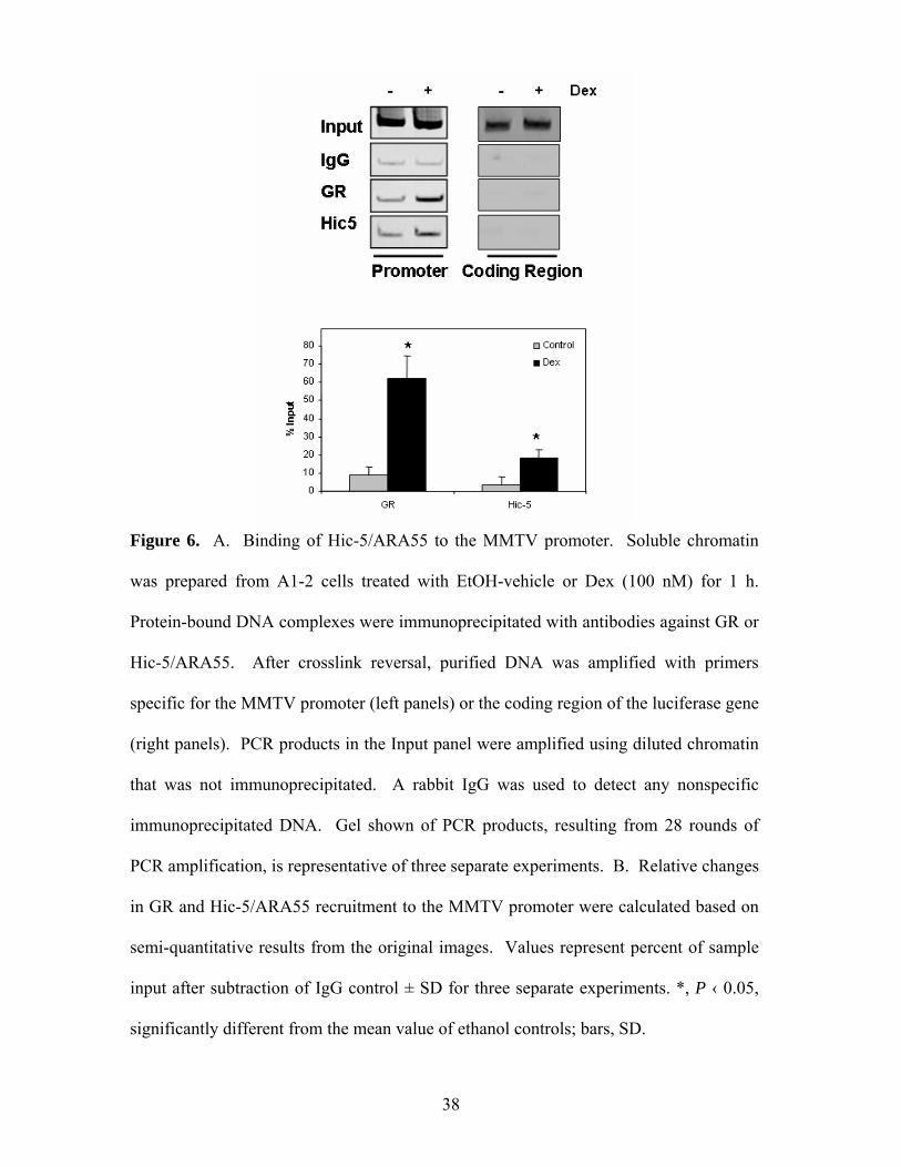
Figure 6. A. Binding of Hic-5/ARA55 to the MMTV promoter. Soluble chromatin
was prepared from A1-2 cells treated with EtOH-vehicle or Dex (100 nM) for 1 h.
Protein-bound DNA complexes were immunoprecipitated with antibodies against GR or
Hic-5/ARA55. After crosslink reversal, purified DNA was amplified with primers
specific for the MMTV promoter (left panels) or the coding region of the luciferase gene
(right panels). PCR products in the Input panel were amplified using diluted chromatin
that was not immunoprecipitated. A rabbit IgG was used to detect any nonspecific
immunoprecipitated DNA. Gel shown of PCR products, resulting from 28 rounds of
PCR amplification, is representative of three separate experiments. B. Relative changes
in GR and Hic-5/ARA55 recruitment to the MMTV promoter were calculated based on
semi-quantitative results from the original images. Values represent percent of sample
input after subtraction of IgG control ± SD for three separate experiments. *, P ‹ 0.05,
significantly different from the mean value of ethanol controls; bars, SD.
38
39
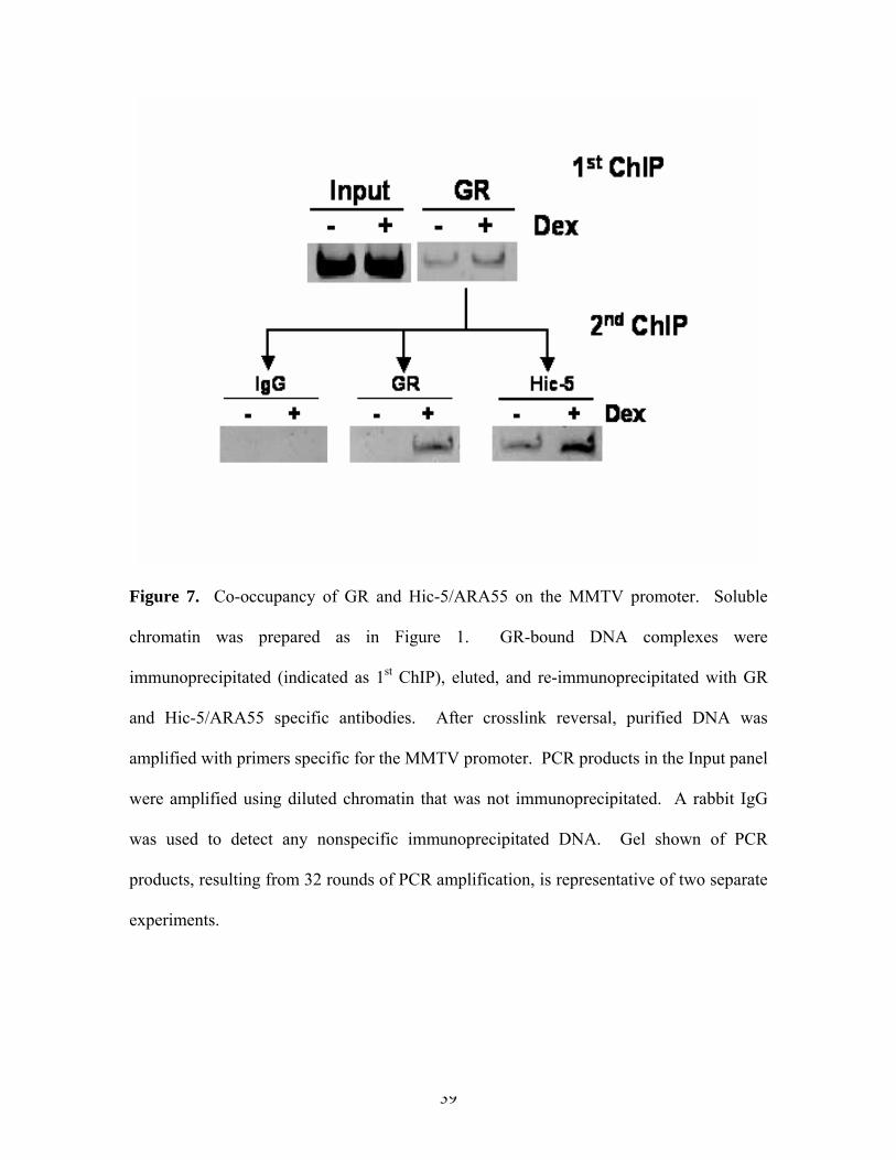
Figure 7. Co-occupancy of GR and Hic-5/ARA55 on the MMTV promoter. Soluble
chromatin was prepared as in Figure 1. GR-bound DNA complexes were
immunoprecipitated (indicated as 1st ChIP), eluted, and re-immunoprecipitated with GR
and Hic-5/ARA55 specific antibodies. After crosslink reversal, purified DNA was
amplified with primers specific for the MMTV promoter. PCR products in the Input panel
were amplified using diluted chromatin that was not immunoprecipitated. A rabbit IgG
was used to detect any nonspecific immunoprecipitated DNA. Gel shown of PCR
products, resulting from 32 rounds of PCR amplification, is representative of two separate
experiments.
responsive promoters, we performed sequential immunoprecipitations followed by ChIP
analysis (Figure 7). A1-2 cells were grown and treated as in Figure 6 and the diluted
chromatin was first immunoprecipitated using antibodies specific for GR. After
extensive washes, the precipitates were reimmunoprecipitated using antibodies specific
for Hic-5/ARA55. As shown in Figure 7, GR containing chromatin segments of the
MMTV promoter also contain Hic-5/ARA55. These data demonstrate that GR and Hic-
5/ARA55 can co-occupy the MMTV promoter in vivo and that this association is
enhanced upon Dex treatment. The sequential immunoprecipitation reduced the
detection of GR in chromatin isolated from cells not treated with hormone. However,
Hic-5/ARA55 was still detected in MMTV chromatin isolated from untreated cells.
Thus, Hic-5/ARA55 may play a role in both basal and hormone-regulated transcription
from the MMTV promoter.
GR and Hic-5/ARA55 associate with endogenous glucocorticoid-responsive
promoters in vivo.
To establish whether both GR and Hic-5/ARA55 are recruited to endogenous promoters,
we initially performed RT-PCR analysis to identify endogenous glucocorticoid
responsive genes in A1-2 cells. Glucocorticoid induction of p21 expression has been
demonstrated in A1-2 cells (119). Although the mechanism responsible for hormone
effects on p21 expression is not known, p21 may be a target of Hic-5/ARA55 action as
overexpression or forced nuclear retention of Hic-5/ARA55 resulted in increased p21 and
c-fos expression in human immortalized fibroblasts (120, 121). To test whether Hic-
40
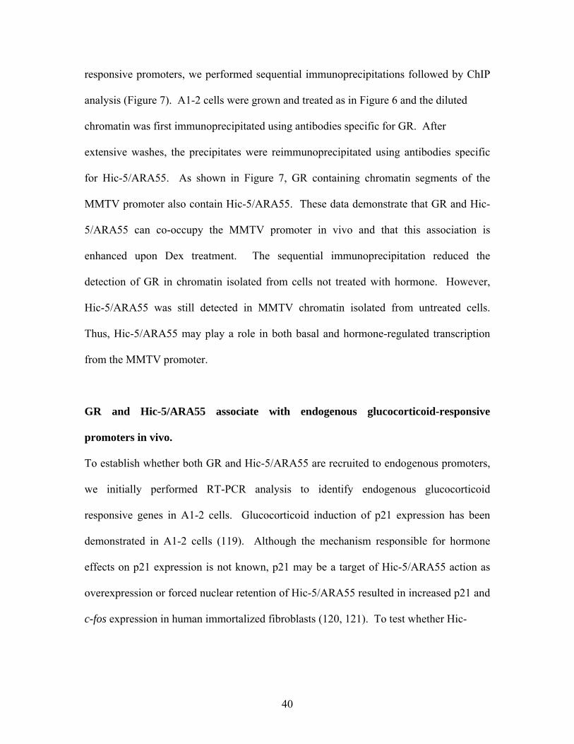
Figure 8. Association of GR and Hic-5/ARA55 with the chromatin of endogenous
promoters in vivo. A. Glucocorticoid responsive gene expression in A1-2 cells was
analyzed by RT-PCR. A1-2 cells were treated with EtOH-vehicle or Dex (100nM) for 10
h. RNA from A1-2 cells was isolated to determine the relative expression of p21, c-fos,
and GAPDH mRNAs. The reverse transcriptase reaction was carried out with 0.5 μg total
RNA. Parallel reactions performed without reverse transcriptase to control for the
presence of contaminant DNA did not generate specific PCR products for any primers
(data not shown). The gel shown of PCR products, resulting from 28 rounds of PCR
amplification, is representative of two separate experiments. B. Chromatin Re-IP
analysis was performed as in Figure 2 using c-fos or p21 promoter-specific primers. The
gels shown of PCR products, resulting from 32 rounds of PCR amplification, are
representative of two separate experiments.
41
5/ARA55 played a role in glucocorticoid induction of these genes in A1-2 cells, we used
ChIP assays to assess promoter occupancy of the p21 and c-fos genes.
A1-2 cells were grown in medium containing charcoal dextran stripped FBS. After 2
days, the cells were treated with ethanol or 100nM Dex for 10 h followed by RT-PCR
analysis. As shown in Figure 8A, we confirmed using RT-PCR that both p21 and c-fos
mRNAs were induced by Dex in A1-2 cells. Furthermore, GR association with the c-fos
and p21 promoters was enhanced by Dex treatment (Figure 8B). Importantly, sequential
ChIP experiments demonstrated that Hic-5/ARA55 was also a component of GR-
containing chromatin at these endogenous promoters. Thus, endogenous Hic-5/ARA55 is
included within GR complexes at the promoter of endogenous genes whose transcription
is regulated by glucocorticoids.
Reduced expression of Hic-5/ARA55 results in decreased GR transactivation.
It has been established that overexpression of Hic-5/ARA55 increases GR-mediated
transcription (45). However, these types of experiments do not reveal whether
endogenous Hic-5/ARA55 is necessary for GR activity. Thus, we used a siRNA
approach to ablate Hic-5/ARA55 expression in A1-2 cells and assess the impact on GR
transactivation. A1-2 cells were analyzed for Hic-5/ARA55 expression in A1-2 cells by
Western blot analysis following transfection with either a control GFP siRNA or Hic-
5/ARA55 siRNA. Densitometric analysis revealed that there was approximately 60%
less Hic-5/ARA55 in cells transfected with the Hic-5/ARA55 siRNA as compared to
control (Figure 9A). Two other distinct Hic-5/ARA55 siRNAs tested were less effective
42
Hic-5Hic-
GR
GAPDH
siG
FP
siH
ic-5
0
10000
20000
30000
40000
50000
60000
70000
80000
siGFP siHic-5
RLU
Control
Dex
*
A B
C
c-fos
p21
GAPDH
- + - + DexsiGFP siHic-5
Hic-5Hic-
GR
GAPDH
siG
FP
siH
ic-5
0
10000
20000
30000
40000
50000
60000
70000
80000
siGFP siHic-5
RLU
Control
Dex
*
A B
C
c-fos
p21
GAPDH
- + - + DexsiGFP siHic-5
c-fos
p21
GAPDH
- + - + DexsiGFP siHic-5
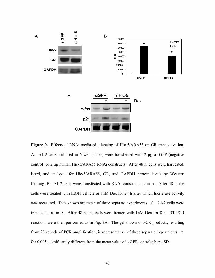
Figure 9. Effects of RNAi-mediated silencing of Hic-5/ARA55 on GR transactivation.
A. A1-2 cells, cultured in 6 well plates, were transfected with 2 μg of GFP (negative
control) or 2 μg human Hic-5/ARA55 RNAi constructs. After 48 h, cells were harvested,
lysed, and analyzed for Hic-5/ARA55, GR, and GAPDH protein levels by Western
blotting. B. A1-2 cells were transfected with RNAi constructs as in A. After 48 h, the
cells were treated with EtOH-vehicle or 1nM Dex for 24 h after which luciferase activity
was measured. Data shown are mean of three separate experiments. C. A1-2 cells were
transfected as in A. After 48 h, the cells were treated with 1nM Dex for 8 h. RT-PCR
reactions were then performed as in Fig. 3A. The gel shown of PCR products, resulting
from 28 rounds of PCR amplification, is representative of three separate experiments. *,
P ‹ 0.005, significantly different from the mean value of siGFP controls; bars, SD.
43
at reducing endogenous Hic-5/ARA55 expression. Nonetheless, the reduction in Hic-
5/ARA55 expression in A1-2 cells by the most effective siRNA was sufficient to
generate reduced GR transactivation, as measured by ligand-dependent luciferase
expression (Figure 9B). Furthermore, this reduction in GR activity was not due to
decreased GR expression in siHic-5/ARA55-transfected cells (Figure 9A). We also
analyzed glucocorticoid-induced c-fos and p21 mRNA expression after transfection with
Hic-5/ARA55 siRNA (Figure 9C). Although there was no reduction in c-fos expression,
there was a reduction in p21 expression upon silencing of Hic-5/ARA55. Because the
silencing of Hic-5/ARA55 expression was not completely effective, we can not conclude
that Hic-5/ARA55 is essential for GR activity. However, these results indicate that
endogenous Hic-5/ARA55 contributes to optimal GR transactivation from a subset of
promoters in A1-2 cells.
Hic-5/ARA55 interaction with coactivators on GR responsive promoters.
Because Hic-5/ARA55 does not possess HAT or methyltransferase activity, it may not
modify histones directly. However, Hic-5/ARA55 may be involved in recruiting other
chromatin modifying coactivators. To determine if Hic-5/ARA55 interacts with other
coactivator proteins at glucocorticoid responsive promoters, we performed sequential
ChIPs, first using antibodies specific for Hic-5/ARA55 followed by re-
immunoprecipitation with anti-SRC-1, anti-TIF-2, anti-RAC3, anti-CBP or anti-p300
antibodies. As shown in Figure 10, TIF-2, RAC3, CBP, and p300, but not SRC-1 were
associated within Hic-5/ARA55-containing chromatin at the MMTV promoter in
glucocorticoid treated A1-2 cells. Interestingly, while TIF-2, RAC3, and CBP were
44
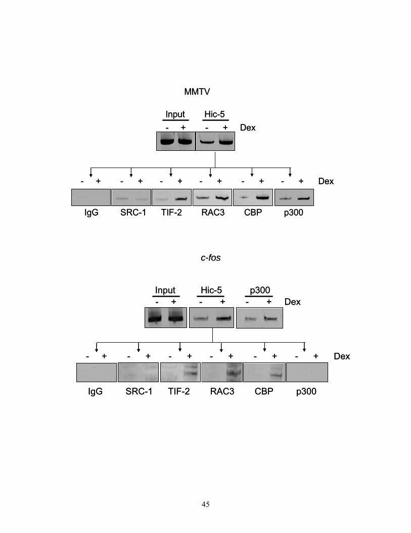
Input Hic-5- + - + Dex
- + - + - + - + - + - + Dex
IgG SRC-1 TIF-2 RAC3 CBP p300
MMTV
Input Hic-5- + - + Dex
- + - + - + - + - + - + Dex
IgG SRC-1 TIF-2 RAC3 CBP p300
MMTV
Input Hic-5 p300- + - + - + Dex
c-fos
IgG SRC-1 TIF-2 RAC3 CBP p300
- + - + - + - + - + - + Dex
Input Hic-5 p300- + - + - + Dex
c-fos
IgG SRC-1 TIF-2 RAC3 CBP p300
- + - + - + - + - + - + Dex
45
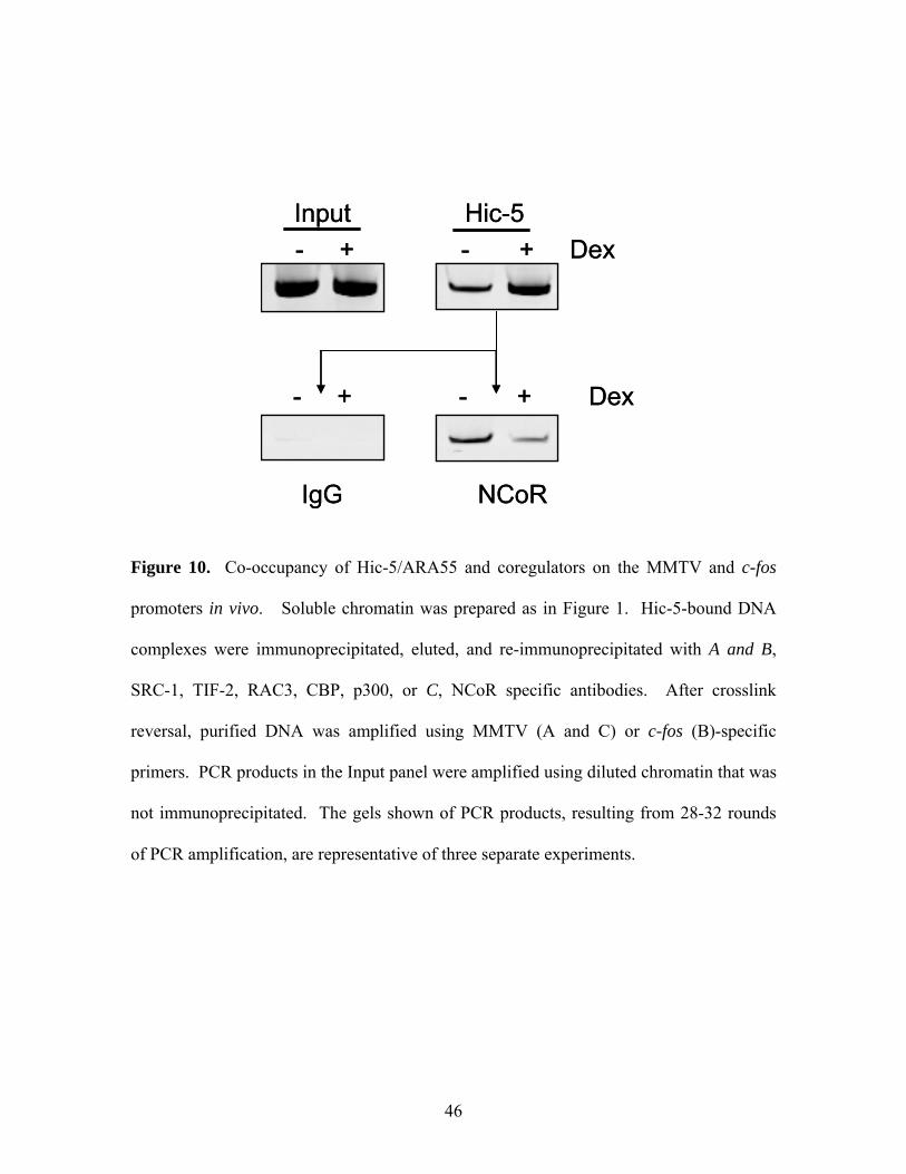
Input Hic-5- + - + Dex
- + - + Dex
IgG NCoR
Input Hic-5- + - + Dex
- + - + Dex
IgG NCoR
Figure 10. Co-occupancy of Hic-5/ARA55 and coregulators on the MMTV and c-fos
promoters in vivo. Soluble chromatin was prepared as in Figure 1. Hic-5-bound DNA
complexes were immunoprecipitated, eluted, and re-immunoprecipitated with A and B,
SRC-1, TIF-2, RAC3, CBP, p300, or C, NCoR specific antibodies. After crosslink
reversal, purified DNA was amplified using MMTV (A and C) or c-fos (B)-specific
primers. PCR products in the Input panel were amplified using diluted chromatin that was
not immunoprecipitated. The gels shown of PCR products, resulting from 28-32 rounds
of PCR amplification, are representative of three separate experiments.
46
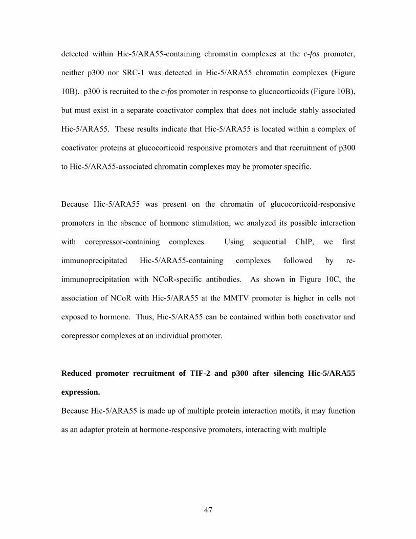
detected within Hic-5/ARA55-containing chromatin complexes at the c-fos promoter,
neither p300 nor SRC-1 was detected in Hic-5/ARA55 chromatin complexes (Figure
10B). p300 is recruited to the c-fos promoter in response to glucocorticoids (Figure 10B),
but must exist in a separate coactivator complex that does not include stably associated
Hic-5/ARA55. These results indicate that Hic-5/ARA55 is located within a complex of
coactivator proteins at glucocorticoid responsive promoters and that recruitment of p300
to Hic-5/ARA55-associated chromatin complexes may be promoter specific.
Because Hic-5/ARA55 was present on the chromatin of glucocorticoid-responsive
promoters in the absence of hormone stimulation, we analyzed its possible interaction
with corepressor-containing complexes. Using sequential ChIP, we first
immunoprecipitated Hic-5/ARA55-containing complexes followed by re-
immunoprecipitation with NCoR-specific antibodies. As shown in Figure 10C, the
association of NCoR with Hic-5/ARA55 at the MMTV promoter is higher in cells not
exposed to hormone. Thus, Hic-5/ARA55 can be contained within both coactivator and
corepressor complexes at an individual promoter.
Reduced promoter recruitment of TIF-2 and p300 after silencing Hic-5/ARA55
expression.
Because Hic-5/ARA55 is made up of multiple protein interaction motifs, it may function
as an adaptor protein at hormone-responsive promoters, interacting with multiple
47
Input
IgG
TIF-2
p300
- + - + DexsiGFP siHic-5
Input
IgG
TIF-2
p300
- + - + DexsiGFP siHic-5
Figure 11. Coactivator recruitment on the MMTV promoter following ablation of Hic-
5/ARA55. A1-2 cells were transfected as in Fig. 4. with vectors expressing siRNA for
Hic-5/ARA55 or GFP. After 72 hours, ChIP analysis was performed as in Fig.1. After
crosslink reversal, purified DNA was amplified with primers specific for the MMTV
promoter. Gel shown of PCR products, resulting from 28 rounds of PCR amplification,
is representative of two separate experiments.
48
coregulator proteins or stabilizing the coactivator complex. In order to assess the
necessity of Hic-5/ARA55 in coactivator recruitment to glucocorticoid-responsive genes,
we performed ChIP assays in cells after silencing Hic-5/ARA55 expression. As shown in
Figure 11, after ablation of Hic-5/ARA55 expression, TIF-2 and p300 recruitment to the
MMTV promoter in response to glucocorticoids was reduced. Interestingly, there was
increased p300 recruitment in the absence of hormone upon silencing Hic-5/ARA55
expression. The corresponding reduction of GR transactivation (Figure 9) and
coactivator recruitment (Figure 11) that results from partial Hic-5/ARA55 ablation
demonstrates the critical role of Hic-5/ARA55 in maintaining the assembly of coactivator
complexes required to bring about efficient glucocorticoid-induced transcription.
Discussion
Here, we showed that endogenous Hic-5/ARA55 is recruited to both stably integrated
viral and endogenous glucocorticoid-responsive promoters in response to hormone
treatment. Furthermore, GR and Hic-5/ARA55 co-occupied these promoters within
stable complexes that can be recovered by sequential ChIPs. We also demonstrated an
association between Hic-5/ARA55 and coactivator-containing complexes at
glucocorticoid-responsive promoters using sequential ChIPs. Using gel shift assays and
co-immunoprecipitations, Shibanuma et al. showed that Hic-5/ARA55 containing a
heterologous nuclear localization signal (NLS) regulated p21 expression by interacting
with Smad3, but not p300 or Sp1 directly in transfected cells (65). While these results
49
imply that Hic-5/ARA55 association with p300 may be cell type- or promoter-specific, it
may not be appropriate to compare the function of endogenous Hic-5/ARA55 in our case
with transfected, overexpressed Hic-5/ARA55 that contains a heterologous NLS to force
robust nuclear retention. However, promoter-specific recruitment of p300 to endogenous
Hic-5/ARA55 chromatin complexes was observed in A1-2 cells, corroborating the results
obtained with exogenously introduced NLS-Hic-5/ARA55 constructs.
Interestingly, for the promoter in which p300 in not a component of Hic-5/ARA55
complexes (i.e. the c-fos promoter), partial ablation of Hic-5/ARA55 was not sufficient to
impact glucocorticoid-regulated transcription. While we cannot rule out the possibility
that more complete ablation of Hic-5/ARA55 would hinder glucocorticoid-induced
transcription from the c-fos promoter, Hic-5/ARA55 may only exert an essential function
to maintain the stability of distinct subsets of coactivator complexes (i.e. p300-
containing). Partial Hic-5/ARA55 ablation also blocked PPARγ-induced expression of a
select set of epithelial-specific genes (66). While the differential recruitment of
coactivators was not examined in this case, these results highlight the importance of Hic-
5/ARA55 in impacting gene-specific effects of nuclear receptors.
Although there have been no reports indicating a repressive function for Hic-5/ARA55 on
nuclear receptor-mediated gene expression, ChIP analysis consistently revealed Hic-
5/ARA55 binding to the MMTV promoter in the absence of hormone. The enhanced
association of NCoR with Hic-5/ARA55 complexes at the MMTV promoter in non-
hormone treated cells suggests that Hic-5/ARA55 may in fact participate in maintaining
50
low basal levels of transcription from this promoter. However, Hic-5/ARA55 was not
essential for limiting MMTV transcription in the absence of hormone as its partial
ablation does not lead to enhanced transcription. Whereas we have observed enhanced
basal transcription from a steroid hormone-regulated gene upon Hic-5/ARA55 ablation in
a prostate cell line, definitive demonstration of Hic-5/ARA55 participation in
transcriptional repression will require more thorough analysis.
Finally, siRNA ablation experiments reported here establish that Hic-5/ARA55 is
required for the stable association of p300 and TIF-2 with the MMTV promoter. Thus,
Hic-5/ARA55 may stabilize select protein complex formation at GR-responsive
promoters by serving as an adaptor molecule.
51
CHAPTER 4 Determining Hic-5/ARA55 expression and function in the prostate.
Hypothesis 2
Because Hic-5/ARA55 expression in the prostate is restricted to the stromal
compartment, it modulates AR-mediated gene expression, products of which are required
for epithelial-stromal interactions in the prostate.
Introduction
As previously mentioned, communication between the epithelial and stromal
compartments of the prostate is crucial for the maintenance of prostate growth and
function (7, 8). Additionally, androgens along with functional AR expression in the
stromal compartment are necessary for prostate development (14). In the stromal
compartment, AR regulates the expression of various growth factors such as KGF (FGF-
7), a member of the fibroblast growth factor family (14, 19-21). KGF is exclusively
expressed in the stromal compartment of the prostate while its receptor, a spliced variant
of the FGF type 2 receptor (FGFR2IIIb), is expressed solely in the epithelial
compartment (3). These characteristics suggest that KGF is a paracrine mediator of
signaling from the stroma to the epithelial compartment that can influence epithelial cell
growth (1, 22).
Along with prostate development, androgens are critical for PCa progression and may be
involved in the transition to hormone refractory PCa (97). AR activation in the absence
52
of circulating androgens in hormone refractory PCa is an area of intense investigation.
Some reports suggest that signaling events issued by cytokines or growth factors such as,
IGF-1, EGF, KGF and IL-6, can induce AR-mediated gene expression in the absence of
androgens (104, 105). Furthermore, KGF expression is significantly increased in prostate
cancer (22). In addition to androgen independent activation of AR, increased expression
of coactivators in PCa cell lines have enhanced AR responsiveness to low levels of
androgen, anti-androgens, and other steroid hormones such as estrogen or progesterone
(44, 106).
In the prostate, Hic-5/ARA55 expression was restricted to the stromal compartment
(111). Interestingly, Hic-5/ARA55 expression was reduced in tumor as compared to
normal tissue (112). Furthermore, Hic-5 mRNA levels were lower in hormone-refractory
PC than that of previously untreated PCa, and in recurrent hormone refractory PCa,
higher Hic-5/ARA55 expression correlated with unfavorable recurrence-free survival as
well as overall survival (122). Because most analyses of coactivator expression in PCa
uses quantitative PCR, it is difficult to ascertain whether this represents reduced
expression of Hic-5/ARA55 within individual cells or alterations in the balance between
stromal/epithelial cell content in tumors.
In this study, we examined Hic-5/ARA55 expression and activity in the prostate. In both
normal and tumor derived prostates, Hic-5/ARA55 expression is confined to the stromal
compartment. We therefore focused our analysis of Hic-5/ARA55 function in a prostate
stromal, myofibroblast cell line, WPMY-1 cells. We show here that Hic-5/ARA55
53
functions as an AR coactivator in WPMY-1 cells and is necessary for effective androgen
induction of the stromal paracrine factor, KGF.
Results
Regional expression of Hic-5/ARA55 in human prostate tissue.
Given Hic-5/ARA55 effects on AR and its role in hormone-dependent growth of prostate
cancer (PCa) cell lines, a number of studies have examined its expression in prostate
tissue. For example, Hic-5/ARA55 mRNA expression, as quantified using PCR, is
reduced in hormone refractory prostate cancer tissue (HRPC) as compared to normal or
benign prostatic hypertrophy (BPH) tissue (112, 122). Since these tissue samples
contained a heterogeneous population of cells, potential cell type specific differences in
Hic-5/ARA55 expression would not be revealed. Therefore, we analyzed prostate tissue
from both normal human donors and prostate cancer patients for Hic-5/ARA55
expression using immunohistochemistry. As shown in Figure 12 (top panels), Hic-
5/ARA55 expression is confined to the stromal compartment of the prostate. The
epithelial compartment was consistently negative for Hic-5/ARA55 expression. This
profile of Hic-5/ARA55 expression in the prostate is consistent with those previously
published using normal human prostates (111). In tumor-derived prostate tissue, Hic-
5/ARA55 was also restricted to stromal cells and not detected in the prostate tumor cells
(Figure 12, bottom panels). In both cases, the Hic-5/ARA55 staining appeared diffuse
and its precise subcellular compartmentalization was not apparent.
54
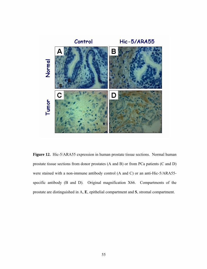
Figure 12. Hic-5/ARA55 expression in human prostate tissue sections. Normal human
prostate tissue sections from donor prostates (A and B) or from PCa patients (C and D)
were stained with a non-immune antibody control (A and C) or an anti-Hic-5/ARA55-
specific antibody (B and D). Original magnification X66. Compartments of the
prostate are distinguished in A, E, epithelial compartment and S, stromal compartment.
55
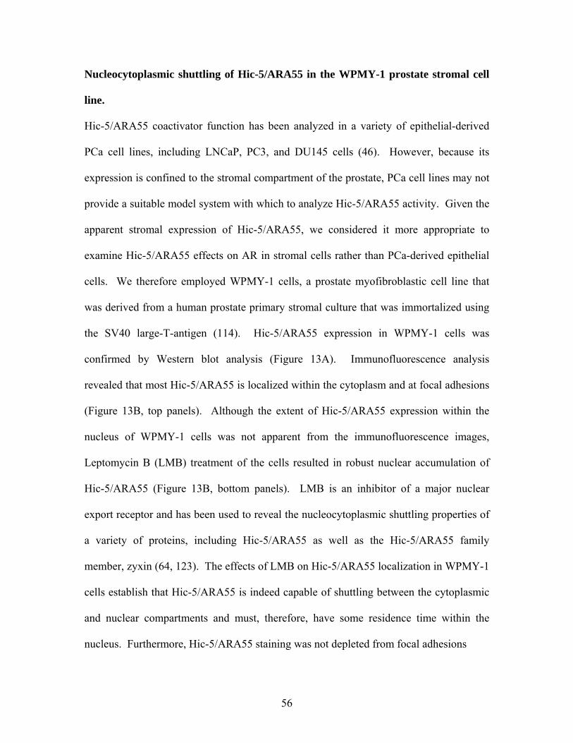
Nucleocytoplasmic shuttling of Hic-5/ARA55 in the WPMY-1 prostate stromal cell
line.
Hic-5/ARA55 coactivator function has been analyzed in a variety of epithelial-derived
PCa cell lines, including LNCaP, PC3, and DU145 cells (46). However, because its
expression is confined to the stromal compartment of the prostate, PCa cell lines may not
provide a suitable model system with which to analyze Hic-5/ARA55 activity. Given the
apparent stromal expression of Hic-5/ARA55, we considered it more appropriate to
examine Hic-5/ARA55 effects on AR in stromal cells rather than PCa-derived epithelial
cells. We therefore employed WPMY-1 cells, a prostate myofibroblastic cell line that
was derived from a human prostate primary stromal culture that was immortalized using
the SV40 large-T-antigen (114). Hic-5/ARA55 expression in WPMY-1 cells was
confirmed by Western blot analysis (Figure 13A). Immunofluorescence analysis
revealed that most Hic-5/ARA55 is localized within the cytoplasm and at focal adhesions
(Figure 13B, top panels). Although the extent of Hic-5/ARA55 expression within the
nucleus of WPMY-1 cells was not apparent from the immunofluorescence images,
Leptomycin B (LMB) treatment of the cells resulted in robust nuclear accumulation of
Hic-5/ARA55 (Figure 13B, bottom panels). LMB is an inhibitor of a major nuclear
export receptor and has been used to reveal the nucleocytoplasmic shuttling properties of
a variety of proteins, including Hic-5/ARA55 as well as the Hic-5/ARA55 family
member, zyxin (64, 123). The effects of LMB on Hic-5/ARA55 localization in WPMY-1
cells establish that Hic-5/ARA55 is indeed capable of shuttling between the cytoplasmic
and nuclear compartments and must, therefore, have some residence time within the
nucleus. Furthermore, Hic-5/ARA55 staining was not depleted from focal adhesions
56
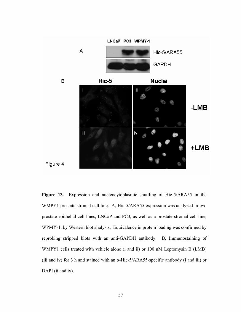
Figure 13. Expression and nucleocytoplasmic shuttling of Hic-5/ARA55 in the
WMPY1 prostate stromal cell line. A, Hic-5/ARA55 expression was analyzed in two
prostate epithelial cell lines, LNCaP and PC3, as well as a prostate stromal cell line,
WPMY-1, by Western blot analysis. Equivalence in protein loading was confirmed by
reprobing stripped blots with an anti-GAPDH antibody. B, Immunostaining of
WMPY1 cells treated with vehicle alone (i and ii) or 100 nM Leptomysin B (LMB)
(iii and iv) for 3 h and stained with an α-Hic-5/ARA55-specific antibody (i and iii) or
DAPI (ii and iv).
57
upon LMB treatment (data not shown), indicating that shuttling of Hic-5/ARA55 most
likely occurs between the cytoplasmic and nuclear compartments.
AR and Hic-5/ARA55 recruitment to the KGF promoter.
Because Hic-5/ARA55 enhances AR-mediated gene expression in transient transfection
assays, we set out to reveal whether Hic-5/ARA55 was localized to endogenous
androgen-responsive promoters in WPMY-1 cells. KGF is a well established marker of
androgen induction in prostate stromal cells (19, 20). As shown in Figures 14A and 14B,
KGF mRNA expression is induced 2-fold in response to treatment with various
androgens. In primary stromal cell cultures, KGF expression is similarly induced
approximately 2-fold by androgen treatment (20). Furthermore, androgen induction of
KGF mRNA expression is blocked by the AR antagonist, hydroxyflutamide, indicating
that androgen induction of KGF mRNA in WPMY-1 cells is indeed mediated by AR.
Because Hic-5/ARA55 has been found to directly interact with AR, we sought to
determine if Hic-5/ARA55 is bound to the KGF promoter using ChIP assays and whether
this binding is affected by androgen treatment. Detailed analysis of the human KGF
promoter revealed that promoter proximal sequences located between -225 to +190 were
necessary for basal KGF expression (124). Although a canonical hormone response
element is not located within this region, there is a half-palindromic glucocorticoid
response element located between positions -178 to -183 around which we designed our
primers for ChIP analysis (124). After stimulation with androgens for 1 hour, both AR
and Hic-5/ARA55 were localized to the KGF promoter (Figure 15). Given the high
58
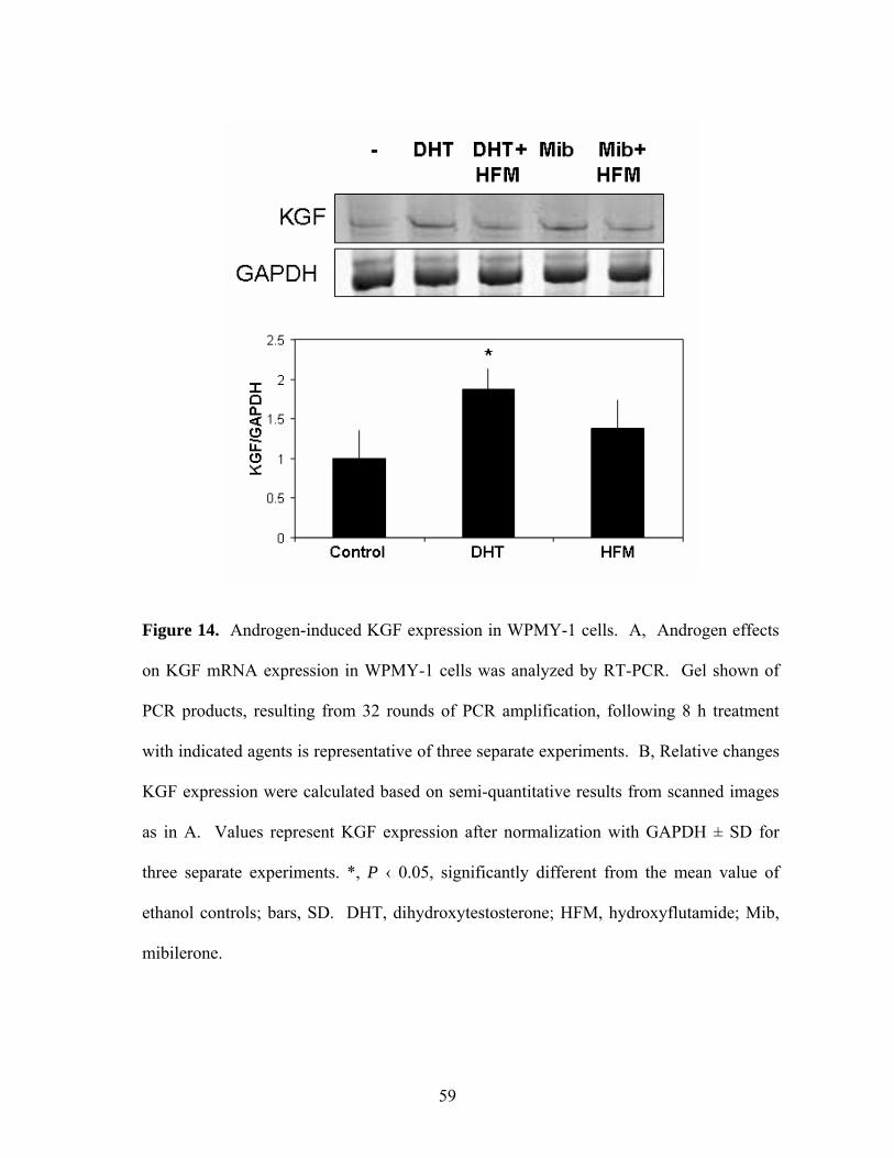
Figure 14. Androgen-induced KGF expression in WPMY-1 cells. A, Androgen effects
on KGF mRNA expression in WPMY-1 cells was analyzed by RT-PCR. Gel shown of
PCR products, resulting from 32 rounds of PCR amplification, following 8 h treatment
with indicated agents is representative of three separate experiments. B, Relative changes
KGF expression were calculated based on semi-quantitative results from scanned images
as in A. Values represent KGF expression after normalization with GAPDH ± SD for
three separate experiments. *, P ‹ 0.05, significantly different from the mean value of
ethanol controls; bars, SD. DHT, dihydroxytestosterone; HFM, hydroxyflutamide; Mib,
mibilerone.
59
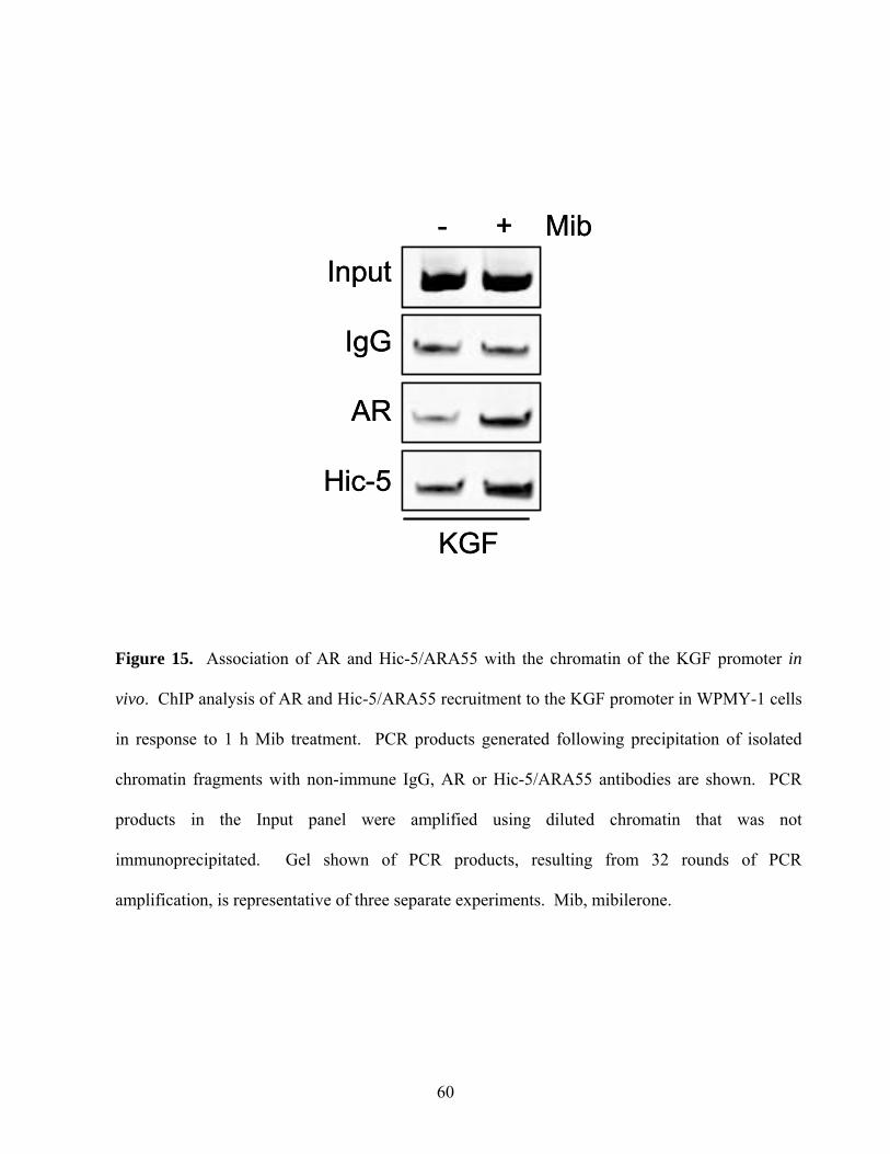
Input
IgG
AR
Hic-5
- + Mib
KGF
Input
IgG
AR
Hic-5
- + Mib
KGF
Figure 15. Association of AR and Hic-5/ARA55 with the chromatin of the KGF promoter in
vivo. ChIP analysis of AR and Hic-5/ARA55 recruitment to the KGF promoter in WPMY-1 cells
in response to 1 h Mib treatment. PCR products generated following precipitation of isolated
chromatin fragments with non-immune IgG, AR or Hic-5/ARA55 antibodies are shown. PCR
products in the Input panel were amplified using diluted chromatin that was not
immunoprecipitated. Gel shown of PCR products, resulting from 32 rounds of PCR
amplification, is representative of three separate experiments. Mib, mibilerone.
60
degree of background obtained in ChIP assays with non-immune IgG, it is difficult to
assess the extent of AR and/or Hic-5/ARA55 binding to the KGF promoter in the absence
of androgen treatment. Nonetheless, the ChIP results from hormone treated cells suggest
that Hic-5/ARA55-mediated coactivation of AR activity in WPMY-1 cells may be
mediated in part by their recruitment to endogenous androgen-responsive promoters.
Ablation of Hic-5/ARA55 expression results in decreased AR transactivation.
Given the localization of Hic-5/ARA55 to the KGF promoter in response to androgen
treatment, we ascertained whether Hic-5/ARA55 was necessary for androgen induction
of KGF expression using a siRNA approach to ablate Hic-5/ARA55 expression in
WPMY-1 cells. WPMY-1 cells were analyzed for Hic-5/ARA55 expression by Western
blot analysis following transfection with either a control GFP siRNA or Hic-5/ARA55
siRNA. Densitometric analysis revealed that there was approximately 60% less Hic-
5/ARA55 in cells transfected with the Hic-5/ARA55 siRNA oligo D as compared to
control (Figure 16A). Although this was the maximal amount of Hic-5/ARA55 ablation
that could be attained with Hic-5/ARA55 siRNA transfections, it was sufficient to reduce
androgen induction of the transfected androgen-inducible mouse mammary tumor virus
(MMTV) promoter (Figure 16B) and of endogenous KGF mRNA expression (Figure
16C). Basal levels of KGF expression were reproducibly higher upon Hic-5/ARA55
ablation, suggesting that Hic-5/ARA55 may negatively regulate basal KGF promoter
activity. Irrespective of these effects of Hic-5/ARA55 on basal KGF promoter activity,
61
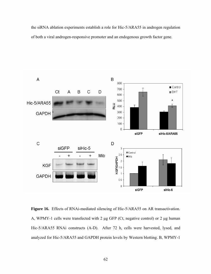
the siRNA ablation experiments establish a role for Hic-5/ARA55 in androgen regulation
of both a viral androgen-responsive promoter and an endogenous growth factor gene.
Figure 16. Effects of RNAi-mediated silencing of Hic-5/ARA55 on AR transactivation.
A, WPMY-1 cells were transfected with 2 μg GFP (Ct, negative control) or 2 μg human
Hic-5/ARA55 RNAi constructs (A-D). After 72 h, cells were harvested, lysed, and
analyzed for Hic-5/ARA55 and GAPDH protein levels by Western blotting. B, WPMY-1
62
cells were transfected with RNAi constructs (GFP and D) as in A as well as the MMTV
promoter-linked reporter. After 48 h, the cells were treated with EtOH-vehicle or DHT
(10-8 M) for 24 h after which luciferase activity was measured. Data shown are mean of
three separate experiments. *, P ‹ 0.05, significantly different from the mean value of
siGFP controls; bars, SD. C, Androgen-induced KGF expression was analyzed after
silencing Hic-5/ARA55. Cells were transfected as in A. After 72 h, the cells were
stimulated with Mib (10-8 M) for 8 h and KGF expression was analyzed by RT-PCR and
normalized to GAPDH expression. D, Relative changes KGF expression were calculated
based on semi-quantitative results from scanned images as in C. Values represent KGF
expression after normalization with GAPDH ± SD for three separate experiments.
Discussion
Our results identify Hic-5/ARA55 as a compartment-specific AR coactivator that has an
impact on androgen regulation of a paracrine growth factor, KGF, in the stroma. The
importance of KGF in prostate gland development has been revealed by various
approaches. For example, KGF neutralizing antibodies inhibit prostate epithelial growth
as well as ductal branching (1). Furthermore, exogenous KGF was sufficient to produce
epithelial growth as well as ductal branching of the prostate in the absence of androgen
(1).
63
Analysis of Hic-5/ARA55 expression had revealed its limited tissue distribution. For
example, immunohistochemical analysis was used to reveal selective expression of Hic-
5/ARA55 in smooth muscle and myoepithelial cells (125). Hic-5/ARA55 was not
detected in epithelial cells of the tissues examined, including the stomach, colon, liver,
skin, and mammary gland (125). Additionally, Hic-5/ARA55 is expressed in the stromal,
but not epithelial compartment of the prostate (111). Here, we confirmed the stromal-
specific expression of Hic-5/ARA55 in normal prostate. Furthermore, Hic-5/ARA55
expression was also restricted to the stromal compartment in prostate tumor tissue.
The stromal-specific expression of Hic-5/ARA55 suggests that this AR coactivator is
unlikely to contribute directly to the development or progression of PCa. However, Hic-
5/ARA55 may play a role in stromal/epithelial cell communication in the prostate since
androgen induction of a stromal-selective paracine factor (i.e. KGF) is lost upon partial
ablation of Hic-5/ARA55. Furthermore, the increase in basal KGF expression upon Hic-
5/ARA55 ablation suggests that this coactivator may interact with other transcription
factors or corepressors to negatively regulate KGF expression in the absence of
androgens (65). Thus, by serving opposing roles to positively or negatively regulate KGF
gene transcription, Hic-5/ARA55 may be critical in sculpting tissue organization in the
prostate, responding to various hormonal and paracrine cues that serve to maintain the
appropriate balance between distinct stromal and epithelial cell layers. Furthermore,
given the role of stromal-derived KGF on the growth, transformation, and invasive
properties of PCa cells, coactivators such as Hic-5/ARA55 may influence tumor
development and progression through its impact on either basal and/or androgen-induced
64
expression of KGF in stroma during both androgen-dependent and androgen-independent
phases (126, 127).
Detailed sequence analysis of the human KGF promoter revealed a half-palindromic
steroid hormone response element between positions -178 to -183, implicating AR
regulation of KGF transcription (124). ChIP analysis of the KGF promoter revealed both
AR and Hic-5/ARA55 recruitment to this region of the KGF promoter in response to
androgens. Although most Hic-5/ARA55 is found at focal adhesions and the cytoplasm
in WPMY-1 cells, Hic-5/ARA55 shuttles between the cytoplasm and the nucleus.
Interestingly, Hic-5/ARA55 was localized to the KGF promoter in the absence of LMB.
This indicates that there may be a small pool of Hic-5/ARA55 resident in the nucleus that
may not be apparent by immunofluorescence analysis, but which nonetheless participates
in transcriptional regulation of androgen-regulated promoters.
In endocrine target tissues, nuclear receptor activity appears critical for tumor
development and/or progression (128). For example, elevated AR expression may be
necessary for PCa progression to an androgen-independent state (100). Furthermore,
increased AR expression correlates with a higher probability of recurrence (101). In
response to elevated expression of AR, mutations in AR, or altered expression of
coactivators, the activity of nuclear receptors within individual cells may exhibit a
heightened response to non-steroidal signals. Our work suggests the existence of another
mechanism for coactivator effects on prostate cancer that does not function in a cell
65

autonomous context. AR action in cells that support the tumor, located in the stromal
compartment, may also be affected by stromal-specific coactivators.
66
Chapter 5 Determining the effects of extracellular matrix (ECM) signaling on GR-
mediated transcription in A1-2 cells.
Hypothesis: Because Hic-5/ARA55 associates with focal adhesion proteins as well as
nuclear receptors, Hic-5/ARA55 may relay extracellular matrix signals to nuclear
receptors, affecting their transactivation properties.
Introduction
Intracellular signaling originating from the extracellular matrix (ECM) is mediated by a
well-established group of receptors known as integrins. Integrins form heterodimer
receptors between members of the α and β families, which are specific for an ECM
component. For example, α5β6 binds fibronectin primarily, whereas α1β1 binds both
laminin and collagen (reviewed in (129). Resulting signals following integrin-ligand
interactions include modulation of cellular migration, proliferation, survival, and
apoptosis. However, the exact mechanism by which ECM signaling influences nuclear
receptor-mediated gene expression in normal as well as tumorigenic tissue is less clear.
Cross-talk between estrogen receptor (ER) and ECM signaling, resulting in differential
cell growth and/or gene expression, has been well studied. Specifically, malignant
mammary cells grown on a collagen IV or laminin-1 basement membrane express higher
levels of ERα, resulting in increased ER-mediated gene expression irrespective of the
67
presence of estradiol (130). Furthermore, analysis of the ERα promoter revealed STAT5
binding sites located within the region that was responsive to laminin-rich basement
membranes, indicating that upregulation of ERα expression may be mediated by STAT5
(131). However, Woodward et al. report that laminin inhibits ERα activity, namely
negatively influencing estrogen-mediated progesterone receptor (PR) expression and
proliferation of breast cancer cells (132). These discrepancies are most likely due to
minor differences in the application of the ECM, culturing conditions, and/or the
differential responsiveness to ECM between different cell lines.
Along with breast cancer cell lines, primary breast epithelial cells also respond to ECM
signaling. Epithelial cells derived from nulliparous mice also exhibit increased
responsiveness to progesterone stimulation when plated on fibronectin or collagen IV
(133). After plating the cells on various ECM, increased proliferation in response to
progesterone treatment was detected in nulliparous-derived but not pregnancy-derived
breast epithelial cells (133). These results indicate that mammary gland responsiveness
to both steroid hormones as well as ECM signaling is dependent on mammary gland
differentiation.
Although the effects of certain ECM components such as collagen IV and laminin-1 on
ERα-mediated gene expression have been reported, the exact mechanism by which ECM
enhances ERα expression and/or activity is unclear. For example, activation of integrin
signaling pathways in response to various ECM components may influence nuclear
receptor phosphorylation, effecting its subcellular localization, coactivator interaction, or
68
protein stability. Alternatively, ECM may influence nuclear receptor-mediated gene
expression in a more direct manner, via interaction of focal adhesion-associated proteins
such as Hic-5/ARA55 with the nuclear receptors, affecting their gene activation.
Along with a possible role for Hic-5/ARA55 in transmitting ECM signals, which in turn
affect nuclear receptor-mediated gene expression, Hic-5/ARA55 also modulates ECM
expression. Using cells that overexpress Hic-5/ARA55, Shibanuma et al. report that Hic-
5/ARA55 expression may influence ECM expression directly (121). Both collagen and
fibronectin expression correlated with Hic-5/ARA55 expression (121). Furthermore,
overexpression of Hic-5/ARA55 led to alternative splicing of fibronectin, which is also
associated with the senescence process (121). The modulation of ECM expression by
Hic-5/ARA55 may provide a feedback loop, serving to ameliorate or alter ECM signaling
events.
Here, we examined GR-mediated gene expression in response to various ECM as well as
Hic-5/ARA55’s role in transmitting ECM-derived signals. Both fibronectin and matrigel
influenced GR transactivation in opposing ways. Matrigel repressed GR-mediated gene
expression by reducing the levels of GR protein. Fibronectin did not influence either GR
or Hic-5/ARA55 expression, subcellular localization or promoter recruitment in the
presence of ligand, suggesting that other mechanisms by which fibronectin may influence
GR transactivation, such as post translational modifications or GR-coactivator
interactions.
69
Results
Effects of ECM on GR-mediated transcription
To determine the effects of ECM signaling on GR-mediated gene expression, we took
advantage of the integrated MMTV promoter in the A-12 cells. Because T47D cells, the
parental cell line of A-12 cells, express α1, α3, α4 and α5 integrin subunits, the
extracellular matrices that were analyzed include: laminin, collagen, fibronectin as well
as a growth factor reduced matrigel (136). Because growth factors such as EGF and IGF-
1 have been reported to activate nuclear receptors-mediated gene expression in the
absence of ligand, we used a growth factor reduced matrigel to analyze the effects of
basement membrane components on GR activity (104). After coating cell culture plates
with the appropriate ECM, A-12 cells were treated with dex (100 nM) for 18 hours after
which luciferase reporter activity was measured. Whereas fibronectin enhanced GR-
mediated luciferase expression in A1-2 cells, matrigel reduced it (Figure 17). However,
neither laminin nor vitronectin affected GR transactivation properties. Furthermore, there
was no change in GR-mediated gene expression when cells were plated on a nonspecific
poly-L-lysine substratum, indicating that the responses of GR activity on both fibronectin
and matrigel were specific and may be mediated by integrin signaling.
Because the effect of fibronectin on GR-mediated gene expression was modest at 100nM
Dex, we analyzed the effect of fibronectin at lower concentrations of ligand. Again,
fibronectin enhanced GR-mediated gene expression at all concentrations examined
70
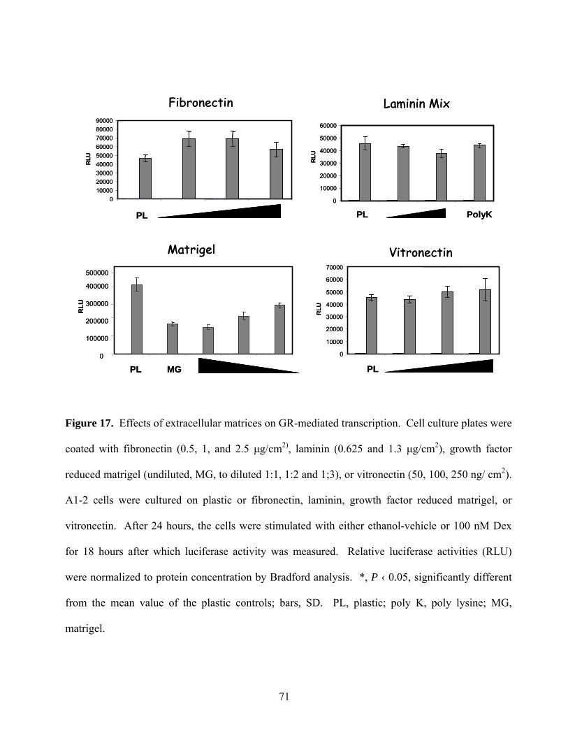
Laminin Mix
0
10000
20000
30000
40000
50000
60000
PL PolyK
RLU
0
100000
200000
300000
400000
500000
RLU
Fibronectin
PL
Vitronectin
PLPL MG
Matrigel
0100002000030000400005000060000700008000090000
RLU
0
10000
20000
30000
40000
50000
60000
70000
RLU
Laminin Mix
0
10000
20000
30000
40000
50000
60000
PL PolyK
RLU
0
100000
200000
300000
400000
500000
RLU
Fibronectin
PL
Vitronectin
PLPL MG
Matrigel
0100002000030000400005000060000700008000090000
RLU
0
10000
20000
30000
40000
50000
60000
70000
RLU
Figure 17. Effects of extracellular matrices on GR-mediated transcription. Cell culture plates were
coated with fibronectin (0.5, 1, and 2.5 μg/cm2), laminin (0.625 and 1.3 μg/cm2), growth factor
reduced matrigel (undiluted, MG, to diluted 1:1, 1:2 and 1;3), or vitronectin (50, 100, 250 ng/ cm2).
A1-2 cells were cultured on plastic or fibronectin, laminin, growth factor reduced matrigel, or
vitronectin. After 24 hours, the cells were stimulated with either ethanol-vehicle or 100 nM Dex
for 18 hours after which luciferase activity was measured. Relative luciferase activities (RLU)
were normalized to protein concentration by Bradford analysis. *, P ‹ 0.05, significantly different
from the mean value of the plastic controls; bars, SD. PL, plastic; poly K, poly lysine; MG,
matrigel.
71
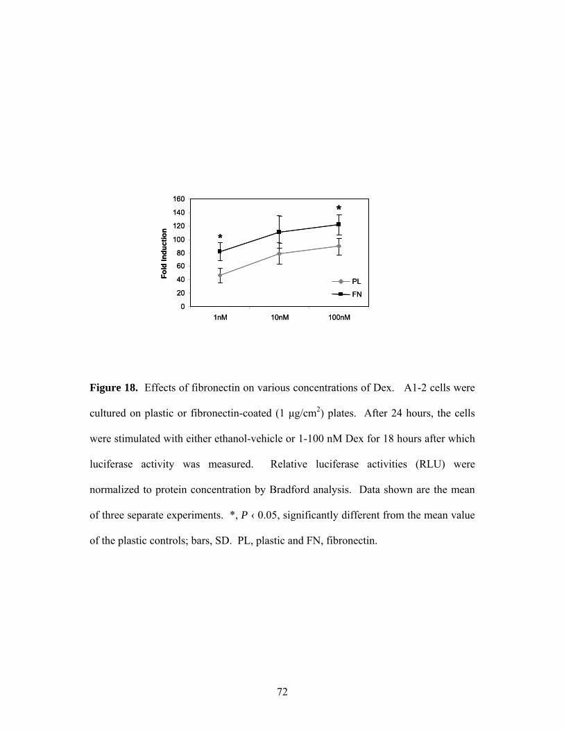
0
20
40
60
80
100
120
140
160
1nM 10nM 100nM
Fold
Indu
ctio
n
PL
FN
*
*
0
20
40
60
80
100
120
140
160
1nM 10nM 100nM
Fold
Indu
ctio
n
PL
FN
*
*
Figure 18. Effects of fibronectin on various concentrations of Dex. A1-2 cells were
cultured on plastic or fibronectin-coated (1 μg/cm2) plates. After 24 hours, the cells
were stimulated with either ethanol-vehicle or 1-100 nM Dex for 18 hours after which
luciferase activity was measured. Relative luciferase activities (RLU) were
normalized to protein concentration by Bradford analysis. Data shown are the mean
of three separate experiments. *, P ‹ 0.05, significantly different from the mean value
of the plastic controls; bars, SD. PL, plastic and FN, fibronectin.
72
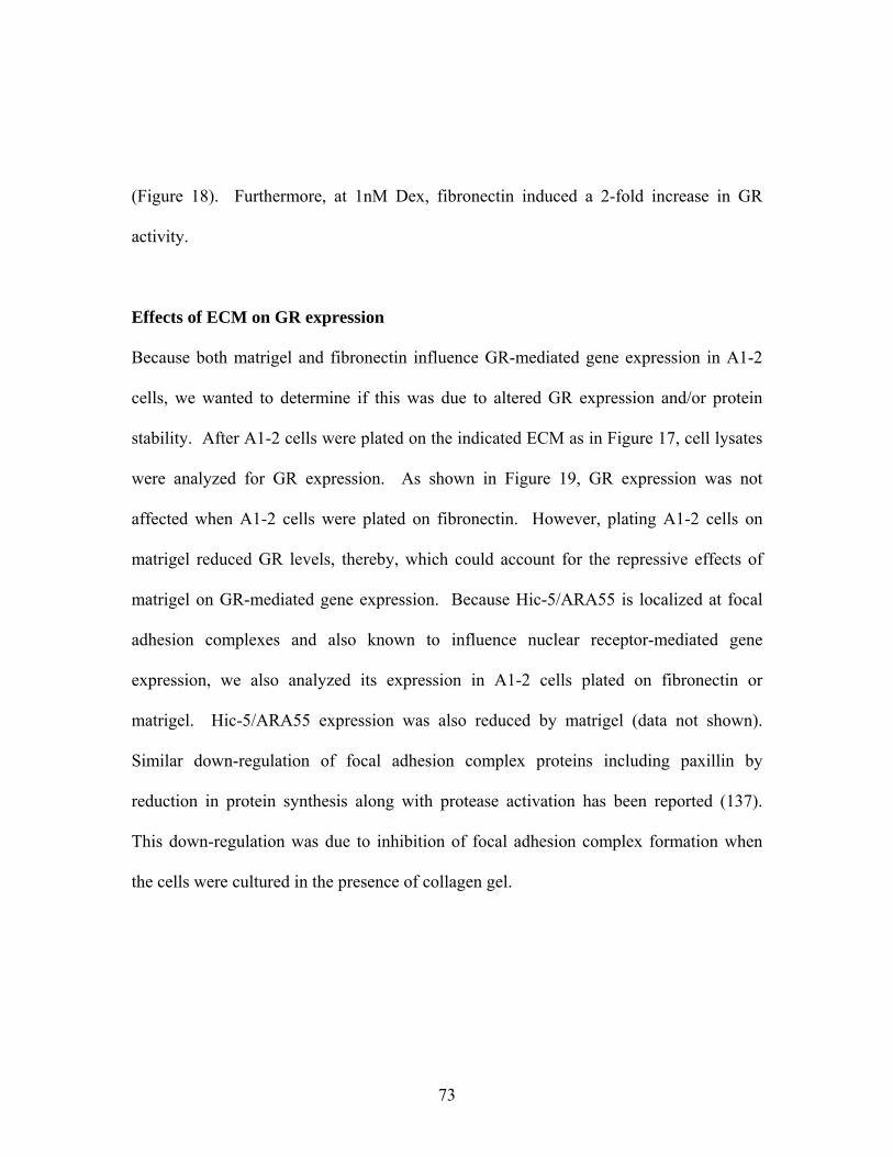
(Figure 18). Furthermore, at 1nM Dex, fibronectin induced a 2-fold increase in GR
activity.
Effects of ECM on GR expression
Because both matrigel and fibronectin influence GR-mediated gene expression in A1-2
cells, we wanted to determine if this was due to altered GR expression and/or protein
stability. After A1-2 cells were plated on the indicated ECM as in Figure 17, cell lysates
were analyzed for GR expression. As shown in Figure 19, GR expression was not
affected when A1-2 cells were plated on fibronectin. However, plating A1-2 cells on
matrigel reduced GR levels, thereby, which could account for the repressive effects of
matrigel on GR-mediated gene expression. Because Hic-5/ARA55 is localized at focal
adhesion complexes and also known to influence nuclear receptor-mediated gene
expression, we also analyzed its expression in A1-2 cells plated on fibronectin or
matrigel. Hic-5/ARA55 expression was also reduced by matrigel (data not shown).
Similar down-regulation of focal adhesion complex proteins including paxillin by
reduction in protein synthesis along with protease activation has been reported (137).
This down-regulation was due to inhibition of focal adhesion complex formation when
the cells were cultured in the presence of collagen gel.
73
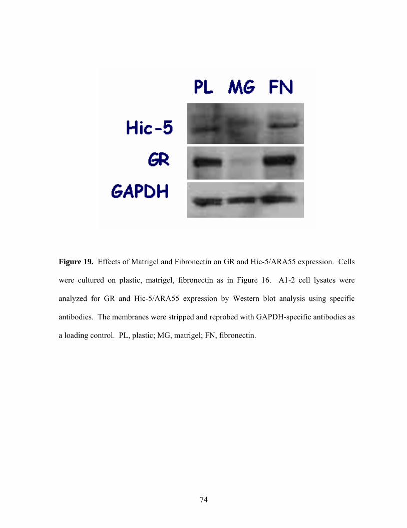
Figure 19. Effects of Matrigel and Fibronectin on GR and Hic-5/ARA55 expression. Cells
were cultured on plastic, matrigel, fibronectin as in Figure 16. A1-2 cell lysates were
analyzed for GR and Hic-5/ARA55 expression by Western blot analysis using specific
antibodies. The membranes were stripped and reprobed with GAPDH-specific antibodies as
a loading control. PL, plastic; MG, matrigel; FN, fibronectin.
74
Figure 20. Effects of fibronectin on nucleocytoplasmic shuttling of GR. Cells were
cultured on plastic or fibronectin in Figure 16. After treatment with vehicle (-) Dex (+,
100nM) for 1 h, A1-2 cells were harvested and fractionated in order to separate the
nuclear and cytoplasmic compartments. Lysates were analyzed for GR expression by
Western blot analysis using an anti-GR-specific antibody. Purity of each fractionation
was analyzed using cytoplasmic or nuclear markers, actin and Sp-1 respectively.
75
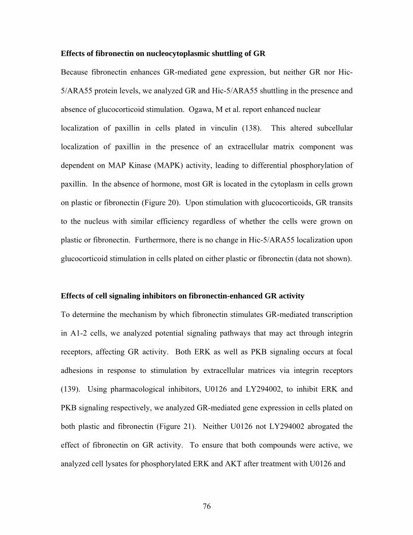
Effects of fibronectin on nucleocytoplasmic shuttling of GR
Because fibronectin enhances GR-mediated gene expression, but neither GR nor Hic-
5/ARA55 protein levels, we analyzed GR and Hic-5/ARA55 shuttling in the presence and
absence of glucocorticoid stimulation. Ogawa, M et al. report enhanced nuclear
localization of paxillin in cells plated in vinculin (138). This altered subcellular
localization of paxillin in the presence of an extracellular matrix component was
dependent on MAP Kinase (MAPK) activity, leading to differential phosphorylation of
paxillin. In the absence of hormone, most GR is located in the cytoplasm in cells grown
on plastic or fibronectin (Figure 20). Upon stimulation with glucocorticoids, GR transits
to the nucleus with similar efficiency regardless of whether the cells were grown on
plastic or fibronectin. Furthermore, there is no change in Hic-5/ARA55 localization upon
glucocorticoid stimulation in cells plated on either plastic or fibronectin (data not shown).
Effects of cell signaling inhibitors on fibronectin-enhanced GR activity
To determine the mechanism by which fibronectin stimulates GR-mediated transcription
in A1-2 cells, we analyzed potential signaling pathways that may act through integrin
receptors, affecting GR activity. Both ERK as well as PKB signaling occurs at focal
adhesions in response to stimulation by extracellular matrices via integrin receptors
(139). Using pharmacological inhibitors, U0126 and LY294002, to inhibit ERK and
PKB signaling respectively, we analyzed GR-mediated gene expression in cells plated on
both plastic and fibronectin (Figure 21). Neither U0126 not LY294002 abrogated the
effect of fibronectin on GR activity. To ensure that both compounds were active, we
analyzed cell lysates for phosphorylated ERK and AKT after treatment with U0126 and
76
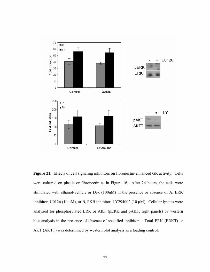
Figure 21. Effects of cell signaling inhibitors on fibronectin-enhanced GR activity. Cells
were cultured on plastic or fibronectin as in Figure 16. After 24 hours, the cells were
stimulated with ethanol-vehicle or Dex (100nM) in the presence or absence of A, ERK
inhibitor, U0126 (10 μM), or B, PKB inhibitor, LY294002 (10 μM). Cellular lysates were
analyzed for phosphorylated ERK or AKT (pERK and pAKT, right panels) by western
blot analysis in the presence of absence of specified inhibitors. Total ERK (ERKT) or
AKT (AKTT) was determined by western blot analysis as a loading control.
77
Figure 22. Effects of fibronectin on GR and Hic-5/ARA55 promoter recruitment in
response to glucocorticoids. The cells were plated on plastic (left lanes) or fibronectin
(right lanes) as before and soluble chromatin was prepared from A1-2 cells treated with
EtOH-vehicle or Dex (100 nM) for 1 h. Protein-bound DNA complexes were
immunoprecipitated with antibodies against GR or Hic-5/ARA55. After crosslink
reversal, purified DNA was amplified with primers specific for the MMTV promoter.
PCR products, resulting from 28 rounds of PCR amplification, in the Input panel were
amplified using diluted chromatin that was not immunoprecipitated. A rabbit IgG was
used to detect any nonspecific immunoprecipitated DNA.
78
LY294002 respectively. Both compounds efficiently inhibited phosphorylation of the
appropriate signaling molecule, indicating that neither ERK nor PKB signaling is
involved in the effect of fibronectin on GR transactivation.
Effects of fibronectin on GR and Hic-5/ARA55 promoter recruitment in response to
glucocorticoids
Because both GR and Hic-5/ARA55 are recruited to glucocorticoid-responsive promoters
in the presence of hormones, we analyzed the effect of fibronectin on GR and Hic-
5/ARA55 promoter recruitment. ChIP analysis of the MMTV promoter in A1-2 cells
plated on plastic or fibronectin revealed that both GR and Hic-5/ARA55 are recruited to
the promoter upon glucocorticoid stimulation (Figure 22). Although there was no
detectable difference in either GR or Hic-5/ARA55 MMTV promoter recruitment in the
presence of Dex, there may be a slight increase in Hic-5/ARA55 promoter binding in the
absence of hormone in cells plated on fibronectin. However, this result is preliminary
and based upon a limited number of experiments.
Discussion
Recent work by various labs has highlighted the responsiveness of nuclear receptors to
ECM components. Specifically, Novaro et al. reported that basement membrane
components such as collagen IV or laminin-1 upregulated ER-mediated gene expression
79
by increasing ERα expression (130, 131). Furthermore, Sisci et al. described increased
ERα nuclear localization and ligand-independent activation of gene expression in cells
plated on either fibronectin or collagen IV (140). In fact, increased ERα activity in both
MCF-7 and MDA-MB-231 breast cancer cells was mediated by increased c-Src
activation after cellular attachment to ECM, thereby affecting cellular mobility (140).
Interestingly, increased expression and activity of c-Src has been reported in various
tumors, including breast cancer, possibly increasing their malignant capabilities (141,
142).
Using A1-2 breast cancer cells, we analyzed various ECM for their ability to modulate
GR-mediated transactivation. Furthermore, because Hic-5/ARA55 is a focal adhesion
complex-associated protein that influences gene expression, it was postulated that it may
transmit signaling events from the ECM to the nucleus. As mentioned previously, it has
been reported that nuclear accumulation of Hic-5/ARA55 occurs in response to oxidants
such as H2O2 (64). Furthermore, using LMB for 1 h in WPMY-1 cells, we were able to
detect most Hic-5/ARA55 in the nucleus, indicating that Hic-5/ARA55 rapidly transits
between cytoplasmic and nuclear compartments (Figure 8).
After analyzing various ECM, we determined that both fibronectin and growth factor
reduced matrigel influenced GR transactivation in A1-2 cells by different mechanisms.
Matrigel negatively influenced GR-mediated gene expression by reducing GR as well as
Hic-5/ARA55 expression levels. Although the exact by which matrigel reduces GR and
Hic-5/ARA55 expression levels is not known, Wang et al. report a similar reduction of
80
focal adhesion complex proteins such as focal adhesion kinase (FAK) and paxillin by
matrigel (137). Both a reduction in protein synthesis as well as activation of calpain
proteinases resulted in decreased focal adhesion protein levels and was mediated by α2β1
integrin activity (137). Following collagen binding, α2β1 integrin signaling may be
involved in focal adhesion complex formation or stabilization by inducing cytoskeletal
rearrangements, ultimately affecting cell migration.
Along with matrigel, fibronectin altered GR-mediated gene expression in A1-2 cells,
increasing GR transactivation. However, fibronectin failed to modulate GR expression,
subcellular trafficking, or promoter recruitment. Hic-5/ARA55 expression and activity
were also unaffected by fibronectin. Furthermore, inhibitors of both ERK and PKB
signaling pathways failed to repress fibronectin-induced GR activation in A1-2 cells.
However, it is possible that integrin signaling through other proteins may be involved,
possibly through c-Src. Furthermore, because the effect of fibronectin on GR-mediated
gene expression was modest, induction of slight changes in GR expression, subcellular
trafficking or promoter recruitment may be undetectable. Alternatively, because ECM
stimulates numerous intracellular signaling events, fibronectin may promote alternative
GR phosphorylation, resulting in enhance GR-mediated gene expression. GR is a
phosphoprotein, capable of being phosphorylated at multiple sites, some of which induce
greater GR nuclear localization and activity (143). A detailed analysis of the
phosphorylation state of GR in cells plated on fibronectin will determine if that is the
mechanism by which fibronectin enhances GR-mediated gene expression.
81
CHAPTER 6 DISCUSSION
The aim of this project was to determine the mechanism by which Hic-5/ARA55 serves
as a nuclear receptor coactivator as well as determining its potential involvement in PCa
progression. Tumorigenesis is a multi-step process which involves changes in not only
the malignant cells but also the surrounding microenvironment of that tumor, including
stromal cells and ECM. Because Hic-5/ARA55 is a nuclear receptor coactivator that is
involved in cellular differentiation as well as growth, analysis of its expression and
activity may illuminate certain aspects of prostate cancer initiation or progression. Our
results identify Hic-5/ARA55 as a compartment-specific AR coactivator that has an
impact on androgen regulation of a paracrine growth factor, KGF, in the stroma.
Hic-5/ARA55-coregulator interactions
In recent years, numerous nuclear receptor-interacting proteins have been identified that
modify chromatin, thereby influencing nuclear receptor-mediated gene expression.
Because assays using reporter gene analysis to determine nuclear receptor activity
revealed that overexpression of most coactivators enhanced the activity of many nuclear
receptors, coactivator specificity was questioned. Hence, it was postulated that
coactivators function in a cell type or promoter-specific manner. Using the MMTV
promoter, Li et al. isolated a specific coactivator complex that associated with PR that
differed to the coactivator complex which bound to GR (14). These results indicate that
combinations of coactivators rather than individual coactivators may be responsible for
82
promoter or hormone-specific gene expression. Furthermore, coactivator knock out
analysis provided evidence that at least in part, coactivator function is not totally
redundant. For example, TIF-2 and RAC3 null mice display reduced reproductive
capability while SRC-1 null mice are exhibit partial hormone resistance (41, 42).
Along with coactivators that modify chromatin, other coactivators such as Hic-5/ARA55
have been discovered whose mechanism of action is largely unknown. Because Hic-
5/ARA55 does not possess a catalytic domain that is responsible for its coactivation
properties, its mechanism of coregulator function has remained undefined. However, it
may serve as an adaptor molecule, either recruiting or stabilizing promoter-specific
protein complexes. LIM proteins are well recognized for their roles as molecular
adaptors, functioning in stabilizing higher order protein complexes at either focal
adhesion complexes or promoter sequences. siRNA ablation experiments reported here
establish that Hic-5/ARA55 is required for the stable association of p300 and TIF-2 with
the MMTV promoter (Figure 6). Thus, Hic-5/ARA55 may stabilize select protein
complex formation at GR-responsive promoters by serving as an adaptor molecule.
Not only does Hic-5/ARA55 interact with various coactivator complexes, but it also
associates with NCoR corepressor complexes in the absence of hormone at nuclear
receptor-responsive promoters (Figure 5C). This suggests that Hic-5/ARA55 is capable
of interacting with other coregulators directly not necessarily via nuclear receptors.
Furthermore, because Hic-5/ARA55 is present on GR-responsive promoters in the
83
absence and presence of glucocorticoids, it may function in coordinating corepressor
release and coactivator recruitment upon glucocorticoid stimulation.
Recently, transducin β-like 1 (TBL1), an adaptor-like protein, has been reported to
mediate the exchange of corepressors for coactivators on nuclear receptor-responsive
promoters in response to ligand (144). TBL1 was initially isolated as part of the
corepressor complex (145). ChIP analysis of nuclear receptor target promoters revealed
prolonged TBL1 promoter association in the presence of ligand (144). Furthermore,
TBL1 recruited proteosome machinery to nuclear receptor target promoters, leading to
degradation of the corepressor complex followed by association of the coactivator
complex (144). Although the possible interaction of Hic-5/ARA55 with components of
the proteosome machinery has not been analyzed, it may provide a mechanism by which
Hic-5/ARA55 interacts with both corepressors and coactivators, culminating in enhanced
nuclear receptor-mediated gene expression.
Although Hic-5/ARA55 was localized to GR-responsive promoters in the absence of
ligand and detectable promoter-bound GR, the mechanism by which Hic-5/ARA55 is
bound in the absence of receptor is unclear. In addition to receptor-independent binding
of Hic-5/ARA55 to the MMTV promoter, we also isolated receptor-independent
localization of NCoR to the MMTV promoter in the absence of ligand. Direct binding of
either Hic-5/ARA55 or NCoR to the MMTV promoter has not been analyzed.
Interestingly, Hic-5/ARA55 does display zinc-dependent DNA binding (146). However,
a specific DNA sequence for which Hic-5/ARA55 is capable of binding has yet to be
84
identified. Instead, it is thought that Hic-5/ARA55 may be capable of binding DNA in a
more non-specific manner. If Hic-5/ARA55 is capable of directly binding the MMTV
promoter, it may function in tethering the NCoR-containing complex to the MMTV
promoter in the absence of ligand. However, it is also possible that both Hic-5/ARA55
and NCoR are binding the MMTV promoter through another, yet unidentified DNA
binding protein.
Although Hic-5/ARA55 interactions with other coactivators as well as NCoR were
observed in the A1-2 cells, we anticipate that similar interactions would be found in the
WMPY-1 stromal cells. Specifically, we detected Hic-5/ARA55 bound to the KGF
promoter in the absence of androgens (Figure 15). Ligand independent promoter
localization of Hic-5/ARA55 was also observed in the A1-2 cells. Furthermore, addition
of ligand enhanced the amount of Hic-5/ARA55 promoter recruitment in both cell lines.
Because Hic-5/ARA55 is found within coactivator as well as corepressor complexes in
A1-2 cells, we anticipate that similar interactions may occur in WPMY-1 cells, possibly
in a promoter-specific manner.
Coactivators and PCa
Because some steroid-responsive nuclear receptors such as AR and ER have been
associated with cancers derived from endocrine target tissues, their expression as well as
coactivator expression and activity has been analyzed in breast as well as PCa. As
previously mentioned, treatment of PCa with anti-androgen therapy initially thwarts
85
tumor growth. However, most PCa eventually progresses into a hormone refractory,
androgen-independent PCa, capable of growth in the presence of anti-androgens.
Although the exact mechanism by which PCa becomes androgen-independent is unclear,
coactivator expression and/or activity may be involved. As previously mentioned, altered
expression of coactivators may enhance ligand-independent AR activity in androgen
independent prostate cancer. For example, increased TIF-2 expression enhanced AR-
mediated gene expression in response to estradiol as well as progesterone (106).
Interestingly, Hic-5/ARA55 expression was reduced in tumor as compared to normal
tissue (112). Furthermore, Hic-5 mRNA levels were lower in hormone-refractory PCa
than that of previously untreated PCa. In recurrent hormone refractory PCa, higher Hic-
5/ARA55 expression correlated with unfavorable recurrence-free survival as well as
overall survival (122). Because most analyses of coactivator expression in PCa
progression use quantitative PCR analysis of whole prostate tissue sections, it is difficult
to ascertain whether this represents reduced expression of Hic-5/ARA55 within
individual cells or alterations in the balance between stromal/epithelial cell content in
tumors. Subsequent analysis of Hic-5/ARA55 expression in primary prostate stromal
cells derived from normal or tumor prostates revealed reduced Hic-5/ARA55 expression
in tumor stromal cells (unpublished data). However, analysis of Hic-5/ARA55
expression in prostate tissue sections indicated that there was some continued expression
of Hic-5/ARA55 in prostate tumors (Figure 12).
Because partially silencing Hic-5/ARA55 expression in WMPY-1 cells resulted in
increased basal KGF expression, we anticipate that reduced Hic-5/ARA55 expression in
86
reactive stromal cells may also result in increased KGF expression. In fact, KGF
expression is significantly increased in prostate cancer (22). Enhanced KGF expression
possibly due to reduced Hic-5/ARA55 expression may result in androgen-independent
activation of AR or increased tumor epithelial proliferation, thereby promoting androgen-
independent PCa progression.
Reactive Stromal Transition
Along with changes in the epithelia compartment, which is the source of tumor cells,
development of an altered stromal microenvironment, termed reactive stroma, carcinoma
associated fibroblasts (CAF) or tumor stroma, enhances tumor cell survival, cellular
proliferation, and migration (81, 82). The presence of undifferentiated myofibroblast
cells which express both smooth muscle cell and fibroblasts markers induces changes in
ECM expression as well (83). Myofibroblasts are commonly found at areas undergoing
tissue remodeling, producing ECM such as collagen I and fibronectin as well as
proteases, including matrix metalloproteinases (MMPs) (84-86). Along with altered
ECM expression by reactive stromal cells, data indicate altered gene expression of
growth factors such as KGF, FGF-2, TGF-β, and IL-6, and matrix metalloproteinases in
reactive stromal cells (22, 89-92). The differential gene expression pattern induced by
the reactive stroma may confer resistance to cell cycle inhibitors or metastatic properties
to the malignant cells. As mentioned previously, TGF-β may be involved in the
progression to a reactive stroma phenotype by inducing myofibroblast formation (83).
Interestingly, TGF-β induces Hic-5/ARA55 expression in fibroblasts although it is
87
unclear whether its expression is upregulated in reactive stromal cells. Because Hic-
5/ARA55 is involved in both ECM and KGF expression in normal prostate stromal cells,
upregulation of Hic-5/ARA55 expression resulting from increased TGF- β stimulation
could result in differential ECM as well as KGF production.
Hic-5/ARA55 and development
During tissue development, progenitor cells terminally differentiate whereby they usually
exit the cell cycle as well as phenotypically differentiate. Hic-5/ARA55 expression has
been shown to both induce and repress cellular differentiation in a cell type-specific
manner. For example, rat calvarial bone cells (RCT-1) differentiate into osteoblasts after
retinoic acid treatment, which decreases cell growth and induces alkaline phosphatase
and α I pro-collagen expression (147). During RCT-1 differentiation, Hic-5/ARA55
expression is upregulated (147). Furthermore, overexpression of Hic-5/ARA55 in RCT-1
cells leads to decreased cellular proliferation as well as increased αI pro-collagen
synthesis, both indicative of osteoblast differentiation (147). Alternatively, during
myogenesis, Hic-5/ARA55 expression correlates with apoptotic myoblasts (148). The
process of myogenesis is characterized by myoblast fusion and induction of muscle-
specific gene expression, culminating in myotubule formation. Myoblasts that fail to
differentiate undergo apoptosis, and Hic-5/ARA55 expression not only blocks
myogenesis but also induces apoptosis in mouse C2C12 myoblasts (148).
88
Along with either promoting or inhibiting cellular differentiation, Hic-5/ARA55 may also
be involved cell type-specific differentiation programs. For example, Hic-5/ARA55 is
expressed in gut epithelial cells during development (66). Furthermore, it serves as a
coactivator for PPARγ-mediated gene expression in intestinal epithelial cells. Partial
ablation of Hic-5/ARA55 expression repressed PPARγ-mediated expression of keratin
20, L-FABP, and KLF. Finally, expression of Hic-5/ARA55 in preadipocytes not only
inhibits adipogenesis but also induces epithelial gene expression in these mesenchymal
cells, suggesting that Hic-5/ARA55 regulates epithelial gene expression in the gut (66).
Interestingly, TBL1, another nuclear receptor associated- adaptor protein, expression is
necessary for PPARγ-induced adipogenesis (144).
We show here a role for Hic-5/ARA55 in KGF expression in WPMY-1 human stromal
cells of the prostate. KGF, a crucial mediator of epithelial-stromal interactions during
prostate development, is regulated by androgens. For example, KGF neutralizing
antibodies inhibit prostate epithelial growth as well as ductal branching (1). Furthermore,
addition of exogenous KGF to a serum-free organ culture system leads to prostate growth
as well as ductal branching, overcoming the requirement for testosterone (1). As shown
in Figure 10, androgen regulation of KGF expression is mediated by AR recruitment to
the KGF promoter. Silencing of Hic-5/ARA55 expression led to increased basal
expression of KGF in the absence of androgens (Figure 16). Furthermore, the increase in
basal KGF expression upon Hic-5/ARA55 ablation suggests that this coactivator may
interact with other transcription factors or corepressors to repress KGF expression in the
absence of androgens (65). By partially silencing Hic-5/ARA55 expression in WPMY-1
89
cells, Hic-5/ARA55-mediated repression may be removed, leading to KGF expression in
the absence of androgens. Furthermore, AR recruitment to the KGF promoter along with
subsequent coactivator interactions in the presence of androgens may negate the
repression by Hic-5/ARA55, resulting in androgen-induced KGF expression. In A1-2
cells, the enhanced association of NCoR with Hic-5/ARA55 complexes at the MMTV
promoter in non-hormone treated cells suggests that Hic-5/ARA55 may, in fact,
participate in maintaining low basal levels of transcription from this promoter. Thus, by
serving opposing roles to positively or negatively regulate KGF gene transcription, Hic-
5/ARA55 may be critical in sculpting tissue organization in the prostate, responding to
various hormonal and paracrine cues that serve to maintain the appropriate balance
between distinct stromal and epithelial cell layers. Indeed analysis of Hic-5/ARA55
expression during prostate development may be beneficial in determining the mechanism
by which Hic-5/ARA55 and AR regulation of KGF expression may be involved in
prostate development.
Hic-5/ARA55 and cell growth
As mentioned previously, Hic-5/ARA55 negatively affects cell growth in both
osteoblasts and myoblasts (147, 148). In nontumorigenic, immortalized human
fibroblasts, overexpression of Hic-5/ARA55 induced senescence (121). Cellular
senescence or cellular aging is characterized by an irreversible growth arrest.
Interestingly, p21 (Cip1/WAF1/sdi1) cyclin-cdk inhibitor, a gene that is induced during
senescence, is upregulated in cells that overexpress Hic-5/ARA55 (121, 149). As shown
90
in Figures 3 and 4, Hic-5/ARA55 is recruited to the p21 promoter after glucocorticoid
stimulation, and silencing Hic-5/ARA55 reduced glucocorticoid-induced p21 expression.
It is possible that overexpression of Hic-5/ARA55 during senescence may induce p21
expression directly, thereby overcoming the need for glucocorticoid induction.
Furthermore, induction of c-fos expression upon serum stimulation is lost in cells
undergoing senescence, inhibiting cellular proliferation (150). In Hic-5/ARA55
overexpressing cells, c-fos induction upon TPA stimulation is lost as well (121).
However, H2O2 treatment, inducing Hic-5/ARA nuclear localization, upregulated c-fos
expression in TIG-7 human fibroblasts (64). In A1-2 cells, we saw no effect on c-fos
expression upon Hic-5/ARA55 depletion, suggesting that Hic-5/ARA55 expression is not
necessary for c-fos induction by glucocorticoids in nonsenescent cells (Figure 4).
The recent demonstration of Hic-5/ARA55 involvement in PPARγ-induced epithelial cell
differentiation program illustrates the importance of “adaptor” coactivators that lack
enzymatic activity in assembling functional coregulator complexes on distinct promoters.
More detailed analysis of this novel family of nuclear receptor coactivators may unlock
multiple nuclear receptor gene networks that utilize LIM domain-containing adaptors to
organize gene-specific coactivator assemblies. In conclusion, given the role of stromal-
derived KGF on the growth, transformation, and invasive properties of PCa cells,
coactivators such as Hic-5/ARA55 may influence tumor development and progression
through its impact on either basal and/or androgen-induced expression of KGF in stroma
during both androgen-dependent and androgen-independent phases (126, 127).
91
REFERENCES
1. Sugimura, Y., Foster, B. A., Hom, Y. K., Lipschutz, J. H., Rubin, J. S., Finch, P.
W., Aaronson, S. A., Hayashi, N., Kawamura, J., and Cunha, G. R. Keratinocyte
growth factor (KGF) can replace testosterone in the ductal branching
morphogenesis of the rat ventral prostate. Int J Dev Biol, 40: 941-951, 1996.
2. Hsing, A. Y., Kadomatsu, K., Bonham, M. J., and Danielpour, D. Regulation of
apoptosis induced by transforming growth factor-beta1 in nontumorigenic rat
prostatic epithelial cell lines. Cancer Res, 56: 5146-5149, 1996.
3. Ittman, M. and Mansukhani, A. Expression of fibroblast growth factors (FGFs)
and FGF receptors in human prostate. J Urol, 157: 351-356, 1997.
4. English, H. F., Drago, J. R., and Santen, R. J. Cellular response to androgen
depletion and repletion in the rat ventral prostate: autoradiography and
morphometric analysis. Prostate, 7: 41-51, 1985.
5. Isaacs, J. T. Development and characteristics of the available animal model
systems for the study of prostatic cancer. Prog Clin Biol Res, 239: 513-576, 1987.
6. Chung, L. W. The role of stromal-epithelial interaction in normal and malignant
growth. Cancer Surv, 23: 33-42, 1995.
7. Byrne, R. L., Leung, H., and Neal, D. E. Peptide growth factors in the prostate as
mediators of stromal epithelial interaction. Br J Urol, 77: 627-633, 1996.
8. Hayward, S. W., Haughney, P. C., Rosen, M. A., Greulich, K. M., Weier, H. U.,
Dahiya, R., and Cunha, G. R. Interactions between adult human prostatic
92
epithelium and rat urogenital sinus mesenchyme in a tissue recombination model.
Differentiation, 63: 131-140, 1998.
9. Collins, A. T., Robinson, E. J., and Neal, D. E. Benign prostatic stromal cells are
regulated by basic fibroblast growth factor and transforming growth factor-beta 1.
J Endocrinol, 151: 315-322, 1996.
10. Dahiya, R., Lee, C., Haughney, P. C., Chui, R., Ho, R., and Deng, G. Differential
gene expression of transforming growth factors alpha and beta, epidermal growth
factor, keratinocyte growth factor, and their receptors in fetal and adult human
prostatic tissues and cancer cell lines. Urology, 48: 963-970, 1996.
11. Hayashi, N. and Cunha, G. R. Mesenchyme-induced changes in the neoplastic
characteristics of the Dunning prostatic adenocarcinoma. Cancer Res, 51: 4924-
4930, 1991.
12. Hayward, S. W., Baskin, L. S., Haughney, P. C., Cunha, A. R., Foster, B. A.,
Dahiya, R., Prins, G. S., and Cunha, G. R. Epithelial development in the rat
ventral prostate, anterior prostate and seminal vesicle. Acta Anat (Basel), 155: 81-
93, 1996.
13. Cooke, P. S., Young, P., and Cunha, G. R. Androgen receptor expression in
developing male reproductive organs. Endocrinology, 128: 2867-2873, 1991.
14. Cunha, G. R. and Lung, B. The possible influence of temporal factors in
androgenic responsiveness of urogenital tissue recombinants from wild-type and
androgen-insensitive (Tfm) mice. J Exp Zool, 205: 181-193, 1978.
15. Cunha, G. R. and Donjacour, A. Stromal-epithelial interactions in normal and
abnormal prostatic development. Prog Clin Biol Res, 239: 251-272, 1987.
93
16. Shimazaki, J., Kurihara, H., Ito, Y., and Shida, K. Testosterone metabolism in
prostate; formation of androstan-17-beta-ol-3-one and androst-4-ene-3, 17-dione,
and inhibitory effect of natural and synthetic estrogens. Gunma J Med Sci, 14:
313-325, 1965.
17. Lin, M. F., Garcia-Arenas, R., Kawachi, M., and Lin, F. F. Regulation of the
expression of prostatic acid phosphatase in LNCaP human prostate carcinoma
cells. Cell Mol Biol Res, 39: 739-750, 1993.
18. Murtha, P., Tindall, D. J., and Young, C. Y. Androgen induction of a human
prostate-specific kallikrein, hKLK2: characterization of an androgen response
element in the 5' promoter region of the gene. Biochemistry, 32: 6459-6464,
1993.
19. Fasciana, C., van der Made, A. C., Faber, P. W., and Trapman, J. Androgen
regulation of the rat keratinocyte growth factor (KGF/FGF7) promoter. Biochem
Biophys Res Commun, 220: 858-863, 1996.
20. Planz, B., Wang, Q., Kirley, S. D., Lin, C. W., and McDougal, W. S. Androgen
responsiveness of stromal cells of the human prostate: regulation of cell
proliferation and keratinocyte growth factor by androgen. J Urol, 160: 1850-1855,
1998.
21. Rubin, J. S., Bottaro, D. P., Chedid, M., Miki, T., Ron, D., Cunha, G. R., and
Finch, P. W. Keratinocyte growth factor as a cytokine that mediates
mesenchymal-epithelial interaction. Exs, 74: 191-214, 1995.
94
22. Leung, H. Y., Mehta, P., Gray, L. B., Collins, A. T., Robson, C. N., and Neal, D.
E. Keratinocyte growth factor expression in hormone insensitive prostate cancer.
Oncogene, 15: 1115-1120, 1997.
23. Pratt, W. B. and Toft, D. O. Steroid receptor interactions with heat shock protein
and immunophilin chaperones. Endocr Rev, 18: 306-360, 1997.
24. Glass, C. K. and Rosenfeld, M. G. The coregulator exchange in transcriptional
functions of nuclear receptors. Genes Dev, 14: 121-141, 2000.
25. Handschin, C. and Meyer, U. A. Regulatory network of lipid-sensing nuclear
receptors: roles for CAR, PXR, LXR, and FXR. Arch Biochem Biophys, 433:
387-396, 2005.
26. Godowski, P. J., Rusconi, S., Miesfeld, R., and Yamamoto, K. R. Glucocorticoid
receptor mutants that are constitutive activators of transcriptional enhancement.
Nature, 325: 365-368, 1987.
27. Brinkmann, A. O., Blok, L. J., de Ruiter, P. E., Doesburg, P., Steketee, K.,
Berrevoets, C. A., and Trapman, J. Mechanisms of androgen receptor activation
and function. J Steroid Biochem Mol Biol, 69: 307-313, 1999.
28. Young, C. Y., Montgomery, B. T., Andrews, P. E., Qui, S. D., Bilhartz, D. L., and
Tindall, D. J. Hormonal regulation of prostate-specific antigen messenger RNA in
human prostatic adenocarcinoma cell line LNCaP. Cancer Res, 51: 3748-3752,
1991.
29. Hanson, R. W. and Reshef, L. Regulation of phosphoenolpyruvate carboxykinase
(GTP) gene expression. Annu Rev Biochem, 66: 581-611, 1997.
95
30. Nissen, R. M. and Yamamoto, K. R. The glucocorticoid receptor inhibits
NFkappaB by interfering with serine-2 phosphorylation of the RNA polymerase II
carboxy-terminal domain. Genes Dev, 14: 2314-2329, 2000.
31. McKenna, N. J., Lanz, R. B., and O'Malley, B. W. Nuclear receptor coregulators:
cellular and molecular biology. Endocr Rev, 20: 321-344, 1999.
32. Onate, S. A., Tsai, S. Y., Tsai, M. J., and O'Malley, B. W. Sequence and
characterization of a coactivator for the steroid hormone receptor superfamily.
Science, 270: 1354-1357, 1995.
33. Bannister, A. J. and Kouzarides, T. The CBP co-activator is a histone
acetyltransferase. Nature, 384: 641-643, 1996.
34. Ogryzko, V. V., Schiltz, R. L., Russanova, V., Howard, B. H., and Nakatani, Y.
The transcriptional coactivators p300 and CBP are histone acetyltransferases.
Cell, 87: 953-959, 1996.
35. Spencer, T. E., Jenster, G., Burcin, M. M., Allis, C. D., Zhou, J., Mizzen, C. A.,
McKenna, N. J., Onate, S. A., Tsai, S. Y., Tsai, M. J., and O'Malley, B. W.
Steroid receptor coactivator-1 is a histone acetyltransferase. Nature, 389: 194-
198, 1997.
36. Swope, D. L., Mueller, C. L., and Chrivia, J. C. CREB-binding protein activates
transcription through multiple domains. J Biol Chem, 271: 28138-28145, 1996.
37. Heery, D. M., Kalkhoven, E., Hoare, S., and Parker, M. G. A signature motif in
transcriptional co-activators mediates binding to nuclear receptors. Nature, 387:
733-736, 1997.
96
38. Darimont, B. D., Wagner, R. L., Apriletti, J. W., Stallcup, M. R., Kushner, P. J.,
Baxter, J. D., Fletterick, R. J., and Yamamoto, K. R. Structure and specificity of
nuclear receptor-coactivator interactions. Genes Dev, 12: 3343-3356, 1998.
39. Ding, X. F., Anderson, C. M., Ma, H., Hong, H., Uht, R. M., Kushner, P. J., and
Stallcup, M. R. Nuclear receptor-binding sites of coactivators glucocorticoid
receptor interacting protein 1 (GRIP1) and steroid receptor coactivator 1 (SRC-1):
multiple motifs with different binding specificities. Mol Endocrinol, 12: 302-313,
1998.
40. Li, X., Wong, J., Tsai, S. Y., Tsai, M. J., and O'Malley, B. W. Progesterone and
glucocorticoid receptors recruit distinct coactivator complexes and promote
distinct patterns of local chromatin modification. Mol Cell Biol, 23: 3763-3773,
2003.
41. Gehin, M., Mark, M., Dennefeld, C., Dierich, A., Gronemeyer, H., and Chambon,
P. The function of TIF2/GRIP1 in mouse reproduction is distinct from those of
SRC-1 and p/CIP. Mol Cell Biol, 22: 5923-5937, 2002.
42. Xu, J., Liao, L., Ning, G., Yoshida-Komiya, H., Deng, C., and O'Malley, B. W.
The steroid receptor coactivator SRC-3 (p/CIP/RAC3/AIB1/ACTR/TRAM-1) is
required for normal growth, puberty, female reproductive function, and mammary
gland development. Proc Natl Acad Sci U S A, 97: 6379-6384, 2000.
43. Xu, J., Qiu, Y., DeMayo, F. J., Tsai, S. Y., Tsai, M. J., and O'Malley, B. W.
Partial hormone resistance in mice with disruption of the steroid receptor
coactivator-1 (SRC-1) gene. Science, 279: 1922-1925, 1998.
97
44. Miyamoto, H., Yeh, S., Wilding, G., and Chang, C. Promotion of agonist activity
of antiandrogens by the androgen receptor coactivator, ARA70, in human prostate
cancer DU145 cells. Proc Natl Acad Sci U S A, 95: 7379-7384, 1998.
45. Yang, L., Guerrero, J., Hong, H., DeFranco, D. B., and Stallcup, M. R. Interaction
of the tau2 transcriptional activation domain of glucocorticoid receptor with a
novel steroid receptor coactivator, Hic-5, which localizes to both focal adhesions
and the nuclear matrix. Mol Biol Cell, 11: 2007-2018, 2000.
46. Fujimoto, N., Yeh, S., Kang, H. Y., Inui, S., Chang, H. C., Mizokami, A., and
Chang, C. Cloning and characterization of androgen receptor coactivator, ARA55,
in human prostate. J Biol Chem, 274: 8316-8321, 1999.
47. Sanchez-Garcia, I. and Rabbitts, T. H. The LIM domain: a new structural motif
found in zinc-finger-like proteins. Trends Genet, 10: 315-320, 1994.
48. Dawid, I. B., Breen, J. J., and Toyama, R. LIM domains: multiple roles as
adapters and functional modifiers in protein interactions. Trends Genet, 14: 156-
162, 1998.
49. Xu, Z., Huang, S., Chang, L. S., Agulnick, A. D., and Brandt, S. J. Identification
of a TAL1 target gene reveals a positive role for the LIM domain-binding protein
Ldb1 in erythroid gene expression and differentiation. Mol Cell Biol, 23: 7585-
7599, 2003.
50. Yang, N., Higuchi, O., Ohashi, K., Nagata, K., Wada, A., Kangawa, K., Nishida,
E., and Mizuno, K. Cofilin phosphorylation by LIM-kinase 1 and its role in Rac-
mediated actin reorganization. Nature, 393: 809-812, 1998.
98
51. Kasai, M., Guerrero-Santoro, J., Friedman, R., Leman, E. S., Getzenberg, R. H.,
and DeFranco, D. B. The Group 3 LIM domain protein paxillin potentiates
androgen receptor transactivation in prostate cancer cell lines. Cancer Res, 63:
4927-4935, 2003.
52. Kassel, O., Schneider, S., Heilbock, C., Litfin, M., Gottlicher, M., and Herrlich, P.
A nuclear isoform of the focal adhesion LIM-domain protein Trip6 integrates
activating and repressing signals at AP-1- and NF-kappaB-regulated promoters.
Genes Dev, 18: 2518-2528, 2004.
53. Guerrero-Santoro, J., Yang, L., Stallcup, M. R., and DeFranco, D. B. Distinct
LIM domains of Hic-5/ARA55 are required for nuclear matrix targeting and
glucocorticoid receptor binding and coactivation. J Cell Biochem, 92: 810-819,
2004.
54. Kadrmas, J. L. and Beckerle, M. C. The LIM domain: from the cytoskeleton to
the nucleus. Nat Rev Mol Cell Biol, 5: 920-931, 2004.
55. Labalette, C., Renard, C. A., Neuveut, C., Buendia, M. A., and Wei, Y.
Interaction and functional cooperation between the LIM protein FHL2,
CBP/p300, and beta-catenin. Mol Cell Biol, 24: 10689-10702, 2004.
56. Muller, J. M., Isele, U., Metzger, E., Rempel, A., Moser, M., Pscherer, A., Breyer,
T., Holubarsch, C., Buettner, R., and Schule, R. FHL2, a novel tissue-specific
coactivator of the androgen receptor. Embo J, 19: 359-369, 2000.
57. Levy, L., Wei, Y., Labalette, C., Wu, Y., Renard, C. A., Buendia, M. A., and
Neuveut, C. Acetylation of beta-catenin by p300 regulates beta-catenin-Tcf4
interaction. Mol Cell Biol, 24: 3404-3414, 2004.
99

58. Martin, B., Schneider, R., Janetzky, S., Waibler, Z., Pandur, P., Kuhl, M.,
Behrens, J., von der Mark, K., Starzinski-Powitz, A., and Wixler, V. The LIM-
only protein FHL2 interacts with beta-catenin and promotes differentiation of
mouse myoblasts. J Cell Biol, 159: 113-122, 2002.
59. Kanungo, J., Pratt, S. J., Marie, H., and Longmore, G. D. Ajuba, a cytosolic LIM
protein, shuttles into the nucleus and affects embryonal cell proliferation and fate
decisions. Mol Biol Cell, 11: 3299-3313, 2000.
60. Petit, M. M., Meulemans, S. M., and Van de Ven, W. J. The focal adhesion and
nuclear targeting capacity of the LIM-containing lipoma-preferred partner (LPP)
protein. J Biol Chem, 278: 2157-2168, 2003.
61. Wang, Y. and Gilmore, T. D. LIM domain protein Trip6 has a conserved nuclear
export signal, nuclear targeting sequences, and multiple transactivation domains.
Biochim Biophys Acta, 1538: 260-272, 2001.
62. Muller, J. M., Metzger, E., Greschik, H., Bosserhoff, A. K., Mercep, L., Buettner,
R., and Schule, R. The transcriptional coactivator FHL2 transmits Rho signals
from the cell membrane into the nucleus. Embo J, 21: 736-748, 2002.
63. Cattaruzza, M., Lattrich, C., and Hecker, M. Focal adhesion protein zyxin is a
mechanosensitive modulator of gene expression in vascular smooth muscle cells.
Hypertension, 43: 726-730, 2004.
64. Shibanuma, M., Kim-Kaneyama, J. R., Ishino, K., Sakamoto, N., Hishiki, T.,
Yamaguchi, K., Mori, K., Mashimo, J., and Nose, K. Hic-5 communicates
between focal adhesions and the nucleus through oxidant-sensitive nuclear export
signal. Mol Biol Cell, 14: 1158-1171, 2003.
100
65. Shibanuma, M., Kim-Kaneyama, J. R., Sato, S., and Nose, K. A LIM protein,
Hic-5, functions as a potential coactivator for Sp1. J Cell Biochem, 91: 633-645,
2004.
66. Drori, S., Girnun, G. D., Tou, L., Szwaya, J. D., Mueller, E., Xia, K., Shivdasani,
R. A., and Spiegelman, B. M. Hic-5 regulates an epithelial program mediated by
PPARgamma. Genes Dev, 19: 362-375, 2005.
67. Moll, R., Schiller, D. L., and Franke, W. W. Identification of protein IT of the
intestinal cytoskeleton as a novel type I cytokeratin with unusual properties and
expression patterns. J Cell Biol, 111: 567-580, 1990.
68. Hinnebusch, B. F., Siddique, A., Henderson, J. W., Malo, M. S., Zhang, W.,
Athaide, C. P., Abedrapo, M. A., Chen, X., Yang, V. W., and Hodin, R. A.
Enterocyte differentiation marker intestinal alkaline phosphatase is a target gene
of the gut-enriched Kruppel-like factor. Am J Physiol Gastrointest Liver Physiol,
286: G23-30, 2004.
69. Roth, K. A., Hertz, J. M., and Gordon, J. I. Mapping enteroendocrine cell
populations in transgenic mice reveals an unexpected degree of complexity in
cellular differentiation within the gastrointestinal tract. J Cell Biol, 110: 1791-
1801, 1990.
70. Horlein, A. J., Naar, A. M., Heinzel, T., Torchia, J., Gloss, B., Kurokawa, R.,
Ryan, A., Kamei, Y., Soderstrom, M., Glass, C. K., and et al. Ligand-independent
repression by the thyroid hormone receptor mediated by a nuclear receptor co-
repressor. Nature, 377: 397-404, 1995.
101
71. Chen, J. D. and Evans, R. M. A transcriptional co-repressor that interacts with
nuclear hormone receptors. Nature, 377: 454-457, 1995.
72. Li, X., Kimbrel, E. A., Kenan, D. J., and McDonnell, D. P. Direct interactions
between corepressors and coactivators permit the integration of nuclear receptor-
mediated repression and activation. Mol Endocrinol, 16: 1482-1491, 2002.
73. Wang, Q., Blackford, J. A., Jr., Song, L. N., Huang, Y., Cho, S., and Simons, S.
S., Jr. Equilibrium interactions of corepressors and coactivators with agonist and
antagonist complexes of glucocorticoid receptors. Mol Endocrinol, 18: 1376-
1395, 2004.
74. Szapary, D., Huang, Y., and Simons, S. S., Jr. Opposing effects of corepressor
and coactivators in determining the dose-response curve of agonists, and residual
agonist activity of antagonists, for glucocorticoid receptor-regulated gene
expression. Mol Endocrinol, 13: 2108-2121, 1999.
75. Papsidero, L. D., Wang, M. C., Valenzuela, L. A., Murphy, G. P., and Chu, T. M.
A prostate antigen in sera of prostatic cancer patients. Cancer Res, 40: 2428-2432,
1980.
76. Catalona, W. J., Smith, D. S., Ratliff, T. L., and Basler, J. W. Detection of organ-
confined prostate cancer is increased through prostate-specific antigen-based
screening. Jama, 270: 948-954, 1993.
77. Huggins, C. B. Commentary on the treatment of prostate cancer. J Urol, 102: 119-
120, 1969.
78. Eisenberger, M. A., Blumenstein, B. A., Crawford, E. D., Miller, G., McLeod, D.
G., Loehrer, P. J., Wilding, G., Sears, K., Culkin, D. J., Thompson, I. M., Jr.,
102
Bueschen, A. J., and Lowe, B. A. Bilateral orchiectomy with or without flutamide
for metastatic prostate cancer. N Engl J Med, 339: 1036-1042, 1998.
79. Hellerstedt, B. A. and Pienta, K. J. The current state of hormonal therapy for
prostate cancer. CA Cancer J Clin, 52: 154-179, 2002.
80. Feldman, B. J. and Feldman, D. The development of androgen-independent
prostate cancer. Nat Rev Cancer, 1: 34-45, 2001.
81. Noel, A., Munaut, C., Boulvain, A., Calberg-Bacq, C. M., Lambert, C. A.,
Nusgens, B., Lapiere, C. M., and Foidart, J. M. Modulation of collagen and
fibronectin synthesis in fibroblasts by normal and malignant cells. J Cell
Biochem, 48: 150-161, 1992.
82. Morrison, C., Thornhill, J., and Gaffney, E. The connective tissue framework in
the normal prostate, BPH and prostate cancer: analysis by scanning electron
microscopy after cellular digestion. Urol Res, 28: 304-307, 2000.
83. Tuxhorn, J. A., Ayala, G. E., Smith, M. J., Smith, V. C., Dang, T. D., and
Rowley, D. R. Reactive stroma in human prostate cancer: induction of
myofibroblast phenotype and extracellular matrix remodeling. Clin Cancer Res,
8: 2912-2923, 2002.
84. DeClerck, Y. A. Interactions between tumour cells and stromal cells and
proteolytic modification of the extracellular matrix by metalloproteinases in
cancer. Eur J Cancer, 36: 1258-1268, 2000.
85. Lagace, R., Grimaud, J. A., Schurch, W., and Seemayer, T. A. Myofibroblastic
stromal reaction in carcinoma of the breast: variations of collagenous matrix and
103
structural glycoproteins. Virchows Arch A Pathol Anat Histopathol, 408: 49-59,
1985.
86. Hanamura, N., Yoshida, T., Matsumoto, E., Kawarada, Y., and Sakakura, T.
Expression of fibronectin and tenascin-C mRNA by myofibroblasts, vascular cells
and epithelial cells in human colon adenomas and carcinomas. Int J Cancer, 73:
10-15, 1997.
87. Hayward, S. W., Cunha, G. R., and Dahiya, R. Normal development and
carcinogenesis of the prostate. A unifying hypothesis. Ann N Y Acad Sci, 784:
50-62, 1996.
88. Olumi, A. F., Grossfeld, G. D., Hayward, S. W., Carroll, P. R., Tlsty, T. D., and
Cunha, G. R. Carcinoma-associated fibroblasts direct tumor progression of
initiated human prostatic epithelium. Cancer Res, 59: 5002-5011, 1999.
89. Giri, D., Ropiquet, F., and Ittmann, M. Alterations in expression of basic
fibroblast growth factor (FGF) 2 and its receptor FGFR-1 in human prostate
cancer. Clin Cancer Res, 5: 1063-1071, 1999.
90. Giri, D., Ozen, M., and Ittmann, M. Interleukin-6 is an autocrine growth factor in
human prostate cancer. Am J Pathol, 159: 2159-2165, 2001.
91. McGarvey, T. W. and Stearns, M. E. Keratinocyte growth factor and receptor
mRNA expression in benign and malignant human prostate. Exp Mol Pathol, 63:
52-62, 1995.
92. Royuela, M., De Miguel, M. P., Bethencourt, F. R., Sanchez-Chapado, M., Fraile,
B., and Paniagua, R. Transforming growth factor beta 1 and its receptor types I
104
and II. Comparison in human normal prostate, benign prostatic hyperplasia, and
prostatic carcinoma. Growth Factors, 16: 101-110, 1998.
93. Sonmez, H., Suer, S., Karaarslan, I., Baloglu, H., and Kokoglu, E. Tissue
fibronectin levels of human prostatic cancer, as a tumor marker. Cancer Biochem
Biophys, 15: 107-110, 1995.
94. Livant, D. L., Brabec, R. K., Pienta, K. J., Allen, D. L., Kurachi, K., Markwart,
S., and Upadhyaya, A. Anti-invasive, antitumorigenic, and antimetastatic
activities of the PHSCN sequence in prostate carcinoma. Cancer Res, 60: 309-
320, 2000.
95. Rabbani, S. A., Harakidas, P., Guo, Y., Steinman, D., Davidsen, S. K., and
Morgan, D. W. Synthetic inhibitor of matrix metalloproteases decreases tumor
growth and metastases in a syngeneic model of rat prostate cancer in vivo. Int J
Cancer, 87: 276-282, 2000.
96. van der Kwast, T. H., Schalken, J., Ruizeveld de Winter, J. A., van Vroonhoven,
C. C., Mulder, E., Boersma, W., and Trapman, J. Androgen receptors in
endocrine-therapy-resistant human prostate cancer. Int J Cancer, 48: 189-193,
1991.
97. Zegarra-Moro, O. L., Schmidt, L. J., Huang, H., and Tindall, D. J. Disruption of
androgen receptor function inhibits proliferation of androgen-refractory prostate
cancer cells. Cancer Res, 62: 1008-1013, 2002.
98. Visakorpi, T., Hyytinen, E., Koivisto, P., Tanner, M., Keinanen, R., Palmberg, C.,
Palotie, A., Tammela, T., Isola, J., and Kallioniemi, O. P. In vivo amplification of
105
the androgen receptor gene and progression of human prostate cancer. Nat Genet,
9: 401-406, 1995.
99. Balk, S. P. Androgen receptor as a target in androgen-independent prostate
cancer. Urology, 60: 132-138; discussion 138-139, 2002.
100. Chen, C. D., Welsbie, D. S., Tran, C., Baek, S. H., Chen, R., Vessella, R.,
Rosenfeld, M. G., and Sawyers, C. L. Molecular determinants of resistance to
antiandrogen therapy. Nat Med, 10: 33-39, 2004.
101. Li, R., Wheeler, T., Dai, H., Frolov, A., Thompson, T., and Ayala, G. High level
of androgen receptor is associated with aggressive clinicopathologic features and
decreased biochemical recurrence-free survival in prostate: cancer patients treated
with radical prostatectomy. Am J Surg Pathol, 28: 928-934, 2004.
102. Culig, Z., Hobisch, A., Cronauer, M. V., Cato, A. C., Hittmair, A., Radmayr, C.,
Eberle, J., Bartsch, G., and Klocker, H. Mutant androgen receptor detected in an
advanced-stage prostatic carcinoma is activated by adrenal androgens and
progesterone. Mol Endocrinol, 7: 1541-1550, 1993.
103. Marcelli, M., Ittmann, M., Mariani, S., Sutherland, R., Nigam, R., Murthy, L.,
Zhao, Y., DiConcini, D., Puxeddu, E., Esen, A., Eastham, J., Weigel, N. L., and
Lamb, D. J. Androgen receptor mutations in prostate cancer. Cancer Res, 60: 944-
949, 2000.
104. Culig, Z., Hobisch, A., Cronauer, M. V., Radmayr, C., Trapman, J., Hittmair, A.,
Bartsch, G., and Klocker, H. Androgen receptor activation in prostatic tumor cell
lines by insulin-like growth factor-I, keratinocyte growth factor, and epidermal
growth factor. Cancer Res, 54: 5474-5478, 1994.
106
105. Hobisch, A., Eder, I. E., Putz, T., Horninger, W., Bartsch, G., Klocker, H., and
Culig, Z. Interleukin-6 regulates prostate-specific protein expression in prostate
carcinoma cells by activation of the androgen receptor. Cancer Res, 58: 4640-
4645, 1998.
106. Gregory, C. W., He, B., Johnson, R. T., Ford, O. H., Mohler, J. L., French, F. S.,
and Wilson, E. M. A mechanism for androgen receptor-mediated prostate cancer
recurrence after androgen deprivation therapy. Cancer Res, 61: 4315-4319, 2001.
107. Anzick, S. L., Kononen, J., Walker, R. L., Azorsa, D. O., Tanner, M. M., Guan,
X. Y., Sauter, G., Kallioniemi, O. P., Trent, J. M., and Meltzer, P. S. AIB1, a
steroid receptor coactivator amplified in breast and ovarian cancer. Science, 277:
965-968, 1997.
108. Janknecht, R. The versatile functions of the transcriptional coactivators p300 and
CBP and their roles in disease. Histol Histopathol, 17: 657-668, 2002.
109. Xu, J. and Li, Q. Review of the in vivo functions of the p160 steroid receptor
coactivator family. Mol Endocrinol, 17: 1681-1692, 2003.
110. Fujimoto, N., Mizokami, A., Harada, S., and Matsumoto, T. Different expression
of androgen receptor coactivators in human prostate. Urology, 58: 289-294, 2001.
111. Li, P., Yu, X., Ge, K., Melamed, J., Roeder, R. G., and Wang, Z. Heterogeneous
expression and functions of androgen receptor co-factors in primary prostate
cancer. Am J Pathol, 161: 1467-1474, 2002.
112. Mestayer, C., Blanchere, M., Jaubert, F., Dufour, B., and Mowszowicz, I.
Expression of androgen receptor coactivators in normal and cancer prostate
tissues and cultured cell lines. Prostate, 56: 192-200, 2003.
107
113. Nordeen, S. K., Kuhnel, B., Lawler-Heavner, J., Barber, D. A., and Edwards, D.
P. A quantitative comparison of dual control of a hormone response element by
progestins and glucocorticoids in the same cell line. Mol Endocrinol, 3: 1270-
1278, 1989.
114. Webber, M. M., Trakul, N., Thraves, P. S., Bello-DeOcampo, D., Chu, W. W.,
Storto, P. D., Huard, T. K., Rhim, J. S., and Williams, D. E. A human prostatic
stromal myofibroblast cell line WPMY-1: a model for stromal-epithelial
interactions in prostatic neoplasia. Carcinogenesis, 20: 1185-1192, 1999.
115. Abosch, A. and Lagenaur, C. Sensitivity of neurite outgrowth to microfilament
disruption varies with adhesion molecule substrate. J Neurobiol, 24: 344-355,
1993.
116. Nishiya, N., Iwabuchi, Y., Shibanuma, M., Cote, J. F., Tremblay, M. L., and
Nose, K. Hic-5, a paxillin homologue, binds to the protein-tyrosine phosphatase
PEST (PTP-PEST) through its LIM 3 domain. J Biol Chem, 274: 9847-9853,
1999.
117. Thomas, S. M., Hagel, M., and Turner, C. E. Characterization of a focal adhesion
protein, Hic-5, that shares extensive homology with paxillin. J Cell Sci, 112 ( Pt
2): 181-190, 1999.
118. Wang, Y. and Gilmore, T. D. Zyxin and paxillin proteins: focal adhesion plaque
LIM domain proteins go nuclear. Biochim Biophys Acta, 1593: 115-120, 2003.
119. Kinyamu, H. K. and Archer, T. K. Estrogen receptor-dependent proteasomal
degradation of the glucocorticoid receptor is coupled to an increase in mdm2
protein expression. Mol Cell Biol, 23: 5867-5881, 2003.
108
120. Kim-Kaneyama, J., Shibanuma, M., and Nose, K. Transcriptional activation of the
c-fos gene by a LIM protein, Hic-5. Biochem Biophys Res Commun, 299: 360-
365, 2002.
121. Shibanuma, M., Mochizuki, E., Maniwa, R., Mashimo, J., Nishiya, N., Imai, S.,
Takano, T., Oshimura, M., and Nose, K. Induction of senescence-like phenotypes
by forced expression of hic-5, which encodes a novel LIM motif protein, in
immortalized human fibroblasts. Mol Cell Biol, 17: 1224-1235, 1997.
122. Miyoshi, Y., Ishiguro, H., Uemura, H., Fujinami, K., Miyamoto, H., Kitamura,
H., and Kubota, Y. Expression of AR associated protein 55 (ARA55) and
androgen receptor in prostate cancer. Prostate, 56: 280-286, 2003.
123. Nix, D. A., Fradelizi, J., Bockholt, S., Menichi, B., Louvard, D., Friederich, E.,
and Beckerle, M. C. Targeting of zyxin to sites of actin membrane interaction and
to the nucleus. J Biol Chem, 276: 34759-34767, 2001.
124. Finch, P. W., Lengel, C., and Chedid, M. Cloning and characterization of the
promoter region of the human keratinocyte growth factor gene. J Biol Chem, 270:
11230-11237, 1995.
125. Yuminamochi, T., Yatomi, Y., Osada, M., Ohmori, T., Ishii, Y., Nakazawa, K.,
Hosogaya, S., and Ozaki, Y. Expression of the LIM proteins paxillin and Hic-5 in
human tissues. J Histochem Cytochem, 51: 513-521, 2003.
126. Planz, B., Oltean, H., Deix, T., Kirley, S. D., Wang, Q. F., McDougal, W. S., and
Marberger, M. Effect of keratinocyte growth factor and activin on cell growth in
the human prostatic cancer cell line LNCaP. World J Urol, 22: 140-144, 2004.
109
127. Ropiquet, F., Huguenin, S., Villette, J. M., Ronfle, V., Le Brun, G., Maitland, N.
J., Cussenot, O., Fiet, J., and Berthon, P. FGF7/KGF triggers cell transformation
and invasion on immortalised human prostatic epithelial PNT1A cells. Int J
Cancer, 82: 237-243, 1999.
128. Hoffmann, J. and Sommer, A. Steroidhormone receptors as targets for the therapy
of breast and prostate cancer-recent advances, mechanisms of resistance, and new
approaches. J Steroid Biochem Mol Biol, 93: 191-200, 2005.
129. Cheresh, D. A. Structural and biologic properties of integrin-mediated cell
adhesion. Clin Lab Med, 12: 217-236, 1992.
130. Novaro, V., Roskelley, C. D., and Bissell, M. J. Collagen-IV and laminin-1
regulate estrogen receptor alpha expression and function in mouse mammary
epithelial cells. J Cell Sci, 116: 2975-2986, 2003.
131. Novaro, V., Radisky, D. C., Ramos Castro, N. E., Weisz, A., and Bissell, M. J.
Malignant mammary cells acquire independence from extracellular context for
regulation of estrogen receptor alpha. Clin Cancer Res, 10: 402S-409S, 2004.
132. Woodward, T. L., Lu, H., and Haslam, S. Z. Laminin inhibits estrogen action in
human breast cancer cells. Endocrinology, 141: 2814-2821, 2000.
133. Xie, J. and Haslam, S. Z. Extracellular matrix regulates ovarian hormone-
dependent proliferation of mouse mammary epithelial cells. Endocrinology, 138:
2466-2473, 1997.
134. Brar, P. K., Dalkin, B. L., Weyer, C., Sallam, K., Virtanen, I., and Nagle, R. B.
Laminin alpha-1, alpha-3, and alpha-5 chain expression in human prepubertal
110
[correction of prepubetal] benign prostate glands and adult benign and malignant
prostate glands. Prostate, 55: 65-70, 2003.
135. Knox, J. D., Cress, A. E., Clark, V., Manriquez, L., Affinito, K. S., Dalkin, B. L.,
and Nagle, R. B. Differential expression of extracellular matrix molecules and the
alpha 6-integrins in the normal and neoplastic prostate. Am J Pathol, 145: 167-
174, 1994.
136. Gui, G. P., Puddefoot, J. R., Vinson, G. P., Wells, C. A., and Carpenter, R. In
vitro regulation of human breast cancer cell adhesion and invasion via integrin
receptors to the extracellular matrix. Br J Surg, 82: 1192-1196, 1995.
137. Wang, Y. K., Wang, Y. H., Wang, C. Z., Sung, J. M., Chiu, W. T., Lin, S. H.,
Chang, Y. H., and Tang, M. J. Rigidity of collagen fibrils controls collagen gel-
induced down-regulation of focal adhesion complex proteins mediated by
alpha2beta1 integrin. J Biol Chem, 278: 21886-21892, 2003.
138. Ogawa, M., Hiraoka, Y., and Aiso, S. Nuclear translocation of Xenopus laevis
paxillin. Biochem Biophys Res Commun, 304: 676-683, 2003.
139. Wozniak, M. A., Modzelewska, K., Kwong, L., and Keely, P. J. Focal adhesion
regulation of cell behavior. Biochim Biophys Acta, 1692: 103-119, 2004.
140. Sisci, D., Aquila, S., Middea, E., Gentile, M., Maggiolini, M., Mastroianni, F.,
Montanaro, D., and Ando, S. Fibronectin and type IV collagen activate ERalpha
AF-1 by c-Src pathway: effect on breast cancer cell motility. Oncogene, 23: 8920-
8930, 2004.
141. Summy, J. M. and Gallick, G. E. Src family kinases in tumor progression and
metastasis. Cancer Metastasis Rev, 22: 337-358, 2003.
111
142. Irby, R. B. and Yeatman, T. J. Role of Src expression and activation in human
cancer. Oncogene, 19: 5636-5642, 2000.
143. Wang, Z., Frederick, J., and Garabedian, M. J. Deciphering the phosphorylation
"code" of the glucocorticoid receptor in vivo. J Biol Chem, 277: 26573-26580,
2002.
144. Perissi, V., Aggarwal, A., Glass, C. K., Rose, D. W., and Rosenfeld, M. G. A
corepressor/coactivator exchange complex required for transcriptional activation
by nuclear receptors and other regulated transcription factors. Cell, 116: 511-526,
2004.
145. Li, J., Wang, J., Nawaz, Z., Liu, J. M., Qin, J., and Wong, J. Both corepressor
proteins SMRT and N-CoR exist in large protein complexes containing HDAC3.
Embo J, 19: 4342-4350, 2000.
146. Nishiya N, Sabe H, Nose K, Shibanuma M. The LIM domains of hic-5
protein recognize specific DNA fragments in a zinc-dependent manner in vitro.
Nucl Acids Res, 18: 4267-4273, 1998.
147. Shibanuma, M. and Nose, K. Forced expression of hic-5, a senescence-related
gene, potentiates a differentiation process of RCT-1 cells induced by retinoic acid.
Int J Biochem Cell Biol, 30: 39-45, 1998.
148. Hu, Y., Cascone, P. J., Cheng, L., Sun, D., Nambu, J. R., and Schwartz, L. M.
Lepidopteran DALP, and its mammalian ortholog HIC-5, function as negative
regulators of muscle differentiation. Proc Natl Acad Sci U S A, 96: 10218-10223,
1999.
112
149. Noda, A., Ning, Y., Venable, S. F., Pereira-Smith, O. M., and Smith, J. R.
Cloning of senescent cell-derived inhibitors of DNA synthesis using an
expression screen. Exp Cell Res, 211: 90-98, 1994.
150. Seshadri, T. and Campisi, J. Repression of c-fos transcription and an altered
genetic program in senescent human fibroblasts. Science, 247: 205-209, 1990.
113