mcur1 facilitates epithelial-mesenchymal transition and
TRANSCRIPT

RESEARCH Open Access
MCUR1 facilitates epithelial-mesenchymaltransition and metastasis via themitochondrial calcium dependent ROS/Nrf2/Notch pathway in hepatocellularcarcinomaMingpeng Jin1†, Jiaojiao Wang1†, Xiaoying Ji1, Haiyan Cao1, Jianjun Zhu1, Yibing Chen2, Jin Yang3, Zheng Zhao4,Tingting Ren1* and Jinliang Xing1*
Abstract
Background: Mitochondrial Ca2+ plays a critical role in tumorigenesis, including cell proliferation and metastasis.Mitochondrial calcium uniporter regulator 1 (MCUR1) has been shown to be frequently upregulated in HCC andpromote cancer cell survival. However, whether MCUR1 is involved in the metastasis of HCC and its underlyingmechanisms remain unknown.
Methods: The effect of MCUR1 expression on epithelial-mesenchymal transition (EMT) in HCC cells was firstevaluated by immunofluorescent staining and Western blot. Then, in vitro invasion and in vivo metastasis assayswere used to evaluate the function of MCUR1 in HCC metastasis. The underlying mechanism has also beenexplored by investigating the effect of MCUR1 on ROS/Nrf2/Notch1 pathway.
Results: MCUR1 expression was significantly higher in HCC with metastasis and associated with tumor progression.MCUR1 promoted in vitro invasion and in vivo metastasis of HCC cells by promoting EMT via Snail. Mechanistically,MCUR1-mediated mitochondrial Ca2+ signaling promoted the EMT of HCC cells by activating ROS/Nrf2/Notch1pathway. Inhibition of ROS production, mitochondrial Ca2+ uptake, Nrf2 expression or Notch1 activity significantlysuppressed MCUR1-induced EMT of HCC cells. In addition, treatment with the mitochondrial Ca2+-buffering proteinparvalbumin significantly inhibited ROS/Nrf2/Notch pathway and MCUR1-induced EMT and HCC metastasis.
Conclusions: Our study provides evidence supporting a metastasis-promoting role for MCUR1-dependent mitochondrialCa2+ uptake in HCC. Our findings suggest that MCUR1 may be a potential therapeutic target for HCC treatment.
Keywords: Mitochondrial calcium uniporter regulator 1, Hepatocellular carcinoma, EMT, Metastasis, Notch 1
© The Author(s). 2019 Open Access This article is distributed under the terms of the Creative Commons Attribution 4.0International License (http://creativecommons.org/licenses/by/4.0/), which permits unrestricted use, distribution, andreproduction in any medium, provided you give appropriate credit to the original author(s) and the source, provide a link tothe Creative Commons license, and indicate if changes were made. The Creative Commons Public Domain Dedication waiver(http://creativecommons.org/publicdomain/zero/1.0/) applies to the data made available in this article, unless otherwise stated.
* Correspondence: [email protected]; [email protected]†Mingpeng Jin and Jiaojiao Wang contributed equally to this work.1State Key Laboratory of Cancer Biology and Experimental Teaching Centerof Basic Medicine, Fourth Military Medical University, 169 Changle West Road,Xi’an 710032, ChinaFull list of author information is available at the end of the article
Jin et al. Journal of Experimental & Clinical Cancer Research (2019) 38:136 https://doi.org/10.1186/s13046-019-1135-x
BackgroundHepatocellular carcinoma (HCC) is one of the mostcommon malignant tumors worldwide. The HCC popu-lation in Asia and Africa accounts for the majority of allcases [1]. HCC has the characteristics of slow onset, highaggressiveness and rapid growth [2]. Despite improve-ments in comprehensive treatment regimen and consid-erable progress made in understanding its epidemiology,etiology, biology, diagnosis and treatment, the long-termprognosis of patients with HCC remains poor. Approxi-mately 90% of cancer death are caused by metastasis, acomplicated process involving tumor cell motility, intra-vasation, circulation, extravasation and growth in newtissues and organs [3]. The increased motility and inva-sive properties of metastatic tumor cells are reminiscentof events that occur during epithelial-mesenchymal tran-sition (EMT). EMT is a process in which epithelial cellslose their cell polarity and cell-cell adhesion, and then gaina mesenchymal phenotype with migratory and invasiveproperties. EMT has been proposed as a vital mechanismfor epithelial cancer cells to acquire a malignant pheno-type, especially invasion and metastasis [4]. EMT activa-tion is usually initiated and controlled by cellular signalsthat respond to extracellular cues, such as TGF-β, Wnt,Notch, and Hedgehog signaling pathways. However, themechanism by which tumors induce EMT to facilitate in-vasion and metastasis remains largely unknown.The calcium signal is a key mechanism well suited to the
rapid translation of signals from the tumor microenviron-ment into cellular responses. In addition to its importantroles in the growth of the primary tumor, calcium signal-ing is also important in the context of cancer cell migra-tion and invasion [5]. Calcium-dependent migration andinvasion has been observed in prostate and breast cancercells [6, 7]. A number of well-known molecular players incellular Ca2+ homeostasis, such as the constituents ofstore-operated Ca2+ entry and calcium release-activatedcalcium channel proteins, have been implicated in theprocess of cancer cell metastasis and migration.As the “power factory” of the cell, mitochondria have
also been generally considered to regulate the intracellu-lar calcium homeostasis. Mitochondrial calcium uptakeis believed to be essential in regulating cellular signalingevents, energy status, reactive oxygen species (ROS) pro-duction and survival [8]. The uptake of Ca2+ by mito-chondria depends on mitochondrial Ca2+ uptakechannel (mitochondrial calcium uniporter, MCU) and itsregulatory subunits, such as MICU1 (mitochondrial cal-cium uptake 1) and MCUR1 (mitochondrial calciumuniporter regulator 1) [9–11]. Previous studies havedemonstrated that MCU complex and its regulatory pro-teins are frequently dysregulated in several types of can-cer, including breast, prostate, ovarian and colorectalcancer [7, 12–14]. Moreover, the expression aberration
of these proteins facilitates proliferation, migration, inva-sion and apoptotic resistance of cancer cells and is fre-quently associated with poor prognosis of cancerpatients [15].In our previous study, we have found that MCU ex-
pression is significantly elevated in HCC cells andMCU-dependent mitochondrial Ca2+ uptake promotesROS production, cell metastasis and poor prognosis ofHCC patients [15]. Moreover, we have also found thatMCUR1-mediated mitochondrial calcium signaling pro-motes HCC cell survival via ROS signaling [16]. Thesedata suggest that MCUR1 may be involved in the metas-tasis and invasion of HCC. However, this hypothesis re-mains to be tested and the underlying mechanisms needfurther investigation.In the present study, we systematically investigated the
effects of MCUR1 on HCC metastasis and theunderlying mechanisms. We have demonstrated thatMCUR1-mediated mitochondrial calcium signaling trig-gers ROS generation and subsequent Notch signalingpathway, which contributes to the EMT of HCC cellsand poor prognosis of patients. Our data provide novelevidence supporting MCUR1 as a potential promisingtherapeutic target for the treatment of HCC patients.
MethodsAntibodies and reagentsAntibodies used in this study were listed inAdditional file 1: Table S1. The mitoROS scavengerMitoTEMPO, Nrf2 activator OPZ, Notch1 inhibitor DAPT,H2O2, histamine were purchased from Sigma-Aldrich(St Louis, MO, USA).
Cell culture and tissueHuman HCC cell lines BEL7402 and MHCC97L werefrom the Shanghai Cell Bank of the Chinese Academy ofSciences (Shanghai, China). HCC cell lines were authenti-cated using short tandem repeat DNA testing by theFMMU Center for DNA Typing in 2018. And all cell lineswere routinely cultured. Human HCC tissue sampleswithout metastasis (n = 74) or with metastasis (n = 63)from surgical HCC patients were obtained in Xijing Hos-pital affiliated with Fourth Military Medical University,Xi’an, China, with signed informed consents. All tissueswere assessed by H&E staining to select suitable regionsfor further examination. The latest follow-up date wasJanuary 2016 and the median follow-up duration was 28.5months (ranging from 2.4 to 85.6months).
Knockdown and forced expression for the target genesA small hairpin RNA (shRNA) specifically targeting thehuman MCUR1 mRNA sequence (5′-AAGGACAUCGUCUACAAAGAU-3′) was cloned into the vector ofpSilencer™ 3.1-H1 puro (Ambion). For overexpression of
Jin et al. Journal of Experimental & Clinical Cancer Research (2019) 38:136 Page 2 of 13
MCUR1, the cDNA of MCUR1 was cloned into the vec-tor of pcDNA™3.1(+) (Invitrogen). All siRNAs were syn-thesized by GenePharma (Shanghai, China) and thesequences were provided in Additional file 1: Table S2.The cDNA of the Ca2+ binding protein parvalbumin(PV) was cloned into pAc1GFP1-Mito vector usingprimers listed in Additional file 1: Table S2 to establishPV-Mito-green fluorescent protein and PV-Mito con-struct. The pAd-Easy adenovirus system was used forgeneration of recombinant adenoviruses carryingmitochondria-targeted PV (Ad-PV-Mito).
Immunofluorescence staining assayCells grown in 15-mm coverglass-bottom dish (NEST,Wuxi, China) were fixed with 4% paraformaldehyde inphosphate-buffered saline (PBS) with 0.2% Triton. Cellswere then blocked for 1 h with 2% bovine serum albu-min followed by incubation with primary antibody over-night at 4 °C and then with fluorophore-conjugatedsecondary antibody (Jackson Immunoresearch, WestGrove, PA). Cells were examined with a confocal laserscanning microscope FV1000 (Olympus).
Western blot analysisProtein expression levels in HCC cells were examined byWestern blot as our previous description [17] . The anti-bodies and their dilutions were listed in theAdditional file 1: Table S1.
Scratch wound healing assayCells were seeded in six-well plates and cultured to al-most total confluence in 24 h, and then a wound wasscratched in each well using a 10-μl pipette tip. Cellswere washed with PBS for several times and then incu-bated in culturing medium without serum. Wound clos-ure was monitored at 0 and 48 h on a Nikon EclipseTS100 microscope. The distance of cell migration at 0and 48 h after scratching was evaluated. Migration rate(%) was calculated as (Migrated distance at indicatedtime - initial distance) / (initial distance) × 100%.
Transwell assaysCell migration and invasion ability was assessed usingtranswell assay. For cell invasion assays, transwellchambers were coated with Matrigel. The upperchamber was seeded with 3 × 10⁴ cells suspended in500 μl of serum-free medium, while the lower cham-ber was filled with 20% FBS medium. After 24 h incu-bation, the cells in the upper chamber were carefullyremoved with a cotton swab. The chamber was thenimmersed in 4% paraformaldehyde for 30 min andstained with 0.1% crystal violet for 5 min. The inva-sive cells attaching to the lower surface of the mem-brane were counted under microscopy. For cell
migration assays, the procedure was similar to thatfor the invasion assays without Matrigel coating.
Nude mice xenograft metastasis modelFor in vivo metastasis assay, five-week-old and weighing18-20 g each nude mice (BALB/c) were randomly di-vided into subgroups (five mice per group). Cells (2.0 ×10⁶ for each mouse) were resuspended in 50 μL PBS andorthotopically injected into the left hepatic lobe of nudemice (male). Eight weeks later, mice were sacrificedunder anesthesia. The liver and lung were resected andfixed with 4% paraformaldehyde. Serial 5-μm sectionswere stained with H.E. for histopathological examin-ation. Metastasis lesions from 10 random high-powerfields were counted. Housing and all other procedureswere performed according to protocols approved by theAnimal Experimentation Ethics Committee of theFourth Military Medical University. All animals receivedhuman care and study protocols were complied with theinstitution’s guidelines.
Immunohistochemical (IHC) stainingFour-μm-thick tissue sections were cut from humanHCC tissue blocks and xenograft tumor nodes. IHC wasperformed as previously described [17]. The intensityand extent of immunostaining were evaluated for allsamples under double-blinded conditions as previouslydescribed [17].
Measurement of mitochondrial Ca2+
For measurement of basal mitochondrial Ca2+, cells weretransiently transfected with the plasmid carrying mito-chondrial matrix-targeted inverse pericam, which was de-fined as mitopericam (kindly provided by Dr. AtsushiMiyawaki from RIKEN Brain Science Institute, Japan).Then cells were examined with a confocal laser scanningmicroscope FV1000 (Olympus, Tokyo, Japan). For meas-urement of dynamic mitochondrial Ca2+, histamine(10 μM) was added after 30 s of baseline recording, 380and 490 nm excitation filters were used together with a540 nm emission filter and images were recorded every 3 s.
Detection of reactive oxygen speciesFluorescence probe DCFH-DA (Beyotime, Beijing,China) was used to detect cellular reactive oxygen spe-cies (ROS). The fluorescence intensity was assessed byflow cytometry with an excitation wavelength of 488 nmand an emission wavelength of 535 nm. The fluorescenceprobe mitoSOX (Invitrogen) was used to detect mito-chondrial reactive oxygen species (mitoROS) under mi-croscopy (FluoView 1000). Images were captured andanalyzed by ImagePro image analysis software (MediaCybernetics, Silver Spring, MD, USA).
Jin et al. Journal of Experimental & Clinical Cancer Research (2019) 38:136 Page 3 of 13

Statistical analysisExperiments were repeated three times, where appropri-ate. Data represent mean ± SD. SPSS 17.0 software(SPSS, Chicago, IL) was used for all statistical analysesand P < 0.05 was considered significant. Unpaired t testswere used for comparisons between 2 groups where ap-propriate. Correlations between measured variables weretested by Spearman’s rank correlation analyses.
ResultsMCUR1 promoted EMT of HCC cellsAs previously described [16], to more effectively investi-gate the phenotype changes, cells with the relatively highor low expression of MCUR1 was respectively selectedto establish the cell models with knockdown or forcedexpression of MCUR1. Our results showed that MCUR1knockdown increased the expression of epithelialmarkers (ZO-1 and E-cadherin) and decreased the ex-pression of the mesenchymal markers (N-cadherin andVimentin), whereas MCUR1 overexpression exhibited anopposite effect (Fig. 1a). Western blot analysis further
validated the observations in IF assay (Fig. 1b). More-over, the expression of EMT-related transcriptional fac-tor Snail but not Slug was significantly affected byMCUR1 (Fig. 1b). Furthermore, Snail overexpression sig-nificantly compensated MCUR1 knockdown-induced re-pression of EMT, while Snail knockdown significantlyinhibited the MCUR1-induced EMT of HCC cells(Fig. 1a, b and Additional file 2: Figure S1f ).As shown in Additional file 2: Figure S1a, our results
showed that HCC cells with MCUR1 knockdown exhib-ited the morphological change from mesenchymal to epi-thelial and had a significantly decreased basalmitochondrial Ca2+ ([Ca2+]m) when compared with thecontrol cells. In contrast, HCC cells with MCUR1 overex-pression displayed opposite results (Additional file 2:Figure S1b and c). We further investigated the effect ofMCUR1 on the capability of mitochondrial Ca2+ uptake.Histamine (InsP3-linked agonist) was used to rapidly ele-vate the intracellular Ca2+ ([Ca2+]c) and thus trigger themitochondrial Ca2+ uptake. Our data indicated that thecapability of mitochondrial Ca2+ uptake was significantly
Fig. 1 MCUR1 promoted EMT of HCC cells. a Immunofluorescent image of epithelial markers (ZO-1 and E-cadherin) and mesenchymalmarkers (N-cadherin and Vimentin) in HCC cells with different MCUR1 expression levels. Cells were transfected with siRNA or expressionvector for 48 h. Scramble: vector encoding control shRNA; shMCUR1 vectors encoding short hairpin RNA (shRNA) against MCUR1. EV:Empty Vector; MCUR1: expression vectors encoding MCUR1; Snail, Snail expression vector; siSnail, siRNA against Snail. b Western blotanalysis of ZO-1, E-cadherin, N-cadherin, Vimentin, Snail and Slug in HCC cells with different MCUR1 expression levels
Jin et al. Journal of Experimental & Clinical Cancer Research (2019) 38:136 Page 4 of 13
inhibited in HCC cells with MCUR1 knockdown, whereasit was clearly increased in those with MCUR1overexpression.Mitochondria serves as a major source of intracellular
ROS (total ROS), which are commonly increased in can-cer cells and drives a series of events such as the secondmessenger in tumor metastasis. Therefore, we determinedwhether the MCUR1 mediated mitochondrial Ca2+ uptakewould affect ROS production. As shown in Add-itional file 2: Figure S1d and e. MCUR1 knockdown sig-nificantly decreased the level of total ROS, whereasoverexpression of MCUR1 increased the total ROS levelin HCC cells. When the level of mROS, which is the maincontribution of total ROS, was measured by mitochondrialsuperoxide indicator MitoSOX Red, similar results wereobserved. These data indicate that the MCUR1 expressionconsiderably affects the production of ROS in HCC cells.
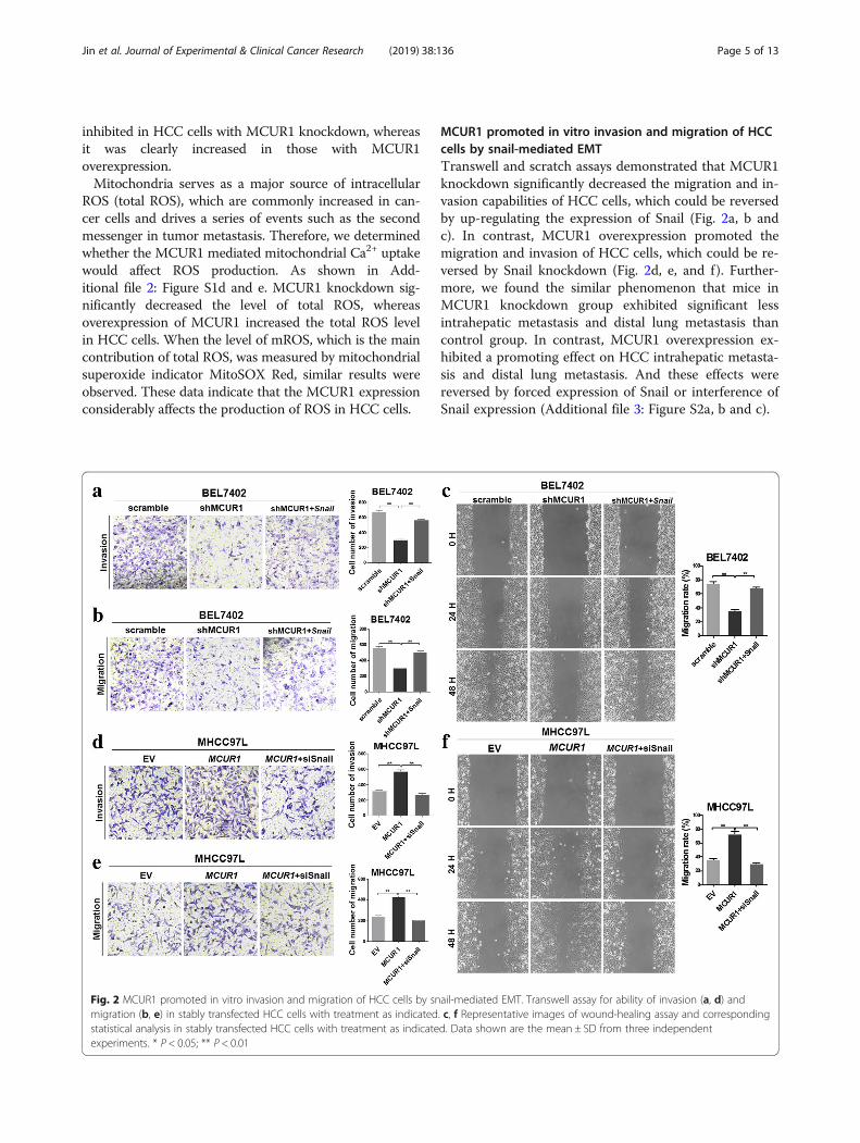
MCUR1 promoted in vitro invasion and migration of HCCcells by snail-mediated EMTTranswell and scratch assays demonstrated that MCUR1knockdown significantly decreased the migration and in-vasion capabilities of HCC cells, which could be reversedby up-regulating the expression of Snail (Fig. 2a, b andc). In contrast, MCUR1 overexpression promoted themigration and invasion of HCC cells, which could be re-versed by Snail knockdown (Fig. 2d, e, and f). Further-more, we found the similar phenomenon that mice inMCUR1 knockdown group exhibited significant lessintrahepatic metastasis and distal lung metastasis thancontrol group. In contrast, MCUR1 overexpression ex-hibited a promoting effect on HCC intrahepatic metasta-sis and distal lung metastasis. And these effects werereversed by forced expression of Snail or interference ofSnail expression (Additional file 3: Figure S2a, b and c).
Fig. 2 MCUR1 promoted in vitro invasion and migration of HCC cells by snail-mediated EMT. Transwell assay for ability of invasion (a, d) andmigration (b, e) in stably transfected HCC cells with treatment as indicated. c, f Representative images of wound-healing assay and correspondingstatistical analysis in stably transfected HCC cells with treatment as indicated. Data shown are the mean ± SD from three independentexperiments. * P < 0.05; ** P < 0.01
Jin et al. Journal of Experimental & Clinical Cancer Research (2019) 38:136 Page 5 of 13
MCUR1 facilitated EMT and HCC metastasis in vivoTo evaluate the role of MCUR1 in tumor EMT and me-tastasis in vivo, we established the orthotopic transplant-ation model of HCC metastasis in nude mice. Themetastasis foci were clearly identified under microscope.However, we did not find the macroscopic metastases inliver and lung tissues possibly due to short time fromcell transplantation to mice sacrifice. Immunohisto-chemical (IHC) assays of xenograft tumors demonstratedthat MCUR1 knockdown induced higher expression ofthe epithelial marker E-cadherin and lower expression ofthe mesenchymal marker Vimentin, whereas MCUR1overexpression had an opposite effect (Fig. 3a). Further-more, mice in MCUR1 knockdown group exhibited sig-nificantly less intrahepatic metastasis and distal lungmetastasis than control group. In contrast, MCUR1 over-expression exhibited a promoting effect on HCC intrahe-patic metastasis and distal lung metastasis (Fig. 3b and c).
MCUR1 expression was associated with HCC metastasisand clinical stage of patients with HCCNext, we evaluated the expression of MCUR1 in HCCwith (n = 64) or without (n = 74) intrahepatic metastasisusing IHC staining. Our results showed that MCUR1was more frequently upregulated in HCC tissues withmetastasis than those without metastasis (Fig. 3d).Moreover, MCUR1 expression was positively correlatedwith TNM stage of HCC (Fig. 3e).
MCUR1 facilitated ROS-induced Nrf2 nuclear translocationto activate the Notch pathwayOur results had showed that MCUR1 considerably in-creased mitochondrial Ca2+ ([Ca2+]m) and ROS in HCCcells. And previous evidence has shown that Nrf2 and itsrepressor protein Keap1 act as major regulators of cellu-lar redox levels, which is attributed in part to the activa-tion of Notch signaling pathway [16, 18]. Therefore, weinvestigated the effect of MCUR1 expression on Nrf2/Notch1 pathway in HCC cells. As shown in Fig. 4a,MCUR1 knockdown significantly decreased the nucleartranslocation of Nrf2, which can be reversed by H2O2.In contrast, MCUR1 overexpression induced Nrf2 nu-clear translocation, which can be abolished by ROS scav-enger MitoTEMPO. To address downstream mediatorsof Nrf2, we observed the Notch pathway to be signifi-cantly changed. Our data showed that MCUR1 knock-down significantly decreased the expression of Notch1in the cytoplasm and its active form NICD1 in nucleusof HCC cells, while MCUR1 overexpression had an op-posite effect on Notch1 and NICD1 expression (Fig. 4cand d). Furthermore, H2O2 significantly reversedMCUR1 knockdown-mediated Notch1 down-regulation,whereas scavenging ROS by MitoTEMPO considerablyinhibited the MCUR1 overexpression-mediated Notch1
activation (Fig. 4c and d). As expected, the MCUR1knockdown-mediated Notch1 inhibition was signifi-cantly reversed by treatment with Oltipraz (OPZ), whichis an activator of Nrf2. However, knockdown of Nrf2 bysiRNA considerably inhibited the MCUR1 overexpress-ion-mediated Notch1 activation (Fig. 4c and d and Add-itional file 4: Figure S3a).
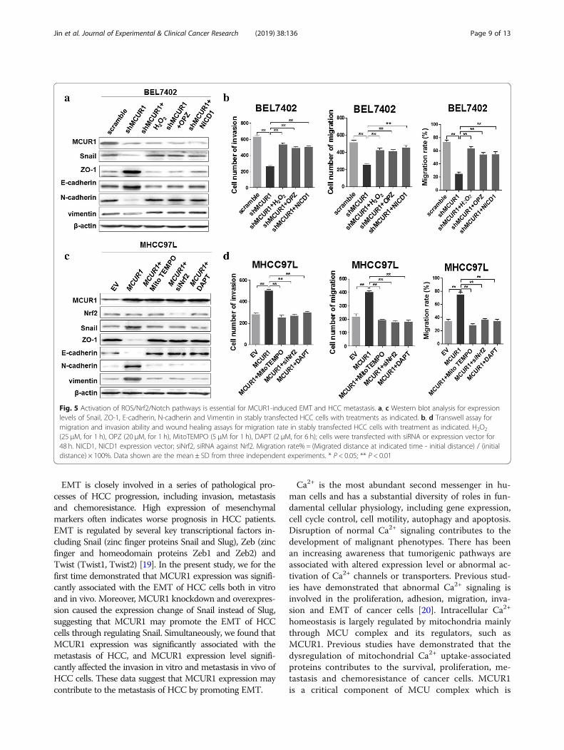
Activation of ROS/Nrf2/Notch pathways was essential forMCUR1-induced EMT and HCC metastasisWe further investigated the functions of ROS/Nrf2/Notch pathway in MCUR1-induced EMT and invasionof HCC cells. Western blot analysis demonstrated thattreatment with H2O2, OPZ or NICD1 significantly re-versed the MCUR1 knockdown-mediated epithelial tran-sition, as indicated by increased expression of ZO-1,E-cadherin and decreased expression of N-cadherin,Vimentin and Snail (Fig. 5a). Moreover, transwell andscratch assays demonstrated that treatment with H2O2,OPZ and NICD1 significantly reversed the MCUR1knockdown-mediated inhibition of HCC cell migrationand invasion (Fig. 5b and Additional file 5: Figure S4a, band c). In contrast, when treated with MitoTEMPO,siNrf2 or Notch1 inhibitor DAPT, the MCUR1overexpression-mediated mesenchymal transition wassignificantly reversed, as indicated by decreased expres-sion of ZO-1 and E-cadherin and increased expressionof N-cadherin, Vimentin and Snail (Fig. 5c). Simultan-eously, MCUR1 overexpression-mediated enhancementof HCC cell migration and invasion was significantly re-versed by MitoTEMPO, siNrf2 or DAPT (Fig. 5d andAdditional file 5: Figure S4d, e and f).
Mitochondrial calcium buffering inhibited ROS/Nrf2/Notchpathway and MCUR1-induced EMT and HCC metastasisOur results had shown that the scavenging of [Ca2+] m
by PV-Mito considerably decreased the mitochondrialCa2+, total ROS and mROS in HCC cells(Additional file 6: Figure S5a, b, c and d). We also shownthat PV-Mito considerably inhibited MCUR1-mediatedactivation of Nrf2/Notch pathway (Fig. 6a). Moreover, wefound that PV-mito significantly reversed theMCUR1-mediated decreased expression of ZO-1 andE-cadherin and increased expression of N-cadherin,Vimentin and Snail (Fig. 6b). MCUR1-mediated increaseof migration and invasion was also reversed significantly byPV-Mito (Fig. 6c and Additional file 6 Figure S5e, f and g).We further tested the effect of calcium buffering on
HCC metastasis in xenograft models. IHC assays dem-onstrated that PV-Mito-recombined adenovirus(Ad-PV-Mito) treatment significantly inhibited the mes-enchymal transition of MCUR1 overexpressed HCC cellsin nude mice (Fig. 6d). Simultaneously, intrahepatic me-tastasis and lung metastasis were also remarkably
Jin et al. Journal of Experimental & Clinical Cancer Research (2019) 38:136 Page 6 of 13
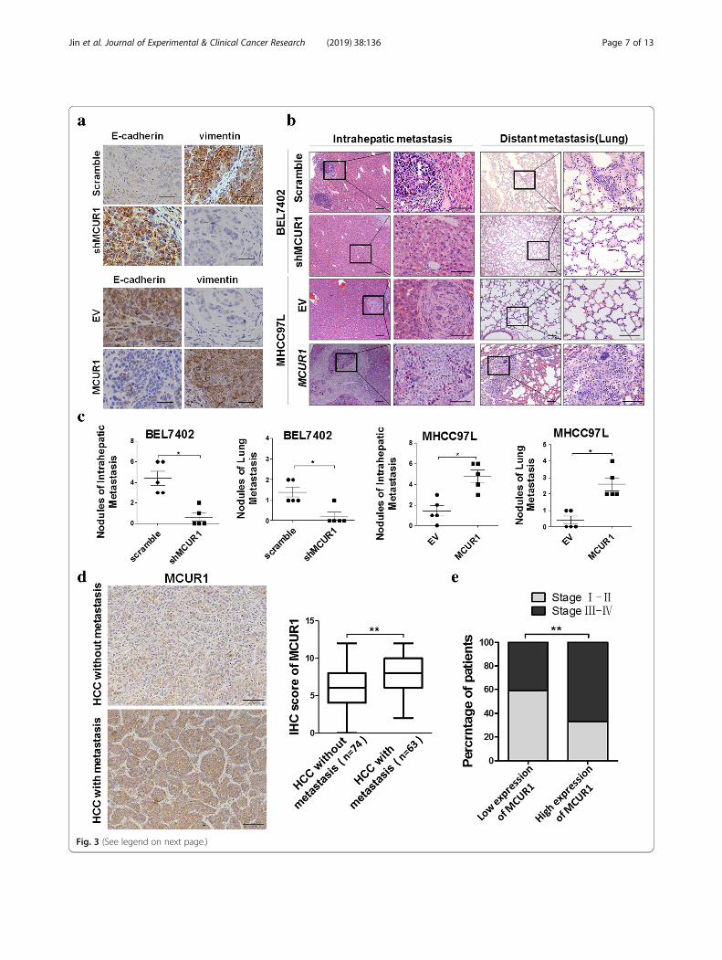
Fig. 3 (See legend on next page.)
Jin et al. Journal of Experimental & Clinical Cancer Research (2019) 38:136 Page 7 of 13

repressed in Ad-PV-Mito group when compared withthe control group (Fig. 6e and f).
DiscussionPrevious studies have demonstrated the important roleof MCUR1 in HCC cell survival. Similar with previousreports, our study further confirmed the clinical signifi-cance of MCUR1 in EMT and HCC metastasis. Moreimportantly, we provided the first evidence indicatinghigh MCUR1 expression in HCC tissues with
metastasis and significant association between MCUR1expression and tumor progression in HCC patients.Moreover, the in vitro and in vivo experiments also firstdemonstrated that MCUR1-induced mitochondrial Ca2+ uptake activated the ROS/Nrf2/Notch signaling andthus facilitated the epithelial-mesenchymal transitionand metastasis in hepatocellular carcinoma (Fig. 6g).Thus, MCUR1 has a great potential to be used as aprognosis marker or therapy target in clinical manage-ment of HCC patients.
(See figure on previous page.)Fig. 3 MCUR1 facilitates epithelial-mesenchymal transition and HCC metastasis. a Representative immunohistochemical staining images of E-cadherin and Vimentin in the orthotopic transplantation nude mice model of HCC metastasis. b Histological analyses of intrahepatic and lungmetastatic nodules from HCC metastasis nude mice model by hematoxylin and eosin (H&E) staining. Images showing representative H&E stainingof liver and lung tissue samples from the different experimental groups (n = 5 /group). c The number of intrahepatic and lung metastasis noduleswas quantified in H.E. sections. d Representative immunohistochemical staining images and IHC score of MCUR1 in HCC without metastasis (n =74) and HCC with metastasis (n = 63). e MCUR1 expression positively correlated with tumor progression of HCC patients. Data shown are themean ± SD from three independent experiments. * P < 0.05; ** P < 0.01
Fig. 4 MCUR1 facilitates ROS-induced Nrf2 nuclear translocation to activate the Notch pathway. a, b, c, d Western blot analysis for expressionlevels of Nrf2, Notch1and NICD1 in stably transfected HCC cells with treatments as indicated
Jin et al. Journal of Experimental & Clinical Cancer Research (2019) 38:136 Page 8 of 13
EMT is closely involved in a series of pathological pro-cesses of HCC progression, including invasion, metastasisand chemoresistance. High expression of mesenchymalmarkers often indicates worse prognosis in HCC patients.EMT is regulated by several key transcriptional factors in-cluding Snail (zinc finger proteins Snail and Slug), Zeb (zincfinger and homeodomain proteins Zeb1 and Zeb2) andTwist (Twist1, Twist2) [19]. In the present study, we for thefirst time demonstrated that MCUR1 expression was signifi-cantly associated with the EMT of HCC cells both in vitroand in vivo. Moreover, MCUR1 knockdown and overexpres-sion caused the expression change of Snail instead of Slug,suggesting that MCUR1 may promote the EMT of HCCcells through regulating Snail. Simultaneously, we found thatMCUR1 expression was significantly associated with themetastasis of HCC, and MCUR1 expression level signifi-cantly affected the invasion in vitro and metastasis in vivo ofHCC cells. These data suggest that MCUR1 expression maycontribute to the metastasis of HCC by promoting EMT.
Ca2+ is the most abundant second messenger in hu-man cells and has a substantial diversity of roles in fun-damental cellular physiology, including gene expression,cell cycle control, cell motility, autophagy and apoptosis.Disruption of normal Ca2+ signaling contributes to thedevelopment of malignant phenotypes. There has beenan increasing awareness that tumorigenic pathways areassociated with altered expression level or abnormal ac-tivation of Ca2+ channels or transporters. Previous stud-ies have demonstrated that abnormal Ca2+ signaling isinvolved in the proliferation, adhesion, migration, inva-sion and EMT of cancer cells [20]. Intracellular Ca2+
homeostasis is largely regulated by mitochondria mainlythrough MCU complex and its regulators, such asMCUR1. Previous studies have demonstrated that thedysregulation of mitochondrial Ca2+ uptake-associatedproteins contributes to the survival, proliferation, me-tastasis and chemoresistance of cancer cells. MCUR1is a critical component of MCU complex which is
Fig. 5 Activation of ROS/Nrf2/Notch pathways is essential for MCUR1-induced EMT and HCC metastasis. a, c Western blot analysis for expressionlevels of Snail, ZO-1, E-cadherin, N-cadherin and Vimentin in stably transfected HCC cells with treatments as indicated. b, d Transwell assay formigration and invasion ability and wound healing assays for migration rate in stably transfected HCC cells with treatment as indicated. H2O2
(25 μM, for 1 h), OPZ (20 μM, for 1 h), MitoTEMPO (5 μM for 1 h), DAPT (2 μM, for 6 h); cells were transfected with siRNA or expression vector for48 h. NICD1, NICD1 expression vector; siNrf2, siRNA against Nrf2. Migration rate% = (Migrated distance at indicated time - initial distance) / (initialdistance) × 100%. Data shown are the mean ± SD from three independent experiments. * P < 0.05; ** P < 0.01
Jin et al. Journal of Experimental & Clinical Cancer Research (2019) 38:136 Page 9 of 13

Fig. 6 (See legend on next page.)
Jin et al. Journal of Experimental & Clinical Cancer Research (2019) 38:136 Page 10 of 13

required for mitochondrial Ca2+ uptake and mainten-ance of normal cellular bioenergetics. MCUR1 silen-cing resulted in a dramatic reduction in the [Ca2+]mito (− ~ 85% in Rhod-2 fluorescence compared tothe control) without modifying the cytosolic Ca2+
content [21]. However, its biological roles in cancerdevelopment remain largely unclear. Our previousstudies have demonstrated that MCUR1 expression isfrequently upregulated in HCC and MCUR1-mediatedCa2+ signaling promotes HCC cell survival. In thisstudy, our findings that chelating mitochondrial Ca2+
by PV-Mito significantly suppressed MCUR1overexpression-induced EMT and invasion of HCCcells further indicate that mitochondrial Ca2+ signal-ing mediates MCUR1-induced HCC metastasis.Mitochondria are an essential source of reactive
oxygen species in most mammalian cells. [Ca2+]m hasbeen reported to play an important role in the gener-ation of ROS. Our previous studies have demon-strated that the overexpression of either MCU orMCRU1 promotes the production of ROS and oxida-tive status in HCC cells. ROS play a key role in manyintracellular signaling pathways which contributes tothe tumorigenesis. Nrf2 as the major effector of ROSin human cells regulates the expression of more than100 genes, including Notch1 [18]. Upon oxidativestress, Nrf2 is detached from its inhibitor Keap-1 andtranslocated to the nucleus to promote the transcrip-tion of target genes by binding to promoter regions[22, 23]. Currently, growing evidences suggest thatconstitutive upregulation of Nrf2 is linked to cancerdevelopment, progression and resistance to radiother-apy. Moreover, Nrf2 promotes the invasion of cancercells and contributes to poor prognosis of patients. Inthis study, we found that similar like H2O2, overex-pression of MCUR1 had a remarkable effect to inducethe Nrf2 nuclear translocation and Notch1 activation,which can be abolished by ROS scavenger. Moreover,Notch1 activation can be suppressed by either Nrf2silencing or H2O2 scavenging, suggesting that ROS/Nrf2/Notch1 axis may act as the downstream signal-ing of MCUR1-induced Ca2+ homeostasis remodeling.
Notch signaling is a critical regulator in embryo devel-opment through the modulation of cell–cell communi-cation [24]. Increasing evidences indicate that Notchsignaling plays important roles in cancer developmentand progression [25, 26]. Elevated Notch1 expressionhas been observed in a series of malignancies includingHCC and is commonly associated with aggressive cancerphenotypes. Notch signaling has been shown to promoteEMT of cancer cells [27, 28]. Previous studies have dem-onstrated that the inhibition of Notch signaling byDAPT leads to the decrease of EMT in HCC cell lines,whereas activation of Notch signaling by Jagged1 pro-motes the increase of EMT [29–31]. In this study, wefound that MCUR1 overexpression promoted the activa-tion of Notch1 signaling, which can be inhibited by Ca2+
chelating, ROS scavenging, or Nrf2 silencing. Moreover,MCUR1-induced EMT as well as Snail transcription canbe suppressed by Nrf2 silencing or Notch1 inhibitorDAPT. These findings suggest that MCUR1 facilitatedHCC EMT, invasion and migration mainly throughNrf2/Notch1 signaling activation.
ConclusionsIn summary, our study first confirmed the clinical signifi-cance of MCUR1 in HCC metastasis. In addition, we dem-onstrated that the essential role of MCUR1 in promotingEMT, invasion and migration through the activation ofROS/Nrf2/Notch signaling by inducing mitochondrial Ca2+ uptake. Therefore, our study suggests that MCUR1 maybe a potential target in HCC treatment.
Additional files
Additional file 1: Table S1. Primary antibodies used forimmunohistochemistry and western blot. Table S2. Sequence of primers.(DOCX 20 kb)
Additional file 2: Figure S1 related to Figure 1. a Phase-contrast pho-tographs showing the morphology of HCC cells before and after stabletransfection with MCUR1 expression vector. b Representative confocalmicroscope images of mitochondrial Ca2+ levels ([Ca2+]m) detected usingmitopericam in HCC cells. Scale bar: 20 μm. c Representative time-courserecording of mitochondrial Ca2+ fluorescence detected using mitoperi-cam. After 30-s baseline recording, [Ca2+]m responses to 10 μM histamine
(See figure on previous page.)Fig. 6 Mitochondrial calcium buffering inhibits ROS/Nrf2/Notch pathway and MCUR1-induced EMT and HCC metastasis. a Western blot analysisfor expression levels of Nrf2, Notch1and NICD1 in stably transfected HCC cells with treatments as indicated. Mitochondrial Ca2+ was buffered bytransient transfection of expression vector encoding parvalbumin with mitochondria target sequence (PV-Mito) for 48 h, where appropriate.b Western blot analysis for expression levels of ZO-1, E-cadherin, N-cadherin, Vimentin and Snail in stably transfected HCC cells with treatments asindicated. c Transwell assays for migration and invasion ability and wound healing assays for migration rate in stably transfected HCC cells withtreatment as indicated. d Representative immunohistochemical staining images of E-cadherin and Vimentin in EV, MCUR1 and the Ad-PV-Mitogroups (injection of PV-Mito adenovirus into tail vein). e Histological analyses of intrahepatic and lung metastatic nodules in EV, MCUR1 and theAd-PV-Mito group (injection of PV-Mito adenovirus into tail vein). Images showing representative H&E staining of liver and lung tissue samplesfrom the different experimental groups (n = 5 /group). f The number of intrahepatic and lung metastasis nodules was quantified in H.E. sections.g The schematic for mechanism underlying epithelial-mesenchymal transition and metastasis facilitated by MCUR1. Data shown are the mean ±SD from three independent experiments. * P < 0.05; ** P < 0.01
Jin et al. Journal of Experimental & Clinical Cancer Research (2019) 38:136 Page 11 of 13
in HCC cells was investigated. Ca2+ response signals were presented asmaximal amplitude fluorescence intensity, which was defined as the max-imal change of [Ca2+]m relative to the basal [Ca2+]m. d mROS levels wereanalyzed by confocal microscope after staining with MitoSOX (4 μM) for10 min in HCC cells. Representative confocal microscope images werepresented. Scale bar: 20 μm. e Intracellular ROS levels were stained withfluorescence dye DCFH-DA then analyzed by flow cytometry in HCC cells.f Western blot analysis of Snail level in MHCC97L cells transiently trans-fected with siRNA, and in BEL7402 cells transiently transfected with ex-pression vector. Scramble: vector encoding control shRNA; shMCUR1vectors encoding short hairpin RNA (shRNA) against MCUR1. EV: EmptyVector; MCUR1: expression vectors encoding MCUR1; Snail, Snail expres-sion vector; siSnail, siRNA against Snail. Data shown are the mean ± SDfrom three independent experiments.*P < 0.05; **P < 0.01. (JPG 5515 kb)
Additional file 3: Figure S2 related to Figure 2. a Histologicalanalyses of intrahepatic and lung metastatic nodules from HCCmetastasis nude mice model by hematoxylin and eosin (H&E) staining.Images showing representative H&E staining of liver and lung tissuesamples from the different experimental groups (n = 5 /group). Snailadenovirus and shSnail adenovirus was injected by tail vein. b Thenumber of intrahepatic and lung metastasis nodules was quantified inH.E. sections. c Western blot analysis for indicated markers was performedwith liver tissue lysates from three representative mice per group. Datashown are the mean ± SD from three independent experiments. * P <0.05; ** P < 0.01. (JPG 3671 kb)
Additional file 4: Figure S3 related to Figure 4. a Western blotanalysis of Nrf2 level in MHCC97L cells transiently transfected with siRNA.(JPG 52 kb)
Additional file 5: Figure S4 related to Figure 5. a, d Transwell assayfor invasion and b, e migration ability of HCC cells with treatment asindicated. c, f Wound healing assays for migration rate in HCC cells withtreatment as indicated. (JPG 9852 kb)
Additional file 6: Figure S5 related to Figure 6. a Co-localization ofthe mitochondria labeled with Mito-tracker (Red) and mitochondrial Ca2+
detected by mitopericam (Green) in MHCC97L cells. Scale bar: 20 μm. bRepresentative traces and quantification of [Ca2+]m in HCC cells PV pro-tein with mitochondrial translocation signal was used to buffer mitochon-drial Ca2+. c Mitochondrial ROS levels were analyzed by confocalmicroscope after staining with MitoSOX (4 μM) for 10 min in HCC cellswith treatment as indicated. Representative confocal microscope imageswere presented. Scale bar: 20 μm. Mitochondrial Ca2+ was buffered bytransient transfection of expression vector encoding parvalbumin withmitochondria target sequence (PV-Mito) for 48 h, where appropriate. dIntracellular ROS levels were analyzed by flow cytometry after stainingwith fluorescence dye DCFH-DA in HCC cells with treatment as indicated.e Transwell assay for invasion and f migration ability of HCC cells withtreatment as indicated.g Wound healing assays for migration rate in HCC cells with treatment asindicated. Data shown are the mean ± SD from three independentexperiments. ** P < 0.01. (JPG 7415 kb)
Abbreviations[Ca2+]m: Mitochondrial Ca2+; EMT: Epithelial-mesenchymal transition;HCC: Hepatocellular carcinoma; IF: Immunofluorescence;IHC: Immunohistochemical; KEAP1: Kelch-like ECH-associated protein 1;MCU: Mitochondrial calcium uniporter; MCUR1: Mitochondrial calciumuniporter regulator 1; MICU1: Mitochondrial calcium uptake 1;mitoROS: mitochondrial reactive oxygen species; Nrf2: Nuclear factorerythroid-2 related factor 2; OPZ: Oltipraz; ROS: Reactive oxygen species;TNM: Tumor Node Metastasis; ZO-1: Zonula occludens-1
AcknowledgementsThe authors thank Hui Zhang for the help in animal study.
FundingThis work was supported by the National Natural Science Foundation ofChina (grants 81572727, 81320108021 and 8187111403) and National BasicResearch Program (grant 2015CB553703).
Availability of data and materialsAll the data related to the study are included within the article and thesupplemental material.
Authors’ contributionsJW and MJ did most experiments, wrote the manuscript; XJ and HCparticipated in the transwell assays; JZ participated in the in vivo study; YChelped write the manuscript; JY and ZZ performed the statistical analyses; JXand TR conceived and designed the overall study, supervised the experiments,and wrote the paper. All authors read and approved the final manuscript.
Ethics approval and consent to participateThe study was approved by the Ethics Committee of the Fourth MilitaryMedical University. Animal research was approved by the Institutional AnimalCare and Use Committee of Fourth Military Medical University. The studywas performed in accordance with the Declaration of Helsinki.
Consent for publicationNot applicable.
Competing interestsThe authors declare that they have no competing interests.
Publisher’s NoteSpringer Nature remains neutral with regard to jurisdictional claims inpublished maps and institutional affiliations.
Author details1State Key Laboratory of Cancer Biology and Experimental Teaching Centerof Basic Medicine, Fourth Military Medical University, 169 Changle West Road,Xi’an 710032, China. 2Center of Genetic & Prenatal Diagnosis, First AffiliatedHospital, Zhengzhou University, Zhengzhou 450052, China. 3Institute ofPreventive Genomic Medicine, School of Life Sciences, Northwest University,Xi’an 710069, China. 4Third Department of Medical Oncology, ShaanxiProvincial Cancer Hospital, Xi’an 710061, China.
Received: 9 December 2018 Accepted: 13 March 2019
References1. Ferlay J, Soerjomataram I, Dikshit R, Eser S, Mathers C, Rebelo M, Parkin DM,
Forman D, Bray F. Cancer incidence and mortality worldwide: sources,methods and major patterns in GLOBOCAN 2012. Int J Cancer. 2015;136(5):E359–86.
2. Giannelli G, Koudelkova P, Dituri F, Mikulits W. Role of epithelial tomesenchymal transition in hepatocellular carcinoma. J Hepatol. 2016;65(4):798–808.
3. Van't Veer LJ, Weigelt B. Road map to metastasis. Nat Med. 2003;9(8):999–1000.
4. Budhu A, Forgues M, Ye QH, Jia HL, He P, Zanetti KA, Kammula US, Chen Y,Qin LX, Tang ZY, et al. Prediction of venous metastases, recurrence, andprognosis in hepatocellular carcinoma based on a unique immuneresponse signature of the liver microenvironment. Cancer Cell. 2006;10(2):99–111.
5. Prevarskaya N, Skryma R, Shuba Y. Calcium in tumour metastasis: new rolesfor known actors. Nat Rev Cancer. 2011;11(8):609–18.
6. Monet M, Lehen'kyi V, Gackiere F, Firlej V, Vandenberghe M, Roudbaraki M,Gkika D, Pourtier A, Bidaux G, Slomianny C, et al. Role of cationic channelTRPV2 in promoting prostate cancer migration and progression toandrogen resistance. Cancer Res. 2010;70(3):1225–35.
7. Tang S, Wang X, Shen Q, Yang X, Yu C, Cai C, Cai G, Meng X, Zou F.Mitochondrial Ca(2)(+) uniporter is critical for store-operated Ca(2)(+) entry-dependent breast cancer cell migration. Biochem Biophys Res Commun.2015;458(1):186–93.
8. Wallace DC. Mitochondria and cancer. Nat Rev Cancer. 2012;12(10):685–98.9. Baughman JM, Perocchi F, Girgis HS, Plovanich M, Belcher-Timme CA,
Sancak Y, Bao XR, Strittmatter L, Goldberger O, Bogorad RL, et al. Integrativegenomics identifies MCU as an essential component of the mitochondrialcalcium uniporter. Nature. 2011;476(7360):341–5.
Jin et al. Journal of Experimental & Clinical Cancer Research (2019) 38:136 Page 12 of 13
10. De Stefani D, Raffaello A, Teardo E, Szabo I, Rizzuto R. A forty-kilodaltonprotein of the inner membrane is the mitochondrial calcium uniporter.Nature. 2011;476(7360):336–40.
11. Mallilankaraman K, Cardenas C, Doonan PJ, Chandramoorthy HC, Irrinki KM,Golenar T, Csordas G, Madireddi P, Yang J, Muller M, et al. MCUR1 is anessential component of mitochondrial Ca2+ uptake that regulates cellularmetabolism. Nat Cell Biol. 2012;14(12):1336–43.
12. Zeng F, Chen X, Cui W, Wen W, Lu F, Sun X, Ma D, Yuan Y, Li Z, Hou N, etal. RIPK1 binds MCU to mediate induction of mitochondrial ca(2+) uptakeand promotes colorectal oncogenesis. Cancer Res. 2018;78(11):2876–85.
13. Chakraborty PK, Mustafi SB, Xiong X, Dwivedi SKD, Nesin V, Saha S, ZhangM, Dhanasekaran D, Jayaraman M, Mannel R, et al. MICU1 drives glycolysisand chemoresistance in ovarian cancer. Nat Commun. 2017;8:14634.
14. Curry MC, Peters AA, Kenny PA, Roberts-Thomson SJ, Monteith GR.Mitochondrial calcium uniporter silencing potentiates caspase-independentcell death in MDA-MB-231 breast cancer cells. Biochem Biophys ResCommun. 2013;434(3):695–700.
15. Ren T, Zhang H, Wang J, Zhu J, Jin M, Wu Y, Guo X, Ji L, Huang Q, Zhang H,et al. MCU-dependent mitochondrial Ca(2+) inhibits NAD(+)/SIRT3/SOD2pathway to promote ROS production and metastasis of HCC cells.Oncogene. 2017;36(42):5897–909.
16. Ren T, Wang J, Zhang H, Yuan P, Zhu J, Wu Y, Huang Q, Guo X, Zhang J, JiL, et al. MCUR1-mediated mitochondrial calcium signaling facilitates cellsurvival of hepatocellular carcinoma via reactive oxygen species-dependentP53 degradation. Antioxid Redox Signal. 2018;28(12):1120–36.
17. Huang Q, Zhan L, Cao H, Li J, Lyu Y, Guo X, Zhang J, Ji L, Ren T, An J, et al.Increased mitochondrial fission promotes autophagy and hepatocellularcarcinoma cell survival through the ROS-modulated coordinated regulationof the NFKB and TP53 pathways. Autophagy. 2016;12(6):999–1014.
18. Ganan-Gomez I, Wei Y, Yang H, Boyano-Adanez MC, Garcia-Manero G.Oncogenic functions of the transcription factor Nrf2. Free Radic Biol Med.2013;65:750–64.
19. Peinado H, Olmeda D, Cano A. Snail, Zeb and bHLH factors in tumourprogression: an alliance against the epithelial phenotype? Nat Rev Cancer.2007;7(6):415–28.
20. Prevarskaya N, Ouadid-Ahidouch H, Skryma R, Shuba Y. Remodelling of Ca2+ transport in cancer: how it contributes to cancer hallmarks? Philos Trans RSoc Lond Ser B Biol Sci. 2014;369(1638):20130097.
21. Marchi S, Pinton P. The mitochondrial calcium uniporter complex: molecularcomponents, structure and physiopathological implications. J Physiol. 2014;592(5):829–39.
22. McMahon M, Itoh K, Yamamoto M, Hayes JD. Keap1-dependentproteasomal degradation of transcription factor Nrf2 contributes to thenegative regulation of antioxidant response element-driven geneexpression. J Biol Chem. 2003;278(24):21592–600.
23. Itoh K, Wakabayashi N, Katoh Y, Ishii T, O'Connor T, Yamamoto M. Keap1regulates both cytoplasmic-nuclear shuttling and degradation of Nrf2 inresponse to electrophiles. Genes Cells. 2003;8(4):379–91.
24. Kofler NM, Shawber CJ, Kangsamaksin T, Reed HO, Galatioto J, Kitajewski J.Notch signaling in developmental and tumor angiogenesis. Genes Cancer.2011;2(12):1106–16.
25. Groeneweg JW, Foster R, Growdon WB, Verheijen RH, Rueda BR. Notchsignaling in serous ovarian cancer. J Ovarian Res. 2014;7:95.
26. Ntziachristos P, Lim JS, Sage J, Aifantis I. From fly wings to targeted cancertherapies: a centennial for notch signaling. Cancer Cell. 2014;25(3):318–34.
27. Mittal S, Subramanyam D, Dey D, Kumar RV, Rangarajan A. Cooperation ofNotch and Ras/MAPK signaling pathways in human breast carcinogenesis.Mol Cancer. 2009;8:128.
28. Collu GM, Brennan K. Cooperation between Wnt and Notch signalling inhuman breast cancer. Breast Cancer Res. 2007;9(3):105.
29. Tschaharganeh DF, Chen X, Latzko P, Malz M, Gaida MM, Felix K, Ladu S,Singer S, Pinna F, Gretz N, et al. Yes-associated protein up-regulates Jagged-1 and activates the Notch pathway in human hepatocellular carcinoma.Gastroenterology. 2013;144(7):1530–42 e1512.
30. Wu F, Yang LY, Li YF, Ou DP, Chen DP, Fan C. Novel role for epidermalgrowth factor-like domain 7 in metastasis of human hepatocellularcarcinoma. Hepatology. 2009;50(6):1839–50.
31. Zhao ZL, Ma SR, Wang WM, Huang CF, Yu GT, Wu TF, Bu LL, Wang YF, ZhaoYF, Zhang WF, et al. Notch signaling induces epithelial-mesenchymaltransition to promote invasion and metastasis in adenoid cystic carcinoma.Am J Transl Res. 2015;7(1):162–74.
Jin et al. Journal of Experimental & Clinical Cancer Research (2019) 38:136 Page 13 of 13