maxine jochelson md director of radiology: breast and ... acr, acog: current standard ... breast...
TRANSCRIPT

Maxine Jochelson MDDirector of Radiology: Breast and Imaging CenterMemorial Sloan Kettering Cancer Center, N.Y., N.Y.Associate Professor of Clinical Radiology Weill Cornell Medical School

Screening:a. Current guidelines vs. USPSTF vs ACS guidelinesb. Why not screen < 50?c. Data for screening average risk women <50d. Screening young women at increased risk
New technologies to improve screening of both groups
Conclusions
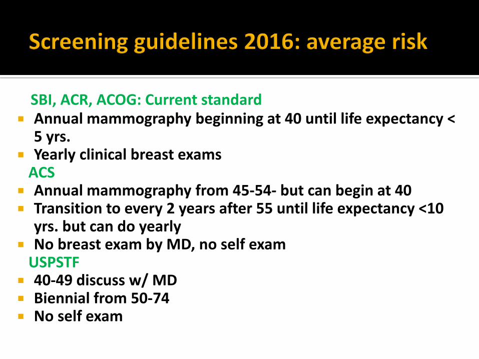
SBI, ACR, ACOG: Current standard Annual mammography beginning at 40 until life expectancy <
5 yrs. Yearly clinical breast exams
ACS Annual mammography from 45-54- but can begin at 40 Transition to every 2 years after 55 until life expectancy <10
yrs. but can do yearly No breast exam by MD, no self exam
USPSTF 40-49 discuss w/ MD Biennial from 50-74 No self exam

Missed cancers/dense breasts Call backs/ false positives leading to anxiety more
frequent in young women Not as much mortality reduction “Over diagnosis”

Must discuss mortality AND morbidity
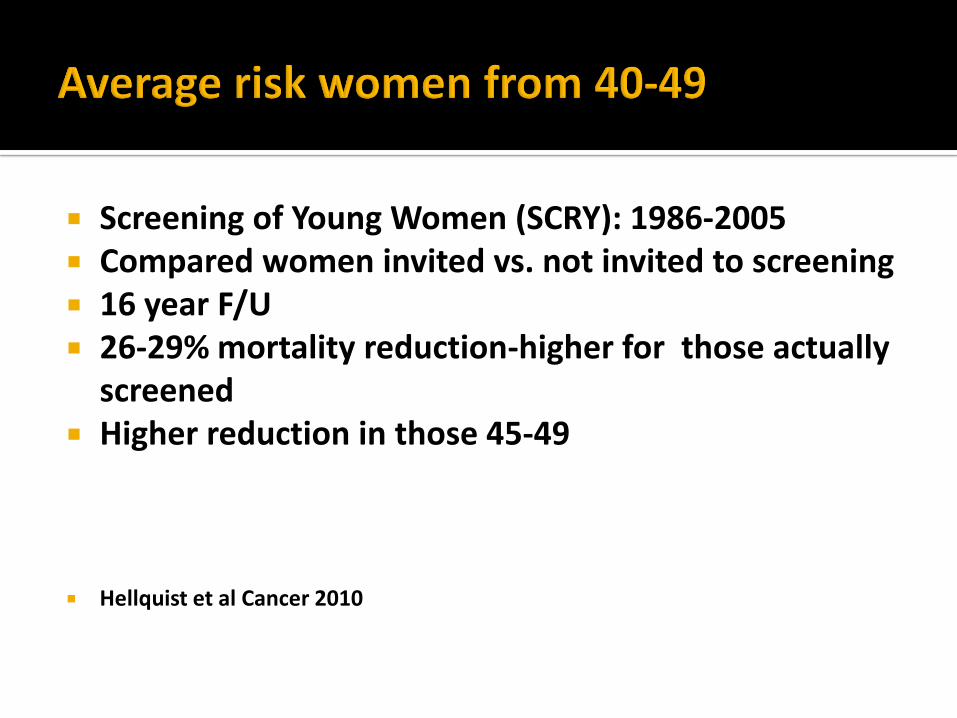
Screening of Young Women (SCRY): 1986-2005 Compared women invited vs. not invited to screening 16 year F/U 26-29% mortality reduction-higher for those actually
screened Higher reduction in those 45-49
Hellquist et al Cancer 2010

Longitudinal prospective cohort 1990-2008 compared mammo detected to MD or pt. detected cancers
N=1977 Mammo detected more likely to be conservable p<
0.001 Mammo detected less likely to receive chemo p<
0.001 5 yr relapse free survival mammo detected 92% vs.
88% p< 0.001
Malmgren et al Radiol 2012

43,351 mammos: 1/3 in their 40’s 205 cancers: 20% in their 40’s > 50% of cancers in women in their 40’s were invasive
Arleo et al AJR 2013

Retrospective 2008-2011 N= 230 patients w/ breast cancer
149 screened/81 non -screened Screened vs. non-screened: Earlier stage p= 0.001Negative nodes p=0.005Smaller tumors p<0.001 Mastectomy: 48% non-screened vs. 30% p=0.1Chemo: 66% vs. 44% p=0.042
Plecha et al AJR 2014

The 6 “Best” Models:
USPSTF D E G M S W #Biennial 50-74: 22% 27% 21% 21% 20% 28% 11,000
STANDARDAnnual 40-84: 38% 49% 32% 29% 35% 54% 36,500
For 25,000 more mammograms/1000 women:6 Model Average Increased Mortality Reduction by 16.3%
Cancer Intervention and Surveillance Modeling Network

Adding annual mammo of women 40-49 to biennial screening 50-74 increases lives saved by 27%
Increases life years gained by 47% Saves 42% more lives & life years than biennial
mammo Need to screen 588 women to save 1 life w/ annual
digital mammo in this age group
Hendrick et al AJR 2014


BRCA 1 or 2 mutation Untested first-degree relative of BRCA carrier Lifetime risk >20%
Defined by BRCAPRO
Other models dependent on family history Chest XRT – 10 to 30 years of age
CA Cancer J Clin 2007

Breast MRI: most sensitive imaging test for breast cancer detection
Sensitivity due to imaging of enhancing neovascularity Limitations include cost (>$4,000.00), claustrophobia,
inability to perform in women w/ metallic implants, Gadolinium allergy & lack of specificity
Not universally available

Tumors create new vessels (angiogenesis)
VEGF
Vessels leak
A-V shunting
Courtesy of Dr. Elizabeth Morris

Cancer yield of different imaging methods, used alone or in combination.
Kuhl C et al. JCO 2010
©2010 by American Society of Clinical Oncology

Annual surveillance with MRI associated with decrease incidence of advanced stage cancer
MRI (n=445)
No MRI (n=830)
p
Cancer 41 (9.2%) 76 (9.2%)
DCIS/stage 1 13.8% 7.2% 0.01
Stage II-IV 1.9% 6.6% 0.02
Warner E et al JCO 2011

BRCA 1 58% <40; 9.7%<30 More interval cancers in younger patients 43% cancers detected only on MRI
46% of ca in BRCA 1
31% of ca in BRCA 2
41%of ca in high risk
47% in moderate risk 9 mm median, 62% ≤ 1 cm 93% overall survival vs. 74.5% in 26 historical cohorts
Rijnsburger et al 2010 JCO

MORTALITY REDUCTIONS: Mammo alone vs. Mammo + MRI
BRCA1 41.9% vs. 50.1%BRCA2 46.8% vs. 61.6%
MRI aloneBRCA1 49.0%BRCA2 61.0%
<40: 1 invasive cancer detected by mammo only: BRCA1 pt. vs. 7 in BRCA2 carriers
Heijnsdijk et al Cancer Epi, Biomarkers &Prevention 2012
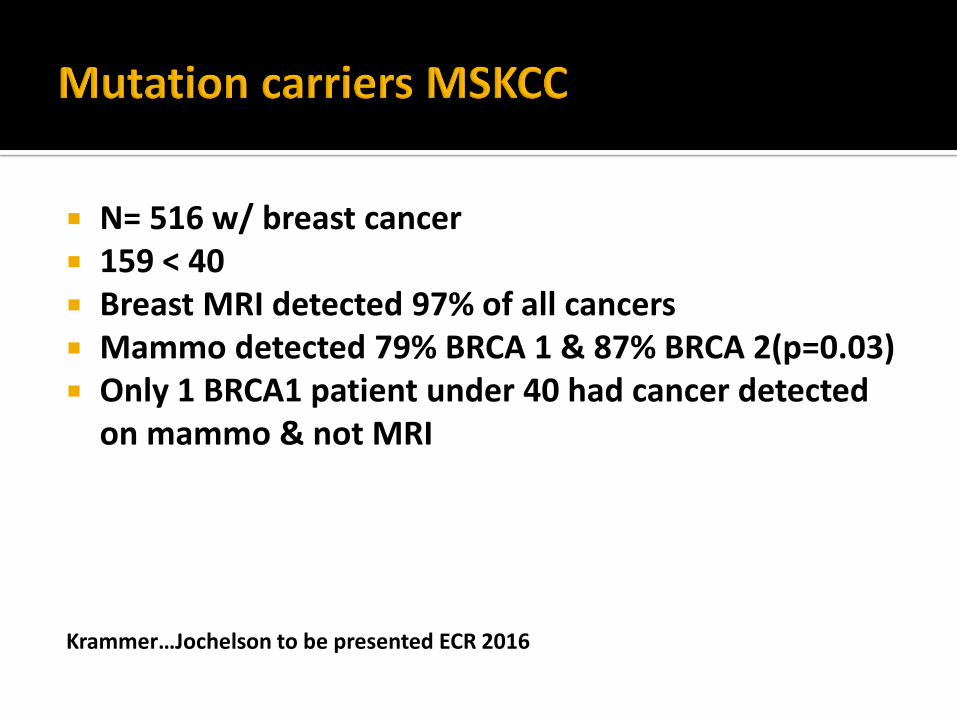
N= 516 w/ breast cancer 159 < 40 Breast MRI detected 97% of all cancers Mammo detected 79% BRCA 1 & 87% BRCA 2(p=0.03) Only 1 BRCA1 patient under 40 had cancer detected
on mammo & not MRI
Krammer…Jochelson to be presented ECR 2016

Not completely resolved Early data seem to suggest MRI/mammo should be
done separately at 6 month intervals rather than both at the same time yearly
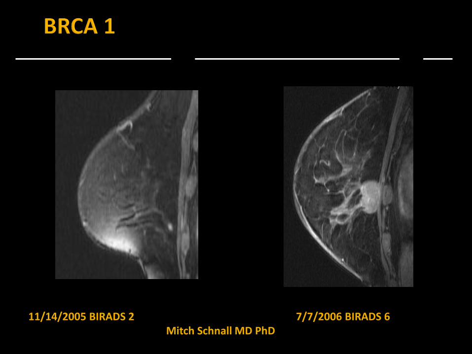
11/14/2005 BIRADS 2 7/7/2006 BIRADS 6Mitch Schnall MD PhD
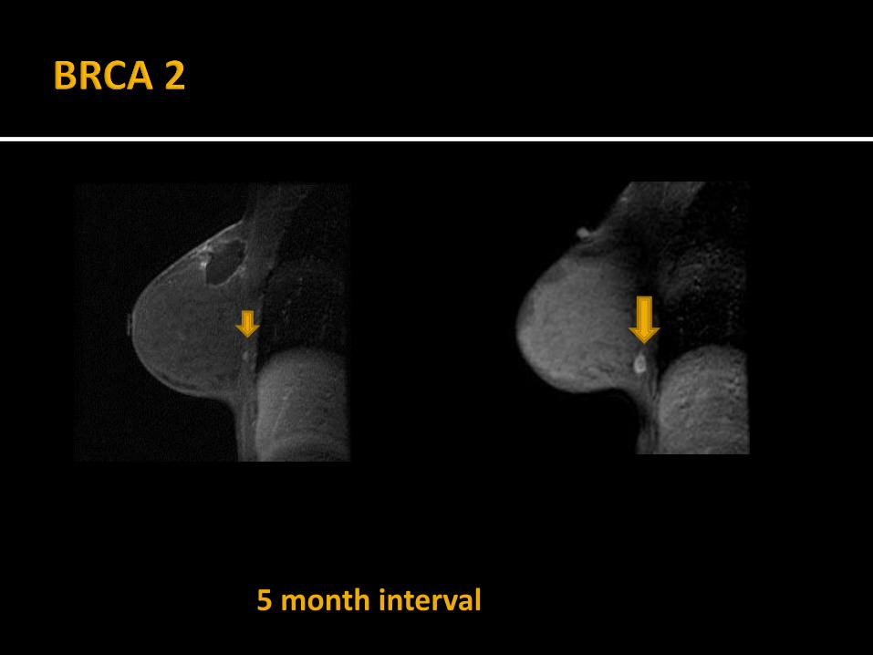
5 month interval

Personal history Family history ADH LCIS Dense breasts
Mammo &??????
DATA FREE ZONE regarding BEST tests to do

Two- fold issue1. 4-6 fold increased risk of breast cancer in women w/ extremely dense breasts c/w fatty breasts
2. Lower sensitivity of mammography in women w/ dense breasts leading to missed & interval cancers
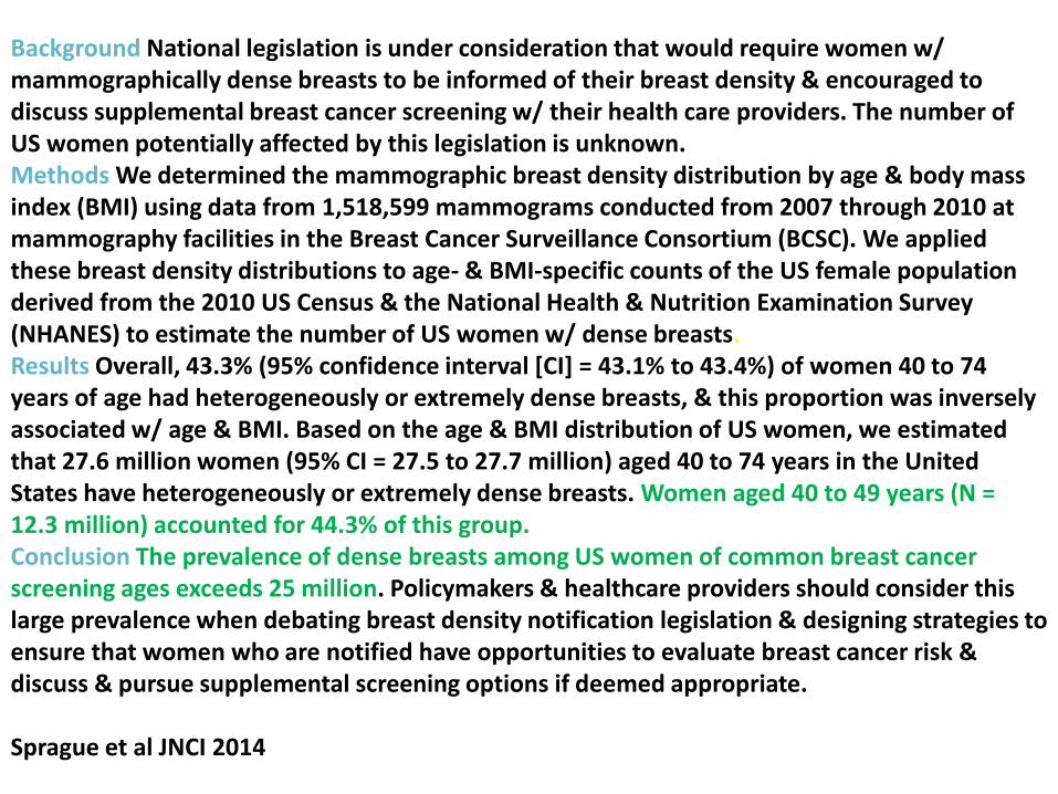
Background National legislation is under consideration that would require women w/ mammographically dense breasts to be informed of their breast density & encouraged to discuss supplemental breast cancer screening w/ their health care providers. The number of US women potentially affected by this legislation is unknown. Methods We determined the mammographic breast density distribution by age & body mass index (BMI) using data from 1,518,599 mammograms conducted from 2007 through 2010 at mammography facilities in the Breast Cancer Surveillance Consortium (BCSC). We applied these breast density distributions to age- & BMI-specific counts of the US female population derived from the 2010 US Census & the National Health & Nutrition Examination Survey (NHANES) to estimate the number of US women w/ dense breasts. Results Overall, 43.3% (95% confidence interval [CI] = 43.1% to 43.4%) of women 40 to 74 years of age had heterogeneously or extremely dense breasts, & this proportion was inversely associated w/ age & BMI. Based on the age & BMI distribution of US women, we estimated that 27.6 million women (95% CI = 27.5 to 27.7 million) aged 40 to 74 years in the United States have heterogeneously or extremely dense breasts. Women aged 40 to 49 years (N = 12.3 million) accounted for 44.3% of this group. Conclusion The prevalence of dense breasts among US women of common breast cancer screening ages exceeds 25 million. Policymakers & healthcare providers should consider this large prevalence when debating breast density notification legislation & designing strategies to ensure that women who are notified have opportunities to evaluate breast cancer risk & discuss & pursue supplemental screening options if deemed appropriate.
Sprague et al JNCI 2014

Current default:
Based on anatomy No radiation exposure Readily available “Inexpensive”

4,897 WOMEN
DENSE BREASTS
31 CANCERS
3/1000 (0.3%) CANCER DETECTION RATE
Kolb et al. 2002

N=2637 women Dense breasts + 1 other risk factor ~3.7 cancers per 1000 Invasive cancers– not DCIS All but 1 node negative 8% biopsy recommendation 9% short term follow up 7.4% positive biopsy rate
Berg W et al. JAMA 2008

N= 72,998 Japanese women 40-49 randomized to US or no US after mammo
Average risk/ dense breasts Sensitivity: 91.1% vs 77% p=0.0004 Specificity: 87.7% vs 91.4% p=0.0001 # of cancers 184 vs 117 Cancers in US group more frequently Stage 0/1
p=0.0194 Will follow for survival advantage
Ohuchi et al Lancet Nov 2015

Initial data from Connecticut experience N= 72,030 mammograms & 8,647 ultrasounds 28 mammographically occult cancers: 3.25/1000 PPV: 6.7% BIRADS 3: 9% US charge $250-reimbursed $72 Professional fee $85 reimbursed $30 Core $2,400
$110,241.00 billed; $60,000 paid/ breast cancer detected
Weigert et al The Breast Journal 2012

N= 935 w/ mixed risks & breast densities 3.2 cancers/1000 women screened Some were diagnostic patients PPV 6.5% 187 BI-RADS 3: 47 BI-RADS 4
$60,267/ cancer diagnosed (likely more since some patients were diagnostic)
Hooley et al Radiol 2012

16/612 (2.6%) breast cancer detected 12 (75%) invasive 14.7 additional cancers per 1000 women screened 9/16 (56%) seen only on MRI
8/9 (89%) invasive (median 9 mm) all node negative
2 (13%) not seen on MR, both DCIS
AVON FUNDED Berg et al JAMA 2012

Technology based on anatomy Peels away overlying tissues Lesion conspicuity improves Improved margin feature analysis Detection of additional lesions May show normal tissue when mass suspected
Improves sensitivity & specificity in both dense & fatty breasts

N=12631 Prospective trial Better detection rates: mammo alone 6.1/1000 vs. mammo
+ tomo 8.0/1000 25(40%) additional INVASIVE cancers detected w/ combo No change in DCIS detection 15% decrease false positives for combination
Skaane et al Radiol 2012

Rafferty et al. Radiology 2013; 266: 104-113
Reader Study DM DM+Tomo
Non-Cancer1 55.1% 16.7%
2 48.8% 30.1%
Cancer1 87.2% 80.4%
2 84.8% 85.7%

Prospective comparison study of 7292 women screened between August 2011- June 2012
CA detection rate:
MG: 5.3/1000
MG+DBT: 8.1/1000
Ciatto et al. Lancet 2013; 14: 583-589

N=13,158 at 4 sites: 7,058 MG/6100 MG+DBT Recall rate:
MG: 12.0%
MG+DBT: 8.4% Decreased recall rates for DBT among all breast
densities and age groups Detection of cancer:
MG: 5.2/1000MG+DBT 5.7/1000
Haas et al. Radiology 2013; 269: 694-700
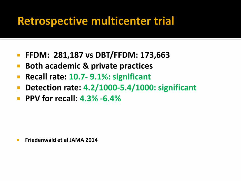
FFDM: 281,187 vs DBT/FFDM: 173,663 Both academic & private practices Recall rate: 10.7- 9.1%: significant Detection rate: 4.2/1000-5.4/1000: significant PPV for recall: 4.3% -6.4%
Friedenwald et al JAMA 2014

CONVENTIONALMAMMOGRAM
TOMOSYNTHESISSLICE 2
INVASIVECARCINOMA
TOMOSYNTHESISSLICE 1
INVASIVECARCINOMA #1
INVASIVECARCINOMA #2


LMLO
Courtesy of Janice Sung MD

LMLO LSMLOLLM
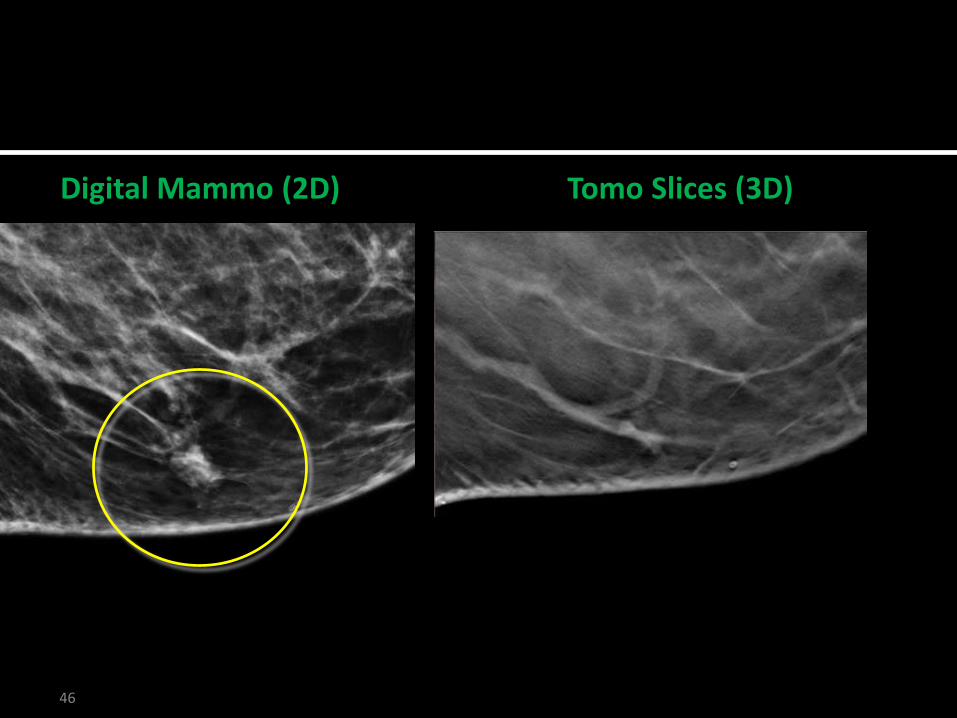
46 46
Digital Mammo (2D) Tomo Slices (3D)

Detects ~additional 1 to2 cancers/1000 Fewer call backs PPV improved Twice the radiation exposure/ still w/i guidelines Takes longer to read More expensive May replace full field digital mammography for
routine screening

Abridged MRI screening protocol could:
Decrease magnet & technologist time
Decrease reading time
Decrease cost, possibly making MRI more accessible

Prospectively read 606 screening MRIs in 443 women
Protocol 3 minutes vs. 17 for full exam
Full abbreviated protocol 28 seconds to read
▪ Sensitivity 100%, Specificity 94.3%
MIP: 2.8 seconds to read
▪ Sensitivity 90.9%
▪ Kuhl et al J Clin Oncol 2014

N= 100 patients w/ known cancers 3 sequences evaluated (15 min to perform) (mean 59
seconds to read) >95% of cancers visualized on a single MRI sequence Sensitivity increased to 100% w/ history & prior exams
Mango et al Eur J Radiol 2014

59 y/o female w/ contralateral breast carcinoma. New 1 cm irregularly enhancing mass in LLOQ: IDCMango et al
First post-contrast First post-contrast sub Subtraction MIP

MRI not universally available Certain patients cannot have MRI due to metallic
implants, claustrophobia or allergy to gadolinium Very expensive Too many false positives
Based on MRI’s ability to detect blood flow for better cancer detection

Omnipaque 350; 1.5 ml/kg (CT contrast)
Injected via power injector: 3ml/sec. First imaging ~ 3 minutes post-injection 4 views with high and low energy images obtained w/i 5
minutes of completed injection Images processed by subtracting out background tissue

Iodinated contrast administrationa. Follow criteria for CT contrast administrationb. Patients have reactions to Gadolinium too
Radiation dose ~20% > routine screening mammogram or the equivalent of one extra image
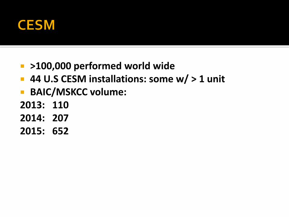
>100,000 performed world wide 44 U.S CESM installations: some w/ > 1 unit BAIC/MSKCC volume:2013: 1102014: 2072015: 652

N=120 UNILATERAL CESM + mammo c/w mammo or mammo
+ US: Pts recalled from screening or problem solving Sensitivity: CESM 93% vs mammo 78% 26% benign lesions enhanced CESM + mammo>mammo alone (p=0.045) & mammo
+ US (trend) CESM + mammo significantly more accurate than
mammo + US due to better specificity
Dromain et al Eur Radiol 2011(Confirmed in multireader study: Breast Cancer Research 2012)

MAMMOGRAPHY: CESM: MRI:
42/52 (81%)50/52 (96%)50/52 (96%)
Jochelson et al Radiol 2013

Multireader study of mammo vs. contrast mammo N=70 pts w/ at least 1 suspicious lesion Sensitivity improved from 35% to 59%
Diekmann et al Eur J Radiol 2011

89 Patients w/ dense breasts 100 lesions Low energy images were read blinded to post contrast
images With CESM, sensitivity improved from 71.5% to 92.7% Specificity improved from 51.8% to 67.9%
Cheung et al Eur Radiol 2014

MAMMO Sensitivity: 96.9% Specificity: 42.0% PPV: 39.7% NPV: 97.1%
CESM Sensitivity: 100% Specificity: 87.7% PPV: 76.2% NPV: 100%
Mean difference between CESM & pathology 1.4mm
Lobbes et al Eur Radio 2014


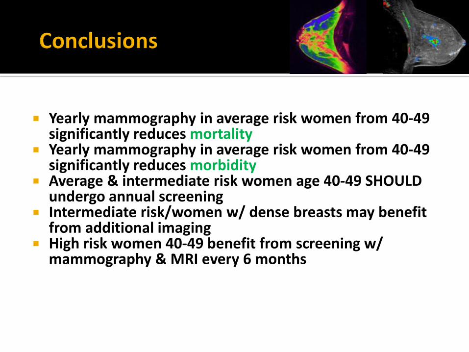
Yearly mammography in average risk women from 40-49 significantly reduces mortality
Yearly mammography in average risk women from 40-49 significantly reduces morbidity
Average & intermediate risk women age 40-49 SHOULD undergo annual screening
Intermediate risk/women w/ dense breasts may benefit from additional imaging
High risk women 40-49 benefit from screening w/ mammography & MRI every 6 months

“Given the weight of the evidence that mammography screening is associated w/ a significant reduction in the risk of dying from breast cancer after age 40 years, a more productive discussion would be focused on how to improve the performance of mammography screening”*
*Oeffinger et al JAMA 2015
Ultrasound, tomosynthesis, MRI & contrast mammo will all detect more cancers than mammo alone
MRI detects ~97% of cancers Tomosynthesis reduces call backs CESM improves sensitivity & specificity Prospective trials comparing the efficacy of these
techniques are underway. Physiology will likely trump anatomy
Proof of clinical advantage will take longer
