maternally irradiated pregnant rats
TRANSCRIPT


OPEN ACCESS Pakistan Journal of Biological Sciences
ISSN 1028-8880DOI: 10.3923/pjbs.2021.207.218
Research ArticleEffect of Bone Marrow Transplantation on the Fetal Skeleton ofMaternally Irradiated Pregnant Rats
Mervat Ahmed Abd Rabou
Department of Biology, College of Science, Jouf University, P.O. Box 2014, Sakaka, Saudi Arabia
AbstractBackground and Objective: Prenatal exposure to ionizing radiation can interfere with embryonic and fetal growth depending on thedose and gestational age. The present study was completed to evaluate the effect of transplanted bone marrow on the fetal skeleton ofpregnant rats exposed to gamma radiation. Materials and Methods: Experimental animals were separated into 5 groups: C group,R7 group, R7+BM group, R14 group and R14+BM group. All pregnant rats were sacrificed on day 20 days of gestation and the skeletal systemsof the fetuses were examined and photographed. This study focused on skull, upper and lower jaw, occipital region, sacral and caudalregion, fore and hind limbs. Results: Gamma rays caused any disturbance in the ossification process of the skull bones, upper and lowerjaws, occipital bones, it caused the loss of some ossification centers in metacarpal bones, metatarsal bones but bone marrowtransplantation greatly reduced the injury that happened because of γ-radiation. Conclusion: This study showed that transplantationof bone marrow post-irradiation in pregnant rats could reduce the hazards of gamma-irradiation in the different regions of the fetalskeleton.
Key words: Bone marrow, radiation, skeleton, fetal, pregnant, gestation, gamma
Citation: Rabou, M.A.A., 2021. Effect of bone marrow transplantation on the fetal skeleton of maternally irradiated pregnant rats. Pak. J. Biol. Sci., 24: 207-218.
Corresponding Author: Mervat Ahmed Abd Rabou, Department of Biology, College of Science, Jouf University, P.O. Box: 2014, Sakaka, Saudi ArabiaTel: 00966537242262
Copyright: © 2021 Mervat Ahmed Abd Rabou. This is an open access article distributed under the terms of the creative commons attribution License, whichpermits unrestricted use, distribution and reproduction in any medium, provided the original author and source are credited.
Competing Interest: The author has declared that no competing interest exists.
Data Availability: All relevant data are within the paper and its supporting information files.

Pak. J. Biol. Sci., 24 (2): 207-218 2021
INTRODUCTION
Exposure to radiation sources used in different appliancesmay be received by professionals known as radiation workers1.Radiation causes the tissue to be ionized and activated whichimpairs normal cell functioning2. Ionizing radiation interactswith matter and causes some modifications. These changesare known as radiation damage and cause structural changesin cells3. High doses of ionizing radiation can cause cell deathdue to damage in DNA strands4. The presence of injurednuclear DNA in the cells may cause cell divisions and genomicinstability5. Radiation exposure thus causes mutagenic effectsand organ-specific changes6.
Gamma irradiation was also found to cause embryonicloss and malformations in mice7. During the organogenesisperiod in pregnancy, radiation may cause malformations insome organs and possibly growth retardation, death offetuses, mental retardation and microcephaly8. The skeletalmalformations in pregnant mice were noted when exposed toa single dose of 2.0 Gy on days 2 and 15 of gestation9. Skeletalelements of mice fetuses maternally exposed to gamma raysshowed very poor ossification in the system especially duringthe organogenesis period10. On day 3, 10 and 14 of gestation,pregnant rats had a lower oscillation in the skull bones, lessoscillation in the vertebral centra and wavy ribs at a singlesublethal dose level of 4 Gy7.
Exposure of gravid mice to electromagnetic field radiationindicated a temporary disruption in the ossification process ofdifferent bones, which only improves after giving birth11. Thefemale mice exposed to 1 Gy of protons irradiation at sixteenweeks old caused the loss of trabecular bone fraction in thetibia and femur12. Bone marrow is a complex tissue comprisingtwo hematopoietic and stromal sections. The stromal divisionis a complex of tissue a hematopoietic microenvironmentcontaining a group of cells called mesenchymal stem cells(MSCs)13.
In recent years, stem cell strategies have become verypopular. Bone marrow MSCs can distinguish between many mesenchymal types of cells, including cartilage,bone and fat cells14. Mesenchymal stem cells are used toheal injured tissue and other tissue damage, includingdamage within the cardiac system, the nervous systemand lunate bone15. Transplantation of bone marrowincreases the level of antioxidants and protects rats fromoxidative stress16.
The current study was done to study the probableprotecting effect of BMT on the skeleton of fetuses ofmaternally irradiated pregnant rats.
MATERIALS AND METHODS
Study area: This research was done at Atomic EnergyAuthority, National Center for Radiation Research andTechnology, Cairo, Egypt from January-March, 2013.
Irradiation: Irradiation was provided by applying Gamma-cell40 (137 Cesium), at the Atomic Energy Authority, NationalCenter for Radiation Research and Technology, Cairo, Egypt.
Experimental animals: Albino rats (Rattus albinus) kept incages with body weights ranging from 120-150 gm were used.The males were separated from females until mating. Twofemales of estrous or proestrus periods were confined withone male. The pregnancy was assured the next morning bythe presence of a plugin the vagina or the occurrence ofspermatozoa in the smear of vaginal content and that dayrepresented the 1st day of gestation.
Bone marrow transplantation: Bone marrow transplantationwas of the same strain (receptors and donors). The donorswere sacrificed and femur bones were gutted and both endswere disjointed. The marrow was put into saline solutionunder sterilized conditions then mixed and expelled from thesyringe several times without a needle to prevent the cellsfrom mechanical damage. The number of cells about75×106±5 were inoculated 1 h after irradiation17. The bonemarrow was injected intraperitoneally in the pregnant rats.
Groups of animals: The gravid rats were separated into 5 groups (six rats in each group), C set (control gravid rats), R7set (Gravid rats exposed to one dose, two grays of γ rays onday 7 of pregnancy), Group R7+BM (Pregnant rats exposed toone dose, two grays of γ rays on the 7th day of gestation thenreceived bone marrow transplantation (75×106±5 cells) viaintraperitoneal dose 1 h after radiation, R14 group (Pregnantrats exposed to one dose, two grays of γ rays on the 14th dayof pregnancy), R14+BM group (Pregnant rats exposed to onedose, two grays of γ rays on the 14th day of gestation andthen received bone marrow transplantation (75×106±5 cells)via intraperitoneal dose 1 h after radiation. All gravid rats weresacrificed on the 20th day of pregnancy.
Skeletal studies: Alizarin red stain was used for evaluatingthe endoskeletal system of fetuses. The endoskeletonswere prepared according to the method of Chahoud andPaumgartten18. The skeletal systems were then examined andphotographed. This study focused on evaluating the following
208

Pak. J. Biol. Sci., 24 (2): 207-218 2021
regions the skull region, upper and lower jaw, occipital region,sacral and caudal region, forelimb and hind limb.
RESULTS
The normal skull of a rat is consists of the premaxilla,maxilla, nasal, frontal, parietal, intraparietal, squamosal, jugal,exoccipital, supraoccipital, basioccipital and tympanicum.(Fig. 1a). Fetuses of the R7 group showed a wide suturebetween the two parietal bones (Fig. 1b) but the treatment
of this group with bone marrow 1 h post-irradiationreconstructed the suture between the dermal bones withdense ossification centers in them (Fig. 1c). Less ossification inthe frontal and parietal bones was observed in fetuses ofgroup R14 (Fig. 1d). The fetuses of group R14+BM showedconspicuous improvement in ossification centers with a widesuture in the midline of the cranium (Fig. 1e).
The Fig. 2a showed normal ossification in the nasal,premaxilla, maxilla and zygomatic arch. Shortness in thepremaxilla bone was observed in the fetuses of the R7 group
Fig. 1(a-e): Cranium of rat fetuses after 20 days of gestationA (control group): Normal ossification of the dermal bones frontal (F), parietal (P) and interparietal (IP), B (R7 group): A large suture between two parietalbones (8), C (R7+BM group): A dense ossification of parietal and frontal bones, D (R14 group): Less ossification in the frontal and parietal bones and all thebones with high porosity, E (R14+BM): Conspicuous improvement in ossification of the dermal bones with large suture in the mild line of the cranium (8)
209
(a) (b)
(c) (d)
(e)

Pak. J. Biol. Sci., 24 (2): 207-218 2021
Fig. 2(a-e): Upper jaw of rat fetuses after 20 days of gestationA (control group): conspicuous ossification in the Premaxilla (Pm), Maxilla (Mx), B (R7 group): Shortness in the premaxilla bone (Pm), C (R7+BM group): Longand thin apex of nasal bone (Na) like rostrum, presence of incisor (8) and thick zygomatic process of maxilla (); D (R14 group): Part of maxilla was ossifiedonly (8); E (R14+BM): Elongated nasal bones (8) ,presence of incisor (>) and thick zygomatic process of maxilla ()
(Fig. 2b). Post-irradiation, bone marrow transplantation in thisgroup showed the nasal summit resembling the beak and theincrease in incisor development and the thick zygomaticprocess of the maxilla (Fig. 2c). Fetuses in the group R14showed only an ossified part of the maxilla bone (Fig. 2d),while fetuses of the R14+BM group showed elongatedbeak-like nasal summit bone and evolution in the incisorgrowth (Fig. 2e).
Conspicuous ossification of the mandible bone wasobserved in the control fetuses (Fig. 3a). Irradiation on the7th day of gestation caused shortness in this bone(Fig. 3b). Fetuses of the R7+BM group showed elongatedmandible bone and evolution in the incisor growth(Fig. 3c). Fetuses of group R14 displayed no ossificationcenters in the mandible bone (Fig. 3d) but the bonemarrow transplantation after exposure to gamma rays on
210
(a) (b)
(c) (d)
(e)

Pak. J. Biol. Sci., 24 (2): 207-218 2021
Fig. 3(a-e): Lower jaw of rat fetuses after 20 days of gestation A (control group): Conspicuous ossification of mandible bone (Mn), B (R7 group): Shortness in the mandible bone, C (R7+BM group): Elongated mandiblebone, D (R14 group): Unossified mandible bone, E (R14+BM): Good ossification in the mandible bone, Note, bone marrow transplantation accelerate thedevelopment of mandible and incisor formation (8) in groups R7+BM and R14+BM
the 14th of gestation accelerated the development ofthe mandible and incisor (Fig. 3e).
The Fig. 4 showed the occipital region in the normal andtreated groups. Fetuses of the control group displayedconspicuous ossification in the supraoccipital, exoccipital andatlas bones (Fig. 4a). The growth rate of the supraoccipital,exoccipital and atlas bones was reduced in the fetuses of theR7 group (Fig. 4b). Bone marrow transplantation one-hour
post-irradiation caused thickness in the supraoccipital bonewith improvement in the wing sides (Fig. 4c). Fetuses ofgroup R14 showed absence of ossification centers in thesupraoccipital, exoccipital and atlas bones (Fig. 4d), whilenormal ossification centers were observed in the occipitalregion of the fetuses of the R14+BM group (Fig. 4e).
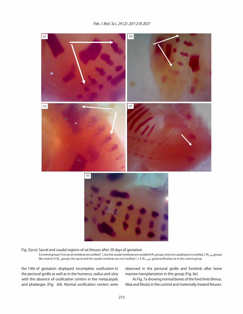
The Fig. 5 showed the sacral and caudal regions of ratfetuses after 20 days of gestation, Fig. 5a normal control.
211
(a) (b)
(c) (d)
(e)

Pak. J. Biol. Sci., 24 (2): 207-218 2021
Fig. 4(a-e): Occipital region of rat fetuses after 20 days of gestationA (control group), conspicuous ossification in the supra occipital (so), ex occipital (eo) and atlas (at), B (R7 group): Reduction in the size of supra occipital,ex occipital and atlas bones, C (R7+BM group): improvement in ossification of occipital bones (so, eo and at), D (R14 group): Absence of ossification centersin the supra occipital, ex occipital and atlas, E (R14+BM): Normal ossification in the occipital bones
Fetuses of group R7 showed five sacral vertebrae were ossifiedbut the caudal vertebrae ossification varies and only onecaudal piece was ossified (Fig. 5b). Fetuses of R7+BM group likecontrol (Fig. 5c). The sacral and the caudal vertebrae arenon-ossified in the R14 group (Fig. 5d). Good ossification as inthe control group was found in the R14+BM group (Fig. 5e).
The pectoral girdle (scapula, supra scapula and clavicle),in the fetuses at 20 days of gestation, showed complete
ossification in the normal and treated fetuses of different sizes.Bones of the forelimb (hummers, radius, ulna, 5 metacarpalsand phalanges) were detected in the normal fetuses (Fig. 6a).Incomplete ossification in the metacarpal bones (3 only) wasobserved in the fetuses of the R7 group (Fig. 6b). Bone marrowtransplantation post-irradiation on the 7th day of pregnancycaused an increase of the number of metacarpals (4),phalanges and nails (Fig. 6c). Fetuses maternally irradiated on
212
(a) (b)
(c) (d)
(e)
Pak. J. Biol. Sci., 24 (2): 207-218 2021
Fig. 5(a-e): Sacral and caudal regions of rat fetuses after 20 days of gestationA (control group): Five sacral vertebrae are ossified (8), but the caudal vertebrae are variable B (R7 group), only one caudal piece is ossified, C (R7+BM group)like control, D (R14 group), the sacral and the caudal vertebrae are non-ossified (8), E (R14+BM), good ossification as in the control group
the 14th of gestation displayed incomplete ossification inthe pectoral girdle as well as in the humerus, radius and ulnawith the absence of ossification centers in the metacarpalsand phalanges (Fig. 6d). Normal ossification centers were
observed in the pectoral girdle and forelimb after bonemarrow transplantation in this group (Fig. 6e).
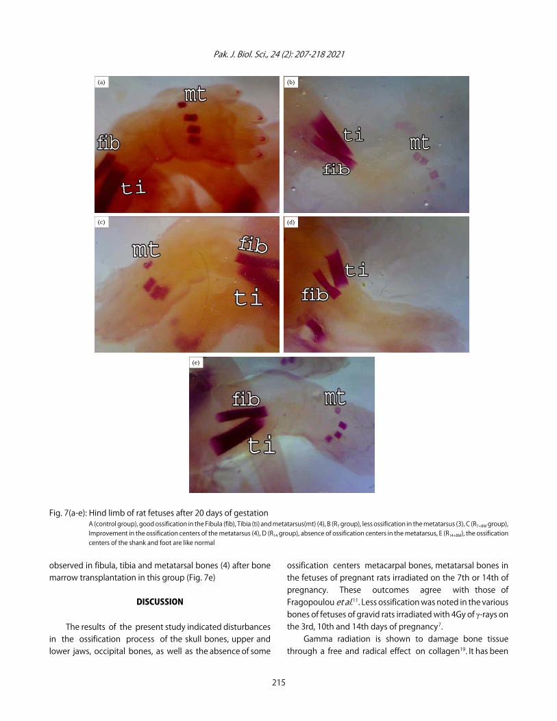
As Fig. 7a showing normal bones of the hind limb (femur,tibia and fibula) in the control and maternally treated fetuses.
213
(b)
(c) (d)
(e)
(a)

Pak. J. Biol. Sci., 24 (2): 207-218 2021
Fig. 6(a-e): Fore limb of rat fetuses after 20 days of gestationA (control group): Conspicuous ossification in the Radius (rad), Ulna (ul) and 5 Metacarpals (mc), weak ossification in nails (8), B (R7 group):Incomplete ossification in metacarpal bones (3), C (R7+BM group): Improvement in the number of metacarpals (4), phalanges (>) and nails (8),D (R14 group): Incomplete ossification in the radius, ulna and absence of metacarpals, E (R14+BM): Good ossification in metacarpals (3) andnails (8)
The ossified pieces of metatarsals were 4 in the normalfetuses and the phalanges were non-ossified. Fewerossification centers were observed in the metatarsal bones(3) of the fetuses whose mother was exposed to gammairradiation on the 7th day of pregnancy (Fig. 7b). Bone
marrow transplantation post-irradiation on the 7th day ofpregnancy indicated progress in ossification centers ofthe metatarsal bones (4) (Fig. 7c). Fetuses of group R14showed the absence of ossification centers in the metatarsalbones (Fig. 7d) but normal ossification centers were
214
(a) (b)
(c) (d)
(e)
Pak. J. Biol. Sci., 24 (2): 207-218 2021
Fig. 7(a-e): Hind limb of rat fetuses after 20 days of gestationA (control group), good ossification in the Fibula (fib), Tibia (ti) and metatarsus(mt) (4), B (R7 group), less ossification in the metatarsus (3), C (R7+BM group),Improvement in the ossification centers of the metatarsus (4), D (R14 group), absence of ossification centers in the metatarsus, E (R14+BM), the ossificationcenters of the shank and foot are like normal
observed in fibula, tibia and metatarsal bones (4) after bonemarrow transplantation in this group (Fig. 7e)
DISCUSSION
The results of the present study indicated disturbancesin the ossification process of the skull bones, upper andlower jaws, occipital bones, as well as the absence of some
ossification centers metacarpal bones, metatarsal bones inthe fetuses of pregnant rats irradiated on the 7th or 14th ofpregnancy. These outcomes agree with those ofFragopoulou et al.11. Less ossification was noted in the variousbones of fetuses of gravid rats irradiated with 4Gy of γ-rays onthe 3rd, 10th and 14th days of pregnancy7.
Gamma radiation is shown to damage bone tissuethrough a free and radical effect on collagen19. It has been
215
(a) (b)
(c) (d)
(e)

Pak. J. Biol. Sci., 24 (2): 207-218 2021
shown that therapeutic doses of radiation have deleteriouseffects on the health of bone tissue, sometimes causingspontaneous fractures and osteoradionecrosis20.
As for pregnant mice exposed to radiation from days 7-10of pregnancy, it is believed that the development of embryosis vulnerable to the outside environment and since limbdevelopment is important for the quality of life the observedincreased rates of polydactyly fetuses is troubling 21.
Skeletal elements of mice fetuses maternally exposed togamma rays showed very poor ossification in the systemafter exposure during organogenesis followed by fetal andpre-implantation periods10.
A decrease in the bone calcium content of rats exposedto 5 or 10 Gy of gamma rays led to increased skeletalmalformation22. The drop in calcium in the bone may be dueto the demineralization of the bone after irradiation23.
The skeletal elements play roles in joined craniofacialmorphogenesis. Because of the highly complex nature ofhead development, craniofacial dysplasias alone accountfor many dimorphic features. Any abnormality or delay in thedevelopment of embryonic cranial constitutes skeletaldeviation abnormality24. Furthermore, ionizing radiation hasbeen implicated in osteoporotic phenotypes, bone loss andfracture risk25.
Multipotent cells which are called human mesenchymalstem cells prove valuable for the treatment of degenerativediseases. MSCs in many tissues and organs can be foundbetween differentiated cells but, unfortunately, a phenotypicalsimilarity impedes robust cells and discrimination fromdifferent tissue harvest26.
The first experimental use of Bone MarrowTransplantation was made for people exposed to radiationafter a nuclear accident27. Transplanted bone marrowsuccessfully replaced marrow damaged by irradiation.Transplantation of the bone marrow is not a surgicalprocedure but a painless infusion equivalent to a bloodtransfusion in the hospital.
In the present study, BMT post-irradiation indicated anoticeable improvement in the bones of the skull, upper andlower jaws, fore limb and hind limb.
MSCs are alleged to be able to distinguish intocardiomyocytes, myoblasts and neurons. Formation of cells ofthe non-mesodermal source may be an outcome of aphenomenon called “stem cell plasticity”, a process oftransdifferentiation that is not limited to formingdifferentiated cell tissue types in which Organ-specific stemcells are located28.
Osteogenesis needs MSCs to be incubated with ascorbicacid phosphate, $-glycerol-phosphate, fetal bovine serum anddexamethasone. MSCs should reveal osteoplastic morphologytogether with a high appearance of alkaline phosphatase andcalcium deposit29.
The hematopoietic stem cell pool is dispersed throughthe places of hematopoietic cell reconstruction in a greatnumber of bone marrow units dispersed throughout theskeleton. Bone marrow sites that are haematopoietically activefunction as an organ system through the stem cell migrationstreams that link all sites via blood and guarantee a local stemcell level which ensures a balance between cell growth andcell removal is maintained30.
Bone marrow energizing made more chondrogenesis inTrochlea (TR) and Medial Femoral Condyle (MFC) blot in adultrabbits, with further chondrocytes and larger chondrogenicfoci seeming in MFC and TR versus on the 21st daypost-operation31.
Bone marrow transplanted cells to osteogenesisimperfecta patients and showed that about 2% of theosteoblasts in the recipient's bone marrow came from thedonor. MSCs can become normal osteoblasts and contributeto the accelerated growth of bones and reduced rates offracture32. Stem cell transplantation can treat osteogenesisimperfecta. Due to the irregular bone form, bone growth wassluggish, bone fractures were common and bone defectsoccurred33.
The application of stem cells populations to boneengineering strategies has the potential to deeply influenceregenerative medicine34. Stem cells from bone marrow correctand restore a proper microenvironment to improvetransplantation procedures and general disease outcomes35.The study recommends that the pregnant must not beexposed to radiation during pregnancy, so that may causesdefects in the skeleton of fetuses.
CONCLUSION
This study revealed the presence of instabilities in theprocess of ossification in the skull bones, upper and lowerjaws, occipital bones of fetal rats maternally irradiatedpregnant rats, as well as the absence of some ossificationcenters in the frontal and parietal bones and all the bones withhigh porosity, conspicuous ossification in the radius and ulna,absence of ossification centers in the metatarsus. Bonemarrow transplantation post-irradiation indicated a noticeableimprovement in the bones of the skull, upper and lower jaws,forelimb and hind limb.
216

Pak. J. Biol. Sci., 24 (2): 207-218 2021
SIGNIFICANCE STATEMENT
This study discovers the possible protective role of bonemarrow transplantation in decreasing the gamma radiationhazards in the skeleton of fetal rat maternally irradiated. Thisstudy will help the researcher to avoid radiation duringpregnancy periods and the importance of bone marrowtransplantation in recovering these disturbances. Thus, a newtheory has to introduce to injury of radiation on the skeletonof fetal rats.
ACKNOWLEDGMENT
Author wish to thank Dr. Omaima Ashry, National Centerfor Radiation Research and Technology, Cairo, Egypt for thequalified support to the fieldwork. The author is deeplygrateful to Dr. Nehal Abo El Naga and Dr. Fatma Eid for theirvaluable support in this study.
REFERENCES
1. Davies, H.E., C.G. Wathen and F.V. Gleeson, 2011. The risks ofradiation exposure related to diagnostic imaging and how tominimise them. BMJ, 342: 589-593.
2. Aggarwal, L., 2014. Biological effects of ionizing radiation.Shodh Prerak, 1 : 342-348.
3. ÇaliÕkan, B. and A.C. ÇaliÕkan, 2018. Interaction with Matterof Ionizing Radiation and Radiation Damages (Radicals). In:Ionizing Radiation Effects and Applications, Djezzar, B. (Ed.).,InTech, London, .
4. Salama, S.F., O.M. Ashry and E.M. Hussein, 2007. Concomitanteffect of ciprofloxacin and echinacea counteracting severityof radiation damage in rats. Egypt. J. Radiat. Sci. Applic.,20: 365-383.
5. Kopjar, N., S. Miocic, S. Ramic, M. Millic and T. Viculin, 2006.Assessment of the radioprotective effects of a mifostine andmelatonin of human lymphocytes irradiated with γ-rays in vitro. Arch. Hig. Rada. Toksikol., 57: 155-163.
6. Zhou, W., J. Xu, K. Zhao, J. Xu, Y. Dong and S. Tong, 2019.Efficacy of human bone marrow mesenchymal stem celltransplantation in repair of radiation-induced damage to theimmune system. Int. J. Clin. Exp. Med., 12: 2651-2658.
7. Al-Shaibani, E.A., H.I. Nadia, O.S. Eissa, M.I. Rady and T.Z. Zaki,2009. Teratological effects of gamma-irradiation during threegestaional intervals in rats. Egypt. J. Hosp. Med., 36: 456-467.
8. Chang, D.S., F.D. Lasley, I.J. Das, M.S. Mendonca andJ.R. Dynlacht, 2014. Radiation Effects in the Embryo and Fetus.In: Basic Radiotherapy Physics and Biology, Chang, D.S., F.D.Lasley, I.J. Das, M.S. Mendonca and J.R. Dynlacht (Eds.).,Springer International Publishing, New York, pp: 313-316.
9. Kim, S., J. Lee, H. Oh, S. Kim and S. Lee, 2001. Dependence ofmalformation upon gestational age and exposed dose ofgamma radiation. J. Rad. Res., 42: 255-264.
10. Gulay, K.C.M., I.B. Tanaka, J. Komura and S. Tanaka, 2018.Effects of continuous gamma-ray exposure in utero in B6C3F1mice on gestation day 18 and at 10 weeks of age. Radiat. Res.,189: 425-440.
11. Fragopoulou, A.F., S.L. Koussoulakos and L.H. Margaritis,2010. Cranial and postcranial skeletal variations induced inmouse embryos by mobile phone radiation. Pathophysiology,17: 169-177.
12. Lloyd, T.E., J. Machamer, K. O'Hara, J.H. Kim andS.E. Collins et al., 2012. The p150 (glued) CAP-Gly domainregulates initiation of retrograde transport at synaptictermini. Neuron, 74: 344-360.
13. Zakrzewski, W., M. Dobrzy½ski, M. Szymonowicz and Z. Rybak,2019. Stem cells: Past, present and future. Stem Cell Res.Ther., Vol. 10. 10.1186/s13287-019-1165-5.
14. Aquino-Martínez, R., N. Artigas, B. Gámez, J.L. Rosa andF. Ventura, 2017. Extracellular calcium promotes boneformation from bone marrow mesenchymal stem cells byamplifying the effects of BMP-2 on SMAD signalling. PLoSONE, Vol. 12. 10.1371/journal.pone.0178158.
15. Bejargafshe, M.J., M. Hedayati, S. Zahabiasli, E. Tahmasbpour,S. Rahmanzadeh and A. Nejad-Moghaddam, 2019. Safetyand efficacy of stem cell therapy for treatment of neuraldamage in patients with multiple sclerosis. Stem Cell Invest.,Vol. 6. 10.21037/sci.2019.10.06.
16. Ashry, O.M., E.M. Hussein and S.F. Salama, 2009. Boosting ofantioxidant defence by interferon-Alfa in irradiated bonemarrow transplanted rats. Egypt. J. Radiat. Sci. Applic.,22: 19-33.
17. Decleve, A., G.B. Gerber, A. Leonard, M. Lambiet-Collier, A. Sassen and J.R. Maisin, 1972. Regeneration of thymus,spleen and bone marrow in X-irradiated AKR mice. Radiat.Res., 51: 318-332.
18. Chahoud, I. and F.J.R. Paumgartten, 2009. Dose-responserelationships of rat fetal skeleton variations: Relevance for riskassessment. Environ. Res., 109: 922-929.
19. Akkus, O., R.M. Belaney and P. Das, 2005. Free radicalscavenging alleviates the biomechanical impairment ofgamma radiation sterilized bone tissue. J. Orthopaedic Res.,23: 838-845.
20. Hamilton, S.A., M.J. Pecaut, D.S. Gridley, N.D. Travis andE.R. Bandstra et al., 2006. A murine model for bone loss fromtherapeutic and space-relevant sources of radiation. J. Appl.Physiol., 101: 789-793.
21. Yang, M.J., J.Y. Liu, Y.F. Wang, H.Y. Lang and X. Miao et al.,2013. Effects of electromagnetic pulse on polydactyly ofmouse fetuses. Theriogenology, 80: 18-23.
217

Pak. J. Biol. Sci., 24 (2): 207-218 2021
22. Saad, T.M. and A.A. Ammar, 2011. Protective role of peanut oilin rats exposed to two different doses of gamma radiationthat produced oxidative stress and bone injury. Egypt. J.Hosp. Med., 44: 380-391.
23. Fukuda, S. and H. Lida, 1990. Effects of clinostat microgravyand heavy particle radiation in bone and calcium metabolismin rats. J. Jap. Bone Morph., 9: 35-44.
24. Zowail, M.E., E.H. Khater, H.F. Waer, N.A. Eltahawy, A. El-Hadyand M. Amr, 2012. Curative effect of bone marrow cellstransplantation and/or low dose gamma irradiation on liverinjuries induced by carbon tetrachloride. Egypt. J. Hosp. Med.,46: 96-114.
25. Rana, T., M.A. Schultz, M.L. Freeman and S. Biswas, 2012. Lossof Nrf2 accelerates ionizing radiation-induced bone loss byupregulating RANKL. Free Radical Biol. Med., 53: 2298-2307.
26. Ragni, E., T. Montemurro, E. Montelatici, C. Lavazza andM. Viganò et al., 2013. Differential microRNA signature ofhuman mesenchymal stem cells from different sourcesreveals an “environmental-niche memory” for bone marrowstem cells. Exp. Cell. Res., 319: 1562-1574.
27. Burrett, A. and M. Gordon, 1993. Bone marrow disorders. Thebiological basis of treatment. 2nd, Blackwell ScientificPublications. Oxford.
28. Lakshmipathy, U. and C. Verfaillie, 2005. Stem cell plasticity.Blood Rev., 19: 29-38.
29. Chase, L.G., U. Lakshmipathy, L.A. Solchaga, M.S. Rao andM.C. Vemuri, 2010. A novel serum-free medium for theexpansion of human mesenchymal stem cells. Stem Cell Res.Ther., Vol. 1. 10.1186/scrt8.
30. Smith-Berdan, S., A. Nguyen, D. Hassanein, M. Zimmer andF. Ugarte et al., 2011. Robo 4 cooperates with cxcr4 to specifyhematopoietic stem cell localization to bone marrow niches.Cell Stem Cell, 8: 72-83.
31. Chen, H., A. Chevrier, C.D. Hoemann, J. Sun, V. Lascau-Comanand M.D. Buschmann, 2013. Bone marrow stimulationinduces greater chondrogenesis in trochlear vs condylarcartilage defects in skeletally mature rabbits. OsteoarthritisCartilage, 21: 999-1007.
32. Hurwitz, S., 1996. Homeostatic control of plasma calciumconcentration. Crit. Rev. Biochem. Mol. Biol., 31: 41-100.
33. Chen, A.K.L., S. Reuveny and S.K.W. Oh, 2013. Application ofhuman mesenchymal and pluripotent stem cell microcarriercultures in cellular therapy: Achievements and futuredirection. Biotechnol. Adv., 31: 1032-1046.
34. Walmsley, G.G., R.C. Ransom, E.R. Zielins, T. Leavitt andJ.S. Flacco et al., 2016. Stem cells in bone regeneration.Stem Cell Rev. Rep., 12: 524-529.
35. Crippa, S., L. Santi, R. Bosotti, P. Porro and M. Bernardo, 2020.Bone marrow-derived mesenchymal stromal cells: A noveltarget to optimize hematopoietic stem cell transplantationprotocols in hematological malignancies and rare geneticdisorders. JCM, Vol. 9. 10.3390/jcm9010002.
218