maternal obstetrical paralysrs · maternal obstetrical paralysrs with a report of two cases by...
TRANSCRIPT

MATERNAL OBSTETRICAL PARALysrs With a Report of Two Cases
BY
JEFFERY C. SPRY LEVERTON, M.D., M.R.C.O.G. Registrar, Southmead Hosfi’taE. Bristol
INJURY to the peripheral nerves of the lower extremities is an interesting but rare complication of pregnancy and labour. Most authorities are in agreement as to the rarity of the condition. Thus, Chalmers (1g4g), in a recent survey of this subject, has collected 146 cases from the literature, the first being described by von Basedow in 1838. Beattie (1933) found an incidence of 3 cases in 8,000 deliveries and Tillman (1935) one of g cases in 18,800, both over a period of 10 years. However, O’Connell (1944) suggests that the condition is more frequent than is realized and that careful neurological examination after labour, particularly difficult or instrumental labour, would reveal many cases showing minimal and transitory signs of nerve affection.
This form of nerve affection has been called traumatic neuritis of the puerperium. Certainly this title serves to emphasize its traumatic or mechanical aetiology and to place in a different category those cases of peripheral neuritis in pregnant and puer- peral women due to toxaemk causes, hypovitaminosis, and puerperal infection. Infection was at one time, in varying degree, a common cause of peripheral paralysis. Matthews Duncan (1886), for example, described with lucid clarity the autopsy findings in a severe case of pelvic cellulitis. The whole region of the psoas major muscle was converted into a huge abscess cavity, and, through it, stretched like fiddle strings, passed the bare nerves.”
Modern textbooks of obstetrics pay scant, if any, attention to this condition’and thus the case histories and discussion that follow may be of some interest.
CASE I . Mrs. B. P., aged ZI years, primigravid, commenced labour on 8th June, 1950, z days after her expected date of delivery. Her pregnancy had been uneventful. At term the foetus appeared large and the head had not engaged; there was, however, no evidence of disproportion. The first stage Iasted 30 hours, during which time the head engaged and the patient complained of occasional attacks of cramp-like pain in the left buttock, thigh, and calf. After 3 hours in the second stage the vertex had reached low mid cavity in the left occipito anterior position but progress had ceased. Under gas, oxygen and ether anaesthesia, low forceps were applied and a male infant delivered without diEculty. The baby, weighing 11 pounds z ounces, breathed spontaneously, later cried lustily and gave rise to no further anxiety. The day after delivery, the patient complained of pain in the left calf muscles and was unable to dorsiflex or evert the foot. A back splint was applied. Three days later all pain had gone but a definite palsy related to the anterior tibia1 and peroneal branches of the external popliteal nerve was present. At no time had there been any objective loss of sensation in the affected limb.
The puerperium otherwise was uncomplicated, and mother and infant were discharged on the 14th postpartum day. The patient was instructed to continue the use of a back splint, except for short periods during the day. An intensive course of physiotherapy was instituted and consisted of massage, exercises and electrical stimulation to the affected muscle groups. Under this regime, recovery was remarkably quick. Eight weeks
1019
J Obs Gyn Brit Emp 1951 V-58
history-of-obgyn.comobgynhistory.net

JOURNAL OF OBSTETRICS AND GYNAECOLOGY I020
postpartum the patient had regained sufficient power of dorsiflexion of her foot to walk without untoward disability. Thigh and calf measure- ments showed only moderate wasting. When seen recently, 8 months after the initial paralysis, the patient looked very well and stated that she could walk normally. However, on examination, slight residual wasting could still be elicited and lull power had not returned to the movement of dorsiflexion. The power of eversion was normal. X-ray pelvimetry was performed. The inlet view (Fig. I) showed asymmetry with absence of any marked sacral promontory and therefore shallow sacro-iliac fossae; this shallowneds is particularly marked on the left side where the joint region actually appears to protrude into the cavity. The lateral view showed .a normal sacral curve with adequate pelvic measurements. The internal con- jugate measured 4% inches. Views taken of the spine showed that there was a lateral tilt of L4 on L5 and compensatory lumbo-dorsal scoliosis. The latter finding is of interest in relation to the disc protrusion theory of O’Connell (1944) and is later discussed in this paper.
CASE 2. Mrs. B. M. C . , aged 38 years, gravida 3, para I, commenced labour on 3rd February, 1951, z days beyond the calculated date. Her first preg- nancy in 1942 had terminated in the normal delivery of a live ma!e infant, weighing 5 pounds 10 ounces. Following this, in 1949, there had been a miscamage a t 16 weeks. Antenatal examination in the present pregnancy showed the patient to be of moderate build (weight 7% stones, height 5 feet) and to possess generous external pelvic measure- ments. The pregnancy had proceeded unevent- fully. At the commencement of labour the foetus presented by the vertex in the left occipito anterior position with the head high and mobile. The first stage, characterized by uterine contractions of good quality throughout, lasted IZ hours. At the end of this period the foetal head was noted to be just engaged. mere was marked foetal distress and it was decided to deliver by forceps. Under spinal anaesthesia (I ml. heavy nupercaine), vaginal examination revealed the head to be just engaged in the left occipito transverse position with evident asynclitism. The antero-posterior diameter of the mid-pelvis appeared to be contracted.
Kielland’s forceps were applied and, with consider- able traction, a stillborn male infant, weighing 7% pounds, was delivered. Autopsy revealed the cause of death to be tentorial tears with haemor- rhage.
Two days after delivery, the patient complained of residual numbness in the right leg and it was noted that there was a well-marked paralysis of the anterior tibia1 nerve, resulting in foot drop. The peronei appear to have escaped damage. The same treatment as detailed in Case I was instituted and the patient was discharged, after an otherwise normal puerperium, on the 14th postpartum day. At the time of writing progress towards recovery, initially slow, is proceeding satisfactorily.
X-ray pelvimetry was performed before dis- charge. The inlet view (Fig. 2) showed considerable flattening and again, as in the first case, the sacro- iliac joint regions were seen to be relatively exposed by the absence of marked sacral promin- ence. The lateral view (Fig. 3) showed well- marked sacral flattening and a small internal con- jugate of 3% inches. The outlet was android in shape.
DISCUSSIQN The majority of cases of lower limb
paralysis following childbirth occur after difficult deliveries-instrumental deliveries of cases demonstrating some degree of cephalo-pelvic disproportion-and the sub- sequent nerve affection is often of the external popliteal division of the sciatic nerve on one or other side. However, there is reported in the literature a number of cases in which the paralysis was ante- partum, followed normal labour or breech presentation, or was bilateral in distribu- tion. Some cases demonstrated involve- ment of the femoral nerve or the whole sciatic nerve.
Thus any theories put forward to explain the occurrence must explain this diversity of syndrome type. The theories which have been advanced, and which are de- tailed below, explain fairly adequately the mechanisms at work in producing the
history-of-obgyn.comobgynhistory.net

FIG. I Showing asymmetry of the inlet and ihallow sacro-iliac fossae.
F IG. 2
Showing flattening of the inlet and shallow sacro-iliac iossae.
J.C.S.L.
history-of-obgyn.comobgynhistory.net

FIG. 3 Lateral view showing well-marked flattening of thc sacrum and reduccd internal coiij ligate
ineasureinent (retouched).
history-of-obgyn.comobgynhistory.net
MATERNAL OBSTETRICAL PARALYSIS
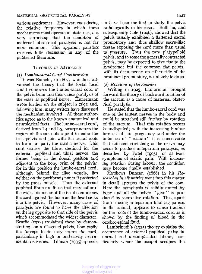
various syndromes. However, considering the relative frequency in which these mechanisms must operate in obstetrics, it is very surprising that the condition of maternal obstetrical paralysis is not far more common. This apparent paradox receives little discussion in any of the published literature.
I021
to have been the first to study the pelvis radiologically in his cases. Both he, and subsequently Cole (1946) , showed that the pelvis usually exhibited a flattened sacral promontory and thus shallow sacro-iliac fossae exposing the cord more than usual to pressure. Thus the rare platypelloid pelvis, and to some the generally contracted pelvis, may be expected to give rise to the syndrome; but the common flat pelvis, with its deep fossae on either side of the prominent promontory, is unlikely to do so.
(2) Rotatiom of the Sacrum Writing in 1925, Lambrinudi brought
forward the theory of backward rotation of the sacrum as a cause of maternal obstet- rical paralysis.
He stated that the lumbo-sacral cord was one of the tautest nerves in the body and could be stretched still further by rotation of the sacrum. That this rotation occurs is undisputed ; with the increasing lumbar Iordosis of late pregnancy and under the influence of “ relaxin ” it is conceivable that sufficient stretching of the nerve may occur to produce antepartum paralysis, as described by Patel (1go5), or at least symptoms of sciatic pain. With increas- ing rotation during labour, the condition may become finally established.
Matthews Duncan (1868) in his Re- searches in Obstetrics went into this matter in detail apropos the pelvis of the cow. Here the symphysis is solidly united by bone and all the pelvic “ give” is pro- duced by sacro-iliac rotation. This, apart from causing antepartum hind leg paresis in the animal, appears to cause traction on the roots of the lumbo-sacral cord as is shown by the finding of blood in the cerebro-spinal fluid.
Lambrinudi’s (1925) theory explains the occurrence of external popliteal palsy in normal and uncomplicated labour, par- ticularly where the occiput occupies the
THEORIES OF AETIOLOGY (I) Lumbo-sacral Cord Compressiort
It was Bianchi, in 1867, who first ad- vanced the theory that the foetal head could compress the lumbo-sacral cord at the pelvic brim and thus cause paralysis of the external popliteal nerve. Hiinermann wrote further on the subject in 1892 and, following him, many writers have discussed the mechanism involved. All these author- ities agree as to the known anatomical and neurological facts. The lumbo-sacral cord, derived from L4 and L5, sweeps across the region of the sacro-iliac joint to enter the true pelvis and join with the sacral roots to form, in part, the sciatic nerve. This cord carries the fibres destined for the external popliteal and tibia1 nerves, the former being in the dorsal position and adjacent to the bony brim of the pelvis; for in this position the lumbo-sacral cord, although behind the iliac vessels, lies neither on the pyriformis nor is it protected by the psoas muscle. Thus the external popliteal fibres are those that may suffer if the widest diameter of the head compresses the cord against the bone as the head sinks into the pelvis. However, many cases of paralysis are found to have the affection on the leg opposite to that side of the pelvis which accommodated the widest diameter. Beattie (1933) explained these by demon- strating, on a dissected pelvis, how easily the forceps blade may injure the cord, particularly in high or mid-cavity instru- mental deliveries. Tillman (1935) appears
history-of-obgyn.comobgynhistory.net
JOURNAL OF OBSTETRICS AND GYNAEXOLOGY I022
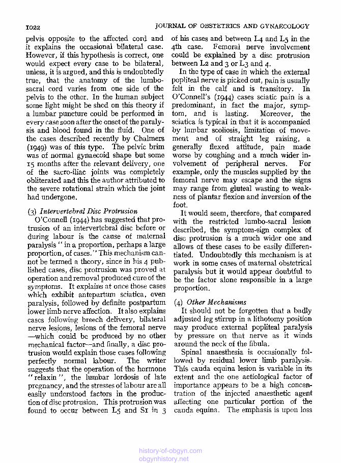
pelvis opposite to the affected cord and it explains the occasional bilateral case. However, if this hypothesis is correct, one would expect every case to be bilateral, unless, it is argued, and this is undoubtedly true, that the anatomy of the lumbo- sacral cord varies from one side of the pelvis to the other. In the human subject some light might be shed on this theory if a lumbar puncture could be performed in every case soon after the onset of the paraly- sis and blood found in the fluid. One of the cases described recently by Chalmers (1949) was of this type. The pelvic brim was of normal gynaecoid shape but some 15 months after the relevant delivery, one of the sacro-iliac joints was completely obliterated and this the author attributed to the severe rotational strain which the joint had undergone.
(3) Imtervertebral Disc Protrusion O’Connell (1944) has suggested that pro-
trusion of an intervertebral disc before or during labour is the cause of maternal paralysis “ in a proportion, perhaps a large proportion, of cases. ” This mechanism can- not be termed a theory, since in his 4 pub- lished cases, disc protrusion was proved at operation and removal produced cure of the symptoms. It explains at once those cases which exhibit antepartum sciatica, even paralysis, followed by definite postpartum lower limb nerve affection. I t also explains cases following breech delivery, bilateral nerve lesions, lesions of the femoral nerve -which could be produced by no other mechanical factor-and finally, a disc pro- trusion would explain those cases following perfectly normal labour. The writer suggests that the operation of the hormone
relaxin ”, the lumbar lordosis of late pregnancy, and the stresses of labour are all easily understood factors in the produc- tion of disc protrusion. This protrusion was found to occur between Lg and SI in 3 -.
of his cases and between L4 and L5 in the 4th case. Femoral nerve involvement could be explained by a disc protrusion between L2 and 3 or L3 and 4.
In the type of case in which the external popliteal nerve is picked out, pain is usually felt in the calf and is transitory. In O’Connell’s (1944) cases sciatic pain is a predominant, in fact the major, symp- tom, and is lasting. Moreover, the sciatica is typical in that it is accompanied by lumbar scoliosis, limitation of move- ment and of straight leg raising, a generally flexed attitude, pain made worse by coughing and a much wider in- volvement of peripheral nerves. For example, only the muscles supplied by the femoral nerve may escape and the signs may range from gluteal wasting to weak- ness of plantar flexion and inversion of the foot.
It would seem, therefore, that compared with the restricted lumbo-sacral lesion described, the symptom-sign complex of disc protrusion is a much wider one and allows of these cases to be easily differen- tiated. Undoubtedly this mechanism is at work in some cases of maternal obstetrical paralysis but it would appear doubtful to be the factor alone responsible in a large proportion.
(4) Other Mechanisms It should not be forgotten that a badly
adjusted leg stirrup in a lithotomy position may produce external popliteal paralysis by pressure on that nerve as it winds around the neck of the fibula.
Spinal anaesthesia is occasionally fol- lowed by residual lower limb paralysis. This cauda equina lesion is variable in its extent and the one aetiological factor of importance appears to be a high concen- tration of the injected anaesthetic agent affecting one particular portion of the cauda eauina. The emDhasis is mon loss
history-of-obgyn.comobgynhistory.net

MATERNAL OBSTETRICAL PARALYSIS
of bladder and sphincter control and the non-traumatic aetiology is reflected in the occasional occurrence of a cranial nerve palsy, particularly the 6th nerve (Bryce- Smith and Macintosh, 1951).
COMMENT In the first case described in this paper
the paralysis was probably caused by lumbo-sacral cord compression. The X-ray view of the inlet reveals a typical exposure of the nerve to pressure by the head and, with a huge infant of head circumference 15Q inches, it can be understood how the leg paralysis was caused and was on the same side as the occiput lay in the pelvis (Fig. I). It would, however, be possible to make out a case of intervertebral disc protrusion since pain, wasting and scoliosis were all present. The fact that no lumbar disc lesion was visible on plain X-ray does not negative the possibility.
The second case demonstrated the lesion in the leg opposite the side of the pelvis occupied by the occiput, Here one of three factors may have caused the syndrome. It is not impossible that, with a contracted inlet, the sincipital portion of the foetal skull may have pressed upon the lumbo- sacralcord. Secondly, the tip of the posterior blade of the Kielland forceps might have traumatized the right cord in the process of rotation of the head from the left occipito transverse to the occipito- anterior position. Beattie (1933) empha- sized this mechanism in explanation of contralateral paralysis and, as already described, it can be demonstrated with a mannikin, a pelvis and a pair of Kielland forceps. Finally the condition might have been brought about by the mechanism of backward rotation of the sacrum, this rotation being accentuated by the forces incidental to a difficult forceps extraction through a contracted mid-pelvic strait.
1023
In passing it must be admitted that the antenatal supervision of this case is worthy of adverse criticism. Clinical estimation of the pelvis at the 36th week failed to show the reduced internal conjugate and the flattened sacrum. It demonstrated, once again, how easily a false sense of security may be engendered if insufficient attention is paid to the birth weight of a previous, apparently normal, delivery.
SUMMARY It is probably true to state that the theory
first enlarged upon by Hunermann (1892) fell into disrepute, for the reasons given, until radiological examination of brim shape became more frequent. Since then, with the realization that the lumbo-sacral cord is well exposed to pressure in certain pelves, the theory has regained acceptance and it undoubtedly accounts for many of the reported cases and the first case described in this paper.
Lambrinudi’s (1925) theory awaits definite proof. It would seem to be the only explanation at present available for exter- nal popliteal palsy per se following normal delivery of a normal-sized infant from a normal pelvis.
Intervertebral disc protrusion un- doubtedly causes maternal obstetrical paralysis of a well defined type in some cases,
About IOO years ago Churchill (1854) wrote on the subject of maternal paralysis following the supposed pressure by the foetus or forceps on pelvic nerves. He stated, “ i f we recollect the number of severe and prolonged instrumental de- liveries which take place without any such result, I think we must reject this pecu- liarity of labour as a necessary or frequent cause.” Whereas, at the present day, this view can no longer escape challenge, it does serve to emphasize the surprising rarity of maternal paralysis in relation to the com-
history-of-obgyn.comobgynhistory.net

1024 JOURNAL OF OBSTETRICS AND GYNAECOLOGY
parative frequency in which the theories of mechanism detailed above might be ex- pected to operate.
I am indebted to Professor G. G. Lennon and to Dr. P. Phillips for their criticism of this paper and for their permission to publish details of cases. I am grateful to Mr. J. E. A. O'Connell for advising me on several points. My thanks are also due to Mr. Godman of the department of Photo- graphy of Bristol University.
REFERENCES
von Basedow (1838) : Wschr. ges Heilk. . 6. 636. Beattie, W. J. H. M. (1933): St. Bart's Hosp.
Rep., 66, 171.
Bryce-Smith, R., and Macintosh, R. R. (1951): Brit. med. J.. 1, 275.
Chalmers, J. A. (1949): J . Obstet. Gynaec. Brit. Emp., 56, 205.
Churchill, F. (1854): Dublin Quart. J . med. Sci., 17, 256.
Cole, J. T. (1946): Amer. J . Obstet. Gynec., 52, 372.
Duncan, J. M. (1868): Researches in Obstetrics. 1 s t edition, p. 140.
Duncan, J. M. (1886): Clinical lectures on the diseases of women. 3rd edition, .p. 256. Churchill, London.
Black, Edinburgh.
Hiinermann (1892) : Arch. Gynuk., 42, 489. Lambrinudi, G. (1925) : Brit. J. Surg., 12, 554. O'Connell, J. E. A. (1944): Surg. Gynec. Obstet.,
Pate1 (1905) : Quoted by Lambrinudi (1925). Tillman. A. J. B. (1935): Amer. J. Obstet. Gynec.,
792 374.
29,660.
history-of-obgyn.comobgynhistory.net