materials science copyright © 2020 picoscale structural insight into … · peng et al., sci. adv....
TRANSCRIPT

Peng et al., Sci. Adv. 2020; 6 : eaay4517 8 April 2020
S C I E N C E A D V A N C E S | R E S E A R C H A R T I C L E
1 of 7
M A T E R I A L S S C I E N C E
Picoscale structural insight into superconductivity of monolayer FeSe/SrTiO3Rui Peng1,2,3*, Ke Zou1,2†, M. G. Han4, Stephen D. Albright2,5, Hawoong Hong6, Claudia Lau2,5, H. C. Xu1,2,3, Yimei Zhu4, F. J. Walker1,2*, C. H. Ahn1,2,5,7*
Remarkable enhancement of the superconducting transition temperature (Tc) has been observed for monolayer (ML) FeSe films grown on SrTiO3 substrates. The atomic-scale structure of the FeSe/SrTiO3 interface is an important determinant of both the magnetic and interfacial electron-phonon interactions and is a key ingredient to under-standing its high-Tc superconductivity. We resolve the atomic-scale structure of the FeSe/SrTiO3 interface through a complementary analysis of scanning transmission electron microscopy and in situ surface x-ray diffraction. We find that the interface is more strongly bonded for a particular registration, which leads to a coherently strained ML. We also determine structural parameters, such as the distance between ML FeSe and the oxide, Se─Fe─Se bond angles, layer-resolved distances between Fe─Se, and registry of the FeSe lattice relative to the oxide. This pico-scale structure determination provides an explicit structural framework and constraint for theoretical approaches addressing the high-Tc mechanism in FeSe/SrTiO3.
INTRODUCTIONIn the pursuit of superconductors with higher superconducting transition temperature (Tc) over the past 100 years, the discovery of substantially enhanced Tc in monolayer-thick FeSe films grown on oxides has stimulated new research and excitement in artificially constructed interfacial superconductors (1–5). The Tc of single–unit cell epitaxial FeSe/SrTiO3 (60 to 109 K) is notably enhanced (2, 6–8) compared to the transition temperature in bulk FeSe (8 K) (9). With the record high Tc for both Fe-based superconductors and monolayer thick films, these materials may meet the demand for thinner superconductors and higher Tc for future device applica-tions. The reported Tc values for FeSe/SrTiO3 and FeSe/BaTiO3 are 20 to 50 K higher (2–4, 6–8) than the highest obtainable Tc in the FeSe system, even with identical carrier doping (10–12), demonstrating the critical role played by the interface, along with charge-electron transfer, which is an important factor behind the higher Tc (2, 13–15). Replica bands observed by angle-resolved photoemission spectroscopy (ARPES) suggest the existence of electron-phonon coupling at the interface (3) that may enhance spin fluctuation–mediated pairing and, hence, the Tc. We note that a recent ARPES study (5) rules out an extrinsic origin (16) of the replica band.
To date, however, the microscopic structure of this interface is still uncertain, which hampers an understanding of interfacial electron- phonon coupling and the magnetic fluctuations in single-layer FeSe. In addition, the high Tc is sensitive to capping layers and annealing process, posing challenges for future applications, which require
stability during processing and exposure in air. FeTe-capped single- layer FeSe/SrTiO3 films show an onset Tc of 40 to 50 K (17), similar to heavily electron-doped multilayer FeSe (10–12), indicating a smearing out of interfacial Tc enhancement with FeTe capping. Moreover, recent in situ ARPES studies suggest a variation of Tc accompanying reduced interfacial electron-phonon coupling strength in samples with slight variations in annealing process (5). More-over, the interfacial structure variation with the oxygen content and surface of SrTiO3 is suggested to be vital to the enhanced super-conductivity (18).
In Fe-based superconductors, structural parameters involving anion height and bond angles are important factors, which affect the correlation strength, magnetic fluctuations (19, 20), and super-conducting Tc (21, 22). For epitaxial films of FeSe, the anion height and bonding angles are directly affected by the bonding, buckling, and strain at the interface. Bonding to the interface also strongly influences electron-phonon coupling with substrate phonons that may enhance the superconductivity directly or is mediated through spin-carrier interactions in the FeSe (3, 5, 23–25). Theoretical calcu-lations of Tc enhancement will thus be sensitive to details of the inter-face structure at the picometer scale (26), as found for diverse materials systems (27). However, the atomic-scale registration and buckling remain unresolved so far (28).
Recent experiments offer important information about the inter-facial structure. Both synchrotron x-ray diffraction (XRD) (15) and scanning transmission electron microscopy (STEM) (14, 29) demon-strate the existence of double TiO2 layers at the interface, which was not included in previous theories (3, 23–25, 30, 31). These studies were performed ex situ on samples with capping layers (14, 15, 29), whose Tc is usually reduced compared with in situ measured samples (14, 17). An additional puzzle from previous STEM characterization suggests a large interlayer distance and weak bonding between SrTiO3 and FeSe (14, 29), which contradicts the expectation of strong inter-facial interactions. To resolve these questions, we perform systematic studies of the interfacial atomic structure using a combined in situ surface XRD (SXRD) and ex situ cross-sectional TEM characterization of the interface. These two techniques determine physical structure with picometer precision, and their combination provides powerful
1Department of Applied Physics, Yale University, New Haven, CT 06520, USA. 2Center for Research on Interface Structures and Phenomena, Yale University, New Haven, CT 06520, USA. 3Laboratory of Advanced Materials, Fudan University, Shanghai 200433, China. 4Department of Condensed Matter Physics and Materials Science, Brookhaven National Laboratory, Upton, NY 11973, USA. 5Department of Physics, Yale University, New Haven, CT 06520, USA. 6Advanced Photon Source, Argonne National Laboratory, Argonne, IL 60439, USA. 7Department of Mechanical Engineering and Materials Science, Yale University, New Haven, CT 06520, USA.*Corresponding author. Email: [email protected] (R.P.); [email protected] (F.J.W); [email protected] (C.H.A.)†Present address: Department of Physics and Astronomy and Stewart Blusson Quantum Matter Institute, University of British Columbia, Vancouver, BC V6T 1Z1, Canada.
Copyright © 2020 The Authors, some rights reserved; exclusive licensee American Association for the Advancement of Science. No claim to original U.S. Government Works. Distributed under a Creative Commons Attribution NonCommercial License 4.0 (CC BY-NC).
on July 29, 2020http://advances.sciencem
ag.org/D
ownloaded from

Peng et al., Sci. Adv. 2020; 6 : eaay4517 8 April 2020
S C I E N C E A D V A N C E S | R E S E A R C H A R T I C L E
2 of 7
insight into interfacial atomic structure and its role in interfacial superconductivity. We resolve the atomic-scale structure of the FeSe/SrTiO3 interface and find that the interface is more strongly bonded for a particular registry, which may facilitate electron transfer, electron-phonon coupling, and interfacial superconductivity. Our findings call for future theoretical models that consider the picometer scale interfacial structures to understand the microscopic mechanism of interfacial superconductivity.
RESULTSAtomic structure of the ( √
_ 13 × √
_ 13 ) R33.7° surface
reconstruction of SrTiO3Surface reconstructions of (2 × 1), (2 × 2), (4 × 4), c(4 × 2), c(4 × 4), c(6 × 2), (√5 × √5) R26.6°, and (√13 × √13) R33.7° have been observed for SrTiO3 surfaces in previous reports (32–38), many of which have an additional TiOx layer. In our study, consistent results are obtained by treating the SrTiO3 surface to obtain a well-ordered ( √ _
13 × √ _
13 ) R33.7° reconstruction with only single–unit cell high steps. Superconductivity of FeSe grown on reconstructed SrTiO3 substrates is confirmed by transport measurement on FeTe capped samples (fig. S1). Therefore, we focus on this kind of interface in this work. Figure 1A shows the reflection high-energy electron diffraction (RHEED) image of a treated SrTiO3 surface before the growth of FeSe. Besides the specular streak and two first-order diffracted streaks, RHEED streaks from surface reconstruction can be observed, hallmarks of a well-ordered ( √
_ 13 × √
_ 13 ) R33.7° SrTiO3 [001] sur-
face. Crystal truncation rods (CTRs) for the bare SrTiO3 surface are measured using synchrotron SXRD. Fractional CTRs are also observed, consistent with the ( √
_ 13 × √
_ 13 ) R33.7° reconstruction pattern (Fig. 1,
G and H, and fig. S2, L to N). The ( √ _
13 × √ _
13 ) R33.7° surface recon-struction is known, along with other surface symmetries (39), to be double TiO2–terminated (32, 33). We find that diffraction unique to the ( √
_ 13 × √
_ 13 ) R33.7° reconstruction remains after the in situ
deposition of FeSe (Fig. 2, B to E), indicating that the reconstructed SrTiO3 surface structure is robust to deposition of FeSe. STEM cross-sectional images of FeSe/SrTiO3 provide additional information to verify the structural models of SrTiO3 surface reconstruction.
As shown in Fig. 1 (B and C), by combining high-angle annular dark-field (HAADF) and annular bright-field (ABF) images, the positions of Ti and O can be triangulated using views along the [100]
and [110] directions. The Ti and O columns are resolvable using the [100] and [110] projections because the Ti atoms are most separated in the two-dimensional images along these two nonequivalent pro-jection directions. The HAADF images are sensitive to the heavier titanium, and the ABF images are sensitive to both titanium and oxygen columns. ABF images can provide robust contrast for both O and Ti for reliable characterization of the interfacial atomic structure. We note that newly developed STEM techniques such as integrated Differential Phase Contrast (iDPC)–STEM (40–44) and ptychography (45, 46) may provide better contrast for light and heavy elements simultaneously with low electron dose. From the HAADF projec-tion of the Ti atom columns along the [100] and [110] directions (Fig. 1, B and C), one sees that the Ti in the double-TiOx layer lies in the hollow formed by the underlying TiO6 octahedra. A triangulation of the oxygen using the ABF images locates oxygen at apical octahedra sites. That is, the oxygen in the double TiO2 layer completes the TiO6 octahedra of the underlying SrTiO3. The coordination of the Ti is different from that found for other double TiO2 reconstructions (34, 35). Here, the STEM images place the Ti in TiO6 hollow sites and not on top of the in-plane oxygen of the TiO6 octahedra, different from the proposed models in (32).
With the 1 × 1 registration of the top TiO2 layer from triangulation, which serves as a starting point for structural analysis of the CTRs, we build a ( √
_ 13 × √
_ 13 ) R33.7° model and let the atoms relax to fit
the intensity of CTRs (Fig. 1, D to H, and fig. S2, C to N). The C4 rotational symmetry is preserved by including diffraction from four incoherent C4-rotated ( √
_ 13 × √
_ 13 ) R33.7° domains in the model.
The diffraction intensities of both the reconstruction and fundamental CTRs are fit to obtain the atomic structure of the ( √
_ 13 × √
_ 13 ) R33.7°
reconstruction (Fig. 1, D to H, and fig. S2, L to N). The resulting struc-ture for one of the C4 domains is shown in Fig. 1 (B and C) and fig. S2B. The cross-sectional view of the resulting structure along [100] and [110] shows double TiOx termination, consistent with our STEM images. The structure from fitting also contains additional details in the atomic structure, such as the picometer buckling of the top three layers of SrTiO3.
Interfacial structure of FeSe on SrTiO3After 1ML FeSe growth on the SrTiO3 substrate, a selection of samples is annealed in situ. Clear differences in the fundamental CTRs are observed for annealed FeSe films, indicating a difference in inter-facial structure. One such difference is a substantially enhanced signal
Fig. 1. Structural probes of the SrTiO3 [001] surface. (A) RHEED image of a SrTiO3 substrate before FeSe growth, showing ( √ _
13 × √ _
13 ) R33.7° reconstruction spots. (B) Side view of the fitted structure (left), the high-angle annular dark-field (HAADF) image (middle), and the corresponding annular bright-field (ABF) image (right) along the [100] direction of the SrTiO3 lattice. (C) Same as (B), but along the [110] direction. (D to F) Data of the fundamental CTRs (red squares) and the corresponding fittings (black curves). (G and H) Same as (D) to (F), but of a reconstruction CTR along two reconstruction rods. r.l.u., reciprocal lattice unit; a.u., arbitrary units.
on July 29, 2020http://advances.sciencem
ag.org/D
ownloaded from
Peng et al., Sci. Adv. 2020; 6 : eaay4517 8 April 2020
S C I E N C E A D V A N C E S | R E S E A R C H A R T I C L E
3 of 7
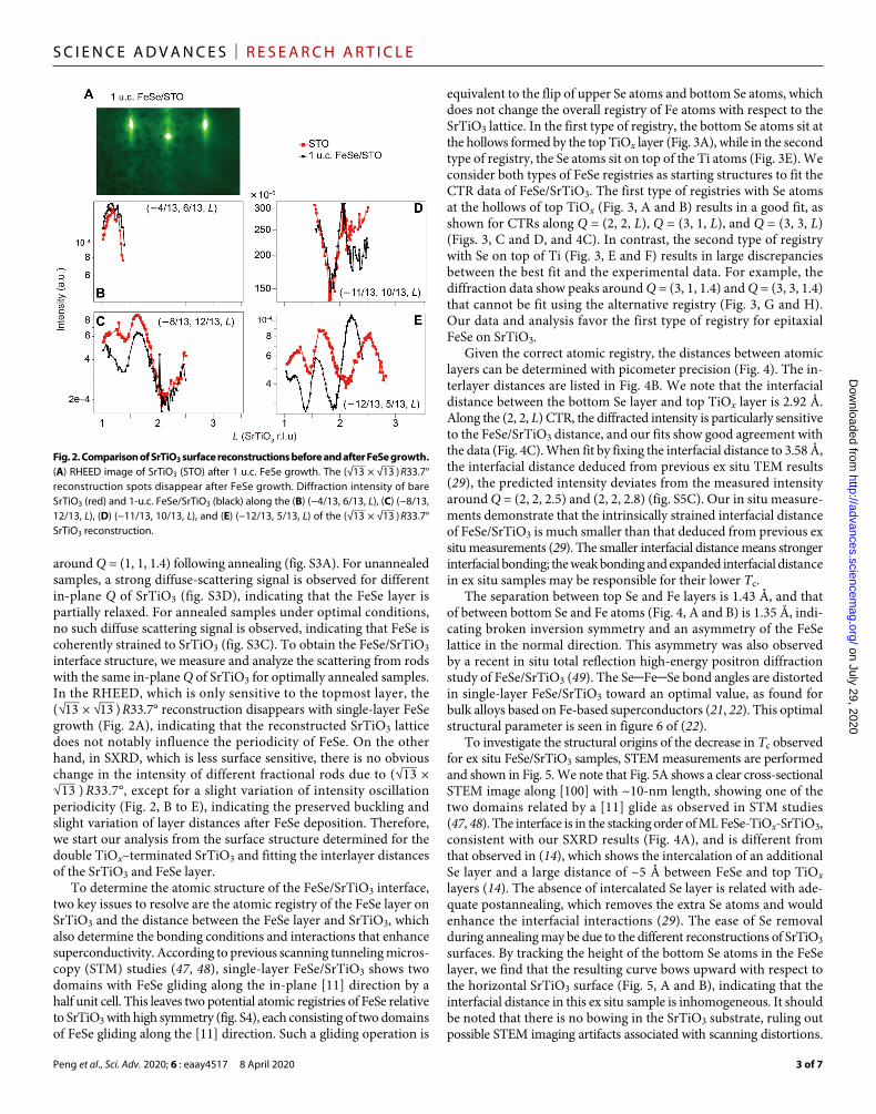
around Q = (1, 1, 1.4) following annealing (fig. S3A). For unannealed samples, a strong diffuse-scattering signal is observed for different in-plane Q of SrTiO3 (fig. S3D), indicating that the FeSe layer is partially relaxed. For annealed samples under optimal conditions, no such diffuse scattering signal is observed, indicating that FeSe is coherently strained to SrTiO3 (fig. S3C). To obtain the FeSe/SrTiO3 interface structure, we measure and analyze the scattering from rods with the same in-plane Q of SrTiO3 for optimally annealed samples. In the RHEED, which is only sensitive to the topmost layer, the ( √ _
13 × √ _
13 ) R33.7° reconstruction disappears with single-layer FeSe growth (Fig. 2A), indicating that the reconstructed SrTiO3 lattice does not notably influence the periodicity of FeSe. On the other hand, in SXRD, which is less surface sensitive, there is no obvious change in the intensity of different fractional rods due to ( √
_ 13 ×
√ _
13 ) R33.7° , except for a slight variation of intensity oscillation periodicity (Fig. 2, B to E), indicating the preserved buckling and slight variation of layer distances after FeSe deposition. Therefore, we start our analysis from the surface structure determined for the double TiOx–terminated SrTiO3 and fitting the interlayer distances of the SrTiO3 and FeSe layer.
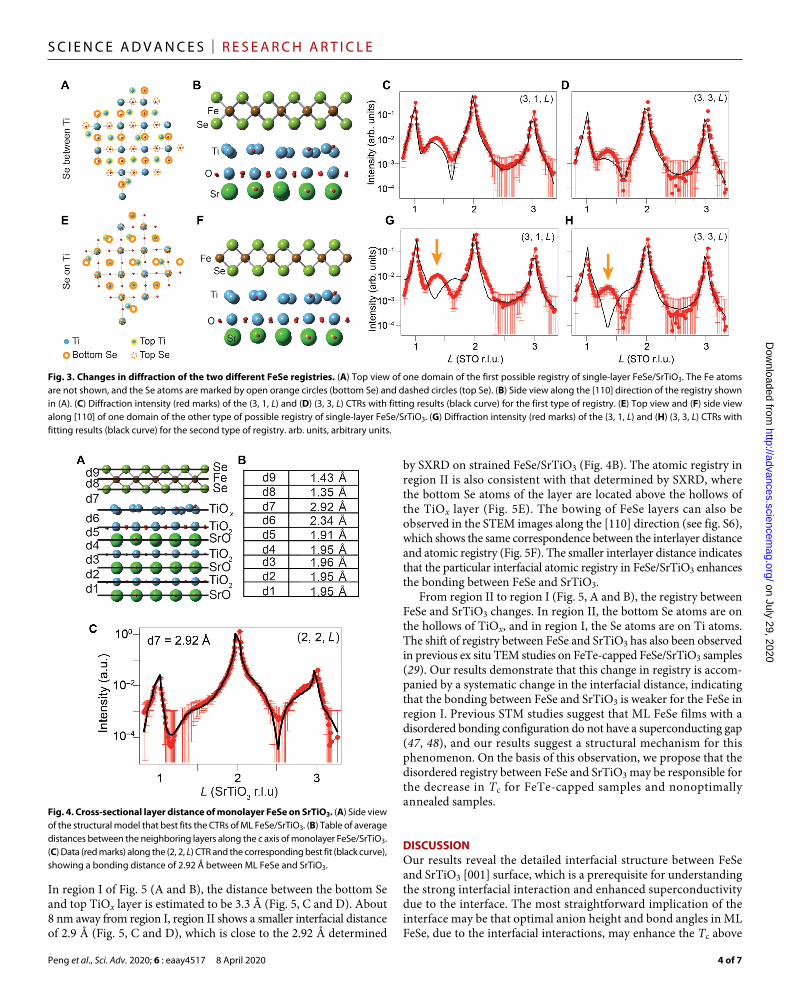
To determine the atomic structure of the FeSe/SrTiO3 interface, two key issues to resolve are the atomic registry of the FeSe layer on SrTiO3 and the distance between the FeSe layer and SrTiO3, which also determine the bonding conditions and interactions that enhance superconductivity. According to previous scanning tunneling micros-copy (STM) studies (47, 48), single-layer FeSe/SrTiO3 shows two domains with FeSe gliding along the in-plane [11] direction by a half unit cell. This leaves two potential atomic registries of FeSe relative to SrTiO3 with high symmetry (fig. S4), each consisting of two domains of FeSe gliding along the [11] direction. Such a gliding operation is
equivalent to the flip of upper Se atoms and bottom Se atoms, which does not change the overall registry of Fe atoms with respect to the SrTiO3 lattice. In the first type of registry, the bottom Se atoms sit at the hollows formed by the top TiOx layer (Fig. 3A), while in the second type of registry, the Se atoms sit on top of the Ti atoms (Fig. 3E). We consider both types of FeSe registries as starting structures to fit the CTR data of FeSe/SrTiO3. The first type of registries with Se atoms at the hollows of top TiOx (Fig. 3, A and B) results in a good fit, as shown for CTRs along Q = (2, 2, L), Q = (3, 1, L), and Q = (3, 3, L) (Figs. 3, C and D, and 4C). In contrast, the second type of registry with Se on top of Ti (Fig. 3, E and F) results in large discrepancies between the best fit and the experimental data. For example, the diffraction data show peaks around Q = (3, 1, 1.4) and Q = (3, 3, 1.4) that cannot be fit using the alternative registry (Fig. 3, G and H). Our data and analysis favor the first type of registry for epitaxial FeSe on SrTiO3.
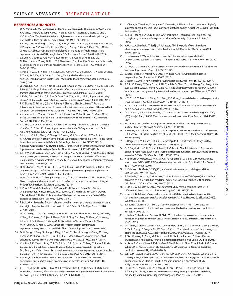
Given the correct atomic registry, the distances between atomic layers can be determined with picometer precision (Fig. 4). The in-terlayer distances are listed in Fig. 4B. We note that the interfacial distance between the bottom Se layer and top TiOx layer is 2.92 Å. Along the (2, 2, L) CTR, the diffracted intensity is particularly sensitive to the FeSe/SrTiO3 distance, and our fits show good agreement with the data (Fig. 4C). When fit by fixing the interfacial distance to 3.58 Å, the interfacial distance deduced from previous ex situ TEM results (29), the predicted intensity deviates from the measured intensity around Q = (2, 2, 2.5) and (2, 2, 2.8) (fig. S5C). Our in situ measure-ments demonstrate that the intrinsically strained interfacial distance of FeSe/SrTiO3 is much smaller than that deduced from previous ex situ measurements (29). The smaller interfacial distance means stronger interfacial bonding; the weak bonding and expanded interfacial distance in ex situ samples may be responsible for their lower Tc.
The separation between top Se and Fe layers is 1.43 Å, and that of between bottom Se and Fe atoms (Fig. 4, A and B) is 1.35 Å, indi-cating broken inversion symmetry and an asymmetry of the FeSe lattice in the normal direction. This asymmetry was also observed by a recent in situ total reflection high-energy positron diffraction study of FeSe/SrTiO3 (49). The Se─Fe─Se bond angles are distorted in single-layer FeSe/SrTiO3 toward an optimal value, as found for bulk alloys based on Fe-based superconductors (21, 22). This optimal structural parameter is seen in figure 6 of (22).
To investigate the structural origins of the decrease in Tc observed for ex situ FeSe/SrTiO3 samples, STEM measurements are performed and shown in Fig. 5. We note that Fig. 5A shows a clear cross-sectional STEM image along [100] with ~10-nm length, showing one of the two domains related by a [11] glide as observed in STM studies (47, 48). The interface is in the stacking order of ML FeSe-TiOx-SrTiO3, consistent with our SXRD results (Fig. 4A), and is different from that observed in (14), which shows the intercalation of an additional Se layer and a large distance of ~5 Å between FeSe and top TiOx layers (14). The absence of intercalated Se layer is related with ade-quate postannealing, which removes the extra Se atoms and would enhance the interfacial interactions (29). The ease of Se removal during annealing may be due to the different reconstructions of SrTiO3 surfaces. By tracking the height of the bottom Se atoms in the FeSe layer, we find that the resulting curve bows upward with respect to the horizontal SrTiO3 surface (Fig. 5, A and B), indicating that the interfacial distance in this ex situ sample is inhomogeneous. It should be noted that there is no bowing in the SrTiO3 substrate, ruling out possible STEM imaging artifacts associated with scanning distortions.
Fig. 2. Comparison of SrTiO3 surface reconstructions before and after FeSe growth. (A) RHEED image of SrTiO3 (STO) after 1 u.c. FeSe growth. The ( √
_ 13 × √
_ 13 ) R33.7°
reconstruction spots disappear after FeSe growth. Diffraction intensity of bare SrTiO3 (red) and 1-u.c. FeSe/SrTiO3 (black) along the (B) (−4/13, 6/13, L), (C) (−8/13, 12/13, L), (D) (−11/13, 10/13, L), and (E) (−12/13, 5/13, L) of the ( √
_ 13 × √
_ 13 ) R33.7°
SrTiO3 reconstruction.
on July 29, 2020http://advances.sciencem
ag.org/D
ownloaded from
Peng et al., Sci. Adv. 2020; 6 : eaay4517 8 April 2020
S C I E N C E A D V A N C E S | R E S E A R C H A R T I C L E
4 of 7
In region I of Fig. 5 (A and B), the distance between the bottom Se and top TiOx layer is estimated to be 3.3 Å (Fig. 5, C and D). About 8 nm away from region I, region II shows a smaller interfacial distance of 2.9 Å (Fig. 5, C and D), which is close to the 2.92 Å determined
by SXRD on strained FeSe/SrTiO3 (Fig. 4B). The atomic registry in region II is also consistent with that determined by SXRD, where the bottom Se atoms of the layer are located above the hollows of the TiOx layer (Fig. 5E). The bowing of FeSe layers can also be observed in the STEM images along the [110] direction (see fig. S6), which shows the same correspondence between the interlayer distance and atomic registry (Fig. 5F). The smaller interlayer distance indicates that the particular interfacial atomic registry in FeSe/SrTiO3 enhances the bonding between FeSe and SrTiO3.
From region II to region I (Fig. 5, A and B), the registry between FeSe and SrTiO3 changes. In region II, the bottom Se atoms are on the hollows of TiOx, and in region I, the Se atoms are on Ti atoms. The shift of registry between FeSe and SrTiO3 has also been observed in previous ex situ TEM studies on FeTe-capped FeSe/SrTiO3 samples (29). Our results demonstrate that this change in registry is accom-panied by a systematic change in the interfacial distance, indicating that the bonding between FeSe and SrTiO3 is weaker for the FeSe in region I. Previous STM studies suggest that ML FeSe films with a disordered bonding configuration do not have a superconducting gap (47, 48), and our results suggest a structural mechanism for this phenomenon. On the basis of this observation, we propose that the disordered registry between FeSe and SrTiO3 may be responsible for the decrease in Tc for FeTe-capped samples and nonoptimally annealed samples.
DISCUSSIONOur results reveal the detailed interfacial structure between FeSe and SrTiO3 [001] surface, which is a prerequisite for understanding the strong interfacial interaction and enhanced superconductivity due to the interface. The most straightforward implication of the interface may be that optimal anion height and bond angles in ML FeSe, due to the interfacial interactions, may enhance the Tc above
Fig. 3. Changes in diffraction of the two different FeSe registries. (A) Top view of one domain of the first possible registry of single-layer FeSe/SrTiO3. The Fe atoms are not shown, and the Se atoms are marked by open orange circles (bottom Se) and dashed circles (top Se). (B) Side view along the [110] direction of the registry shown in (A). (C) Diffraction intensity (red marks) of the (3, 1, L) and (D) (3, 3, L) CTRs with fitting results (black curve) for the first type of registry. (E) Top view and (F) side view along [110] of one domain of the other type of possible registry of single-layer FeSe/SrTiO3. (G) Diffraction intensity (red marks) of the (3, 1, L) and (H) (3, 3, L) CTRs with fitting results (black curve) for the second type of registry. arb. units, arbitrary units.
Fig. 4. Cross-sectional layer distance of monolayer FeSe on SrTiO3. (A) Side view of the structural model that best fits the CTRs of ML FeSe/SrTiO3. (B) Table of average distances between the neighboring layers along the c axis of monolayer FeSe/SrTiO3. (C) Data (red marks) along the (2, 2, L) CTR and the corresponding best fit (black curve), showing a bonding distance of 2.92 Å between ML FeSe and SrTiO3.
on July 29, 2020http://advances.sciencem
ag.org/D
ownloaded from

Peng et al., Sci. Adv. 2020; 6 : eaay4517 8 April 2020
S C I E N C E A D V A N C E S | R E S E A R C H A R T I C L E
5 of 7
that because of electron doping of FeSe from interfacial charge transfer. The interfacial interactions themselves may also play an important role in directly enhancing Tc. For example, cooperative pairing mechanisms involving both the spin/orbital fluctuation–mediated pairing in electron-doped FeSe and coupling between FeSe electrons and the SrTiO3 surface phonons have been proposed to explain the enhanced Tc in FeSe/SrTiO3 (50, 51). According to the simplified electrostatic model of electron-phonon coupling, a smaller interlayer distance and thus enhanced two-dimensional screening by ML FeSe would increase the dielectric anisotropy at the interface (3, 50). Therefore, they have a net effect of increasing the total electron-phonon coupling strength (50, 51), particularly increasing the long-range forward scattering–type component (50, 51). This long-range forward scattering electron- phonon coupling is thought to enhance Tc in ML FeSe on SrTiO3 more effectively than the short-range electron-phonon coupling of Bardeen-Cooper-Schrieffer theory (51). Consequently, the effective interfacial electron-phonon interaction would be enhanced by the smaller interlayer distance in the bonding-ordered film. Moreover, the interfacial registry and bonding may also modify the electron- phonon coupling and interfacial polaronic behavior (52) in a non-trivial way beyond the current simplified electrostatic model and affect the interfacial superconductivity. These mechanisms could explain both the enhancement of Tc in ML FeSe/SrTiO3 relative to bulk FeSe and the reduced Tc in ML FeSe that has been capped by FeTe (17) or has been annealed nonoptimally (5), where the Tc is close to that of optimally electron-doped FeSe multilayers without a Tc boost from the interface. For a comprehensive understanding on the interfacial structure and superconductivity, our studies call for future theoretical models that consider structural parameters such as interfacial registry and bonding.
In conclusion, we resolve the registry and buckling of atoms at the interface of the FeSe/SrTiO3 interfacial superconductor with picometer resolution. For FeSe films that relax, a variation of bonding distance related with interfacial atomic registration is observed. The well-ordered FeSe registration is strongly bonded to SrTiO3 only for a particular registry. Strong bonding may facilitate electron-transfer and electron-phonon coupling, which are crucial for enhanced superconductivity. Furthermore, the strong bonding for this par-
ticular registry acts to strain the ML FeSe to the reported optimal bond angle of iron-based superconductors, which may further enhance Tc. Our experiments call for the development of new theoretical models that include the interfacial bonding details, to explicitly understand these effects on the interfacial superconductivity.
MATERIALS AND METHODSIn practice, it has been observed that ML FeSe films with an enhanced Tc can only be grown on properly treated SrTiO3 surfaces, which usually show some form of surface reconstruction in RHEED (3) and an ordered additional layer of TiO2 on the surface (32–38). To obtain reliable results of the structural analysis, we prepare SrTiO3 substrates with a single and uniform surface reconstruction. By annealing SrTiO3 substrates to 1000°C for 10 hours in flowing oxygen, a reproducible SrTiO3 surface with uniform ( √
_ 13 × √
_ 13 ) R33.7°
surface reconstruction and flat terraces is obtained. Atomic force microscopy characterization of the surface reveals terraces ~250 nm wide separated by steps with a single–unit cell height, indicating a single surface termination (36–38). FeSe thin films are grown on the ( √ _
13 × √ _
13 ) R33.7° reconstructed surface by codepositing Fe and Se with a flux ratio of 1:10 on top of the SrTiO3 substrate at 500°C. The FeSe films are annealed at 530°C under 1.4 × 10⁻⁹ mbar vacuum for 53 hours. The superconductivity of the films is confirmed by trans-port measurement on FeTe-capped samples (fig. S1). In situ SXRD measurements of bare ( √
_ 13 × √
_ 13 ) R33.7° and ML FeSe deposited
on this surface are conducted at 33ID-E of the Advanced Photon Source, Argonne National Lab. In addition, STEM studies are per-formed on FeTe-capped FeSe/SrTiO3 samples (14 unit cells of FeTe grown on 8 unit cells of FeSe grown on SrTiO3) using a JEOL ARM 200CF equipped with a cold-field emission gun and double-spherical aberration correction at Brookhaven National Laboratory.
SUPPLEMENTARY MATERIALSSupplementary material for this article is available at http://advances.sciencemag.org/cgi/content/full/6/15/eaay4517/DC1
Fig. 5. STEM images of the FeSe/SrTiO3 interface. (A) HAADF cross-sectional TEM image of capped FeSe/SrTiO3. The red curve shows the location of the bottom Se atoms, which deviates from the horizontal plane shown by the white dashed line. Region I and region II show different registries of FeSe on SrTiO3 and different bonding distances between FeSe and SrTiO3. (B) Corresponding ABF image from the same area as (A). (C) Comparison of the HAADF images in region I and region II, showing different bonding distances. (D) Same as (C), but these are ABF images. (E) Cross-sectional structure along the [100] direction from synchrotron x-ray CTR studies. (F) HAADF image along the [110] direction with the smallest interfacial bonding distance (left) and the cross-sectional structure along the [110] direction resolved on the basis of synchrotron x-ray CTR studies (right).
on July 29, 2020http://advances.sciencem
ag.org/D
ownloaded from
Peng et al., Sci. Adv. 2020; 6 : eaay4517 8 April 2020
S C I E N C E A D V A N C E S | R E S E A R C H A R T I C L E
6 of 7
REFERENCES AND NOTES 1. Q.-Y. Wang, Z. Li, W.-H. Zhang, Z.-C. Zhang, J.-S. Zhang, W. Li, H. Ding, Y.-B. Ou, P. Deng,
K. Chang, J. Wen, C.-L. Song, K. He, J.-F. Jia, S.-H. Ji, Y.-Y. Wang, L.-L. Wang, X. Chen, X.-C. Ma, Q.-K. Xue, Interface-induced high-temperature superconductivity in single unit-cell fese films on SrTiO3. Chinese Phys. Lett. 29, 037402 (2012).
2. S. L. He, J. He, W. Zhang, L. Zhao, D. Liu, X. Liu, D. Mou, Y. B. Ou, Q. Y. Wang, Z. Li, L. Wang, Y. Peng, Y. Liu, C. Chen, L. Yu, G. Liu, X. Dong, J. Zhang, C. Chen, Z. Xu, X. Chen, X. Ma, Q. Xue, X. J. Zhou, Phase diagram and electronic indication of high-temperature superconductivity at 65 K in single-layer FeSe films. Nat. Mater. 12, 605–610 (2013).
3. J. J. Lee, F. T. Schmitt, R. G. Moore, S. Johnston, Y.-T. Cui, W. Li, M. Yi, Z. K. Liu, M. Hashimoto, Y. Zhang, D. H. Lu, T. P. Devereaux, D.-H. Lee, Z.-X. Shen, Interfacial mode coupling as the origin of the enhancement of Tc in FeSe films on SrTiO3. Nature 515, 245–248 (2014).
4. R. Peng, H. C. Xu, S. Y. Tan, H. Y. Cao, M. Xia, X. P. Shen, Z. C. Huang, C. H. P. Wen, Q. Song, T. Zhang, B. P. Xie, X. G. Gong, D. L. Feng, Tuning the band structure and superconductivity in single-layer FeSe by interface engineering. Nat. Commun. 5, 5044 (2014).
5. Q. Song, T. L. Yu, X. Lou, B. P. Xie, H. C. Xu, C. H. P. Wen, Q. Yao, S. Y. Zhang, X. T. Zhu, J. D. Guo, R. Peng, D. L. Feng, Evidence of cooperative effect on the enhanced superconducting transition temperature at the FeSe/SrTiO3 interface. Nat. Commun. 10, 758 (2019).
6. J.-F. Ge, Z.-L. Liu, C. Liu, C.-L. Gao, D. Qian, Q.-K. Xue, Y. Liu, J.-F. Jia, Superconductivity above 100 K in single-layer FeSe films on doped SrTiO3. Nat. Mater. 14, 285–289 (2015).
7. P. K. Biswas, Z. Salman, Q. Song, R. Peng, J. Zhang, L. Shu, D. L. Feng, T. Prokscha, E. Morenzoni, Direct evidence of superconductivity and determination of the superfluid density in buried ultrathin FeSe grown on SrTiO3. Phys. Rev. B 97, 174509 (2018).
8. Z. C. Zhang, Y.-H. Wang, Q. Song, C. Liu, R. Peng, K. A. Moler, D. Feng, Y. Wang, Onset of the Meissner effect at 65 K in FeSe thin film grown on Nb-doped SrTiO3 substrate. Sci. Bull. 60, 1301–1304 (2015).
9. F.-C. Hsu, J.-Y. Luo, K.-W. Yeh, T.-K. Chen, T.-W. Huang, P. M. Wu, Y.-C. Lee, Y.-L. Huang, Y.-Y. Chu, D.-C. Yan, M.-K. Wu, Superconductivity in the PbO-type structure -FeSe. Proc. Natl. Acad. Sci. U.S.A. 105, 14262–14264 (2008).
10. B. Lei, J. H. Cui, Z. J. Xiang, C. Shang, N. Z. Wang, G. J. Ye, X. G. Luo, T. Wu, Z. Sun, X. H. Chen, Evolution of high-temperature superconductivity from a low-Tc Phase tuned by carrier concentration in FeSe thin flakes. Phys. Rev. Lett. 116, 077002 (2016).
11. Y. Miyata, K. Nakayama, K. Sugawara, T. Sato, T. Takahashi, High-temperature superconductivity in potassium-coated multilayer FeSe thin films. Nat. Mater. 14, 775–779 (2015).
12. C. H. P. Wen, H. C. Xu, C. Chen, Z. C. Huang, X. Lou, Y. J. Pu, Q. Song, B. P. Xie, M. Abdel-Hafiez, D. A. Chareev, A. N. Vasiliev, R. Peng, D. L. Feng, Anomalous correlation effects and unique phase diagram of electron-doped FeSe revealed by photoemission spectroscopy. Nat. Commun. 7, 10840 (2016).
13. H. M. Zhang, D. Zhang, X. Lu, C. Liu, G. Zhou, X. Ma, L. Wang, P. Jiang, Q.-K. Xue, X. Bao, Origin of charge transfer and enhanced electron–phonon coupling in single unit-cell FeSe films on SrTiO3. Nat. Commun. 8, 214 (2017).
14. W. W. Zhao, M. Li, C. Z. Chang, J. Jiang, L. Wu, C. Liu, J. S. Moodera, Y. Zhu, M. H. W. Chan, Direct imaging of electron transfer and its influence on superconducting pairing at FeSe/SrTiO3 interface. Sci. Adv. 4, eaao2682 (2018).
15. K. Zou, S. Mandal, S. D. Albright, R. Peng, Y. Pu, D. Kumah, C. Lau, G. H. Simon, O. E. Dagdeviren, X. He, I. Božović, U. D. Schwarz, E. I. Altman, D. Feng, F. J. Walker, S. Ismail-Beigi, C. H. Ahn, Role of double TiO2 layers at the interface of FeSe/SrTiO3 superconductors. Phys. Rev. B 93, 180506 (2016).
16. F. M. Li, G. A. Sawatzky, Electron phonon coupling versus photoelectron energy loss at the origin of replica bands in photoemission of FeSe on SrTiO3. Phys. Rev. Lett. 120, 237001 (2018).
17. W.-H. Zhang, Y. Sun, J.-S. Zhang, F.-S. Li, M.-H. Guo, Y.-F. Zhao, H.-M. Zhang, J.-P. Peng, Y. Xing, H.-C. Wang, T. Fujita, A. Hirata, Z. Li, H. Ding, C.-J. Tang, M. Wang, Q.-Y. Wang, K. He, S.-H. Ji, X. Chen, J.-F. Wang, Z.-C. Xia, L. Li, Y.-Y. Wang, J. Wang, L.-L. Wang, M.-W. Chen, Q.-K. Xue, X.-C. Ma, Direct observation of high-temperature superconductivity in one-unit-cell FeSe films. Chinese Phys. Lett. 31, 017401 (2014).
18. G. M. Gong, H. Yang, Q. Zhang, C. Ding, J. Zhou, Y. Chen, F. Meng, Z. Zhang, W. Dong, F. Zheng, P. Zhang, L. Yang, L. Gu, Q.-K. Xue, L. Wang, Oxygen vacancy modulated superconductivity in monolayer FeSe on SrTiO3−. Phys. Rev. B 100, 224504 (2019).
19. X. H. Niu, S. D. Chen, J. Jiang, Z. R. Ye, T. L. Yu, D. F. Xu, M. Xu, Y. Feng, Y. J. Yan, B. P. Xie, J. Zhao, D. C. Gu, L. L. Sun, Q. Mao, H. Wang, M. Fang, C. J. Zhang, J. P. Hu, Z. Sun, D. L. Feng, A unifying phase diagram with correlation-driven superconductor-to-insulator transition for the 122☆series of iron chalcogenides. Phys. Rev. B 93, 054516 (2016).
20. Z. P. Yin, K. Haule, G. Kotliar, Kinetic frustration and the nature of the magnetic and paramagnetic states in iron pnictides and iron chalcogenides. Nat. Mater. 10, 932–935 (2011).
21. C.-H. Lee, A. Iyo, H. Eisaki, H. Kito, M. T. Fernandez-Diaz, T. Ito, K. Kihou, H. Matsuhata, M. Braden, K. Yamada, Effect of structural parameters on superconductivity in fluorine-free LnFeAsO1–y (Ln = La, Nd). J. Phys. Soc. Jpn. 77, 083704 (2008).
22. H. Okabe, N. Takeshita, K. Horigane, T. Muranaka, J. Akimitsu, Pressure-induced high-Tc superconducting phase in FeSe: Correlation between anion height and Tc. Phys. Rev. B 81, 205119 (2010).
23. Z.-X. Li, F. Wang, H. Yao, D.-H. Lee, What makes the Tc of monolayer FeSe on SrTiO3 so high: A sign-problem-free quantum Monte Carlo study. Sci. Bull. 61, 925–930 (2016).
24. Y. Wang, A. Linscheid, T. Berlijn, S. Johnston, Ab initio study of cross-interface electron-phonon couplings in FeSe thin films on SrTiO3 and BaTiO3. Phys. Rev. B 93, 134513 (2016).
25. L. Rademaker, Y. Wang, T. Berlijn, S. Johnston, Enhanced superconductivity due to forward scattering in FeSe thin films on SrTiO3 substrates. New J. Phys. 18, 022001 (2016).
26. S. Coh, M. L. Cohen, S. G. Louie, Large electron–phonon interactions from FeSe phonons in a monolayer. New J. Phys. 17, 073027 (2015).
27. S. Ismail-Beigi, F. J. Walker, A. S. Disa, K. M. Rabe, C. H. Ahn, Picoscale materials engineering. Nat. Rev. Mater. 2, 17060 (2017).
28. I. Bozovic, C. Ahn, A new frontier for superconductivity. Nat. Phys. 10, 892–895 (2014). 29. F. S. Li, Q. Zhang, C. Tang, C. Liu, J. Shi, C. N. Nie, G. Zhou, Z. Li, W. Zhang, C.-L. Song, K. He,
S. Ji, S. Zhang, L. Gu, L. Wang, X.-C. Ma, Q.-K. Xue, Atomically resolved FeSe/SrTiO3(001) interface structure by scanning transmission electron microscopy. 2D Mater. 3, 024002 (2016).
30. H. Y. Cao, S. Y. Tan, H. J. Xiang, D. L. Feng, X. G. Gong, Interfacial effects on the spin density wave in FeSe/SrTiO3 thin films. Phys. Rev. B 89, 014501 (2014).
31. Y. J. Zhou, A. J. Millis, Charge transfer and electron-phonon coupling in monolayer FeSe on Nb-doped SrTiO3. Phys. Rev. B 93, 224506 (2016).
32. D. M. Kienzle, A. E. Becerra-Toledo, L. D. Marks, Vacant-site octahedral tilings on SrTiO3 (001), the ( √
_ 13 × √
_ 13 ) R33.7° surface, and related structures. Phys. Rev. Lett. 106, 176102
(2011). 33. M. Naito, H. Sato, Reflection high-energy electron-diffraction study on the SRTIO3
surface-structure. Physica C Supercond. 229, 1–11 (1994). 34. R. Herger, P. R. Willmott, O. Bunk, C. M. Schlepütz, B. Patterson, B. Delley, V. L. Shneerson,
P. F. Lyman, D. K. Saldin, Surface structure of SrTiO3(001). Phys. Rev. B Condens. Matter. 76, 195435 (2007).
35. R. Herger, P. R. Willmott, O. Bunk, C. M. Schlepütz, B. D. Patterson, B. Delley, Surface of strontium titanate. Phys. Rev. Lett. 98, 076102 (2007).
36. O. E. Dagdeviren, G. H. Simon, K. Zou, F. J. Walker, C. Ahn, E. I. Altman, U. D. Schwarz, Surface phase, morphology, and charge distribution transitions on vacuum and ambient annealed SrTiO3(100). Phys. Rev. B 93, 195303 (2016).
37. N. Erdman, O. Warschkow, M. Asta, K. R. Poeppelmeier, D. E. Ellis, L. D. Marks, Surface structures of SrTiO3 (001): A TiO2-rich reconstruction with ac(4 × 2) unit cell. J. Am. Chem. Soc. 125, 10050–10056 (2003).
38. N. Erdman, L. D. Marks, SrTiO3(001) surface structures under oxidizing conditions. Surf. Sci. 526, 107–114 (2003).
39. T. Matsuda, Y. Yoshida, K. Mitsuhara, Y. Kido, The structure of SrTiO3(001)-2 × 1 surface analyzed by high-resolution medium energy ion scattering coupled with ab initio calculations. J. Chem. Phys. 138, 244705 (2013).
40. I. Lazić, E. G. T. Bosch, S. Lazar, Phase contrast STEM for thin samples: Integrated differential phase contrast. Ultramicroscopy 160, 265–280 (2016).
41. I. Lazic, E. G. T. Bosch, Analytical review of direct STEM imaging techniques for thin samples, in Advances in Imaging and Electron Physics, P. W. Hawkes, Ed. (Elsevier, 2017), vol. 199, pp. 75–184.
42. E. Yücelen, I. Lazić, E. G. T. Bosch, Phase contrast scanning transmission electron microscopy imaging of light and heavy atoms at the limit of contrast and resolution. Sci. Rep. 8, 2676 (2018).
43. H. Nahor, Y. Kauffmann, S. Lazar, D. Shilo, W. D. Kaplan, Discerning interface atomistic structure by phase contrast in STEM: The equilibrated Ni-YSZ interface. Acta Mater. 154, 71–78 (2018).
44. D. S. Song, X. Zhang, C. Lian, H. Liu, I. Alexandrou, I. Lazić, E. G. T. Bosch, D. Zhang, L. Wang, R. Yu, Z. Cheng, C. Song, X. Ma, W. Duan, Q. Xue, J. Zhu, Visualization of dopant oxygen atoms in a Bi2Sr2CaCu2O8+ superconductor. Adv. Funct. Mater. 29, 1903843 (2019).
45. S. Gao, P. Wang, F. Zhang, G. T. Martinez, P. D. Nellist, X. Pan, A. I. Kirkland, Electron ptychographic microscopy for three-dimensional imaging. Nat. Commun. 8, 163 (2017).
46. Y. Jiang, Z. Chen, Y. Han, P. Deb, H. Gao, S. Xie, P. Purohit, M. W. Tate, J. Park, S. M. Gruner, V. Elser, D. A. Muller, Electron ptychography of 2D materials to deep sub-ångström resolution. Nature 559, 343–349 (2018).
47. Z. Li, J.-P. Peng, H.-M. Zhang, W.-H. Zhang, H. Ding, P. Deng, K. Chang, C.-L. Song, S.-H. Ji, L. Wang, K. He, X. Chen, Q.-K. Xue, X.-C. Ma, Molecular beam epitaxy growth and post-growth annealing of FeSe films on SrTiO3: A scanning tunneling microscopy study. J. Phys Condens. Matter 26, 265002 (2014).
48. Q. Fan, W. H. Zhang, X. Liu, Y. J. Yan, M. Q. Ren, R. Peng, H. C. Xu, B. P. Xie, J. P. Hu, T. Zhang, D. L. Feng, Plain s-wave superconductivity in single-layer FeSe on SrTiO3 probed by scanning tunnelling microscopy. Nat. Phys. 11, 946–952 (2015).
on July 29, 2020http://advances.sciencem
ag.org/D
ownloaded from
Peng et al., Sci. Adv. 2020; 6 : eaay4517 8 April 2020
S C I E N C E A D V A N C E S | R E S E A R C H A R T I C L E
7 of 7
49. Y. Fukaya, G. Zhou, F. Zheng, P. Zhang, L. Wang, Q.-K. Xue, S.-I. Shamoto, Asymmetrically optimized structure in a high-Tc single unit-cell FeSe superconductor. J. Phys. Condens. Matter 31, 055701 (2019).
50. D.-H. Lee, What makes the Tc of FeSe/SrTiO3 so high? Chinese Phys. B 24, 117405 (2015). 51. M. L. Kulić, O. V. Dolgov, The electron-phonon interaction with forward scattering peak is
dominant in high Tc superconductors of FeSe films on SrTiO3 (TiO2). New J. Phys. 19, 013020 (2017).
52. S. Y. Zhang, T. Wei, J. Guan, Q. Zhu, W. Qin, W. Wang, J. Zhang, E. W. Plummer, X. Zhu, Z. Zhang, J. Guo, Enhanced superconducting state in FeSe/SrTiO3 by a dynamic interfacial polaron mechanism. Phys. Rev. Lett. 122, 066802 (2019).
Acknowledgments Funding: This work was supported by AFOSR grant FA9550-15-1-0472. The XRD data were taken at beamline 33ID-E at the Advanced Photon Source and supported by the U.S. Department of Energy, Office of Science, Office of Basic Energy Sciences, under contract no. DE-AC02-06CH11357. The work at Brookhaven National Laboratory was supported by the U.S. Department of Energy, Office of Science, Office of Basic Energy Sciences, under contract no.DESC0012704. TEM sample preparation using FIB was performed at the Center
for Functional Nanomaterials, Brookhaven National Laboratory. Author contributions: R.P. and K.Z. conducted the thin film growth. R.P., K.Z., S.D.A., H.H., C.L., and F.J.W. conducted the SXRD experiments. R.P., S.D.A., H.C.X., and F.J.W. analyzed the SXRD data. M.G.H. and Y.Z. conducted the STEM experiments. R.P., S.D.A., H.C.X., F.J.W., and C.H.A. wrote the paper. C.H.A. and F.J.W. are responsible for project direction, planning, and infrastructure. Competing interests: The authors declare that they have no competing interests. Data and materials availability: All data needed to evaluate the conclusions in the paper are present in the paper and/or the Supplementary Materials. Additional data related to this paper may be requested from the authors.
Submitted 20 June 2019Accepted 14 January 2020Published 8 April 202010.1126/sciadv.aay4517
Citation: R. Peng, K. Zou, M. G. Han, S. D. Albright, H. Hong, C. Lau, H. C. Xu, Y. Zhu, F. J. Walker, C. H. Ahn, Picoscale structural insight into superconductivity of monolayer FeSe/SrTiO3. Sci. Adv. 6, eaay4517 (2020).
on July 29, 2020http://advances.sciencem
ag.org/D
ownloaded from

3Picoscale structural insight into superconductivity of monolayer FeSe/SrTiO
AhnRui Peng, Ke Zou, M. G. Han, Stephen D. Albright, Hawoong Hong, Claudia Lau, H. C. Xu, Yimei Zhu, F. J. Walker and C. H.
DOI: 10.1126/sciadv.aay4517 (15), eaay4517.6Sci Adv
ARTICLE TOOLS http://advances.sciencemag.org/content/6/15/eaay4517
MATERIALSSUPPLEMENTARY http://advances.sciencemag.org/content/suppl/2020/04/06/6.15.eaay4517.DC1
REFERENCES
http://advances.sciencemag.org/content/6/15/eaay4517#BIBLThis article cites 51 articles, 2 of which you can access for free
PERMISSIONS http://www.sciencemag.org/help/reprints-and-permissions
Terms of ServiceUse of this article is subject to the
is a registered trademark of AAAS.Science AdvancesYork Avenue NW, Washington, DC 20005. The title (ISSN 2375-2548) is published by the American Association for the Advancement of Science, 1200 NewScience Advances
License 4.0 (CC BY-NC).Science. No claim to original U.S. Government Works. Distributed under a Creative Commons Attribution NonCommercial Copyright © 2020 The Authors, some rights reserved; exclusive licensee American Association for the Advancement of
on July 29, 2020http://advances.sciencem
ag.org/D
ownloaded from