masters submission: march, 2005 student: natalie anderson...
TRANSCRIPT

(c) 2
004
Victor
ia Univ
ersit
y
1
MASTERS SUBMISSION: March, 2005
Student: Natalie Anderson
ID: 3519268
Intended submission as: Journal Article
Journal of choice: JOM (Journal of Osteopathic Medicine)
Article length: 3575 (Including front pages, references and appendix)
Instructions to authors for JOM: Attached.
RELEASE SIGNATURE:
PRINCIPAL INVESTIGATOR:
Mr.Cameron Gosling
Contact: 03 99191290

(c) 2
004
Victor
ia Univ
ersit
y
2
The Effect of a Specific Isometric Muscle Energy Technique onthe Range of Opening of the Temporomandibular Joint
Natalie Anderson, Mr.Cameron GoslingOsteopathic Medicine, School of Health Sciences, Victoria University, PO Box 14428 MCMC, Melbourne 8001, Australia
Student Number: 3519268
March 2005
This thesis is submitted in partial fulfilment of the requirements of the Degree: MastersHealth Sciences (Osteopathy).
ABSTRACT
Objective: To determine whether a specific muscle energy technique had an
effect on the vertical range of opening of the mandible of the TMJ compared to
therapeutic jaw exercises and non-intervention control group.
Subjects: Twentyone (N=21) participants aged at least 18 years were recruited into the
study via notices pinned up in the Victoria university teaching clinic.
Design: Participants were randomly allocated in to a group to receive an application of a
specific isometric MET, a therapeutic jaw exercise or a non-intervention control group.
Baseline measurements of jaw opening distance were obtained from the groups prior to
the application of any therapy. All groups completed a temporomandibular dysfunction
symptom checklist to determine the symptoms of temporomandibular joint dysfunction,
immediately before the intervention 30 minutes after the application of the therapeutic
jaw exercises and one week post intervention. A questionnaire to determine the patients’
own perception of pain was administered prior to the intervention, immediately
after, 30 minutes following and one week post intervention. In this study only the
outcome on range of jaw opening is discussed.
Setting: Victoria University teaching clinic

(c) 2
004
Victor
ia Univ
ersit
y
3
Data analysis: Three separate ANOVAs were used for each variable (jaw
opening, pain and symptom score) they were conducted to test for differences
between the groups. Analysis of jaw opening utilized a 3 x 6 (group x time)
ANOVA while pain and symptoms used 3 x 3 (group x time) ANOVAs.
Significance was set at P ≤ 0.05. A power analysis of previous research1 has
indicated a very large effect size (Cohen’s f = 1.3) for jaw opening.
Results: The results of this study indicate that MET and therapeutic jaw exercises
are useful techniques in improving TMJ range of opening (P= 0.000).
Conclusions: Within the limitations of the study, the results support the
hypothesis that the application of a specific Osteopathic treatment, in this case
MET and therapeutic jaw exercises can improve the range of opening in the TMJ.
The results of this study provide a basis for further research as they present
valuable outcomes for Osteopaths treating jaw dysfunction.
Key Indexing Terms: MET, temporomandibular dysfunction, osteopathic
treatment, therapeutic jaw exercises.

(c) 2
004
Victor
ia Univ
ersit
y
4
INTRODUCTION
Temporomandibular dysfunction comprises a constellation of signs and symptoms
including joint tenderness and pain on function, restricted jaw movement, clicking, jaw
locking and tenderness in the muscles of mastication.2 Hypertonicity of the primary
muscles of mastication (temporalis, masseter, medial and lateral pterygoids), regardless
of aetiology, may reduce the mobility of the temporomandibular joint (TMJ) resulting in
a restricted range of mouth opening. This restriction is one of the signs of
temporomandibular dysfunction, as is pain, locking, headaches and tinnitus. Studies have
reported that as much as 75% of the general population will have some type of TMD3 and
it is estimated that more than 85 to 90% of people will display one or more of the TMD
symptoms in their lifetime.4,5,6
The measurement of motion at the TMJ
Range of mouth opening measured using an inter-incisal or linear technique, involving
the measurement of the distance between the incisal surface of a mandibular central
incisor and the corresponding maxillary central incisor edge when the mouth is
maximally opened, has been used extensively in research conducted by Carlson.7 In
previous research it had been determined that the measurement should include the
amount of overlap between the upper and lower central incisors when in the occlusal
position, plus the inter-incisally measured range of opening.8-15 Because this study was an
investigation of functional mouth opening ability the inter-incisal measure was the most
appropriate measurement method. It is inexpensive, non-invasive, easy to use and
applicable for use in general practice.5 An inter-incisal distance of less than 40mm
represents a restricted range of motion 8,9,11,12,16,17while normal range of mandibular

(c) 2
004
Victor
ia Univ
ersit
y
5
opening is expected to be between 45 and 60mm for males, and 40 to 55mm for
females.10 Mandibular opening should be free of joint noise, smooth and without
deviation.8
Treatment of TMD modalities
Surgical, dental, pharmacological and physical therapy modalities have all been used
previously to treat TMD. Manual therapy, be it osteopathy, chiropractic or physiotherapy
aims to relieve pain, restore function, and prevent recurrence of pain and dysfunction by
reducing muscle spasm, inflammation, and regaining normal range of motion of at least
40mm of mandibular opening in people suffering TMD.16-19 Manual therapy treatment
protocols have included transcutaneous electrical nerve stimulation (TENS), microcurrent
electrical nerve stimulation (MENS), moist/deep heat, vapocoolant sprays, ultrasound,
cryotherapy, mobilization, therapeutic exercises, laser therapy, iotntophoresis,
phonophoresis, electroacupuncture and biofeedback with varying degrees of
success.2,13,14,18,20-26
Therapeutic Jaw Exercises
Muscle exercises are of great value in the treatment of TMD. Therapeutic manual therapy
and exercises have yielded favourable results in the rehabilitation of TMD.14,24,26
Stretching exercises are designed to increase the range of motion of the mandible. The
purpose of exercises for the masticatory system are (1) to achieve relaxation of tense
muscles, (2) to retain coordination and rhythmic muscle function, (3) to increase
mandibular range of motion (isotonic exercises) (4) to increase muscular strength
(isometric exercises). Such activity stimulates the muscle spindles and Golgi tendon
organs reducing excessive activity. The principle is that when a muscle is actively

(c) 2
004
Victor
ia Univ
ersit
y
6
contracted, its antagonists are reflexly relaxed. Therefore opening the mouth against
resistance tends to relax contracted elevator muscles and vice versa for opening muscles.
The fact that physical therapy is non-invasive and does not appear to be fraught with
irreversible changes makes it a very applicable vehicle in the area of clinical TMJ
dysfunction management.18,27
Muscle Energy Technique
Muscle energy (MET) is a technique whereby the patient actively uses their muscles
against a counterforce produced by the practitioner. The practitioner controls the
intensity, timing and direction. According to Greenman28, MET can be used to “lengthen
a shortened, contracted or spastic muscle; to strengthen a physiologically weakened
muscle or group of muscles; to reduce localized oedema and relieve passive congestion
(the muscles are the pump of the lymphatic and venous systems); and to mobilize an
articulation with restricted mobility”.
They are used primarily by osteopaths to treat muscles with excessive tension that limit
joint motion.29,30 However, treatment of the TMJ using MET has not commonly been
documented, although it may have a beneficial outcome on the limited range of motion
frequently associated with TMD.1 Malone9 advocates the use of “hold – relax” techniques
(similar to muscle energy technique) on the mandibular elevators (masseter, temporalis,
and medial pterygoid) to improve functional mobility of the TMJ, and range of mouth

(c) 2
004
Victor
ia Univ
ersit
y
7
opening. MET treatment of the TMJ must be considered as a valid treatment approach for
TMD if it can be shown to improve functional range.
The present study examines whether a specific muscle energy technique would have an
effect on the vertical range of opening of the mandible, pain and dysfunction of the TMJ
compared to therapeutic jaw exercises and non-intervention control group.

(c) 2
004
Victor
ia Univ
ersit
y
8
METHODS
Participants
Volunteer participants (N = 21) with a restricted mandibular range of motion of 40mm or
less measured inter-incisally were recruited for the study. Symptomatic and
asymptomatic participants (age:26.14 ±10.41 range = 19 – 58 years, , males = 7, females
=14) were recruited from the student osteopathic teaching clinic at Victoria University.
Volunteers were excluded if they had been previously diagnosed with a systemic
arthropathy such as Rheumatoid Arthritis, or malignant tumours of the face or jaw;
previous history of jaw or TMJ surgery or fracture; or have had dental/orthodontic
treatment in the past seven days.13,16 All volunteers signed informed consent prior to
participating and the Victorian University Human Research Ethics Committee approved
the study.
Procedure
Mandibular range of opening was measured inter-incisally, and not including the degree
of overlap between the teeth when in the closed position. This involved a measurement
being taken with a transparent ruler as the participant opened their mouth to the
maximum possible distance. The distance between the edges of the upper central incisors,
and the lower central incisors was determined as the inter-incisor range of opening.7, 9,
11,16,17

(c) 2
004
Victor
ia Univ
ersit
y
9
Measurements of jaw opening were made pre intervention, immediately after, 5 minutes,
10 minutes, 20 minutes, 30 minutes and one week after the application of the intervention
The participants were then allocated into three groups via randomised computer
allocation, (i.e. group A – control group, group B –MET intervention group, group C –
therapeutic jaw exercises) each with an equal number of participants. The control group
did not receive any treatment.
The MET technique used in this study involved the treating practitioner placing gloved
thumbs on the lower molars on both sides of the participant’s jaw, whilst the participant
lay supine with the mouth open. The participant was asked to attempt closing the jaw
using 20% of their total effort as the practitioner provided an equal resistance with the
thumbs, so that no movement occurred. The treating practitioner instructed the participant
to ensure that the force of contraction was approximately 20% of their total effort and
was not excessive or likely to cause pain or muscle soreness. After a five second
contraction, the participant relaxed the jaw muscles, and then the practitioner gently
opened the jaw to the maximal distance possible and the participant was again asked to
attempt closing the jaw using 20% of their total effort. This contract-relax procedure was
repeated five times.1,29
(c) 2
004
Victor
ia Univ
ersit
y
10
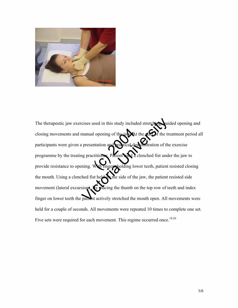
The therapeutic jaw exercises used in this study included stretching, guided opening and
closing movements and manual opening of the jaw. At the start of the treatment period all
participants were given a presentation and practical demonstration of the exercise
programme by the treating practitioner. Patient used a clenched fist under the jaw to
provide resistance to opening. With fingers holding lower teeth, patient resisted closing
the mouth. Using a clenched fist held on the side of the jaw, the patient resisted side
movement (lateral excursion). By placing the thumb on the top row of teeth and index
finger on lower teeth the patient actively stretched the mouth open. All movements were
held for a couple of seconds. All movements were repeated 10 times to complete one set.
Five sets were required for each movement. This regime occurred once.18,26

(c) 2
004
Victor
ia Univ
ersit
y
11
The treating practitioner instructed the participant to ensure that the force of contraction
was approximately 20% of their total effort and was not excessive or likely to cause pain
or muscle soreness.
The non intervention group had measurements of jaw opening taken. Participants were
asked do lie on a treatment table, in a comfortable position. Measurements of jaw
opening were made again 5, 10, 20 and 30 minutes later. Measurements were taken one
week post testing date. They received no therapeutic intervention. A measure to
determine the patients own perception of pain was administered prior to the non
intervention, immediately after, 30 minutes following and one week post intervention.
All treatments were performed in the student osteopathic clinic at Victoria University. All
measurements were taken with the subject lying supine on the treatment table, while the
investigator stood to the right hand side.

(c) 2
004
Victor
ia Univ
ersit
y
12
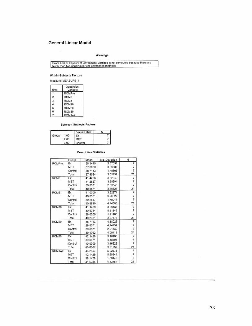
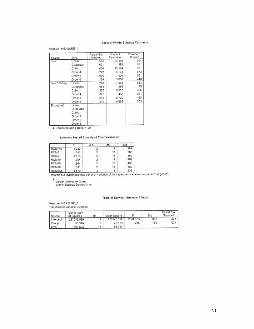
ANALYSIS OF DATA
All range of opening data is expressed as mean (M) ± standard deviation (SD). A split
plot ANOVA was used to assess changes within and between groups and analysed using
SPSS version 12.0. Further analysis to determine differences between groups at 30
minutes and 1 week post intervention was achieved by ANCOVA using pre-intervention
scores as the covariate. Effect sizes for groups are reported as _2 and significance was set
at P ≤ 0.05.
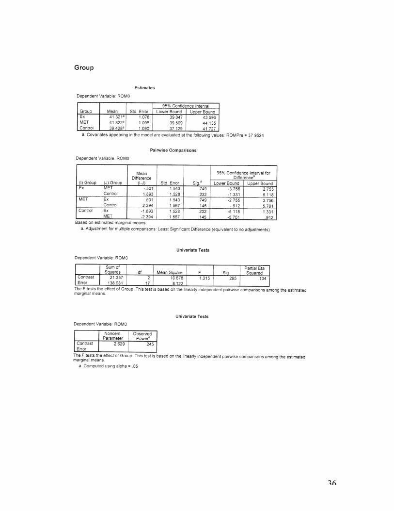
RESULTS
All volunteers completed the treatment and no adverse events were reported for any of
the interventions. There was no significant difference between groups for mean range of
jaw opening pre test (F = 0.545 P = 0.589) although the MET group did have a mean
score about 1 cm lower than the other two groups. Mean range of opening for all subjects
pre intervention was 37.95 ± 3.06 cm. It was observed that there was a significant mean
increase in jaw opening over time (F=13.93 P=0.000). This is highlighted in figure 1,
which represents the range of jaw opening before and after the MET procedure,
therapeutic jaw exercise and control.

(c) 2
004
Victor
ia Univ
ersit
y
13
Mean Scores For Range of Motion VS Intervention
33
34
35
36
37
38
39
40
41
42
43
44
Pre 0 5 10 20 30 Followup
Time
Dis
tanc
e (m
m)
Tmj Exercises
MET
Control
Figure 1.
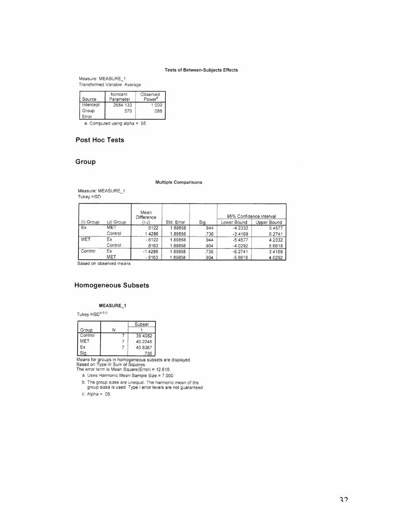
The ANCOVA analysis demonstrated a significant difference between groups at 30
minutes (F = 3.611 P = 0.049) and one week (F = 16.188 P = 0.000) post intervention.
Further Post Hoc analyses showed that for the 30 minute time interval the difference
occurred between the therapeutic jaw exercises and control groups (P = 0.016) while at
one week post intervention the difference occurred between the control and the
therapeutic jaw exercise groups (P = 0.000) as well as the control and MET groups (P =
0.000). This is represented by a percentage of change between pre intervention and 30
minutes of 10.71% in the therapeutic jaw exercise group and a 7.72% change for the

(c) 2
004
Victor
ia Univ
ersit
y
14
MET group. The control group displayed a change of 3.32%. The percentage of change
demonstrated at 7 days post intervention was 13.48% in the therapeutic jaw exercise
group and 13.90% for the MET group. The control group showed a change of 1.11%. All
percentage mean differences and standard mean deviations are reported in tables 1 and 2
below.
Table 1
Percentage change between pre intervention scores and 30 minutes post intervention
mean % change % standard deviation
THE 10.71 3.37
MET 7.72 3.90
Control 3.32 1.11
Table2
Percentage change between pre intervention scores and 7 days post intervention
mean % change % standard deviation
THE 13.48 36.85
MET 13.90 46.03
Control 1.11 24.63
(c) 2
004
Victor
ia Univ
ersit
y
15
DISCUSSION
The results of this study suggest that a specific isometric muscle energy technique to the
muscles of mandibular elevation and the application of therapeutic jaw exercises can
improve the range of jaw opening in persons with restricted jaw movement.
Immediately post intervention both TMJ and TJE groups had an increase in range of jaw
opening of 4mm and 3mm respectively.
It is interesting to note that in the TJE group the range of motion actually decreased at the
20 minute interval. An explanation for this could be attributed to muscle fatigue and
micro tearing of the muscles of mastication due to repetitive motion under load. This
theory is supported by Pertes18 and Magnusson.26
Both the MET group and the TJE group all had an increased range of motion sustained
for 7 days post intervention of greater than 5mm. The control group also had an increase
in range of motion immediately post intervention; this was sustained at the 20 and 30
minute intervals but was all but lost 7 days post. It is possible that an increase in range of
motion is achieved by simply repeatedly opening the jaw. This is not as significant when
compared to the interventions.

(c) 2
004
Victor
ia Univ
ersit
y
16
The present study measures range of motion of the TMJ using < 40 mm of opening as a
guide to dysfunction. In contrast other studies have focused on pain or the completion of
the temporomandibular dysfunction checklist as an indication of dysfunction or a
combination of all three.32
Pain is the most common presenting complaint and the most difficult to evaluate it is an
unpleasant sensory and emotional experience that is always subjective and therefore
difficult to measure.33,34 The assessment of temporomandibular dysfunction using a check
list strongly involves subjective data making it difficult to obtain reliable results.32
In comparison measurement of the range of motion of the temporomandibular joint using
a plastic ruler and the protocol described in text involves objective measurements
demonstrated to be reliable.16
Physical therapy modalities aim to improve the range of opening to a liner measure
greater than 40mm.5,8 This was supported in the current study by an increased in jaw
opening of 13.48% in the TMJ group and by 13.90% in the MET group over a seven day
period. It is previously reported that a multi disciplinary approach to TMD is of the
greatest benefit to the patient.7,18,24,31
CONCLUSION
This study shows that applications of either a specific MET for the muscles of the TMJ or
specifically designed TJE produce an increase in range of motion in the TMJ. The MET
application was ultimately mildly more effective being sustained 7 days post intervention
however both seem to be useful in the treatment of TMD and should be practiced with
(c) 2
004
Victor
ia Univ
ersit
y
17
caution in the clinical setting until further studies demonstrate the clinical effectiveness of
the combined use of these treatment modalities in both asymptomatic and symptomatic
populations.

(c) 2
004
Victor
ia Univ
ersit
y
18
References:
1. Freshwater Z, Gosling C. The effect of a specific isometric muscle energy technique
on range of opening of the temporomandibular joint: a pilot study. Abstract in the
Journal of Osteopathic Medicine. 2003;(6):36.
2. Gray RJ, Quayle AA, Hall CA, Schofield MA. Physiotherapy in the treatment of
temporomandibular joint disorders: a comparative study of four treatment methods.
British Dental Journal. 1994;176:257-261.
3. Conti PC, Ferreira PM, Pegoraro LF, Conti JV, Salvador MC. A cross-sectional study
of prevalence and etiology of signs and symptoms of temporomandibular disorders in
high school and university students. Journal of Orofacial Pain. 1996;10(3):254-262.
4. Zarb GA, Carlsson GE, Sessle BJ, Mohl ND. In Zarb GA, Carlsson GE and Rugh JD
(eds). Clinical Management. Temporomandibular Joint and Masticatory Muscle
disorders. Copenhagen.1994:529-548.
5. Nicolakis P, Erdogmus B, Kopf A, Nicolakis M, Piehslinger E, Fialka-Moser V.
Effectiveness of exercise therapy in patients with myofascial pain dysfunction syndrome.
Journal of Oral Rehabilitation. 2002;29:362-368.
6. Dimitroulis G, Gremillion HA, Dolwick MF, Walter JH. Temporomandibular
disorders 2.Non-surgical treatment. Australian Dental Journal. 1995;40(6):373-376.
7. Carlson C, Bertrand P, Ehrlich D, Maxwell A, Burton R. Physical self-regulation
training for the management of temporamandibular disorders. Journal of Orofacial Pain.
2001;15(1):47-55.
(c) 2
004
Victor
ia Univ
ersit
y
19
8. Agerberg G. Maximal mandibular movements in young men and women. Swedish
Dental Journal. 1974;67(2):81-100.
9. Malone T, McPoil T, Nitz A. Orthopedic and sports physical therapy, 3rd ed, Sydney,
Mosby; 1997: 556-593.
10. Curl D. The visual range of motion scale: Analysis of mandibular gait in a
chiropractic setting. Journal of Manipulative and Physiological Therapeutics.
1992;15(2): 115 – 122.
11. Friction J, Schiffman E. Reliability of a craniomandibular index. Journal of Dental
Research. 1986;65(11): 1359 –1364.
12. Kropmans T, Dijkstra P, Stegenga B, Stewart R, deBont L. The smallest detectable
difference in outcome variables related to painful restriction of the temporomandibular
joint. Journal of Dental Research. 1999;78(3): 784 – 789.
13. Taylor K, Newton R, Personius W & Bush, F. Effects of interferential current
stimulation for treatment of subjects with recurrent jaw pain. Physical Therapy. 1987;
67(3):346-350.
14. Waide F, Bade D, Lovasko J, Montana J. Clinical management of a patient following
temporomandibular joint arthroscopy. Physical Therapy. 1992;72(5): 355 – 364.
15. Dijkstra P, De Bont L, Stegenga B, Boering G. Angle of mouth opening
measurement: reliability of a technique for temporomandibular joint mobility assessment.
Journal of Oral Rehabilitation. 1995; 22(4):263-268.
16. Austin B, Shupe S. The role of physical therapy in recovery after temporomandibular
joint surgery. Journal of Oral and Maxillofacial Surgery. 1993; 51:495-498.

(c) 2
004
Victor
ia Univ
ersit
y
20
17. Kraus S. TMJ disorders; management of the craniomandibular complex, Melbourne,
Churchill Livingstone; 1988.
18. Pertes R, Gross S. Clinical Management of Temporomandibular Disorders and
Orofacial Pain. Chicago, Quintessence Publishing Co, Inc; 1995.
19. Dworkin SF, Huggins KH, LeResch L and co-workers. Epidemiology of signs and
symptoms in temporomandinular disorders: clinical signs and symptoms in cases and
controls. Journal of the American Dental Association. 1990; 120(3): 273-281.
20. Hukisson, E. Visual Analogue Scales. In: Melzack R, ed. Pain Measurement and
Assessment. New York: Raven Press; 1983: 33-37.
21. Bolton J, Wilkinson R. Responsiveness of pain scales: A comparison of three pain
intensity measures in chiropractic patients. Journal of Manipulative and Physiological
Therapies. 1998;21(1): 1-7.
22. Anonymous. Examination, diagnosis and management of temporomandibular
disorders. Journal of the American Dental Association, 1983;106:75.
23. DiFabio R. Physical therapy for patients with TMD: A descriptive study of treatment,
disability, and health status. Journal of Orofacial Pain. 1998;12(2):124 –134.
24. Bell, W.E. Temporomandibular Disorders: Classification, Diagnosis, Management.
3rd ed. Baltimore: Year Book Medical Publishers; 2001.
25. Royder, J.O. Structural Influences in Temporomandibular Joint Pain and
Dysfunction. Journal of AOA. 1981; 80(7): 460:467.
26. Magnusson T, Syren M. Theapeutic jaw exercises and interocclusal appliance
therapy. Swedish Dental Journal. 1999; 23(1): 27-37.
(c) 2
004
Victor
ia Univ
ersit
y
21
27. Kirk-Ws J, Calabrese DK. Clinical evaluation of physical therapy in the management
of internal derangement of temporomandibular joint. Journal of Oral Maxillofacial
Surgery. 1989; 47:113.
28. Greenman PE. Principles of Manual Medicine, second ed., Baltimore: Lippincott
Williams & Wilkins Co.; 1996.
29. Chaitow L. Muscle energy techniques. Melbourne: Churchill Livingstone; 2001.
30. Rex LH. Muscle Energy Technique – An overview. Athletic Therapy Today.1996;
November: 38 – 40.
31. Clark GT, Adachi NY, Doran MR. Physical therapy as an adjunct to
temporomandibular disorders: a review. Journal American Dietetic Association.
1990;121:151-61.
32. Nicolakis P, Erdogmus B, Kopf A, Djaber-Ansari A, Piehslinger E, Fialka-Moser V.
Exercise Therapy for Craniomandibular Disorders. Archives of Physical Medicine and
Rehabilitation. 2000; 81:1137-1142.
33. Dimitroulis G, Temporomandibular Disorders:a clinical update. British Medical
Journal. 1998; 317:190-194.
34. Bolton J, Responsiveness of Pain Scales: A Comparison of Three Pain Intensity
Measures in Chiropractic Patients. Journal of Manipulative and Physiological
Therapeutics. 1998; 21(1):1-7.

(c) 2
004
Victor
ia Univ
ersit
y
22

(c) 2
004
Victor
ia Univ
ersit
y
23

(c) 2
004
Victor
ia Univ
ersit
y
24

(c) 2
004
Victor
ia Univ
ersit
y
25
(c) 2
004
Victor
ia Univ
ersit
y
26

(c) 2
004
Victor
ia Univ
ersit
y
27

(c) 2
004
Victor
ia Univ
ersit
y
28

(c) 2
004
Victor
ia Univ
ersit
y
29

(c) 2
004
Victor
ia Univ
ersit
y
30
(c) 2
004
Victor
ia Univ
ersit
y
31
(c) 2
004
Victor
ia Univ
ersit
y
32

(c) 2
004
Victor
ia Univ
ersit
y
33

(c) 2
004
Victor
ia Univ
ersit
y
34

(c) 2
004
Victor
ia Univ
ersit
y
35
(c) 2
004
Victor
ia Univ
ersit
y
36

(c) 2
004
Victor
ia Univ
ersit
y
37

(c) 2
004
Victor
ia Univ
ersit
y
38

(c) 2
004
Victor
ia Univ
ersit
y
39

(c) 2
004
Victor
ia Univ
ersit
y
40

(c) 2
004
Victor
ia Univ
ersit
y
41

(c) 2
004
Victor
ia Univ
ersit
y
42
(c) 2
004
Victor
ia Univ
ersit
y
43

(c) 2
004
Victor
ia Univ
ersit
y
44