mass spectrometry - download.e-bookshelf.de · karen r. jonscher, lei jin, john c. cambier, shaikh...
TRANSCRIPT


MASS SPECTROMETRY HANDBOOK

WILEY SERIES ON PHARMACEUTICAL SCIENCE AND BIOTECHNOLOGY: PRACTICES, APPLICATIONS AND METHODS
Series Editor:
Mike S. LeeMilestone Development Services
Mike S. Lee • Integrated Strategies for Drug Discovery Using Mass Spectrometry
Birendra Pramanik, Mike S. Lee, and Guodong Chen • Characterization of Impurities and Degradants Using Mass Spectrometry
Mike S. Lee and Mingshe Zhu • Mass Spectrometry in Drug Metabolism and Disposition: Basic Principles and Applications
Mike S. Lee (editor) • Mass Spectrometry Handbook
MASS SPECTROMETRY HANDBOOK
EDITED BY
MIKE S. LEEMilestone Development Services
A JOHN WILEY & SONS, INC., PUBLICATION

Copyright © 2012 by John Wiley & Sons, Inc. All rights reserved.
Published by John Wiley & Sons, Inc., Hoboken, New Jersey.Published simultaneously in Canada.
No part of this publication may be reproduced, stored in a retrieval system, or transmitted in any form or by any means, electronic, mechanical, photocopying, recording, scanning, or otherwise, except as permitted under Section 107 or 108 of the 1976 United States Copyright Act, without either the prior written permission of the Publisher, or authorization through payment of the appropriate per-copy fee to the Copyright Clearance Center, Inc., 222 Rosewood Drive, Danvers, MA 01923, (978) 750-8400, fax (978) 750-4470, or on the web at www.copyright.com. Requests to the Publisher for permission should be addressed to the Permissions Department, John Wiley & Sons, Inc., 111 River Street, Hoboken, NJ 07030, (201) 748-6011, fax (201) 748-6008, or online at http://www.wiley.com/go/permissions.
Limit of Liability/Disclaimer of Warranty: While the publisher and author have used their best efforts in preparing this book, they make no representations or warranties with respect to the accuracy or completeness of the contents of this book and specifically disclaim any implied warranties of merchantability or fitness for a particular purpose. No warranty may be created or extended by sales representatives or written sales materials. The advice and strategies contained herein may not be suitable for your situation. You should consult with a professional where appropriate. Neither the publisher nor author shall be liable for any loss of profit or any other commercial damages, including but not limited to special, incidental, consequential, or other damages.
For general information on our other products and services or for technical support, please contact our Customer Care Department within the United States at (800) 762-2974, outside the United States at (317) 572-3993 or fax (317) 572-4002.
Wiley also publishes its books in a variety of electronic formats. Some content that appears in print may not be available in electronic formats. For more information about Wiley products, visit our web site at www.wiley.com.
Library of Congress Cataloging-in-Publication Data:
Mass spectrometry handbook / edited by Mike S. Lee. p. cm. Includes index. ISBN 978-0-470-53673-5 (cloth) 1. Mass spectrometry–Handbooks, manuals, etc. I. Lee, Mike S., 1960– QD96.M3M36 2012 543'.65–dc23 2011034171
Printed in the United States of America.
ISBN: 9780470536735
10 9 8 7 6 5 4 3 2 1
CONTENTS
FOREWORD xi
PREFACE xiii
CONTRIBUTORS xvii
SECTION I BIOTECHNOLOGY/PROTEINS 1
1 Targeted Proteomics Using Immunoaffinity Purification 3Karen R. Jonscher, Lei Jin, John C. Cambier, Shaikh M. Rahman, and Jacob E. Friedman
2 Mass Spectrometry-Based Methods to Investigate Posttranslational Protein Modifications by Lipid Peroxidation Products 23Navin Rauniyar and Laszlo Prokai
3 Imaging Mass Spectrometry (IMS) for Biological Application 41Yuki Sugiura, Ikuko Yao, and Mitsutoshi Setou
4 Methodologies for Identifying Microorganisms and Viruses by Mass Cataloging of RNAs 85George W. Jackson, Rafal Drabek, Mithil Soni, Roger McNichols, Richard C. Willson, and George E. Fox
SECTION II PHARMACEUTICAL 107
5 Preclinical Pharmacokinetics: Industrial Perspective 109Ayman El-Kattan and Manthena Varma
6 LC-MS in Drug Metabolism and Pharmacokinetics: A Pharmaceutical Industry Perspective 119Wenying Jian, Wilson Shou, Richard W. Edom, Naidong Weng, and Mingshe Zhu
7 Quantitative Mass Spectrometry in Support of Pharmacokinetic Studies 171Xiaoying Xu, Wenkui Li, and Francis L.S. Tse
v
vi Contents
8 Determination of Pharmacokinetic Parameters by HPLC-MS/MS and UPLC-MS/MS 191Margrét Thorsteinsdóttir, Baldur Bragi Sigurðsson, and Gísli Bragason
9 Methods for Screening Enantioselective Interactions in the Solution Phase Using ESI-MS 209Kevin A. Schug
10 Hydrogen/Deuterium Exchange Mass Spectrometry (HDX MS) in the Studies of Architecture, Dynamics, and Interactions of Biopharmaceutical Products 227Igor A. Kaltashov, Cedric E. Bobst, and Rinat R. Abzalimov
11 TOF-SIMS Applications to Bioimaging and Biomolecule Evaluation Methods 243Satoka Aoyagi
12 Accelerator Mass Spectrometry in Pharmaceutical Development 259Benjamin J. Stewart, Graham Bench, Bruce A. Buchholz, Kurt W. Haack, Michael A. Malfatti, Ted J. Ognibene, and Kenneth W. Turteltaub
SECTION III CLINICAL ANALYSIS 271
13 Mass Spectrometry in Clinical Analysis: Screening for Inborn Errors in Metabolism 273Donald H. Chace
14 Mass Spectrometry for Steroid Analysis 297William J. Griffiths, Michael Ogundare, Anna Meljon, and Yuqin Wang
SECTION IV FORENSICS 339
15 Forensic Applications of Isotope Ratio Mass Spectrometry 341Sarah J. Benson
16 Analysis of Triacetone Triperoxide Explosive by Mass Spectrometry 373Michael E. Sigman and C. Douglas Clark
SECTION V SPACE EXPLORATION 389
17 Mass Spectrometry in Solar System Exploration 391Paul V. Johnson, Luther W. Beegle, and Isik Kanik
18 Application of GC × GC–TOFMS to the Characterization of Extraterrestrial Organic Matter 407Jonathan S. Watson
SECTION VI HOMELAND SECURITY 417
19 Methods of Mass Spectrometry in Homeland Security Applications 419Ünige A. Laskay, Erin J. Kaleta, and Vicki H. Wysocki
Contents vii
20 Homeland Security 441Christina L. Crawford and Herbert H. Hill, Jr.
21 Mass Spectrometry in Homeland Security 477Yasuaki Takada
22 Measurements of Surface Contaminants and Sorbed Organics Using an Ion Trap Secondary Ion Mass Spectrometer 491Gary S. Groenewold, Anthony D. Appelhans, Garold L. Gresham, and John E. Olson
23 Determination of Actinides: Determination of Low-Concentration Urine Uranium 235/238 Isotope Ratios 509R. Steven Pappas
SECTION VII FOOD ANALYSIS 529
24 Mass Spectrometry in Agriculture, Food, and Flavors: Selected Applications 531Maciej Stobiecki, Piotr Kachlicki, and Henryk Jeleń
25 Top-Down Proteomic Identification of Protein Biomarkers of Food-Borne Pathogens Using MALDI-TOF-TOF-MS/MS 559Clifton K. Fagerquist and Omar Sultan
SECTION VIII ENVIRONMENTAL 575
26 Determination of Dithiocarbamate Fungicides in Food by Hydrophilic Interaction Liquid Chromatography/Mass Spectrometry 577Wolfgang Schwack
27 Disinfectant and By-Product Analysis in Water Treatment by Membrane Introduction Mass Spectrometry 593Chongzheng Na and Terese M. Olson
28 Proton Transfer Reaction Mass Spectrometry (PTR-MS) 605Yujie Wang, Chengyin Shen, Jianquan Li, Haihe Jiang, and Yannan Chu
29 Determination of Chlorinated Compounds in Dialysis Water and in Biological Fluids/Matrices 631Diana Poli
SECTION IX GEOLOGICAL 645
30 Mass Spectrometry Techniques for Analysis of Oil and Gas Trapped in Fluid Inclusions 647Simon C. George, Herbert Volk, and Adriana Dutkiewicz
31 LA-MC-ICP-MS Applied to U-Pb Zircon Geochronology 675Alain Cocherie and Michèle Robert
32 Hydrocarbon Processing 707Maoqi Feng, Thomas Andrews, and Eloy Flores III
viii Contents
33 Hydrocarbon Processing: MALDI-MS of Polydisperse Hydrocarbon Samples 725Alan A. Herod
34 Renewable Energy: Mass Spectrometry in Biofuel Research 749Ingvar Eide and Kolbjørn Zahlsen
SECTION X ARCHAEOLOGY 763
35 Mass Spectrometry in Archaeology 765Robert Hedges and James McCullagh
36 Archaeometric Data from Mass Spectrometric Analysis of Organic Materials: Proteins, Lipids, Terpenoid Resins, Lignocellulosic Polymers, and Dyestuff 797Maria Perla Colombini, Francesca Modugno, and Erika Ribechini
37 Laser Ablation ICP-MS in Archaeology 829Hector Neff
38 Spatially Resolved MS in the Study of Art and Archaeological Objects 845Giuseppe Spoto
39 Laser Ablation–Inductively Coupled Plasma Mass Spectrometry for the Investigation of Archaeological Samples 859Martín Resano, Esperanza García-Ruiz, and Frank Vanhaecke
SECTION XI SURFACE ANALYSIS 885
40 Mass Spectrometry in Semiconductor Research 887Stefan Flege and Wolfgang Ensinger
41 Analysis of Thin and Thick Films 943Philippe Le Coustumer, Patrick Chapon, Agnès Tempez, Yuriy Popov, George Thompson, Igor Molchan, Nicolas Trigoulet, Peter Skeldon, Antonino Licciardello, Nunzio Tuccitto, Ivan Delfanti, Katrin Fuhrer, Marc Gonin, James Whitby, Markus Hohl, Christian Tanner, Nerea Bordel Garcia, Lara Lobo Revilla, Jorge Pisonero, Rosario Pereiro, Cristina Gonzalez Gago, Alfredo Sanz Medel, Mihai Ganciu Petcu, Ani Surmeian, Constantin Diplasu, Andreea Groza, Norbert Jakubowski, Roland Dorka, Stela Canulescu, Johann Michler, Philippe Belenguer, Thomas Nelis, Abdellatif Zahri, Philippe Guillot, Laurent Thérèse, Arnaud Littner, Richard Vaux, Julien Malherbe, Frédéric Huneau, Fred Stevie, and Hugues François-Saint-Cyr
42 SIMS for Organic Film Analysis 961Taoufiq Mouhib and Arnaud Delcorte
43 Ceramics: Contribution of Secondary Ion Mass Spectrometry (SIMS) to the Study of Crystal Chemistry of Mica Minerals 1017Luisa Ottolini, Emanuela Schingaro, and Fernando Scordari
SECTION XII POLYMERS 1061
44 ETV-ICPMS for Analysis of Polymers 1063Maite Aramendía Marzo, Martín Resano, and Frank Vanhaecke
Contents ix
45 Polymers 1079Maurizio S. Montaudo and Salvatore Battiato
46 Mass Spectroscopy in Polymer Research 1107Jale Hacaloglu and Talat Yalcin
47 Laser Mass Spectrometry Applied to the Analysis of Polymers 1135Jérôme Bour and David Ruch
SECTION XIII ANALYTICAL TECHNIQUES 1143
48 Measuring Thermodynamic Properties of Metals and Alloys 1145Evan H. Copland and Nathan S. Jacobson
49 High-Performance Thin-Layer Chromatography–Mass Spectrometry for Analysis of Small Molecules 1181Gertrud E. Morlock
50 Laser Ionization Mass Spectrometry of Inorganic Ions 1207Julius Pavlov and Athula B. Attygalle
51 Mass Spectrometry in the SSITKA Studies 1229L.G. Pinaeva, E.M. Sadovskaya, A.P. Suknev, V.B. Goncharov, and B.S. Bal’zhinimaev
52 Proton Transfer Reaction Mass Spectrometry: Applications in the Life Sciences 1257Elena Crespo, Marco M.L. Steeghs, Simona M. Cristescu, and Frans J.M. Harren
INDEX 1283

FOREWORD
It is a pleasure to provide this foreword to the Handbook of Mass Spectrometry, edited by Dr. Mike S. Lee, a PhD graduate of my research group at the University of Florida 25 years ago. Mike is not only an outstanding scientist and a visionary in how mass spectrometry can drive science in a diverse range of disciplines; he is also a master at assembling and leading a team of experts, as he has ably demonstrated with this volume.
Mass spectrometry, although barely a hundred years old, has become the dominant force in modern analyti-cal chemistry. It provides unparalleled levels of sensitiv-ity and selectivity for trace analysis, and an impressive range of capabilities and application. Some of these unique capabilities arise from the unique feature of mass spectrometry (compared to other spectrometric methods) that the sample itself (matter) passes through the spectrometer and is separated and detected. Thus mass spectrometry is both a spectrometric method and a separation method!
Many of the capabilities of modern mass spectrometry arise from the remarkable advances in instrumentation over the past 30 years, many of which are reviewed in this handbook. Advances in ionization techniques have expanded the applicability of mass spectrometry from small, volatile, and thermally stable molecules to large, nonvolatile, and labile molecules, including intact pro-teins and polymers. The coupling of mass spectrometry with separation techniques (gas chromatography [GC], liquid chromatography [LC], capillary electrophoresis [CE], and even a second stage of mass spectrometry) has established it as the standard for trace mixture analysis. Innovations in mass analyzers continue to bring improved performance in terms of mass resolution, mass range, and sensitivity. And perhaps most impressively, the pace of advances in mass spectrometry instrumentation and methodologies has not slacked off—we continue to see remarkable advances every year.
I often date the “coming of age” of modern analytical mass spectrometry to a 1982 quote from Chemical & Engineering News:
Mass spectrometry has advanced to the point that it’s no longer (as has been said) . . . “the method of choice – if there’s no other way.”
Indeed, mass spectrometry is the method of choice for an amazing range of applications, from structure deter-mination of proteins to forensic toxicology, from funda-mental studies of reaction kinetics to imaging tissues. And that breadth of use and dominance of mass spec-trometry is well represented in the chapters assembled here.
The remarkable growth of mass spectrometry is well represented in the growth of attendance at the Annual Meeting on Mass Spectrometry and Allied Topics of the American Society for Mass Spectrometry, from 700 attendees in the mid-1970s to 7000 today. This reflects not only the expanding scope of application of the tech-nique, but also the ease with which modern mass spec-trometers can be mastered by users new to the field, without needing to understand the underlying funda-mentals. This handbook provides in its 13 sections and 52 chapters an excellent overview of that wide range of applications. The breadth of coverage makes this an excellent resource for practicing mass spectrometrists as well as to those new to the field.
Welcome to a hopefully stimulating journey through modern mass spectrometry and its breadth of applications!
Richard A. Yost
University of FloridaOctober 2011
xi

PREFACE
Mass spectrometry is an integral part of modern research in academic, industrial, and clinical laboratories. The Handbook of Mass Spectrometry represents the current state-of-the-art practices in these laboratory settings. The purpose of the handbook is to provide a unique reference that allows for easy access to a variety of applications that involve mass spectrometry. The intent of the handbook is to provide a resource for beginners, practitioners, and experts to obtain vital back ground, current approaches, and real-world methodology. Further, the handbook can also be viewed as an interac-tive time capsule to perhaps delineate “where we are,” “where we came from,” and “where we are headed” with regard to these specific applications—current and emerging. Thus, the handbook is not intended to be comprehensive, but rather to provide unique, in-depth information on specific techniques and experiences.
The evolution of mass spectrometry has been both dramatic and fascinating. Trace analytical measurement, specifically the demand for trace mixture analysis, has created an increased demand for this powerful tool. In many cases, the preference for the trace mixture sample type has transformed the mass spectrometer into a gold standard platform for qualitative and quantitative assays.
In its simplest form, a mass spectrometer can be viewed as a molecular weighing machine. Much like we regularly weigh ourselves in the morning to provide an early, facile benchmark for personal health and well-being, mass spectrometers are being used for a similar function. Specifically, a mass spectrometer is routinely used to monitor the “well-being” of a specific analyte. Moreover, the confirmation each analyte (structure or amount), or ensemble of analytes, often provides a sur-
rogate benchmark into a specific process that relates to a biological or chemical condition.
Regardless of the application, mass spectrometry-based methods can be organized into two areas of ana-lytical focus: qualitative (“What is it?”) and quantitative (“How much is there?”) analysis. Similar to the building of a picture puzzle—starting with the edges (the molec-ular ion!) to define the size of the puzzle and/or set a defined limit to where all remaining subsequent puzzle pieces (fragment ions!) may fit inside the edges—the use of mass spectrometry provides a powerful way to quickly and confidently “define the edges” by providing molecular weight information.
Molecular weight can then become a surrogate for confirmation or even be used for the identification of a targeted compound, particularly when used in conjunc-tion with an authentic standard or chromatographic technique, for example. Advanced studies that involve two or more dimensions of mass analysis can also be used to obtain specific structural detail (fragment ions that correspond to specific pieces of the picture puzzle!) or more selectivity to enable powerful approaches for high throughput quantitation. Moreover, similar to how high-definition televisions are improving our entertain-ment experience, the higher resolution mass spectrom-etry (and chromatography!) technologies are poised to provide a benefit to the scientific community in perhaps a highly routine manner.
Thus, the diverse contributions to the handbook are essentially unified based on the puzzle analogy. Confident and definitive “What is it?” and/or “How much is there?” information is obtained via molecular weight measurements provided by the mass spect-rometer. The specific mass spectrometer and, of course,
xiii
xiv PReFACe
specific chemistries (i.e., sample preparation, chro-matography, ionization) help to define the analytical method.
Although the handbook is not necessarily designed to be comprehensive, the contributions represent an impressive array of critical work from diverse areas ranging from biological studies to food analysis to envi-ronmental analysis to archaeology. each chapter in the handbook contains several compulsory elements: (1) essential background and history of the application; (2) detailed analytical methodology; and finally, (3) valu-able references for more in-depth study.
each contributor has provided critical updates in their respective field of expertise. Both current and emerging trends are highlighted. Perhaps a distinguish-ing feature of the handbook is that nearly all of the chapters provide a detailed description of the actual methodologies used in their respective laboratory—specifically intended so that others may initiate similar work in their respective laboratory. We hope that this unique feature will allow broad base interest and use for all scientists!
Certainly, the handbook is quite diverse in scope and application. The handbook is organized into 13 sec-tions—starting with life sciences and culminating with specialized analytical techniques. Section I provides an exciting perspective on the recent applications of mass spectrometry for the identification of proteins and pep-tides. These methods represent the emerging role of mass spectrometry in biology-related fields to assist with the determination of both process and function. The section also provides the recent methodology used for imaging studies on biological systems as well as the profiling of microorganisms and viruses. The current state-of-the-art work performed in the pharmaceutical industry is featured in Section II. A continuum of work that begins with drug discovery activities such as phar-macokinetics (surrogate studies to determine dosing regimen in humans) as well as mass spectrometry methods for screening, characterization, and imaging are featured in Section II. The pharmaceutical section concludes with perspectives into drug development with the use of accelerator mass spectrometry. exciting growth and, perhaps, a renaissance, is currently experi-enced in the field of clinical analysis. Section III pro-vides a timely and critical update on the use of mass spectrometry for the screening of inborn errors and steroid analysis in a clinical laboratory setting. The dis-tinct criteria and features necessary for a clinical labo-ratory—as opposed to a research setting—are powerfully represented and easily understood. Forensics is indeed a challenging area of focus that requires diverse analyti-cal tools as well as a strict protocol of analysis—from sampling to preparation to analysis to reporting. Section
IV contains two important applications of mass spec-trometry in this field. The use of isotope ratio mass spectrometry is highlighted followed by a specific appli-cation that describes the analysis of the explosive triac-etone triperoxide. Section V addresses the important role of mass spectrometry in programs involved with space exploration. A fascinating perspective on the use of mass spectrometry for solar system exploration is provided. This chapter is followed by work that features the use of gas chromatography (GC)/gas chromatogra-phy–mass spectrometry (GC-MS) for the characteriza-tion of extraterrestrial organic matter. Travel and safety has been greatly impacted over the past decade. Section VI contains the recent work that describes the various uses of mass spectrometry for homeland security. Specific methods are detailed along with the require-ments and challenges for this specialized application. The safety of our food and subsequent food supply is of critical worldwide importance. The role of mass spec-trometry for food analysis is highlighted in Section VII. A perspective on agriculture, food and flavors is pro-vided to give the reader some historical perspectives and background in food analysis. The recent mass spec-trometry application of “top-down” proteomic methods for the identification of biomarkers of foodborne patho-gens highlights future direction and analysis formats. Perhaps a cornerstone of commercial applications of mass spectrometry is in the field of environmental anal-ysis. Section VIII contains the recent work that details how mass spectrometry is used to monitor targeted ana-lytes such as fungicides, commercial by-products, and targeted carcinogens. Section IX focuses on geology. In this section, the authors provide their unique perspec-tive on mass spectrometry applications that address the analysis of oil and gas, geochronology, and hydrocarbon processing. The section concludes with a chapter on the current status and prospects for renewable energy. Mass spectrometry methods have made significant contribu-tions to archaeology. Section X focuses on recent work to give the reader historical and background informa-tion as well as specific studies that require careful field work (collection of the actual samples!) along with trace analysis using mass spectrometry-based methods. Surface analysis is a challenging area of study with very specific criteria for analysis. Section XI provides per-spective and recent methods in the area of semiconduc-tor research, organic film analysis, and characterization of ceramic materials. Section XII provides perspective on the role and uses of mass spectrometry in polymer research. Background and methodology are highlighted from three leading laboratories. Specialized analytical techniques are presented in Section XIII. The section begins with a chapter on the approaches used for the measurement of metals and alloys followed by a variety

PReFACe xv
of interesting techniques that involve the use of thin layer chromatography, laser ionization, steady-state iso-topic transient kinetic analysis, and proton transfer reac-tion mass spectrometry.
It is my sincere hope that the handbook provides the information and details to assist scientists with current work as well as inspire future studies. Also, because of the vast content of work, it is hoped that seemingly unrelated applications provide helpful insight into novel uses of mass spectrometry and promote new areas of research.
Finally, I wish to acknowledge the contributions of many—authors, collaborators, editors, and families—
who made this handbook possible. Also, along with the terrific editorial staff at John Wiley & Sons, I would like to give a special acknowledgment to Gladys Mok, Managing editor at John Wiley & Sons, for her signifi-cant contributions and premier support during this project.
Mike S. Lee
Milestone Development ServicesAugust 2011

CONTRIBUTORS
Rinat R. Abzalimov, PhD, Department of Chemistry, University of Massachusetts-Amherst, Amherst, MA
Thomas Andrews, Division of Chemistry and Chemical Engineering, Southwest Research Institute, San Antonio, TX
Satoka Aoyagi, PhD, Department of Regional Development, Faculty of Life and Environmental Science, Shimane University, Matsue-shi, Shimane, Japan
Anthony D. Appelhans, Idaho National Laboratory, Interfacial Chemistry Department, Idaho Falls, ID
Maite Aramendía Marzo, PhD, Centro Universitario de la Defensa, Academia General Militar, Carretera de Huesca, Zaragoza, Spain
Athula B. Attygalle, PhD, Center for Mass Spectrometry, Department of Chemistry, Chemical Biology, and Biomedical Engineering, Stevens Institute of Technology, Hoboken, NJ
B.S. Bal’zhinimaev, PhD, Boreskov Institute of Catalysis, Siberian Branch of the Russian Academy of Sciences, Novosibirsk, Russia
Salvatore Battiato, Institute of Chemistry and Technology of Polymers, National Research Council of Italy, Catania, Italy
Luther W. Beegle, PhD, Jet Propulsion Laboratory, California Institute of Technology, Pasadena, CA
Philippe Belenguer, PhD, University of Toulouse, France
Graham Bench, PhD, Lawrence Livermore National Laboratory, Center for Accelerator Mass Spectrometry, Livermore, CA
Sarah J. Benson, PhD, Australian Federal Police, Forensic & Data Centres, Canberra, ACT, Australia
Cedric E. Bobst, PhD, Department of Chemistry, University of Massachusetts-Amherst, Amherst, MA
Nerea Bordel Garcia, PhD, University of Oviedo, Spain
Jérôme Bour, PhD, Department of Advanced Materials and Structures, Centre de Recherche Public Henri Tudor (CRPHT), Esch sur Alzette, Luxembourg
Gísli Bragason, BSc, MBA, ArcticMass, Sturlugata, Reykjavik, Iceland
Bruce A. Buchholz, PhD, Lawrence Livermore National Laboratory, Center for Accelerator Mass Spectrometry, Livemore, CA
John C. Cambier, PhD, Integrated Department of Immunology, National Jewish Medical and Research Center, Denver, CO
Stela Canulescu, PhD, EMPA Materials Science and Technology, Switzerland
Donald H. Chace, PhD, MSFS, Pediatrix Analytical, The Center for Research and Education, Pediatrix Medical Group, Sunrise, FL
Patrick Chapon, Horiba Jobin Yvon, Longjumeau, France
Yannan Chu, PhD, Laboratory of Environmental Spectroscopy, Anhui Institute of Optics and Fine Mechanics, Chinese Academy of Sciences, Hefei, China
C. Douglas Clark, BS, National Center for Forensic Science, University of Central Florida, Orlando, FL
Alain Cocherie, PhD, BRGM, Orleans, France
xvii
xviii CONTRIBUTORS
Maria Perla Colombini, PhD, Dipartimento di Chimica e Chimica Industriale, Università di Pisa, Pisa, Italy
Evan H. Copland, PhD, CSIRO, Sydney, Australia
Christina L. Crawford, Department of Chemistry, Washington State University, Pullman, WA
Elena Crespo, PhD, Life Science Trace Gas Facility, Molecular and Laser Physics, Institute of Molecules and Materials, Radboud University, Nijmegen, The Netherlands
Simona M. Cristescu, PhD, Life Science Trace Gas Facility, Molecular and Laser Physics, Institute of Molecules and Materials, Radboud University, Nijmegen, The Netherlands
Arnaud Delcorte, PhD, Institute of Condensed Matter and Nanosciences—Bio and Soft Matter (IMCN/BSMA), Université catholique de Louvain, Croix de Sud, Louvain-la-Neuve, Belgium
Ivan Delfanti, PhD, University of Catania, Italy
Constantin Diplasu, PhD, National Institute of Lasers, Plasmas and Radiation Physics, Romania
Roland Dorka, PhD, ISAS Dortmund, Germany
Rafal Drabek, PhD, BioTex, Inc, Houston, TX
Adriana Dutkiewicz, PhD, School of Geosciences, University of Sydney, Sydney, NSW, Australia
Richard W. Edom, PhD, Janssen Research & Development, Raritan, NJ
Ingvar Eide, PhD, Statoil ASA, Research Centre, Department of Energy and Environment, Trondheim, Norway
Ayman El-Kattan, PhD, Department of Pharma-cokinetics, Dynamics and Metabolism, Pfizer Global Research and Development, Groton, CT
Wolfgang Ensinger, PhD, Department of Materials Science, Technische Universität Darmstadt, Darmstadt, Germany
Clifton K. Fagerquist, PhD, United States Department of Agriculture, Western Regional Research Center, Agricultural Research Service, Albany, CA
Maoqi Feng, PhD, Division of Chemistry and Chemical Engineering, Southwest Research Institute, San Antonio, TX
Stefan Flege, PhD, Department of Materials Science, Technische Universität Darmstadt, Darmstadt, Germany
Eloy Flores III, Division of Chemistry and Chemical Engineering, Southwest Research Institute, San Antonio, TX
George E. Fox, PhD, Department of Biology and Biochemistry, University of Houston, Texas
Hugues François-Saint-Cyr, PhD, CAMECA, France
Jacob E. Friedman, PhD, Departments of Pediatrics, Biochemistry and Molecular Genetics, and Reproductive Sciences, University of Colorado School of Medicine, Aurora, CO
Katrin Fuhrer, PhD, Tofwerk AG, Switzerland
Mihai Ganciu Petcu, PhD, National Institute of Lasers, Plasmas and Radiation Physics, Romania
Esperanza García-Ruiz, PhD, Department of Analyti-cal Chemistry, University of Zaragoza, Zaragoza, Spain
Simon C. George, PhD, Department of Earth and Planetary Sciences, Macquarie University, Sydney, NSW, Australia
V.B. Goncharov, PhD, Boreskov Institute of Catalysis, Siberian Branch of the Russian Academy of Sciences, Novosibirsk, Russia
Marc Gonin, PhD, Tofwerk AG, Switzerland
Cristina Gonzalez Gago, PhD, University of Oviedo Spain
Garold L. Gresham, Idaho National Laboratory, Interfacial Chemistry Department, Idaho Falls, ID
William J. Griffiths, PhD, Institute of Mass Spectrometry, School of Medicine, Swansea University, Swansea, Wales, UK
Gary S. Groenewold, PhD, Idaho National Laboratory, Interfacial Chemistry Department, Idaho Falls, ID
Andreea Groza, PhD, National Institute of Lasers, Plasmas and Radiation Physics, Romania
Philippe Guillot, PhD, University of Albi, France
Kurt W. Haack, PhD, Lawrence Livermore National Laboratory, Biosciences and Biotechnology Division, Livermore, CA
Jale Hacaloglu, PhD, Chemistry Department, Middle East Technical University, Ankara, Turkey
Frans J.M. Harren, PhD, Life Science Trace Gas Facility, Institute for Molecules and Materials, Radboud University, Nijmegen, The Netherlands
Robert Hedges, PhD, Research Laboratory for Archaeology, University of Oxford, Oxford, UK
CONTRIBUTORS xix
Alan A. Herod, PhD, Chemical Engineering Department, Imperial College London, London, UK
Herbert H. Hill, Jr., PhD, Department of Chemistry, Washington State University, Pullman, WA
Markus Hohl, PhD, Tofwerk AG, Switzerland
Frédéric Huneau, PhD, University of Bordeaux, France
George W. Jackson, PhD, BioTex, Inc, Houston, TX
Nathan S. Jacobson, PhD, National Aeronautics and Space Administration, Glenn Research Center, Cleveland, OH
Norbert Jakubowski, PhD, ISAS Dortmund, Germany
Henryk Jeleń, PhD, Faculty of Food Science and Nutrition, University of Life Sciences, Poznań, Poland
Wenying Jian, PhD, Janssen Research & Development, Raritan, NJ
Haihe Jiang, PhD, Laboratory of Environmental Spectroscopy, Anhui Institute of Optics and Fine Mechanics, Chinese Academy of Sciences, Hefei, China
Lei Jin, PhD, Integrated Department of Immunology, University of Colorado School of Medicine, National Jewish Medical and Research Center, Denver, CO
Paul V. Johnson, PhD, Jet Propulsion Laboratory, California Institute of Technology, Pasadena, CA
Karen R. Jonscher, PhD, Department of Anesthesiology, University of Colorado School of Medicine, Aurora, CO
Piotr Kachlicki, PhD, Institute of Plant Genetics, Polish Academy of Sciences, Poznań, Poland
Erin J. Kaleta, PhD, Department of Chemistry and Biochemistry, University of Arizona, Tucson, AZ
Isik Kanik, PhD, Jet Propulsion Laboratory, California Institute of Technology, Pasadena, CA
Igor A. Kaltashov, PhD, Department of Chemistry, University of Massachusetts-Amherst, Amherst, MA
Ünige A. Laskay, PhD, Department of Chemistry and Biochemistry, University of Arizona, Tucson, AZ
Philippe Le Coustumer, PhD, University of Bordeaux, France
Jianquan Li, PhD, Laboratory of Environmental Spectroscopy, Anhui Institute of Optics and Fine Mechanics, Chinese Academy of Sciences, Hefei, PR, China
Wenkui Li, PhD, Novartis Pharmaceuticals Corporation, Drug Metabolism & Bioanalytics, East Hanover, NJ
Antonino Licciardello, PhD, University of Catania, Italy
Arnaud Littner, PhD, ALMA Consulting Group, Gennevilliers, France
Lara Lobo Revilla, PhD, University of Oviedo, Spain
Michael A. Malfatti, PhD, Lawrence Livermore National Laboratory, Biosciences and Biotechnology Division, Livermore, CA
Julien Malherbe, PhD, NIST Washington, DC
James McCullagh, PhD, Chemistry Research Laboratory, Department of Chemistry, University of Oxford, Oxford, UK
Roger McNichols, PhD, BioTex, Inc, Houston, TX
Anna Meljon, PhD, Institute of Mass Spectrometry, School of Medicine, Swansea University, Swansea, Wales, UK
Johann Michler, PhD, EMPA Materials Science and Technology, Switzerland
Francesca Modugno, PhD, Dipartimento di Chimica e Chimica Industriale, Università di Pisa, Pisa, Italy
Igor Molchan, PhD, The University of Manchester, UK
Maurizio S. Montaudo, PhD, Institute of Chemistry and Technology of Polymers, National Research Council of Italy, Catania, Italy
Gertrud E. Morlock, PhD, Institute of Food Chemistry, University of Hohenheim, Stuttgart, Germany
Taoufiq Mouhib, PhD, Institute of Condensed Matter and Nanosciences—Bio and Soft Matter (IMCN/BSMA), Université catholique de Louvain, Croix de Sud, Louvain-la-Neuve, Belgium; Ecole Supérieure de Technologie, Université Hassan, Berrechid, Morocco
Chongzheng Na, PhD, Department of Civil Engineering and Geological Sciences, University of Notre Dame, Notre Dame, IN
Hector Neff, PhD, Department of Anthropology, Institute for Integrative Research in Materials, Environments, and Society (IIRMES), California State University-Long Beach, Long Beach, CA
Thomas Nelis, PhD, University of Toulouse, France
Ted J. Ognibene, PhD, Lawrence Livermore National Laboratory, Center for Accelerator Mass Spectrometry, Livermore, CA
Michael Ogundare, PhD, Institute of Mass Spectrometry, School of Medicine, Swansea University, Swansea, Wales, UK.
John E. Olson, PhD, Idaho National Laboratory, Interfacial Chemistry Department, Idaho Falls, ID

xx CONTRIBUTORS
Terese M. Olson, PhD, Department of Civil and Environmental Engineering, University of Michigan, Ann Arbor, MI
Luisa Ottolini, Consiglio Nazionale delle Ricerche (CNR)-Istituto di Geoscienze e Georisorse, Sezione di Pavia, Pavia, Italy
R. Steven Pappas, PhD, U.S. Centers for Disease Control and Prevention, National Center for Environmental Health, Division of Laboratory Sciences, Emergency Response and Air Toxicants Branch, Atlanta, GA
Julius Pavlov, Center for Mass Spectrometry, Department of Chemistry, Chemical Biology, and Biomedical Engineering, Stevens Institute of Technology, Hoboken, NJ
Rosario Pereiro, PhD, University of Oviedo, Spain
L.G. Pinaeva, PhD, Boreskov Institute of Catalysis, Siberian Branch of the Russian Academy of Sciences, Novosibirsk, Russia
Jorge Pisonero, PhD, University of Oviedo, Spain
Diana Poli, PhD, Department of Clinical Medicine, Nephrology, and Health Sciences, University of Parma, Parma, Italy
Yuriy Popov, Horiba Jobin Yvon, France
Laszlo Prokai, PhD, DSc, Department of Molecular Biology & Immunology, University of North Texas Health Science Center at Fort Worth, Fort Worth, TX
Shaikh M. Rahman, PhD, Department of Pediatrics, University of Colorado School of Medicine, Aurora, CO
Navin Rauniyar, PhD, Department of Molecular Biology & Immunology, University of North Texas Health Science Center at Fort Worth, Fort Worth, TX
Martín Resano, PhD, Department of Analytical Chemistry, University of Zaragoza, Zaragoza, Spain.
Erika Ribechini, PhD, Department of Chemistry and Industrial Chemistry, University of Pisa, Pisa, Italy
David Ruch, Department of Advanced Materials and Structures, Centre de Recherche Public Henri Tudor (CRPHT), Esch sur Alzette, Luxembourg
Michèle Robert, engineer, BRGM, Orléans, France
E.M. Sadovskaya, PhD, Boreskov Institute of Catalysis, Siberian Branch of the Russian Academy of Sciences, Novosibirsk, Russia
Alfredo Sanz Medel, PhD, University of Oviedo, Spain
Emanuela Schingaro, Dipartimento Geominer alogico, Università degli Studi di Bari, Bari, Italy
Kevin A. Schug, PhD, Department of Chemistry & Biochemistry, The University of Texas at Arlington, Arlington, TX
Wolfgang Schwack, PhD, University of Hohenheim, Institute of Food Chemistry, Stuttgart, Germany
Fernando Scordari, Dipartimento Geomineralogico, Università degli Studi di Bari, Bari, Italy
Mitsutoshi Setou, PhD, Hamamatsu University School of Medicine, Department of Molecular Anatomy, Hamamatsu, Shizuoka, Japan
Chengyin Shen, PhD, Laboratory of Environmental Spectroscopy, Anhui Institute of Optics and Fine Mechanics, Chinese Academy of Sciences, Hefei, China
Wilson Shou, PhD, Applied Biotechnology, Bristol-Myers Squibb, Wallingford, CT
Michael E. Sigman, PhD, Department of Chemistry and National Center for Forensic Science, University of Central Florida, Orlando, FL
Baldur Bragi Sigurðsson, MSc, ArcticMass, Sturlugata, Reykjavik, Iceland
Peter Skeldon, PhD, The University of Manchester, UK
Mithil Soni, PhD, BioTex, Inc, Houston, TX
Giuseppe Spoto, PhD, Dipartimento di Scienze Chimiche, Università di Catania, Catania, Italy
Marco M.L. Steeghs, PhD, Life Science Trace Gas Facility, Molecular and Laser Physics, Institute of Molecules and Materials, Radboud University, Nijmegen, The Netherlands
Fred Stevie, PhD, North Carolina State University, Raleigh, NC
Benjamin J. Stewart, PhD, Lawrence Livermore National Laboratory, Center for Accelerator Mass Spectrometry, Livermore, CA
Maciej Stobiecki, PhD, Institute of Bioorganic Chemistry, Polish Academy of Sciences, Poznań, Poland
Yuki Sugiura, PhD, Hamamatsu University School of Medicine, Department of Molecular Anatomy, Hamamatsu, Shizuoka, Japan
A.P. Suknev, PhD, Boreskov Institute of Catalysis, Siberian Branch of the Russian Academy of Sciences, Novosibirsk, Russia

CONTRIBUTORS xxi
Omar Sultan, MS, United States Department of Agriculture, Western Regional Research Center, Agricultural Research Service, Albany, CA
Ani Surmeian, PhD, National Institute of Lasers, Plasmas and Radiation Physics, Romania
Yasuaki Takada, PhD, Hitachi Ltd, Central Research Laboratory, Kokubunji-shi, Tokyo, Japan
Christian Tanner, Tofwerk AG, Switzerland
Agnès Tempez, PhD, Horiba Jobin Yvon, France
Laurent Thérèse, PhD, University of Albi, France
George Thompson, PhD, The University of Manchester, UK
Margrét Thorsteinsdóttir, Cand.Pharm., PhD, Faculty of Pharma ceutical Sciences, University of Iceland, Hagi, Reykjavik, Iceland
Nicolas Trigoulet, PhD, The University of Manchester, UK
Francis L.S. Tse, PhD, Novartis Pharmaceuticals Corporation, Drug Metabolism & Bioanalytics, East Hanover, NJ
Nunzio Tuccitto, PhD, University of Catania, Italy
Kenneth W. Turteltaub, PhD, Lawrence Livermore National Laboratory, Biosciences and Biotechnology Division, Livermore, CA
Frank Vanhaecke, PhD, Department of Analytical Chemistry, Ghent University, Ghent, Belgium
Manthena Varma, PhD, Department of Pharmaco-kinetics, Dynamics and Metabolism, Pfizer Global Research and Development, Groton, CT
Richard Vaux, Alma Consulting Group, Lyon, France
Herbert Volk, CSIRO Earth Science and Resource Engineering, North Ryde, NSW, Australia
Yujie Wang, PhD, Laboratory of Environmental Spectroscopy, Anhui Institute of Optics and Fine Mechanics, Chinese Academy of Sciences, Hefei, China
Yuqin Wang, PhD, Institute of Mass Spectrometry, School of Medicine, Swansea University, Swansea, Wales, UK.
Jonathan S. Watson, PhD, Planetary and Space Sciences Research Institute, The Open University, Milton Keynes, Buckinghamshire, UK
Naidong Weng, PhD, Janssen Research & Development, Raritan, NJ
James Whitby, PhD, Tofwerk AG, Switzerland; EMPA Materials Science and Technology, Switzerland
Richard C. Willson, PhD, Department of Chemical and Biomolecular Engineering, Department of Biology and Biochemistry, University of Houston; The Methodist Hospital Research Institute, Houston, TX
Vicki H. Wysocki, PhD, Department of Chemistry and Biochemistry, University of Arizona, Tuscon, Arizona
Xiaoying Xu, PhD, China Novartis Institute for BioMedical Research Co., Ltd., Shanghai Pudong Software Park, Zhangjiang Hi-Tech Park, Pudong, China
Talat Yalcin, PhD, Chemistry Department, Izmir Institute of Technology, İzmir, Turkey
Ikuko Yao, PhD, Department of Medical Chemistry, Kansai Medical University, Moriguchi, Osaka, Japan
Kolbjørn Zahlsen, SINTEF Materials & Chemistry, Department of Biotechnology, Trondheim, Norway
Abdellatif Zahri, PhD, University of Toulouse, France
Mingshe Zhu, PhD, Department of Biotransformation, Bristol-Myers Squibb Research and Development, Princeton, NJ

SECTION I
BIOTECHNOLOGY/PROTEINS

1TARGETED PROTEOMICS USING IMMUNOAFFINITY PURIFICATION
Karen R. Jonscher, Lei Jin, John C. Cambier, Shaikh M. Rahman, and Jacob E. Friedman
Mass Spectrometry Handbook, First Edition. Edited by Mike S. Lee.© 2012 John Wiley & Sons, Inc. Published 2012 by John Wiley & Sons, Inc.
3
1.1 INTRODUCTION
Proteins are multimodular and multifunctional, interact-ing in complex networks that drive cellular function. Pathological alterations in signaling networks are thought to result in a number of diseases, particularly cancer. Understanding the roles and consequences of protein–protein interactions is therefore a fundamental goal in systems biology. The two-hybrid approach [1] emerged in the early 1990s as the first method to assay whether two proteins interact in a pair-wise fashion. A number of bait–target strategies were subsequently developed [2], including techniques exploiting affinity purifications coupled to mass spectrometry (MS) to rapidly identify potentially novel protein interactions. Initial studies were performed in yeast [3,4] and were subsequently expanded to mammalian models [5]. Since MS-based proteomics is not necessarily limited to spe-cific sites or to specific proteins, it represents an unbiased and direct approach to studying cellular processes [6].
As recently described [6,7], immunoaffinity purifica-tion has emerged as the most frequently employed method for multiprotein complex purification. Its success is based on the principle that multiple members of a complex may be captured when one complex member is enriched, regardless of whether the com-plexed proteins are directly bound to the target protein. Additionally, purification of posttranslational modifica-tions has been used extensively to globally profile modi-fied proteins throughout cellular networks [8,9] and
provides invaluable insights into signal transduction mechanisms.
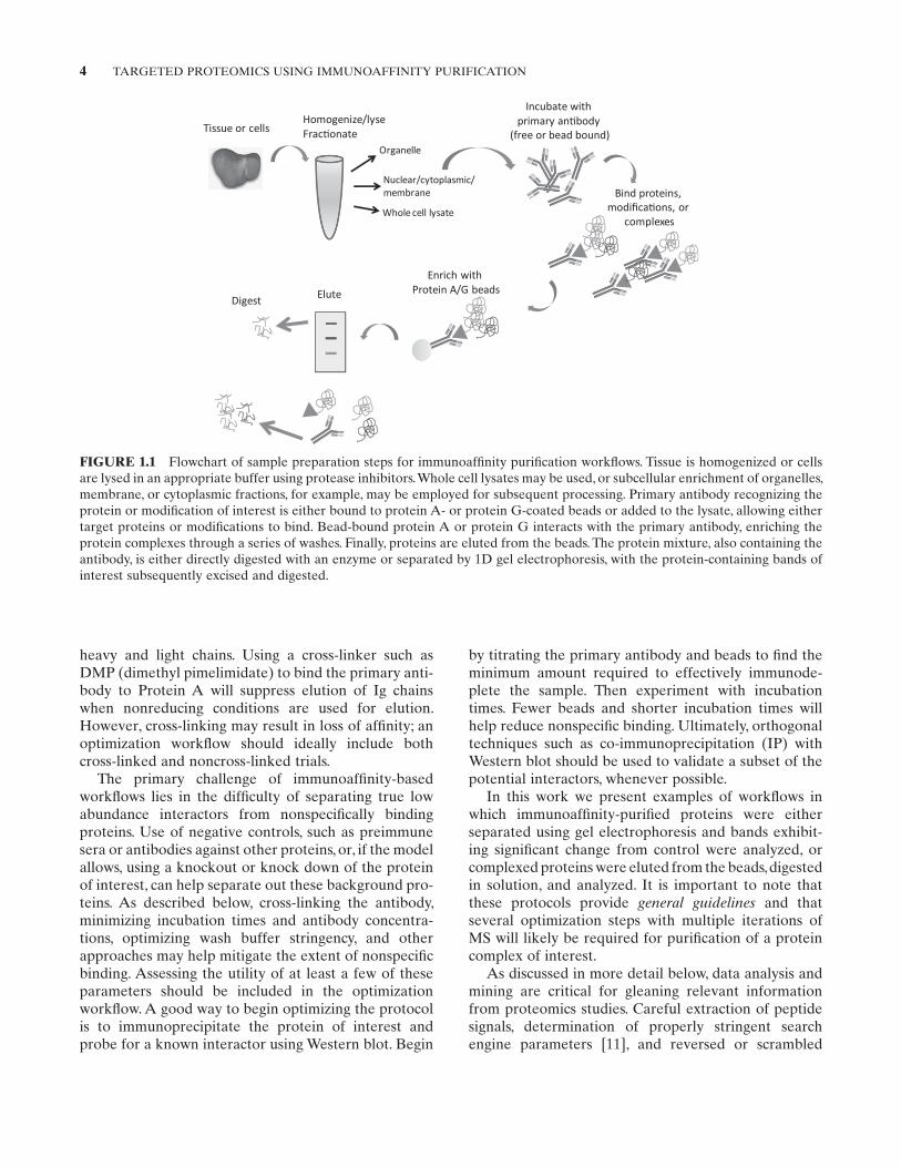
A summary of typical steps employed to generate samples using an immunoaffinity-based approach is illustrated in Figure 1.1 and described in detail for two example applications below. Following purification, peptide mixtures resulting from the digestion of bands or eluates are analyzed using tandem mass spectrome-try (MS/MS) and proteins are identified by database searching and spectral matching.
A gel-based approach may be useful when two condi-tions are being compared—bands exhibiting visual dif-ferences can be excised to yield data most likely to contrast biologically significant results (note that inter-esting low abundance proteins may be covered up by more abundant nonspecific proteins). Another useful method, gel-enhanced liquid chromatography–tandem mass spectrometry (GeLC-MS/MS), has also emerged for the analysis of complex protein mixtures [10] and can be applied to the separation of immunoaffinity eluates. In this approach, a protein-containing gel lane is chopped into equivalent sections, digested, and peptide mixtures analyzed. When complexed protein levels are extremely low or sample is limited, elution followed by in-solution digestion may provide a better option, as less protein is lost to sample handling. An important caveat to note is that a protein of interest may be “covered up” by comigrating background or nonspecific proteins. This is a particular concern for proteins that may comigrate with immunoglobulin (Ig)
4 TArGETEd ProTEoMICS USInG IMMUnoAFFInITy PUrIFICATIon
by titrating the primary antibody and beads to find the minimum amount required to effectively immunode-plete the sample. Then experiment with incubation times. Fewer beads and shorter incubation times will help reduce nonspecific binding. Ultimately, orthogonal techniques such as co-immunoprecipitation (IP) with Western blot should be used to validate a subset of the potential interactors, whenever possible.
In this work we present examples of workflows in which immunoaffinity-purified proteins were either separated using gel electrophoresis and bands exhibit-ing significant change from control were analyzed, or complexed proteins were eluted from the beads, digested in solution, and analyzed. It is important to note that these protocols provide general guidelines and that several optimization steps with multiple iterations of MS will likely be required for purification of a protein complex of interest.
As discussed in more detail below, data analysis and mining are critical for gleaning relevant information from proteomics studies. Careful extraction of peptide signals, determination of properly stringent search engine parameters [11], and reversed or scrambled
heavy and light chains. Using a cross-linker such as dMP (dimethyl pimelimidate) to bind the primary anti-body to Protein A will suppress elution of Ig chains when nonreducing conditions are used for elution. However, cross-linking may result in loss of affinity; an optimization workflow should ideally include both cross-linked and noncross-linked trials.
The primary challenge of immunoaffinity-based workflows lies in the difficulty of separating true low abundance interactors from nonspecifically binding proteins. Use of negative controls, such as preimmune sera or antibodies against other proteins, or, if the model allows, using a knockout or knock down of the protein of interest, can help separate out these background pro-teins. As described below, cross-linking the antibody, minimizing incubation times and antibody concentra-tions, optimizing wash buffer stringency, and other approaches may help mitigate the extent of nonspecific binding. Assessing the utility of at least a few of these parameters should be included in the optimization workflow. A good way to begin optimizing the protocol is to immunoprecipitate the protein of interest and probe for a known interactor using Western blot. Begin
FIGURE 1.1 Flowchart of sample preparation steps for immunoaffinity purification workflows. Tissue is homogenized or cells are lysed in an appropriate buffer using protease inhibitors. Whole cell lysates may be used, or subcellular enrichment of organelles, membrane, or cytoplasmic fractions, for example, may be employed for subsequent processing. Primary antibody recognizing the protein or modification of interest is either bound to protein A- or protein G-coated beads or added to the lysate, allowing either target proteins or modifications to bind. Bead-bound protein A or protein G interacts with the primary antibody, enriching the protein complexes through a series of washes. Finally, proteins are eluted from the beads. The protein mixture, also containing the antibody, is either directly digested with an enzyme or separated by 1d gel electrophoresis, with the protein-containing bands of interest subsequently excised and digested.
ExPErIMEnTAL ProToCoLS 5
database searching leads to an output data set where significance of the identifications may be established with score or probability cutoffs. Although a false discovery rate of ∼1% is often employed in more global approaches [12], establishing criteria for two peptide “hits” to a protein with peptide probabilities of ∼95% is sufficient to provide a false discovery rate approach-ing 0% for immunoaffinity purification applications. Following identification, data mining is employed to obtain functional information about the proteins to begin to decipher mechanisms that may be triggered by the interaction. The tools used often include those developed for microarray analysis, where gene ontology information is used to cluster proteins with similar cellular compartments, functions, or pathways [13–16]. downstream assays based on these results, including IP of proteins identified in the complex, allow inve-stigators to begin to elucidate mechanisms driving cellular function.
1.2 EXPERIMENTAL PROTOCOLS
Materials and Solutions
• Cell Lysis Buffer. 0.33% 3-[(3-cholamidopropyl)dimethylammonio]-1-propanesulfonate (CHAPS); 150 mM naCl; 10 mM sodium pyrophosphate; 10 mM Tris-HCl pH 7.4; 1 mM phenylmethylsulfo-nyl fluoride (PMSF); 0.4 mM ethylenediaminetet-raacetic acid (EdTA); 1.8 mg/mL iodoacetamide (IAA); 10 mM naF; 2 mM na3Vo4; and 1 µg/mL each of aprotinin, leupeptin, and pepstatin.
• Tissue Lysis Buffer. Buffer A (10 mM HEPES pH 7.9, 1.5 mM KCl, 10 mM MgCl2, 0.5 mM dithioth-reitol (dTT), 0.1% IGEPAL CA-630 (Sigma-Aldrich, St. Louis, Mo), and 0.5 mM PMSF) and Buffer B (20 mM HEPES pH 7.9, 25% glycerol, 1.5 mM MgCl2, 420 mM naCl, 0.5 mM dTT, 0.2 mM EdTA, 0.5 mM PMSF, and 4 µM leu-peptin). (Note: These buffers were selected for the analysis of liver proteomes, as described below. other lysis buffers may be more appropriate for the sample/proteins under investigation. A search of recent literature should provide some direction regarding appropriate buffer selection.)
• reagents for preparation of magnetic beads as described below.
• Protein A or protein G beads. (Note: Either Sepharose [GE Healthcare, Piscataway, nJ] beads or magnetic beads may be used; however, magnetic beads are preferred as they tend to exhibit less non-specific binding than Sepharose beads.)
• Pipette tips cut 5 mm from the top (will avoid dam-aging the beads if using Protein A/G Sepharose beads).
• Laemmli buffer.• High performance liquid chromatography (HPLC)-
grade water (Honeywell Burdick and Jackson [Morristown, nJ] or other high quality liquid chromatography–mass spectrometry [LC/MS]-grade water).
• desalt spin columns.• reagents for in-gel or in-solution digestion as listed
below.
Equipment
• refrigerated centrifuge• rotary mixer• Vacuum centrifuge• Gel electrophoresis apparatus and 10% polyacryl-
amide gel• nanoscale HPLC, tandem mass spectrometer
Lysis
note that the cell numbers and tissue amounts pre-sented here are a guideline. These numbers should be increased if complexed proteins are of low abundance.
Cell Lysis
1. Lyse ∼5 × 10⁸ cells using 500–1000 µL cell lysis buffer at 4°C overnight. note that if low abun-dance or weakly interacting proteins are to be analyzed, increase the cell numbers to as much as 1010 (as shown by Malovannaya and coworkers [17]).
2. In the morning, centrifuge lysates at 12,000 × g at 4°C for 20 min. remove supernatants to a clean tube.
Tissue Lysis
1. Homogenize ∼100 mg of tissue using a mortar and pestle over liquid nitrogen. For identification of potentially weakly binding complexes, increase tissue amount to 10–20 g (following Moresco et al. [18]) and increase lysis buffer volume to 5–10 mL.
2. Add homogenized tissue to 0.5 mL of tissue lysis buffer A (ice cold) and ultrasonicate three times, 15 s each. Place sample tubes in an ice bath for at least 1 min between sonications.
3. Incubate samples on ice for 30 min, then centri-fuge at 14,000 × g, 4°C for 10 min. remove the supernatant (cytoplasmic fraction) to clean tube.
4. resuspend the membrane/organelle fraction pellet in 0.2 mL ice-cold tissue lysis buffer B and
6 TArGETEd ProTEoMICS USInG IMMUnoAFFInITy PUrIFICATIon
incubate on ice for 30 min. Following centrifuga-tion at 14,000 × g for 30 min at 4°C, remove the supernatant to clean tube.
Note: For all subsequent steps, be sure to keep samples on ice or at 4°C, however freezing lysates before the immunoaffinity purification should be avoided [18]. If necessary, store lysates at −80°C prior to use.
Total Protein Quantification
Use either the Bradford or bicinchoninic acid (BCA) method to quantify total protein concentrations follow-ing the manufacturer’s instructions for a microwell plate assay. Make sure that the lysis buffer components are compatible with the manufacturer’s stated levels. Try several dilutions to ensure the sample concentration is within the linear range of the assay.
Immunoaffinity Purification
Immunoaffinity purification may be accomplished using either soluble antibodies or antibodies cross-linked to beads. Generally the first step of the optimization should be done using cross-linked antibodies; cross-linking sig-nificantly reduces contaminating signals from Ig light and heavy chains. Procedures for both approaches are provided below.
Immunoaffinity Purification Using Magnetic Beads
Reagents for Magnetic Bead Preparation (Dynabeads Are Typically Used)
• Citrate Phosphate Buffer, pH 5.0. 25 mM citric acid, 50 mM sodium phosphate (na2HPo4)
• 0.2 M Triethanolamine (TEA), pH 8.2. 3.71 g triethanolamine-HCl/100 mL water
• 20 mM DMP. 5.4 mg dMP-2HCl per milliliter of TEA buffer
• 50 mM Tris pH 7.5• PBS-T. 0.01% Tween-20 (Thermo Fisher Scientific,
Waltham, MA) in phosphate buffered saline• 0.1 M glycine pH 2.5–2.7• Storage Solution. PBS-T with 0.02% sodium azide
dynabeads (Invitrogen Corp., Carlsbad, CA) are pack-aged as a 5% slurry. Prepare 0.5–1.0 mL of slurry to obtain 25–50 µL of packed beads. A rule of thumb is that 1 mL of slurry binds ∼300 µg of antibody. Incubate with about 400 µg of the primary antibody.
Equilibrate Dynabeads
Centrifuge beads briefly, place tube in a magnetic rack, and remove the supernatant. Add 1 mL citrate phosphate buffer, vortex, spin briefly in a minifuge (1 s to remove
bead solution from cap), and place tubes in a magnetic rack. remove supernatant. repeat two more times.
Incubate with Primary Antibody
Prepare 400 µg of primary antibody in 1 mL citrate phosphate buffer and add to beads. reducing the volume may improve binding and may be included in subsequent optimization steps. rotate tube end over end for 2–3 h at room temperature.
Wash
Centrifuge briefly, place tubes in a magnetic rack, remove supernatant, and add 1 mL citrate phosphate buffer and wash three times as described in the section “Equilibrate dynabeads.” Wash two times more with 1 mL 0.2 M triethanolamine-HCl.
Cross-Link
remove final TEA wash from the beads using the magnet and add 1 mL of dMP solution. Incubate 30 min at room temperature, rotating end over end.
Clean Up
Using the magnet, remove the dMP solution and incu-bate beads with 50 mM Tris for 15 min to remove free cross-linking reagent.
Wash
Wash beads three times with PBS-T.
Remove Free Antibody
Incubate the beads with 0.1 M glycine for 5 min, rotat-ing end over end at 4°C.
Wash
Wash beads three times with PBS-T.
Store Beads
Bring beads back to original packaged volume in storage solution for storage at 4°C. For a 1 mL stock, use 950 µL of storage solution.
To use, wash beads three times using 1 mL PBS, then three time with 1 mL lysis buffer. Use Western blots and bead titration to determine the minimum amount of beads to use and the minimum amount of time to incu-bate. A starting point for optimization may be 20 µL of packed beads and 2 h of incubation at 4°C. Elute as described below.
Immunoaffinity Purification Using Protein A/G Sepharose Beads
Incubation with Primary Antibody
Add 3–20 µg of primary antibody to the supernatant and incubate for 2 h to overnight at 4°C. This step