martini’s visual anatomy and physiology - gserianne.com · martini’s visual anatomy and...
TRANSCRIPT

1
Martini’s VisualAnatomy and Physiology
First Edition
Martini Ober
1
Chapter 24
Water, Electrolytes, and Acid/Base Balance
Lecture 19
Lecture Overview
• Overview
• Fluid (water) balance– Compartments
– Body fluid composition
I l fl id hif
2
– Intercompartmental fluid shifts
• Electrolyte balance
• Acid-base balance– Buffer systems
– Acidosis and alkalosis
Overview
• Our survival depends upon maintaining a normal volume and composition of– Extracellular fluid (ECF)
– Intracellular fluid (ICF)
3
• Ionic concentrations and pH are critical
• Three interrelated processes– Fluid balance (How does water move?)
– Electrolyte balance (What is an electrolyte?)
– Acid-base balance (What is normal pH?)
2
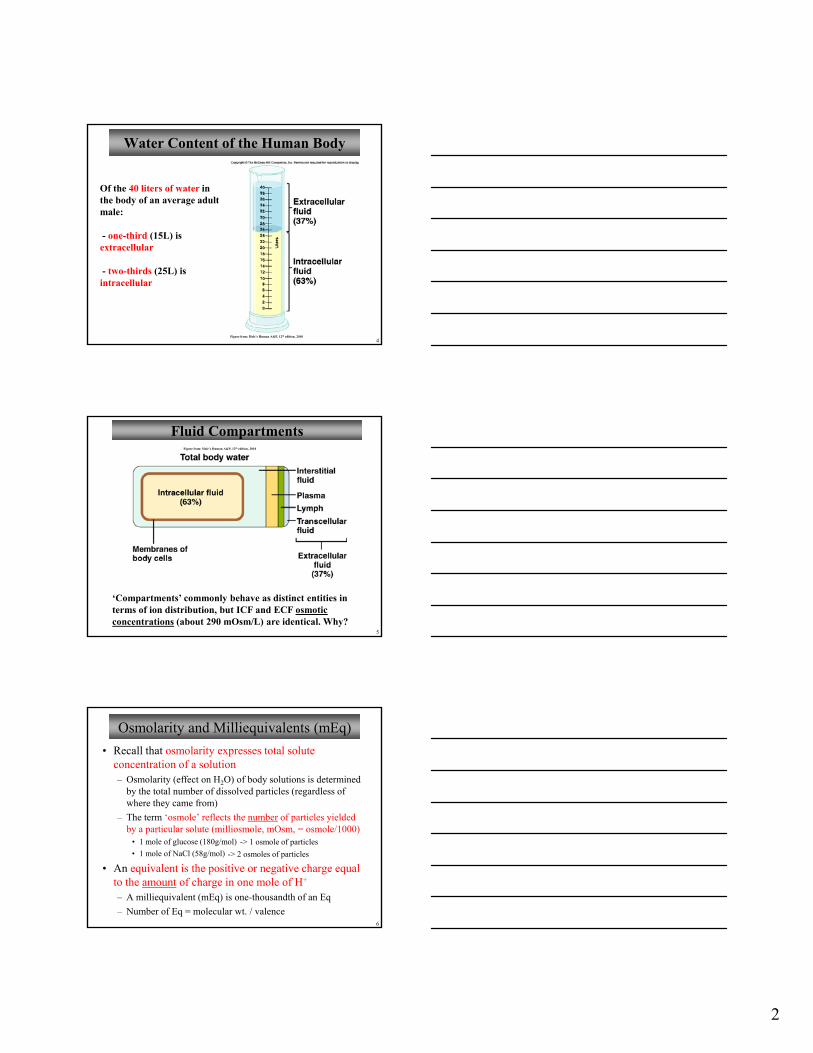
Water Content of the Human Body
Of the 40 liters of water in the body of an average adult male:
one third (15L) is
4
- one-third (15L) is extracellular
- two-thirds (25L) is intracellular
Figure from: Hole’s Human A&P, 12th edition, 2010
Fluid CompartmentsFigure from: Hole’s Human A&P, 12th edition, 2010
5
‘Compartments’ commonly behave as distinct entities in terms of ion distribution, but ICF and ECF osmotic concentrations (about 290 mOsm/L) are identical. Why?
Osmolarity and Milliequivalents (mEq)
• Recall that osmolarity expresses total solute concentration of a solution– Osmolarity (effect on H2O) of body solutions is determined
by the total number of dissolved particles (regardless of where they came from)
The term ‘osmole’ reflects the number of particles yielded
6
– The term osmole reflects the number of particles yielded by a particular solute (milliosmole, mOsm, = osmole/1000)
• 1 mole of glucose (180g/mol)
• 1 mole of NaCl (58g/mol)
• An equivalent is the positive or negative charge equal to the amount of charge in one mole of H+
– A milliequivalent (mEq) is one-thousandth of an Eq
– Number of Eq = molecular wt. / valence
-> 1 osmole of particles
-> 2 osmoles of particles

3
Body Fluid Ionic Composition
ECF major ions:
- sodium, chloride, and bicarbonate
Figure from: Hole’s Human A&P, 12th edition, 2010
7
ICF major ions:
- potassium, magnesium, and phosphate (plus negatively charged proteins)
You should know these chemical symbols and charges of ions
Movement of Fluids Between Compartments
Water moves between mesothelial surfaces: peritoneal, pleural, and pericardial cavities as well as the synovial membranes. It also moves between the blood and CSF
Figure from: Hole’s Human A&P, 12th edition, 2010
9
Net movements of fluids between compartments result fromdifferences in hydrostatic and osmotic pressures
the blood and CSF and through the fluids of the eye and ear
Fluid (Water) Balance
10
* urine production is the most important regulator of water balance (water in = water out)
Figure from: Hole’s Human A&P, 12th edition, 2010

4
Water Balance and ECF Osmolarity
• Regulation of water intake• increase in osmotic pressure of ECF → osmoreceptors in hypothalamic thirst center → stimulates thirst and drinking
• Regulation of water output
11
• Obligatory water losses (must happen)• insensible water losses (lungs, skin)• water loss in feces• water loss in urine (min about 500 ml/day)
• increase in osmotic pressure of ECF → ADH is released• concentrated urine is excreted• more water is retained
• LARGE changes in blood vol/pressure → Renin and ADH release
Fluid Imbalance
12Figure from: Saladin, Anatomy & Physiology, McGraw Hill, 2007
Dehydration and OverhydrationDehydration
• osmotic pressure increases in extracellular fluids• water moves out of cells• osmoreceptors in h th l ti l t d
Overhydration• osmotic pressure decreases in extracellular fluids• water moves into cells• osmoreceptors inhibited i h th l
13
hypothalamus stimulated• hypothalamus signals posterior pituitary to release ADH• urine output decreases
in hypothalamus• hypothalamus signals posterior pituitary to decrease ADH output• urine output increases
‘Drunken’ behavior (water intoxication), confusion, hallucinations, convulsions, coma, death
Severe thirst, wrinkling of skin, fall in plasma volume and decreased blood pressure, circulatory shock, death

5
Electrolyte Balance
Electrolyte balance is important:
1. Regulates fluid (water) balance
Figure from: Hole’s Human A&P, 12th edition, 2010
14
2. Concentrations of individual electrolytes can affect cellular functions
Na+: major cation in ECF (plasma: 136-142 mEq/L; Avg ≈ 140)
K+: major cation in ICF (plasma: 3.8-5.0 mEq/L; Avg ≈ 4.0)
Regulation of Osmolarity
Osmolarity i l t d
Recall: [Na+] Osmolarity
15
Figures from: Martini, Anatomy & Physiology, Prentice Hall, 2001
is regulated by altering H2O content
Fluid Volume Regulation and [Na+]Estrogens are chemically similar to aldosterone and enhance NaCl absorption by renal tubules
Glucocorticoids can also enhance tubular reabsorption of Na+
16
Figures from: Martini, Anatomy & Physiology, Prentice Hall, 2001
reabsorption of Na+
Volume is regulated by altering Na+
content

6
Summary Table of Fluid and Electrolyte Balance
Condition Initial Change Initial Effect Correction Result
Change in OSMOLARITY
(**Corrected by change in H2O levels)
H2O in the ECF
Na+ concentration,
ECF osmolarity
Thirst → H2O intake
ADH → H2O output H2O in the ECF
H2O in the ECF
Na+ concentration,
ECF osmolarity
Thirst → H2O intake
ADH → H2O output H2O in the ECF
17
Change in VOLUME(**Corrected by change
in Na+ levels)
H2O/Na+ in the ECF volume,
BP
Renin-angiotensin: ThirstADH
aldosterone vasoconstriction
H2O intake Na+/H2O reabsorption
H2O loss
H2O/Na+ in the ECF volume,
BP
Natriuretic peptides: ThirstADH
aldosterone
H2O intake Na+/H2O reabsorption
H2O loss
You should understand this table
Potassium Balance
Potassium loss generally occurs via the urine. The rate of tubular secretion of K+ varies with:
1. Changes in the [K+]
Figure from: Hole’s Human A&P, 12th edition, 2010
18
in the ECF
2. Changes in pH
3. Aldosterone levels
Remember that Na+ can be exchanged for H+ or K+ in the nephron tubules
Calcium Balance
[Ca2+] in ECF is about 5 mEq/L
Figure from: Hole’s Human A&P, 12th edition, 2010
19

7
Strengths of Acids and Bases
• Weak acids ionize less completely and release fewer H+
(**allows them to act as buffers)
• Strong acids ionize more completely and release more H+
20
• Weak bases ionize less completely and bind fewer H+
• Strong bases ionize more completely and bind more H+
( allows them to act as buffers)
Sources of Hydrogen Ions
Figure from: Hole’s Human A&P, 12th edition, 2010
21
Some H+ is also absorbed from the digestive tract
Regulation of Hydrogen Ion Concentration
1. chemical acid-base buffer systems (physical buffers)• first line of defense• can tie-up acids or bases, but cannot eliminate them• act in seconds
2 respiratory excretion of carbon dioxide
22
2. respiratory excretion of carbon dioxide• a physiological buffer (can eliminate excess acid indirectly via CO2)• minutes
3. renal excretion of hydrogen ions• a physiological buffer (can eliminate excess metabolic acids directly, e.g., keto-, uric, lactic, phosphoric)• hours to a day

8
Acid-Base Buffer Systems
Bicarbonate System• the bicarbonate ion converts a strong acid to a weak acid• carbonic acid converts a strong base to a weak base• an important buffer of the ECF (~ 25 mEq/L)
H+ + HCO3- ↔ H2CO3↔ CO2 + H2O
Strong acid Weak acid
23
H HCO3 H2CO3 CO2 H2O
Phosphate System• the monohydrogen phosphate ion converts a strong acid to a weak acid• the dihydrogen phosphate ion converts a strong base to a weak base
H+ + HPO4-2 ↔ H2PO4
-
Strong acid Weak acid
Acid-Base Buffer Systems
Protein Buffer SystemICF, plasma proteins, Hb
NH2 group accepts
COOH group releases
Most plentiful and powerful chemical buffer system
24
phydrogen ions when pH falls
hydrogen ions when pH rises
-
Figure from: Martini, Anatomy & Physiology, Prentice Hall, 2001
Respiratory Excretion of Carbon Dioxide
A physiologicalbuffer system
Figure from: Hole’s Human A&P, 12th edition, 2010
25

9
Renal Excretion of Hydrogen Ions
*The kidney is most powerful and versatile acid-base system in the body
Figure from: Hole’s Human A&P, 12th edition, 2010
26
Note that secretion of H+
relies on carbonic anhydrase activity within tubular cells
Net result is secretion of H+ accompanied by the
Buffering Mechanisms in the Kidney
27
H accompanied by the (1)retention of HCO3
-
Figure from: Martini, Anatomy & Physiology, Prentice Hall, 2001
Production of new HCO3
-
(2)
Summary of Acid-Base BalanceFigure from: Hole’s Human A&P, 12th edition, 2010
28
Know this slide!

10
Acidosis and Alkalosis
If the pH of arterial blood drops to 6.8 or rises to 8.0 for more than a few hours, survival is jeopardized
Classified according to:
1. Whether the cause is respiratory (CO2),
t b li ( th
29
or metabolic (other acids, bases)
2. Whether the blood pH is acid or alkaline
Figure from: Hole’s Human A&P, 12th edition, 2010
Acidosis
(hypopnea)
Figure from: Hole’s Human A&P, 12th edition, 2010
30
Respiratory acidosis Metabolic acidosis
Nervous system depression, coma, death
AlkalosisFigure from: Hole’s Human A&P, 12th edition, 2010
31
Respiratory alkalosis Metabolic alkalosis
Nervousness, tetany, convulsions, death

11
Acidosis and Alkalosis
• What would be the indications of acidosis and alkalosis in terms of changes in pH and PCO2? pH and HCO3
-?
• How would the body try to compensate for
32
– Acidosis• Respiratory• Metabolic
– Alkalosis• Respiratory• Metabolic
See Lab Guide Handout: Marieb, Human Anatomy & Physiology, Pearson, 2004
Flow chart for Acidosis/Alkalosis
Three things to check: 1) pH – 7.35-7.452) pCO2 – 35-45 mm Hg3) HCO3
- - 22 – 26 mEq/LpH
33
acidosis alkalosis
pCO2
respiratory metabolic respiratory metabolic
HCO3- pCO2 HCO3
-
pCO2
Comp Comp CompCompNo Comp
No CompNo CompNo Comp
pCO2
HCO3-
Norm HCO3
-Norm HCO3
- HCO3-
Norm pCO2
Norm pCO2
Review
• There are two major fluid compartments of the body– Intracellular
• About 2/3 of body’s fluid
l d h fl id i hi ll
34
• Includes the fluid within cells
• Major ions: K+, Mg2+, PO43-, Proteins
– Extracellular• About 1/3 of body’s fluid
• Includes interstitial fluid, plasma, lymph, and transcellular fluid
• Major ions: Na+, Cl-, HCO3-

12
Review
• There are two major forces affecting movement of fluid between compartments– Hydrostatic Pressure– Osmotic Pressure
• Fluid balance
35
• Fluid balance– Amount of water you take in is equal to the
amount of water you lose to the environment– Intake of water in food/drink is the most
important source of fluid– Kidney regulation of water is the most
important regulator of water loss
Review
• Electrolyte balance– Balance: Gains and losses of every electrolyte are equal
– Electrolyte balance is important because• It directly affects water balance
• Electrolyte concentrations affect cell processes
36
– Na+ (aldosterone, ADH, ANP)• Increased [Na+ ] in ECF -> ↑ ADH, ↑ ANP
• Decreased [Na+ ] in ECF -> ADH, ↑ aldosterone
– K+ ([K+] in plasma, aldosterone)• Increased [K+ ] in ECF -> increased secretion, ↑ aldosterone
• Decreased [K+ ] in ECF -> decreased secretion, aldosterone,
Review
• Electrolyte balance (cont’d)– Ca2+ (PTH, calcitriol, calcitonin)
• Increase in ECF -> calcitonin promotes bone deposition• Decrease in ECF -> PTH , calcitriol
– ↑ intestinal absorption– ↑ bone resorption
37
– Ca2+ secretion, ↑ PO43- secretion
• Acid-base balance– Production of H+ is exactly offset by the loss of H+
– Major mechanisms of maintaining• acid-base (chemical) buffer systems: HCO3
-, PO43-, protein
• respiratory excretion of carbon dioxide• renal excretion of hydrogen ions

13
Review
• Acidosis (pH < 7.35)– Excessive H+ in the plasma
– Respiratory acidosis
– Metabolic acidosis
• Alkalosis (pH > 7 45)
38
• Alkalosis (pH > 7.45)– Insufficient H+ in the plasma
– Respiratory alkalosis
– Metabolic alkalosis
• Compensations
Review
Electrolyte Concentration Range (mEq/L)
Typical Value (mEq/L)
Na+
136 - 142 140
K+
3.8 - 5.0 4.0
Ca2+
4.5 – 5.8 5.0
39
Cl-
96 - 106 105
HCO3-
24 - 28 25