markov propagation of allosteric effects in biomolecular systems
TRANSCRIPT

Markov propagation of allosteric effects inbiomolecular systems: application to GroEL–GroES
Chakra Chennubhotla and Ivet Bahar*
Department of Computational Biology, School of Medicine, University of Pittsburgh, Pittsburgh, PA, USA* Corresponding author. Department of Computational Biology, School of Medicine, University of Pittsburgh, Biomedical Science Tower 3, Fifth Avenue, Pittsburgh,PA 15213, USA. Tel: þ 1 412 648 3332; Fax: þ 1 412 648 3163; E-mail: [email protected]
Received 6.12.05; accepted 11.5.06
We introduce a novel approach for elucidating the potential pathways of allosteric communicationin biomolecular systems. The methodology, based on Markov propagation of ‘information’ acrossthe structure, permits us to partition the network of interactions into soft clusters distinguishedby their coherent stochastics. Probabilistic participation of residues in these clusters defines thecommunication patterns inherent to the network architecture. Application to bacterial chaperonincomplex GroEL–GroES, an allostery-driven structure, identifies residues engaged in intra- and inter-subunit communication, including those acting as hubs and messengers. A number of residues aredistinguished by their high potentials to transmit allosteric signals, including Pro33 and Thr90 atthe nucleotide-binding site and Glu461 and Arg197 mediating inter- and intra-ring communication,respectively. We propose two most likely pathways of signal transmission, between nucleotide-and GroES-binding sites across the cis and trans rings, which involve several conserved residues.A striking observation is the opposite direction of information flow within cis and trans rings,consistent with negative inter-ring cooperativity. Comparison with collective modes deduced fromnormal mode analysis reveals the propensity of global hinge regions to act as messengers in thetransmission of allosteric signals.Molecular Systems Biology 4 July 2006; doi:10.1038/msb4100075Subject Categories: structural biology; proteinsKeywords: allosteric effects; chaperonins; information propagation; Markov process; network model
Introduction
A central goal in structural biology is to understand themechanism of allosteric communication in supramolecularsystems. Allostery is the cooperative process by which localeffects propagate across the structure, often to regions spatiallydistant from initiation sites. A prime example of allostery is thetransition of hemoglobin from low-affinity (T; tense) to high-affinity (R; relaxed) state upon oxygen binding. Two classicalmodels have been proposed to describe allosteric transitionsin such multimeric proteins: the Monod–Wyman–Changeux(MWC) (Monod et al, 1965) and Koshland–Nemethy–Filter(KNF) (Koshland et al, 1966) models. The former assumesa concerted, all-or-none change in all subunits, whereas thelatter allows for the sequential transition of individualsubunits. Although these models, and more complicated ones,are still being debated, the emerging view from experimentaland computational studies is that many regulatory multimericproteins, and in particular allosteric enzymes, possess theintrinsic ability to undergo MWC-like changes in structure,conferred by their three-dimensional (3-d) network of inter-residue interactions (in the absence of ligands). Suchcooperative effects, enhanced by structural symmetry in thecase of oligomers (Changeux and Edelstein, 2005), are usuallytriggered or stabilized upon ligand binding at particular
susceptible sites (Tobi and Bahar, 2005). Recent computationalstudies also lend support to the intrinsic role of inter-residuecontact topology in defining functional dynamics (Bahar andRader, 2005; Ma, 2005). Yet, the pathways of signal transduc-tion favored by the network of inter-residue contacts insupramolecular structures remain to be established.
The allostery-driven system we explore here is the bacterialchaperonin system GroEL–GroES, a widely studied molecularchaperone that assists folding of a subset of Escherichia coliproteins (Thirumalai and Lorimer, 2001; Saibil and Ranson,2002; Horovitz and Willison, 2005). GroEL has a cylindricalstructure, 150 A long and 140 Awide, consisting of 14 identicalchains/subunits organized in two back-to-back stacked ringsof seven subunits each (Xu et al, 1997) (Figure 1). Each chain iscomposed of three domains, equatorial (E1 and E2), inter-mediate (I1 and I2) and apical (A), sequentially ordered asE1-I1-A-I2-E2. During the allosteric cycle that mediatesprotein folding, the rings alternate between open (cis) andclosed (trans) forms in an ATP-regulated manner, providingaccess to, or release from, the central cavity where the foldingof encapsulated (partially folded or misfolded) protein/peptide is assisted.
Chaperonin function requires an efficient communicationbetween distant locations on the complex. For example, ATPbinding to E-domains is accompanied by a cooperative
& 2006 EMBO and Nature Publishing Group Molecular Systems Biology 2006 msb4100075-E1
Molecular Systems Biology (2006) doi:10.1038/msb4100075& 2006 EMBO and Nature Publishing Group All rights reserved 1744-4292/06www.molecularsystemsbiology.com000036

conformational change (T-R) that facilitates the binding ofco-chaperonin GroES at the A-domains in the same (cis) ring,whereas substrate binding and ATP binding to opposite (trans)ring triggers the release of GroES, substrate protein and ADPfrom the cis ring. Although several studies have been under-taken to unravel the mechanism of allostery in GroEL–GroES(Xu et al, 1997; Ma and Karplus, 1998; Sigler et al, 1998; deGroot et al, 1999; Ma et al, 2000; Thirumalai and Lorimer,2001; Kass and Horovitz, 2002; Keskin et al, 2002; Horovitzand Willison, 2005), the underlying pathways of allostericcommunication remain to be elucidated, as well as the keyinteractions that mediate the intra-ring (positive) and inter-ring (negative) cooperativity.
In this paper, we introduce a novel approach for unravelingpotential pathways of signal transduction in large structures.Motivated by the recent success of network-based approachesin exploring structural motifs/mechanisms for allostericcommunication (Keskin et al, 2002; Xu et al, 2003), we modelstructures as networks of residues (Bahar et al, 1997; Halilogluet al, 1997; Hinsen, 1998; Doruker et al, 2000; Atilgan et al,2001) and study the Markov propagation of ‘information’across these networks. We rely on the premise that signaling/communication ability is an intrinsic property of the 3-dstructure and that naturally selected structures are thosepredisposed to optimally meet these functional requirements.We show how a Markovian network formalism based oninformation theory (Kullback, 1959; McLachlan and Basford,1988; Chennubhotla and Jepson, 2005) and spectral graphmethods (Chung, 1997) helps understand the pathways ofcommunication between spatially distant sites and signifi-cantly reduces the complexity of the problem. In particular, weanalyze how information diffuses between ATP-binding andGroES-binding sites in GroEL–GroES.
Our approach is structure-based, rather than sequence-based (Lock-less and Ranganathan, 1999; Kass and Horovitz,2002; Stan et al, 2003; Suel et al, 2003), in that it maps a full-atom representation into a hierarchy of networks of decreasing
resolution, performs the analysis of dominant patterns inreduced space(s) and reconstructs the detailed models witha minimal loss of information (Figure 2). Such dimensionalityreduction algorithms based on Markov propagation stochas-tics expressed in terms of Fokker–Planck operator have beenshown to convey useful information on the diffusion processesin complex systems (Bahar et al, 1994; Coifman et al, 2005;Nadler et al, 2006). The communication properties at differentlevels of the hierarchy allow for partitioning the complexstructure into soft clusters. The probabilistic distribution ofamino acids in these clusters, or the so-called ownership ofresidues by the clusters, permits us to evaluate the commu-nication entropies of individual residues. Residues distin-guished by high entropies possess a high potential to transmitallosteric signals, hence their identification as sites of largeallosteric potential (Ming and Wall, 2005).
Notably, our analysis provides evidence for the critical roleplayed by the GroES mobile loops Glu18–Ala33 in establishingthe communication with GroEL cis ring. Also, we identifythe residues that establish positive intra-ring cooperativity.Negative cooperativity between the two rings is suggestedby the tendency of the two rings to engage in intra-ringcommunication of opposite rotational direction. The cis ringE-domains exhibit stronger intra-ring coupling than their transring counterparts in the presently examined ADP-boundcomplex, and they unambiguously emerge as centers withthe highest propensity for ‘broadcasting’ perturbations to theentire structure. Finally, we identify residues acting as hubsand/or messengers for collecting and passing informationacross the network and we determine maximum likelihoodcommunication pathways from nucleotide-binding site to co-chaperonin mobile loop across the rings. A significant propertythat emerges from the comparison of the present results withthose from normal mode analysis of GroEL–GroES collectivedynamics is the propensity of the global hinge sites identifiedwith the Gaussian network model (GNM) (Bahar et al, 1997;Haliloglu et al, 1997; Bahar and Rader, 2005) to play the critical
GroEL-GroES complex Cis ring subunit Trans ring subunit
Apical (A)
Equatorial (E)
Intermediate (I)Intermediate (I)
Equatorial (E)
Apical (A)
GroeES
Tra
ns r
ing
Cis
rin
g
Gro
EL
ADP
A B C
Figure 1 Structure of the GroEL–GroES complex. (A) Space-filling model from the crystal structure determined by Xu et al (1997) (Protein Data Bank (PDB): 1AON).GroEL has a cylindrical structure, composed of two rings, termed the cis and trans rings, depending on the position of the GroES cap. Each ring in GroEL is composedof seven subunits. One subunit in each ring is shown in color (red, green and blue) in (A), along with one of the chains of the heptameric co-chaperonin (shown in gray/slate). The colored subunits on the GroES cap and the GroEL cis and trans rings correspond to three representative chains (identified as chains R, D and K in the PDBfile (Berman et al, 2000)) whose communication dynamics will be examined below (see Figure 3C). (B) and (C) display ribbon diagrams of these two subunits belongingto the cis and trans rings, respectively. Each subunit consists of three domains, A, I and E, which refer to the apical, intermediate and equatorial domains, respectively.The corresponding residue ranges are: [A] Met193–Gly375; [I] Cys138–Gly192 (I1) and Val376–Gly410 (I2); and [E] Met1–Pro137 (E1) and Val411–Pro525 (E2).ADP molecule bound to the equatorial domain of the cis ring subunit is displayed in pink in panel B.
Markov propagation of allosteric effects in biomolecular systemsC Chennubhotla and I Bahar
msb4100075-E2 Molecular Systems Biology 2006 & 2006 EMBO and Nature Publishing Group

role of messengers in the transmission of allosteric signals, aswill be demonstrated below.
Markov process of network communication
A discrete-time, discrete-state Markov process is defined bysetting the communication probability between residue pairsto be a function of their interaction strength, also calledaffinity. In particular, the {ij}th element of the affinity matrixA¼{aij} is defined as
aij ¼NijffiffiffiffiffiffiffiffiffiffiNiNj
p ð1Þ
where Nij is the total number of atom–atom contacts madebetween residues vi and vj based on a cutoff distance ofrc¼4.5 A and (Ni, Nj) are the total numbers of heavy atoms inthe individual residues (vi, vj). The network may be alterna-tively viewed as a mass-spring system where pairs of(interacting) residues are connected by elastic springs withforce constant aij¼aji. The self-contact aii is similarly defined,but all bonded pairs are excluded. This representation capturesto a first approximation the strong (weak) interactionsexpected to take place between residue pairs with large(small) numbers of atom–atom contacts, and removes biasesdue to size effects.
Using a measure of the local interaction density at eachresidue, given by dj ¼
Pni¼1 aij ¼
Pnj¼1 aji (where n is total
number of residues in the network), which in matrix form isD¼diag{dj}, we define mij ¼ d�1
j aij as the conditional prob-ability of transmitting information to residue vi in one time stepgiven that the signal is initially positioned at residue vj. Note,dj serves as a normalizing factor to ensure
Pni¼1 mij ¼ 1: The
conditional probability matrix M ¼ fmijg; also called theMarkov transition matrix, given by
M ¼ AD�1 ð2Þ
fully defines the stochastics of signal diffusion over thenetwork of residues.
Suppose the probability of initiating the Markov propaga-tion process at node j is pj
0. Then, the probability of reachingresidue vi in one time step is mijpj
0. In matrix notation,the probability of ending up on any of the residuesv ¼ ½v1; v2; . . . ; vn after one time step is given by thedistribution p1¼Mp0, where pk ½pk
1; . . . ; pkn: Clearly, this
process can be iterated, so that after b steps we have
pb ¼ Mbp0 ð3Þ
Assume there is a path connecting every pair of residues in thenetwork. Then, as b-N, the Markov chain pb approachesa unique stationary distribution p, the elements of whichare given by pi ¼ di=
Pnk¼1 dk: Whereas the evolution of the
diffusion process is a function of the starting distribution, thestationary distribution is invariant to the details of initiation(Norris, 1997).
Hierarchical model reduction based on Markovstochastics
For a clear understanding of the most probable mechanismsof signal propagation across the structure, we mapped theoriginal structure onto successively lower resolution networkmodels, while maintaining its stochastic characteristics(Figure 2). To build a hierarchy of intermediate resolutionnetworks, we devise two sets of new operators: R for modelreduction and K for model expansion/reconstruction (seeMaterials and methods). The {ij}th element R
ð0;LÞij describes
the probabilistic participation of residue vi in cluster j athierarchical level L. Alternatively, R
ð0;LÞij may be viewed as a
measure of the ownership of residue vi by cluster j generatedat level L. The maximal ownership of a given residue vi isdefined as
Rð0;LÞi;max max ðRð0;LÞ
ik Þ ð4Þ
Step ii : Coarse-grained analysis{δ}
{π}?
Ste
p i :
Coa
rse-
grai
ning
Ste
p iii
: R
econ
stru
ctio
n
K(L, L–1)
K(1,0)
R(1,2)
R(0,1)
(Level 0)
(Level L)
Figure 2 Hierarchical network decomposition overview. Step i: mapping of thestructure to its optimal reduced level representation (coarse-graining); step ii:structural/dynamic analysis—for example, GNM analysis of collective dynamics(Bahar et al, 1997; Haliloglu et al, 1997)—in the reduced space; and step iii:reconstruction of the detailed structure dynamics. The communication/couplingsof residues at a given level are assumed to obey a Markov propagation processconsistent with the distribution of atom–atom contacts in the original structure.Steps i and iii are achieved by two sets of operators, R for model reduction andK for model reconstruction (Chennubhotla and Jepson, 2005) explained inMaterials and methods. Several models of intermediate complexity (not shown)are usually generated between the highest and lowest levels. R and K ateach level ensure that similar stochastic characteristics (signal propagationprobabilities and stationary distribution of communication) are retained betweensuccessive levels of the hierarchy. In particular, the reduction operator Rðl;lþ1Þ
propagates residue information from level l to lþ 1. Successive multiplicationof such transformation matrices, as in Rð0;LÞ ¼
QL�1l¼0 Rðl;lþ1Þ; ensures the
passage of information from the original, or the highest, resolution representation(level 0) to the most reduced level (level L) of the hierarchy. The arrows on theright illustrate the most cooperative motion (counter-rotation of the two rings)predicted by elastic network model analysis of GroEL–GroES(ADP)7 (Keskinet al, 2002).
Markov propagation of allosteric effects in biomolecular systemsC Chennubhotla and I Bahar
& 2006 EMBO and Nature Publishing Group Molecular Systems Biology 2006 msb4100075-E3

for 1pkpc(L), where c(L) is the total number of clusters atlevel L. R
ð0;LÞi;max provides a measure of the involvement of residue
vi in the cluster that maximally owns it. In particular, thisproperty reflects the potential of a residue for serving as a hubin any cluster derived at level L.
We also define the entropy Sð0;LÞi of residue vi as
Sð0;LÞi �
Xk
Rð0;LÞik lnR
ð0;LÞik ð5Þ
where the summation is performed over all clusters. Sð0;LÞi
provides a measure of the communication ability of residuevi between clusters at level L. High entropy values refer toresidues more or less uniformly shared by multiple clusters,thus playing the key role of a messenger in transmittingsignals/perturbations. These are also referred to as siteshaving a high allosteric potential.
Soft partitioning of structure into stochasticallycoherent clusters
The GroEL–GroES(ADP)7 structure (Xu et al, 1997) of n¼8015residues/nodes was mapped into a series of hierarchicallyreduced representations, comprised of c(L)¼1316, 483, 133, 35and 21 nodes for L¼1–5, using the Markov chain propagationtheory described in Materials and methods.
Each node at a given reduced level is a coherent clusterthat provides a soft partitioning of the original structure,with regard to its communication stochastics, over increas-ingly large distance ranges. Thus, in contrast to the determi-nistic assignment of one-node-per-residue in the originalnetwork (level ‘0’), each cluster probabilistically contains asubset of residues, quantitatively expressed by its ownershipdistribution.
We focus on reduced level 4 (c¼35) as the highest level thatprovides new insights into Markov propagation stochastics(level 5 practically partitions the complex into its 21 mono-meric chains). Five sets (I–V) of seven symmetrically relatedclusters (Figure 3A) are obtained, centered near the GroESchains (clusters 1–7), the domains A–I (8–14) and E (15–21)of the cis ring and the domains E–I1 (22–28) and A–I2 (29–35)of the trans ring. Interestingly, the GroEL subunits are eachpartitioned into two clusters, although they comprise threestructural domains E, I and A. Domain-I residues play apivotal role, being integrated into E- or A-centered clusters,and their type and strength of participation in these clustersdiffer in the cis and trans rings. Panel B in Figure 3 illustratesthe probabilistic participation of residues in two exampleclusters (encircled in panel A). The color code from red to blueindicates the gradually decreasing strength/probability ofbeing owned by the particular clusters. A more quantitativeanalysis of these ownerships and comparison with experi-mental data are presented next.
Intra- and inter-subunit couplings and biologicalimplications
Five ownership distributions (labeled 1–7, 11, 18, 25 and32 in Figure 3D; colored curves) are displayed, representativeof each respective type (I–V) of clusters (Table I). Thechain identifiers (A–G for cis subunits, H–N for trans
subunits and O–U for GroES chains) adopted in the PDB(Berman et al, 2000) are shown along the abscissa. Wefocus on the subunits that are predominantly owned by thefive representative clusters, that is, chains D, E, K, J and R,the locations of which on the 3-d structure are shown inFigure 3C. The results are valid for all seven symmetricallyrelated clusters/subunits. The gray curve in each paneldepicts the maximal responsibility R
ð0;LÞi;max (equation (4)),
assumed by residue vi in the overall communication stochas-tics of the network.
The ownerships of individual clusters reveal severalfunctional features. Below is a summary of results and relevantexperimental data by various groups (see also Table I andSupplementary Table S1 (Supplementary information)).
A
D
B CI:1–7
II: 8–14
III: 15–21
IV: 22–28
V: 29–35
A0.80.4
0.80.4
0.80.4
0.80.4O
wne
rshi
p di
strib
utio
ns0.80.4
B C D E F G H I J K L M N
1–7
1118
2532
O – U
A B C D E F G H I J K L M N O – U
Figure 3 Soft partitioning of the chaperonin complex into stochasticallycoherent clusters. A total of 35 clusters are identified at reduced level 4, eachshown by a different color in (A). Owing to the seven-fold cylindrical symmetry,five distinct types of clusters are observed: clusters (I) 1–7 centered on theGroES monomers, (II) 8–14 centered on AI domains of the cis ring subunits, (III)15–21 around the E-domain of cis ring subunits, (IV) 22–28 near the E–Idomains of trans ring subunits and (V) 29–35 near A–I domains of trans ringsubunits. (B) displays The soft participation of residues in two example clustersencircled in panel A, clusters 11 (top) and 32 (bottom) respectively, by ribbondiagrams, color-coded red–orange–yellow–green–cyan in the order of decreas-ing probabilistic participation. Results are valid for all cylindrically related sevensubunits along the heptameric rings, but for illustration we focus on chains D(yellow) and E (magenta) on cis ring, J (pink) and K (green) on trans ring and R(blue) on GroES, in (C). (D) shows ownership distributions (curves in color) forrepresentative clusters of different types, numbered 1–7, 11, 18, 25 and 32, withthe associated clusters shown in color on the ribbon diagrams to the right. Thelabels on the abscissa indicate the chain identities: A–G on cis ring, H–N ontrans ring and O–U on GroES cap. The gray curve in each panel shows themaximal responsibility curve deduced from the maxima of all ownership curves.The portions of the ownership curves, which overlap with the maximalresponsibility curve, define the hard clusters displayed in panel A.
Markov propagation of allosteric effects in biomolecular systemsC Chennubhotla and I Bahar
msb4100075-E4 Molecular Systems Biology 2006 & 2006 EMBO and Nature Publishing Group
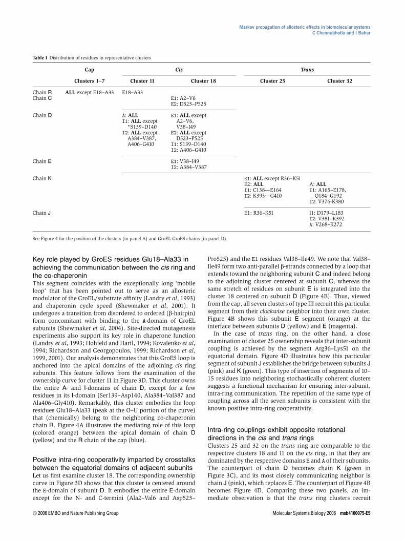
Key role played by GroES residues Glu18–Ala33 inachieving the communication between the cis ring andthe co-chaperoninThis segment coincides with the exceptionally long ‘mobileloop’ that has been pointed out to serve as an allostericmodulator of the GroEL/substrate affinity (Landry et al, 1993)and chaperonin cycle speed (Shewmaker et al, 2001). Itundergoes a transition from disordered to ordered (b-hairpin)form concomitant with binding to the A-domain of GroELsubunits (Shewmaker et al, 2004). Site-directed mutagenesisexperiments also support its key role in chaperone function(Landry et al, 1993; Hohfeld and Hartl, 1994; Kovalenko et al,1994; Richardson and Georgopoulos, 1999; Richardson et al,1999, 2001). Our analysis demonstrates that this GroES loop isanchored into the apical domains of the adjoining cis ringsubunits. This feature follows from the examination of theownership curve for cluster 11 in Figure 3D. This cluster ownsthe entire A- and I-domains of chain D, except for a fewresidues in its I-domain (Ser139–Asp140, Ala384–Val387 andAla406–Gly410). Remarkably, this cluster embodies the loopresidues Glu18–Ala33 (peak at the O–U portion of the curve)that (chemically) belong to the neighboring co-chaperoninchain R. Figure 4A illustrates the mediating role of this loop(colored orange) between the apical domain of chain D(yellow) and the R chain of the cap (blue).
Positive intra-ring cooperativity imparted by crosstalksbetween the equatorial domains of adjacent subunitsLet us first examine cluster 18. The corresponding ownershipcurve in Figure 3D shows that this cluster is centered aroundthe E-domain of subunit D. It embodies the entire E-domainexcept for the N- and C-termini (Ala2–Val6 and Asp523–
Pro525) and the E1 residues Val38–Ile49. We note that Val38–Ile49 form two anti-parallel b-strands connected by a loop thatextends toward the neighboring subunit C and indeed belongto the adjoining cluster centered at subunit C, whereas thesame stretch of residues on subunit E is integrated into thecluster 18 centered on subunit D (Figure 4B). Thus, viewedfrom the cap, all seven clusters of type III recruit this particularsegment from their clockwise neighbor into their own cluster.Figure 4B shows this subunit E segment (orange) at theinterface between subunits D (yellow) and E (magenta).
In the case of trans ring, on the other hand, a closeexamination of cluster 25 ownership reveals that inter-subunitcoupling is achieved by the segment Arg36–Lys51 on theequatorial domain. Figure 4D illustrates how this particularsegment of subunit J establishes the bridge between subunits J(pink) and K (green). This type of insertion of segments of 10–15 residues into neighboring stochastically coherent clusterssuggests a functional mechanism for ensuring inter-subunit,intra-ring communication. The repetition of the same type ofcoupling across all the seven subunits is consistent with theknown positive intra-ring cooperativity.
Intra-ring couplings exhibit opposite rotationaldirections in the cis and trans ringsClusters 25 and 32 on the trans ring are comparable to therespective clusters 18 and 11 on the cis ring, in that they aredominated by the respective domains E and A of their subunits.The counterpart of chain D becomes chain K (green inFigure 3C), and its most closely communicating neighbor ischain J (pink), which replaces E. The counterpart of Figure 4Bbecomes Figure 4D. Comparing these two panels, an im-mediate observation is that the trans ring clusters recruit
Table I Distribution of residues in representative clusters
Cap Cis Trans
Clusters 1–7 Cluster 11 Cluster 18 Cluster 25 Cluster 32
Chain R ALL except E18–A33 E18–A33Chain C E1: A2–V6
E2: D523–P525
Chain D A: ALLI1: ALL except
*S139–D140I2: ALL except
A384–V387,A406–G410
E1: ALL exceptA2–V6,V38–I49
E2: ALL exceptD523–P525
I1: S139–D140I2: A406–G410
Chain E E1: V38–I49I2: A384–V387
Chain K E1: ALL except R36–K51E2: ALLI1: C138—E164I2: K393—G410
A: ALLI1: A165–E178,
Q184–G192I2: V376-K380
Chain J E1: R36–K51 I1: D179–L183I2: V381–K392A: V268–K272
See Figure 4 for the position of the clusters (in panel A) and GroEL-GroES chains (in panel D).
Markov propagation of allosteric effects in biomolecular systemsC Chennubhotla and I Bahar
& 2006 EMBO and Nature Publishing Group Molecular Systems Biology 2006 msb4100075-E5

segments (colored orange) from their counterclockwiseneighbors when viewed from the cap, as opposed to theengagement of clockwise neighbors by cis ring subunits. Wenote that the two rings exhibit comparable contact topologyand communication stochastics, when examined in isolation.However, their particular back-to-back arrangement within thequaternary structure results in opposite angular directions ofcommunication around the cylindrical axis of symmetry. Thisanti-correlation in the communication directions of the tworings is suggestive of an intrinsic, structure-induced tendencyto convey signals in opposite directions, which seemsconsistent with the well-known negative cooperativity be-tween the two rings (Yifrach and Horovitz, 1995).
Coherence of trans ring assisted by inter-subunitcommunication between apical domainsClusters 32 and 11 are both centered on the A-domains of therespective chains K and D. A striking difference between themis, however, the close involvement of an intra-ring neighbor Jin the former, whereas the latter is confined to the single chainD. In other words, a coupling between adjacent subunits ontrans ring is ensured by the apical-domain-centered clusters,
whereas their counterparts in the cis ring do not entail anyintra-ring communication, but rather a coupling to the capchain R. Two segments from I-domains of chain J are recruitedby chain K: Asp179–Leu183 and Val381–Lys392 (Figure 4C).
This type of I–A domain inter-subunit coupling operatingexclusively in trans ring may fulfill a functional requirement,in that the trans ring, when compared to cis, lacks thestabilizing effect of a bound co-chaperonin cap. Among thetwo stretches of residues that achieve this stabilizing effect, wenote, in particular, that Glu386 on chain J forms a salt bridgewith Arg197 on chain K (Figure 4C). This interaction has beenpointed out to be crucially important for interlocking the sevensubunits in their ‘closed’ form assumed in the trans ring; in the‘open’ form of the subunits (i.e. cis ring), on the other hand,these salt bridges are broken in concert with the conforma-tional changes that accommodate the binding of ATP and co-chaperonin GroES (Braig et al, 1994; Yifrach and Horovitz,1998; Ma et al, 2000).
Key structural elements mediating allostericinteractions
The present analysis permits us to identify two classes of keyresidues: those serving as hubs in the individual clusters ata reduced level representation, and those mediating thecommunication between these clusters.
Hubs are distinguished by peaks in the maximal responsi-bility Ri
max distributions, shown by the (identical) gray curvesin Figure 3D, also reproduced in Figure 5A. The hubs identifiedon the cis ring are Ile333–Asp334, Lys321–Val323, Glu214–Ser217. Interestingly, the hubs coincide with b-strands terminior b-hairpins, or helices, suggesting a role for secondarystructures in mediating allosteric communication. Val323 wasalso identified as an important site for allosteric communica-tion in the study of Kass and Horovitz (2002). We note that nohub center is located on the intermediate domains. TheI-domains indeed serve as regions for communication(messengers), rather than collection (hubs), of signals. As tothe trans ring, Arg350–Glu355 in the apical domains andVal128–Glu129 serve as hub centers. Finally, we note that theGroES chains yield very high peaks in the Ri
max distributions(Figure 3D). The corresponding hubs are found to be Glu50–Glu53.
The residues in the second class are shared by multipleclusters, and take on the job of messengers between theseclusters. They are distinguished by their high allostericpotential, that is, their high ‘entropy’ values, Si
(0,L), deducedfrom their stochastic participation in different clusters(equation (5)). Both the red curve in Figure 5A and B andthe color-coded ribbon diagram in panel C (colored from red-to-blue with decreasing entropy) reveal that the E-domains onthe cis ring possess the highest entropies in the GroEL–GroEScomplex. The caps are distinguished by their low entropies.
In terms of the ability to communicate information, thepredicted entropies point to the propensity of the cis ringE-domains to ‘broadcast’ information to all other structuralparts, and in particular to transmit allosteric signals across thering–ring interface. The highest entropy residues presentlyidentified (peaks in Figure 5B) are Glu461–Val464, and Thr30–Lys34 in both cis and trans rings. Strikingly, the mutant E461K
Val381-Lys392
Glu386Arg197
Asp179-Leu183
JC
K
Glu18-Ala33
D
A
Val38-Ile49
Ala384-Val387
D
B
Arg36-Lys51 J
R
KD
Figure 4 A closer view of intra- and inter-subunit couplings at the interfaceof the clusters. Results are shown for the representative chains D (yellow) andE (magenta) on cis ring, J (pink) and K (green) on trans ring and R (blue) onGroES. The chain segments that establish the communication between clustersare colored orange. (A) Residues Glu18–Ala33 in the mobile loop (orange) ofGroES chain R integrated into the cluster centered at the A-domain of chain Destablish the communication between cis ring and co-chaperonin. (B) Positiveintra-ring cooperativity imparted by the coupling of chain E residues, Val38–Ile49and Ala384-Val387 shown in orange, into cluster 18 dominated by chain D.(C) Inter-subunit couplings at trans ring A-domains. Cluster 32 embodies theA-domain of chain K, but also captures a few residues (Val381–Lys392,Asp179–Leu183; shown in orange) from chain J. (D) Cluster 25 is centered onthe E-domain of chain K on trans ring. Note that this cluster engages E1residues Arg36–Lys51 from chain J. Negative cooperativity between the tworings can be compared by comparing panels B and D, both corresponding to theE-domains of the respective cis and trans rings. The residues serving asmessengers (orange) between the subunits belong to either clockwise (panel B)or counterclockwise (panel D) neighbors, as viewed from the cap, that is, the tworings have opposite rotational direction of inter-subunit couplings. The distributionof different chain residues in the examined representative clusters is listed inTable I.
Markov propagation of allosteric effects in biomolecular systemsC Chennubhotla and I Bahar
msb4100075-E6 Molecular Systems Biology 2006 & 2006 EMBO and Nature Publishing Group

has been shown in a recent study by Saibil and co-workers(Sewell et al, 2004) to cause defective folding. This behaviorhas been explained by the fact that the signal induced by ATPbinding to the trans ring could not be transmitted across thering–ring interface, and consequently the cis ring could notrelease its bound GroES. This observation lends support to therole of these highest entropy residues in ensuring thecommunication between the two rings. Finally, we note thatATP-binding residues generally exhibit high entropies, con-sistent with the requirement to propagate signals to distalregions. The filled circles in Figure 5B refer to the amino acidsPro33, Thr90, Gly88, Ile493, Leu31, Gly32, Thr91, Asp495,Gly415 and Asp87 that coordinate ADPs in the examinedstructure.
Proposed path(s) of communication betweennucleotide- and co-chaperonin-binding sites
Among the residues located in the nucleotide-binding site, twohighly conserved residues are distinguished by their largenumber (X15) of atom–atom contacts with ADPs: Thr90 andPro33. We focused on these two residues as initiation loci forsignal transmission, and examined how the allosteric com-munication with the co-chaperonin-binding site is established.The GroES residues Ile25 and Gly24, reported in previous workto be conserved (Stan et al, 2003), were found here to act ascommunication cores (highest Ri
max) within the loop residues.So we explored the maximum likelihood pathways originatingfrom Thr90/Pro33 in the trans ring and ending at GroES-Ile25/Gly24, by using an algorithm similar to the shortest-pathalgorithm of Dijkstra’s (Cormen et al, 1990). In particular, wedefine the distance between two residues vi and vj in thenetwork as �log(mij); so the higher the communicationprobability, the lower the distance between nodes, and thesedistances in turn are taken into consideration in evaluating themaximum-likelihood path.
Figure 6A displays the resulting two maximum likelihoodpathways. The first, labeled (I), runs between Thr90 (subunit
K) and Ile25 (subunit R) following the scheme T90- V94-N97-T101-K105-A109 on subunit K, succeeded byK105-E102-I99-A508-Q505-R501-E409-E408-R404-V174-A370-K371-M193-I332-I220-F219-V240-A241 on subunit D and ending at I25-G24 on the capchain R. The shortest path originating from Pro33 (labeled (II)in Figure 6A), on the other hand, is P33-G32-I454-R452-on chain K succeeded by E461-E460-C458-K34-P33-N153-S154-D155-V158-L161-L187-V189-V190-Q194-F195-R197-G198-Y199-P202-Y203on chain E and I305-G306-K311-E310-M233-L234 onchain D, and finally ending at G23-G24 on the cap chain R.Interestingly, these two paths, both originating from the sameATP-binding region on subunit K in the trans ring evolvethrough different subunits (D and E) on the cis ring to merge atthe same target site on the co-chaperonin mobile loop.
Several residues on these pathways exhibit interestingfeatures: the boldfaced ones are conserved within theHsp60/GroEL family (Fenton et al, 1994; Stan et al, 2003);and those written in italic are distinguished by their highcommunication entropies. We note that the intra-subunitinteraction between residues E409 and R501 has been pointedto be implicated in the allosteric mechanism of GroEL(Aharoni and Horovitz, 1997). We also note that D155 hasbeen pointed out to disrupt the intra-ring cooperativity of theATP-bound ring (Danziger et al, 2003; Horovitz and Willison,2005). Inter-ring cooperativity was observed by Aharoni andHorovitz (1996) to be disrupted in the GroEL double mutant(R13G, A126V), while the double mutant was functional bothin vivo and in vitro (Fenton et al, 1994). These residues couldnot be observed in the presently examined maximum like-lihood pathways.
The on-pathway residues at the interface between the cisand trans rings (A109, K105, R452, E461) should be expectedto play a key role in assisting the communication between thetwo rings. We note among them Glu461 in cis subunit E that isreceiving signal from R452 on trans subunit K. Note thatGlu461 is the residue whose substitution by lysine has been
Cis subunit residues
B E1 I1 I2 Apical E2
Cis ring subunits →← Trans ring subunits
Gro
ES
Allo
ster
ic P
oten
tial
GroEL–GroES residues
A C
0.5
8
6
4
2
0.5
1048 2096 3144 4192 5240 6288 7336
50045040035030025020015010050
1
→←
Figure 5 Regions of high broadcasting ability. (A) Residues acting as hubs lie on the peaks of maximal responsibility curve shown in gray, whereas residues sharedby multiple clusters take on the job of messengers. They have high entropy values (red curve). (B) A detailed representation of the entropy curve for subunit D in thecis ring. Blue circles represent residues making at least two atom–atom contacts with the ADPs in the GroEL–GroES(ADP)7 complex. All amino acids in/near thenucleotide-binding pocket are located in this high entropy region. (C) Entropy values as a color-coded ribbon diagram. Code: red–orange–yellow–green–blue, inthe order of decreasing entropy. Equatorial domains of the cis ring subunits possess the highest entropy values, whereas the cap residues are distinguished by theirlow entropies.
Markov propagation of allosteric effects in biomolecular systemsC Chennubhotla and I Bahar
& 2006 EMBO and Nature Publishing Group Molecular Systems Biology 2006 msb4100075-E7

shown in a recent work by Saibil and co-workers (Sewell et al,2004) to impede the release of the co-chaperonin (bound to thecis ring). This long-range effect has been shown to be owingto the disruption of the inter-ring transmission of the signalfrom ATP-binding site on the trans ring, caused by the of theoriginally out-of-register alignment of subunits across the rings(K, D and E in the present case) into in-register alignment(Sewell et al, 2004). The present analysis demonstrates thatGlu461 (on cis subunit E) is indeed on the most probablepathway, and significantly, it forms an intermolecular salt
bridge (with R452 on trans subunit K) at the interface betweenthe two rings.
Additionally, the above analysis focuses on inter-ringcommunication. Of interest is to assess pathways of intra-ringcommunication as well (Figure 6). We focus in particular onR197, a conserved residue whose mutation to alanine has beenpointed out in previous work to elicit allosteric changes (Whiteet al, 1997). As pointed out above, Arg197 participates in thesmall set of residues (see Table I and Supplementary Table S1(Supplementary information)) that establish the inter-subunitcommunication in the trans ring, by way of forming a saltbridge with Glu386. In order to assess the role of Arg197in ensuring/facilitating the intra-ring communication, weexplored the maximum likelihood pathways originating fromArg197 on trans ring and ending, again, on Arg197 inneighboring subunit in the same ring (an effect that propagatesacross all seven subunits in the ring). In Figure 6B, residueR197 is shown in red on subunits K and J. Calculations wereperformed both for the wild-type protein and the R197Amutant generated in silico. We obtained the path
½A197 ! K277 ! D253 ! V254 ! E255K !½K272 ! V273 ! A274 ! A275 ! V276 ! K277 ! A197J
for the mutant, as opposed to the path
½R197K ! ½E386 ! K390 ! K393 ! E214 ! V324 ! T330 ! R197 J
for the original structure. Here the subscripts refer to thesubunits involved, and conserved residues (Fenton et al,1994) are shown in boldface. Clearly, the wild-type sequencesamples a much shorter pathway. The passage across the twosubunits is readily achieved through the salt bridge R197–E386at the initiation step. In Figure 6B, residue Glu386 is shown inmagenta on subunit J. A cascade of charge–charge interactionsis implicated in this pathway, suggesting an effective signaltransfer mechanism. The most probable path undertaken bythe mutant R197A, on the other hand, involves four additionalresidues in subunit K, before reaching the neighboring subunitJ. In particular, the interaction with the conserved residueK277 seems to favor this longer pathway.
Correlation with collective dynamics: physical roleof hubs and messengers
Significant progress has been made in recent years inimproving our understanding of the collective dynamics ofbiomolecular systems in relation to their biological functionsand interactions, using normal mode analyses with coarse-grained models (Bahar and Rader, 2005; Ma, 2005). The GNM(Bahar et al, 1997; Haliloglu et al, 1997) is such a model widelyresorted to in recent years. Based on statistical mechanicaltheory of macromolecular networks (Flory, 1976), the GNMprovides a rapid assessment of the key mechanical sites (e.g.,hinges) that coordinate global (domain) motions (see e.g. Yangand Bahar (2005) for a systematic analysis of enzymedynamics). The application of the GNM and its extensions toGroEL–GroES (Keskin et al, 2002) has shown that concertedcounter-rotations of the cis and trans rings are the mostprobable collective motions intrinsically favored by the overallarchitecture, in accord with the motions deduced from the
B
K
J
Arg197 Arg197
Glu386
A
I
II
Figure 6 Inter- and intra-ring communication pathways. (A) Two maximumlikelihood pathways (red spheres), labeled I and II, originating from residuesThr90 and Pro33 respectively, on subunit K near the nucleotide-binding site, andending in residue Gly24 on the GroES mobile loop. The ADP molecule nearsubunit D is shown in cyan. (B) Maximum likelihood pathway originating fromArg197 (red) on subunit K and ending in Arg197 (red) on subunit J. The pathwayis shown in yellow, achieved readily through the salt bridge Arg197–Glu386.Residue Glu386 on subunit J is shown in magenta. Also shown in blue is thepathway computed for the mutant R197A. The latter involves four additionalresidues in subunit K.
Markov propagation of allosteric effects in biomolecular systemsC Chennubhotla and I Bahar
msb4100075-E8 Molecular Systems Biology 2006 & 2006 EMBO and Nature Publishing Group

comparison of X-ray or cryo-EM structures determined indifferent states.
The approach taken by the GNM analysis is to deduce thespectrum of normal modes from the eigenvalue decompositionof the Kirchhoff matrix of inter-residue contacts, G, specific tothe examined structure/network topology. A bridge betweensignal propagation dynamics presented here (based oninformation theory, graph theoretic methods and Markovianstochastics) and the GNM dynamics (based on fundamentalconcepts of solid state physics and macromolecular statisticalmechanics) is the relationship
G ¼ D � A ð6Þ
Equation (6) identically holds provided that the nonzerooff-diagonal elements of A and C are defined as aij ¼ Gij ¼ 1 inline with the original GNM, or as aij ¼ Gij ¼ Nij=
ffiffiffiffiffiffiffiffiffiffiffiffiffiðNiNjÞ
pafter the presently defined affinity matrix (equation (1)). Thelatter provides a more detailed description of inter-residueinteractions than the original GNM, as it incorporatesinformation on the density of atom–atom contacts in additionto the information on network connectivity. C is alsoreferred to as the combinatorial Laplacian in graph theory(Chung, 1997).
The equilibrium dynamics of the network is fully defined byC; in particular the mean-square fluctuations of residues arereadily computed from the relation DrT
i Dri ¼ 3kBTg ½G�1ii
where ½G�1ii designates the ith diagonal element of theinverse of G, kB is the Boltzmann factor, T is the temperatureand g is the spring constant that uniformly scales theamplitudes of fluctuations, without affecting their residuedistributions (for a review see Chennubhotla et al, 2005).
We used the presently introduced Markov chain-basedhierarchy to build reduced Kirchhoff matrices, eG; at increas-ingly lower levels of resolution, ranging from c¼8015 nodes atthe full-residue representation to c¼21 nodes at level L¼5,congruent with the eA matrices derived at various hierarchicallevels. We repeated the GNM analysis at successive levels toexplore how well the original (L¼0) fluctuation behavior ispreserved upon mapping the structure to its lower resolutioncounterparts, performing the GNM analysis in the reducedspaces and reconstructing the fluctuation dynamics at theoriginal level (see the scheme in Figure 2). We call this newapproach the hierarchical GNM (see Chennubhotla and Bahar,2006 and Supplementary information for more details). Inaddition to this ‘fidelity’ test, we also explored how thecorrelation between theoretically predicted mean-squarefluctuations of residues and experimental data, X-ray crystal-lographic B-factors in this case, varies by adopting variouslevels of coarse-graining.
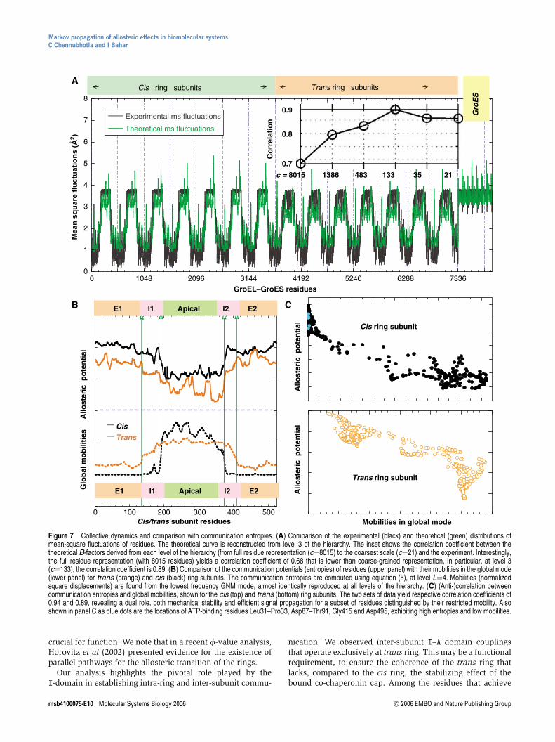
Figure 7A displays the comparison of the fluctuations ofresidues, DrT
i Dri
� �; obtained at various levels of Markov
hierarchy with those indicated by the X-ray crystallographicB-factors (black curve) Bi ¼ ð8p2=3Þ DrT
i Dri
� �reported in the
PDB (Xu et al, 1997). The theoretical fluctuations computedat various levels are hardly distinguishable and are allrepresented by the red curve. As shown in the inset, acorrelation coefficient of 0.86 is achieved between experi-mental and theoretical fluctuation distributions after mappingthe structure of 8015 residues into a network of 21 soft clusters.Thus, the fluctuation profile of residues is accurately predicted
despite a drastic reduction in the complexity of the examinednetwork. Interestingly, a maximum correlation coefficient of0.89 is obtained at an intermediate level of resolution, c¼133,which may be attributed to an optimal elimination of noisein the data.
Next, we examine the role of hubs and messengers in thecontext of the collective dynamics of GroEL/GroES. It is alsoof interest to elucidate the type of involvement of residuesdistinguished here by their high allosteric potentials (maximain entropy curve (red) in Figure 5A) in the most cooperative(global) modes of motion of the entire complex. A comparisonof the entropy profiles of residues in the cis and trans ringsubunits with their mobilities in the global mode of thechaperonin is presented in Figure 7B. The mobility profilesrepresent the distributions of square displacements in thelowest frequency mode, determined by eigenvalue decom-position of eGðLÞ for the entire complex. We note that the globalmodes are also verified to be maintained with a correlationcoefficient of 0.99 at all levels 0pLp5 of the hierarchy. Thehigh mobility of the apical domain delimited at the A–Iinterfaces is revealed in Figure 7B, while the equatorialdomains are rather stable. An immediate observation is theanti-correlation between the two sets of profiles in the top andbottom panels of Figure 7B. This property is more evident inpanel C, where a decrease in allosteric potential is accom-panied by an increase in mobility, in general. Thus, the minimain the global mode profiles, which have been shown innumerous applications to play a critical role (acting as hingesor anchors) in coordinating the functional dynamics ofbiomolecules (Bahar and Rader, 2005; Ma, 2005), act asmessengers in the transmission of allosteric signals. Alsoshown in Figure 7C as blue dots are the locations of ATP-binding residues Leu31–Pro33, Asp87–Thr91, Gly415 andAsp495, exhibiting high entropies and low mobilities.
Conclusions
In the present study, we introduced a new methodology forelucidating the pathways of allosteric communication in largebiomolecular systems. The approach is based on Markovpropagation of information/perturbation over the network ofinteractions that stabilize a given structure. The approachpermits us to simplify the original complex structure as acollection of ‘soft clusters’ at different levels of resolution.Each cluster probabilistically ‘owns’ groups of residuesobeying coherent signal propagation stochastics. Thoseresidues more or less equally ‘shared’ by adjoining clustersare instrumental in transmitting information across clustersand serve as messengers, whereas those almost completelyowned by a given cluster serve as hubs. This descriptiongreatly simplifies our understanding of the dominant mechan-isms and key elements that mediate the communication acrossdistant regions in complex structures.
Using this new framework, we identified two pathways thatare most likely to mediate the communication between theATP- and co-chaperonin-binding sites (Figure 5D), comprisingmany conserved residues as well as those serving as hubs ordetected in previous studies to be critical for chaperoninmachinery. Two alternative mechanisms seem viable inas-much as this coupling between E- and distal A-domains is
Markov propagation of allosteric effects in biomolecular systemsC Chennubhotla and I Bahar
& 2006 EMBO and Nature Publishing Group Molecular Systems Biology 2006 msb4100075-E9
crucial for function. We note that in a recent f-value analysis,Horovitz et al (2002) presented evidence for the existence ofparallel pathways for the allosteric transition of the rings.
Our analysis highlights the pivotal role played by theI-domain in establishing intra-ring and inter-subunit commu-
nication. We observed inter-subunit I–A domain couplingsthat operate exclusively at trans ring. This may be a functionalrequirement, to ensure the coherence of the trans ring thatlacks, compared to the cis ring, the stabilizing effect of thebound co-chaperonin cap. Among the residues that achieve
0
1
2
3
4
5
6
7
8
0 1048 2096 3144 4192 5240 6288 7336
Experimental ms fluctuations
Theoretical ms fluctuations
0.7
0.9
0.8
c = 8015 21351334831386
Co
rrel
atio
n
Cis ring subunits
Gro
ES
Trans ring subunits
GroEL–GroES residues
Mea
n s
qu
are
flu
ctu
atio
ns
(Å2 )
Cis ring subunit
Mobilities in global mode
Trans ring subunit
0 100 200 300 400 500Cis/trans subunit residues
E1 I1 I2 Apical E2
__Cis
__ Trans
E1 I1 I2 Apical E2Glo
bal
mo
bili
ties
A
llost
eric
po
ten
tial
A
B CA
llost
eric
po
ten
tial
A
llost
eric
po
ten
tial
Figure 7 Collective dynamics and comparison with communication entropies. (A) Comparison of the experimental (black) and theoretical (green) distributions ofmean-square fluctuations of residues. The theoretical curve is reconstructed from level 3 of the hierarchy. The inset shows the correlation coefficient between thetheoretical B-factors derived from each level of the hierarchy (from full residue representation (c¼8015) to the coarsest scale (c¼21) and the experiment. Interestingly,the full residue representation (with 8015 residues) yields a correlation coefficient of 0.68 that is lower than coarse-grained representation. In particular, at level 3(c¼133), the correlation coefficient is 0.89. (B) Comparison of the communication potentials (entropies) of residues (upper panel) with their mobilities in the global mode(lower panel) for trans (orange) and cis (black) ring subunits. The communication entropies are computed using equation (5), at level L¼4. Mobilities (normalizedsquare displacements) are found from the lowest frequency GNM mode, almost identically reproduced at all levels of the hierarchy. (C) (Anti-)correlation betweencommunication entropies and global mobilities, shown for the cis (top) and trans (bottom) ring subunits. The two sets of data yield respective correlation coefficients of0.94 and 0.89, revealing a dual role, both mechanical stability and efficient signal propagation for a subset of residues distinguished by their restricted mobility. Alsoshown in panel C as blue dots are the locations of ATP-binding residues Leu31–Pro33, Asp87–Thr91, Gly415 and Asp495, exhibiting high entropies and low mobilities.
Markov propagation of allosteric effects in biomolecular systemsC Chennubhotla and I Bahar
msb4100075-E10 Molecular Systems Biology 2006 & 2006 EMBO and Nature Publishing Group

this stabilizing effect, we note, in particular, Glu386, whichforms an inter-subunit salt bridge with Arg197, confirmingprevious results (see Table I and Supplementary Table S1(Supplementary information)). We also studied the effect ofthe mutation R197A on intra-ring communication. R197 on thetrans ring E-domain exhibits a strong preference to commu-nicate with its salt-bridge-forming partner E386 on the nearestcis ring subunit in the wild-type structure, whereas the mutantR197A generated in silico preferentially interacts with K277and D253 on the same (trans) ring, and reaches the cis ring viaa substantially longer pathway.
A most striking observation is that inter-subunit couplingsoccur in opposite directions in cis and trans rings. Intriguingly,the information flows presently identified within the cis andtrans rings have well-defined preferences for opposite rota-tions (clockwise versus counterclockwise, when viewed fromthe cap). For example, subunit D engages E in the cis ring,whereas K engages J in the trans ring (see Figure 4B and D). Itshould be noted, however, that this cooperativity is differentin character than the one observed within rings. The intra-ringcooperativity is inherently ensured by the association ofneighboring clusters via residues that are being shared,repeated across all seven subunits, consistent with a classicalMWC cooperativity (Monod et al, 1965; Horovitz and Willison,2005). Such an integration of neighboring clusters is notobserved across the interface between the two rings. The lackof a close cluster association at the interface, in contrast to thatoccurring within the rings, suggests that the stochastics ofintra- and inter-ring couplings differ in their character andtimescale, reminiscent of MWC type (intra-ring) interactionsnested in KNF-like (Koshland et al, 1966) inter-ring inter-actions (Horovitz and Willison, 2005).
The present analysis takes account of residue specificitiesin an approximate way (through the numbers of atom–atomcontacts for each residue pair) and does not distinguishbetween strong or weak interactions. No electrostatic andsolvent effects are included either. Despite the neglect of thesephysical features, the method offers a plausible model forinformation propagation between distinct functionally rele-vant sites of GroEL–GroES. The property rigorously taken intoconsideration here is the intricate topology of inter-residuecontacts and local packing/interaction densities in the nativestate. The present analysis thus lends support to the dominantrole of inter-residue contact topology in defining cooperativeinteraction mechanisms/pathways on a global scale, in accordwith recent computational studies based on coarse-grainedmodels (Bahar and Rader, 2005).
Identification of hubs and messengers raises implicationsfor protein design. In the network communication paradigm,the equatorial domains of the cis ring emerge unambiguouslyas the regions with the highest propensity for ‘broadcasting’the perturbations occurring in their domain. In particular, theE-domain residues Ser463 and Glu461 emerge as the aminoacids with the highest allosteric potential (see equation (5);Figure 5). The critical role of Glu461 in establishing thenegative inter-ring cooperativity is now established by therecent work of Saibil and co-workers (Sewell et al, 2004),whereas that of Ser463 remains to be experimentallyconfirmed. The high broadcasting ability does not necessarilyimply efficient transmission to a given target because the signal
can be dissipated in many directions. Yet, the observedinvolvement of messengers in maximum likelihood pathwaysmay have other functional implications, for example, inducingother co-operative effects such as processing the substrateprotein. We also examined the role of hubs and messengers inthe context of collective dynamics of GroEL/GroES deducedfrom normal mode analysis. A significant property thatemerges is that the minima in global mode profiles, whichhave been shown to play a critical role in coordinatingfunctional dynamics of biomolecules, act as messengers in thetransmission of allosteric signals. Automated identification ofsuch residues serving as hubs and messengers in a givenarchitecture, as proposed and illustrated here for GroEL–GroES, may help in efficient design and modification of proteinstructure and function.
Materials and methods
Deriving stationary distribution in the reducedmodel
We begin by expressing the stationary distribution p¼[p1,p2,y,pn] asa probabilistic mixture of latent distributions,
p ¼ Kd ð7Þwhere d ¼ ½d1; d2; � � � ; dc is an unknown stationary distribution in areduced (c-dimensional) representation of the structure, K ¼ fKijg isan n c non-negative kernel matrix with elements Kij and columns Kj
being latent probability distributions that each sum to 1 and c � n.The kernel matrix acts as an expansion operator, mapping the low-dimensional distribution d to a high-dimensional distribution p (seeFigure 2). The high-dimensional distributions are referred to as high-resolution, or low-level, descriptions, and the low-dimensionaldistributions, as low-resolution, or high-level, descriptions.
We derive a maximum likelihood approximation for d using anEM type algorithm (McLachlan and Basford, 1988). To this aim, weminimize the Kullback–Liebler distance measure (Kullback, 1959)between the two probability distributions p and Kd, given by
L ¼ �Xn
i¼1
pi lnXc
j¼1
Kijdj þX
i
pi ln pi ð8Þ
A maximum likelihood estimate is obtained by minimizing
E ¼ �Xn
i¼1
pi lnXc
j¼1
Kijdj þ lXc
j¼1
dj � 1
!ð9Þ
subject to the constraintPc
j¼1 dj ¼ 1, ensured by the Lagrangemultiplier l. Setting the derivative of E with respect to dj to be zero,we obtain Xn
i¼1
piKijdjPck¼1 Kikdk
¼ ldj ð10Þ
The contribution made by kernel j to a node i (or to its stationaryprobability pi) is given by Kij (or by the product Kijdj), and hence we candefine an ownership of node i in the high-resolution representation bya node j in the low resolution as
Rij ¼KijdjPc
k¼1 Kikdkð11Þ
We note that the mapping between the two resolutions is notdeterministic, but probabilistic in the sense that
Pcj¼1 Rij ¼ 1:
Using this relation, and the equalitiesPc
j¼1 dj ¼ 1 andPn
i¼1 pi ¼ 1;summing over j in equation (10) gives l¼1. This further leads to thestationary distribution d at the coarse scale
dj ¼Xn
i¼1
piRij ð12Þ
Markov propagation of allosteric effects in biomolecular systemsC Chennubhotla and I Bahar
& 2006 EMBO and Nature Publishing Group Molecular Systems Biology 2006 msb4100075-E11

Following Bayes theorem, Kij can be related to the updated d values as
Kij ¼Rijpi
djð13Þ
In summary, the operators K and R and stationary distribution d arecomputed using the following EM type procedure: (1) select an initialestimate for K and d (see Supplementary information for details);(2) E-step: compute ownership maps R using equation (11); (3) M-step:estimate d and update K using equations (12) and (13) respectively;and finally, (4) repeat E- and M-steps until convergence.
Transition and affinity matrices at reduced levels
The Markov chain propagation at the reduced representation obeys theequation qkþ1 ¼ eMqk; where qk is the coarse scale m-dimensionalprobability distribution after k steps of the random walk. We expand qk
into the fine scale using pk ¼ Kqk; and reduce pk back to the coarsescale by using the ownership value Ri,j as in qkþ1
j ¼Pn
i¼1 pki Ri;j:
Substituting equation (11) for ownerships, followed by the expressionfor pk, in the equation for qkþ1
j ; we geteM ¼ diag ðdÞKT diag ðKdÞ�1K ð14ÞUsing the definition of eM; and the corresponding stationary
distribution d, we generate a symmetric affinity matrix eA that describesthe node–node interaction strength in the low-resolution network
eA ¼ eM diag ðdÞ ð15ÞTo summarize, we use the stationary distribution p and Markovtransition matrix M at the fine scale to derive a transition matrix eM; andits stationary distribution d, at the coarse scale. It is obvious that thisprocedure can be repeated recursively to build a hierarchy of lowerresolution network models.
Application to GroEL–GroES: interactions/affinities at different hierarchical levels
We examined the Markov process of inter-residue communication inthe structure GroEL–GroES(ADP)7, crystallographically characterizedby Sigler and co-workers (Xu et al, 1997), also known as the R0 0 stateof the bacterial chaperonin (Thirumalai and Lorimer, 2001). First, theinter-residue affinity matrix A based on all atom–atom contacts (a totalof E106 interactions, based on an interaction range of 4.5 A betweenall non-bonded heavy atoms) is constructed, which is then used inequation (2) to derive the fine-scale Markov transition matrix M.
The kernel selection algorithm (Supplementary information)applied to Mb (b¼4) yields 1316 (reduced level 1) kernels. Using thesekernels as an initialization, a recursive application of the EM procedurederives stationary distributions d (equation (12)), updated expansionmatrices K (equation (13)), reduced level probability transitionmatrices eM (equation (14)) and the corresponding residue interactionmatrices eA (equation (15)). The respective dimensions of eA turn out tobe 483 (level 2) , 133 (level 3), 35 (level 4) and 21 (level 5). We note thatthe individual subunits of the GroEL–GroES are distinguished by theirstrong intra-subunit interactions, and a number of inter-subunitcontacts are maintained at all levels, which presumably establish thecommunication across the protein at all levels. The dimension of thereduced model, c, is automatically defined during the kernel selectionat each level of the hierarchy. The method thus avoids the arbitrarychoices of sampling density and interaction cutoff distances atdifferent hierarchical levels.
Supplementary information
Supplementary information is available at the Molecular SystemsBiology website (www.nature.com/msb).
AcknowledgementsSupport from NIH Grant #R33 GM068400-01A2 is gratefullyacknowledged.
References
Aharoni A, Horovitz A (1996) Inter-ring communication is disrupted inthe GroEL mutant Arg13-Gly; Ala126-Val with known crystalstructure. J Mol Biol 258: 732–735
Aharoni A, Horovitz A (1997) Detection of changes in pairwiseinteractions during allosteric transitions: coupling between localand global conformational changes in GroEL. Proc Natl Acad Sci 94:1698–1702
Atilgan AR, Durell SR, Jernigan RL, Demirel MC, Keskin O, Bahar I(2001) Anisotropy of fluctuation dynamics of proteins with anelastic network model. Biophys J 80: 505
Bahar I, Rader AJ (2005) Coarse-grained normal mode analysis instructural biology. Curr Opin Struct Biol 15: 1–7
Bahar I, Erman B, Monnerie L (1994) Effect of molecular structures onlocal chain dynamics: analytical approaches and computationalmethods. Adv Polym Sci 116: 151–206
Bahar I, Atilgan AR, Erman B (1997) Direct evaluation of thermalfluctuations in protein using a single parameter harmonic potential.Folding Des 2: 173–181
Berman HM, Westbrook J, Feng Z, Gilliland G, Bhat TN, Weissig H,Shindyalov IN, Bourne PE (2000) The Protein Data Bank. NucleicAcids Res 28: 235–242
Braig K, Otwinowski Z, Hegde R, Boisvert D, Joachimiak A, HorwichAL, Sigler PB (1994) The crystal structure of the bacterialchaperonin at 2.8a. Nature 371: 578–586
Changeux JP, Edelstein SJ (2005) Allosteric mechanisms of signaltransduction. Science 308: 1424–1428
Chennubhotla C, Bahar I (2006) Markov models for hierarchicalcoarse-graining of large protein dynamics. Lecture Notes inComputer Science 3909: 379–393
Chennubhotla C, Jepson A (2005) Hierarchical eigensolver fortransition matrices in spectral methods. In LK Saul, Y Weiss &L Bottou (eds) Adv. in Neural Information Processing Systems 17,273–280. Cambridge, MA: MIT Press
Chennubhotla C, Rader AJ, Yang LW, Bahar I (2005) Elastic networkmodels for understanding biomolecular machinery: from enzymesto supramolecular assemblies. Phys Biol 2: S173–S180
Chung FRK (1997) Spectral Graph Theory, CBMS Lectures. AM. Math.Soc., Providence, RL, USA
Coifman RR, Lafon S, Lee AB, Maggioni M, Nadler B, Warner F, ZuckerSW (2005) Geometric diffusion as a tool for harmonic analysisand structure definition of data, part I:. Proc Natl Acad Sci USA102: 21
Cormen TH, Leiserson CE, Rivest RL (1990) Introduction toAlgorithms. Cambridge, MA, USA: MIT Press
Danziger O, Rivenzon-Segal D, Wolf S, Horovitz A (2003) Conversionof the allosteric transition of GroEL from concerted to sequential bythe single mutation. Proc Natl Acad Sci USA 100: 13797–13802
de Groot BL, Vriend G, Berendsen HJC (1999) Conformational changesin the chaperonin GroEL: new insights into the allostericmechanism. J Mol Biol 286: 1241–1249
Doruker P, Atilgan AR, Bahar I (2000) Dynamics of proteins predictedby molecular dynamics simulations and analytical approaches:application to a-amylase inhibitor. Proteins 40: 512–524
Fenton W, Kashi Y, Furtak K, Horwich A (1994) Residues in chaperoninGroEL required for polypeptide binding and release. Nature 371:614–619
Flory PJ (1976) Statistical thermodynamics of random networks. ProcRoy Soc London A 351: 351–378
Haliloglu T, Bahar I, Erman B (1997) Gaussian dynamics of foldedproteins. Phys Rev Lett 79: 3090–3093
Hinsen K (1998) Analysis of domain motions by approximate normalmode calculations. Proteins 33: 417
Hohfeld J, Hartl FU (1994) Role of the chaperonin cofactor hsp10 inprotein folding and sorting in yeast mitochondria. J Cell Biol 126:305–315
Horovitz A, Willison KR (2005) Allosteric regulation of chaperonins.Curr Opin Struct Biol 15: 1–6
Markov propagation of allosteric effects in biomolecular systemsC Chennubhotla and I Bahar
msb4100075-E12 Molecular Systems Biology 2006 & 2006 EMBO and Nature Publishing Group

Horovitz A, Amir A, Danziger O, Kafri G (2002) f value analysis ofheterogeneity in pathways of allosteric transitions: evidence forparallel pathways of ATP-induced conformational changes inGroEL ring. Proc Natl Acad Sci USA 99: 14095–14097
Kass I, Horovitz A (2002) Mapping pathways of allostericcommunication in GroEL by analysis of correlated mutations.Proteins 48: 611–617
Keskin O, Bahar I, Flatow D, Covell DG, Jernigan RL (2002) Molecularmechanisms of chaperonin GroEL–GroES function. Biochemistry41: 491–501
Koshland DE, Nemethy G, Filmer D (1966) Comparison ofexperimental binding data and theoretical models in proteinscontaining subunits. Biochemistry 5: 365–385
Kovalenko O, Yifrach O, Horovitz A (1994) Residue lysine-34 inGroES modulates allosteric transitions in GroEL. Biochemistry 33:14974–14978
Kullback S (1959) Information Theory and Statistics. NY: DoverPublications
Landry SJ, Zeilstra-Ryalls J, Fayet O, Georgopoulos C, Gierasch LM(1993) Characterization of a functionally important mobile domainof GroES. Nature 364: 255–258
Lockless SW, Ranganathan R (1999) Evolutionarily conservedpathways of energetic connectivity in protein families. Science286: 295–299
Ma J (2005) Usefulness and limitations of normal mode analysis inmodeling dynamics of biomolecular complexes. Curr Opin StructBiol 13: 373–380
Ma J, Karplus M (1998) The allosteric mechanism of the chaperoninGroEL: a dynamic analysis. Proc Natl Acad Sci USA 95: 8502–8507
Ma J, Sigler PB, Xu ZH, Karplus M (2000) A dynamic model for theallosteric mechanism of GroEL. J Mol Biol 302: 303–313
McLachlan GJ, Basford KE (1988) Mixture Models: Inference andApplications to Clustering. Marcel Dekker, New York
Ming D, Wall ME (2005) Quantifying allosteric effects in proteins.Proteins Struct Funct Bioinformatics 59: 697–707
Monod J, Wyman J, Changeux JP (1965) Allosteric proteins andcellular control systems. J Mol Biol 12: 88–118
Nadler B, Lafon S, Coifman RR, Kevrekidis IG (2006) Diffusion maps,spectral clustering, and the reaction coordinates of dynamicalsystems. J Appl Comp Harmonic Anal (in press)
Norris JR (1997) Markov Chain. Cambridge: Cambridge UniversityPress
Richardson A, Georgopoulos C (1999) Genetic analysis of thebacteriophage t4-encoded cochaperonin gp31. Genetics 152:1449–1457
Richardson A, van der Vies SM, Keppel F, Taher A, Landry SJ,Georgopoulos C (1999) Compensatory changes in GroEL/gp31affinity as a mechanism for allele-specific genetic interaction. J BiolChem 274: 52–58
Richardson A, Schwager F, Landry SJ, Georgopoulos C (2001) Theimportance of a mobile loop in regulating chaperonin/co-chaperonin interaction: humans versus Escherichia coli. J BiolChem 276: 4981–4987
Saibil HR, Ranson NR (2002) The chaperonin folding machine. TrendsBiochem Sci 627–632
Sewell BT, Best RB, Chen S, Roseman AM, Farr GW, Horwich AL, SaibilHR (2004) A mutant chaperonin with rearranged inter-ringelectrostatic contacts and temperature-sensitive disassociation.Nat Struct Mol Biol 11: 1128–1133
Shewmaker F, Maskos K, Simmerling C, Landry S (2001) Thedisordered mobile loop of GroES folds into a defined beta-hairpinupon binding GroEL. J Biol Chem 276: 31257–31264
Shewmaker F, Kerner MJ, Hayer-Hartl M, Klein G, Georgopoulos C,Landry SJ (2004) A mobile loop order–disorder transitionmodulates the speed of chaperoin cycling. Protein Sci 13:2139–2148
Sigler PB, Xu ZH, Rye HS, Burston WA, Fenton SG, Horwich AL (1998)Structure and function in GroEL-mediated protein folding. AnnuRev Biochem 67: 581–608
Stan G, Thirumalai D, Lorimer G, Brooks B (2003) Annealing functionof GroEL: structural and bioinformatic analysis. Biophys Chem 100:453–467
Suel GM, Lockless SW, Wall MA, Ranganathan R (2003) Evolutionarilyconserved networks of residues mediate allosteric communicationin proteins. Nat Struct Biol 10: 59–68
Thirumalai D, Lorimer GH (2001) Chaperonin-mediated proteinfolding. Annu Rev Biophys Biomol Struct 30: 245–269
Tobi D, Bahar I (2005) Structural changes involved in protein bindingcorrelate with intrinsic motions of proteins in the unbound state.Proc Natl Acad Sci USA 102: 18908–18913
White HE, Chen S, Roseman AM, Yifrach O, Horovitz A, Saibil HR(1997) Structural basis of allosteric changes in the GroEL mutantArg197-Ala. Nat Struct Mol Biol 4: 690–694
Xu C, Tobi D, Bahar I (2003) Allosteric changes in protein structurecomputed by a simple mechanical model: hemoglobin t-r2transition. J Mol Biol 333: 153–168
Xu ZH, Horwich AL, Sigler PB (1997) The crystal structure of theasymmetric GroEL–GroES(ADP)7 chaperonin complex. Nature388: 741–750
Yang LW, Bahar I (2005) Coupling between catalytic site and collectivedynamics: a requirement for mechanochemical activity ofenzymes. Structure 13: 893–904
Yifrach O, Horovitz A (1995) Nested cooperativity in the ATPaseactivity of the oligomeric chaperonin GroEL. Biochemistry 34:5303–5308
Yifrach O, Horovitz A (1998) Mapping the transition state of theallosteric pathway of GroEL by protein engineering. J Am Chem Soc120: 13262–13263
Markov propagation of allosteric effects in biomolecular systemsC Chennubhotla and I Bahar
& 2006 EMBO and Nature Publishing Group Molecular Systems Biology 2006 msb4100075-E13