marisa de campos santana - ufrgs
TRANSCRIPT

Universidade Federal Do Rio Grande Do Sul Instituto De Biociência
Programa De Pós Graduação Em Botânica
Thesis for Degree of Doctor in
Joint Supervision
Contribution to the Taxonomy and Phylogeny of Phellinus sensu lato
(Hymenochaetaceae, Basidiomycota) in Southern Brazil
Marisa de Campos Santana
Porto Alegre, 2014


Marisa de Campos Santana
Contribution to the Taxonomy and Phylogeny of Phellinus
sensu lato (Hymenochaetaceae, Basidiomycota) in Southern
Brazil
Thesis submitted to the Graduate Program in the
Universidade Federal do Rio Grande do Sul (UFRGS),
as a partial fulfillment of the requirements for the
degree of Doctor of Science: Botany.
Orientadora: Profa. Dra. Rosa Mara Borges da Silveira
Co-orientador: Dr. Gerardo Lucio Robledo
Segundo orientador: Profº. Dr. Stephan Declerck
Porto Alegre, 2014


Université catholique de Louvain Faculté d’Ingénierie Biologique, Agronomique et
Environnementale
Contribution to the Taxonomy and Phylogeny of Phellinus sensu lato
(Hymenochaetaceae, Basidiomycota) in Southern Brazil
Par
Marisa DE CAMPOS SANTANA
Soutenance le 29 septembre 2014 devant le juri d´examen:
Promoteurs Profa. Dra. Rosa Mara Borges da Silveira
Profº. Stephan Declerck
Juri d´examen: Dr. Cony Decock
Profª. Dra. Patrícia Valente da Silva
Profª. Dra. Tatiana Teixeira Souza Chies
Profº. Gilberto Coelho
Dr. Gabriel Castillo Cabello
Dr. Jérôme Degreef
Thèse présentée en vue de l’obtention en cotutelle du grade de doctour en sciences
agronomiques et ingénierie biologique et de docteur en sciences botaniques.


DEDICATE:DEDICATE:DEDICATE:DEDICATE:
TO MY MOTHER CARMELITA R. DE CAMPOS
AND
IN THE LOVELY MEMORY OF MY BROTHER: ITAMAR DE CAMPOS SANTANA (TITO).


ix
ACKNOWLEDGMENTSACKNOWLEDGMENTSACKNOWLEDGMENTSACKNOWLEDGMENTS
I am very especially grateful to my mother, Carmelita Ribeiro de Campos and my
father, Venceslau Santana for unconditional love, understanding and affection, for support
received along these years of study, for trusting in me, for being always at my side and for
the incentive so I could proceed in this journey.
Prof. Dra. Rosa Mara Borges da Silveira for accepting to advise me in this project
and to believe in it, for all infrastructures placed at my disposal;
Dr. Gerardo Lucio Robledo, for spending much of his time to teach me a little of his
large knowledge on Hymenochaetaceae, especially during my stay in Córdoba/Argentina.
Thank you especially for friendship, the shared “mates”, and the discussions about
taxonomy and phylogeny of the group and about life.
Prof Stephan Declerck, for accepting me during the year 2012 in his laboratory
(Earth and Life Institute - Microbiology –ELIM/UCL), for supporting my work and accepting
the international convention for the joint supervision of thesis.
I also thank Dr. Cony Decock, for contagious passion for the Fungi Kingdom, for
discussions about hymenochaetoid, for the teachings about the fascinating world of fungi,
mycology and science in general. For receiving me during the sandwich internship, for
providing infrastructure of laboratories and resources, for sharing, in several moments, his
office with me, besides the strict supervision of taxonomic and phylogenetic approach.
More especially, I am grateful for the privilege of his friendship and for all the shared
moments, which helped me a lot on my adaptation period in Louvain-la-Neuve-Belgium.
The words are not enough to express my gratefulness…Merci beaucoup mon ami!
Being a lucky person, I had an extra “advisor”. All pages in this work would not
suffice to thank the help given by Drª. Clarice Loguercio-Leite, my friend and master
degree advisor. I do not forget her eternal teachings, precious advices and invaluable trust.
In personal terms, I am still grateful for all optimism words, affection and understanding.
For all this and much more, thank you!
Drª Tatiana Chies, Drª Patrícia Valente, Dr. Gilberto Coelho, Dr. Gabriel Castillo and
Dr. Jérôme Degreef, who, as members of qualification examination board, contributed
with important and enriching suggestions;
x
I would like to thank the staff of the Mycothèque of the UCL (BCCM/MUCL):
Stéphanie Huet and Celine Bivort, who teached me molecular biology thecniques, and the
other technician, who contributed in some way for this work accomplishment.
I am grateful to Mário Amalfi, who is also co-author of some publications, for all the
adives and help in phylogenetic analysis.
I express my gratefulness to all teachers and employees from “Departamento de
Botânica da UFRGS”, in particular to the team of ICN herbarium from UFRGS, especially
Camila Carneiro, Márcia Pinheiro and Mateus de Oliveira, for availability, agility and
competence.
The curators and employees of other herbaria, who gently loaned their collections
for this study.
The Brazilian people, through research fomentation agencies: CNPq (GD
149756/2010-0), for PhD scholarship, CAPES for sandwich internship scholarship (PDSE,
process 8296/11-1) and UFRGS for financial support which enable this project execution.
The colleagues of “Laboratório de Micologia, UFRGS”, especially my friends: Paula
Santos, Mauro Westphalen, Mateus Arduvino Reck, Alice Gerlach, Natália Tedy, Emerson
Luiz Gumboski and Eduardo Soares Rossetto for fellowship in field works, for friendship
and acquaintanceship during these years.
I am also grateful to my friends from close and from far away:
““““......We recognize t..We recognize t..We recognize t..We recognize true friends in the most difficult moments...”rue friends in the most difficult moments...”rue friends in the most difficult moments...”rue friends in the most difficult moments...”
To all my Brazilian friends, especially Alice Gerlach, my dear friend and sister from the
heart, for true friendship, for the several shared moments, for constant availability, for the
straight, carrying, affection, Chaplin sections, fellowship in laboratory at Saturdays, for this
helpful work revising the text. Be sure that you are very special; I will take you with me
always!
Paula Santos, for friendship, affection, for contagious peace, for the prompt and
indispensable help translating and revising English, for companionship in several moments.
I have no words to express my gratefulness!

xi
Guilherme A. Roesler, for being so present in my life, for understanding, incentive,
help, for mild reprimands, stimulus and teachings; but mainly, for believing in my potential,
in my dreams and projects... for sharing the joy of my achievements.
To my family LLN: Cinthya I. Becerra, Diana Rh, Pamela, Marychuy, Yurelkys
Fernandez, Diogo Buarque Franzosi, , , , Ugo Sangiorgi, , , , Vivian Genaro Motti, , , , Koen Buyens,
Prudence, Madia and other Latin-American and European friends: who received me,
helped me, and with who I learned so much. Muchas gracias, muito obrigada, merci
beaucoup!
To “cordobéses”, friends from Córdoba-Argentina, who gently received me and
cheered my stay in Argentina, especially Luciana Hernández (Luli), Laura S. Dominguez and
Noelia Cofre.
Muito obrigada, muchas gracias, merci beaucoup, to everyone that directly or no,
contributed to this work accomplishment.

xii
CONTENTS
LIST OF FIGURES...........................................................................................................xiv
LIST OF TABLES..............................................................................................................xv
INDEX OF SPECIES IDENTIFIED ...............................................................................xvi
PUBLICATIONS.............................................................................................................xviii
GLOSSARY AND ABBREVIATIONS...........................................................................xix
RESUME .............................................................................................................................xx
RESUMO...........................................................................................................................xxii
1. INTRODUCTION ...........................................................................................................1
1.1. General Introduction...............................................................................................1
1.2. Micodiversity - Estimated total fungal number......................................................3
1.3. Taxonomy of the Fungi Kingdom: Nomenclature and Classification....................5
1.4. Ecological and economic importance of the group ................................................8
1.5. Defining species in Fungi .......................................................................................9
1.6. Knowledge on Phellinus Quélet – a historic summary ........................................12
1.7. Taxonomic diversity of Phellinus in Brazil..........................................................15
2. OBJECTIVES.................................................................................................................21
2.1. General objective..................................................................................................21
2.2. Specific objectives................................................................................................21
3. MATERIAL AND METHODS.....................................................................................22
3.1. Area of study ........................................................................................................22
3.2. Field works ...........................................................................................................23
3.2.1. Sampling and material preservation .........................................................25
3.3. Morphological characterization............................................................................25
3.3.1. Macromorphological analysis...................................................................25
3.3.2. Micromorphological analysis ...................................................................26
3.4. Mycological terms, scientific names and herbarium acronyms ...........................27
3.5. Revision of herbaria and type studies...................................................................27
3.6. Identification.........................................................................................................28

xiii
3.7. Isolation and culture .............................................................................................28
3.7.1. Culture medium ........................................................................................28
3.7.2. Obtainment of polisporic cultures ............................................................28
3.8. Phylogenetic studies based on molecular data .....................................................29
3.8.1. DNA extraction ........................................................................................29
3.8.2. DNA concentration in samples.................................................................30
3.8.3. Amplification............................................................................................30
3.8.4. Utilization of agarose gel electrophoresis system for verification of PCR’s
......................................................................................................... ...................31
3.8.5. Sequencing ...............................................................................................32
3.8.6. Edition and sequence alignment ...............................................................32
3.8.7. Phylogenetic analysis ...............................................................................33
Chapter I ............................................................................................................................34
Chapter II............................................................................................................................84
Chapter III ..........................................................................................................................88
Chapter IV ........................................................................................................................111
4. GENERAL DISCUSSION...........................................................................................139
5. CONCLUSIONS...........................................................................................................142
6. PERPECTIVES. ...........................................................................................................144
7. REFERENCES. ............................................................................................................145

xiv
LIST OF FIGURES
Figure 01 - Atlantic Forest remnants, showing the current situation of the Biome. Source:
Fundação SOS Mata Atlântica and INPE (2013). ................................................................03
Figure 02 - Phylum of Fungi Kingdom and the approximate number of described species
in each group (from Blackwell, 2011; Kirk et al., 2008). ....................................................05
Figure 03 - Phylogeny and classification of Fungi. Agaricomycotina. Source: Hibbett et al.
(2007). .................................................................................................................................07
Figure 04 - Koppen-Geiger climate type of South America. Source: Peel et al. (2007).....22
Figure 05 - Vegetal formations from Brazilian South Region. Source: Iganci et al. 2011
(modified). ...........................................................................................................................23
Figure 06 - Visualization of PCR products from regions ITS, LSU, tef1-α and rpb2
inultraviolet light. .................................................................................................................32

xv
LIST OF TABLES
Table 01 - Deforestation of the Atlantic Forest from 2011-2013: Fundação SOS Mata
Atlântica and INPE (2013). ..................................................................................................02
Table 02. - Phellinus s.l. species from Southen Brazil. .......................................................18
Table 03. - Localities where collections were performed and and number of specimens
collected................................................................................................................................23

xvi
INDEX OF SPECIES IDENTIFIED
Aurificaria luteoumbrina. .....................................................................................................38
Cyclomyces iodinus ..............................................................................................................39
Fomitiporella umbrinella .....................................................................................................40
Fomitiporia apiahyna ...........................................................................................................41
Fomitiporia bambusarum .....................................................................................................42
Fomitiporia dryophila. .........................................................................................................43
Fomitiporia neotropica ........................................................................................................44
Fomitiporia sp. (F. robusta complex) ..................................................................................45
Fulvifomes durissimus ..........................................................................................................47
Fulvifomes fastuosus ............................................................................................................47
Fulvifomes melleoporus........................................................................................................48
Fulvifomes membranaceus ...................................................................................................48
Fulvifomes merrillii. .............................................................................................................49
Fulvifomes rhytiphloeus .......................................................................................................49
Fulvifomes rimosus...............................................................................................................50
Fuscoporia contigua.............................................................................................................51
Fuscoporia ferrea .................................................................................................................52
Fuscoporia gilva var. gilva ..................................................................................................54
Fuscoporia gilva var. licnoide..............................................................................................55
Fuscoporia gilva var. scruposa ............................................................................................56
Fuscoporia palmicola...........................................................................................................56
Fuscoporia rhabarbarina .....................................................................................................57
Fuscoporia wahlbergii .........................................................................................................58
Inonotus linteus ....................................................................................................................60
Inonotus micantissimus ........................................................................................................60
Inonotus patouillardii ...........................................................................................................61
Inonotus portoricensis ..........................................................................................................62
Inonotus pseudoglomeratus ..................................................................................................62
Inonotus splitgerberi ............................................................................................................63
Inonotus tropicalis................................................................................................................63
Inonotus sp............................................................................................................................64

xvii
Phellinus anchietanus...........................................................................................................65
Phellinus calcitratus .............................................................................................................66
Phellinus caryophylleus........................................................................................................67
Phellinus detonsus ................................................................................................................67
Phellinus sp. (P. gabonensis morpho-ecological complex) ................................................68
Phellinus lopezii ...................................................................................................................69
Phellinus shaferi ...................................................................................................................69
Phylloporia aff. spathulata (P. spathulata morpho-ecological complex)............................70
Phelinus amazonicus ............................................................................................................99
Phylloporia subchrysita......................................................................................................126
Phylloporia turbinata .........................................................................................................128
Phylloporia neopectinata ...................................................................................................131
Phylloporia loguerciae ......................................................................................................132

xviii
PUBLICATIONS
This thesis is based on the work presented in the following papers and manuscripts.
The papers are appended to the thesis, and are referred to by capital roman numbers in the
text.
I - Manuscript .....................................................................................................................34
Campos-Santana M, Decock C, Robledo G, Silveira R.M. 2014.
A Synopsis of the poroid Hymenochaetaceae (Agaricomycetes, Basidiomyceta) in
Southern Brazil.
Cryptogamie Mycologie: submitted
II - Manuscript....................................................................................................................84
Campos-Santana M, Amalfi M, Robledo G, Silveira R.M., Decock C.
Fomitiporia neotropica, a new species from South America evidenced by multilocus
phylogenetic analyses.
Mycological Progress: Mycological Progress 13:601-615
III - Manuscript ..................................................................................................................88
Campos-Santana M, Amalfi M, Silveira R.M., Decock C. 2014.
Multilocus DNA-based phylogenetic analysis reveal two new species lineages in the
P. gabonensis / P. caribaeo-quercicolus species complex, of which Phellinus
amazonicus sp. nov.
Fungal Diversity: to be submitted
IV - Manuscript ................................................................................................................111
Campos-Santana M, Amalfi M, Silveira R.M., Robledo G, Decock C. 2014.
Morphological, DNA sequences, and ecological data evidence four undescribed
Phylloporia species from Southern Brazil.
Mycologia: to be submitted

xix
GLOSSARY AND ABBREVIATIONS
AIC – Akaike Information Criterion
Amyloid – (of spores, etc.), stained blue by iodine; cf. dextrinoid.
Basidiome – (pl.-ata), a basidium-producing organ: basidiocarp; carpophore; fruitbody;
hymenophore; sporophore.
BI – Bayesian Inference.
Cyanophilous – (of spores, etc), readily absorbing a blue stain such as cotton blue.
Dextrinoid – of spores, etc.), stained yellowish-brown or reddish brown by Melzer’s
iodine.
Dimitic – having hyphae of two kinds (generative and skeletalhyphae which are thick-
walled, aseptate).
EPS – exopolysaccharides
Hypha – (pl. hyphae), one of the filaments of a mycelium.
INPE – National Institute for Space Research in Brazil
ML – Maximum Likelihood
Monomitic – Having hyphae of one kind (generative hyphae which are branched, septate,
with or without clamp connexions, thin to thick-walled, and of unlimited length.
MP – Maximum Parsimony
MEA – Malt Extract Agar
Pileus – The hymenium-supporting part of the basidioma of non-resupinate
Agaricomycetes.
s.l – From Latin, sensu lato (in a broad sense).
s.s. – From Latin, sensu strito (in a narrow sense).
UF – Federal unit/ State of Brazil
Xanthochroique – All structures become dark in 2% KOH.
≡ – Basionym
Seta – (pl. -ae) (Lat., a bristle), (1) a stiff hair, generally.

xx
RESUME
Contribution à la Taxonomie et Phylogénie de Phellinus sensu lato
(Hymenochaetaceae, Basidiomycota) dans le Sud du Brésil
Phellinus s.l. a été créé par Quélet en 1886. Il comprend actuellement 180 espèces,
soit près de la moitié du nombre total d'espèces d'Hymenochaetaceae. Les caractères
généralement considérés pour la definition du genre incluent des basidiomes à réaction
xanthochroique positive permanente, une trame des tubes jaune à brun, un système
d'hyphes dimitique pour l'essentiel avec des hyphes génératives à septa simples (i.e
absence de boucles), des hyphes squelettiques brunâtres, et des éléments de type sétoïdes
au niveau de l'hyménium ou plus rarement de la trame. Au niveau de leur physiologie (ou
de leur biologie nutritionnelle), les Phellinus s.l. sont lignivores, possédant un système
enzymatique capable de dégrader préférentiellement la lignine, et également partiellement
la cellulose et l'hémicellulose, des composés de la paroi cellulaire du bois, produisant à
terme une pourriture blanche. Ils se développent tant sur bois vivant (et sont des parasites
parfois économiquement importants) ou sur bois mort. Les Phellinus sont généralement les
champignons lignivores les plus divers et les mieux représentés dans les forêts tropicales.
Ils participent ainsi activement au maintien des écosystèmes forestiers. Des études
récentes, de morphologie fine et le développement d'approches complémentaires, non-
morphologiques, de type biochimique d'abord, génomique ensuite, ont toutefois démontré
que la conception généralement admise par la majorité des auteurs modernes (Larsen and
Cobb-Poulle 1990, Ryvarden, 1991; Fischer, 1996; Góes-Neto et al, 2001) est largement
hétérogène et polyphylétique. En conséquence, de nombreux groupes morphologiques plus
cohérents et monophylétiques ont émergés (ou ré-émergés) et ont été reconnus comme des
genres satellites indépendants plus ou moins larges selon les cas. Certains de ces genres
avaient été reconnus par des auteurs anciens, et ont pu être exhumés de la litérature; pour
d'autre groupes des genres ont dus être crées. Citons par ex. les Fomitiporia, Fomitiporella,
Fulvifomes, Fuscoporia, Porodaedalea, …. Afin d'élargir les connaissances de ces
champignons dans la Région Sud du Brésil, une étude taxonomique a été réalisée à partir
de révisions d'herbier et de l'analyse des spécimens collectés en 2010, 2011 et 2013, dans
les trois États du sud du Brésil. Les données de la littérature ont également été rassemblées
avec l'objectif de fournir un cadre des connaissances actuelles du groupe dans la région

xxi
étudiée. Plus de 600 spécimens d'Hymenochaetaceae poroïdes ont été analysés. Quarante-
quatre espèces, distribuées dans neuf genres, ont été identifiées. De ces espèces et
d'espèces mentionnées dans la littérature, 26 sont connus pour Paraná, 25 pour Santa
Catarina et 35 pour Rio Grande do Sul. Fomitiporia neotropica, Phellinus amazonicus,
Phylloporia subchrysita, Phylloporia neopectinata, Phylloporia turbinata et Phylloporia
loguerciae sont décrites comme nouvelle pour la science. Neuf espèces sont cités pour la
première fois au Paraná, 10 pour Santa Catarina, 16 pour Rio Grande do Sul, 14 pour la
Région Sud du Brésil, neuf sont mentionnés pour la première fois au Brésil et une pour
l’Amérique du Sud. En plus, deux nouvelles combinaisons sont proposées, Fomitiporia
bambusarum et Fulvifomes rhytiphloeus. Les descriptions et illustrations des structures
microscopiques et les photos sont fournies pour les nouvelles espèces. Également, les clés
sont fournies pour l'identification des genres et des espèces connues pour la région d'étude.
Mots clés: Diversité, taxonomie, relations phylogénétiques, Hymenochaetaceae

xxii
RESUMO
Contribuição à Taxonomia e Filogenia de Phellinus sensu lato (Hymenochaetaceae,
Basidiomycota) na Região Sul do Brasil
Phellinus s.l. foi criado por Quélet em 1886 e compreende atualmente 180 espécies,
quase a metade do número total das espécies de Hymenochaetaceae. As características
consideradas para definir o gênero incluem basidiomas com reação xantocroica positiva e
permanente, superfície dos poros amarela a marrom, sistema hifal dimítico essencialmente
com hifas generativas com septo simples (ou seja, sem fíbulas), hifas esqueletais castanhas
e elementos do tipo setoides, no himênio ou, raramente, na trama. Em sua fisiologia (ou
biologia nutricional), as espécies de Phellinus s.l. são lignocelulolíticas ou xilófilas,
possuem um sistema enzimático capaz de degradar a lignina da madeira, causando
podridão branca. Desenvolvem-se tanto na madeira viva (e, às vezes, são parasitas de
grande importância econômica) ou madeira morta, participando assim ativamente na
manutenção dos ecossistemas florestais. Estas espécies são geralmente os fungos xilófilos
mais diversos e com maior representatividade nas florestas tropicais. Estudos recentes,
enfatizando a morfologia do grupo e o desenvolvimento de abordagens complementares,
não morfológicas, bioquímicas e genéticas, têm mostrado que este gênero é amplamente
heterogêneo e polifilético (Larsen and Cobb-Poulle 1990; Ryvarden, 1991; Fischer, 1996;
Góes-Neto et al., 2001). Em consequência, muitos grupos morfológicos mais coerentes e
monofiléticos emergiram (ou reemergiram), e foram reconhecidos como gêneros satélites
independentes. Alguns destes gêneros tinham sido reconhecidos por autores anteriores e
foram exumados da literatura para que outros gêneros fossem devidamente criados, como,
por exemplo, os gêneros Fomitiporia, Fulvifomes, Fuscoporia, Fomitiporia,
Porodaedalea, … Com objetivo de ampliar o conhecimento sobre esses organismos na
Região Sul do Brazil, um estudo taxonômico foi conduzido a partir de revisões de alguns
herbários e análises de espécimes coletados entre os anos de 2010 e 2013, nos três Estados
da Região Sul do Brazil. Dados da literatura também foram compilados com o objetivo de
fornecer um quadro do conhecimento atual sobre o grupo estudado. Foram examinadas
mais de 600 coletas de Hymenochaetaceae poroides, onde foram reconhecidas 44 espécies,
pertencentes a nove gêneros. Dessas espécies e das espécies citadas na literatura, 26 são
conhecidas para o estado do Paraná, 25 para Santa Catarina e 35 para o Rio Grande do Sul.

xxiii
Fomitiporia neotropica, Phellinus amazonicus, Phylloporia subchrysita, Phylloporia
neopectinata, Phylloporia turbinata e Phylloporia loguerciae são propostas como espécies
novas. Nove espécies são citadas pela primeira vez para o Paraná, dez para Santa Catarina,
16 para o Rio Grande do Sul, 14 para a Região Sul do Brasil, nove são citadas pela
primeira vez para o Brasil e uma para a América do Sul. Além disso, são propostas duas
novas combinações, Fomitiporia bambusarum e Fulvifomes rhytiphloeus. Descrições,
ilustrações das estruturas microscópicas e fotos são fornecidas para as novas espécies.
Além disso, são fornecidas chaves para a identificação dos gêneros e das espécies
conhecidos para a área de estudo.
Palavras chave: Diversidade, taxonomia, relações filogenéticas, Hymenochaetaceae

1
1. INTRODUCTION
1.1. General Introduction
Systematics is at the foundation of all biological knowledge. Since Darwin’s
Theory of Evolution until ecology and biogeography, all biological studies depend on
knowledge and quantification of the object of study. Until the present days, Linnaeus’
classic taxonomy, which is dedicated to describe and inventory organisms, so that they can
be named and classified in a general reference system (Amorim 2002; Judd et al. 2009), is
fundamental for Biology studies.
“…Taxonomy is often undervalued as a glorified form of filing—
with each species in its folder, like a stamp in its prescribed place
in an album; but taxonomy is a fundamental and dynamic science,
dedicated to exploring the causes of relationships and similarities
among organisms. Classifications are theories about the basis of
natural order, not dull catalogues compiled only to avoid chaos…”
Stephen Jay Gould, Wonderful Life (1989).
Therefore, identification, classification, nomenclature, in other words,
systematics, is the science for the knowledge of biological diversity. Biodiversity, many
times, is represented as the number of species that live in a particular place. It may also
design genetic diversity among and inside populations of one species in particular, or
diversity of communities in which these species are living and interacting.
Biodiversity is essential to maintain a dynamic balance of elements and climate
on Earth. It is our main source of food, medicines, but also it has an intrinsic value that
constitutes nature beauty, source of inspiration and pleasure. We are surrounded by
biodiversity and we are part of it; in that sense, it is our ethical responsability to protect it.
The current biodiversity on Earth is the result of 3.5 billions of years of evolution. After
mass extinctions during various geological times, a significant recovery of biodiversity
always took many millions of years to happen, so nothing allows us to think that the event
currently ongoing will have a different issue. It is necessary to point out that species rise,
prosper and fade naturally; however, we are facing a current extinction rate 100 to 200

2
times higher than what would be expected without human interference (Convention on
Biological Diversity - CDB 2000).
The concept of biodiversity has evolved inside a context of human activities,
magnified by population growth; ecosystems experience degradation increasingly fast and
generalized. Certain ecosystems, such as forests (for instance, the Atlantic Forest in Brazil
– Table 1) and the species once inhabiting them, are disappearing at an accelerated rate.
TABLE 1. Deforestation of the Atlantic Forest from 2011-2013. Source: Fundação SOS
Mata Atlântica and INPE (2013).
*In the second column: MG (Minas Gerais); PI (Piauí); BA (Bahia); PR (Paraná); SC
(Santa Catarina); MS (Mato Grosso do Sul); PE (Pernambuco); RS (Rio Grande do Sul);
SE (Sergipe); RN (Rio Grande do Norte); SP (São Paulo); GO (Goiás); AL (Alagoas); ES
(Espírito Santo); RJ (Rio de Janeiro); CE (Ceará); PB (Paraíba).
In Brazil, the Atlantic Rain Forest, once a global biodiversity hotspot (Fundação
SOS Mata Atlântica 2013) included originally an area equivalent to 1,315,469 km2. Today,
remants of forest above 100 hectares cover about 8.5% of the original forest areas (Figure
1). Overall, only 12.5% of the original forest remains, consisting mostly of rather small
surface, above 3 hectares.

3
Fig. 1. Atlantic Forest remnants, showing the current situation of the Biome. Source:
Fundação SOS Mata Atlântica and INPE (2013).
Facing such data situation, it is urgent to have a broader knowledge about its
biodiversity, which needs to be protected. The two sides of Systematics: taxonomy and
phylogenetic, provide, in this way, a larger comprehension about biodiversity elements,
which is necessary for an effective management, for conservation and sustainable use of
biodiversity.
In this context, the present thesis has as main purpose to increase the knowledge
of a poorly investigated component of the biological diversity that is the Mycota. More
specifically, we will study more deeply the diversity of the wood-decaying Phellinus sensu
lato (Hymenochaetaceae) in Southern Brazil, besides to enlarge its global knowledge,
through a systematic study (with a morphological and phylogenetic approach), in order to
establish taxa circumscription as well their relationships.
1.2. Mycodiversity - Estimated total fungal numbers

4
The Kingdom Fungi presents an astonishing diversity (Burfort et al. 2003; Schmit
and Mueller 2007), holding one of the largest lineage among eukaryotes, since it’s
ancestral until derivative forms (Blackwell 2011). Despite the increasing number of fungal
studies, it is still difficult to estimate the real mycodiversity, which hampers phylogenetic,
ecological and biogeographical characterization of the group. Bass and Richards (2011)
emphasized that the main difficulties we faced in the attempt of estimate the global fungal
richness is the uncertainty regarding the number of described species, the incomplete
inventories, the high level of morphological conservatism of species and the lack of
knowledge about ecology and geographical distribution of fungal species.
The most "popular" mycodiversity estimates, despite being conservative
(Blackwell 2011, Hawksworth 2001), was presented by Hawksworth (1991), who
proposed the existence of 1.5 millions of fungal species. This hypothesis is based in a ratio
fungi/plants of 6:1 estimated for the temperate regions. The same author in 2004 calculated
that about 100,000 fungal species (only 7% of estimated number) have been described in
the whole world and that approximately 1,200 additional species are described yearly.
Hawksworth (1991) also highlighted that, for a good estimate about mycodiversity, some
data such as geographical distribution, endemism rate and host specificity should be
considered.
Recent works state that ratio of fungi to plants would be approximately 10.5:1,
hence, increasing even more the estimate of unknown species (Blackwell 2011).
Hawksworth (2012) considering taxonomic studies in tropical regions and recent
phylogenetic studies, revised also his estimate, and proposed a range of 1.5 to 3 millions of
fungal species. Many phylogenetic studies have demonstrated the existence of more
"cryptic" species than would be expected and as a consequence, have shown that the
number of fungal species might be much higher (Bass and Richards 2011).
According to Kirk et al. (2008), the difference between the number of described
and estimated fungal species (Figure 2) is enormous. The authors also commented that
51% of the 16,013 species registered at Index of Fungi between the years of 1981 and 1999
were not originating from tropical regions. When, fungal surveys are intensive and
prolonged, this percentage increase in the tropics, where the percentage of undescribed
species would range between 60 to 85%, depending on group and habitats.

5
Therefore, based on the estimates of the overall fungal species and the current
rate of species description, Hawksworth (2001) Mueller and Schmit (2007) and Blackwell
(2011) calculated that about 1,000 – 1,200 years of taxonomic studies would be still
necessary to achieve the complete knowledge of fungal diversity.
Fig. 2. Phylum of Kingdom Fungi and the approximate number of described species in
each group (from Blackwell 2011; Kirk et al. 2008).
Reinforcing such statement, Schmit and Mueller (2007) noticed that additional
works, especially those with samples originating from tropical regions, as well as rigorous
studies in order to establish circumscription and species distribution, are crucial to improve
the knowledge about fungal diversity.
1.3. Taxonomy of the Kingdom Fungi: Nomenclature and Classification
“…it is probably of great historical significance that Darwin
himself expressed the thought that the possibility of arranging
organisms in a hierarchic system is explainable only by assuming
a phylogenetic relationship among them…”
Hennig W. (1966). Phyllogenetic Systematics. p.20.

6
The poor knowledge about fungal diversity reflects on the taxonomic proposal of
the kingdom. According to Hibbett et al. (2007), R. T. Moore only conducted the
validation of the name Fungi according to the International Code of Nomenclature for
Algae, Fungi, and Plants, including the Latin diagnosis in 1980. Fifteen years later,
Hawksworth et al. (1995) elevated the basidiomycetes to the rank of Division
(Basidiomycota). The situation has changed with Kirk et al. (2001); the Class
Basidiomycetes divided in Subclasses Tremellomycetidae Locq. and Agaricomycetidae
Parmasto, published respectively in 1984 and 1986 (David 2002), were accepted. These
names were based on the genera Tremella Pers. and Agaricus L., in observance to article
7.1 from International Code of Botanical Nomenclature for Algae, Fungi and Plants -
Melbourne Code (McNeill 2012).
Along with the other groups, Basidiomycetes still present numerous taxonomic
problems, position inside the classification system. Recently, the AFTOL project –
Assembling the Fungal Tree of Life – (Lutzoni et al. 2004), launched an international team
by mycologists that has been working on molecular phylogenetic, morphological and
reproductive biology studies. These works have changed substantially the classification of
the Mycota. In this way, macroscopic basidiomycetes (for example, mushrooms, bracket
fungi and polypores), have been placed at Subphylum Agaricomycotina Doweld and three
classes are accepted to include the different orders (Figure 3): Auriculariales J. Schröt,
Corticiales K. H. Larss, Gloeophyllales Thorn, Polyporales Gäum., Thelephorales Oberw.,
Hymenochaetales Oberw., inside class Agaricomycetes (Binder et al. 2005; Hibbett at al.
2007). Thereby, fungi containing pores (poliporoid) belong to the Agaricomycetes, in
which they are distributed in several (still) polyphyletic orders, such as Polyporales Gäum,
Russulales Kreisel ex P.M. Kirk, P.F. Cannon & J.C. David and Hymenochaetales Oberw.,
quantitatively the three most important.

7
Fig. 3. Phylogeny and classification of Fungi. Agaricomycotina. Source: Hibbett et al.
(2007).
The Hymenochaetales were proposed by Oberwinkler in 1977. Macro- and
micromorphological characteristics of this order are exceedingly variable. It includes taxa
with different types of hymenophore (corticoid, hydnoid or poroid) and basidiomata
(resupinate, pileate or stipitate), all causing wood white rot (though Coltricia / Coltriciella
potentially also are symbiotic cf. 1.4), and whose main characters are xantochroic reaction,
the simple-septate in generative hyphae and the frequent occurrence of setae. However,
Parmasto and Parmasto in 1979 indicated that a xanthochroic reaction is not specific for
Hymenochaetales. Hymenochaetales was later extended to include Schizoporaceae Jülich
(that differs from Hymenochaetaceae Donk among other thaits in having basidiomata
without xanthocroic reaction, and hyaline, variably clamped hyphae).
Larson et al. (2006) performed a molecular phylogeny for the Hymenochaetales
supported the idea that the occurrence of dolipores with continuous parenthesomes and the
possibility that this structure is a synapomorphy for Hymenochaetales have gained
considerable interest.
Currently, the order comprises 48 genera and 610 species (Kirk et al. 2008).
8
Hymenochaetaceae, in the current delimitation (Ryvarden 2004), encompasses a
group of white rotting fungi, whose basidiomata present permanent positive xantochromic
reaction, yellow to brown tubes trama, generative hyphae with simple septum (absence of
clamps), monomitic or dimitic hyphal system, and often occurring setoid structures (setae).
The generic circumscription in Hymenochaetaceae sensu strito is under constant
change (Corner 1991). For instance, Phellinus, which comprises almost half of the total
number of species of Hymenochaetaceae, as currently understood is more likely
polyphyletic. Many "old" Phellinus species forming species complexes (Larsen and Cobb-
poule 1990; Ryvarden 1991; Fischer 1996; Góes-Neto et al. 2001) were transferred to
other genera. Phellinus is then reducing significatively and will result in fine in a
morphologically homogeneous and phylogenetically monophyletic entity.
1.4. Ecological and economic importance of the group
Living as saprobes, parasites or symbionts, and being primarily decomposers, the
fungi play fundamental roles in all ecosystems (Schmit and Mueller 2007).
Regarding their ability to decompose the wood components, the polyporoid
basidiomycetes can be classified into two groups; the first group degrades mainly /
primarily cellulose and hemicellulose, leaving much lignin residuals, are called brown rot
fungi; the second group has the ability to degrade lignin and some cellulose, hemicellulose,
leaving much of the cellulose fibers, and are called white rot fungi. Species of
Hymenochaetaceae are mainly lignocellulolytic, able to secrete enzymes that degrade the
components of vegetal cellular wall (cellulose, hemicellulose and lignin), obtaining the
required nutrients to their development (Akhtar et al. 1997; Carlile et al. 2001; Jeong et al.
2005; Larsson et al. 2006).
Because most species of hymenochetoid fungi are wood-decayers, they may be at
the origin of economic losses when parasiting tree species of economic value or degrading
wood used in constructions and artifacts, (Gilbertson and Ryvaden 1986).
The cellulolytic enzymes produced by wood decaying fungi have been
intensively studied, in order to being used ex-situ such as for instance in the bioremediation
of industrial pollutants. A relevant aspect to be considered, regarding several species of
Hymenochaetaceae, is their potential use in bioremediation of soils contaminated by toxic

9
industrial residues (Balan and Monteiro 2001; Novotný et al. 2001; Larsson et al. 2006). A
good example of fungi used in bioremediation is Phellinus pseudopunctatus A. David and
Fuscoporia gilva (Schwein.) T. Wagner & M. Fisch. (Balan and Monteiro 2001; Novotný
et al. 2001).
On the other hand, being used since antiquity as food or in popular medicine, the
fungi have today important role in drugs production. As from their secondary metabolites,
they have revealed themselves as promising sources for obtainment of new components
with antiviral, antifungic, antibiotic, antioxidant, antidiabetic properties, among others
(Wang et al. 2004; Wang et al. 2013). Several fungal species belonging to Phellinus s.l.,
for example P. igniarius (L.) Quél., P. hartigii (Allesch. & Schnabl) Pat., P. pini (Brot.) A.
Ames, P. linteus (Berk. & M.A. Curt.) Teng, P. baumii Pilát, Fuscoporia gilva, among
others, produce larger amounts of exopolysaccharides (EPS), pharmacologically important
due their remarkable biological activities, such as antitumor activity and elimination of free
radicals (Song et al. 1995; Han 1999; Jeong et al. 2005).
1.5. Defining species in Fungi
“... It is really laughable to see what different ideas are prominent
in various naturalists' minds when they speak of species; in some,
resemblance is everything and descent of little weight in some,
resemblance seems to go for nothing, and Creation the reigning
idea. In some, descent is the key, in some, sterility an unfailing
test, with others it is not worth a farthing. It all comes, I believe,
from trying to define the indefinable...” Darwin (1887), p. 88.
Letter from Darwin to Hooker, 24 December 1856.
Species concept, as most biologists understand today, root down to the 17th and
18th centuries, with the publication of Ray [1686, Historia Plantarum: Species Hactenus
Editas Aliasque Insuper Multas Noviter Inventas and Descriptas Complectens], in which
he considered that the adaptations of organisms were evidence of God's benevolence, and
later on with work by Buffon [1749, Histoire Naturelle, Génerale et Particulière, avec la
Déscription du Cabinet du Roi].

10
Darwin, in the 19th century, believed that the varieties were the first stage of
speciation. As for the creationists, he considered the species were real entities created by
the Creator, and varieties were local and temporary products of the nature. Thus, Darwin's
theory transformed the species in an arbitrary invention of imagination of the taxonomists
(Bowler 1989).
“...Species are groups of actually or potentially interbreeding
natural populations, which are reproductively isolated from other
such populations...” Mayr (1942).
According to Mayr (2001), the species is the primary unit of evolution and it is
impossible to talk about evolution or about any aspect of biology, without having a clear
understanding of the meaning of species.
Accurate species delimitations are important to understand factors driving
diversification, and have implications for ecological and conservation studies (de Queiroz
2005). Controversies often occur because of the existence of different views towards the
definition of species (Mayr 2001). Biologists, especially systematists, have been debated
on species concepts for a very long time. The debate itself has had many nuances. Some
systematists have only been interested in discriminating all the discrete, current taxonomic
variations without any concern about the processes that might have produced these
variations. Although their propensity to describe species has been belittled by some,
without their efforts we would know far less about the diversity of the natural world
(Cracraft 2000).
Species concepts have been reviewed many times in the past. In a review by
Mayden (1997), 22 species concepts were listed from taxonomic literature. According to
Hull (1997), Perkins (2000) and Richards (2010) these concepts can be arranged in three
broad classes:
- Similarity between organisms (dealing with the phenotypes, morphology or
physiology, behavior – e.g. occupation of distinct niches);
- Involving evolutionary processes (biological species – possibility of mating,
reproductive isolation – evolutionary species);
- Phylogenetic or lineage based concepts.

11
All these concepts are usefull to delimit species, but also contain certain
limitations in regards to a unified species concept. For example, the traditional concept
stating that a species is defined by interbreeding individuals is inapplicable for asexual
species.
The application of the species concept of within the Mycota presents several
difficulties, because little is known about the magnitude of intrapopulational variability,
the life cycles are varied and complex and the reproduction, in addition of being extremely
complex, can affect evolutionary patterns in ways we do not yet understand (Petersen and
Hughes 1999).
Until the middle of the 20th century, the Morphological Species Concept (MSC)
was the basis for fungal classification. According to Wiens and Servedio (2000), although
there is currently considerable interest in the use of DNA sequences to infer the limit a
certain fungal species, most of them are still defined based on the presence of diagnostic
morphological characters, and in comparison with herbarium specimens. Thus, from a
strictly morphological point of view, a species in the Mycota is a group of organisms
congruent in the macro- and micro-morphological characteristics of their reproductive
structures.
Taylor et al. (2000) noted that with few exceptions, nearly 100.000 described
fungi were diagnosed by morphological or other phenotypic traits (e.g. growth at different
temperatures, production of secondary metabolites or the presence of pigments). The main
advantage of MSC is that it has been widely used in the Mycota, and comparisons can be
made between taxa. However, Hawksworth et al. (1995) argued that the disadvantage of
MSC is that frequently the species recognized in this way comprise more than one species
when diagnosed through other methods.
As far as Fungi are concerned, classical morphological characters were repeatedly
shown to be insufficient to separate species; additional parameters have to be considered,
including molecular phylogenetics, also known as molecular systematic, or ecological data,
to reach in fine a more global species concept. Appropriate taxonomy of a group of
organisms should reflect its phylogenetic relationships, based on correct inferences of its
evolutionary history.
Quaedvlieg et al. (2014) reported that the Biological Species Concept (BSC)
emphasizes reproductive isolation (Wright 1940; Mayr 1942); the Morphological Species

12
Concept (MSC) emphasizes morphological divergence; the Ecological Species Concept
(ESC) emphasizes adaptation to a particular ecological niche (van Valen 1976); the
Phylogenetic Species Concept (PSC) emphasizes nucleotide (non) divergence (Hennig
1966); the Genealogical Concordance Phylogenetic Species Recognition (GCPSR) (an
adaptation of the PSC) uses the phylogenetic concordance of unlinked genes to indicate a
lack of genetic exchange and thus, evolutionary independence of lineages (Taylor et al.
2000; de Queiroz 2007). Quaedvlieg et al. (2014) combined multiple complementary
descriptors that would result in a “Consolidated Species Concept” (CSC).
With the introduction of phylogenetic studies, DNA-based the existence of
cryptic, morphologically closely related species (Bickford et al. 2007) challenged the
traditional concept of fungal species. Within the Basidiomycota, Hymenochaetaceae is
one of the exemple of such shift in defining species (and generic) concepts (Decock et al.
2005; Decock et al. 2006; Amalfi et al. 2010; Campos-Santana et al. 2014). Several cases
indicated that the diversity of species has been underestimated with traditional taxonomic
characters and several new taxa have been recognized, in correlation with overlooked
morphological (Baltazar et al. 2009; Baltazar et al. 2010; Raymundo et al. 2013;
Valenzuela et al. 2013) or biogeographical (Decock and Stalpers 2006; Decock et al. 2007)
characters.
In the genera Fomitiporia, Phellinus, Phylloporia..., the delimitation of species
based on morphological characters seems to be relativety well supported by phylogenetic
studies (e.g. Decock et al. 2006; Amalfi el al. 2010; Valenzuela et al. 2011).
It is possible that there will be an ideal and unique concept of species. However,
the criteria for recognizing species can still be different in different groups. According to
Hey (1997), to have significance as "species" does not simplify the process of specimen
identification. Instead, this job has proven as difficult as measuring the genetic drift and,
then, decide if the data contains evidence of partitions or not.
1.6. Knowledge on Phellinus Quélet – a historic summary
Phellinus s.l. is a large, diverse group containing various cryptic generic entities
and species. Numerous morphological, anatomical, biological (e.g. nuclear behavior),
biochemical (pigmentation composition) and ecological characters, pointed to the

13
heterogeneity of the genus (Dai 1995/ 1999; Fischer 1996; Wagner and Fischer 2002).
Since the genus was created by Quélet in 1886, distinct taxonomic arrangements have been
proposed in the attempt to solve problems of its delimitation.
According to Larsen and Cobb-Poulle (1990), Quélet (1886) described Phellinus
to accommodate polyporoid, and pileate wood-inhabiting fungi species with, brown annual
and perennial forms basidiomata. In addition Larsen and Cobb-Poulle (1990) included the
following characters to describe the genus: resupinate to pileate basidiomata, the pileus
sessile to stiptitate, annual to perennial, solitary to gregarious, often imbricate, corky to
ligneous; upper surface pubescent to tomentose or finally glabrous, frequently with a thin
black cuticle, usually concentrically grooved and radially cracked, rusty to dark brown;
margin curved, obtuse to thin, sterile; hymenophore rusty brown to dull brown; 2-11 pores
for millimeter; stratified tube layers in perennial forms and occasionally in annual forms;
context thin to thick, zoned to no zoned, fibrous to ligneous, rusty brown to dull brown,
often with intercalated black lines and generally with a thin upper black cuticle forming a
crust; setal hyphae absent or present in trama, context or hymenium; hyphal system
dimitic, trimitic or monomitic, clamps absent; skeletal hyphae rarely branched, usually
thick walled and without septae, but frequently with adventitious septae; generative hyphae
usually septate and thin walled, branched; binding hyphae rare, asseptate, thick walled;
basidia hyaline, globose to clavate, bearing 2-4 sterigmata; basidiospores globose,
subglobose, ellipsoid, cylindrical or oboclavate, hyaline to pigmented, yellow to brown,
thin to thick walled, sometimes dextrinoid to cyanophilous in cotton blue, never amyloid.
All structures become dark in 2% KOH.
Fiasson and Niemelä (1984) emphasized the difficulty to define Phellinus,
because of the numerous characters shared with other genera. Ryvarden and Gilbertson
(1994) recognized the genus with perennial basidiomata and a dimitic hyphal system.
Wagner and Fischer (2001) questionned much of these features. The differentiation of
generative to skeletal hyphae is not always as clear as it is in other polypores and the
hyphal system may be clearly monomitic. Besides, several species inside the group are
cited as annuals (Wagner and Fischer 2002).
More recently, Ryvarden (2004) presented a more restrictive description of the
genus: perennial basidiomata, resupinate to pileate, solitary or imbricate with decurrent
pore layers; pileus if present, yellow, rusty brown and gray to black, tomentose, hispid,
14
glabrous or deeply cracked. Pore surface brownish. Hyphal system dimitic, generative
hyphae hyaline and with simple septum, skeletal hyphae yellow to rusty brown, most thick
walled and larger than generative; hymenial and tramal setae present or absent; setoid
hyphae absent or present in the margin, context or hymenium; basidiospores globose to
cylindrical, smooth, hyaline to rusty brown; cosmopolitan genus.
Looking back at the history of Phellinus, the importance of Murrill’s works
(1907) has to be noticed. Murrill (1907) proposed several new genera such as Fuscoporia,
Fomitiporia or Fomitiporella, among others, without considering Phellinus. Despite of the
disagreement of various authors, who considered of weak taxonomic value the generic
concepts developped by Murrill, recent works evidenced that some morphologically
homogeneous and monophyletic groups of species were worth recognized at generic level,
for which some of the genera described by Murrill could be used (Decock et al. 2005,
2007; Fiasson and Niemelä 1984; Bondártseva et al. 1992; Fischer 1996; Dai 1999;
Wagner and Fischer 2001, 2002).
Larsen and Cobb-Poule, in 1990, provided a compilation of morphological data
for all taxa described in Phellinus sensu Ryvarden and Gilbertson (1987), besides
proposing synonyms, host relationships and geographical distribution. This resulted in the
recognition of 154 species and 67 forms and varieties.
Another attempt to solve the taxonomic problems of the group was undertaken
by Bondártseva et al. (1992). These authors combined morphological and biochemical
data (chromatographic analysis of stirilpirones) of 35 Phellinus species from Cuba and
proposed the transfer of 18 of them into various genera including Fomitiporia, Fulvifomes,
Fuscoporia and Phellinidium. According to the authors (op. cit.), the denomination of
Phellinus must be conserved, not only for those species which present features shared with
the type species (Polyporus igniarius L.:Fr.), but also as a "temporary" repositary for those
species that did not find an appropriate place inside limits of other genera.
Few years later, Fischer (1996) analyzing 23 Phellinus species inside well-
known species complexes concluded that only P. robustus and associated species (P.
robustus complex) appeared cleared delimited on the basis of nuclear behavior, DNA
contents and sexuality. Accordingly, the author proposed that the species of this complex,
namely P. hartigii, P. robustus and P. punctatus should be transferred into Fomitiporia.
Another important contribution, using classical methods, was made by Dai in 1999, with

15
67 species of Phellinus from East Asia, transferring some of them to other more
homogeneous genera, such as Cyclomyces, Fomitiporia, Phellinidium and Pyrrhoderma.
However, since the remnants species still formed a heterogeneous group, the author
suggested also treating some established species complexes already as subgenera instead
of genera, waiting to gather more date for the still vast number of poorly known tropical
species. In the work performed by Wagner and Fischer (2001), the authors reviewed 42
European species of Hymenochaetales; their results allowed recognizing five genera:
Phellinus, Fomitiporia, Fuscoporia, Phellinidium and Porodaedalea.
Afterwards Wagner and Fischer (2002), in a molecular study containing 99 species
of Hymenochaetales from Europe, Asia, Oceania, Central and North America, reiterated
the polyphyly of Phellinus. According to these authors (op. cit.), several clades were
resolved representing natural groups, which are in accordance with most taxonomic
concepts previously showed, based in European species (Fiasson and Niemelä 1984,
Wagner and Fischer 2001). More recently, Jeong et al. (2005) concluded that more taxa
should be studied, in order to create a complete and reliable conclusion about phylogeny
of Hymenochaetales, and consequently of Phellinus.
1.7. Taxonomic diversity of Phellinus in Brazil
Brazilian mycology only obtained expression by sporadic works published in
other countries, resulting from contribution of Europeans naturalists in their expeditions in
South America (Fidalgo 1962). Most of these studies were summerized in the form of
checklists based on literature and / or revision of herbarium specimens, such as those
published for the Amazon region (Gomes-Silva and Gibertoni 2009a), the Cerrado biome
(Gibertoni and Drechsler-Santos 2010), Mangrove (Baltazar et al. 2009), Atlantic Rain
Forest (Baltazar and Gibertoni 2009) and Semiarid areas (Drechsler-Santos et al. 2009). In
the Midwest region only three works were published, one with field studies in the Cerrado
ecosystem (Sampaio 1916), another with the compilation of material collected in areas of
the Amazon and Cerrado domains (M. Fidalgo 1968) and more recently an inventory of
species that occur in the Pantanal Rio Negro (Bononi et al. 2008). In the northeast,
taxonomic studies were developed in several States (Torrend 1940; Batista and Bezerra
1960; Maia 1960; Kimbrough et al. 1995; Maia et al. 1996, 2002; Góes-Neto 1999) and

16
both areas in the Atlantic Forest Domain (Cavalcanti 1976; Góes-Neto et al. 2000, 2003;
Maia and Gibertoni 2002; Gibertoni and Cavalcanti 2003; Gibertoni et al. 2003, 2004a, b,
c, 2007; Silva and Gibertoni 2006), as in semiarid (Góes-Neto 1996; Góes-Neto and Baseia
2006; Gusmão and Marques 2006; Gusmão et al. 2006; Drechsler-Santos et al. 2008d,
2010). In the north, all the studies were developed in the Amazonian areas (Capelari and
Maziero 1988; Bononi 1992; Jesus 1996; Campos et al. 2005; Gomes-Silva and Gibertoni
2009b; Gomes-Silva et al. 2009, 2010). In the Southeast Region, most studies were
conducted in areas of the Atlantic Forest (Fidalgo and M.Fidalgo 1957; Fidalgo et al. 1960;
Bononi 1979a, b, c, 1984b; Bononi et al. 1981; Almeida Filho et al. 1993; Jesus 1993;
Gugliotta and Capelari 1995; Capelari et al. 1998; Gugliotta and Bononi 1999; Vital et al.
2000; Louza and Gugliotta 2007; Leal and Gugliotta 2008; Abraham et al. 2009), with
some in areas of the Cerrado Domain (Fidalgo et al. 1965).
In addition of the above mentioned works, developed by Brazilian researchers,
others were developed by foreigners such as Hjortstam (2000, 2007), Hjortstam and
Ryvarden (1982, 1993, 2005a, b, 2007), Ryvarden (2004).
According to Fidalgo (1968), in the beginnings of the 20th century, the pioneer
works of Johann Rick with polyporoid fungi, of Theissen in 1911, also with polyporoid
fungi and ascomycetes, and of Torrend (1915), dedicated to myxomycetes and polyporoid
studies, contributed to the advance of knowledge of mycobiota from Rio Grande do Sul
State. The taxonomic studies of macrofungi performed by Brazilian scientists started only
in 1969, with the works of Profª Maria Henriqueta Homrich, about gasteromycetes
diversity (Silveira et al. 2006).
Regarding the Santa Catarina State, the study of fungal diversity started in 1815
with Alberto de Chamisso. The field works in the State were carried out in 1883 by Ernest
Henrich Ule, and later on, in 1890, expeditions were undertaken by Friederich Alfred
Gustav Jobst Möller. The collections resulting from these field works were studied and
published by several foreign authors, such as Pazschke in 1892, Hennings in 1897,
Bresadola in 1896 and Theissen (1911). Reitz (1949) cited 54 herbarium specimens
collected by Rick from Itapiranga. However, there is no other record in literature
(Loguercio-Leite 1990).
According to Groposo and Loguercio-Leite (2005), the fungal diversity of Santa
Catarina started to be studied for Brazilian mycologist in 1986, with Loguercio-Leite, and

17
the creation of a research group at “Laboratório de Micologia” from “Universidade Federal
de Santa Catarina” - UFSC. Nevertheless, there are records at Herbarium FLOR-UFSC of
periodic collections, especially of xylophilous agaricomycetes (polyporoid) since 1983 by
members of “Laboratório de Micologia”.
In Paraná State, the fungal studies initiated with field works undertaken by
Meijer, in the end of the decade of 70 (Rajchenberg and Meijer 1990).
Several works, dealing with Fungi in Paraná (Rajchenberg and Meijer 1990;
Ryvarden and Meijer 2002; Meijer 2006), Santa Catarina (Loguercio-Leite 1990;
Willerding and Loguercio-Leite 1994; Loguercio-Leite and Wright 1995; Gerber and
Loguercio-Leite 1997/ 2000; Gonçalves and Loguercio-Leite 2001; Groposo 2002; Furlani
and Loguercio-Leite 2005; Loguercio-Leite et al. 2008; Campos-Santana and Loguercio-
Leite 2008; Drechsler-Santos et al. 2008 a, b; Trierveiler-Pereira 2008; Baltazar et al.
2009; Baltazar et al. 2010; Gerlach et al. 2013; Borba-Silva et al. 2013), and Rio Grande
do Sul (Souza 1977; Job 1990; Silveira and Guerrero 1991; Azevedo and Guerrero 1993;
Coelho 1994; Coelho and Wright 1996; Groposo and Loguercio-Leite 2002; Reck and
Silveira 2008; Coelho et al. 2009) present descriptions of Phellinus species and other
related genera, such as: Aurificaria, Cyclomyces, Dichochaete, Fuscoporia, Fomitiporia,
Hydnochaete, Hymenochaete, Inonotus and Phylloporia.
In Brazil, 1.730 species of basidiomycetes are known, and the Hymenochaetaceae
family is represented by 22 genera and 136 species (List of Species of Flora of Brazil
2012).
According to the above cited literature and data from Groposo et al. (2007),
Baltazar and Gibertoni (2009) and the Lista de Espécies da Flora do Brasil (2012), 56
Phellinus species are recorded from Brazil of which 48 are cited from Southern Brazil
(Table 2). This represents 26.7 % of total diversity of Phellinus species, according to data
presented by Kirk et al. (2008) that point out the existence wordlwide of 180 species. The
online catalogue of fungal names, index fungorum (www.indexfungorum.org), cites 468
names given to taxa included in Phellinus.

18
TABLE 2. Phellinus s.l. species from Southern Brazil.
SPECIES RS SC PR
P. allardii (Bres.) S. Ahmad X
P. apiahynus (Speg.) Rajchenb. & J. E. Wright X X X
P. bambusarum (Rick) M. J. Larsen X X X
P. bambusinus (Pat.) Pat. X X
P. calcitratus (Berk. & M. A. Curtis) Ryvarden X
P. callimorphus (Lév.) Ryvarden X X
P. caryophylleus (Cooke) Ryvarden [Rick (1960), as Polyporus
caryophylleus (Cooke) Lloyd] X
P. cesatii (Bres.) Ryvarden X
P. contiguus (Pers.) Pat. [Rick (1960) for RS, as Hexagonia
dubiosa Rick] X X
P. dependens (Murrill) Ryvarden [Rick (1960), as Fomes
dependens (Murrill) Sacc. & Trotter] X
P. disciples (Berk.) Ryvarden [Rick (1960), as Polystictus discipes Berk.]
X
P. everhartii (Ellis & Galloway) A. Ames X
P. fastuosus (Lév.) S. Ahmad X X
P. ferreus (Pers.) Bourdout & Galzin X X
P. ferrugineovelutinus (Henn.) Ryvarden X
P. ferruginosus (Schrad.) Pat. [Loguercio-Leite et al., 2008b for SC, as Fuscoporia ferruginosa (Schrad.) Murrill]
X X
P. flavomarginatus (Murrill) Ryvarden X X
P. gilvus (Schwein.) Pat. X X X
P. glaucescens (Petch) Ryvarden X
P. grenadensis (Murril) Ryvarden X X X
P. linteus (Berk. & M. A. Curtis) Teng. X X

19
P. maxonii (Murril) D. A. Reid X
P. melonodermus (Pat.) M. Fidalgo X
P. merrillii (Murrill) Ryvarden [as Fomitiporella merrillii (Murrill) Teixeira]
X
P. nilgheriensis (Mont.) G. Cunn. X
P. palmicola (Berk. & M. A. Curtis) Ryvarden [Rick (1960), as Poria palmicola (Berk. & M. A. Curtis) Cooke]
X
P. pectinatus (Klotzsch) Quél. [Theissen (1911) for RS, as Fomes
haskarlii (Lév.) Bres and for SC, as Fomes pectinatus (Klotzsch) Gillet]
X X X
P. portoricensis (Overh.) O. Fidalgo X X
P. pseudopunctatus A. David et al. X
P. pullus (Mont. & Berk.) Ryvarden X
P. punctatiformis (Murrill) Ryvarden X X
P. punctatus (Fr.) Pilát X X X
P. rhabarbarinus (Berk.) G. Cunn. X X
P. rickii Teixeira X
P. rimosus (Berk.) Pilát X
P. robustus (P. Karst.) Bourdot & Galzin X X
P. sancti-georgii (Pat.) Ryvarden X
P. sarcites (Fr.) Ryvarden X
P. senex (Nees & Mont.) Imazeki [Loguercio-Leite et al. (1990) as Fomes senex (Nees & Mont.) Cooke
X X
P. spinescens J. E. Wrigth & G. Coelho X
P. tabaquilio Urcelay, Robledo & Rajchenb. X
P. tricolor (Bres.) Kotl. X
P. tropicalis M. J. Larsen & Lombard X X
P. umbrinellus (Bres.) S. Herrera & Bondartseva X X X
P. undulatus (Murrill) Ryvarden X
P. vaninii Ljub. X

20
P. viticola (Schwein.) Donk [Rick (1960) for RS, as Trametes
isabellina Fr.] X
P. wahlbergii (Fr.) D. A. Reid X X X
TOTAL 33 26 18
*RS (State Rio Grande do Sul); SC (State Santa Catarina); PR (State Paraná)
Regarding the knowledge of micodiversity in the neighboring countries, the
situation is very variable. In Argentina, the study of polypores has a long tradition starting
from the early works of Spegazzini in the last decades of 19th century (Spegazzini 1880;
Rajchenberg and Wright 1987, 1998), followed by those from Wright and Deschamps
(1972, 1975, 1977), Rajchenberg (1984, 2006), Popoff (2000) and, more recently, Robledo
(2009) and Robledo and Urcelay (2009), only to mention a brief resumé of publications.
Nevertheless, most of the published work has focused on their morphology, taxonomy and
biology. Based on the works of Wright and Blumenfled (1984), Wright and Wright (2005)
and Rajchenberg and Robledo (2013) in remnants of Argentina Atlantic Forest, thirty-six
species of Phellinus s.l. were recorded.
In Uruguay, there are few data on macrofungi. Most of the published works (e.g.
Heuhs et al. 1994, Sequeira and Tálice 2004, Piaggio 2008) too have focused on their
morphology.

21
2. OBJECTIVES
2.1. General objective
The general objective of this work was to provide a comprehensive, modern
taxonomic treatment of Phellinus s.l. in the Southern Brazil and neighboring Neotropical
zone, through a morphological and molecular approach.
2.2. Specific objectives
o To increase the knowledge on Phellinus s.l. diversity in Southern Brazil,
implementing a more integrated species concept;
o To provide tools for identification of species occurring in the studied areas such
as dichotomic keys, descriptions and illustrations; reference DNA sequences;
o To infer the phylogenetic relationships of several alliances of species of
Phellinus s.s. and several segregated genera (Fomitiporia, Fuscoporia and Phylloporia)
with the congeneric species from other biogeographical domains in South America or other
continents;
o To raise questions about the species concepts in some complex groups, as
Phellinus s.s., Fomitiporia, Fuscoporia and Phylloporia, confronting the traditional
morphological concepts with other such as molecular, DNA-based phylogenetic concepts,
and including other data such as ecology;
o To propose broader concepts, including ecological and biogeographical data;
o To the ICN herbarium collection with the deposited of increase the fungal
collection the collected specimens;
o To develop the local culture collection by obtaining pure cultures of the
different species and storage at UFRGS (Universidade Federal do Rio Grande do Sul,
Brazil) and MUCL (Mycothèque de l'Université catholique de Louvain, Belgium).

22
3. MATERIAL AND METHODS
3.1. Area of Study
The studied area i was the Southern Region of Brazil, that includes the States of
Paraná (PR), Santa Catarina (SC) and Rio Grande do Sul (RS) called South Region. The
region presents a terrestrial area of about 576.409 km2. It is limited to the northwest and
north by the States of Mato Grosso do Sul and São Paulo and to the south by Uruguay, to
the west by Argentina and Paraguay, and and at east by the Atlantic Ocean.
Following the climatic classification of Köppen (Figure 4), according Peel et al.
(2007), the region has humid subtropical climate with mild summers (Cfa). In that sense,
four seasons are well defined although the rains, in general, are evenly distributed through
the year. The exception is the Meridional Plateau which has humid subtropical climates
with hot summers (Cfb) (local forests are dominated by Araucaria angustifolia).
Fig. 4. Koppen-Geiger climate type of South America. Source: Peel et al. (2007).
Among the six Brazilian terrestrial biomes, two are predominant in the Southern
Region: the Atlantic Forest and Southern Fields or Pampa. According to Leite (2002) and
IBGE (2013), among the main phytophysionomies present at the region (Figure 5), the

23
following ecosystems were surveyed: Tropical Forest, Subtropical Seasonal Forest,
Subtropical Mixed Forest and Subtropical Highland Grasslands.
Fig. 5: Vegetal formations from Brazilian South Region. Source: Iganci et al. 2011
(modified).
3.2. Field Works
Forty-nine field works were carried out, privileging well preserved areas or
Conservation Units. The specimens were collected from March 2010 to April 2013. The
visited locations are listed as follows:
TABLE 3.: Localities where collections were performed and number of specimens
collected.
State Locality Geographical Coordinates Approx.
Nº of Specimens Collected
PR Antonina 25°25'44" S/48°42'43" W -
PR Céu Azul, Parque Nacional do Iguaçu
25°08'38'' S/53°48'42'' W 011

24
PR Foz do Iguaçu, Parque Nacional do Iguaçu
25°22'24" S/54°02'33" W 038
PR Guaraqueçaba 25°18'25" S/48°19'44" W 002
PR Matinhos, APA Guaratuba 25°52'58" S/48°34'30" W 026
PR Morretes, Estrada da Graciosa 25°28'37" S/48°50'02" W 003
PR Piraquara, Morro do Canal 25°30'55" S/48°58'53" W 018
SC Florianópolis, Morro da Lagoa 27°33'38" S/48°27'13" W 017
SC Florianópolis, Unidade de Conservação Ambiental Desterro (UCAD)
27°31'26,4"S/48°30'31,7"W 014
SC Itapuá, RPPN Volta Velha 26°07'01" S/48°36'58"W 038
SC Joinville, Morro da Caixa d’água 26°18'14" S / 8°50'45"W 034
SC Joinville, Vale do Piraí 26°18'13" S / 48°50'44" W 012
SC Mondaí, Linha Sanga Forte 27°06'10" S / 53°24'07" W 015
SC Mondaí, Linha Uruguai 27°06'10" S / 53°24'07" W 005
SC São Francisco do Sul 26°14'36" S / 48°38'17" W 002
RS Caçapava o Sul, Pedra do Segredo 30°30'43" S / 53°29'27" W 068
RS Derrubadas, Parque Estadual do Turvo
27°08'44" S / 53°53'10"W 036
RS Dom Pedro de Alcântara, RPPN from Professor Luis Batista
29°22'08" S / 49°51'00" W 039
RS Guaíba, Fazenda São Maximiano 30°10'52" S / 51°22'53" W 013
RS Morrinhos do Sul, Morro da Perdida
29°21'54" S / 49°56'05" W 012
RS Porto Alegre, Morro Santana 30°03' S / 51°07' W 031
RS Riozinho 29°38'28" S / 50°27'09" W 015
RS Santa Maria, Morro da Caturrita 30°00'15" S - 53°47'54" S / 29°41'33" W - 54°07'39" W
012
RS Santa Maria, Morro do Elefante 30°00'15" S - 53°47'54" S / 29°41'33" W - 54°07'39" W
011
RS São Francisco de Paula, Centro de Pesquisas e Conservação da Natureza, PRÓ-MATA – PUC
29°27' S - 29°35' S / 50°15'W 012
RS São Francisco de Paula, Floresta 29°25'22,4" S / 50°23'11,2" W 074

25
Nacional de São Francisco de Paula (FLONA-SFP)
RS São Francisco de Paula, Hotel Veraneio Hampel
29°26'52" S / 50°35'02" W 013
RS Viamão, Parque Estadual de Itapuã 30°27'S – 30°20' S/51°03' W – 50°50' W
025
RS Viamão, Parque Saint’ Hilaire 30°5' S / 51°5' W 009
TOTAL 605
3.2.1. Sampling and material preservation
The sampling procedures followed conventional techniques utilized for polyporoid
fungi. By the picking moment, were noted the following data: place and date, collectors’
name, collect number; besides specimens related characters (number of basidiomata, type
of insertion) and substrate. Whenever possible, the basidiomata were photographed, still in
their substrate, with a digital camera. Then, the materials were detached from substrate
with the aid of a pocket knife or a machete.
Each specimen was packed, in an individual envelope or newspaper, in order to
avoid basidiospores mixture (Guerrero and Homrich 1999). At the end of each field work,
the specimens were placed in paper bags, and transported to Laboratory of Mycology at
UFRGS.
In the laboratory, the basidiomata were maintained above the stand for at least 5
days, for drying. When robust, the basidiomata were dried at incubator containing lamp of
45ºC, during 3 to 5 days (Fidalgo and Bononi 1989), or in an electric food dehydrator (Fun
Kitchen, Brazil), for 48h, at temperature up to 40ºC. Before inclusion at herbarium, the
specimens were maintained in plastic bags and temporarily stored at freezer (about -6ºC).
For deposit and incorporation at Herbarium ICN from Departamento de Botânica, Instituto
de Biociências, Universidade Federal do Rio Grande do Sul, it was filled a spreadsheet
with materials identification data, proceeding to labels and envelops production to
specimens packing. Duplicates of some materials were sent to Herbarium MUCL.
3.3. Morphological characterization
3.3.1. Macromorphological analysis

26
The microscopic analyses were undertaken by naked eye, with the aid of a
magnifying glass or a stereoscopic microscope. The specimens were firstly analyzed at the
field, being evaluated for the following elements: basidiomata (type of insertion in
substrate, pileus form, consistence and dimensions), upper surface (aspect and color), and
margin (aspect and color). In laboratory, with the aid of a stereoscopic microscope, it was
observed the hymenial surface (form, color, form and number of pores by millimeter, and
tubes length) and context (color, thickness and aspect). The measures were obtained with a
millimeter ruler. The color of pileus upper and bottom surface, of tubes and context were
defined using the color cards of Kornerup and Wanscher (1963) and Munsell (1975) as
references. All changes in macroscopic features after material drying were annotated.
3.3.2. Micromorphological analysis
For microscopic observations, were made free hand sections from basidiome parts
(context, hymenium), under stereoscopic microscope, with the aid of a steel blade. The
sections were mounted between slide and cover slip for observation at optical microscope,
with the following solutions:
-Aqueous solution of 1% phloxine (cytoplasmic dye) + 3% KOH (moisturizing),
following methods described by Ryvarden (1991).
-Melzer's reagent (IKI), consisting of an iodine base, which is utilized to determine
presence of polysaccharides constituents of walls microstructures (especially hyphae,
basidiospores and cystidia) from basidiome. The results are designed as negative (absence
of reaction) or positive: amyloid reaction (bluish) and/or dextrinoid reaction (reddish
brown), according to Singer (1975).
-Cotton Blue (CB), for verification of cyanophilic reaction.
-Aqueous solution of 4% NaOH, to separate hyphae, facilitating the hyphal system
analyses. In this analysis, the context and hymenophorous sections were treated for 24h
and 72h at 60ºC.
As from these preparations (undertaken preferentially with fresh material), the
following structures were analyzed, regarding their morphology, dimensions and
coloration: hyphae (types), sterile elements (cystidia, cystidiole, and setae), basidioles,
basidia and basidiospores. In each preparation, with the aid of an ocular micrometer, 30

27
measurements were undertaken for each structure. The statistic data for all measurements
were carried out at Microsoft Excel®, and the abbreviations of these data are presented
according to Coelho (2005): being, n=x/y (number of measured structures), x (number of
measurements) e/y (number of basidiomata), L (length), W (width), Lm (length average),
Wm (width average), Q (length/width quotient), Qm (Q average).
The nomenclature of basidiospores shape follows the proposed classifications by
Stalpers (1978). The microscopic structures were illustrated with the aid of a light tube,
with 1000x and 2000x magnification.
3.4. Mycological terms, scientific names and herbarium acronyms
In this work, the classification proposed by Kirk et al. (2008), following that
suggested by Hibbett et al. (2007), is adopted. Scientific names, independently of their
taxonomic category, are italicized. The author names of genera and species were cited
following the base data MycoBank (http://www.mycobank.org) and the herbarium
acronyms are according to Thiers (http://sweetgum.nybg.org/ih/), continuously updated.
3.5. Revision of herbaria and type studies
In order to enhance the knowledge about occurrence and group distribution, besides
comparison and confirmation of identifications, the deposited material at national and
international herbaria were analyzed. The herbaria whose collections were loaned are cited
bellow:
• ICN: Instituto de Biociências, Universidade Federal do Rio Grande do Sul,
Porto Alegre, RS, Brazil;
• PACA: Instituto Anchietano de Pesquisas, UNISINOS, São Leopoldo, RS,
Brazil;
• FLOR: Departamento de Botânica, Universidade Federal de Santa Catarina, SC,
Brazil;
• LPS: Instituto de Botânica Carlos Spegazzini, Museo de La Plata, Universidad
Nacional de La Plata, Buenos Aires, Argentina;

28
• BAFC: Departamento de Ciencias Biológicas, Facultad de Ciencias Exactas y
Naturales,Universidad de Buenos Aires, Buenos Aires, Argentina;
• CORD: Facultad de Ciencias Exactas, Físicas y Naturales, Museo Botánico,
Universidad Nacional de Córdoba, Córdoba, Argentina;
• NY: New York Botanical Garden, New York;
• MUCL: Mycothèque, Earth and Life Institute, Université catholique de
Louvain, Louvain-La-Neuve, Belgium.
3.6. Identification
As from macro and microscopic analysis, all the specimens collected were
identified to species, following mainly the works of Ryvarden and Johansen (1980),
Gilbertson and Ryvarden (1986/ 1987), Dai (1999), Larssen and Cobb-Poule (1990),
Fiasson and Niemelä (1984), Wagner and Fischer (2001/ 2002) and Ryvarden (2004).
3.7. Isolation and culture
Polypores cultures of collected species were obtained in order to get material for
molecular studies. In this way, the procedures were followed as bellow.
3.7.1. Culture medium
It was used Malt Extract Agar (MEA), complemented with antibiotic
(Cloranfenicol) and antimycotic (Benomyl), previously sterilized in autoclave (120° C, for
20 minutes) with the following composition:
- Distilled water. ......................................................500 ml
- Agar...........................................................................10 g
- Malt Extract.................................................................6 g
- Antibiotic (cloranfenicol).....................................0.025 g
- Antimycotic (benomyl 2ppm) (0.1 g/10ml) ...........0.1 ml
3.7.2. Obtainment of polisporic cultures

29
For polisporic cultures obtainment (to achieve polisporic cultures), the basidiome
or part of it, were placed in moist chamber, over sterilized slides, with hymenophore
facedown, obtaining this way, the spores’ deposition. After, the basidiospores were
inoculated in Petri dishes with MEA medium. These dishes were incubated at 25 ºC, in
complete darkness, for spores’ germination. After mycelium development, the cultures
were subcultured to test-tubes containing MEA medium, capped with cotton and recovered
with aluminum sheets or kitchen film, in order to avoid contaminations, and incubated at
25ºC.
In incubator, under the above referred conditions, the tubes were maintained until
mycelium growth. Afterwards, the tubes were stocked in refrigerator at 4 ºC, for posterior
use.
For obtaining the cultures, were also used little cubes, of about 1mm3 from the most
inner part of context or hymenophore of fresh basidiome. These little portions were putted
directly on Petri dishes with MEA medium. The procedures were similar to those
previously described for polisporic cultures. All procedures were performed in laminar
flow cabinet, in order to reduce contamination chances.
3.8. Phylogenetic studies based on molecular data
The molecular studies were performed at Earth and Life Institute (ELI-ELIM) of
Université catholique de Louvain (UCL), Belgian. For molecular studies, were used both
samples collected in South Brazil, as those available at MUCL.
3.8.1. DNA extraction
DNA was preferentially extracted from polisporic cultures or from those obtained
from inner parts of basidiome context, following Lee and Taylor (1990) protocol, modified
for Binder and Hibbett (2003). In the impossibility of cultures obtainment, the DNA was
extracted from little portions of dry basidiome, preserved in silica gel or directly from
herbarium material.
The cultures were inoculated in Erlenmeyer bottles, containing 100 ml of sterile
liquid medium (malt extract 2%), and maintained in beater for about 10 days, at 25 ºC.

30
After the incubation period and with the fungal growth in submerged culture, were used
biomass pieces for DNA extraction. When basidiome portions were used, these were
macerated with a sterile steel blade, placed in Eppendorf tubes and submerged in Lysis
buffer for about two weeks, and subsequently submitted to extraction protocol.
The extraction procedure followed the innuPREP Plant DNA kit protocol for DNA
extraction (www.analytik-jena.de), that discuss, basically, with homogenization of biologic
material, followed by cellular and lysis using proteinase K and buffer solution Sodium
Lauryl Sulfate – SLS (detergent), on pre-filtration, RNA digestion through a RNAse
treatment, DNA fixation with buffer solution “Binding solution” (SBS), DNA membrane
fixed washing with buffer solution “High Salt” (HS) and “Washing solution” (MS)
containing ethylic alcohol, and DNA ebullition with biomol water.
3.8.2. DNA concentration in samples
The DNA samples obtained in laboratory were evaluated regarding its
concentration in spectrophotometer, being considered 10µm/ml as optimal concentration
for amplification performance.
The determination and visualization of DNA amounts present in samples were
undertaken in 1% agarose gel electrophoresis.
3.8.3. Amplification
A small amount of purified DNA was used for sample amplifications, through
“Polimerase Chain Reaction” (PCR). The amplification procedure consisted basically on
adding 5 µL of DNA solution in 20 µL of reagents mix. For mix preparation were added
0.75 µL of each specific primer, 10 µL of TaqPolimerase and 8.5 µL of ultra pure H2O.
The solution in which the reaction occurred (mix PCR GOTAQ COLORLESS) was
prepared in ice bath and placed in sterile plastic micro tubes. The PCR reactions were
performed in thermocycler, with specific programs to amplification, with the synthesis of
new DNA strands. Negative controls (without DNA samples) were included to detect
contamination in reagents. Each PCR cycle presented three fundamental stages, which
were: denaturation, hybridization and elongation.
The amplified regions and the primers utilized in this work were:

31
- “internal transcribed spacer” ITS (ITS1, 5.8S, ITS2) − non codifying region,
containing approximately 560bp.
- “nuclear ribosomal large subunit rRNA gene” 28S (LSU) − containing
approximately 1300bp.
- rpb2 − well conserved and codifying region, located in the second larger proteic
subunit RNA polymerase II, contains a variable region, between the conserved 6 and 7
domains, which can be useful for fungi phylogeny in lower taxonomic levels (Liu et al.
1999). It has approximately 780bp.
- “translation elongation factor 1α” (tef1-a − housekeeping-gene) − region that
codifies the 1 alpha elongation factor. It has approximately 1640bp.
The primers used in this study were: ITS1, ITS2, ITS3, ITS4
(http://biology.duke.edu/fungi/mycolab/primers.htm) for ITS; bRPB2–6F/bRPB2-7 1R
(Matheny 2005) for rpb2; 2212R, 1953R, 983F and 2218R (Rehner and Buckley 2005;
Matheny et al. 2007) for tef1-α e LR0R, LR3, LR3R, LR5 for nuclear LSU
http://biology.duke.edu/fungi/mycolab/primers.htm).
3.8.4. Utilization of agarose gel electrophoresis system for verification of PCR’s
The PCR products were verified in 1% agarose gel. Were 5 µL of PCR products
were added to 5µL of loading buffer and applied in the gel. The voltage and time were
established to allow the ideal samples separation. The parameters used for 1% agarose gel
were: intensity of 200 mA, tension of 100 V and time of approximately 18 minutes. When
the gel was visualized under ultraviolet, the unique and homogeneous bands were
identified as positive results (Figura 7).

32
Fig. 6. Visualization of PCR products from regions ITS, LSU, tef1-α and rpb2 in
ultraviolet light.
3.8.5. Sequencing
The sequencing reactions were performed at Macrogen Inc., Korea. A specific
protocol was followed, using the same primers of PCR reactions. After receiving the
obtained sequences, these were corrected by naked eye through Sequencher 5.0
(genecodes.com) program, for exclusion of low resolution portions or ambiguous bases.
BLAST searches were undertaken at GenBank (www.ncbi.nlm.nih.gov/blast/) to confront
and analyze the sequenced fragments, and to search for additional sequences of interest
species, for utilization at phylogenetic trees construction.
3.8.6. Edition and sequence alignment
After checked and edited one by one, the sequences of nucleotides were
automatically aligned using Clustal X v2.0.11 (Tompson et al. 1997). It was also used
alignments deposited at TreeBASE
"http://purl.org/phylo/treebase/phylows/study/TB2:S12874" (Amalfi and Decock 2013), as
a base for additional sequences alignments. The ambiguous regions and gaps were detected
using Gblocks 0.91b program (Castresana 2000; http://molevol.cmima.csic.es/castresana/
Gblocks.html). Afterwards, a visual scanning was performed, to verify the presence of

33
other ambiguous regions. The alignment was also manually adjusted if required, with text
editor in PAUP* (versão 4.0b10). All generated alignments and phylogenetic trees were
deposited at TreeBASE bank (http://treebase.org/treebase-web/home.html). The access
numbers for this information, when available, are provided at manuscripts of this work.
The indels (insertions and deletions) present, mainly at ITS region (Decock et al.
2007), were recodified as binaries characters with the simple indel codification method,
implemented at SeqState software (Müller 2005). This method aims to maximize the
phylogenetic information of aligned sequences or regions in which the main position of
gaps in relation to other can be defined safelty, therefore, with an algorithm, the method
can be applied to every pattern of insertions and deletions theoretically viable.
3.8.7. Phylogenetic analysis
As from alignment sequences and indels codification, it was possible to carry out
the phylogenetic analysis individually for each gene and the combined analysis, with three
different methods: Maximum Parsimony (MP) with PAUP* 4.0b10 (Swofford 2003),
Bayesian Inference (BI) with MrBayes v3.1.2 and Maximum Likelihood (ML) with
RAxML 7.0.4 (Stamatakis 2006). The program ModelTest 3.7 (Posada and Crandall 1998)
was used for estimation of the best evolutive models for ITS, LSU, tef1-α e rpb2, based on
Akaike Information Criterion (AIC).

34
Campos-Santana M., Decock C., Robledo G. and Silveira R.M. 2014.
A Synopsis of the poroid Hymenochaetaceae (Agaricomycetes, Basidiomycetes) in
Southern Brazil.
[Cryptogamie Mycologie: submitted]
This article provides a commented synopsis of poroid Hymenochaetaceae species
in Southern Brazil. Two new combinations (Fomitiporia bambusarum and Fulvifomes
rhytiphloeus) are proposed; Fomitiporia dryophyla, Fulvifomes durissimus and Phellinus
lopezzi are recorded for the first time from Brazil. New records from Paraná, Santa
Catarina and Rio Grande do Sul are also presented. Keys to genera and species are
provided.
Chapter I

35

36
A Synopsis of the poroid Hymenochaetaceae (Basidiomycota) in Southern Brazil
Marisa de CAMPOS SANTANA�, Gerardo ROBLEDO 2, Cony DECOCK 3 & Rosa
Mara SILVEIRA1
1Universidade Federal do Rio Grande do Sul, Programa de Pós-Graduação em Botânica,
Avenida Bento Gonçalves 9500, 91501-970, Porto Alegre, RS, Brazil.Corresponding
author�: [email protected]
2Instituto Multidisciplinario de Biología Vegetal, UniversidadNacional de Córdoba,
C.C.495, 5000 Córdoba, Argentina
3Mycothèque de l'Université catholique de Louvain (MUCL, BCCM
TM), Earth and Life
Institute – Microbiology (ELIM), Université catholique de Louvain, Croix du Sud 2 bte
L7.05.06, B-1348 Louvain-la-Neuve, Belgium.
Abstract – A synopsis of the current knowledge about the poroid Hymenochaetaceae from
Southern Brazil (States Paraná, Santa Catarina and Rio Grande do Sul) is presented. Forty-
two species belonging to nine genera are reported from the areas surveyed. An annotated,
partly illustrated checklist and identification keys are provided. The new combinations
Fomitiporia bambusara and Fulvifomes rhytiphloeus are also proposed.
Key words: Hymenochetales / Taxonomy / Neotropics / Atlantic Forest
INTRODUCTION
Hymenochaetaceae was formally described by Donk in 1948. It includes taxa
whose basidiomata present permanent positive xantochroic reaction – dark discoloration in
alkali –, yellow to brown tubes trama, simple septate generative hyphae, mono- to dimitic
hyphal system, and variable occurrence of setoid structures such as hymenial or extra-
hymenial setae or setal hyphae. The family encompasses a group of wood-decomposing
causing white rot of wood (Holf et al. 2004).

37
Polypores have been continuously surveyed in Southern Brazil, mainly in the last
two decades (Drechsler-Santos et al. 2008a, Campos-Santana and Loguercio-Leite 2008a
2010, Silveira et al. 2008, Baltazar and Gibertoni 2009, Campacci and Gugliotta 2009,
Coelho et al. 2009, Meijer 2010, Westphalen et al. 2010, Gerlach et al. 2013). However,
the poroid Hymenochaetaceae have been less addressed than other groups.
In this paper, we present a synopsis of the poroid Hymenochaetaceae species in
Southern Brazil based on revision of herbarium material, newly collected material, and
literature data.
MATERIALS AND METHODS
Original (INC) Brazilian materials were collected in the States of Paraná (PR),
Santa Catarina (SC) and Rio Grande do Sul (RS), between 22º30’and 33º45’S / 48º02’and
57º40’W. The Southern Region of Brazil comprises 576,409.6 km2 and the climate is
tropical to subtropical. Two Biomes types, the Atlantic Forest and the Pampa, were
surveyed.
Macro- and microscopical descriptions of the specimens followed Campos-Santana
et al. (2013). Colors are described according to Kornerup and Wanscher (1981). To study
the staining reaction of the basidiospores and hyphae, sections of the basidiomata were
examined in Melzer's reagent, lactic acid cotton blue, and KOH 4%. All microscopic
measurements were done in Melzer's reagent. In presenting the size range of several
microscopic elements, 5% of the measurements at each end of the range are given in
parentheses when relevant. The following abbreviations are used: ave = arithmetic
mean/average, Q = the ratio of length/width of basidiospores, and aveq = arithmetic mean
of the ratio R. Voucher material was deposited at ICN.
RESULTS (TAXONOMY)
Key to the known genera of poroid Hymenochaetaceae
1’. Basidiomata stipitate, meso / pleuropodal......................................................Phylloporia
1’. Basidiomata sessile, resupinate to pileate .......................................................................2
2. Basidiomata fully resupinate ......................................................................................3
2’. Basidiomata reflexed to pileate ..................................................................................6

38
3. Basidiospores dextrinoid ............................................................................... Fomitiporia
3’. Basidiospores not dextrinoid ...........................................................................................4
4. Basidiospores hyaline, thin- to slightly thick-walled; basidiospores print white;
hymenial setae present, straights to lanceolate ..........................................Fuscoporia
4’. Basidiospores yellow to brownish, thick-walled; basidiospores print cream to pale
brown; hymenial setae absent or present then ventricose...........................................5
5. Basidiomata cushion-shaped; setae present, ventricose; basidiospores yellowish;
basidiospores print cream-colored...................................................................... Phellinus
5’. Basidiomata effused; setae absent; basidiospores brownish; basidiospores print brown ..
..................................................................................................................... Fomitiporella
6. Basidiospores dextrinoid .......................................................................... Fomitiporia
6’. Basidiospores not dextrinoid ......................................................................................7
7. Basidiospores hyaline to pale yellow in KOH; basidiospores print white to cream .......8
7’. Basidiospores distinctly brown coloured (olivaceous, rust to dark brown);
basidiospores print brown..............................................................................................10
8. Basidiospores yellowish, distinctly thick-walled; basidiospores print cream colored;
setae ventricose ............................................................................................. Phellinus
8’. Basidiospores hyaline, thin- (to slightly thick-walled); basidiospores print white.....9
9. Hyphal system dimitic; setae lanceolate.........................................................Fuscoporia
9’. Hyphal system monomitic; setae subulate .....................................................Cyclomyces
10. Basidiospores olivaceous brown................................................................ Aurificaria
10’Basidiospores rust brown or darker...........................................................................11
11. Setae (hymenial/extra-hymenial) present ..................................................... Inonotus s.l.
11’. Setae (hymenial/extra-hymenial) absent ........................................................ Fulvifomes
Aurificaria D.A. Reid, Kew Bull. 17(2): 278, 1963.
Aurificaria luteoumbrina (Romell) D.A. Reid, Kew Bull. 17: 279, 1963.
≡ Phaeoporus luteoumbrinus Romell, Bihang K. Svenska vet. akad. handlingar 26:
27, 1901.
Description: Ryvarden (2004).

39
Distribution in Southern Brazil: Santa Catarina (Gerber and Loguercio-Leite 2000,
Drechsler-Santos et al. 2008).
Specimen examined: BRAZIL, SANTA CATARINA: Joinville, Bairro Paranaguamirim,
11/X/2010, Campos-Santana 292 (ICN).
Other specimen examined: ARGENTINA, CÓRDOBA: Jujuy, Dpto. Ledesma, Parque
Nacional Calilegua, Sendero Pedemontano, 01/IV/2008, Robledo 1825 (CORD).
Remark: The species is characterized by pileate basidiomata, with dimidiate to flabelliform
pilei, soft when fresh, drying rigid and curved, a monomitic hyphal system, hyaline to pale
brown basidiospores, discoloring to olivaceous brown in KOH, and the lack of setae. The
pileus surface has a hard cuticle.
Cyclomyces Fr., Linnaea 5: 512, 1830
Cyclomyces iodinus (Mont.) Pat., Essai taxonomique sur les familles et les genres des
Hyménomycètes 98, 1900. Figs 1–2
≡ Polyporus iodinus Mont., Ann. Sci. Nat., Bot. 16: 108, 1841.
Description: Ryvarden (2004).
Distribution in Southern Brazil: Paraná (Ryvarden and Meijer 2002, Meijer 2006) and
Santa Catarina (Loguercio-Leite and Wright 1991, Gerber 1996, Gonçalves and Loguercio-
Leite 2001, Groposo and Loguercio-Leite 2005). First record from Rio Grande do Sul.
Specimens examined: BRAZIL, PARANÁ: Matinhos, APA Guaratuba, 13/XI/2010,
Campos-Santana 411 (ICN); Foz do Iguaçu, Parque Nacional do Iguaçu, Trilha do Poço
Preto, 12/XII/2010, Campos-Santana 467 (ICN); ibid., Trilha da Bananeira, 13/XII/2010,
Campos-Santana 480 (ICN); SANTA CATARINA: Itapuá, RPPN Volta Velha,
23/II/2011, Campos-Santana 512 (ICN); ibid., Campos-Santana 519 (ICN); RIO
GRANDE DO SUL: São Francisco de Paula, Floresta Nacional de São Francisco de Paula
(FLONA-SFP), 26/III/2010, Campos-Santana 38 (ICN).
Other specimen examined: ARGENTINA, CATAMARCA: Dpto. Paclín, Arroyo Los
laureles, Robledo 1733 (CORD).

40
Remark: The species is distinguished by its dimidiate to flattened flabeliform pilei, with a
tomentose and concentrically sulcate pileal surface and microscopically in having a
monomitic hyphal system, abundant, straight hymenial setae and cylindrical to ellipsoid
basidiospores, 4.0–4.5 × 2.0–2.5 µm (Ryvarden 2004).
Fomitiporella Murrill, North Am. Flora 9: 12, 1907.
Fomitiporella umbrinella (Bres.) Murrill, North Am. Flora 9(1): 13, 1907. Fig. 3
≡ Poria umbrinella Bres., Hedwigia 35: 282, 1896.
Description: Núñez and Ryvarden (2000), Ryvarden (2004).
Distribution in Southern Brazil: Paraná (Rajchenberg and Meijer 1990, Ryvarden and
Meijer 2002, Meijer 2006); Santa Catarina (Loguercio-Leite 1990, as Poria umbrinella,
Loguercio-Leite and Wright 1991a, 1995, Groposo and Loguercio-Leite 2005) and Rio
Grande do Sul (Rick 1960, as Poria umbrinella Bres.).
Specimens examined: BRAZIL, SANTA CATARINA: Florianópolis, Morro da Lagoa da
Conceição, 07/VIII/1987, Loguercio-Leite & Jimena Furlani 12 (FLOR 10495); ibid.,
06/X/2010, Campos-Santana 259 (ICN); ibid., Campos-Santana 263 (ICN); RIO
GRANDE DO SUL: Dom Pedro de Alcântara, RPPN do professor Luis Baptista, Campos-
Santana 19 (ICN); ibid., Campos-Santana 20 (ICN); São Francisco de Paula, Floresta
Nacional de São Francisco de Paula (FLONA-SFP), 26/III/2010, Campos-Santana 49
(ICN); ibid., 07/VI/2010, Campos-Santana 186 (ICN); ibid., 22/05/2011, Campos-Santana
571 (ICN); São Francisco de Paula, Hotel Veraneio Hampel, 27/III/2010, Campos-Santana
56 (ICN); ibid., Santa Maria, Itaara, Parque Pinhal, 02/VIII/1991, G. Coelho 3-1 (ICN
97803); São Leopoldo, 1940, J. Rick 18720 (PACA 8674).
Remark: The species is characterized by a resupinate, effused and adnate basidiomata,
with 8–9 pores / mm, and small, ellipsoid (with a flattened side), thick-walled, ochre brown
to dark reddish brown basidiospores, 4.0–4.5(–5.0) × 3.0–3.5(–4.0) µm, and absence of
setae.
Phylogenetic analysis (Wagner and Fischer 2002, Larsson et al. 2006) showed that F.
umbrinellus clustered with other species with similar characters in an independent
Fomitiporella clade, distant from the Phellinus s.s. clade and related to the genera

41
Phylloporia, Inocutis and Fulvifomes. Fomitiporella, Fulvifomes and Phylloporia have
yellow to brown, ellipsoid basidiospores, with a flattened side and lack setae.
Fomitiporia Murrill, North Am. Flora 9: 7, 1907.
Key to Fomitiporia species
1. Basidiomata pileate .........................................................................................................2
1’. Basidiomata resupinate, effused, cushion-shaped, or pseudopileate...............................3
2. Pores 5–6/mm; pileus rimose with age; basidiospores 6.0–7.5 × 6.0–6.5 µm;
basidiomata applanate to ungulate................... Fomitiporia sp. (F. robusta complex)
2’. Pores 7–10/mm; pileus not rimose; basidiospores 5.0–6.0 × 4.0–5.0 µm;
basidiomata triquetrous ...................................................................... F. apiahyna s.l.
3. Hymenial setae present....................................................................................................4
3’. Hymenial setae absent .....................................................................................................5
4. Hymenial setae slightly ventricose, apex acute; basidiospores 4.0–5.0 × 3.5–4.5µm;
on bamboo ......................................................................................... F. bambusarum
4’. Hymenial setae fusiform, slightly ventricose or lageniform, apex pointed to
rounded; basidiospores 5.0–7.0(–7.5) × 4.5–7.0 µm; on other hosts ...........................
................................................................................................................F. neotropica
5. Basidiomata cushion-shaped, pseudopileate; basidiospores (5.5–) 6.0–8.0(–8.5) × (5.0–
5.7–7.3 (–7.5) µm .......................................................................................... F. dryophila
5’. Basidiomata resupinate, effused; basidiospores 5–7(–7.5) × 4.5–7 µm......F. neotropica
Fomitiporia apiahyna (Spegazzini) Robledo, Decock & Rajchenberg s.l., Mycologia
102(6): 1315, 2010. Fig. 4
≡ Fomes apiahynus Speg., Bol. Acad. Nac. Cien., Córdoba 11(4): 438, 1889.
Description: Loguercio-Leite and Wright (1995), Ryvarden (2004).
Distribution in Southern Brazil: Paraná (Rajchenberg and Meijer 1990, Meijer 2006)
Santa Catarina (Loguercio-Leite and Wright 1991, 1995, Gerber 1996, Groposo and
Loguercio-Leite 2002, Drechsler-Santos et al. 2008a); Rio Grande do Sul (Groposo and
Loguercio-Leite 2002).

42
Specimens examined: BRAZIL, PARANÁ: Ponta Grossa, Parque Estadual de Vila Velha,
30/IV/1989, A.de Méijer 1225 (BAFC 31979); SANTA CATARINA: Itapuá, RPPN Volta
Velha, Trilha da Casa de Vidro, 24/IV/2013, Campos-Santana 661 (ICN).
Other specimens examined: ARGENTINA, MISSIONES: Parque Nacional do Iguaçu,
27/X/1973, Wright, Dechamps & Del (BAFC 24382, Cult. 2626, holotype of Phellinus
elegans); BRAZIL, SÃO PAULO: Apiaí, V/1888, J.Puiggari 1438 (BAFC 24922,
holotype of Fomes apiahynus).
Remark: The species concept used here follows Ryvarden (2004) and should be considered
as sensu lato (or sensu Ryvarden 2004). However, as shown by Amalfi and Decock (2013),
F. apihayna sensu lato (2004) encompasses more than one species; the F. apiahyna lineage
comprises at least 4 distinct phylogenetic species in the Neotropics (Amalfi and Decock
2013). Fomitiporia apiahyna s.l. is characterized by pileate basidiomata, small triquetrous
pileus the surface of which thinly sulcate, 7–10 pores / mm, lack of hymenial setae and
globose to subglobose, thick-walled basidiospores 5–6 × 4–5 µm. Wright and Blumenfeld
(1984) described this species as Phellinus elegans, with thin-walled basidiospores. In our
materials, the basidiospores are typically thick-walled and dextrinoid, as previously
described (Loguercio-Leite and Wright 1995, Ryvarden 2004).
Fomitiporia bambusarum (Rick) Campos-Santana & Decock comb. nov.
[Mycobank MB 809550] Figs 5–7
≡ Poria bambusarum Rick, Brotéria, Ci. Nat. 6: 146, 1937 (basionym).
≡ Phellinus rickianus J.E. Wright & J.R. Deschamps, Mycotaxon 21: 414, 1984.
Description: Larsen and Cobb-Poulle (1990).
Distribution in Southern Brazil: Paraná (Rajchenberg and Meijer 1990, Ryvarden and
Meijer 2002, Meijer 2006); Santa Catarina (Gerber and Loguercio-Leite 2000, Drechsler-
Santos et al. 2008, Loguercio-Leite et al. 2008b); Rio Grande do Sul (Coelho et. al. 2009).
Specimens examined: BRAZIL, PARANÁ: Piraquara, Morro do Canal, 12/XI/2010,
Campos-Santana 378 (ICN); ibid., Campos-Santana 394 (ICN); ibid., Campos-Santana
395 (ICN); RIO GRANDE DO SUL: Dom Pedro de Alcântara, RPPN do professor Luis
Baptista, 12/III/2010, Campos-Santana 2 (ICN); ibid., Campos-Santana 14 (ICN); ibid.,
13/VIII/2011, Campos-Santana 630 (ICN); ibid., Campos-Santana 633 (ICN); ibid.,

43
Campos-Santana 637 (ICN); ibid., Mourinhos do Sul, Lajeadinho, 13/III/2010, Campos-
Santana 26 (ICN); ibid., Campos-Santana 28 (ICN); ibid., Campos-Santana 29 (ICN); São
Salvador, 1939 (Fungi Rickiani 13938, PACA, lectotype of Poria bambusarum); ibid.,
Santa Maria, Distrito de Boca do Monte, EPAGRO, 26/III/2003, leg. G. Coelho382-7 (ICN
139047); ibid., 01/VI/2006, G. Coelho.
Other specimens examined: São Francisco de Paula, Potreiro Velho, Pró-Mata, Três
Forquilhas trail, 10/VI/2005, G. Coelho et al., (ICN139044, holotype of Fomitiporia
sanctichampagnatii G. Coelho, R.M.Silveira & Rajchenb); ibid., 01/VI/2006, G.Coelho et
al., (ICN 139201, F. sanctichampagnatii); ibid., (ICN 139202, F. sanctichampagnatii);
Ibid., (ICN 139203, F. sanctichampagnatii).
Remark: the globose to subglobose, hyaline, and dextrinoid basidiospores clearly point
toward Fomitiporia, as already noted by Coelho et al. (2009). For a more detailed
discussion on this taxon see Rajchenberg (1987a), Rajchenberg (1987b), Larsen and Cobb-
Poulle (1990), and Coelho et al. (2009).
Fomitiporia bambusara belongs to a bamboo-specific species complex, which
includes F. sanctichampagnatii, F. spinescens and Phellinus garuhapensis. Fomitiporia
bambusara and F. sanctichampagnatii are differentiated by their pore size, respectively
(8–)9–11(–12)/mm and 2–5/mm; Fomitiporia spinescens is differentiated in having
spinulated setae, whereas P. garuhapensis in having undextrinoid basidiospores (Coelho et
al. 2009). A phylogenetic approach is desirable to solve the relationships within this
complex.
Hjortstam and Ryvarden (1990) also revised the holotype of Lopharia bambusae and
determined it as “cfr. Phellinus punctatus (Fr.) Pilát.”
Fomitiporia dryophila Murrill, North Am. Flora 9(1): 8, 1907.
≡ Poria dryophila (Murrill) Sacc. & Trotter, Sylloge Fungorum 73: 11, 1948.
Description: Decock et. al. (2007), Raymundo et al. (2012).
Distribution in Southern Brazil: First record from Brazil.
Specimen examined: BRAZIL, RIO GRANDE DO SUL: São Francisco do Sul, Floresta
Nacional de São Francisco de Paula (FLONA-SFP), 07/VI/2010, Campos-Santana 190
(ICN).

44
Remark: Fomitiporia dryophila is characterized by resupinate to pseudopileate
basidiomata, developing a black pseudopileus, becoming rimose in aging, and by the lack
of hymenial setae.
The taxonomic status of the species has been for a long time questioned (Lowe
1966). Fomitiporia dryophila was considered as a synonym of Fomes robustus (Lowe
1957) or of F. punctata (Lowe 1966, Fiasson and Niemelä 1984, Fischer 1996, 2002,
Ryvarden 1985, 1991, Gilbertson and Ryvarden 1987). However, critical morphological
analysis and phylogenetic studies recognized F. dryophila as a distinct species (Decock et
al. 2007). Decock et al. (2007) separated this species from F. punctata by having a brighter
pores surface and by forming a black and rimose pseudopileus. Fomitiporia robusta is
distinguished by its true pileate basidiomata. Fomitiporia langloissi is a related species
living sympatrically with F. dryophyla in Southeastern United States and Northeastern
Mexico. It is distinguished by having effused resupinate basidiomata and smaller
basidiospores [6.0–6.5(–7.0) × 5.0–6.5 µm].
Fomitiporia punctata was reported from Southern Brazil as F. punctata or Phellinus
punctatus (Campos-Santana and Loguercio-Leite 2008, Ryvarden & Meijer 2002, Silveira
and Guerrero 1991). However, very likely, F. punctata is restricted to the temperate areas
of the Northern hemisphere and absent from the Neotropics. The voucher specimens of F.
punctata in Southern Brazil should be revised and compared to F. dryophila.
Fomitiporia neotropica Campos-Santana, Amalfi, R.M. Silveira, Robledo & Decock,
Mycol. Progr., 2014.
Description: Campos-Santana et al. (2013).
Distribution in Southern Brazil: Santa Catarina and Rio Grande do Sul (Campos-Santana
et al. 2013).
Specimens examined: BRAZIL, SANTA CATARINA: Florianópolis, Unidade de
Conservação Ambiental Desterro - UCAD, 02/X/2010, Campos-Santana 253/10 (ICN
190599; culture ex-MUCL 54206); RIO GRANDE DO SUL: Itapuã, Parque Estadual de
Itapuã, 16/X/2010, Campos-Santana 319/10 (ICN 190600; culture ex-MUCL 54212);
ibid., Morrinhos do Sul, Lajeadinho, 13/III/2010, Campos-Santana 030/10 (ICN 190598;

45
culture ex-MUCL 54196); ibid., Porto Alegre, Refúgio da Vida Silvestre da UFRGS,
approx, 16/VIII/2011, Campos-Santana 644/11 (ICN 190601; culture ex-MUCL 54246).
Other specimens examined: ARGENTINA, CORDOBA: San Justo, Miramar, Mar
Chiquita, 29/VII/2007, Robledo 1713 (MUCL 49549; culture ex-MUCL 49549); ibid.,
Jujuy province, Parque Nacional Calilegua, Sendero La Junta, IV/2008, M. Amalfi, AR
7508 (holotype, MUCL 51335; isotype NY, culture ex-holotype MUCL 51335, CBS);
ibid., M. Amalfi, AR 7608 (MUCL 51336, culture ex- MUCL 51336). FRENCH
GUIANA: Regina, Nouragues Natural Reserve, CNRS "inselberg" research plots, “grand
Plateau”, 04/VIII/2010, C. Decock, FG-10-263 (MUCL 53114, culture ex-MUCL 53114);
ibid., 21/VII/2013, C. Decock, FG-13-789 (MUCL 55071, culture ex-MUCL 55071).
Remark: Fomitiporia neotropica was described from the Neotropics on the basis of
collections originating from Argentina, Brazil and French Guiana (Campos-Santana et al.
2014). Hymenial setae are variably present in this taxon; most of the collections examined
lack setae, which were only present in two collections from Argentina.
Fomitiporia maxonii Murrill has been reported from Southern Brazil (Ryvarden &
Meijer (2002). The voucher specimens at the origin of these citations should be carefully
revised; they might represent F. neotropica.
Fomitiporia sp. (F. robusta (P. Karst.) Fiasson & Niemelä complex), Karstenia 24: 25,
1984. Fig. 8
≡ Fomes robustus P. Karst., Bidrag till Kännedom av Finlands Natur och Folk 48:
467, 1889.
Description: Larsen and Cobb-Poulle (1990), Ryvarden (2004).
Distribution in Southern Brazil: Rio Grande do Sul (Rick 1960, as Fomes robustus P.
Karst.; Loguercio-Leite et al. 2008a); Santa Catarina (Loguercio-Leite et al. 2008a).
Specimens examined: BRAZIL, SANTA CATARINA: Florianópolis, Morro da Lagoa da
Conceição, 27/V/2011, Campos-Santana 596 (ICN); RIO GRANDE DO SUL: São
Francisco de Paula, Floresta Nacional de São Francisco de Paula (FLONA-SFP),
24/V/2010, Campos-Santana 155 (ICN); ibid., Campos-Santana 163 (ICN); ibid.,
07/VI/2010, Campos-Santana 172 (ICN); ibid., Campos-Santana 187 (ICN); Viamão,
Parque Estadual de Itapuã, 16/X/2010, Campos-Santana 311 (ICN); Santa Maria,

46
Caturrita, Sítio Aldorino, 05/IV/1992, G. Coelho 16-8 (ICN 97785, Phellinus robustus);
São Leopoldo, 01/XII/1994, J. Rick 22977 (PACA 7585).
Remark: Our collections are characterized by pileate, ungulate basidiomata, the pileus
surface deeply concentrically furrowed and becoming rimose in aging, with radial cracks
forming small cubic blocks, overall grayish to black. The macro- and micromorphological
characters of our collections points towards F. robusta sensu Ryvarden (2004).
However, the concept of F. robusta used by Ryvarden (2004) should be considered
as sensu lato. Phylogenetic analysis demonstrated that the F. robusta concept in North
America (Gilbertson and Ryvarden 1987) encompassed several species, as for instance F.
bakeri or F. calkinsii (Vlasák and Kout 2011). Fomitiporia robusta s.s. forms a species
clade within a holarctic lineage, together with most of the species spanning over the north
temperate area (Decock et al. 2007, Amalfi and Decock 2013). Fomitiporia robusta s.s. is
more likely restricted to Eurasia (Amalfi et al. 2010, 2012, Amalfi and Decock 2013,
Vlasák and Kout 2011), absent from North America and, a fortiori, from the Neotropics.
As it was not possible to obtain molecular data, the species was accepted as
Fomitiporia sp.
Fulvifomes Murrill, Northern Polypores (5): 49, 1914.
Key to Fulvifomes species (basidiospores color are noted in KOH)
1. Basidiomata resupinate....................................................................................................2
1’. Basidiomata pileate .........................................................................................................3
2. Pores circular; basidiospores ellipsoid, yellowish brown, 3.0–4.0 × 2.5–3.0 µm ........
........................................................................................................ F. membranaceus
2’. Pores angular; basidiospores ovoid to ellipsoid, yellowish to pale golden brown,
4.0–5.0 × 3.0–3.5 µm...........................................................................F. melleoporus
3. Pores 4–5/mm; pileal surface distinctly rimose with age................................ F. rimosus
3’. Pores 7–10/mm; pileal surface not rimose with age........................................................4
4. Context dark fulvous to reddish-brown, up to 6 mm thick...........................F. merrillii
4’. Context golden-brown or bright with a silky luster, up to 15 mm thick.....................5
5. Basidiospores thin-walled, yellow brown…. ............................................. F. durissimus
5’. Basidiospores thick-walled, golden to rusty brown ........................................................6

47
6. Context golden-brown to more cinnamon or ferruginous ........................F. fastuosus
6’. Context golden-brown or bright with a silky luster, up to 15 mm thick ......................
............................................................................................................ F. rhytiphloeus
Fulvifomes durissimus (Lloyd) Bondartseva & S. Herrera, Mikol. Fitopatol. 26(1): 13,
1992.
≡ Fomes durissimus Lloyd, Mycol. Writings 6(62): 943, 1920.
Description: Roy (1979), Herrera and Bondartseva (1985).
Distribution in Southern Brazil: This is the first record from Brazil.
Specimen examined: BRAZIL, RIO GRANDE DO SUL: São Francisco de Paula,
Floresta Nacional de São Francisco de Paula (FLONA-SFP), 08/XI/2010, Campos-Santana
375 (ICN).
Remark: The species is characterized by applanate to ungulate basidiomata, the pileus
surface concentrically furrowed, 7–10 pores/mm, lack of hymenial setae and setal hyphal,
and subglobose, thin-walled, dark reddish brown (in KOH) basidiospores (Herrera and
Bondartseva 1985).
Fulvifomes fastuosus (Lév.) Bondartseva & S. Herrera, Mikol. Fitopatol. 26(1): 13, 1992.
Fig. 9
≡ Polyporus fastuosus Lév., Ann. Sci. Nat., Bot. 2: 190, 1844.
Description: Núñes and Ryvarden (2000).
Distribution in Southern Brazil: Paraná (Gerber and Loguercio-Leite 2000, Ryvarden &
Meijer 2002, Meijer 2006, as Fulvifomes fastuosus (Lév.) Bondartseva & S. Herrera); Rio
Grande do Sul (Teixeira 1950, Rick 1960, as Fomes fastuosus (Lév.) Cooke).
Specimens examined: BRAZIL, PARANÁ: Céu Azul, Trilha Manuel Gomes,
14/XII/2010, Campos-Santana 483 (ICN); RIO GRANDE DO SUL: Santa Maria,
Barragem Ibucuú-mirim, 12/XI/1991, G. Coelho 4-2 (ICN 97699).
Remark: In our collection and the ICN – 97699, we noticed that this species have ungulate
basidiome, with the pileus surface concentrically furrowed, 7–10 pores/mm, lack of setae,

48
and subglobose dark reddish brown (in KOH) basidiospores as noted by Núñez and
Ryvarden (2000) and Ryvarden (2004).
Fulvifomes melleoporus (Murrill) Baltazar & Gibertoni, Mycotaxon 111: 205, 2010.
≡ Fomitiporella melleopora Murrill, North Am. Flora 9(1): 13, 1907.
Description: Larsen and Cobb-Poulle (1990), Ryvarden (2004).
Distribution in Southern Brazil: Rio Grande do Sul (Westphalen et al. 2010, as Phellinus
melleoporus). First record from Paraná and Santa Catarina.
Specimens examined: BRAZIL, RIO GRANDE DO SUL: Viamão, Parque Estadual de
Itapuã, 16/X/2010, Campos-Santana 309 (ICN); ibid., Campos-Santana 316 (ICN);
Derrubadas, Parque Estadual do Turvo, 26/X/2010, Campos-Santana 341 (ICN).
Remark: Fulvifomes melleoporus is characterized by a resupinate, effused basidiomata.
The pore surface is golden brown, discoloring to dark purplish brown in older specimens,
and the pores are angular, 5–7/mm. The basidiomata lack setae and the basidiospores are
ellipsoid to subglobose, yellow to golden brown basidiospores, 3.0–5.0 × 3.0–4.0 µm. This
is the first record for Paraná and Santa Catarina.
We follow here the taxonomic placement in Fulvifomes as proposed by Baltazar &
Gibertoni (2010). However, the resupinate basidiomata and the yellowish basidiospores
might indicate a different alliance of species within the Hymenochaetaceae. Affinities
might be searched for in Fomitiporella.
DNA-based phylogentic inferences would be desirable to ascertain its affinities and
those of other related taxa such as Fulvifomes membranaceus for instance.
Fulvifomes membranaceus (J.E. Wright & Blumenf.) Baltazar & Gibertoni, Mycotaxon
111: 206, 2010.
≡ Phellinus membranaceus J.E. Wright & Blumenf., Mycotaxon 21: 422, 1984.
Description: Larsen and Cobb-Poulle (1990).
Distribution in Southern Brazil: First record from Southern Brazil.
Specimen examined: BRAZIL, RIO GRANDE DO SUL: Santa Maria, 14/V/2010,
Campos-Santana 137 (ICN).

49
Remark: The species is characterized by resupinate basidiomata, with a hard consistency,
lack of setae and hyaline to pale yellow, thick-walled basidiospores, 3.0–4.0 × 2.5–3.0 µm.
The resupinate habit and yellowish basidiospores might also indicate a different alliance of
species (cf. remark under F. melleoporus).
Fulvifomes merrillii (Murrill) Baltazar & Gibertoni, Mycotaxon 111: 206, 2010. Fig. 10
≡ Pyropolyporus merrillii Murrill, Bull. Torrey Bot. Club 34: 479, 1907.
Description: Núñez and Ryvarden (2000).
Distribution in Southern Brazil: Paraná (Rajchenberg and Meijer 1990, Meijer 2006, as
Fomitiporella merrillii (Murrill) Teixeira). First record from Rio Grande do Sul.
Specimens examined: BRASIL, RIO GRANDE DO SUL: São Francisco de Paula,
Floresta Nacional de São Francisco de Paula (FLONA-SFP), 24/V/2010, Campos-Santana
153 (ICN); ibid., 22/V/2011, Campos-Santana 573 (ICN); ibid., Campos-Santana 580
(ICN).
Other specimen examined: PHILIPPINE, PALAWAN: Culion, XII/1902, Murrill #3579,
BAFC 27760 (Ex. NY 883, type of Pyropolyporus merrillii Murrill.).
Remark: The species is distinguished by its ungulate basidiomata, with a concentrically
furrowed pileus surface, reddish brown, darkening with age er, not rimose, the poorly
developed to absent context, small pores, absence of setae and broadly ellipsoid dark
reddish brown basidiospores.
Fulvifomes rhytiphloeus (Mont.) Campos-Santana & Robledo comb. nov.
[Mycobank MB 809551] Figs 11–13
≡ Polyporus rhytiphloeus Mont., Ann. Sci. Nat., Bot. 5: 369, 1856 (basionym).
Description: Larsen and Cobb-Poulle (1990), Ryvarden (2004).
Distribution in Southern Brazil: Santa Catarina (Campos-Santana and Loguercio-Leite
2010). First record from Paraná.
Specimens examined: BRAZIL, PARANÁ: Foz do Iguaçu, Parque Nacional do Iguaçu,
Trilha do Poço Preto, 12/XII/2010, Campos-Santana 453(ICN); ibid., Campos-Santana

50
460 (ICN); ibid., Campos-Santana 469 (ICN); ibid., Trilha da Bananeira, 13/XII/2010,
Campos-Santana 476 (ICN); Campos-Santana 477 (ICN); Céu Azul, Trilha Manoel
Gomes, 14/XII/2010, Campos-Santana 484 (ICN); Campos-Santana 489 (ICN); SANTA
CATARINA: Itapuá, RPPN Volta Velha, Trilha da Casa de Vidro, 23/02/2011, Campos-
Santana 525 (ICN); Mondaí, Linha Uruguai, Campos Santana, Santana & Zanella 77,
15/VI/2006 (FLOR 32218); ibid., Campos-Santana & Santana 257, 290, 25/V/07 (FLOR
32219, FLOR 32220).
Remark: Fulvifomes rhytiphloeus is distinguished by its flattened basidiome with a
yellowish brown context, discoloring to red in KOH, and a thin black line separating an
upper thin, ochre brown tomentum.
The thick-walled, brown to dark reddish brown, and ellipsoid to broadly ellipsoid
basidiospores and the absence of setae are characteristic of Fulvifomes rather Phellinus s.s;
hence the species is transferred to Fulvifomes. Besides, the species is close to Fulvifomes
fastuosus in having coriaceous consistence and by the presence of a thin black line in
context.
Fulvifomes rimosus (Berk.) Fiasson & Niemelä, Karstenia 24(1): 26, 1984. Fig. 14
≡ Polyporus rimosus Berk, London J. Botany 4: 54, 1845.
Description: Núñez and Ryvarden (2000).
Distribution in Southern Brazil: Rio Grande do Sul (Theissen 1911, Rick 1960, as Fomes
rimosus (Berk.) Cooke; Teixeira 1950).
Specimens examined: BRAZIL, RIO GRANDE DO SUL: São Francisco de Paula,
Floresta Nacional de São Francisco de Paula (FLONA-SFP), 24/V/2010, Campos-Santana
158 (ICN); Caçapava do Sul, Pedra do Segredo, 06/VI/2011, Campos-Santana 612 (ICN).
Other specimen examined: PORTUGAL, Curaçao, Savonet (BAFC 29368, ex S, type of
Fomes subendothejus Bres.).
Remark: Fulvifomes rimosus is characterized by having the pileus surface typically
blackened and rimose with age, relatively large pores and basidiospores 6.0–7.5 × 5.0–6.0
µm. Macroscopically, it ressembles Phellinus chaquensis, but the latter has hymenial setae
and yellowish basidiospores (characters that connect P. chaquensis to Phellinus s.s.).

51
Fuscoporia Murrill, North Am. Flora 9: 3, 1907.
Key to Fuscoporia species
1. Basidiomata resupinate....................................................................................................2
1’. Basidiomata pileate .........................................................................................................4
2. Extra-hymenial setae present .....................................................................F. contigua
2’. Extra-hymenial setae absent .......................................................................................3
3. Pores large, 2–3/mm.....................................................................................F. palmicola
3’. Pores small, 6–7/mm .......................................................................................... F. ferrea
4. Hymenial setae apically hamate, hooked..........................................F. wahlbergii s.l.
4’. Hymenial setae apically straight .................................................................................5
5. Pores 5–7/mm; context brown.........................................................................F. gilva s.l.
5’. Pores 7–(–10)/mm; context yellow ........................................................F. rhabarbarina
Fuscoporia contigua (Pers.) G. Cunn, Bull. New Zealand Dept. Sci. Industr. Res. 73: 4,
1948. Figs 15–16
≡ Boletus contiguus Pers., Synopsis methodica fungorum 2: 544, 1801.
Description: Ryvarden (1978).
Distribution in Southern Brazil: Santa Catarina (Groposo et al. 2007, Loguercio-Leite et
al. 2008a) and Rio Grande do Sul (Rick 1960, as Hexagonia dubiosa Rick, Poria suberis
(Durieu & Mont.) Cooke, Poria bicolor Bres. and Poria cryptacantha (Mont.) Cooke;
Rajchenberg 1987; Groposo et al. 2007; Loguercio-Leite et al. 2008a). First record from
Paraná.
Specimens examined: BRAZIL, PARANÁ: Céu Azul, Parque Nacional do Iguaçu, Trilha
Manuel Gomes, 14/XII/2010, Campos-Santana 491 (ICN); SANTA CATARINA:
Joinville, Bairro Paranaguamirim, 11/X/2010, Campos-Santana 293 (ICN); Itapuá, RPPN
Volta Velha, Trilha do Sambaqui, 24/II/2011, Campos-Santana 528 (ICN); ibid., Campos-
Santana 534 (ICN); Mondaí, Linha Sanga Forte, 25/IV/2011, Campos-Santana 538 (ICN);
RIO GRANDE DO SUL: Riozinho, 10/IV/ 2010, Campos-Santana 62 (ICN); ibid.,
Campos-Santana 63 (ICN); ibid., Campos-Santa 65 (ICN); ibid., Campos-Santana 66
(ICN); Santa Maria, Sítio Aldorino, G. Coelho 20-3 (ICN 97696); ibid., 07/VI/1993, G.

52
Coelho 40-3 (ICN 97697); Viamão, Parque Estadual de Itapuã, 16/X/2010, Campos-
Santana 327 (ICN); ibid., Campos-Santana 334 (ICN); ibid., Campos-Santana 335 (ICN);
Derrubadas, Parque Estadual do Turvo, 26/X/2010, Campos-Santana 349 (ICN); ibid.,
29/X/2010, Campos-Santana 369 (ICN); ibid., 26/X/2010, JMB 502 (ICN).
Other specimen examined: ARGENTINA, BUENOS AIRES: La Plata, Bosque,
VII/1906, C. Spegazzini (LPS 21507, type of Daedalea effusa Speg.).
Remark: The species is characterized by a resupinate basidiomata, with large and irregular
pores, long hymenial setae and presence of extrahymenial setae in marginal areas and in
decayed wood, and hyaline thin-walled basidiospores.
Fuscoporia ferrea (Pers.) G. Cunn., Bull. New Zealand Dept. Sci. Industr. Res. 73: 7,
1948. Figs 17–18
≡ Polyporus ferreus Pers., Mycol.Eur. 2: 89, 1825.
Description: Ryvarden (1978), Larsen and Cobb-Poulle (1990).
Distribution in Southern Brazil: Santa Catarina (Loguercio-Leite and Wright 1991, 1995,
Groposo et al. 2007, Loguercio-Leite et al. 2008b, as F. ferrea; Drechsler-Santos et al.
2008) and Rio Grande do Sul (Rick 1960, as Poria subcanescens Rick, Poria vestita Rick
and Poria cinnamomea Rick; Rajchenberg 1987, Silveira and Guerrero 1991, Groposo et
al. 2007, as Fuscoporia ferrea (Pers.) G. Cunn.). First record from Paraná.
Specimens examined: BRAZIL, PARANÁ: Piraquara, Morro do Canal, 12/XI/2010,
Campos-Santana 387 (ICN); SANTA CATARINA: Florianópolis, Morro da Lagoa da
Conceição, 06/X/2010, Campos-Santana 258 (ICN); ibid., Campos-Santana 265 (ICN);
ibid., 27/V/2011, Campos-Santana 597 (ICN); ibid., Costa da Lagoa, 25/V/1985,
Loguercio-Leite & Zanin (FLOR 10133); Joinville, Bairro Paranaguamirim, 11/X/2010,
Campos-Santana 297 (ICN); São Francisco do Sul, 30/04/2013, Campos-Santana 673
(ICN); RIO GRANDE DO SUL: Caçapava do Sul, Pedra do Segredo, 16/IV/2010,
Campos-Santana 103 (ICN); Derrubadas, Parque Estadual do Turvo, 26/X/2010, Campos-
Santana 342 (ICN); ibid., Campos-Santana 343 (ICN); ibid., Campos-Santana 350 (ICN);
Cambará do Sul, Itaimbezinho, 09/XII/1989, Silveira & Guerrero 243 (ICN 80536); Dom
Pedro de Alcântara, RPPN do Professor Luis Baptista, 12/III/2010, Campos-Santana 04
(ICN); ibid., Campos-Santana 08 (ICN); ibid., 13/VIII/2011, Campos-Santana 632 (ICN);
Guaíba, Fazenda São Maximiano, 22/VIII/2010, Campos-Santana 240 (ICN); Cambará do

53
Sul, Itaimbezinho, 09/XII/1989, Silveira & Guerreiro 243 (ICN80536); São Francisco de
Paula, Centro de Pesquisas e Conservação da Natureza, PRÓ-MATA – PUC, 08/XI/2010,
Campos-Santana 374 (ICN); Viamão, Parque Estadual de Itapuã, 16/X/2010, Campos-
Santana 310 (ICN); ibid., Campos-Santana 314 (ICN); ibid., Campos-Santana 317 (ICN);
ibid., Campos-Santana 320 (ICN); ibid., Campos-Santana 365 (ICN); ibid., Campos-
Santana 368 (ICN).
Remark: this species is recognized by its resupinate, widely effused basidiomata, circular
pores 6–7/mm, and hymenial setae 30–36 µm long (Ryvarden and Johansen 1980).
Fuscoporia contigua presents similar basidiomata and basidiospores but differs in
having larger pores (2–3/mm), longer hymenial setae (45–75 × 5.0–7.5 µm) and the
presence of the (extra-hymenial) tramal setae, 75–190 × 7.0–10 µm. Fuscoporia
ferruginosa (Schrad.) Murrill is macroscopically similar to F. ferrea. According to
Gilbertson (1979), the presence of setal hyphae, longer (up to 65 µm) hymenial setae and
wider basidiospores (3–3.5 µm) distinguish F. ferruginosa from F. ferrea.
Fuscoporia gilva (Schwein.) T. Wagner & M. Fisch., Mycologia 94(6): 1013, 2002.
Figs 19–21
≡ Boletus gilvus Schwein., Schriften Berlin. Ges. Naturf.Freunde 1: 96, 1822.
Fuscoporia gilva is one of the most common species in Southern Brazil. It is a very
variable species (Fidalgo and Fidalgo 1968); more likely, molecular and critical
morphological studies will show the current species concept to encompass other taxonomic
entities.
In this work, we segregated our collections into three distinct varieties on the basis of
macro-morphological characters first and subsequently micro-morphological
characteristics, following Wright et al. (1988).
Table 1 − Comparison of three varieties of Fuscoporia gilva

54
Fuscoporia gilva var. gilva (Schwein.) T. Wagner & M. Fisch.
Distribution in Southern Brazil: Paraná, Santa Catarina and Rio Grande do Sul.
Specimens examined: BRAZIL, PARANÁ: Piraquara. Morro do Canal, 12/XI/2010,
Campos-Santana 392 (ICN); ibid., Matinhos, 13/XI/2010, Campos-Santana 409 (ICN);
SANTA CATARINA: Joinville, Bairro Paranaguamirim, 11/X/2010, Campos-Santana
300 (ICN); ibid., Campos-Santana 425 (ICN); Itapuá, RPPN Volta Velha, 21/II/2011,
Campos-Santana 496 (ICN); ibid., 24/II/2011, Campos-Santana 529 (ICN); RIO
GRANDE DO SUL: Caçapava do Sul, Pedra do Segredo, 16/IV/2010, Campos-Santana
119 (ICN); Santa Maria, Morro da Caturrita, 15/V/2010, Campos-Santana 141 (ICN);
Dom Pedro de Alcântara, RPPN do professor Luis Baptista, 11/VI/2010, Campos-Santana
201 (ICN); Porto Alegre, Refúgio da Vida Silvestre-UFRGS, 21/VI/2010, Campos-
Santana 221 (ICN); ibid., 16/VIII/2011, Campos-Santana 643 (ICN); São Francisco de
Paula, Floresta Nacional de São Francisco de Paula (FLONA-SFP), 22/V/2011, Campos-
Santana 574 (ICN).
Other specimens examined: ARGENTINA, JUJUY: Abra de Cañas, 07/XI/1973, Cordo
et al., J29 (LPS 45513); PARAGUAI: Guarapí, 1880, Balansa 3396 (LPS 24978, type of
Polyporus balansae Seg.).
Remark: Fuscoporia gilva s.l. is characterized by an annual, pileate to effused-
reflexed, mainly dimidiate basidiomata, abundant subulate hymenial setae, 18–32 × 4.0–
8.0 µm, and ovoid to ellipsoid, hyaline and thin-walled basidiospores, 2.5–4.0 × 1.5–2.5
µm. The pilei are applanate and imbricate, frequently sulcate, up to 8 × 9 × 2 cm, with a
glabrous to slightly velutinous pileus surface when young (see table 1).

55
Fuscoporia gilva var. licnoide (Mont.) Lloyd in Corner, Trans. Br. Mycol. Soc., 17(1–2):
1932.
≡ Polyporus licnoides Mont., Ann. Sci. Nat., Bot. 13: 204, 1840.
Distribution in Southern Brazil: Paraná, Santa Catarina and Rio Grande do Sul.
Specimens examined: BRAZIL, PARANÁ: Matinhos, 13/XI/2010, Campos-Santana 406
(ICN); ibid., Campos-Santana 413 (ICN); ibid., Campos-Santana 419 (ICN); ibid.,
Campos-Santana 421 (ICN); ibid., Foz do Iguaçu, Parque Nacional do Iguaçu, Trilha do
Poço Preto, 12/XII/2010, Campos-Santana 458 (ICN); 12/XII/2010, ibid., Campos-
Santana 459 (ICN); ibid., Trilha da Bananeira, 13/XII/2019, Campos-Santana 479 (ICN);
ibid., Céu Azul, Trilha Manuel Gomes, 14/XII/2010, Campos-Santana 486 (ICN); ibid.,
Campos-Santana 488 (ICN); SANTA CATARINA: Florianópolis, Unidade de
Conservação Ambiental Desterro (UCAD), 02/X/2010, Campos-Santana 245 (ICN); ibid.,
Campos-Santana 248 (ICN); Joinville, Bairro Paranaguamirim, 11/X/2010, Campos-
Santana 291 (ICN); ibid., Campos-Santana 294 (ICN); ibid., Campos-Santana 296 (ICN);
ibid., Campos-Santana 303 (ICN); ibid., 15/XI/2010, Campos-Santana 432 (ICN);
Mondaí, Linha Sanga Forte, 25/IV/2011, Campos-Santana 537 (ICN); RIO GRANDE
DO SUL: Caçapava do Sul, Pedra do Segredo, 16/IV/2010, Campos-Santana 114 (ICN);
Dom Pedro de Alcântara, RPPN do Professor Luis Baptista, 11/06/2010, Campos-Santana
194 (ICN); ibid., Campos-Santana 196 (ICN); ibid., Campos-Santana 205 (ICN); ibid.,
13/VII/ 2011, Campos-Santana 627 (ICN); ibid., Campos-Santana 631 (ICN); ibid.,
Campos-Santana 634 (ICN); ibid., Campos-Santana 638 (ICN); ibid., Campos-Santana
639 (ICN); Derrubadas, Parque Estadual do Turvo, 29/10/2010, Campos-Santana 358
(ICN); ibid., Campos-Santana 362 (ICN); Porto Alegre, Refúgio da Vida Silvestre-
UFRGS, 21/VI/2010, Campos-Santana 217 (ICN); ibid., 17/V/2011, Campos-Santana 551
(ICN); 16/XI/2011 Campos-Santana 641 (ICN); Santa Maria, 14/V/2010, Campos-Santana
132 (ICN); ibid., Campos-Santana 133 (ICN); ibid., Campos-Santana 134 (ICN); ibid.,
Morro da Caturrita, 15/V/2010, Campos-Santana 147 (ICN); ibid., Campos-Santana 148
(ICN); ibid., Campos-Santana 149 (ICN); ibid., Campos-Santana 150 (ICN); São
Francisco do de Paula, Floresta Nacional de São Francisco de Paula (FLONA-SFP),
24/V/2010, Campos-Santana 156 (ICN).

56
Remark: Fuscoporia gilva var. licnoides differs from F. gilva var. gilva in having thin
basidiome, effused to pileate, the pileus surface concentrically zoned. The basidiospore and
hymenial setae are also slightly larger than in var. gilva (see table 1).
Fuscoporia gilva var. scruposa (Fr.) Corner, Trans. Br. Mycol. Soc., 17(1-2): 79, 1932.
≡ Polyporus gilvus var. scruposus (Fr.) Bres., Hedwigia Ser. Bot. 56(4): 292 (1915).
Distribution in Southern Brazil: Paraná, Santa Catarina and Rio Grande do Sul.
Specimens examined: BRAZIL, PARANÁ: Matinhos, 13/XI/2010, Campos-Santana 416
(ICN); SANTA CATARINA: Itapuá, RPPN Volta Velha, Trilha Apegatur, 22/II/2011,
Campos-Santana 505 (ICN); RIO GRANDE DO SUL: Dom Pedro de Alcântara, RPPN
do professor Luis Baptista, 12/III/2010, Campos-Santana 06 (ICN); ibid.,11/VI/2010,
Campos-Santana 197 (ICN); Porto Alegre, Refúgio da Vida Silvestre-UFRGS,17/V/2011,
Campos-Santana 549 (ICN); ibid., Campos-Santana 550 (ICN); ibid., 31/V/2011, Campos-
Santana 608 (ICN); Mourinhos do Sul, Morro da Perdida, 13/III/2010, Campos-Santana
21 (ICN); São Francisco de Paula, Floresta Nacional de São Francisco de Paula (FLONA-
SFP), 26/III/2010, Campos-Santana 38 (ICN); ibid., Campos-Santana 48 (ICN); ibid.,
21/V/2011, Campos-Santana 556 (ICN); ibid., Hotel Veraneio Hampel 27/03/2010,
Campos-Santana 52 (ICN).
Remark: Fuscoporia gilva var. scruposa and F. gilva var. gilva are morphologically very
similar, both at macro- and microscopic level; both taxa share thick basidiomes and
identical basidiospore and hymenial setae. Fuscoporia gilva var. scruposa differs mainly
by having the pileus surface radially scrupose (see table 1).
Fuscoporia palmicola (Berk. & M.A. Curtis) Bondartseva & S. Herrera, Mikol. Fitopatol.
26(1): 13, 1992. Figs 22–23
≡ Polyporus palmicola Berk. & M.A. Curtis, Bot. J. Linn. Soc. 10: 317, 1869.
Description: Raymundo et al. (2013), Ryvarden (2004).
Distribution in Southern Brazil: Rio Grande do Sul (Rick 1960, as Poria palmicola (Berk.
& M.A. Curtis) Cooke). First record from Santa Catarina.

57
Specimen examined: BRAZIL, SANTA CATARINA: Itapuá, RPPN Volta Velha, Trilha
do Sambaqui, 24/II/2011, Campos-Santana 535 (ICN).
Other specimens examined: ARGENTINA, MISIONES: Santa Ana, 14/I/2993, Ibénez
Cristina 135 (LPS 45254).
Remark: Fuscoporia palmicola is characterized by effused-reflexed to pileate basidiomata,
large pores (1–2 / mm) and long hymenial setae. It is related to F. contigua, which is
distinguished by having fully resupinate basidiomata, extrahymenial setae, shorter
hymenial setae and larger basidiospores.
Fuscoporia rhabarbarina (Berk.) Groposo, Log.-Leite & Góes-Neto, Mycotaxon 101: 61,
2007. Figs 24–25
≡ Polyporus rhabarbarinus Berk., Ann. Mag. Nat. Hist. 3: 388, 1839.
Description: Gerber and Loguercio-Leite (1997).
Distribution in Southern Brazil: Santa Catarina (Gerber 1996, Gerber and Loguercio-
Leite 1997, Groposo and Loguercio-Leite 2002, Drechsler-Santos et al. 2008, as P.
rhabarbarinus, Groposo et al. 2007) and Rio Grande do Sul (Groposo and Loguercio-Leite
2002, as Phellinus rhabarbarinus (Berk.) G. Cunn., Groposo et al. 2007). First record from
Paraná.
Specimens examined: BRASIL, PARANÁ: Foz do Iguaçu, Parque Nacional do Iguaçú,
Trila do Poço Preto, 12/XII/2010, Campos-Santana 470 (ICN); SANTA CATARINA:
Florianópolis, Morro da Lagoa da Conceição, 26/XII/1988, Loguercio-Leite & Furlani 186
(FLOR 10929); RIO GRANDE DO SUL: Cachoeirinha, Reserva Biológica Tancredo
Neves, 11/VIII/1997, Groposo 149 (PACA 85544); Riozinho, 10/IV/2010, Campos-
Santana 446 (ICN).
Other specimen examined: ARGENTINA, SALTA: Santa Victoria, Quadrada El
Nogalar, 19/III/1986, Palau 467 (BAFC 30716).
Remark: Fuscoporia rhabarbarina is characterized by the yellow context, glabrous pileus
in sulcate zones, distinct black crust and ventricose hymenial setae.
58
Fuscoporia wahlbergii (Fr.) T. Wagner & M. Fisch., Mycol. Res. 105(7): 780, 2001.
Figs 26–27
≡ Trametes wahlbergii Fr., Bihang K. Svenska vet. akad. Handlingar 1848: 131,
1849.
Description: Larsen and Cobb-Poulle (1990), Ryvarden and Gilberson (1994), Ryvarden
(2004).
Distribution in Southern Brazil: Paraná (Ryvarden and Meijer 2002, Meijer 2006); Santa
Catarina (Loguercio-Leite and Wright 1991, 1995, Gerber 1996, Gonçalves and
Loguercio-Leite 2001, Groposo and Loguercio-Leite 2005, Groposo et al. 2007, as F.
wahlbergii); Rio Grande do Sul (Silveira and Guerrero 1991, Groposo et al. 2007, as
Fuscoporia wahlbergii (Fr.) T. Wagner & M. Fisch.).
Specimens examined: BRAZIL, PARANÁ: Céu Azul, Parque Nacional do Iguaçu, Trilha
Manuel Gomes, 14/12/2010, Campos-Santana 487 (ICN); Foz do Iguaçu, Parque Nacional
do Iguaçu, Trilha do Poço Preto, 12/12/2010, Campos-Santana 450 (ICN); Piraquara,
Morro do Canal, 12/11/2010, Campos-Santana 379 (ICN); SANTA CATARINA:
Florianópolis, Unidade de Conservação Ambiental Desterro (UCAD), 02/10 2010,
Campos-Santana 245 (ICN); Itapuá, RPPN Volta Velha, Sede, 21/II/2011, Campos-
Santana 498 (ICN); ibid., Campos-Santana 499 (ICN); ibid., Trilha do Apegatur,
22/02/2011, Campos-Santana 503 (ICN); ibid., Campos-Santana 508 (ICN); ibid., Trilha
da Casa de Vidro, 23/II/2011, Campos-Santana 520 (ICN); Trilha do Sambaqui,
24/II/2011, Campos-Santana 530 (ICN); Mondaí, Linha Sanga Forte, 25/IV/2011,
Campos-Santana 540 (ICN); RIO GRANDE DO SUL: Derrubadas, Parque Estadual do
Turvo, 26/10/2010, Campos-Santana 356 (ICN); Dom Pedro de Alcântara, RPPN do
professor Luis Baptista, 12/III/2010, Campos-Santana 09 (ICN); ibid., 16/VI/2010,
Campos-Santana 203 (ICN); ibid., 13/08/2011, Campos-Santana 626 (ICN); Mourrinhos
do Sul, Morro da Perdida, 13/III/2010, Campos-Santana 25 (ICN); São Francisco de Paula,
Floresta Nacional de São Francisco de Paula (FLONA-SFP), 26/III/2010, Campos-Santana
35 (ICN); ibid., 24/V/2010, Campos-Santana 152 (ICN); ibid., Campos-Santana 162
(ICN); ibid., Campos-Santana 165 (ICN); ibid., Campos-Santana 168 (ICN); ibid.,
07/VI/2010, Campos-Santana 189 (ICN); ibid., 22/V/2011, Campos-Santana 564 (ICN);
ibid., Campos-Santana 575 (ICN); ibid., Campos-Santana 586 (ICN); Viamão, Parque
Saint’ Hilaire, 08/XI/2011, Campos-Santana 652 (ICN).

59
Other specimens examined: AUSTRALIA, VICTORIA: Maite Rain Forest, 2001,
Burdsall, H.H. (FP 140105); PHILIPPINE, MINDANAO: Lake Lanao, Camp Keithly,
IX/1907, Mary S. Clemens 58431-2 (NY 00743051, type for Pyropolyporus subextensus
Murrill); ibid., Davão, 22/IV/1904, E. B. Copeland “E” (NY 00743052, Pyropolyporus
subextensus); JAMAICA, Monkey Hill, July 11, 1904, Miss W.J. Robinson (NY
00743008, type for Pylopolyporus robinsoniae Murrill).
Remark: This species is recognized by a perennial basidiomata, roughly sulcate, a
velutinate to tomentose pileus, and microscopically by straight to more commonly apically
hooked hymenial setae and hyaline broadly ellipsoide basidiospores.
Inonotus P. Karst. Meddeland. Soc. Fauna Fl. Fenn. 5: 39, 1879.
Key to Inonotus species
1. Basidiomata resupinate to effuse-reflexed ......................................................................2
1’. Basidiomata pileate .........................................................................................................4
2. Pores 4–6/mm; hymenial setae and setal hyphae absent ..............................................
................................................................................................................. Inonotus sp.
2’. Pores > 6/mm; hymenial setae and setal hyphae present or absent ............................3
3. Setal hyphae in the dissepiments; hymenial setae up to 40 µm long; basidiospores
globose / subglobose, yellow pale brown, up to 13 µm; pores 6–7/mm, round.................
................................................................................................................I. micantissimus
3’. Setal hyphae absent; hymenial setae up to 25 µm long; basidiospores subglobose,
hyaline, up to 5 µm long; pores 7–9/mm.........................................................I. tropicalis
4. Setal hyphal present ....................................................................................................5
4’. Setal hyphal absent .....................................................................................................7
5. Pores 8–10/mm; basidiospores subglobose, 3.5–5.0 × 3.0–4.0 µm ..................................
..................................................................................................................I. portoricensis
5’. Pores < 8/mm; basidiospores ellipsoid, > 5 × 4 µm........................................................6
6. Pileal surface cracking; pore surface umber to sepia often with a yellowish tint;
pores 3–4/mm; basidiospores 6.0–8.0 × 4.0–5.5 µm....................................................
..............................................................................................................I. patouillardii
6’. Pileal surface glabrous, concentrically sulcate, with a black cuticle in section; pore
surface rusty brown; pores 4–6/mm, basidiospores 5.0–6.0 × 4.0–4.5 µm ..................

60
.................................................................................................... I. pseudoglomeratus
7. Hymenial setae ventricose or subulate, dark brown, 20.0–32.0 × 4.0–7.5 µm; darkening
in KOH ................................................................................................................ I. linteus
7’. Hymenial setae absent; turning red in KOH................................................I. splitgerberii
Inonotus linteus (Berk. & M.A. Curtis) Teixeira, Rev. Bras. Bot. 15(2): 126, 1992.
≡ Polyporus linteus Berk. & M.A. Curtis, Proc. Am. Acad. Arts Sci. 4: 122, 1858.
Description: Ryvarden (2004).
Distribution in Southern Brazil: Paraná (Ryvarden and Meijer 2002, Meijer 2006; Santa
Catarina (Drechsler-Santos et al. 2008, Campos-Santana and Loguercio-Leite 2008b, as
Phellinus linteus (Berk. & M.A. Curtis) Teng.). First record from Rio Grande do Sul.
Specimens examined: BRAZIL, PARANÁ: Foz do Iguaçu, Parque Nacional do Iguaçu,
Trilha do Poço Preto, 12/XII/2010, Campos-Santana 463 (ICN); ibid., Campos-Santana
472 (ICN); SANTA CATARINA: Joinville, Bairro Paranaguamirim, 15/XI/2010,
Campos-Santana 424 (ICN); ibid., Salto Veloso, 24/VIII/1992, Willerding, Folle, Cantú
and Bridi 180 (FLOR 10902); ibid, Major Gercino, 11/VIII/1993, Willerding and Atanazio
279 (FLOR10906); ibid, 290, 11/VIII/1993 (FLOR 10909); RIO GRANDE DO SUL:
Derrubadas, Parque Estadual do Turvo, 26/X/2010, Campos-Santana 338 (ICN); ibid.,
Campos-Santana 399 (ICN); Caçapava do Sul, Pedra do Segredo, 07/VI/2010, Campos-
Santana 618 (ICN).
Remark: Diagnostic characteristics of this species are the pale golden brown, ovoid to
subglobose basidiospores and variably abundant setae (Ryvarden 2004).
The species concept adopted here follows Ryvarden (2004). However, as
demonstrated by Tian et al. (2013) and Vlasák et al. (2013), I. linteus sensu Ryvarden
(2004) is a species complex; in addition to I. linteus s.s., three other taxa occur in the
Neotropics, viz., I. cubensis Y.C. Dai et al., I. pseudolinteus Vlasák & Y.C. Dai and I.
sideroxylicola Vlasák & Y.C. Dai. A phylogenetic approach is desirable to ascertain the
species concept in southern Brazil.
Inonotus micantissimus (Rick) Rajchenb., Nord. J. Bot. 7(5): 565, 1987. Figs 28–30
≡ Poria micantissima Rick, Iheringia Ser. Bot. 7: 287, 1960.

61
Description: Ryvarden (2004).
Distribution in Southern Brazil: Paraná (Ryvarden and Meijer 2002, Meijer 2006); Rio
Grande do Sul (Rick 1960, as Poria micantissima Rick; Rajchenberg 1987). This is the
first record from Santa Catarina.
Specimens examined: BRAZIL, SANTA CATARINA: São Francisco do Sul,
30/IV/2013, Campos-Santana 672 (ICN); RIO GRANDE DO SUL: Santa Maria,
Caturrita, Sítio Aldorindo, 1992, G. Coelho 20-6 (ICN 97676); Itaara, Parque Pinhal,
07/VI/1992, G. Coelho 24-13 (ICN 97677).
Other specimen examined: ARGENTINA, CÓRDOBA: Códoba, Jujuy, Dpto. Ledesma,
Parque Nacional Calilegua, Sendero Momota, 07/III/2005, Robledo 400 (CORD).
Remark: the species is easily identified by a remarkable combination of characters
including abundant setal hyphae and large (10.0–13.0 × 8.0–12.0 µm, fide Ryvarden
2004), subglobose to ovoid and yellowish basidiospores. There is no other Neotropical
Inonotus species with such large basidiospores.
Inonotus patouillardii (Rick) Imazeki, Bull. Natl. Sci. Mus. 6: 105, 1943. Figs 31–33
≡ Polystictus patouillardii Rick, Brotéria, Sér. Bot. 6: 89, 1907.
Description: Núñez and Ryvarden (2000), Gottlieb et al. (2002).
Distribution in Southern Brazil: Paraná (Ryvarden and Meijer 2002, Meijer 2006); Santa
Catarina (Rick 1960, as Phellinus patouillardii; Loguercio-Leite and Wright 1991,
Drechsler-Santos et al. 2008, Campos-Santana and Loguercio-Leite 2008b) and Rio
Grande do Sul (Rajchenberg 1987).
Specimens examined: BRAZIL, PARANÁ: Foz do Iguaçu, Parque Nacional do Iguaçu,
Trilha da Bananeira, 13/XII/2010, Campos-Santana 474 (ICN); ibis., Céu Azul, Trilha
Manoel Gomes, 14/XII/2010, Campos-Santana 493 (ICN); SANTA CATARINA:
Florianópolis, Ratones, 27/I/1989, Loguercio-Leite & Furlani 394 (FLOR 10700); ibid.,
Rio Tavares, 18/IX/1985, M.A.Da Ré & P. Ivo (FLOR 10192); Mondaí, Linha Sanga Forte,
15/IV/06, Campos-Santana & Santana 66 (FLOR 32208); ibid., Linha Uruguai, 27/XII/06,
Campos-Santana, Santana & Rodrigues-Souza 198 (FLOR 32240).

62
Remark: Inonotus patouillardii is characterized by a zonate pileus with alternate brown
and black zones, a hard, lustrous context, large setal hyphae and yellow, ovoid to ellipsoid,
thick-walled basidiospores.
In its present circumpscription, the species has a pantropical distribution, also
reported from Africa and Asia. According to Gottlieb et al. (2002), the current species
concept could correspond to a species complex.
Inonotus portoricensis (Overh.) Baltazar & Gibertoni, Mycotaxon 111: 206, 2010.
Figs 34–35
≡ Fomes portoricensis Overh., Scientific Survey of Porto Rico and the Virgin Islands
8(1): 158, 1926.
Description: Fidalgo (1968).
Distribution in Southern Brazil: This is the first record from Southern Brazil.
Specimens examined: BRAZIL, RIO GRANDE DO SUL: Dom Pedro de Alcântara,
RPPN do professor Luis Baptista, 12/III/2010, Campos-Santana 17 (ICN); SANTA
CATARINA: Florianópolis, Unidade de Conservação Ambiental Desterro (UCAD),
02/X/2010, Campos-Santana 247 (ICN).
Remark: Inonotus portoricensis is recognized by a pileate basidiomata, presence of setal
hyphae and hymenial setae and globose to ellipsoid, thin-walled, basidiospores first
yellowish then pale rusty brown at maturity, 4.0–6.0 × 4.0–5.5 µm.
The taxonomic placement of this species has been debated; it has been considered as
belonging either to Inonotus (Baltazar & Gibertoni 2010) or Phellinus (Borba-Silva et al.
2013, Ryvarden 2004).
We follow here the taxonomic placement in Inonotus. The presence of setal hyphae
in the hymenophoral trama and hymenial setae, and the brown basidiospores would point
better toward Inonotus sensu Wagner and Fischer (2002) than to Phellinus s.s. Molecular
data are desirable to ascertain the affinities of this species.
Inonotus pseudoglomeratus Ryvarden, Synopsis Fung. 15: 78, 2002.
Description: Ryvarden (2004, 2005).

63
Distribution in Southern Brazil: This is its first record from Southern Brazil.
Specimen examined: BRAZIL, SANTA CATARINA: Florianópolis, Unidade de
Conservação Ambiental Desterro (UCAD), 02/X/2010, Campos-Santana 244 (ICN).
Remark: Inonotus pseudoglomeratus is characterized by a pileate basidiomata. The pileus
is dimidiate pileus with a (strongly) contracted base, and concentrically sulcate. The pore
surface is olivaceous yellow pore surface. Both setal hyphae and hymenial setae are
present.
Inonotus pseudoglomeratus is comparable to I. patouillardii Ryvarden (2004). They
mainly differ by the size of the pores and basidiospores, respectively 4–6 pores / mm and
5.0–6.0 × 4.0–4.5 µm and 3–4 pores / mm and 6.0–8.0 × 4.0–5.5 µm.
Inonotus splitgerberi (Mont.) Ryvarden, Norw. J. Bot. 19: 232, 1972.
Fig. 36
≡ Polyporus splitgerberi Mont., Ann. Sci. Nat., Bot. 16: 109,1841.
Description: Ryvarden (2004).
Distribution in Southern Brazil: Paraná (Rajchenberg and Meijer 1990, Ryvarden and
Meijer 2002, Meijer 2006); Santa Catarina and Rio Grande do Sul. (Theissen 1911, as
Polyporus shulfuratus (Fr.) Trotter, Baltazar and Gibertoni 2009, Westphalen et al. 2010).
Specimens examined: BRAZIL, RIO GRANDE DO SUL: Guaíba, Fazenda São
Maximiano, 21/VIII/2010, Campos-Santana 231 (ICN); Viamão, Parque Saint-Hilaire,
1992, R. T. Guerrero & R. M. Silveira (ICN 97684); Santa Maria, Camobí, Cidade dos
Meninos, 25/V/1993, G. Coelho 39-1 (ICN 97 683).
Remark: the species is characterized by hyaline to pale golden yellow basidiospores and
absence of setae. According to Ryvarden (2004), another remarkable character of I.
splitgerberi is the cherry red discoloration of the basidiomata in KOH, a feature also
known in Inonotus dentatus Decock & Ryvarden (Ryvarden 2004). All other Inonotus
species turn dark brown to black in KOH.
Inonotus tropicalis (M.J. Larsen & Lombard) T. Wagner & M. Fisch., Mycologia 94:
1009, 2002. Figs 37–38

64
≡ Poria rickii Bres., Ann. Mycol. 18(1-3): 37, 1920.
Description: Larsen and Lombard (1988).
Distribution in Southern Brazil: Paraná (Meijer 2006); Rio Grande do Sul [Rick (1960),
as Poria rickii Bres.].
Specimens examined: BRAZIL, PARANÁ, Morro do Canal, 12/XI/2010, Campos-
Santana 383 (ICN); RIO GRANDE DO SUL: Dom Pedro de Alcântara, RPPN do
professor Luis Baptista, 12/III/2010, Campos-Santana 15 (ICN); ibid.,16/XI/2010,
Campos-Santana 199 (ICN), ibid., 11/VI/2010, Campos-Santana 206 (ICN); São
Francisco de Paula, Floresta Nacional de São Francisco de Paula (FLONA- SFP),
21/V/2011, Campos-Santana 560 (ICN); ibid., Centro de Pesquisas e Conservação da
Natureza, PRÓ-MATA – PUC/RS, 25/VI/2010, Campos-Santana 222 (ICN); Santa Maria,
Parque Pinhal, I/1992, G. Coelho 8-5 (ICN 97791).
Remark: Inonotus tropicalis has a resupinate basidiomata. Microscopically, it is
charaterized by a dimitic hyphal system, small pores (7–9/mm), small and abundant
hymenial setae (15.0–25.0 × 5.0–9.0 µm), and hyaline, subglobose basidiospores, 4.0–5.0
× 2.5–3.5(–4.0) µm.
The taxonomic position of I. tropicalis is still subject to debate. Larsen & Lombard
(1988) described this species (as Phellinus tropicalis) with annual basidiomata and two
kinds of contextual hyphae, generative hyphae, and thick-walled, infrequently simple-
septate skeletal hyphae. Previously, Lowe (1966) pointed out that P. tropicalis (as Poria
rickii) has an annual to biennial basidiomata with a monomitic hyphal system with simple-
septate.
Wagner and Fischer (2002) transfered this taxon to Inonotus sensu Wagner and
Fischer (2002) after phylogenetic analysis based on rDNA nLSU sequence data.
Inonotus sp.
Description: none.
Distribution in Southern Brazil: Santa Catarina and Paraná.
Specimens examined: BRAZIL, PARANÁ: Céu Azul, Parque Estadual do Iguaçu,
14/XII/2010, Campos-Santana 492 (ICN); SANTA CATARINA: Alfredo Wagner,

65
Reserva Rio das Furnas, Gerlach et al. 14, 01/IX/2007 (FLOR 32325); ibid., Gerlach &
Giovanka 76, 07/VII/2008, (FLOR 32324), ibid., ipse 109, 07/VII/2008 (FLOR 32326).
Remark: Inonotus sp. is characterized by a dense, heterogeneous context, a black line
present between several layers and by the absence of setae. This species could be
compared to I. venezuelicus Ryvarden (Robledo et al. 2006, Ryvarden 2004), from which
it differs in having multilayered context and smaller pores (4–6/mm versus 3–4/mm).
Phellinus Quél. Enchiridion Fungorum in Europa media et praesertim in Gallia Vigentium:
172, 1886.
Key to Phellinus species
1. Basidiomata pileate .........................................................................................................2
1’. Basidiomata resupinate to effused...................................................................................3
2. Pores 8–10/mm; hymenial setae acuminate, 16.0–25.0 × 6.5–7.5 µm .........................
................................................................................................................ P. calcitratus
2’. Pores 6–7/mm; hymenial setae ventricose, 20.0–28.0 × 7.0–10.0 µm .........................
...........................................................................................................P. caryophylleus
3. Pores ≥ 7/mm (7–11/mm)................................................................................................4
3’. Pores 5–8/mm..................................................................................................................6
4. Hymenial setae straight..............................................................................P. detonsus
4’. Hymenial setae hooked...............................................................................................5
5. Pore surface deep tobacco brown; basidiospores globose to subglobose (3.0–)3.5–4.5 ×
2.5–3.5(–4.0) µm .................................................................................................P. lopezii
5’. Pore surface light to dark brown; basidiospores broadly ellipsoid 4.5–5.5 × 3.5–4.0 µm.
.............................................................................Phellinus sp. (P. gabonensis complex)
6. Setal hyphal present; hymenial setae conical to ventricose; pore surface light
brown; basidiospores (hyaline) to pale yellowish 3.5–4.5 × 3.0–4.0 µm...........................
................................................................................................................... P. anchietanus
6’. Setal hyphal absent; hymenial setae subventricose to acuminate; pore surface
reddish-brown; basidiospores pale yellow, 3.5–4.5 × 3.0–4.0 µm.....................P. shaferi
Phellinus anchietanus Decock & Ryvarden, Cryptogam. Mycol. 18: 222, 1997.

66
Description: Decock and Ryvarden (1997), Ryvarden (2004).
Distribution in Southern Brazil: Rio Grande do Sul (Decock and Ryvarden 1997).
Specimen examined: BRAZIL, RIO GRANDE DO SUL: São Salvador, 1942, [Fungi
Rickiani 13938, PACA, leg, J.Rick as Poria chromatica Berkeley & Cooke (Rick 1960)].
Remark: This species is easily recognized by the combination of the following
characteristics: resupinate basidiomata; presence of setal hyphae and hymenial setae, the
latter straight to commonly apically curved to distinctly hamate; small, subglobose,
(hyaline) to pale yellowish basidiospores. According to Decock and Ryvarden (1997),
these characteristics are unique within the genus and make the species distinct. Phellinus
lopezii and Phellinus undulatus also have curved to hooked hymenial setae but both lack
setal hyphae.
The taxonomic placement of this species might be reconsidered, however. The
presence of both setal hyphae and hymenial setae would indicate better affinities with
several species of Inonotus sensu Wagner and Fischer (2002).
Phellinus calcitratus (Berk. & M.A. Curtis) Ryvarden, Norw. J. Bot. 19: 234, 1972.
Figs 39–40
≡ Polyporus calcitratus Berk. & M.A. Curtis, Bot. J. Linn. Soc. 10: 314, 1869.
Description: Lowe (1957), Ryvarden and Johansen (1980).
Distribution in Southern Brazil: Rio Grande do Sul (Rick 1960, as Fomes calcitratus
(Berk. & M.A. Curtis) Cooke).
Specimens examined: BRAZIL, RIO GRANDE DO SUL: Caçapava do Sul, Pedra do
Segredo, 16/IV/2010, Campos-Santana 85 (ICN); ibid., Campos-Santana 92 (ICN); Santa
Maria, Morro da Caturrita, 15/V/2010, Campos-Santana 151 (ICN); Santa Maria, Itaara,
Parque Pinhal, 1992, G. Coelho 14-5 (ICN 97693); ibid., 08/III/1993, G. Coelho 37-4 (ICN
97694).
Remark: Phellinus calcitratus is an interesting poroid Hymenochaetaceae. As observed by
Góis-Neto et al. (2000) and Ryvarden (2004), P. calcitratus can be easily distinguished
from the other species of the genus by its sharply zoned pileus, the slightly translucent and

67
cartilaginous tubes and a black line below the tomentum. Our specimens has 8–10
pores/mm, hymenial setae, 16.0–25.0 × 6.5–7.5 µm, and basidiospores measuring 4.5–6.0
× 4.5–6.5 µm, as previously reported by Ryvarden (2004).
Phellinus caryophylleus (Cooke) Ryvarden, Norw. J. Bot. 19: 234, 1972.
Figs 41–42
≡ Fomes caryophylleus Cooke, Grevillea 15(73): 21, 1886.
Description: Ryvarden (2004), Ryvarden and Johansen (1980).
Distribution in Southern Brazil: Rio Grande do Sul (Rick 1960, as Polyporus
caryophylleus (Cooke) Lloyd). First record from Paraná and Santa Catarina.
Specimens examined: BRAZIL, PARANÁ: Foz do Iguaçu, Parque Nacional do Iguaçu,
Trilha do Poço Preto, 12/XII/2010, Campos-Santana 462 (ICN); ibid., Campos-Santana
464 (ICN); ibid., Campos-Santana 468 (ICN); SANTA CATARINA: Mondaí, Linha
Uruguai, 10/XII/2010, Campos-Santana 436 (ICN).
Remark: The examined material is show the typical features of this species, incuding a
perennial, pileate, broadly attached basidiomata, a velutinous to tomentose pileus and small
pores (6–7/mm). Microscopically it is characterized by mostly ventricose, dark brown
hymenial setae, 20.0–28.0 × 7.0–10.0 µm and yellow to rusty brown, subglobose
basidiospores, 5.0–6.0 × 4.0–5.5 µm.
Ryvarden (2004) pointed out that this species is similar to Inonotus linteus (under
Phellinus linteus) mainly because of the dark reddish-brown pore surface, the small pores
and subglobose basidiospores. Inonotus linteus lacks the black line below a persistent
tomentum, besides the setae are slender and not as distinctly ventricose as in P.
caryophyllaceus. Phellinus calcitratus differs in the slender acuminate setae.
Phellinus detonsus (Fr.) Ryvarden, Synopsis Fung. 19: 173, 2004.
≡ Polyporus detonsus Fr., Linnaea 5: 519, 1830.
Description: Ryvarden (2004).
Distribution in Southern Brazil: This is its first record from Southern Brazil.

68
Specimens examined: BRAZIL, PARANÁ: Matinhos, 13/XI/2010, Campos-Santana 398
(ICN); RIO GRANDE DO SUL: Mampituba, Silveirão, 12/I/2008, M.A. Reck 007/08
(ICN 154008); Porto Alegre, Refúgio da Vida Silvestre – UFRGS, 31/V/2011, Campos-
Santana 600 (ICN).
Remark: Phellinus detonsus is easily recognizable by the resupinate basidiomata, a
reddish-brown to brown pore surface, 9–11 pores/mm, round. Microscopically it can be
identified by the ventricose, hymenial setae scattered, acuminate, dark brown, 16.0–26.0 ×
5.0–7.5 µm and subglobose to ellipsoid, hyaline and with age pale yellow basidiospores,
3.0–4.0(–5.0) × 2.5–3.0 µm, as described by Ryvarden (2004).
Phellinus sp. (P. gabonensis Decock & Yombiyeni morpho-ecological complex), Mycol.
Prog. 10: 351-362, 2011. Figs 43–44
Description of P. gabonensis: Yombiyeni et al. (2011).
Distribution in Southern Brazil: This is a first record from South America.
Specimens examined: BRAZIL, SANTA CATARINA: Itapuá, RPPN Volta Velha,
23/II/2011, Campos-Santana 515 (ICN); ibid., Campos-Santana 516 (ICN); ibid., Campos-
Santana 655 (ICN); RIO GRANDE DO SUL: Dom Pedro de Alcântara, RPPN do
professor Luis Baptista, 12/III/2010, Campos-Santana 13 (ICN).
Remark: This species is recognized by the thickly cushion-shaped basidiomata, ventricose,
apically curved hymenial setae, and broadly ellipsoid, slightly thick-walled, pale yellowish
basidiospores, accumulating in a cream spore print. These morphological characteristics
are also found (nearly) identical in P. caribaeo-quercicolus (Decock et al. 2005), P.
gabonensis (Yombiyeni et al. 2011), P. ellipsoideus (Dai and Cui 2011, Cui and Decock
2013), and P. castanopsidis (Cui et al. 2013). These species form a morphological
complex. Our collections from Southern Brazil also share with P. gabonensis the same
type of habitat; hence they form a morpho-ecological complex.
However, a single sequence (ITS region) from a collection from southern Brazil
shows that it is more closely related to P. caribaeo-quercicolus than to P. gabonensis.
Phelinus caribaeo-quercicolus is found northerly in the Caribbean area, in Cuba and
southern Florida, and grows (presumably) exclusively on Quercus. More material and

69
multilocus sequences data are necessary to ascertain the status of the material from
southern Brazil.
Phellinus lopezii M. Mata & Ryvarden, Synopsis Fung. 27: 60, 2010.
Description: Mata and Ryvarden (2010).
Distribution in Southern Brazil: New record for Brazil.
Specimen examined: BRAZIL, RIO GRANDE SUL: Dom Pedro de Alcântara, RPPN do
professor Luis Baptista, 2009, Reck 232 (ICN).
Remark: Phellinus lopezii is characterized by round pores, 8–10/mm and abundant
hymenial setae, ventricose to acuminante, mostly hooked, 13.0–27.0 × 6.0–11.0 µm. The
basidiospores are small, globose to subglobose, thin walled, pale yellow, (3.0–)3.5–4.5 ×
2.5–3.5 µm (Mata and Ryvarden 2010). Phellinus undulatus (Murrill) Ryvarden is similar
but has angular pores, 4–6/mm and broadly ellipsoid and hyaline basidiospores (Mata and
Ryvarden 2010).
Phellinus undulatus belongs to the Inonotus sensu Wagner and Fischer (2002) clade
(Yombiyeni et al. 2011). The morphology of P. lopezii also points toward Inonotus.
Molecular data are desirable to ascertain the affinities of this species.
Phellinus shaferi (Murrill) Ryvarden, Norw. J. Bot. 19: 235, 1972. Figs 45–46
≡ Fuscoporella shaferi Murrill, North Am. Fl. 9(1): 7, 1907.
Description: Larsen and Cobb-Poulle (1990), Valenzuela et al. (2012).
Distribution in Southern Brazil: First record from Southern Brazil.
Specimens examined: BRAZIL, PARANÁ: Piraquara, Morro do Canal, 12/XI/2010,
Campos-Santana 385 (ICN); ibid., Foz do Iguaçu, Parque Nacional do Iguaçu, Trilha do
Poço Preto, 12/XII/2010, Campos-Santana 456 (ICN); SANTA CATARINA: Itapuá,
RPPN Volta Velha, Trilha da Casa de Vidro, 29/IV/2013, Campos-Santana 658 (ICN);
RIO GRANDE DO SUL: São Francisco de Paula, Floresta Nacional de São Francisco de
Paula (FLONA-SFP), 07/VI/2010, Campos-Santana 170 (ICN).
Remark: Phellinus shaferi is characterized by resupinate basiomata, pores surface cracked
with age, yellowish brown to dark brown, subventricose to acuminate hymenial setae

70
(15.0–23 × 5.5–10.0 µm) and ellipsoid, thin-walled, pale yellow to rusty brown
basidiospores, 3.5–4.5 × 3.0–4.0 µm.
Phylloporia Murrill, Torreya 4: 141, 1904
Phylloporia aff. spathulata (Hook.) Ryvarden (P. spathulata morpho-ecological type),
Synopsis Fung. 5: 196, 1991. Fig. 47
≡ Boletus spathulatus Hook., Syn. Pl. 1: 9, 1822.
Description: Núñez and Ryvraden (2000), Wagner and Ryvarden (2002), Ryvarden
(2004).
Distribution in Southern Brazil: Paraná, Santa Catarina and Rio Grande do Sul (Baltazar
et al. 2012).
Specimens examined: BRAZIL, RIO GRANDE DO SUL: Caçapava do Sul, Pedra do
Segredo, 16/IV/2010, Campos-Santana 127(ICN); Santa Maria, Itaara, Parque Pinhal,
17/VI/1993, G. Coelho 43-08 (ICN 97845).
Remark: This species was for a long time accepted in Coltricia Gray because of its
stipitate basidiomata. However, its small coloured spores, a duplex context with a thin
black line below a pileal tomentum indicate Phylloporia Murrill (Baltazar et al. 2010a,
Wagner and Ryvarden 2002). Its generic position is also supported by molecular data
(Wagner and Ryvarden 2002).
Phylloporia veraecrucis (Sacc.) Ryvarden, another species with stipitate basidiomata,
differs mainly by its slightly larger basidiospores (4.0–4.5 × 3.0–3.5 µm versus 3.0–4.0 ×
2.0–3.0 µm in P. spathulata) (Wagner and Ryvarden 2002).
CONCLUSIONS
Hymenochaetaceae from Southern Brazil had been previously studied by Baltazar et
al. (2009a), Baltazar et al. (2009b) and Gerlach et al. (2013). In the present study, ten
species are added for the areas. Furthermore, the distributions of several species are
extended.

71
Two new combinations are also proposed, Fomitiporia bambusarum and Fulvifomes
rhytiphloeus. Nevertheless, the generic placement of numerous species is still uncertain or
debated. The generic entities, as presently circumscribed, are still large, morphologically
heterogenous, and more likely phylogenitically polyphyletic. Critical morphological
studies complemented by DNA-based phylogenetic studies are higly necessary to better
circumscribed the different genera in Hymenochataceae.
Acknowledgements – The first author would like to thank all colleagues that helped
during the field work. We also acknowledge the direction of the surveyed areas for
granting permits to collect. This study is a partial result of the first author’s Ph.D. thesis,
with a scholarship provided by the Brazilian government CNPq and CAPES (process
8296/11-1). Cony Decock gratefully acknowledges the financial support received from the
Belgian State – Belgian Federal Science Policy through the BCCM research program and
the FNRS / FRFC (convention FRFC 2.4544.10).
Figure captions
Figs 1–2. Cyclomyces iodinus. 1. Hymenial setae. 2. Basidiospores (1 and 2 scale bar = 10
µm).
Figs 3–4. Fomitiporella umbrinella. 3. Basidiospores; Fomitiporia apiahyna. 4.
Basidiospores (3 and 4, scale bar = 10 µm).
Figs 5–7. Fomitiporia bambusarum. 5. Basidiomata in situ (scale bar = 3 cm). 6. Hymenial
setae. 7. Basidiospores (6 and 7, scale bar = 10 µm).
Figs 8–9. Fomitiporia sp. (F. robusta complex). 8. Basidiospores; Fulvifomes fastuosus. 9.
Basidiospores (8 and 9, scale bar = 10 µm).
Fig. 10. Fulvifomes merrillii. 10. Basidiospores (scale bar = 10 µm).
Figs 11–13. Fulvifomes rhytiphloeus. 11. Pileus surface. 12. Pore surface [Source:
Campos-Santana and Loguercio Leite 2008; 11 and 12, scale bar = 6 cm (modified)]. 13.
Basidiospores (scale bar = 10 µm).
Fig. 14. Fulvifomes rimosus. 14. Basidiospores (scale bar = 10 µm).
Figs 15–16. Fuscoporia contigua. 15. Extra-hymenial setae. 16. Basidiospores (15 and 16,
scale bar = 10 µm).

72
Figs 17–18. Fuscoporia ferrea. 17. Hymenial setae. 18. Basidiospores (17 and 18, scale
bar = 10 µm).
Figs 19–21. Fuscoporia gilva. 19. Hymenial setae. 20. Basidiospores. 21. Hymenium (19,
20 and 21, scale bar = 10 µm).
Fig. 22–23. Fuscoporia palmicola. 22. Hymenial setae. 23. Basidispores (22 and 23, scale
bar = 10 µm).
Figs 24–25. Fuscoporia rhabarbarina. 24. Hymenial setae. 25. Basidiospores (24 and 25,
scale bar = 10 µm).
Figs 26–27. Fuscoporia wahlbergii. 26. Hymenial setae. 27. Basidiospores (26 and 27,
scale bar = 10 µm).
Figs 28–30. Inonotus micantissimus. 28. Basidiospores. 29. Hymenial setae. 30. Setal
hyphae (28, 29 and 30, scale bar = 10 µm).
Fig. 31–33. Inonotus patouillardii. 31. Hymenial setae. 32. Basidiospores. 33. Setal hyphae
(31, 32 and 33, scale bar = 10 µm).
Figs 34–35. Inonotus portoricensis. 34. Basidiospores. 35. Setal hyphae (34 and 35, scale
bar = 10 µm).
Fig. 36. Inonotus splitgerberi. 36. Basidiospores (scale bar = 10 µm).
Figs 37–38. Inonotus tropicalis. 37. Hymenial setae. 38. Basidispores (37 and 38, scale bar
= 10 µm).
Figs 39–40. Phellinus calcitratus. 39. Hymenial setae. 40. Basidiospores (39 and 40, scale
bar = 10 µm).
Figs 41–42. Phellinus caryophylleus. 41. Hymenial setae. 42. Basidiospores (41 and 42,
scale bar = 10 µm).
Figs 43–44. Phellinus sp. (P. gabonensis morpho-ecological complex). 43. Hymenial setae.
44. Basidiospores (43 and 44, scale bar = 10 µm).
Fig. 45–46. Phellinus shaferi. 45. Hymenial setae. 46. Basidiospores (45 and 46, scale bar
= 10 µm).
Fig. 47. Phylloporia spathulata. Basidiospores (scale bar = 5 µm).

73

74

75

76

77

78

79

80

81

82

83

84
Campos-Santana M., Amalfi, R.M. Silveira, Robledo G. and Decock C. 2014.
Fomitiporia neotropica, a new species from South America evidenced by multilocus
phylogenetic analyses
[Mycological Progress 13: 601-615. DOI 10.1007/s11557-013-0943-1]
This paper results from an in-deep revision of Neotropical species of Fomitiporia
presenting resupinate, effused basidiomata. Based on molecular phylogenetic analysis,
morphology and biogeographical distribution, Fomitiporia neotropica Campos-Santana et
al. was proposed as a new species. The species has an ample distribution in South
American in distinct ecosystems with variable humidity regime. Fomitiporia neotropica is
morphologically variable regarding the presence/absence of hymenial setae. The range of
divergent positions in the DNA sequences used in this study (ITS, 28S, partial tef1-α, and
rpb2), between specimens from distant origins, is of the same magnitude as that between
specimens of other related species, such as F. langloisii, F. dryophila, F. maxonii or F.
mediterranea. A key to the species from the F. langloisii lineage is given.
Chapter II

85

86

87

88
Campos-Santana, Amalfi, R.M. Silveira and Decock. 2014.
Multilocus, DNA- based phylogenetic analysis reveal two new species lineages in the
P.gabonensis / P. caribaeo-quercicolus species complex, including Phellinus
amazonicus sp. nov. [Fungal Diversity: to be submitted]
Phellinus amazonicus nom. prov. is described based in morphological, molecular
and ecological data, from several collections from Ecuador and French Guiana tropical
forests. Phellinus amazonicus is morphologically similar from P. gabonensis, which is
distributed along the occidental border of Equatorial Guinea – Congolian forest. Phellinus
amazonicus and Ph. gabonensis also occupy an ecological niche in their respective habitat.
However, in an evolutionary perspective, phylogenetic inferences based in three DNA loci
(ITS, partial LSU, TEF1 - α) demonstrate that P. amazonicus is closer from P. caribaeo -
quercicolus than from P. gabonensis. Several collections originating in Southern Brazil
and morphological closely similar to P. amazonicus, represent also a undescribed species.
However, to few molecuar data are presently available to infer its relationships with P.
amazonicus.
Chapter III

89

90
Short Title: Phellinus amazonicus sp. nov.
Multilocus, DNA-based phylogenetic analysis reveal three new species lineages in the
P. gabonensis / P. caribaeo-quercicolus species complex, including Phellinus
amazonicus sp. nov.
Marisa de Campos Santana
Universidade Federal do Rio Grande do Sul, Programa de Pós-Graduação em Botânica,
Avenida Bento Gonçalves 9500, 91501-970, Porto Alegre, RS, Brazil.
Mario Amalfi
Mycothèque de l'Université catholique de Louvain (MUCL, BCCMTM
), Earth and Life
Institute – Microbiology (ELIM), Université catholique de Louvain, Croix du Sud 2 bte
L7.05.06,
B-1348, Louvain-la-Neuve, Belgium.
Rosa Mara Borges da Silveira
Universidade Federal do Rio Grande do Sul, Programa de Pós-Graduação em Botânica,
Avenida Bento Gonçalves 9500, 91501-970, Porto Alegre, RS, Brazil.
Cony Decock�
Mycothèque de l'Université catholique de Louvain (MUCL, BCCMTM
), Earth and Life
Institute – Microbiology (ELIM), Université catholique de Louvain, Croix du Sud 2 bte
L7.05.06,
B-1348, Louvain-la-Neuve, Belgium.
Abstract: Species complexes in poroid Hymenochaetaceae are well documented in the
temperate areas. Potential species complexes are less known in tropical areas, however. In
the last ten years, four phylogenetically closely related Phellinus species were described
from tropical /subtropical areas, which are characterized by cushion-shaped basidiomata,
ventricose, apically curved hymenial setae, and broadly ellipsoid, slightly thick-walled
basidiospores. They are P. caribaeo-quercicolus, P. gabonensis, P. ellipsoideus, and P.
castanopsidis. During the studies of South American Phellinus s.l., a phylogenetic
91
approach based on DNA multilocus (ITS, partial LSU, tef1-α) revealed two new
Neotropical lineages or phylogenetic species in the vicinity of these four species clade:
Phellinus amazonicus sp. nov. and Phellinus PS 1. Phellinus amazonicus sp. nov. is
described on the basis of multiple collections originating from the rainforests of Ecuador
and French Guiana. Phellinus amazonicus is a morphological sibling of P. gabonensis.
Furthermore, both species occupy the same ecological niche in analogous rainforest
ecosystem in their respective distribution range. This impedes to define unequivocally a
morpho- or an ecological species concept. Phellinus PS 1 is known from two DNA data
sets from two collections originating from northeastern Argentina and Southeastern Brazil.
These two collections slightly differ from P. amazonicus but we refrain describing for the
time because of the paucity of the specimens available. In an evolutionary perspective, the
three Neotropical species, P. amazonicus, P. caribaeo-quercicolus, and Phellinus PS 1
form a clade, sharing a closely related genetic background indicating a common origin. A
scenario of dispersion and allopatric speciation might be plausible in the Neotropics. A
third lineage (Phellinus PS 2) is shown in the vicinity of the Asian P. ellipsoideus and P.
castanopsidis. It is suspected to represent P. setulosus. Species delineation in this complex
by the sole morphology proved challenging, as it is for other species complexes in
Phellinus. There is a need to implement other species concept within Hymenochaetaceae
involving more descriptors (ecological / biogeographical / molecular / biological) to
develop a more integrate concept (or implementing a Consolidated Species Concept).
Key words: Phellinus sensu lato, Hymenochaetaceae, polypores, North America,
Biogeography.
Introduction
Phellinus is one of the major genera in the Hymenochaetaceae (Basidiomycota). It
was once the largest genus in the family in term of species number (Corner 1991, Larsen
and Cobb-Poulle 1990; Ryvarden 2004). In the last two decades, the commonly accepted
Phellinus concept (Corner 1991, Gilbertson and Ryvarden 1987, Larsen and Cobb-Poulle
1990, Ryvarden and Gilbertson 1994) was shown to encompass multiple morphologically
homogeneous and phylogenetically monophyletic entities, which were worth recognized at
generic level. Many species were consequently redistributed into these different multiple
generic entities. Nevertheless, in 2008 (Kirk 2008), Phellinus sensu lato (s.l.) still
contained about 180 species.

92
Phellinus s.l. (and more globally the poroid Hymenochaetaceae) is also well known
for containing so-called morphological species complexes. For historical reasons, these
complexes are mainly documented in north temperate areas. It includes for instance the
Phellinus ignarius, the Fomitiporia robusta, or the Porodaedalea pini complexes (Amalfi
et al. 2012, Tomšovský 2010a, b, Vlasák and Kout 2010). These complexes would
encompass a number of taxonomic “entities” for which divergences (genetic, biological,
ecological) occurred without clear indications of morphological changes (e.g. Fischer and
Binder 2004, Tomšovský 2010a, b). The delimitation of these entities using classical
macro- and/or microscopic features only proved therefore challenging; additional
descriptors were considered as critical to circumscribe taxa. It included mostly
autecological requirements, such as the host relationships (preference / specificity).
However, the importance of ecological features for characterizing taxonomic entities has
been variously interpreted (e.g. Rizzo et al 2003, Tomšovský 2010a, b). Validation is
almost case by case. For instance, presumed host specificities within P. ignarius or the P.
(Porodaedalea) pini complexes (Fischer 1994, 1995, Fischer and Binder 1995, Niemelä
1975) were only partially confirmed by molecular data (Tomšovský 2010a, b). Pieri and
Rivoire (2000) also questioned the status and, as a consequence, the host specialization of
F. erecta (David et al 1982) and P. juniperinus (Bernicchia 1990), two Fomitiporia (or
Phellinus of the Fomitiporia alliance) species from Mediterranean areas, presumably
having different hosts (Quercus versus Juniperus).
Application of the Phylogenetic Species (PS) concept using the principle of
multiple gene genealogy concordance (GC, Taylor et al 2000, 2006) helps to evidence
diversity within Hymenochaetoid complexes (e.g. Amalfi et al. 2010, Decock et al. 2007,
Vlasák and Kout 2011). A posteriori, it may validate the pertinence of questionable
ecological descriptors and do more to circumscribe the (bio) geographical distribution
range of the various entities (Tomšovský 2010a, b). Finally, as suggested by Amalfi et al.
(2012), integrating morphological, ecological, biogeographical, and DNA sequence data
would yield a more complete (holistic) concept of the species. Such holistic concept has
been recently highlighted within the Ascomycota; Quaedvlieg et al. (2014) proposed a
“consolidated species concept” on a similar basis.
Species complex are less documented in the tropical areas, more likely because
these areas remain still poorly explored (Yombiyeni et al. 2011). Nonetheless, as far as the
New World is concerned, Decock et al. (2007), Amalfi et al. (2012) and Amalfi & Decock

93
(2013) showed the existence of multiple clades within the presumed Neotropical
Fomitiporia punctata or F. apiahyna morphospecies. Decock et al. (2013) also evidenced
multiple lineages within Phylloporia, composed of collections that would have entered the
Phylloporia spathulata morphospecies. Tian et al. (2013) and Vlasák et al. (2013) also
demonstrated that the I. linteus species concept encompassed several species in the
Neotropics, recognized by combination of both morphological and molecular data.
In the last decade, four phylogenetically closely related Phellinus species were
described from tropical/subtropical areas. These species are all characterized by cushion-
shaped basidiomata, ventricose, apically curved hymenial setae, and broadly ellipsoid,
slightly thick-walled basidiospores; they are P. caribaeo-quercicolus (Decock et al. 2005),
P. gabonensis (Yombiyeni et al. 2011), P. ellipsoideus (originally described as
Fomitiporia ellipsoidea, Cui et al. 2011), and P. castanopsidis. These species are
morphologically very similar, hence forming a morphological complex. They have
different ecologies and/or habitat and distribution ranges. In a phylogenetic perspective,
they form a well defined lineage within Phellinus (Yombiyeni et al. 2011, Cui and Decock
2013). They have suggested affinities with Phellinus setulosus (Decock et al. 2005,
Yombiyeni et al. 2011).
Pursuing the revision of the Neotropical poroid Hymenochaetaceae, the taxonomic
status of several collections from Southern Brazil firstly identified to P. gabonensis, based
on gross morphological resemblance (Campos-Santana and Silveira 2011) was brought
into question by molecular data. These collections were also compared to other specimens
originating from Argentina, Ecuador and French Guiana. Phylogenetic inferences based on
three DNA loci (partial LSU, ITS, and tef1-α) show firstly the South American collections
to be distributed over two lineages, and secondly these two clades proved to be distant
from the P. gabonensis lineage. They are therefore interpreted as belonging to two distinct,
phylogenetic species. One species for which numerous collections are available is
proposed below as Phellinus amazonicus. The second species, for which a single DNA
data set is known, needs more material to be adequately described.
Materials and methods
Collection localities of the new taxa.— MUCL materials of the new taxon were
collected in French Guiana and in Ecuador. In French Guiana, specimens originated from

94
the Nouragues, CNRS "Inselberg" research plots, in the homonymous Nouragues Natural
Reserve (NNR) (approximately 04°05.5’ N, 52°40.6’ W), along the bank' hills of the
Marouini river (approximately 04°05.5’ N, 52°40.6’ W), and in the littoral forest. In
Ecuador, they originated from the Yasuni Biological Station (including the CTFS research
plot) within the homonymous Biosphere Reserve (YBR) (approximately 76°24’1.8”W,
0°40’16.7”S). These forests belong to the Guiana shield very humid forest (average
precipitation is about 3000 mm/year in the Nouragues) and the western Amazonian at the
Yasuni rainforest, respectively. White sand forests in French Guiana are Littoral forests.
Specimens from Brazil were collected in state Rio Grande do Sul, Dom Pedro de
Alcântara, RPPN do Professor Luis Baptista and state Santa Catarina, Itapuá, RPPN Volta
Velha in the Atlantic Forest. The Atlantic rain forest is in the Neotropical region. The
climate in the region is warm and humid, of the Cfa type, according to the Köppen Climate
Classification, with distributed rainfall along the year and hot summer.
Material.— Herbarium specimens are preserved at MUCL with a duplicate at CAY
(specimens from French Guiana), PUCE (specimens from Ecuador), NY (herbarium
acronyms are from Thiers B. [continuously updated]). Types are deposited at MUCL, CAY
(ISOTYPES) and NY (HOLOTYPE). Specimens from Brazil are hosted at ICN with a
duplicate at MUCL. MUCL original strains were all isolated from basidiome tissues during
field works, on malt extract agar with 2 ppm benomyl (benlate) and 50 ppm
chloramphenicol, and later, when necessary, purified from bacteria in the laboratory.
Living cultures (strains) are preserved at MUCL, with ex-holotype strains deposited at the
CBS.
Morphology and anatomy.— Basidiomata colors are described according to
Kornerup and Wanscher (1981). Specimens were examined in Melzer's reagent, lactic acid
Cotton blue (Kirk et al. 2001), and KOH 4 %. All microscopic measurements were done in
Melzer's reagent. In presenting the size range of the microscopic elements, 5% of the
measurements were excluded from each end and are given in parentheses, when relevant.
Ave = arithmetical mean, Q = ratio of length/width of basidiospores, and aveQ =
arithmetical mean of the ratio Q. Thirty elements per specimen were measured for each
microscopic character.
Sequencing.— DNA extraction, amplification and sequencing of the nuclear
ribosomal 5’ end of the LSU, ITS regions (including 5.8S) and partial tef1-α gene were

95
described in Amalfi and Decock (2013) and Yombiyeni et al. (2011). Sequencing reactions
were performed at Macrogen Ltd, Korea with the primers LROR, LR3, LR3R, LR5 for the
LSU; ITS1, ITS2, ITS3, ITS4 for the ITS
(http://biology.duke.edu/fungi/mycolab/primers.htm, White et al. 1990); and 2212R,
1953R, 983F, 2218R for the tef1-α (Rehner and Buckley 2005, Matheny et al. 2007).
Phylogenetic analysis.— 41 collections representing 8 species / potential species
were included in the phylogenetic analysis (TABLE I). Nucleotide sequences were
automatically aligned with Clustal X (version 2.0.11) (Thompson et al. 1997) then
manually adjusted when necessary with the text editor in PAUP* (version 4.0b10).
Phellinus sp. MUCL 52000 was designated as outgroup. The dataset used in the present
study to infer phylogenetic inferences is the same as used previously by Yombiyeni et al.
(2011), implemented with collections from South America and Asia (Table I). Alignments
will be deposited at TreeBASE (http://www.treebase.org/treebase/index.html). The
methodologies and parameters for running phylogenetic analyses are described in details in
Yombiyeni et al. (2011) and not repeated here in details.
Phylogenetic analyzes were performed separately for each gene region using
maximum parsimony (MP) as implemented in PAUP* version 4.0b10 (Swofford 2003) and
Bayesian inference (BI) as implemented in MrBayes v3.1.2 (Ronquist and Huelsenbeck
2003).

96
For MP analyses, gaps were treated as missing. The most parsimonious trees
(MPT) for each data set were identified using heuristic searches with 1000 random
addition sequences, further evaluated by bootstrap analysis, retaining clades compatible
with the 50% majority-rule in the bootstrap consensus tree. Analysis conditions were: tree
bisection addition branch swapping (TBR), starting tree obtained via stepwise addition,
steepest descent not in effect, MULTREES effective. A bootstrap support value (BS) above
75% was considered significant.
Evolution models for Bayesian inference were estimated using the HLRT
(hierarchical Likelihood Ratio Test) as implemented in Modeltest 3.7 (Posada and Crandall
1998). Bayesian analyses were implemented in two independent runs, each with four
simultaneous independent chains for three million generations for each dataset, starting
from random trees, and keeping one tree every 1000th generation. All trees sampled after

97
convergence (ave. standard deviation of split frequencies <0.01 and confirmed using
Tracer v1.4 (Rambaut and Drummond 2007) were used to reconstruct a 50% majority-rule
consensus tree (BC) and to estimate posterior probabilities. The Bayesian posterior
probability (BPP) of each node was estimated based on the frequency at which the node
was resolved among the sampled trees with the consensus option of 50% majority-rule
(Simmons et al. 2004). A BPP above 0.95 was considered a significant value.
Results
Phylogenetic analysis.— All the French Guiana, Ecuador, Brazil and Argentina
collections have an almost identical 5’ end of the nuc LSU. The ITS dataset is composed of
652 total characters, gaps included. The overall length of the tef1 region located between
exons 4 and 8 ranged from 1151 to 1159 bps. Variations were observed between
collections, ranging from 0–4 positions. The best-fit model to the ITS dataset was HKY+G
with unequal base frequencies (A= 0.2321, C= 0.2043, G= 0.2564, T= 0.3071) and a
gamma distribution shape parameter of 0.5440. A heuristic search with 100 random
additions produced numerous equally parsimonious trees but one main topology mostly
identical to the BC tree (FIG. 1).
Morphological analysis.— Morphologically, the collections examined from
Ecuador (6) and French Guiana (18) are very homogeneous as far as their basidiomata,
hyphal system, vegetative hyphae differentiation, hymenial setae size and shape, and
basidiospores are concerned. The vegetative hyphae are short, skeletal hyphae of limited
growth as described in other taxa, including P. setulosus (Corner 1991) or P. gabonensis
(Yombiyeni et al. 2011). The collections from Brazil and Argentina share most of these
morphological features.

98
FIG. 1. The 50% majority-rule consensus tree from Bayesian inference of the combined
nuclear ITS, LSU and tef1 sequences. Thickened branches in bold indicate bootstrap
support greater than 70% and Bayesian posterior probability greater than 0.95.For selected
nodes parsimony bootstrap support value and Bayesian posterior probabilities are
respectively indicated to the left and right of slashes ( / ).
Taxonomic conclusions.— As shown in FIG. 1, the phylogenetic inferences
resolved the collections from French Guiana and Ecuador as a distinct, well-supported
clade. This clade is closely related to and sister clade to the Phellinus caribaeo-quercicolus
clade. It is interpreted as a distinct species. The collections from Brazil and Argentina form
a second well-supported clade, basal to the two species clade French Guiana / Ecuador and
P. caribaeo-quercicolus).

99
Although locally common and sometimes with conspicuous basidiomata, no name
could be found for the French Guiana / Ecuador species (Larsen and Cobb-Poulle 1990,
Ryvarden 2004). It is described below as Phellinus amazonicus. The two collections from
Brazil and Argentina represent another phylogenetic species. However due to the paucity
of material currently available, we refrain to describe it for the time being.
Taxonomy
Phellinus amazonicus nom. prov. Campos-Santana and Decock sp. nov. FIGS. 2 -12
MycoBank: MB XXXX.
Etymology: “amazonicus”, from the Amazonian rainforest.
Basidiomata perennial, resupinate to effused when young, following the substrate, soon
cushion-shaped to thickly cushion-shaped, up to nodulose; individual cushion extending up
to 600m m in the longest dimension (cushions growing side by side may fuse resulting in
large compound basidiomata extending over longer surface, > 1 m, 50–200 mm wide, from
3 mm thick in young specimens up to 100 mm thick in the thickest part of multilayered
specimens, strongly adnate and very difficult to cut off from the substrate; consistency very
hard, woody; margin 0.5–4 mm wide, densely and very minutely velutinous when young,
up to pruinose, indurating in old, pulvinate, multilayered specimens, gradually mimetic
with the surrounding wood, (whitish) to yellowish brown (5E[7–8]) at the very margin,
turning light brown to rusty (6E[7–8]); pore surface light to dark brown (6F[ 4–7]),
(chocolate brown 6E[4–8], leather, cocoa brown), glancing with light, then appearing
paler, light to golden brown (6D[3–4], camel to cafè au lait); pores regular, round to
ellipsoid when growing on standing trunk, 7–10/mm, (75–) 90–130 (–140) µm diam (ave =
102 µm); dissepiments entire, thin to thick, (20–) 25–75µm (ave = 39 µm); subiculum very
thin to almost absent, negligible compared to the thickness of the tube layers, concolorous
with the older tube layer; tube layer single to multiple, with numerous individual, weakly
distinct layers in old specimens, each 2–35 mm thick, and totaling up to 100 mm thick, in
some specimens; tube layers brown to dark brown (6[E–F]6–7, cocoa brown to burnt
amber), the older layers light brown (6D[5–6], sunburn to camel, 7F[4–5). Hyphal system
dimitic (of the fifth degree, Corner 1991) identical in the context and hymenophoral trama;
generative hyphae hyaline to yellowish, thin-walled, slightly branched, 1.5–3 µm wide;
generative hyphae as skeletal hyphae of limited growth, (30–) 47–100 (–125) µm long (ave

100
= 56.8 µm), 2.0– 2.5 µm diam at the basal septum to 2.5–3.0 (–4 µm) diam (ave = 2.6 µm)
in the main part, densely packed, with sub-parallel orientation, straight or occasionally
geniculated, especially near the base, then with small, lateral aborted process pale brown,
ending rounded, thick-walled but with the lumen open.
Hymenium: basidia barrel-shaped, 6.5–8.5 × 4.0–6.0 µm, with four small
sterigmata; basidioles subglobose to barrel-shaped; basidiospores (ellipsoid to) broadly
ellipsoid, thin-walled hyaline first, soon distinctly thick-walled, faintly yellowish when
mature, pale creamy in dense basidiospores print, 0–1 guttate, negative in Melzer’s
reagent, 4.5–5.5 (–6.0) × (3.5–) 4.0–5.0 (–5.5) µm (ave = 5.0 × 4.3 µm) R = 1.0– 1.45
(aveR = 1.2); hymenial setae always present, rare to commonly abundant, mono-, bi-, or
occasionally three-rooted, occasionally with a small hyphal-like base, acuminate to
symmetrically or unilaterally ventricose, straight to curved, occasionally slightly sinuous,
the apex acute, straight to commonly curved, or slightly (to strongly) hamate, hooked,
(13.0–) 15.0 –23.0 (–25.0) × (4.5–) 5.0–9.5 (–10.0) µm (ave = 17.2 × 6.6 µm);
chlamydospores not observed in the basidiomata.
Chlamydospores produces in in vitro culture (Fig.12), numerous, subglobose to
globose, thick-walled, hyaline to brownish, 8.0–15.0 × 8.0–11.0 µm (ave = 10.9 × 9.5 µm),
R = 1.0– 1.50 (aveR = 1.15).
Type of rot: white pockets rot (Fig.9).
Ecology (substrate, host, habitat): known from dead, standing or fallen trunks,
usually large (commonly ≥ 50 cm diam), once on at the base of a living tree, the
basidiomata developing on the side, underneath, or covering internal walls of hollowed
trunks, of various angiosperms including Dimorphandra polyandra (Caesalpiniaceae) and
Minguartia guianensis (Olacaceae), in humid Neotropical rainforest.
Distribution: known from the Northeastern rainforest of the Guiana shield, French
Guiana, Trinidad, and the western edge of the Amazonian rainforest in Ecuador.
Specimens examined: ECUADOR, PROV. ORELLANA: Yasuni Biosphere reserve
/ Yasuni National Park, in the vicinity of the Biological Station, approx. 0°41’ S – 76°24’
W, sendero Mirador, on a dead fallen trunk, approx. 50 cm diam, unidentified angiosperm,
01 Jul. 2008, C. Decock, EC-08-49 (MUCL 51476; culture ex-MUCL 51476); ibid., on a
dead fallen trunk, approx. 80 cm diam, unidentified angiosperm, C. Decock, EC-08-50;

101
ibid., on a dead fallen trunk, unidentified angiosperm, C. Decock, EC-08-51 (MUCL
51478; culture ex-MUCL51478); ibid., C. Decock, EC-08-52; ibid. on a dead fallen trunk,
approx. 80 cm diam, unidentified angiosperm, 03 Jul. 2008, C. Decock, EC-08-62 (MUCL
51483; culture ex-MUCL 51483); ibid., CTFS-STRI Forest Dynamics Plot, on a dead
fallen trunk, unidentified angiosperm, approx. 80 cm diam, 04 Jul. 2008, C. Decock, EC-
08-70 (MUCL 51487; culture ex-MUCL 51487). FRENCH GUIANA, MUNICIPALITY
OF REGINA: Nouragues Natural Reserve, CNRS "inselberg" research station, approx.
4°05’ N - 52°41’W, Grand Plateau, dead fallen trunk, hollowed, basidiomata covering the
internal wall, 28 Jul. 2010, C. Decock, FG-10-136 (MUCL 53036, culture ex. MUCL
53036 ); ibid., Petit Plateau, on a dead standing trunk, broken at about 4 m high,
unidentified angiosperm, from the base up to approx. 1 m high, 30 Jul. 2010, C. Decock
FG-10-172 (MUCL 53056, culture ex. MUCL 53056 ); ibid., Petit Plateau, on a dead
standing trunk, approx. 50 cm diam, unidentified angiosperm, 01 Aug. 2010, C. Decock,
FG-10-217 (MUCL 53084, culture ex. MUCL 53084); ibid., Petit Plateau, fallen trunk,
underneath, unidentified angiosperm, 02 Aug. 2010, C. Decock, FG-10-222; ibid., Petit
Plateau, on a dead stump, unidentified angiosperm, 02 Aug. 2010, C. Decock, FG-10-234
(MUCL 53095, culture ex. MUCL 53095); ibid., Grand Plateau, dead fallen trunk, approx.
80-90 cm diam, unidentified angiosperm, 04 Aug. 2010, C. Decock, FG-10-269 (MUCL
53117, culture ex. MUCL 53117); ibid., Petit Plateau, on a dead stump, approx. 90 cm
diam, 06 Aug. 2010, C. Decock, FG-10-282; ibid., Petit Plateau, on a dead fallen trunk,
unidentified angiosperm, approx. 60-70 cm diam, 06 Aug. 2010, C. Decock, FG-10-288
(MUCL 53128, culture ex. MUCL 53128); ibid., on the way to the so-called terrasses, at
the Nouragues inselberg, on a dead fallen trunk, 10 Aug. 2010, C. Decock, FG-10-326
(MUCL 53141, culture ex. MUCL 53141 ); ibid., Petit Plateau, approx. intersection of
tracks (layons) 21 & G, at the base of a living trunk, Minquartia guianensis (Olacaceae), 28
Jun. 2011, C. Decock, FG-11-378; ibid., on the way to the so-called terrasses, Nouragues
inselberg, on a dead piece of wood, approx. 40 cm diam, 29 Jun. 2011, C. Decock, FG-11-
422 (MUCL 53686; culture ex-MUCL 53686); ibid., Grand Plateau, K-L × 15-16, on a
dead fallen branch, approx. 30 cm diam, unidentified angiosperm, 03 Jul. 2011, C. Decock,
FG-11-501 (MUCL 53722, culture ex. MUCL 53722); on a dead standing trunk, at the
base, 16 Jul. 2013, C. Decock, FG-13-751 (MUCL 55075, culture ex. MUCL 55075);
MUNICIPALITY OF MARIPASOULA: Marouini river, approx. 02.72854°N –
054.00389° W, elev. approx. 135 masl, forest, on a dead fallen trunk, approx. 50-60 cm

102
diam, 11 Jul. 2013, C. Decock, FG-13-720 (MUCL 55050, culture ex. MUCL 55050); ibid,
dead fallen trunk, underneath, C. Decock, FG-13-729 (MUCL 55053, culture ex. MUCL
55053); MUNICIPALITY OF REGINA: along the track to the Inselberg Savane Roche
Virginie, on a dead fallen trunk with mosses, 07 Apr. 2014, C. Decock, FG-14-818
(MUCL 55283, culture ex-MUCL 55283); MUNICIPALITY OF AWALA YALIMAPO:
Réserve Naturelle Amana, on a dead fallen trunk, unidentified angiosperm, 15 Apr. 2014,
C. Decock, FG-14-860 (MUCL 55321, culture ex-MUCL 55321); dead fallen trunk and
roots, possibly Dimorphandra polyandra (Caesalpiniaceae), 17 Apr. 2014, C. Decock, FG-
14-893 (MUCL 55326, culture ex-MUCL 55326). TRINIDAD [plants of Trinidad, British
Indies]: Brazil, forest, 06 Mar 1921, F.J. Seaver, 3065 (NYBG); Mora forest, east of
Sangre Grande, 10 Apr 1921, F.J. Seaver, without number (NYBG).
Remarks. — Phellinus amazonicus is well characterized by commonly thickly
cushion-shaped basidiomata, short skeletal hyphae, ventricose, apically curved to hamate
hymenial setae, and broadly ellipsoid, pale yellowish, slightly thick-walled basidiospores.
The basidiomata are found mostly on dead, standing or fallen trunk, commonly ≥ 50 cm
diam.
The species might be locally common. It has been repeatedly observed in the two
research plots (Grand and Petit plateaux, covering an area of approx. 120 ha) at the
Inselbergs camp, Nouragues Research Station, French Guiana, and in the 50 ha parcels of
the CTFS-STRI Forest Dynamics Plot, Yasuni Biosphere Reserve, Ecuador. It has been
also repeatedly observed in all other surveyed plots in French Guiana, in humid dense
forest and in costal, drier forest on white sandy soils. In the white sand forest, it has been
found on dead and once at the base of a living, large (> 1 m diam at the base) trunk of
Dimorphandra polyandra (Caesalpiniaceae).
In a biogeographical perspective, the species is known to date from the northeastern
(French Guiana) and western (Ecuador) edges of the Amazonian rainforest ecosystem. It
occurs more likely also in Trinidad, insular South America (although the identity of the
unique specimen should be confirmed by molecular data). Its distribution between these
two locations and its northern and southern limits of distribution are unknown, preventing
to define the distribution pattern (for instance pan-Amazonian or peri-Amazonian
distribution).
In phylogenetic and biogeographical perspectives, P. amazonicus should be
compared to Phellinus PS 1 and to P. caribaeo-quercicolus. Phellinus PS 1 is known from

103
southern locations in Northeastern Argentina and neighboring areas of Southeastern Brazil.
Phellinus caribaeo-quercicolus is known from northern locations in the Caribbean and
southern Florida (USA). Phellinus caribaeo-quercicolus is also the P. amazonicus closest
relative. The main morphological and ecological features of P. amazonicus also call to
mind P. gabonensis, a species spanning over the western edge of the Guineo-congolian
rainforest.
The morphological distinctions between P. amazonicus and from PS 1 are
uncertain, and might reveal subtle. Phellinus PS 1 could differ in having extended
pulvinate sheets at the lower face of fallen trunks, in a way similar to the basidiomata of P.
ellipsoideus, contrasting with the well-delimited, cushion-shaped basidiomata of P.
amazonicus. Microscopically, we could not find any unequivocal character that could be
used to separate the two species.
Phellinus amazonicus and P. caribaeo-quercicolus differ in slight morphological
features, such as the gross habit of the basidiomata. The upper or lateral margins of the
basidiomata become rimose with age in P. caribaeo-quercicolus, while it remains entire in
P. amazonicus. More obviously, both species differ in their ecological requirements in
term of host specificity / preference, habitat, and distribution range. Phellinus caribaeo-
quercicolus is known primarily from living trunks or branches of Quercus spp. (Fagaceae)
in monospecific Quercus cubana or mixed Q. cubana -Pinus stands in western Cuba
(Decock et al. 2005). It is also recorded from Southern Florida (USA) on Quercus,
(Vlasák, http://mykoweb.prf.jcu.cz/polypores/list_phellinus.html). In Cuba, Quercus
forests are open with the trees distant from each other. The local climate is characterized
by marked rain seasonality, with a six month long, dry period. This habitat and this
precipitation regime are very different from the hyper humid, dense forests of French
Guiana and Ecuador.
Phellinus amazonicus and Phellinus gabonensis are very comparable, both in their
morphology and ecology (substrate and habitat type). They are also hardly distinguishable
on the sole basis of their morphology. They develop both identical dense, well-delimited
pulvinate basidiomata, the margins of which indurate in older specimens but without
becoming rimose (Yombiyeni et al. 2011). Their microscopic characteristics are also very
comparable (Yombiyeni et al. 2011). They are vicariant species occupying identical
ecological niches in rainforest ecosystems of similar physiognomy (Yombiyeni et al.

104
2011). They form a morpho-ecological complex, and are distinguished by their
geographical distribution and genetics.
Phellinus amazonicus could be compared also to the East Asian P. castanopsidis
and P. ellipsoideus (Cui and Decock 2013). Phellinus ellipsoideus differs from P.
amazonicus in forming extended pulvinate sheets underneath large dead fallen trunks,
differing from the compact, dense, restricted cushion-like basidiomata of P. amazonicus.
Phellinus castanopsidis form pulvinate basidiomata on living trunks of Castanopsis, a
subtropical Asian Fagaceae (Cui and Decock 2013).
Despite being locally frequent, and sometimes with very conspicuous basidiomata,
we could not find a name within Phellinus s.l. (Larsen and Cobb-Poulle 1990) that would
have applied to this species. Nevertheless, during a revision of the concept of Fuscoporia
wahlbergii (Fr.) T. Wagner & M. Fisch. over the Neo- and Paleotropics, two specimens
both originating from Trinidad and identified to Pyropolyporus robinsoniae Murrill in NY
(see list of specimens) were found to represent P. amazonicus.
Pyropolyporus robinsoniae is currently accepted as a synonym of F. wahlbergii
(Larsen and Cobb-Poulle 1990, Ryvarden 1990). The specimen (NY) 3065 was annotated
“abnormally developed” (anonymous) and as “P. robinsoniae” by Lowe (1957). The
revision of the type specimen of P. robinsoniae confirmed the main features, viz. pileate
basidiomata with concentrically sulcate pileus, and subulate, apically hamate setae. These
features indeed point toward F. wahlbergii.
Phellinus setulosus could be compared too, as already noted by Decock et al.
(2007) and Yombiyeni et al. (2011). Phellinus setulosus has typically pileate basidiomata.
It is, taken sensu stricto (e.g. Corner 1991), more likely, a Southeast Asian endemic. Its
occurrence in the Neotropics might be re-evaluated. Misidentifications with P. amazonicus
are not to be excluded. During the revision of the F. wahlbergii concept (cf. above), a pure
culture held at USDA, Madison, USA under Phellinus wahlbergii, is here shown to be
affine to Phellinus ellipsoideus and P. castanopsidis (Phellinus PS2, Fig. 1). This culture is
of uncertain origin. It should originate from CSIRO (Commonwealth Scientific and
Industrial Research Organisation, Australia) and there is no data about the locality of the
voucher specimen at the origin of this strain).
This strain might represent Phellinus setulosus, what should be ascertained by
examination of the voucher specimen or gathering of other DNA sequence data from other
confirmed P. setulosus specimens.

105
FIGS. 2 - 9. Phellinus amazonicus; basidiomata in situ. 2: General view of a young
basidiomata (MUCL 52487); 3. Mature basidiomata on a standing tree (MUCLMX09-
125). 4. Basidiomata on a fallen tree (MUCLMX-09-125). 5. Obclavate basidiomata
developping on a standing tree (MUCL52486). 6. General view of the habitat. 7. Small
cushion-shaped basidiomata (MUCL 52535). 8. Thick, old basidiomata inside a decayed
fallen tree. 9. White pockets rot.

106
FIGS. 10-12. Phellinus amazonicus, MUCL 51487. 10. Hymenial setae. 11. Basidiospores.
12. Chlamydospores. Scale bar=5 µm

107
Phellinus PS 1.
Specimens examined: ARGENTINA, PROV. MISIONES: Parque Nacional Iguazú,
approx 25°41’43” S, 54°26’12” W, on a dead fallen standing trunk, Ocotea sp.
(Lauraceae), M. Amalfi (MUCL; culture ex- MUCL); BRASIL, RIO GRANDE DO SUL:
Dom Pedro de Alcântara, approx 29°22’10”S, 49°50’59”W, on a dicotyledonous dead
wood, unidentified angiosperm, 12 Mar. 2010, Campos Santana 013/10, (ICN); SANTA
CATARINA: Itapuá, Reserva Particular do Patrimônio Natural (RPPN) de Volta Velha,
approx 26°04’05”S, 48°37’30”W, on a dicotyledonous dead wood, unidentified
angiosperm, 23 Feb. 2011, Campos Santana 515 and 516/11 (ICN); 29 Apr 2013, Campos
Santana 655/13 (ICN);
Phellinus PS 1 is known from two collections originating from eastern Argentina
and southeastern Brazil. It forms extended sheets under fallen trunks. It was observed in
the Subtropical Rain Forest phytogeographical area belonging to the Paranaense Region
and Tropical Forest (Iganci et al 2011). Additional collections from southern Brazil are
morphologically similar. More sequence data set are desirable to describe this species.
Diversity within the tropical / subtropical Phellinus species with pulvinate
basidiomata and hooked setae.– Previous works demonstrated the occurrence of so-called
“cryptic species” in the poroid Hymenochaetales, with no or few “indication of perceptible
morphological change” (Fischer and Binder 2004). These species are best evidenced by
molecular data, considered alone [phylogenetic species concept / recognition], or linked to
ecological or bio-geographical aspects (see for instance Amalfi and Decock 2013, Amalfi
et al. 2010, Campos-Santana et al. 2014, Decock et al. 2007, Fischer et al. 2005, Fischer
and Binder 2004, Fischer 2002). In our case, the collections from French Guiana and
Ecuador are identical (sibling) to specimens of P. gabonensis (Yombiyeni et al. 2011), and
likely identical to the collections from Brazil and Argentina. Nonetheless, given their
genetic and distribution specificities, a specific taxonomic status is proposed for collections
from French Guiana and Ecuador. Specimens from southern locations, in northeastern
Argentina and southeastern Brazil belong to a second distinct entity that would be worth
recognized at specific status.
The apparent homogeneity of morphological characters, with overlaps of form or
size range or the occurrence of subtle differences renders phenotypical identification
difficult in this complex. This prevents to define unequivocal morphospecies. Furthermore,

108
both species occupies similar ecological niche in analogous ecosystems, therefore
preventing to define an ecological species concept. They form a morpho-ecological
complex. Phellinus caribaeo-quercicolus and Ph. amazonicus are very closely related but
well distinct in their autecologies; they represent vicariant taxa.
Gilbertson and Ryvarden (1987) noted that within the F. robusta complex,
speciation could have resulted from “[physiological] adaptation to different substrates and
vastly different environmental factors”. It might be the case for P. caribaeo-quercicolus, P.
amazonicus and Phellinus sp. 1. The multiplicity of phylogenetic species evidenced using
multiple loci and having potentially differential ecological requirements support this
hypothesis.
Amalfi et al. (2010) indicated the need to consider other descriptors for species
description in Hymenochaetaceae. These descriptors should include, as a rule, combination
of descriptors, in addition to morphological data, genomic (multilocus), ecological and
biogeographical data, and in the best case, biological data, to define a more “holistic” or
“Integrate” species concept. (Puillandre et al. 2012). A similar approach was developed in
a complex of Ascomycota (Quaedvlieg et al. 2014). Quaedvlieg et al. (2014) combining
multiple complementary descriptors resulted in a “Consolidated Species Concept” (CSC).
This should certainly be extended to poroid Hymenochaetaceae. However, still few
ecological parameters are systematically collected to add to the species concept.
The Phellinus caribaeo-quercicolus complex could be tentatively distributed into
two morphological / ecological groups: P. gabonensis and P. amazonicus form thick,
cushion shaped basidiomata underneath or on the side of dead fallen or standing trunks.
They occur in very humid rainforest. Phellinus caribaeo-quercicolus and P. castanopsidis
form cushion-shaped basidiomata on living trees (both on Fagaceae) and subtropical, warm
Fagaceous forests. Phellinus ellipsoideus form very large, convex sheets underneath fallen
trunk (Cui and Dai 2008) as it might be the case for Phellinus sp. 1. Both occur in
subtropical rainforests.
Acknowledgements.
Marisa de Campos Santana acknowledges the financial support received from CAPES
(process 8296/11-1) that enabled one year research stay at MUCL, Université catholique
de Louvain, Belgium and CNPq (Brazil). Cony Decock gratefully acknowledges the
financial support received from the Belgian State – Belgian Federal Science Policy through

109
the BCCM research program, the FNRS / FRFC (convention FRFC 2.4544.10), and the
CIUF/CUD through a PIC program "reinforcement of the fungal expertise in Ecuador via
case studies of fungal plants interactions in selected ecosystems and the development of
biotechnology-oriented fungal resource centers", that allowed fungal diversity studies in
Ecuador. The authors also thank warmly Stéphanie Huret for their help with the
sequencing program.

110

111
Campos-Santana M., Amalfi M., Silveira R.M., Robledo G. and Decock C. 2014.
Morphological, DNA sequences, and ecological data evidence four undescribed Phylloporia species from Southern Brazil.
[Mycological Progress to be submitted]
This paper proposed four new species of Phylloporia from Southern Brazil.
Phylloporia loguerciae, P. neopectinata, P. turbinata and P. subchrysita are described on
the basis of morphological and molecular data, during taxonomical and phylogenic studies
of poroid Hymenochaetaceae from Atlantic Rainforest and Southern Fields (Pampa)
ecosystem. They represent four phylogenetic species (PS). Morphologically, they belong to
two distinct morpho-ecological complexes, viz. the P. pectinata (Klotzsch) Ryvarden and
the P. chrysita (Berk.) Ryvarden morpho-ecological.
Chapter IV

112

113
Short Title: Four undescribed Phylloporia species from Southern Brazil
Four undescribed Phylloporia species from the Brazilian Atlantic Rain Forest and
Southern Fields based on morphological, DNA sequences, and ecological data.
Marisa de Campos Santana
Universidade Federal do Rio Grande do Sul, Programa de Pós-Graduação em
Botânica, Avenida Bento Gonçalves 9500, 91501-970, Porto Alegre, RS,
Brazil.
Mario Amalfi
Mycothèque de l'Université catholique de Louvain (MUCL, BCCMTM
), Earth
and Life Institute – Microbiology (ELIM), Université catholique de Louvain,
Croix du Sud 2 bte L7.05.06, B-1348, Louvain-la-Neuve, Belgium.
Rosa Mara Borges da Silveira
Universidade Federal do Rio Grande do Sul, Programa de Pós-Graduação em
Botânica, Avenida Bento Gonçalves 9500, 91501-970, Porto Alegre, RS,
Brazil.
Gerardo Robledo
Instituto Multidisciplinario de Biología Vegetal, Universidad Nacional de
Córdoba, C.C.495, 5000, Córdoba, Argentina
Cony Decock�
Mycothèque de l'Université catholique de Louvain (MUCL, BCCMTM
), Earth
and Life Institute – Microbiology (ELIM), Université catholique de Louvain,
Croix du Sud 2 bte L7.05.06, B-1348, Louvain-la-Neuve, Belgium.
Abstract.— During taxonomical and phylogenetic studies of poroid Hymenochaetaceae
from the Atlantic Rainforest and Southern Fields (Pampa) ecosystem, in southern Brazil,
several collections of Phylloporia were found to represent undescribed species.

114
Phylogenetic inferences carried out from DNA sequence data of the 5' end of the LSU and
the most exhaustive data set available to date distributed these collections into four
terminal clades / branches, distinct from all other species clade shown to date. They
represent four phylogenetic species (PS). Morphologically, they belong to two distinct
morpho-ecological types, viz. the P. pectinata (Klotzsch) Ryvarden and the P. chrysita
(Berk.) Ryvarden morpho-ecological type. They are described and illustrated as
Phylloporia subchrysita that forms soft basidiomata with a thick tomentum, embracing
small stem of liana; P. neopectinata, P. loguerciae, and P. turbinata with multiple,
gregarious, small basidiomata emerging from on small trunks.
Key words: Hymenochaetaceae, molecular phylogeny, taxonomy, Neotropical mycobiota
Introduction
Phylloporia Murrill (Hymenochaetaceae, Basidiomycota) has received much attention since
Wagner and Ryvarden (2002) redefined the generic concept with 12 accepted species.
Nowadays 24 species are accepted in the genus (Cui et al. 2010, Decock et al. 2013, Ipulet
and Ryvarden 2005, Valenzuela et al. 2011, Zhou 2013, Zhou and Dai 2012). With the
exception of P. resupinata Douanla-Meli & Ryvarden, that belongs to the Fomitiporella
clade (Decock et al. 2013), all other Phylloporia species for which DNA sequence data are
available were shown to be closely related, and Phylloporia sensu Wagner and Ryvarden
(2002) is a monophyletic entity (Decock et al. 2013). It is worth noting that DNA sequence
data of P. parasitica Murrill, the type species of Phylloporia, are still missing (Decock et al.
2013).
Yombiyeni et al. (2014) discussed the possibility of aggregating Phylloporia species
into several morphotypes, to which would correspond a rather specific ecology, thereby
defining morpho-ecological types. It includes for instance the P. pectinata or the P.
spathulata (Hook.) Ryvarden morpho-ecological types (MET). The P. spathulata MET
correspond to species with stipitate basidiomata that emerge from soil, the mycelial phase
likely connected to buried roots, and a variably mono- to dimitic hyphal system. The P.
pectinata MET encompasses species with small pileate basidiomata, mostly turbinate,
gregarious, emerging simultaneously in large number from small stemmed trunk, and a
dimitic hyphal system. Nevertheless, in a phylogenetic perspective, the data currently

115
available does not support a close genetic background of the various species pertaining to
these MET (Yombiyeni et al. 2014).
The knowledge about the diversity of Neotropical Phylloporia has been summarized
by Ryvarden (2004). Six species were then reported from this vast area. They are P. chrysita,
P. frutica (Berk. & M.A. Curtis) Ryvarden, P. parasitica, P. pectinata, P. spathulata, and P.
verae-crucis (Berk.) Ryvarden. Valenzuela et al. (2011) added two species, P. ulloai R.
Valenzuela, T. Raymundo and P. rzedowskii R. Valenzuela & Decock, on the basis of
collections originating from eastern Mexico, whereas Decock et al. (2013) added P.
nouraguensis Decock & Castillo, found growing at apices of twigs of a bushy Myrtaceae in a
peculiar forest covering upper slope of a granitic inselberg in French Guiana. As far as Brazil
is concerned, Baltazar and Gibertoni (2009) and Gibertoni and Drechsler-Santos (2010) noted
locally five Phylloporia species: Phylloporia chrysita, P. frutica, P. pectinata, P. ribis
(Schumach.) Ryvarden and P. spathulata.
During extensive fieldworks in the Atlantic Rainforest and Southern Fields (Pampa)
from Southern Brazil (States of Santa Catarina and Rio Grande do Sul), between 2010 to
2013, about 600 specimens of poroid Hymenochaetaceae were collected. As a continuation
of the studies of this material (de Campos Santana et al., 2013), several collections of
Phylloporia were revised. These collections could be sorted into two morpho-ecological
types, viz. the P. pectinata and the P. chrysita MET. Phylogenetic inferences based on the
5’ end of the nuclear LSU resolved these collections into 4 distinct lineages, hence
defining 4 phylogenetic species. They are described and illustrated here below.
Materials and methods
Collection localities.— Specimens from Brazil were collected in Rio Grande do
Sul: Caçapava do Sul, Pedra do Segredo (approx. 30º30’44” S, 53º29’29” W); Porto
Alegre, Refúgio da Vida Silvestre, UFRGS (approx. 30º21’ - 30º26’S, 50º54’ - 50º03’ W),
Dom Pedro de Alcântara (29º22’10” S, 49º50’59” W), Derrubadas, Parque Estadual do
Turvo (approx. 27º08’44” S, 53º53’10” W) and Santa Catarina, Mondaí, Linha Uruguai
(approx. 27º06’16’S and 53º24’07’W). Two vegetation types are predominant in this
region: the Atlantic Forest and the Southern Fields, also known as Pampa. According to
Pell et al. (2007), following the climatic classification of Köppen, the region has humid

116
subtropical climate with four well defined seasons and mild summers (Cfa). The rains, in
general, are evenly distributed through the year.
Specimens.— The type specimens of the new species are deposited at ICN
(Holotype) with isotypes at MUCL and NY (Herbarium acronyms are according Thiers,
continuously updated). Living cultures are preserved at the MUCL with a duplicate at ICN
and the type strains of the new species deposited at the CBS. The strains used in this work
were isolated from basidiospores (obtained from spore prints) on malt extract agar with 50
ppm chloramphenicol.
Specimen description. — Colors are described according to Kornerup and
Wanscher (1981). Sections of the basidiomata were incubated for one hour at 40°C in
NaOH 3% solution, then carefully dissected under a stereomicroscope and examined in
NaOH 3% solution at room temperature (Decock et al., 2010, 2013). To study the staining
reaction of the basidiospores and hyphae, sections of the basidiomata were examined in
Melzer's reagent, lactic acid cotton blue, and KOH 4%. All microscopic measurements
were done in Melzer's reagent. In presenting the size range of several microscopic
elements, 5% of the measurements at each end of the range are given in parentheses when
relevant. The following abbreviations are used: ave = arithmetic mean, Q = the ratio of
length/width of basidiospores, and aveq = arithmetic mean of the ratio R. As a rule, 30
microscopic elements of the basidiomata (pores / basidiospores / chlamydospores / hyphae)
were measured from each specimen.
Sequencing.— DNA extraction, amplification and sequencing of the 5' end of the
nuclear ribosomal LSU rRNA gene were as described in Decock et al. (2013) and
Yombiyeni et al. (2014).
Phylogenetic analysis.— 101 specimens and cultures representing 56 taxa or
potential species clades were included in the phylogenetic analysis. Materials and
sequences used in this study are listed in TABLE I. Nucleotide sequences were automatically
aligned with Clustal X (version 2.0.11) (Thompson et al., 1997). The alignment was then
manually adjusted as necessary with the text editor in PAUP* (version 4.0b10). Inonotus
micantissimus, MUCL52413, a species of the Inonotus sensu Wagner and Fischer (2002)
clade, was designated as outgroup (Larsson et al., 2006).
Phylogenetic analyses were performed using maximum parsimony (MP) as
implemented in PAUP* 4.0b10 (Swofford, 2003), Bayesian inference (BI) as implemented

117
in MrBayes v3.1.2 (Huelsenbeck and Ronquist, 2001), and Maximum likelihood (ML)
searches were conducted with RAxML 7.0.4 (Stamatakis, 2006). The general time
reversible model (GTR), using proportion of invariant sites and distribution of rates at
variable sites modeled on a discrete gamma distribution with four rate classes, was
estimated as the best-fit likelihood model of evolution for Bayesian inference and
Maximum likelihood analyses. The Akaike Information Criterion (AIC) as implemented in
Modeltest 3.7 was used (Posada and Crandall, 1998).
Bayesian analyses were implemented with two independent runs, each with four
simultaneous independent chains for ten million generations, and keeping one tree every
1000th generation. The tree with the best likelihood value served as the starting tree for the
Bayesian analyses. All trees sampled after convergence [average standard deviation of split
frequencies < 0.01, confirmed using Tracer v1.4 (Rambaut and Drummond, 2007)] were
used to reconstruct a 50% majority-rule consensus tree (BC) and to estimate posterior
probabilities. The posterior probability (BPP) of each node was estimated based on the
frequency at which the node was resolved among the sampled trees with the consensus
option of 50% majority-rule (Simmons et al., 2004). Clades with BPP above 0.95 were
considered strongly supported by the data.
Maximum likelihood (ML) searches conducted with RAxML involved 1000
replicates under the GTRGAMMAI model, with all model parameters estimated by the
program. In addition 1000 rapid bootstrap (ML BS) replicates were run with the same
GTRGAMMAI model. Clades with maximum likelihood bootstrap values of 85% or
greater were considered to be significantly supported.
For MP analyses, Gaps were treated as fifth bases. The most parsimonious trees
(MPT) were identified using heuristic searches with 100 random addition sequences,
further evaluated by bootstrap analysis, retaining clades compatible with the 50% majority-
rule in the bootstrap consensus tree. Analysis conditions were tree bisection addition branch
swapping, starting tree obtained via stepwise addition, steepest descent not in effect,
MulTrees effective. Clades with bootstrap support value (BS) above 90% were considered
strongly supported by the data.

118
TABLE I. List of species / specimens (country of origin, collection reference, substrate / host) and their accession numbers of sequences
used in the phylogenetic analyses.
Genera / Species Country of origin
Collection reference Substrate / host LSU GenBank Accession
Aurificaria
A. luteoumbrina (Romell) D.A. Reid
Puerto Rico LF 39116 Pinus sylvestris AY059033
Coltricia
Argentina MUCL 47643, Robledo 728
Root, unidentified angiosperm HM635663
Argentina CORD, Robledo 219 Root, unidentified angiosperm KC136219 Argentina CORD, Robledo 218 Root, unidentified angiosperm KC136220
C. cf. stuckertiana (Speg.) Rajchenb. & J.E. Wright
Argentina CORD, Robledo 281 Root, unidentified angiosperm KC136221 Argentina CORD, Robledo 351 Root, unidentified angiosperm KC136226
Fomitiporella F. caryophylli (Racib.) T. Wagner & M. Fisch.
India BBS 448.76 Shorea robusta AY059021
F. cavicola (Kotl. & Pouzar) T. Wagner & M. Fisch.
UK N 153 Fagus sylvatica AY059052
Fulvifomes F. kawakamii (M.J. Larsen et al.) T. Wagner & M. Fisch.
USA CBS 428.86 Casuarina equisetifolia AY059028
F. robiniae (Murrill) Murrill USA CBS 211.36 Robinia pseudoacacia AY411825
Inocutis I. jamaicensis (Murrill) A.M. Gottlieb et al.
USA Gilb. 14740 Quercus virginia AY059048

119
I. rheades (Pers.) Fiasson & Niemelä
Germany TW 385 Populus tremula AF311019
Inonotus I. micantissimus Rick) Rajchenb. Mexico MUCL 52413 wood, Unidentified angiosperm HM635663
Phylloporia Gabon MUCL 54511/ NY (T) Root, unidentified angiosperm KJ743248 P. afrospathulata Yombiyeni et al. Gabon MUCL 53983 (PT) Root, unidentified angiosperm KJ743249
P. bibulosa (Lloyd) Ryvarden Pakistan Ahmad 27088 Peristropha bicalyculata AF411824 P. cf. capucina (Mont.) Ryvarden Argentina CORD, Robledo 1610 Stem, unidentified angiosperm KJ651919
Puerto Rico N.W. Legon Unidentified angiosperm AF411821 Mexico MUCL 52763 Unidentified angiosperm HM635665 Mexico MUCL 52764 Unidentified angiosperm HM635666
P. chrysita (Berk.) Ryvarden
Mexico MUCL 52862 Neopringle sp. HM635667 China IFP, Dai 11014 (T) Root, Crataegus sp. JF712922 P. crataegi L.W. Zhou & Y.C. Dai China IFP, Dai 11016 (PT) Root, Crataegus sp. JF712923
P. ephedrae (Woron.) Parmasto Turkmenistan TAA 72-2 Ephedra sp. AF411826 China IFP, Li 199 (T) Living Fontanesia sp. JF712925 P. fontanesiae L.W. Zhou & Y.C.
Dai China IFP, Li 194 (PT) Living Fontanesia sp. JF712924 Mexico MUCL 52762 Unidentified angiosperm HM635668 Mexico ENCB TR&RV858 Unidentified angiosperm HM635669
P. cf. frutica (Berk. & M.A. Curtis) Ryvarden
Mexico MUCL 52863 Unidentified angiosperm HM635670 P. fulva Yombiyeni & Decock Gabon MUCL 54472 / NY (T) Trunk, unidentified angiosperm KJ743247
China IFP, Dai 4103 (PT) Unidentified angiosperm JF712926 P. gutta L.W. Zhou & Y.C. Dai China IFP, Dai 4197 (T) Abelia sp. JF712927
P. hainaniana Y.C. Dai & B.K. Cui
China IFP, Dai 9640 (T) Twig, unidentified angiosperm JF712928
Gabon MUCL 54468 / NY (T) Trunk, Crotonogyne manniana KJ743250 Gabon MUCL 54469 (PT) Trunk, Crotonogyne manniana KJ743251
P. inonotoides Yombiyeni & Decock
Gabon MUCL 54470 (PT) Trunk, Crotonogyne manniana KJ743252 Argentina CORD, Robledo 1624 Stem, Magfadyena unguis-cati KJ651920 Phylloporia loguerciae nom. prov.
Argentina CORD, Robledo 1134 Stem, unidentified liana KJ651917

120
Argentina CORD, Robledo 429 Dead stem, Magfadyena unguis-
cati KJ651913
Brazil Isa 437 (T) / MUCL 54226
Unidentified angiosperm KJ743270
RDC MUCL 52865 Root, unidentified angiosperm HM635671 P. minutispora Ipulet & Ryvarden Uganda O, Ipulet 706 (IT) Root, unidentified angiosperm JF712929 China IFP, Dai 10625 (PT) Living Nandina domestica JF712931 P. nandinae L.W. Zhou & Y.C.
Dai China IFP, Dai 10588 (T) Living Nandina domestica JF712930 Brazil Isa 352 (T) Small-stemmed dead standing
trunk, unidentified angiosperm KJ743267
Brazil Isa 553 Trunk, unidentified angiosperm KJ743266 Brazil Isa 640 / MUCL 54295 Trunk, unidentified angiosperm KJ743269
Phylloporia neopectinata nom.
prov.
Brazil Isa 552 / MUCL 54288 Trunk, unidentified angiosperm KJ743268 French Guiana
MUCL53816 (T) Living twig, Myrcia sp. KC136222
French Guiana
MUCL53817 (PT) Living twig, Myrcia sp. KC136223
P. nouraguensis Decock & Castillo
French Guiana
MUCL53818 (PT) Living twig, Myrcia sp. KC136224
P. oblongospora Y.C. Dai & H.S. Yuan
China IFP, Zhou 179 (T) Branch, unidentified angiosperm JF712932
China IFP, Cui 2219 (PT) Bush, unidentified angiosperm JF712933 P. oreophila L.W. Zhou & Y.C. Dai China IFP, Cui 9503 (T) Fallen, unidentified angiosperm JF712934 P. pectinata (Klotzsch) Ryvarden Australia R. Coveny 113 Trunk, Rhodania rubescens AF411823
Gabon MUCL / GA-12-813 Living trunk, Melastomataceae KJ743253 Gabon MUCL / GA-12-846 Living trunk, Melastomataceae KJ743254 Gabon MUCL / GA-12-816 Living trunk, Melastomataceae KJ743255 Gabon MUCL / GA-12-814 Living trunk, Melastomataceae KJ743256 Gabon MUCL / GA-12-815 Living trunk, Melastomataceae KJ743257
Phylloporia ME pectinata
Gabon MUCL / GA-12-812 Living trunk, Melastomataceae KJ743281

121
P. resupinata Douanla-Meli & Ryvarden
Cameroon O, DMC 476 (IT) Trunk, Entandrophragma sp. JF712935
P. ribis (Schumach.: Fr.) Ryvarden Germany MF 82-828 Ribes uva-crispa AF311040 Mexico MUCL 52868 (T) Branch, Hybanthus mexicanus HM635672 Mexico MUCL 52859 (PT) Branch, Hybanthus mexicanus HM635673 Mexico MUCL 52860 (PT) Branch, Hybanthus mexicanus HM635674
P. rzedowskii R. Valenz. & Decock
Mexico MUCL 52861 (PT) Branch, Hybanthus mexicanus HM635675 Argentina CORD, Robledo 1220 Trunk, unidentified angiosperm KC136225 Argentina CORD, Robledo 526 Living twig, Allophyllus edulis KJ651914 Argentina CORD, Robledo 527 Living twig, Allophyllus edulis KJ651915
Phylloporia sp.
Argentina CORD, Robledo 968 Living twig, Allophyllus edulis KJ651916 Ecuador MUCL 52864 Root, unidentified angiosperm HM635676 French Guiana
MUCL, FG-11-506 Root, unidentified angiosperm KC136227
French Guiana
MUCL, FG-11-462 Root, unidentified angiosperm KC136228
French Guiana
MUCL, FG-13-721 Trunk, unidentified angiosperm KJ743263
French Guiana
MUCL, FG-13-722 Trunk, unidentified angiosperm KJ743264
French Guiana
MUCL, FG-13-670 Trunk, unidentified angiosperm KJ743262
French Guiana
MUCL, FG-13-754 Root, unidentified angiosperm KJ743261
French Guiana
MUCL, FG-10-321 Trunk, unidentified angiosperm KJ743277
French Guiana
MUCL, FG-13-726 Root, unidentified angiosperm KJ743279
French Guiana
MUCL, FG-13-749 Root, unidentified angiosperm KJ743280
Phylloporia sp.
Cuba MUCL 43733 No data KJ743278
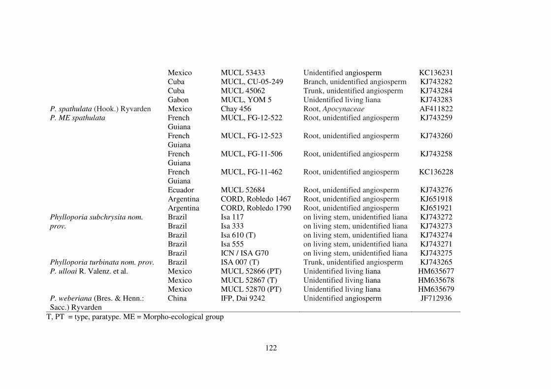
122
Mexico MUCL 53433 Unidentified angiosperm KC136231 Cuba MUCL, CU-05-249 Branch, unidentified angiosperm KJ743282 Cuba MUCL 45062 Trunk, unidentified angiosperm KJ743284 Gabon MUCL, YOM 5 Unidentified living liana KJ743283
P. spathulata (Hook.) Ryvarden Mexico Chay 456 Root, Apocynaceae AF411822 French Guiana
MUCL, FG-12-522 Root, unidentified angiosperm KJ743259
French Guiana
MUCL, FG-12-523 Root, unidentified angiosperm KJ743260
French Guiana
MUCL, FG-11-506 Root, unidentified angiosperm KJ743258
French Guiana
MUCL, FG-11-462 Root, unidentified angiosperm KC136228
Ecuador MUCL 52684 Root, unidentified angiosperm KJ743276 Argentina CORD, Robledo 1467 Root, unidentified angiosperm KJ651918
P. ME spathulata
Argentina CORD, Robledo 1790 Root, unidentified angiosperm KJ651921 Brazil Isa 117 on living stem, unidentified liana KJ743272 Brazil Isa 333 on living stem, unidentified liana KJ743273 Brazil Isa 610 (T) on living stem, unidentified liana KJ743274 Brazil Isa 555 on living stem, unidentified liana KJ743271
Phylloporia subchrysita nom.
prov.
Brazil ICN / ISA G70 on living stem, unidentified liana KJ743275 Phylloporia turbinata nom. prov. Brazil ISA 007 (T) Trunk, unidentified angiosperm KJ743265
Mexico MUCL 52866 (PT) Unidentified living liana HM635677 Mexico MUCL 52867 (T) Unidentified living liana HM635678
P. ulloai R. Valenz. et al.
Mexico MUCL 52870 (PT) Unidentified living liana HM635679 P. weberiana (Bres. & Henn.: Sacc.) Ryvarden
China IFP, Dai 9242 Unidentified angiosperm JF712936
T, PT = type, paratype. ME = Morpho-ecological group

123
Results
Phylogenetic analysis.— Within Phylloporia, the length of the LSU fragment ranged
from 866 to 884 bps. The alignment of the 101 sequences resulted in 951 positions of
which 20 were excluded, 489 were constant, and 370 were parsimony informative.
Using the Akaike information criterion of MrModeltest 2.3 (Posada and Crandall
1998), the best-fit model for the nucLSU data set was GTR+I+G with unequal base
frequencies (A = 0.2468, C = 0.1901, G = 0.3211, T = 0.2419), a gamma distribution shape
parameter of 0.5390, and a proportion of invariable sites of 0.3482.
The MP analysis produced 238 most parsimonious trees (1924 steps, consistency
index (CI) 0.332, retention index (RI) 0.678. The two Bayesian runs converged to stable
likelihood values after 3.898.000 generations and 6102 stationary trees from each analysis
were used to compute a 50% majority rule consensus tree in PAUP* and to calculate
posterior probabilities. In the ML searches with RAxML, the nuc-LSU alignment had 448
distinct patterns with a proportion of gaps and undetermined characters of 7.75%.
The strict consensus of the 4 most parsimonious trees were mostly identical to the
BC tree and to the optimal ML tree (tree score of -lnL = - 8920.620553). One of the
equally most parsimonious trees is presented in Fig. 1.
The topologies of the trees regarding the recovery and the relative positions of the
different major poroid Hymenochaetales generic entities considered were identical in all the
phylogenetic inferences, in accordance with previous results (Decock et al., 2013,
Valenzuela et al., 2011, Wagner and Fischer, 2002). The Phylloporia clade is very well
supported (BS 98% / BPP 1.0 / ML BS 100%) [Fig. 1].

124

125
FIG. 1. Phylogenetic relationships of Phylloporia species inferred from nucLSU rDNA
sequences. The maximum likelihood tree was rooted with Inonotus micantissimus
MUCL52413. Black dots on branches represent BPP greater than 0.95 and ML/BS greater
than 85%; grey dots on branches denote BPP greater than 75% and ML/BS grater than
65%.
In the phylogenetic inferences, our Phylloporia collections from Brazil were
distributed into 4 terminal clades / branches (Fig. 1, PSs 1–4), within the Phylloporia
lineage. These four clades / branches are distant from all other species clades / branches
shown to date (Decock et al., 2013); they represent distinct phylogenetic units, or
phylogenetic species.
Subsequent examinations of the various collections pertaining to each of these
clades allowed evidencing combinations of morphological features that correspond to two
morphotypes / morphospecies as defined by Wagner and Ryvarden (2002). They also
correspond to two distinct ecologies, thereby defining morpho-ecological types.

126
Phylloporia PS 1 belongs to the P. chrysita morpho-ecological type, characterized by soft
basidiomata with a thick tomentum, a monomitic hyphal system, and broadly ellipsoid to
subglobose basidiospores. Phylloporia PSs 2–4 are characterized by small, triquetrous to
conical basidiomata, gregarious, emerging simultaneously in large number from small
stemmed trunks, a dimitic hyphal system, and globose to subglobose basidiospores. They
belong to the P. pectinata morpho-ecological type.
Taxonomy
Phylloporia subchrysita nom. prov. Campos-Santana, R. M. Silveira & Decock sp. nov.
Figs. 2A-D; 4A
MycoBank: MBxxxxx
Etymology: “subchrysita”, refers to the resemblance with P. chrysita.
Basidiomata annual, pileate, sessile; pileus solitary or in small number (1–4 specimens),
broadly attached, nodulose, semi-ungulate to semicircular to fan-shaped or amplectens
(encircling the branch or stem ) in pole view, occasionally laterally fused forming
compound basidiomata, sometimes projecting from a lateral tomentose base, extending up
to 70 mm long, 40 mm wide, 10 mm in the thickest part, light in weight; pileus surface
dull, spongy, soft, with alternate (wavy) concentric bands of variable height, overall in
brown shade, mostly cocoa to rust brown (6E[6–8]) darker toward the basis, dark brown
(chestnut brown, 6F7); margin obtuse, entire, up to 0.5 mm wide, yellowish white to pale
yellow (4A[2–3]), lighter than pore surface and the pileus; pore surface plane to slightly
concave, yellowish brown (5D[4–6)], yellowish orange (4B[6–8]) becoming brown (4E4)
to yellowish brown (4E[5–6]) when mature; pores small, (7–) 8–9 (–10) / mm, (80–) 90–
130 (–140) µm diam, round to ellipsoid, occasionally in defined rows; dissepiments entire,
agglutinated when dry, about 30–60 µm thick with many crystals; context duplex, with a
thin black line (resinous) separating an upper tomentum and a lower context; upper
tomentum a thick trichoderme, soft, spongious, wavy, in alternate concentric ridges of
variable height, from 2–7 mm thick, cinnamon brown (6D6) to brown (6E[4–5)]); lower
context denser, thin, up to 1 mm, much paler than the tomentum, grayish yellow (4C[6–7])
to dark yellow (4C8) fibrous (with bundle of hyphae under the lens); tube layer up to 0.3–
1.0 mm deep, concolorous or only slightly darker than the lower context.

127
Hyphal system monomitic in all parts; generative hyphae simple septate, thin- to
slightly thick-walled, hyaline to faintly yellowish, scarcely branched, negative in Melzer’s
reagent; in the hymenophoral trama hyphae, hyphae thin- to slightly thick-walled, the
lumen widely open, with long aseptate segments, hyaline to pale yellow, darker in KOH,
2.0–4.0 (–5) µm diam (ave = 3.5 µm); in the context hyphae tightly packed, slightly
interwoven, thick-walled but the lumen widely open, pale golden brown to golden brown,
(4.0–) 4.5–5.0 (–5.5) µm diam (ave = 4.1 µm); the upper tomentum as a thick trichoderme,
with erected to prostrate, thick-walled hyphae, yellowish to brown, mostly unbranched,
6.0–8.0 (–9) µm diam.
Hymenium: basidia clavate, hyaline in KOH, up to 9–10 (–11) × 4–5 µm long with
four sterigmata; basidioles identical in shape but slightly smaller; cystidia or other
hymenial element absent; basidiospores abundant, broadly ellipsoid to subglobose,
appearing somewhat angular on drying, thick-walled, pale yellowish in KOH, without
reaction in Melzer’s reagent, 3.0–4.0 (–5.0) × 2.0–3.0 (–4.0) µm (ave = 3.7 × 2.7 µm), Q =
1.25–1.5 (aveQ = 1.39).
Substrate and host: on living stem, unidentified liana.
Know distribution: Known from the Brazilian Atlantic rainforest and Brazilian
Pampa Biomes, Rio Grande do Sul.
HOLOTYPUS. BRAZIL, RIO GRANDE DO SUL. Refúgio da Vida Silvestre-
UFRGS, approx. 30°03’ S, 51°07’W, elev. approx. 130 m, on living stem, unidentified
liana, 31 May 2011, de Campos-Santana 610/11 (ICN 177689); isotypus in herbaria NY
and MUCL. Reference nLSU sequence: KJ743274
Additional specimens examined: BRAZIL, RIO GRANDE DO SUL: Caçapava do
Sul, approx. 30º30’44”S, 53º29’29”W, elev. approx. 444 masl, on the bark of a small-
stemmed dead standing, unidentified liana, 16/IV/2010, Campos-Santana 117/10 (ICN
177687); Porto Alegre, Refúgio da Vida Silvestre da UFRGS, approx. 30°03’ S, 51°07’W,
elev. approx. 130 m, on living stem, unidentified liana, 06/VI/2011, Campos-Santana
555/11 (ICN 177688); ibid., Viamão, Parque Estadual de Itapuã, approx. 30°27’ S –
30°20’ S, 51°03’ W – 50°50’ W, on living stem, unidentified liana,16/X/2010, Campos-
Santana 333/10 (ICN 177700).
Remarks: Phylloporia subchrysita has thick, irregular, nodulose to amplectens
basidiomata, with a thick tomentum, a monomitic hyphal system, and ellipsoid to

128
subglobose basidiospores, 3.0–4.0 (–5.0) × 2.0–3.0 (–4.0). The basidiomata are emerging
solitary or in small number, from stems of liana.
The overall basidiomata habit, the tick tomentum, the hyphal system, and the
ecology point toward Phylloporia chrysita. Phylloporia subchrysita differs from P.
chrysita in having smaller pores (8–9 (–10) / mm versus 6–8 / mm (fide Ryvarden 2004) and
slightly larger, more ellipsoid basidiospores (distinctly subglobose in P. chrysita, fide
Ryvarden 2004).
Phylloporia subchrysita could be compared also to P. ulloai. Both species share
amplectens basidiomata, a thick tomentum, a thin tube layer, the monomitic hyphal system,
and the basidiospores shape and size. Phylloporia subchrysita and P. ulloai differ by their
pore size, respectively 8–9 (–10) / mm and 6–8 / mm (Valenzuela et al. 2011).
Phylloporia subchrysita could be compared also to P. frutica, both species have
which differs in having much larger pores (2–4 / mm), the darker pore surface (cinnamon
to rusty brown) and by the context formed by a dense dark cinnamon to rusty brown layer
near the tubes.
Phylloporia turbinata nom. prov. Campos-Santana, R. M. Silveira & Decock sp. nov.
Figs. 2E-G; 4
BMycoBank: MBxxxxx
Etymology: “turbinata”, attached to the substrate by a small vertex and similar to a small
inverted cone.
Basidiomata annual, pileate, sessile, gregarious, emerging simultaneously in large number;
pileus turbinate, attached by a small vertex, pendant, small, up to 1.0–3.0 cm long, 0.5–1.5cm
wide and up to 0.7 cm thick in single pilei; and 1–2 mm wide at the attachment point; pileus
surface dull, narrowly, faintly concentrically sulcate and with hirsute ridges, overall brown,
mostly cocoa to rust brown (6E[6–8]), discoloring to dark brown (chestnut brown, 6F[5–
7]), eventually blackish toward the basis; margin thin, acute, entire, concolorous with the
pileus lower half; pore surface plane to concave, yellowish brown (5E[5–7]); pores very
small, (10–) 11–13 / mm, 60–100 µm diam (ave = 70 µm), regular, round, occasionally in
well defined rows; dissepiments thin, entire, agglutinated when dry, about 30–40 (–45) µm
diam (ave = 38 µm); context duplex, with a thin (10 µm) black line separating an upper
tomentum and a lower context; upper tomentum a short trichoderme, shortly velutinous

129
(under the lens), up to 160 µm thick, from the base light brown, mostly cinnamon, (6D[5–
6]), with a soft corky consistency; lower context compacter, denser, 0.5–1.0 mm, very thin
to the margin, brown, (5D[5–6], ligth brown), shiny, fibrous (with bundle of hyphae under
the lens), corky; tube layer up to 1 mm deep, concolorous with the pores surface.
Hyphal system dimitic; generative hyphae simple septate, thin- to thick-walled,
hyaline to pale yellow, sometimes branched, 1.5–3.0 (–4) µm diam (ave= 2.5 µm); skeletal
hyphae dominantin all parts; in the context, the skeletal hyphae descending, parallel to long
axis, arising from a generative hyphae, and of limited growth, measured up to 180 µm
long, (3.5–) 4.0–5.0 µm diam (ave = 4.4 µm), golden brown, darker (brown) in alkali, thick-
to very thick-walled with the lumen wide to narrow, aseptate or with few secondary septa
near the apices; upper trichoderme with free ending, thick-walled hyphae, 4.0–5.0 µm (ave
= 4.6 µm); in the hymenophoral trama skeletal hyphae measured from 115 –170 µm long,
2.5–3.0 µm diam at the basal septa to mostly 3.5–4.5 µm diam (ave = 3.9 µm) in the main
part, thick-walled to very thick-walled, the lumen mostly straight, or with local
constrictions, aseptate throughout but with a few secondary septa near the apices, golden
brown, darker (brown).
Hymenium: basidia clavate, hyaline in KOH, 4-sterigmata, up to 7.0–8.0 × 4.0–5.0
µm long with four sterigmata; basidioles slightly pyriform, hyaline, 6.5–7.0 × 4.0–5.0 µm;
basidiospores broadly ellipsoid to subglobose, appearing somewhat angular on drying, thick-
walled, smooth, pale yellowish in KOH, without reaction in Melzer’s reagent, (2.5–) 3.0–4.0
× 2.0–3.0 (–3.5) µm, (ave = 3.2 × 2.2 µm), Q = 1.33–1.5 (aveQ = 1.48).
Substrate and host: growing on trunk of living, unidentified angiosperm.
Known distribution: known only from the type locality in Brazilian Atlantic
rainforest in Rio Grande do Sul.
HOLOTYPUS. BRAZIL, RIO GRANDE DO SUL. Dom Pedro de Alcântara,
RPPN do Professor Luis Baptista, approx. 29º22’10”S, 49º50’59”W, elev. approx. 37m, on
the bark of a small-stemmed dead standing trunk, unidentified angiosperm, 12 March
2010, Campos-Santana 007/10 (ICN 177690); isotypus in herbaria NY et MUCL.
Reference nLSU sequence: KJ743265.
Remarks.— Phylloporia turbinata belongs to the Phylloporia pectinata morpho-
ecological type, characterized by gregarious, small basidiomata, a hard consistency, a
dimitic hyphal system. It differs from P. pectinata in having smaller basidiomata, not
exceeding 3 cm (commonly > 5 cm in P. pectinata fide Corner 1991).

130
FIG. 2. A, B, C, D: Phylloporia subchrysita. A. Pore surface, B. Longitudinal section of
the tubes, C. Basidiomata (A, scale bar =.1 mm; B, scale bar =.2 mm; C/D, scale bar =. 3

131
cm). E, F, G: Phylloporia turbinata. E. Pore surface, F. Longitudinal section of the tubes,
G.Basidiomata, (E, scale bar =.1 mm; F, scale bar =.1.5 mm; G, =.3 cm).
Phylloporia neopectinata nom. prov. Campos-Santana & Decock sp. nov.
Figs. 3A-C; 4C
MycoBank: MBxxxxx
Etymology: “neopectinata”, similar P. pectinata in the Neotropics.
Basidiomata annual, pileate, sessile, gregarious, emerging in clusters of up to 70 individuals,
mostly superposed; pileus mostly turbinate, attached by a very small vertex and pendant, 0.5–
2 mm wide at the attachment point, small, projecting 1–3 cm long, 0.7–1.2cm wide, up to 0.7
cm thick; pileus surface dull, narrowly, faintly concentrically sulcate and hirsute, the lower
half mostly cocoa to rust brown (6E[6–8]), the upper half, darker, dark brown (chestnut
brown, 6F[5–7]), eventually blackish at the very base; margin sterile distinct, thin, entire,
soft consistency when fresh, drying corky; concolour with the pileus lower half; pore
surface plane to concave, yellowish to brownish orange whitish at the very marginal areas
margin, soon light brown (5D[5–7]), then yellowish brown (5E[ 4–5]); pores very small,
regular, mostly round, (9–)10–13(–14) / mm, (60–) 70–90 mm diam (ave = 76 µm);
dissepiments smooth, entire, thin, 20–50 µm thick (ave = 35 µm); upper tomentum a short
velutinous trichoderme, up to ≤ 140 µm thick, from the base light brown, mostly cinnamon,
(6D[5–6]), with a soft corky consistency; context compacter, dense, mostly homogeneous
and without black line (5µm), then with a very thin, soft spongy (watery) when fresh, soft
corky consistency, up to 1.5 mm thick; lower trama compacter, dense, with squared crystals
abundant; tube layer single, up to 1.5 mm deep at the base, very thin to the margin.
Hyphal system dimitic, generative hyphae with simple septa, thin- to mostly thick-
walled, scarly branched, 1.5–3.0 µm (ave = 2.4 µm), hyaline to mostly pale yellow, forming
a network holding the spore mass into the tubes; context dominated by skeletal hyphae,
parallel to long axis; mensured up to 250 µm long, 2.0–3.0 µm diam at the basal septa, with
widening to 3.5–5.0 µm (ave = 4.2 µm), golden brown to darky brown, thick-walled, lumen
wide to narrow, mostly aseptate throughout or with secondary septa; upper trichoderme with
prostrate to erected hyphae, thick-walled, pale golden yellow, 4.5–5.5 µm; hymenophoral
trama dominated by skeletal hyphae, slightly thick-walled, the lumen widely open, at the
basal septa, but with long aseptate segments or with occasional with secondary septa,

132
sparingly branched, hyaline to pale golden yellow, darker in KOH, 1.5–3.0 µm diam, (ave=
2.20 µm),
Hymenium: basidioles subglobose to pediculate; basidia not observed; cystidia none;
basidiospores oblong ellipsoid to subglobose, distinctly thick-walled, smooth-walled, pale
yellowish in KOH, without reaction in Melzer’s reagent, (2.5–) 3.0–4.0 × 2.0–3.0 µm (ave =
3.1 × 2.1 µm), Q = (1.34–1.5 (aveQ = 1.47).
Substrate and host: growing on stem of living, undetermined.
Know distribution: Brazilian Atlantic rainforest in the States of Rio Grande do Sul.
HOLOTYPUS. BRAZIL RIO GRANDE DO SUL. Derrubadas, Parque Estadual do
Turvo, approx. 27º08’44”S, 53º53’10”W, elev. between 100m and 400m, on the small-
stemmed dead standing trunk, unidentified angiosperm, 26 October 2010, Campos-Santana
353/10 (ICN 177691; culture MUCL 54295); isotypus in herbaria NY and MUCL.
Reference nLSU sequence: KJ743267.
Additional specimens examined: BRAZIL RIO GRANDE DO SUL: Porto Alegre,
Refúgio da Vida Silvestre da UFRGS, approx. 30°03’ S, 51°07’W, elev. approx. 130 m, on
the small-stemmed dead standing trunk, unidentified angiosperm, 18/VIII/2011, Campos-
Santana 640 (INC 177699); ibid., 17/V/2011, Campos-Santana 553 (ICN 177692); ibid.,
Campos-Santana 552/11 (ICN 177698; culture MUCL 54288).
Remarks. — Phylloporia neopectinata belongs also to the P. pectinata morpho-
ecological type. The host relationship is unknown for the time being.
Phylloporia turbinata and P. neopectinata are sympatric in the southern areas of
the Atlantic forest. From a morphological point of view, these species are very similar and
the distinction between both species proved challenging. Subtle differences include the
yellow to brown orange pore surface, soon light brown and the irregular black line in F.
neopectinata. The pore surface is yellow brown and there is a regular black line (100 µm
thick) in P. turbinata. In our phylogenetic analysis based on nLSU sequences these two-
species clades appear distinct (FIG. 1).
Phylloporia loguerciae nom. prov. Campos-Santana, Robledo & Decock sp. nov.
Figs. 3D-F; 4D
MycoBank: MBxxxxx

133
Etymology: The species is named in honor of Dr. Clarice Loguercio-Leite, for her valuable
contribution to the Mycology, especially in Southern Brazil.
Basidiomata perennial, sessile, gregarious or in small number; pilei semicircular to
dimidiate, woody-hard, thinly applanate in section, the margin enrolling inward on drying,
projecting up 5 cm long, 4 cm wide, up to ≤ 0.7 cm thick, faintly concentrically sulcate,
finely velutinate, adpressed velutinate (under the lens), cork-colored, grayish orange to the
margin then dull light brown to brown (6[D–E]6, cinnamon to cocoa brown) toward the
base, dark brown on aging or weathering (6F6); margin corrugated, sterile distinct,
yellowish when dry (4B4, grayish yellow); pore surface yellow brown to cinnamon; pores
rounded to ellipsoid entire, (7–) 8 –10/ mm, 80–110 µm diam (ave = 95 µm); dissepiments
non agglutinated, 15–40 µm thick (ave = 26 µm); context duplex, dark brown (6F[ 6–8]) to
(7F[ 5–6]) with a thin black line separating an upper short tomentum and a lower trama;
upper tomentum a short trichoderme, shortly velutinous (under the lens), up to ≤ 70 µm
thick, brown (6E[ 6–8]); lower trama compacter, denser, corky, 0.5–1.0 mm thick, light
brown (6D[5–6]); tubes bi- stratified, in some parts with a thin black line separating the
layers, to ≤ 5 mm at the deepest, light brown (6D[6–8]).
Hyphal system mono- to incompletely dimitic; generative hyphae simple-septate,
simple to branched, thin- to moderately thick-walled, hyaline to yellowish; in the
hymenophoral trama hyphae slightly interwoven, hyaline toward the dissepiments,
yellowish to golden yellow deeper in the trama, turning darker, yellowish brown in alkali,
little branched, thin- to thick-walled, with the lumen widely open, 3.0–5.5 µm diam; in the
context hyphae tightly packed, slightly interwoven, yellowish to golden brown, darker
brown in alkali, little branched, thick-walled but with the lumen widely open, (3.0–) 3.5–
4.5 (–5.0) diam (ave = 4.0 µm); pileus upper tomentum a trichoderme with erected to
prostrate, thick-walled hyphae, yellowish to brown, mostly unbranched, 3.0–4.5 µm diam;
Hymenium: basidia not observed; cystidia or other sterile hymenial elements
absent; basidiospores mainly ellipsoid, with the adaxial side occasionally flattened
(perhaps on drying), thick-walled, pale yellowish, IKI -, (3.0–) 4.0–5.0 × (2.0–) 3.0–4.0
µm, Q = 1.25–1.5 (ave = 3.9 × 2.9 µm; aveQ = 1.4).
Substrate and host: growing on stem of living, undetermined.
Know distribution: Brazilian Atlantic rainforest in the States of Santa Catarina and
Argentina.

134
HOLOTYPUS. BRAZIL. SANTA CATARINA, Mondaí, Linha Uruguai, approx.
27º06”16’S and 53º24”07’W, elev. 235m, on the small-stemmed dead standing trunk,
unidentified angiosperm, 10.XII.2010, Campos-Santana 437/10 (ICN 177693; culture
MUCL 54226); isotypus in herbaria NY and MUCL. Reference nLSU sequence:
KJ743270.
Additional specimens examined: ARGENTINA: stem, Magfadyena unguis-cati,
Robledo 429; ibid., stem, unidentified liana Robledo 1134, Dead stem, Magfadyena
unguis-cati Robledo 1624.
Remarks. — Phylloporia loguerciae belongs to the P. pectinata morpho-ecological
type. It is recognized by its sessile basidiomata, emerging in groups, though in low
number, semicircular to dimidiate pileus, irregular pores, (7–) 8 –10/ mm, tubes bi-
stratified. The hyphal system is uncertain, intermediate between mono- to (incompletely)
dimitic. This set of characteristic separates P. loguerciae from the other species treated in
this work or belonging to the P. pectinata morpho-ecological type. Phylloporia loguerciae
also differs from the P. turbinata and P. neopectina in having commonly much large pilei,
reaching up to 5 cm long, 4 cm wide, up to ≤ 0.7 cm thick. The host relationships are
unknown for the time being.

135
FIG. 3. A, B, C,: Phylloporia neopectinata. A. Longitudinal section of the tubes, B. Pore
surface, C. Basidiomata (A, scale bar =.1 mm; B, scale bar =.1 mm; C, scale bar =. 3 cm).
D, E, F, G: Phylloporia loguerciae. D. Pore surface, E. Longitudinal section of the tubes, F
and G. Basidiomata, (E, scale bar =.1 mm; F, scale bar =.5 mm; G/H, =.3 cm).
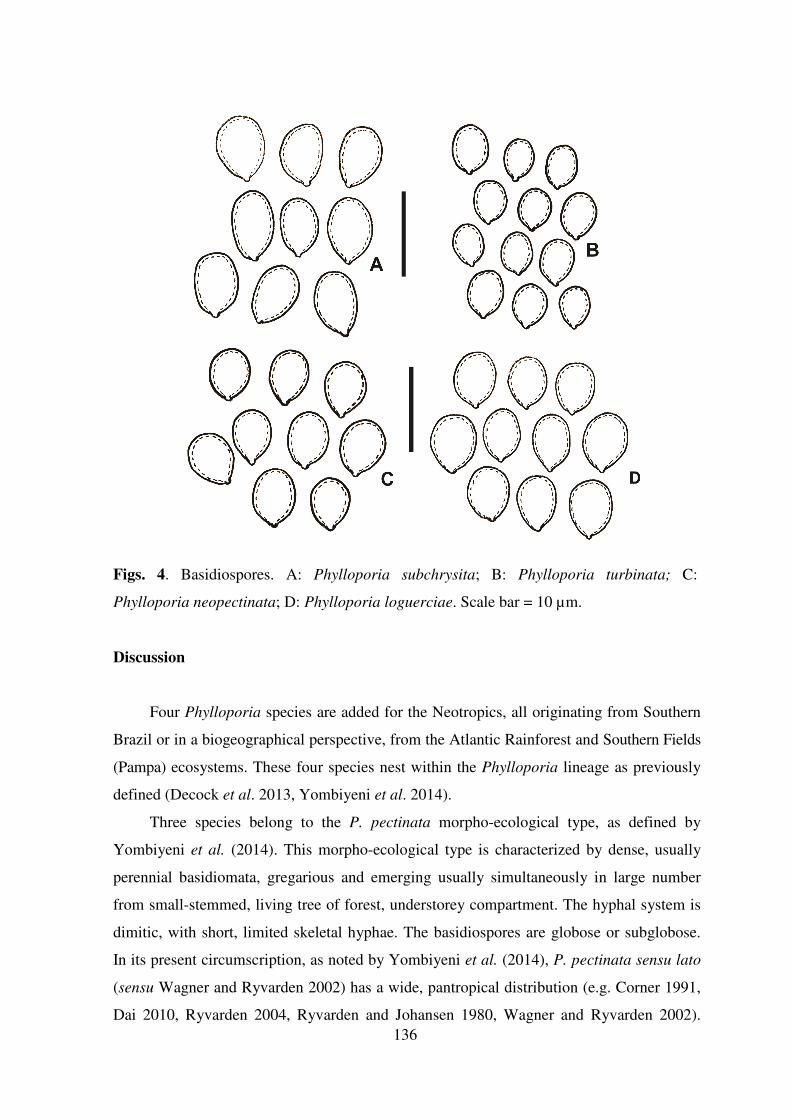
136
Figs. 4. Basidiospores. A: Phylloporia subchrysita; B: Phylloporia turbinata; C:
Phylloporia neopectinata; D: Phylloporia loguerciae. Scale bar = 10 µm.
Discussion
Four Phylloporia species are added for the Neotropics, all originating from Southern
Brazil or in a biogeographical perspective, from the Atlantic Rainforest and Southern Fields
(Pampa) ecosystems. These four species nest within the Phylloporia lineage as previously
defined (Decock et al. 2013, Yombiyeni et al. 2014).
Three species belong to the P. pectinata morpho-ecological type, as defined by
Yombiyeni et al. (2014). This morpho-ecological type is characterized by dense, usually
perennial basidiomata, gregarious and emerging usually simultaneously in large number
from small-stemmed, living tree of forest, understorey compartment. The hyphal system is
dimitic, with short, limited skeletal hyphae. The basidiospores are globose or subglobose.
In its present circumscription, as noted by Yombiyeni et al. (2014), P. pectinata sensu lato
(sensu Wagner and Ryvarden 2002) has a wide, pantropical distribution (e.g. Corner 1991,
Dai 2010, Ryvarden 2004, Ryvarden and Johansen 1980, Wagner and Ryvarden 2002).

137
Ryvarden (2004) reported the species from the Neotropics. Rajchenberg and de Meijer
(1990), Ryvarden and de Meijer (2002), de Meijer (2006), Theissen (1911), and
Sobestiansky (2005) reported also the species from Southern Brazil. Our phylogenetic
studies show that our Brazilian collections entering the current concept of P. pectinata (e.g.
sensu Wagner and Ryvarden 2002) to be distributed over 3 terminal lineages (Fig. 1, PS 2-
4). These lineages are distant from the reference P. pectinata lineage (Wagner and Fischer
2002).
Phylloporia pectinata was originally described from southern India. The current
species concept (e.g. Corner 1991, Ryvarden 2004, Ryvarden and Johansen 1980, Wagner
and Ryvarden 2002) obviously encompasses multiple cryptic species (see also Yombiyeni
et al. 2014). This renders any unequivocal circumscription of morphospecies concept very
challenging if ever possible. The combination of morphological, molecular, and ecological
data (host relationships) are highly desirable to better understand the diversity within this
morpho-ecological complex. In the meantime, as suggested by Yombiyeni et al. (2014), it
is recommend using the current [morphological] concepts of P. pectinata with caution and
as sensu lato.
Phylloporia subchrysita is morphologically related to P. chrysita and to P. ulloai, a
taxon described from eastern Mexico (Valenzuela et al. 2011), which is also its closest
relative for the time being. Inversely, the P. chrysita clade is distantly related to the P.
subchrysita / P. ulloai clade. These three species share a sessile basidiomata, commonly
amplectens, a thick, spongy tomentum, a thin tube layer, and a monomitic hyphal system.
Phylloporia subchrysita and P. ulloai share the same ecology, both growing on living stem
of liana. They form another morpho-ecological alliance within Phylloporia.
Acknowledgements
Marisa de Campos Santana acknowledges financial support received from CAPES (process
8296/11-1) and CNPq (Brazil) that enabled her one-year research stay at MUCL,
Université catholique de Louvain, Belgium. Cony Decock gratefully acknowledges the
financial support received from the FNRS / FRFC (convention FRFC 2.4544.10) and from
the Belgian State–Belgian Federal Science Policy through the BCCMTM research
program. Thanks are extended also to Stéphanie Huret for her help with the sequencing
program.

138

139
4. GENERAL DISCUSSION
The species diagnosis depends on the application of operational concepts (species
concept), such as morphological species, biological species or phylogenetic species
concepts, as defined by Taylor et al (2000).
Taxonomy based on morphological characters only (Morphological Species Concept)
for Hymenochaetaceae, hence for Phellinus s.l., is extremely complex, given the high
number of described species to the genus, the range of phenotypic plasticity and their wide
geographical distribution. The phylogenetic analysis based on ITS, LSU, rpb2 and tef1-α
regions, considered alone or altogether, have shown suitable for identification and
delimitation of species, as in the case of Fomitiporia neotropica, Phellinus amazonicus,
Phylloporia subchrysita, P. turbinata, P. neopectinata and P. loguerciae (Chapters II –
IV), besides providing a phylogenetic outline which can be used to evaluate morphological
characters, biogeographical hypothesis and occurrence of cryptic species.
Studies concerning the occurrence and taxonomy of Hymenochaetaceae, including
phylogenetic inference, have being carried out mainly in Europe and North America.
However, these analyses are relatively limited due to the paucity of species from other
geographical, mainly tropical regions hosting the vast majority of the known species.
Yang (2011) stated that systematic and phylogeny of Fungi based on molecular data
evolved quickly in the last two decades. Nevertheless, morphological and ultrastructural
characters, ecological features, and biochemical characters such as e.g. secondary
metabolites are equally important for comprehension of evolution in Fungi Kingdom. For
instance, many hypotheses proposed in the last century, based on morphology,
ultrastructure, pigment or metabolite structure have corroborated with molecular
approaches in the last two decades. Several methodologies, therefore, are required for
better comprehending fungal diversity.
Using the sequences generated from Brazilian samples and others sequences
available in Genbank, phylogenetic analysis were made, rooted with an out group based on
previous works (Larsson et al., 2006; Decock et al. 2007).
For Fomitiporia neotropica, Phellinus amazonicus and the four new Phylloporia
species, the taxa selection for inclusion in analysis was possible thanks to previous analysis
and large dataset made by Decock et al. at MUCL. Besides, macro-micromorphological
analysis of all specimens and an ecological characterization were carried out.

140
In the present work, more than 6oo specimens of poroid Hymenochaetaceae were
examined, including specimens collected by the author, other researchers and specimens
deposited in local herbaria. The analyzed materials were not only from Southern Brazil,
but also from Europe and South America, thanks to the herbarium MUCL. The list of
genera and species studied is presented in Chapter I. The new species descriptions,
discussions, illustrations and comments concerning studied species are presented in the
articles included in this thesis, as Chapters (I-IV). However, some taxonomic problems to
be solved will be presented in forthcoming works, still under preparation. This is the case
of Phellinus sp. (P. gabonensis Decock and Yombiyeni morpho-ecological complex) and
some species belonging to Fuscoporia (of which the Fuscoporia wahlbergi complex).
From the species identified, the most abundant was found to be Fuscoporia gilva
var. licnoide (with 80 specimens), followed by Fuscoporia wahlbergii (with 54) and
Fuscoporia ferrea (with 44). Some species appear to be rare, with few records. It is the
case of Phellinus lopezzi, Phellinus detonsus, Fomitiporia dryophila and Fulvifomes
durissimus for instance. Combining the results of the specimens analyzed with literatture
records, 63 poroid Hymenochaetaceae species are recognized from Southern Brazil,
corresponding to nine genera. The most represented genera with are Phellinus (8 species),
Fuscoporia (8 species) and Inonotus (8 species).
Rio Grande do Sul is the most representative state, with the higher number of
species in the studied area, with 35 species. 26 species were found in Paraná and 25 from
Santa Catarina. Only 18 species are shared by these three States.
In the manuscripts presented in this work, six new species are described, based in
phylogenetic analysis, including multilocus sequences (Chapter II, III and IV). Two new
combinations are also proposed. New records from Brazil and Southern Brazil are also
presented (Chapter I).
-Fomitiporia neotropica, Phellinus amazonicus, Phylloporia loguerciae,
Phylloporia neopectinata, Phylloporia turbinata and Phylloporia subchrysita are described
as new to science, based on molecular phylogenetic analyzes, including multilocus
sequences.
-Two new combinations are proposed: Fomitiporia bambusarum and Fulvifomes
rhytiphloeus.
-Phellinus sp. (P. gabonensis Decock and Yombiyeni morpho-ecological complex)
is a new record for South America.

141
-New occurrences are considered to Brazil: Fomitiporia dryophyla, Fulvifomes
durissimus and Phellinus lopezzi.
Many of the species found in this work were unknown for region or for any of the
three states, and had their geographical distribution extended.
-Fomitiporia dryophila, Fomitiporia neotropica, Fulvifomes durissimus,
Fulvifomes membranaceus, Inonotus portoricensis, Inonotus pseudoglomeratus, Phellinus
detonsus, Phellinus sp. (P. gabonensis Decock and Yombiyeni morpho-ecological
complex), Phellinus lopezii, Phellinus shaferi, Phylloporia loguerciae, Phylloporia
neopectinata, Phylloporia turbinata and Phylloporia subchrysita, are new records for the
Southern Brazil.
-Cyclomyces iodinus, Fomitiporia dryophila, Fulvifomes durissimus, Fulvifomes
membranaceus, Fulvifomes merrillii, Fomitiporia neotropica, Inonotus linteus, Inonotus
portoricensis, Phellinus detonsus, Phellinus sp. (P. gabonensis Decock and Yombiyeni
morpho-ecological complex), Phellinus lopezii, Phellinus shaferi, Phylloporia loguerciae,
Phylloporia neopectinata, Phylloporia turbinata and Phylloporia subchrysita, are
registered for the first time to Rio Grande do Sul.
-Are considered to be new records from Santa Catarina state: Fuscoporia
palmicola, Fomitiporia neotropica, Inonotus micantissimus, Inonotus portoricensis,
Inonotus pseudoglomeratus, Phellinus caryophylleus, Phellinus detonsus, Phellinus sp. (P.
gabonensis Decock and Yombiyeni morpho-ecological complex), Phellinus shaferi and
Phylloporia loguerciae.
-Fulvifomes rhytiphloeus, Fuscoporia contigua, Fuscoporia ferrea, Fuscoporia
rhabarbarina, Inonotus sp., Phellinus anchietanus, Phellinus caryophylleus, Phellinus
detonsus and Phellinus shaferi are registered for the first time to Paraná state.

142
5. CONCLUSIONS
Hymenochaetaceae constitutes one of the larger groups of wood-inhabiting, wood-
decomposing fungi. They are diverse and play diverse key ecological roles in all forest
ecosystems. They may enhance tree growth by forming symbiotic, mycorrhizal
associations, benefical for both partners. It is the case for instance of the Coltricioid
species. At the opposite range, they may be parasitic to the root system or trunk, eventually
killing the tree. It is the case for instance of Phylloporia or Fulvifomes species. Most of
them participate in the recycling of the dead wood as saprotrophs.
The understanding of the forest functioning passes through the knowledge of all its
individual components, biotic or abiotic (who / what is there? Who is doing what?) and of
all their interactions, (who interact / with who?). Here is the key role of the taxonomists.
Identifications of wood-inhabiting Hymenochaetaceae are confronted to several
major problems. Still, the vast majority of species are defined based on diagnostic
morphological characters and comparison with voucher herbarium specimens – viz type
specimens – that are usually old and in very variable conditions. The morphological
species concept is many times limited by the paucity of characters available, and a very
uncertain interpretation about their variability. Little is known about the extent of
variability within populations and their pertinence for delimiting species. Life cycles are
variable as well as complex and may affect evolutionary patterns in ways that are difficult
to interpret (Petersen and Hughes 1999). Furthermore, many times, the species concept
used is based on material originating from the northern temperate area or various tropical
areas and extended uncritically to other areas.
Therefore, delimiting species concept on the sole basis of a few uncertain
morphological features and uncertain reference material reveal many times challenging for
the taxonomists working with Hymenochataceae. A fortiori, dealing with such
morphospecies concepts is almot unachievable for the end users of taxonomic data that are
biologists, ecologists, and conservation biologists.
Other descriptors are necessary, as highlighted by Amalfi et al. (2012) and Amalfi
and Decock (2013), to delimit species in the Hymenochaeaceae. These includes now
molecular, DNA sequence data, but should also integrate ecological, biological and
biogeographical data. Integrating multiple complementary data to achieve a "global" or
"holistic" species concept was dealt with in detail in Ascomycota, exemplifed by

143
Quaedvlieg et al. (2014). Quaedvlieg et al. (2014) developed an approach similar to that
suggested by Decock et al. (2007), Amalfi et al. (2011) and Amalfi and Decock (2013)
integrating multiple complementary descriptors that results in a “Consolidated Species
Concept” (CSC). This should certainly be applied more extensively to the all poroid
Hymenochaetaceae.
In this thesis, we studied in much detail the case of several “morphospecies”
concepts including the Fomitiporia "punctata" (F. robusta complex), the Phellinus
gabonensis morpho-ecological complex, the Phylloporia pectinata and the P. chrysita
morphospecies / morpho-ecological types.
So far, 24 species of Phylloporia were accepted and validly published. Four species
are here added. However, the species number is more likely larger and many species might
be described when the host relationships will be better understood. Phylloporia species are
thought to be host specific but the host is still rarely considered.
In the present thesis, we have tried to apply a broader concept to the poroid
Hymenochaetaceae in Southern Brazil. It was not possible in the time available to cover in
such way the totality of Phellinus s.l species occurring in the areas considered. The
synopsis provides a first step to undertake more detailed studies. We are conscient that the
species concept used here might reveal more complex. The examples treated in the other
publication and manuscripts argue for such a multilple approach.
A considerable improvement in this work accomplishment was only possible when
came the opportunity to work directly with colleagues having complementary specialities
in different research groups.
We believe we gave the first step towards this integration, and we hope that this
represents the beginning of a cooperation network involving research groups in Brazil
(from South to Northeast) and other countries.
We hope that this thesis and the manuscripts will be of utility for those who are
interested in the study of the Fungi. Finally, we believe that this work has contributed not
only for the knowledge of the studied group, and might be also useful as a divulgation tool
for these organisms to students and interested in mycology, including ecologist,
conservation.

144
6. PERSPECTIVES
In Brazil, there are few specialists in Hymenochaetaceae. An analysis of the studies
involving the family and published until now in Brazil, allows us to point two great gaps to
be filled: the lack of in-deep integrated approaches to characterize and define the species
present in Brazil; the lack of field studies and development of pure culture in order to
develop molecular, DNA-based phylogenetic studies, and ahead, physiological studies.
Based on the results presented in the articles that compose chapters I − IV, it can be
stated that the number of poroid Hymenochaetaceae found in Southern Brazil (Table I and
Chapter I) represents a result which could be taken as reference, but cautiously.
In that sense, genera such as Fuscoporia, Phellinus s.s, Fulvifomes, Fomitiporia,
Phylloporia and Inonotus, that were well represented in Southern Brazil, with a high
number of species, might be selected as as topics for other PhD thesis.
Preliminary results on other species such as Fuscoporia wahlbergii and the other
species of the genus as F. gilva, with multiple lineages inside a morphospecies concept still
bring argument to have a different approach. In that sense, there are strong reasons to
believe that the work is still at the beginning.
Furthermore, despite the fact that molecular phylogenetic studies showed
Phylloporia to be monophyletic, it is still necessary to include sequences of Phylloporia
parasitica to confirm the taxonomic status of the genus. In addition, it is necessary to
include specimens from still unexplored regions, which will contribute, in a significant
way, to understand the evolutionary history of the genus.
Ecological aspects, such as distribution and specialization levels with hosts in
particular, are important to characterize Hymenochaetoid species, as well as consolidate
specific concepts. In this way, other ecosystems or other forest domains also deserve to be
explored to complement the data about geographical distribution and ecology of
Hymenochaetaceae.

145
7. REFERENCES
Abrahão MC, Gugliotta AM, Gomes E. 2009. Poliporoides (Basidiomycota) em fragmentos de mata no perímetro urbano de São José do Rio Preto, São Paulo, Brasil. Revista Brasileira de Botânica 32(3): 427–440.
Akhtar M, Blanchette RA, Kirk TK. 1997. Fungal delignification and Biomechanical pulping of wood. Advance in Biochemical Engineering / Biotechnology 57: 160– 193.
Almeida Filho OM, Bueno R, Bononi VLR. 1993. Algumas espécies de fungos Basidiomycetes de manguezais do Estado de São Paulo. Hoehnea 20(1/2): 87–92.
Amalfi M, Yombiyeni P, Decock C. 2010. Fomitiporia in sub-Saharan Africa: Morphology and multigene phylogenetic analysis support three new species from the Guineo-Congolian rainforest. Mycologia 102: 1303–1317.
Amalfi M, Raymundo T, Valenzuela R, Decock C. 2012. Fomitiporia cupressicola sp. nov., a parasite on Cupressus arizonica, and additional unnamed clades in the southern USA and northern Mexico, determined by multilocus phylogenetic analyses. Mycologia 104: 880–893.
Amalfi M, Decock C. 2013. Fomitiporia castilloi sp. nov. and evidence for multiples clades around F. apiahyna in Meso - and South America, representing potential species. Mycologia 105:873–887.
Amorim DS. 2002. Fundamentos de sistemática filogenética. Holos Editora, Ribeirão Preto, 136p.
Azevedo CPL, Guerrero RT. 1993. Estudo biossistemático de espécies do gênero Hymenochaete (Basidiomycetes) no Rio Grande do Sul. Insula 22: 143–176.
Balan DSL, Monteiro RTR. 2001. Decolorization of textile índigo dye by ligninolytic fungi. Journal of Biotechnology 89: 141–145.
Baltazar JM, Trierveiler-Pereira L, Loguercio-Leite C. 2009a. A checklist of
xylophilous basidiomycetes (Basidiomycota) in mangroves. Mycotaxon 107: 221–
224.

146
Baltazar JM, Trierveiler-Pereira L, Loguercio-Leite C, Ryvarden L. 2009b. Santa Catarina
Island mangroves 3: a new species of Fuscoporia. Mycotaxon 101(6): 859–63.
Baltazar JM, Gibertoni TB. 2009. A checklist of the aphyllophoroid fungi (Basidiomycota)
recorded from the Brazilian Atlantic Forest. Mycotaxon 109: 439–442.
Baltazar JM, Gibertoni TB. 2010. New combinations in Phellinus s.l. and Inonotus s.l.
Mycotaxon 111: 205–208.
Baltazar JM, Ryvarden L, Gibertoni TB. 2010. The genus Coltricia in Brazil: new records
and two new species. Mycologia 102: 1253–1262.
Baltazar JM, Drechsler-Santos ER, Ryvarden L, Cavalcanti MAQ, Gibertoni TB. 2012.
Contribution to the knowledge of polypores (Agaricomycetes) from the Atlantic
forest and Caatinga, with new records from Brazil. Mycosphere 3(3): 267–280.
Bass D, Richards TA. 2011. Three reason to re-evaluate fungal diversity 'on Earth and in
the ocean'. Fungal Biology Reviews 25: 159–164.
Batista AC, Bezerra JL. 1960. Basidiomycetes vulgares em o Nordeste Brasileiro. Instituto
de Micologia 294: 1–30.
Bernicchia A. 1990. Polyporaceae s.l. in Italia. Bologna, Italy: Istituto di Patologia
Vegetale, Bologna: pp. 1–594.
Bernicchia A. 2005. Polyporaceae s.l. in Italia. Fungi Europaei, ed. Candusso, Italy: pp. 1–
808.
Bickford D, Lohman DJ, Sodhi NS, Ng PKL, Meier R, Winker K, Ingram KK, Das I.
2007. Cryptic species as a window on diversity and conservation. Trends in Ecology
& Evolution 22: 148–155.

147
Binder M, Hibbett DS, Larsson KL, Larsson E, Langer E, Langer G. 2005. The
phylogenetic distribution of resupinate forms across the major clades of mushroom-
forming fungi (Homobasidiomycetes). Systematics and Biodiversity 3(2): 113–157.
Blackwell M. 2011. The Fungi: 1, 2, 3…5.1 million species? American Journal of Botany
98(3): 426–438.
Bondártseva MA, Herrera S, Sandoval D, Sejas F. 1992. Taxonomical problems of the
Cuban Hymenochaetaceous fungi. Mikologiya I Fitopatologiya 26: 1–13.
Bononi VLR, Trufem SFB, Grandi RAP. 1981. Fungos macroscópicos do Parque Estadual
das Fontes do Ipiranga, São Paulo, Brasil, depositados no Herbário do Instituto de
Botânica. Rickia 9: 37–53.
Bononi VLR, Oliveira AKM, Quevedo JR, Gugliotta AM. 2008. Fungos macroscópicos do
Pantanal do Rio Negro, Mato Grosso do Sul, Brasil. Hoehnea 35: 489–511.
Bononi VLR. 1979a. Basidiomicetos do Parque Estadual da Ilha do Cardoso: I. Espécies
hidnóides. Rickia 8: 63–74.
Bononi VLR. 1979b. Basidiomicetos do Parque Estadual da Ilha do Cardoso: II.
Hymenochaetaceae. Rickia 8: 85–99.
Bononi VLR. 1979c. Basidiomicetos do Parque Estadual da Ilha do Cardoso: III. Espécies
clavarióides, teleforóides e estereóides. Rickia 8: 105–121.
Bononi VLR. 1984b. Basidiomicetos do Parque Estadual da Ilha do Cardoso: IV. Adições
às famílias Hymenochaetaceae, Stereaceae e Thelephoraceae. Rickia 11: 43–52.
Bononi VLR. 1992. Fungos macroscópicos de Rio Branco, Acre, Brasil. Hoehnea 19(1/2):
31–37.

148
Borba-Silva MA, Ferreira-Lopes V, Drechsler-Santos ER. 2013. New records of
Flaviporus hydrophilus and Phellinus portoricensis (Fungi: Polypores). Check List
9(4):815–817.
Boulet B. 2003. Les champignons des arbres de l'est de l'Amérique du Nord. Les
publications du Québec, Québec, Canada: pp. 1–727.
Bowler PJ. 1989. Evolution, the history of an idea. Berkeley: University of
Carlifornia Press, 432p.
Brazee NJ, Lindner DL, Fraver S, D’Amato AW, Milo AM. 2012. Wood-
inhabiting, polyporoid fungi in aspen-dominated forests managed for biomass in the
U.S. Lake States. Fungal Ecology 5: 600–609.
Burford EP, Kierans M, Gadd GM. 2003. Geomycology: fungi in mineral substrata.
Mycologist 17(3): 98–103.
Campacci TVS, Gugliotta AMA. 2009. Review of Amauroderma in Brazil, with A.
oblongisporum newly recorded from the neotropics. Mycotaxon 110: 423–436.
Campos EL, Sótão HM, Cavalcanti MAQ, Luz AB. 2005. Basidiomycetesde Manguezais
da APA de Algodoal – Maiandeua, Para, Brasil. Boletim do Museu Paraense Emílio
Goeldi. Serie Ciências Naturais 1(1): 141–146.
Campos-Santana M, Loguercio-Leite C. 2008. First records of xylophilous Basidiomycetes
(Fungi) in Mondaí, Santa Catarina (Southern Brazil). Biotemas 21(2): 19–26.
Campos-Santana M, Loguercio-Leite C. 2010. Mycodiversity of xylophylous
Basidiomycetes (Basidiomycota, Fungi) in Mondaí, Santa Catarina, Brazil: a new
addition. Biotemas 23(1): 13–18.
Campos-Santana M, Silveira RMB. 2011. Phellinus gabonensis (Hymenochaetaceae
Agaricomycota): In: VII Congreso Latinoamericano de Micología, 2011, Costa Rica.

149
Resúmenes y Programa Latino-Micología innovación y progreso VII Congreso
Latinoamericano de Micología 1:71.
Campos-Santana M, Amalfi M, Robledo G, Borges da Silveira RM, Decock C. 2014.
Fomitiporia neotropica, a new species from South America evidenced by multilocus
phylogenetic analyses. Mycological Progress 3: 601–615.
Capelari M, Maziero R. 1988. Fungos Macroscópicos do Estado de Rondônia, Região dos
Rios Jaru e Ji-Parana. Hoehnea 15: 28–36.
Capelari M, Gugliotta AM, Figueiredo MB. 1998. O estudo de fungos macroscópicos no
Estado de São Paulo. In: C.A. Joly & C.E.M. Bicudo (eds.). Biodiversidade do
Estado de São Introdução – 28 Paulo, Brasil: Síntese do conhecimento ao final do
século XX. 2: Fungos macroscópicos e plantas. FAPESP, São Paulo, pp. 11–23.
Carlile MJ, Watkinson SC, Gooday GW. 2001. The Fungi. Elsevier Academic Press, 588p.
Carranza-Morse J. 1992. Pore fungi of Costa Rica. II. Mycotaxon 43: 351–369.
Castresana J. 2000. Selection of conserved blocks from multiple alignments for their use in
phylogenetic analysis. Molecular Biology and Evolution 17: 540–552.
Cavalcanti MAQ. 1976. Introdução ao conhecimento dos basidiomicetos poliporóides da
zona da mata de Pernambuco. 200f. Tese de Livre-Docência, Universidade Federal
de Pernambuco, Recife.
Coelho G, Wright JE. 1996. Phellinus spinescens sp. nov. on bamboo from South America.
Mycotaxon 59: 383–387.
Coelho G, da Silveira RMB, Guerrero RT, Rajchenberg M. 2009. On poroid
Hymenochaetales growing on bamboos in Southern Brazil and NE Argentina. Fungal
Diversity 36: 1–8.

150
Coelho G. 1994. Himenoquetáceas com poros (Basidiomycotes) do limite sul da Serra
Geral em Santa Maria, RS. 112 f. Dissertação (Mestrado em Botânica). Universidade
Federal do Rio Grande do Sul, Brasil, 1994.
Convenção Da Diversidade Biológica. Brasília, 2000. Disponível em:
<http://www.mma.gov.br/estruturas/sbf_dpg/_arquivos/cdbport.pdf Acesso em: 20
novembro. 2013.
Corner EJH. 1991. Ad Polyporaceas VII. The xanthochroic polypores. Beihefte zur Nova
Hedwigia 101: 1–175.
Cracraft J. 2000. Species concepts in theoretical and applied biology: a systematic debate
with consequences. Em: Wheeler QD, Meier R (eds.). Species Concepts and
Phylogenetic Theory: a Debate. New York, Columbia University Press.
Cui BK, Dai YC. 2008. Wood-rotting fungi in eastern China 2. A new species of
Fomitiporia from Wanmulin Nature Reserve, Fujian Province. Mycotaxon 105: 343–
348.
Cui BK, Yuan HS, Dai YC. 2010. Two new species of Phylloporia (Basidiomycota,
Hymenochaetaceae) from China. Mycotaxon 113: 171–178.
Cui BK, Du P, Dai YC. 2011. Three new species of Inonotus (Basidiomycota,
Hymenochaetaceae) from China. Mycological Progress 10: 107–114.
Cui BK, Decock C. 2013. Phellinus castanopsidis sp. Nov. (Hymenochaetaceae) from
southern China, with preliminary phylogeny based on rDNA sequences. Mycological
Progress 12: 341–351.
Dai Y-C, Zhang X-Q, Zhou T-S. 2001. New and noteworthy species of
Hymenochaetaceae from China. Mycosystema 20: 16–21.

151
Dai Y-C, Cui BK, Decock C. 2008. A new species of Fomitiporia
(Hymenochaetaceae, Basidiomycota) from China based on morphological and
molecular characters. Mycological Research 112: 375–380.
Dai YC. 1995. Changbai wood-rotting fungi 3. The genus Phellinidium (Basidiomecetes)
and a new species, P. aciferum. Annales Botanici Fennici 32: 63–73.
Dai YC. 1999. Phellinus sensu lato (Aphyllophorales, Hymenochaetaceae) in East Asia.
Acta Botanica Fenica 166: 1–115.
Dai YC. 2010. Hymenochaetaceae (Basidiomycota) in China. Fungal Diversity 45: 131–
343.
Dai YC, Cui BK. 2011. Fomitiporia ellipsoidea has the large fruiting body among the
fungi. Fungal Biology 115: 813–814.
Darwin C. 1887. The life and letters of Charles Darwin, including an autobiographical
chapter. Vol. II. New York: Appleton.
David A, Dequatre B, Fiasson JL. 1982. Two new Phellinus with globose, cyanophilous
spores. Mycotaxon 14: 160–174.
David A, Rajchenberg M. 1985. Pore fungi from French Antilles and Guiana. Mycotaxon
22: 285–325.
David JC. 2002. A Preliminary Catalogue of the Names of Fungi above the
RankofOrder.Constancea83.16.<http://ucjeps.berkeley.edu/constancea/83/>. Acesso
em 02/03/2011.
de Queiroz K. 2005. Aunified concept of species and its consequences for the future of
taxonomy. Proceedings of the California Academy of Sciences. 56: 196–215.

152
de Queiroz K. 2007. Species Concepts and Species Delimitation. Systematic Biology
56(6): 879–886.
Decock C, Ryvarden L. 1997. Phellinus anchietanus (Basidiomycetes, phyllophorales) sp.
nov. from Brazil. Cryptogamie Mycologie 18: 22–225.
Decock C, Bitew A, Castillo G. 2005. Fomitiporia tenuis and Fomitiporia aethiopica
(Basidiomycetes, Hymenochaetales), two undescribed species from the Ethiopian
Highlands: taxonomy and phylogeny. Mycologia 97: 124–132.
Decock C, Stalpers JA. 2006. Proposal to conserve the name Perenniporia against
Physisporus with a conserved type (Basidiomycota). Taxon. 55(1): 227–227.
Decock C, Herrera Figueroa S, Robledo G, Castillo G. 2006. Phellinus caribaeo-
quercicolus sp. nov., parasitic on Quercus cubana, taxonomy and preliminary
phylogeny. Mycologia 98: 265–274.
Decock C, Herrera Figueroa S, Robledo G, Castillo G. 2007. Fomitiporia punctata
(Basidiomycota, Hymenochaetales) and its presumed taxonomic synonyms in
America: taxonomy and phylogeny of some species from tropical/subtropical areas.
Mycologia 99: 733–752.
Decock C, Valenzuela R, Castillo G. 2010. Studies in Perenniporia s.l.: Perenniporiella
tepeitensis comb. nov., an addition to Perenniporiella. Cryptogamie Mycologie 31:
419–429.
Decock C, Amalfi M, Castillo. 2013. Phylloporia nouraguensis (Hymenochaetales,
Basidiomycota), an undescribed species from the Neotropics. Cryptogamie
Mycologie 34: 15–27.
Domański S. 1972. Basidiomycetes: Aphyllophorales. Polyporaceae I. (resupinatae),
Mucronoporaceae I (resupinatae). US Department of Agriculture Foreign Scientific
Publication, Washington DC, USA.

153
Domański S, Orlos SH, Skirgiello A. 1967. Grzyby (Zagwiowate II, Szczeciniakowate
II). – Panstwowe Wydawnictwo Naukowe, 395 pp. (1973 - Fungi: Polyporaceae
II (pileatae), Mucronoporaceae II (pilaetae) - U.S. Dep. Commerce, Springfield,
pp. 332)
Drechsler-Santos ER, Groposo C, Loguercio-Leite C. 2008a. Additions to the knowledge
of lignocellulolytic Basidiomycetes in forests from Santa Catarina, Southern Brazil.
Mycotaxon 103: 197–200.
Drechsler-Santos ER, Groposo C, Loguercio-Leite C. 2008b. New records of
lignocellulolytic fungi (Basidiomycetes) from the Atlantic Rain Forest in State of
Santa Catarina, Brazil. Hoehnea 35(1): 57–61.
Drechsler-Santos ER, Wartchow F, Baseia IG, Gibertoni TB, Cavalcanti MAQ. 2008d.
Revision of the Herbarium URM I. Agaricomycetes from the semi-arid region of
Brazil. Mycotaxon 104: 9–18.
Drechsler-Santos ER, Gibertoni TB, Góes-Neto A, Cavalcanti MAQ. 2009. A re-
evaluation of the lignocellulolytic Agaricomycetes from the Brazilian semi-arid
region. Mycotaxon 08: 241–244.
Drechsler-Santos ER, Santos PJP, Gibertoni TB, Cavalcanti MAQ. 2010.
Ecological aspects of Hymenochaetaceae in an area of Caatinga (semi-arid) in
Northeast Brazil. Fungal Diversity 42: 71–78.
Fiasson JL, Niemelä T. 1984. The Hymenochaetales: a revision of the European poroid
taxa. Karstenia 24: 14–28.
Fidalgo MEPK. 1968a. Contribution to the fungi of Mato Grosso, Brasil. Rickia 3: 171–
219.

154
Fidalgo O, Fidalgo MEPK. 1957. Revisão de Fungi São Paulensis. Arquivos do Museu
Nacional 43: 157–188.
Fidalgo O, Fidalgo MEPK, Furtado JS. 1965. Fungi of the "cerrado" region of São Paulo.
Rickia 2: 55–71.
Fidalgo O, Fidalgo MEPK. 1968. Polyporaceae from Venezuela I. Memoirs of the New
York Botanical Garden 17: 1–34.
Fidalgo O, Bononi VLR. 1989. Técnicas de coleta, preservação e herborização de material
botânico. Instituto de Botânica, São Paulo.
Fidalgo O. 1962. Rick, o pai da micologia brasileira. Rickia 1: 3–11.
Fidalgo O. 1968. Introdução a história da micologia brasileira. Rickia 3: 1–44.
Fischer M, Binder M. 1995. Phellinus species on Betula. Mating tests, RFLP analysis of
enzymatically amplified rDNA, and relations to Phellinus alni. Karstenia 35: 67–84.
Fischer M, Binder M. 2004. Species recognition, geographic distribution, and host-
pathogen relationships: a case study in a group of lignicolous Basidiomycetes,
Phellinus s.l. Mycologia 96: 799–811.
Fischer M, Edwards J, Cunnington JH, Pascoe IG. 2005. Basidiomycetous pathogens on
grapevine: a new species from Australia - Fomitiporia australiensis. Mycotaxon 92:
85–96.
Fischer M. 1994. Pairing tests in the Phellinus pini group. Mycologia 86: 524–539.
Fischer M. 1995. Phellinus igniarius and its closest relatives in Europe. Mycological
Research 99: 735–744.

155
Fischer M. 1996. On the species complexes within Phellinus: Fomitiporia revisited.
Mycological Research 100: 1459–1467.
Fischer M. 2002. A new wood-decaying Basidiomycete species associated with Esca of
grapevine: Fomitiporia mediterranea (Hymenochaetales). Mycological Progress 1:
315–324.
Frøslev TG, Matheny PB, Hibbett DS. 2005. Lower level relationships in the
mushroom genus Cortinarius (Basidiomycota, Agaricales): a comparison of RPB1,
RPB2 and ITS phylogenies. Molecular Phylogenetics and Evolution 37: 602–618.
Fundação SOS Mata Atlântica/INPE. 2013. Atlas dos remanescentes florestais da Mata
Atlântica e ecossistemas associados no período de 2012–2013. São Paulo: Fundação
SOS Mata Atlântica/INPE.
Furlani J, Loguercio-Leite C. 2005. Basidiomycetes Lignolíticos em Santa Catarina: Rio
Tavares, Ilha de Santa Catarina. Insula 34: 7–13.
Gerber AL, Loguercio Leite C. 1997. New records of polypores (Aphyllophorales) from
southern Brazil. Mycotaxon 62: 305–318.
Gerber AL, Loguercio Leite C. 2000. Polyporoid wood-rotting fungi (Basidiomycetes) II -
New record from southern Brazil. Mycotaxon 76: 175–185.
Gerber AL. 1996. Fungos xilófilos poróides (Aphyllophorales) no Morro da Lagoa da
Conceição, Ilha de Santa Catarina, SC, Brasil. Insula 25: 3–68.
Gerlach ACL, Campos-Santana M, Gutjahr MC, Loguercio-Leite C. 2013. Wood-decaying
Agaricomycetes (Basidiomycota, Fungi): new records for the state of Santa Catarina,
Brazil. Acta Botanica Brasilica 27(2): 460–464.

156
Gibertoni TB, Cavalcanti MAQ. 2003. A Mycological Survey of the Aphyllophorales
(Basidiomycota) of the Atlantic Rain Forest in the State of Pernambuco, Brazil.
Mycotaxon 87: 203–211.
Gibertoni TB, Parmasto E, Cavalcanti MAQ. 2003. Non-poroid Hymenochaetaceae
(Basidiomycota) of the Atlantic Rain Forest in Northeast Brazil. With a preliminary
check list of Brazilian species. Mycotaxon 87: 437–443.
Gibertoni TB, Ryvarden L, Cavalcanti MAQ. 2004a. New records of Aphyllophorales
(Basidiomycota) in the Atlantic Rain Forest in Northeast Brasil. Acta Botânica
Brasílica 18(4): 975–979.
Gibertoni TB, Ryvarden L, Cavalcanti MAQ. 2004b. Poroid fungi (Basidiomycota) of the
Atlantic Rain Forest in Northern Brazil. Synopsis Fungorum 18: 33–46.
Gibertoni TB, Ryvarden L, Cavalcanti MAQ. 2004c. Studies in neotropical polypores 18.
New species from Brazil. Synopsis Fungorum 18: 44–56.
Gibertoni TB, Santos PJP, Cavalcanti MAQ. 2007. Ecological aspects of Aphyllophorales
in the Atlantic Rain Forest in Northeast Brazil. Fungal Diversity 25: 49–67.
Gibertoni TB, Drechsler-Santos ER. 2010. Lignocellulolytic Agaricomycetes from the
Brazilian Cerrado biome. Mycotaxon 111: 87–90.
Gilbertson RL, Ryvarden L. 1986. North America Polypores. Abortiporus –
Lindtneria. Fungi Flora, Oslo, v. 1, 433p.
Gilbertson RL, Ryvarden L. 1987. North American Polypores. Part 2. Megasporoporia –
Wrightoporia. Fungiflora, Oslo, Norway: 433–885.
Gilbertson RL. 1979. The genus Phellinus (Aphyllophorales: Hymenochaetaceae) in
western North America. Mycotaxon 9(1): 51–89.

157
Góes-Neto A, Loguercio Leite C, Guerrero RT. 2000. Taxonomy and qualitative
ecological aspects of poroid Hymenochaetales in a Brazilian seasonal tropical forest.
Mycotaxon 76: 197–211.
Góes-Neto A, Loguercio-Leite C, Guerrero RT. 2001. Morphological Cladistic Analysis of
Tropical Hymenochaetales (Basidiomycota). Mycotaxon 79: 467–479.
Góes-Neto A, Marques MFO, Andrade JD, Santos DS. 2003. Lignicolous
Aphyllophoroid Basidiomycota in an Atlantic forest fragment in the semi-arid
caatinga region of Brazil. Mycotaxon 88: 359–364.
Góes-Neto A, Baseia IG. 2006. Filo Basidiomycota. In: L.F.P. Gusmão & L.C. Maia
(eds.). Diversidade e Caracterização dos Fungos do Semi-Árido Brasileiro. Recife.
Associação Plantas do Nordeste 2: 141–160.
Góes-Neto A. 1996. Biodiversidade de Mixomicetos e Fungos Macroscópicos da Reserva
Biológica de Una e Áreas Adjacentes (Bahia, Brasil). Sitientibus 15: 91–108.
Góes-Neto A. 1999. Polypore diversity in the State of Bahia, Brazil: A historical review.
Mycotaxon 72: 43–56.
Gomes-Silva AC, Gibertoni TB. 2009a. Checklist of the Aphyllophoraceous fungi
(Agaricomycetes) of the Brazilian Amazonia. Mycotaxon 108: 319–322.
Gomes-Silva AC, Gibertoni TB. 2009b. Revisão do herbário URM III. Novas ocorrências
de Aphyllophorales (Basidiomycetes) para Amazônia brasileira. Revista Brasileira de
Botânica 32: 585–594.
Gomes-Silva AC, Ryvarden L, Gibertoni TB. 2009. New and interesting species of
Hymenochaetaceae from the Brazilian Amazonia Mycological Progress 8: 273–279.
Gomes-Silva AC, Ryvarden L, Gibertoni TB. 2010. Notes on Trametes from the Brazilian
Amazonia. Mycotaxon 113: 61–71.

158
Gonçalves GVC, Loguercio Leite C. 2001. Biodiversidade de fungos xilófilos
(Basidiomycetes), na Unidade de Conservação Ambiental Desterro (UCAD), Ilha de
Santa Catarina, SC. Brazil. Insula 30: 1–19.
Gottlieb AM, Wright JE, Moncalvo JM. 2002. Inonotus s.l. in Argentina - morphology,
cultural characters and molecular analyses. Mycological Progress 1: 299–313.
Groposo C, Loguercio Leite C. 2002. Fungos poliporóides xilófilos (Basidiomycetes) da
Reserva Biológica Tancredo Neves, Cachoeirinha, Rio Grande do Sul, Brasil.
Iheringia 57(1): 39–59.
Groposo C, Loguercio Leite C. 2005. Contribution to the lignocellulolytic fungi
(Basidiomycetes) of the Atlantic Rain Forest in Southern Brazil. Mycotaxon 92: 103–
106.
Groposo C, Loguercio Leite C, Góes-Neto A. 2007. Fuscoporia (Basidiomycota,
Hymenochaetales) in Southern Brazil. Mycotaxon 101(1): 55–63.
Groposo C. 2002. Inventário da Biodiversidade da Micota (Basidiomycetes)
Lignocelulolítica no Parque Estadual da Serra do Tabuleiro, Santa Catarina, Brasil.
Monografia (Graduação em Ciências Biológicas), Universidade Federal de Santa
Catarina, Florianópolis, 2002.
Guerrero RT, Homrich MH. 1999. Fungos Macroscópicos Comuns no Rio Grande do Sul.
2. ed. Porto Alegre: Universidade/UFRGS. 124 p.
Gugliotta AM, Capelari M. 1995. Polyporaceae from Ilha do Cardoso, SP, Brazil.
Mycotaxon 56: 107–113.
Gugliotta AM, Bononi VLR. 1999. Polyporaceae do Parque Estadual da Ilha do Cardoso,
São Paulo, Brasil. Boletim do Instituto de Botânica 12: 1–112.
159
Gusmão LFP, Marques MFO. 2006. Diversity of Fungi in the Brazilian Semi-arid. In: L.P.
Queiroz, A. Rapini & A.M. Giulietti (eds.). Towards Greater Knowledge of the
Brazilian Semi-arid Biodiversity. Ministerio da Ciencia e Tecnologia, Brasilia, pp.
83–86.
Gusmão LFP, Melo E, Franca F, Barbosa FF. 2006. O Semi-Arido Brasileiro e os Fungos.
In: L.F.P. Gusmão & L.C. Maia (eds.). Diversidade e Caracterização dos Fungos do
Semi-Árido Brasileiro. Associação Plantas do Nordeste II, Recife, pp. 19–26.
Hann TA. 1999. BioEdit: a user-friendly biological sequence alignment editor and analysis
program for Windows 95/98/NT. Nucleic Acids Symposium Series 41: 95–98.
Hawksworth DL, Kirk PM, Sutton BC, Pegler DN. 1995. Ainsworth & Bisby’s Dictionary
of the fungi. 8. ed. Egham, International Mycological Institute. 616 p.
Hawksworth DL. 1991. The fungal dimension of biodiversity: magnitude,
significance and conservation. Mycological Research 95: 641–655.
Hawksworth DL. 2001. The magnitude of fungal diversity: the 1.5 million species estimate
revisited. Mycological Research 105(12): 1422–1432.
Hawksworth DL. 2012. Global species numbers of fungi: are tropical studies and
molecular approches contributing to a more robust estimate? Biodiversity and
Conservation 21: 2425–2433.
Hennig W. 1966. Phylogenetic Systematics. University of Illinois Pres, Urbana (translatec
by D. D. Davis and R. Zangerl), 263p.
Herrera Figueroa S, Bondartseva MA. 1985. Especies del género Phellinus
(Basidiomycetes: Hymenochaetaceae) nuevas o poco conocidas en Cuba (II). Acta
Botanica Cubana 36: 1–18.

160
Heuhs L, Fogel E, Scaiola G. 1994. Intoxicación por hongos Amanita phalloides. Revista
Médica del Uruguay 10(2): 138–141.
Hey J. 1997. A reduction of species resolves the species problem.
[http://lifesci.rutgers.edu/~heylab/sconcept/A_Reduction_of_Species.pdf].
Hibbett DS, Binder M, Bischoff JF, Blackwell M, Cannon PF, Eriksson OE, Huhndorf S,
James T, Kirk PM, Lücking R, Thorsten-Lumbsch H, Matheny PB, Mclaughlin DJ,
Powell MJ, Redhead S, Schoch CL, Spatafora JW, Stalpers JA, Vilgalys R, Aime
MC, Aptroot A, Bauer R, Begerow D, Benny GL, Castlebury LA, Crous PW, Dai
YC, Gams W, Geiser DM, Griffith GW, Gueidan C, Hawksworth DL, Hestmark G,
Hosaka K, Humber RA, Hyde KD, Ironside JE, Kõljalg U, Kurtzman CP, Larsson
KH, Lichtwardt R, Longcore J, Miadlikowska J, Miller A, Moncalvo JM, Mozley-
Standridge S, Oberwinkler F, Parmasto E, Reeb V, Rogers JD, Roux C, Ryvarden L,
Sampaio JP, Schüßler A, Sugiyama J, Thorn RG, Tibell L, Untereiner WA, Walker
C, Wang Z, Weir A, Weiss M, White MM, Winka K, Yao YJ, Zhang N. 2007. A
higher-level phylogenetic classification of the Fungi. Mycological Research 111(5):
509–547.
Hjortstam K, Ryvarden L. 1982. Studies in Tropical Corticiaceae (Basidiomycetes) IV.
Type studies of taxa described by J. Rick. Mycotaxon 15: 261–276.
Hjortstam K, Ryvarden L. 1990. Lopharia and Porostereum (Corticiacae). Fungiflora,
Synopsis Fungorum 4, 68 p.
Hjortstam K, Ryvarden L. 1993. Two new South America species of Corticiaceae
(Basidiomycetes) with amyloid spores. Mycotaxon 47:81–85.
Hjortstam K, Ryvarden L. 2005a. New taxa and new combinations in tropical corticioid
fungi (Basidiomycotina, Aphyllophorales). Synopsis Fungorum 20: 33–41.
Hjortstam K, Ryvarden L. 2005b. Notes on the genus Epithele from tropical South
America (Basidiomycotina, Aphyllophorales). Synopsis Fungorum 20: 23–32.

161
Hjortstam K, Ryvarden L. 2007. Checklist of corticioid fungi (Basidiomycotina) from the
tropics, subtropics and the southern hemisphere. Synopsis Fungorum 22: 27–146.
Hjortstam K. 2000. Two new species of Phanerochaete (Basidiomycotina,
Aphyllophorales), and a key to species from subtropical areas. Karstenia 40: 53–62.
Hjortstam K. 2007. A new species of Paullicorticium (Basidiomycotina, Aphyllophorales)
from Brazil. Synopsis Fungorum 22: 11–13.
Hoff JA, Klopfenstein NB, Mcdonlad GI, Tonn JR, Kim MS, Zambino PJ, Hessburg PF,
Rogers JD, Peever TL, Carris LM. 2004. Fungal endophytes in woody roots of
Douglas-fir (Pseudotsuga menziesi) and ponderosa pine (Pinus ponderosa). Forest
Pathology 34: 255–271.
Huelsenbeck JP, Ronquist F. 2001. MRBAYES: Bayesian inference of phylogeny.
Bioinformatics 17: 754–755.
Hull DL. 1997. The ideal species concept – and why we can’t get it. In: Claridge MF,
Dawah HA, Wilson MR, eds. Species: The Units of Biodiversity. New York, USA:
Chapman & Hall, pp. 357–380.
IBGE - Instituto Brasileiro de Geografia E Estatística. 2013. Geografia do Brasil. Região
Sul. Rio de Janeiro: IBGE, v. 2. 419p.
Iganci JRV, Heiden G, Miotto STS, Pennington RT. 2011. Campos de Cima da Serra: the
Brazilian Subtropical Highland Grasslands show an unexpected level of plant
endemism. Botanical Journal of the Linnean Society 167: 378–393.
Ipulet P, Ryvarden L. 2005. New and interesting polypores from Uganda. Synopsis
Fungorum 20, Fungiflora, Oslo, Norway: pp. 87–99.

162
Jahn H. 1967. Die resupinaten Phellinus - Arten in Mitteleuropa. Westfälische Pilzbriefe 6:
37–124.
Jeong WJ, Young WL, Jin SL, Hack SJ. 2005. Phylogeny of Phellinus and related genera
inferred from combined data of ITS and mitocondrial SSU rDNA sequences. Journal
of Biodiversity and Conservation 15(5): 1028–1038.
Jesus MA. 1993. Basidiomicetos lignocelulolíticos de Floresta Nativa e de Pinus elliottii
Engelm. do Parque Estadual das Fontes do Ipiranga, São Paulo, SP. Hoehnea 20(1/2):
119–126.
Jesus MA. 1996. Contribution to the knowledge of wood-rotting fungi in Brazil. II. Check-
list of fungi from Maraca Island, Roraima State. Mycotaxon 57: 323–328.
Job DJ. 1990. Le genre Hymenochaete dans le zone tempérées de I’hémisphère sud.
Mycologia Helvetica 4:1–51.
Judd WS, Campbell CS, Kellogg EA, Stevens PF, Donoghue MJ. 2009. Sistemática
vegetal: um enfoque filogenético. 3ª ed. São Paulo: ArtMed., Porto Alegre. 632p.
Kimbrough JW, Alves MH, Maia LC. 1995. Basidiomycetes saprofitos presentes em
troncos vivos e em folhedo de “Sombreiro” (Clitoria fairchildiana [Benth.] Howard).
Biologica Brasilica 6(1/2): 51–56.
Kirk PM, Cannon PF, David JC, Stalpers JA. 2001. Ainsworth & Bisby’s
Dictionary of the Fungi, 9. ed. Wallingford, Oxon: CAB International 624p.
Kirk PM, Cannom PF, David JC, Stalpers JA. 2008. Ainsworth & Bisby’s
Dictionary of the Fungi, 10. ed. Wallingford, Oxon: CAB International, 771p.
Kornerup A, Wanscher JH. 1963. Taschenlexikon der Farben. Ed. Musterschmidt, Zurich.
282p.

163
Kornerup AJ, Wanscher H. 1978. Methuen handbook of colour. 3th ed, Eyre Methuen,
London. 282p.
Kornerup A, Wanscher JH. 1981. Methuen handbook of color, 3rd Edition. Fletcher & Son
Ltd, Great Britain: 282p.
Larsen MJ, Lombard FF. 1988. Studies in the genus Phellinus. I. The identity of Phellinus
rickii with notes on its facultative synonyms. Mycologia 80: 72–76.
Larsen MJ, Cobb-Poulle LA. 1990. Phellinus (Hymenochaetaceae): a survey of the World
Taxa. Synopsis Fungorum 3: 1–206.
Larsson KH, Parmasto E, Fischer M, Langer E, Nakasone KK, Redhead SA. 2006.
Hymenochaetales: a molecular phylogeny for the hymenochaetoid clade. Mycologia
98(6): 926–936.
Leal GR, Gugliotta AM. 2008. Criptógamos do Parque Estadual das Fontes do Ipiranga,
São Paulo, SP. Fungos, 9: Meripilaceae. Hoehnea 35(1): 99–110.
Lee SB, Taylor JW. 1990. Isolation of DNA from fungal mycelia and single cells. In:
Innis, MA, Gelfand DH, Sninsky JJ, White TJ. PCR protocols, a guide to methods
and applications. Academic Press, San Diego, pp. 282–287.
Leite PF. 2002. Contribuição ao conhecimento fitoecológico do sul do Brasil. Ciência &
Ambiente 24: 51–73.
Lista de Espécies da Flora do Brasil 2012. Jardim Botânico do Rio de Janeiro. Disponível
em: <http://floradobrasil.jbrj.gov.br/>. Acesso em: 12 Maio. 2014
Liu YJ, Whelen S, Hall BD. 1999. Phylogenetic relationships among ascomycetes:
evidence from an RNA polymerase II subunit. Molecular Biology Evolution 16(12):
1799 –1808.

164
Loguercio Leite C, Wright JE. 1991. Contribution to a biogeographical study of the austro-
american xylophilous polypores (Aphyllophorales) from Santa Catarina Island, SC.,
Brazil. Mycotaxon 41(1): 161–167.
Loguercio Leite C, Wright JE. 1995. The genus Phellinus (Hymenochaetaceae) on the
Island of Santa Catarina, Brazil. Mycotaxon 54: 361–388.
Loguercio Leite C, Michels J, Baltazar JM. 2008a. Austro-American lignolytic polypores
(Agaricomycetes) – new records for Southern Brazil. Mycotaxon 104: 205–213.
Loguercio Leite C, Michels J, Baltazar JM. 2008b. New records of lignocellulolytic
basidiomycetes (Fungi): Parque Estadual da Serra do Tabuleiro (P.E.S.T.), Santa
Catarina, Brazil. Biotemas 21(3): 7–14.
Loguercio-Leite C. 1990. Políporos (Basidiomycotina) xilófilos de la Ilha de Santa
Catarina, Santa Catarina, Brasil. 312 f. Tese (Doutorado em Ciências Biológicas).
Universidad de Buenos Aires, Buenos Aires, Argentina, 1990.
Loguercio Leite C. 1990. Revisão histórica sobre fungos poliporóides (Aphyllophorales)
xilófilos de Santa Catarina, Brasil. Insula 20: 3–10.
Louza GSG, Gugliotta AM. 2007. Polyporus Fr. (Polyporaceae) no Parque Estadual das
Fontes do Ipiranga, São Paulo, SP, Brasil. Hoehnea 34(3): 365–382.
Lowe JL. 1957. Polyporaceae of North America. The genus Fomes. State University
College of Forestry at Syracuse University, Technical Publication 80: 1–97.
Lowe JL. 1966. Polyporaceae of North America. The genus Poria. State University
College of Forestry at Syracuse University, Technical Publication 90: 1–183.
Lutzoni F, Kauff F, Cox CJ, McLaughlin D, Celio G, Dentinger B, Padamsee M, Hibbett
D, James TY, Baloch E, Grube M, Reeb V, Hofstetter V, Schoch C, Arnold AE,
Miadlikowska J, Spatafora J, Johnson D, Hambleton S, Crockett M, Shoemaker R,

165
Sung G-H, Lücking R, Lumbsch T, O’Donnell K, Binder M, Diederich P, Ertz D,
Gueidan C, Hansen K, Harris RC, Hosada K, Lim Y-W, Matheny B, Nishida H,
Pfister D, Rogers J, Rossman A, Schmitt I, Sipman H, Stone J, Sugiyama J, Yahr R,
Vilgalys R. 2004. Assembling the fungal tree of life: progress, classification, and
evolution of subcellular traits. American Journal of Botany 91:1446–1480.
Maia HS. 1960. Fungos Diversos. Publicações do Instituto de Micologia do Recife 267: 1–
51.
Maia LC, Barros ST, Cavalcanti M. 1996. Fungos. In: E.V.S.B. Sampaio, R. Barbosa & S.
Mayo (eds.). Estudos botânicos no Nordeste: progressos e perspectivas. Sociedade
Botânica do Brasil, Seção Regional Pernambuco, Recife, pp. 9–35.
Maia LC, Gibertoni TB. 2002. Fungos registrados no semi-árido nordestino. In: E.V.S.B.
Sampaio, A.M. Giulietti, J. Virginio & C.F.L. Gamarra-Rojas (eds.). Vegetação e
Flora da Caatinga. Associação Plantas do Nordeste/APNE e Centro Nordestino de
Informações sobre Plantas/CNIP, Recife, pp. 163–176.
Maia LC, Yano-Melo A, Cavalcanti MA. 2002. Diversidade de Fungos no Estado de
Pernambuco. In: M. Taberelli & J.M.C. Silva (eds.). Diagnóstico da biodiversidade
de Pernambuco. Editora Massangana, Recife, pp. 15–50.
Mason-Gamer RJ, Kellogg EA. 1996. Testing for phylogenetic conflict among molecular
data sets in the tribe Triticeae (Gramineae). Systematic Biology 45: 524–545.
Mata M, Ryvarden L. 2010. Studies in neotropical polypores 27. More new and interesting
species from Costa Rica. Synopsis Fungorum 27: 59–72.
Matheny PB, Wang Z, Binder M, Curtis JM, Lim YW, Nilsson RH, Hughes KW, Petersen
RH, Hofstetter V, Ammirati JF, Schoch C, Langer GE, McLaughlin DJ, Wilson AW,
Crane PE, Frøslev T, Ge ZW, Kerrigan RW, Slot JC, Vellinga EC, Liang ZL, Aime
MC, Baroni TJ, Fischer M, Hosaka K, Matsuura K, Seidl MT, Vaura J, Hibbett

166
DS. 2007. Contributions of rpb2 and tef1 to the phylogeny of mushrooms and
allies (Basidiomycota, Fungi). Molecular Phylogenetics and Evolution 43: 430–451.
Matheny PB. 2005. Improving phylogenetic inference of mushrooms with rpb1 and rpb2
nucleotide sequences (Inocybe, Agaricales). Molecular Phylogenetics and Evolution
35: 1–20.
Mayden RL. 1997. A hierarchy of species concepts: the denouement in the saga of the
species problem. In: Claridge MF, Dawah HA, Wilson MR (eds.). Species: the units
of biodiversity. London: Chapman & Hall.
Mayr E. 1942. Systematics and the Origin of Species. New York, USA: Columbia
University Press.
Mayr E. 2001. What Evolution Is. - 327 pp. (Basic Books) New York.
MCNeill J, Barrie FR, Buck WR, Demoulin V, Greuter W, Hawksworth DL, Herendeen
PS, Knapp S, Marhold K, Prado J, Prud’homme van Reine WF, Smith GF, Wiersema
JH, Turland NJ. 2012. Código International de Nomenclatura para Algas, Fungos e
Plantas (Código de Melbourn). Melbourne: Koeltz Scientific Books, 2012.
Disponível em: <http://www.iapt-taxon.org/nomen/main.php>. Acesso em: 26 jun.
2013.
Meijer AAR. 2006. Preliminary list of the macromycetes from the Brazilian state of
Paraná. Boletim do Museu Botânico Municipal 68: 1–55.
Meijer AAR de. 2010. Preliminary list of the macromycetes from the Brazilian state of
Paraná: corrections and updating. Boletim do Museu Botânico Municipal 72: 1–10.
Miadlikowska J, Lutzoni F. 2004. Phylogenetic classification of peltigeralean fungi
(Peltigerales, Ascomycota) based on ribosomal RNA small and large subunits.
American Journal of Botany 91: 449–464.

167
Moncalvo JM, Nilsson RH, Koster B, Dunham SM, Bernauer T, Matheny PB, Porter TM,
Margaritescu S, Weiß M, Garnica S, Danell E, Langer G, Langer E, Larsson E,
Larsson KH, Vilgalys R. 2006. The cantharelloid clade: dealing with incongruent
gene trees and phylogenetic reconstruction methods. Mycologia 98: 937–948.
Mueller GM, Schmidt JP. 2007. Fungal Biodiversity: what do we know? What do we
predict? Biodiversity and Conservation 16:1–5.
Müller K. 2005. SeqState—primer design and sequence statistics for phylogenetic DNA
data sets, Applied Bioinformatics 4:65–69.
Munsell L. 1975. Munsell soil color charts.U. S. Dep. Agric. Hand 18 – Soil Survey
Manual.
Murril WA. 1907. (Agaricales) Polyporaceae (conclusio). North American Flora 9(1): 1–
72.
Niemelä T. 1975. On Fennoscandian polypores. IV. Phellinus igniarius, P. nigricans and
P. populicola sp. nov. Annales Botanici Fennici 12: 93–122.
Novotný C, Rawal B, Bhatt M, Patel M, Sasek V, Molitoris HP. 2001. Capacity of Irpex
lacteus and Pleurotus ostreatus for decolorization of chemically different dyes.
Journal of Biotechnology 89: 113–122.
Nuñez M, Ryvarden L. 2000. East Asian Polypores. Ganodermataceae and
Hymenochaetaceae. Oslo, Fungiflora, 168 p.
Oberwinkler F. 1977. Das neue System der Basidiomyceten. In: Frey, W., Hurka, H.,
Oberwinkler, F. [Eds.]. Beiträge zur Biologie der niederen Pflanzen. New York:
Gustav Fischer Verlag. pp. 59 – 104.
Parmasto E, Parmasto I. 1979. The xanthochroic reaction in Aphyllophorales. Mycotaxon
8: 201 – 232.

168
Peel MC, Finlayson BL, McMahon TA. 2007. "Updated world map of the Köppen-Geiger
climate classification". Hydrology and Earth System Sciences 11: 1633–1644.
Perkins SL. 2000. Species concepts and malaria parasites: detecting a cryptic species of
Plasmodium. Proceeding of the Royal Society of London 267: 2345–2350.
Petersen RH, Hugles KW. 1999. Species and speciation in mushrooms. BioSciense 49:
440–452.
Piaggio M. 2008. Aproximación a la biodiversidad en hongos del Uruguay.
Simbiosis, 9(10): 12–14.
Pieri M, Rivoire B. 2000. Le genre Phellinus. Quelques espèces rares ou critiques en
France, avec une clé des espèces du genre Phellinus s.l. signalées en Europe
occidentale. Bulletin de la Société Mycologique de France. 116: 305–331.
Pilotti M, Gervasi F, Brunetti A. 2005. Molecular Identification of Fomitiporia
mediterranea and Eutypa lata/Libertella blepharis in Platanus × acerifolia. Journal
of Phytopathology153: 193–202.
Popoff OF. 2000: Novedades sobre corticoides y polıporos (Basidiomycetes) xilofilos del
Nordeste Argentino y Paraguay. PhD thesis, Universidad Nacional de Córdoba,
Córdoba (Argentina), pp. 338.
Posada D, Crandall KA. 1998. Modeltest: testing the model of DNA substitution.
Bioinformatics 14: 817–818.
Puillandre N, Lambert A, Brouillet S, Achaz G. 2012. ABGD, Automatic Barcode Gap
Discovery for primary species delimitation. Molecular Ecology 21:1864–1877.

169
Quaedvlieg W, Binder M, Groenewald JZ, Summerell BA, Carnegie AJ, Burgess TI,
Crous PW. 2014. Introducing the Consolidated Species Concept to resolve species in
the Teratosphaeriaceae. Persoonia 33: 1–40.
Rajchenberg M, Wright JE. 1987. Type studies of Corticiaceae and Polyporaceae
Aphyllophorales) described by C. Spegazzini. Mycologia 79: 246–264.
Rajchenberg M, Wright JE. 1998. Two interesting Hymenochaetaceae
(Aphyllophorales) from Argentina. Folia Cryptogamica Estonica 33: 119–122.
Rajchenberg M, Meijer A. 1990. New and noteworthy polypores from Paraná and São
Paulo States, Brazil. Mycotaxon 38: 173–185.
Rajchenberg M, Robledo GL. 2013. Pathogenic polypores in Argentina. Forest Pathology
43: 171–184
Rajchenberg M. 1984. Basidiomicetos xilofilos de la region mesopotamica
(Republica Argentina), V. Polıporos resupinados. Revista de Investigaciones
Agropecuarias. INTA 19: 1–105.
Rajchenberg M. 1987a. Type studies of Polyporaceae (Aphyllophorales) described by J.
Rick. Nordic Journal of Botany 7(5): 553–568.
Rajchenberg M. 1987b. Xylophilous Aphyllophorales (Basidiomycetes) from the southern
Andean forest, Additions and corrections II. Sydonia 40: 235–249.
Rajchenberg M. 2006. Los Polıporos (Basidiomycetes) de los Bosques Andino Patagonicos
de Argentina. Polypores (Basidiomycetes) from the Patagonian Andes Forests of
Argentina. Version bilingue. Bibliot. Mycol. Band 201, J. Cramer Verlag, Stuttgart
300p.
Rambaut A, Drummond AJ. 2007. Tracer v1.4, [http://beast.bio.ed.ac.uk/Tracer]. RAxML
Web-Servers. Systematic Biology 75: 758–771.

170
Raymundo T, Valenzuela R, Bautista-Hernandez S, Esqueda M, Cinfuentes J, Pacheco-
Mota L. 2012. El género Fuscoporia (Hymenochaetales-Basidiomycota) en México.
Revista Mexicana de Biodiversidad 84: 50–60.
Raymundo T, Decock C, Valenzuela R, Amalfi M, Cinfuentes J, Pacheco-Mota L. 2013.
New record of the genus Fomitiporia (Hymenochaetales, Basidiomycota) in Mexico.
Revista Mexicana de Biodiversidad 83: 313–328.
Reck MA, Silveira RMB. 2008. Polyporales (Basidiomycota) no Parque Estadual de
Itapuã, Viamão, Rio Grande do Sul. Revista Brasileira de Biociências 6(3): 301–
314.
Reeb V, Lutzoni F, Roux C. 2004. Contribution of RPB2 to multilocus phylogenetic
studies of the euascomycetes (Pezizomycotina, Fungi) with special emphasis on the
lichen-forming Acarosporaceae and evolution of polyspory. Molecular Phylogenetics
and Evolution 32: 1036–1060.
Rehner SA, Buckley E. 2005. A Beauveria phylogeny inferred from nuclear ITS and EF1-a
sequences: evidence for cryptic diversification and links to Cordyceps teleomorphs.
Mycologia 97:84–98.
Rick J. 1960. Basidiomycetes eubasidii in Rio Grande do Sul – Brasilia. 4. Meruliaceae,
Polyporaceae, Boletaceae. Iheringia 7: 193–295.
Rizzo DM, Gieser PT, Burdsall HH. 2003. Phellinus coronadensis: a new species from
southern Arizona, USA. Mycologia 95: 74–79.
Robert V, Stegehuis G, Stalpers J. 2005. The MycoBank engine and related databases.
http://www.mycobank.org.

171
Robledo GL, Urcelay C, Domínguez L, Rajchenberg M. 2006. Taxonomy, ecology and
biogeography of Polypores (Basidiomycetes) from Argentinian Polylepis woodlands.
Canadian Journal of Botany 84: 1561–1572.
Robledo GL, Urcelay C. 2009. Hongos de la madera en arboles nativos del Centro de
Argentina. Córdoba, Argentina: Ed. Univ. Nacional de Córdoba, 201p.
Robledo GL. 2009: Hongos degradadores de la madera de las Yungas argentinas.
Diversidad de polıporos (Aphyllophorales, Basidiomycota). PhD thesis,
Universidad Nacional de Córdoba, Córdoba (Argentina), 418p.
Ronquist F, Huelsenbeck JP. 2003. MRBAYES 3: Bayesian phylogenetic inference under
mixed models. Bioinformatics 19:1572–1574.
Ronquist F, Huelsenbeck JP, van der Mark P. 2005. MrBayes 3.1 manual.
Tallahassee, Florida: School of Computational Science, Florida State
University.
Roy A. 1979. Taxonomy of Fomes durissimus. Mycologia 71(5): 1005 –1009.
Ryvarden L, Johansen I. 1980. A preliminary polypore flora of East Africa. Oslo:
Fungiflora, 636p.
Ryvarden L, Gilbertson RL. 1994. European polypores 2. Meripilus–Tyromyces. Synopsis
Fungorum 7, Oslo, Norway: Fungiflora: 392–743.
Ryvarden L, de Meijer AAR. 2002. Studies in Neotropical Polypores 14. New species
from the State of Paraná, Brazil. Synopsis Fungorum 15, Oslo, Norway: Fungiflora
15: 34–69.
Ryvarden L. 1978. The Polyporaceae of North Europe. Volume 2 (Inonotus – Tyromyces),
vol 2. Fungiflora, Oslo, pp. 219–507.

172
Ryvarden L. 1985. Type studies in the Polyporaceae 17. Species described by W.A.
Murrill. Mycotaxon 23: 169–198.
Ryvarden L. 1987. New and noteworthy polypores from tropical America. Mycotaxon 28:
525–541.
Ryvarden L. 1990. Type studies in the Polyporaceae - 22. Species described by C. G.
Lloyd in Polyporus. Mycotaxon 38: 83–102.
Ryvarden L. 1991. Genera of Polypores. Nomenclature and taxonomy. Synopsis Fungorum
5, Oslo, Norway: Fungiflora: 1–363.
Ryvarden L. 2004. Neotropical Polypores Part. 1 – Introduction, Ganodermataceae &
Hymenochaetaceae. Synopsis Fungorum 19: 1–229.
Ryvarden L. 2005. The genus Inonotus. A synopsis. Synopsis Fungorum 21: 1–149.
Sampaio AJ. 1916. A flora de Matto Grosso. Arquivos do Museu Nacional 19: 1–127.
Schmit JP, Mueller GM. 2007. An estimate of lower limit of global fungal diversity.
Biodiversity and Conservation 16: 99–111.
Sequeira AY, Tálice N. 2008. Hongos comestibles y venenosos del Uruguay.
Almanaque 2008, Banco de Seguros del Estado. Montevideo: Mosca.
Silva GT, Gibertoni TB. 2006. Aphyllophorales (Basidiomycota) em áreas urbanas da
Região Metropolitana do Recife, PE, Brasil. Hoehnea 33(4): 533–543.
Silveira RMB, Guerrero RT. 1991. Aphylloporales poliporóides (Basidiomycetes) do
Parque Nacional de Aparados da Serra, RS. Boletim do Instituto de Biociências 48:
1–127.

173
Silveira RMB, Coelho G, Cortez VG. 2006. Estudos sobre Macromicetos no sul do Brasil.
In: Mariath JE de A; Santos RP. dos (Orgs.). Os avanços da Botânica no início do
século XXI: morfologia, fisiologia, taxonomia, ecologia e genética: Conferências
Plenárias e Simpósios do 57º Congresso Nacional de Botânica. Porto Alegre:
Sociedade Botânica do Brasil, pp. 232–236.
Silveira RMB, Reck MA, Graf LV, Sá FN. 2008. Polypores from a Brazilian pine forest in
Southern Brazil: pileate species. Hoehnea 35: 619–630.
Simmons MP, Ochoterena H. 2000. Gaps as characters in sequence-based
phylogenetic analyses. Systematic Biology 49: 369–381.
Simmons MP, Pickett KM, Miya M. 2004. How meaningful are Bayesian support values?
Molecular Biology and Evolution 21: 188–199.
Singer R. 1975. The Agaricales in modern taxonomy. 3th Edition. Stuttgart: Vaduz. J.
Cramer, Germany. 912p.
Sobestiansky G. 2005. Contribution to a macromycete survey of the states of Rio Grande
do Sul and Santa Catrina in Brazil. Brazilian Archives of Biology and Technology
48: 437–457.
Song KS, Cho SM, Lee JH, Kim HM, Han SB, Ko KS, Yoo ID. 1995. B-lymphocyte
stimulating polysaccharide from mushroom Phellinus linteus. Chemical and
Pharmaceutical Bulletin 43: 2105–2108.
Souza MA. 1977. Contribuição ao estudo do gênero Phellinus da Reserva Florestal do
Parque Estadual do Turvo, Tenente Portela, Rio Grande do Sul. 143 f. Dissertação
(Mestrado em Botânica Sistemática). Universidade Federal do Rio Grande do Sul,
Porto Alegre, 1977.
Spegazzini C. 1880: Fungi Argentini. Anales de la Sociedad Científica Argentina. 9: 278–
285.

174
Stalpers JA. 1978. Identification of wood-inhabiting Aphyllophorales in pure culture.
Studies in Mycologie 16: 1–248.
Stamatakis A, Hoover P, Rougemont J. 2008. A rapid bootstrap algorithm for the RAxML
Web-Servers. Systematic Biology 75: 758–771.
Swofford DL. 2002. PAUP: Phylogenetic Analysis Using Parsimony. Version 4.0b10.
Laboratory of Molecular Systematic, Smithsonian Institute, Washington DC.
Swofford DL. 2003. PAUP*. Phylogenetic Analysis Using Parsimony (*and Other
Methods). Version 4. Sinauer Associates, Sunderland, Massachusetts.
Taylor JW, Geiser DM, Jacobson DJ, Kroken S, Kasuga T, Geiser DM, Hibbett DS, Fisher
MC. 2000. Phylogenetic species recognition and species concepts in fungi. Fungal
Genetics and Biology 31: 21–32.
Taylor JW, Turner E, Pringle A, Dettman J, Johannesson H. 2006. Fungal species:
thoughts on their recognition, maintenance and selection. In: Gadd, G.M., Watkinson,
S.C., Dyer, P.S. (Eds.), Fungi in the Environment. pp. 313–339.
Teixeira AR. 1950. Himenomicetos brasileiros – V Polyporaceae – 2. Bragantia 10(4):
113–122.
Theissen F. 1911. Polyporaceae austro-brasilienses imprimis rio grandenses. Kaiserlich-
Königlichen Hof- und Staatsdruckerei, Wien: 60 p.
Thompson JD, Gibson TJ, Plewniak F, Jeanmougin F, Higgins DG. 1997. The
CLUSTAL_X windows interface: flexible strategies for multiple sequence alignment
aided by quality analysis tools. Nucleic Acids Research 25: 4876–4882.
Tian XM, Yu HY, Zhou LW, Decock C, Vlasák J, Dai YC. 2013. Phylogeny and
taxonomy of the Inonotus linteus complex. Fungal Diversity 58: 159–169.

175
Tomšovský M, Sedlák P, Jankovský L. 2010a. Species recognition and phylogenetic
relationships of European Porodaedalea (Basidiomycota, Hymenochaetales).
Mycological Progress 9: 225–233.
Tomšovský M, Vampola P, Sedlák P, Byrtusová Z, Jankovský L. 2010b. Delimitation of
central and northern European species of the Phellinus ignarius group
(Basidiomycota, Hymenochaetales) based on analysis of ITS and translation
elongation factor 1 alpha DNA sequences. Mycological Progress 9: 431–445.
Torrend C. 1915. Les Myxomycetes du Brésil, connus jusqu'ici. Brotéria, Série Botanica.
13: 72–88.
Torrend C. 1940. As Poliporáceas da Bahia e Estados Limítrofes. In: Anais da 1ª Reunião
Sul-Americana de Botânica (1938) 2: 325–341.
Treiveiler-Pereira L. 2008. Levantamento de Basidiomycetes (Basidiomycota, Fungi)
xilófilos nos manguezais do Itacorubi e Rio Tavares, Ilha de Santa Catarina, SC,
Brasil. 125 f. Trabalho de Conclusão de Curso (Graduação em Ciências
Biológicas). Universidade Federal de Santa Catarina, Florianópolis.
Untereiner WA, Bonjean B, Decock C, Evrard P, de Frahan MH, Jamin N, Massart L,
Nélissen L, Robert V, Bosschaerts M, Guissart F, De Brabandere J. 1998. MUCL
Catalogue of strains (Fungi-Yeast). 3e Ed. Published by the Belgian Office for
Scientific, Technical and Cultural Affairs, Brussels, Belgium.
Valenzuela R, Raymundo T, Cifuentes J, Castillo G, Amalfi M, Decock C. 2011. Two
undescribed species of Phylloporia from Mexico based on morphological and
phylogenetic evidence. Mycological Progress. 10: 341–349.
Valenzuela R, Raymundo T, Decock C, Esqueda M. 2012. Aphyllophoroid fungi from
Sonora, México 2. New records from Sierra de Álamos–Río Cuchujaqui Biosphere
Reserve. Mycotaxon 122: 51–59.

176
Valenzuela R, Raymundo T, Cifuentes J. 2013. El género Inonotus s.l.
(Hymenochaetales: Agaricomycetes) en México. Revista Mexicana de
Biodiversidad, S70–S90.
Van Valen L. 1976. Ecological species, multispecies, and oaks. Táxon 25: 233–239.
Vital DM, Capelari M, Gugliotta AM, Bononi VLR. 2000. Bryophytes on Fungi. Tropical
Bryology 19: 31–40.
Vlasák J, Kout J. 2011. Pileate Fomitiporia species in the USA. New combinations
Fomitiporia calkinsii and F. bakeri. Mycological Progress 10: 445–452.
Vlasák J, Kout J, Vlasák J Jr, Ryvarden L. 2011. New records of polypores from Southern
Florida. Mycotaxon 118: 159–176.
Vlasák J, Li HJ, Zhou LZ, Dai YC. 2013. A further study on Inonotus linteus complex
(Hymenochaetales, Basidiomycota) in tropical America. Phytotaxa 124(1): 25–36.
Wagner T, Fischer M. 2001. Natural groups and revised system for the European poroid
Hymenochaetales (Basidiomycota) supported by nLSU rDNA sequence data.
Mycological Research 105(7): 773–782.
Wagner T, Fischer M. 2002. Proceeding toward a natural classification of the worldwide
taxa Phellinus s.l and Inonotus s.l. and phylogenetic relationship of allied genera.
Mycologia 94: 998–1016.
Wagner T, Ryvarden L. 2002. Phylogeny and taxonomy of the genus Phylloporia
(Hymenochaetales). Mycological Progress 1: 105–116.
Wang H, Liu YM, Qi ZM, Wang SY, Liu SX, Li X, Wang HJ, Xia XC. 2013. An
overview on natural polysaccharides with antioxidant properties. Current Medicinal
Chemistry 20(23): 2899–2913.

177
Wang Z, Luo D, Liang Z. 2004. Structure of polysaccharides from the fruiting body of
Hericium erinaceus Pers. Carbohydrate Polymers 57: 241–247.
Westphalen MC, Reck MA, Silveira RMB. 2010. Polypores from Morro Santana, Rio
Grande do Sul, Brazil. Hoehnea 37: 647–662.
White TJ, Bruns T, Lee S, Taylor JW. 1990. Amplification and direct sequencing of fungal
ribosomal RNA genes for phylogenetics. In: Innis MA, Gelfand DH, Sninsky JJ,
White TJ (eds) PCR Protocols: a guide to methods and applications.
Academic, New York, pp 315– 322.
Wiens JJ, Servedio MR. 2000. Species delimitation in systematics: Inferring diagnostic
differences between species. Proceedings of the Royal Society of London Series B-
Biological Sciences 267: 631–636.
Willerding A, Loguercio-Leite C. 1994. Hymenochaetaceae Donk. I: Phellinus Quélet no
Estado de Santa Catarina. São Leopoldo, Resumos XLV Congresso Nacional de
Botânica, 322p.
Wright JE, Deschamps JR. 1972. Basidiomicetos xilofagos de los Bosques
Andinopatagonicos. Revista de Investigaciones Agropecuárias. INTA ser. 5 Pat. Veg.
9:111–195.
Wright JE, Deschamps JR. 1975. Basidiomicetos xilofilos de la Region Mesopotamica II.
Los generos Daedalea, Fomitopsis, Heteroporus, Laetiporus, Nigroporus,
Rigidoporus, Perenniporia y Vanderbylia. Revista de Investigaciones
Agropecuárias. INTA ser. 5 Pat. Veg. 12: 127–204.
Wright JE, Deschamps JR. 1977. Basidiomicetos xilofilos de la Region Mesopotamica III.
Los generos Bjerkandera, Gloeophyllum, Gloeoporus, Hirschioporus,
Hydnopolyporus, Phaeocoriollelus, Pycnoporus y Xerotinus. Revista de
Investigaciones Agropecuárias. INTA ser. 5 Pat. Veg. 13: 27–70.

178
Wright JE, Blumenfeld S. 1984. New South American species of Phellinus
(Hymenochaetaceae). Mycotaxon 21: 413–425.
Wright JE, Deschamps JR, Blumenfeld S. 1988. Basidiomycetos xilófilos de la región
mesopotámica IV: Especies poroides de la familia Hymenochaetaceae (Inédito).
Wright JE, Wright AM. 2005. Checklist of the Mycobiota of Iguazu National Park
(Misiones, Argentina). Boletín de la Sociedad Argentina de Botánica 40: 23–44.
Wright S. 1940. The statistical consequences of Mendelian heredity in relation to
speciation. Pages 161–183 in The new systematics (J.Huxley, ed.). Oxford University
Press, London.
Yombiyeni P, Douanla-Meli C, Amalfi M, Decock C. 2011. Poroid Hymenochaetaceae
from Guineo-Congolian rainforest: Phellinus gabonensis sp. nov. from Gabon –
Taxonomy and phylogenetic relationships. Mycological Progress 10(3): 351–362.
Yombiyeni P, Balezi A, Amalfi M, Decock C. 2014. Poroid Hymenochaetaceae from the
Guineo-Congolian rainforest: addition to Phylloporia, determined by phylogenetic
analyses, morphological, and ecological data. Mycologia (in press).
Zhou LW, Dai YC. 2012. Phylogeny and taxonomy of Phylloporia
(Hymenochaetales): new species and a worldwide key to the genus.
Mycologia 104: 211–222.
Zhou LW, Xue HJ. 2012. Fomitiporia pentaphylacis and F. tenuitubus spp. nov.
(Hymenochaetales, Basidiomycota) from Guangxi, southern China. Mycological
Progress 11: 907–913.
Zhou LW. 2013. Phylloporia tiliae sp. nov. from China. Mycotaxon 124: 361–365.