marine sponges as a model for cellular recognition michaelle exhume patty els environmental testing...
TRANSCRIPT

Marine Sponges as a Model for Cellular Recognition
Michaelle Exhume
Patty Els
Environmental Testing Laboratories
Mr. Edward Irwin
Authentic Science Research
Freeport High School

Key Words
Hydrophilic – having an affinity for waterGlycoprotein – a group of protein
containing carbohydratesMicrociona porifera – red spongeCliona celata – yellow sponge
Introduction
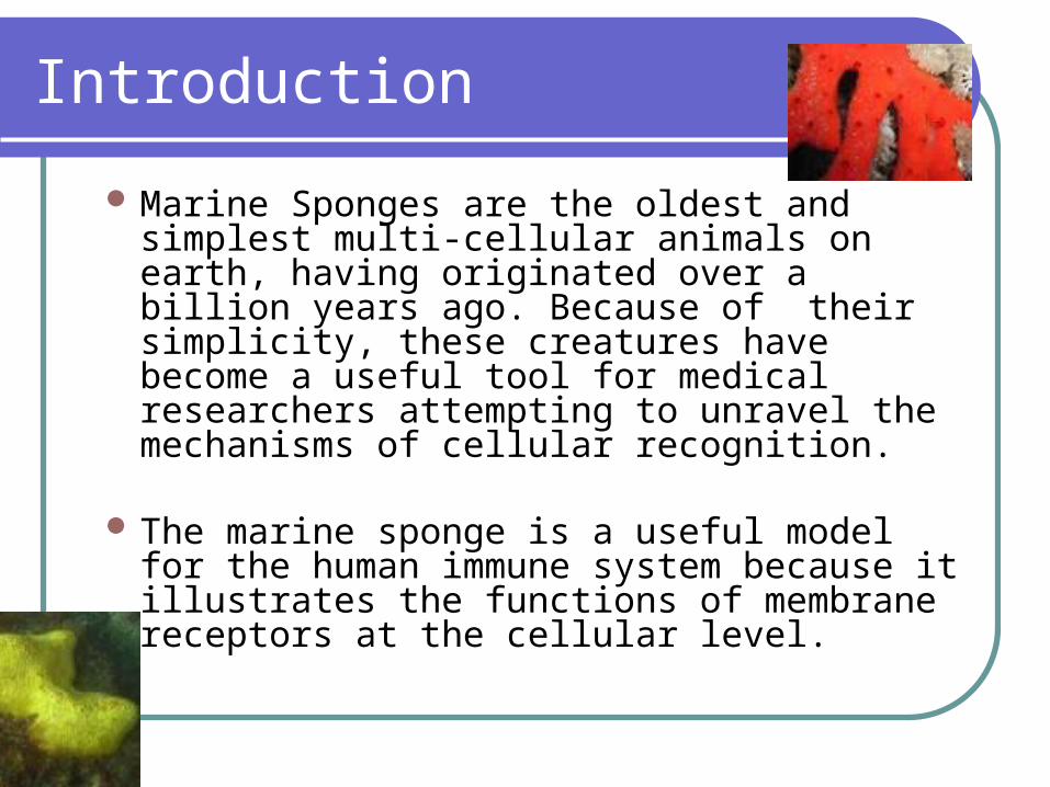
Marine Sponges are the oldest and simplest multi-cellular animals on earth, having originated over a billion years ago. Because of their simplicity, these creatures have become a useful tool for medical researchers attempting to unravel the mechanisms of cellular recognition.
The marine sponge is a useful model for the human immune system because it illustrates the functions of membrane receptors at the cellular level.

Introduction (continued….)
Most receptor molecules are glycoprotein. Glycoprotein have carbohydrates attached to them. (Carbohydrates have the general molecular formula CH2O, and thus were once thought to represent "hydrated carbon". However, the arrangement of atoms in carbohydrates has little to do with water molecules. ) The carbohydrate consists of short, usually branched, chains of sugars and nitrogen-containing amino acids. Sugars are very hydrophilic thanks to their many –OH hydroxyl groups. Their presence make glycoprotein far more hydrophilic than they would be otherwise.
Abstract
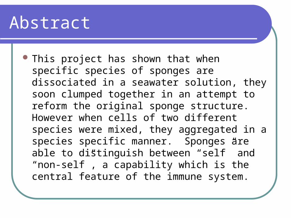
This project has shown that when specific species of sponges are dissociated in a seawater solution, they soon clumped together in an attempt to reform the original sponge structure. However when cells of two different species were mixed, they aggregated in a species specific manner. Sponges are able to distinguish between “self” and “non-self”, a capability which is the central feature of the immune system.

Objective
This research was designed to identify a specific protein involved in cellular reformation of sponges, using the red sponge, microciona porifera, and the yellow sponge, cliona celata.
We are also trying to control the sponge’s ability to identify self.
This can later be used to understand the immune system better. Hence helping us in organ transplants.

Experimental Overview
Experiment 1 – Cells were dissociated into Petri-dishes containing a salt water solution. The sizes of the aggregates were measured over time. Experiment 2 - Cells dissociated from each species were mixed and the sizes of aggregates were noted. Experiment 3 – An extract of cliona celata was mixed with the dissociated cells of microciona porifera. Preliminary results suggest that a factor isolated from cliona celata is required for microciona porifera to re-aggregate

Materials
Petri dishSponges – Microciona Porifera and
Cliona CelataCentrifuge tubeTissue sieveLumbriculus WormsSalt water

Procedure
Experiment I Step 1: A piece of Microciona Porifera and Cliona Celata was placed in a Petri dish and crushed
through a screen tissue sieve. Step 2: The resultants were than place in a Petri dish divided into 3. Dish 1- Microciona Porifera alone Dish 2- Cliona Celata alone Dish 3-Cliona Celata and Microciona Porifera mixed Experiment III Both sponges were placed in a Petri dish and dissociated through a screen tissue sieve. The resulting liquid was than placed in a 4 part Petri dish as follows: Dish 1 – Microciona Porifera Dish 2 – Cliona Celata Dish 3 – Microciona Porifera and Cliona Celata Dish 4 – Microciona Porifera and an extract of Cliona Celata The sizes of the aggregates were then followed on a day-to-day basis.

Procedure (continued… )
As a second source of evidence for cellular recognition, we used lumbriculus regenerating worms to see if it would have an effect on cell reaggregation.

Hypothesis
Initially it was assumed that the yellow sponge (Cleona celata) and the red sponge (Microciona porifera) would both aggregate, regardless of species.
After analyzing the first series of experiments, it was then postulated that the red sponge uses a mechanism to recognize its own cells in an attempt to reform the original organism.

Results
Day 0 Day 6

Sponge structure

Discussion
According to the results of the experiment, marine sponges can be used to study the functions of the immune system. Preliminary results suggests that the “cell-to-cell” recognition system of the marine sponge, Microciona porifera, is similar to antigen antibody recognition, in that cell-surface proteins are involved. When whole sponge cells are mixed, aggregation occurred only between the Microciona cells, indicating its ability to recognize self.

Conclusion
Preliminary results suggest that a factor isolated from the yellow sponge, Cliona Celata enables the cells of the red sponge, Mircrociona Porifera to reaggregate further.

Future Work
In the future, more experiments will be conducted using the red and yellow sponge with a purpose of trying to figure out the factor in the yellow sponge that allows the red sponge to reaggregate.
Then, a carbohydrate specific stain (PAS) will be used to identify surface receptors.

References
1. Jamie Talan (Staff Writer) Receptors“ Cellular signposts tell tissue its job”, Newsday August 4, 2006
2. Journal of Morphology, The fine structure of muocytes in the sponges Microciona prolifera (Ellis and Solander) and Tedania ignis (Duchassaing and Michelotti), Volume 118, Issue 2, Pages 167-181, Published online: 6 Feb 2005
3. J.Biol. “Two cell surface proteins bind the sponge Microciona prolifera aggregation factor” Chem., Vol. 263, Issue 17, 8498-8508, June 1988
4. S, Yum N. kojima, S.-i. Hakomori, S. Kudo, S.Inoue, and Y. Inoue, Binding of rainbow trout is mediated by strong carohydrate-to-carbohydrate interaction between (KDN) GM3 (deaminated neuraminyl ganglioside) and Gg3-like epitope PNAS, March 5, 2002; 99(5): 2854 – 2859.
5. Introduction to Porifera http://webmit.edu/esgbio/www/cb/membranes/gf.html
6. http://www.ucmp.berkeley.edu/porifera/porifera.html
7. Genevieve Thiers “What is chitin?” - © 2002 Pagewise http://wywy.essortment.com/whatischitin_rkkh.htm

Acknowledgements
Mr. Edward IrwinPatty Els
ETLCarlos Strauss
Freeport High SchoolJuan Cuba
Ricardo CubaMSKCC
Dr. Sat BhattacharyaThe Harlem Children Society

Gratitude
Thank
You!