marine fungal community associated with standing plants of...
TRANSCRIPT

UNIVERSIDADE DE LISBOA
FACULDADE DE CIÊNCIAS
Marine fungal community associated with standing
plants of Spartina maritima (Curtis) Fernald
Doutoramento em Biologia
Especialidade em Microbiologia
Maria da Luz Jeremias Cardinha do Maio Calado
Tese orientada por:
Prof. Doutora Margarida Barata e Prof. Doutor Ka-Lai Pang
Documento especialmente elaborado para a obtenção do grau de doutor
2016

UNIVERSIDADE DE LISBOA
FACULDADE DE CIÊNCIAS
Marine fungal community associated with standing
plants of Spartina maritima (Curtis) Fernald
Doutoramento em Biologia
Especialidade em Microbiologia
Maria da Luz Jeremias Cardinha do Maio Calado
Tese orientada por:
Prof. Doutora Margarida Barata e Prof. Doutor Ka-Lai Pang
Júri:
Presidente:
● Doutor José Simões
Vogais:
● Doutor Nelson Lima
● Doutora Raquel Coelho
● Doutor Alan Phillips
● Doutor José Paulo Sampaio
● Doutora Maria Isabel Caçador
● Doutora Margarida Barata
Documento especialmente elaborado para a obtenção do grau de doutor
Fundação para a Ciência e Tecnologia (FCT)
2016
3
Tese apresentada à Universidade de Lisboa para obtenção do grau de
Doutor em Biologia, especialidade em Microbiologia
O trabalho de investigação apresentado nesta tese foi apoiado
financeiramente pela Fundação para a Ciência e Tecnologia pela atribuição
da Bolsa de doutoramento com a referência SFRH/BD/48525/2008
A tese deve ser citada como:
Calado ML (2016) Marine fungal community associated with standing plants
of Spartina maritima (Curtis) Fernald. Ph.D. Thesis, Faculty of Sciences of
University of Lisbon
Thesis presented at University of Lisbon for obtaining doctoral degree in
Biology, Microbiology
The research work presented in this thesis was supported financially by
Foundation for Science and Technology (FCT) through a Ph.D. grant with
the reference SFRH/BD/48525/2008
This thesis should be cited as:
Calado ML (2016) Marine fungal community associated with standing plants
of Spartina maritima (Curtis) Fernald. Ph.D. Thesis, Faculty of Sciences of
University of Lisbon

4
5
TABLE OF CONTENTS
ACKNOWLEDGMENTS
7
RESUMO
9
ABSTRACT
13
LIST OF FIGURES
15
LIST OF TABLES
17
THESIS FRAMEWORK
19
CHAPTER 1 – General Introduction
21
1.1 Marine fungi – a brief review
23
1.2 Salt marsh fungi
31
1.3 Objectives
51
1.4 References
51
CHAPTER 2 - Diversity and ecological characterization of sporulating higher
filamentous marine fungi associated with Spartina maritima (Curtis) Fernald in two
Portuguese salt marshes
63
CHAPTER 3 - Ecological preferences of marine fungi associated with standing
decaying plants of Spartina maritima (Curtis) Fernald
111
CHAPTER 4 – Final overview
159
4.1 General conclusions
161
4.2 Future perspectives
167
4.3 References
167

6
7
ACKNOWLEDGMENTS
This thesis represents the end of a long, demanding and challenging, but undoubtedly enriching
and rewarding journey. It definitely made me grow professionally and personally. However, the
execution and conclusion of this journey would not have been possible without the help and
support of several persons, to whom I would like to address my heartfelt gratitude.
First of all, I would like to thank my supervisors Professor Margarida Barata and Professor Ka-
Lai Pang for kindly receiving me in their labs, giving me all the conditions I needed to develop
my research work, and for their constant support, availability, constructive discussions, helpful
“brainstormings” and professional guidance throughout the development of this project. Also, I
am truly grateful for their generosity, trust and belief in me, friendship and encouraging and
motivational words whenever needed.
Secondly, I would like to thank to
- Luís Carvalho, for his indispensable help in the statistical analysis of data and valuable and
constructive suggestions;
- Professor Manuela Carolino, for her kindness and support, and Catarina Gouveia, for being a
great lab mate during the long time I spent under the microscope;
- Rita Severino and Cristina Oliveira for their precious help in the culture experiment;
- Professor Cristina Cruz, for accepting me in her work team and laboratory;
- Professor Cristina Figueiredo, for her kindly help and expertise in the lyophilisation process;
- and all academic and non-academic staff from the Department of Plant Biology that
contributed somehow for this work in different ways and time moments.
Thirdly, I would like to express my sincere gratitude to my friends
- Egídia Azevedo and Sílvia Mendonça, for their friendship and encouragement along this path;
- Shine, Yufen, Cha Cha, Su Su, Iris, Alex and all my Taiwanese colleagues and friends, for
welcoming me into their lovely and beautiful country, for being (together with Professor Ka-Lai
Pang) my surrogate family for 8 months, and for turning the most challenging part of this thesis
into one of the most remarkable experiences of my life;
- António Miranda, for being a great office mate and friend and for his peculiar sense of humour
that turned our lunch moments into stress-release and laughter therapies;
- Joana Rosado, for her friendship in the real sense of the word (thank you for being always
present!).
Finally, I am really grateful to my family, particularly

8
- Sérgio, for his love, constant encouragement, patience and companionship, and for helping
me overcoming frustrating time periods of the research life and difficult periods of my personal
life (you definitely changed my life…for the best!);
- my nephews Rafael and Lucas, who born during this journey, for colouring my life and for
making me discover the powerful love, joy, happiness and privilege of being an aunt;
- women of my life to whom I dedicate my thesis (this would never have been possible without
our strong and powerful bond!): my sister Isa, my aunts Florbela and Inês, my grandmothers
Joana (my sweet star, who unfortunately did not have the chance to testify this moment, but
was and will always be the pillar of our family) and Constança, and especially my mother (my
anchor), for supporting me at every step of this process and during the hardest moments of my
and our lives.
9
RESUMO
A presente tese pretendeu contribuir para o aumento do conhecimento relativo às comunidades
de fungos marinhos superiores que colonizam ecossistemas intertidais temperados, dada a
escassez de estudos desenvolvidos neste domínio em geral, e em Portugal em particular.
Especificamente pretendeu-se inventariar as espécies fúngicas associadas a uma das
macrófitas mais importantes e dominantes dos sapais da costa portuguesa, a Spartina maritima
(Curtis) Fernald, e compreender melhor a dinâmica da comunidade e diversos aspectos da
ecologia de cada espécie fúngica.
O estudo foi desenvolvido em dois sapais (Castro Marim e Ria de Aveiro) com características
distintas, no que respeita à localização geográfica, configuração física, estado de conservação
e representatividade da planta hospedeira.
O sapal de Castro Marim situa-se no troço final do estuário do Guadiana, na costa sudeste de
Portugal (37.23° N, 7.42° W), e está incluído numa Reserva Natural; apresenta, por isso,
estatutos especiais de protecção e uma comunidade de S. maritima bem conservada, que se
distribui paralelamente ao rio numa faixa contínua. O sapal da lagoa costeira da Ria de Aveiro
localiza-se na costa noroeste de Portugal (40.62° N, 8.74° W) e integra uma complexa rede de
canais sujeita a fortes pressões antrópica; nesta área, a comunidade de S. maritima está muito
fragmentada e dispersa ao longo da faixa de vegetação.
A amostragem da comunidade de fungos marinhos envolveu a recolha de 195 plantas inteiras,
maduras, enraizadas e em posição natural de S. maritima durante 2 anos (de Outubro de 2010
a Agosto de 2012), com uma periodicidade de 2 meses; recolheram-se 20 plantas de cada área
de estudo nos primeiros 3 meses, e 15 plantas nos restantes períodos. Em laboratório, cada
planta, previamente lavada e seca ao ar, foi separada em nove categorias de substrato, de
acordo com as estruturas vegetais que a compunham e estado fisiológico das mesmas;
especificamente, em bainhas vivas, bainhas senescentes, bainhas em decomposição, caules
vivos, caules senescentes, caules em decomposição, limbos vivos, limbos senescentes e
limbos em decomposição. As denominações “vivo”, “senescente” e “em decomposição”,
caracterizaram tecidos verdes, amarelos e acastanhados, com estrutura física nada, pouco ou
bastante alterada respectivamente.
A identificação dos fungos marinhos recorreu a dois métodos distintos, mas complementares;
(1) a observação directa de estruturas fúngicas (esporos, estruturas de frutificação e
hipopódios) e (2) a sequenciação da região ITS (Internal Transcribed Spacers) do DNA
ribossomal nuclear (rDNA).
O primeiro método envolveu a análise individual de cada substrato vegetal à lupa; as estruturas
fúngicas detectadas nesse substrato foram observadas ao microscópio e identificadas com
base nas suas características morfológicas, recorrendo a chaves dicotómicas específicas para
identificação de fungos marinhos. Adicionalmente registou-se a distribuição vertical e
densidade das estruturas fúngicas de cada espécie. No período de Fevereiro de 2012 a Agosto
10
de 2012, foram ainda recolhidas 5 plantas extras, as quais foram lavadas e imediatamente
observadas. Algumas estruturas de frutificação (ascocarpos ou picnídios) diferenciadas neste
material vegetal fresco foram extraídas e utilizadas na obtenção de culturas puras, através do
método de esporo único. Os fungos isolados foram preservados por três métodos distintos: (1)
crescimento activo em Corn Meal Agar (CMA) preparado com água do mar diluída (50%), a 4
°C; (2) discos de micélio imersos em água do mar estéril (50%), a 4 °C; e (3) discos de micélio
imersos em solução aquosa de glicerol (10%), a -80 ºC. Duas culturas puras de cada espécie,
de cada local, foram seleccionadas para determinar a taxa de crescimento vegetativo em CMA
preparado com água destilada e água do mar diluída (50%).
Após esta análise, agrupou-se o material vegetal pertencente à mesma categoria de substrato
e proveniente de todas plantas recolhidas no mesmo período de amostragem e local de estudo.
Todos os substratos vegetais foram posteriormente liofilizados.
A identificação molecular dos fungos marinhos foi realizada apenas a partir dos substratos
recolhidos no primeiro ano de amostragem. A metodologia molecular envolveu, numa primeira
fase, a extracção do DNA nuclear das culturas puras, e amplificação e sequenciação da região
ITS do rDNA; estas sequências constituíram uma colecção de referência para comparação e
identificação dos fungos presentes nas amostras vegetais. Posteriormente, o DNA nuclear dos
88 substratos vegetais foi igualmente extraído, e a mesma região genómica amplificada por
primers com especificidade para fungos. Os amplicões obtidos de cada substrato foram
clonados no sentido de isolar cada sequência ITS; no total, obtiveram-se 1037 clones. No
sentido de seleccionar os clones de DNA recombinante representativos de cada biblioteca da
região ITS de cada substrato vegetal, foi realizada uma análise de perfis de restrição (RFLP);
os diferentes perfis de restrição de cada biblioteca foram sequenciados.
Estas sequências foram comparadas com sequências depositadas na base de dados pública
internacional GenBank e com as sequências da colecção de referência (culturas puras), e
identificadas até à menor categoria taxonómica possível. A identificação destas sequências
permitiu a extrapolação e identificação das restantes sequências extraídas de todos os
substratos vegetais analisados.
A conjugação dos métodos morfológico e molecular revelou-se fundamental para o
conhecimento da diversidade dos fungos marinhos superiores associados a S. maritima, na
medida em que permitiu confirmar ou corrigir as identificações dos fungos mais comuns
realizadas por cada um dos métodos e complementar o inventário com as espécies mais
infrequentes. De uma maneira geral, houve uma concordância entre os dois métodos no que
respeita à representatividade de cada fungo nas comunidades, nos substratos vegetais e nas
áreas de estudo. No total, foram identificados 45 fungos nas primeiras fases do processo de
decomposição de plantas de S. maritima. Tal como em estudos semelhantes anteriores
desenvolvidos em sistemas intertidais, esta comunidade revelou ser dominada por fungos
pertencentes ao filo Ascomycota e à classe Dothideomycetes.
A comparação das comunidades de fungos marinhos associadas a S. maritima e outras
espécies de Spartina evidenciou a existência de um grupo nuclear (core group) de espécies
11
que surge associado a este genéro de plantas hospedeiras, independentemente da sua
localização geográfica. Este grupo inclui espécies em associação exclusiva com plantas do
género Spartina, como Anthostomella spissitecta, Byssothecium obiones, Buergenerula
spartinae, Mycosphaerella sp. I, Phaeosphaeria halima, Phaeosphaeria spartinicola; espécies
que colonizam igualmente outras plantas intertidais de climas temperados, como Leptosphaeria
marina e Sphaerulina orae-maris; e espécies cosmopolitas, que surgem em diversos substratos
de climas tropicais e temperados, como Aniptodera chesapeakensis e Dictyosporium
pelagicum. Apesar dos fungos Lulworthia sp. 1 e Stagonospora sp. 1 não terem sido
identificados até à espécie, estes géneros taxonómicos são comumente observados em
plantas de Spartina.
As espécies B. obiones, B. spartinae, Mycosphaerella sp. I, P. halima e P. spartinicola foram
registadas como muito frequentes na comunidade geral amostrada neste estudo e presentes
em todos ou na maioria dos períodos de amostragem, em concordância com estudos
semelhantes realizados com outras espécies de Spartina. Este estudo revelou que existem, no
entanto, espécies muito frequentes nas comunidades associadas a S. maritima mas ausentes
noutras espécies de Spartina, como o fungo cosmopolita Natantispora retorquens, e vice-versa.
As espécies infrequentes Anthostomella spissitecta, Camarosporium roumeguerii, Ceriporia
lacerata, Coniothyrium obiones, Cryptococcus mangaliensis, Decorospora gaudefroyi,
Erythrobasidium hasegawianum, Halosarpheia trullifera, Leptosphaeria marina, Penicillium
chrysogenum e Stagonospora haliclysta foram registadas pela primeira vez em associação com
o género Spartina e/ou com S.maritima.
A presença ou ausência das espécies nas comunidades fúngicas associadas à mesma ou
outra espécie de Spartina poderá estar relacionada com diferenças na estrutura física e
composição química das plantas hospedeiras e/ou factores macro ou microambientais.
Os fungos marinhos nas duas comunidades amostradas neste estudo e particularmente os
mais frequentes apresentaram padrões de distribuição verticais específicos nas plantas de S.
maritima em posição natural, i.e. localizavam-se na mesma posição vertical relativa. A posição
relativa na planta parece reflectir o grau de adaptação das espécies fúngicas às condições
marinhas. As diferentes estratégias de reprodução sexuada e assexuada ao longo das plantas
representam e evidenciam algumas das estratégias adoptadas pelos fungos marinhos para se
adaptarem; os fungos incluídos na classe Sordariomycetes, com ascos unitunicados e
mecanismos passivos de libertação de esporos, dominam as partes basais das plantas,
enquanto os fungos incluídos na classe Dothideomycetes, com ascos bitunicados e
mecanismos activos de libertação de esporos, ocupam principalmente as partes aéreas. Os
dados sugerem que os fungos que surgem nas partes basais das plantas são marinhos
obrigatórios, enquanto os fungos das partes superiores são marinhos facultativos. As partes
intermédias representam, por isso, uma zona de transição e sobreposição dos fungos que
colonizam exclusivamente os ecossistemas marinhos e os que podem provir de ecossistemas
fluviais ou terrestres. Apesar de revelarem diferentes dependências e tolerâncias à salinidade
em condições naturais, todos estes fungos dominantes cresceram em meio de cultura sem
12
salinidade, mas com uma taxa de crescimento mais elevada em meio de cultura preparado
com água do mar. Estes resultados evidenciaram a elevada plasticidade destes fungos de se
adaptarem a diferentes condições ambientais.
As espécies dominantes da comunidade foram também as que exibiram uma área de
distribuição vertical mais extensa, e que surgiram em mais de uma estrutura vegetal, nos
diferentes estados fisiológicos, ao longo de todo o período de amostragem. A presença destas
espécies sapróbias em tecidos vegetais vivos sugere que estas possam iniciar o processo de
colonização como endófitas.
A conjugação de todos os resultados indicia que os padrões de distribuição vertical, e a
ocorrência e o papel ecológico dos fungos mais frequentes dependem da fase do ciclo de vida
da planta e disponibilidade dos substratos vegetais, das condições microambientais dos
substratos e adaptação aos ciclos de submersão-emersão, e dos potenciais fungos
competitores. Durante o processo de decomposição de S. maritima, os fungos marinhos
obrigatórios B. obiones, Lulworthia sp. 1 e N. retorquens assumem um papel ecológico mais
activo na decomposição das bainhas e caules inferiores; o fungo marinho facultativo
Mycosphaerella sp. I, na decomposição dos limbos superiores; os fungos marinhos facultativos
P. halima e Stagonospora sp. 1, na decomposição das bainhas e limbos superiores; e os
fungos marinhos facultativos P. spartinicola e B. spartinae, na decomposição de todas as
estruturas vegetais.
Em suma, este estudo contribuiu para um enriquecimento do conhecimento da composição
específica, diversidade e dinâmica das comunidades de fungos marinhos superiores
associadas a plantas maduras, enraizadas e em posição natural de S. maritima, bem como dos
requisitos e papel ecológicos de cada espécie na decomposição destas plantas hospedeiras.
Palavras-chave: fungos marinhos; Spartina maritima; decomposição; requisitos ecológicos;
potencial papel ecológico
13
ABSTRACT
The major purpose of this thesis was to complement the current knowledge regarding marine
fungal communities and particularly those inhabiting Portuguese temperate salt marshes.
Specifically, this study mainly intended to assess the species composition and diversity of the
fungal communities associated with one of the most dominant macrophytes in these
ecosystems, Spartina maritima (Curtis) Fernald, and to contribute to a better understanding of
community dynamics and key ecological aspects of the fungi. The study was conducted in two
geographically and physically distinct salt marshes, Castro Marim and Ria de Aveiro, where 195
mature, standing live plants were collected over a 2-year period (October 2010 to August 2012)
from each study site. Each air-dried plant was separated into nine substrate categories
according to the vegetative structure (leaf sheaths, stems and leaf blades) and physiological
state of each structure (live, senescent and decaying). Identification of marine fungi was
performed by two distinct, but complementary methods, i.e. direct observation of fungal
structures (fruit bodies, spores and hyphopodia) and sequencing of the internal transcribed
spacer regions of rDNA (ITS). The first method involved an individual observation of each
substrate under dissecting- and light microscopes for detection of fungal structures; fungal taxa
were morphologically identified using specific dichotomous keys for marine fungi. The vertical
position and density of fruiting structures produced by each identified fungus was also recorded.
The most frequent fungi were isolated in pure cultures by single spore method. Plant materials
from the same substrate category, sampling period and study site were mixed and freeze-dried.
Only the plant samples from the first sampling period were used for molecular identification of
fungi. This second method involved DNA extraction of pure fungal isolates and plant samples,
and amplification of the ITS region. Amplicons from plant samples were cloned in order to
isolate individual amplicons of mixed PCR products. ITS sequences of the 1037 clones obtained
from the plant samples were submitted to a restriction fragment length polymorphism analysis
(RFLP); clones with different digestion profiles were sequenced. Phylogenetic analyses were
performed with sequences of clones, fungal isolates and BLAST best-hits. A comparison
between morphological and molecular methods revealed a general agreement in taxonomic
assignments and representativeness of each fungus in the community, vegetative structure and
study site. The combination of both methods was demonstrated to be crucial for a more realistic
and accurate representation of the fungal community. Forty-five fungal taxa were recorded in S.
maritima samples; 91% of these were filamentous ascomycetes, included in the
Dothideomycetes and Sordariomycetes. The majority of the fungal species most frequently
recorded in this study were previously described from other species of Spartina. Nevertheless,
the studied fungal community also included other infrequent species that represent new records
for the genus Spartina and/or S. maritima plants, e.g. Anthostomella spissitecta,
Camarosporium roumeguerii, Ceriporia lacerata, Coniothyrium obiones, Cryptococcus
mangaliensis, Decorospora gaudefroyi, Erythrobasidium hasegawianum, Halosarpheia trullifera,

14
Leptosphaeria marina, Penicillium chrysogenum and Stagonospora haliclysta. The presence or
absence of species in fungal communities may be related with intra- and interspecific
differences in the physical structure and chemical composition of the host plants and/or macro
and microenvironmental factors.
Similarly to other grass-like plants, the results also demonstrated that the marine fungi are
vertically distributed along standing plants of S. maritima. Moreover, the most frequent fungal
taxa exhibited wide vertical distribution ranges, a high investment in the production of fruiting
structures and were present during all the sampling period on senescent and decaying
vegetative structures. The majority of these fungi were also found on live plant tissues, which
indicated that these saprobic species might initiate the colonisation of plant substrates as
endophytes. These findings suggested that the vertical distribution patterns, and occurrence
and ecological role of most frequent fungi depend on the phase of plant life cycle and substrate
availability, micro-environmental conditions of substrates and adaptation to
submersion/exposure cycles, and potential fungal competitors. During the decay process of S.
maritima, the obligate marine fungi Natantispora retorquens, Byssothecium obiones and
Lulworthia sp.1 seem to be involved in the complete decomposition of lower leaf sheaths and
stems; facultative marine fungi Mycosphaerella sp. I, of leaf blades; facultative marine fungi
Phaeosphaeria halima and Stagonospora sp. 1, of upper standing leaves; and Buergenerula
spartinae and Phaeosphaeria spartinicola, of all vegetative structures.
Key words: marine fungi; Spartina maritima; decomposition; ecological requirements; potential
ecological role

15
LIST OF FIGURES
CHAPTER 1
Fig. 1 Fruiting structures of ascomycetous fungi
24
Fig. 2 Intertidal ecosystems
25
CHAPTER 2
Fig. 1 Study sites
69
Fig. 2 Vertical distribution of fungal taxa on standing S. maritima in Castro Marim and Ria de
Aveiro salt marshes
77
Fig. 3 Cluster dendrogram based on Bray-Curtis similarity of fungal communities colonizing basal,
middle and top plant portions in each sampling period and study site, considering the frequency of
occurrence of fungal taxa
79
Fig. 4 Two-dimensional DCA plot expressing the fungal taxa and the 3 vertical plant portions
spatial distributions based on frequency of occurrence of fungal taxa in each portion, in each
sampling period and in each study site
80
Fig. 5 Bimonthly variation of the frequency of occurrence (%) of each fungal taxon in Castro Marim
and Ria de Aveiro salt marshes, and average variation of the frequencies of occurrence in each
sampling period
82
Fig. 6 Bimonthly variation of frequency of occurrence (%) of high-density of fungal structures
produced by each fungal taxon in Castro Marim and Ria de Aveiro salt marshes, and average
variation of the frequencies of occurrence in each sampling period
83
Fig. 7 Bimonthly variation in the average height of host plants and vertical position of fungal taxa
that occurred in most of the sampling periods on the plants in Castro Marim and Ria de Aveiro salt
marshes
84
Fig. 8 Daily growth rate of 8 fungal taxa isolated from Castro Marim and 6 fungal taxa isolated
from Ria de Aveiro salt marshes under two culture conditions, media with diluted seawater and
with distilled water
86
Fig. S1 Marine fungal taxa associated with Spartina maritima standing plants
104
CHAPTER 3
Fig. 1 Phylogenetic consensus trees based on a maximum parsimony analysis for the
Dothideomycetes and Sordariomycetes, showing the placement of clone sequences, isolate
sequences and BLAST best-hit sequences
127
Fig. 2 Number of amplicons assigned to each fungal taxon retrieved from Castro Marim and Ria
de Aveiro plant samples
130
Fig. 3 Cluster dendrogram based on Bray-Curtis similarities of plant substrates, considering the
presence/absence of fungal taxa on each plant substrate, in each sampling period and in each
study site
144
16
Fig. 4 Nonmetric multidimensional scaling plot based on Bray-Curtis similarities of plant substrates
considering the presence/absence of fungal taxa in each plant substrate, in each sampling period
and in each study site
144
Fig. 5 Two-dimensional DCA plot expressing the spatial distributions of fungal taxa and the nine
plant substrates based on the frequencies of occurrence of fungal taxa in each plant substrate, in
each sampling period and in each study site
146
CHAPTER 4
Fig. 1 Illustrative representation of vertical distribution of very frequent and frequent fungi on
standing plants of Spartina maritima
163
17
LIST OF TABLES
CHAPTER 1
Table 1 Filamentous fungi associated with Juncus roemerianus, Spartina spp. and Phragmites
australis in marsh ecosystems
35
CHAPTER 2
Table 1 Abiotic conditions in Guadiana estuary and Ria de Aveiro coastal lagoon
70
Table 2 Percent frequency of occurrence of fungal taxa and species diversity indices in
communities in Castro Marim and Ria de Aveiro salt marshes and on average between the sites
75
Table 3 Diversity indices and number of records in the 3 vertical portions of the plants in Castro
Marim and Ria de Aveiro salt marshes
78
CHAPTER 3
Table 1 Fungal taxa identified on Spartina maritima samples by molecular methods
122
Table 2 Number of sampling periods during the first year in which each plant substrate was
collected, and absolute and estimated species richness using Chao 2 and Jacknife 2 estimators in
each plant substrate, study site (CM Castro Marim; AV Ria de Aveiro) and combination of both
study sites
133
Table 3 Percent frequencies of occurrence (V very frequent: >20%; F frequent: 10-20%; I
infrequent: <10%, according to the 3 categories proposed by Tan et al (1989)) and/or presences
( ̶ ) of fungal taxa on different plant substrates in Castro Marim salt marsh in each sampling period,
identified by morphological and molecular methods respectively
135
Table 4 Percent frequencies of occurrence (V very frequent: >20%; F frequent: 10-20%; I
infrequent: <10%, according to the 3 categories proposed by Tan et al (1989)) and/or presences
( ̶ ) of fungal taxa on different plant substrates in Ria de Aveiro salt marsh in each sampling period,
identified by morphological and molecular methods respectively
139

18
19
THESIS FRAMEWORK
This thesis is organized into four chapters.
Chapter 1 (General Introduction) comprises an overview of the current knowledge concerning
marine fungi in general (Marine fungi – a brief review), and fungi inhabiting salt marshes in
particular (Salt marsh fungi). This last review was included in the recently published volume
“Marine Fungi and fungal-like organisms”, as a book chapter. This introduction also addresses
the main gaps and limitations to the understanding of marine fungal communities and the
objectives of the present study.
The original research work performed in this study was included in two papers published in
international scientific journals, which represent chapters 2 and 3. The structure and original
content of the published papers were maintained in the essential but the formatting style was
changed; all the chapters follow the same formatting and bibliographical rules.
Chapter 2 (Diversity and ecological characterization of sporulating higher filamentous
marine fungi associated with Spartina maritima (Curtis) Fernald in two Portuguese salt
marshes) presents an inventory of fungal taxa associated with standing plants of Spartina
maritima assessed by morphological identification of fungal structures. The biotic and abiotic
factors that may determine the presence and abundance of fungal species on Spartina maritima
and their distribution along the vertical axis of these host plants are also enumerated.
Chapter 3 (Ecological preferences of marine fungi associated with standing decaying
plants of Spartina maritima (Curtis) Fernald) presents the results from the molecular
identification of fungi on live, senescent and decaying leaf sheaths, stems and leaf blades of
standing plants of Spartina maritima. In this chapter, the molecular and morphological methods
adopted in this study are compared. The presence and prevalence of fungi on each plant
substrate are used to assess their ecological preferences and infer about their ecological role in
the decay of Spartina maritima.
Chapter 4 (Final overview) highlights the main results of this study, pointing out some
knowledge gaps that should be approach in future studies.
Over the past 6 years, many new species have been described from marine habitats and many
taxonomic ambiguities have been resolved as a result of the application of more accurate and
improved molecular tools. This has led to dramatic changes in nomenclatural rules and
taxonomic classification of fungi after the chapter “Salt marsh fungi” and the paper “Diversity
and ecological characterization of sporulating higher filamentous marine fungi associated with
Spartina maritima (Curtis) Fernald in two Portuguese salt marshes” were written. The terms
anamorph and teleomorph were replaced to asexual and sexual morphs respectively, and
sequenced asexual morphs were transferred from the artificial group “anamorphic fungi” to
different taxonomic categories within the Ascomycota and Basidiomycota; holomorphic fungal

20
species, for which both sexual and asexual morphs were demonstrated to be connected, were
designated by only one name. These new rules were followed in the paper “Ecological
preferences of marine fungi associated with standing decaying plants of Spartina maritima
(Curtis) Fernald”.

21
CHAPTER 1 - General Introduction

22
23
1.1 Marine fungi – A brief review
Marine fungi represent an ecological group of fungi that occur from inshore regions to deep
oceanic waters (Fell and Newell 1998; Hyde et al. 1998), composed primarily by higher
filamentous fungi included in the Basidiomycota (Ustilaginomycetes and Agaricomycetes) and
Ascomycota (Dothideomycetes, Eurotiomycetes, Laboulbeniomycetes, Lecanoromycetes,
Leotiomycetes, Lichinomycetes, Arthoniomycetes and Sordariomycetes) (Kohlmeyer and
Kohlmeyer 1979; Hyde et al. 2000; Jones and Pang 2012). Most marine fungi belong to the
Dothideomycetes and Sordariomycetes, particularly to Halosphaeriaceae (Jones et al. 2009;
Sakayaroj et al. 2011; Jones and Pang 2012; Pang 2012).
Marine fungi include species with a wide range of nutritional modes, i.e. fungal species that
establish a parasitic or symbiotic mycorrhizal, lichenoid or endophytic relationship with several
hosts, and saprobes on dead organic material of plant and animal origin (Kohlmeyer and
Kohlmeyer 1979; Hyde et al. 1998; Kohlmeyer et al. 2004; Jones 2011a; Richards et al. 2012).
The majority of marine fungal species are decomposers of plant materials, particularly of woody
substrates (Kohlmeyer and Kohlmeyer 1979; Hyde et al. 1998; Jones 2000; Pointing and Hyde
2000). Inherent to their metabolic activities, saprobic marine fungi, especially filamentous ones,
play an important functional and ecological role in the nutrient recycling and energy flow in
marine ecosystems (Newell 1993, 1996; Hyde and Lee 1995; Hyde et al. 1998; Newell and
Porter 2000; Pang and Jones 2012). The strategy adopted by mycelial fungi implicates a
penetrating growth mode by expanding hyphal tips combined with enzyme´s activity (Torzilli and
Andrykovitch 1986; Newell 1996; Lyons et al. 2003; Raghukumar 2004b). Marine fungi are
widely recognized by the diverse range of extracellular biologically-important enzymes involved
in the degradation of recalcitrant cell wall materials, such as cellulases, laccases, lignin
peroxidases and Mn-dependent peroxidases (Gessner 1980; Torzilli and Andrykovitch 1986;
Bergbauer and Newell 1992; Newell et al. 1996b; Pointing et al. 1998; Raghukumar 2002,
2004a; Lyons et al. 2003; Raghukumar 2004b; Jones 2011a). Biotechnological potential of
lignocellulolytic marine fungi in bioremediation has been widely investigated (Newell et al.
1996b; Raghukumar 2002, 2004a; Raghukumar 2004b; Jones 2011a). In addition, marine fungi
represent an important source of structurally unique bioactive secondary metabolites with
antimicrobial, anticancer, anti-inflammatory and analgesic properties (Bugni and Ireland 2004;
dela Cruz et al. 2006; Schulz et al. 2008; Jones 2011a; Ebel 2012; Singh et al. 2012; Overy et
al. 2014a). The production of these metabolites is species-specific (dela Cruz et al. 2006) and
apparently is not affected by the geographical origin of species (Schulz et al. 2008). Eighty out
of over 1000 metabolites that have been characterized to date were isolated from marine fungi
(Overy et al. 2014a). Although the reasons associated with the production of these metabolites
are not totally known, one of the main purposes might be a chemical defense strategy in
response to interference competition (Pointing et al. 2000; Jensen and Fennical 2002;
24
Panebianco et al. 2002); some of these compounds were demonstrated to limit spore
germination or fungal growth (Miller 2000). For endophytes, these metabolites might also play
an important role in the communication with host species and for adaptation of the hosts to
environmental stress (Meng et al. 2011).
Nevertheless their trophic strategy, marine fungal species are physiologically and
morphologically adapted to marine environments.
Given the high salinity level of marine environments, marine fungi exhibit different mechanisms
in order to maintain homeostasis in their cells; some fungal species synthesize or absorb
compatible solutes from surrounding water to their cytoplasm and accumulate them in
compartmentalized vacuoles, while others pump sodium ions out of cells (Jennings and Garrill
2000; Jones 2000). Most marine fungi show an optimal vegetative growth and synthesis of
secondary metabolites in a saline medium than without any marine salts (Masuma et al. 2001;
dela Cruz et al. 2006; Huang et al. 2011; Pang et al. 2011; Overy et al. 2014a).
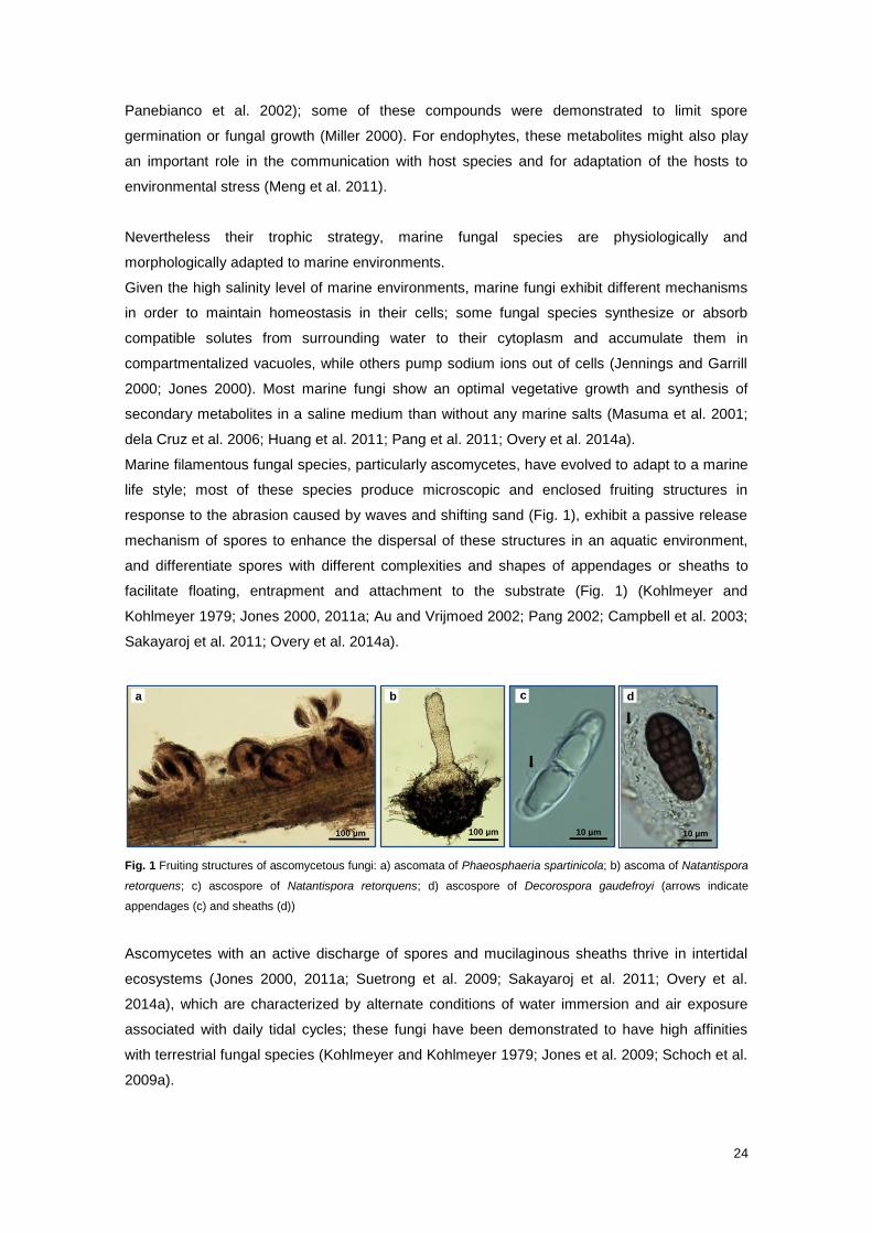
Marine filamentous fungal species, particularly ascomycetes, have evolved to adapt to a marine
life style; most of these species produce microscopic and enclosed fruiting structures in
response to the abrasion caused by waves and shifting sand (Fig. 1), exhibit a passive release
mechanism of spores to enhance the dispersal of these structures in an aquatic environment,
and differentiate spores with different complexities and shapes of appendages or sheaths to
facilitate floating, entrapment and attachment to the substrate (Fig. 1) (Kohlmeyer and
Kohlmeyer 1979; Jones 2000, 2011a; Au and Vrijmoed 2002; Pang 2002; Campbell et al. 2003;
Sakayaroj et al. 2011; Overy et al. 2014a).
Fig. 1 Fruiting structures of ascomycetous fungi: a) ascomata of Phaeosphaeria spartinicola; b) ascoma of Natantispora
retorquens; c) ascospore of Natantispora retorquens; d) ascospore of Decorospora gaudefroyi (arrows indicate
appendages (c) and sheaths (d))
Ascomycetes with an active discharge of spores and mucilaginous sheaths thrive in intertidal
ecosystems (Jones 2000, 2011a; Suetrong et al. 2009; Sakayaroj et al. 2011; Overy et al.
2014a), which are characterized by alternate conditions of water immersion and air exposure
associated with daily tidal cycles; these fungi have been demonstrated to have high affinities
with terrestrial fungal species (Kohlmeyer and Kohlmeyer 1979; Jones et al. 2009; Schoch et al.
2009a).
10 µm 100 µm 100 µm 10 µm
a b c d
25
Most filamentous ascomycetes possess a delimiting membrane, which prevents the premature
expansion of the appendages and sheaths before they are released into the surrounding water
(Jones 2011a).
Although the biogeography of marine fungi is still not fully understood (Jones and Pang 2012),
some fungal species have been exclusively found in tropical, subtropical or temperate climate
regions, while others were found to be cosmopolitan species (Kohlmeyer and Kohlmeyer 1979;
Hyde and Lee 1995; Hyde et al. 1998; Jones et al. 1998; Sarma and Hyde 2001; Alias et al.
2010; Jones and Pang 2012).
Among the several factors that could determine the macro-geographical distribution of marine
fungi, the availability of substrates for colonization and water temperature and salinity are
apparently the most important controlling key-factors (Hyde and Lee 1995; Jones 2000, 2011a;
Sarma and Hyde 2001; Kohlmeyer et al. 2004; Jones and Pang 2012).
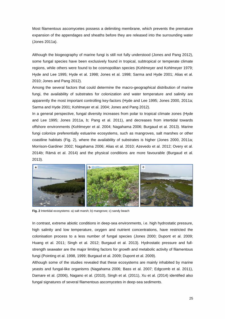
In a general perspective, fungal diversity increases from polar to tropical climate zones (Hyde
and Lee 1995; Jones 2011a, b; Pang et al. 2011), and decreases from intertidal towards
offshore environments (Kohlmeyer et al. 2004; Nagahama 2006; Burgaud et al. 2013). Marine
fungi colonize preferentially estuarine ecosystems, such as mangroves, salt marshes or other
coastline habitats (Fig. 2), where the availability of substrates is higher (Jones 2000, 2011a;
Morrison-Gardiner 2002; Nagahama 2006; Alias et al. 2010; Azevedo et al. 2012; Overy et al.
2014b; Rämä et al. 2014) and the physical conditions are more favourable (Burgaud et al.
2013).
Fig. 2 Intertidal ecosystems: a) salt marsh; b) mangrove; c) sandy beach
In contrast, extreme abiotic conditions in deep-sea environments, i.e. high hydrostatic pressure,
high salinity and low temperature, oxygen and nutrient concentrations, have restricted the
colonisation process to a less number of fungal species (Jones 2000; Dupont et al. 2009;
Huang et al. 2011; Singh et al. 2012; Burgaud et al. 2013). Hydrostatic pressure and full-
strength seawater are the major limiting factors for growth and metabolic activity of filamentous
fungi (Pointing et al. 1998, 1999; Burgaud et al. 2009; Dupont et al. 2009).
Although some of the studies revealed that these ecosystems are mainly inhabited by marine
yeasts and fungal-like organisms (Nagahama 2006; Bass et al. 2007; Edgcomb et al. 2011),
Damare et al. (2006), Nagano et al. (2010), Singh et al. (2011), Xu et al. (2014) identified also
fungal signatures of several filamentous ascomycetes in deep-sea sediments.
1 a b c
26
At a more reduced scale, the influence of biotic and physical factors in the colonization process
of each fungal species, such as tolerance to air exposure or submergence conditions, substrate
exclusivity and competitive abilities, varies from species to species (Gessner 1977; Poon and
Hyde 1998b; Alias and Jones 2000a, b; Barata 2002, 2006; Panebianco et al. 2002; Buchan et
al. 2003; Lyons et al. 2005; Al-Nasrawi and Hughes 2012). According to Jones (2000, 2011b)
the presence of many fungi depends on a consortium of factors interacting together.
Even though ascomycetous and basidiomycetous yeasts have been frequently reported from
the same marine environments, either free floating or attached to a substrate (Raghukumar
2004b; Gadanho and Sampaio 2005; Nagahama 2006; Edgcomb et al. 2011; Fell et al. 2011;
Fell 2012; Jones and Fell 2012), these fungi have been neglected or excluded from this
ecological group (Jones et al. 2009; Jones 2011a). This may be attributable to the difficulties in
morphological identification of species and in the understanding of their life traits. Many species
of yeasts retrieved from intertidal and deep-sea environments were demonstrated to be
physiologically and phylogenetically related to terrestrial fungi (Alker et al. 2001; Nagahama
2006; Edgcomb et al. 2011; Fell 2012; Burgaud et al. 2013; Overy et al. 2014a), which raises
some doubts about the origin of fungal propagules. Few yeast species have been found to be
autochthonous to marine environments, particularly basidiomycetes in deep-sea sediments
(Nagahama 2006). Gadanho and Sampaio (2005), Burgaud et al. (2009) and Edgcomb et al.
(2011) demonstrated, though, that some yeasts inhabiting sea-floor and/or hydrothermal vent
fauna were metabolically and functionally active.
The boundaries between terrestrial/freshwater and marine fungi are not always clear and the
definition of marine fungi is still being discussed among the scientific community (Pang and
Mitchell 2005; Jones et al. 2009; Overy et al. 2014a). The definition of marine fungi proposed by
Kohlmeyer and Kohlmeyer (1979) was, for more than 30 years, the most widely accepted and
consensual one. These authors distinguish obligate and facultative marine fungi based on the
ecological dependency of fungal species on marine conditions to germinate and/or grow
vegetatively, produce and disperse spores or vegetative propagules and reinitiate their life
cycle. Obligate marine fungi include the species that grow and sporulate exclusively in a marine
or estuarine habitat and are permanently or intermittently submerged, whereas facultative
marine fungi include species from freshwater or terrestrial environments able to grow and
possibly also to sporulate in the marine habitats. This dependency inferred from an active
growth in marine environment has not been, though, easy to test or prove, considering that
among the fungi recovered from coastal to offshore marine ecosystems that grew vegetatively
in culture medium, some might be present as dormant propagules in those ecosystems
(Raghukumar and Raghukumar 1999; Singh et al. 2011; Jones et al. 2015).
The limited number of morphologically and molecularly well-documented obligate marine fungi
that are preserved in herbarium and/or in axenic cultures as reference collections has also been

27
hampering the classification of newly reported fungal species into obligate or facultative marine
fungi (Rämä et al. 2014).
Other terrestrial-like fungi morphologically different from facultative marine fungi have been
frequently reported from deep-sea sediments and from other offshore substrates (Morrison-
Gardiner 2002; Damare et al. 2006; Burgaud et al. 2009; Nagano et al. 2010; Singh et al. 2011,
2012; Sakayaroj et al. 2012). Some of these fungi have also been recorded in hypersaline solar
salterns (Nayak et al. 2012). The close relationship with terrestrial taxa and the uncertainly of
whether these fungi were metabolically active in marine ecosystems prompted some authors to
adopt a more generic term to designate these fungi, such as marine-derived or ubiquitous fungi
(Burgaud et al. 2009; Jones 2011a; Overy et al. 2014a). These terms encompasse mostly
mitosporic fungi included in genera Alternaria, Aspergillus, Cladosporium, Fusarium, Penicillium,
Phoma and Trichoderma (Morrison-Gardiner 2002; Damare et al. 2006; Burgaud et al. 2009;
Nagano et al. 2010; Singh et al. 2011, 2012; Sakayaroj et al. 2012; Overy et al. 2014a).
Nevertheless, some facultative marine fungi and “marine-derived fungal species” were
demonstrated by laboratory simulation experiments or molecular analysis to be dominant and
metabolically active in marine environments, particularly in deep-sea habitats; these fungi have
been hypothesized to play a much greater role in these ecosystems than truly marine fungi (i.e.
obligate marine fungi or marine fungi sensu strictu) as a consequence of their physiological and
metabolic versatility in response to different ecological conditions (Raghukumar and
Raghukumar 1998; Raghukumar and Raghukumar 1999; Damare et al. 2006; Burgaud et al.
2009, 2013; Huang et al. 2011; Singh et al. 2011, 2012). Facultative marine fungi and “marine-
derived fungi” have been postulated to reach deep oceanic habitats in the form of spores
transported by wind or fungal inocula attached to vegetal substrates and/or particulate organic
matter (Damare et al. 2006; Singh et al. 2011). Osmo- and halotolerance of these fungi have
been explained as result of a long-term evolution process (Damare et al. 2006; Huang et al.
2011).
The recent recognition of marine environments as potential hot spots for chemically new
secondary metabolites produced by marine fungi (mostly “marine-derived fungi”) has promoted
the increase of studies in these environments with biotechnological purposes, in both
mycological and chemical research fields (Jones 2011a; Overy et al. 2014a). However, the lack
of consensus in the terminology used to classify the fungi has contributed to different
classifications of the same or new species based on personal interpretation of the terms.
In an attempt to uniformise the terminology, Overy et al. (2014a) and Jones et al. (2015) argued
that truly marine fungi may be distinguished from terrestrial counterparts based on their
ecological roles; these fungi are functionally active in marine ecosystems. Jones et al. (2015)
referred also that the frequency of occurrence of fungi on marine ecosystems might be used as
a criterion to distinguish marine from terrestrial fungi.
For decades, marine fungi have been classified based exclusively on the morphology of their
fruiting structures combined with geographical distribution, host spectrum and asexual morphs
28
(Kohlmeyer and Kohlmeyer 1979; Kohlmeyer and Volkmann-Kohlmeyer 1991; Hyde et al. 2000;
Jones et al. 2009). For some taxonomic groups, the ultrastructure and ontogeny of spore
appendages and sheaths were also considered in the classification process (Jones and Moss
1978; Pang 2002; Jones 2011a).
However, with the advent of molecular techniques and particularly DNA sequence analysis (e.g.
nuclear ribosomal genes), morphological features were demonstrated not to be efficient in
delineating some genera or distinguish species (Campbell et al. 2005; Pang and Mitchell 2005;
Aveskamp et al. 2010; Sakayaroj et al. 2011), such as cryptic species (Jones 2011a, b).
Moreover, molecular methods revealed that the majority of evolutionary reconstructions based
on morphological characters, nutritional modes and ecologies were unrealistic. As pointed out
by Nagahama (2006), sequence-based identification process provides scalable genetic
distances that enable a better interpretation of phylogenetic relationships between fungal
species. Also, some characters classically used in taxonomy and systematics, such as spore
appendages, ascus dehiscence and hamathecium structures, have been demonstrated to be
homoplastic (Spatafora et al. 1998; Schoch et al. 2009a; Zhang et al. 2009; Jones 2011b;
Sakayaroj et al. 2011); within the two most representative classes of Ascomycota
(Dothideomycetes and some Sordariomycetes), morphological characters may either represent
retained ancestral or new traits, as a consequence of a convergent or parallel evolution to adapt
to similar environmental conditions and selection pressures (Spatafora et al. 1998; Schoch et al.
2009a, b; Zhang et al. 2009; Sakayaroj et al. 2011). This finding has been hampering the
construction of taxonomic keys able to distinguish phylogenetic groups based on morphological
characteristics.
Because some genes were demonstrated to be highly conserved, a combined use of multiple
genes in a multilocus sequence typing approach has proved to be more phylogenetically
informative and can resolve different taxonomic issues (Campbell et al. 2003, 2005; Schoch et
al. 2009a, b; Suetrong et al. 2009; Aveskamp et al. 2010; Sakayaroj et al. 2011; Jones et al.
2012; Pang 2012).
Apart from clarifying phylogenetic relationships between morphological similar species,
molecular methods also have been contributed to the understanding of the origin of marine
fungi. Higher marine fungi were demonstrated to have a polyphyletic origin (Kohlmeyer and
Kohlmeyer 1979). More recent molecular phylogenetic data revealed that deep-branching
fungal sequences were found more frequently in terrestrial than marine environments (Richards
et al. 2012). These findings suggested that principal lineages of marine fungi were derived from
terrestrial ancestors; multiple and independent transitions from terrestrial to marine environment
had occurred along the evolutionary scale (Spatafora et al. 1998; Hyde et al. 2000; Schoch et
al. 2009a, b; Jones and Pang 2012; Richards et al. 2012; Overy et al. 2014a). Schoch et al.
(2009b) also hypothesized that all ascomycetous fungi were derived from a saprobic/non-
lichenised ancestor, producer of apothecioid ascomata. These transitions that might have
occurred gradually from terrestrial to freshwater and then to marine environments were
29
accompanied by further morphological adaptations in fungal structures and in the spore-
dispersion strategy (Vijaykrishna et al. 2006; Sakayaroj et al. 2011; Jones and Pang 2012).
The identification based on DNA sequences has enabled also the discovery of many novel
lineages of unculturable and/or non-fruiting fungi (Pang and Mitchell 2005; Jones 2011a, b;
Richards et al. 2012) and links between sexual and asexual morphs (Aveskamp et al. 2010;
Abdel-Wahab and Bahkali 2012; Wijayawardene et al. 2012; Jones et al. 2015), contributing to
a more realistic and accurate estimate of total diversity of fungi in marine environments. Since
2011, the “one fungus, one name” system was approved, ending with the system of dual
nomenclature applied to pleomorphic fungal species (Wijayawardene et al. 2012; Hibbett and
Taylor 2013).
Even though a large number of marine species have been already sequenced, some of these
species could not be assigned to any taxonomic position given the low representativeness of
their gene sequences in public databases and absence of phylogenetically related fungi (Jones
et al. 2009, 2012; Jones and Pang 2012). The isolation and sequencing of all described fungi is
thus fundamental to confirm their morphology-based taxonomic placement (Suetrong et al.
2009; Jones 2011a; Jones et al. 2012, 2015; Pang 2012) and taxonomic placement of related
species as well. Many marine fungi, in particular members of the Dothideomycetes, await
assignment to a family or order (Schoch et al. 2009a).
Finally, molecular methods have contributed for a better understanding of ecology, functional
role and geographical distribution of already described species (Pang and Mitchell 2005;
Richards et al. 2012).
Currently, 1,112 marine fungal species have been reported from marine environments (Jones et
al. 2015). This very recent estimate includes truly or “marine-derived fungi” (yeasts and
filamentous fungi) and other basal fungal lineages (e.g. Blastocladiomycota and
Chytridiomycota).
Jones (2011b) estimated 10,000 fungal species in marine environments if marine-derived,
cryptic and unculturable filamentous fungal species and yeasts were considered. Although this
number might be overestimated (Overy et al. 2014a), the increase of survey effort or
examination of new substrates (e.g. seaweeds, intertidal plants, coral reefs, sediments, water),
new habitats (e.g. deep-sea environments) and new geographical locations (e.g. Africa, South
America and Arctic regions), will certainly contributes to a significant rise of fungal diversity
(Jones 2011a, b; Jones et al. 2015; Alias et al. 2010; Suetrong et al. 2009; Pang and Jones
2012).
According to Jones et al. (2015), only the total documentation of all fungal and fungal-like
species in marine environments enable the entire understanding of phylogenetic relationships
between species and taxonomic identity and ecology of each species.

30
31
1.2 Salt marsh fungi
Calado ML and Barata M
Introduction: Salt marsh ecosystem functioning and the importance of the microbial
community
Salt marsh ecosystem
Salt marshes represent coastal marine ecosystems that occur mainly in temperate and high-
latitude estuaries (Allen and Pye 1992; Simas et al. 2001) and are exposed to low
hydrodynamic conditions and periodic tidal flooding (Simas et al. 2001). They are plastic,
dynamic systems created by the combined action of water, sediments and vegetation, and
constitute a typical example of open ecosystems (Chapman 1977; Boorman 1999). Salt
marshes have long been recognized as being one of the most productive ecosystems in the
world (Kohlmeyer and Kohlmeyer 1979; McLusky and Elliott 2004) due to their high primary
production rates (Bouchard and Lefeuvre 2000; McLusky and Elliott 2004). A number of
emergent macrophytes, in particular Spartina spp., Juncus roemerianus and Phragmites
australis, are grass-like plants that thrive in such an environment and represent one of the main
sources of nutrients and organic matter (Teal 1962; Christian et al. 1990; Newell et al. 1996b;
Van Ryckegem et al. 2006). The primary production of these macrophytes is essentially
composed of highly refractory lignocellulosic compounds, such as lignin, hemicellulose and
cellulose (Maccubbin and Hodson 1980; Benner et al. 1984a, b; Torzilli and Andrykovitch 1986;
Newell et al. 1996b; Lyons et al. 2010), and hence only a small fraction is consumed as living
tissue (Teal 1962; Maccubbin and Hodson 1980); most of the production is actually converted
into detritus, which either remains in the salt marsh or is transported to coastal waters (Teal
1962; Asaeda et al. 2002). For these emergent macrophytes, the decay process is initiated in
the standing crops, and continues after abscission and deposition of dead plant material onto
the marsh surface (Fell and Hunter 1979; Newell and Fallon 1989; Newell et al. 1989, 1998;
Christian et al. 1990; Samiaji and Barlocher 1996; Gessner 2001; Van Ryckegem et al. 2006;
Menéndez and Sanmartí 2007). Much of the decay of marsh grass tissue takes place above the
sediment (Newell and Porter 2000).
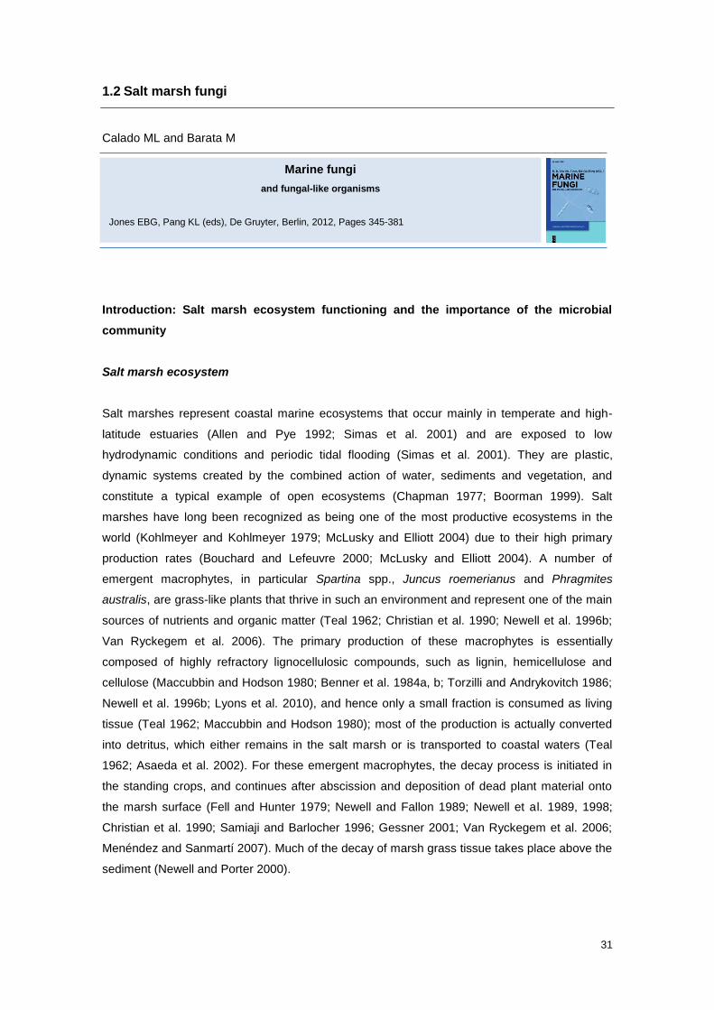
Marine fungi
and fungal-like organisms
Jones EBG, Pang KL (eds), De Gruyter, Berlin, 2012, Pages 345-381
32
Decomposer microbial community
The decomposer microbial community, including fungi and bacteria, assumes a fundamental
ecological role in the degradation of plant material, which is enriched with structural polymers,
and in the consequent release of nutrients that are essential to the metabolism of a wide marine
community (Benner et al. 1984b; Boorman 1999; Newell and Porter 2000; Lyons et al. 2005).
Though the role of fungi in this process has been long neglected, several studies have
highlighted the importance of the metabolic activities of these saprobic microorganisms on the
biogeochemical carbon and nutrients cycles, and in the energy fluxes within these ecotonal
marine ecosystems (Gessner and Goos 1973; Torzilli and Andrykovitch 1986; Newell 1996;
Newell et al. 1996b; Hyde et al. 1998; Gessner et al. 2007).
Saprobic fungi that colonize standing-dead tissues of salt marsh grasses initiate the decay
process (Torzilli and Andrykovitch 1986; Samiaji and Barlocher 1996; Lyons et al. 2005), and
represent the main secondary producers of the microbial community (Newell and Fallon 1989;
Newell et al. 1989, 1996a, b, 2000a; Newell 1996, 2001a; Castro and Freitas 2000; Gessner
2001; Findlay et al. 2002; Van Ryckegem et al. 2006, 2007). Bacteria may become more active
in the latter phase of decomposition (i.e., when the plant material collapses onto the marsh
sediment surface) (Benner et al. 1984b; Newell et al. 1989; Newell and Porter 2000). However,
Buchan et al. (2003) and Lyons et al. (2005) demonstrated that metabolically-active bacteria
and fungi co-occur on Spartina detritus, which contradicts this idea of temporally segregated
interventions during the decay process, but apparently without establishing species-specific
ecological associations.
In the microbial community associated with standing-decaying tissues of emergent
macrophytes, there is a clear dominance of fungi over bacteria. This is expressed in biomass
and productivity. This dominance occurs because the morphological and physiological
characteristics of the saprobic fungi confer an adaptive advantage on the use and degradation
of this substrate. In fact, in addition to their ability to tolerate a wide range of environmental
conditions, fungi can degrade the most resistant substrates in a more efficient manner than
bacteria. Filamentous fungi are well suited to penetrate substrates with their rigid cell walls,
apical growth and ability to produce lignocellulose-degrading enzymes (Torzilli 1982; Newell
1996; Newell et al. 1996b; Raghukumar 2004b). Saprobic fungi act on the surface or within the
tissues of macrophytes by the production of lignocellulolytic enzymes and physical penetration
of the host cell walls, bringing about decomposition of senescent tissues (Newell and Porter
2000). Additionally, saprobic fungi have the ability to retain and convert inorganic nitrogen into
fungal biomass during the initial phases of plant tissue decomposition (Findlay et al. 2002; Van
Ryckegem et al. 2006, 2007) and immobilize this nutrient from the surrounding environment
(Newell 1996; Van Ryckegem et al. 2006). The incorporation of nitrogen into fungal biomass,
together with the extracellular enzymes produced during the process, results in a nutritive
enrichment of substrates, which in turn becomes more palatable to several animal consumers
(Raghukumar 2004b). The fungal community associated with the decomposition of macrophytes
33
is composed mainly of ascomycetes (Gessner and Kohlmeyer 1976; Newell et al. 1996a,
2000a; Newell 2001a, b; Barata 2002; Buchan et al. 2003; Van Ryckegem and Verbeken
2005a, b, c).
Mycota of salt marshes: biotic and abiotic factors affecting community structure
Despite similar general biophysical characteristics, salt marsh ecosystems present some
environmental and ecological variations that will reflect on the composition and dynamics of the
fungal community. The marine fungal community in salt marshes, as in other ecosystems, is
composed of ubiquitous species, which occur on a broad range of substrates and environmental
conditions, and also by other species that appear to be strictly associated with particular
ecological niches (Gessner and Kohlmeyer 1976). The presence of a given fungus in the
ecosystem depends on an appropriate combination of various biotic and abiotic factors, which
vary according to species. These diverse factors include:
(i) degree of host/substrate specificity (Apinis and Chesters 1964; Newell and Porter
2000; Blum et al. 2004; Torzilli et al. 2006; Lyons et al. 2010);
(ii) ability to interact and compete with other microorganisms (Torzilli and Andrykovitch
1986; Buchan et al. 2003; Lyons et al. 2005);
(iii) vulnerability/resistance to predation (Newell and Wasowski 1995; Newell 2001a, b);
and
(iv) ecological requirements, such as water (Newell et al. 1996a; Poon and Hyde
1998a) and oxygen availability (Wong and Hyde 2002; Menéndez and Sanmartí
2007), dissolved organic nutrients (Newell et al. 1996a, 2000a; Newell and Porter
2000; Newell 2001b), salinity (Van Ryckegem and Verbeken 2005c), and
temperature (Castro and Freitas 2000; Van Ryckegem et al. 2007).
Host/substrate specificity
Among the intrinsic biological and environmental factors mentioned, the host/substrate
specificity - which is related to the chemical and structural composition of plant tissues - appears
to be primarily responsible for determining fungal community composition and productivity (Fell
and Hunter 1979; Newell and Porter 2000; Newell et al. 2000a; Blum et al. 2004; Torzilli et al.
2006; Van Ryckegem et al. 2006, 2007; Lyons et al. 2010). This specificity occurs during the
selection process of the host plant species to be colonized, but also in the choice of the plant
tissue.
34
Host plant and associated fungal diversity
Studies of salt marsh fungi associated with diverse host plants reveal no overlap between the
fungal-decay communities, which emphasizes the general high-level specificity with the
chemical and structural characteristics of each plant (Newell and Porter 2000; Blum et al. 2004;
Torzilli et al. 2006). Torzilli et al. (2006) compared the mycota associated with four salt marsh
plants — S. alterniflora, J. roemerianus, Distichlis spicata and Sarcocornia perennis — and
concluded that the greater the similarity between the type of plant tissues, the greater is the
similarity between the associated fungal communities. The same conclusion was reached by
Lyons et al. (2010), who found the same major ascomycetes on various species of Spartina (S.
alterniflora, S. foliosa, S. alterniflora x S. foliosa, S. densiflora).
Walker and Campbell (2010) inventoried the fungal community associated with S. alterniflora
and J. roemerianus using morphological and molecular approaches, and obtained different
results. The morphological analyses revealed different species on host plants, but terminal-
restriction fragment length polymorphism community profiles showed that more than 50% of the
fungal terminal-restriction fragments were found on both plants. The authors suggested that the
absence of fruiting structures of the same species on S. alterniflora and J. roemerianus might
indicate that some fungi are able to colonize but not sporulate on both hosts, and thus might be
host-specific to complete their lifecycle.
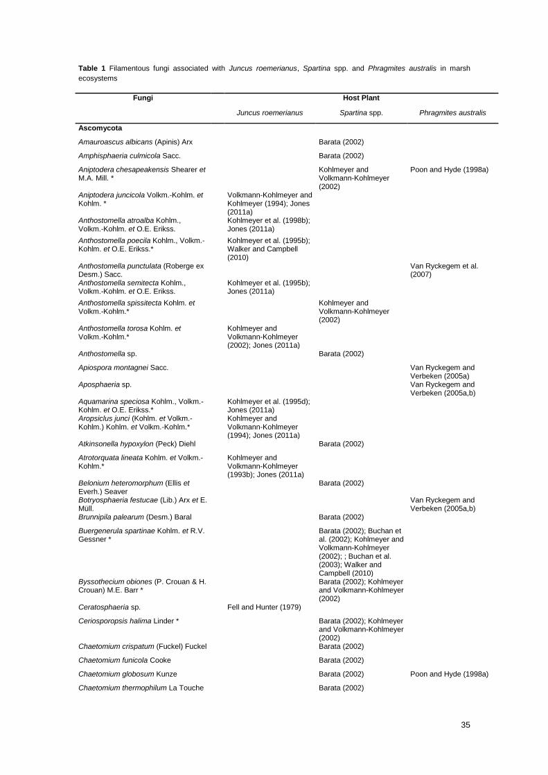
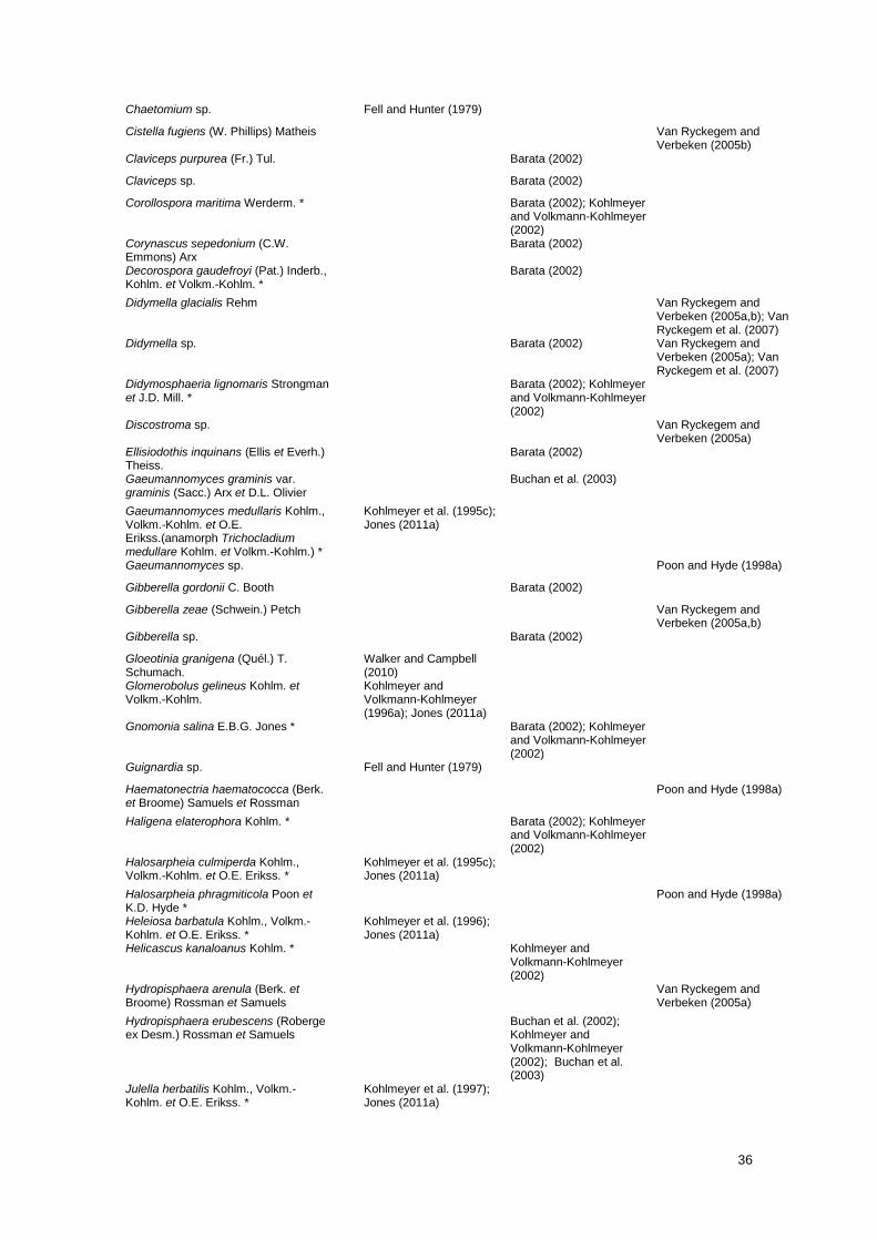
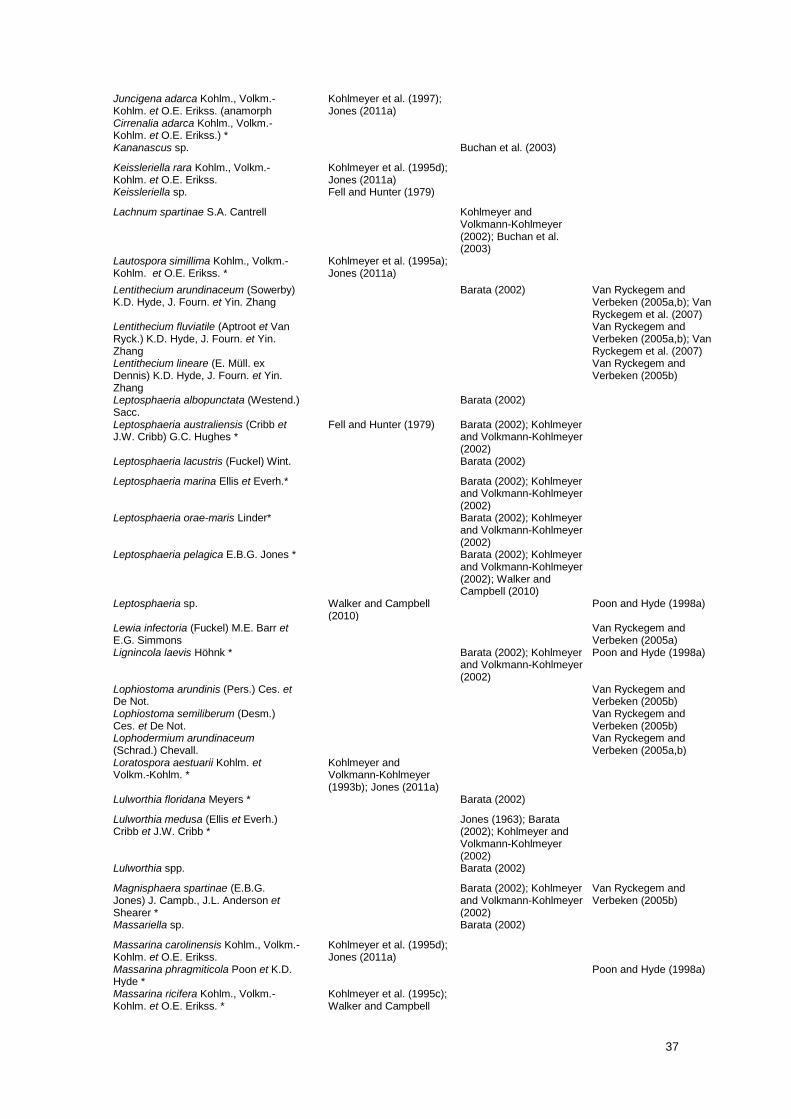
A comparison of species composition of fungal communities associated with the main primary
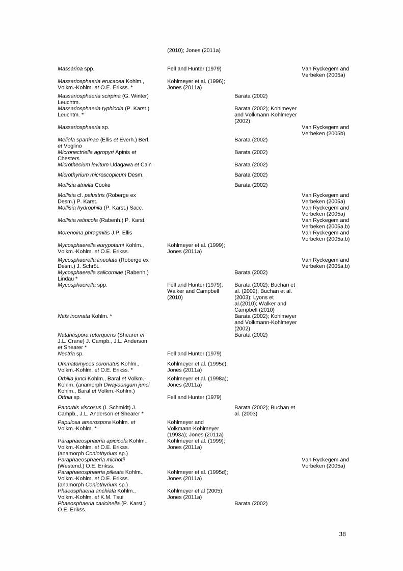
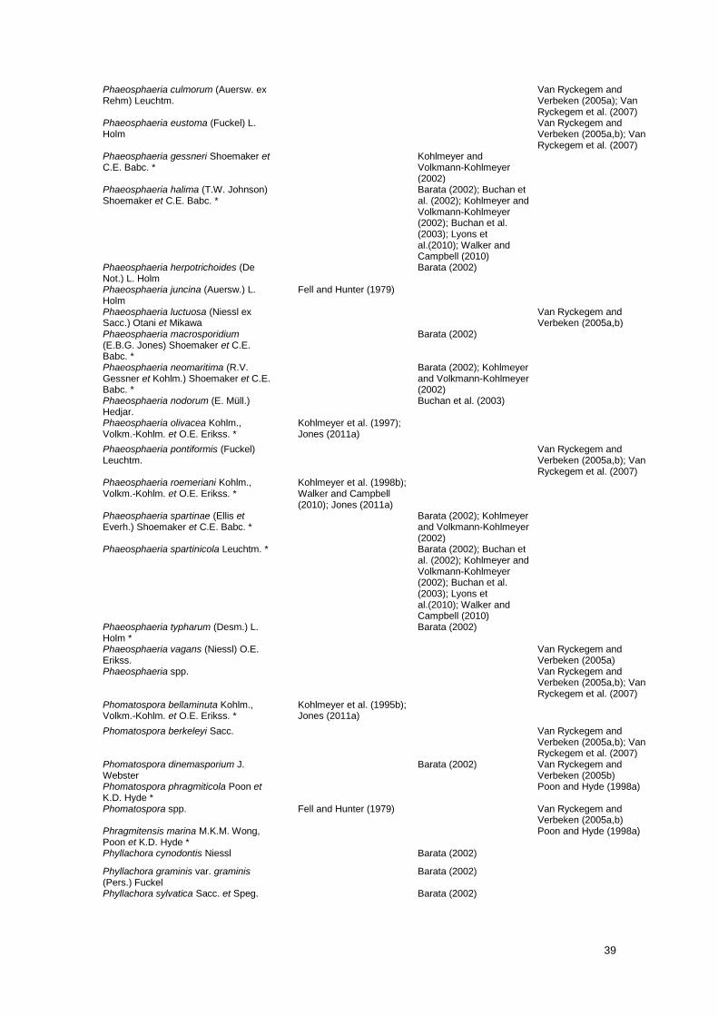
producers in marsh ecosystems (Table 1) confirms the above observation.
35
Table 1 Filamentous fungi associated with Juncus roemerianus, Spartina spp. and Phragmites australis in marsh
ecosystems
Fungi Host Plant
Juncus roemerianus Spartina spp. Phragmites australis
Ascomycota
Amauroascus albicans (Apinis) Arx Barata (2002)
Amphisphaeria culmicola Sacc. Barata (2002)
Aniptodera chesapeakensis Shearer et M.A. Mill. *
Kohlmeyer and Volkmann-Kohlmeyer (2002)
Poon and Hyde (1998a)
Aniptodera juncicola Volkm.-Kohlm. et Kohlm. *
Volkmann-Kohlmeyer and Kohlmeyer (1994); Jones (2011a)
Anthostomella atroalba Kohlm., Volkm.-Kohlm. et O.E. Erikss.
Kohlmeyer et al. (1998b); Jones (2011a)
Anthostomella poecila Kohlm., Volkm.-Kohlm. et O.E. Erikss.*
Kohlmeyer et al. (1995b); Walker and Campbell (2010)
Anthostomella punctulata (Roberge ex Desm.) Sacc.
Van Ryckegem et al. (2007)
Anthostomella semitecta Kohlm., Volkm.-Kohlm. et O.E. Erikss.
Kohlmeyer et al. (1995b); Jones (2011a)
Anthostomella spissitecta Kohlm. et Volkm.-Kohlm.*
Kohlmeyer and Volkmann-Kohlmeyer (2002)
Anthostomella torosa Kohlm. et Volkm.-Kohlm.*
Kohlmeyer and Volkmann-Kohlmeyer (2002); Jones (2011a)
Anthostomella sp. Barata (2002)
Apiospora montagnei Sacc. Van Ryckegem and Verbeken (2005a)
Aposphaeria sp. Van Ryckegem and Verbeken (2005a,b)
Aquamarina speciosa Kohlm., Volkm.-Kohlm. et O.E. Erikss.*
Kohlmeyer et al. (1995d); Jones (2011a)
Aropsiclus junci (Kohlm. et Volkm.-Kohlm.) Kohlm. et Volkm.-Kohlm.*
Kohlmeyer and Volkmann-Kohlmeyer (1994); Jones (2011a)
Atkinsonella hypoxylon (Peck) Diehl Barata (2002)
Atrotorquata lineata Kohlm. et Volkm.-Kohlm.*
Kohlmeyer and Volkmann-Kohlmeyer (1993b); Jones (2011a)
Belonium heteromorphum (Ellis et Everh.) Seaver
Barata (2002)
Botryosphaeria festucae (Lib.) Arx et E. Müll.
Van Ryckegem and Verbeken (2005a,b)
Brunnipila palearum (Desm.) Baral Barata (2002)
Buergenerula spartinae Kohlm. et R.V. Gessner *
Barata (2002); Buchan et al. (2002); Kohlmeyer and Volkmann-Kohlmeyer (2002); ; Buchan et al. (2003); Walker and Campbell (2010)
Byssothecium obiones (P. Crouan & H. Crouan) M.E. Barr *
Barata (2002); Kohlmeyer and Volkmann-Kohlmeyer (2002)
Ceratosphaeria sp. Fell and Hunter (1979)
Ceriosporopsis halima Linder * Barata (2002); Kohlmeyer and Volkmann-Kohlmeyer (2002)
Chaetomium crispatum (Fuckel) Fuckel Barata (2002)
Chaetomium funicola Cooke Barata (2002)
Chaetomium globosum Kunze Barata (2002) Poon and Hyde (1998a)
Chaetomium thermophilum La Touche Barata (2002)
36
Chaetomium sp. Fell and Hunter (1979)
Cistella fugiens (W. Phillips) Matheis Van Ryckegem and Verbeken (2005b)
Claviceps purpurea (Fr.) Tul. Barata (2002)
Claviceps sp. Barata (2002)
Corollospora maritima Werderm. * Barata (2002); Kohlmeyer and Volkmann-Kohlmeyer (2002)
Corynascus sepedonium (C.W. Emmons) Arx
Barata (2002)
Decorospora gaudefroyi (Pat.) Inderb., Kohlm. et Volkm.-Kohlm. *
Barata (2002)
Didymella glacialis Rehm Van Ryckegem and Verbeken (2005a,b); Van Ryckegem et al. (2007)
Didymella sp. Barata (2002) Van Ryckegem and Verbeken (2005a); Van Ryckegem et al. (2007)
Didymosphaeria lignomaris Strongman et J.D. Mill. *
Barata (2002); Kohlmeyer and Volkmann-Kohlmeyer (2002)
Discostroma sp. Van Ryckegem and Verbeken (2005a)
Ellisiodothis inquinans (Ellis et Everh.) Theiss.
Barata (2002)
Gaeumannomyces graminis var. graminis (Sacc.) Arx et D.L. Olivier
Buchan et al. (2003)
Gaeumannomyces medullaris Kohlm., Volkm.-Kohlm. et O.E. Erikss.(anamorph Trichocladium medullare Kohlm. et Volkm.-Kohlm.) *
Kohlmeyer et al. (1995c); Jones (2011a)
Gaeumannomyces sp. Poon and Hyde (1998a)
Gibberella gordonii C. Booth Barata (2002)
Gibberella zeae (Schwein.) Petch Van Ryckegem and Verbeken (2005a,b)
Gibberella sp. Barata (2002)
Gloeotinia granigena (Quél.) T. Schumach.
Walker and Campbell (2010)
Glomerobolus gelineus Kohlm. et Volkm.-Kohlm.
Kohlmeyer and Volkmann-Kohlmeyer (1996a); Jones (2011a)
Gnomonia salina E.B.G. Jones * Barata (2002); Kohlmeyer and Volkmann-Kohlmeyer (2002)
Guignardia sp. Fell and Hunter (1979)
Haematonectria haematococca (Berk. et Broome) Samuels et Rossman
Poon and Hyde (1998a)
Haligena elaterophora Kohlm. * Barata (2002); Kohlmeyer and Volkmann-Kohlmeyer (2002)
Halosarpheia culmiperda Kohlm., Volkm.-Kohlm. et O.E. Erikss. *
Kohlmeyer et al. (1995c); Jones (2011a)
Halosarpheia phragmiticola Poon et K.D. Hyde *
Poon and Hyde (1998a)
Heleiosa barbatula Kohlm., Volkm.-Kohlm. et O.E. Erikss. *
Kohlmeyer et al. (1996); Jones (2011a)
Helicascus kanaloanus Kohlm. * Kohlmeyer and Volkmann-Kohlmeyer (2002)
Hydropisphaera arenula (Berk. et Broome) Rossman et Samuels
Van Ryckegem and Verbeken (2005a)
Hydropisphaera erubescens (Roberge ex Desm.) Rossman et Samuels
Buchan et al. (2002); Kohlmeyer and Volkmann-Kohlmeyer (2002); Buchan et al. (2003)
Julella herbatilis Kohlm., Volkm.-Kohlm. et O.E. Erikss. *
Kohlmeyer et al. (1997); Jones (2011a)
37
Juncigena adarca Kohlm., Volkm.-Kohlm. et O.E. Erikss. (anamorph Cirrenalia adarca Kohlm., Volkm.-Kohlm. et O.E. Erikss.) *
Kohlmeyer et al. (1997); Jones (2011a)
Kananascus sp. Buchan et al. (2003)
Keissleriella rara Kohlm., Volkm.-Kohlm. et O.E. Erikss.
Kohlmeyer et al. (1995d); Jones (2011a)
Keissleriella sp. Fell and Hunter (1979)
Lachnum spartinae S.A. Cantrell Kohlmeyer and Volkmann-Kohlmeyer (2002); Buchan et al. (2003)
Lautospora simillima Kohlm., Volkm.-Kohlm. et O.E. Erikss. *
Kohlmeyer et al. (1995a); Jones (2011a)
Lentithecium arundinaceum (Sowerby) K.D. Hyde, J. Fourn. et Yin. Zhang
Barata (2002) Van Ryckegem and Verbeken (2005a,b); Van Ryckegem et al. (2007)
Lentithecium fluviatile (Aptroot et Van Ryck.) K.D. Hyde, J. Fourn. et Yin. Zhang
Van Ryckegem and Verbeken (2005a,b); Van Ryckegem et al. (2007)
Lentithecium lineare (E. Müll. ex Dennis) K.D. Hyde, J. Fourn. et Yin. Zhang
Van Ryckegem and Verbeken (2005b)
Leptosphaeria albopunctata (Westend.) Sacc.
Barata (2002)
Leptosphaeria australiensis (Cribb et J.W. Cribb) G.C. Hughes *
Fell and Hunter (1979) Barata (2002); Kohlmeyer and Volkmann-Kohlmeyer (2002)
Leptosphaeria lacustris (Fuckel) Wint. Barata (2002)
Leptosphaeria marina Ellis et Everh.* Barata (2002); Kohlmeyer and Volkmann-Kohlmeyer (2002)
Leptosphaeria orae-maris Linder* Barata (2002); Kohlmeyer and Volkmann-Kohlmeyer (2002)
Leptosphaeria pelagica E.B.G. Jones * Barata (2002); Kohlmeyer and Volkmann-Kohlmeyer (2002); Walker and Campbell (2010)
Leptosphaeria sp. Walker and Campbell (2010)
Poon and Hyde (1998a)
Lewia infectoria (Fuckel) M.E. Barr et E.G. Simmons
Van Ryckegem and Verbeken (2005a)
Lignincola laevis Höhnk * Barata (2002); Kohlmeyer and Volkmann-Kohlmeyer (2002)
Poon and Hyde (1998a)
Lophiostoma arundinis (Pers.) Ces. et De Not.
Van Ryckegem and Verbeken (2005b)
Lophiostoma semiliberum (Desm.) Ces. et De Not.
Van Ryckegem and Verbeken (2005b)
Lophodermium arundinaceum (Schrad.) Chevall.
Van Ryckegem and Verbeken (2005a,b)
Loratospora aestuarii Kohlm. et Volkm.-Kohlm. *
Kohlmeyer and Volkmann-Kohlmeyer (1993b); Jones (2011a)
Lulworthia floridana Meyers * Barata (2002)
Lulworthia medusa (Ellis et Everh.) Cribb et J.W. Cribb *
Jones (1963); Barata (2002); Kohlmeyer and Volkmann-Kohlmeyer (2002)
Lulworthia spp. Barata (2002)
Magnisphaera spartinae (E.B.G. Jones) J. Campb., J.L. Anderson et Shearer *
Barata (2002); Kohlmeyer and Volkmann-Kohlmeyer (2002)
Van Ryckegem and Verbeken (2005b)
Massariella sp. Barata (2002)
Massarina carolinensis Kohlm., Volkm.-Kohlm. et O.E. Erikss.
Kohlmeyer et al. (1995d); Jones (2011a)
Massarina phragmiticola Poon et K.D. Hyde *
Poon and Hyde (1998a)
Massarina ricifera Kohlm., Volkm.-Kohlm. et O.E. Erikss. *
Kohlmeyer et al. (1995c); Walker and Campbell
38
(2010); Jones (2011a)
Massarina spp. Fell and Hunter (1979) Van Ryckegem and Verbeken (2005a)
Massariosphaeria erucacea Kohlm., Volkm.-Kohlm. et O.E. Erikss. *
Kohlmeyer et al. (1996); Jones (2011a)
Massariosphaeria scirpina (G. Winter) Leuchtm.
Barata (2002)
Massariosphaeria typhicola (P. Karst.) Leuchtm. *
Barata (2002); Kohlmeyer and Volkmann-Kohlmeyer (2002)
Massariosphaeria sp. Van Ryckegem and Verbeken (2005b)
Meliola spartinae (Ellis et Everh.) Berl. et Voglino
Barata (2002)
Micronectriella agropyri Apinis et Chesters
Barata (2002)
Microthecium levitum Udagawa et Cain Barata (2002)
Microthyrium microscopicum Desm. Barata (2002)
Mollisia atriella Cooke Barata (2002)
Mollisia cf. palustris (Roberge ex Desm.) P. Karst.
Van Ryckegem and Verbeken (2005a)
Mollisia hydrophila (P. Karst.) Sacc. Van Ryckegem and Verbeken (2005a)
Mollisia retincola (Rabenh.) P. Karst. Van Ryckegem and Verbeken (2005a,b)
Morenoina phragmitis J.P. Ellis Van Ryckegem and Verbeken (2005a,b)
Mycosphaerella eurypotami Kohlm., Volkm.-Kohlm. et O.E. Erikss.
Kohlmeyer et al. (1999); Jones (2011a)
Mycosphaerella lineolata (Roberge ex Desm.) J. Schröt.
Van Ryckegem and Verbeken (2005a,b)
Mycosphaerella salicorniae (Rabenh.) Lindau *
Barata (2002)
Mycosphaerella spp. Fell and Hunter (1979); Walker and Campbell (2010)
Barata (2002); Buchan et al. (2002); Buchan et al. (2003); Lyons et al.(2010); Walker and Campbell (2010)
Naïs inornata Kohlm. * Barata (2002); Kohlmeyer and Volkmann-Kohlmeyer (2002)
Natantispora retorquens (Shearer et J.L. Crane) J. Campb., J.L. Anderson et Shearer *
Barata (2002)
Nectria sp. Fell and Hunter (1979)
Ommatomyces coronatus Kohlm., Volkm.-Kohlm. et O.E. Erikss. *
Kohlmeyer et al. (1995c); Jones (2011a)
Orbilia junci Kohlm., Baral et Volkm.-Kohlm. (anamorph Dwayaangam junci Kohlm., Baral et Volkm.-Kohlm.)
Kohlmeyer et al. (1998a); Jones (2011a)
Otthia sp. Fell and Hunter (1979)
Panorbis viscosus (I. Schmidt) J. Campb., J.L. Anderson et Shearer *
Barata (2002); Buchan et al. (2003)
Papulosa amerospora Kohlm. et Volkm.-Kohlm. *
Kohlmeyer and Volkmann-Kohlmeyer (1993a); Jones (2011a)
Paraphaeosphaeria apicicola Kohlm., Volkm.-Kohlm. et O.E. Erikss. (anamorph Coniothyrium sp.)
Kohlmeyer et al. (1999); Jones (2011a)
Paraphaeosphaeria michotii (Westend.) O.E. Erikss.
Van Ryckegem and Verbeken (2005a)
Paraphaeosphaeria pilleata Kohlm., Volkm.-Kohlm. et O.E. Erikss. (anamorph Coniothyrium sp.)
Kohlmeyer et al. (1995d); Jones (2011a)
Phaeosphaeria anchiala Kohlm., Volkm.-Kohlm. et K.M. Tsui
Kohlmeyer et al (2005); Jones (2011a)
Phaeosphaeria caricinella (P. Karst.) O.E. Erikss.
Barata (2002)
39
Phaeosphaeria culmorum (Auersw. ex Rehm) Leuchtm.
Van Ryckegem and Verbeken (2005a); Van Ryckegem et al. (2007)
Phaeosphaeria eustoma (Fuckel) L. Holm
Van Ryckegem and Verbeken (2005a,b); Van Ryckegem et al. (2007)
Phaeosphaeria gessneri Shoemaker et C.E. Babc. *
Kohlmeyer and Volkmann-Kohlmeyer (2002)
Phaeosphaeria halima (T.W. Johnson) Shoemaker et C.E. Babc. *
Barata (2002); Buchan et al. (2002); Kohlmeyer and Volkmann-Kohlmeyer (2002); Buchan et al. (2003); Lyons et al.(2010); Walker and Campbell (2010)
Phaeosphaeria herpotrichoides (De Not.) L. Holm
Barata (2002)
Phaeosphaeria juncina (Auersw.) L. Holm
Fell and Hunter (1979)
Phaeosphaeria luctuosa (Niessl ex Sacc.) Otani et Mikawa
Van Ryckegem and Verbeken (2005a,b)
Phaeosphaeria macrosporidium (E.B.G. Jones) Shoemaker et C.E. Babc. *
Barata (2002)
Phaeosphaeria neomaritima (R.V. Gessner et Kohlm.) Shoemaker et C.E. Babc. *
Barata (2002); Kohlmeyer and Volkmann-Kohlmeyer (2002)
Phaeosphaeria nodorum (E. Müll.) Hedjar.
Buchan et al. (2003)
Phaeosphaeria olivacea Kohlm., Volkm.-Kohlm. et O.E. Erikss. *
Kohlmeyer et al. (1997); Jones (2011a)
Phaeosphaeria pontiformis (Fuckel) Leuchtm.
Van Ryckegem and Verbeken (2005a,b); Van Ryckegem et al. (2007)
Phaeosphaeria roemeriani Kohlm., Volkm.-Kohlm. et O.E. Erikss. *
Kohlmeyer et al. (1998b); Walker and Campbell (2010); Jones (2011a)
Phaeosphaeria spartinae (Ellis et Everh.) Shoemaker et C.E. Babc. *
Barata (2002); Kohlmeyer and Volkmann-Kohlmeyer (2002)
Phaeosphaeria spartinicola Leuchtm. * Barata (2002); Buchan et al. (2002); Kohlmeyer and Volkmann-Kohlmeyer (2002); Buchan et al. (2003); Lyons et al.(2010); Walker and Campbell (2010)
Phaeosphaeria typharum (Desm.) L. Holm *
Barata (2002)
Phaeosphaeria vagans (Niessl) O.E. Erikss.
Van Ryckegem and Verbeken (2005a)
Phaeosphaeria spp. Van Ryckegem and Verbeken (2005a,b); Van Ryckegem et al. (2007)
Phomatospora bellaminuta Kohlm., Volkm.-Kohlm. et O.E. Erikss. *
Kohlmeyer et al. (1995b); Jones (2011a)
Phomatospora berkeleyi Sacc. Van Ryckegem and Verbeken (2005a,b); Van Ryckegem et al. (2007)
Phomatospora dinemasporium J. Webster
Barata (2002) Van Ryckegem and Verbeken (2005b)
Phomatospora phragmiticola Poon et K.D. Hyde *
Poon and Hyde (1998a)
Phomatospora spp. Fell and Hunter (1979) Van Ryckegem and Verbeken (2005a,b)
Phragmitensis marina M.K.M. Wong, Poon et K.D. Hyde *
Poon and Hyde (1998a)
Phyllachora cynodontis Niessl Barata (2002)
Phyllachora graminis var. graminis (Pers.) Fuckel
Barata (2002)
Phyllachora sylvatica Sacc. et Speg. Barata (2002)
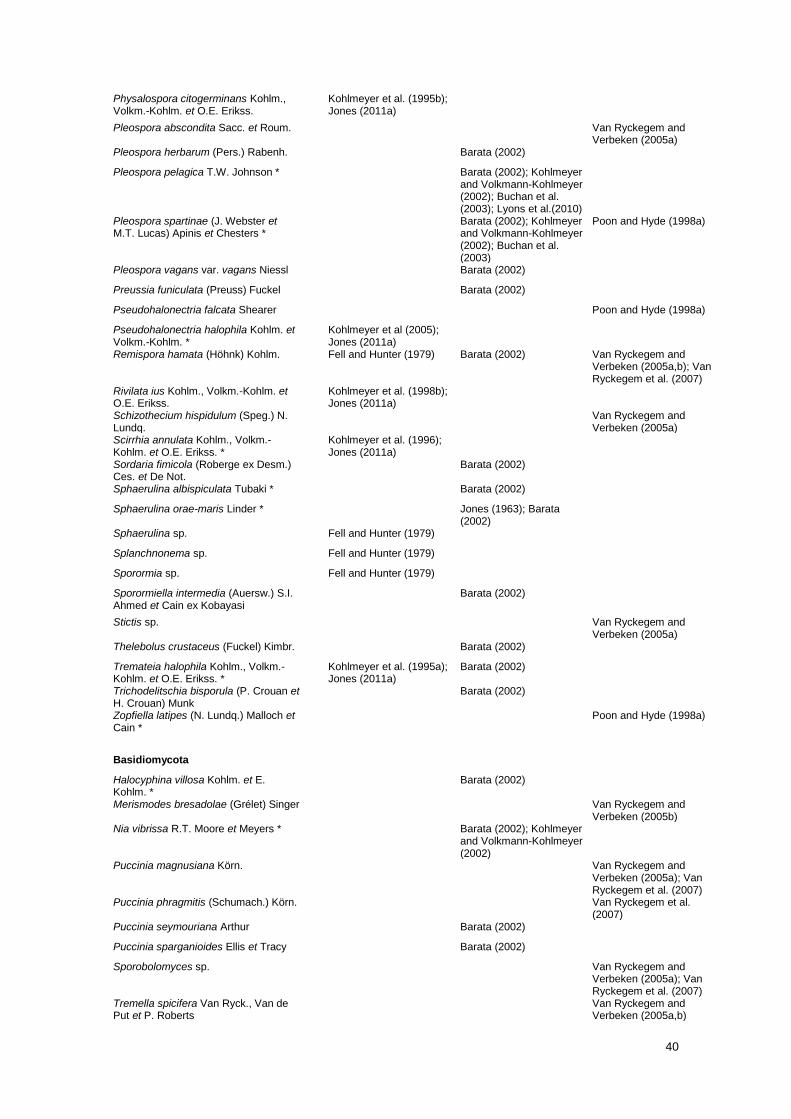
40
Physalospora citogerminans Kohlm., Volkm.-Kohlm. et O.E. Erikss.
Kohlmeyer et al. (1995b); Jones (2011a)
Pleospora abscondita Sacc. et Roum. Van Ryckegem and Verbeken (2005a)
Pleospora herbarum (Pers.) Rabenh. Barata (2002)
Pleospora pelagica T.W. Johnson * Barata (2002); Kohlmeyer and Volkmann-Kohlmeyer (2002); Buchan et al. (2003); Lyons et al.(2010)
Pleospora spartinae (J. Webster et M.T. Lucas) Apinis et Chesters *
Barata (2002); Kohlmeyer and Volkmann-Kohlmeyer (2002); Buchan et al. (2003)
Poon and Hyde (1998a)
Pleospora vagans var. vagans Niessl Barata (2002)
Preussia funiculata (Preuss) Fuckel Barata (2002)
Pseudohalonectria falcata Shearer Poon and Hyde (1998a)
Pseudohalonectria halophila Kohlm. et Volkm.-Kohlm. *
Kohlmeyer et al (2005); Jones (2011a)
Remispora hamata (Höhnk) Kohlm. Fell and Hunter (1979) Barata (2002) Van Ryckegem and Verbeken (2005a,b); Van Ryckegem et al. (2007)
Rivilata ius Kohlm., Volkm.-Kohlm. et O.E. Erikss.
Kohlmeyer et al. (1998b); Jones (2011a)
Schizothecium hispidulum (Speg.) N. Lundq.
Van Ryckegem and Verbeken (2005a)
Scirrhia annulata Kohlm., Volkm.-Kohlm. et O.E. Erikss. *
Kohlmeyer et al. (1996); Jones (2011a)
Sordaria fimicola (Roberge ex Desm.) Ces. et De Not.
Barata (2002)
Sphaerulina albispiculata Tubaki * Barata (2002)
Sphaerulina orae-maris Linder * Jones (1963); Barata (2002)
Sphaerulina sp. Fell and Hunter (1979)
Splanchnonema sp. Fell and Hunter (1979)
Sporormia sp. Fell and Hunter (1979)
Sporormiella intermedia (Auersw.) S.I. Ahmed et Cain ex Kobayasi
Barata (2002)
Stictis sp. Van Ryckegem and Verbeken (2005a)
Thelebolus crustaceus (Fuckel) Kimbr. Barata (2002)
Tremateia halophila Kohlm., Volkm.-Kohlm. et O.E. Erikss. *
Kohlmeyer et al. (1995a); Jones (2011a)
Barata (2002)
Trichodelitschia bisporula (P. Crouan et H. Crouan) Munk
Barata (2002)
Zopfiella latipes (N. Lundq.) Malloch et Cain *
Poon and Hyde (1998a)
Basidiomycota
Halocyphina villosa Kohlm. et E. Kohlm. *
Barata (2002)
Merismodes bresadolae (Grélet) Singer Van Ryckegem and Verbeken (2005b)
Nia vibrissa R.T. Moore et Meyers * Barata (2002); Kohlmeyer and Volkmann-Kohlmeyer (2002)
Puccinia magnusiana Körn. Van Ryckegem and Verbeken (2005a); Van Ryckegem et al. (2007)
Puccinia phragmitis (Schumach.) Körn. Van Ryckegem et al. (2007)
Puccinia seymouriana Arthur Barata (2002)
Puccinia sparganioides Ellis et Tracy Barata (2002)
Sporobolomyces sp. Van Ryckegem and Verbeken (2005a); Van Ryckegem et al. (2007)
Tremella spicifera Van Ryck., Van de Put et P. Roberts
Van Ryckegem and Verbeken (2005a,b)
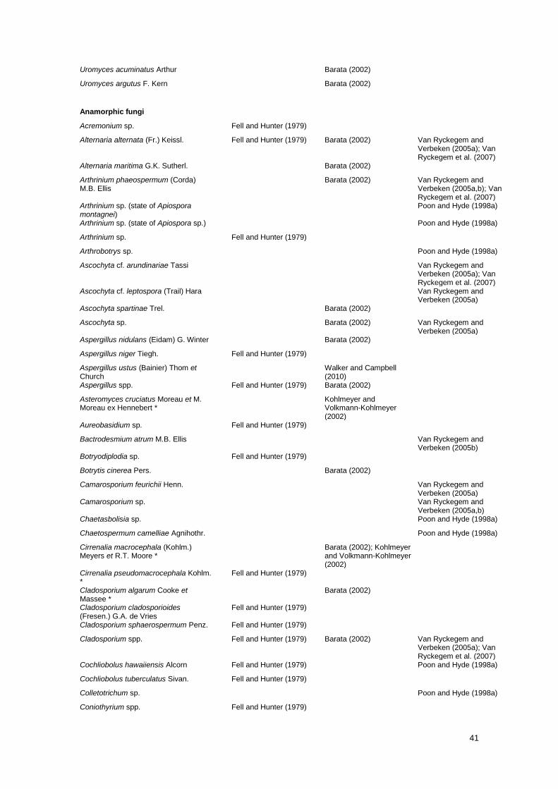
41
Uromyces acuminatus Arthur Barata (2002)
Uromyces argutus F. Kern Barata (2002)
Anamorphic fungi
Acremonium sp. Fell and Hunter (1979)
Alternaria alternata (Fr.) Keissl. Fell and Hunter (1979) Barata (2002) Van Ryckegem and Verbeken (2005a); Van Ryckegem et al. (2007)
Alternaria maritima G.K. Sutherl. Barata (2002)
Arthrinium phaeospermum (Corda) M.B. Ellis
Barata (2002) Van Ryckegem and Verbeken (2005a,b); Van Ryckegem et al. (2007)
Arthrinium sp. (state of Apiospora montagnei)
Poon and Hyde (1998a)
Arthrinium sp. (state of Apiospora sp.) Poon and Hyde (1998a)
Arthrinium sp. Fell and Hunter (1979)
Arthrobotrys sp. Poon and Hyde (1998a)
Ascochyta cf. arundinariae Tassi Van Ryckegem and Verbeken (2005a); Van Ryckegem et al. (2007)
Ascochyta cf. leptospora (Trail) Hara Van Ryckegem and Verbeken (2005a)
Ascochyta spartinae Trel. Barata (2002)
Ascochyta sp. Barata (2002) Van Ryckegem and Verbeken (2005a)
Aspergillus nidulans (Eidam) G. Winter Barata (2002)
Aspergillus niger Tiegh. Fell and Hunter (1979)
Aspergillus ustus (Bainier) Thom et Church
Walker and Campbell (2010)
Aspergillus spp. Fell and Hunter (1979) Barata (2002)
Asteromyces cruciatus Moreau et M. Moreau ex Hennebert *
Kohlmeyer and Volkmann-Kohlmeyer (2002)
Aureobasidium sp. Fell and Hunter (1979)
Bactrodesmium atrum M.B. Ellis Van Ryckegem and Verbeken (2005b)
Botryodiplodia sp. Fell and Hunter (1979)
Botrytis cinerea Pers. Barata (2002)
Camarosporium feurichii Henn. Van Ryckegem and Verbeken (2005a)
Camarosporium sp. Van Ryckegem and Verbeken (2005a,b)
Chaetasbolisia sp. Poon and Hyde (1998a)
Chaetospermum camelliae Agnihothr. Poon and Hyde (1998a)
Cirrenalia macrocephala (Kohlm.) Meyers et R.T. Moore *
Barata (2002); Kohlmeyer and Volkmann-Kohlmeyer (2002)
Cirrenalia pseudomacrocephala Kohlm. *
Fell and Hunter (1979)
Cladosporium algarum Cooke et Massee *
Barata (2002)
Cladosporium cladosporioides (Fresen.) G.A. de Vries
Fell and Hunter (1979)
Cladosporium sphaerospermum Penz. Fell and Hunter (1979)
Cladosporium spp. Fell and Hunter (1979) Barata (2002) Van Ryckegem and Verbeken (2005a); Van Ryckegem et al. (2007)
Cochliobolus hawaiiensis Alcorn Fell and Hunter (1979) Poon and Hyde (1998a)
Cochliobolus tuberculatus Sivan. Fell and Hunter (1979)
Colletotrichum sp. Poon and Hyde (1998a)
Coniothyrium spp. Fell and Hunter (1979)

42
Cremasteria cymatilis Meyers et R.T. Moore
Fell and Hunter (1979)
Cumulospora marina I. Schmidt * Kohlmeyer and Volkmann-Kohlmeyer (2002)
Curvularia protuberata R.R. Nelson et Hodges
Fell and Hunter (1979)
Curvularia sp. Fell and Hunter (1979)
Cytoplacosphaeria phragmiticola Poon et K.D. Hyde *
Poon and Hyde (1998a)
Cytoplacosphaeria rimosa Petr. Van Ryckegem and Verbeken (2005a,b)
Cytoplea sp. Poon and Hyde (1998a)
Deightoniella roumeguerei (Cavara) Constant.
Van Ryckegem et al. (2007)
Dendrostilbella sp. Poon and Hyde (1998a)
Dictyosporium oblongum (Fuckel) S. Hughes
Van Ryckegem and Verbeken (2005b); Van Ryckegem et al. (2007)
Dictyosporium pelagicum (Linder) G.C. Hughes ex E.B.G. Jones *
Barata (2002); Kohlmeyer and Volkmann-Kohlmeyer (2002)
Dictyosporium toruloides (Corda) Guég.
Jones (1963)
Drechslera sp. Fell and Hunter (1979)
Dumortieria sp. Fell and Hunter (1979)
Epicoccum nigrum Link Fell and Hunter (1979) Barata (2002)
Flagellospora sp. Fell and Hunter (1979)
Floricola striata Kohlm. et Volkm.-Kohlm.
Kohlmeyer and Volkmann-Kohlmeyer (2000); Jones (2011a)
Fusarium incarnatum (Desm.) Sacc. Walker and Campbell (2010)
Fusarium spp. Fell and Hunter (1979) Van Ryckegem and Verbeken (2005a,b)
Geniculosporium sp. Fell and Hunter (1979)
Gliocladium sp. Fell and Hunter (1979)
Gliomastix spp. Fell and Hunter (1979) Poon and Hyde (1998a)
Halenospora varia (Anastasiou) E.B.G. Jones *
Barata (2002); Kohlmeyer and Volkmann-Kohlmeyer (2002)
Haplobasidion lelebae Sawada ex M.B. Ellis
Fell and Hunter (1979)
Hendersonia culmicola Sacc. Barata (2002)
Hendersonia culmiseda Sacc. Van Ryckegem and Verbeken (2005a); Van Ryckegem et al. (2007)
Humicola sp. Fell and Hunter (1979)
Hymenopsis chlorothrix Kohlm. et Volkm.-Kohlm. *
Kohlmeyer and Volkmann-Kohlmeyer (2001a); Jones (2011a)
Hyphopolynema juncatile Kohlm. et Volkm.-Kohlm.
Kohlmeyer and Volkmann-Kohlmeyer (1999); Jones (2011a)
Khuskia oryzae H.J. Huds. Fell and Hunter (1979)
Kolletes undulatus Kohlm. et Volkm.-Kohlm.
Kohlmeyer et al (2005); Jones (2011a)
Koorchaloma galateae Kohlm. et Volkm.-Kohlm. *
Kohlmeyer and Volkmann-Kohlmeyer (2001b); Jones (2011a)
Koorchaloma spartinicola V.V. Sarma, S.Y. Newell et K.D. Hyde *
Buchan et al. (2003)
Leptosphaerulina chartarum Cec. Roux Fell and Hunter (1979)
Memnoniella echinata (Rivolta) Galloway
Fell and Hunter (1979)
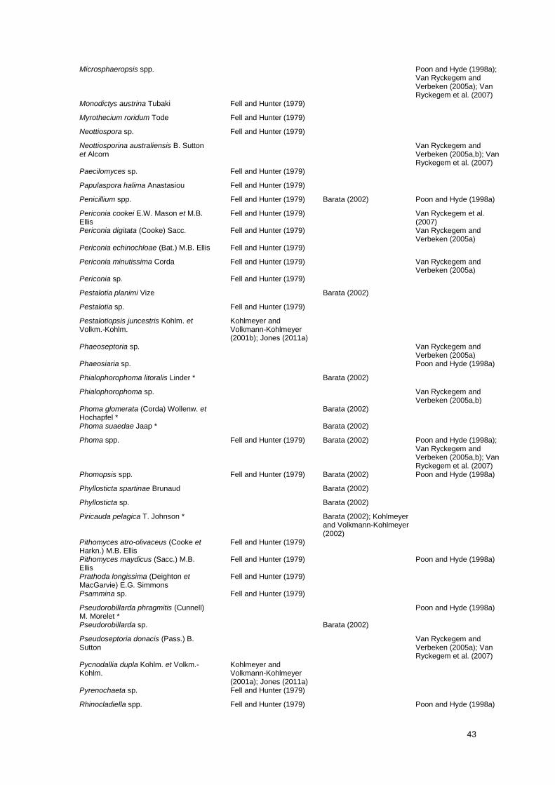
43
Microsphaeropsis spp. Poon and Hyde (1998a); Van Ryckegem and Verbeken (2005a); Van Ryckegem et al. (2007)
Monodictys austrina Tubaki Fell and Hunter (1979)
Myrothecium roridum Tode Fell and Hunter (1979)
Neottiospora sp. Fell and Hunter (1979)
Neottiosporina australiensis B. Sutton et Alcorn
Van Ryckegem and Verbeken (2005a,b); Van Ryckegem et al. (2007)
Paecilomyces sp. Fell and Hunter (1979)
Papulaspora halima Anastasiou Fell and Hunter (1979)
Penicillium spp. Fell and Hunter (1979) Barata (2002) Poon and Hyde (1998a)
Periconia cookei E.W. Mason et M.B. Ellis
Fell and Hunter (1979) Van Ryckegem et al. (2007)
Periconia digitata (Cooke) Sacc. Fell and Hunter (1979) Van Ryckegem and Verbeken (2005a)
Periconia echinochloae (Bat.) M.B. Ellis Fell and Hunter (1979)
Periconia minutissima Corda Fell and Hunter (1979) Van Ryckegem and Verbeken (2005a)
Periconia sp. Fell and Hunter (1979)
Pestalotia planimi Vize Barata (2002)
Pestalotia sp. Fell and Hunter (1979)
Pestalotiopsis juncestris Kohlm. et Volkm.-Kohlm.
Kohlmeyer and Volkmann-Kohlmeyer (2001b); Jones (2011a)
Phaeoseptoria sp. Van Ryckegem and Verbeken (2005a)
Phaeosiaria sp. Poon and Hyde (1998a)
Phialophorophoma litoralis Linder * Barata (2002)
Phialophorophoma sp. Van Ryckegem and Verbeken (2005a,b)
Phoma glomerata (Corda) Wollenw. et Hochapfel *
Barata (2002)
Phoma suaedae Jaap * Barata (2002)
Phoma spp. Fell and Hunter (1979) Barata (2002) Poon and Hyde (1998a); Van Ryckegem and Verbeken (2005a,b); Van Ryckegem et al. (2007)
Phomopsis spp. Fell and Hunter (1979) Barata (2002) Poon and Hyde (1998a)
Phyllosticta spartinae Brunaud Barata (2002)
Phyllosticta sp. Barata (2002)
Piricauda pelagica T. Johnson * Barata (2002); Kohlmeyer and Volkmann-Kohlmeyer (2002)
Pithomyces atro-olivaceus (Cooke et Harkn.) M.B. Ellis
Fell and Hunter (1979)
Pithomyces maydicus (Sacc.) M.B. Ellis
Fell and Hunter (1979) Poon and Hyde (1998a)
Prathoda longissima (Deighton et MacGarvie) E.G. Simmons
Fell and Hunter (1979)
Psammina sp. Fell and Hunter (1979)
Pseudorobillarda phragmitis (Cunnell) M. Morelet *
Poon and Hyde (1998a)
Pseudorobillarda sp. Barata (2002)
Pseudoseptoria donacis (Pass.) B. Sutton
Van Ryckegem and Verbeken (2005a); Van Ryckegem et al. (2007)
Pycnodallia dupla Kohlm. et Volkm.-Kohlm.
Kohlmeyer and Volkmann-Kohlmeyer (2001a); Jones (2011a)
Pyrenochaeta sp. Fell and Hunter (1979)
Rhinocladiella spp. Fell and Hunter (1979) Poon and Hyde (1998a)
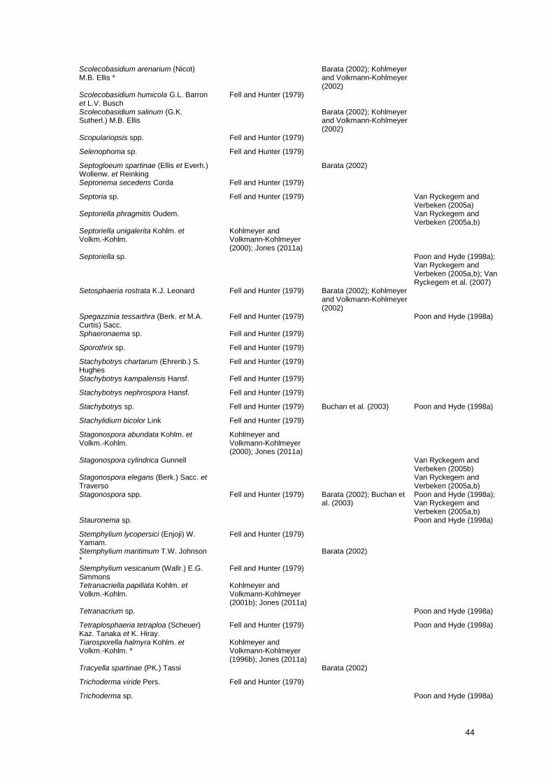
44
Scolecobasidium arenarium (Nicot) M.B. Ellis *
Barata (2002); Kohlmeyer and Volkmann-Kohlmeyer (2002)
Scolecobasidium humicola G.L. Barron et L.V. Busch
Fell and Hunter (1979)
Scolecobasidium salinum (G.K. Sutherl.) M.B. Ellis
Barata (2002); Kohlmeyer and Volkmann-Kohlmeyer (2002)
Scopulariopsis spp. Fell and Hunter (1979)
Selenophoma sp. Fell and Hunter (1979)
Septogloeum spartinae (Ellis et Everh.) Wollenw. et Reinking
Barata (2002)
Septonema secedens Corda Fell and Hunter (1979)
Septoria sp. Fell and Hunter (1979) Van Ryckegem and Verbeken (2005a)
Septoriella phragmitis Oudem. Van Ryckegem and Verbeken (2005a,b)
Septoriella unigalerita Kohlm. et Volkm.-Kohlm.
Kohlmeyer and Volkmann-Kohlmeyer (2000); Jones (2011a)
Septoriella sp. Poon and Hyde (1998a); Van Ryckegem and Verbeken (2005a,b); Van Ryckegem et al. (2007)
Setosphaeria rostrata K.J. Leonard Fell and Hunter (1979) Barata (2002); Kohlmeyer and Volkmann-Kohlmeyer (2002)
Spegazzinia tessarthra (Berk. et M.A. Curtis) Sacc.
Fell and Hunter (1979) Poon and Hyde (1998a)
Sphaeronaema sp. Fell and Hunter (1979)
Sporothrix sp. Fell and Hunter (1979)
Stachybotrys chartarum (Ehrenb.) S. Hughes
Fell and Hunter (1979)
Stachybotrys kampalensis Hansf. Fell and Hunter (1979)
Stachybotrys nephrospora Hansf. Fell and Hunter (1979)
Stachybotrys sp. Fell and Hunter (1979) Buchan et al. (2003) Poon and Hyde (1998a)
Stachylidium bicolor Link Fell and Hunter (1979)
Stagonospora abundata Kohlm. et Volkm.-Kohlm.
Kohlmeyer and Volkmann-Kohlmeyer (2000); Jones (2011a)
Stagonospora cylindrica Gunnell Van Ryckegem and Verbeken (2005b)
Stagonospora elegans (Berk.) Sacc. et Traverso
Van Ryckegem and Verbeken (2005a,b)
Stagonospora spp. Fell and Hunter (1979) Barata (2002); Buchan et al. (2003)
Poon and Hyde (1998a); Van Ryckegem and Verbeken (2005a,b)
Stauronema sp. Poon and Hyde (1998a)
Stemphylium lycopersici (Enjoji) W. Yamam.
Fell and Hunter (1979)
Stemphylium maritimum T.W. Johnson *
Barata (2002)
Stemphylium vesicarium (Wallr.) E.G. Simmons
Fell and Hunter (1979)
Tetranacriella papillata Kohlm. et Volkm.-Kohlm.
Kohlmeyer and Volkmann-Kohlmeyer (2001b); Jones (2011a)
Tetranacrium sp. Poon and Hyde (1998a)
Tetraplosphaeria tetraploa (Scheuer) Kaz. Tanaka et K. Hiray.
Fell and Hunter (1979) Poon and Hyde (1998a)
Tiarosporella halmyra Kohlm. et Volkm.-Kohlm. *
Kohlmeyer and Volkmann-Kohlmeyer (1996b); Jones (2011a)
Tracyella spartinae (PK.) Tassi Barata (2002)
Trichoderma viride Pers. Fell and Hunter (1979)
Trichoderma sp. Poon and Hyde (1998a)
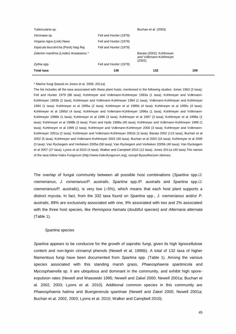
45
Tubercularia sp. Buchan et al. (2003)
Veronaea sp. Fell and Hunter (1979)
Virgaria nigra (Link) Nees Fell and Hunter (1979)
Xepicula leucotricha (Peck) Nag Raj Fell and Hunter (1979)
Zalerion maritima (Linder) Anastasiou * Barata (2002); Kohlmeyer and Volkmann-Kohlmeyer (2002)
Zythia spp. Fell and Hunter (1979)
Total taxa 136 132 109
* Marine fungi (based on Jones et al. 2009, 2011a)
The list includes all the taxa associated with these plant hosts, mentioned in the following studies: Jones 1963 (3 taxa);
Fell and Hunter 1979 (88 taxa); Kohlmeyer and Volkmann-Kohlmeyer 1993a (1 taxa); Kohlmeyer and Volkmann-
Kohlmeyer 1993b (2 taxa); Kohlmeyer and Volkmann-Kohlmeyer 1994 (1 taxa); Volkmann-Kohlmeyer and Kohlmeyer
1994 (1 taxa); Kohlmeyer et al 1995a (2 taxa); Kohlmeyer et al 1995b (4 taxa); Kohlmeyer et al 1995c (4 taxa);
Kohlmeyer et al 1995d (4 taxa); Kohlmeyer and Volkmann-Kohlmeyer 1996a (1 taxa); Kohlmeyer and Volkmann-
Kohlmeyer 1996b (1 taxa); Kohlmeyer et al 1996 (3 taxa); Kohlmeyer et al 1997 (3 taxa); Kohlmeyer et al 1998a (1
taxa); Kohlmeyer et al 1998b (3 taxa); Poon and Hyde 1998a (40 taxa); Kohlmeyer and Volkmann-Kohlmeyer 1999 (1
taxa); Kohlmeyer et al 1999 (2 taxa); Kohlmeyer and Volkmann-Kohlmeyer 2000 (3 taxa); Kohlmeyer and Volkmann-
Kohlmeyer 2001a (2 taxa); Kohlmeyer and Volkmann-Kohlmeyer 2001b (3 taxa); Barata 2002 (115 taxa); Buchan et al
2002 (5 taxa); Kohlmeyer and Volkmann-Kohlmeyer 2002 (40 taxa); Buchan et al 2003 (16 taxa); Kohlmeyer et al 2005
(3 taxa); Van Ryckegem and Verbeken 2005a (58 taxa); Van Ryckegem and Verbeken 2005b (40 taxa); Van Ryckegem
et al 2007 (27 taxa); Lyons et al 2010 (4 taxa); Walker and Campbell 2010 (12 taxa); Jones 2011a (45 taxa).The names
of the taxa follow Index Fungorum (http://www.indexfungorum.org), except Byssothecium obiones.
The overlap of fungal community between all possible host combinations (Spartina spp./J.
roemerianus, J. romerianus/P. australis, Spartina spp./P. australis and Spartina spp./J.
roemerianus/P. australis), is very low (5%), which means that each host plant supports a
distinct mycota. In fact, from the 332 taxa found on Spartina spp., J. roemerianus and/or P.
australis, 89% are exclusively associated with one, 9% associated with two and 2% associated
with the three host species, like Remispora hamata (doubtful species) and Alternaria alternata
(Table 1).
Spartina species
Spartina appears to be conducive for the growth of saprobic fungi, given its high lignocellulose
content and non-lignin cinnamyl phenols (Newell et al. 1996b). A total of 132 taxa of higher
filamentous fungi have been documented from Spartina spp. (Table 1). Among the various
species associated with this standing marsh grass, Phaeosphaeria spartinicola and
Mycosphaerella sp. II are ubiquitous and dominant in the community, and exhibit high spore-
expulsion rates (Newell and Wasowski 1995; Newell and Zakel 2000; Newell 2001a; Buchan et
al. 2002, 2003; Lyons et al. 2010). Additional common species in this community are
Phaeosphaeria halima and Buergenerula spartinae (Newell and Zakel 2000; Newell 2001a;
Buchan et al. 2002, 2003; Lyons et al. 2010; Walker and Campbell 2010).
46
Juncus roemerianus
Another salt marsh grass, J. roemerianus, supports a surprisingly high number of fungal taxa
(Table 1). Most of the fungi associated with this host have been collected and identified by the
Kohlmeyers and their colleagues in marshes located on the east coast of the United States.
Jones (2011a) has recently updated the list of fungi documented by Kohlmeyer and Volkmann-
Kohlmeyer (2001c). One-hundred-thirty-six fungi have been identified on J. roemerianus (Table
1). Newell and Porter (2000) have pointed out that Loratospora aestuarii, Papulosa amerospora,
Aropsiclus junci, Anthostomella poecila, Physalospora citogerminans, Scirrhia annulata,
Massarina ricifera and Tremateia halophila are the most common fungi occurring on this
substrate.
Phragmites australis
P. australis, another host that has been extensively surveyed for fungi, is a cosmopolitan grass
that has a widespread worldwide distribution, colonizing not only intertidal marshes but also
freshwater and terrestrial habitats in temperate and subtropical regions. Over 300 fungi have
been reported for this plant (Wong and Hyde 2001, 2002), of which 109 species were detected
in intertidal marshes in Hong Kong (Table 1).
Poon and Hyde (1998a) identified 41 species associated with P. australis in an intertidal
subtropical marsh in Hong Kong, from which Massarina phragmiticola, Phomatospora
phragmiticola and Cytoplacosphaeria phragmiticola were described as new species (Table 1).
Wong et al. (1998) described Phragmitensis marina from the same locality, while Poon and
Hyde (1998a) noted that Lignincola laevis, Trichoderma sp., Halosarpheia phragmiticola and
Colletotrichum sp. were the most common species on this host.
Host tissue preference
In addition to host-specificity, the majority of the saprobic fungi also exhibit other ecological
requirements that determine its occurrence on different parts of the host plant (Newell and
Wasowski 1995; Newell et al. 1996b; Newell 2001a; Kohlmeyer and Volkmann-Kohlmeyer
2001c). This pattern may be a result of a nutritive preference for a particular substrate and/or
interactions of mutualism, parasitism or competition that are established among the different
species of fungi.
P. spartinicola and Mycosphaerella sp. II, for example, occur preferably on the leaf blades of
Spartina spp. and are involved in lignocellulose degradation (Bergbauer and Newell 1992;
Newell et al. 1996b; Newell and Porter 2000); both species seem to establish a highly efficient
mutualistic relationship that suppresses potential competitors (Newell and Porter 2000; Newell
2001a; Buchan et al. 2003). B. spartinae is dominant on leaf sheaths of Spartina spp., occurring

47
in non-melanized patches (Newell 2001a), but it can also colonize the leaf blades, developing
characteristic melanotic patches (Buchan et al. 2002). These findings support the hypothesis of
Newell and Porter (2000) that the melanization of leaf blades by B. spartinae is the result of
competition between this species and the complex of P. spartinicola and Mycosphaerella sp. II.
In a given host plant, the colonization of the diverse plant tissues by saprobic fungi may or may
not proceed simultaneously. Van Ryckegem and Verbeken (2005b) have observed that fungal
sexual-reproductive structures in P. australis stems only appeared after three months of
senescence, when nearly 50% of the leaf sheath tissue was decomposed. This time lag in the
colonization process was attributed by the fact that the stems were more recalcitrant and
consequently less susceptible to fungal breakdown than the sheaths (i.e., fewer stomata, more
sclerenchymatous tissue and thicker cuticle).
Species composition of the fungal community associated with a particular plant tissue does not
generally remain unchanged, since the plant material undergoes physical and chemical
changes during decomposition and different fungi require specific nutrients for their metabolism.
Fell and Hunter (1979) directly compared the fungal communities colonizing J. roemerianus
leaves of different physiological states and distinguished fungi that occurred mainly or
exclusively in living, senescent or dead-standing leaves. Buchan et al. (2002, 2003) reached the
same conclusions, having found fungi present in early- or late-decay blades of S. alterniflora
and other species in both decay stages. Van Ryckegem and Verbeken (2005b) argued that the
reduction of carbon resources may lead to an overlap of ecological niches, thereby contributing
to an increase in competition between species; in this context, the species with higher antibiotic
activity and with a broader spectrum of enzymes would certainly be favoured.
Thus, in a particular decaying plant tissue, one can frequently detect patterns in the succession
of fungi, with total or partial replacement of the colonizing species (Kohlmeyer and Volkmann-
Kohlmeyer 2001c; Buchan et al. 2002, 2003; Van Ryckegem and Verbeken 2005a, b; Van
Ryckegem et al. 2007). Van Ryckegem and Verbeken (2005a) followed the decay and
simultaneous colonization process of the leaf sheaths of P. australis by saprobic fungi, and
characterized three successive phases:
(i) The process begins with a pioneer community composed of few species, like
Sporobolomyces sp., Alternaria alternata, Cladosporium sp., Septoriella sp., Phoma
sp. and Phaeosphaeria sp. (weak pathogens, biotrophic species and opportunistic
saprotrophs). These exhibit a low tolerance to stress.
(ii) The second phase includes a more closed and mature community, with a high
diversity of more competitive species, such as Stictis sp. and Lophodermium
arundinaceum.
(iii) The third phase presents an impoverished community dominated by few stress-
tolerant species and/or highly competitive taxa, like Lentithecium arundinaceum and
Mycosphaerella lineolata.
48
The presence of dominant fungi in plant material, however, seems to be independent of the
degree of decay of plant tissue (Buchan et al. 2002), which apparently only has effects on the
spore-expulsion rate (Newell 2001a; Buchan et al. 2003).
In the latter stages of decomposition, the dead plant tissues finally detach and reach the marsh
sediment surface, which offers different microenvironmental conditions. Under these new abiotic
conditions, a shift in species composition of the fungal community occurs with an alteration of
the productivity of the community (Newell et al. 1989; Kohlmeyer and Volkmann-Kohlmeyer
2001c; Van Ryckegem and Verbeken 2005a, b; Van Ryckegem et al. 2006, 2007).
Ecological requirements
Tidal regime
In marsh ecosystems fungi usually display patterns of vertical distribution on the colonized
emergent macrophytes, which go beyond their nutritive requirements and ability to compete with
other fungi but also reflect their tolerance limits to environmental conditions, particularly tidal
flooding (Gessner 1977; Barata 1997, 2002; Poon and Hyde 1998b; Kohlmeyer and Volkmann-
Kohlmeyer 2001c; Van Ryckegem and Verbeken 2005a, b, c; Van Ryckegem et al. 2006,
2007). Vertical distribution of fungi may distinguish between obligate and facultative marine
species. Gessner (1977), Kohlmeyer and Volkmann-Kohlmeyer (2001c) and Barata (2002)
characterized the mycota associated with S. alterniflora, S. maritima and J. roemerianus. They
found that in general terrestrial halotolerant fungi occurred at the tips of the leaves, which rarely
or never become submerged. Obligate marine fungi occupy the lower periodically-inundated
portions of the leaves, and facultative marine fungi overlap both plant portions.
Seasonality
Environmental fluctuations inherent to seasonality do not seem to interfere with the species
composition of fungal communities associated with a particular marsh grass (Torzilli et al. 2006).
Nevertheless, seasonality has significant impact on the differentiation of reproductive structures
(Newell 2001a) and the abundance of several species (Buchan et al. 2003), as well as on the
biomass and productivity of the entire fungal community (Samiaji and Barlocher 1996; Castro
and Freitas 2000; Newell and Porter 2000; Newell 2001b).
Even though Castro and Freitas (2000) and Newell (2001b) have shown that fungal biomass
and productivity increased during winter and spring and diminished during the summer and fall
periods, the potential reasons for these phenomena were different. Castro and Freitas (2000)
regarded the decrease of fungal biomass during summer with higher air temperatures and
salinity resulted in less favorable moisture conditions for fungal survival. Contrarily, Newell
(2001b) suggested that the higher tides and higher rainfall that occurred in the late summer and
fall during that particular study, could have contributed to a reduction of fungal biomass and

49
productivity because the leaves had become more accessible to mycophagous invertebrates
and/or bacterial competitors, and had lost more nutrients through leaching.
Nitrogen
The availability of nitrogen in plant tissues and the surrounding environment is likely to be one
of the key factors that regulate decomposer activity (Torzilli and Andrykovitch 1986) and fungal
productivity (Newell et al. 1996a, 2000a; Newell 2001b). In fact, the studies previously
mentioned have demonstrated that when nitrogen increases, fungal productivity also increases;
this observation seems to suggest that nitrogen might be the limiting factor in these
ecosystems.
Water supply
Newell et al. (1996a) have demonstrated that the fungal community is more productive in a
normal regime of repeated drying/wetting episodes, being incapable of achieving higher
production when water supply is constant, which proves that this community is well adapted to
these environments. On the other hand, Poon and Hyde (1998b) and Wong and Hyde (2002)
compared the fungal community associated with P. australis in two intertidal areas subjected to
distinct water-availability regimes, and observed that fungal diversity was higher under periodic
submersion than under a permanent one. Barata (2006) also confirmed this finding in a study
performed with S. maritima baits in two zones of a salt marsh that were exposed to different
periods of submergence; the intertidal zone was the most favourable place for colonization by a
high diversity of marine fungi. Newell (1995) added that when the plant tissues become dry
during a dry season, the colonizing fungi can interrupt their metabolic activity and resume when
humid conditions are restored, without any loss of biomass.
Pollution
The impact of anthropogenic pollution on fungal community dynamics is still poorly understood
(Pointing and Hyde 2000). However, some studies carried out in contaminated salt marshes
found that the fungal communities remain unchanged, which suggest a higher resistance and
resilience of fungal and plant communities to these pollutants (Newell and Wall 1998; Newell et
al. 2000b; Wall et al. 2001).
Conclusion
Despite diverse studies of salt marsh fungi, it is quite possible that the total fungal diversity
associated with these ecosystems is not yet known. Neither are the abiotic and biotic factors
50
that determine the presence of a given fungal species. The continuing investment in this field of
investigation is therefore fundamental, and should encompass different geographic regions
and/or in other host plants in order to fill this knowledge gap.
51
1.3 Objectives
The overall purpose of this Ph.D. project is to contribute to filling knowledge gaps in diversity,
biogeography and ecology of marine fungi, particularly of fungal communities associated with
standing plants of one of the most dominant macrophytes of Portuguese temperate salt
marshes, Spartina maritima (Curtis) Fernald. Specifically, this project intended to (1) assess the
diversity of the fungal communities inhabiting Spartina maritima in two geographically and
physically distinct salt marshes, (2) discriminate the ecological requirements of each fungal
taxon (tolerance to fluctuating conditions of salinity and humidity, and nutritive preferences) and
(3) infer about the potential ecological role of the most frequent fungi in the decomposition of the
host plants.
1.4 References
Abdel-Wahab MA, Bahkali AHA (2012) Taxonomy of filamentous anamorphic marine fungi:
morphology and molecular evidence. In: Jones EBG, Pang K-L (eds) Marine Fungi and
fungal-like organisms. De Gruyter, Berlin, pp 65–90
Alias SA, Jones EBG (2000a) Colonization of mangrove wood by marine fungi at Kuala
Selangor mangrove stand, Malaysia. Fungal Divers 5: 9–21
Alias SA, Jones EBG (2000b) Vertical distribution of marine fungi on Rhizophora apiculata at
Morib mangrove, Selangor, Malaysia. Mycoscience 41:431–436
Alias SA, Zainuddin N, Jones EBG (2010) Biodiversity of marine fungi in Malaysian mangroves.
Bot Mar 53:545–554
Alker AP, Smith GW, Kim K (2001) Characterization of Aspergillus sydowii (Thom et Church), a
fungal pathogen of Caribbean sea fan corals. Hydrobiologia 460:105–111
Allen JRL, Pye K (1992) Coastal saltmarshes: their nature and importance. In: Allen JRL, Pye K
(eds) Saltmarshes: morphodynamics, conservation, and engineering significance.
Cambridge Univ Press, Cambridge, pp 1–18
Al-Nasrawi HG, Hughes AR (2012) Fungal diversity associated with salt marsh plants Spartina
alterniflora and Juncus roemerianus in Florida. Jordan J Biol Sci 5:247–254
Apinis AE, Chesters CGC (1964) Ascomycetes of some salt marshes and sand dunes. Trans
Brit Mycol Soc 47:419–435
Asaeda T, Nam LH, Hietz P, Tanaka N, Karunaratne S (2002) Seasonal fluctuations in live and
dead biomass of Phragmites australis as described by a growth and decomposition model:
Implications of duration of aerobic conditions for litter mineralization and sedimentation.
Aquat Bot 73:223–239
52
Au DTW, Vrijmoed LLP (2002) A comparative ultrastructural study of ascospore sheaths in
selected marine Loculoascomycetes. In: Hyde KD (ed) Fungi in marine environments.
Fungal Diversity Research Series 7, pp 81–91
Aveskamp MM, de Gruyter J, Woudenberg JHC, Verkley GJM, Crous PW (2010) Highlights of
the Didymellaceae: A polyphasic approach to characterise Phoma and related pleosporalean
genera. Stud Mycol 65:1–60
Azevedo E, Rebelo R, Caeiro MF, Barata M (2012) Use of drift substrates to characterize
marine fungal communities from the west coast of Portugal. Mycologia 104:623–632
Barata M (1997) Fungos marinhos superiores associados a Spartina maritima em Estuários da
Costa Portuguesa. Ph.D. Thesis, Faculty of Sciences of University of Lisbon
Barata M (2002) Fungi on the halophyte Spartina maritima in salt marshes. In: Hyde KD (ed)
Fungi in marine environments. Fungal Diversity Research Series 7, Hong Kong, pp 179–193
Barata M (2006) Marine fungi from Mira river salt marsh in Portugal. Rev Iberoam Micol 23:179–
184
Bass D, Howe A, Brown N, Barton H, Demidova M, Michelle H, Li L, Sanders H, Watkinson SC,
Willcock S, Richards TA (2007) Yeast forms dominate fungal diversity in the deep oceans.
Proc R Soc B Biol Sci 274:3069–3077
Benner R, Maccubbin AE, Hodson RE (1984a) Preparation, characterization, and microbial
degradation of specifically radiolabeled [14C] lignocelluloses from marine and freshwater
macrophytes. Appl Environ Microbiol 47:381–389
Benner R, Newell SY, Maccubbin AE, Hodson RE (1984b) Relative contributions of bacteria
and fungi to rates of degradation of lignocellulosic detritus in salt-marsh sediments. Appl
Environ Microbiol 48:36–40
Bergbauer M, Newell SY (1992) Contribution to lignocellulose degradation and DOC formation
from a salt marsh macrophyte by the ascomycete Phaeosphaeria spartinicola. FEMS
Microbiol Ecol 86:341–348
Blum LK, Roberts MS, Garland JL, Mills AL (2004) Distribution of microbial communities
associated with the dominant high marsh plants and sediments of the United States East
Coast. Microb Ecol 48:375–388
Boorman LA (1999) Salt marshes – present functioning and future change. Mangroves Salt
Marshes 3:227–241
Bouchard V, Lefeuvre J-C (2000) Primary production and macro-detritus dynamics in a
European salt marsh: carbon and nitrogen budgets. Aquat Bot 67:23–42
Buchan A, Newell SY, Moreta JIL, Moran MA (2002) Analysis of internal transcribed spacer
(ITS) regions of rRNA genes in fungal communities in a southeastern U.S. salt marsh.
Microb Ecol 43:329–40
Buchan A, Newell SY, Butler M, Biers EJ, Hollibaugh JT, Moran MA (2003) Dynamics of
bacterial and fungal communities on decaying salt marsh grass. Appl Environ Microbiol
69:6676–6687
53
Bugni TS, Ireland CM (2004) Marine-derived fungi: a chemically and biologically diverse group
of microorganisms. Nat Prod Rep 21:143–163
Burgaud G, Le Calvez T, Arzur D, Vandenkoornhuyse P, Barbier G (2009) Diversity of
culturable marine filamentous fungi from deep-sea hydrothermal vents. Environ Microbiol
11:1588–1600
Burgaud G, Woehlke S, Rédou V, Orsi W, Beaudoin D, Barbier G, Biddle J, Edgcomb V (2013)
Deciphering the presence and activity of fungal communities in marine sediments using a
model estuarine system. Aquat Microb Ecol 70:45–62
Campbell J, Anderson JL, Shearer CA (2003) Systematics of Halosarpheia based on
morphological and molecular data. Mycologia 95:530–552
Campbell J, Volkmann-Kohlmeyer B, Gräfenhan T, Spatafora JW, Kohlmeyer J (2005) A re-
evaluation of Lulworthiales: relationships based on 18S and 28S rDNA. Mycol Res 109:556–
568
Castro P, Freitas H (2000) Fungal biomass and decomposition in Spartina maritima leaves in
the Mondego salt marsh (Portugal). Hydrobiologia 428:171–177
Chapman VJ (1977) Wet Coastal Ecosystems – Ecosystems of the world. Elsevier Scientific
Publishing Company, New York
Christian RR, Bryant WL, Brinson MM (1990) Juncus roemerianus production and
decomposition along gradients of salinity and hydroperiod. Mar Ecol Prog Ser 68:137–145
Damare S, Raghukumar C, Raghukumar S (2006) Fungi in deep-sea sediments of the Central
Indian Basin. Deep Sea Res Part I:14–27
Dela Cruz TE, Wagner S, Schulz B (2006) Physiological responses of marine Dendryphiella
species from different geographical locations. Mycol Prog 5:108–119
Dupont J, Magnin S, Rousseau F, Zbinden M, Frebourg G, Samadi S, Richer de Forges B,
Jones EBG (2009) Molecular and ultrastructural characterization of two ascomycetes found
on sunken wood off Vanuatu Islands in the deep Pacific Ocean. Mycol Res 113:1351–1364
Ebel R (2012) Natural products from marine-derived fungi. In: Jones EBG, Pang K-L (eds)
Marine Fungi and fungal-like organisms. De Gruyter, Berlin, pp 411–440
Edgcomb VP, Beaudoin D, Gast R, Biddle JF, Teske A (2011) Marine subsurface eukaryotes:
the fungal majority. Environ Microbiol 13:172–183
Fell JW (2012) Yeasts in marine environments. In: Jones EBG, Pang K-L (eds) Marine fungi and
fungal-like organisms. De Gruyter, Berlin, pp 91–102
Fell JW, Hunter IL (1979) Fungi associated with the decomposition of the black rush, Juncus
roemerianus, in South Florida. Mycologia 71:322–342
Fell JW, Newell SY (1998) Biochemical and molecular methods for the study of marine fungi. In:
Cooksey KE (ed) Molecular Approaches to the Study of the Ocean. Chapman & Hall,
London, pp 259–283
Fell JW, Statzell-Tallman A, Scorzetti G, Gutiérrez MH (2011) Five new species of yeasts from
fresh water and marine habitats in the Florida Everglades. Antonie Van Leeuwenhoek
99:533–549
54
Findlay SEG, Dye S, Kuehn KA (2002) Microbial growth and nitrogen retention in litter of
Phragmites australis compared to Typha angustifolia. Wetlands 22:616–625
Gadanho M, Sampaio JP (2005) Occurrence and diversity of yeasts in the mid-Atlantic ridge
hydrothermal fields near the Azores archipelago. Microb Ecol 50:408–417
Gessner MO (2001) Mass loss, fungal colonisation and nutrient dynamics of Phragmites
australis leaves during senescence and early aerial decay. Aquat Bot 69:325–339
Gessner MO, Gulis V, Kuehn KA, Chauvet E, Suberkropp K (2007) Fungal decomposers of
plant litter in aquatic ecosystems. In: Kubice CP, Druzhinina IS (eds) Environmental and
Microbial Relationships – The mycota IV. Springer-Verlag, Berlin, pp 301–324
Gessner R V (1977) Seasonal occurrence and distribution of fungi associated with Spartina
alterniflora from Rhode Island estuary. Mycologia 69:477–491
Gessner R V (1980) Degradative enzyme production by salt-marsh fungi. Bot Mar 23:133–139
Gessner R V, Goos RD (1973) Fungi from decomposing Spartina alterniflora. Can J Bot 51:51–
55
Gessner R V, Kohlmeyer J (1976) Geographical distribution and taxonomy of fungi from salt
marsh Spartina. Can J Bot 54:2023–2037
Hibbett DS, Taylor JW (2013) Fungal systematics: is a new age of enlightenment at hand? Nat
Rev Microbiol 11:129–33
Huang J, Lu C, Qian X, Huang Y, Zheng Z, Shen Y (2011) Effect of salinity on the growth,
biological activity and secondary metabolites of some marine fungi. Acta Oceanol Sin
30:118–123
Hyde KD, Lee SY (1995) Ecology of mangrove fungi and their role in nutrient cycling: what gaps
occur in our knowledge? Hydrobiologia 295:107–118
Hyde KD, Jones EBG, Leaño E, Pointing SB, Poonyth AD, Vrijmoed LLP (1998) Role of fungi in
marine ecosystems. Biodivers Conserv 7:1147–1161
Hyde KD, Sarma VV, Jones EBG (2000) Morphology and taxonomy of higher marine fungi. In:
Hyde KD, Pointing SB (eds) Marine Mycology – A practical approach. Fungal Diversity
Research Series 1, Fungal Diversity Press, Hong Kong, pp 172–204
Jennings DH, Garrill A (2000) Physiological studies. In: Hyde KD, Pointing SB (eds) Marine
Mycology – A practical approach. Fungal Diversity Research Series 1, Fungal Diversity
Press, Hong Kong, pp 92–112
Jensen PR, Fennical W (2002) Secondary metabolites from marine fungi. In: Hyde KD (ed)
Fungi in marine environments. Fungal Diversity Research Series 7, pp 293–315
Jones EBG (1963) Marine fungi: II. Ascomycetes and deuteromycetes from submerged wood
and drift Spartina. Trans Br Mycol Soc 43: 135–144
Jones EBG (2000) Marine fungi: some factors influencing biodiversity. Fungal Divers 4:53–73
Jones EBG (2011a) Fifty years of marine mycology. Fungal Divers 50:73–112
Jones EBG (2011b) Are there more marine fungi to be described? Bot Mar 54:343–354
Jones EBG, Moss ST (1978) Ascospore appendages of marine Ascomycetes: An evaluation of
appendages as taxonomic criteria. Mar Biol 49:11–26
55
Jones EBG, Fell JW (2012) Basidiomycota. In: Jones EBG, Pang K-L (eds) Marine Fungi and
fungal-like organisms. De Gruyter, Berlin, pp 49–63
Jones EBG, Pang K-L (2012) Introduction Marine fungi. In: Jones EBG, Pang K-L (eds) Marine
Fungi and fungal-like organisms. De Gruyter, Berlin, pp 1–13
Jones EBG, Vrijmoed LLP, Alias SA (1998) Intertidal marine fungi from San Juan Island and
comments on temperate water species. Bot J Scotl 50:177–184
Jones EBG, Sakayaroj J, Suetrong S, Somrithipol S, Pang K-L (2009) Classification of marine
Ascomycota, anamorphic taxa and Basidiomycota. Fungal Divers 35:1–187
Jones EBG, Hyde KD, Pang K-L, Suetrong S (2012) Phylogeny of the Dothideomycetes and
other classes of marine Ascomycota. In: Jones EBG, Pang K-L (eds) Marine fungi and
fungal-like organisms. De Gruyter, Berlin, pp 17–34
Jones EBG, Suetrong S, Sakayaroj J, Bahkali AH, Abdel-Wahab MA, Boekhout T, Pang K-L
(2015) Classification of marine Ascomycota, Basidiomycota, Blastocladiomycota and
Chytridiomycota. Fungal Divers. doi: 10.1007/s1322501503394
Kohlmeyer J, Kohlmeyer E (1979) Marine mycology - The higher fungi. Academic Press, Inc.,
New York
Kohlmeyer J, Volkmann-Kohlmeyer B (1991) Illustrated key to the filamentous higher marine
fungi. Bot Mar 34:1–61
Kohlmeyer J, Volkmann-Kohlmeyer B (1993a) Two new genera of Ascomycotina from saltmarsh
Juncus. Syst Ascomyc 11:95–106
Kohlmeyer J, Volkmann-Kohlmeyer B (1993b) Atrotorquata and Loratospora: new ascomycete
genera on Juncus roemerianus. Syst Ascomyc 12:7–22
Kohlmeyer J, Volkmann-Kohlmeyer B (1994) Aropsiclus nom. nov. (Ascomycotina) to replace
Sulcospora Kohlm. & Volkm.-Kohlm. Syst Ascomyc 13:24
Kohlmeyer J, Volkmann-Kohlmeyer B (1996a) Fungi on Juncus roemerianus. 6. Glomerobolus
gen. nov., the first ballistic member of Agonomycetales. Mycologia 88:328–337
Kohlmeyer J, Volkmann-Kohlmeyer B (1996b) Fungi on Juncus roemerianus. 7. Tiarosporella
halmyra sp. nov. Mycotaxon 59:79–83
Kohlmeyer J, Volkmann-Kohlmeyer B (1999) Fungi on Juncus roemerianus. 13.
Hyphopolynema juncatile sp. nov. Mycotaxon 70:489–495
Kohlmeyer J, Volkmann-Kohlmeyer B (2000) Fungi on Juncus roemerianus. 14. Three new
coelomycetes, including Floricola, anam.-gen. nov. Bot Mar 43:385–392
Kohlmeyer J, Volkmann-Kohlmeyer B (2001a) Fungi on Juncus roemerianus: new
coelomycetes with notes on Dwayaangam junci. Mycol Res 105:500–505
Kohlmeyer J, Volkmann-Kohlmeyer B (2001b) Fungi on Juncus roemerianus. 16. More new
coelomycetes, including Tetranacriella, gen. nov. Bot Mar 44:147–156
Kohlmeyer J, Volkmann-Kohlmeyer B (2001c) The biodiversity of fungi on Juncus roemerianus.
Mycol Res News 105:1411–1412

56
Kohlmeyer J, Volkmann-Kohlmeyer B (2002) Fungi on Juncus and Spartina: New marine
species of Anthostomella, with a list of marine fungi known on Spartina. Mycol Res 106:365–
374
Kohlmeyer J, Volkmann-Kohlmeyer B, Eriksson OE (1995a) Fungi on Juncus roemerianus. 2.
New Dictyosporous Ascomycetes. Bot Mar 38:165–174
Kohlmeyer J, Volkmann-Kohlmeyer B, Eriksson OE (1995b) Fungi on Juncus roemerianus. 3.
New Ascomycetes. Bot Mar 38:175–186
Kohlmeyer J, Volkmann-Kohlmeyer B, Eriksson OE (1995c) Fungi on Juncus roemerianus. 4.
New marine ascomycetes. Mycologia 87:532–542
Kohlmeyer J, Volkmann-Kohlmeyer B, Eriksson OE (1995d) Fungi on Juncus roemerianus. New
marine and terrestrial ascomycetes*. Mycol Res 100:393–404
Kohlmeyer J, Volkmann-Kohlmeyer B, Eriksson OE (1996) Fungi on Juncus roemerianus. 8.
New bitunicate ascomycetes. Can J Bot 74:1830–1840
Kohlmeyer J, Volkmann-Kohlmeyer B, Eriksson OE (1997) Fungi on Juncus roemerianus. 9.
New obligate and facultative marine Ascomycotina. Bot Mar 40:291–300
Kohlmeyer J, Baral H-O, Volkmann-Kohlmeyer B (1998a) Fungi on Juncus roemerianus. 10. A
new Orbilia with ingoldian anamorph. Mycologia 90:303–309
Kohlmeyer J, Volkmann-Kohlmeyer B, Eriksson OE (1998b) Fungi on Juncus roemerianus. 11.
More new ascomycetes. Can J Bot 76:467–477
Kohlmeyer J, Volkmann-Kohlmeyer B, Eriksson OE (1999) Fungi on Juncus roemerianus. 12.
Two new species of Mycosphaerella and Paraphaeosphaeria (Ascomycotina). Bot Mar
42:505–511
Kohlmeyer J, Volkmann-Kohlmeyer B, Newell SY (2004) Marine and estuarine mycelial
eumycota and oomycota. In: Mueller GM, Bills GF, Foster MS (eds) Biodiversity of fungi –
inventory and monitoring methods. Elsevier Academic Press, San Diego, California, pp 533–
545
Kohlmeyer J, Volkmann-Kohlmeyer B, Tsui CKM (2005) Fungi on Juncus roemerianus. 17. New
ascomycetes and the hyphomycete genus Kolletes gen. nov. Bot Mar 48:306–317
Lyons JI, Newell SY, Buchan A, Moran MA (2003) Diversity of ascomycete laccase gene
sequences in a southeastern US salt marsh. Microb Ecol 45:270–281
Lyons JI, Newell SY, Brown RP, Moran MA (2005) Screening for bacterial-fungal associations in
a south-eastern US salt marsh using pre-established fungal monocultures. FEMS Microbiol
Ecol 54:179–187
Lyons JI, Alber M, Hollibaugh JT (2010) Ascomycete fungal communities associated with early
decaying leaves of Spartina spp. from central California estuaries. Oecologia 162:435–442
Maccubbin AE, Hodson RE (1980) Mineralization of detrital lignocelluloses by salt marsh
sediment microflora. Appl Environ Microbiol 40:735–740
Masuma R, Yamaguchi Y, Noumi M, Omura S, Namikoshi M (2001) Effect of sea water
concentration on hyphal growth and antimicrobial metabolite production in marine fungi.
Mycoscience 42:455–459
57
McLusky DS, Elliott M (2004) The Estuarine Ecosystem – Ecology, threats, and management,
3rd edition. Oxford University Press, Oxford
Menéndez M, Sanmartí N (2007) Geratology and decomposition of Spartina versicolor in a
brackish Mediterranean marsh. Estuar Coast Shelf Sci 74:320–330
Meng L, Sun P, Tang H, Li L, Draeger S, Schulz B, Krohn K, Hussain H, Zhang W, Yi Y (2011)
Endophytic fungus Penicillium chrysogenum, a new source of hypocrellins. Biochem Syst
Ecol 39:163–165
Miller JD (2000) Screening for secondary metabolites. In: Hyde KD, Pointing SB (eds) Marine
Mycology – A practical approach. Fungal Diversity Research Series 1, Fungal Diversity
Press, Hong Kong, pp 158–171
Morrison-Gardiner S (2002) Dominant fungi from Australian coral reefs. Fungal Divers 9:105–
121
Nagahama T (2006) Yeast biodiversity in freshwater, marine and deep-sea environments. In:
Rosa CA, Peter G (eds) Biodiversity and ecophysiology of yeasts. Springer, Berlin, pp 241–
262
Nagano Y, Nagahama T, Hatada Y, Nunoura T, Takami H, Miyazaki J, Takai K, Horikoshi K
(2010) Fungal diversity in deep-sea sediments – the presence of novel fungal groups.
Fungal Ecol 3:316–325
Nayak SS, Gonsalves V, Nazareth SW (2012) Isolation and salt tolerance of halophilic fungi
from mangroves and solar salterns in Goa – India. Indian J Geo-Marine Sci 41:164–172
Newell SY (1993) Decomposition of shoots of a salt-marsh – Methodology and dynamics of
microbial assemblages. In: Jones JG (ed) Advances in Microbial Ecology – Volume 13.
Plenum Press, New York, pp 301–326
Newell SY (1995) Minimizing ergosterol loss during preanalytical handling and shipping of
samples of plant litter. Appl Env Microbiol 61:2794–2797
Newell SY (1996) Established and potential impacts of eukaryotic mycelial decomposers in
marine/terrestrial ecotones. J Exp Mar Bio Ecol 200:187–206
Newell SY (2001a) Spore-expulsion rates and extents of blade occupation by ascomycetes of
the smooth-cordgrass standing-decay system. Bot Mar 44:277–285
Newell SY (2001b) Multiyear patterns of fungal biomass dynamics and productivity within
naturally decaying smooth cordgrass shoots. Limnol Oceanogr 46:573–583
Newell SY, Fallon RD (1989) Litterbags, leaf tags, and decay of nonabscised intertidal leaves.
Can J Bot 67:2324–2327
Newell SY, Wasowski J (1995) Sexual productivity and spring intramarsh distribution of a key
salt-marsh microbial secondary producer. Estuaries 18:241–249
Newell SY, Wall VD (1998) Response of saltmarsh fungi to presence of mercury and
polychlorinated biphenyls at a Superfund Site. Mycologia 90:777–784
Newell SY, Porter D (2000) Microbial secondary production from saltmarsh-grass shoots, and
its known and potential fates. In: Weinstein MP, Kreeger DA (eds) Concepts and
controversies in tidal marsh ecology. Kluwer Academic Publishers, Dordrecht, pp 159–185
58
Newell SY, Zakel KL (2000) Measuring summer patterns of ascospore release by saltmarsh
fungi. Mycoscience 41:211–215
Newell SY, Fallon RD, Miller JD (1989) Decomposition and microbial dynamics for standing,
naturally positioned leaves of the salt-marsh grass Spartina alterniflora. Mar Biol 101:471–
481
Newell SY, Arsuffi TL, Palm LA (1996a) Misting and nitrogen fertilization of shoots of a
saltmarsh grass: effects upon fungal decay of leaf blades. Oecologia 108:495–502
Newell SY, Porter D, Lingle WL (1996b) Lignocellulolysis by ascomycetes (fungi) of a saltmarsh
grass (smooth cordgrass). Microsc Res Tech 33:32–46
Newell SY, Arsuffi TL, Palm LA (1998) Seasonal and vertical demography of dead portions of
shoots of smooth cordgrass in a south-temperate saltmarsh. Aquat Bot 60:325–335
Newell SY, Blum LK, Crawford RE, Dai T, Dionne M (2000a) Autumnal biomass and potential
productivity of salt marsh fungi from 29° to 43° north latitude along the United States Atlantic
Coast. Appl Environ Microbiol 66:180–185
Newell SY, Wall VD, Maruya KA (2000b) Fungal biomass in saltmarsh grass blades at two
contaminated sites. Arch Environ Contam Toxicol 38:268–273
Overy DP, Bayman P, Kerr RG, Bills GF (2014a) An assessment of natural product discovery
from marine (sensu strictu) and marine-derived fungi. Mycology 5:145–167
Overy DP, Berrue F, Correa H, Hanif N, Hay K, Lanteigne M, Mquilian K, Duffy S, Boland P,
Jagannathan R, Carr GS, Vansteeland M, Kerr RG (2014b) Sea foam as a source of fungal
inoculum for the isolation of biologically active natural products. Mycology 5:130–144
Panebianco C, Tam WY, Jones EBG (2002) The effect of pre-inoculation of balsa wood by
selected marine fungi and their effect on subsequent colonisation in the sea. Fungal Divers
10:77–88
Pang K-L (2002) Systematics of the Halosphaeriales: which morphological characters are
important? In: Hyde KD (ed) Fungi in marine environments. Fungal Diversity Research
Series 7, pp 35–57
Pang K-L (2012) Phylogeny of the marine Sordariomycetes. In: Jones EBG, Pang K-L (eds)
Marine fungi and fungal-like organisms. De Gruyter, Berlin, pp 35–47
Pang K-L, Mitchell JI (2005) Molecular approaches for assessing fungal diversity in marine
substrata. Bot Mar 48:332–347
Pang K-L, Jones EBG (2012) Epilogue: importance and impact of marine mycology and fungal-
like organisms: challenges for the future. In: Jones EBG, Pang K-L (eds) Marine fungi and
fungal-like organisms. De Gruyter, Berlin, pp 509–517
Pang K-L, Chow RKK, Chan C-W, Vrijmoed LLP (2011) Diversity and physiology of marine
lignicolous fungi in Arctic waters: a preliminary account. Polar Res 30:1–5
Pointing SB, Hyde KD (2000) Marine mycology into the new millenium. In: Hyde KD, Pointing
SB (eds) Marine Mycology – A practical approach, 14th edn. Fungal Diversity Research
Series 1, Fungal Diversity Press, Hong Kong, pp 337–341
59
Pointing SB, Vrijmoed LLP, Jones EBG (1998) A qualitative assessment of lignocellulose
degrading enzyme activity in marine fungi. Bot Mar 41:293–298
Pointing SB, Buswell JA, Jones EBG, Vrijmoed L (1999) Extracellular cellulolytic enzyme
profiles of five lignicolous mangrove fungi. Mycol Res 103:696–700
Pointing SB, Fryar SC, Hyde KD, Mouzouras R (2000) Assessment of wood decay, fungal
interactions and colonization. In: Hyde KD, Pointing SB (eds) Marine Mycology – A practical
approach. Fungal Diversity Research Series 1, Fungal Diversity Press, Hong Kong, pp 113–
136
Poon MOK, Hyde KD (1998a) Biodiversity of intertidal estuarine fungi on Phragmites at Mai Po
marshes, Hong Kong. Bot Mar 41:141–156
Poon MOK, Hyde KD (1998b) Evidence for the vertical distribution of saprophytic fungi on
senescent Phragmites australis culms at Mai Po marshes, Hong Kong. Bot Mar 41:285–292
Raghukumar C (2002) Bioremediation of coloured pollutants by terrestrial versus facultative
marine fungi. In: Hyde KD (ed) Fungi in marine environments. Fungal Diversity Research
Series 7, pp 317–344
Raghukumar C (2004a) Marine fungi and their enzymes for decolorization of colored effluents.
In: Ramaiah N (ed) Marine microbiology: facets and opportunities. National Institute of
Oceanography, Goa, pp 145–158
Raghukumar S (2004b) The role of fungi in marine detrital processes. In: Ramaiah N (ed)
Marine microbiology: Facets and opportunities. National Institute of Oceanography, Goa, pp
91–101
Raghukumar C, Raghukumar S (1998) Barotolerance of fungi isolated from deep-sea sediments
of the Indian Ocean. Aquat Microb Ecol 15: 153–163
Raghukumar S, Raghukumar C (1999) Marine fungi: a critique. Aquat Microbiol Newsl 38:26–27
Rämä T, Nordén J, Davey ML, Mathiassen GH, Spatafora JW, Kauserud H (2014) Fungi ahoy!
Diversity on marine wooden substrata in the high North. Fungal Ecol 8:46–58
Richards TA, Jones MDM, Leonard G, Bass D (2012) Marine fungi: their ecology and molecular
diversity. Ann Rev Mar Sci 4:495–522
Sakayaroj J, Pang K-L, Jones EBG (2011) Multi-gene phylogeny of the Halosphaeriaceae: its
ordinal status, relationships between genera and morphological character evolution. Fungal
Divers 46:87–109
Sakayaroj J, Preedanon S, Phongpaichit S, Buatong J, Chaowalit P, Rukachaisirikul V (2012)
Diversity of endophytic and marine-derived fungi associated with marine plants and animals.
In: Jones EBG, Pang K-L (eds) Marine Fungi and fungal-like organisms. De Gruyter, Berlin,
pp 291–328
Samiaji J, Barlocher F (1996) Geratology and decomposition of Spartina alterniflora Loisel in a
New Brunswick saltmarsh. J Exp Mar Bio Ecol 201:233–252
Sarma V V, Hyde KD (2001) A review on frequently occurring fungi in mangroves. Fungal
Divers 8:1–34
60
Schoch CL, Crous PW, Groenewald JZ, Boehm EWA, Burgess TI, de Gruyter J, de Hoog GS,
Dixon LJ, Grube M, Gueidan C, Harada Y, Hatakeyama S, Hirayama K, Hosoya T, Huhndorf
SM, et al (2009a) A class-wide phylogenetic assessment of Dothideomycetes. Stud Mycol
64:1–15
Schoch CL, Sung GH, López-Giráldez F, Townsend JP, Miadlikowska J, Hofstetter V,
Robbertse B, Matheny PB, Kauff F, Wang Z, Gueidan C, Andrie RM, Trippe K, Ciufetti LM,
Wynns A, et al (2009b) The Ascomycota tree of life: A phylum-wide phylogeny clarifies the
origin and evolution of fundamental reproductive and ecological traits. Syst Biol 58:224–239
Schulz B, Draeger S, dela Cruz TE, Rheinheimer J, Siems K, Loesgen S, Bitzer J, Schloerke O,
Zeeck A, Kock I, Hussain H, Dai J, Krohn K (2008) Screening strategies for obtaining novel,
biologically active, fungal secondary metabolites from marine habitats. Bot Mar 51:219–234
Simas T, Nunes JP, Ferreira JG (2001) Effects of global climate change on coastal salt
marshes. Ecol Modell 139:1–15
Singh P, Raghukumar C, Verma P, Shouche Y (2011) Fungal community analysis in the deep-
sea sediments of the central Indian Basin by culture-independent approach. Microb Ecol
61:507–517
Singh P, Wang X, Leng K, Wang G (2012) Diversity and ecology of marine-derived fungi. In:
Jones EBG, Pang K-L (eds) Marine fungi and fungal-like organisms. De Gruyter, Berlin, pp
383–408
Spatafora JW, Volkmann-Kohlmeyer B, Kohlmeyer J (1998) Independent terrestrial origins of
the Halosphaeriales (marine Ascomycota). Am J Bot 85:1569–1580
Suetrong S, Schoch CL, Spatafora JW, Kohlmeyer J, Volkmann-Kohlmeyer B, Sakayaroj J,
Phongpaichit S, Tanaka K, Hirayama K, Jones EBG (2009) Molecular systematics of the
marine Dothideomycetes. Stud Mycol 64:155–173
Teal JM (1962) Energy flow in the salt marsh ecosystem of Georgia. Ecology 43:614–624
Torzilli AP (1982) Polysaccharidase production and cell wall degradation by several salt marsh
fungi. Mycologia 74:297–302
Torzilli AP, Andrykovitch G (1986) Degradation of Spartina lignocellulose by individual and
mixed cultures of salt-marsh fungi. Can J Bot 64:2211–2215
Torzilli AP, Sikaroodi M, Chalkley D, Gillevet PM (2006) A comparison of fungal communities
from four salt marsh plants using automated ribosomal intergenic spacer analysis (ARISA).
Mycologia 98:690–698
Van Ryckegem G, Verbeken A (2005a) Fungal diversity and community structure on
Phragmites australis (Poaceae) along a salinity gradient in the Scheldt estuary (Belgium).
Nova Hedw 80:173–197
Van Ryckegem G, Verbeken A (2005b) Fungal ecology and succession on Phragmites australis
in a brackish tidal marsh. I. Leaf sheaths. Fungal Divers 19:157–187
Van Ryckegem G, Verbeken A (2005c) Fungal ecology and succession on Phragmites australis
in a brackish tidal marsh. II. Stems. Fungal Divers 20:209–233

61
Van Ryckegem G, Van Driessche G, Van Beeumen JJ and Verbeken A (2006) The estimated
impact of fungi on nutrient dynamics during decomposition of Phragmites australis leaf
sheaths and stems. Microb Ecol 52:564–574
Van Ryckegem G, Gessner MO, Verbeken A (2007) Fungi on leaf blades of Phragmites
australis in a brackish tidal marsh: diversity, succession, and leaf decomposition. Microb
Ecol 53:600–611
Vijaykrishna D, Jeewon R, Hyde KD (2006) Molecular taxonomy, origins and evolution of
freshwater ascomycetes. Fungal Divers 23:351–390
Volkmann-Kohlmeyer B, Kohlmeyer J (1994) A new Aniptodera from saltmarsh Juncus. Bot Mar
37:109–113
Wall VD, Alberts JJ, Moore DJ, Newell SY, Pattanayek M, Pennings SC (2001) The effect of
mercury and PCBs on organisms from lower trophic levels of a Georgia salt marsh. Arch
Environ Con Tox 40:10–17
Walker AK, Campbell J (2010) Marine fungal diversity: a comparison of natural and created salt
marshes of the north-central Gulf of Mexico. Mycologia 102:513–521
Wijayawardene N, McKenzie EHC, Hyde KD (2012) Towards incorporating anamorphic fungi in
a natural classification – checklist and notes for 2011. Mycosphere 3:157–228
Wong MKM, Poon MOK, Hyde KD (1998) Phragmitensis marina gen. et sp. nov., an intertidal
saprotroph from Phragmites australis in Hong Kong. Bot Mar 41:379–382
Wong MKM, Hyde KD (2001) Diversity of fungi on six species of Gramineae and one species of
Cyperaceae in Hong Kong. Mycol Res 105:1485–1491
Wong MKM, Hyde KD (2002) Fungal saprobes on standing grasses and sedges in a subtropical
aquatic habitat. In: Hyde KD (ed) Fungi in marine environments. Fungal Divers Res Series 7,
pp 195–212
Xu W, Pang K-L, Luo Z-H (2014) High fungal diversity and abundance recovered in the deep-
sea sediments of the Pacific Ocean. Microb Ecol 68:688–698
Zhang Y, Schoch CL, Fournier J, Crous PW, de Gruyter J, Woudenberg JHC, Hirayama K,
Tanaka K, Pointing SB, Spatafora JW, Hyde KD (2009) Multi-locus phylogeny of
Pleosporales: A taxonomic, ecological and evolutionary re-evaluation. Stud Mycol 64:85–102

62
63
CHAPTER 2 – Diversity and ecological characterization of
sporulating higher filamentous marine fungi associated
with Spartina maritima (Curtis) Fernald in two
Portuguese salt marshes

64
65
Diversity and ecological characterization of sporulating higher filamentous marine fungi
associated with Spartina maritima (Curtis) Fernald in two Portuguese salt marshes
Maria da Luz Calado, Luís Carvalho, Ka-Lai Pang, Margarida Barata
Maria da Luz Calado (), Luís Carvalho, Margarida Barata
Centre for Ecology, Evolution and Environmental Changes (Ce3C), Faculty of Sciences of University of Lisbon, Edifício
C2, 5º Piso, Campo Grande, 1749-016 Lisbon, Portugal
Tel.: +351 217500577; Fax: +351 217500028
Department of Plant Biology, Faculty of Sciences of the University of Lisbon, Edifício C2, 2º Piso, Campo Grande, 1749-
016 Lisbon, Portugal
Tel.: +351 217500047; Fax: +351 217500048
E-mail: [email protected]
Ka-Lai Pang
Institute of Marine Biology and Centre of Excellence for the Oceans, National Taiwan Ocean University, 2 Pei-Ning
Road, Keelung 20224, Taiwan , Republic of China
Microbial Ecology
2015, Volume 70, Issue 3, pp 612-633
66
Abstract
Fungal communities associated with early stages of decomposition of Spartina maritima (Curtis)
Fernald were assessed in two geographically distinct salt marshes in Portugal by direct
observation of fungal sporulating structures. Twenty-three fungal taxa were identified from 390
plant samples, 11 of which were common to both study sites. Natantispora retorquens,
Byssothecium obiones, Phaeosphaeria spartinicola, Phoma sp. 1 and Stagonospora sp. were
the most frequent fungal taxa in the studied communities. The fungal species Anthostomella
spissitecta, Camarosporium roumeguerii, Coniothyrium obiones, Decorospora gaudefroyi,
Halosarpheia trullifera, Leptosphaeria marina and Stagonospora haliclysta were recorded for
the first time on S. maritima plants; with the exception of C. roumeguerii and L. marina, all of
these species were also new records for Portugal. The differences between species
composition of the communities associated with S. maritima were attributed to differences in
abiotic conditions of the salt marshes. Although the fungal taxa were distributed differently along
the host plants, common species to both fungal communities were found on the same relative
position, e.g. B. obiones, Lulworthia sp. and N. retorquens occurred on the basal plant portions,
Buergenerula spartinae, Dictyosporium pelagicum and Phoma sp. 1 on the middle plant
portions and P. spartinicola and Stagonospora sp. on the top plant portions. The distinct vertical
distribution patterns reflected species-specific salinity requirements and flooding tolerance, but
specially substrate preferences. The most frequent fungi in both communities also exhibited
wider distribution ranges and produced a higher number of fruiting structures, suggesting a
more active key role in the decay process of S. maritima.
Key words
Marine fungi; Spartina maritima; salt marsh; vertical distribution patterns; species-specific
ecological requirements
Introduction
Marine fungi constitute an ecological group of fungi that colonize marine environments, ranging
from intertidal zones to open ocean areas (Fell and Newell 1998). The greatest diversity of
marine fungi, though, is found in estuarine ecotones, given the higher productivity and
availability of substrates for colonization (e.g. Hyde and Lee 1995; Jones 2000; Sarma and
Hyde 2001; Kohlmeyer et al. 2004; Kis-Papo 2005; Gessner et al. 2007; Alias et al. 2010). It is,
in fact, in salt marshes and mangroves that saprobic marine fungi play a key role in the
ecosystem´s ecological balance and dynamics by contributing to the degradation of complex
organic matter and recycling of nutrients.
67
Spartina species, one of the most dominant primary producers in temperate salt marshes
(Castillo et al. 2010), represent simultaneously one of the main substrates for saprobic marine
fungi. These cordgrasses are highly enriched with lignocellulose (c.a. 75% of total biomass;
Maccubbin and Hodson 1980; Hodson et al. 1984) and strictly depend on an active
decomposition process to release the nutrients into the surrounding environment. This process
is mainly triggered and carried out by ascomycetous fungi (e.g. Torzilli and Andrykovitch 1986;
Bergbauer and Newell 1992; Newell 1996; Newell et al. 1996b; Newell and Porter 2000).
Likewise in other grass-like plants, the major involvement of fungal species in the
decomposition process occurs during the early stages when the senescent organs are still
attached to standing-live plants, in natural positions (Newell and Fallon 1989; Newell et al.
1989; Samiaji and Barlocher 1996; Castro and Freitas 2000; Lyons et al. 2010). In fact,
senescence and consequent decomposition processes may begin even before these plants
have reached physiological maturity, occurring gradually from the outer and lower vegetative
structures towards the inner and higher structures (Newell 2001a). Saprobic ascomycetous
fungi were found to dominate the living microbial biomass on standing-decaying shoots of
cordgrasses in the form of mycelia and reproductive structures, being the principal secondary
producers (Newell et al. 1989, 1996a; Newell 1996, 2001a; Newell and Porter 2000).
The marine fungal colonizers of intertidal cordgrasses exhibit species-specific ecological
patterns that determine their distribution on the plants. Barata (2002), Cornick et al. (2005),
Kohlmeyer and Volkmann-Kohlmeyer (2001, 2002) and Kohlmeyer et al. (1995, 2005)
considered the tidal regime, the vertical distribution of fungi in standing grasses and the
definition proposed by Kohlmeyer and Kohlmeyer (1979) to distinguish between obligate and
facultative marine fungi and set ecological boundaries; obligate marine fungi colonize
preferentially lower portions of the plants, halotolerant terrestrial fungi inhabit aerial non-
immersed parts and facultative marine fungi occur in between. The differentiation of these
ecological groups of fungi based exclusively on this criterion is not easy or reliable in all
circumstances since there are other factors interfering in the vertical distribution of fungi on host
plants, such as plant tissue type (Sadaba et al. 1995; Barata 2002; Gessner et al. 2007) or
interspecific competition (Newell and Porter 2000; Newell 2001a; Buchan et al. 2003). The
relative subjectivity of the criterion to distinguish facultative from obligate fungi prompted Jones
et al. (2009) to consider some of the described facultative fungal species as obligate fungal
species. Nevertheless, on the dependence of personal opinion of the criterion, it is important to
clarify the possible origin and ecological requirements of each fungus in order to better
understand its role in functioning of the ecosystem.
According to the classification of Jones et al. (2009), the current list of obligate marine fungi
associated with Spartina spp. includes 53 fungal species (Calado and Barata 2012); most of
these species were identified on standing-decaying culms of Spartina alterniflora Loisel in North
American salt marshes (e.g. Gessner 1977; Newell and Wasowski 1995; Samiaji and Barlocher
1996; Newell et al. 2000a; Newell 2001a; Buchan et al. 2002; Walker and Campbell 2010; Al-
Nasrawi and Hughes 2012). A comparison among studies on different Spartina species in
68
geographically distant salt marshes using different morphological and/or molecular approaches
highlighted a core group of marine fungi composed by the same ascomycetous species.
Specifically, Phaeosphaeria spartinicola, Mycosphaerella sp. II and Phaeosphaeria halima have
been mentioned as ubiquitous and dominant colonizers of Spartina leaf blades (e.g. Gessner
1977; Kolmeyer and Kohlmeyer 1979; Newell and Wasowski 1995; Newell and Zakel 2000;
Newell et al. 2000a; Newell 2001a; Buchan et al. 2002, 2003; Lyons et al. 2010; Walker and
Campbell 2010) and Buergenerula spartinae on leaf sheaths (Newell 2001a) and Byssothecium
obiones on stems (Newell et al. 1996b; Barata 2002). These mentioned ascomycetous fungi
were found to play an important functional role in the degradation of lignocellulosic secondary
walls of plant cells (Benner et al. 1984; Torzilli and Andrykovitch 1986; Bergbauer and Newell
1992; Newell et al. 1996b; Newell and Porter 2000; Lyons et al. 2003). The presence of these
fungal species on Spartina plants over a wide geographic range and the absence from other
standing plants colonizing the same habitat suggested that these saprobic fungi are host-genus
exclusive (Torzilli et al. 2006; Walker and Campbell 2010; Al-Nasrawi and Hughes 2012). Host
exclusivity was proposed by Zhou and Hyde (2001) to apply in the cases of an exclusive
occurrence of a strictly saprobic fungus on a particular or on a restricted range of related host
plants, which does not reveal any symbiotic phase during its life cycle.
Although this core group of fungi is considerably well-known, in terms of species composition
and general ecological preferences, there are still gaps in understanding the ecology of each
fungal species and its specific role on decomposition process.
South European salt marshes are dominated by Spartina maritima (Curtis) Fernald, one of the
main primary producers of these ecosystems (Castillo et al. 2008; Sousa et al. 2008), and the
marine mycota associated with this plant has been surprisingly poorly investigated. Barata
(2002) surveyed S. maritima standing plants from three salt marshes situated in the central west
coast of Portugal and identified 20 fungal taxa; in one of these salt marshes, Barata (2006)
recorded 26 colonizers of S. maritima baits exposed to different submersion conditions.
Azevedo et al. (2012) also inventoried the saprobic marine mycota associated with S. maritima,
but from drift substrates collected in four Portuguese west coast beaches; 31 fungal taxa were
recorded on S. maritima stems. Although the fungal community associated with standing plants
and drift stems included some common fungal species and belonging to the core group, both
substrates were dominated by different fungal species (Barata 2002; Azevedo et al. 2012).
Therefore, and in a general perspective, the present study intends to be the first comprehensive
study of fungi associated with S. maritima in Portugal, providing key information on ecological
requirements of fungi inhabiting standing-live plants.
Specifically, this study aims to contribute to: (1) the inventory of higher filamentous marine fungi
associated with S. maritima, (2) a better understanding of ecology and functional role of fungi in
early stages of decomposition of S. maritima and (3) the evaluation of the effects of seasonality
and environmental parameters on fungal community by comparing two Portuguese salt
marshes with distinct geographical locations, biophysical structures, anthropogenic pressures
and representativeness of this host plant. Fungal species were identified by direct observation
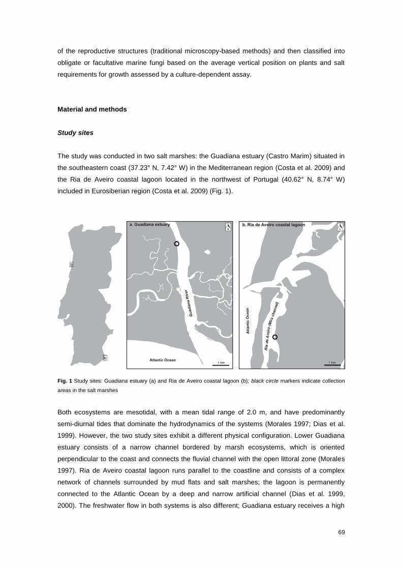
69
of the reproductive structures (traditional microscopy-based methods) and then classified into
obligate or facultative marine fungi based on the average vertical position on plants and salt
requirements for growth assessed by a culture-dependent assay.
Material and methods
Study sites
The study was conducted in two salt marshes: the Guadiana estuary (Castro Marim) situated in
the southeastern coast (37.23° N, 7.42° W) in the Mediterranean region (Costa et al. 2009) and
the Ria de Aveiro coastal lagoon located in the northwest of Portugal (40.62° N, 8.74° W)
included in Eurosiberian region (Costa et al. 2009) (Fig. 1).
Fig. 1 Study sites: Guadiana estuary (a) and Ria de Aveiro coastal lagoon (b); black circle markers indicate collection
areas in the salt marshes
Both ecosystems are mesotidal, with a mean tidal range of 2.0 m, and have predominantly
semi-diurnal tides that dominate the hydrodynamics of the systems (Morales 1997; Dias et al.
1999). However, the two study sites exhibit a different physical configuration. Lower Guadiana
estuary consists of a narrow channel bordered by marsh ecosystems, which is oriented
perpendicular to the coast and connects the fluvial channel with the open littoral zone (Morales
1997). Ria de Aveiro coastal lagoon runs parallel to the coastline and consists of a complex
network of channels surrounded by mud flats and salt marshes; the lagoon is permanently
connected to the Atlantic Ocean by a deep and narrow artificial channel (Dias et al. 1999,
2000). The freshwater flow in both systems is also different; Guadiana estuary receives a high
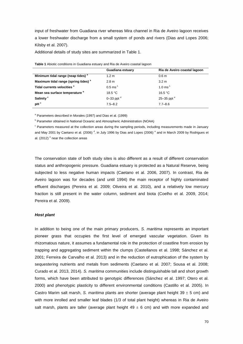
70
input of freshwater from Guadiana river whereas Mira channel in Ria de Aveiro lagoon receives
a lower freshwater discharge from a small system of ponds and rivers (Dias and Lopes 2006;
Kilsby et al. 2007).
Additional details of study sites are summarized in Table 1.
Table 1 Abiotic conditions in Guadiana estuary and Ria de Aveiro coastal lagoon
Guadiana estuary Ria de Aveiro coastal lagoon
Minimum tidal range (neap tides) a 1.2 m 0.6 m
Maximum tidal range (spring tides) a 2.8 m 3.2 m
Tidal currents velocities a 0.5 ms
-1 1.0 ms
-1
Mean sea surface temperature b 18.5 °C 16.5 °C
Salinity c 0–33 ppt
d 25–35 ppt
e
pH c 7.5–8.2 7.7–8.6
a Parameters described in Morales (1997) and Dias et al. (1999)
b Parameter obtained in National Oceanic and Atmospheric Administration (NOAA)
c Parameters measured at the collection areas during the sampling periods, including measurements made in January
and May 2001 by Caetano et al. (2006) d, in July 1996 by Dias and Lopes (2006)
e and in March 2009 by Rodrigues et
al. (2012) e near the collection areas
The conservation state of both study sites is also different as a result of different conservation
status and anthropogenic pressure. Guadiana estuary is protected as a Natural Reserve, being
subjected to less negative human impacts (Caetano et al. 2006, 2007). In contrast, Ria de
Aveiro lagoon was for decades (and until 1994) the main receptor of highly contaminated
effluent discharges (Pereira et al. 2009; Oliveira et al. 2010), and a relatively low mercury
fraction is still present in the water column, sediment and biota (Coelho et al. 2009, 2014;
Pereira et al. 2009).
Host plant
In addition to being one of the main primary producers, S. maritima represents an important
pioneer grass that occupies the first level of emerged vascular vegetation. Given its
rhizomatous nature, it assumes a fundamental role in the protection of coastline from erosion by
trapping and aggregating sediment within the clumps (Castellanos et al. 1998; Sánchez et al.
2001; Ferreira de Carvalho et al. 2013) and in the reduction of eutrophication of the system by
sequestering nutrients and metals from sediments (Caetano et al. 2007; Sousa et al. 2008;
Curado et al. 2013, 2014). S. maritima communities include distinguishable tall and short growth
forms, which have been attributed to genotypic differences (Sánchez et al. 1997; Otero et al.
2000) and phenotypic plasticity to different environmental conditions (Castillo et al. 2005). In
Castro Marim salt marsh, S. maritima plants are shorter (average plant height 39 5 cm) and
with more inrolled and smaller leaf blades (1/3 of total plant height) whereas in Ria de Aveiro
salt marsh, plants are taller (average plant height 49 6 cm) and with more expanded and
71
larger leaf blades (1/2 of total plant height). In addition to intraspecific differences in
morphology, both communities exhibited different distribution patterns; in Castro Marim salt
marsh, S. maritima community forms extensive monotypic beds along the riverside whereas in
Ria de Aveiro salt marsh, community is fragmented and disrupted in relatively small and
dispersed patches.
Although both communities of S. maritima follow the natural phenological cycle, with a growing
season occurring during spring to early summer, plants of different maturation phases are
present throughout all seasons.
Sampling procedure
Mature standing-live plants experiencing the same daily tidal wet-dry cycles (i.e. occupying the
same topographic level, with similar height and containing green, senescent and early-decay
plant tissues) were randomly collected in Castro Marim and Ria de Aveiro salt marshes (2.45
and 2.37 m above the Portuguese hydrographic zero, respectively), bimonthly over a 2-year
period (October 2010 to August 2012). Twenty plants were collected each of the first 3 sampling
periods and 15 plants afterwards (a total of 390 plants). Five additional plants were also
collected in each period of the last sampling year (February 2012 to August 2012) for isolation
of marine fungi (a total of 20 plants). Plant samples were placed in plastic bags and returned to
laboratory.
Morphology-based species identification
The collected plants were carefully rinsed with running tap water to remove fine-grained
sediments and seaweeds and air-dried. Each air-dried plant was sequentially analysed from the
basal to the top portion and from the external vegetative structures (leaf sheaths and blades)
towards the more internal structures (stems). Fungal structures (fruit bodies, spores and
hyphopodia) observed on each vegetative structure were picked up under a dissecting
microscope (Wild M8) mounted into a drop of sterile seawater on a slide examined under a light
microscope (Leitz Laborlux S, with Normaski) with detailed morphology recorded. The fungi
were identified using the dichotomous keys of Kohlmeyer and Kohlmeyer (1979), Kohlmeyer
and Volkmann-Kohlmeyer (1991), Hyde and Sarma (2000) and Jones et al. (2009). The vertical
position of identified fungi was also recorded, as well as the density of produced fungal
structures; for more than 10 fungal structures per square centimeter of colonized vegetative
structure, the density was considered high. The fungal structures were photographed and
preserved on microscope slides after replacement of seawater by glycerin and sealed with
several layers of nail varnish. Moreover, some of the identified fungal structures were
maintained on the original dry plant material and included in the personal collection of M. Barata
(Herbarium of the University of Lisbon - LISU).
72
Isolation of marine fungi and preservation of pure cultures
Cultures were obtained by single spore method, according to the conventional procedures of
Vrijmoed (2000). Five fruiting structures (ascomata or pycnidia) of a given fungal taxon growing
on fresh plant materials were transferred into a drop of sterile seawater on a microscope slide
and squashed to force the discharge of the spores. This suspension of spores was then
transferred with a Pasteur pipette onto gridded plates containing cornmeal agar made with aged
diluted seawater (CMA/sw 50%) and supplemented with chloramphenicol (0.05%), one drop per
square. Plates were incubated at room temperature for 1–2 days until germination of the
spores. Each germinated spore was then transferred onto a new CMA/sw plate.
In order to establish a culture collection, each isolated fungus was subcultured and preserved
by three methods: (1) one colony was maintained on CMA/sw plate at 4 °C, (2) plugs removed
from the growing margin of four colonies were transferred to McCartney bottles filled with sterile
diluted seawater (50%) and kept at 4 °C, and (3) to cryotubes filled with glycerol (10%) and
stored at -80 ºC.
Growth rates
Growth rates of selected fungi were determined in cornmeal agar media made with diluted
seawater (CMA/sw 50%) and with distilled water (CMA/dw) at room temperature (18–25 °C).
With this purpose, an agar disc was cut from the growing edge of fungal colonies and inoculated
at the intersection point of two perpendicular lines previously drawn on the bottom of CMA/sw
50% and CMA/dw plates; three replicates were performed for each monospore isolate. The
colony growth was assessed every 2 days, for 30 days, by measuring and averaging the colony
diameter along the two perpendicular axes.
Data analyses
Diversity indices
The following diversity indices were calculated in order to better characterize and compare the
fungal communities inhabiting Castro Marim and Ria de Aveiro salt marshes: Shannon diversity
index (H´=-∑ 𝑝𝑖ln(𝑝𝑖)Si=1 , where s is the number of fungal taxa in the community and pi is the
proportion of occurrences of fungal taxon i relative to total number of occurrences), Shannon´s
equitability index (E=H´/Hmax, where Hmax=ln s) and Sorenson similarity index (SI=2j/(a + b),
where “j” is the number of common fungal taxa to both sites, a is the number of fungal taxa in
one site and b is the number of fungal taxa in the other site). The comparison of Shannon
diversity indices between study sites was performed based on randomization procedures of
bootstrapping using the PAST v2.17c statistical software (Hammer et al. 2001). P values were
73
estimated by resampling and randomly redistributing the data 1000 times (Efron and Tibshirani
1986); differences were considered statistically significant for p value<0.05.
The average number of fungal taxa per plant sample (total number of fungal occurrences
divided by the total number of plant samples) was also determined for Castro Marim and Ria de
Aveiro salt marshes and for the assembly of the two salt marshes.
Frequencies of occurrence and vertical distribution patterns – Total and in each sampling
period
The percent frequency of occurrence for each taxon in the fungal community was assessed
(number of plant samples colonized by a specific fungus divided by the total number of plant
samples x 100). Fungal taxa were grouped according to the percent frequency of occurrence
and the classification proposed by Tan et al. (1989) in very frequent (>20%), frequent (10–20%)
and infrequent (<10%).
The average vertical distribution data of common fungal taxa in both study sites were compared
by Student´s t tests, using IBM SPSS v22.0 statistical software (IBM Corporation, Somers, NY).
In an attempt to better discriminate vertical distribution patterns and ecological requirements of
fungal taxa in the two salt marshes, three vertical microhabitats were defined by separating the
plant samples in three equally portions based on maximum plants height (basal 0–20 cm,
middle >20–40 cm, top >40–60 cm). For each plant portion, the same diversity indices
(Shannon, Equitability and Sorensen similarity indices) were determined; comparisons among
Shannon diversity indices in plant portions were performed adopting the same procedures
described above. Additionally, frequencies of occurrence of each fungal taxon, in each sampling
period, in each plant portion were calculated. These dataset matrices were used to perform a
preliminary Cluster Analysis with Bray-Curtis similarity measure; seven samples were
considered outliers, given the atypical and divergent behavior and excluded from the
subsequent analyses. The reconstructed dataset matrices were then used to perform another
cluster analysis and a detrended correspondence analysis, using the PAST v2.17c statistical
software.
The effect of seasonality on fungal communities was assessed by analysing the variations of
the frequencies of occurrence and vertical positions of all fungal taxa during the two sampling
years; the former parameters were interpreted graphically, and the second parameters were
tested statistically using IBM SPSS v22.0 software. A two-way analysis of variance (ANOVA)
was performed in order to test the effect of sampling periods and fungal taxa on the total
variations of vertical distributions in both communities. After this procedure, a new one-way
ANOVA was performed for each taxon to evaluate the statistical significance of its vertical
distribution variation.
74
Flooding regime in Castro Marim and Ria de Aveiro study sites
The flooding conditions in both study sites were assessed, given the differences in the physical
configuration of intertidal systems and in the morphology of host plant. The percentage of days
in each month that Spartina plants were totally submerged, at least once, was determined,
considering average plants height, tidal range (high tides height) and the topographic position of
the plants on both salt marshes. The average time per day that the plants remained flooded at
each sampling site was estimated using a model developed by Serôdio and Catarino (2000).
The frequency and time length of flooding were also determined for basal, middle and top plant
portions.
Vegetative growth rates of fungal isolates
The growth rates were extracted from linear regression equation of colony diameter increase
over 30 days. The differences between growth rates in the two culture media were assessed
with Student´s t tests, using IBM SPSS v22.0 statistical software.
Results
Fungal diversity
Twenty-three sporulating higher filamentous marine fungi were recorded from the total 390
analysed plants, with 20 and 14 fungal taxa occurring in Castro Marim and Ria de Aveiro salt
marshes, respectively (Table 2; Fig. S1, Online Resource). The average number of fungi per
plant was found to be five in both sites.
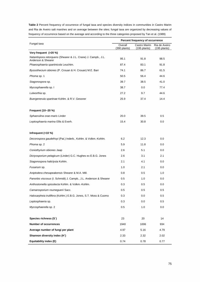
75
Table 2 Percent frequency of occurrence of fungal taxa and species diversity indices in communities in Castro Marim
and Ria de Aveiro salt marshes and on average between the sites; fungal taxa are organized by decreasing values of
frequency of occurrence based on the average and according to the three categories proposed by Tan et al. (1989)
Fungal taxa
Percent frequency of occurrence
Overall (390 plants)
Castro Marim (195 plants)
Ria de Aveiro (195 plants)
Very frequent (>20 %)
Natantispora retorquens (Shearer & J.L. Crane) J. Campb., J.L. Anderson & Shearer
95.1 91.8 98.5
Phaeosphaeria spartinicola Leuchtm. 87.4 83.1 91.8
Byssothecium obiones (P. Crouan & H. Crouan) M.E. Barr 74.1 86.7 61.5
Phoma sp. 1 50.5 56.4 44.6
Stagonospora sp. 39.7 38.5 41.0
Mycosphaerella sp. I 38.7 0.0 77.4
Lulworthia sp. 27.2 9.7 44.6
Buergenerula spartinae Kohlm. & R.V. Gessner 25.9 37.4 14.4
Frequent (10–20 %)
Sphaerulina orae-maris Linder 20.0 39.5 0.5
Leptosphaeria marina Ellis & Everh. 15.4 30.8 0.0
Infrequent (<10 %)
Decorospora gaudefroyi (Pat.) Inderb., Kohlm. & Volkm.-Kohlm. 6.2 12.3 0.0
Phoma sp. 2 5.9 11.8 0.0
Coniothyrium obiones Jaap 2.6 5.1 0.0
Dictyosporium pelagicum (Linder) G.C. Hughes ex E.B.G. Jones 2.6 3.1 2.1
Stagonospora haliclysta Kohlm. 2.1 4.1 0.0
Fusarium sp. 1.0 2.1 0.0
Aniptodera chesapeakensis Shearer & M.A. Mill. 0.8 0.5 1.0
Panorbis viscosus (I. Schmidt) J. Campb., J.L. Anderson & Shearer 0.5 1.0 0.0
Anthostomella spissitecta Kohlm. & Volkm.-Kohlm. 0.3 0.5 0.0
Camarosporium roumeguerii Sacc. 0.5 0.5 0.5
Halosarpheia trullifera (Kohlm.) E.B.G. Jones, S.T. Moss & Cuomo 0.3 0.0 0.5
Leptosphaeria sp. 0.3 0.0 0.5
Mycosphaerella sp. 2 0.5 1.0 0.0
Species richness (S´) 23 20 14
Number of occurrences 1940 1006 934
Average number of fungi per plant 4.97 5.16 4.79
Shannon diversity index (H´) 2.33 2.32 2.02
Equitability index (E) 0.74 0.78 0.77
76
The fungal communities of Castro Marim and Ria de Aveiro salt marshes were mostly
composed of the Ascomycota, representing 60% (12) and 71% (10) of the total number of
fungal taxa and 76% (769) and 82% (762) of the number of occurrences, respectively. The
remaining fungal taxa found in both study sites belong to asexual fungi, mainly coelomycetes
(30% in Castro Marim and 21% in Ria de Aveiro). Ascomycetous fungi were restricted to
Dothideomycetes, Sordariomycetes and Sordariomycetes incertae sedis; Pleosporales,
Microascales (i.e. Halosphaeriaceae) and Capnodiales were the most representative orders,
with 33, 25 and 17% in Castro Marim and 20, 30 and 20% in Ria de Aveiro, respectively.
Although the diversity of fungal community was significantly higher in Castro Marim than Ria de
Aveiro (p<0.01), both communities revealed a similar high equitability value.
The results also evidenced a high overlap between fungal communities of Castro Marim and
Ria de Aveiro salt marshes regarding species composition and common taxa. From the 23 total
fungal taxa associated with S. maritima, 48% (11) were common between the study sites.
Natantispora retorquens, B. obiones, P. spartinicola, Phoma sp. 1 and Stagonospora sp. were
very frequent in both communities. Sorensen´s index revealed a similarity of 0.65 between both
fungal communities.
The main differences between the two fungal communities were the number of exclusive
infrequent fungal taxa, which was higher in Castro Marim than in Ria de Aveiro salt marsh (8 vs
2). Moreover, the former study site included two frequent and one very frequent exclusive fungal
taxa, namely Leptosphaeria marina, Decorospora gaudefroyi and Phoma sp. 2, respectively,
while Ria de Aveiro harboured only one very frequent exclusive fungal species, Mycosphaerella
sp. I. Sphaerulina orae-maris was very frequent in Castro Marim and infrequent in Ria de Aveiro
salt marsh.
Vertical distribution of fungi
Fungal taxa inhabiting S. maritima in both study sites were distributed vertically along the plant,
displaying distinct distribution patterns; some were restricted to the upper or lower portions of
the plants while others spanned widely along the plant, showing different extents of substrate
occupation (Fig. 2).
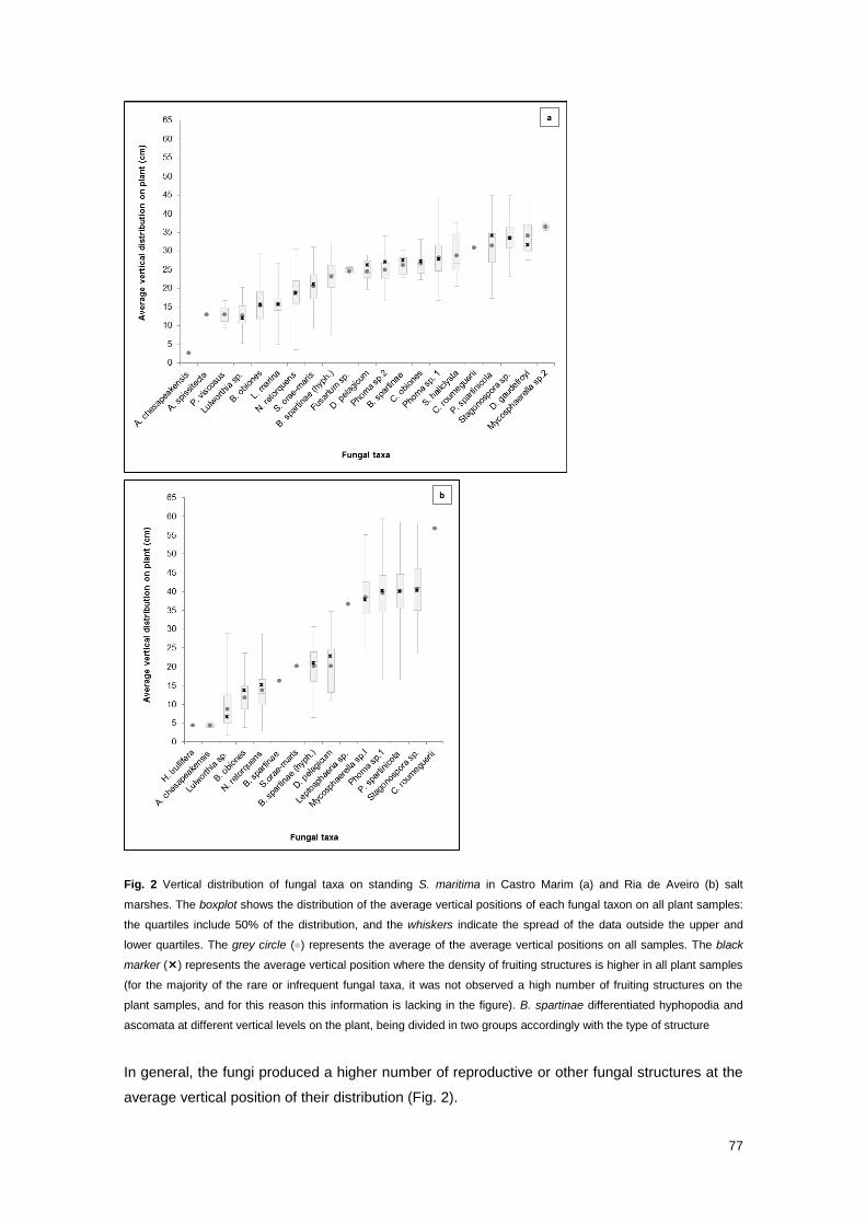
77
Fig. 2 Vertical distribution of fungal taxa on standing S. maritima in Castro Marim (a) and Ria de Aveiro (b) salt
marshes. The boxplot shows the distribution of the average vertical positions of each fungal taxon on all plant samples:
the quartiles include 50% of the distribution, and the whiskers indicate the spread of the data outside the upper and
lower quartiles. The grey circle ( ) represents the average of the average vertical positions on all samples. The black
marker () represents the average vertical position where the density of fruiting structures is higher in all plant samples
(for the majority of the rare or infrequent fungal taxa, it was not observed a high number of fruiting structures on the
plant samples, and for this reason this information is lacking in the figure). B. spartinae differentiated hyphopodia and
ascomata at different vertical levels on the plant, being divided in two groups accordingly with the type of structure
In general, the fungi produced a higher number of reproductive or other fungal structures at the
average vertical position of their distribution (Fig. 2).

78
A comparison between vertical distribution data of common fungal taxa in Castro Marim and Ria
de Aveiro salt marshes demonstrated that the differences were statistically significant (p<0.05),
except for Dictyosporium pelagicum. However, the shared fungal taxa appeared to occupy the
same relative vertical position on the plants, despite the variations on the absolute vertical
distribution.
Fungal subcommunities on basal, middle and top portions of the plants in both study sites were
shown to be considerably different by diversity indices (Table 3), cluster analysis (Fig. 3) and
Detrended Correspondence Analysis (DCA; Fig. 4).
Table 3 Diversity indices and number of records in the 3 vertical portions of the plants in Castro Marim and Ria de
Aveiro salt marshes
Castro Marim Ria de Aveiro
Basal Middle Top Basal Middle Top
Species richness (S´) 13 18 4 10 11 5
Number of records 380 542 20 388 301 237
Shannon index (H´) 1.68 2.35 1.30 1.28 1.78 1.35
Equitability index (E) 0.66 0.81 0.94 0.56 0.74 0.84
Basal x Middle
Middle x Top
Top x Basal
Basal x Middle
Middle x Top
Top x Basal
Sorensen similarity index 0.65 0.36 0.24 0.67 0.50 0.27
Middle portion yielded the highest species richness and diversity than either basal or top
portions (Table 3). The differences in the Shannon indices were statistically significant between
basal/middle and middle/top portions (p<0.001), but not between basal/top portions in Castro
Marim and Ria de Aveiro salt marshes (p>0.05).
The fungal subcommunities inhabiting basal and middle plant portions revealed higher
similarities considering species richness and fungal taxa composition in both study sites than
those in basal/top portions and in middle/top portions.
The cluster and DCA analyses, which provided an integrated overview of spatial arrangement of
fungal community based on the frequencies of occurrence of fungal taxa in each vertical plant
portion corroborated the existence of three distinct microhabitats supporting distinct fungal
subcommunities (Figs. 3 and 4).
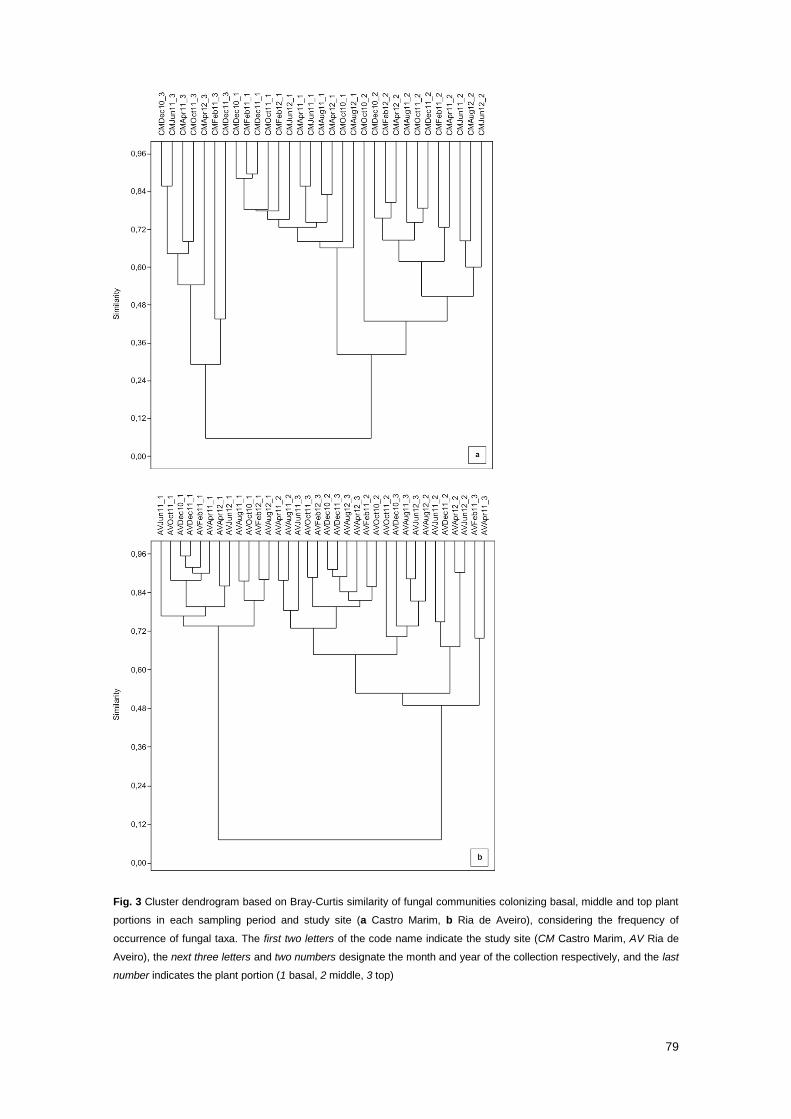
79
Fig. 3 Cluster dendrogram based on Bray-Curtis similarity of fungal communities colonizing basal, middle and top plant
portions in each sampling period and study site (a Castro Marim, b Ria de Aveiro), considering the frequency of
occurrence of fungal taxa. The first two letters of the code name indicate the study site (CM Castro Marim, AV Ria de
Aveiro), the next three letters and two numbers designate the month and year of the collection respectively, and the last
number indicates the plant portion (1 basal, 2 middle, 3 top)

80
Fig. 4 Two-dimensional DCA plot expressing the fungal taxa and the three vertical plant portions spatial distributions
based on frequency of occurrence of fungal taxa in each portion in each sampling period and in each study site (a
Castro Marim, b Ria de Aveiro). The black pentagons ( ), dark grey triangles ( ) and light grey polygons ( )
correspond to basal, middle and top plant portions, respectively. The two-letter code represent fungal taxa: AC
Aniptodera chesapeakensis, BO Byssothecium obiones, BS Buergenerula spartinae ascomata, BS(hyp) Buergenerula
spartinae hyphopodia, CO Coniothyrium obiones, CR Camarosporium roumeguerii, DG Decorospora gaudefroyi, DP
Dictyosporium pelagicum, LM Leptosphaeria marina, Lu Lulworthia sp., My1 Mycosphaerella sp. I, My2 Mycosphaerella
sp. 2, NR Natantispora retorquens, Ph1 Phoma sp. 1, Ph2 Phoma sp. 2, PS Phaeosphaeria spartinicola, PV Panorbis
viscosus, SH Stagonospora haliclysta, SO Sphaerulina orae-maris, St Stagonospora sp.

81
The cluster analysis (Fig. 3) performed with Castro Marim dataset separated first the top plant
portion (ca. 0.29 of similarity) and then basal (ca. 0.31 of similarity) from middle portion,
coinciding exactly with the defined microhabitats; with Ria de Aveiro dataset, the analysis only
distinguished clearly the basal plant portion from the middle and top portions (ca. 0.42 of
similarity).
The DCA reinforced the results from the previous analysis (Fig. 4). Along the axis 1, with the
higher eigenvalue (Castro Marim: 0.52; Ria de Aveiro: 0.86) and explanatory power, there was
a clear spatial separation of the three plant portions in Castro Marim dataset, which was not so
evident between middle and top portions in Ria de Aveiro dataset. The graphical separation of
basal, middle and top portions followed the natural vertical sequence of microhabitats, which
confirmed the higher similarity of fungal subcommunities between adjacent plant portions.
The DCA analysis also highlighted specific ecological niches by plotting the distribution of fungal
taxa across plant portions. Fungal taxa were distributed along the axis 1 following the vertical
distribution on the standing plants in Castro Marim and Ria de Aveiro salt marshes, from the top
to the basal plant portions. The spatial proximity of each fungal taxon to a certain plant portion
in the plot suggested higher affinities to that particular microhabitat. Thus, the results evidenced
a subcommunity associated with basal portions, mainly represented by B. obiones, Lulworthia
sp. and N. retorquens in both salt marshes, and Panorbis viscosus and L. marina in Castro
Marim; a subcommunity colonizing middle portions composed by B. spartinae, D. pelagicum
and Phoma sp. 1 in both study sites and Coniothyrium obiones, D. gaudefroyi, Phoma sp. 2 and
S. orae-maris in Castro Marim; and a subcommunity associated with upper portions composed
by P. spartinicola and Stagonospora sp. in both salt marshes, Mycosphaerella sp. I in Ria de
Aveiro, and Mycosphaerella sp. 2 and Stagonospora haliclysta in Castro Marim.
Both axes 2 and 3 presented a lower eigenvalue in Castro Marim (axis 2: 0.12; axis 3: 0.06) and
Ria de Aveiro (axis 2: 0.08; axis 3: 0.06) datasets, explaining little variation in the data.
Comparisons of micro-environmental conditions on the three plant portions revealed some
differences. Specifically, it was observed a decrease in flooding time (from 8 to 2 daily hours)
and frequency (from 100 to 50% of the days per month) along the vertical axis of the plants,
from the basal upwards to the top portions, in both salt marshes; although it was not measured
in this study, this vertical gradient of tidal flooding reflected obviously in salinity and water
availability levels in each plant portion. Middle and top plant portions in Castro Marim salt marsh
remained slightly longer and were more frequently submerged than analogous portions in Ria
de Aveiro salt marsh. Furthermore, it was found that the vegetative structures in each plant
portion were different: basal portions included mostly a senescent naked stem or a stem
enwrapped by leaf sheaths; middle portions included mainly a stem enwrapped by leaf sheaths
and leaf blades; top portions included mostly upright-standing leaf blades. Nevertheless, the
host plants from the two salt marshes presented some differences in the proportions of
vegetative structures included in analogous portions, as a result of differences in the
morphology. The main differences were found in the middle plant portions; Castro Marim plants

82
included mostly the stem and leaf sheaths whereas Ria de Aveiro plants also included leaf
blades in this portion.
Seasonality
The effects of seasonality on fungal community dynamics and particularly in the frequencies of
occurrence of the most frequent fungal taxa in the two study sites (Fig. 5) and for fungi
producing a high density of fruiting structures (Fig. 6) were investigated.
Fig. 5 Bimonthly variation of the frequency of occurrence (%) of each fungal taxon in Castro Marim (a) and Ria de
Aveiro (b) salt marshes and average variation of the frequencies of occurrence in each sampling period: BO
Byssothecium obiones, BS Buergenerula spartinae, DG Decorospora gaudefroyi, LM Leptosphaeria marina, Lu
Lulworthia sp., MyI Mycosphaerella sp. I, NR Natantispora retorquens, Ph1 Phoma sp. 1, Ph2 Phoma sp. 2, PS
Phaeosphaeria spartinicola, SO Sphaerulina orae-maris, St Stagonospora sp.
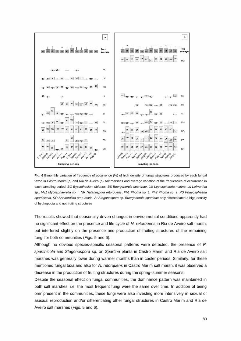
83
Fig. 6 Bimonthly variation of frequency of occurrence (%) of high density of fungal structures produced by each fungal
taxon in Castro Marim (a) and Ria de Aveiro (b) salt marshes and average variation of the frequencies of occurrence in
each sampling period: BO Byssothecium obiones, BS Buergenerula spartinae, LM Leptosphaeria marina, Lu Lulworthia
sp., My1 Mycosphaerella sp. I, NR Natantispora retorquens, Ph1 Phoma sp. 1, Ph2 Phoma sp. 2, PS Phaeosphaeria
spartinicola, SO Sphaerulina orae-maris, St Stagonospora sp. Buergenerula spartinae only differentiated a high density
of hyphopodia and not fruiting structures
The results showed that seasonally driven changes in environmental conditions apparently had
no significant effect on the presence and life cycle of N. retorquens in Ria de Aveiro salt marsh,
but interfered slightly on the presence and production of fruiting structures of the remaining
fungi for both communities (Figs. 5 and 6).
Although no obvious species-specific seasonal patterns were detected, the presence of P.
spartinicola and Stagonospora sp. on Spartina plants in Castro Marim and Ria de Aveiro salt
marshes was generally lower during warmer months than in cooler periods. Similarly, for these
mentioned fungal taxa and also for N. retorquens in Castro Marim salt marsh, it was observed a
decrease in the production of fruiting structures during the spring–summer seasons.
Despite the seasonal effect on fungal communities, the dominance pattern was maintained in
both salt marshes, i.e. the most frequent fungi were the same over time. In addition of being
omnipresent in the communities, these fungi were also investing more intensively in sexual or
asexual reproduction and/or differentiating other fungal structures in Castro Marim and Ria de
Aveiro salt marshes (Figs. 5 and 6).
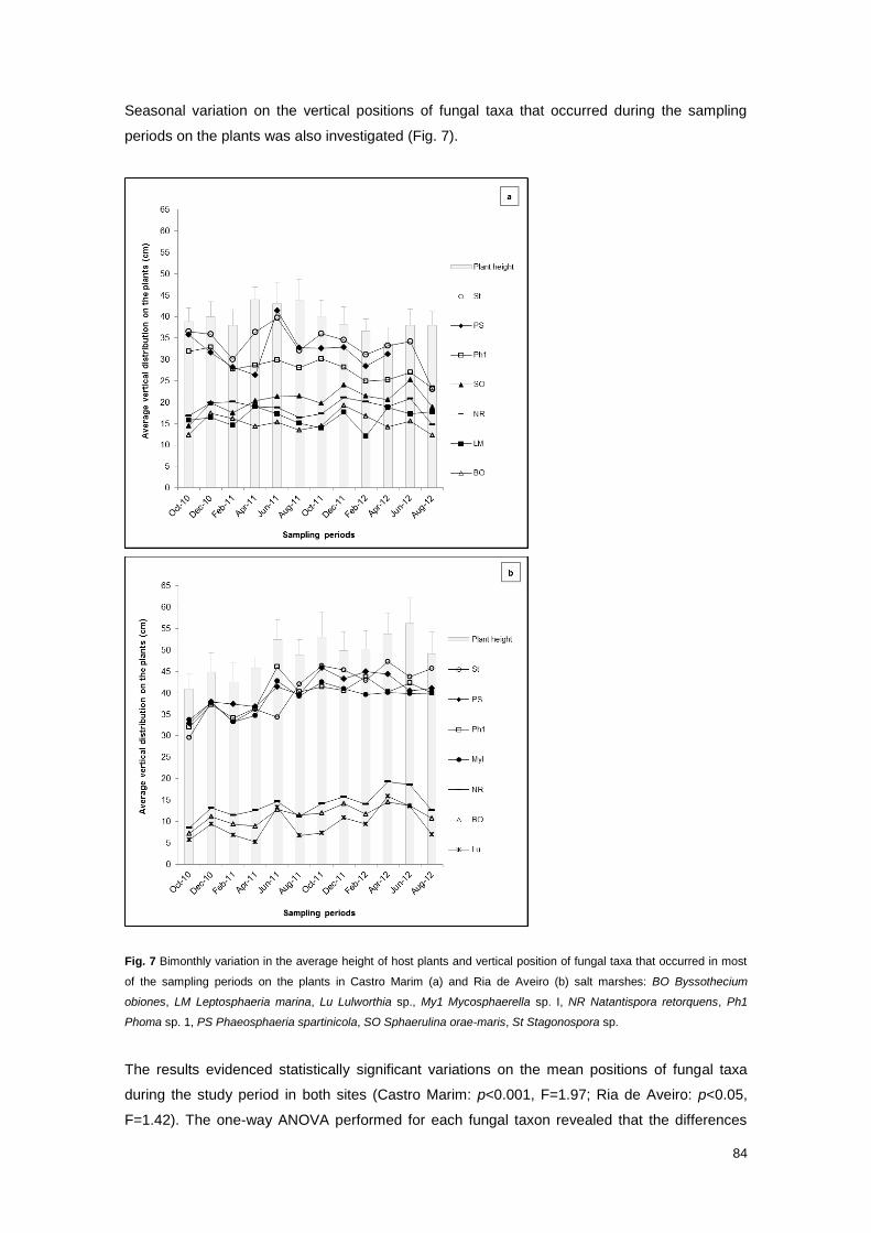
84
Seasonal variation on the vertical positions of fungal taxa that occurred during the sampling
periods on the plants was also investigated (Fig. 7).
Fig. 7 Bimonthly variation in the average height of host plants and vertical position of fungal taxa that occurred in most
of the sampling periods on the plants in Castro Marim (a) and Ria de Aveiro (b) salt marshes: BO Byssothecium
obiones, LM Leptosphaeria marina, Lu Lulworthia sp., My1 Mycosphaerella sp. I, NR Natantispora retorquens, Ph1
Phoma sp. 1, PS Phaeosphaeria spartinicola, SO Sphaerulina orae-maris, St Stagonospora sp.
The results evidenced statistically significant variations on the mean positions of fungal taxa
during the study period in both sites (Castro Marim: p<0.001, F=1.97; Ria de Aveiro: p<0.05,
F=1.42). The one-way ANOVA performed for each fungal taxon revealed that the differences
85
were statistically significant (p<0.05) for all fungal taxa, except for L. marina. However, the
relative mean position of each fungus on the plants seemed to be maintained, as well as the
spatial pattern of occupancy along the vertical axis of the plant by different fungal taxa.
Salinity requirements of fungi
To confirm the salinity requirements for the isolated fungi with vertical distribution pattern on the
standing plants in natural environment, a culture experiment was performed. Fifteen strains
were randomly selected from the 57 isolated fungi from Castro Marim salt marsh representing 8
fungal taxa, namely B. spartinae (2 strains), B. obiones (2 teleomorph strains), L. marina (2
strains), N. retorquens (2 strains), P. spartinicola (2 strains), Phoma sp. 1 (2 strains), S. orae-
maris (1 strain) and Stagonospora sp. (2 strains); 13 strains were selected from the 66 isolated
fungi from Ria de Aveiro salt marsh representing 6 fungal taxa, specifically B. obiones (2
teleomorph strains and 2 anamorph strains), Lulworthia sp. (2 strains), N. retorquens (1 strain),
P. spartinicola (2 strains), Phoma sp. 1 (2 strains) and Stagonospora sp. (2 strains).
The comparison between mycelia growth rates under two different culture conditions, on media
lacking and containing diluted seawater, provided additional information about the ecological
preferences of each taxon (Fig. 8).
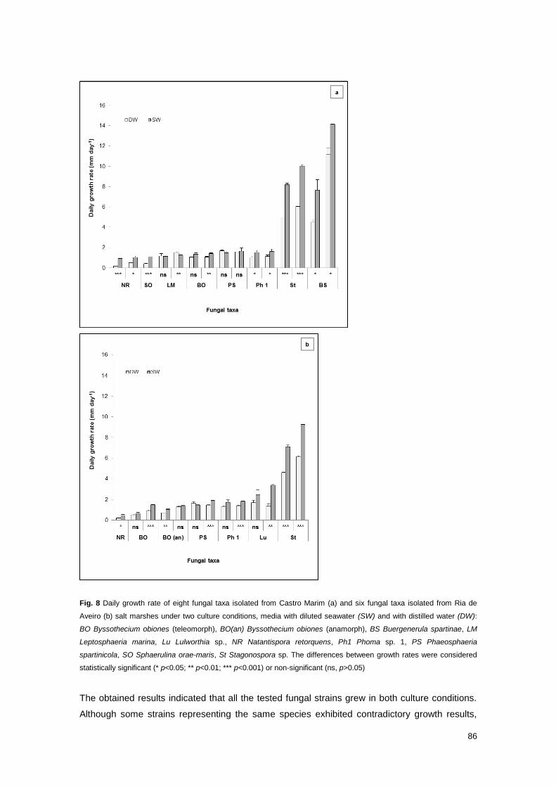
86
Fig. 8 Daily growth rate of eight fungal taxa isolated from Castro Marim (a) and six fungal taxa isolated from Ria de
Aveiro (b) salt marshes under two culture conditions, media with diluted seawater (SW) and with distilled water (DW):
BO Byssothecium obiones (teleomorph), BO(an) Byssothecium obiones (anamorph), BS Buergenerula spartinae, LM
Leptosphaeria marina, Lu Lulworthia sp., NR Natantispora retorquens, Ph1 Phoma sp. 1, PS Phaeosphaeria
spartinicola, SO Sphaerulina orae-maris, St Stagonospora sp. The differences between growth rates were considered
statistically significant (* p<0.05; ** p<0.01; *** p<0.001) or non-significant (ns, p>0.05)
The obtained results indicated that all the tested fungal strains grew in both culture conditions.
Although some strains representing the same species exhibited contradictory growth results,
87
the majority of strains showed significantly higher growth under saline conditions. An exception
was L. marina, which grew faster in media without seawater, and P. spartinicola, which grew
equally well under both conditions. Additionally, the results revealed different growth rates
among fungal taxa, with Lulworthia sp., Stagonospora sp. and B. spartinae displaying the
highest growth rates on both media.
Discussion
Fungal diversity
In the present study, 23 fungal taxa were identified associated with early stages of
decomposition of S. maritima; 20 fungal taxa inhabited Castro Marim salt marsh and 14
occurred in Ria de Aveiro salt marsh (Table 2). Both fungal communities were predominantly
represented by the Ascomycota, particularly Dothideomycetes and Sordariomycetes. The clear
preference and dominance of these taxonomic groups in intertidal habitats, characterized by
alternate cycles of immersion and exposure, have been widely documented in several studies
(e.g. Gessner and Kohmeyer 1976; Gessner 1977; Newell et al. 1989, 1996a, 2000a; Samiaji
and Barlocher 1996; Alias and Jones 2000; Barata 2002; Al-Nasrawi and Hughes 2012).
The species richness and diversity of fungal communities colonizing S. maritima in Castro
Marim and Ria de Aveiro salt marshes were similar to those found in the same host plant by
Barata (2002, 2006), in S. alterniflora by Gessner (1977), Samiaji and Barlocher (1996) and Al-
Nasrawi and Hughes (2012), and in Spartina densiflora Brongn. by Peña and Arambarri (1998).
Both fungal communities from Castro Marim and Ria de Aveiro salt marshes were found to be
well-balanced, without a clear dominance. These findings corroborated observations by
Gessner et al. (2007) and Van Ryckegem et al. (2007), who denoted that fungal communities
associated with Spartina spp. are not particularly complex, with a low diversity and few
dominant species.
Fungal species composition of communities associated with Spartina spp.
A comparison between the species composition of the studied fungal communities associated
with S. maritima and the list of marine fungi reported from Spartina species (Kohlmeyer and
Kohlmeyer 1979; Barata 2002; Kohlmeyer and Volkmann-Kohlmeyer 2002; Calado and Barata
2012) revealed nine common fungal species. These species can be categorized into three
groups according to their geographical distribution and substrate specificity: (1) host-genus-
exclusive fungi that have been described from different Spartina species in different
geographical locations, namely Anthostomella spissitecta, B. spartinae, B. obiones,
Mycosphaerella sp. I and P. spartinicola (Gessner & Kohlmeyer 1976; Peña and Arambarri
1998; Barata 2002; Kohlmeyer and Volkmann-Kohlmeyer 2002; Cornick et al. 2005; Walker and
88
Campbell 2010); (2) temperate fungi that have a broad substrate preference, such as L. marina
and S. orae-maris, which were found on Spartina spp. (Gessner and Kohlmeyer 1976;
Cavaliere 1977; Kohlmeyer and Kohlmeyer 1979; Shoemaker and Babcock 1989; Peña and
Arambarri 1998; Barata 2002) and also on Juncus roemerianus (Cavaliere 1977; Kohlmeyer
and Kohlmeyer 1979) and driftwood (Cavaliere 1977; Kohlmeyer and Kohlmeyer 1979; Peña
and Arambarri 1996; Figueira and Barata 2007); and (3) cosmopolitan fungal species that have
also been recorded on a wide variety of substrates, such as Aniptodera chesapeakensis and D.
pelagicum in temperate (Jones et al. 1998; Barata 2006; Figueira and Barata 2007) and tropical
climates (Sadaba et al. 1995; Poon and Hyde 1998a; Hyde and Sarma 2006; Alias et al. 2010;
Khan and Manimohan 2011; Manimohan et al. 2011).
In this study, B. spartinae, B. obiones and P. spartinicola, mentioned in the literature as the
main colonizers of decaying Spartina plants (Gessner 1977; Newell and Wasowski 1995;
Newell and Zakel 2000; Newell et al. 2000a; Newell 2001a; Barata 2002; Buchan et al. 2002,
2003; Cornick et al. 2005; Lyons et al. 2010; Walker and Campbell 2010), were also very
frequent or frequent in both studied communities.
The high number of common fungal species colonizing different Spartina hosts corroborated the
existence of a very stable core group of fungi, mainly dominated by the same host-exclusive
fungi. This core group apparently is not much affected by variations in abiotic conditions
(Gessner and Kohlmeyer 1976).
Different Spartina species, however, supported some different fungal species. This finding,
previously demonstrated by Blum et al. (2004) and Lyons et al. (2010), was attributed to the
higher variation in the morphology and chemical composition between different host species
than within the same species. In fact, and as pointed out by several authors (Gessner and
Kohlmeyer 1976; Torzilli et al. 2006), the substrate quality appears to be primarily responsible
for determining fungal community composition. This reason could explain the absence of some
fungal species that have been frequently collected from other Spartina species on S. maritima
plants in this study and in a similar study performed by Barata (2002), as well as the exclusive
presence of other fungal species in these communities.
In concordance with Barata (2002) study, N. retorquens was found to be the most frequent and
dominant species in the two studied fungal communities, although it has not been reported from
other Spartina species. The absence of N. retorquens on these host plants is not easy to
explain, considering that this fungal species has been collected from driftwood in temperate
regions (Jones et al. 1998; Figueira and Barata 2007; Azevedo et al. 2012) and from different
substrates in tropical climates (Sadaba et al. 1995; Prasannarai and Sridhar 2001; Alias et al.
2010). However, it could be related with the fact that most of studies that inventoried Spartina
spp. focused mainly or exclusively on leaf blades. Similarly, P. viscosus, another cosmopolitan
species that has been described from temperate and tropical regions (Peña and Arambarri
1996; Jones et al. 1998; Prasannarai and Sridhar 2001; Figueira and Barata 2007; Alias et al.
2010), was reported for the first time on standing plants of Spartina by Barata (2002), on drift
stems of the same host plant by Azevedo et al. (2012), and collected again in this study.

89
Even though the high overlapping of fungal communities associated with the same host
species, it were found some variations in the mycota associated with standing plants of S.
maritima in different salt marshes in terms of species composition and frequency patterns.
S. maritima was found to be a new host plant for seven fungal species; A. spissitecta, C.
obiones, D. gaudefroyi, L. marina and S. haliclysta were exclusively collected from Castro
Marim plants, Halosarpheia trullifera was exclusively present in Ria de Aveiro plants and
Camarosporium roumeguerii occurred in both study sites (Table 2). From all the mentioned
fungal species, only A. spissitecta and L. marina have been previously described from other
Spartina species. C. roumeguerii, C. obiones and D. gaudefroyi have been observed inhabiting
other salt marsh plants (Inderbitzin et al. 2002; Abdel-Wahab and Bahkali 2012). S. haliclysta
and H. trullifera have been found colonizing the seaweed Pelvetia canaliculata (Abdel-Wahab
and Bahkali 2012) and driftwood in temperate regions (Peña and Arambarri 1996; Jones et al.
1998), respectively. With the exception of C. roumeguerii and L. marina, all the other fungal
species were also new records for Portugal.
Although the differences in sampling methods applied in this study and in Barata (2002) study
may explain some differences between surveyed fungal communities, these are more likely to
have resulted from different environmental conditions in the study sites. Similarly, this last
reason could explain the differences in the fungal communities from Castro Marim and Ria de
Aveiro salt marshes. The higher species richness and diversity found in Castro Marim salt
marsh may be attributed to suitable environmental conditions given by a more preserved
habitat; these conditions may favour the colonization and reproduction of less well-adapted
species.
The fact that S. maritima community is more reduced and fragmented in Ria de Aveiro marsh
and the vestigial presence of mercury in this study site (not measured in the present study, but
mentioned by Coelho et al. 2009, 2014 and Pereira et al. 2009) might have provided less
favourable conditions for colonization by occasional and infrequent species. The total mercury,
as demonstrated by Coelho et al. (2009), accumulates more in old leaves than in stems,
although in lower concentrations than in belowground biomass. These conditions seemed to
have no effects on the most frequent fungal taxa in the community. The resistance of dominant
saprobic ascomycetous fungi associated with S. alterniflora to several potentially toxic pollutants
was already demonstrated by Newell and Wall (1998) and Newell et al. (2000b); they measured
the living fungal biomass and sexual productivities of dominant fungi in standing-decaying leaf
blades in the presence of mercury, methylmercury, polychlorinated biphenyls, chlorinated
organocyclic insecticide toxaphene, chromium, copper, lead and polycyclic aromatic
hydrocarbons and showed that these biological parameters were not affected by the presence
of the toxicants. Moreover, the omnipresence of dominant fungal species in different Spartina
communities of different states of conservation was also revealed by Cornick et al. (2005) –
stable versus declining beds of Spartina anglica C. E. Hubbard – and Walker and Campbell
(2010) – natural versus created S. alterniflora salt marshes.

90
However, the absence of Mycospharella sp. I in Castro Marim salt marsh, as well as the
absence and infrequent occurrence of L. marina and S. orae-maris respectively in Ria de Aveiro
salt marsh, was more difficult to interpret under an ecological perspective. The absence of L.
marina from three salt marshes highly exposed to anthropogenic pressure surveyed by Barata
(2002) and the absence of S. orae-maris from the most polluted one (Barata 2002) suggested
that both species may require habitats with favourable conservation status to occur.
On the other hand, the slightly differences in the tidal regime in Castro Marim and Ria de Aveiro
salt marshes, which reflected on the flooding patterns and salinity exposure in the study sites,
might have limited the colonization by L. marina, S. orae-maris and Mycosphaerella sp. I.
Although S. maritima plants occurred at a higher topographic level in Castro Marim salt marsh,
they were shorter (39 cm) and submerged more frequently during the sampling period (79%
days per month); S. maritima plants in Ria de Aveiro salt marsh colonized a lower topographic
level but were taller (49 cm), being totally submerged less frequently (60% days per month). In
addition to different flooding frequency, both study sites presented different salinity ranges,
varying more in Castro Marim than in Ria de Aveiro salt marsh (Table 1).
Furthermore, and as pointed out by Torzilli et al. (2006) and Lyons et al. (2010), the intraspecific
morphological variations in host plants, which implied differences in their chemical composition,
may have restricted the colonization process to the more well-adapted species. Also, the
general smaller size of leaf blades of S. maritima in Castro Marim might have promoted the
interspecific competition among fungi, conditioning the colonization by Mycosphaerella sp. I.
Newell and Zakel (2000) observed that Mycosphaerella sp. II tended to produce more
ascospores in larger and thicker leaf blades.
Although these last enumerated hypotheses to explain the presence/absence of fungal species
in the communities are merely speculative, this study clearly demonstrated the importance of
environmental factors (biotic and abiotic) for the colonization of some fungal taxa, especially
less frequent ones.
Vertical distribution patterns of fungi
The fungal taxa colonizing S. maritima plants were found to exhibit vertical distribution patterns
on the host plants in Castro Marim and Ria de Aveiro salt marshes (Fig. 2). This finding is in
concordance with similar studies that focused on fungal communities inhabiting mangroves
trees and shrubs (Sadaba et al. 1995) and other standing grasses distributed from brackish
(Poon and Hyde 1998a, b; Van Ryckegem and Verbeken 2005a, b; Van Ryckegem et al. 2007)
to more saline tidal marshes (Gessner 1977; Kohlmeyer and Kohlmeyer 1979; Barata 2002; Al-
Nasrawi and Hughes 2012). Fungal taxa occupy their own ecological niche, as a consequence
of species-specific ecological requirements (i.e. chemical composition of the substrate, and
temperature, salinity and moisture of the microhabitat) and interspecific competition (Jones
2000).
91
The separation of basal, middle or top portions of the plants in both study sites based on the
distribution of fungal taxa (Figs. 3 and 4) emphasized the importance of micro-environmental
conditions for the colonization and establishment of ecological niches. An integration of all
results highlighted some ecological patterns, particularly of the most representative fungal taxa
in the community (Figs. 2 and 4): N. retorquens, B. obiones, Lulworthia sp. and L. marina (in
Castro Marim) occurred mostly in the more frequently flooded plant portions (basal portions)
associated with stems and/or leaf sheaths; B. spartinae, S. orae-maris and Phoma sp. 1
occupied preferentially the middle portions, colonizing stems and/or sheaths and basal portions
of leaf blades; P. spartinicola, Stagonospora sp. and Mycosphaerella sp. I (in Ria de Aveiro)
were found in the less inundated top portion of the plants, mainly associated with leaf blades.
The less obvious separation between middle and top portions in Ria de Aveiro plants
established by the cluster analysis (Fig. 3) might have resulted from differences in plant heights;
the plants were considerable shorter in the first four sampling periods than in the remaining
period (Fig. 7). As a consequence, the fungal communities that were mainly found on the top
portions during the period June 2011–August 2012, were detected on the middle portion during
the period October 2010–April 2011. Therefore, and considering the fact that the relative
positions of fungi on the standing plants were maintained, it was not attributed any biological
meaning for this result.
The fact that all fungal taxa occurred more frequently on the same plant portion of Castro Marim
and Ria de Aveiro plants, i.e. basal, middle or top portion, even though the morphological
differences between the host plants suggested a clear preference for the micro-environmental
conditions of the colonized microhabitat. However, none of the fungal taxa was exclusively
restricted to one particular microhabitat; in fact, the majority was observed in two plant portions,
and only P. spartinicola and Phoma sp. 1 were detected in all portions in Castro Marim and Ria
de Aveiro salt marshes. This finding, in addition to the differences in the absolute distribution
ranges of common fungal taxa in plants from both salt marshes (Fig. 2) and the seasonal
variation of vertical position of fungi on the plants (Fig. 7), suggested that the plant substrate
might be the major key factor determining distribution boundaries. This reason may also explain
the higher similarity between middle and top plant portions in Ria de Aveiro salt marsh based on
the frequency of occurrence of fungal taxa harboured in those microhabitats (Fig. 4), which
were found to be more similar in terms of the proportion of vegetative structures available for
colonization.
The distribution ranges of fungal taxa, determined in this study by the vertical positions of
fruiting structures on standing plants, were assumed to be more realistic for the species more
frequently collected than for the infrequent ones. This assumption was complemented with the
argument that a high density of fruiting structures implies substantial supportive matrix of an
active mycelium (Newell and Porter 2000) to infer about the importance of the fungi on the
decay process. Thus, it was hypothesized that fungal taxa that were producing more fruiting
structures over a larger distribution area, such as B. obiones and N. retorquens on leaf sheaths
and P. spartinicola, Phoma sp. 1 and Stagonospora sp. on leaf blades, were presumably
92
assuming a more active role in the decomposition of colonized plant tissues. However, the
absence/paucity of fruiting structures does not directly indicate if a fungus is absent/less
abundant on the substrate, but probably that the required species-specific biotic and abiotic
conditions for reproduction were not achieved.
Some of the ecological niches revealed in the present study have already been documented in
similar studies performed with S. maritima and also with other species of Spartina; specifically,
the higher occurrence and dominance of P. spartinicola and Mycosphaerella sp. I in the top of
the canopy on leaf blades (Gessner 1977; Newell and Wasowski 1995; Newell and Zakel 2000;
Newell et al. 2000a; Newell 2001a; Barata 2002; Buchan et al. 2002, 2003; Lyons et al. 2010;
Walker and Campbell 2010; Al-Nasrawi and Hughes 2012). Newell and Wasowski (1995)
demonstrated that the extent of occupancy of fruiting structures on S. alterniflora produced by
P. spartinicola is not affect by the frequency of flooding, but rather by the colonized vegetative
structure, i.e. the lower percentage occupancy was found on the leaf sheaths. B. spartinae
observed in present study and Barata (2002) study (as ascomata and hyphopodia) on leaf
sheaths and stems in the middle portion of S. maritima plants has been mostly recorded on
Spartina leaf blades and sheaths in the middle-top portions (Newell and Wasowski 1995; Newell
et al. 2000a; Newell 2001a; Cornick et al. 2005; Walker and Campbell 2010; Al-Nasrawi and
Hughes 2012). In addition to host exclusivity, the general agreement between this study and
previous studies indicated that these fungi also presented a high degree of preference for
vegetative structures and for particular vertical positions on standing plants.
The sequential vertical positions of Lulworthia sp., N. retorquens, B. obiones, S. orae-maris, D.
pelagicum, B. spartinae, Stagonospora sp., P. spartinicola and Phoma sp. 1 along S. maritima
plants described by Barata (2002) was confirmed in the present study, with slight variations.
Even though there are similarities in the relative positions and colonized vegetative structures of
common fungal taxa found in this study and Barata (2002) study, the absolute positions were
different.
Thus, this study demonstrated that although the vertical distribution patterns of fungi resulted
from the combined effect of micro-environmental conditions and substrate preference, it is this
last biological factor that exerts a greater influence in determining the distribution range of these
fungi.
Ecological characterization of fungi
Even though most of the fungal species recorded in this study are considered as obligate
marine fungi by Jones et al. (2009), Barata (2002) presented some strong evidences to support
the classification into obligate or facultative marine fungi. The higher or lower tolerance of fungi
to salinity, air exposure and water submersion conditions that influences their vertical
distribution on standing plants may, in fact, be related with their origin and physiological and
morphological adaptations. In agreement with Barata (2002) observations, both fungal
communities from Castro Marim and Ria de Aveiro salt marshes did not included terrestrial or

93
halotolerant fungi since the plants were normally totally submerged twice a day during high
tides. Therefore, the results from the present study corroborate the classification of Lulworthia
sp., N. retorquens and B. obiones into obligate marine fungi and Stagonospora sp. and P.
spartinicola into facultative marine fungi, which were found on basal and top portions of the
plants, respectively. Lulworthia sp. and N. retorquens were frequently collected by Barata
(2006) from S. maritima baits exposed to permanent and temporary submersion conditions,
which reinforce the argument that these fungi are highly adapted to marine environments.
Moreover, Sadaba et al. (1995) also recorded N. retorquens on basal portions of Acanthus
ilicifolius, an herbaceous mangrove standing plant.
The average vertical positions and distribution ranges of obligate and facultative fungi were
taken into account to establish a virtual threshold value to distinguish from other fungal taxa.
The threshold value (22 cm) was found to be situated in the middle plant portion, which means
that this microhabitat constituted a vertical transition area for obligate and facultative marine
fungi. As a transition zone, this microhabitat was colonized by fungal taxa both of the basal and
top plant portions, which led to the greatest fungal richness, number of occurrences and
diversity in both study sites (Table 3).
With this assumption, the fungal species more frequently recorded on basal portions, such as P.
viscosus and L. marina, and on top portions, such as Mycosphaerella sp. I, Mycosphaerella sp.
2 and S. haliclysta, are likely to be obligate and facultative marine fungi, respectively.
The classification of the fungal taxa located in the middle plant portions was, though, more
complicated. Nevertheless, and considering the established threshold value, the present study
confirmed the classification of S. orae-maris as an obligate marine fungus and D. pelagicum, B.
spartinae and Phoma sp. 1 as facultative marine fungi proposed by Barata (2002). Moreover,
the results suggested that C. obiones, D. gaudefroyi and Phoma sp. 2, which occurred in the
middle portion of Castro Marim plants, are facultative marine fungi.
Although the low occurrence of some fungal taxa in both salt marshes did not enable to
distinguish their real distribution range, the presence of A. chesapeakensis, H. trullifera and A.
spissitecta on lower plant portions and of Fusarium sp., Leptosphaeria sp. and C. roumeguerii
on top plant portions might indicate that these species are obligate and facultative marine fungi,
respectively. A. spissitecta was also found in lower portions of Spartina plants, being classified
by Kohlmeyer and Volkmann-Kohlmeyer (2002) as an obligate marine fungus. H. trullifera was
more frequently recorded by Jones and Kuthubutheen (1989) on submerged mangrove wood,
which suggested that this species is, indeed, an obligate marine species.
A focus on the morphology of reproductive structures and mechanism of spores dispersal of the
fungal taxa present along the vertical axis of the host plants seemed to corroborate the
distinction previously made. As pointed out by Hyde and Lee (1995), Alias and Jones (2000)
and Hyde and Sarma (2006), the subcommunities inhabiting the basal and top plant portions
possessed, in general, morphological characteristics that well adapt them to marine and
terrestrial environments respectively. The group of marine fungi that colonized the basal
microhabitat included fungal taxa with membranous (e.g. N. retorquens), carbonaceous (e.g. B.
94
obiones) and coriaceous (e.g. Lulworthia sp.) ascomata, whereas the majority of the fungal taxa
that occurred on the upper plant portions produced coriaceous ascomata, i.e. more resistant to
desiccation imposed by a terrestrial habitat. Regarding ascus morphology and spore-discharge
mechanism, the Sordariomycetes with dissolving unitunicate asci and passive spore-discharge
dominated the basal portions, while the Dothideomycetes with bitunicate asci and an active
spore-discharge inhabited the top portions. These findings are in agreement with Fell and
Newell (1998), Alias and Jones (2000), Barata (2002) and Hyde and Sarma (2006) studies.
According with Kohlmeyer and Kohlmeyer (1979), the spore dispersal mechanism through a
forceful ejection has probably evolved in terrestrial habitats, whereas a passive release of
spores directly in water is more likely to have evolved in aquatic species, given the spores are
easily washed away by tidal currents. The hypothesis that the active mechanism for spores
discharge has a terrestrial origin was also proposed by Jones and Kuthubutheen (1989)
referring to some mangrove fungi. No clear correspondence was found between the vertical
position of fungal taxa on the plants and the colour, presence/morphology of spore appendages
and position of reproductive structures on the plant tissues (i.e. immersed, erumpent,
superficial); most of the fungal reproductive structures were immersed on the substrate.
The evidences showed in this study supported the existence of obligate and facultative marine
fungi colonizing different positions on intertidal standing plants, with distinct morphological
adaptations and possibly distinct origins.
Seasonality
The results revealed that fungal composition of the communities of Castro Marim and Ria de
Aveiro salt marshes did not considerably change during the study period, with the most frequent
fungi present in all sampling periods. This finding, which is in agreement with previous studies
conducted in intertidal ecosystems (Buchan et al. 2003; Torzilli et al. 2006; Walker and
Campbell 2010), reinforced the observation of Gessner (1977) of a characteristic, resilient and
stable community associated with Spartina species.
The occurrences and production of fruiting structures by frequent and very frequent fungal taxa
in both communities, though, varied over the sampling time, except for N. retorquens in Ria de
Aveiro salt marsh (Figs. 5 and 6). In general, the variations in the frequencies of occurrence of
fungal taxa and of high density of fruiting structures produced by the same species did not
follow a regular pattern. For this reason, these variations cannot be directly related with the
seasonal variations of temperature and humidity or inclusively with the seasonal variation of
nitrogen content in decaying vegetative structures of Spartina plants (Newell 2001b; Cartaxana
and Catarino 2002).
However, the reduction in the frequency of occurrence of P. spartinicola and Stagonospora sp.
in the two communities during the warmer periods suggested an effect of seasonality on the life
cycle of these fungal species. The climatic factors also seemed to have affected the production
of fruiting structures by the same species and N. retorquens in Castro Marim salt marsh. The
95
interference of seasonality in the life cycle of fungal species and particularly the general
decrease of fungal biomass and productivity during the warmest months have been already
demonstrated in previous studies (Samiaji and Barlocher 1996; Castro and Freitas 2000; Newell
and Porter 2000; Newell 2001b). Newell (2001a) documented higher percentages of released
spores for P. spartinicola during cooler seasons. In contrast, Buchan et al. (2003) study
revealed that the abundance of P. spartinicola did not change with the seasonality. The
differences between these two studies could be related with the applied methodologies.
The lack of obvious seasonal patterns pointed to a requirement of longer studies to better
discriminate the effect of seasonality in fungal community dynamics and avoid biased
conclusions. The variations in the vertical distribution of most frequent fungal taxa during the
study period seemed not to be directly related with seasonality but either with the phenological
growth patterns of the host plants.
Salinity requirements of fungi
The results from the culture experiment demonstrated that B. obiones, B. spartinae, Lulworthia
sp., N. retorquens, L. marina, P. spartinicola, Phoma sp. 1, S. orae-maris and Stagonospora sp.
grew on media lacking and containing seawater (Fig. 8), which suggests that there is not an
absolute requirement of sodium chloride at concentrations found in seawater for growth.
However, the growth rates in the two culture media were, in general, statistically different and
higher under saline conditions, even for fungal taxa previously classified into facultative marine
fungi, such as B. spartinae, Phoma sp. 1 and Stagonospora sp. The results from this
experiment are in agreement with reported observations in similar studies (Jones and Jennings
1964; Sguros and Simms 1964; Jones 2000; Masuma et al. 2001; De la Cruz et al. 2006;
Huang et al. 2011; Jones 2011; Pang et al. 2011; Burgaud et al. 2013) that marine fungi were
capable of growing vegetatively without marine salts, although they generally exhibit an optimal
growth under higher concentrations of salinity. The ability to grow without marine salts and
tolerate salinity fluctuations likely confers an adaptive and competitive ecological advantage
over their terrestrial counterparts in intertidal habitats subjected to intermittent dilution by
freshwater inputs (Sguros and Simms 1964), i.e. seasonal precipitation and continuous
freshwater discharges from adjacent rivers.
The fluctuations in salinity were demonstrated by some studies, though, to interfere in the
production of antimicrobial metabolites by fungal species (Masuma et al. 2001; Huang et al.
2011) and in their sporulation (Jones 2011).
Even though the general tendency of fungal taxa to grow better in the presence of marine salts,
two fungal species revealed different vegetative growth patterns. P. spartinicola demonstrated a
higher physiological plasticity than the other fungal taxa to adapt to different culture media
conditions, being able to grow to the same extent on media with and without sea salts; this
behavior under culture conditions reinforced its classification into facultative marine fungi. L.
marina was the only species showing a better growth on culture media without marine salts,
96
which contradicted the observations made on the field; the interpretation of its response was not
straightforward since there is no additional evidence that this fungal species colonizes other
less saline habitats.
The fact that the results from the culture experiment did not totally corroborate the field
observations, suggests that it is not possible to distinguish obligate from facultative marine fungi
based exclusively on vegetative growth responses. This means that is not recommended to
apply the current definition of marine fungi in laboratory context, as argued by Kohlmeyer
(1974), even with the certainty that all tested fungi are active in the community.
However, this experiment was important to demonstrate the high physiological plasticity and
versatility of marine fungal taxa to adapt to different abiotic conditions, as well as species-
specific salinity requirements. The differences among vegetative growth rates and particularly
the faster growth of Lulworthia sp., B. spartinae and Stagonospora sp. (Fig. 8) may indicate that
these fungal taxa have, in fact, high growth rates or, alternatively, that they were exploring more
efficiently this particular artificial substrate.
Final remarks
This study, conducted in a less surveyed geographical region, supported the existence of a
stable core group of fungi associated with Spartina species. Besides being dominated by the
same host-exclusive ascomycetous fungi, the studied fungal communities also included other
saprobic fungi exclusive to S. maritima, and seven new records were documented for this host
plant and five for Portugal. This study also confirmed the species-specific vertical distribution
patterns of fungi along the standing plants, which were attributed mainly to the substrate
availability and to a lesser extent, the micro-environmental conditions of the habitat. The most
frequent fungal taxa in the two communities revealed a high tolerance to salinity fluctuations
and exhibited wide vertical distribution ranges and a high investment in the production of fruiting
structures. These findings suggested that these fungal species were well-established and
adapted to the intertidal habitat, exploring efficiently the substrate and consequently assuming
an important and active key role in the early stages of decomposition of S. maritima.
Acknowledgements
The authors would like to thank to Cristina Oliveira and Rita Severino for their precious help in
the culture experiment. This study was supported financially by Foundation for Science and
Technology (FCT) through a Ph.D. grant (SFRH/BD/48525/2008).

97
References
Abdel-Wahab MA, Bahkali AHA (2012) Taxonomy of filamentous anamorphic marine fungi :
morphology and molecular evidence. In: Jones EBG, Pang K-L (eds) Marine Fungi and
fungal-like organisms. De Gruyter, Berlin, pp 65–90
Alias SA, Jones EBG (2000) Vertical distribution of marine fungi on Rhizophora apiculata at
Morib mangrove, Selangor, Malaysia. Mycoscience 41:431–436
Alias SA, Zainuddin N, Jones EBG (2010) Biodiversity of marine fungi in Malaysian mangroves.
Bot Mar 53:545–554
Azevedo E, Rebelo R, Caeiro MF, Barata M (2012) Use of drift substrates to characterize
marine fungal communities from the west coast of Portugal. Mycologia 104:623-632
Al-Nasrawi HG, Hughes AR (2012) Fungal diversity associated with salt marsh plants Spartina
alterniflora and Juncus roemerianus in Florida. Jordan J Biol Sci 5:247–254
Barata M (2002) Fungi on the halophyte Spartina maritima in salt marshes. In: Hyde KD (ed)
Fungi in marine environments. Fungal Diversity Press, Hong Kong, pp 179–193
Barata M (2006) Marine fungi from Mira river salt marsh in Portugal. Rev Iberoam Micol 23:179–
184
Benner R, Newell SY, Maccubbin AE, Hodson RE (1984) Relative contributions of bacteria and
fungi to rates of degradation of lignocellulosic detritus in salt-marsh sediments. Appl Environ
Microbiol 48:36–40
Bergbauer M, Newell SY (1992) Contribution to lignocellulose degradation and DOC formation
from a salt marsh macrophyte by the ascomycete Phaeosphaeria spartinicola. FEMS
Microbiol Ecol 86:341–348
Blum LK, Roberts MS, Garland JL, Mills AL (2004) Distribution of microbial communities
associated with the dominant high marsh plants and sediments of the United States East
Coast. Microb Ecol 48:375–388
Buchan A, Newell SY, Moreta JIL, Moran MA (2002) Analysis of internal transcribed spacer
(ITS) regions of rRNA genes in fungal communities in a southeastern U.S. salt marsh.
Microb Ecol 43:329–40
Buchan A, Newell SY, Butler M, Biers EJ, Hollibaugh JT, Moran MA (2003) Dynamics of
bacterial and fungal communities on decaying salt marsh grass. Appl Environ Microbiol
69:6676–6687
Burgaud G, Woehlke S, Rédou V, Orsi W, Beaudoin D, Barbier G, Biddle JF, Edgcomb VP
(2013) Deciphering the presence and activity of fungal communities in marine sediments
using a model estuarine system. Aquat Microb Ecol 70:45–62
Caetano M, Vale C, Falcão M (2006) Particulate metal distribution in Guadiana estuary
punctuated by flood episodes. Estuar Coast Shelf Sci 70:109–116
Caetano M, Fonseca N, Vale RCC (2007) Mobility of Pb in salt marshes recorded by total
content and stable isotopic signature. Sci Total Environ 380:84–92
98
Calado ML, Barata M (2012) Salt marsh fungi. In: Jones EBG, Pang K-L (eds) Marine Fungi and
fungal-like organisms. De Gruyter, Berlin, pp 345–381
Cartaxana P, Catarino F (2002) Nitrogen resorption from senescing leaves of three salt marsh
plant species. Plant Ecol 159:95–102
Castellanos EM, Heredia C, Figueroa ME, Davy AJ (1998) Tiller dynamics of Spartina maritima
in successional and non-successional mediterranean salt marsh. Plant Ecol 137:213–225
Castillo JM, Redondo S, Wharmby C, Figueroa ME, Luque T, Castellanos EM, Davy AJ (2005)
Environmental determination of shoot height in populations of the cordgrass Spartina
maritima. Estuaries 28:761–766
Castillo JM, Leira-Doce P, Rubio-Casal AE, Figueroa E (2008) Spatial and temporal variations
in aboveground and belowground biomass of Spartina maritima (small cordgrass) in created
and natural marshes. Estuar Coast Shelf Sci 78:819–826
Castillo JM, Rubio-Casal AE, Figueroa E (2010) Cordgrass biomass in coastal marshes. In:
Momba M (ed) Biomass. Sciyo, Rijeka, pp 1–26
Castro P, Freitas H (2000) Fungal biomass and decomposition in Spartina maritima leaves in
the Mondego salt marsh (Portugal). Hydrobiologia 428:171–177
Cavaliere AR (1977) Marine flora and fauna of the Northeastern United States higher fungi:
Ascomycetes, Deuteromycetes, and Basidiomycetes. National Oceanic and Atmospheric
Administration, National Marine Fisheries Service, Washing D.C.
Coelho JP, Pereira ME, Duarte AC, Pardal MA (2009) Contribution of primary producers to
mercury trophic transfer in estuarine ecosystems: possible effects of eutrophication. Mar
Pollut Bull 58:358–365
Coelho JP, Pato P, Henriques B, Picado A, Lillebø AI, Dias JM, Duarte AC, Pereira ME, Pardal
MA (2014) Long-term monitoring of a mercury contaminated estuary (Ria de Aveiro,
Portugal): the effect of weather events and management in mercury transport. Hydrol
Process 28:352–360
Cornick J, Standwerth A, Fisher PJ (2005) A preliminary study of fungal endophyte diversity in a
stable and declining bed of Spartina anglica Hubbard. Mycologist 19:24–29
Costa JC, Arsénio P, Monteiro-Henriques T, Neto C, Pereira E, Almeida T, Izco J (2009)
Finding the boundary between eurosiberian and mediterranean salt marshes. J Coast Res
1340–1344
Curado G, Rubio-Casal AE, Figueroa E, Grewell BJ, Castillo JM (2013) Native plant restoration
combats environmental change: development of carbon and nitrogen sequestration capacity
using small cordgrass in European salt marshes. Environ Monit Assess 185:8439–8449
Curado G, Rubio-Casal AE, Figueroa E, Castillo JM (2014) Potential of Spartina maritima in
restored salt marshes for phytoremediation of metals in a highly polluted estuary. Int J
Phytoremediation 16:1209–1220
Dela Cruz TE, Wagner S, Schulz B (2006) Physiological responses of marine Dendryphiella
species from different geographical locations. Mycol Prog 5:108–119

99
Dias JM, Lopes JF (2006) Implementation and assessment of hydrodynamic, salt and heat
transport models: The case of Ria de Aveiro Lagoon (Portugal). Environ Model Softw 21:1–
15
Dias JM, Lopes JF, Dekeyser I (1999) Hydrological characterisation of Ria de Aveiro, Portugal,
in early summer. Oceanol Acta 22:473–485
Dias JM, Lopes JF, Dekeyser I (2000) Tidal propagation in Ria de Aveiro lagoon, Portugal. Phys
Chem Earth, Part B Hydrol Ocean Atmos 25:369–374
Efron B, Tibshirani R (1986) Bootstrap methods for standard errors, confidence intervals, and
other measures of statistical accuracy. Stat Sci 1:54–75
Fell JW, Newell SY (1998) Biochemical and molecular methods for the study of marine fungi. In:
Cooksey KE (ed) Molecular approaches to the study of the ocean. Chapman & Hall, London,
pp 259–283
Ferreira de Carvalho J, Chelaifa H, Boutte J, Poulain J, Couloux A, Wincker P, Bellec A,
Fourment J, Bergès H, Salmon A, Ainouche M (2013) Exploring the genome of the salt-
marsh Spartina maritima (Poaceae, Chloridoideae) through BAC end sequence analysis.
Plant Mol Biol 83:591–606
Figueira D, Barata M (2007) Marine fungi from two sandy beaches in Portugal. Mycologia
99:20–23
Gessner MO, Gulis V, Kuehn KA, Chauvet E, Suberkropp K (2007) Fungal decomposers of
plant litter in aquatic ecosystems. In: Kubice CP, Druzhinina IS (eds) Environental and
microbial relationships - Mycota IV. Springer, Berlin, pp 301–324
Gessner RV (1977) Seasonal occurrence and distribution of fungi associated with Spartina
alterniflora from Rhode Island estuary. Mycologia 69:477–491
Gessner RV, Kohlmeyer J (1976) Geographical distribution and taxonomy of fungi from salt
marsh Spartina. Can J Bot 54:2023–2037
Hammer Ø, Harper DAT, Ryan PD (2001) PAST: paleontological statistics software package for
education and data analysis. Palaeontol Electron 4:1–9
Hodson RE, Christian RR, Maccubbin AE (1984) Lignocellulose and lignin in the salt marsh
grass Spartina alterniflora: initial concentrations and short-term, post-depositional changes in
detrital matter. Mar Biol 81:1–7
Huang J, Lu C, Qian X, Huang Y, Zheng Z, Shen Y (2011) Effect of salinity on the growth,
biological activity and secondary metabolites of some marine fungi. Acta Oceanol Sin
30:118–123
Hyde KD, Lee SY (1995) Ecology of mangrove fungi and their role in nutrient cycling: what gaps
occur in our knowledge? Hydrobiologia 295:107–118
Hyde KD, Sarma VV (2000) Pictorial key to higher marine fungi. In: Hyde KD, Pointing SB (eds)
Marine Mycology – A practical approach. Fungal Diversity Press, Hong Kong, pp 205–270
Hyde KD, Sarma VV (2006) Biodiversity and ecological observations on filamentous fungi of
mangrove palm Nypa fruticans Wurumb (Liliopsida–Arecales) along the Tutong river, Brunei.
Indian J Mar Sci 35:297–307
100
Inderbitzin P, Kohlmeyer J, Volkmann-Kohlmeyer B, Berbee ML (2002) Decorospora, a new
genus for the marine ascomycete Pleospora gaudefroyi. Mycologia 94:651–659
Jones EBG (2000) Marine fungi : some factors influencing biodiversity. Fungal Divers 4:53–73
Jones EBG (2011) Fifty years of marine mycology. Fungal Divers 50:73–112
Jones EBG, Jennings DH (1964) The effect of salinity on the growth of marine fungi in
comparison with non-marine species. Trans Br Mycol Soc 47:619–625
Jones EBG, Kuthubutheen AJ (1989) Malaysian mangrove Fungi. Sydowia 41:160 – 169.
Jones EBG, Vrijmoed LLP, Alias SA (1998) Intertidal marine fungi from San Juan Island and
comments on temperate water species. Bot J Scotl 50:177–184
Jones EBG, Sakayaroj J, Suetrong S, Somrithipol S, Pang KL (2009) Classification of marine
Ascomycota , anamorphic taxa and Basidiomycota. Fungal Divers 35:1–187
Khan SS, Manimohan P (2011) Diversity and abundance of marine fungi on driftwood collected
from Kerala State and Lakshadweep Islands, India. Mycosphere 2:223–229
Kilsby CG, Tellier SS, Fowler HJ, Howels TR (2007) Hydrological impacts of climate change on
the Tejo and Guadiana Rivers. Hydrol Earth Syst Sci 11:1175–1189
Kis-Papo (2005) Marine fungal communities. In: Dighton J, White JF, Oudemans P (eds) The
Fungal Community–its organization and role in the ecosystem. Taylor & Francis, Boca
Raton, pp 61–92
Kohlmeyer J (1974) On the definition and taxonomy of higher marine fungi. Veröff Inst
Meeresforsch Bremerh 5:263–286
Kohlmeyer J, Kohlmeyer E (1979) Marine mycology–the higher fungi. Academic Press, Inc.,
New York
Kohlmeyer J, Volkmann-Kohlmeyer B (1991) Illustrated key to the filamentous higher marine
fungi. Bot Mar 34:1–61
Kohlmeyer J, Volkmann-Kohlmeyer B (2001) The biodiversity of fungi on Juncus roemerianus.
Mycol Res News 105:1411–1412
Kohlmeyer J, Volkmann-Kohlmeyer B (2002) Fungi on Juncus and Spartina: New marine
species of Anthostomella, with a list of marine fungi known on Spartina. Mycol Res 106:365–
374
Kohlmeyer J, Volkmann-Kohlmeyer B, Eriksson OE (1995) Fungi on Juncus roemerianus. New
marine and terrestrial ascomycetes*. Mycol Res 100:393–404
Kohlmeyer J, Volkmann-Kohlmeyer B, Newell SY (2004) Marine and estuarine mycelial
eumycota and oomycota. In: Mueller GM, Bills GF, Foster MS (eds) Biodiversity of fungi–
inventory and monitoring methods. Elsevier Academic Press, San Diego, California, pp 533–
545
Kohlmeyer J, Volkmann-Kohlmeyer B, Tsui CKM (2005) Fungi on Juncus roemerianus. 17. New
ascomycetes and the hyphomycete genus Kolletes gen. nov. Bot Mar 48:306–317
Lyons JI, Newell SY, Buchan A, Moran MA (2003) Diversity of ascomycete laccase gene
sequences in a southeastern US salt marsh. Microb Ecol 45:270–281
101
Lyons JI, Alber M, Hollibaugh JT (2010) Ascomycete fungal communities associated with early
decaying leaves of Spartina spp. from central California estuaries. Oecologia 162:435–442
Maccubbin AE, Hodson RE (1980) Mineralization of detrital lignocelluloses by salt marsh
sediment microflora. Appl Environ Microbiol 40:735–740
Manimohan P, Amritha M, Sairabanu N (2011) A comparison of diversity of marine fungi on
three co-habiting mangrove plants. Mycosphere 2:533–538
Masuma R, Yamaguchi Y, Noumi M, Omura S, Namikoshi M (2001) Effect of sea water
concentration on hyphal growth and antimicrobial metabolite production in marine fungi.
Mycoscience 42:455–459
Morales JA (1997) Evolution and facies architecture of the mesotidal Guadiana River delta
(S.W. Spain-Portugal). Mar Geol 138:127–148
Newell SY (1996) Established and potential impacts of eukaryotic mycelial decomposers in
marine/terrestrial ecotones. J Exp Mar Bio Ecol 200:187–206
Newell SY (2001a) Spore-expulsion rates and extents of blade occupation by ascomycetes of
the smooth-cordgrass standing-decay system. Bot Mar 44:277–285
Newell SY (2001b) Multiyear patterns of fungal biomass dynamics and productivity within
naturally decaying smooth cordgrass shoots. Limnol Oceanogr 46:573–583
Newell SY, Fallon RD (1989) Litterbags, leaf tags, and decay of nonabscised intertidal leaves.
Can J Bot 67:2324–2327
Newell SY, Wasowski J (1995) Sexual productivity and spring intramarsh distribution of a key
salt-marsh microbial secondary producer. Estuaries 18:241–249
Newell SY, Wall VD (1998) Response of saltmarsh fungi to presence of mercury and
polychlorinated biphenyls at a Superfund Site. Mycologia 90:777–784
Newell SY, Porter D (2000) Microbial secondary production from saltmarsh-grass shoots, and
its known and potential fates. In: Weinstein MP, Kreeger DA (eds) Concepts and
controversies in tidal marsh ecology. Kluwer Academic Publishers, Dordrecht, pp 159–185
Newell SY, Zakel KL (2000) Measuring summer patterns of ascospore release by saltmarsh
fungi. Mycoscience 41:211–215
Newell SY, Fallon RD, Miller JD (1989) Decomposition and microbial dynamics for standing,
naturally positioned leaves of the salt-marsh grass Spartina alterniflora. Mar Biol 101:471–
481
Newell SY, Arsuffi TL, Palm LA (1996a) Misting and nitrogen fertilization of shoots of a
saltmarsh grass: effects upon fungal decay of leaf blades. Oecologia 108:495–502
Newell SY, Porter D, Lingle WL (1996b) Lignocellulolysis by ascomycetes (fungi) of a saltmarsh
grass (smooth cordgrass). Microsc Res Tech 33:32–46
Newell SY, Blum LK, Crawford RE, Dai T, Dionne M (2000a) Autumnal biomass and potential
productivity of salt marsh fungi from 29° to 43° north latitude along the United States Atlantic
Coast. Appl Environ Microbiol 66:180–185
Newell SY, Wall VD, Maruya KA (2000b) Fungal biomass in saltmarsh grass blades at two
contaminated sites. Arch Environ Contam Toxicol 38:268–273
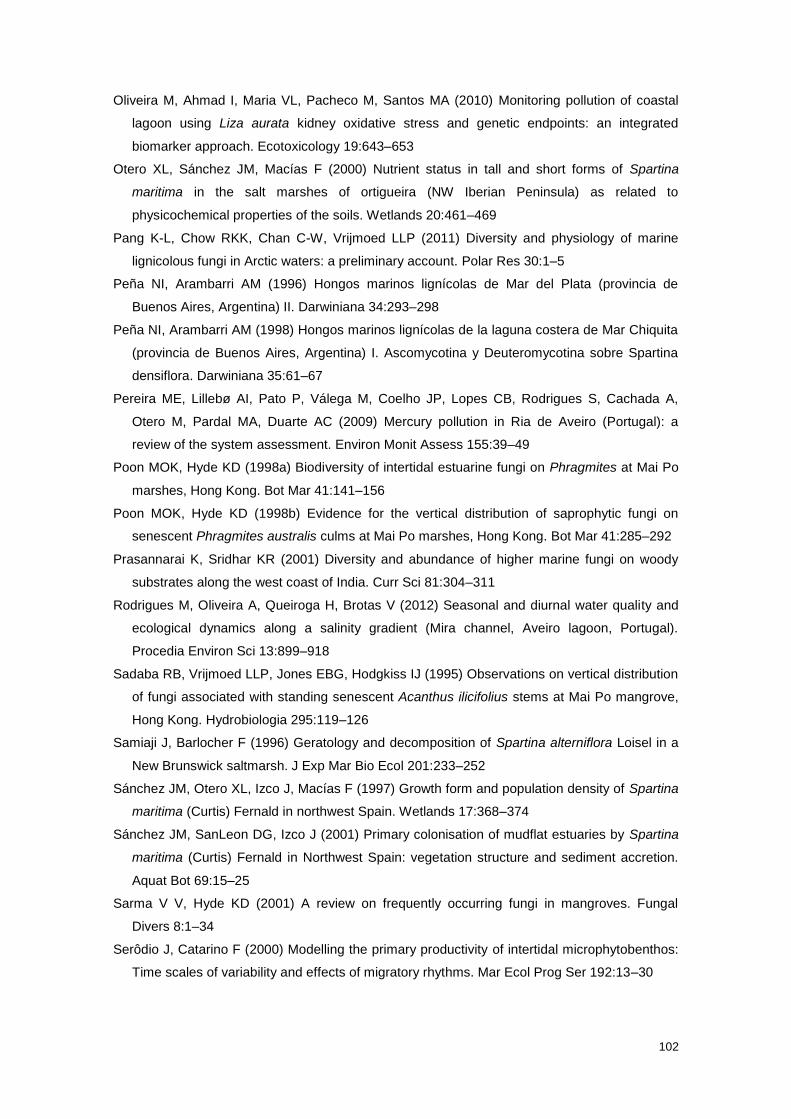
102
Oliveira M, Ahmad I, Maria VL, Pacheco M, Santos MA (2010) Monitoring pollution of coastal
lagoon using Liza aurata kidney oxidative stress and genetic endpoints: an integrated
biomarker approach. Ecotoxicology 19:643–653
Otero XL, Sánchez JM, Macías F (2000) Nutrient status in tall and short forms of Spartina
maritima in the salt marshes of ortigueira (NW Iberian Peninsula) as related to
physicochemical properties of the soils. Wetlands 20:461–469
Pang K-L, Chow RKK, Chan C-W, Vrijmoed LLP (2011) Diversity and physiology of marine
lignicolous fungi in Arctic waters: a preliminary account. Polar Res 30:1–5
Peña NI, Arambarri AM (1996) Hongos marinos lignícolas de Mar del Plata (provincia de
Buenos Aires, Argentina) II. Darwiniana 34:293–298
Peña NI, Arambarri AM (1998) Hongos marinos lignícolas de la laguna costera de Mar Chiquita
(provincia de Buenos Aires, Argentina) I. Ascomycotina y Deuteromycotina sobre Spartina
densiflora. Darwiniana 35:61–67
Pereira ME, Lillebø AI, Pato P, Válega M, Coelho JP, Lopes CB, Rodrigues S, Cachada A,
Otero M, Pardal MA, Duarte AC (2009) Mercury pollution in Ria de Aveiro (Portugal): a
review of the system assessment. Environ Monit Assess 155:39–49
Poon MOK, Hyde KD (1998a) Biodiversity of intertidal estuarine fungi on Phragmites at Mai Po
marshes, Hong Kong. Bot Mar 41:141–156
Poon MOK, Hyde KD (1998b) Evidence for the vertical distribution of saprophytic fungi on
senescent Phragmites australis culms at Mai Po marshes, Hong Kong. Bot Mar 41:285–292
Prasannarai K, Sridhar KR (2001) Diversity and abundance of higher marine fungi on woody
substrates along the west coast of India. Curr Sci 81:304–311
Rodrigues M, Oliveira A, Queiroga H, Brotas V (2012) Seasonal and diurnal water quality and
ecological dynamics along a salinity gradient (Mira channel, Aveiro lagoon, Portugal).
Procedia Environ Sci 13:899–918
Sadaba RB, Vrijmoed LLP, Jones EBG, Hodgkiss IJ (1995) Observations on vertical distribution
of fungi associated with standing senescent Acanthus ilicifolius stems at Mai Po mangrove,
Hong Kong. Hydrobiologia 295:119–126
Samiaji J, Barlocher F (1996) Geratology and decomposition of Spartina alterniflora Loisel in a
New Brunswick saltmarsh. J Exp Mar Bio Ecol 201:233–252
Sánchez JM, Otero XL, Izco J, Macías F (1997) Growth form and population density of Spartina
maritima (Curtis) Fernald in northwest Spain. Wetlands 17:368–374
Sánchez JM, SanLeon DG, Izco J (2001) Primary colonisation of mudflat estuaries by Spartina
maritima (Curtis) Fernald in Northwest Spain: vegetation structure and sediment accretion.
Aquat Bot 69:15–25
Sarma V V, Hyde KD (2001) A review on frequently occurring fungi in mangroves. Fungal
Divers 8:1–34
Serôdio J, Catarino F (2000) Modelling the primary productivity of intertidal microphytobenthos:
Time scales of variability and effects of migratory rhythms. Mar Ecol Prog Ser 192:13–30
103
Sguros PL, Simms J (1964) Role of marine fungi in the biochemistry of the oceans–IV Growth
responses to seawater inorganic macroconstituents. J Bacteriol 88:346–355
Shoemaker RA, Babcock CE (1989) Phaeosphaeria. Can J Bot 67:1500 – 1599
Sousa AI, Lillebø AI, Caçador I, Pardal MA (2008) Contribution of Spartina maritima to the
reduction of eutrophication in estuarine systems. Environ Pollut 156:628–635
Tan TK, Leong WF, Jones EBG (1989) Succession of fungi on wood of Avicennia alba and A.
lanata in Singapore. Can J Bot 2686–2691
Torzilli AP, Andrykovitch G (1986) Degradation of Spartina lignocellulose by individual and
mixed cultures of salt-marsh fungi. Can J Bot 64:2211–2215
Torzilli AP, Sikaroodi M, Chalkley D, Gillevet PM (2006) A comparison of fungal communities
from four salt marsh plants using automated ribosomal intergenic spacer analysis (ARISA).
Mycologia 98:690–698
Van Ryckegem G V, Verbeken A (2005a) Fungal ecology and succession on Phragmites
australis in a brackish tidal marsh. I. Leaf sheaths. Fungal Divers 19:157–187
Van Ryckegem G V, Verbeken A (2005b) Fungal ecology and succession on Phragmites
australis in a brackish tidal marsh. II. Stems. Fungal Divers 20:209–233
Van Ryckegem G V, Gessner MO, Verbeken A (2007) Fungi on leaf blades of Phragmites
australis in a brackish tidal marsh: diversity, succession, and leaf decomposition. Microb
Ecol 53:600–611
Vrijmoed LLP (2000) Isolation and culture of higher filamentous fungi. In: Hyde KD, Pointing SB
(eds) Marine Mycology – a practical approach. Fungal Diversity Press, Hong Kong, pp 1–20
Walker AK, Campbell J (2010) Marine fungal diversity: a comparison of natural and created salt
marshes of the north-central Gulf of Mexico. Mycologia 102:513–521
Zhou D, Hyde KD (2001) Host-specificity, host- exclusivity, and host-recurrence in saprobic
fungi. Mycol Res 105:1449-1457
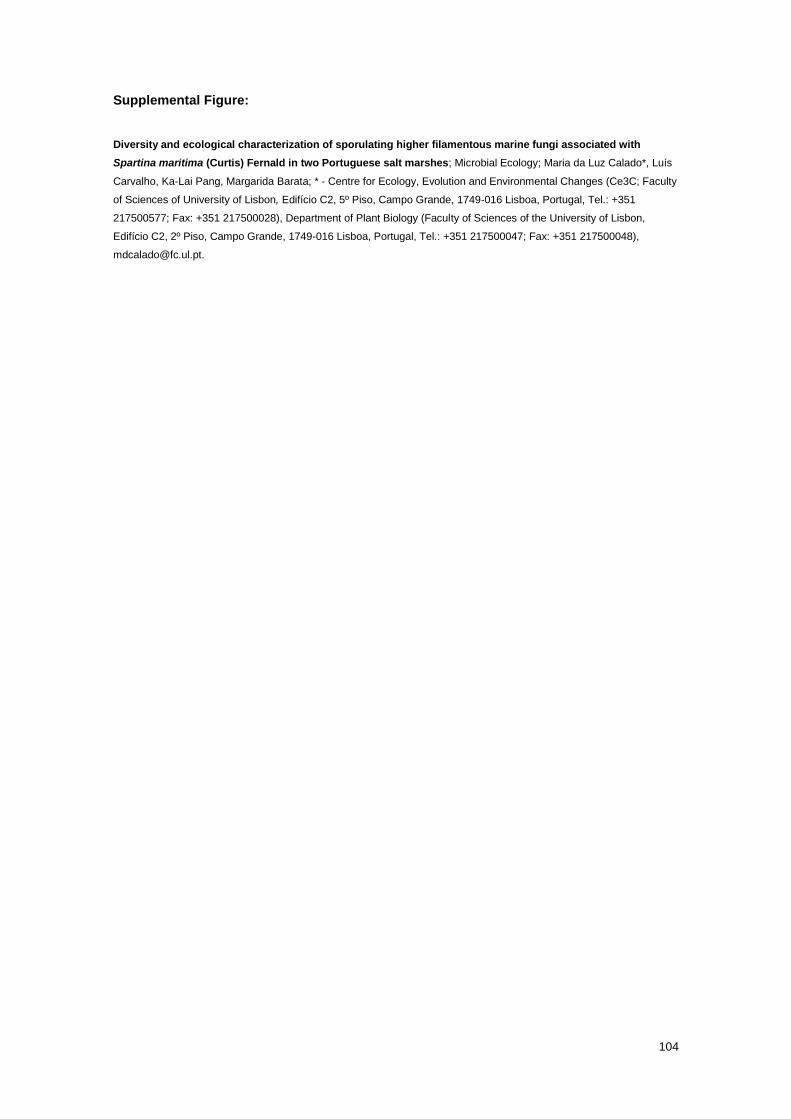
104
Supplemental Figure:
Diversity and ecological characterization of sporulating higher filamentous marine fungi associated with
Spartina maritima (Curtis) Fernald in two Portuguese salt marshes; Microbial Ecology; Maria da Luz Calado*, Luís
Carvalho, Ka-Lai Pang, Margarida Barata; * - Centre for Ecology, Evolution and Environmental Changes (Ce3C; Faculty
of Sciences of University of Lisbon, Edifício C2, 5º Piso, Campo Grande, 1749-016 Lisboa, Portugal, Tel.: +351
217500577; Fax: +351 217500028), Department of Plant Biology (Faculty of Sciences of the University of Lisbon,
Edifício C2, 2º Piso, Campo Grande, 1749-016 Lisboa, Portugal, Tel.: +351 217500047; Fax: +351 217500048),
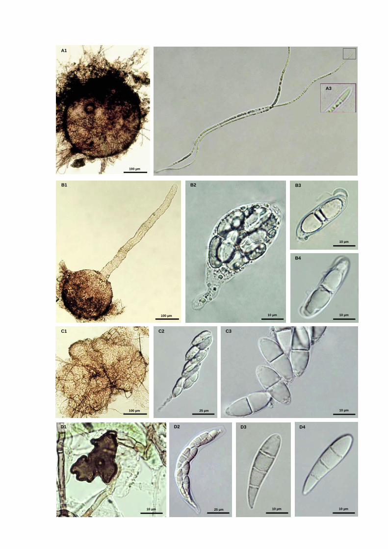
105
100 µm
B1
100 µm
A1
100 µm
C1
25 µm
C2
10 µm
C3
10 µm
D1
25 µm
D2
10 µm
D3
10 µm
D4
10 µm
B2
10 µm
B3
10 µm
B4
A3
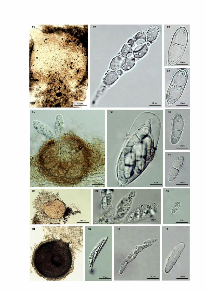
106
100 µm
E1
10 µm
E2
10 µm
E3
10 µm
E4
H1 H2
25 µm
H3
10 µm
H4
25 µm
25 µm
F1
10 µm
F2 F3
10 µm
F4
10 µm
100 µm
G1
10 µm
G2
10 µm
G3

107
100 µm
I1
25 µm
I2
10 µm
I3
10 µm
I4
10 µm 25 µm 10 µm
J1 J2 J3
25 µm
K1
25 µm
K2
10 µm
K3
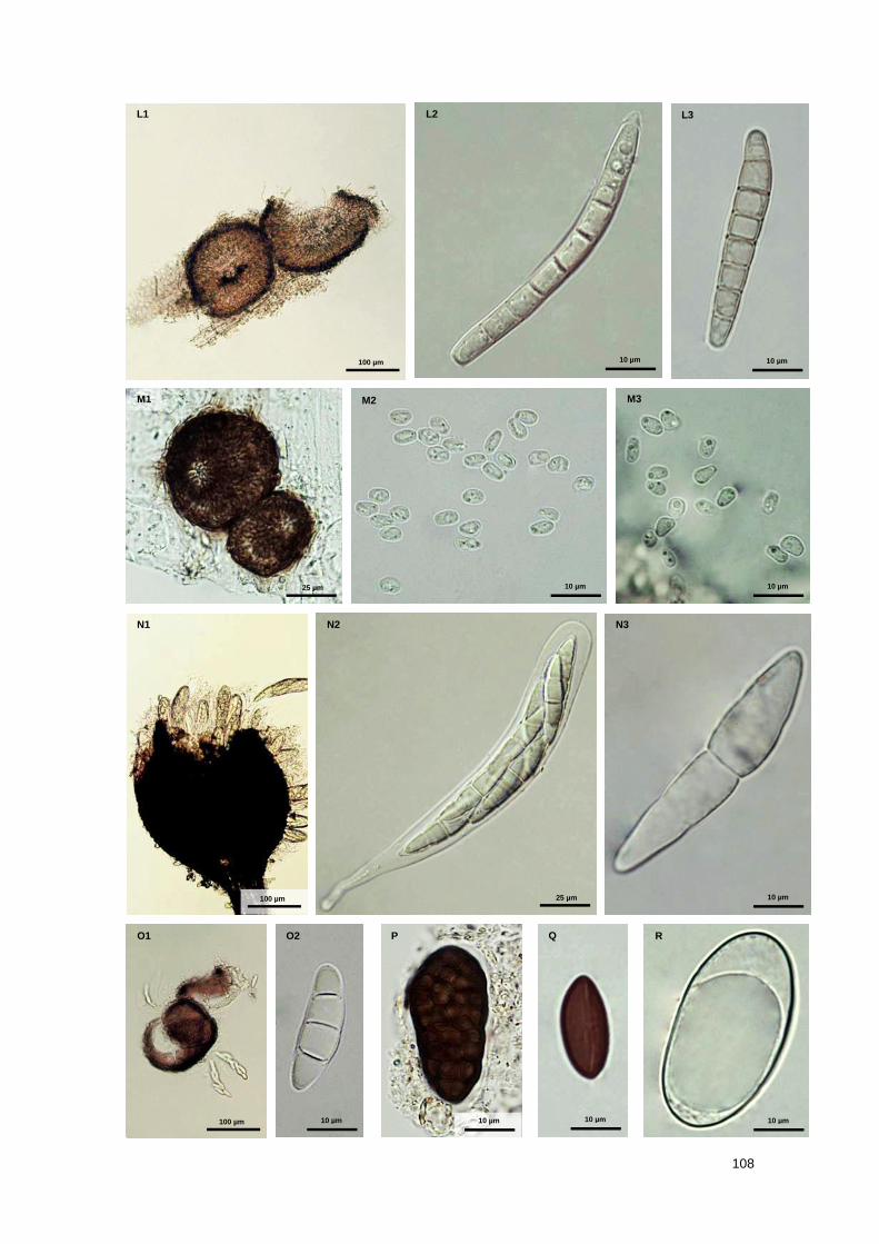
108
100 µm
L1
10 µm
L2
10 µm
L3
25 µm
M1
10 µm
M2
10 µm
M3
10 µm 10 µm 100 µm 10 µm 10 µm
O1 O2 P Q R
100 µm
N1
25 µm
N2
10 µm
N3
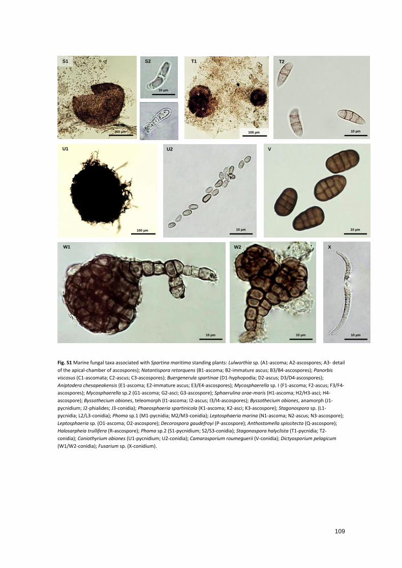
109
Fig. S1 Marine fungal taxa associated with Spartina maritima standing plants: Lulworthia sp. (A1-ascoma; A2-ascospores; A3- detail
of the apical-chamber of ascospores); Natantispora retorquens (B1-ascoma; B2-immature ascus; B3/B4-ascospores); Panorbis
viscosus (C1-ascomata; C2-ascus; C3-ascospores); Buergenerula spartinae (D1-hyphopodia; D2-ascus; D3/D4-ascospores);
Aniptodera chesapeakensis (E1-ascoma; E2-immature ascus; E3/E4-ascospores); Mycosphaerella sp. I (F1-ascoma; F2-ascus; F3/F4-
ascospores); Mycosphaerella sp.2 (G1-ascoma; G2-asci; G3-ascospore); Sphaerulina orae-maris (H1-ascoma; H2/H3-asci; H4-
ascospore); Byssothecium obiones, teleomorph (I1-ascoma; I2-ascus; I3/I4-ascospores); Byssothecium obiones, anamorph (J1-
pycnidium; J2-phialides; J3-conidia); Phaeosphaeria spartinicola (K1-ascoma; K2-asci; K3-ascospore); Stagonospora sp. (L1-
pycnidia; L2/L3-conidia); Phoma sp.1 (M1-pycnidia; M2/M3-conidia); Leptosphaeria marina (N1-ascoma; N2-ascus; N3-ascospore);
Leptosphaeria sp. (O1-ascoma; O2-ascospore); Decorospora gaudefroyi (P-ascospore); Anthostomella spissitecta (Q-ascospore);
Halosarpheia trullifera (R-ascospore); Phoma sp.2 (S1-pycnidium; S2/S3-conidia); Stagonospora halyclista (T1-pycnidia; T2-
conidia); Coniothyrium obiones (U1-pycnidium; U2-conidia); Camarosporium roumeguerii (V-conidia); Dictyosporium pelagicum
(W1/W2-conidia); Fusarium sp. (X-conidium).
100 µm
U1
10 µm
U2
10 µm
V
10 µm
W1
10 µm
W2
10 µm
X
100 µm
S1
10 µm
S2
10 µm
S3
100 µm
T1
10 µm
T2

110
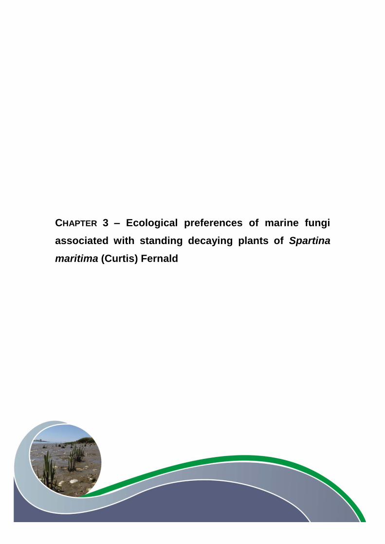
111
CHAPTER 3 – Ecological preferences of marine fungi
associated with standing decaying plants of Spartina
maritima (Curtis) Fernald

112
113
Ecological preferences of marine fungi associated with standing decaying plants of
Spartina maritima (Curtis) Fernald
Maria da Luz CALADO a, b, *
, Luís CARVALHOa, b
, Margarida BARATAa, b
, Ka-Lai PANGc
a Centre for Ecology, Evolution and Environmental Changes (Ce3C), Faculty of Sciences of University of Lisbon,
Edifício C2, 5º Piso, Campo Grande, 1749-016 Lisbon, Portugal
b Department of Plant Biology, Faculty of Sciences of the University of Lisbon, Edifício C2, 2º Piso, Campo Grande,
1749-016 Lisbon, Portugal
c Institute of Marine Biology and Centre of Excellence for the Oceans, National Taiwan Ocean University, 2 Pei-Ning
Road, Keelung 20224, Taiwan , Republic of China
* Corresponding author. E-mail address: [email protected] (M.L. Calado)
2016, in revision for new submission

114
Abstract
Fungal communities inhabiting live, senescent and decaying leaf sheaths, stems and leaf
blades of standing plants of Spartina maritima (Curtis) Fernald in two Portuguese salt marshes
were assessed by fruiting structures- and ITS sequence-based identification methods. Forty-five
fungi were found on early decaying plants. The molecular method enabled identification of
infrequent ascomycetes and basidiomycetes (filamentous and yeasts), and the asexual morph
of Byssothecium obiones and Phaeosphaeria halima. The results suggested that the
occurrence and ecological role of most frequent fungi on different plant substrates depend on
the phase of plant life cycle, micro-environmental conditions of substrates and potential fungal
competitors. Specifically, B. obiones, Natantispora retorquens and Lulworthia sp. 1 seem to be
involved in the decay of lower culms; Mycosphaerella sp. I, of leaf blades; P. halima and
Stagonospora sp. 1, of upper standing leaves; and Buergenerula spartinae and Phaeosphaeria
spartinicola, of all vegetative structures. The presence of these fungi on live vegetative
structures suggested an earlier endophytic colonisation.
Keywords
Marine fungi; Spartina maritima; early stages of decay; leaf sheaths; leaf blades; stems;
ecological preferences; potential ecological roles; ITS rDNA libraries
Introduction
Spartina species represent one of the main dominant halophytes in temperate salt marsh
ecosystems, contributing significantly to the annual biomass production and nutrient budget
(Castillo et al. 2010; Curado et al. 2013). Given the fact that the bulk of Spartina biomass is
composed of cell wall recalcitrant polymers, particularly lignocellulose ( 75%; Maccubbin and
Hodson 1980; Hodson et al. 1984; Torzilli and Andrykovitch 1986), the release of nutrients to
the surrounding environment strictly depends on the degradative activity of microbial
communities, particularly of saprobic fungi (Gessner 1980; Torzilli and Andrykovitch 1986;
Bergbauer and Newell 1992). The decomposition of Spartina species, similarly with other grass-
like plants, is initiated in senescent vegetative structures that remain in natural position (Newell
et al. 1989; Newell 1993; Castro and Freitas 2000; Newell and Porter 2000; Menéndez and
Sanmartí 2007). The senescence, which may begin very early in the growing season (Samiaji
and Barlocher 1996; Menéndez and Sanmartí 2007), occurs gradually from the outer and lower
vegetative structures towards the inner and higher structures (Samiaji and Barlocher 1996;
Kneib et al. 1997; Newell 2001a). Typically, a sequential abscission of the more decomposed
leaf blades occurs, but the leaf sheaths remain attached to stems until complete breakdown
(Healy and Walters 1994; Menéndez and Sanmartí 2007). The stems, enwrapped by

115
successive and overlapping layers of leaf sheaths that arise from each node, only start to
senesce when become more exposed to air, i.e. when the surrounding leaf sheaths are
fragmented or detached from the stems.
The senescent process is induced by external and internal factors and involves physiological
changes and chemical transformation of the plant tissues (Buchanan-Wollaston 1997;
Hortensteiner and Feller 2002). Although most antimicrobial and more complex polymers
remain after senescence (Valiela et al. 1979; Wilson et al. 1986a,b; Graça et al. 2000), some
soluble tannins and phenolics may leach out quickly during the initial stages of decay process
(Raghukumar 2004). Decomposition is initiated by mycelial fungal species that previously
inhabit internal photosynthetic plant tissues as endophytes and/or are able to withstand the
antimicrobial substances in the senescent material (Raghukumar 2004; Cornick et al. 2005).
Some endophytic fungal species demonstrated physiological adaptability to switch to a saprobic
life style after a physical or maturational change in the host plants, like senescence (Alva et al.
2002; Kumaresan and Suryanarayanan 2002; Promputtha et al. 2007, 2010). Among these,
some endophytes were shown to be morphologically and phylogenetically similar with their
saprobic counterparts (Alva et al. 2002; Kumaresan and Suryanarayanan 2002; Promputtha et
al. 2007, 2010).
Pioneer mycelial decomposers combine their filamentous and pervasive growth mode with the
production of extracellular enzymes to initiate an extensive lysis with physical disruption and
chemical transformation of intact plant tissues (Newell et al. 1996b; Hyde et al. 1998; Newell
and Porter 2000; Kis-Papo 2005). Simultaneously, some pioneer fungi have the ability to
metabolize some antimicrobial substances, such as cinnamic acids, ameliorating
microenvironmental conditions for subsequent fungal decomposers (Newell 1993).
Ascomycetous fungi play a more relevant role during this initial standing-decay phase,
dominating the microbial assemblages (Newell 1993, 1996, 2001a; Newell and Porter 2000). As
the decomposition carried out by these fungi proceeds, the organic matter content in the various
vegetative structures decreases (Buchan et al. 2003); a considerable fraction of dissolved
organic matter is readily leached out, and about 10–20% is converted into fungal biomass in
form of mycelia and reproductive structures (Newell et al. 1989, 1996a, 2000; Newell and
Wasowski 1995; Samiaji and Barlocher 1996; Newell and Porter 2000; Newell 2001b). The
substrate remains potentially nutrient-depleted with high lignin and low nitrogen contents in final
stages of decomposition (Torzilli and Andrykovitch 1986). The physical and chemical changes
of Spartina substrates during decomposition have been postulated to interfere with fungal
community dynamics (Gessner 1977; Buchan et al. 2003; Raghukumar 2004; Barata 2006).
Previous studies on the decomposition process of Spartina species mainly focused on the
dominant fungi associated with standing decaying leaf blades of S. alterniflora in U.S. salt
marshes (Newell and Wasowski 1995; Newell and Zakel 2000; Newell et al. 2000; Newell
2001a; Buchan et al. 2002; Lyons et al. 2010; Walker and Campbell 2010).
The fungal community associated with Spartina maritima (Curtis) Fernald, one of the main
pioneer colonisers of salt marsh mudflats in southern European and North African coastlines
116
(Sánchez et al. 2001) and one of the dominant macrophytes in Portuguese salt marshes, has
been poorly investigated: Barata (1997, 2002) and Calado et al. (2015) together inventoried
fungi on standing plants from five geographically and physically distinct salt marshes; Barata
(2006) surveyed stem baits exposed to different abiotic conditions in one of these salt marshes;
and Azevedo et al. (2012) analysed drift stems collected in four Portuguese sandy beaches.
The identification of species relied on the morphological features of fungal structures produced
at the time of collection and/or after incubation.
The advent of molecular techniques has contributed greatly to overcome the major drawbacks
of traditional culture- and microscopy-based techniques in documenting fungal diversity and to
provide a more accurate taxonomic classification of fungi (Buchan et al. 2002; Lyons et al.
2005; Pang and Mitchell 2005; Torzilli et al. 2006; Walker and Campbell 2010; Jones 2011a;
Abdel-Wahab and Bahkali 2012). Internal transcribed spacers (ITS) of the rDNA operon region
have been frequently used for fungal species identification because they are present in multiple
copies within each cell, easily amplifiable by Polymerase Chain Reaction (PCR) and highly
variable in sequence (Buchan et al. 2002; Nilsson et al. 2006; Arnold 2007; Aveskamp et al.
2010; Schoch et al. 2012; Velmurugan et al. 2013). Except for some taxonomic groups which
have very conserved ITS, this region is widely accepted as the most powerful universal DNA
barcode for fungi (Schoch et al. 2012; Toju et al. 2012). Even though many marine fungal
species have been already sequenced, ITS sequences for a number of marine fungi are not yet
available in international databases for sequence comparison (Nilsson et al. 2006; Porter and
Golding 2011; Schoch et al. 2012). For this reason, and also the systematic biases associated
with PCR amplification and/or cloning techniques (Buchan et al. 2002, 2003; Pang and Mitchell
2005; Toju et al. 2012), molecular techniques should be coupled with other methods to study
fungal community dynamics.
Thus, the present study intended to provide a more realistic representation of fungal community
associated with standing plants of S. maritima in salt marsh ecosystems, by combining
morphological and molecular approaches. Specifically, the main purposes were to complement
the list of fungi associated with S. maritima recently published by Calado et al. (2015) and to
infer about ecological preferences and involvement of these fungi during the decay process of
standing leaf sheaths, leaf blades and true stems, using morphological (fungal structures
identification) and molecular (ITS sequencing) techniques.
Material and Methods
Study sites, host plant and sampling procedure
The study was performed in two salt marshes located in distinct geographical regions, the
Guadiana estuary (Castro Marim) situated in the southeastern coast (37.23° N, 7.42° W), and

117
the Ria de Aveiro coastal lagoon in the northwest of Portugal (40.62° N, 8.74° W). Both estuary
and coastal lagoon also differ in physical configuration and conservation status.
Field sampling procedure involved a bimonthly collection of mature standing plants in Castro
Marim and Ria de Aveiro salt marshes, over a two-year period (October 2010 to August 2012).
Twenty plants were collected on the first three sampling periods and 15 plants afterwards (a
total of 390 plants); five additional plants during the second sampling year were also collected
for isolation of marine fungi.
A detailed description of the study sites, S. maritima communities and sampling criteria can be
found in Calado et al. (2015).
Morphology-based species identification
The collected plants were carefully rinsed with running tap water to remove fine-grained
sediments and seaweeds, and air-dried. Each air-dried plant was first separated into three
categories according to the vegetative structure –leaf blade, leaf sheath and stem – and
subsequently into three further subcategories based on the visually observed physiological state
of the structure – live (green parts), senescent (yellow parts, with the physical structure still
intact) and decaying (brown parts, with the physical structure visibly altered). This separation
resulted in nine different samples that were designated as substrates, i.e. live leaf sheaths,
senescent leaf sheaths, decaying leaf sheaths, live stems, senescent stems, decaying stems,
live leaf blades, senescent leaf blades and decaying leaf blades.
Fungal structures (fruit bodies, spores and hyphopodia) observed on each plant substrate were
picked up under a dissecting microscope (Wild M8), mounted in a drop of sterile seawater on a
slide, examined under a light microscope (Leitz Laborlux S, with Normaski) with detailed
morphology recorded. The fungi were identified using the dichotomous keys of Kohlmeyer and
Kohlmeyer (1979), Kohlmeyer and Volkmann-Kohlmeyer (1991), Hyde and Sarma (2000) and
Jones et al. (2009).
Plant substrates collected in the same study site and sampling date were mixed together, frozen
at -80 °C and freeze-dried for three days in a Christ Alpha I-5 apparatus at 10-1
mbar and -42 °C.
Isolation of marine fungi and preservation of pure cultures
Several strains of the most frequent fungal taxa were isolated by single spore method
(described in detail by Calado et al. 2015). Each pure culture was maintained in active growth in
cornmeal agar made with sterile aged diluted seawater (CMA/sw 50 %) at 4 °C, and as mycelial
discs (7 mm diameter) in McCartney bottles filled with sterile diluted seawater (50%) at 4 °C and
in cryotubes filled with glycerol (10%) at -80 °C.
118
DNA extraction and amplification of ITS genes
Twenty-three fungal isolates (belonging to the nine most frequently collected fungal taxa;
Calado et al. 2015) obtained from S. maritima were used to create a reference database for
comparing and identifying fungal sequences recovered from 88 lyophilized plant samples
(representing the first sampling year, October 2010 to August 2011).
Genomic DNA was extracted from axenic cultures using a standard phenol–chloroform protocol
adapted from Pang et al. (2008). The DNA extraction from plant samples involved the
maceration of c.a. 0.05 g of each plant sample to a fine powder in liquid nitrogen and a thermal
incubation of the material (water bath at 70 °C for 15 min, liquid nitrogen for 2 min and water
bath at 70 °C for 15 min); the subsequent steps of the procedure were identical to the procedure
of DNA extraction from axenic cultures.
Amplification and sequencing of the ITS regions of pure fungal cultures were conducted using
primer pairs ITS5 (5´ GGAAGTAAAAGTCGTAACAAGG 3´)/ ITS4 (5´
TCCTCCGCTTATTGATATGC 3´) (White et al., 1990). The primer pairs ITS1-F_KYO1 (5´
CTHGGTCATTTAGAGGAASTAA 3´)/ ITS4_KYO3 (5´CTBTTVCCKCTTCACTCG 3´) (Toju et
al., 2012) were used for selective amplification of fungal rDNA in mixed DNA samples (plant
materials). Both primer pairs cover partial sequences of 18S rRNA, complete sequences of
ITS1, 5.8S rRNA and ITS2, and partial sequences of 28S rRNA genes.
PCR amplifications were performed in 25 µL reaction volumes, containing 0.8 µM of each
primer, 12.5 µL of Taq Premix (Cat.No. RT803A, Bioman, New Taipei City, Taiwan) and 1 µL of
genomic DNA. PCR was carried out on a thermocycler (Biometra T3000) using the following
parameters: initial denaturation for 5 min step at 95 °C, 34 cycles of 30 s at 95 °C
(denaturation), 30 s at 55 °C (primer annealing) and 30 s at 72 °C (elongation), then a final
elongation step of 5 min at 72 °C.
Amplicons of pure fungal isolates were directly sent to Genomics Biosci. & Tech. (New Taipei
City, Taiwan) for sequencing, using the same pair of PCR primers.
Amplicons from plant samples were cloned in order to isolate individual amplicons of the mixed
PCR products; in this study, cloning and restriction analysis were preferred over the next-
generation sequencing approach given the expected high number and lengths of the amplicons.
ITS region clone libraries construction and restriction profiles analysis
PCR products from each plant sample were purified using the EasyPure PCR/Gel Extraction Kit
(BIOMAN, Scientific CO, LTD, Taiwan), following the manufacturer’s instructions. Purified
amplicons were ligated into a TA cloning vector, using the RBC TA Cloning Vector Kit (RBC
Bioscience Corp., Taiwan). After ligation, the mixture was used directly to transform Escherichia
coli DH5α competent cells using RBC HIT Competent Cells Kit (RBC Bioscience Corp.,
Taiwan). Detection of transformed cells with recombinant DNA was performed through blue-
white screening technique. Twenty white colonies were selected, and each one was transferred

119
into Luria Broth medium; a colony PCR was conducted in 20 µL reaction volume, with 2 µL of
every bacterial suspension, 0.5 µM of each previously used primers and 10 µL of Taq Premix.
ITS region clone libraries from 88 plant samples included a total of 1037 sequences. A
restriction fragment length polymorphism (RFLP) analysis was used to categorise
phylogenetically related sequences (Pang and Mitchell 2005).
Several restriction enzymes were tested on ITS of pure fungal isolates of the most common
fungi on S. maritima and HpyF31 (DdeI) produced patterns that enabled differentiation of
different fungal species (results not shown). Restriction enzyme digestion reaction followed the
manufacturer´s instructions (Cat.No. ER1881, Thermo Scientific, New Taipei City, Taiwan). All
the clones that exhibited different restriction profiles in the same plant sample were selected.
Three hundred out of 1037 clones were sent to Genomics Biosci. & Tech. (New Taipei City,
Taiwan) for sequencing in one direction.
Sequence alignment and phylogenetic analyses
ITS sequences of the 23 pure fungal isolates were assembled and manually adjusted to obtain
a consensus sequence using DNA Baser Sequence Assembler v4.10 software (2014; Heracle
BioSoft, www.DnaBaser.com). These sequences, as well as the 300 single-stranded full-length
sequences retrieved from plant samples, were compared with those sequences available in
GenBank database of National Center for Biotechnology Information (NCBI). The fungal ITS
sequence-based classification relies on the phylogenetic relationships among species, using the
BLAST best-hit method to classify an unknown sequence (Porter and Golding 2011). In this
study, the sequences were identified to lowest possible taxonomic level based on a cut-off value
of 97% of sequence similarity and 90% of sequence cover for species proposed by Nilsson et
al. (2012) and Blaalid et al. (2013). Identification name provided by reference database was
always adopted over the one provided by public database whenever there was a higher
homology between the unknown sequence and a reference sequence. All the BLAST best-hits
were retrieved from GenBank for further analysis. Even though BLAST top hits were mostly
selected, the next best hits were retrieved whenever there was a better proposal in the list with
similar cover/identity rates, i.e. of a fungal species that had been previously identified by
morphological methods but not isolated in culture.
In a preliminary approach, all the sequences assigned to the same taxonomic families were
grouped together, aligned by ClustalW and submitted to a phylogenetic maximum-parsimony
analysis using Molecular Evolutionary Genetics Analysis (MEGA) v.6.06 software (Tamura et al.
2013); operational taxonomic units (OTUs) were then defined. One representative sequence
was randomly chosen from each defined OTU; 62 out of 300 clones were selected and sent to
Genomics Biosci. & Tech. (New Taipei City, Taiwan) for sequencing the complementary strand.
The sequences of representative clones, isolates and BLAST hits representing the main
taxonomic classes were automatically aligned by ClustalW and refined manually with
ambiguous regions of alignment removed using MEGA v.6.06 software.
120
Phylogenetic and neighbor-joining distance analyses were additionally performed using the
same software, in order to confirm the OTUs previously established. Phylogenetic trees were
constructed using maximum-parsimony and neighbor-joining methods. Maximum-parsimony
trees were performed using 100 heuristic searches with random stepwise addition of
sequences, tree-bisection-reconnection (TBR) branch swapping algorithm and maximum
number of trees to retain set to 100. Characters were equally weighted and gaps were treated
as missing data. Neighbor-joining trees were performed using p-distance, Jukes-Cantor,
Kimura-2 and Logdet parameter substitution models. Topological robustness of the
phylogenetic trees in both maximum-parsimony and distance analyses was estimated by
performing 1000 bootstrap replicates. Neighbor-joining distance analysis was based on a
pairwise-distance model.
Data analyses
A more accurate representation of species richness in different plant substrates from Castro
Marim and Ria de Aveiro salt marshes was obtained by combining the results provided by
morphological and molecular methods from the first-sampling year. Since the number of fungal
taxa recorded on each plant substrate might represent a downward-biased estimator of the total
species richness (Gotelli and Colwell 2010), species richness in the same substrates was also
calculated using Chao 2 and Jacknife 2 estimators (SChao2 = Sobs+(m-1
m)
Q1 (Q1-1)
2 (Q2+1) and SJack2 =
Sobs+[𝑄1(2𝑚−3)
𝑚−
𝑄2(𝑚−2)2
𝑚(𝑚−1)], where Sobs is the total observed number of fungal taxa, m the number
of samples, Q1 the number of fungal taxa that occurred in only one sample, and Q2 the number
of fungal taxa that occur in two samples); according to Gotelli and Colwell (2010), these
estimators are more appropriate to replicated presence/absence data, correcting the observed
species richness based on the frequencies of the very rarest species. A sample-based
rarefaction was not performed because one of the plant substrates included only two samples.
Species composition of fungal communities inhabiting different plant substrates of Castro Marim
and Ria de Aveiro salt marshes were also compared based on presence/absence and
percentage frequencies of occurrence of fungal taxa in communities. Presence/absence of
fungal taxa detected by morphological and/or molecular methods on each plant substrate (live,
senescent and decaying leaf sheaths, stems and leaf blades), sampling period and study site
during the first sampling year was compiled in two dataset matrices (one matrix per each study
site). Percentage frequencies of occurrence of fungal taxa identified by morphological method
on each plant substrate, sampling period and study site during the total sampling period were
also determined and organized in two complementary dataset matrices (one matrix per each
study site); percentage frequencies of occurrence of fungi were obtained from the number of
plants in which a specific fungus occurred on each substrate divided by the total number of
plants x 100. Contrary to the general studies, the binary data (presence/absence) were herein
more informative than frequency data because they included the results provided by two
121
different but complementary methods; moreover, they did not depend on the fruiting patterns of
fungi, and attributed the same weight to frequent and infrequent species. Nevertheless,
percentage frequencies of occurrence of fungi on plant substrates throughout the 2-year study
period provided robust information on fruiting preferences of marine fungi.
Given the complexity of datasets, multivariate analyses were carried out using the PAST v2.17c
statistical software (Hammer et al. 2001) to reduce and summarize the data.
A preliminary and exploratory Cluster analysis was performed on sample-by-species matrices
with binary data using Bray-Curtis similarity measure index. This index is suggested as an ideal
coefficient to be used for the construction of similarity matrices given its effectiveness in dealing
with datasets containing multiple blocks of zeros (Rees et al. 2004). The values identified as
outliers were excluded from further analyses.
Additionally to the Cluster analysis, a Nonmetric multidimensional scaling (MDS) analysis was
also applied on the reconstructed dataset matrices. MDS analysis, which is one of the most
widely accepted ordination techniques in microbial ecology, only displays the samples in the
plot based on values generated in a similarity matrix (Clarke and Warwick 2001; Rees et al.
2004; Ramette 2007). Another ordination method, a Detrended Correspondence Analysis
(DCA), was also performed with relative frequency data in order to confirm patterns in
dimensional coordinate plots; DCA is appropriate to model relative frequency data, enabling a
simultaneous visualization of the samples and species in the ordination plot along an
environmental gradient. Bray-Curtis index was selected for MDS analysis, whereas chi-squared
index was implicit in DCA.
Differences between a priori groups (i.e. plant substrates) established on the basis on the
presence/absence of species in the samples were statistically assessed through a one-way
ANOSIM. A post hoc test to assess the significance of pairwise comparisons between
substrates was also carried out.
Results and Discussion
Molecular identification of ITS sequences retrieved from plant samples
The majority of the 1037 ITS sequences recovered from S. maritima plant samples (99%) were
identified. As demonstrated previously by Toju et al. (2012), the primers pair ITS1-F_KYO1/
ITS4_KYO3 used in this study was highly fungal-specific, considering the low number of non-
fungal sequences recovered with these primers (0.1%); in contrast, most of the sequences
amplified by the primers pair ITS5/ ITS4 belonged to S. maritima (86%).
The restriction enzyme HpyF31 (DdeI) was demonstrated to be able to provide better
discriminatory results for fungal sequences than HaeIII, frequently used in T-RF technique
(Buchan et al. 2002; Walker and Campbell 2010).

122
Fungal ITS sequence length (including partial 18S and 28S and full length 5.8S rDNA) of the
representative clones from both Castro Marim and Ria de Aveiro salt marshes ranged from 365
to 674 bp. These sequences were identified to the lowest taxonomic rank possible and grouped
in OTUs based on results from BLAST searches and phylogenetic analyses (Table 1, Figs. 1a,
b).
Table 1 Fungal taxa identified on Spartina maritima samples by molecular methods. Pure fungal cultures previously
isolated by morphological methods and representative clones assigned to the same fungal species on the basis of
similar BLAST results and positions on phylogenetic trees, were grouped together. Best BLAST hits of clones and pure
fungal isolates are mentioned, as well as homology scores between sequences
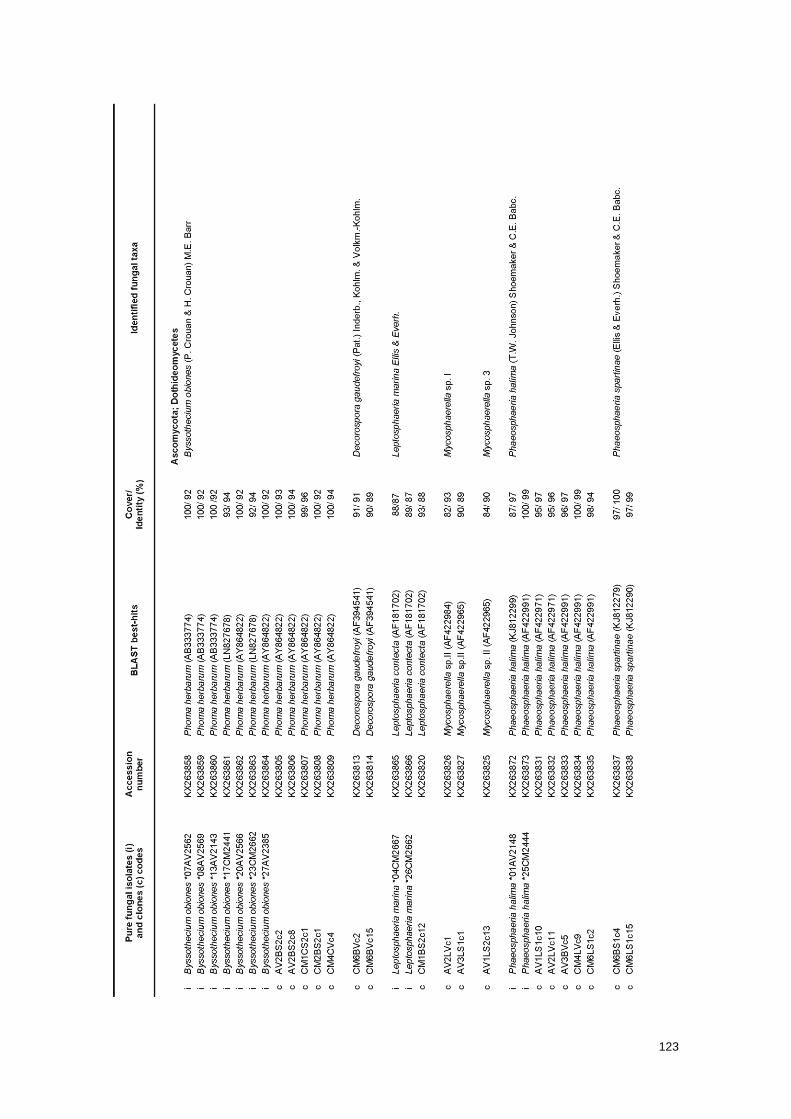
123
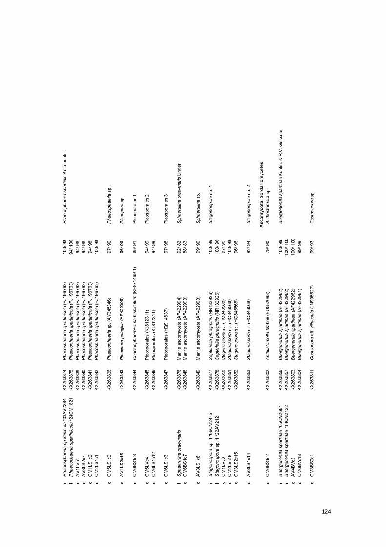
124
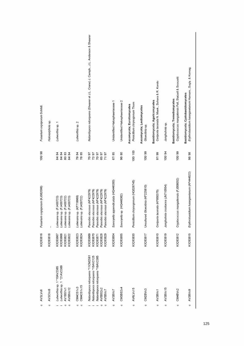
125
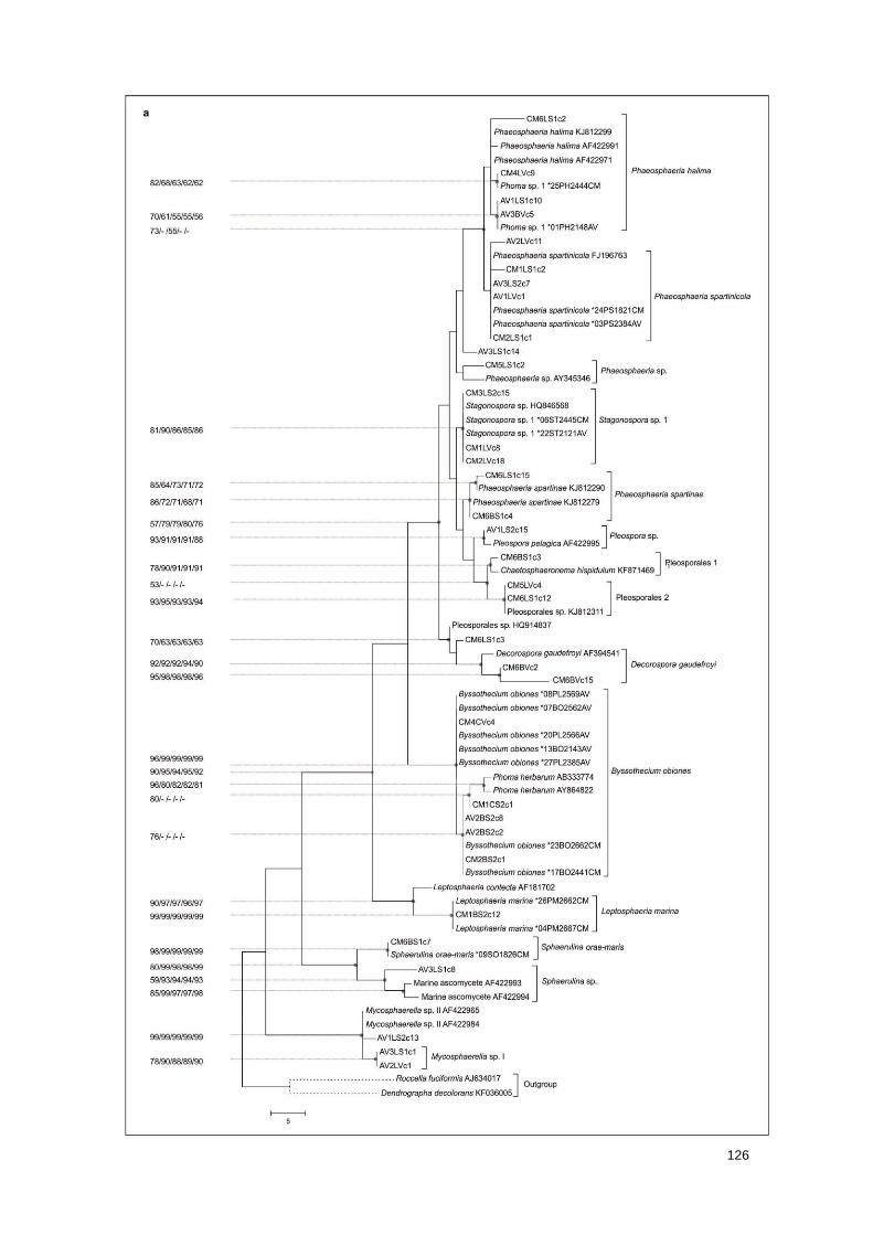
126
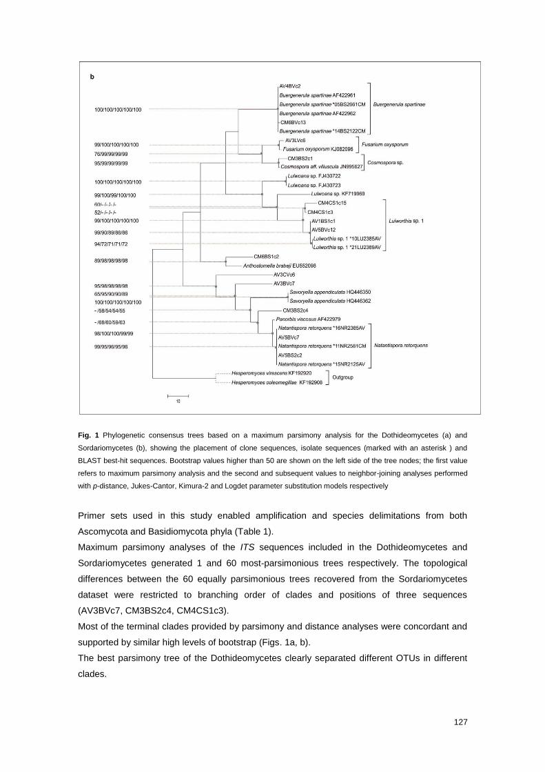
127
Fig. 1 Phylogenetic consensus trees based on a maximum parsimony analysis for the Dothideomycetes (a) and
Sordariomycetes (b), showing the placement of clone sequences, isolate sequences (marked with an asterisk ) and
BLAST best-hit sequences. Bootstrap values higher than 50 are shown on the left side of the tree nodes; the first value
refers to maximum parsimony analysis and the second and subsequent values to neighbor-joining analyses performed
with p-distance, Jukes-Cantor, Kimura-2 and Logdet parameter substitution models respectively
Primer sets used in this study enabled amplification and species delimitations from both
Ascomycota and Basidiomycota phyla (Table 1).
Maximum parsimony analyses of the ITS sequences included in the Dothideomycetes and
Sordariomycetes generated 1 and 60 most-parsimonious trees respectively. The topological
differences between the 60 equally parsimonious trees recovered from the Sordariomycetes
dataset were restricted to branching order of clades and positions of three sequences
(AV3BVc7, CM3BS2c4, CM4CS1c3).
Most of the terminal clades provided by parsimony and distance analyses were concordant and
supported by similar high levels of bootstrap (Figs. 1a, b).
The best parsimony tree of the Dothideomycetes clearly separated different OTUs in different
clades.
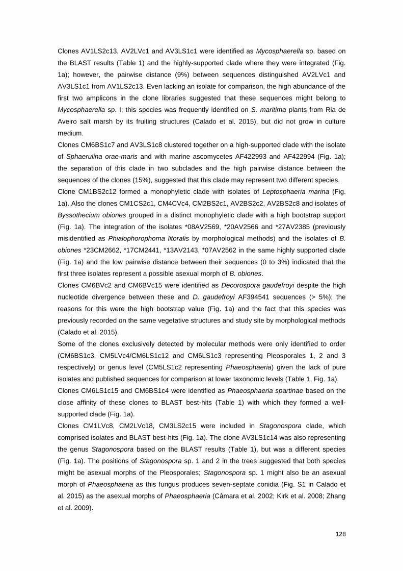
128
Clones AV1LS2c13, AV2LVc1 and AV3LS1c1 were identified as Mycosphaerella sp. based on
the BLAST results (Table 1) and the highly-supported clade where they were integrated (Fig.
1a); however, the pairwise distance (9%) between sequences distinguished AV2LVc1 and
AV3LS1c1 from AV1LS2c13. Even lacking an isolate for comparison, the high abundance of the
first two amplicons in the clone libraries suggested that these sequences might belong to
Mycosphaerella sp. I; this species was frequently identified on S. maritima plants from Ria de
Aveiro salt marsh by its fruiting structures (Calado et al. 2015), but did not grow in culture
medium.
Clones CM6BS1c7 and AV3LS1c8 clustered together on a high-supported clade with the isolate
of Sphaerulina orae-maris and with marine ascomycetes AF422993 and AF422994 (Fig. 1a);
the separation of this clade in two subclades and the high pairwise distance between the
sequences of the clones (15%), suggested that this clade may represent two different species.
Clone CM1BS2c12 formed a monophyletic clade with isolates of Leptosphaeria marina (Fig.
1a). Also the clones CM1CS2c1, CM4CVc4, CM2BS2c1, AV2BS2c2, AV2BS2c8 and isolates of
Byssothecium obiones grouped in a distinct monophyletic clade with a high bootstrap support
(Fig. 1a). The integration of the isolates *08AV2569, *20AV2566 and *27AV2385 (previously
misidentified as Phialophorophoma litoralis by morphological methods) and the isolates of B.
obiones *23CM2662, *17CM2441, *13AV2143, *07AV2562 in the same highly supported clade
(Fig. 1a) and the low pairwise distance between their sequences (0 to 3%) indicated that the
first three isolates represent a possible asexual morph of B. obiones.
Clones CM6BVc2 and CM6BVc15 were identified as Decorospora gaudefroyi despite the high
nucleotide divergence between these and D. gaudefroyi AF394541 sequences (> 5%); the
reasons for this were the high bootstrap value (Fig. 1a) and the fact that this species was
previously recorded on the same vegetative structures and study site by morphological methods
(Calado et al. 2015).
Some of the clones exclusively detected by molecular methods were only identified to order
(CM6BS1c3, CM5LVc4/CM6LS1c12 and CM6LS1c3 representing Pleosporales 1, 2 and 3
respectively) or genus level (CM5LS1c2 representing Phaeosphaeria) given the lack of pure
isolates and published sequences for comparison at lower taxonomic levels (Table 1, Fig. 1a).
Clones CM6LS1c15 and CM6BS1c4 were identified as Phaeosphaeria spartinae based on the
close affinity of these clones to BLAST best-hits (Table 1) with which they formed a well-
supported clade (Fig. 1a).
Clones CM1LVc8, CM2LVc18, CM3LS2c15 were included in Stagonospora clade, which
comprised isolates and BLAST best-hits (Fig. 1a). The clone AV3LS1c14 was also representing
the genus Stagonospora based on the BLAST results (Table 1), but was a different species
(Fig. 1a). The positions of Stagonospora sp. 1 and 2 in the trees suggested that both species
might be asexual morphs of the Pleosporales; Stagonospora sp. 1 might also be an asexual
morph of Phaeosphaeria as this fungus produces seven-septate conidia (Fig. S1 in Calado et
al. 2015) as the asexual morphs of Phaeosphaeria (Câmara et al. 2002; Kirk et al. 2008; Zhang
et al. 2009).
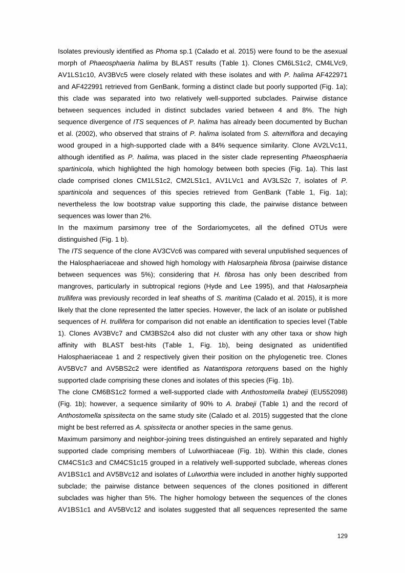
129
Isolates previously identified as Phoma sp.1 (Calado et al. 2015) were found to be the asexual
morph of Phaeosphaeria halima by BLAST results (Table 1). Clones CM6LS1c2, CM4LVc9,
AV1LS1c10, AV3BVc5 were closely related with these isolates and with P. halima AF422971
and AF422991 retrieved from GenBank, forming a distinct clade but poorly supported (Fig. 1a);
this clade was separated into two relatively well-supported subclades. Pairwise distance
between sequences included in distinct subclades varied between 4 and 8%. The high
sequence divergence of ITS sequences of P. halima has already been documented by Buchan
et al. (2002), who observed that strains of P. halima isolated from S. alterniflora and decaying
wood grouped in a high-supported clade with a 84% sequence similarity. Clone AV2LVc11,
although identified as P. halima, was placed in the sister clade representing Phaeosphaeria
spartinicola, which highlighted the high homology between both species (Fig. 1a). This last
clade comprised clones CM1LS1c2, CM2LS1c1, AV1LVc1 and AV3LS2c 7, isolates of P.
spartinicola and sequences of this species retrieved from GenBank (Table 1, Fig. 1a);
nevertheless the low bootstrap value supporting this clade, the pairwise distance between
sequences was lower than 2%.
In the maximum parsimony tree of the Sordariomycetes, all the defined OTUs were
distinguished (Fig. 1 b).
The ITS sequence of the clone AV3CVc6 was compared with several unpublished sequences of
the Halosphaeriaceae and showed high homology with Halosarpheia fibrosa (pairwise distance
between sequences was 5%); considering that H. fibrosa has only been described from
mangroves, particularly in subtropical regions (Hyde and Lee 1995), and that Halosarpheia
trullifera was previously recorded in leaf sheaths of S. maritima (Calado et al. 2015), it is more
likely that the clone represented the latter species. However, the lack of an isolate or published
sequences of H. trullifera for comparison did not enable an identification to species level (Table
1). Clones AV3BVc7 and CM3BS2c4 also did not cluster with any other taxa or show high
affinity with BLAST best-hits (Table 1, Fig. 1b), being designated as unidentified
Halosphaeriaceae 1 and 2 respectively given their position on the phylogenetic tree. Clones
AV5BVc7 and AV5BS2c2 were identified as Natantispora retorquens based on the highly
supported clade comprising these clones and isolates of this species (Fig. 1b).
The clone CM6BS1c2 formed a well-supported clade with Anthostomella brabeji (EU552098)
(Fig. 1b); however, a sequence similarity of 90% to A. brabeji (Table 1) and the record of
Anthostomella spissitecta on the same study site (Calado et al. 2015) suggested that the clone
might be best referred as A. spissitecta or another species in the same genus.
Maximum parsimony and neighbor-joining trees distinguished an entirely separated and highly
supported clade comprising members of Lulworthiaceae (Fig. 1b). Within this clade, clones
CM4CS1c3 and CM4CS1c15 grouped in a relatively well-supported subclade, whereas clones
AV1BS1c1 and AV5BVc12 and isolates of Lulworthia were included in another highly supported
subclade; the pairwise distance between sequences of the clones positioned in different
subclades was higher than 5%. The higher homology between the sequences of the clones
AV1BS1c1 and AV5BVc12 and isolates suggested that all sequences represented the same

130
species, i.e. Lulworthia sp. 1. The higher proximity of this unidentified species of Lulworthia to
Lulwoana (Table 1) than to other species of Lulworthia may be related to the polyphyly of the
genus (Campbell et al. 2005; Jones 2011b; Pang 2012).
Phylogenetic trees also separated the sequences assigned to Nectriaceae in a well-supported
clade, which was, in turn, divided in two subclades representing the genus Fusarium and
Cosmospora (Fig. 1b); clones CM3LVc6 and CM3BS2c1 were identified as Fusarium
oxysporum and Cosmospora sp. respectively, based on the tree and affinity to BLAST best-hits
(Table 1, Fig. 1b).
Sequences of the isolates of Buergenerula spartinae and sequences retrieved from GenBank
formed a monophyletic, highly supported clade with clones AV4BVc2 and CM6BVc13, with a
high similarity (> 99%) (Fig. 1b).
In total, the sequences of the 54 representative clones retrieved from the plant samples
represented 33 fungal taxa (Table 1). These sequences were then used as a reference
database to identify the remaining 842 sequences (436 and 406 retrieved from Castro Marim
and Ria de Aveiro samples respectively) (Fig. 2).
131
Fig. 2 Number of amplicons assigned to each fungal taxon retrieved from Castro Marim (a) and Ria de Aveiro (b) plant
samples: BO Byssothecium obiones; DG Decorospora gaudefroyi; LM Leptosphaeria marina; MyI Mycosphaerella sp. I;
My3 Mycosphaerella sp. 3; PH Phaeosphaeria halima; PR Phaeosphaeria spartinae; PS Phaeosphaeria spartinicola; Ph
Phaeosphaeria sp.; Pl Pleospora sp. ; Pl 1 Pleosporales 1; Pl 2 Pleosporales 2; Pl 3 Pleosporales 3; SO Sphaerulina
orae-maris; Sp Sphaerulina sp.; St 1 Stagonospora sp. 1; St 2 Stagonospora sp. 2; An Anthostomella sp.; BS
Buergenerula spartinae; CO Cosmospora sp.; Fu Fusarium oxysporum; Ha Halosarpheia sp.; Lu 1 Lulworthia sp. 1; Lu
2 Lulworthia sp. 2; NR Natantispora retorquens; Ha1 Unidentified Halosphaeriaceae 1; Ha2 Unidentified
Halosphaeriaceae 2; PC Penicillium chrysogenum; Gl Gloeotinia sp.; CL Ceriporia lacerata; Ju Junghuhnia sp.; CM
Cryptococcus mangaliensis; EH Erythrobasidium hasegawianum. Ascomycota and Basidiomycota are represented by
grey and white colors respectively, and Dothideomycetes, Sordariomycetes, Eurotiomycetes and Leotiomycetes
classes by light, medium, medium-dark and dark gray respectively
The results revealed that ITS amplicons more frequently recovered from plant samples
corresponded to the most frequently fungi recorded by fruiting structures in the same samples,
i.e. B. obiones, L. marina, Lulworthia sp. 1, Mycosphaerella sp. I, P. halima, P. spartinicola and
Stagonospora sp. 1 (Fig. 2; Calado et al. 2015). This means that the abundance of amplicons in
the clone libraries reflected the real frequencies of occurrence of fungi in Castro Marim and Ria
de Aveiro salt marshes. The only exceptions were N. retorquens and S. orae-maris in Castro
Marim plants samples, and B. spartinae on samples from both study sites; the first species was
not recovered by molecular methods, the second was detected only once, and the third was
represented by a very few amplicons (Fig. 2). One of the reasons for these exceptions might be
the high melanisation of cell walls of fungal taxa that may interfere in the extraction of DNA
(Buchan et al. 2002). The exclusive absence of sequences of N. retorquens from Castro Marim
samples was totally unexpected and inexplicable.
Even though the identification of the sequences retrieved from plant samples relied mainly on
the comparison with published sequences, the sequences of fungal isolates revealed a crucial
importance in the identification of the most abundant ITS amplicons in the clone libraries (Table
1, Fig. 2). The entire ITS regions of the frequent fungal taxa B. obiones, L. marina, Lulworthia
sp. 1, N. retorquens, S. orae-maris and Stagonospora sp. 1 were sequenced for the first time in
this study. Moreover, the isolation and sequencing of ITS rDNA of sexual and asexual morph of
B.obiones enabled to establish a connection; the asexual morph of B. obiones identified in this
study might correspond to the one previously described by Kohlmeyer and Kohlmeyer (1979).
Since most of fungal species associated with Spartina plants are host-exclusive and temperate
fungi (Calado et al. 2015), there is still a lack of fungal ITS sequences recovered from species
of Spartina and salt marsh ecosystems in these databases. Most of the BLAST best-hits were
provided by Buchan et al. (2002) study that focused on the mycota inhabiting leaf blades of S.
alterniflora.
The majority of other sequences and singletons exclusively retrieved by molecular methods
were also not represented in public databases and thus were not identified to species level.
Exceptions were the ascomycetes D. gaudefroyi, F. oxysporum, Penicillium chrysogenum and
P. spartinae and the basidiomycetes Ceriporia lacerata, Cryptococcus mangaliensis and
Erythrobasidium hasegawianum.
132
Fungal species richness of communities associated with S. maritima
A total of 45 fungal taxa, mostly Ascomycota, were recorded in S. maritima samples; 22 (49%),
12 (27%) and 11 (24%) were identified by molecular, morphological and both methods
respectively (Table 1; Calado et al. 2015). The primer sets and cloning procedure enabled the
detection of the most frequent fungi recorded by morphological methods (Calado et al. 2015),
but also other infrequent, inconspicuous and/or non-sporulating fungal species for which the
morphological approach was unable to detect and identify. Basidiomycetes C. lacerata, C.
mangaliensis and E. hasegawianum, as well as the ascomycete P. chrysogenum, were reported
for the first time on Spartina substrates. The low representation of single-celled and filamentous
basidiomycetes in both salt marshes is consistent with previous studies performed in intertidal
(Gessner and Kohlmeyer 1976; Samiaji and Barlocher 1996; Barata 2002; Calado and Barata
2012) or marine ecosystems in general (Kohlmeyer and Kohlmeyer 1979; Jones et al. 2009;
Jones and Fell 2012).
The species richness obtained in this study may be slightly overestimated based on the fact that
some infrequent fungi identified by morphological and molecular methods might represent the
same fungus, such as the following pairs of fungal taxa: A. spissitecta and Anthostomella sp.;
Fusarium sp. and F. oxysporum; and H. trullifera and Halosarpheia sp..
Nevertheless, the number of fungal taxa was found to be higher in comparison with other fungal
communities inhabiting S. maritima (Barata 1997, 2002) or different Spartina species (Gessner
1977; Peña and Arambarri 1998; Al-Nasrawi and Hughes 2012), which might be related to the
combined methodology used herein.
Similarly as reported by Calado et al. (2015) throughout the observation of fruiting structures,
the combination of morphological and molecular methods confirmed that the fungal community
inhabiting Castro Marim salt marsh was more species rich than the community of Ria de Aveiro;
34 and 26 fungal taxa were recorded in Castro Marim and Ria de Aveiro plant samples
respectively (Fig. 2; Calado et al. 2015). The higher species richness in Castro Marim, which
resulted mainly from a high number of less frequent fungi, suggested that this well-preserved
study site may offer more suitable biotic and abiotic conditions for fungal colonisation than Ria
de Aveiro salt marsh, especially for more sensitive and vulnerable species.
Species-specific ecological preferences
This study focused on a particular phase of S.maritima life cycle, which implied different
representativeness of the nine plant substrates at the time of collection, in terms of presence
and proportion of plant material. This resulted from the time-lag that occurred between the onset
of senescence and decay processes in different parts of Spartina species. In addition to these
differences, substrates were positioned differently along the vertical axis of the standing plants,
presenting different micro-environmental conditions in terms of water and salinity contents
(Gessner 1977; Kohlmeyer and Kohlmeyer 1979; Barata 2002; Calado et al. 2015).
133
In this early decay phase, only leaf sheaths under different physiological states were present in
all sampling periods and study sites (Table 2), and in similar proportions (data not shown).
Table 2 Number of sampling periods during the first year in which each plant substrate was collected, and absolute and
estimated species richness using Chao 2 and Jacknife 2 estimators in each plant substrate, study site (CM Castro
Marim; AV Ria de Aveiro) and combination of both study sites
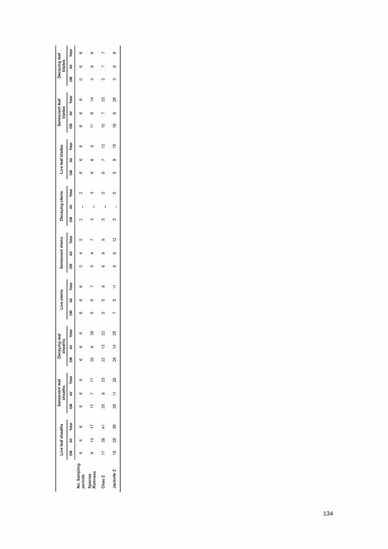
134
135
Unlike the leaf sheaths that remained attached to the stems as decomposition progressed, most
of the lower and decayed leaf blades had already abscised before the collection of plants; this
implied that only the uppermost leaf blades, which were mostly alive or senescent, were
available at the moment of collection. Most of stems were totally green, even in the lower naked
portions, which explained the absence or scarcity of senescent and decaying stems during the
sampling time.
The low representativeness of senescent and decaying stems and decaying leaf blades might
explain the low species richness observed and estimated by Chao2 and Jacknife 2 estimators
for these substrates in comparison with the remaining ones (Table 2); this hypothesis was
based mainly on previously studies that documented a high percentage of colonisation, species
richness and diversity on drift decaying stems of S. maritima (Azevedo et al. 2012) and
decaying leaf blades of S. alterniflora (Buchan et al. 2002; Walker and Campbell 2010).
Similarly, the high species richness observed and estimated for live, senescent and decaying
leaf sheaths in both study sites (Table 2) might have resulted from a high availability of these
substrates at the time of collection, but may also be related with the morphochemical
characteristics of these vegetative structures. Leaf sheaths are wider, with a lower phenolic
content, more aerenchyma and structurally tougher but less lignified than leaf blades (Anderson
1974; Graça et al. 2000); stems are more heavily lignified (Hodson et al. 1984) and with a lower
nitrogen content than leaves (Cartaxana and Catarino 1997; Curado et al. 2013).
Even though the estimates of species richness pointed out for an insufficient sampling effort
(Table 2), the detection of the most frequent fungal taxa by both methods suggested that it was
the minimum required to adequately record the most important fungal taxa occurring in this
phase of S. maritima life cycle.
The comparison of the species composition of communities inhabiting the nine plant substrates
revealed that it varied mostly between different vegetative structures (Tables 3 and 4).
Table 3 Percent frequencies of occurrence (V very frequent: >20%; F frequent: 10-20%; I infrequent: <10%, according
to the 3 categories proposed by Tan et al (1989)) and/or presences ( ̶ ) of fungal taxa on different plant substrates in
Castro Marim salt marsh in each sampling period, identified by morphological and molecular methods respectively; the
superscript letters mark those frequencies of occurrence that were calculated based exclusively on the presence of
hyphopodia (h) or spores (
s)
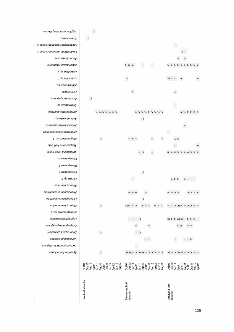
136
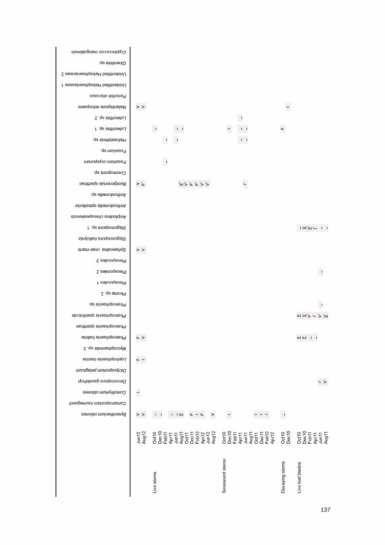
137
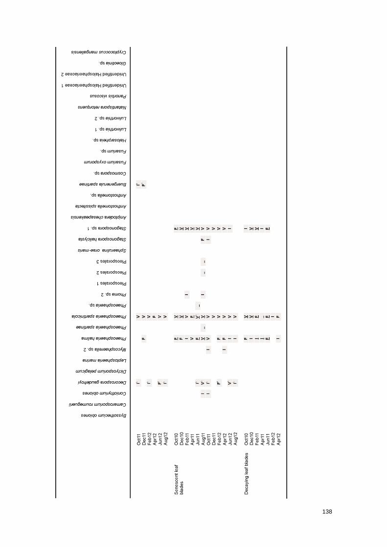
138
139
Table 4 Percent frequencies of occurrence (V very frequent: >20%; F frequent: 10-20%; I infrequent: <10%, according
to the 3 categories proposed by Tan et al (1989)) and/or presences ( ̶ ) of fungal taxa on different plant substrates in Ria
de Aveiro salt marsh in each sampling period, identified by morphological and molecular methods respectively; the
superscript letters mark those frequencies of occurrence that were calculated based exclusively on the presence of
hyphopodia (h) or spores (
s)
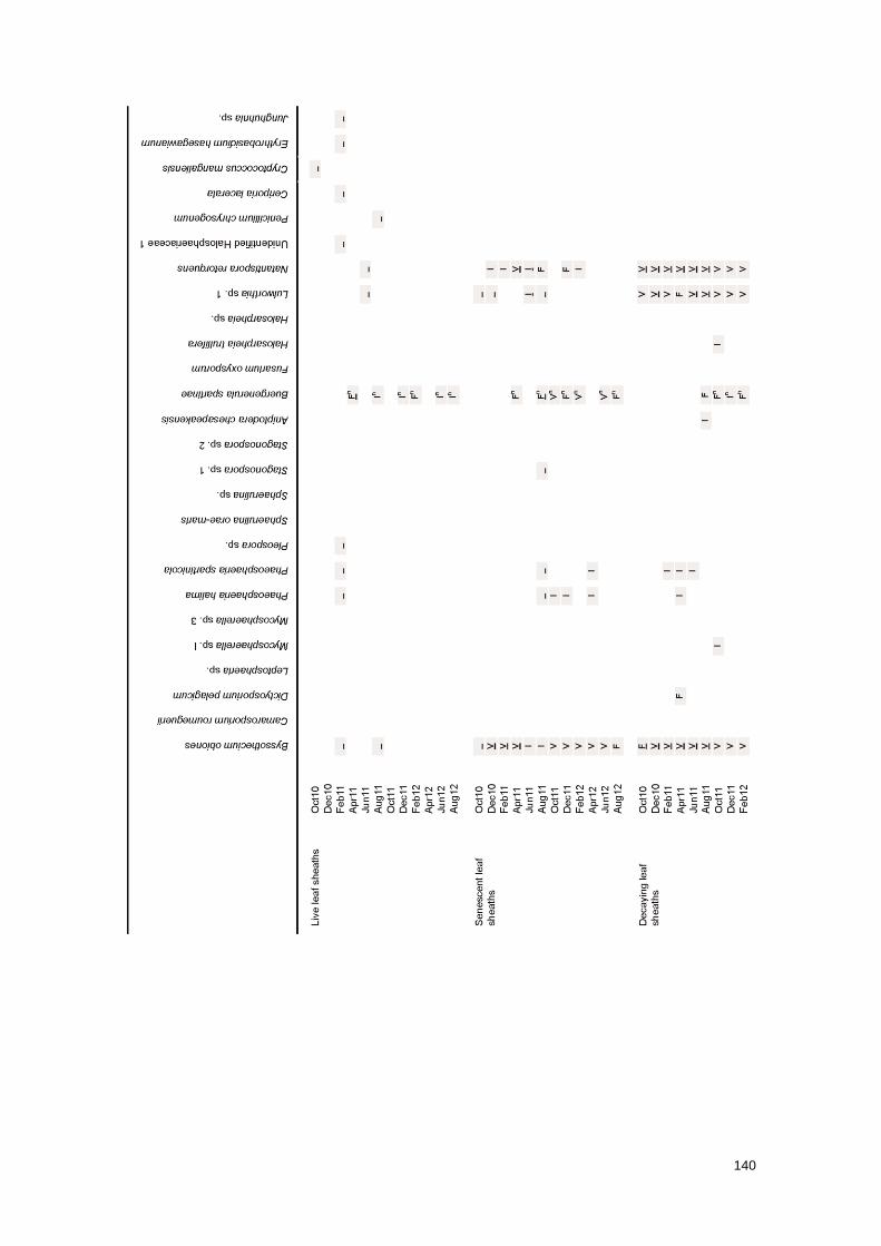
140

141
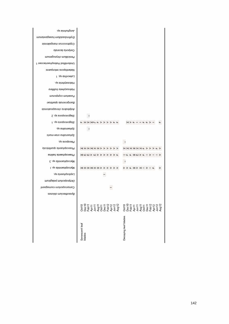
142

143
The most frequent fungi found in both salt marshes demonstrated a clear prevalence on leaf
sheaths, stems and/or leaf blades regardless their physiological state (Tables 3 and 4).
Specifically, B. obiones, Lulworthia sp. 1 and N. retorquens were recorded on leaf sheaths and
stems; P. spartinicola, P. halima and Stagonospora sp. 1 on leaf sheaths and blades; and B.
spartinae hyphopodia on all vegetative structures.
Mycosphaerella sp. I, an exclusive and very frequent fungus from Ria de Aveiro salt marsh, was
found on leaf blades under different physiological states. L. marina and S. orae-maris, two
exclusive and frequent fungi from Castro Marim salt marsh, were only identified on senescent
and decaying leaf sheaths.
Even though the remaining species were recorded on particular plant substrates, their
infrequency did not allow to accurately clarify their ecological preferences during this phase of
plant life cycle.
The comparison of different plant substrates based on the presence/absence of fungal taxa in
all the sampling periods of the first year corroborated most of the above-mentioned ecological
tendencies (Figs. 3 and 4).
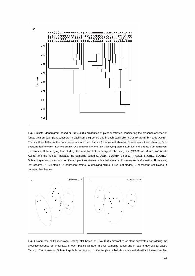
144
Fig. 3 Cluster dendrogram based on Bray-Curtis similarities of plant substrates, considering the presence/absence of
fungal taxa on each plant substrate, in each sampling period and in each study site (a Castro Marim; b Ria de Aveiro).
The first three letters of the code name indicate the substrate (LLs-live leaf sheaths, SLs-senescent leaf sheaths, DLs-
decaying leaf sheaths, LSt-live stems, SSt-senescent stems, DSt-decaying stems, LLb-live leaf blades, SLb-senescent
leaf blades, DLb-decaying leaf blades), the next two letters designate the study site (CM-Castro Marim, AV-Ria de
Aveiro) and the number indicates the sampling period (1-Oct10, 2-Dec10, 3-Feb11, 4-Apr11, 5-Jun11, 6-Aug11).
Different symbols correspond to different plant substrates: live leaf sheaths, senescent leaf sheaths, decaying
leaf sheaths, live stems, senescent stems, decaying stems, + live leaf blades, senescent leaf blades,
decaying leaf blades
Fig. 4 Nonmetric multidimensional scaling plot based on Bray-Curtis similarities of plant substrates considering the
presence/absence of fungal taxa in each plant substrate, in each sampling period and in each study site (a Castro
Marim; b Ria de Aveiro). Different symbols correspond to different plant substrates: live leaf sheaths, senescent leaf

145
sheaths, decaying leaf sheaths, live stems, senescent stems, decaying stems, + live leaf blades,
senescent leaf blades, decaying leaf blades
Cluster analysis and MDS ordination clearly differentiated the samples that represent different
vegetative structures, i.e. leaf sheaths, stems and leaf blades, regardless of their physiological
states. However, based on the s-stress values provided by MDS analyses (Castro Marim: 0.17;
Ria de Aveiro: 0.30), the ordination of Castro Marim samples in the plot represented well the
relationships among them, while the Ria de Aveiro samples seem to be more randomly
distributed in the 2-dimensional ordination space.
Both analyses revealed a higher similarity between the communities inhabiting leaf sheaths and
blades in Castro Marim, and leaf sheaths and stems in Ria de Aveiro salt marsh; stems and leaf
blades yielded the most dissimilar fungal communities.
ANOSIM analyses showed significant differences in the fungal communities on plant substrates
from both salt marshes (R=0.7, p<0.001); in the majority of posterior pairwise comparisons, no
significant differences were found among substrates representing the same vegetative structure
(data not shown).
The comparison of the same plant substrates based on the fruiting patterns of fungal colonisers
along a two-year study period by a DCA (Fig. 5) analysis confirmed the existence of three main
ecological niches.
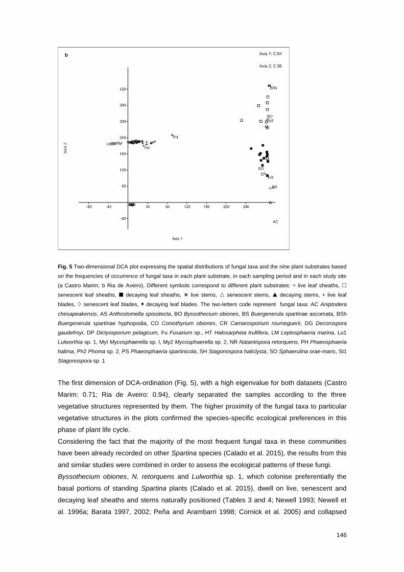
146
Fig. 5 Two-dimensional DCA plot expressing the spatial distributions of fungal taxa and the nine plant substrates based
on the frequencies of occurrence of fungal taxa in each plant substrate, in each sampling period and in each study site
(a Castro Marim; b Ria de Aveiro). Different symbols correspond to different plant substrates: live leaf sheaths,
senescent leaf sheaths, decaying leaf sheaths, live stems, senescent stems, decaying stems, + live leaf
blades, senescent leaf blades, decaying leaf blades. The two-letters code represent fungal taxa: AC Aniptodera
chesapeakensis, AS Anthostomella spissitecta, BO Byssothecium obiones, BS Buergenerula spartinae ascomata, BSh
Buergenerula spartinae hyphopodia, CO Coniothyrium obiones, CR Camarosporium roumeguerii, DG Decorospora
gaudefroyi, DP Dictyosporium pelagicum, Fu Fusarium sp., HT Halosarpheia trullifera, LM Leptosphaeria marina, Lu1
Lulworthia sp. 1, MyI Mycosphaerella sp. I, My2 Mycosphaerella sp. 2, NR Natantispora retorquens, PH Phaeosphaeria
halima, Ph2 Phoma sp. 2, PS Phaeosphaeria spartinicola, SH Stagonospora haliclysta, SO Sphaerulina orae-maris, St1
Stagonospora sp. 1
The first dimension of DCA-ordination (Fig. 5), with a high eigenvalue for both datasets (Castro
Marim: 0.71; Ria de Aveiro: 0.94), clearly separated the samples according to the three
vegetative structures represented by them. The higher proximity of the fungal taxa to particular
vegetative structures in the plots confirmed the species-specific ecological preferences in this
phase of plant life cycle.
Considering the fact that the majority of the most frequent fungal taxa in these communities
have been already recorded on other Spartina species (Calado et al. 2015), the results from this
and similar studies were combined in order to assess the ecological patterns of these fungi.
Byssothecium obiones, N. retorquens and Lulworthia sp. 1, which colonise preferentially the
basal portions of standing Spartina plants (Calado et al. 2015), dwell on live, senescent and
decaying leaf sheaths and stems naturally positioned (Tables 3 and 4; Newell 1993; Newell et
al. 1996a; Barata 1997, 2002; Peña and Arambarri 1998; Cornick et al. 2005) and collapsed

147
and unrooted stems (Gessner 1977). Leptosphaeria marina, a fungus that also occurs on the
basal plant portions (Calado et al. 2015), is found inhabiting standing-senescent and -decaying
leaf sheaths of S. maritima (Table 3) and rooted- and unrooted-decayed stems of Spartina spp.
(Gessner and Kohlmeyer 1976). Sphaerulina orae-maris, a coloniser of middle plant portions
(Calado et al. 2015), colonises standing-senescent and -decaying leaf sheaths of S. maritima
(Table 3; Barata 1997, 2002); this species was also identified on stem baits of the same host
plant (Barata 2006). B. spartinae, another species that occupies middle plant portions (Calado
et al. 2015), develops hyphopodia under certain circumstances, which strongly adhere and
penetrate the surfaces of living, senescent and decaying leaves and stems of Spartina plants
(Tables 3 and 4; Gessner 1977; Kohlmeyer and Kohlmeyer 1979; Barata 1997). Hyphopodia
were demonstrated by Onyile and Gessner (1982) to be produced at a wide range of
environmental conditions, including less favorable conditions for fungal growth. The presence of
hyphopodia may indicate that the ideal conditions for sexual reproduction were not totally
achieved. In this study, ascomata of B. spartinae were exclusively and infrequently recorded on
decaying leaf sheaths (Tables 3 and 4). However, these structures have been previously
reported from standing-decaying leaf blades and stems (Gessner 1977; Kohlmeyer and
Kohlmeyer 1979; Newell and Wasowski 1995; Newell et al. 1996a, 2000; Barata 1997, 2002;
Newell and Porter 2000; Newell 2001a; Buchan et al. 2002; Walker and Campbell 2010) and
drift material (Peña and Arambarri 1996; Azevedo et al. 2012) from S. maritima and other
species of Spartina. Phaeosphaeria halima, a coloniser of middle plant portions (Calado et al.
2015), inhabits decaying leaf blades of Spartina spp., co-occurring with P. spartinicola and
Stagonospora sp. (Tables 3 and 4; Newell and Wasowski 1995; Newell and Porter 2000; Newell
and Zakel 2000; Newell 2001a; Buchan et al. 2002, 2003; Lyons et al. 2003; Walker and
Campbell 2010). In this study, this species was also recorded on living and senescent leaf
blades and on leaf sheaths in all physiological states. However, and contrary to similar studies,
this species only differentiated asexual reproductive structures; the factors that trigger the
asexual or sexual reproduction in holomorphic species are not yet clearly understood.
Phaeosphaeria spartinicola and Stagonospora sp. 1 extend their distribution area to the top leaf
blades, where they are more frequent (Calado et al. 2015) and dominant (Newell and Wasowski
1995; Newell and Porter 2000; Newell and Zakel 2000; Newell 2001a; Buchan et al. 2002;
Walker and Campbell 2010). Even though these last mentioned studies had only reported P.
spartinicola from decaying leaf blades, this species and Stagonospora sp. 1 were identified
herein on leaves under all physiological states (Tables 3 and 4); on leaf sheaths, they occupy a
small area directly adjacent to the ligule. Barata (2006) and Azevedo et al. (2012) also reported
P. spartinicola and unidentified species of Stagonospora from stem baits and drift stems of S.
maritima, although with a much lower frequency of occurrence. Mycosphaerella sp. I, a
coloniser of the top plant portions (Calado et al. 2015), occurs exclusively on leaf blades,
particularly on the most decayed ones (Table 4; Newell and Wasowski 1995; Barata 1997;
Newell 2001a; Walker and Campbell 2010); in this study, this species was also found on live
and senescent leaf blades. A positive interaction between P. spartinicola and Mycosphaerella
148
sp. II in leaf blades of S. alterniflora to suppress competitors (e.g. P. halima, B. spartinae) has
already been suggested by some authors (Newell and Porter 2000; Newell 2001a; Buchan et al.
2003; Walker and Campbell 2010), and might actually have occurred in this study with a
different species of Mycosphaerella.
Even though the low frequency of the remaining fungal species limited an understanding of their
ecological patterns, the combination of data from this and similar studies that focused on fungal
communities associated with Spartina spp. confirmed or complemented the information about
the ecology of species: A. spissitecta occurs on decaying leaf sheaths (Table 3; Kohlmeyer and
Volkmann-Kohlmeyer 2002); A. chesapeakensis colonises senescent leaf sheaths and stems
(Tables 3 and 4; Barata 2006); D. pelagicum occurs on senescent and decaying leaf sheaths
and decaying stems (Tables 3 and 4; Barata 1997, 2002, 2006; Azevedo et al. 2012); F.
oxysporum is found on all vegetative structures (Tables 3 and 4; Al-Nasrawi and Hughes 2012);
P. spartinae inhabits senescent and decaying leaves and decaying stems (Table 3; Newell
1993; Newell et al. 1996a; Barata 1997; Peña and Arambarri 1998); P. viscosus occurs on
decaying leaves and stems (Table 3; Buchan et al. 2002, 2003; Barata 2006; Azevedo et al.
2012).
In conclusion, the results suggested an enrichment of fungi along the decay process of leaf
sheaths, stems and leaf blades instead of a real succession of fungal species. Despite the
decrease of soluble organic compounds and increase of recalcitrant materials and interspecific
competition for space and nutrients as the decomposition progresses, more decayed substrates
presented low content of antimicrobial substances. In addition to the enrichment of species,
senescent and decaying vegetative structures represented the substrates where the majority of
fungal taxa produced fruiting structures (87% and 83% respectively; Tables 3 and 4). This is in
agreement with other studies (Newell 1993, 2001a; Newell and Wasowski 1995; Newell and
Zakel 2000; Buchan et al. 2002; Cornick et al. 2005). Fungal fruiting structures represent the
end result of the decay process and indicate a substantial amount of supportive mycelium within
the plant substrate (Newell and Porter, 2000). According to Van Ryckegem and Verbeken
(2005), the reduction of carbon compounds and/or changes in the proportions of the different
carbon substrates may trigger fungal reproduction.
Moreover, the results revealed that the occurrence of most frequent fungi on different substrates
of Spartina depends on three main factors: (1) phase of plant life cycle and specifically the
availability of each substrate, (2) vertical position of substrates on standing plants and
associated micro-environmental conditions (Calado et al. 2015), and (3) potential fungal
competitors inhabiting the same substrates. Each of these factors might interfere more or less in
the colonisation process of each fungus, depending on its ecological requirements. The majority
of these fungal taxa initiate the colonisation process on a particular vegetative structure that
might proceed to other vegetative structure throughout the decay process. This fact indicates
that dominant fungal taxa exhibited less nutritive restrictions and high physiological versatility in
adapting to different ecological niches.

149
Potential ecological roles
The high occurrence of B. spartinae, B. obiones, Lulworthia sp. 1, Mycosphaerella sp. I, N.
retorquens, P. halima, P. spartinicola and Stagonospora sp. 1 throughout all decay stages of
the same vegetative structure (Tables 3 and 4, Fig. 5) strongly indicated an high ecological
importance as saprobes. With the exception of N. retorquens, all the remaining fungal species
were demonstrated to be involved on the decay process of Spartina spp. and particularly in the
digestion of lignocellulose present in secondary walls and middle-lamellar layer of cell (Torzilli
and Andrykovitch 1986; Bergbauer and Newell 1992; Newell et al. 1996b; Newell and Porter
2000; Buchan et al. 2002, 2003; Lyons et al. 2003, 2010). The marine fungi L. marina and S.
orae-maris seem to be also playing an important ecological role as saprobes in the leaf sheaths
that have already undergone natural senescence (Table 3).
The detection of B. spartinae, B. obiones, Lulworthia sp. I, Mycosphaerella sp. I, N. retorquens,
P. halima, P. spartinicola and Stagonospora sp. 1 on live plant tissues, either by molecular,
morphological or both methods together, raises some questions about the life style of these
species before the onset of senescence. It is not possible to unequivocally conclude if these
fungi were present as functional or metabolically inactive propagules since the surfaces of plant
samples were not-sterilised and the genetic molecule used in this study was not appropriate for
selection of active members of community. According to Edgcomb et al. (2011), RNA-based
clone libraries are more indicative for identifying active species rather than DNA-based clone
libraries. Moreover, the presence of fruiting structures on living tissues did not clearly indicate if
the associated mycelial network was inside or outside the plant tissues.
Despite the limitations imposed by the methodology, the host plants did not reveal any visible
disease symptoms suggesting that the fungi were not parasites or pathogens. The only
exception might be B. spartinae, given that hyphopodial-entry mechanism has been associated
with a parasitic phase in the species life cycle (Kohlmeyer and Gessner 1976; Kohlmeyer and
Kohlmeyer 1979). Hyphopodia may represent part of the strategy of the species to gain
competitive advantage in the colonisation of substrate (Kohlmeyer and Kohlmeyer 1979; Barata
1997).
An early endophytic colonisation of healthy plant tissues by the remaining frequent fungi would
certainly give them a competitive advantage over pioneer and latter fungal saprobes and
explain their dominance during the decay process. This hypothesis was also postulated by
Newell (1996) to explain the omnipresence of P. spartinicola on standing decaying leaf blades
of Spartina plants. Even though these frequent species were not mentioned before as marine
endophytes of Spartina plants (Nagahama 2006; Kandalepas et al. 2015), there is no reason for
excluding this hypothesis. The genus Lulworthia, Mycosphaerella, Phaeosphaeria and
Stagonospora have been demonstrated to include endophytic species associated with intertidal
plants (Elsebai et al. 2009; Xing et al. 2011; Sakayaroj et al. 2012; Kandalepas et al. 2015).
150
Moreover, B. obiones, Mycosphaerella sp. I, P. halima and P. spartinicola exhibited a degree of
specificity with Spartina spp. (Calado et al. 2015), as has been demonstrated for endophytic
fungi (Arnold 2007).
For the infrequent fungi, any interpretation about their ecological role would be merely
speculative. Moreover, it is also plausible that some of these fungal taxa that have never been
documented in marine environments might represent airborne contaminants.
Final remarks
The combination of morphological and molecular methods provided a more accurate
representation of the fungal community associated with standing plants of S. maritima; 45
fungal taxa were identified on early decaying plants. Moreover, it contributed to a better
understanding of community dynamics and ecological patterns and potential roles of the most
frequent fungi in an early phase of decay of the host plant. The integration of the results from
this study and similar ones that focused on Spartina decay system revealed that the presence
and ecological role of dominant fungi on different substrates depends on the combination of
three main factors: phase of plant life cycle, micro-environmental conditions of substrates and
potential fungal competitors.
Further studies should be performed in order to clarify the ecological role of these fungi before
the beginning of senescence.
Acknowledgements
This study was supported financially by Foundation for Science and Technology (FCT) through
a PhD grant (SFRH/BD/48525/2008).
References
Abdel-Wahab MA, Bahkali AHA (2012) Taxonomy of filamentous anamorphic marine fungi:
morphology and molecular evidence. In: Jones EBG, Pang K-L (eds) Marine Fungi and
fungal-like organisms. De Gruyter, Berlin, pp 65–90
Al-Nasrawi HG, Hughes AR (2012) Fungal diversity associated with salt marsh plants Spartina
alterniflora and Juncus roemerianus in Florida. Jordan J. Biol. Sci. 5: 247–254
Alva P, McKenzie EHC, Pointing SB, Pena-Muralla R, Hyde KD (2002) Do sea grasses harbour
endophyte? In: Hyde KD (ed) Fungi in marine environments. Fungal Diversity Research
Series 7, Hong Kong, pp 167–178

151
Anderson CE (1974) A review of structure in several North Carolina salt marsh plants. In:
Reimold RJ, Queen WH (eds) Ecology of halophytes. Academic Press, Inc., New York, pp
307–344
Arnold AE (2007) Understanding the diversity of foliar endophytic fungi: progress, challenges,
and frontiers. Fungal Biol. Rev. 21: 51–66
Aveskamp MM, de Gruyter J, Woudenberg JHC, Verkley GJM, Crous PW (2010) Highlights of
the Didymellaceae: a polyphasic approach to characterise Phoma and related pleosporalean
genera. Stud. Mycol. 65: 1–60
Azevedo E, Rebelo R, Caeiro MF, Barata M (2012) Use of drift substrates to characterize
marine fungal communities from the west coast of Portugal. Mycologia 104: 623–632
Barata M (1997) Fungos marinhos superiores associados a Spartina maritima em Estuários da
Costa Portuguesa. Ph.D. Thesis, Faculty of Sciences of University of Lisbon
Barata M (2002) Fungi on the halophyte Spartina maritima in salt marshes. In: Hyde KD (ed)
Fungi in marine environments. Fungal Diversity Research Series 7, Hong Kong, pp 179–193
Barata M (2006) Marine fungi from Mira river salt marsh in Portugal. Rev. Iberoam. Micol. 23:
179–184
Bergbauer M, Newell SY (1992) Contribution to lignocellulose degradation and DOC formation
from a salt marsh macrophyte by the ascomycete Phaeosphaeria spartinicola. FEMS
Microbiol. Ecol. 86: 341–348
Blaalid R, Kumar S, Nilsson RH, Abarenkov K, Kirk PM, Kauserud H (2013) ITS1 versus ITS2
as DNA metabarcodes for fungi. Mol. Ecol. Resour. 13: 218–224
Buchan A, Newell SY, Moreta JIL, Moran MA (2002) Analysis of internal transcribed spacer
(ITS) regions of rRNA genes in fungal communities in a southeastern U.S. salt marsh.
Microb. Ecol. 43: 329–340
Buchan A, Newell SY, Butler M, Biers EJ, Hollibaugh JT, Moran MA (2003) Dynamics of
bacterial and fungal communities on decaying salt marsh grass. Appl. Environ. Microbiol. 69:
6676–6687
Buchanan-Wollaston V (1997) The molecular biology of leaf senescence. J. Exp. Bot. 48: 181–
199
Calado ML, Barata M (2012) Salt marsh fungi. In: Jones EBG, Pang K-L (eds) Marine fungi and
fungal-like organisms. De Gruyter, Berlin, pp 345–381
Calado ML, Carvalho L, Pang K-L, Barata M (2015) Diversity and ecological characterization of
sporulating higher filamentous marine fungi associated with Spartina maritima (Curtis)
Fernald in two Portuguese salt marshes. Microb. Ecol. 70: 612-633
Câmara MPS, Palm ME, Berkum PV, O´Neill NR (2002) Molecular phylogeny of Leptosphaeria
and Phaeosphaeria. Mycologia 94: 630–640
Campbell J, Volkmann-Kohlmeyer B, Gräfenhan T, Spatafora JW, Kohlmeyer J (2005) A re-
evaluation of Lulworthiales: relationships based on 18S and 28S rDNA. Mycol. Res. 109:
556–568

152
Cartaxana P, Catarino F (1997) Allocation of nitrogen and carbon in an estuarine salt marsh in
Portugal. J. Coast. Conserv. 3: 27–34
Castillo JM, Rubio-Casal AE, Figueroa E (2010) Cordgrass biomass in coastal marshes. In:
Momba MNB, Bux F (eds) Biomass. Sciyo, Rijeka (InTech), pp 1–26
Castro P, Freitas H (2000) Fungal biomass and decomposition in Spartina maritima leaves in
the Mondego salt marsh (Portugal). Hydrobiologia 428: 171–177
Clarke KR, Warwick RM (2001) Change in marine communities: an approach to statistical
analysis and interpretation. PRIMER-E, Plymouth
Cornick J, Standwerth A, Fisher PJ (2005) A preliminary study of fungal endophyte diversity in a
stable and declining bed of Spartina anglica Hubbard. Mycologist 19: 24–29
Curado G, Rubio-Casal AE, Figueroa E, Grewell BJ, Castillo JM (2013) Native plant restoration
combats environmental change: development of carbon and nitrogen sequestration capacity
using small cordgrass in European salt marshes. Environ. Monit. Assess. 185: 8439–8449
Edgcomb VP, Beaudoin D, Gast R, Biddle JF, Teske A (2011) Marine subsurface eukaryotes:
the fungal majority. Environ. Microbiol. 13: 172–183
Elsebai MF, Kehraus S, Gutschow M, Konig GM (2009) New Polyketides from the marine-
derived fungus Phaeosphaeria spartinae. Nat. Prod. Commun. 4: 1463–1468
Gessner RV (1977) Seasonal occurrence and distribution of fungi associated with Spartina
alterniflora from Rhode Island estuary. Mycologia 69: 477–491
Gessner RV (1980) Degradative enzyme production by salt-marsh fungi. Bot. Mar. 23: 133–139
Gessner RV, Kohlmeyer J (1976) Geographical distribution and taxonomy of fungi from salt
marsh Spartina. Can. J. Bot. 54: 2023–2037
Gotelli NJ, Colwell RK (2010) Estimating species richness. In: Magurran AE, McGill BJ (eds)
Biological diversity: frontiers in measurement and assessment. Oxford University Press,
Oxford, pp 39–54
Graça MA, Newell SY, Kneib RT (2000) Grazing rates of organic matter and living fungal
biomass of decaying Spartina alterniflora by three species of salt-marsh invertebrates. Mar.
Biol. 136: 281–289
Hammer Ø, Harper DAT, Ryan PD (2001) PAST: Paleontological statistics software package for
education and data analysis. Palaeontol. Electron. 4: 1–9
Healy B, Walters K (1994) Oligochaeta in Spartina stems: the microdistribution of Enchytraeidae
and Tubificidae in a salt marsh, Sapelo Island, USA. Hydrobiologia 278: 111–123
Hodson RE, Christian RR, Maccubbin AE (1984) Lignocellulose and lignin in the salt marsh
grass Spartina alterniflora: initial concentrations and short-term, post-depositional changes in
detrital matter. Mar. Biol. 81: 1–7
Hortensteiner S, Feller U (2002) Nitrogen metabolism and remobilization during senescence. J.
Exp. Bot. 53: 927–937
Hyde KD, Lee SY (1995) Ecology of mangrove fungi and their role in nutrient cycling: what gaps
occur in our knowledge? Hydrobiologia 295: 107–118
153
Hyde KD, Sarma VV (2000) Pictorial key to higher marine fungi. In: Hyde KD, Pointing SB (eds)
Marine Mycology – A practical approach. Fungal Diversity Press, Hong Kong, pp 205–270
Hyde KD, Jones EBG, Leaño E, Pointing SB, Poonyth AD, Vrijmoed LLP (1998) Role of fungi in
marine ecosystems. Biodivers. Conserv. 7: 1147–1161
Jones EBG (2000) Marine fungi : some factors influencing biodiversity. Fungal Divers. 4: 53–73
Jones EBG (2011a) Fifty years of marine mycology. Fungal Divers. 50: 73–112
Jones EBG (2011b) Are there more marine fungi to be described? Bot. Mar. 54: 343–354
Jones EBG, Fell JW (2012) Basidiomycota. In: Jones EBG, Pang K-L (eds) Marine Fungi and
fungal-like organisms. De Gruyter, Berlin, pp 49–63
Jones EBG, Sakayaroj J, Suetrong S, Somrithipol S, Pang K-L (2009) Classification of marine
Ascomycota, anamorphic taxa and Basidiomycota. Fungal Divers. 35: 1–187
Kandalepas D, Blum MJ, Van Bael SA (2015) Shifts in symbiotic endophyte communities of a
foundational salt marsh grass following oil exposure from the deepwater horizon oil spill.
PLoS One. doi: 10.1371/journal.pone.0122378
Kirk PM, Cannon PF, Minter DW, Stalpers JA (2008) Dictionary of the Fungi, 10th ed. CAB
International, Wallingford
Kis-Papo T (2005) Marine fungal communities. In: Dighton J, White JF, Oudemans P (eds) The
fungal community – Its organization and role in the ecosystem. Taylor & Francis, Boca
Raton, pp 61–92
Kneib RT, Newell SY, Hermeno ET (1997) Survival, growth and reproduction of the salt-marsh
amphipod Uhlorchestia spartinophila reared on natural diets of senescent and dead Spartina
alterniflora leaves. Mar. Biol. 128: 423–431
Kohlmeyer J, Gessner RV (1976) Buergenerula spartinae sp. nov., an Ascomycete from salt
marsh cordgrass, Spartina alterniflora. Can. J. Bot. 54: 1759–1766
Kohlmeyer J, Kohlmeyer E (1979) Marine mycology – The higher fungi. Academic Press, Inc.,
New York
Kohlmeyer J, Volkmann-Kohlmeyer B (1991) Illustrated key to the filamentous higher marine
fungi. Bot. Mar. 34: 1–61
Kohlmeyer J, Volkmann-Kohlmeyer B (2002) Fungi on Juncus and Spartina: New marine
species of Anthostomella, with a list of marine fungi known on Spartina. Mycol. Res. 3: 365–
374
Kumaresan V, Suryanarayanan TS (2002) Endophyte assemblages in young, mature and
senescent leaves of Rhizophora apiculata: evidence for the role of endophytes in mangrove
litter degradation. Fungal Divers. 9: 81–91
Lyons JI, Newell SY, Buchan A, Moran MA (2003) Diversity of ascomycete laccase gene
sequences in a southeastern US salt marsh. Microb. Ecol. 45: 270–281
Lyons JI, Newell SY, Brown RP, Moran MA (2005) Screening for bacterial-fungal associations in
a south-eastern US salt marsh using pre-established fungal monocultures. FEMS Microbiol.
Ecol. 54: 179–187
154
Lyons JI, Alber M, Hollibaugh JT (2010) Ascomycete fungal communities associated with early
decaying leaves of Spartina spp. from central California estuaries. Oecologia 162: 435–442
Maccubbin AE, Hodson RE (1980) Mineralization of detrital lignocelluloses by salt marsh
sediment microflora. Appl. Environ. Microbiol. 40: 735–740
Menéndez M, Sanmartí N (2007) Geratology and decomposition of Spartina versicolor in a
brackish Mediterranean marsh. Estuar. Coast. Shelf. Sci. 74: 320–330
Nagahama T (2006) Yeast biodiversity in freshwater, marine and deep-sea environments. In:
Rosa CA, Peter G (eds) Biodiversity and ecophysiology of yeasts. Springer, Berlin, pp 241–
262
Nakagiri A, Tubaki K (1987) Pleomorphy in marine fungi: teleomorph-anamorph connections in
the Halosphaeriaceae. In: Sugiyama J (ed) Pleomorphic fungi: the diversity and its
taxonomic implications. Kodansha, Tokyo, pp 79–191
Newell SY (1993) Decomposition of shoots of a salt-marsh – Methodology and dynamics of
microbial assemblages. In: Jones JG (ed) Advances in Microbial Ecology – Volume 13.
Plenum Press, New York, pp 301–326
Newell SY (1996) Established and potential impacts of eukaryotic mycelial decomposers in
marine/terrestrial ecotones. J. Exp. Mar. Bio. Ecol. 200: 187–206
Newell SY (2001a) Spore-expulsion rates and extents of blade occupation by ascomycetes of
the smooth-cordgrass standing-decay system. Bot. Mar. 44: 277–285
Newell SY (2001b) Multiyear patterns of fungal biomass dynamics and productivity within
naturally decaying smooth cordgrass shoots. Limnol. Oceanogr. 46: 573–583
Newell SY, Wasowski J (1995) Sexual productivity and spring intramarsh distribution of a key
salt-marsh microbial secondary producer. Estuaries 18: 241–249
Newell SY, Porter D (2000) Microbial secondary production from saltmarsh-grass shoots, and
its known and potential fates. In: Weinstein MP, Kreeger DA (eds) Concepts and
controversies in tidal marsh ecology. Kluwer Academic Publishers, Dordrecht, pp 159–185
Newell SY, Zakel KL (2000) Measuring summer patterns of ascospore release by saltmarsh
fungi. Mycoscience 41: 211–215
Newell SY, Fallon RD, Miller JD (1989) Decomposition and microbial dynamics for standing,
naturally positioned leaves of the salt-marsh grass Spartina alterniflora. Mar. Biol. 101: 471–
481
Newell SY, Arsuffi TL, Palm LA (1996a) Misting and nitrogen fertilization of shoots of a
saltmarsh grass: effects upon fungal decay of leaf blades. Oecologia 108: 495–502
Newell SY, Porter D, Lingle WL (1996b) Lignocellulolysis by ascomycetes (fungi) of a saltmarsh
grass (smooth cordgrass). Microsc. Res. Tech. 33: 32–46
Newell SY, Blum LK, Crawford RE, Dai T, Dionne M (2000) Autumnal biomass and potential
productivity of salt marsh fungi from 29° to 43° north latitude along the United States Atlantic
Coast. Appl. Environ. Microbiol. 66: 180–185

155
Nilsson RH, Ryberg M, Kristiansson E, Abarenkov K, Larsson K-H, Kõljalg U (2006) Taxonomic
reliability of DNA sequences in public sequences databases: A fungal perspective. PLoS
One 1: 1–4
Nilsson RH, Tedersoo L, Abarenkov K, Ryberg M, Kristiansson E, Hartmann M, Schoch CL,
Nylander JAA, Bergsten J, Porter TM, Jumpponen A, Vaishampayan P, Ovaskainen O,
Hallenberg N, Bengtsson-Palme J, et al. (2012) Five simple guidelines for establishing basic
authenticity and reliability of newly generated fungal ITS sequences. MycoKeys 4: 37–63
Panebianco C, Tam WY, Jones EBG (2002) The effect of pre-inoculation of balsa wood by
selected marine fungi and their effect on subsequent colonisation in the sea. Fungal Divers.
10: 77–88
Pang K-L (2012) Phylogeny of the marine Sordariomycetes. In: Jones EBG, Pang K-L (eds)
Marine Fungi and fungal-like organisms. De Gruyter, Berlin, pp 35–47
Pang K-L, Mitchell JI (2005) Molecular approaches for assessing fungal diversity in marine
substrata. Bot. Mar. 48: 332–347
Pang K-L, Jones EBG, Vrijmoed LLP (2008) Autecology of Antennospora (Fungi: Ascomycota:
Sordariomycetidae: Halosphaeriales) and its phylogeny. Raffles Bull. Zool. 19: 1–10
Peña NI, Arambarri AM (1996) Hongos marinos lignícolas de Mar del Plata (provincia de
Buenos Aires, Argentina) II. Darwiniana 34: 293–298
Peña NI, Arambarri AM (1998) Hongos marinos lignícolas de la laguna costera de Mar Chiquita
(provincia de Buenos Aires, Argentina) I. Ascomycotina y Deuteromycotina sobre Spartina
densiflora. Darwiniana 35: 61–67
Porter TM, Golding GB (2011) Are similarity- or phylogeny-based methods more appropriate for
classifying internal transcribed spacer (ITS) metagenomic amplicons ? New Phytol. 192:
775–782
Promputtha I, Lumyong S, Dhanasekaran V, McKenzie EHC, Hyde KD, Jeewon R (2007) A
phylogenetic evaluation of whether endophytes become saprotrophs at host senescence.
Microb. Ecol. 53: 579–590
Promputtha I, Hyde KD, McKenzie EHC, Peberdy JF, Lumyong S (2010) Can leaf degrading
enzymes provide evidence that endophytic fungi becoming saprobes? Fungal Divers. 41:
89–99
Raghukumar S (2004) The role of fungi in marine detrital processes, in: Ramaiah, N. (Ed.),
Marine microbiology: facets and opportunities. National Institute of Oceanography, Goa, pp
91–101
Ramette A (2007) Multivariate analyses in microbial ecology. FEMS Microbiol. Ecol. 62: 142–
160
Rees GN, Baldwin DS, Watson GO, Perryman S, Nielsen DL (2004) Ordination and significance
testing of microbial community composition derived from terminal restriction fragment length
polymorphisms: application of multivariate statistics. Antonie Van Leeuwenhoek 86: 339–347
Sakayaroj J, Preedanon S, Phongpaichit S, Buatong J, Chaowalit P, Rukachaisirikul V (2012)
Diversity of endophytic and marine-derived fungi associated with marine plants and animals.
156
In: Jones EBG, Pang K-L (eds) Marine Fungi and fungal-like organisms. De Gruyter, Berlin,
pp 291–328
Samiaji J, Barlocher F (1996) Geratology and decomposition of Spartina alterniflora Loisel in a
New Brunswick saltmarsh. J. Exp. Mar. Bio. Ecol. 201: 233–252
Sánchez JM, SanLeon DG, Izco J (2001) Primary colonisation of mudflat estuaries by Spartina
maritima (Curtis) Fernald in Northwest Spain: vegetation structure and sediment accretion.
Aquat. Bot. 69: 15–25
Schoch CL, Seifert KA, Huhndorf S, Robert V, Spouge JL, Levesque CA, Chen W (2012)
Nuclear ribosomal internal transcribed spacer (ITS) region as a universal DNA barcode
marker for Fungi. Proc. Natl. Acad. Sci. USA 109: 6241–6246
Tamura K, Stecher G, Peterson D, Filipski A, Kumar S (2013) MEGA6: Molecular evolutionary
genetics analysis version 6.0. Mol. Biol. Evol. 30: 2725–2729
Tan TK, Leong WF, Jones EBG (1989) Succession of fungi on wood of Avicennia alba and A.
lanata in Singapore. Can. J. Bot.: 2686–2691
Toju H, Tanabe AS, Yamamoto S, Sato H (2012) High-coverage ITS primers for the DNA-based
identification of ascomycetes and basidiomycetes in environmental samples. PLoS One
7: 1–11
Torzilli AP, Andrykovitch G (1986) Degradation of Spartina lignocellulose by individual and
mixed cultures of salt-marsh fungi. Can. J. Bot. 64: 2211–2215
Torzilli AP, Sikaroodi M, Chalkley D, Gillevet PM (2006) A comparison of fungal communities
from four salt marsh plants using automated ribosomal intergenic spacer analysis (ARISA).
Mycologia 98: 690–698
Valiela I, Koumjian L, Swain T, Teal JM, Hobbie JE (1979) Cinnamic acid inhibition of detritus
feeding. Nature 280: 55–57
Van Ryckegem GV, Verbeken A (2005) Fungal ecology and succession on Phragmites australis
in a brackish tidal marsh. II. Stems. Fungal Divers. 20: 209–233
Velmurugan S, Prasannakumar C, Manokaran S, Ajith Kumar T, Samkamaleson A, Palavesam
A (2013) DNA barcodes for marine fungal identification and discovery. Fungal Ecol. 6: 408–
418
Walker AK, Campbell J (2010) Marine fungal diversity: a comparison of natural and created salt
marshes of the north-central Gulf of Mexico. Mycologia 102: 513–521
White TJ, Bruns T, Lee S, Taylor J (1990) Amplification and direct sequencing of ribossomal
RNA genes for phylogenetics. In: Innis MA, Gelfand DH, Sninsky JJ, White TJ (eds) PCR
protocols: a guide to methods and applications. Academic Press, Inc., London, pp 315–322
Wilson JO, Buchsbaum R, Valiela I, Swain T (1986a) Decomposition in salt marsh ecosystems:
phenolic dynamics during decay of litter of Spartina alterniflora. Mar. Ecol. Prog. Ser. 29:
177–187
Wilson JO, Valiela I, Swain T (1986b) Carbohydrate dynamics during decay of litter of Spartina
alterniflora. Mar. Biol. 92: 277–284
157
Xing XK, Chen J, Xu MJ, Lin WH, Guo SX (2011) Fungal endophytes associated with
Sonneratia (Sonneratiaceae) mangrove plants on the south coast of China. For. Pathol. 41:
334–340
Zhang Y, Schoch CL, Fournier J, Crous PW, de Gruyter J, Woudenberg JHC, Hirayama K,
Tanaka K, Pointing SB, Spatafora JW, Hyde KD (2009) Multi-locus phylogeny of
Pleosporales: A taxonomic, ecological and evolutionary re-evaluation. Stud. Mycol. 64: 85–
102

158
159
CHAPTER 4 – Final overview

160
161
4.1 General conclusions
The present study contributed to a better understanding of marine fungal communities
associated with Spartina plants in temperate salt marshes. Specifically, it complemented the
inventories of marine mycota in these ecosystems and in Portugal, shedding light on key
aspects of the ecology of fungi inhabiting standing-decaying plants of Spartina maritima.
As expected, the combination of traditional morphological and molecular identification methods
was demonstrated to be advantageous. The results revealed a high agreement between the
identification based on direct observation of fruiting structures and on nuclear ITS sequences
concerning with assignments to fungal species and representativeness of each fungus in the
community, plant portions (basal, middle and top portions), plant substrates (live, senescent and
decaying leaf sheaths, stems and leaf blades) and study site (Castro Marim and Ria de Aveiro
salt marshes). On the other hand, the use of both methods was crucial for obtaining a more
realistic estimate of fungal diversity associated with S. maritima plants since it helped
overcoming the major drawbacks inherent to morphological and molecular methods.
The fungal communities associated with S. maritima and other Spartina species over a wide
geographic range were demonstrated to be very similar, in terms of species diversity,
composition and frequency patterns. These communities are not particularly complex or
diverse, being dominated by the same host-genus-exclusive species, i.e. Buergenerula
spartinae, Byssothecium obiones, Phaeosphaeria halima and Phaeosphaeria spartinicola; these
fungal species integrate a very stable core group that, apparently, is not much affected by
variations in environmental conditions inherent to different latitudinal positions, different
morphochemical structure of other Spartina host plants or seasonality. Apart from these
frequent host-exclusive fungi, also cosmopolitan fungi Aniptodera chesapeakensis and
Dictyosporium pelagicum, frequently reported from other Spartina species, were observed in
this study. Even though the frequent fungal taxa Lulworthia sp. 1 and Stagonospora sp. 1 were
not identified to species level, the two genera are usually well-represented in fungal
communities associated with Spartina plants.
Beyond this similarity, the communities are composed by other fungal species, whose
occurrence seems to be determined by other macro and/or micro-environmental factors.
Natantispora retorquens, a cosmopolitan species reported from several tropical ecosystems,
was found to be one of the most frequent and dominant species in the fungal communities
associated with S. maritima, although it has not been reported from other Spartina species.
Similarly, Panorbis viscosus, another cosmopolitan species that has been described from
temperate and tropical regions, was infrequently collected in this, Barata (2002) and Azevedo et
al. (2012) studies, on plants of S. maritima.
Mycosphaerella sp. I, an host-genus-exclusive fungus, very frequent in Ria de Aveiro salt marsh
and in other temperate salt marshes, was absent in Castro Marim salt marsh. On the other
hand, the temperate non-host-exclusive fungi Sphaerulina-oraemaris and Leptosphaeria marina
162
were found to be very frequent in Castro Marim salt marsh but infrequent and absent,
respectively, in Ria de Aveiro salt marsh; similarly, Phaeosphaeria spartinae, a very common
fungi in communities associated with Spartina species, was only retrieved by molecular
methods from plant samples of Castro Marim salt marsh. L. marina, as well as infrequent
ascomycetous species Anthostomella spissitecta, Camarosporium roumeguerii, Coniothyrium
obiones, Decorospora gaudefroyi, Fusarium oxysporum, Halosarpheia trullifera, Penicillium
chrysogenum and Stagonospora haliclysta, and basidiomycetes Ceriporia lacerata,
Cryptococcus mangaliensis and Erythrobasidium hasegawianum, were reported for the first time
on S. maritima plants. From all these fungal species, only A. spissitecta, F. oxysporum and L.
marina have been previously described from other Spartina species.
The presence/absence of these host-genus-exclusive or cosmopolitan fungal species in the
communities of S. maritima may be attributed to differences in the status of conservation, tidal
regime and physical configuration of salt marsh ecosystems, intraspecific morphological
variations in host plants, or even nutrient or space competition phenomena.
The fungal taxa colonizing standing plants of S. maritima were found to exhibit vertical
distribution patterns (Fig. 1). Even though the absolute vertical positions of fungal structures
produced by common frequent species varied from one salt marsh to another, the relative
positions of these structures along the plants were maintained.
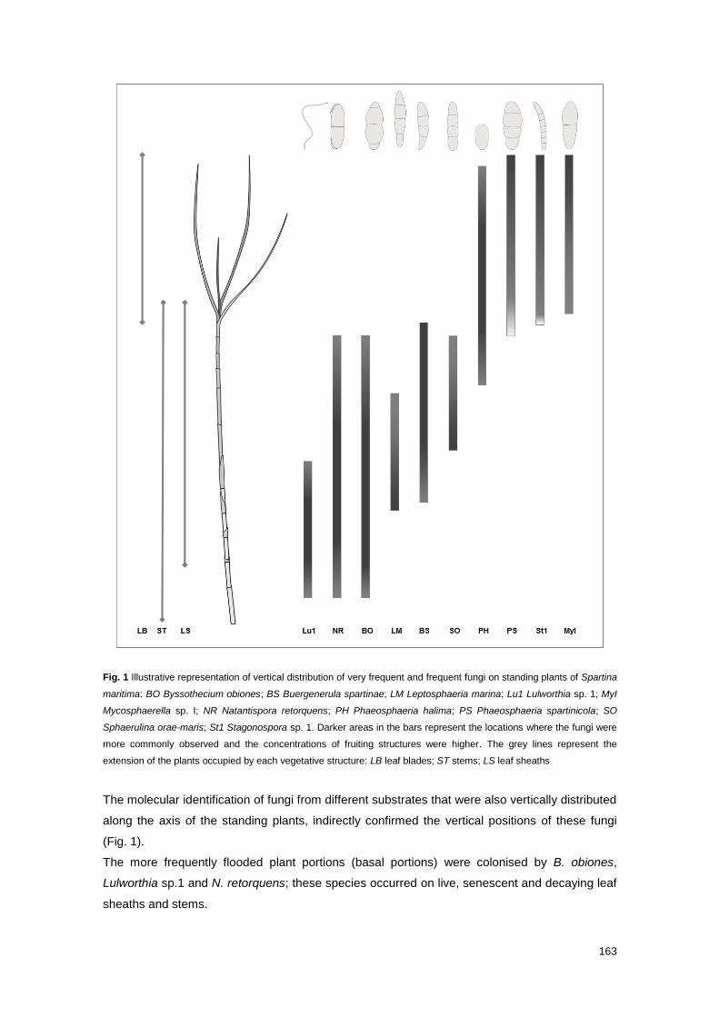
163
Fig. 1 Illustrative representation of vertical distribution of very frequent and frequent fungi on standing plants of Spartina
maritima: BO Byssothecium obiones; BS Buergenerula spartinae; LM Leptosphaeria marina; Lu1 Lulworthia sp. 1; MyI
Mycosphaerella sp. I; NR Natantispora retorquens; PH Phaeosphaeria halima; PS Phaeosphaeria spartinicola; SO
Sphaerulina orae-maris; St1 Stagonospora sp. 1. Darker areas in the bars represent the locations where the fungi were
more commonly observed and the concentrations of fruiting structures were higher. The grey lines represent the
extension of the plants occupied by each vegetative structure: LB leaf blades; ST stems; LS leaf sheaths
The molecular identification of fungi from different substrates that were also vertically distributed
along the axis of the standing plants, indirectly confirmed the vertical positions of these fungi
(Fig. 1).
The more frequently flooded plant portions (basal portions) were colonised by B. obiones,
Lulworthia sp.1 and N. retorquens; these species occurred on live, senescent and decaying leaf
sheaths and stems.

164
The middle plant portions were inhabited mainly by B. spartinae and P. halima; these two
species were detected on culms and leaves in all physiological states, respectively. L. marina
and S. orae-maris were also reported on basal and middle plant portions respectively, but
occurred only on senescent and decaying leaf sheaths.
The less inundated plant portions (top portions) were colonised by Mycosphaerella sp. I, P.
spartinicola and Stagonospora sp.1; Mycosphaerella sp. I was found exclusively on leaf blades
whereas P. spartinicola and Stagonospora sp.1 were observed on leaf blades and adjacent
areas of leaf sheaths in all physiological states.
B. obiones, B. spartinae, Mycosphaerella sp. I, N. retorquens, P. halima, P. spartinicola and
Stagonospora sp. I were vertically distributed over larger areas of standing plants of S. maritima
(Fig. 1).
Even though the variations in the plants´ heights and proportions of different substrates during
decay process and along the axis of standing plants, the vertical distribution range of each
fungus seemed to be maintain. The fact that fungal taxa occupied the same relative vertical
positions regardless of the phase of plant life cycle, suggests that the distribution range and
abundance of these fungi might be determined by the combination of three factors: salinity and
flooding conditions, availability of plant substrate and potential fungal competitors inhabiting the
same substrate.
The influence of salinity and flooding conditions in the distribution of fungi was clearly revealed
by the dominance of the Sordariomycetes, with dissolving unitunicate asci and passive spore-
discharge, in the basal portions and of the Dothideomycetes, with bitunicate asci and an active
spore-discharge, in the top portions. Distinct morphologies of reproductive structures and
mechanisms of spore’s dispersal of the fungal taxa distributed along the standing plants reflect
different adaptation strategies to marine or terrestrial environments. This finding, which has
already been described in some studies (Fell and Newell 1998; Alias and Jones 2000; Barata
2002; Hyde and Sarma 2006), justified the classification of these fungi into obligate and
facultative marine fungi. This classification, which followed the same criteria used by Kohlmeyer
and his colleagues to distinguish obligate and facultative marine fungi in standing plants of
Juncus roemerianus (e.g. Kohlmeyer & Kohlmeyer 2001), goes beyond a merely interpretation
of the initial definition proposed by the same authors (Kohlmeyer and Kohlmeyer 1979). The
criteria used by Kohlmeyers in their classification have been criticized by some authors as being
subjective and dependent on personal opinion. Moreover, these authors argued that the
tolerance or dependency to seawater submergence cannot be used as a criterion to infer about
the origin of fungi; Jones et al. (2009, 2015), one of the critics to Kohlmeyers´ classification,
considered all the fungal species recorded in this study as obligate marine fungi. Even though it
may be proved that the fungal species designated in this study as facultative marine fungi are
not of terrestrial origin but exclusively marine, the information reported herein about the
tolerance or dependency on marine conditions remains unchanged since it is factual. Therefore,
and while the concepts are not redefined based on more objective and unequivocal criteria and
supported by strong ecological evidences, there is no need to adopt a new classification. A
165
successful redefinition and standardisation of the concepts should rely on a better and more
consistent knowledge of the ecology and biology of each fungus.
The results from this study and the comparison with similar ones (e.g. Gessner and Kohlmeyer
1976; Peña and Arambarri 1996; Barata 1997, 2002, 2006; Azevedo et al. 2012) also revealed
that the occurrence of fungi on different plant substrates depends on their availability during
each phase of plant life cycle. Moreover, the majority of the most frequent species is not strictly
associated with a particular vegetative structure, but is capable to colonise different vegetative
structures located in a specific vertical position along the decomposition of Spartina plants as
soon as the structures become exposed and more accessible.
The presence and abundance of fungi in each ecological niche might also depend on their
competitive abilities.
Additionally to the species-specific ecological requirements, the results from this study
contributed to infer about species-specific ecological roles during early stages of decay of S.
maritima, particularly of the most frequent fungal taxa in the communities. The presence of B.
obiones, B. spartinae, L. marina, Lulworthia sp. 1, Mycosphaerella sp. I, N. retorquens, P.
halima, P. spartinicola, S. orae-maris and Stagonospora sp. 1 on senescent and decaying
vegetative structures in the majority or all the sampling periods indicates a very stable saprobic
community; the seasonality only seemed to interfere in number of fruiting structures produced
by these fungi.
The majority of these fungi were also recorded on live tissues by morphological and/or
molecular methods. The identification of hyphopodia of B. spartinae on live stems and leaf
sheaths indicated a parasitic phase in the species life cycle. The occurrence of B. obiones,
Lulworthia sp. 1, Mycosphaerella sp. I, N. retorquens, P. halima, P. spartinicola and
Stagonospora sp. 1 on live healthy plant tissues suggested that these fungi might start
colonising the plant as endophytes to gain a competitive advantage over the other pioneer
fungal saprobes; this strategy may thus explain the high frequency of these fungal taxa later in
the decay process.
All of these findings point out for the existence of a very stable and well-adapted fungal
community involved in the decay process of Spartina plants. Most frequent obligate marine
fungi inhabiting lower and middle plant portions play a major key-role in the decomposition of
leaf sheaths and stems, whereas facultative marine fungi inhabiting middle and top plant
portions assume a major role in the decay of upper stems and leaf sheaths and/or leaf blades.
These fungi exhibit higher ecological plasticity and lesser nutritive requirements than the
remaining fungi in the community, being able to explore more efficiently the plant substrates.
Among all the frequent fungi, B. spartinae and P. spartinicola seem to be more active and
intensively involved in the decomposition of Spartina species, since they were found on all
vegetative structures of standing decaying and drift plants of Spartina spp..

166
167
4.2 Future perspectives
Even though the substantial increase of general knowledge regarding marine fungi that came
with the advent and optimisation of molecular techniques, there are still many gaps in this
subject. The search for new fungi has been encouraged mainly by the awareness of the
potential biotechnological or pharmaceutical applications of secondary metabolites and
enzymes produced marine fungi. Therefore, this has been contributing for a change in the main
research paradigm, i.e. the purposes of any research project cannot be exclusively scientific or
related with management and conservation strategies in order to be funded, but have also to
have a direct or indirect applicability to the society and/or industries.
Nevertheless the research motivation, it is crucial to invest more intensively in the assessment
of the diversity of the communities on the less surveyed substrates, habitats and/or
geographical locations, as well as in the understanding of the dynamics of these communities
and ecology and functional role of each fungus in each particular ecosystem, employing
microscopy, culture and molecular techniques. All the fungal species recorded in each study
should be isolated and preserved in reference collections and/or their genomic sequences,
preferentially of barcoding genes, deposited in public repositories, in order to facilitate
taxonomic assignments of unknown or misidentified fungi. Moreover, the metabolic profiles and
biotechnological potential of all isolates should be explored.
A holistic and integrated knowledge is required to ensure the success of any management
strategy in a conservation or exploration and economic profitability perspective.
Moreover, this knowledge will elucidate about the origin, phylogeny and metabolic dependency
of fungi on marine conditions, as well as their ecological importance in marine ecosystems,
facilitating the redefinition of the terms used to classify the fungi.
4.3 References
Alias SA, Jones EBG (2000) Vertical distribution of marine fungi on Rhizophora apiculata at
Morib mangrove, Selangor, Malaysia. Mycoscience 41:431–436
Azevedo E, Rebelo R, Caeiro MF, Barata M (2012) Use of drift substrates to characterize
marine fungal communities from the west coast of Portugal. Mycologia 104:623–632
Barata M (1997) Fungos marinhos superiores associados a Spartina maritima em Estuários da
Costa Portuguesa. Ph.D. Thesis, Faculty of Sciences of University of Lisbon
Barata M (2002) Fungi on the halophyte Spartina maritima in salt marshes. In: Hyde KD (ed)
Fungi in marine environments. Fungal Diversity Research Series 7, Hong Kong, pp 179–193
Barata M (2006) Marine fungi from Mira river salt marsh in Portugal. Rev Iberoam Micol 23:179–
184
168
Fell JW, Newell SY (1998) Biochemical and molecular methods for the study of marine fungi. In:
Cooksey KE (ed) Molecular Approaches to the Study of the Ocean. Chapman & Hall,
London, pp 259–283
Gessner R V, Kohlmeyer J (1976) Geographical distribution and taxonomy of fungi from salt
marsh Spartina. Can J Bot 54:2023–2037
Hyde KD, Sarma VV (2006) Biodiversity and ecological observations on filamentous fungi of
mangrove palm Nypa fruticans Wurumb (Liliopsida – Arecales) along the Tutong river,
Brunei. Indian J Mar Sci 35:297–307
Jones EBG, Sakayaroj J, Suetrong S, Somrithipol S, Pang K-L (2009) Classification of marine
Ascomycota, anamorphic taxa and Basidiomycota. Fungal Divers 35:1–187
Jones EBG, Suetrong S, Sakayaroj J, Bahkali AH, Abdel-Wahab MA, Boekhout T, Pang K-L
(2015) Classification of marine Ascomycota, Basidiomycota, Blastocladiomycota and
Chytridiomycota. Fungal Divers. doi: 10.1007/s1322501503394
Kohlmeyer J, Kohlmeyer E (1979) Marine mycology - The higher fungi. Academic Press, Inc.,
New York
Kohlmeyer J, Volkmann-Kohlmeyer B (2001) The biodiversity of fungi on Juncus roemerianus.
Mycol Res News 105:1411–1412
Peña NI, Arambarri AM (1996) Hongos marinos lignícolas de Mar del Plata (provincia de
Buenos Aires, Argentina) II. Darwiniana 34:293–298