march 25, 2011 p.o. box 360 5 - new jersey · march 25, 2011 ms. mary ray new jersey commission ......
TRANSCRIPT

March 25, 2011 Ms. Mary Ray New Jersey Commission on Brain Injury Research P.O. Box 360 5th Floor, room 502 Trenton, New Jersey 08625 Re: Final Report - NJCBIR Grant #09-3206-BIR-E Role of Bone Marrow cells in Repair of the Blood Brain Barrier after Injury John Glod, MD, PhD, PI Dear Ms. Ray, Attached please find an original and five copies of my final report for the above referenced grant. I would like to thank the New Jersey Commission on Brain Injury Research for providing funding to support this important research. Should you have any questions, please do not hesitate to contact me at 732-235-8864 or [email protected] Thank you again. Sincerely,
John Glod, MD, PhD Assistant Professor of Pediatrics UMDNJ-Robert Wood Johnson Medical Schol Attachments: original and five copies

COVER PAGE
1. Program Director’s Name, Address, Telephone Number: John Glod, MD, PHD
Cancer Institute of New Jersey Division of Pediatric Hematology/Oncology 195 Little Albany Street New Brunswick, NJ 08903 732-235-8864
2. Name of Institution:
UMDNJ-Robert Wood Johnson Medical School 3. Grant Title:
Role of Bone Marrow cells in Repair of the Blood Brain Barrier after Injury 4. Grant Number:
09-3206-BIR-E-2 5. Grant Period Covered by the Report:
6/1/2008 – 5/31/2010; No cost extension approved to 12/31/2010 6. Date of Submission of Report:
3/25/2011

John Glod, MD, PhD Grant # 09-3206-BIR-E-2 Role of Bone Marrow cells in Repair of the Blood Brain Barrier after Injury
BODY OF REPORT
I. Original aims of the project:
The repair of damaged vasculature is an important component of the pathophysiology of traumatic brain injury. Recent evidence suggests that bone marrow cells such as macrophages and mesenchymal stromal cells (MSCs) are critical for this process. Mesenchymal stromal cells localize to areas of injury in the brain as well as tumors within the central nervous system. Evidence from other laboratories show that the bone marrow is a source of pericytes and our preliminary data demonstrate that glial factors induce the expression of pericyte-like features by MSCs, suggesting that they are the precursors to pericytes. We hypothesize that: 1) MSCs are a source of pericytes during vascular repair in the brain and 2) Macrophages present at a site of brain injury are required for effective recruitment of MSCs and their incorporation into repairing vasculature. In order to test this hypothesis we have pursued the following specific aims: Specific Aim #1: Determine whether MSCs adopt a pericyte phenotype in response to the microenvironment at a site of brain injury.
Specific Aim #2: Define the role of macrophages in recruitment of MSCs to the site of brain injury. 2. Project Successes We have submitted a manuscript for publication. The manuscript is currently in revision. We have demonstrated that macrophages are important in the reformation of the blood brain barrier following injury. Our work shows that blood vessels are less abundant following injury in the context of macrophage depletion. Additionally, experiments suggest that very large diameter vessels may be less impacted in the setting of macrophage depletion. We have shown that human MSCs show in vitro migration in response to factors produced by macrophages in both a cell line model (U937) and primary culture human macrophages, identified soluble factors that are involved in macrophage-induced MSC migration, and identified the JNK signaling pathway as an important component in macrophage / MSC interactions. We have also begun to identify changes in MSC expression of cytokines and cytokine receptors in response to soluble factors produced by macrophages.

John Glod, MD, PhD Grant # 09-3206-BIR-E-2 Role of Bone Marrow cells in Repair of the Blood Brain Barrier after Injury 3. Project Challenges We have not identified significant changes in pericyte phenotype related to macrophage depletion. We have also not demonstrated conclusively that mesenchymal stromal cells develop into pericytes following injury. 4. Implications for future research and/or clinical treatment The requirement for macrophages for optimal repair of the BBB following injury has important clinical implications. Treatments that increase macrophage mobilization such as administration of hematopoietic growth factors may have the potential of improving recover following traumatic brain injury. This work also suggests that factors that impair macrophage production such as infection may lead to increased tissue repair following brain injury. 5. Plans to continue the research, including applications submitted to other sources for ongoing support. We are continuing with work looking at the interaction between macrophages and mesenchymal stromal cells at the site of injury. We further plan to better characterize changes in the time course and architecture of vascular remodeling after traumatic brain injury and better defining the role of macrophages in this process. We plan to use the work funded by the NJCBIR as the basis for an NIH grant application. 6. Explain how you have leveraged NJCBIR funding to obtain additional federal or other support for brain injury research and list the appropriate funding organizations. NJCBIR funding has been used to generate data that will form the basis of planned applications for federal NIH funding. 7. List and including a copy of all publications emerging from this research including those used in preparation. Macrophages play a key role in early blood brain barrier reformation after hypothermic brain injury. Koneru, R, Kobiler, D., Lehrer, S., Li, J, van Rooijen, N., Banerjee, D., and Glod, J. Manuscript in revision.
John Glod, MD, PhD Grant # 09-3206-BIR-E-2 Role of Bone Marrow cells in Repair of the Blood Brain Barrier after Injury See attached 8. Financial Summary. Final Financial Report was submitted on 1/13/11 by the UMDNJ Grants Office – see attached.


John Glod, MD, PhD Grant # 09-3206-BIR-E-2 Role of Bone Marrow cells in Repair of the Blood Brain Barrier after Injury
PUBLICATIONS AND PRESENTATIONS All papers, presentations, chapter, and abstracts should mention that the research was supported by a grant from the New Jersey Commission on Brain Injury Research. Copies must be sent to the NJCBIR office. Copies must be sent to the NJCBIR office, even if you inadvertently forgot to cite NJCBIR support. See attached.

Macrophages Play a Key Role in early Blood Brain Barrier Reformation after
Hypothermic Brain Injury
Rajeth Koneru3, David Kobiler3,5, Shoshona Lehrer3,5, Jiadong Li3, Nico van Rooijen4,
Debabrata Banerjee1,2 , and John Glod2,3
Departments of 1Medicine, 2Pharmacology and 3Pediatrics, The Cancer Institute of New Jersey. Robert Wood Johnson Medical School, University of Medicine and Dentistry of New Jersey; 4Department of Molecular Cell Biology, Vrije Universiteit Medical Center, Amsterdam, The Netherlands. 5Current address IIBR, Ness Ziona, Israel.
Key words: blood brain barrier (BBB); macrophage
Running title: Macrophage depletion delays BBB reformation after injury
Correspondence: John Glod, M.D., Ph.D., [email protected]; The Cancer Institute of
New Jersey, RWJMS/UMDNJ, 195 Little Albany street, New Brunswick, New Jersey
08903, USA. Telephone: 732-235-9854; Fax: 732-235-8234
Abstract
The repair of damaged vasculature is an important component of the pathophysiology of
traumatic brain injury. In addition to re-establishing perfusion to areas supplied by damaged
vessels, the integrity of the blood brain barrier (BBB) must be reformed at sites of injury. Recent
evidence suggests that bone marrow-derived cells such as macrophages are critical for this
process. Cells of the monocyte/macrophage lineage may play a role in formation of the BBB
through modulation of the phenotype of microvascular endothelial cells or potentially through a
structural role in the vasculature We have used liposomal clodronate to deplete monocytes and
tissue macrophages. This method led to a marked reduction in the accumulation of F4/80-
expressing cells at sites of hypothermic brain injury in a murine model. The reduction in
macrophages at the injury site was accompanied by a decrease in neovascularization following
the injury. We also evaluated the reformation of the blood brain barrier after injury. In control
animals the permeability of the BBB at the injury to FITC-labeled albumin returned to normal
levels by seven days post-injury. In macrophage-depleted mice leakage of albumin was still
observed at seven days post-injury. These results suggest that macrophages play an important
role in both post-traumatic revascularization as well as early repair of the BBB after injury.
Introduction
A critical component of tissue repair following traumatic brain injury (TBI) is the
reformation of the brain vasculature. Evidence suggests that reformation of the vascular network
following traumatic injury to the CNS is important for functional recovery. Inhibition of
angiogenesis after TBI in a rodent model through vascular endothelial growth factor (VEGF)
blockade led to increases in the area of hemorrhage and increases in serum markers of neuronal
injury as well as a decrease in vascular density (Skold et al., 2006). Conversely, increased
angiogenesis following experimental CNS injury after treatment with atorvastatin or VEGF led to
improved functional recovery in a TBI model (Lu et al., 2004) and decreased secondary tissue
degeneration in a spinal cord contusion model (Widenfalk et al., 2003). Another important
component to vascular repair in the CNS is restoration of blood brain barrier (BBB) integrity.
The BBB is a complex system contributed to by endothelial cells, astroglia, perivascular
macrophages, and pericytes that regulates the movement of substances from the circulation to the
brain parenchyma and vice versa (Abbott, 2000; Kniesel and Wolburg, 2000; Lee et al., 2009;
Rubin and Staddon, 1999). Impaired BBB function is seen after brain injury and is related to
neuronal injury (Dinapoli et al., 2007; Ping et al., 2005). In addition to TBI, impaired BBB
function has been implicated in the pathophysiology of diseases including Alzheimer disease,
multiple sclerosis and CNS infection (Adams et al., 2002; Annunziata, 2003; Floris et al., 2004;
Jellinger, 2002; Ujiie et al., 2003). Thus, outcome after brain injury may be impacted by both
revascularization as well as reformation of the blood brain barrier.
Cells of the monocyte / macrophage lineage play an important role in blood vessel
growth. An increase in tissue macrophages accompanies angiogenesis after
stroke(Manoonkitiwongsa et al., 2001) and VEGF mediated angiogenesis in the brain(Croll et al.,
2004) and depletion of peripheral blood monocytes inhibits vascularization of the choroid of the
eye (Espinosa-Heidmann et al., 2003; Sakurai et al., 2003) and collateral artery growth in a limb
ischemia model(Heil et al., 2002). Macrophages may perform multiple functions during
angiogenesis including production of pro-angiogenic factors such as VEGF and placenta-derived
growth factor (PDGF) and disruption of the extracellular matrix during vessel growth (Fujiyama
et al., 2003; Moldovan et al., 2000). Populations of monocytes and macrophages may also play a
structural role in new blood vessels. Myeloid cells have been reported to assume endothelial
characteristics (Fernandez Pujol et al., 2000; Fernandez Pujol et al., 2001; Glod et al., 2006;
Harraz et al., 2001; Havemann et al., 2003; Nakul-Aquaronne et al., 2003; Rehman et al., 2003;
Schmeisser et al., 2001) and perivascular macrophages contribute to both the blood-retinal and
blood-nerve barriers(Gray et al., 2007; Mendes-Jorge et al., 2009).

We sought to determine the importance of macrophages in repair of brain vasculature
following traumatic brain injury. Treatment of mice with liposomal clodronate led to decreased
peripheral blood monocytes and a marked reduction in the accumulation of F4/80 expressing cells
at sites of hypothermic brain injury. Macrophage depletion led to decreased vascularization
following injury and also a delay in the reconstitution of the blood brain barrier as measured by
FITC-albumin leakage. These results suggest that macrophages play an important role in both
vascular growth and reconstitution of the BBB following traumatic brain injury.
Methods
Animal Studies/Injury Model
Three to four week old female Swiss-Webster mice were purchased from Taconic Farms and
utilized in the brain injury model. The experimental group was treated with liposomal
clodronate, a monocyte depleting agent(van Rooijen et al., 1996). Clodronate (a gift from Roche
Diagnostics 120 GmbH, Mannheim, Germany) was encapsulated into liposomes as described
previously(Van Rooijen and Sanders, 1994). Mice were injected via tail vein with 100μl of either
liposomal clodronate or PBS liposomes 3-4 days prior to hypothermic injury, on the day of
injury, and every 3-4 days thereafter until the time of sacrifice. Hypothermic brain injury was
performed as previously described (Nag, 1996). Animals were deeply anesthetized and placed in
a David Kopf stereotaxic apparatus. The skin surface was cleansed with 100% ethanol. A small
incision was made in the skin of the skull. The coordinates of the bregma were noted and the
injury was located 1mm anterior and 1.5 mm lateral of bregma. A cold stainless steel 2 mm
diameter cylindrical rod was set in liquid nitrogen for a few minutes. After removal from liquid
nitrogen the rod was immediately placed on the injury site for 60 seconds. The site was then
cleansed with betadine and the skin was closed with a surgical staple. All animal procedures
were approved by the Animal Care and Use Committee of RWJMS.
Vessel Quantitation
On the day of sacrifice mice were deeply anesthetized and perfused with phosphate
buffered saline (PBS) through the left ventricle. Brains were removed, placed in a dry-ice
cooled solution of isopentane for 10 seconds, and then placed in a cryomold on dry-ice in the
proper orientation for coronal tissue sectioning. The mold was filled with Tissue-Tek O.C.T.
compound (Sakura Finetek). After the O.C.T. solidified, the tissue was placed in a Lecia
cryostat for 1 hour at -20 degrees. Ten micron sections were cut and adhered to microscope
slides and placed in methanol at 4 degrees for 10 minutes then washed in cold PBS. Tissue
sections were then stained for expression of vonWillebrand factor to label blood vessels.
All tissue sections were imaged using a Nikon Eclipse C1 Confocal Microscope. Images
were obtained using a 60X oil lens. Vessel quantitation was performed using Adobe Photoshop
CS3 software. Each injury site was divided into fields of 3000 pixels X 1800 pixels. Vasculature
was quantitated in sections at the injury site as well as sections of uninjured cortex. The number
of pixels that were labeled for von Willebrand factor (VWF) was determined for each field using
a magic wand tool. The von Willebrand staining per field was averaged across a minimum of
seven fields for each animal.
Immunoflourscence
The following primary antibodies were used in these experiments: anti-human von Willebrand
Factor (Dako), anti-mouse F4/80 (Abcam), and rabbit anti-GFAP (Sigma). Labeled goat anti-
rabbit and anti-rat secondary antibodies were purchased from Molecular Probes. Slides were
immersed in a dish containing blocking buffer (consisted of 10% goat serum (Dako) + 0.01%
Triton X-100 (Sigma) in PBS) for 1 hour in a humidified chamber at 37 degrees. The tissue
sections were covered with primary antibody diluted in blocking buffer. After incubation for 1

hour at 37°C in humidified chamber, excess liquid was blotted from slides and then rinsed three
times in PBS for five minutes per wash. Secondary antibody labeling was performed in a similar
manner. Slides were mounted in Vectashield with DAPI (Vector Labs). All tissue sections were
imaged using a Nikon Eclipse C1 Confocal Microscope. Images were obtained using a 60X oil
lens.
BBB leakage
Swiss-Webster mice (female) approx 3-4 weeks old were purchased from Taconic Farms and
utilized in Blood Brain Barrier leakage study. Mice were injected through the tail vein with
either liposomal clodronate or liposomes containing PBS 3-4 days prior to cold shock injury, on
the day of injury, and every 3-4 days thereafter until the time of sacrifice. Hypothermic brain
injury was performed as described above. On the day of sacrifice mice were injected i.p. with an
anesthetic (Ketaset(100mg/ml) /Acepromazine(1:2)). Thirty minutes prior to the time of sacrifice
mice were injected via tail vein with 100μl of Sodium Flourescein labeled albumin (2.5% in
PBS). Five minutes prior to the time of sacrifice mice were injected via tail vein with 100μl of
Rhodamine Dextran (2.5% w/v in PBS). At the time of sacrifice, mice were decapitated and the
brain hemispheres were removed, weighed and stored in separate tubes on dry ice. The tissue
was homogenized and centrifuged. The supernatant was collected and the concentration of
Fluorescien albumin and rhodamine dextran in each hemisphere were determined using a
fluorimeter. The concentrations of Rhodamine dextran and FITC-albumin in the uninjured
hemisphere were used to calculate the amount of FITC-albumin present in the vasculature. The
intravascular FITC-albumin was then subtracted from the total FITC-albumin in the injured
hemisphere to calculate the amount of leakage.
Amount of leakage = Alb(inj) – (Alb(con)*Rhod(inj)/Rhod(con))

Results
Treatment with liposomal clodronate results in decreased accumulation of F4/80 expressing cells
after brain injury.
Swiss-Webster mice were treated with liposomal clodronate every four days beginning four days
prior to injury. Animals were sacrificed seven days post-injury. Hematoxylin and eosin staining
of tissue sections through the injury site demonstrated an overall decrease in cellularity in animals
treated with liposomal clodronate (Figure 1a). More specifically, immunohistochemical staining
for the macrophage marker F4/80 showed a decrease in the accumulation of macrophages at the
injury site in animals that were treated with liposomal clodronate (Figure 1b). Tissue sections
through injury sites were also stained for glial fibrillary acidic protein. Both control animals as
well as mice treated with liposomal clodronate had a brisk gliosis present at day 7 after injury
(Figure 1c).
Macrophage depletion decreases the angiogenic response following cold injury.
Angiogenesis following injury was quantitated. Tissue sections from control animals and
animals treated with liposomal clodronate were stained for the endothelial protein von Willebrand
factor. In preliminary experiments vessel density following hypothermic injury increased until
day seven following injury and then plateaued (Figure 2a). Other investigators have reported a
similar time-course of vascularization following CNS injury (Nag, 2002). Revascularization
following injury was then compared between animals treated with clodronate and controls. A
decrease in vessel density between control animals and macrophage-depleted animals was evident
at day 7 after injury and persisted throughout the length of the experiment (Figure 2b and 2c).
Animals treated with liposomal clodronate had an approximately 45% decrease in von Willebrand
factor staining at day 7 after injury (5392 +/- 3113 pixels per field compared to 9890 +/- 4161
pixels per field; p=0.0147) (Figure 2b). There was no significant difference in the vascular

density between liposomal clodronate treated and control mice in an area of normal brain adjacent
to the injury site (2267+/-1718 pixels per field compared to 3058+/-1924 pixels; p=.0967) (B).
At day fourteen mice treated with liposomal clodronate had an area of staining of 7866 +/- 872
pixels per field compared to 11918+/- 1052 pixels per field for control mice (p = 0.003). Again
there was no significant difference in the vascular density in areas of normal brain adjacent to the
injury site (6621+/-289 pixels per field compared to 5191+/-1448 pixels per field; p=0.15) (n= 5
for mice treated with liposomal clodronate and n=3 for control mice) (C).
Macrophage depletion causes a delay in blood brain barrier repair after injury
In order to assess the role of macrophages in BBB repair, the leakage of FITC-labeled
albumin was measured after injury. In control mice albumin leakage was seen at 3 days after
injury, but by 7 days after injury the degree of albumin extravasation at the injury site was
indistinguishable form the contra lateral hemisphere (Figure 3). This rate of reformation of the
BBB after injury is similar to results seen by others (Nag, 2002). In mice that were treated with
liposomal clodronate albumin leakage continued through day 7 after injury. At day three
following injury the leakage of FITC-albumin was the same in both the liposomal clodronate
treated and control animals (301 +/- 126 compared to 259 +/- 153; p=0.7). A non-significant
trend toward increased FITC-albumin leak was seen in the liposomal clodronate treated animals
at day 5. At seven days after injury there was continued leakage of FITC-albumin in the
clodronate treated animals and minimal leakage in the control animals (314 +/- 51 compared to
22 +/- 70; p=0.005). These data indicate that the presence of macrophages is important for
reformation of the BBB following injury.
Discussion
Angiogenesis and reformation of the blood brain barrier play a critical role in the re-
establishment of homeostasis in the central nervous system following traumatic injury. While the
role of macrophages in blood vessel formation has been described for a number of organ systems,
the impact of macrophages on the reformation of the specialized vasculature of the brain is not
completely understood. A relationship between macrophage accumulation and cerebrovascular
angiogenesis following injury has been described by several groups and accumulation of
macrophages coincides with changes in microvessel density following focal cerebral ischemia as
well as cold injury in animal models (Manoonkitiwongsa et al., 2001; Nourhaghighi et al., 2003).
Macrophages are likely to have a variety of important functions during this process including the
“Clean-up” of necrotic debris(Manoonkitiwongsa et al., 2001) and regulation of blood vessel
growth and barrier integrity through the elaboration of factors such as angiopoietin-1 and
angiopoietin-2 (Nourhaghighi et al., 2003). Here we provide direct evidence that macrophages
are required for vascular proliferation following injury by demonstrating that macrophage
depletion impairs the formation of new blood vessels. Additionally, we demonstrate directly that
decreased levels of macrophage infiltration impairs another critical component of vascular repair
in the central nervous system, the reformation of the blood brain barrier.
Our data show that depletion of macrophage accumulation at an injury site perturbs the
increase in vascular density that is seen following brain injury. Macrophages are likely to
promote angiogenesis through a number of mechanisms. Macrophages secrete important pro-
angiogenic cytokines such as IL-8 (Carmi et al., 2009) and IL-1 (Hong et al., 2009) and enhance
degradation and reformation of the extracellular matrix through the production of matrix
metalloproteinases (Giraudo et al., 2004; Johnson et al., 2004). A more precise understanding of
the exact role of macrophages in these critical aspects of brain angiogenesis is complicated and
awaits further study.
Our data also provide evidence for an important role for macrophages in reformation of
the blood brain barrier. Brain macrophages have been implicated previously in maintenance of
BBB integrity. Using in vitro co-culture systems it has been reported that peripheral blood
macrophages enhance the tightness of a BBB model (Glod et al., 2006; Zenker et al., 2003) and
perivascular microglial cells within the CNS have been shown to participate in the CNS vascular
through the uptake of macromolecules(Mato et al., 1996). Others have reported that perivascular
macrophages migrate to areas of vascular leak and participate in barrier formation in the blood
retinal barrier (Mendes-Jorge et al., 2009) and the area postrema (Willis et al., 2007).
Additionally, macrophage infiltration into the CNS in an experimental autoimmune
encephalomyelitis model was associated with areas with a more intact BBB (Ladewig et al.,
2009) suggesting that macrophages may be associated with local BBB repair.
Interestingly, the degree of angiogenesis, macrophage infiltration, and the degree of BBB
dysfunction have all been related to neuronal damage or functional recovery after injury.
However, the relationship between these processes and recovery are complicated. While
angiogenic agents such as vascular endothelial growth factor have been reported to have
neuroprotective effects (Carmeliet and Storkebaum, 2002; Ferrara and Gerber, 2001; Sun et al.,
2003), recent information suggests that increased angiogenesis facilitated by the administration of
VEGF may be accompanied by increased macrophage infiltration as well as neuronal
damage(Manoonkitiwongsa et al., 2006). A subset of macrophages have been shown to play an
important anti-inflammatory role following spinal cord injury(Shechter et al., 2009). It is
possible that subtle changes in the inflammatory response have a significant impact on the extent
and timing of revascularization and reinstitution of the blood brain barrier following traumatic
injury.
In summary, macrophages play an extensive role in vascular repair in the brain after
injury. Their impact is not confined to elaboration of pro-angiogenic factors but also includes
roles in the regulation of the vascular architecture and BBB integrity. A better understanding of
the interactions between macrophages and the repairing CNS vasculature could identify
therapeutic targets for improving recovery after brain injury.

Acknowledgment
This work was supported by the New Jersey Commission on Brain Injury; grant
No.08-3206-BIR-E-1 to J.G.
Author Disclosure Statement
No competing financial interests exist.
Figure Legends
Figure 1: Treatment with liposomal clodronate leads to decreased accumulation of
macrophages and impaired resolution of necrosis following hypothermic brain injury.
Mice were treated with liposomal clodronate of phosphate buffered saline before and
after hypothermic brain injury. Seven days after injury the animals were sacrificed and
tissue sections were taken through the injury sites. H & E staining shows a decrease in
the level of cellularity at the injury site of animals treated with clodronate as well as an
accumulation of necrotic debris compared to controls (A). Staining for the macrophage
marker F4/80 demonstrates a marked decrease in the accumulation of macrophages at the
injury site in animals that were treated with liposomal clodronate compared to control
animals (B). Additionally, immunohistochemical staining for glial fibrillary acidic
protein shows brisk gliosis after injury in both the treated and untreated animals(C).
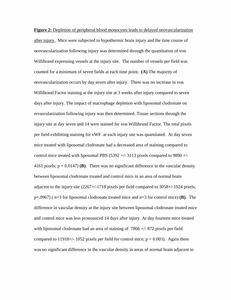
Figure 2: Depletion of peripheral blood monocytes leads to delayed neovascularization
after injury. Mice were subjected to hypothermic brain injury and the time course of
neovascularization following injury was determined through the quantitation of von
Willibrand expressing vessels at the injury site. The number of vessels per field was
counted for a minimum of seven fields at each time point. (A) The majority of
neovascularization occurs by day seven after injury. There was no increase in von
Willibrand Factor staining at the injury site at 3 weeks after injury compared to seven
days after injury. The impact of macrophage depletion with liposomal clodronate on
revascularization following injury was then determined. Tissue sections through the
injury site at day seven and 14 were stained for von Willibrand Factor. The total pixels
per field exhibiting staining for vWF at each injury site was quantitated. At day seven
mice treated with liposomal clodronate had a decreased area of staining compared to
control mice treated with liposomal PBS (5392 +/- 3113 pixels compared to 9890 +/-
4161 pixels; p = 0.0147) (B). There was no significant difference in the vascular density
between liposomal clodronate treated and control mice in an area of normal brain
adjacent to the injury site (2267+/-1718 pixels per field compared to 3058+/-1924 pixels;
p=.0967) ( n=3 for liposomal clodronate treated mice and n=3 for control mice) (B). The
difference in vascular density at the injury site between liposomal clodronate treated mice
and control mice was less pronounced 14 days after injury. At day fourteen mice treated
with liposomal clodronate had an area of staining of 7866 +/- 872 pixels per field
compared to 11918+/- 1052 pixels per field for control mice; p = 0.003). Again there
was no significant difference in the vascular density in areas of normal brain adjacent to

the injury site (6621+/-289 pixels per field compared to 5191+/-1448 pixels per field;
p=0.15) (n= 5 for mice treated with liposomal clodronate and n=3 for control mice) (C).
Figure 3: Reformation of the blood brain barrier is delayed in mice treated with
liposomal clodronate. At various time points following hypothermic brain injury animals
were injected with FITC-labeled albumin and the leakage of albumin in the injured
hemisphere was calculated. Animals were injected intravenously with FITC-labeled
albumin. One hour later animals were injected intravenously with rhodamine-labeled
dextran (average MW approximately 70,000) and immediately sacrificed. The brains
were bisected and homogenized and the amount of FITC-albumin present in the
circulation was calculated using a ratio of rhodamine to fluorescene in the non-injured
hemisphere. The amount of FITC-albumin that had leaked into the parenchyma of the
injured hemisphere was then calculated in arbitrary fluorescence units. (A) In normal
controls a peak of albumin leak was observed on day two following injury with the
integrity of the BBB returning to baseline levels by day seven. (B) At day three
following injury the leakage of FITC-albumin was the same in both the liposomal
clodronate treated and control animals (301 +/- 126 compared to 259 +/- 153; p=0.7). A
non-significant trend toward increased FITC-albumin leak was seen in the liposomal
clodronate treated animals at day 5. At seven days after injury there was continued
leakage of FITC-albumin in the clodronate treated animals and minimal leakage in the
control animals (314 +/- 51 compared to 22 +/- 70; p=0.005). (n=4 mice in each group)
References Abbott, N.J., (2000). Inflammatory mediators and modulation of blood-brain barrier
permeability. Cell Mol Neurobiol. 20, 131-47. Adams, S., Brown, H., Turner, G., (2002). Breaking down the blood-brain barrier:
signaling a path to cerebral malaria? Trends Parasitol. 18, 360-6. Annunziata, P., (2003). Blood-brain barrier changes during invasion of the central
nervous system by HIV-1. Old and new insights into the mechanism. J Neurol. 250, 901-6.
Carmeliet, P., Storkebaum, E., (2002). Vascular and neuronal effects of VEGF in the nervous system: implications for neurological disorders. Semin Cell Dev Biol. 13, 39-53.
Carmi, Y., Voronov, E., Dotan, S., Lahat, N., Rahat, M.A., Fogel, M., Huszar, M., White, M.R., Dinarello, C.A., Apte, R.N., (2009). The role of macrophage-derived IL-1 in induction and maintenance of angiogenesis. J Immunol. 183, 4705-14.
Croll, S.D., Ransohoff, R.M., Cai, N., Zhang, Q., Martin, F.J., Wei, T., Kasselman, L.J., Kintner, J., Murphy, A.J., Yancopoulos, G.D., Wiegand, S.J., (2004). VEGF-mediated inflammation precedes angiogenesis in adult brain. Exp Neurol. 187, 388-402.
Dinapoli, V.A., Huber, J.D., Houser, K., Li, X., Rosen, C.L., (2007). Early disruptions of the blood-brain barrier may contribute to exacerbated neuronal damage and prolonged functional recovery following stroke in aged rats. Neurobiol Aging.
Espinosa-Heidmann, D.G., Suner, I.J., Hernandez, E.P., Monroy, D., Csaky, K.G., Cousins, S.W., (2003). Macrophage depletion diminishes lesion size and severity in experimental choroidal neovascularization. Invest Ophthalmol Vis Sci. 44, 3586-92.
Fernandez Pujol, B., Lucibello, F.C., Gehling, U.M., Lindemann, K., Weidner, N., Zuzarte, M.L., Adamkiewicz, J., Elsasser, H.P., Muller, R., Havemann, K., (2000). Endothelial-like cells derived from human CD14 positive monocytes. Differentiation. 65, 287-300.
Fernandez Pujol, B., Lucibello, F.C., Zuzarte, M., Lutjens, P., Muller, R., Havemann, K., (2001). Dendritic cells derived from peripheral monocytes express endothelial markers and in the presence of angiogenic growth factors differentiate into endothelial-like cells. Eur J Cell Biol. 80, 99-110.
Ferrara, N., Gerber, H.P., (2001). The role of vascular endothelial growth factor in angiogenesis. Acta Haematol. 106, 148-56.
Floris, S., Blezer, E.L., Schreibelt, G., Dopp, E., van der Pol, S.M., Schadee-Eestermans, I.L., Nicolay, K., Dijkstra, C.D., de Vries, H.E., (2004). Blood-brain barrier permeability and monocyte infiltration in experimental allergic encephalomyelitis: a quantitative MRI study. Brain. 127, 616-27.
Fujiyama, S., Amano, K., Uehira, K., Yoshida, M., Nishiwaki, Y., Nozawa, Y., Jin, D., Takai, S., Miyazaki, M., Egashira, K., Imada, T., Iwasaka, T., Matsubara, H., (2003). Bone marrow monocyte lineage cells adhere on injured endothelium in a monocyte chemoattractant protein-1-dependent manner and accelerate reendothelialization as endothelial progenitor cells. Circ Res. 93, 980-9.
Giraudo, E., Inoue, M., Hanahan, D., (2004). An amino-bisphosphonate targets MMP-9-expressing macrophages and angiogenesis to impair cervical carcinogenesis. J Clin Invest. 114, 623-33.
Glod, J., Kobiler, D., Noel, M., Koneru, R., Lehrer, S., Medina, D., Maric, D., Fine, H.A., (2006). Monocytes form a vascular barrier and participate in vessel repair after brain injury. Blood. 107, 940-6.
Gray, M., Palispis, W., Popovich, P.G., van Rooijen, N., Gupta, R., (2007). Macrophage depletion alters the blood-nerve barrier without affecting Schwann cell function after neural injury. J Neurosci Res. 85, 766-77.
Harraz, M., Jiao, C., Hanlon, H.D., Hartley, R.S., Schatteman, G.C., (2001). CD34- blood-derived human endothelial cell progenitors. Stem Cells. 19, 304-12.
Havemann, K., Pujol, B.F., Adamkiewicz, J., (2003). In vitro transformation of monocytes and dendritic cells into endothelial like cells. Adv Exp Med Biol. 522, 47-57.
Heil, M., Ziegelhoeffer, T., Pipp, F., Kostin, S., Martin, S., Clauss, M., Schaper, W., (2002). Blood monocyte concentration is critical for enhancement of collateral artery growth. Am J Physiol Heart Circ Physiol. 283, H2411-9.
Hong, T.M., Teng, L.J., Shun, C.T., Peng, M.C., Tsai, J.C., (2009). Induced interleukin-8 expression in gliomas by tumor-associated macrophages. J Neurooncol. 93, 289-301.
Jellinger, K.A., (2002). Alzheimer disease and cerebrovascular pathology: an update. J Neural Transm. 109, 813-36.
Johnson, C., Sung, H.J., Lessner, S.M., Fini, M.E., Galis, Z.S., (2004). Matrix metalloproteinase-9 is required for adequate angiogenic revascularization of ischemic tissues: potential role in capillary branching. Circ Res. 94, 262-8.
Kniesel, U., Wolburg, H., (2000). Tight junctions of the blood-brain barrier. Cell Mol Neurobiol. 20, 57-76.
Ladewig, G., Jestaedt, L., Misselwitz, B., Solymosi, L., Toyka, K., Bendszus, M., Stoll, G., (2009). Spatial diversity of blood-brain barrier alteration and macrophage invasion in experimental autoimmune encephalomyelitis: a comparative MRI study. Exp Neurol. 220, 207-11.
Lee, H.S., Han, J., Bai, H.J., Kim, K.W., (2009). Brain angiogenesis in developmental and pathological processes: regulation, molecular and cellular communication at the neurovascular interface. FEBS J. 276, 4622-35.
Lu, D., Goussev, A., Chen, J., Pannu, P., Li, Y., Mahmood, A., Chopp, M., (2004). Atorvastatin reduces neurological deficit and increases synaptogenesis, angiogenesis, and neuronal survival in rats subjected to traumatic brain injury. J Neurotrauma. 21, 21-32.
Manoonkitiwongsa, P.S., Jackson-Friedman, C., McMillan, P.J., Schultz, R.L., Lyden, P.D., (2001). Angiogenesis after stroke is correlated with increased numbers of macrophages: the clean-up hypothesis. J Cereb Blood Flow Metab. 21, 1223-31.
Manoonkitiwongsa, P.S., Schultz, R.L., Whitter, E.F., Lyden, P.D., (2006). Contraindications of VEGF-based therapeutic angiogenesis: effects on macrophage density and histology of normal and ischemic brains. Vascul Pharmacol. 44, 316-25.

Mato, M., Ookawara, S., Sakamoto, A., Aikawa, E., Ogawa, T., Mitsuhashi, U., Masuzawa, T., Suzuki, H., Honda, M., Yazaki, Y., Watanabe, E., Luoma, J., Yla-Herttuala, S., Fraser, I., Gordon, S., Kodama, T., (1996). Involvement of specific macrophage-lineage cells surrounding arterioles in barrier and scavenger function in brain cortex. Proc Natl Acad Sci U S A. 93, 3269-74.
Mendes-Jorge, L., Ramos, D., Luppo, M., Llombart, C., Alexandre-Pires, G., Nacher, V., Melgarejo, V., Correia, M., Navarro, M., Carretero, A., Tafuro, S., Rodriguez-Baeza, A., Esperanca-Pina, J.A., Bosch, F., Ruberte, J., (2009). Scavenger function of resident autofluorescent perivascular macrophages and their contribution to the maintenance of the blood-retinal barrier. Invest Ophthalmol Vis Sci. 50, 5997-6005.
Moldovan, N.I., Goldschmidt-Clermont, P.J., Parker-Thornburg, J., Shapiro, S.D., Kolattukudy, P.E., (2000). Contribution of monocytes/macrophages to compensatory neovascularization: the drilling of metalloelastase-positive tunnels in ischemic myocardium. Circ Res. 87, 378-84.
Nag, S., (1996). Cold-injury of the cerebral cortex: immunolocalization of cellular proteins and blood-brain barrier permeability studies. J Neuropathol Exp Neurol. 55, 880-8.
Nag, S., (2002). The blood-brain barrier and cerebral angiogenesis: lessons from the cold-injury model. Trends Mol Med. 8, 38-44.
Nakul-Aquaronne, D., Bayle, J., Frelin, C., (2003). Coexpression of endothelial markers and CD14 by cytokine mobilized CD34+ cells under angiogenic stimulation. Cardiovasc Res. 57, 816-23.
Nourhaghighi, N., Teichert-Kuliszewska, K., Davis, J., Stewart, D.J., Nag, S., (2003). Altered expression of angiopoietins during blood-brain barrier breakdown and angiogenesis. Lab Invest. 83, 1211-22.
Ping, A., Chun, Z.X., Xue, X.Y., (2005). Bradykinin preconditioning induces protective effects against focal cerebral ischemia in rats. Brain Res. 1059, 105-12.
Rehman, J., Li, J., Orschell, C.M., March, K.L., (2003). Peripheral blood "endothelial progenitor cells" are derived from monocyte/macrophages and secrete angiogenic growth factors. Circulation. 107, 1164-9.
Rubin, L.L., Staddon, J.M., (1999). The cell biology of the blood-brain barrier. Annu Rev Neurosci. 22, 11-28.
Sakurai, E., Anand, A., Ambati, B.K., van Rooijen, N., Ambati, J., (2003). Macrophage depletion inhibits experimental choroidal neovascularization. Invest Ophthalmol Vis Sci. 44, 3578-85.
Schmeisser, A., Garlichs, C.D., Zhang, H., Eskafi, S., Graffy, C., Ludwig, J., Strasser, R.H., Daniel, W.G., (2001). Monocytes coexpress endothelial and macrophagocytic lineage markers and form cord-like structures in Matrigel under angiogenic conditions. Cardiovasc Res. 49, 671-80.
Shechter, R., London, A., Varol, C., Raposo, C., Cusimano, M., Yovel, G., Rolls, A., Mack, M., Pluchino, S., Martino, G., Jung, S., Schwartz, M., (2009). Infiltrating blood-derived macrophages are vital cells playing an anti-inflammatory role in recovery from spinal cord injury in mice. PLoS Med. 6, e1000113.
Skold, M.K., Risling, M., Holmin, S., (2006). Inhibition of vascular endothelial growth factor receptor 2 activity in experimental brain contusions aggravates injury

outcome and leads to early increased neuronal and glial degeneration. Eur J Neurosci. 23, 21-34.
Sun, Y., Jin, K., Xie, L., Childs, J., Mao, X.O., Logvinova, A., Greenberg, D.A., (2003). VEGF-induced neuroprotection, neurogenesis, and angiogenesis after focal cerebral ischemia. J Clin Invest. 111, 1843-51.
Ujiie, M., Dickstein, D.L., Carlow, D.A., Jefferies, W.A., (2003). Blood-brain barrier permeability precedes senile plaque formation in an Alzheimer disease model. Microcirculation. 10, 463-70.
Van Rooijen, N., Sanders, A., (1994). Liposome mediated depletion of macrophages: mechanism of action, preparation of liposomes and applications. J Immunol Methods. 174, 83-93.
van Rooijen, N., Sanders, A., van den Berg, T.K., (1996). Apoptosis of macrophages induced by liposome-mediated intracellular delivery of clodronate and propamidine. J Immunol Methods. 193, 93-9.
Widenfalk, J., Lipson, A., Jubran, M., Hofstetter, C., Ebendal, T., Cao, Y., Olson, L., (2003). Vascular endothelial growth factor improves functional outcome and decreases secondary degeneration in experimental spinal cord contusion injury. Neuroscience. 120, 951-60.
Willis, C.L., Garwood, C.J., Ray, D.E., (2007). A size selective vascular barrier in the rat area postrema formed by perivascular macrophages and the extracellular matrix. Neuroscience. 150, 498-509.
Zenker, D., Begley, D., Bratzke, H., Rubsamen-Waigmann, H., von Briesen, H., (2003). Human blood-derived macrophages enhance barrier function of cultured primary bovine and human brain capillary endothelial cells. J Physiol. 551, 1023-32.