manual: pcmv-tag epitope tagging mammalian expression · pdf filepcmv-tag 2 vector map ......
TRANSCRIPT
pCMV-Tag Epitope Tagging Mammalian Expression Vectors
INSTRUCTION MANUAL Catalog #211172 (pCMV-Tag 2), #211173 (pCMV-Tag 3), #211174 (pCMV-Tag 4), and
#211175 (pCMV-Tag 5)
Revision A
For In Vitro Use Only 211172-12
LIMITED PRODUCT WARRANTY This warranty limits our liability to replacement of this product. No other warranties of any kind, express or implied, including without limitation, implied warranties of merchantability or fitness for a particular purpose, are provided by Agilent. Agilent shall have no liability for any direct, indirect, consequential, or incidental damages arising out of the use, the results of use, or the inability to use this product.
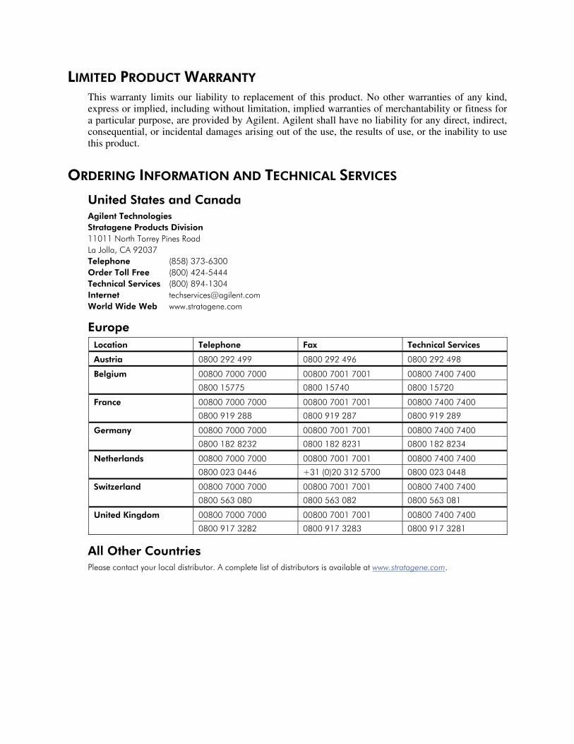
ORDERING INFORMATION AND TECHNICAL SERVICES
United States and Canada Agilent Technologies Stratagene Products Division 11011 North Torrey Pines Road La Jolla, CA 92037 Telephone (858) 373-6300 Order Toll Free (800) 424-5444 Technical Services (800) 894-1304 Internet [email protected] World Wide Web www.stratagene.com
Europe Location Telephone Fax Technical Services
Austria 0800 292 499 0800 292 496 0800 292 498
00800 7000 7000 00800 7001 7001 00800 7400 7400 Belgium
0800 15775 0800 15740 0800 15720
00800 7000 7000 00800 7001 7001 00800 7400 7400 France
0800 919 288 0800 919 287 0800 919 289
00800 7000 7000 00800 7001 7001 00800 7400 7400 Germany
0800 182 8232 0800 182 8231 0800 182 8234
00800 7000 7000 00800 7001 7001 00800 7400 7400 Netherlands
0800 023 0446 +31 (0)20 312 5700 0800 023 0448
00800 7000 7000 00800 7001 7001 00800 7400 7400 Switzerland
0800 563 080 0800 563 082 0800 563 081
00800 7000 7000 00800 7001 7001 00800 7400 7400 United Kingdom
0800 917 3282 0800 917 3283 0800 917 3281
All Other Countries Please contact your local distributor. A complete list of distributors is available at www.stratagene.com.

pCMV-Tag Epitope Tagging Mammalian Expression Vectors
CONTENTS Materials Provided.............................................................................................................................. 1 Storage Conditions.............................................................................................................................. 1 Additional Materials Required .......................................................................................................... 2 Notices to Purchaser ........................................................................................................................... 3 Introduction......................................................................................................................................... 4
pCMV-Tag 2 Vector Map ..................................................................................................... 5 pCMV-Tag 3 Vector Map ..................................................................................................... 6 pCMV-Tag 4 Vector Map ..................................................................................................... 7 pCMV-Tag 5 Vector Map ..................................................................................................... 8
Preparation of Host Strains.............................................................................................................. 10 Reviving the Host Strain ..................................................................................................... 10 Preparing a –80°C Bacterial Glycerol Stock ....................................................................... 10
Preparing the pCMV-Tag Vectors .................................................................................................. 11 Ligating the Insert............................................................................................................................. 12 Transformation ................................................................................................................................. 13 Verification of Insert Percentage, Size, and Orientation............................................................... 13
Polymerase Chain Reaction Amplification of DNA from Individual Colonies .................. 13 Troubleshooting ................................................................................................................................ 15 Preparation of Media and Reagents ................................................................................................ 16 References .......................................................................................................................................... 17 Endnotes............................................................................................................................................. 17 MSDS Information............................................................................................................................ 17

pCMV-Tag Epitope Tagging Mammalian Expression Vectors 1
pCMV-Tag Epitope Tagging Mammalian Expression Vectors
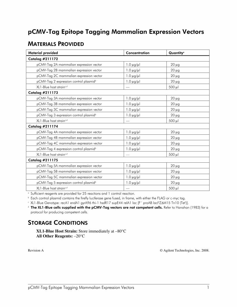
MATERIALS PROVIDED Material provided Concentration Quantitya
Catalog #211172
pCMV-Tag 2A mammalian expression vector 1.0 μg/μl 20 μg
pCMV-Tag 2B mammalian expression vector 1.0 μg/μl 20 μg
pCMV-Tag 2C mammalian expression vector 1.0 μg/μl 20 μg
pCMV-Tag 2 expression control plasmidb 1.0 μg/μl 20 μg
XL1-Blue host strainc,d — 500 μl
Catalog #211173
pCMV-Tag 3A mammalian expression vector 1.0 μg/μl 20 μg
pCMV-Tag 3B mammalian expression vector 1.0 μg/μl 20 μg
pCMV-Tag 3C mammalian expression vector 1.0 μg/μl 20 μg
pCMV-Tag 3 expression control plasmidb 1.0 μg/μl 20 μg
XL1-Blue host strainc,d — 500 μl
Catalog #211174
pCMV-Tag 4A mammalian expression vector 1.0 μg/μl 20 μg
pCMV-Tag 4B mammalian expression vector 1.0 μg/μl 20 μg
pCMV-Tag 4C mammalian expression vector 1.0 μg/μl 20 μg
pCMV-Tag 4 expression control plasmidb 1.0 μg/μl 20 μg
XL1-Blue host strainc,d — 500 μl
Catalog #211175
pCMV-Tag 5A mammalian expression vector 1.0 μg/μl 20 μg
pCMV-Tag 5B mammalian expression vector 1.0 μg/μl 20 μg
pCMV-Tag 5C mammalian expression vector 1.0 μg/μl 20 μg
pCMV-Tag 5 expression control plasmidb 1.0 μg/μl 20 μg
XL1-Blue host strainc,d — 500 μl a Sufficient reagents are provided for 25 reactions and 1 control reaction. b Each control plasmid contains the firefly luciferase gene fused, in frame, with either the FLAG or c-myc tag. c XL1-Blue Genotype: recA1 endA1 gyrA96 thi-1 hsdR17 supE44 relA1 lac [F´ proAB lacIqZΔM15 Tn10 (Tetr)]. d The XL1-Blue cells supplied with the pCMV-Tag vectors are not competent cells. Refer to Hanahan (1983) for a
protocol for producing competent cells.
STORAGE CONDITIONS XL1-Blue Host Strain: Store immediately at –80°C All Other Reagents: –20°C
Revision A © Agilent Technologies, Inc. 2008.

2 pCMV-Tag Epitope Tagging Mammalian Expression Vectors
ADDITIONAL MATERIALS REQUIRED T4 DNA ligase Taq DNA polymerase Taq DNA polymerase buffer TE buffer§
CIAP
Falcon® 2059 polypropylene tubes (15-ml) Water baths (37°C and 42°C)

pCMV-Tag Epitope Tagging Mammalian Expression Vectors 3
NOTICES TO PURCHASER
CMV Promoter The use of the CMV Promoter is covered under U.S. Patent Nos. 5,168,062 and 5,385,839 owned by the University of Iowa Research Foundation and licensed FOR RESEARCH USE ONLY. For further information, please contact UIRF at 319-335-4546.
FLAG® License Agreement The enclosed DNA expression vector and/or antibody are specifically adpated for a method of producing selected protein molecules covered by one or more of the following patents owned by Sigma-Aldrich Co.: U.S. Patent Nos. (5,011912, 4,703,004, 4,782,137 and 4,851,341;EP Patent No. 150,126 (Austria, Belgium, Switzerland, France, United Kingdom, Italy, Netherlands and Sweden); EP Patent No. 335,899 (Belgium, Switzerland, Germany, France, United Kingdom, Italy, Luxembourg and Sweden); German Patent No. P3584260.1; Canadian Patent No. 1,307,752; and Japanese Patent Nos. 1,983,150 and 2,665,359. Your payment includes a limited license under these patents to make only the following uses of these products:
A. Vector License: You may use the enclosed vector to transform cells to produce proteins containing the amino acid sequence DYKDDDDK for research purposes provided, however, such research purposes do not include binding an unlicensed antibody to any portion of this amino acid sequence nor using such proteins for the preparation of antibodies having an affinity for any portion of this amino acid sequence. B. Antibody License: You may only use the enclosed antibody for research purposes to perform a method of producing a protein in which the protein is expressed in a host cell and purified by use of the antibody in accordance with a claim in one of the above patents in force in a country where the use actually occurs so long as: (1) you perform such method with a DNA expression vector licensed from Sigma-Aldrich Co.; and (2) you do not bind (or allow others to bind) an unlicensed antibody to any DYKDDDDK epitope of any fusion protein that is produced by use of the method.
This license does not include any rights under any other patents. You are not licensed to use the vector and/or antibody in any manner or for any purposed not recited above. As used above, the term “unlicensed antibody” means any antibody which Sigma-Aldrich Co. has not expressly licensed pursuant to Paragraph B, above. Sigma-Aldrich Co. hereby expressly retains all rights in the above listed patents not expressly licensed hereunder. If the terms and conditions of this License Agreement are acceptable to you, then you may open the vessel(s) containing the vector and/or antibody and, through such act of opening a vessel, will have shown your acceptance to these terms and conditions. If the terms and conditions of this License Agreement are not acceptable to you, then please return the vessel(s) unopened to Stratagene for a complete refund of your payment. For additional licensing information or to receive a copy of any of the above patents, please contact the Sigma-Aldrich Co. licensing department at telephone number 314-771-5765.

4 pCMV-Tag Epitope Tagging Mammalian Expression Vectors
INTRODUCTION The epitope tagging technique involves fusion of a protein of interest to a peptide epitope that is recognized by a readily available antibody. With this technique, expression of the fusion protein is monitored using a tag-specific antibody, allowing a new protein to be studied without generating a new, specific antibody to that protein. Epitope tagging can be used to localize gene products in living cells, identify associated proteins, track the movement of fusion proteins within the cell, or characterize new proteins by immunoprecipitation. The Stratagene vectors pCMV-Tag 2, pCMV-Tag 3, pCMV-Tag 4, and pCMV-Tag 5 are a series of epitope tagging mammalian expression vectors. pCMV-Tag 2 (Figure 1) is an N-terminal FLAG® tagging vector, pCMV-Tag 3 (Figure 2) is an N-terminal c-myc tagging vector, pCMV-Tag 4 (Figure 3) is a C-terminal FLAG tagging vector and pCMV-Tag 5 (Figure 4) is a C-terminal c-myc tagging vector. Each vector is available in three different reading frames to simplify subcloning. These reading frames, designated as A, B, and C, differ only by one or two bases. Thus, each pCMV-Tag vector has a reading frame that will allow cloning a gene of interest so that it is fused correctly with the epitope tag. Tagged constructs generated in the pCMV-Tag vectors can be transfected into mammalian cells and the fusion protein can be easily characterized using commercially available antibodies. The pCMV-Tag vectors are derived from the pCMV-Script vector and contain sequences for either the FLAG or c-myc epitope at either the N or C terminus. These specific epitope tags are small, highly immunoreactive, and are not likely to interfere with the function of the target protein. The synthetic FLAG epitope is composed of eight amino acid residues (DYKDDDDK).1 The c-myc epitope is derived from the human c-myc gene and contains ten amino acid residues (EQKLISEEDL).2 In addition to the epitope tag sequences, the pCMV-Tag vectors contain features for expression of fusion proteins in eukaryotic cells. The cytomegalovirus (CMV) promoter allows constitutive expression of the cloned DNA in a wide variety of mammalian cell lines. The neomycin-resistance gene is under control of both the prokaryotic β-lactamase promoter to provide kanamycin resistance in bacteria and the SV40 early promoter to provide G418 resistance in mammalian cells. The multiple cloning site (MCS) of the pCMV-Tag vectors allows for a variety of cloning strategies, resulting in either C-terminal or N-terminal fusions with either FLAG or c-myc. A Kozak consensus sequence provides optimal expression of the fusion protein when the N-terminal FLAG epitope is used.3 Other cloning options, which require fusion proteins to include their own translational start sequence, are also possible. The relative locations of the features for each vector are listed in Table 1. The pCMV-Tag 2, pCMV-Tag 3, pCMV-Tag 4, and pCMV-Tag 5 expression control plasmids are included for use as positive controls to confirm that the CMV promoter is active in the cell line used for transfection. Each control plasmid contains the firefly luciferase gene fused, in reading frame, with either the FLAG or c-myc tag.

pCMV-Tag Epitope Tagging Mammalian Expression Vectors 5
pCMV-Tag 2 Vector Map
Feature Nucleotide Position
CMV promoter 1–602
T3 promoter and T3 primer binding site [5´ AATTAACCCTCACTAAAGGG 3´] 620–639
FLAG tag 682–705
multiple cloning site 706–779
T7 promoter and T7 primer binding site [3´ CGGGATATCACTCAGCATAATG 5´] 823–844
SV40 polyA signal 856–1239
f1 origin of ss-DNA replication 1377–1683
bla promoter 1708–1832
SV40 promoter 1852–2190
neomycin/kanamycin resistance ORF 2225–3016
HSV-thymidine kinase (TK) polyA signal 3017–3475
pUC origin 3604–4271
FIGURE 1 Circular map of the pCMV-Tag 2A–2C vectors, featuring eukaryotic expression, FLAG epitope tagging, neomycin and kanamycin resistance, T3 and T7 RNA promoters, and single-stranded rescue. The positions listed in the table above correspond to pCMV-Tag 2A. See Table 1 for the complete list of feature positions for the pCMV-Tag 2A–2C vectors.
pUC ori
f1 orineo/kan
P CMV
P blaP SV40
TK pA SV40 pA
FLAGMCS
pCMV-Tag 24.3 kb
pCMV-Tag 2 Multiple Cloning Site Region(sequence shown 620–843)
Acc I/Sal I Xho IHind IIIEcoR V
...G ATA TCA AGC TTA TCG ATA CCG TCG ACC TCG AGG GGG GGC CCG GTA CCT...
Apa I
AA TTA ACC CTC ACT AAA GGG AAC AAA AGC TGG AGC TCC ACC GCG GTG GCG GCC GCC ACC ATG...MT3 promoter
KOZAK
* In pCMV-Tag 2A, no bases inserted; in in pCMV-Tag 2B, A inserted; pCMV-Tag 2C, AA inserted
BamH I EcoR ISrf I Pst I
...GAT TAC AAG GAT GAC GAC GAT AAG * GCC CGG GCG GAT CCC CCG GGC TGC AGG AAT TC...Y K DD D K
FLAG tag
DD
...TAATTAATTAAGGTACCAGGTAAGTGTACCCAATTCGCCCTATAGTGAGTCGTATTA
MULTIPLE STOP CODONS
T7 promoter

6 pCMV-Tag Epitope Tagging Mammalian Expression Vectors
pCMV-Tag 3 Vector Map
Feature Nucleotide Position
CMV promoter 1–602
T3 promoter and T3 primer binding site [5´ AATTAACCCTCACTAAAGGG 3´] 620–639
c-myc tag 679–708
multiple cloning site 709–782
T7 promoter and T7 primer binding site [3´ CGGGATATCACTCAGCATAATG 5´] 826–847
SV40 polyA signal 859–1242
f1 origin of ss-DNA replication 1380–1686
bla promoter 1711–1835
SV40 promoter 1855–2193
neomycin/kanamycin resistance ORF 2228–3019
HSV-thymidine kinase (TK) polyA signal 3020–3478
pUC origin 3607–4274
FIGURE 2 Circular map of the pCMV-Tag 3A–3C vectors, featuring eukaryotic expression, myc epitope tagging, neomycin and kanamycin resistance, T3 and T7 RNA promoters, and single-stranded rescue. The positions listed in the table above correspond to pCMV-Tag 3A. See Table 1 for the complete list of feature positions for the pCMV-Tag 3A–3C vectors.
pUC ori
f1 orineo/kan
P CMV
P blaP SV40
TK pA SV40 pA
mycMCS
pCMV-Tag 34.3 kb
pCMV-Tag 3 Multiple Cloning Site Region(sequence shown 620–846)
EcoR I
BamH ISrf I Pst I
...GAG CAG AAA CTC ATC TCT GAA GAG GAT CTG * GCC CGG GCG GAT CCC CCG GGC TGC ...Q K SL E
myc tag
IE
...AGG AAT TCG ATA TCA AGC TTA TCG ATA CCG TCG ACC TCG AGG GGG GGC CCG GTA CCT...
Xho IHind IIIEcoR V Apa IAcc I/Sal I
AA TTA ACC CTC ACT AAA GGG AAC AAA AGC TGG AGC TCC ACC GCG GTG GCG GCC GCC ATG...M
KOZAK
T3 promoter
...TAATTAATTAAGGTACCAGGTAAGTGTACCCAATTCGCCCTATAGTGAGTCGTATTA
MULTIPLE STOP CODONS
T7 promoter
* In pCMV-Tag 3A, no bases inserted; in in pCMV-Tag 3B, A inserted; pCMV-Tag 3C, AA inserted
E LD

pCMV-Tag Epitope Tagging Mammalian Expression Vectors 7
pCMV-Tag 4 Vector Map
Feature Nucleotide Position
CMV promoter 1–602
T3 promoter and T3 primer binding site [5´ AATTAACCCTCACTAAAGGG 3´] 620–639
multiple cloning site 651–743
FLAG tag 744–767
T7 promoter and T7 primer binding site [3´ CGGGATATCACTCAGCATAATG 5´] 819–840
SV40 polyA signal 852–1235
f1 origin of ss-DNA replication 1373–1679
bla promoter 1704–1828
SV40 promoter 1848–2186
neomycin/kanamycin resistance ORF 2221–3012
HSV-thymidine kinase (TK) polyA signal 3013–3471
pUC origin 3600–4267
FIGURE 3 Circular map of the pCMV-Tag 4A–4C vectors, featuring eukaryotic expression, FLAG epitope tagging, neomycin and kanamycin resistance, T3 and T7 RNA promoters, and single-stranded rescue. The positions listed in the table above correspond to pCMV-Tag 4A. See Table 1 for the complete list of feature positions for the pCMV-Tag 4A–4C vectors.
pUC ori
f1 orineo/kan
P CMV
P blaP SV40
TK pA SV40 pA
FLAGMCS
pCMV-Tag 44.3 kb
pCMV-Tag 4 Multiple Cloning Site Region(sequence shown 620–839)
Acc I/Sal I
T3 promoterA ATT AAC CCT CAC TAA AGG GAA CAA AAG CTG GAG CTC CAC CGC GGT GGC GGC CGC TCT A...
BamH I EcoR I
...GC CCG GGC GGA TCC CCC GGG CTG CAG GAA TTC GAT ATC AAG CTT ATC GAT ACC GTC GAC*...
BstX I
Y K DD D KFLAG tag
DD...CTC GAG GAT TAC AAG GAT GAC GAC GAT AAG TAG GGCCCGGTACCT...
Xho I
Srf I
Sac II
Pst I
Not I
Hind IIIEcoR V
Sac I
...TAATTAATTAAGGTACCAGGTAAGTGTACCCAATTCGCCCTATAGTGAGTCGTATTA
MULTIPLE STOP CODONS
T7 promoter
STOP
* In pCMV-Tag 4A, no bases inserted; in in pCMV-Tag 4B, A inserted; pCMV-Tag 4C, AA inserted

8 pCMV-Tag Epitope Tagging Mammalian Expression Vectors
pCMV-Tag 5 Vector Map
Feature Nucleotide Position
CMV promoter 1–602
T3 promoter and T3 primer binding site [5´ AATTAACCCTCACTAAAGGG 3´] 620–639
multiple cloning site 651–743
c-myc tag 741–770
T7 promoter and T7 primer binding site [3´ CGGGATATCACTCAGCATAATG 5´] 822–843
SV40 polyA signal 855–1238
f1 origin of ss-DNA replication 1376–1682
bla promoter 1707–1831
SV40 promoter 1851–2189
neomycin/kanamycin resistance ORF 2224–3015
HSV-thymidine kinase (TK) polyA signal 3016–3474
pUC origin 3603–4270
FIGURE 4 Circular map of the pCMV-Tag 5A–5C vectors, featuring eukaryotic expression, myc epitope tagging, neomycin and kanamycin resistance, T3 and T7 RNA promoters, and single-stranded rescue. The positions listed in the table above correspond to pCMV-Tag 5A. See Table 1 for the complete list of feature positions for the pCMV-Tag 5A–5C vectors.
pUC ori
f1 orineo/kan
P CMV
P blaP SV40
TK pA SV40 pA
mycMCS
pCMV-Tag 54.3 kb
pCMV-Tag 5 Multiple Cloning Site Region(sequence shown 620–842)
T3 promoterA ATT AAC CCT CAC TAA AGG GAA CAA AAG CTG GAG CTC CAC CGC GGT GGC GGC CGC TCT A...
BstX I Sac II Not I
Acc I/Sal IBamH I EcoR I
...GC CCG GGC GGA TCC CCC GGG CTG CAG GAA TTC GAT ATC AAG CTT ATC GAT ACC GTC GAC*...
Srf I Pst I Hind IIIEcoR V
Sac I
...CTC GAG CAG AAA CTC ATC TCT GAA GAG GAT CTG TAG GGCCCGGTACCT...
Xho IQ K SL E L
myc tag
IE DE
...TAATTAATTAAGGTACCAGGTAAGTGTACCCAATTCGCCCTATAGTGAGTCGTATTAMULTIPLE STOP CODONS
T7 promoter
STOP
* In pCMV-Tag 5A, no bases inserted; in in pCMV-Tag 5B, A inserted; pCMV-Tag 5C, AA inserted
pCMV-Tag Epitope Tagging Mammalian Expression Vectors 9
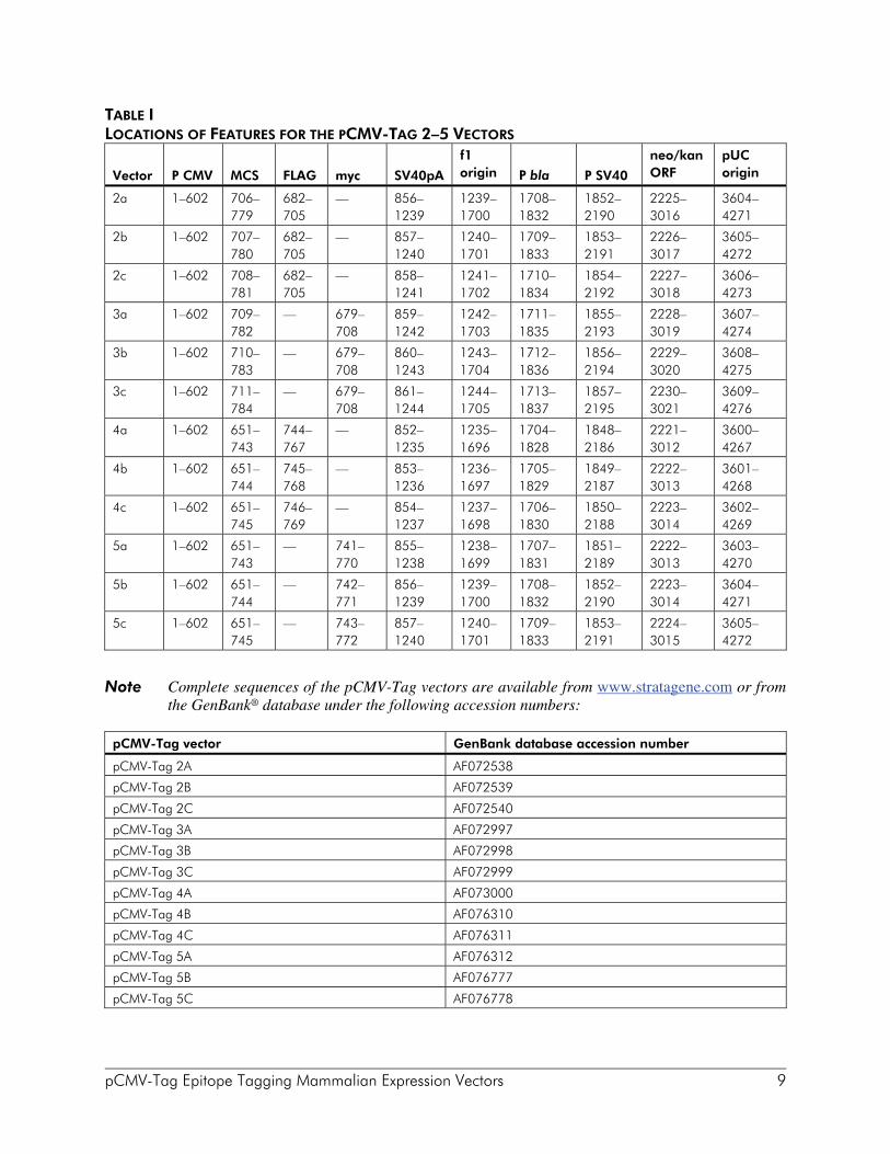
TABLE I LOCATIONS OF FEATURES FOR THE PCMV-TAG 2–5 VECTORS
Vector
P CMV
MCS
FLAG
myc
SV40pA
f1 origin
P bla
P SV40
neo/kan ORF
pUC origin
2a 1–602 706–779
682–705
— 856–1239
1239–1700
1708–1832
1852–2190
2225–3016
3604–4271
2b 1–602 707–780
682–705
— 857–1240
1240–1701
1709–1833
1853–2191
2226–3017
3605–4272
2c 1–602 708–781
682–705
— 858–1241
1241–1702
1710–1834
1854–2192
2227–3018
3606–4273
3a 1–602 709–782
— 679–708
859–1242
1242–1703
1711–1835
1855–2193
2228–3019
3607–4274
3b 1–602 710–783
— 679–708
860–1243
1243–1704
1712–1836
1856–2194
2229–3020
3608–4275
3c 1–602 711–784
— 679–708
861–1244
1244–1705
1713–1837
1857–2195
2230–3021
3609–4276
4a 1–602 651–743
744–767
— 852–1235
1235–1696
1704–1828
1848–2186
2221–3012
3600–4267
4b 1–602 651–744
745–768
— 853–1236
1236–1697
1705–1829
1849–2187
2222–3013
3601–4268
4c 1–602 651–745
746–769
— 854–1237
1237–1698
1706–1830
1850–2188
2223–3014
3602–4269
5a 1–602 651–743
— 741–770
855–1238
1238–1699
1707–1831
1851–2189
2222–3013
3603–4270
5b 1–602 651–744
— 742–771
856–1239
1239–1700
1708–1832
1852–2190
2223–3014
3604–4271
5c 1–602 651–745
— 743–772
857–1240
1240–1701
1709–1833
1853–2191
2224–3015
3605–4272
Note Complete sequences of the pCMV-Tag vectors are available from www.stratagene.com or from
the GenBank® database under the following accession numbers:
pCMV-Tag vector GenBank database accession number
pCMV-Tag 2A AF072538
pCMV-Tag 2B AF072539
pCMV-Tag 2C AF072540
pCMV-Tag 3A AF072997
pCMV-Tag 3B AF072998
pCMV-Tag 3C AF072999
pCMV-Tag 4A AF073000
pCMV-Tag 4B AF076310
pCMV-Tag 4C AF076311
pCMV-Tag 5A AF076312
pCMV-Tag 5B AF076777
pCMV-Tag 5C AF076778

10 pCMV-Tag Epitope Tagging Mammalian Expression Vectors
PREPARATION OF HOST STRAINS
Reviving the Host Strain
Note The host strain may thaw during shipment. The vial should be stored immediately at –20° or –80°C, but the strain remains viable longer if stored at –80°C. Avoid repeated freeze–thaw cycles to maintain extended viability.
1. Revive the stored cells by scraping off splinters of solid ice with a sterile wire loop.
2. Streak the splinters onto an LB-tetracycline agar plate.§
3. Incubate the plate overnight at 37°C.
4. Seal the plate with Parafilm® laboratory film and store the plate at 4°C for up to 1 week.
5. Restreak the colonies onto a fresh plate every week.
Preparing a –80°C Bacterial Glycerol Stock
1. In a sterile 50-ml conical tube, inoculate 10 ml of LB-tetracycline broth§ with one or two colonies from the plate. Grow the cells to late log phase (OD600 = 0.8–1.0).
2. Add 4.5 ml of a sterile glycerol–LB solution (5 ml of glycerol + 5 ml of LB broth) to the bacterial culture from step 1. Mix well.
3. Aliquot into sterile centrifuge tubes (1 ml/tube). This preparation may be stored at –20°C for 1–2 years or at –80°C for more than 2 years. § See Preparation of Media and Reagents.
pCMV-Tag Epitope Tagging Mammalian Expression Vectors 11
PREPARING THE pCMV-TAG VECTORS
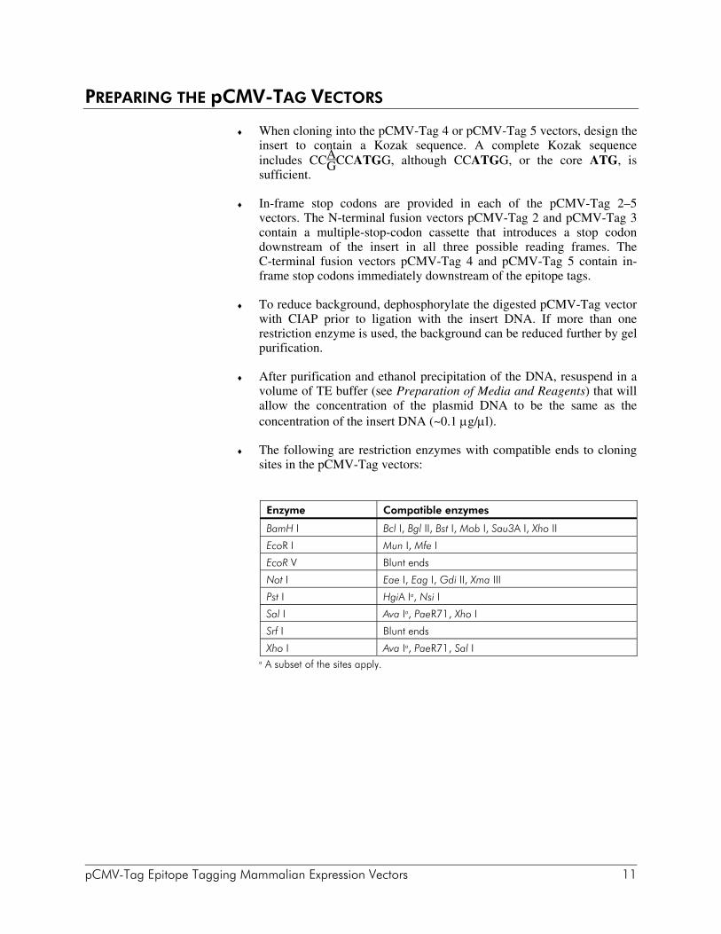
♦ When cloning into the pCMV-Tag 4 or pCMV-Tag 5 vectors, design the insert to contain a Kozak sequence. A complete Kozak sequence includes CCG
ACCATGG, although CCATGG, or the core ATG, is sufficient.
♦ In-frame stop codons are provided in each of the pCMV-Tag 2–5 vectors. The N-terminal fusion vectors pCMV-Tag 2 and pCMV-Tag 3 contain a multiple-stop-codon cassette that introduces a stop codon downstream of the insert in all three possible reading frames. The C-terminal fusion vectors pCMV-Tag 4 and pCMV-Tag 5 contain in-frame stop codons immediately downstream of the epitope tags.
♦ To reduce background, dephosphorylate the digested pCMV-Tag vector with CIAP prior to ligation with the insert DNA. If more than one restriction enzyme is used, the background can be reduced further by gel purification.
♦ After purification and ethanol precipitation of the DNA, resuspend in a volume of TE buffer (see Preparation of Media and Reagents) that will allow the concentration of the plasmid DNA to be the same as the concentration of the insert DNA (~0.1 μg/μl).
♦ The following are restriction enzymes with compatible ends to cloning sites in the pCMV-Tag vectors:
Enzyme Compatible enzymes
BamH I Bcl I, Bgl II, Bst I, Mob I, Sau3A I, Xho II
EcoR I Mun I, Mfe I
EcoR V Blunt ends
Not I Eae I, Eag I, Gdi II, Xma III
Pst I HgiA Ia, Nsi I
Sal I Ava Ia, PaeR71, Xho I
Srf I Blunt ends
Xho I Ava Ia, PaeR71, Sal I a A subset of the sites apply.
12 pCMV-Tag Epitope Tagging Mammalian Expression Vectors
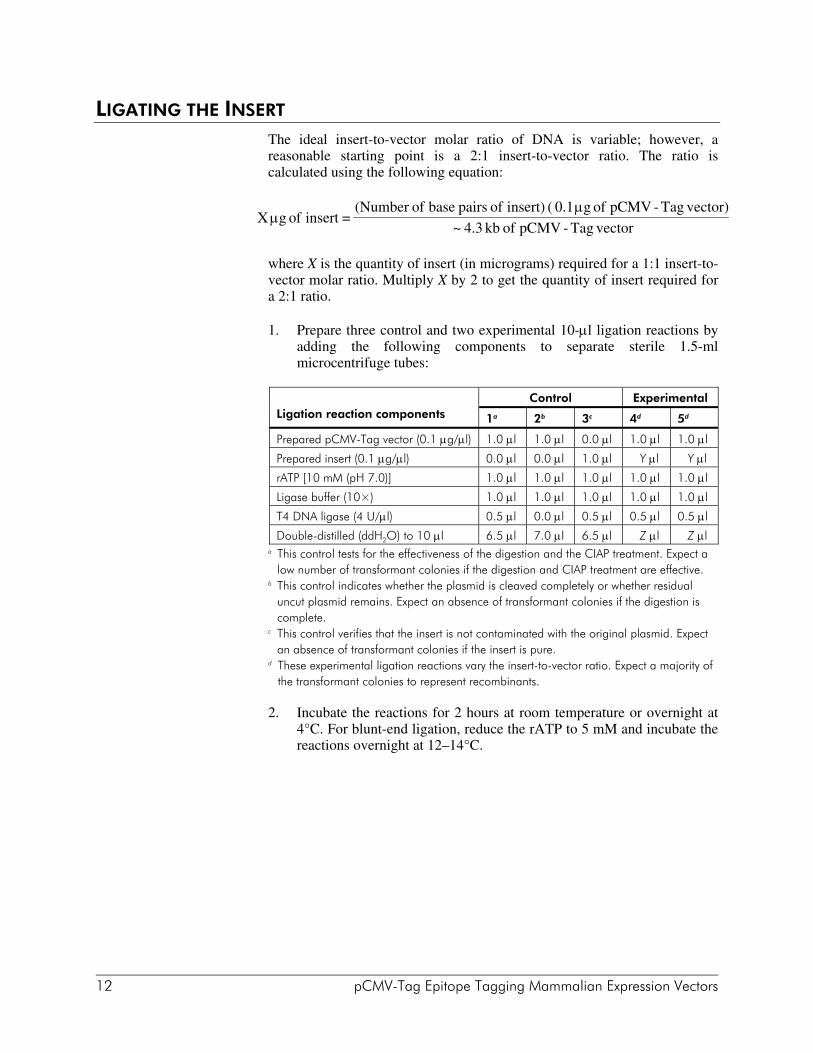
LIGATING THE INSERT The ideal insert-to-vector molar ratio of DNA is variable; however, a reasonable starting point is a 2:1 insert-to-vector ratio. The ratio is calculated using the following equation:
where X is the quantity of insert (in micrograms) required for a 1:1 insert-to-vector molar ratio. Multiply X by 2 to get the quantity of insert required for a 2:1 ratio.
1. Prepare three control and two experimental 10-μl ligation reactions by adding the following components to separate sterile 1.5-ml microcentrifuge tubes:
Control Experimental
Ligation reaction components 1a 2b 3c 4d 5d
Prepared pCMV-Tag vector (0.1 μg/μl) 1.0 μl 1.0 μl 0.0 μl 1.0 μl 1.0 μl
Prepared insert (0.1 μg/μl) 0.0 μl 0.0 μl 1.0 μl Y μl Y μl
rATP [10 mM (pH 7.0)] 1.0 μl 1.0 μl 1.0 μl 1.0 μl 1.0 μl
Ligase buffer (10×) 1.0 μl 1.0 μl 1.0 μl 1.0 μl 1.0 μl
T4 DNA ligase (4 U/μl) 0.5 μl 0.0 μl 0.5 μl 0.5 μl 0.5 μl
Double-distilled (ddH2O) to 10 μl 6.5 μl 7.0 μl 6.5 μl Z μl Z μl a This control tests for the effectiveness of the digestion and the CIAP treatment. Expect a
low number of transformant colonies if the digestion and CIAP treatment are effective. b This control indicates whether the plasmid is cleaved completely or whether residual
uncut plasmid remains. Expect an absence of transformant colonies if the digestion is complete.
c This control verifies that the insert is not contaminated with the original plasmid. Expect an absence of transformant colonies if the insert is pure.
d These experimental ligation reactions vary the insert-to-vector ratio. Expect a majority of the transformant colonies to represent recombinants.
2. Incubate the reactions for 2 hours at room temperature or overnight at 4°C. For blunt-end ligation, reduce the rATP to 5 mM and incubate the reactions overnight at 12–14°C.
X g of insert = (Number of base pairs of insert) ( 0.1 g of pCMV - Tag vector)
~ 4.3 kb of pCMV - Tag vectorμ
μ

pCMV-Tag Epitope Tagging Mammalian Expression Vectors 13
TRANSFORMATION Transform competent bacteria with 1–2 μl of the ligation reaction, and plate the transformed bacteria on LB-kanamycin agar plates (see Preparation of Media and Reagents). Please see reference 4 for a transformation protocol.
Note The XL1-Blue cells supplied with the pCMV-Tag vectors are not competent cells. Refer to Hanahan (1983) for a protocol for producing competent cells.4 (Competent cells with transformation efficiencies ≥5 × 10⁹ cfu/μg are available from the Stratagene Products Division.)
VERIFICATION OF INSERT PERCENTAGE, SIZE, AND ORIENTATION Individual colonies can be examined to determine the percentage of vectors with inserts and the insert size and orientation by PCR directly from the colony or by restriction analysis.
Polymerase Chain Reaction Amplification of DNA from Individual Colonies
The presence and size of a DNA insert in a pCMV-Tag vector may be determined by PCR amplification of DNA from individual colonies.
1. Prepare a PCR amplification reaction containing the following components:
4.0 μl of 10× Taq DNA polymerase buffer 0.4 μl of dNTP mix (25 mM each dNTP) 40.0 ng of T3 primer 40.0 ng of T7 primer 0.4 μl of 10% (v/v) Tween® 20 1.0 U of Taq DNA polymerase dH2O to a final volume of 40 μl
Vector Primer Nucleotide sequence (5´ to 3´)
pCMV-Tag vector T3 AATTAACCCTCACTAAAGGG
T7 GTAATACGACTCACTATAGGGC
2. Stab a transformed colony with a sterile toothpick and swirl the colony into a reaction tube. Immediately following inoculation into the reaction mixture, remove the toothpick and streak onto antibiotic-containing patch plates for future reference.
14 pCMV-Tag Epitope Tagging Mammalian Expression Vectors
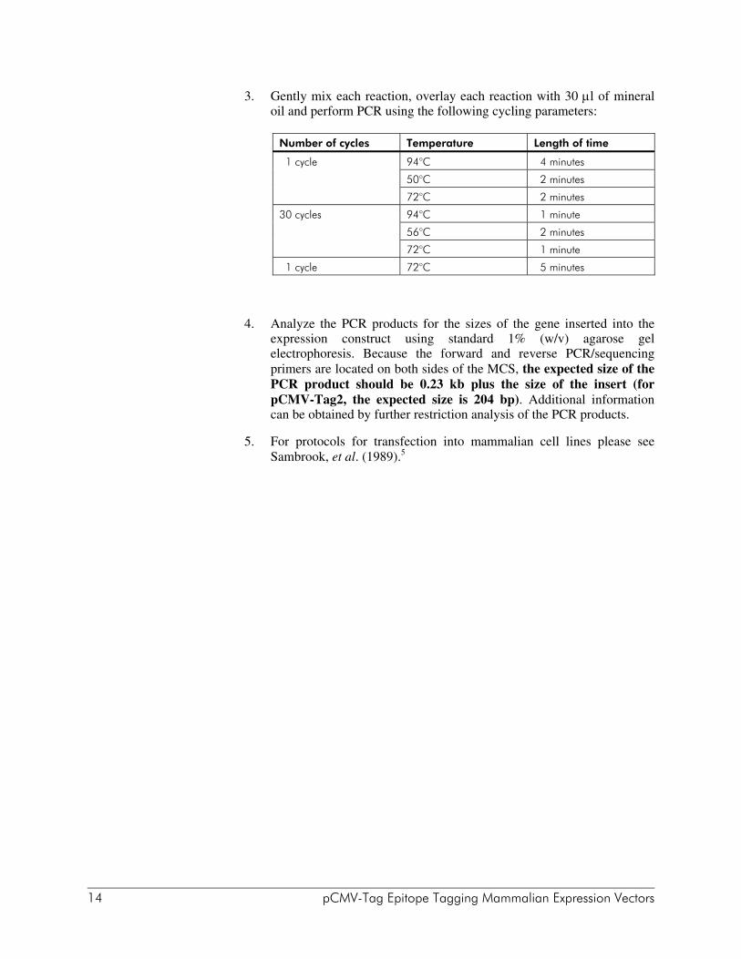
3. Gently mix each reaction, overlay each reaction with 30 μl of mineral oil and perform PCR using the following cycling parameters:
Number of cycles Temperature Length of time
1 cycle 94°C 4 minutes
50°C 2 minutes
72°C 2 minutes
30 cycles 94°C 1 minute
56°C 2 minutes
72°C 1 minute
1 cycle 72°C 5 minutes
4. Analyze the PCR products for the sizes of the gene inserted into the expression construct using standard 1% (w/v) agarose gel electrophoresis. Because the forward and reverse PCR/sequencing primers are located on both sides of the MCS, the expected size of the PCR product should be 0.23 kb plus the size of the insert (for pCMV-Tag2, the expected size is 204 bp). Additional information can be obtained by further restriction analysis of the PCR products.
5. For protocols for transfection into mammalian cell lines please see Sambrook, et al. (1989).5

pCMV-Tag Epitope Tagging Mammalian Expression Vectors 15
TROUBLESHOOTING Observation Suggestions
Insert is cloned out of frame. Sequence to ensure correct reading frame. Reclone if insert is out of frame
The promoter is not active in the cell line used. Use the provided control plasmid
Transfer of proteins is poor. Repeat transfer and optimize time of transfer, current and gel concentration and/or use molecular weight markers that cover the range to be transferred
Membrane preparation is inadequate. Ensure proper membrane hydration
Primary or secondary concentration is too low. Titrate antibody conjugates for optimum concentrations
Protein has degraded during storage of the membrane. Use fresh blots
Poor isolation of tagged protein. Use a different cell lysis procedure
Western analysis does not detect fusion protein
Proteolytic cleavage. Include protease inhibitors in lysis buffer
Insufficient blocking solution may have been used or the membrane was not thoroughly washed. Check the concentration of the blocking solution and/or wash thoroughly
Too much protein was loaded on gel. Load less protein on gel
Contamination by fingerprints and/or keratin has occurred. Use fresh membranes. Avoid touching the membrane. Use gloves and blunt forceps
The membrane produces excessive background
The concentration of the anti-FLAG, anti-c-myc, or secondary anti-mouse antibody is too high. Check the concentration of the antibodies and dilute if necessary

16 pCMV-Tag Epitope Tagging Mammalian Expression Vectors
PREPARATION OF MEDIA AND REAGENTS
LB Agar (per Liter) 10 g of NaCl 10 g of tryptone 5 g of yeast extract 20 g of agar Add deionized H2O to a final volume of
1 liter Adjust pH to 7.0 with 5 N NaOH Autoclave Pour into petri dishes (~25 ml/100-mm plate)
LB Broth (per Liter) 10 g of NaCl 10 g of tryptone 5 g of yeast extract Add deionized H2O to a final volume of
1 liter Adjust to pH 7.0 with 5 N NaOH Autoclave
LB–Kanamycin Agar (per Liter) Prepare 1 liter of LB agar Autoclave Cool to 55°C Add 5 ml of 10-mg/ml-filter-sterilized
kanamycin Pour into petri dishes (~25 ml/100-mm plate)
LB–Tetracycline Agar (per Liter) Prepare 1 liter of LB agar Autoclave Cool to 55°C Add 1.25 ml of 10 mg/ml-filter-sterilized
tetracycline Pour into petri dishes (~25 ml/100-mm plate) Store plates in a dark, cool place or cover
plates with foil if left out at room temperature for extended periods as tetracycline is light-sensitive
LB–Tetracycline Broth (per Liter) Prepare 1 liter of LB broth Autoclave Cool to 55°C Add 1.25 ml of 10 mg/ml-filter-sterilized
tetracycline Store broth in a dark, cool place as
tetracycline is light-sensitive
TE Buffer 10 mM Tris-HCl (pH 7.5) 1 mM EDTA

pCMV-Tag Epitope Tagging Mammalian Expression Vectors 17
REFERENCES 1. Hopp, T., Prickett, K., Price, V., Libby, R., March, C. et al. (1988) BioTechnology
6:1204-1210. 2. Evan, G. I., Lewis, G. K., Ramsay, G. and Bishop, J. M. (1985) Mol Cell Biol
5(12):3610-6. 3. Kozak, M. (1991) J Biol Chem 266(30):19867-70. 4. Hanahan, D. (1983) J Mol Biol 166(4):557-80. 5. Sambrook, J., Fritsch, E. F. and Maniatis, T. (1989). Molecular Cloning: A Laboratory
Manual. Cold Spring Harbor Laboratory Press, Cold Spring Harbor, NY.
ENDNOTES Falcon® is a registered trademark of Becton Dickinson and Company. FLAG® is a registered trademark of Sigma-Aldrich Co. GenBank® is a registered trademark of the U. S. Department of Health and Human
Services. Parafilm® is a registered trademark of American Can Company. Tween® is a registered trademark of ICI Americas, Inc.
MSDS INFORMATION The Material Safety Data Sheet (MSDS) information for Stratagene products is provided on the web at http://www.stratagene.com/MSDS/. Simply enter the catalog number to retrieve any associated MSDS’s in a print-ready format. MSDS documents are not included with product shipments.