mandible integrity and material properties of the
TRANSCRIPT

applied sciences
Article
Mandible Integrity and Material Properties of thePeriodontal Ligament during Orthodontic ToothMovement: A Finite-Element Study
Heng-Li Huang 1,2,†, Ming-Tzu Tsai 3,†, Shih-Guang Yang 4, Kuo-Chih Su 3,5, Yen-Wen Shen 1,6
and Jui-Ting Hsu 1,2,*1 School of Dentistry, College of Medicine, China Medical University, Taichung 404, Taiwan;
[email protected] (H.-L.H.); [email protected] (Y.-W.S.)2 Department of Bioinformatics and Medical Engineering, Asia University. Taichung 413, Taiwan3 Department of Biomedical Engineering, Hungkuang University, Taichung 433, Taiwan;
[email protected] (M.-T.T.); [email protected] (K.-C.S.)4 Master Program for Biomedical Engineering, China Medical University, Taichung 404, Taiwan;
[email protected] Department of Medical Research, Taichung Veterans General Hospital, Taichung 407, Taiwan6 Department of Dentistry, China Medical University and Hospital, Taichung 404, Taiwan* Correspondence: [email protected]† These authors contributed equally to this work.
Received: 31 March 2020; Accepted: 22 April 2020; Published: 24 April 2020�����������������
Abstract: We used the finite-element method (FEM) to investigate the effects of jawbone modelintegrity and the material properties of the periodontal ligament (PDL) on orthodontic tooth movement.Medical imaging software and computer-aided design software were used to create finite-elementmodels of a partial and complete mandibles based on dental cone beam computed tomographyimages of the human skull. Additionally, we exerted an orthodontic force on the canine crown inthe direction of an orthodontic miniscrew under a lower molar root to compare the von Mises strainon the canine PDL in three models: a partial mandible model under orthodontic force (Model 1),a complete mandible model under orthodontic force (Model 2), and a complete mandible modelunder orthodontic force with clench occlusion in the intercuspal position (ICP; Model 3). Additionally,in the complete mandible model under orthodontic force with ICP occlusion, we analyzed the effectsof a PDL with a low (Model 4), moderate (Model 5), and high (Model 6) linear elastic modulus and aPDL a bilinear elastic modulus (Model 7). The simulation results for mandible integrity indicatedthat the maximum von Mises strains on the canine PDL for Models 1, 2, and 3 were 0.461, 0.394, and1.811, respectively. Moreover, for the models with different PDL material properties, the maximumvon Mises strains on the canine PDLs for Models 4, 5, 6, and 7 were 6.047, 2.594, 0.887, and 1.811,respectively. When the FEM was used to evaluate tooth movement caused by orthodontic force,the transformation of a complete mandible model into a partial mandible model or alteration of theelastic modulus of the PDL influenced the biomechanical responses of the PDL. Additionally, theincorporation of daily ICP occlusion resulted in a larger effect.
Keywords: finite-element method; orthodontic; periodontal ligament; mandible
1. Introduction
Over the past 40 years, the finite-element method (FEM) has been widely used to studybiomechanics in the fields of orthopedics and dentistry. Finite-element models enable researchersto understand data, such as the distribution of stress and strain, that are difficult to record using
Appl. Sci. 2020, 10, 2980; doi:10.3390/app10082980 www.mdpi.com/journal/applsci

Appl. Sci. 2020, 10, 2980 2 of 10
measurement devices. The FEM is widely used in dentistry for the research and developmentof artificial implants and surgical instruments, prediction of jawbone growth and developmentalbehaviors, orthodontic treatment or oral surgery, and preoperative and postoperative evaluations [1,2].According to pressure–tension theory [3–6] in orthodontics, tooth movement is mainly caused by thebiomechanical response of the periodontal ligament (PDL) surrounding the tooth. An orthodonticbracket exerts force on a tooth to compress or stretch the PDL. Previous studies have indicated thatbone resorption and apposition are required in the compression and tension zones, respectively [3–6].In other words, tooth movement is achieved through bone remodeling [3–6]. Finite-element modelingof the jawbone and orthodontic force can be used to evaluate changes in the stress, strain, andhydrostatic pressure of the PDL surrounding the tooth. In coordination with pressure–tension theory,FEM can be used to predict tooth movement and movement direction.
In the literature, FEM has been used for models with a single tooth [7–9], partial jawbone modelswith several teeth [10–12], and complete jawbone models with all teeth [13–16]. Additionally, thesefinite-element models have been used to explore the effects of the design and material properties ofdifferent orthodontic forces, brackets, and archwires on the biomechanical responses of the PDL duringtooth movement. In studies that have employed the FEM to analyze such biomechanical responses,the assumed material properties of the PDL have affected the simulation results. Some studies haveassumed that the PDL is a linear elastic material [10,11,17–20], but animal and cadaver studies [21–24]have indicated that the actual PDL is more similar to a bilinear elastic material. Other researchers haveassumed that the PDL is a bilinear elastic material for conducting FEM simulations of biomechanicalresponses in tooth movement [25–28].
Numerous researchers have used FEM to analyze the biomechanical responses of the PDL duringtooth movement due to orthodontic treatment. However, most of these studies have developedpartial jawbone models with several teeth and used orthodontic force only as a loading condition.Therefore, in the present study, we evaluated the effect of partial and complete mandible models onthe biomechanical responses of the PDL with and without occlusal force. Additionally, we discuss thedetermination of the material properties of the PDL by using a finite-element model.
2. Materials and Methods
2.1. Geometry of the Finite-Element Model
Two finite-element models were employed in this study, namely a partial mandible model and acomplete mandible model. The complete mandibular model included the cortical and cancellous bones,all teeth except the first premolar, and the PDLs for all teeth except for the first premolar. A bracket wasfixed to a canine crown, and an orthodontic miniscrew was placed between the first and second molarroots. The partial mandible model included the jawbone from the right mandibular lateral incisor tothe second molar. Similar to the complete mandibular model, the partial mandibular model includedthe cortical bone, cancellous bone, the PDL, an orthodontic bracket, and a miniscrew. For both thepartial and complete mandible models, the contours of the cortical bone, cancellous bone, and teethwere added to a dental cone beam computed tomography image of a human skull by using Mimics15.0 (Materialise, Leuven, Belgium) before the image was segmented and saved in stereolithography(STL) format. Subsequently, the STL file was input into Geomagic Design X (3D Systems, ICP, RockHill, SC, USA) and transformed into a solid model. Since the PDL was excessively thin, we expandedthe tooth model by 0.25 mm and performed a Boolean operation with the teeth and mandible to obtaina solid PDL model. Subsequently, we used SolidWorks (Swanson Analysis, Canonsburg, PA, USA)to illustrate the brackets and miniscrews (Figure 1). Finally, the finite-element package of ANSYSWorkbench (Swanson Analysis, Canonsburg, PA, USA) was used to establish the material properties,meshing, boundary conditions, and loading conditions for all components of the solid model, therebyenabling the development of solutions. The numbers of elements and nodes in the partial mandiblemodel were 44,827 and 71,548, respectively. The numbers of elements and nodes in the complete

Appl. Sci. 2020, 10, 2980 3 of 10
mandible model were 151,296 and 235,603, respectively. The interfaces between the model componentswere assumed to have perfect bonding.
Appl. Sci. 2020, 10, 2980 3 of 11
model were 151,296 and 235,603, respectively. The interfaces between the model components were assumed to have perfect bonding.
Figure 1. (a,b) Solid and (c,d) finite-element mesh models: (a,c) partial mandible and (b,d) complete mandible.
2.2. Material Properties of the Finite-Element Model
In addition to analyzing the effect of jawbone integrity on the finite-element model, we explored the effects of PDL material properties on biomechanical responses. We used four material settings for the PDL, namely three linear elastic materials and one bilinear elastic material, in accordance with previous studies. The material property settings are presented in Table 1.
Table 1. Material properties used in finite-element models.
Component Elastic Modulus (MPa) Poisson’s
Ratio
Reference
Bracket 230000 0.3 Wu et al., 2011 [29]
Miniscrew
Cortical bone 14900 0.3 Poppe et al., 2002 [22]
Cancellous bone 460 0.3
Tooth 18600 0.3 Leung et al., 2008 [30]
PDL
Low linear elastic 0.044 0.45 Cattaneo et al., 2005 [31]
Middle linear
elastic 0.17
0.45 Cattaneo et al., 2005 [31]
High linear elastic 0.68 0.45 Salehi et al., 2015 [20]
Bilinear elastic E1 = 0.15; E2 = 0.50; 0.3 Kawarizadeh et al., [21] Poppe et al., 2002
Figure 1. (a,b) Solid and (c,d) finite-element mesh models: (a,c) partial mandible and (b,d) completemandible.
2.2. Material Properties of the Finite-Element Model
In addition to analyzing the effect of jawbone integrity on the finite-element model, we exploredthe effects of PDL material properties on biomechanical responses. We used four material settings forthe PDL, namely three linear elastic materials and one bilinear elastic material, in accordance withprevious studies. The material property settings are presented in Table 1.
Table 1. Material properties used in finite-element models.
Component Elastic Modulus(MPa) Poisson’s Ratio Reference
Bracket230000 0.3 Wu et al., 2011 [29]
MiniscrewCortical bone 14900 0.3 Poppe et al., 2002 [22]
Cancellous bone 460 0.3Tooth 18600 0.3 Leung et al., 2008 [30]
PDL
Low linear elastic 0.044 0.45 Cattaneo et al., 2005 [31]Middle linear elastic 0.17 0.45 Cattaneo et al., 2005 [31]High linear elastic 0.68 0.45 Salehi et al., 2015 [20]
Bilinear elasticE1 = 0.15; E2 = 0.50;
0.3Kawarizadeh et al., [21]Poppe et al., 2002 [22]Ultimate strain E12:
6.3%

Appl. Sci. 2020, 10, 2980 4 of 10
2.3. Loading and Boundary Conditions
As done previously [11,18,25], we constrained the mesial and distal sides of the partial mandiblemodel as boundary conditions. Additionally, we applied a loading condition of 2 N to simulateorthodontic force between the bracket and the miniscrew (Figure 2). For the complete mandible model,we used two sets of boundary and loading conditions. One model did not use occlusal force andsimulated only 2 N of orthodontic force. To establish the boundary condition, this model was fullyfixed at the top of the condyle. The second complete mandible model simulated an orthodontic forceof 2 N in the intercuspal position (ICP) occlusion mode (Table 2.). In a previous study [32], a modelwas employed to stimulate the force of six muscles, namely the superficial masseter, deep masseter,medial pterygoid, anterior temporalis, middle temporalis, and posterior temporalis. Force was exertedon the mandible to achieve ICP occlusion. In addition to completely fixing the model at the top of thecondyle, we considered the ICP occlusal mode by completely constraining the top of the incisal crownas a boundary condition (Figure 2).
Table 2. Muscular force and constraints in the intercuspal position (ICP) occlusal mode. Raw data wereobtained from Korioth and Hannam [32].
Occlusal Mode Side DirectionMuscular Force (N)
SM DM MP AT MT PT
Intercuspalposition (ICP)
Right
Force 76.2 21.2 136.3 12.6 5.7 3.0
FX −15.8 −11.6 66.3 −1.9 −1.3 −0.6FY −31.9 7.6 −50.9 −0.6 2.9 2.6FZ 67.3 16.1 107.8 12.5 4.8 1.4
Left
Force 76.2 21.2 136.3 12.6 5.7 3.0
FX 15.8 11.6 −66.3 1.9 1.3 0.6FY −31.9 7.6 −50.9 −0.6 2.9 2.6FZ 67.3 16.1 107.8 12.5 4.8 1.4
Muscles: SM, superficial masseter; DM, deep masseter, MP, medial pterygoid; AT, anterior temporalis; MT, middletemporalis; PT, posterior temporalis.
Appl. Sci. 2020, 10, 2980 4 of 11
Ultimate strain E12:
6.3%
[22]
2.3. Loading and Boundary Conditions
As done previously [11,18,25], we constrained the mesial and distal sides of the partial mandible model as boundary conditions. Additionally, we applied a loading condition of 2 N to simulate orthodontic force between the bracket and the miniscrew (Figure 2). For the complete mandible model, we used two sets of boundary and loading conditions. One model did not use occlusal force and simulated only 2 N of orthodontic force. To establish the boundary condition, this model was fully fixed at the top of the condyle. The second complete mandible model simulated an orthodontic force of 2 N in the intercuspal position (ICP) occlusion mode (Table 2.). In a previous study [32], a model was employed to stimulate the force of six muscles, namely the superficial masseter, deep masseter, medial pterygoid, anterior temporalis, middle temporalis, and posterior temporalis. Force was exerted on the mandible to achieve ICP occlusion. In addition to completely fixing the model at the top of the condyle, we considered the ICP occlusal mode by completely constraining the top of the incisal crown as a boundary condition (Figure 2).
Table 2. Muscular force and constraints in the intercuspal position (ICP) occlusal mode. Raw data were obtained from Korioth and Hannam [32].
Occlusal Mode Side Direction Muscular Force (N)
SM DM MP AT MT PT
Intercuspal position (ICP)
Right
Force 76.2 21.2 136.3 12.6 5.7 3.0
FX −15.8 −11.6 66.3 −1.9 −1.3 −0.6
FY −31.9 7.6 −50.9 −0.6 2.9 2.6
FZ 67.3 16.1 107.8 12.5 4.8 1.4
Left
Force 76.2 21.2 136.3 12.6 5.7 3.0
FX 15.8 11.6 −66.3 1.9 1.3 0.6
FY −31.9 7.6 −50.9 −0.6 2.9 2.6
FZ 67.3 16.1 107.8 12.5 4.8 1.4 Muscles: SM, superficial masseter; DM, deep masseter, MP, medial pterygoid; AT, anterior temporalis; MT, middle temporalis; PT, posterior temporalis.
Figure 2. Loading and boundary conditions in finite-element simulations. (a) Partial mandible model
under orthodontic force. (b) Complete mandible model under orthodontic force. (c) Complete mandiblemodel under orthodontic force with ICP occlusion.
2.4. Evaluation Parameters
In this study, finite-element simulations were performed to analyze the integrity of the jawbonemodel and the effect of material property settings on the biomechanical responses of the PDL. Table 3lists the finite-element simulation groups for these two aspects. Tooth movement is mainly caused by
Appl. Sci. 2020, 10, 2980 5 of 10
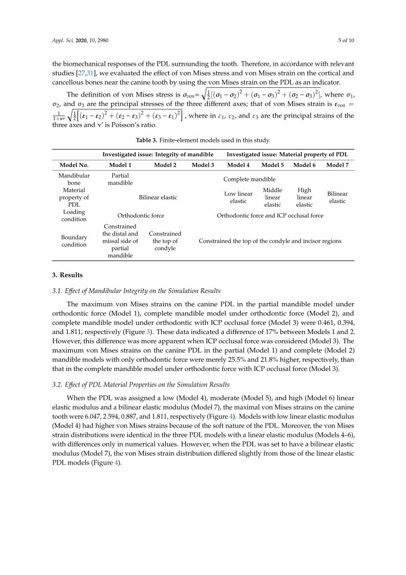
the biomechanical responses of the PDL surrounding the tooth. Therefore, in accordance with relevantstudies [27,31], we evaluated the effect of von Mises stress and von Mises strain on the cortical andcancellous bones near the canine tooth by using the von Mises strain on the PDL as an indicator.
The definition of von Mises stress is σvon=
√12 [(σ1 − σ2)
2 + (σ1 − σ3)2 + (σ2 − σ3)
2], where σ1,σ2, and σ3 are the principal stresses of the three different axes; that of von Mises strain is εvon =
11+ν′
√12
[(ε1 − ε2)
2 + (ε2 − ε3)2 + (ε3 − ε1)
2]
, where in ε1, ε2, and ε3 are the principal strains of thethree axes and ν’ is Poisson’s ratio.
Table 3. Finite-element models used in this study.
Investigated issue: Integrity of mandible Investigated issue: Material property of PDL
Model No. Model 1 Model 2 Model 3 Model 4 Model 5 Model 6 Model 7
Mandibularbone
Partialmandible Complete mandible
Materialproperty of
PDLBilinear elastic Low linear
elastic
Middlelinearelastic
Highlinearelastic
Bilinearelastic
Loadingcondition Orthodontic force Orthodontic force and ICP occlusal force
Boundarycondition
Constrainedthe distal andmissal side of
partialmandible
Constrainedthe top ofcondyle
Constrained the top of the condyle and incisor regions
3. Results
3.1. Effect of Mandibular Integrity on the Simulation Results
The maximum von Mises strains on the canine PDL in the partial mandible model underorthodontic force (Model 1), complete mandible model under orthodontic force (Model 2), andcomplete mandible model under orthodontic with ICP occlusal force (Model 3) were 0.461, 0.394,and 1.811, respectively (Figure 3). These data indicated a difference of 17% between Models 1 and 2.However, this difference was more apparent when ICP occlusal force was considered (Model 3). Themaximum von Mises strains on the canine PDL in the partial (Model 1) and complete (Model 2)mandible models with only orthodontic force were merely 25.5% and 21.8% higher, respectively, thanthat in the complete mandible model under orthodontic force with ICP occlusal force (Model 3).
3.2. Effect of PDL Material Properties on the Simulation Results
When the PDL was assigned a low (Model 4), moderate (Model 5), and high (Model 6) linearelastic modulus and a bilinear elastic modulus (Model 7), the maximal von Mises strains on the caninetooth were 6.047, 2.594, 0.887, and 1.811, respectively (Figure 4). Models with low linear elastic modulus(Model 4) had higher von Mises strains because of the soft nature of the PDL. Moreover, the von Misesstrain distributions were identical in the three PDL models with a linear elastic modulus (Models 4–6),with differences only in numerical values. However, when the PDL was set to have a bilinear elasticmodulus (Model 7), the von Mises strain distribution differed slightly from those of the linear elasticPDL models (Figure 4).

Appl. Sci. 2020, 10, 2980 6 of 10Appl. Sci. 2020, 10, 2980 6 of 11
Figure 3. Distribution of von Mises strain in the canine periodontal ligament (PDL): (a) partial mandible model under orthodontic force; (b) complete mandible model under orthodontic force; and (c) complete mandible model under orthodontic force with ICP occlusal force.
3.2. Effect of PDL Material Properties on the Simulation Results
When the PDL was assigned a low (Model 4), moderate (Model 5), and high (Model 6) linear elastic modulus and a bilinear elastic modulus (Model 7), the maximal von Mises strains on the canine tooth were 6.047, 2.594, 0.887, and 1.811, respectively (Figure 4). Models with low linear elastic modulus (Model 4) had higher von Mises strains because of the soft nature of the PDL. Moreover, the von Mises strain distributions were identical in the three PDL models with a linear elastic modulus (Models 4–6), with differences only in numerical values. However, when the PDL was set to have a bilinear elastic modulus (Model 7), the von Mises strain distribution differed slightly from those of the linear elastic PDL models (Figure 4).
Figure 3. Distribution of von Mises strain in the canine periodontal ligament (PDL): (a) partial mandiblemodel under orthodontic force; (b) complete mandible model under orthodontic force; and (c) completemandible model under orthodontic force with ICP occlusal force.
3.3. Effect of Mandibular Integrity and PDL Material Properties on Bone
In addition to the effect of jawbone integrity on von Mises strain in the PDL, the effects oforthodontic force on von Mises stress in the cortical and cancellous bones near the canine PDL werenegligible in the partial (Model 1) and complete (Model 2) mandible models. However, the simulationresults of these models differed considerably from those of the complete mandible model underorthodontic and ICP occlusal forces (Model 3; Table 4). In addition, when the PDL was modeled with abilinear elastic modulus (Model 7), the von Mises stresses in the cortical and cancellous bone werebetween those when the PDL was modeled with a low linear elastic modulus (Model 4) and high linearelastic modulus (Model 6; Table 4).

Appl. Sci. 2020, 10, 2980 7 of 10Appl. Sci. 2020, 10, 2980 7 of 11
Figure 4. Distribution of von Mises strain in the canine PDL: models with (a) low linear elastic modulus, (b) moderate linear elastic modulus, (c) high linear elastic modulus, and (d) bilinear elastic modulus.
3.3. Effect of Mandibular Integrity and PDL Material Properties on Bone
In addition to the effect of jawbone integrity on von Mises strain in the PDL, the effects of orthodontic force on von Mises stress in the cortical and cancellous bones near the canine PDL were negligible in the partial (Model 1) and complete (Model 2) mandible models. However, the simulation results of these models differed considerably from those of the complete mandible model under orthodontic and ICP occlusal forces (Model 3; Table 4). In addition, when the PDL was modeled with a bilinear elastic modulus (Model 7), the von Mises stresses in the cortical and cancellous bone were between those when the PDL was modeled with a low linear elastic modulus (Model 4) and high linear elastic modulus (Model 6; Table 4).
Table 4. Finite-element simulation results.
Investigated Issue: Integrity of Mandible Investigated Issue: Material
Model 1 Model 2 Model 3 Model 4
Model 5
Model 6
Model 7
Max von Mises strain on canine’s PDL
0.461 0.394 1.811 6.047 2.594 0.887 1.811
Max von Mises stress on cortical bone around canine’s PDL
0.498 0.765 7.5375 6.25 8.3067 14.343 7.5375
Max von Mises stress on cancellous bone
around canine’s PDL 0.0599 0.0794 0.6914 0.5622 1.2387 1.5905 0.6914
Unit of von Mises strain: None; Unit of von Mises stress: MPa.
4. Discussion
Numerous studies have used the FEM to analyze the biomechanical responses of the PDL during tooth movement due to orthodontic treatment. However, most such studies have used the FEM to develop partial jawbone models. The few studies that have created complete jawbone models have overlooked the occlusal force encountered in daily life. Therefore, we established
Figure 4. Distribution of von Mises strain in the canine PDL: models with (a) low linear elastic modulus,(b) moderate linear elastic modulus, (c) high linear elastic modulus, and (d) bilinear elastic modulus.
Table 4. Finite-element simulation results.
Investigated Issue: Integrity of Mandible Investigated Issue: Material
Model 1 Model 2 Model 3 Model 4 Model 5 Model 6 Model 7
Max von Mises strainon canine’s PDL 0.461 0.394 1.811 6.047 2.594 0.887 1.811
Max von Mises stresson cortical bone
around canine’s PDL0.498 0.765 7.5375 6.25 8.3067 14.343 7.5375
Max von Mises stresson cancellous bone
around canine’s PDL0.0599 0.0794 0.6914 0.5622 1.2387 1.5905 0.6914
Unit of von Mises strain: None; Unit of von Mises stress: MPa.
4. Discussion
Numerous studies have used the FEM to analyze the biomechanical responses of the PDL duringtooth movement due to orthodontic treatment. However, most such studies have used the FEM to developpartial jawbone models. The few studies that have created complete jawbone models have overlooked theocclusal force encountered in daily life. Therefore, we established partial and complete mandible modelsto analyze the effect of jawbone model integrity on the biomechanical reactions of the PDL by using theFEM. The simulation results suggested that the effects of the partial and complete mandible models onvon Mises strain in the canine PDL were negligible when only orthodontic force was considered. However,the occlusal force that individuals exert in daily life had a substantial effect on the von Mises strain in thePDL. This effect was even larger when the PDL was assigned a linear elastic modulus.
The FEM is an excellent method for evaluating tooth movement caused by orthodontic treatmentbecause it can simulate orthodontic forces of different amounts and directions. The biomechanicalresponses of the PDL can be evaluated, and the direction and rate of tooth movement can be predictedaccording to pressure–tension theory. Numerous researchers have used the FEM to investigate toothmovement during orthodontic treatment, but most studies have created only partial jawbone models.Although some studies have established complete jawbone models, their goals were to analyze theeffects of occlusal modes on jawbone biomechanical response [32] or on the PDL [33]. However,

Appl. Sci. 2020, 10, 2980 8 of 10
whether occlusal force interferes with the mechanical response of the PDL during orthodontic treatmenthas remained unclear.
Our simulation results suggest that the maximum von Mises strains in the canine PDL differedby 17.0% between the partial and complete mandible models. This difference was caused by thedifference in method used for setting the boundary conditions. The partial mandible model involvesa finite-element simulation for the mesial and distal sides. The proximal side of the mandible nearthe side incisors was a fixed surface that was too close to the canine tooth and was moving in thedistal direction. Therefore, when the canine PDL exerted orthodontic force against the mesial side, thestrain distribution differed substantially from that in the complete mandible model, which was subjectonly to orthodontic force. The distal side of the canine PDL was farther from the completely fixedpartial mandible mesial side. Thus, the effect on strain distribution was weaker. This demonstrates thatboundary conditions should be applied farther from the tooth requiring movement in future modelsof tooth movement due to orthodontic treatment. In other words, for a partial mandible, a largerarea must be modeled to mitigate effects on the simulation results. Moreover, our simulation resultssuggested that the maximum von Mises strain in the ICP occlusal mode was 4.6-times higher than thatin the complete mandible model without ICP occlusion. Therefore, the occlusal force encounteredin daily life had a considerable effect. However, during orthodontic treatment, the jawbone wasnot constantly subjected to occlusal force. This study only demonstrated that occlusal force couldaffect simulation results. Therefore, we recommend that the analysis of tooth movement caused byorthodontic force should incorporate occlusal force.
Numerous studies have assumed that the PDL is a linear elastic material [10,11,17–20]. However,the PDL is mainly composed of collagen fibers [34], and its material properties are more similar to thoseof a nonlinear elastic material. Numerous studies have directly measured PDL material properties,but relevant studies have suggested that the material properties of soft tissues are easily affected byfreeze–thaw cycles at temperatures of >20 ◦C [35]. Unfrozen human cadaver jawbone specimensare unavailable, and experiments on most human cadaver bones or tooth specimens can only beconducted after a freeze–thaw cycle. Poppe et al. [22] and Liu et al. [24] have conducted mechanicalexperiments on mandibular fragments of human corpses to directly measure the elastic modulus ofthe PDL. Experimental results have suggested that the PDL is a bilinear elastic material. Kawarizadehet al. [21] and Ziegler et al. [23] have measured the PDL elastic modulus in rat and miniature pigspecimens and have also demonstrated that the PDL has bilinear elastic properties.
Our simulation results for the bilinear elastic PDL model differed from those for the linear elasticPDL model, which is in agreement with the findings of other studies [31,36]. When the PDL wasassigned a low linear, moderate linear, high linear, and bilinear elastic modulus, the maximum vonMises strains on the canine PDL were 6.047, 2.594, 0.887, and 1.811, respectively. However, because ofthe limitations of the different finite-element models and methods of exerting orthodontic force, wewere unable to conduct an absolute-value comparison between our simulation results and those ofother studies. However, the simulation results suggested that strain was more concentrated in the PDLwhen the PDL was assumed to have a linear elastic modulus, and it was more evenly distributed whenthe PDL was assumed to have a bilinear elastic modulus.
Although we established a complete mandible model and simulated the ICP occlusal mode, thisstudy had several limitations. First, orthodontic wire was not used in the finite-element model. Thus,friction between brackets and orthodontic wire was not considered. Second, bone is an inhomogeneousanisotropic tissue with a trabecular structure. In this study, bone had linear elastic, isotropic, andhomogenous material properties, in accordance with those adopted in most relevant studies. Third,we simulated only the ICP occlusal mode; other occlusal modes may have different effects on the PDL.
5. Conclusions
When the FEM was employed to evaluate tooth movement due to orthodontic force, thebiomechanical response of the PDL was affected by mandible integrity; thus the complete mandible

Appl. Sci. 2020, 10, 2980 9 of 10
model was converted into a partial mandible model. Furthermore, the incorporation of daily ICPocclusion resulted in a larger effect. In addition, assumptions regarding the material properties of thePDL affected the biomechanical response of the PDL.
Author Contributions: Conceptualization, H.-L.H., S.-G.Y. and J.-T.H.; methodology, S.-G.Y. and J.-T.H.;writing—original draft preparation, H.-L.H., Y.-W.S., M.-T.T., K.-C.S. and J.-T.H.; writing—M.-T.T. and J.-T.H.All authors have read and agreed to the published version of the manuscript.
Funding: This research was supported by Ministry of Science and Technology, Taiwan (Grant number: MOST108-2221-E-039-004) and China Medical University, Taiwan (Grant number: CMU108-MF-86).
Conflicts of Interest: The authors declare no conflict of interest.
References
1. Hsu, M.-L.; Chang, C.-L. Application of Finite Element Analysis in Dentistry. Finite Elem. Anal. Sciyo 2010.[CrossRef]
2. Piccioni, M.A.R.; Campos, E.A.; Saad, J.R.C.; de Andrade, M.F.; Galvão, M.R.; Rached, A.A. Application ofthe finite element method in Dentistry. RSBO Rev. Sul-Bras. Odontol. 2013, 10, 369–377.
3. Cattaneo, P.; Dalstra, M.; Melsen, B. Strains in periodontal ligament and alveolar bone associated withorthodontic tooth movement analyzed by finite element. Orthod. Craniofacial Res. 2009, 12, 120–128.[CrossRef] [PubMed]
4. Chang, H.-W.; Huang, H.-L.; Yu, J.-H.; Hsu, J.-T.; Li, Y.-F.; Wu, Y.-F. Effects of orthodontic tooth movement onalveolar bone density. Clin. Oral Investig. 2012, 16, 679–688. [CrossRef] [PubMed]
5. Masella, R.S.; Meister, M. Current concepts in the biology of orthodontic tooth movement. Am. J. Orthod.Dentofac. Orthop. 2006, 129, 458–468. [CrossRef] [PubMed]
6. Will, L.A. Orthodontic Tooth Movement: A Historic Prospective. Front Oral Biol. 2016, 18, 46–55.7. Tanne, K.; Sakuda, M.; Burstone, C.J. Three-dimensional finite element analysis for stress in the periodontal
tissue by orthodontic forces. Am. J. Orthod. Dentofac. Orthop. 1987, 92, 499–505. [CrossRef]8. Sardarian, A.; Shahidi, S.; Boushehri, S.G.; Geramy, A. The effect of vertical bracket positioning on torque and
the resultant stress in the periodontal ligament—A finite element study. Prog. Orthod. 2014, 15, 50. [CrossRef]9. Sugii, M.M.; Barreto, B.C.F.; Francisco Vieira-Junior, W.; Simone, K.R.I.; Bacchi, A.; Caldas, R.A. Extruded
upper first molar intrusion: Comparison between unilateral and bilateral miniscrew anchorage. Dent. PressJ. Orthod. 2018, 23, 63–70. [CrossRef]
10. Caballero, G.M.; de Carvalho Filho, O.A.; Hargreaves, B.O.; de Araújo Brito, H.H.; Junior, P.A.A.M.;Oliveira, D.D. Mandibular canine intrusion with the segmented arch technique: A finite element methodstudy. Am. J. Orthod. Dentofac. Orthop. 2015, 147, 691–697. [CrossRef]
11. Field, C.; Ichim, I.; Swain, M.V.; Chan, E.; Darendeliler, M.A.; Li, W.; Li, Q. Mechanical responses toorthodontic loading: A 3-dimensional finite element multi-tooth model. Am. J. Orthod. Dentofac. Orthop.2009, 135, 174–181. [CrossRef] [PubMed]
12. Gerami, A.; Dadgar, S.; Rakhshan, V.; Jannati, P.; Sobouti, F. Displacement and force distribution of splintedand tilted mandibular anterior teeth under occlusal loads: An in silico 3D finite element analysis. Prog.Orthod. 2016, 17, 16. [CrossRef] [PubMed]
13. Liao, Z.; Elekdag-Turk, S.; Turk, T.; Grove, J.; Dalci, O.; Chen, J.; Zheng, K.; Ali Darendeliler, M.; Swain, M.;Li, Q. Computational and clinical investigation on the role of mechanical vibration on orthodontic toothmovement. J. Biomech. 2017, 60, 57–64. [CrossRef] [PubMed]
14. Zhou, X.; Xia, Z.; Gan, Y.; Zhang, D.; Xiong, J.; Fang, P.; Li, G.; Zhao, Q. Orthodontic force simulationof Tooth-PDL-Bone Complex under archwire loading. In Proceedings of the 38th Annual InternationalConference of the IEEE Engineering in Medicine and Biology Society (EMBC), Lake Buena Vista, FL, USA,16–20 August 2016; pp. 6030–6033.
15. Namburi, M.; Nagothu, S.; Kumar, C.S.; Chakrapani, N.; Hanumantharao, C.H.; Kumar, S.K. Evaluating theeffects of consolidation on intrusion and retraction using temporary anchorage devices—A FEM study. Prog.Orthod. 2017, 18, 2. [CrossRef]
16. Chen, J.; Li, W.; Swain, M.V.; Ali Darendeliler, M.; Li, Q. A periodontal ligament driven remodeling algorithmfor orthodontic tooth movement. J. Biomech. 2014, 47, 1689–1695. [CrossRef]

Appl. Sci. 2020, 10, 2980 10 of 10
17. Bouton, A.; Simon, Y.; Goussard, F.; Teresi, L.; Sansalone, V. New finite element study protocol: Clinicalsimulation of orthodontic tooth movement. Int. Orthod. 2017, 15, 165–179. [CrossRef]
18. de Souza, F.I.; Poi, W.R.; da Silva, V.F.; Martini, A.P.; Melo, R.A.; Panzarini, S.R.; Rocha, E.P. Stress distributionin delayed replanted teeth splinted with different orthodontic wires: A three-dimensional finite elementanalysis. Dent. Traumatol. 2015, 31, 190–195. [CrossRef]
19. McCormack, S.W.; Witzel, U.; Watson, P.J.; Fagan, M.J.; Groening, F. Inclusion of periodontal ligament fibresin mandibular finite element models leads to an increase in alveolar bone strains. PLoS ONE 2017, 12,e0188707. [CrossRef]
20. Salehi, P.; Gerami, A.; Najafi, A.; Torkan, S. Evaluating stress distribution pattern in periodontal ligament ofmaxillary incisors during intrusion assessed by the finite element method. J. Dent. 2015, 16, 314–322.
21. Kawarizadeh, A.; Bourauel, C.; Jäger, A. Experimental and numerical determination of initial tooth mobilityand material properties of the periodontal ligament in rat molar specimens. Eur. J. Orthod. 2003, 25, 569–578.[CrossRef]
22. Poppe, M.; Bourauel, C.; Jäger, A. Determination of the elasticity parameters of the human periodontalligament and the location of the center of resistance of single-rooted teeth a study of autopsy specimens andtheir conversion into finite element models. J. Orofac. Orthop. Fortschr. Kieferorthopädie 2002, 63, 358–370.[CrossRef] [PubMed]
23. Ziegler, A.; Keilig, L.; Kawarizadeh, A.; Jäger, A.; Bourauel, C. Numerical simulation of the biomechanicalbehaviour of multi-rooted teeth. Eur. J. Orthod. 2005, 27, 333–339. [CrossRef] [PubMed]
24. Liu, T.-C.; Chang, C.-H.; Wong, T.-Y.; Liu, J.-K. Finite element analysis of miniscrew implants used fororthodontic anchorage. Am. J. Orthod. Dentofac. Orthop. 2012, 141, 468–476. [CrossRef] [PubMed]
25. Hartmann, M.; Dirk, C.; Reimann, S.; Keilig, L.; Konermann, A.; Jager, A.; Bourauel, C. Influence of toothdimension on the initial mobility based on plaster casts and X-ray images: A numerical study. J. Orofac.Orthop. 2017, 78, 285–292. [CrossRef]
26. Verna, C.; Cattaneo, P.M.; Dalstra, M. Corticotomy affects both the modus and magnitude of orthodontictooth movement. Eur. J. Orthod. 2018, 40, 107–112. [CrossRef]
27. Papageorgiou, S.N.; Keilig, L.; Hasan, I.; Jäger, A.; Bourauel, C. Effect of material variation on thebiomechanical behaviour of orthodontic fixed appliances: A finite element analysis. Eur. J. Orthod.2016, 38, 300–307. [CrossRef]
28. Papageorgiou, S.N.; Keilig, L.; Vandevska-Radunovic, V.; Eliades, T.; Bourauel, C. Torque differences due to thematerial variation of the orthodontic appliance: A finite element study. Prog. Orthod. 2017, 18, 6. [CrossRef]
29. Wu, J.-H.; Wang, H.-C.; Chen, C.-M.; Lu, P.-C.; Lai, S.-T.; Lee, K.-T.; Du, J.-K. Pullout strengths of orthodonticpalatal mini-implants tested in vitro. J. Dent. Sci. 2011, 6, 200–204. [CrossRef]
30. Leung, M.T.-C.; Lee, T.C.-K.; Rabie, A.B.M.; Wong, R.W.-K. Use of miniscrews and miniplates in orthodontics.J. Oral Maxillofac. Surg. 2008, 66, 1461–1466. [CrossRef]
31. Cattaneo, P.; Dalstra, M.; Melsen, B. The finite element method: A tool to study orthodontic tooth movement.J. Dent. Res. 2005, 84, 428–433. [CrossRef]
32. Korioth, T.; Hannam, A. Deformation of the human mandible during simulated tooth clenching. J. Dent. Res.1994, 73, 56–66. [CrossRef] [PubMed]
33. Koolstra, J.; Van Eijden, T. Three-dimensional dynamical capabilities of the human masticatory muscles.J. Biomech. 1999, 32, 145–152. [CrossRef]
34. Pöschke, A.; Krähling, B.; Failing, K.; Staszyk, C. Molecular characteristics of the equine periodontal ligament.Front. Vet. Sci. 2018, 4, 235. [CrossRef] [PubMed]
35. Hongo, M.; Gay, R.E.; Hsu, J.-T.; Zhao, K.D.; Ilharreborde, B.; Berglund, L.J.; An, K.-N. Effect of multiplefreeze–thaw cycles on intervertebral dynamic motion characteristics in the porcine lumbar spine. J. Biomech.2008, 41, 916–920. [CrossRef] [PubMed]
36. Hemanth, M.; Deoli, S.; Raghuveer, H.; Rani, M.; Hegde, C.; Vedavathi, B. Stress induced in periodontalligament under orthodontic loading (Part II): A comparison of linear versus non-linear FEM study. J. Int.Oral Health JIOH 2015, 7, 114–118. [PubMed]
© 2020 by the authors. Licensee MDPI, Basel, Switzerland. This article is an open accessarticle distributed under the terms and conditions of the Creative Commons Attribution(CC BY) license (http://creativecommons.org/licenses/by/4.0/).