managing dental fluorosis: traditional healer versus ... · a - intra-oral periapical radiographs...
TRANSCRIPT

JSM Dentistry
Cite this article: Kemoli AM (2017) Managing Dental Fluorosis: Traditional Healer versus Clinic Approaches. JSM Dent 5(2): 1085.
Central
*Corresponding authorArthur Kemoli, Department of Pediatric Dentistry & Orthodontics, University of Nairobi, P.O. Box: 34848 00100 Nairobi, Kenya, Email:
Submitted: 05 May 2017
Accepted: 16 May 2017
Published: 18 May 2017
ISSN: 2333-7133
Copyright© 2017 Kemoli
OPEN ACCESS
Keywords•Fluorosis•Dental aesthetics•Abrasion•Herbalist•Veneers
Case Report
Managing Dental Fluorosis: Traditional Healer versus Clinic ApproachesArthur M. Kemoli*Department of Pediatric Dentistry & Orthodontics, University of Nairobi, Kenya
Abstract
Dental fluorosis is a major dental concern in many areas in the world with its associated aesthetic and occlusal problems due to cracking of the affected teeth. The present case report details a 14 year-old girl, with moderate fluorosis and who sought treatment of the discolouration associated with the fluorosis from a herbalist. At the end of the treatment, her aesthetics was not just poorer, but there was also severe dental sensitivity emanating from her upper anterior teeth. As a result, she sought treatment at a local dental clinic, where the damage was successfully repaired with good aesthetic results and complete elimination of dental sensitivity. The case study amplifies the imperative need for oral health education to the general population, and in particular, increased knowledge on aetiology, consequences and proper managment of dental fluorosis.
INTRODUCTIONHuman intake of fluoride can have both beneficial effects
on tooth development and dental caries prevention, but this substance can also have detrimental effects in the form of dental fluorosis [1]. Dental fluorosis results from over-exposure by a child to high amounts of fluoride during the age of 1-8 years. This exposure can occur through the use of fluoridated toothpastes, mouthwashes, fluoride supplements when the growing child unknowingly ingests some of these substances, and through the consumption of highly fluoridated drinking water. The earliest manifestation of dental fluorosis can be seen in the form of increase enamel porosity along the striae of Retzius [2]. In its mild forms, dental fluorosis is clinically seen on the dental enamel as lacy white to chalky white specks and streaks. However, in its severe forms, the affected teeth assume some discoloration, which ranges from yellow to dark brown with surface irregularities in the form of enamel pits. The diagnosis of dental fluorosis is clinical, and indices like the Dean [3] and Thylstrup Fejerskov Indices [4] have been developed to help in determining of the severity of dental fluorosis. In the case of the Dean Index, the severity of dental fluorosis ranges from questionable, through very mild, mild, moderate to severe forms. For Thylstrup Fejerskov Index (TFI), the dental fluorosis is scored from TFI 1 to 9, where TFI 1-3 is mild, TFI 4-5 is moderate and TFI 6-9 is severe [5].
Dental fluorosis can be psychologically distressing to the affected child on grounds of aesthetics and occasionally, dental sensitivity. The condition tends to be difficult to manage, but certain treatment regimes have been employed to meet the various needs of the patient. Vital bleaching is one example
of managing dental fluorosis, and helps to remove dark spots formed as a result of the penetration of food coloring agents into the hypo mineralized areas. Micro-abrasion is another regime of treatment, with which the defects within the enamel that do not exceed tenths of a millimeter are removed [6]. In the very severe cases of fluorosis, considerations have been made of the use of veneers and dental crowns to ameliorate the poor aesthetics and or loss of tooth material.
CASE REPORTA 14 year-old girl, accompanied wih her sister, consulted
a local Paediatric dental clinic in Nairobi, Kenya, complaining of discoloured and sensitive anterior teeth. The history of the complaint indicated that although discolouration had been there fora long time, the sensitivity had began about 7 months after seeking help for the discolouration from a local herbalist. The herbalist had used a spongesoaked in a liquid to rub against her upper anterior teeth to remove the stains. However, the patient was not only disatisfied with the yellowish discolouration that ensued, but she also reported that her teeth became very sensitive.
At the time of the consultation at the dental clinic, the patient was in good general health in spite of the patient having never received any immunizations during her childhood on grounds of her parents’ religious beliefs. Intra-oral examination showed a patient who was in full permanent dentition, except for the unerupted permanent third molars. Her oral hygiene was just fair, and she had generalized moderate fluorosis, with the anterior upper teeth from canine to canine having been severely abraded, with a huge loss of tooth material on their labial
Kemoli (2017)Email:
JSM Dent 5(2): 1085 (2017) 2/3
Central
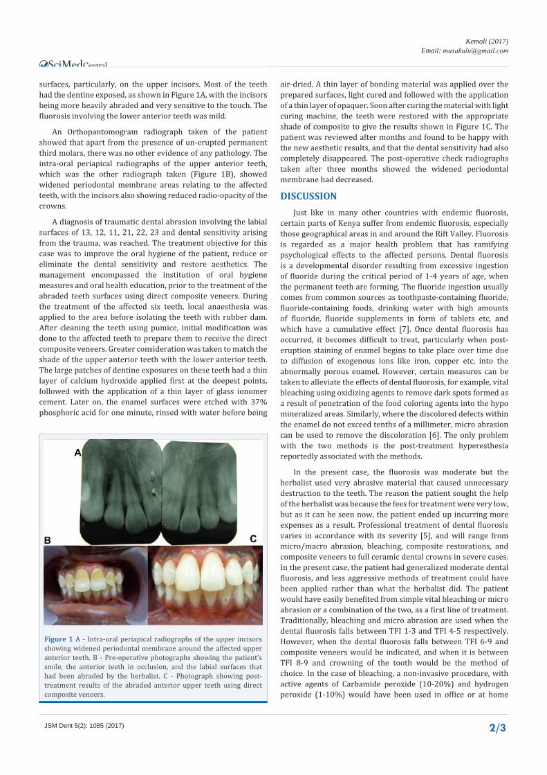
surfaces, particularly, on the upper incisors. Most of the teeth had the dentine exposed, as shown in Figure 1A, with the incisors being more heavily abraded and very sensitive to the touch. The fluorosis involving the lower anterior teeth was mild.
An Orthopantomogram radiograph taken of the patient showed that apart from the presence of un-erupted permanent third molars, there was no other evidence of any pathology. The intra-oral periapical radiographs of the upper anterior teeth, which was the other radiograph taken (Figure 1B), showed widened periodontal membrane areas relating to the affected teeth, with the incisors also showing reduced radio-opacity of the crowns.
A diagnosis of traumatic dental abrasion involving the labial surfaces of 13, 12, 11, 21, 22, 23 and dental sensitivity arising from the trauma, was reached. The treatment objective for this case was to improve the oral hygiene of the patient, reduce or eliminate the dental sensitivity and restore aesthetics. The management encompassed the institution of oral hygiene measures and oral health education, prior to the treatment of the abraded teeth surfaces using direct composite veneers. During the treatment of the affected six teeth, local anaesthesia was applied to the area before isolating the teeth with rubber dam. After cleaning the teeth using pumice, initial modification was done to the affected teeth to prepare them to receive the direct composite veneers. Greater consideration was taken to match the shade of the upper anterior teeth with the lower anterior teeth. The large patches of dentine exposures on these teeth had a thin layer of calcium hydroxide applied first at the deepest points, followed with the application of a thin layer of glass ionomer cement. Later on, the enamel surfaces were etched with 37% phosphoric acid for one minute, rinsed with water before being
air-dried. A thin layer of bonding material was applied over the prepared surfaces, light cured and followed with the application of a thin layer of opaquer. Soon after curing the material with light curing machine, the teeth were restored with the appropriate shade of composite to give the results shown in Figure 1C. The patient was reviewed after months and found to be happy with the new aesthetic results, and that the dental sensitivity had also completely disappeared. The post-operative check radiographs taken after three months showed the widened periodontal membrane had decreased.
DISCUSSIONJust like in many other countries with endemic fluorosis,
certain parts of Kenya suffer from endemic fluorosis, especially those geographical areas in and around the Rift Valley. Fluorosis is regarded as a major health problem that has ramifying psychological effects to the affected persons. Dental fluorosis is a developmental disorder resulting from excessive ingestion of fluoride during the critical period of 1-4 years of age, when the permanent teeth are forming. The fluoride ingestion usually comes from common sources as toothpaste-containing fluoride, fluoride-containing foods, drinking water with high amounts of fluoride, fluoride supplements in form of tablets etc, and which have a cumulative effect [7]. Once dental fluorosis has occurred, it becomes difficult to treat, particularly when post-eruption staining of enamel begins to take place over time due to diffusion of exogenous ions like iron, copper etc, into the abnormally porous enamel. However, certain measures can be taken to alleviate the effects of dental fluorosis, for example, vital bleaching using oxidizing agents to remove dark spots formed as a result of penetration of the food coloring agents into the hypo mineralized areas. Similarly, where the discolored defects within the enamel do not exceed tenths of a millimeter, micro abrasion can be used to remove the discoloration [6]. The only problem with the two methods is the post-treatment hyperesthesia reportedly associated with the methods.
In the present case, the fluorosis was moderate but the herbalist used very abrasive material that caused unnecessary destruction to the teeth. The reason the patient sought the help of the herbalist was because the fees for treatment were very low, but as it can be seen now, the patient ended up incurring more expenses as a result. Professional treatment of dental fluorosis varies in accordance with its severity [5], and will range from micro/macro abrasion, bleaching, composite restorations, and composite veneers to full ceramic dental crowns in severe cases. In the present case, the patient had generalized moderate dental fluorosis, and less aggressive methods of treatment could have been applied rather than what the herbalist did. The patient would have easily benefited from simple vital bleaching or micro abrasion or a combination of the two, as a first line of treatment. Traditionally, bleaching and micro abrasion are used when the dental fluorosis falls between TFI 1-3 and TFI 4-5 respectively. However, when the dental fluorosis falls between TFI 6-9 and composite veneers would be indicated, and when it is between TFI 8-9 and crowning of the tooth would be the method of choice. In the case of bleaching, a non-invasive procedure, with active agents of Carbamide peroxide (10-20%) and hydrogen peroxide (1-10%) would have been used in office or at home
Figure 1 A - Intra-oral periapical radiographs of the upper incisors showing widened periodontal membrane around the affected upper anterior teeth. B - Pre-operative photographs showing the patient’s smile, the anterior teeth in occlusion, and the labial surfaces that had been abraded by the herbalist. C - Photograph showing post-treatment results of the abraded anterior upper teeth using direct composite veneers.

Kemoli (2017)Email:
JSM Dent 5(2): 1085 (2017) 3/3
Central
Kemoli AM (2017) Managing Dental Fluorosis: Traditional Healer versus Clinic Approaches. JSM Dent 5(2): 1085.
Cite this article
by the patient. For micro abrasion, the use of pumice paste and 37% phosphoric acid gel rubbed over pigmented enamel surface or with 10% chloridic acid solution would have sufficed [8]. The post-operative sensitivity resulting from the use of the two procedures would be treated using re mineralization agents like fluorides.
The reckless use of macro-abrasion as was the case by the herbalist, led to dentine exposure, which in turn led to dental sensitivity and pulpal reaction, as seen by the widened periodontal membrane area. It is obvious that the traditional healer did not have the requisite knowledge nor the skills needed to treat the condition, even when using the abrasion method [9]. Cases of moderate dental fluorosis, bleaching, direct composite veneer or indirect veneers will usually be the preferred treatment, while ceramic crowns become appropriate for cases where there is especially severe loss of occlusal height. With crowning, the age of the patient should be considered, besides the extensive tooth material that is normally removed during the preparation of the teeth for crowning [10].
CONCLUSIONLack of knowledge and appropriate skills by traditional
healers claiming to treat the consequences of dental fluorosis can lead to detrimental results that may be more expensive to deal with than the initial problem. It is important for the oral health workers to educate patients on the importance of seeking professional help to manage their dental problems rather than resorting to cheap and unprofessional means of treatment that could result in detrimental effects to their dentition.
REFERENCES1. Mc Kay FS, Black GV. Investigation of mottled teeth: an endemic
imperfection of the enamel of teeth here for unknown in literature of dentistry. Dent Cosmos. 1916; 58: 129.
2. Fejerskov O, Johnson NW. Siverstone LM. The ultra structure of fluorosis human dental enamel. Scand J Dent Res. 1994; 82: 357-372.
3. Dean HT. Classification of mottled enamel diagnosis. J Am Dent Assoc. 1934; 21: 421-426.
4. Thylstrup A, Fejerskov O. Clinical appearance of dental fluorosis in permanent teeth in relation to histologic changes. Community Dent Oral Epidemiol. 1978; 6: 315-328.
5. Akapata ES. Occurrence and management of dental fluorosis. Int dent J. 2001; 52: 325-333.
6. Catani DB, Hugo FN, Cypriano S, Sousa Mda L, Cury JA. Relationship between fluoride levels in the public water supply and dental fluorosis]. Rev Saude Pulica. 2007; 41:732-739.
7. Rodrigues CRMD, Ramires-Romito ACD, Zardetto CGDC. Abordagem educative-preventiva em odontopadiatria. In: Cardoso RJA, Goncalves EAN Odontopediatria. Sao Paulo. Arte Crencia. 2002; 113-136.
8. Seale NS, Thrash WJ. Systematic assessment of colour removal following vital bleaching of intrinsically stained teeth. J Dent Res. 1985; 64: 457-459.
9. Wong FS, Winter GB. Effectiveness of micro abrasion technique for improvement of dental aesthetics. Br Dent J. 2002; 193: 155-1558.
10. Ash, MM Ramfjörd SP. Occlusion (4th Ed.). Philadelphia: Saunders Company. 1995; 409-422.