management of spasticity - michigan...spasticity and weakness spastic muscles don’t grow as fast...
TRANSCRIPT

Cerebral Palsy
Eileen Donovan, MD
Pediatric Physical Medicine and Rehabilitation

Definition
Disorder of the development of posture and movement, causing activity limitations that are attributed to non-progressive disturbances that occurred in the developing fetal or infant brain
It is the most common motor disability of childhood

Definition
3 major criteria
A neuromotor control deficit that alters movement or posture
A non-progressive brain lesion
Brain injury either before birth or in the first year(s) of life

What it is NOT
Progressive
Genetic/Hereditary
Traumatic

Cerebral Palsy
Although the brain lesion in not progressive, the musculoskeletal pathology is certainly progressive

Musculoskeletal progression in CP
Static (Brain lesion)
Progressive
(Musculoskeletal deformity)
Spasticity and weakness
Spastic muscles don’t grow as fast as bones
Fixed contracture
Bony torsion (twist)
Joint instability
Dislocation or degenerative changes
Graham HK, Eur J Neurol, 2001

Progressive musculoskeletal deformities

Incidence
2-3 per 1000 live births
700,000 children and adults with CP in USA
Relatively constant despite medical advancements in maternal, perinatal and NICU care
Possibly due to improving survival rates in very premature infants
Risk factors for CP
In the majority of cases in full term infants, the etiology is unknown

Risk factors for CP
Prematurity – <37 wks
Risk increases with increasing prematurity
34 weeks – 3 important developments for survival
Lungs are developed
Suck reflex has developed
Germinal matrix of the brain is mature; blood vessels less likely to “leak”
CSHCS doesn’t use gestational age for eligibility

Risk factors for CP
Low birth weight (< 2500 gm) or very low birth weight (<1500 gm)
Incident in premature and LBW infants is 40-150/1000 live births
1/3 of children with CP had a birth weight <2500 grams
Incidence is 30 times higher if birth weight <1500 grams
Children <999 gm (approx 2 lbs) are eligible for CSHCS
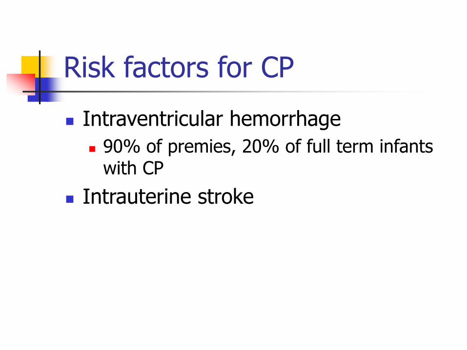
Risk factors for CP
Intraventricular hemorrhage
90% of premies, 20% of full term infants with CP
Intrauterine stroke
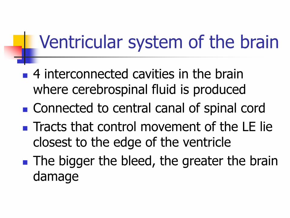
Ventricular system of the brain
4 interconnected cavities in the brain where cerebrospinal fluid is produced
Connected to central canal of spinal cord
Tracts that control movement of the LE lie closest to the edge of the ventricle
The bigger the bleed, the greater the brain damage

Ventricular system of the brain

Intraventricular hemorrhages
Grade I – A small amount of blood; stays in the ventricle
Grade II – A larger amount of blood; intraventricular; normal ventricle size
Usually have no neurological sequella

Intraventricular hemorrhages
Grade III – Even larger amount of blood; intraventricular; ventricular dilation
Grade IV – Blood spills outside of the ventricle and into the actual brain tissue
Usually have neurological sequella, and this qualifies a child for CSHCS

Periventricular leukomalacia

Risk factors for CP
Intrauterine infections/chorioamnionitis TORCH
Hyperbilirubinemia Kernicterus – associated with dystonic CP
and neurosensory hearing loss
Multiple gestation
“Vanishing twin” phenomenon
Twin-to-twin transfusion

Risk factors for CP
Hypoxia?
<10% of children with CP have history of anoxia/hypoxia
Documented anoxia/hypoxia is a risk factor
Acidosis, bradycardia
Neonatal encephalopathy
Hypoxic ischemic encephalopathy (HIE) on MRI
Eligible for CSHCS
Respiratory difficulties, abnormal tone, seizures
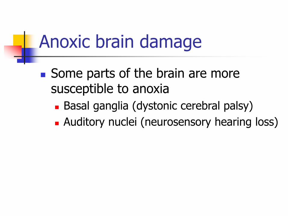
Anoxic brain damage
Some parts of the brain are more susceptible to anoxia
Basal ganglia (dystonic cerebral palsy)
Auditory nuclei (neurosensory hearing loss)

CP can be associated with:
Cognitive impairment (50%)
Seizures (50%)
Learning disabilities
Visual problems
Strabismus (75%), ROP, cortical blindness (HIE), hemianopsia (HP)
Incontinence

CP can be associated with:
Speech delays / hearing problems
Sensorineural deafness in hypoxia, TORCH, kernicteris, bacterial meningitis
GERD / Constipation / Failure to thrive
Aspiration pneumonia
Orthopedic complications
Dislocated hips, scoliosis, joint contracture
Decreased bone density

Functional Problems
Gross motor/Mobility
Learning
Fine motor
Feeding

Classification of CP
Type of movement disorder
Anatomic distribution

Type of movement disorder
Spastic – 70-85%
Dyskinetic (dystonic, athetoid) – 5-10%
Ataxic – 5%
Mixed – 10%
Hypotonic – 3%
Often overlapping/not clear cut

Hypotonic/Ataxic
Rare, therefore all children should receive a thorough diagnostic work-up for other neurological conditions

Spasticity
Increased tone/resistance to movement
Assessed by Deep tendon reflexes
Passive mobilization Movement through the full ROM should take
less than one second

Anatomic distribution of CP
Quadriplegia (32%) Diplegia (24%) Hemiplegia (30%)

Spastic Quadriplegia
All 4 extremities spastic; hypotonic trunk
Grade IV IVH
Majority with cognitive impairment
Seizures in >50%
High risk of aspiration pneumonia
Highest risk for orthopedic complications (scoliosis, hip dislocation)
50% achieve minimal ambulatory skills with an assistive device

Spastic Diplegia
Lower extremities more spastic than upper extremities
Grade III IVH
25-33% have seizures or cognitive impairment
80-90% are ambulatory with or without AD
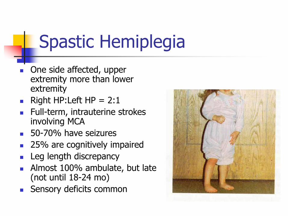
Spastic Hemiplegia
One side affected, upper extremity more than lower extremity
Right HP:Left HP = 2:1
Full-term, intrauterine strokes involving MCA
50-70% have seizures
25% are cognitively impaired
Leg length discrepancy
Almost 100% ambulate, but late (not until 18-24 mo)
Sensory deficits common

Common Presentations of CP
Delayed development of motor milestones
Early handedness
Persistence of primitive reflexes
Presence of pathologic reflexes
Failure to develop protective extension responses
Russman BS, etal, Spasticity 2002

Diagnosis
History, physical exam
No laboratory tests are diagnostic
EEG may be indicated
Head imaging may be helpful Ultrasound, MRI
>80% of kids with CP have abnormal findings
BAERs if indicated

Examination
Early hypotonia (especially trunkal)
Spasticity in extremities, develops over time
Brisk DTRs
Clonus
Upgoing plantar reflexes (Babinski)
Synergistic movement patterns
Joint contractures

Orthopedic problems
Scoliosis
Hip subluxation/dislocation
Knee flexion contractures
Patella alta
Foot deformities

Scoliosis
Neuromuscular
Risk increases with severity 70% in quads
Curves over 40 degrees tend to progress
Complicated by skin breakdown, joint contractures, pelvic tilt

Hip subluxation

Hip dislocation

Risks for hip dislocation
Scissoring pattern – adduction, often with hip flexion
Hip abduction < 35 degrees
Hip flexion contractures >20 degrees are risk factors
Persistent coxa valga
Excessive femoral anteversion
Shallow acetabulum

Risks for hip dislocation
Diagnosis Quadriplegia – 80%
Dystonia – 40%
Diplegia – 20%
Hemiplegia – 1%
Ambulatory Status Non-ambulatory – 70-90%
Ambulatory – 0-40% Graham, AACPDM presentation, 2006

Knee problems
Knee flexion contractures
Patella alta
Abnormally high patella
In CP, caused by prolonged positioning in flexion or by overactive quadriceps in crouch gait
In adolescence, can be painful

Patella alta

Foot Deformities in Child with Cerebral Palsy
Pronation
Supination

Pronation in CP
From Dormans

Supination Deformity in CP
From Dormans

Why don’t all kids with CP qualify for CSHCS?
Chronic
Sub-specialist-PM&R, Neuro, Ortho
Severity-(here’s the stopper!)
Therapy is not considered an indicator of severity
So we are looking for the need for interventions like equipment, spasticity medication, Botox injections, alcohol blocks, serial casting, surgery, etc

Treatment options
Therapy/Orthotics/ Equipment
Oral medications
Chemodenervation Botulinum toxin/
Phenol blocks
Neurosurgery ITB
SDR
Orthopedic surgery
Least invasive
Most invasive

Therapy (very simplified!!)
Occupational therapy Fine motor skills, Activities of Daily Living
Equipment
Physical therapy Gross motor skills
Equipment
Speech therapy Language, communication
Augmentative communication devices

Serial casting for joint contractures
A series of casts is applied weekly to gradually stretch the muscle
Might use Botox before
Bracing or splinting afterward
Preferable to a muscle lengthening surgery

Bracing (Orthotics)

Knee braces

SWASH Orthosis
Standing, Walking and Sitting Hip Orthosis
Controls dynamic hip scissoring

Other Equipment
Bathseats/shower seats
Strollers
Wheelchairs
Standers
Walking assistive device
Safe hospital beds
Augmentative Communication devices

Bath seats

Strollers/Wheelchairs
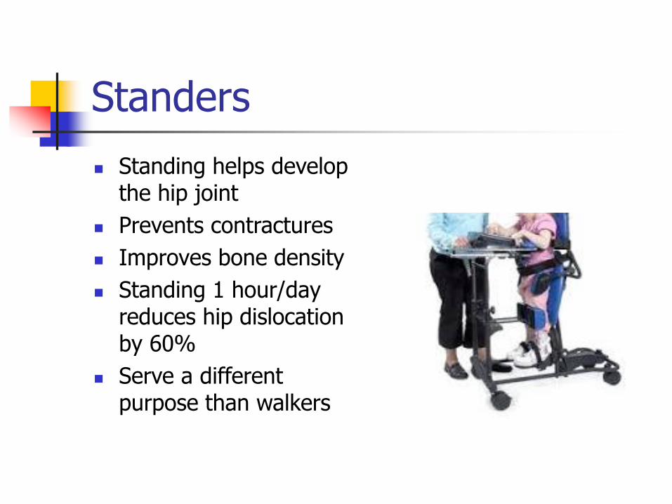
Standers
Standing helps develop the hip joint
Prevents contractures
Improves bone density
Standing 1 hour/day reduces hip dislocation by 60%
Serve a different purpose than walkers

Walkers

Safe Hospital Beds
It’s easy to see how a child could get entrapped in a standard hospital bed
The FDA identified 7 zones of entrapment
Safer bed technology was developed

Safe Hospital Beds
Beds by George
Sleep Safe
Pedicraft

Treatment of Scoliosis
Bracing
Surgery

Bracing/positioning for scoliosis

Treating spasticity
If spasticity interferes with Functioning
Positioning
Comfort
Care
If spasticity is not useful (ie: transfers)
If treatment is expected to provide improvement

Treating spasticity
Positioning
Oral medication
Chemodenervation
Surgery

Orthotics/Positioning
Bracing / Splinting
Positioning – biomechanical alignment is key!
Consider skin tolerance and wearing time

Effects of Biomechanical Alignment on Spasticity
From Cusick

Oral medications
Treat systemic spasticity, but have systemic side effects
Most common side effect is drowsiness
Oral medications
Benzodiazepines (Valium, Klonopin)
Baclofen (Lioresal)
Dantrolene sodium (Dantrium)
Tizanidine (Zanaflex)
Clonidine (Catapres)

Chemodenervation
Injectable therapy which results in local muscle weakening
Temporary and titratable
Botulinum toxin
Phenol or ethyl alcohol

Botulinum toxin
Temporarily weakens a muscle (3 months)
Creates a “window of opportunity”
Adjunct to serial casting, intense therapy
Don’t need to use anesthesia (vs alcohol blocks)
Topical anesthetic

Phenol/Ethyl Alcohol Injections
Motor nerve block or motor point block Can only be used on motor nerves (not
sensory)
Obturator and musculocutaneous nerves
Causes axonal protein denaturation
Results usually last 6-12 months
Done under anesthesia

Surgical Treatments
Intrathecal Baclofen Pump
Selective Dorsal Rhizotomy
Orthopedic surgeries

Intrathecal baclofen pump

Intrathecal Baclofen Pump
Implantable, programmable pump, controlled by telemetry
Baclofen – dosing significantly less that oral dose, so “no” side effects
Medication stable in pump up to 6 months
Lots of flexibility in dosing
Reduces risk of hip dislocation

Selective Dorsal Rhizotomy
EMG guided sectioning of afferent nerve rootlets from L2-S2
Interruption of reflex arc
Often “unmasks” underlying weakness
Not flexible dosing
Not done (locally) as much as ITB pump

Orthopedic Surgery
Lengthening Procedures (Muscular)
Tendo-achilles lengthening
Hip adductor lengthening
Hamstring lengthening
Selective Percutaneous Myofascial Lenghtening (Percs); New Jersey

Orthopedic Surgery
Rotational surgeries (Bony)
Varus Derotation Osteotomy (VDRO)
Tibia / fibula osteotomy
