management of complicated pneumonia in children: evidence
TRANSCRIPT
American Journal of Pediatrics 2020; 6(3): 240-252 http://www.sciencepublishinggroup.com/j/ajp doi: 10.11648/j.ajp.20200603.22 ISSN: 2472-0887 (Print); ISSN: 2472-0909 (Online)
Review Article
Management of Complicated Pneumonia in Children: Evidence Beyond Guidelines
Abdullah Saeed Al-Shamrani
Department of Pediatrics, Prince Sultan Military Medical City (PSMMC), Alfaisal University, Riyadh, Saudi Arabia
Email address:
To cite this article: Abdullah Saeed Al-Shamrani. Management of Complicated Pneumonia in Children: Evidence Beyond Guidelines. American Journal of
Pediatrics. Vol. 6, No. 3, 2020, pp. 240-252. doi: 10.11648/j.ajp.20200603.22
Received: May 20, 2020; Accepted: June 4, 2020; Published: June 16, 2020
Abstract: What is already known? Complicated pneumonia is an area of debate, and a rapid diagnosis is essential for patient survival. Due to the importance of stage-adapted therapeutic decisions, different classification systems have been established. Depending on the stage of the disease, both antimicrobial and interventional approaches are indicated. Conservative management remains the mainstay for the management of parapneumonic effusion, and continuous pleural fluid drainage is not necessary in some children. However, an established consensus worldwide for the management of complicated pneumonia and thoracic empyema with different therapeutic algorithms lacks clear evidence to evaluate complex cases with minimally invasive intervention versus open decortication. Such controversy concerning the best surgical approach persists, especially for sick patients in intensive care who are not doing well despite chest tube or fibrinolytic agents. This article aimed to review current treatment standards for children with different phases of thoracic empyema and complicated pneumonia, this review article will discuss the usefulness of different diagnostic methods and most recent updates on management. 1. Outline the definition, pathophysiology and common causes 2. Review the diagnosis and current treatment standards for complicated pneumonia 3. Review different surgical approaches and their outcomes 4. Provide an update on the recent utilization of video-assisted thoracoscopy (VATS) 5. Review evidence regarding the best fibrinolytic agent 6. Review evidence concerning when to indicate decortication 7. Provide a simplified pathway for the management of complicated pneumonia (figure 9).
Keywords: Pneumonia, Bronchopneumonia, Complicated Pneumonia, Empyema
1. Introduction
Inpatient management of previously healthy children (aged three months to 14 years) who presented with bacterial pneumonia complicated by pleural effusion or empyema.
Definitions and Staging (1) Clinical criteria for acute bacterial pneumonia: acute
presentation of high fever, cough, shortness of breath, tachypnea with localized physical and radiological signs of infection and may include anorexia or other systemic illness.
(2) Parapneumonic effusion (PPE): pleural effusion that is associated with pneumonia.
(3) Pleural effusion: fluid that escapes from blood vessels or lymphatics into the pleural cavity.
(4) Complicated effusion: effusion with pH <7.2 and/or positive for Gram staining or culture and/or multiple
loculations (5) Simple effusion: pleural effusion that does not meet the
criteria for complicated effusion or that is less than 1 cm in width on a lateral decubitus film.
(6) Thoracic empyema: an infectious condition of the pleural cavity, leading to pus formation; various underlying infectious diseases with pneumonia are the most common cause.
(7) Pus: A fluid product of inflammation, comprising a liquid containing leukocytes and debris of dead cells and tissues. Clinically, this may comprise a milky-yellow thick material, and/or bad odor and/or increased white blood cells, biochemical pH <7.1 and LDH >1000 IU.
(8) Exudative (early): the inflammatory process in the first few days associated with underlying pneumonia, leading to the accumulation of clear and thin fluid in the pleural cavity often with a low white blood cell count and near-normal pH

241 Abdullah Saeed Al-Shamrani: Management of Complicated Pneumonia in Children: Evidence Beyond Guidelines
and LDH (often called simple PPE). See Table 1. (9) Fibropurulent (intermediate): the inflammatory process
includes the deposition of fibrin in the pleural space, leading to septation and the formation of loculations. There is an increase in white blood cells, with fluid thickening (complicated PPE) eventually becoming clear pus (empyema). The presence of septations (fibrinous strands within the
pleural fluid) causes loculation of the pleural fluid, and fluids will not flow easily.
(10) Organizational (late): the third stage or empyema where fibroblasts infiltrate the pleural cavity, and the thin intrapleural membranes are reorganized to become thick and nonelastic (the “peel”). This pleural peel will prevent lung re-expansion—known as “tapped lung”—which will prevent lung function. Frank pus or Gram staining or culture might be positive.
2. Methods
We searched PubMed up to May 2020 using the following words in different combinations: pneumonia, complicated pneumonia, bronchopneumonia, empyema, pleural effusion, pathophysiology, management, Video Assisted Thoracoscopic Surgery (VATS), decortication, complication, antibiotic, and guideline. Search filter include publications in the last 10 years, human species, and age less than 18 years. With these search 528 were identified. Papers were excluded based on titles and abstract. More than 50 studies were includes. The included studies where highly selected based on the best evidence available. Guidelines and Cochrane reviews were intensively reviewed. We understood the difficulty of creating guideline especially for common pediatric illness like complicated pneumonia, for this, the evidence was inspired from the recommendations in the center of evidence-based medicine website (www.cebm.net), listed by Burns et al [1].
3. General Concept
Community-acquired pneumonia is a leading cause of death and a major cause of morbidity worldwide [2]. Pleural effusion is the most common presentation of pleural disease [3]. The incidence of pleural effusion and empyema is increasing [4, 5]. In the United States, the rate of pneumonia is 20–40 per 100,000 mainly as community-acquired pneumonia, and PPE was reported in 20%–40% of admitted patients [6, 7]. In Europe, a significant increase in PPE cases occurred, from 1.25 to 16.7/100,000 patients [8-15]. Presently, the mortality is decreasing compared with earlier reports [8].
3.1. Clinical Presentation and Assessment
3.1.1. Clinical History
Patients with PPE/empyema usually present with the same complaints of pneumonia and include fever, cough, decrease appetite, tachypnea and difficulty of breathing. If the child presents with chest pain that often radiates to the ipsilateral shoulder and increases with inspiration, pleuritic irritation and possible effusion are suspected; often, the patient prefers
to lie on the affected side [16, 17]. Infection in the lower lobes often radiates to the abdomen and could mimic acute abdomen. Those diagnosed with pneumonia and continue to be sick for more than two days should be evaluated for potential pleural effusion. The duration of the disease is crucial as well as determining whether the patient received prior antibiotics, had a vaccination, had a previous similar illness in the family, has a history of underlying risk factors such as immunodeficiency, lung malformation, and foreign body [10].
3.1.2. Physical Examination
Pleural effusion is suspected in patients who are unwell with respiratory distress and unilateral decreased rib excursion, bulging of the involved hemithorax, reduced breath sound and tactile fremitus on the affected area, dullness on percussion and potential friction rub, especially with mild effusion. Other prominent findings include marked shifting of the trachea and mediastinal structures to the unaffected side. Low oxygen saturation blows of 92% are consistent with severe infection. In approximately one-third of the cases, mild scoliosis occurs on the affected side. Hydration should be monitored carefully because the patient is expected to have a poor appetite and increased loss of insensible water loss. Auscultation of the heart may show displacement of the heart sound. Figure clubbing might be positive in severe or chronic empyema [18].
3.2. Pathophysiology
Pleural fluid in the pleural cavity often represents an equilibrium between the filtration process (fluid formation) and absorption (fluid removal) [19]. Fluid movement in the pleural surface between the vascular compartment and pleural space is controlled by the Starling law at a filtration rate of less than 0.1 ml/kg/h. Pleural fluid works as a lubricant and is under subatmospheric pressure; in healthy children, the pleural fluid is continuously produced and the content is 0.3 ml/kg—1.5 g/l of protein (LDH is the predominant protein), <100/µl of cells and a normal pH (7.35 mmHg) [19, 20]. Normally, the pleural space is free of air because of the difference between the total gas pressure in the venous systems and pleural space.
3.3. Pleural Effusion (PE)
An imbalance between pleural fluid formation and drainage will result in pleural effusion. Hemostasis altered by the immune process of infection leads to increased vascular permeability and an influx of inflammatory cells (neutrophils, lymphocytes, and eosinophils) into the pleural space that produce several cytokines (e.g., interleukin (IL-1, IL-6, IL-8, platelet-activating factor) released by mesothelial cells lining the pleural space, and tumor necrosis factor). This fluid, called exudate, is the first stage of empyema. When the infection progresses, the fluid will become thicker with bacterial invasion across the damaged epithelium and neutrophils. Additionally, the coagulation cascade will be activated, leading to fibrin formation and septation, and the pleural fluid, pH and glucose levels fall while the LDH levels increase; this

American Journal of Pediatrics 2020; 6(3): 240-252 242
purulent fluid is very consistent with empyema [20-23].
3.4. Causative agents
The incidence of empyema complicating community-acquired pneumonia is increasing and causes
Significant childhood morbidity. Streptococcus pneumonia is the commonest organism implicated in PPE, especially in patients with incomplete vaccination [5, 8, 24-28]. Regarding pneumococcal serotypes, type 1, 3 and 19A displayed strong associations with pneumococcal PPE and empyema worldwide [5, 29]. Streptococcus pyogenes is a common organism, while staph aureus is the predominant organism post trauma in young age or developing countries [24, 25, 30]. Other organisms, such as Gram-negative bacteria (Pseudomonas aeruginosa or Klebsiella pneumonia) are common in chronic patients or those with a prolonged history of admission. [15, 23, 31, 32]. H. influenza is very rare currently due to eradication by effective vaccination. [30]. Anaerobes (Bacteroid) play an important role in aspiration or poor dental hygiene [31]. Mycobacterium tuberculosis and Candida rarely cause complicated pneumonia in immune-compromised patients [32]. Mycoplasma is a common cause of empyema in 15% of school-aged children [33, 34]. Although viruses are the commonest cause of respiratory illness, they rarely cause pleural effusion, and many viruses that have been reported to cause effusion, particularly influenza or HINI, adeno, respiratory syncytial virus, were associated with immune-compromised patients [15, 35].
4. Investigations
Imaging—this procedure aims to determine the presence of fluid, differentiate simple parapneumonic effusion from an empyema and assess the complexity of the latter [36].
4.1. Chest X-ray
This is the simplest and least expensive method to identify PE, but it is not specific. Obliteration of the costophrenic angle is the earliest radiological sign of pleural fluid accumulation; as the volume of effusion increases, the characteristic “meniscus” sign can be seen on chest X-ray [16, 36]. Effusion accumulates in the subpulmonic location, spills into the costophrenic sulcus posteriorly, anteriorly, and laterally and then surrounds the lung, forming a meniscus sign that can be seen clearly on either AP or PA film. A mediastinal shift away from the affected side suggests the presence of fluid (Figure 1) [37]. In the supine position, the whole affected side is homogeneous without classical signs of effusion; when there is white out, then collapse consolation should be ruled out carefully. Minimal additive information can be obtained from lateral film; scoliosis can be detected by chest X-ray and is usually resolved spontaneously [16].
In the lateral decubitus film, the child lying on the affected side provides valuable information about the quantity and quality of the effusion. A fluid layer of more than 10 mm
between the inside of the chest wall and lung suggests the presence of an adequate amount of fluid for thoracocentesis (Figure 2). Non-shifting fluid suggests either thick fluid or loculation [38, 39]. While the supine chest X-ray is less helpful than erect in detecting pleural fluid or even air [38].
Figure 1. AP heterogeneous opacity on the left-sided lung, saluting both
costophrenic and cardiophrenic angles with a shift of the trachea to the
right-sided lung and the absence of an air bronchogram suggest large
effusion.
Figure 2. Left lateral decubitus film shows a free fluid level of almost 1 cm
in width with some ground glass opacity in the left upper zone.
Recommendation:
1. Chest X-ray is a simple and the least expensive method
to identify pleural effusion.
2. Obliteration of the costophrenic angle is an early sign.
3. Any child with suspected pleural effusion should undergo
chest X-ray with lateral decubitus film performed to
evaluate the presence, size and mobility of the suspected
effusion.
4. Lateral decubitus film is still inadequately used in the
emergency, and its utilization should be encouraged.
5. Further imaging should be used before proceeding to
further management when the plain X-ray does not show
complete free-flowing fluid or if another diagnosis, such
as an abscess, is considered.
4.2. CT Scan
CT plays a role in complicated pneumonia and is an excellent tool to evaluate lung parenchyma. CT with contrast enhancement is the preferred tool to assess loculated pleural fluid and detects endobronchial obstruction with a mucous

243 Abdullah Saeed Al-Shamrani: Management of Complicated Pneumonia in Children: Evidence Beyond Guidelines
plug or foreign body and mediastinal abnormalities [40, 41]. It can differentiate pleural effusion from pleural thickening and lung abscess, and determine the loculation site (Figure 3). It is very useful as guidance for intervention. Due to the high radiation and risk of sedation, CT is indicated in limited conditions, such as the failure of medical therapies or failure to drain after initial aspiration [40, 41]. It is rarely indicated in the acute phase with some limitations because it is not helpful to differentiate PPE from empyema [37]. Many surgeons request a routine CT to use as a ‘road map’ when performing minimally invasive endoscopic surgery where direct visual access is limited. The routine use of CT scanning in empyema should be discouraged if it will result in increased exposure of children to unnecessary radiation, in some patients already on nephrotoxic medications such as vancomycin or aminoglycoside where contrast might exaggerate the risk of acute kidney injury, further, associated with increased costs and resource burden [42-45].
Figure 3. CT scan axial view shows a thick, nodular pleural surface, marked
effusion, a collapsed right lung, and a shift of the mediastinum to the left
side.
Recommendation:
1. CT should not be used as a routine procedure in
patients with PPE.
2. CT is an excellent tool to evaluate lung parenchyma and
determine the loculation
3. CT can be used as guidance (road map) before
intervention.
4. CT can be used when there is failure to drain after
intervention or atypical presentation.
4.3. Ultrasonography
Chest ultrasonography is frequently used to assess pleural disease detected on chest X-ray, and it is becoming more popular due to its portability and ease of use for different types of patients—inpatient, outpatient or critical cases. It can estimate pleural fluid and differentiate free from loculated fluids. Furthermore, it can detect pleural thickening and is very useful to allocate the best site for chest tube insertion or thoracocentesis (Figure 4) [37, 46-49]. The pattern of echogenicity is important; anechoic effusion is usually a transudate, while the presence of echogenicity within pleural effusion usually indicates an exudate, further, septations or
internal echogenicity are suggested exudates [37]. Ultrasound scanning is now the preferred investigation modality in children because no sedation is necessary and it involves no radiation; however, it is operator dependent [50-52].
Figure 4. Ultrasound view showing loculated fluid, multiple septation, and a
thin fluid level.
Recommendation:
1. Ultrasonography is examiner dependent and can
estimate the amount of pleural fluid.
2. -It distinguishes pleural thickening from effusion, and
utilization should be encouraged.
3. -It can be used as guidance before intervention.
4. -Echogenic pleural fluid is often an exudate.
5. Ventilation-perfusion (VQ) scans VQ scans play no role in the management of the acute
phases of PPE and empyema. Further VQ scans may show some abnormalities up to a decade in children with a history of empyema [53, 54].
4.4. Bronchoscopy
This procedure has not been studied well in children. Lavage can retrieve organisms from the lower airway, but it is an invasive procedure while the patient is sick and distressed. Additionally, the usefulness of lavage compared with pleural tap has not been studied. In most cases, there is no indication for bronchoscopy and bronchoalveolar lavage, and it is not routinely recommended. Lavage can be requested in limited conditions when the patient shows no improvement and no apparent organism or underlying immunodeficient condition or fistula formation. [55, 56].
Recommendations:
1. There is no indication for flexible bronchoscopy in
complicated pneumonia; lavage is limited if an
unknown organism is present or in
immunocompromised cases.
4.5. Blood Test
4.5.1. Acute-phase Reactants
Acute-phase reactants are a class of proteins that increase or decrease in response to infection or inflammation, and the white blood cell count, neutrophil and lymphocyte count, C-reactive protein (CRP), and erythrocyte sedimentation rate (ESR) are usually performed as initial blood work in

American Journal of Pediatrics 2020; 6(3): 240-252 244
suspected complicated pneumonia. Acute-phase reactants cannot help to differentiate bacterial from viral infections but can be used as markers for the severity of infection and progress of inflammation [57, 58]. Procalcitonin is a popular and sensitive marker for bacterial infection. [59-61]. Several studies have investigated the usefulness of acute-phase reactants but have concluded it is an insensitive tool to differentiate bacterial from viral illnesses [62]. Viruses are rarely a causative agent in effusion, but virulent viruses such as H1N1, coronavirus, and influenza virus can cause significant PPE [63, 64]. No study has assessed the relationship between acute-phase reactants and the development of complicated effusion [16].
Recommendations:
1. Acute-phase reactants are usually evaluated by initial blood work for suspected complicated pneumonia
2. APRs are helpful to assess the severity and progress of infection
3. APRs are insensitive to differentiate between viral and bacterial infections
4.5.2. Blood Cultures
Blood cultures are helpful in the management of PPE according to British guidelines [16]. The chance of culture positivity is 6%–33% worldwide, and the yield is usually affected by previous antibiotic use and facility used for interventions. [17, 65-67].
Recommendations:
1-Blood cultures should be performed in all patients with
PPE
2-The yield is higher in patients who have not received
antibiotics
4.5.3. Albumin
The albumin level is often low in complicated pneumonia, but replacement therapy is rarely needed [16].
4.6. Sputum
If the child can expectorate, it should be sent for Gram staining and culture to reveal lower respiratory infection. Induced sputum is an alternative method to retrieve organisms in younger patients [16].
4.7. Pleural Fluid Analysis
In healthy children, the pleural fluid level is approximately 0.3 ml/kg, with a normal pH, a protein level less than 1.5 d/l, a white blood cell count of less than 100 cc/µl, and a predominance of lymphocytes. The chemical and cellular compositions of pleural fluid are very helpful in the management of pleural effusion and empyema. The gross appearance might suggest underlying disease; gross purulent fluid usually indicates empyema, while the presence of blood might suggest strep. Pneumonia that is tan and thick in appearance might suggest staph aureus, while putrid pneumonia suggests the presence of anaerobes [5, 21, 67]. Thin and clear fluids are usually suggestive of transudate.
Table 1. Characteristics features of pleural fluids
Exudate Transudate
Fluids Purulent Clear Odor May be offensive Odourless Viscous Non- Viscous Protein Protein >30 g/l Protein < 30 g/l - viscous Pleural: serum protein Serum protein> 0.5 Serum protein< 0.5 Pleural: serum LDH Serum LDH > 0.6 Serum LDH < 0.6
Pleural LDH PF: serum LDH> 0.66 Serum LDH LDH > 200 IU/L
Pleural LDH< 0.66 Serum LDH Or LDH < 200 IU/L
67. Light RW (1995) a new classification of parapneumonic effusions and empyema. Chest 108 (2): 299–301.
Pleural fluids are usually divided into three stages:
4.7.1. Exudative Stage
The pleural fluid is usually clear free-flowing exudative fluid with a predominance of neutrophils, and characterized by negative bacterial cultures, a glucose level greater than 60 mg/dL, a pH above 7.20, a lactic acid dehydrogenase (LDH) level less than three times the upper limit of normal for serum (often <1,000 units/L) and a low white cell count. Pleural fluid that develops during this stage is usually considered insignificant when ≤1 cm on lateral chest X-ray or significant when it is ≥1 cm on lateral decubitus X-ray [16, 20, 66-71].
4.7.2. Fibropurulent Stage
This second stage is characterized by the deposition of fibrin clots and fibrin membranes in the pleural space, leading to loculations. The loculation is accelerated by bacterial
invasion from pulmonary lung tissue across the damaged endothelium, leading to migration of neutrophils, activation of the coagulation cascade and formation of septation within the pleural fluid [66-71].
The pleural fluid in this stage is often turbid and characterized by positive bacteria on Gram staining or culture. The cytology shows neutrophil predominance. A reduced pleural fluid pH to acidic with increasing LDH suggests the accumulation of lactic acid and increased carbon dioxide production from bacterial invasion and increase inflammation. This stage called “complicated” parapneumonic effusion and shows a pH below 7.20, a sugar level less than 60 mg/dL, and pleural LDH more than three times the upper limit of normal [21, 66-71].
4.7.3. Organized Stage
This is the final stage characterized by fibroblasts that

245 Abdullah Saeed Al-Shamrani: Management of Complicated Pneumonia in Children: Evidence Beyond Guidelines
proliferate and invade the pleural cavity, and the thin intrapleural membranes are reorganized to become a thick pleural peel. These solid fibrous pleural peels form thick non-elastic pleura, preventing lung re-expansion (“trapped lung”), causing impaired gas exchange, and increasing the risk for continued infection [16, 67-71]. The clinical course varies considerably, from spontaneous healing with residual defects of lung function to chronic forms of empyema with a high risk of complications, such as a trapped restricted lung, bronchopleural fistula and septicemia [20, 67-71].
Table 2. Classification of parapneumonic effusion.
Simple PE Complicated PE Empyema
Gross inspection Thin Thick Thick, rigid Color Clear Turbid Pus PH >7.3 7.1-7.3 <7.1 LDH (IU) <500 >1000 >1000 Glucose (mg/dl) >60 <40 <40 Cell <1000 >5000 Variable Microbiology sterile Positive +/-
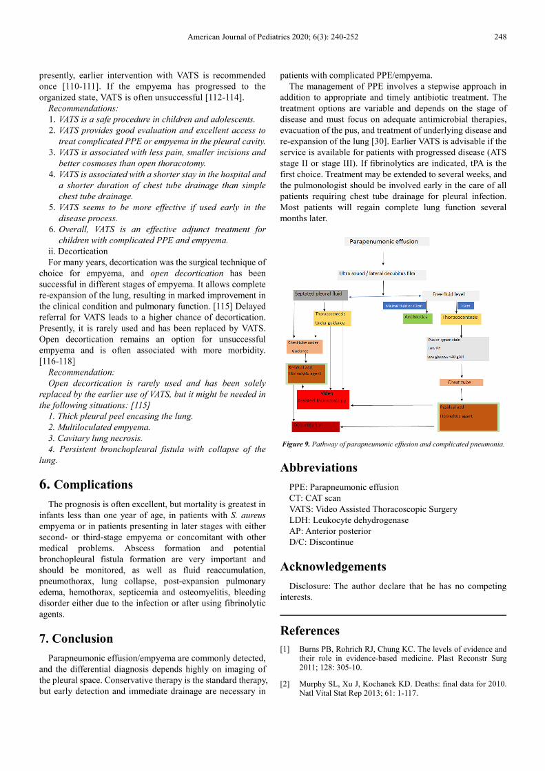
5. Management
The management of PPE and empyema involves a stepwise approach in addition to appropriate and timely antibiotic treatment. The therapeutic options include the following: antibiotics and observation, therapeutic thoracocentesis, tube thoracostomy, intrapleural instillation of fibrinolytics, video-assisted thoracoscopy with the breakdown of adhesions and/or decortication, and open thoracotomy.
5.1. Goals for Treatment
The main goals in the management of complicated pneumonia are as follows:
1. Treatment of the ongoing infection 2. Drainage of the pleural fluid or pus if indicated 3. Re-expansion of the lung 4. Prevention of complications All children with complicated pneumonia need admission.
[16] Appropriate antimicrobial therapies are often adequate in treating such cases. The presence of PPE does not alter the empirical decision to choose initial antibiotics. [16] Broad-spectrum antibiotics such as third-generation cephalosporin (cefotaxime or ceftriaxone) are used initially; some experts add clindamycin to cover staphylococcal infection and anaerobes, and then modify according to the progress and cultures. If the culture is positive for staph and the patient is still sick, vancomycin or linezolid is an option for therapy [74]. Other therapies include antipyretics, intravenous fluids and oxygen. Analgesia is an essential treatment to keep the child comfortable, particularly in the presence of a chest tube or post intervention. Chest physiotherapy is commonly used; however, the evidence is not beneficial and should not be performed in children with complicated pneumonia, and early mobilization and exercise should be encouraged [16]. If the effusion is enlarging and/or compromising, respiratory function should not be managed only by antibiotics; drainage should also be indicated [75].
Recommendations:
1. All children with PPE or empyema should be admitted to
the hospital
2. The patient's clinical status and vital signs should be
evaluated regularly
3. If a child has persistent pyrexia or is unwell 48 hours
after admission for pneumonia, PPE/empyema must be
excluded
4. Imaging alone should not be used to decide on the choice
of management
5. Chest physiotherapies are not helpful.
5.2. Surgical Interventions
Surgical intervention is a clinical decision when there is continuous fever for more than 48 hours despite appropriate antibiotics. The criterion for PPE that needs pleural drainage is large/loculated effusion or the presence of frank pus.
5.2.1. Thoracocentesis
This simple and safe method to determine the cause of pleural effusion is less invasive and better tolerated than the chest tube. The usual site is the fifth to seventh mid axillary line, and the optimal site can be guided by chest X-ray or ultrasonography [16, 76, 77]. The pleural fluid should be analyzed for the biochemical profile, cell count and differential, glucose, protein, LDH, pH, Gram staining and cultures. The pleural culture might be positive in 48% of patients, especially those who did not receive previous antibiotics [70]. Stage 1 (exudative stage) is often treated with antibiotics alone but also early stage 2 with effusion exceeding 1 cm on lateral decubitus or approximately 50% of the hemithorax (Figure 5). Thoracocentesis or even repeated thoracocentesis is helpful to monitor biochemical changes and monitor the progress of infection [66, 67].
Figure 5. Thoracocentesis under ultrasonography guidance using a 6F
catheter.
Recommendations:
1. Diagnostic thoracocentesis is indicated in all patients
with pleural effusion of a significant size (>10 mm on
lateral decubitus film)
2. The pleural fluid should be noted for color, smell, and

American Journal of Pediatrics 2020; 6(3): 240-252 246
consistency and should be analyzed for Gram staining,
culture (aerobic and anaerobic) and sensitivity, cell
count as well as the biochemistry profile (pH, LDH,
glucose and protein).
5.2.2. Chest Tube (CT)
Controversy remains concerning the optimal treatment of pediatric CPPE [80]. A chest tube is indicated in complicated PPE or empyema, and the safe triangle is the preferred site. In the mid axillary line, this triangle is bordered by the anterior border of the latissimus dorsi, the lateral border of the pectoralis major muscle, a line superior to the horizontal level of the nipple, and an apex below the axilla. A more posterior position is suggested by the images is another option (Figure 6). The size of the chest tube is determined according to the patient’s age: three different sizes are recommended (8-14 F/16-24F/>24F. Well-trained health professionals are recommended to perform the procedure, such as surgeons, interventional radiologists or intensivists. The tube should be hooked to an underwater seal with a negative pressure of -10 to -20 mmHg, and the drain should be below the level of the patient. Chest X-ray should be repeated after insertion of the chest tube [16]. The tube should be kept until the drain is minimal, an average of 1 ml/kg/day. The chest tube should be monitored to determine if it drains well or if bubbling occurs [16]. The tube should be clamped if the drain exceeds 10 ml/kg for a minimum of one hour to avoid potential pulmonary edema. Daily assessment of the amount of the drain and presence of bubbling and respiratory status should be monitored. The average duration of the chest tube is 4–5 days in complicated pneumonia, and the drain should be removed when there is clinical resolution, with clamping for a few hours before removal is indicated [81].
Figure 6. Chest tube in situ on the right-sided lung before D/C.
Recommendations:
1. A chest tube is indicated in complicated pneumonia; a
safe triangle is the best site for insertion
2. Chest tube insertion should be performed by an expert
physician
3. The chest tube may be kept if the drain exceeds 10 ml/kg
4. The chest tube should never be clamped if bubbling
occurs
5. Chest X-ray is routinely indicated post intervention
5.2.3. CT+ Fibrinolysis
The adjunctive use of intrapleural fibrinolytics has helped increase the drainage volume in certain adult patients and is considered one of the acceptable approaches identified to manage pediatric patients. National and international guidelines recognize adjunctive intrapleural fibrinolytic therapy as an accepted therapeutic option.[82-84] Intrapleural fibrinolytics are recognized to shorten the hospital stay and are recommended for any complicated PPE (thick fluid with loculations) or empyema (overt pus). These guidelines also state that there is no evidence that any of the 3 fibrinolytics (streptokinase, urokinase, and alteplase) is more effective than another; only urokinase has been studied in a randomized controlled trial in children; thus, it is recommended. They decrease the viscosity of the gelatinous component of the pleural fluids and partially derides the restrictive pleural fibrin sheets [85, 86].
i. Streptokinase A dose of 250,000 units in 100 ml of 0.9% saline or a total
dosage of 12,000 U/kg/d of streptokinase in 50 mL of 0.9% saline solution is instilled into the drainage tube; the tube is clamped on the completion of instillation. Throughout the process, the patients prefer to be rotated into several positions to facilitate pleural distribution for 2 hours, and then the tubes should be unclamped and placed back on -5 to 20 cm of H2O suction; coagulation should be performed before intervention. The drainage effusion amount should be monitored and recorded daily, with recommended therapy ranging from 3 to 5 days. The success rate with streptokinase is 70%–90%, usually in three doses but up to 6 doses can be helpful [86-90].
ii. Urokinase This is a thrombolytic agent formed in the kidney that is
more potent than streptokinase. A dose of 100,000 units in 100 ml of 0.9% saline for 2 to 12 hours is safe and effective, shortens the hospital stay, and increased pleural fluid drainage. A total of 40,000 units of urokinase in 40 ml saline is used (10,000 units in 10 ml of saline if less than 1 year old) [91-94].
iii. Tissue Plasminogen Activator (tPA) Various intrapleural dosage regimens have been used: 2 to 5
mg diluted in up to 40 mL of normal saline or 0.1 mg/kg diluted in 10 to 100 mL of normal saline. The maximum doses have ranged from 3 mg to 6 mg.
Some retrospective reviews of PPE observed no bleeding episodes associated with intrapleural (tPA) administration in children, while others have noted minor bleeding in drained pleural fluid in some patients [95, 96].
An isolated case report of intrapleural hemorrhage related to the intrapleural administration of a single dose of tPA (3 mg; 0.1 mg/kg diluted in 30 mL of normal saline) was observed in a 6-year-old boy with pleural effusion [84, 97]. Scant data exist on Dornase alfa in children; however,
247 Abdullah Saeed Al-Shamrani: Management of Complicated Pneumonia in Children: Evidence Beyond Guidelines
it has been used in adults with a favorable outcome [100, 101].
Table 3. Fibrinolytic doses for treatment of PPE /empyema.
Types Doses Duration
tPA 0.1mg/kg in 10-40ml of normal saline Daily for 3-6 days
Urokinase 40 000 units in 40 mL 0.9% saline for children 1 year and older. 10 000 units in 10 mL 0.9% saline for
children younger than 1 year. It should be administered twice daily (with a 4-hour dwell time) Daily for 3-6 days
Streptokinase 25000iu /kg in 50-100 saline intrapleurally via a chest tube with clamping for 4 hours; a maximum dose of
250 000 IU per instillation. Every day for 7 days
Recommendations:
1. Urokinase is safe in children.
2. Urokinase can help to avoid the need for open
thoracotomy.
3. Urokinase use has been associated with a decreased
hospital stay.
4. Urokinase use has been associated with a significant
increase in pleural fluid drainage; however, the
significance is unclear.
5. When urokinase is used, a large-bore chest tube may not
be necessary for drainage and smaller ones such as the
pigtail catheter might be sufficient.
6. The use of urokinase is an effective adjunct treatment for
complicated PPE and empyema.
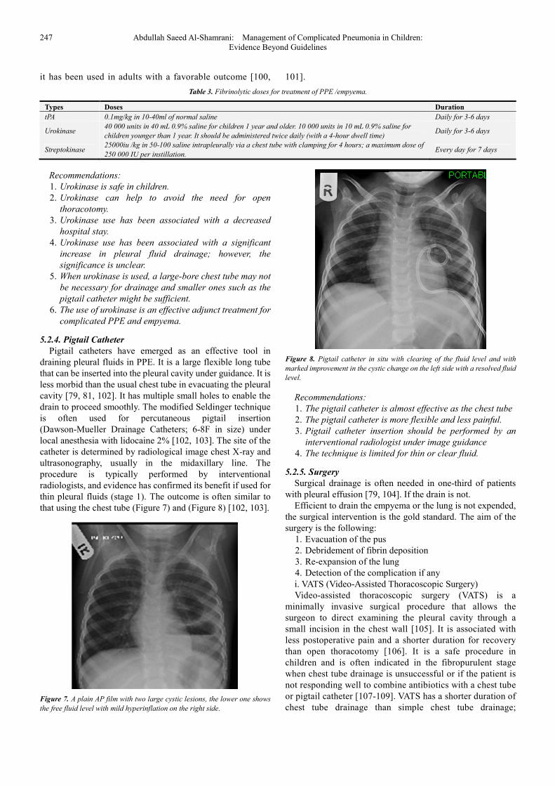
5.2.4. Pigtail Catheter
Pigtail catheters have emerged as an effective tool in draining pleural fluids in PPE. It is a large flexible long tube that can be inserted into the pleural cavity under guidance. It is less morbid than the usual chest tube in evacuating the pleural cavity [79, 81, 102]. It has multiple small holes to enable the drain to proceed smoothly. The modified Seldinger technique is often used for percutaneous pigtail insertion (Dawson-Mueller Drainage Catheters; 6-8F in size) under local anesthesia with lidocaine 2% [102, 103]. The site of the catheter is determined by radiological image chest X-ray and ultrasonography, usually in the midaxillary line. The procedure is typically performed by interventional radiologists, and evidence has confirmed its benefit if used for thin pleural fluids (stage 1). The outcome is often similar to that using the chest tube (Figure 7) and (Figure 8) [102, 103].
Figure 7. A plain AP film with two large cystic lesions, the lower one shows
the free fluid level with mild hyperinflation on the right side.
Figure 8. Pigtail catheter in situ with clearing of the fluid level and with
marked improvement in the cystic change on the left side with a resolved fluid
level.
Recommendations:
1. The pigtail catheter is almost effective as the chest tube
2. The pigtail catheter is more flexible and less painful.
3. Pigtail catheter insertion should be performed by an
interventional radiologist under image guidance
4. The technique is limited for thin or clear fluid.
5.2.5. Surgery
Surgical drainage is often needed in one-third of patients with pleural effusion [79, 104]. If the drain is not.
Efficient to drain the empyema or the lung is not expended, the surgical intervention is the gold standard. The aim of the surgery is the following:
1. Evacuation of the pus 2. Debridement of fibrin deposition 3. Re-expansion of the lung 4. Detection of the complication if any i. VATS (Video-Assisted Thoracoscopic Surgery) Video-assisted thoracoscopic surgery (VATS) is a
minimally invasive surgical procedure that allows the surgeon to direct examining the pleural cavity through a small incision in the chest wall [105]. It is associated with less postoperative pain and a shorter duration for recovery than open thoracotomy [106]. It is a safe procedure in children and is often indicated in the fibropurulent stage when chest tube drainage is unsuccessful or if the patient is not responding well to combine antibiotics with a chest tube or pigtail catheter [107-109]. VATS has a shorter duration of chest tube drainage than simple chest tube drainage;
American Journal of Pediatrics 2020; 6(3): 240-252 248
presently, earlier intervention with VATS is recommended once [110-111]. If the empyema has progressed to the organized state, VATS is often unsuccessful [112-114].
Recommendations:
1. VATS is a safe procedure in children and adolescents.
2. VATS provides good evaluation and excellent access to
treat complicated PPE or empyema in the pleural cavity.
3. VATS is associated with less pain, smaller incisions and
better cosmoses than open thoracotomy.
4. VATS is associated with a shorter stay in the hospital and
a shorter duration of chest tube drainage than simple
chest tube drainage.
5. VATS seems to be more effective if used early in the
disease process.
6. Overall, VATS is an effective adjunct treatment for
children with complicated PPE and empyema.
ii. Decortication For many years, decortication was the surgical technique of
choice for empyema, and open decortication has been successful in different stages of empyema. It allows complete re-expansion of the lung, resulting in marked improvement in the clinical condition and pulmonary function. [115] Delayed referral for VATS leads to a higher chance of decortication. Presently, it is rarely used and has been replaced by VATS. Open decortication remains an option for unsuccessful empyema and is often associated with more morbidity. [116-118]
Recommendation:
Open decortication is rarely used and has been solely
replaced by the earlier use of VATS, but it might be needed in
the following situations: [115] 1. Thick pleural peel encasing the lung.
2. Multiloculated empyema.
3. Cavitary lung necrosis.
4. Persistent bronchopleural fistula with collapse of the
lung.
6. Complications
The prognosis is often excellent, but mortality is greatest in infants less than one year of age, in patients with S. aureus empyema or in patients presenting in later stages with either second- or third-stage empyema or concomitant with other medical problems. Abscess formation and potential bronchopleural fistula formation are very important and should be monitored, as well as fluid reaccumulation, pneumothorax, lung collapse, post-expansion pulmonary edema, hemothorax, septicemia and osteomyelitis, bleeding disorder either due to the infection or after using fibrinolytic agents.
7. Conclusion
Parapneumonic effusion/empyema are commonly detected, and the differential diagnosis depends highly on imaging of the pleural space. Conservative therapy is the standard therapy, but early detection and immediate drainage are necessary in
patients with complicated PPE/empyema. The management of PPE involves a stepwise approach in
addition to appropriate and timely antibiotic treatment. The treatment options are variable and depends on the stage of disease and must focus on adequate antimicrobial therapies, evacuation of the pus, and treatment of underlying disease and re-expansion of the lung [30]. Earlier VATS is advisable if the service is available for patients with progressed disease (ATS stage II or stage III). If fibrinolytics are indicated, tPA is the first choice. Treatment may be extended to several weeks, and the pulmonologist should be involved early in the care of all patients requiring chest tube drainage for pleural infection. Most patients will regain complete lung function several months later.
Figure 9. Pathway of parapneumonic effusion and complicated pneumonia.
Abbreviations
PPE: Parapneumonic effusion CT: CAT scan VATS: Video Assisted Thoracoscopic Surgery LDH: Leukocyte dehydrogenase AP: Anterior posterior D/C: Discontinue
Acknowledgements
Disclosure: The author declare that he has no competing interests.
References
[1] Burns PB, Rohrich RJ, Chung KC. The levels of evidence and their role in evidence-based medicine. Plast Reconstr Surg 2011; 128: 305-10.
[2] Murphy SL, Xu J, Kochanek KD. Deaths: final data for 2010. Natl Vital Stat Rep 2013; 61: 1-117.
249 Abdullah Saeed Al-Shamrani: Management of Complicated Pneumonia in Children: Evidence Beyond Guidelines
[3] Helena Tresinha, Mocelin and Gilberto Bueno Fischer, epidemiology, presentation and treatment of pleural effusion. Paerdiatric respiratory review. 2002; (3), 292-297.
[4] Rees JH, Spencer DA, Parikh D, et al. Increase in incidence of childhood empyema in West Midlands, UK. Lancet 1997; 349: 402.
[5] Carrie L. Byington, LaShonda Y. Spencer, Timothy A. Johnson, Andrew T. Pavia, Daniel Allen, Edward O. Mason, Sheldon Kaplan, Karen C. Carroll, Judy A. Daly, John C. Christenson, And Matthew H. Samore. An Epidemiological Investigation of a Sustained High Rate of Pediatric Parapneumonic Empyema: Risk Factors and Microbiological Associations. Clinical Infectious Diseases 2002; 34: 434–40.
[6] Heffner JE, McDonald J, Baebieri C et al, Management of parapenumonic effusion. An analysis of physician practice patterns. Arch Surg 1995, 130: 433-438.
[7] Danasekaran R, Mani G, and Annadurai K. Prevention of healthcare-associated infections: protecting patients, saving lives. Int J Community Med Public Health. 2014; 1 (1): 67–68.
[8] Hernandez-Bou, S.; Garcia-Garcia, J. J.; Esteva, C.; Gene, A.; Luaces, C.; Munoz Almagro, C. Pediatric parapneumonic pleural effusion: Epidemiology, clinical characteristics, and microbiological diagnosis. Pediatr. Pulmonol. 2009, 44, 1192–1200.
[9] Playford SD, Smyth AR, Stewart RJ. Increase in incidence of childhood empyema. Thorax 1997; 52: 932.
[10] Krenke, K., Urbankowska, E., Urbankowski, T., Lange, J., & Kulus, M. Clinical characteristics of 323 children with parapneumonic pleural effusion and pleural empyema due to community acquired pneumonia. Journal of Infection and Chemotherapy. 2016; 22 (5), 292–297.
[11] Harris, M.; Clark, J.; Coote, N.; Fletcher, P.; Harnden, A.; McKean, M.; Thomson, A.; British Thoracic Society Standards of Care Committee. British Thoracic Society guidelines for the management of community acquired pneumonia in children: Update 2011. Thorax 2011, 66, 1–2.
[12] Light RW, Girard WM, Jenkinson SG, et al. Parapneumonic effusions. Am J Med 1980; 69: 507-12.
[13] Light RW. Parapneumonic effusions and empyema. Proc Am Thorac Soc 2006; 3: 75-80.
[14] Li, S. T.; Tancredi, D. J. Empyema hospitalizations increased in US children despite pneumococcal conjugate vaccine. Pediatrics 2010, 125, 26–33.
[15] Yu, D.; Buchvald, F.; Brandt, B.; Nielsen, K. G. Seventeen-year study shows rise in parapneumonic effusion and empyema with higher treatment failure after chest tube drainage. Acta Paediatr. 2014, 103, 93–99.
[16] British Thoracic Society Standards of Care Committee. British Thoracic Society guidelines for the management of community acquired pneumonia in childhood. Thorax 2002; 57 (I): 1–24.
[17] Mukherjee. S, Langrouge B., Rosenthal M and Balfourlynn I. M. Incidence and outcome of scoliosis in children with pleural effusion Pediatr Pulmonol. 2007; 42: 221–224.
[18] Kalantri S, Joshi R, Lokhande T, Singh A, Morgan M, Colford JM, Pai M. Accuracy and reliability of physical signs in the diagnosis of pleural effusion. Respir Med. 2007 Mar; 101 (3):
431-8.
[19] Miserocchi G. Physiology and pathophysiology of pleural turnover. Eur Respir J. 1997; 10: 219–2.
[20] Davies CH, Gleeson FV, Davies RJO. BTS guidelines on the management of pleural infection. Thorax 2003; 58 (II): 18–28.
[21] Quadri A, Thomson AH. Pleural fluids associated with chest infection. Paediatr Respir Rev 2002; 3: 349–55. 5.
[22] Kroegel C, Anthony VB. Immunobiology of pleural inflammation: potential implications for path genesis, diagnosis and therapy. Eur Respir J. 1997; 10: 2411–8.
[23] Adam jedaffi, and Ian M. Balfour-Lynn. Management of empyema Pediatric Pulmonology 2005; 40: 148–156.
[24] Patrick M. Meyer Sauteur, Ariane Burkhard, Ueli Moehrlen, Christa Relly, Christian Kellenberger, Kerstin Ruoss and Christoph Berge. Pleural Tap-Guided Antimicrobial Treatment for Pneumonia with Parapneumonic Effusion or Pleural Empyema in Children: A Single-Center Cohort Study. J. Clin. Med. 2019; 8, 698.
[25] Nyambat B, Kilgore PE, Yong DE, et al. Survey in childhood empyema in Asia: implications for detecting the unmeasured burden of culture negative bacterial disease. BMC Infect Dis. 2008; 8: 90.
[26] Spencer DA, Iqbal SM, Hasan A, Hamilton L. Empyema thoracic is still increasing in UK Children. BMJ. 2006; 332 (7553): 1333.
[27] Blaschke AJ, Heyrend C, Byington CL, et al. Molecular analysis improves pathogen identification and epidemiologic study of pediatric parapneumonic empyema. Pediatr Infect Dis J. 2011; 30: 289-294.
[28] Liese, J. G., Schoen, C., van der Linden, M., Lehmann, et al. Changes in the incidence and bacterial aetiology of paediatric parapneumonic pleural effusions/empyema in Germany, 2010-2017: a nationwide surveillance study. Clin Microbiol Infect. 2019; 25 (7): 857-64.
[29] Jessica Kurian, Terry L. Levin1, Bokyung K. Han, Benjamin H. Taragin and Samuel Weinstein Comparison of Ultrasound and CT in the Evaluation of Pneumonia Complicated by Parapneumonic Effusion in Children, American Journal of Roentgenology, American Journal of Roentgenology. 2009; 193: 1648-1654.
[30] Picazo J, Ruiz-Contreras J, Casado-Flores J, et al; HERACLES Study Group. Expansion of serotype coverage in the universal pediatric vaccination calendar: short term effects on age- and serotype-dependent incidence of invasive pneumococcal clinical presentations in Madrid, Spain. Clin Vaccine Immunol. 2013; 20: 1524-1530.
[31] Martin Reichert & Matthias Hecker & Biruta Witte & Johannes Bodner& Winfried Padberg & Markus AWeigand5 & Andreas Hecker1 stage-directed therapy of pleural empyema. Langenbecks Arch Surg. 2017; 402 (1): 15-26.
[32] Yu-Chao Lin, Hung-Jen Chen, Yi-Heng Liu, Chuen-Ming, Shih Wu-Huei Hsu, Chih-Yen Tu. A 30-Month Experience of Thoracic Empyema in a Tertiary Hospital: Emphasis on Differing Bacteriology and Outcome between the Medical Intensive Care Unit (MICU) and Medical Ward. South Med J. 2008 May; 101 (5): 484-9.
American Journal of Pediatrics 2020; 6(3): 240-252 250
[33] Patra, P. K. Unusual complication of Mycoplasma pneumonia in a five-year-old child. Australasian Medical Journal. 2013; 6 (2): 73–74.
[34] Kowthar S. Hassan and Ghalib Al-Khadouri. Mycoplasma pneumoniae Pneumonia with Worsening Pleural Effusion despite Treatment with Appropriate Antimicrobials. Sultan Qaboos Univ Med J. 2018; May18 (2): e239-242.
[35] Nestor, J., Huggins, T., Kummerfeldt, C., DiVietro, M., Walters, K., & Sahn, S. Viral diseases affecting the pleura. Journal of Clinical Virology. 2013; 58 (2): 367–373.
[36] R. E. Strachan, T. Gulliver, A. Martin, T. McDonald, G. Nixon, R. Roseby, S. Ranganathan, H. Selvadurai, G. Smith, S. Suresh, L. Teoh, J. Twiss, C. Wainwright, A. Jaffe. Paediatric Empyema Thoracis: Recommendations for Management. Position statement from the Thoracic Society of Australia and New Zealand. https://www.thoracic.org.au/journalpublishing/command/download_file/id/24/filename/PaediatricEmpyemaThoracisPositionStatementTSANZFINAL. pdf.
[37] Falaschi F, et al., [Comparison of computerized tomography and magnetic resonance in the assessment of benign and malignant pleural diseases]. Radiol Med, 1996. 92 (6): p. 713-8. 42. Blackmore CC, et al., Pleural fluid volume estimation: a chest radiograph prediction rule. Acad Radiol, 1996. 3 (2): 103-9.
[38] Hallifax, R. J., Talwar, A., Wrightson, J. M., Edey, A., & Gleeson, F. V. State-of-the-art: Radiological investigation of pleural disease. Respiratory Medicine. 2017; 124: 88–99.
[39] King S, Thomson A. Radiological perspectives in empyema. Br Med Bull 2002; 61: 203–14.
[40] Mukherjee S., Gleeson FV. Radiology in pleural disease: state of the art. Respirology. 2004; 9: 300–12.
[41] Tan Kendrick AP, Ling H, Subramanian R, Joseph VT. The value of early CT in complicated childhood pneumonia. Pediatr Radiol.. 2002; 32 (1): 16-2.
[42] Donnelly LF, Klosterman LA. CT appearance of parapneumonic effusions in children: findings are not specific for empyema. Am J Roentgenol. 1997; 169: 179–82.
[43] Kearney SE, Davies CW, Davies RJ, Gleeson FV. Computed tomography and ultrasound in parapneumonic effusions and empyema. Clin Radiol. 2000; 55 (7): 542–7.
[44] Jaffe A., Calder A D, Owens C M, Stanojevic, S Sonnappa Role of routine computed tomography in paediatric pleural empyema. Thorax 2008; 63: 897–902.
[45] Brenner DJ, Hall EJ. Computed tomography: an increasing source of radiation exposure. N Engl J Med. 2007; 357: 2277–84.
[46] Yonggeng Goh and Jeevesh Kapur. Sonography of the pediatric chest. American Institute of Ultrasound in Medicine, J Ultrasound Med. 2016; 35: 1067–1080.
[47] Merriam MA, Cronan JJ, Dorfman GS, et al. Radiographically guided percutaneous catheter drainage of pleural collections. Am J Roentgenol. 1988; 151: 1113–6.
[48] Yang PC, Luh KT, Chang DB, et al. Value of sonography in determining the nature of pleural effusion: analysis of 320 cases. Am J Roentgenol. 1992; 159: 29–33.
[49] Lomas DJ, Padley SG, Flower CD. The sonographic appearances of pleural fluid. Br J Radiol 1993; 66: 619–24.
[50] Eibenberger KL, Dock WI, Ammann ME, et al. Quantification of pleural effusions: sonography versus radiography. Radiology. 1994; 191: 681–4.
[51] Stavas J, van Sonnenberg E, Casola G, et al. Percutaneous drainage of infected and non-infected thoracic fluid collections. J Thoracic Imaging. 1987; 2: 80–7.
[52] Chen CH, Chen W, Chen HJ, Yu YH, Lin YC, Tu CY, Hsu WH. Transthoracic ultrasonography in predicting the outcome of small-bore catheter drainage in empyemas or complicated parapneumonic effusions. Ultrasound Med Biol. 2009; 35 (9): 1468–1474.
[53] Lange J, Krolicki L, Chiemelewska-Swewczyk D, Peradzynska J. Role of pulmonary scintigraphy in children post parapneumonic effusion and empyema. Eur Resp J. 2009; 18 (33), 170.
[54] Mew RC, Jaffe A, Biassoni L, Sonnappa S. Ventilation-perfusion scans in children treated for empyema. Thorax 2009; 64 (3): 273.
[55] IM Balfour – Lynn et al; BTS guideline for the management of pleural effusion in children. Thorax 2002; 57; 343-347.
[56] Takaoka Kazuo, Inoue Shoichi and Ohira Seiji Central Bronchopleural Fistulas Closed by Bronchoscopic Injection of Absolute Ethanol. Chest. 2002; 122 (1): 374-8.
[57] Nohynek H, Valkeila E, Leinonen M, et al. Erythrocyte sedimentation rate, white blood cell count and serum C- reactive protein in assessing etiologic diagnosis of acute lower respiratory infections in children. Pediatr Infect Dis J 1995; 14: 484–90.
[58] Korppi M, Heiskanen-Kosma T, Leinonen M. White blood cells, C-reactive protein and erythrocyte sedimentation rate in pneumococcal pneumonia in children. Eur Respir J 1997; 10: 1125–9.
[59] Schuetz P, Albrich W, Mueller B. Procalcitonin for diagnosis of infection and guide to antibiotic decisions: past, present and future. BMC Med. 2011; 9: 107.
[60] Korppi M, Remes S. Serum procalcitonin in pneumococcal pneumonia in children. Eur Respir J. 2001; 17: 623–7.
[61] Toikka P, Irjala K, Juven T, et al. Serum procalcitonin, C-reactive protein and interleukin-6 for distinguishing bacterial and viral pneumonia in children. Pediatr Infect Dis J. 2000; 19: 598–602.
[62] Virkki R, Juven T, Rikalainen H, et al. Differentiation of bacterial and viral pneumonia in children. Thorax. 2002; 57: 438–41.
[63] Alvarez-Lerma F, Marín-Corral J, Vila C, Masclans JR, González de Molina FJ, Martín Loeches I, Barbadillo S, Rodríguez A; H1N1 GETGAG/SEMICYUC Study Group. Delay in diagnosis of influenza A (H1N1) pdm09 virus infection in critically ill patients and impact on clinical outcome. Crit Care. 2016; 20 (1): 337.
[64] Atamna A, Babitch T, Bracha M, Sorek N, Haim BZ, Elis A, Bishara J, Avni T. Statins and outcomes of hospitalized patients with laboratory-confirmed 2017-2018 influenza. Eur J Clin Microbiol Infect Dis. 2019; 38 (12): 2341-8.
251 Abdullah Saeed Al-Shamrani: Management of Complicated Pneumonia in Children: Evidence Beyond Guidelines
[65] Hardie W, Bokulic R, Garcia VF, et al. Pneumococcal pleural empyemas in children. Clin Infect Dis. 1996; 22: 1057–63.
[66] Ori et al. Pleural Effusion in Pediatric Population. Pediatrics in Review. 2002; 23 (12): 417-426.
[67] Light RW. a new classification of parapneumonic effusions and empyema. Chest 1995; 108 (2): 299–301.
[68] Andrews NC, Parker EF, Shaw RR, Wilson NJ, Webb WR (1962) Management of non tuberculous empyema: a statement of the subcommittee on surgery. Am Rev Respir Dis. 1962; 85: 935.
[69] Muers MF. Streptokinase for empyema. Lancet 1997; 349 (9064): 1491–2.
[70] Colice GL, Curtis A, Deslauriers J, Heffner J, Light R, Littenberg B, Sahn S, Weinstein RA, Yusen RD (2000) Medical and surgical treatment of parapneumonic effusions: an evidence-based guideline. Chest. 118 (4): 1158–71.
[71] Hamm H, Light RW. Parapneumonic effusion andempyema. Eur Respir J 1997; 10: 1150.
[72] Lindsay McCauley, Nathan Dean. Pneumonia and empyema: causal, casual or unknown J Thorac Dis 2015; 7 (6): 992-998.
[73] Kartik Chandra Mandal, Gobinda Mandal, Pankaj Halder, Dipanwita Mitra, Bidyut Debnath, and Mala Bhattacharya. Empyema Thoracis in Children: A 5-Year Experience in a Tertiary Care Institute. J Indian Assoc Pediatr Surg. 2019 Jul-Sep; 24 (3): 197–202.
[74] Chibuk TK, Cohen E, Robinson JL, Mahant S, Hartfield DS; Paediatric complicated pneumonia: Diagnosis and management of empyema Paediatr Child Health. 2011; 16 (7): 425-7.
[75] Edward carter, John Waldhausen, Weiya Zhang, Lucas Hoffman and Gregory Peddling. Management of children with empyema: pleural drainage is not always necessary. Pediatr Pulmonol. 2010; 45 (2) 475-80.
[76] Baumer H. J. “guidelines review – parapneumonic effusion and empyema, Arch Dis of childhood: 2005: 90: 21–24.
[77] Janahi IA, Fakhoury K. Management and prognosis of parapneumonic effusion and empyema in children. Up-to-date. 2012. http://www.uptodate.com/contents/management-and-prognosis-of-parapneumonic-effusionand-empyema-in-children. Accessed March 14, 2014.
[78] Shoseyov, D., Bibi, H., Shatzberg, G., Klar, A., Akerman, J., Hurvitz, H., & Maayan, C. Short-term Course and Outcome of Treatments of Pleural Empyema in Pediatric Patients. Chest. 2002; 121 (3), 836–840.
[79] Farjah F, Symons RG, Krishnadasan B, Wood DE, Flum DR. Management of pleural space infections: a population-based analysis. J Thorac Cardiovasc Surg. 2007; 133 (2): 346–351.
[80] Loizzi, M., De Palma, A., Pagliarulo, V., Loizzi, D., & Sollitto, F. Pulmonary Infections of Surgical Interest in Childhood. Thoracic Surgery Clinics. 2012; 22 (3), 387–401.
[81] Segerer FJ, Seeger K, Maier A, Hagemann C, et al. Therapy of 645 children with parapneumonic effusion and empyema a German nationwide surveillance study. Pediatr Pulmonol. 2017; 52 (4): 540-7.
[82] Bradley JS, Byington CL, Shah SS, et al, and the Pediatric Infectious Diseases Society and the Infectious Diseases Society of America. The management of community-acquired pneumonia in infants and children older than 3 months of age: Clinical Practice Guidelines by the Pediatric Infectious Diseases Society and the Infectious Diseases Society of America. Clin Infect Dis. 2011; 53 (7): 25-76.
[83] Balfour-Lynn IM, Abrahamson E, Cohen G, et al; Paediatric Pleural Diseases Subcommittee of the BTS Standards of Care Committee. BTS guidelines for the management of pleural infection in children. Thorax. 2005; 60 (1): 11-21.
[84] Joyce A. Generali, and Dennis J. Cada, Alteplase: Pleural Effusion (Parapneumonic) and Empyema in chie Hosp Pharm 2013; 48 (11): 912–918.
[85] Yao CT, Wu JM, Liu CC, Wu MH, Chuang HY, Wang JN. Treatment of complicated parapneumonic pleural effusion with intrapleural streptokinase in children. Chest. 2004; 125: 566–571.
[86] Aydoğan, M., Aydoğan, A., Özcan, A., Tugay, M., Gokalp, A. S., & Arısoy, E. S. Intrapleural streptokinase treatment in children with empyema. European Journal of Pediatrics. 2007; 167 (7), 739–744.
[87] Cohen E, et al, Cost-effectiveness of competing strategies for the treatment of pediatric empyema. Pediatrics. 2008; 121: 1250–7.
[88] Ekta Singh, Anuj Kumar, D. Y. Shrikhande, P. Nigwekar. Role of Intrapleural Streptokinase in Children with Empyema - Randomised Controlled Trial. International Journal of Contemporary Medical Research. 2017, 4 (11): 2221-4.
[89] Joseph L. Mathew, Vemlish Soni et al. Intrapleural streptokinase is effective and safe for children with multi Loculated empyema regardless of the time from disease onset. Acta Paediatr. 2018; 107 (12): 2165-71.
[90] Mathew, J. L. (2019). Intrapleural Fibrinolytic Therapy in Empyema Thoracic: Where are we now and where do we go from here? The Indian J Pediatr. 2019; 86 (12): 1081-2.
[91] Thomson AH, Hull J, Kumar MR, Wallis C, Balfour Lynn IM. Randomized trial of intrapleural urokinase in the treatment of childhood empyema. Thorax. 2002; 57 (4): 343–347.
[92] Marhuenda C., Barcelo, C., Fuentes, I., Guillen, G., Cano, I., Lopez, M., Moreno-Galdo, A. (2014). Urokinase versus VATS for Treatment of Empyema: A Randomized Multicenter Clinical Trial. PEDIATRICS, 134 (5), 1301–07.
[93] Stefanutti, G., Ghirardo, V., Barbato, A., & Gamba, P. Evaluation of a pediatric protocol of intrapleural urokinase for pleural empyema: A prospective study. Surgery. 2010; 148 (3): 589–594.
[94] Thomson A H, J Hull, M R Kumar, C Wallis, I M Balfour Lynn Randomized trial of intrapleural urokinase in the treatment of childhood empyema. Thorax 2002; 57 343-347.
[95] Wells RG, Havens PL. Intrapleural fibrinolysis for parapneumonic effusion and empyema in children. Radiology. 2003; 228 (2): 370-378.
[96] Ray TL, Berkenbosch JW, Russo P, Tobias JD. Tissue plasminogen activator as an adjuvant therapy for pleural empyema in pediatric patients. J Intens Care Med. 2004; 19 (1): 44-50.
American Journal of Pediatrics 2020; 6(3): 240-252 252
[97] Hendaus MA, Abushahin A. Intrapleural hemorrhage due to alteplase use in a 6-year-old boy with pleural effusion. Int J Gen Med. 2013; 6: 233-236.
[98] Peter SD, Tsao K, Spilde TL, et al. Thoracoscopic decortication vs tube thoracostomy with fibrinolysis for empyema in children: A prospective, randomized trial. J Pediatr Surg. 2009; 44 (1): 106-111.
[99] Weinstein M, Restrepo R, Chait PG, Connolly B, Temple M, Macarthur C. Effectiveness and safety of tissue plasminogen activator in the management of complicated parapneumonic effusions Pediatrics. 2004; 113 (3): 182-5.
[100] Michael H, Livingston, Sanjay Mahant, Felix Ratjen, et al. Intrapleural Dornase and TissuePlasminogen Activator in pediatric empyema (DTPA): a study protocol for arandomized controlled trial. Trials 2017; 18: 293.
[101] Najib M. Rahman, Nicholas A. Maskell, Alex West, Richard Teoh, et al. Intrapleural use of tissue plasminogen activator and DNase in pleural infection. N Engl J Med 2011; 365 (6): 518-26.
[102] Adel Salah Bediwy and Hesham Galal Amer. Pigtail Catheter Use for Draining Pleural Effusions of Various Etiologies. International Scholarly Research Network. 2012; Article ID 143295, 6 pages.
[103] Chien-Heng Lin, Wei-Ching Lin, Jeng-Sheng Chang: Comparison of Pigtail Catheter with Chest Tube for Drainage of Parapneumonic Effusion in Children. Pediatrics and Neonatology. 2011); 52: 337-41.
[104] Redden, M. D., Chin, T. Y., & van Driel, M. L. (2017). Surgical versus non-surgical management for pleural empyema. Cochrane Database of Systematic Reviews.
[105] Kamal Kumar, Sujatha Basker, [...], and Archana Matthias. Anaesthesia for Pediatric Video Assisted Thoracoscopic Surgery. J Anaesthesiol Clin Pharmacol. 2011; 27 (1): 12–16.
[106] Landerenear R J, Hazelrigg SR, Mack HJ, et al. Post-operative pain -related morbidity: Video- assisted thoracic surgery versus thoracotomy. Ann Thorac Surg. 1993; 56: 1285–9.
[107] Mocelin et al. “epidemiology, presentation and Rx of PE” Ped. Resp. Review 2002; 3 (4): 292-297.
[108] Waseem M, Hajjar, Iftikhar Ahmed, Sami A Al-Nassar, Rawan k Alsultan, Waad A Alwgait, Hanoof H. Alkhalf, Shekhar C. Bisht, Video-assisted thoracopsic decortication for the
management of late stage pleural empyema, is it feasible? Ann thorac med. 2016; 11 (1): 71–78.
[109] Knebel, R., Fraga, J. C., Amantea, S. L., & Isolan, P. B. S. Videothoracoscopic surgery before and after chest tube drainage for children with complicated parapneumonic effusion. Jornal de Pediatriar 2018; 94 (2), 140–145.
[110] Shah SS, Di Cristina CM, Bell LM, Ten Have TR, Metlay JP. Primary early thoracoscopy andReduction in length of hospital stay and additional procedures among children with complicated Pneumonia: results of a multi-center retrospective cohort study. Arch Pediatr Adolesc Med. 2008; 162 (7): 675–81.
[111] Kurt BA, Winter halter KM, Connors RH, Betz BW, and Winters JW. Therapy of parapneumonic effusions in children: video-assisted thoracoscopic surgery versus conventional thoracostomy drainage. Pediatrics 2006; 118 (3): 547–53.
[112] Swanson SJ, Herndon 2nd JE, D’ Amico TA, Demmy TL, McKenna RJ Jr, Green MR, Sugarbaker DJ. Video-assisted thoracic Surgery lobectomy: report of CALGB 39802–a prospective, multi institution feasibility study. J Clin Oncol 2007; 25 (31): 4993–7.
[113] Yan TD, Black D, Bannon PG, McCaughan BC. Systematic review and meta-analysis of randomized and nonrandomized trials on safety and efficacy of video-assisted thoracic surgery lobectomy for early-stage non-small-cell lung cancer. J Clin Oncol. 2009; 27 (15): 2553–2562.
[114] Demmy TL, Nwogu C. Is video-assisted thoracic surgery lobectomy better? Quality of life considerations. Ann Thorac Surg. 2008; 85 (2): 719–28.
[115] Gockce M, Okur E, Baysungur V, et al. Lung decortication for chronic empyema: effect on pulmonary function and thoracic asymmetry in later period. Eur J cardiothorac Surg. 2009; 36: 754-758.
[116] Andrade- Alegre R, Garisto JD, Zebede S. Open thoracotomy and decortication for chronic empyema. (Clinics (Sap Paulo) 2008; 63 (6): 789-793.
[117] Grotenhuis BA, Janssen PJ, Erenberg JP. The surgical treatment for stage III Empyema: the effect on the lungs. Minerva Chir 2008; 63 (1): 23-27.
[118] Menon, P., Kanojia, R., & Rao, K. L. Empyema thoracis: Surgical management in children. Journal of Indian Association of Pediatric Surgeons 2009; 14 (3), 85-93.