management of a patient with tracheomalacia and ... · through the lma-classic, and spontaneous...
TRANSCRIPT

CASE REPORTS/CASE SERIES
Management of a patient with tracheomalacia and supraglotticobstruction after thyroid surgery
Prise en charge d’un patient atteint de tracheomalacie etd’obstruction supraglottique apres une chirurgie de la thyroıde
Corina Lee, MD ChB • Richard M. Cooper, MD •
David Goldstein, MD
Received: 30 May 2011 / Accepted: 27 July 2011 / Published online: 6 August 2011
� Canadian Anesthesiologists’ Society 2011
Abstract
Purpose We describe an unusual combination of
dynamic supraglottic, glottic, subglottic, and intrathoracic
airway obstructions following a total thyroidectomy. These
problems were anticipated, documented videographically,
and managed preemptively.
Clinical features Following a total thyroidectomy, we
replaced the endotracheal tube with a laryngeal mask
airway, namely, the LMA-ClassicTM, in a patient with
symptomatic tracheal compression and probable obstruc-
tive sleep apnea. Spontaneous ventilation was observed
bronchoscopically through the LMA-Classic. Supraglottic
swelling, extraglottic collapse on inspiration, and intra-
thoracic collapse on expiration were documented prior to
recovery. These observations were of sufficient concern to
warrant reinsertion of the endotracheal tube and sub-
sequent tracheal extubation over a tube exchanger.
Thereafter, we provided face-mask continuous positive
airway pressure using a Boussignac mask with an endo-
tracheal ventilation catheter in situ.
Conclusions Acute airway collapse following thyroid
surgery is a rare and potentially serious complication.
Diagnosis by conventional methods may be insensitive.
Difficulties may not be apparent until the patient becomes
distressed after tracheal extubation, and this circumstance
will worsen airway compromise. In such a state, re-
establishing the airway can become life-threatening. We
describe the preemptive identification, physiologic mani-
festations, and management of the supraglottic and
subglottic obstruction exemplified by this case.
Resume
Objectif Nous decrivons une combinaison inhabituelle
d’obstructions dynamiques supraglottique, glottique, sous-glottique
et intrathoracique des voies aeriennes apres une
thyroıdectomie totale. Ces problemes ont ete anticipes,
documentes par video et pris en charge de facon
preventive.
Elements cliniques A la suite d’une thyroıdectomie totale,
nous avons remplace la sonde endotracheale par un masque
larynge, le LMA-ClassicTM, chez un patient presentant une
compression tracheale symptomatique et une apnee
obstructive du sommeil probable. La ventilation spontanee
a ete observee par bronchoscopie via le LMA-Classic. Un
œdeme supraglottique, un collapsus extraglottique a
l’inspiration et un collapsus intrathoracique a l’expiration
ont ete documentes avant le reveil. Ces observations etaient
suffisamment preoccupantes pour justifier la reinsertion de
la sonde endotracheale suivie de l’extubation tracheale sur
un echangeur de sonde. Par la suite, nous avons mis en
place une ventilation a pression positive continue avec un
masque facial de Boussignac et un catheter endotracheal de
ventilation in situ.
Electronic supplementary material The online version of thisarticle (doi:10.1007/s12630-011-9570-y) contains supplementarymaterial, which is available to authorized users.
C. Lee, MD ChB � R. M. Cooper, MD
Department of Anesthesia, University of Toronto and University
Health Network, Toronto, ON, Canada
R. M. Cooper, MD (&)
Department of Anesthesia and Pain Management,
Toronto General Hospital, 200 Elizabeth Street, 3EN-421,
Toronto, ON M5G 2C4, Canada
e-mail: [email protected]
D. Goldstein, MD
Department of Otolaryngology-Head & Neck Surgery,
University of Toronto and University Health Network,
Toronto, ON, Canada
123
Can J Anesth/J Can Anesth (2011) 58:1029–1033
DOI 10.1007/s12630-011-9570-y

Conclusion Un collapsus aigu des voies aeriennes apres
une thyroıdectomie est une complication rare mais
potentiellement grave. Le diagnostic realise a l’aide des
methodes conventionnelles pourrait ne pas etre suffisamment
sensible pour detecter ce type de complication. Les difficultes
peuvent etre dissimulees jusqu’a ce que le patient s’agite
apres l’extubation tracheale, et une telle situation pourrait
egalement compromettre davantage les voies aeriennes.
Dans un tel etat, le retablissement des voies aeriennes peut
devenir fatal. Nous decrivons l’identification preventive,
les manifestations physiologiques et la prise en charge des
obstructions supraglottiques et sous-glottiques illustrees
par ce cas.
Symptomatic tracheal compression from thyroid enlarge-
ment is relatively rare in the developed world. Even in
endemic areas, compression sufficient to result in post-
thyroidectomy tracheomalacia is rare,1,2 while its very
existence in the developed world is questioned.3-5 In this
case report, we discuss the recognition and management of
a patient with symptomatic tracheal compression and
document supraglottic, glottic, subglottic, and intrathoracic
airway obstruction following a total thyroidectomy. Pho-
tographs and video images were obtained and published
with the written consent of the patient.
Case report
A 43-yr-old male with a thyroid goiter presented for total
thyroidectomy. He was a 15-pack-yr smoker with recently
diagnosed hypertension. On examination, he had a large
neck circumference but a normal interincisor gap and
thyromental distance, a full range of cervical and tempo-
romandibular motion, and a modified Mallampati 2
oropharyngeal view. His body mass index was 36 kg�m-2,
and he had clinical symptoms of obstructive sleep apnea
(OSA), including witnessed snoring, apnea, and daytime
somnolence. Preoperative pulmonary function testing and a
sleep study had been requested, but the patient did not
comply.
He was symptomatic with mild stridor at rest, which
worsened with exercise and recumbency. He had some
difficulty in swallowing, and his family history was posi-
tive for thyroid cancer. Arterial blood gases were not
obtained. A chest x-ray showed tracheal compression and
deviation with evidence of perihilar bronchiectasis. The
ultrasound showed bilaterally enlarged thyroid lobes (each
[ 10 cm) and multiple nodules. Computer tomography
revealed a large heterogeneous thyroid compressing the
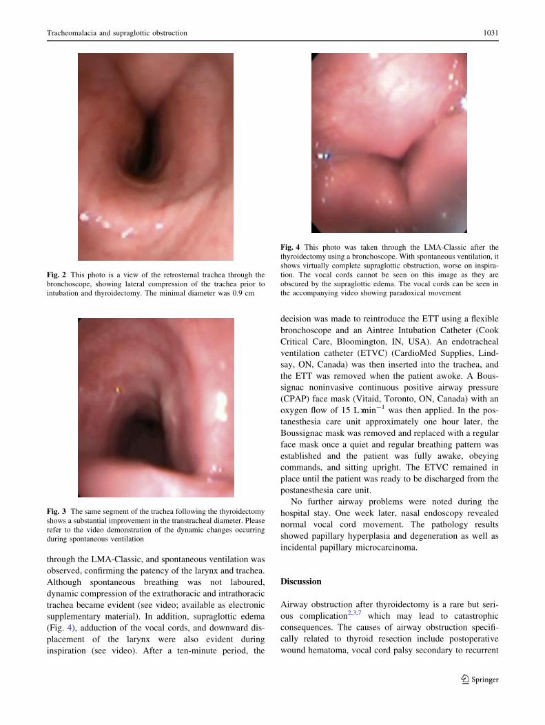
proximal trachea at the level of the thoracic inlet (Fig. 1)
with the narrowest diameter of 0.9 cm.
In the operating room, oximetry, electrocardiogram,
noninvasive blood pressure cuff, and invasive arterial
catheterization were instituted. Sedation was achieved with
a remifentanil infusion 0.07-0.1 lg�kg-1�min-1. Topical
lidocaine anesthesia was administered using a mucosal
atomization device (LMA AtomizationTM, Vitaid, Toronto
ON, Canada). This was supplemented with lidocaine
ointment (5%) applied to the base of the tongue using a
laryngoscope blade. A flexible bronchoscope loaded with a
reinforced 6.0-cm internal diameter tracheal tube was
inserted orally (Teleflex Medical, Research Triangle, NC,
USA) with the patient in the sitting position. The supra-
glottic structures were somewhat redundant with rightward
deviation and lateral compression of the tracheal lumen
(Fig. 2). A 6.0-mm internal diameter reinforced endotra-
cheal tube (ETT) was advanced into the trachea without
difficulty. General anesthesia was then induced with pro-
pofol 100 mg and fentanyl 150 lg. Muscle relaxation was
facilitated with rocuronium 40 mg. Anesthesia was main-
tained with an end-tidal sevoflurane concentration of 1.5-
2%. Surgery was uneventful and a total thyroidectomy was
completed delivering a multinodular gland measuring
12 9 4.4 9 3.7 cm (right), 11 9 5 9 5.3 cm (left), and
6.9 9 4.3 9 4.8 cm (isthmus).
At the conclusion of the surgical procedure, the oro-
pharynx was suctioned, a size 5 laryngeal mask airway,
namely, the LMA-ClassicTM (LMA; Vitaid, Toronto ON,
Canada) was introduced behind the ETT (‘‘Bailey maneu-
ver’’),6 and the latter was removed. Proper positioning with
a good glottic view was confirmed bronchoscopically
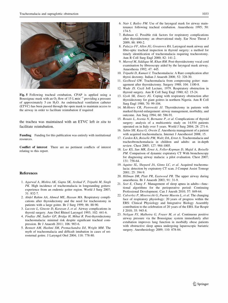
through the LMA-Classic, and positive pressure ventilation
revealed a patent trachea with no collapse and minimal
compression (Fig. 3). Neuromuscular blockade was
reversed with neostigmine 2.5 mg and glycopyrrolate
0.4 mg. The flexible bronchoscope was reintroduced
Fig. 1 Computed tomography scan between the clavicular heads
showing bilateral thyroid enlargement with tracheal deviation and
compression. There is substantial enlargement of the isthmus
1030 C. Lee et al.
123
through the LMA-Classic, and spontaneous ventilation was
observed, confirming the patency of the larynx and trachea.
Although spontaneous breathing was not laboured,
dynamic compression of the extrathoracic and intrathoracic
trachea became evident (see video; available as electronic
supplementary material). In addition, supraglottic edema
(Fig. 4), adduction of the vocal cords, and downward dis-
placement of the larynx were also evident during
inspiration (see video). After a ten-minute period, the
decision was made to reintroduce the ETT using a flexible
bronchoscope and an Aintree Intubation Catheter (Cook
Critical Care, Bloomington, IN, USA). An endotracheal
ventilation catheter (ETVC) (CardioMed Supplies, Lind-
say, ON, Canada) was then inserted into the trachea, and
the ETT was removed when the patient awoke. A Bous-
signac noninvasive continuous positive airway pressure
(CPAP) face mask (Vitaid, Toronto, ON, Canada) with an
oxygen flow of 15 L�min-1 was then applied. In the pos-
tanesthesia care unit approximately one hour later, the
Boussignac mask was removed and replaced with a regular
face mask once a quiet and regular breathing pattern was
established and the patient was fully awake, obeying
commands, and sitting upright. The ETVC remained in
place until the patient was ready to be discharged from the
postanesthesia care unit.
No further airway problems were noted during the
hospital stay. One week later, nasal endoscopy revealed
normal vocal cord movement. The pathology results
showed papillary hyperplasia and degeneration as well as
incidental papillary microcarcinoma.
Discussion
Airway obstruction after thyroidectomy is a rare but seri-
ous complication2,3,7 which may lead to catastrophic
consequences. The causes of airway obstruction specifi-
cally related to thyroid resection include postoperative
wound hematoma, vocal cord palsy secondary to recurrent
Fig. 3 The same segment of the trachea following the thyroidectomy
shows a substantial improvement in the transtracheal diameter. Please
refer to the video demonstration of the dynamic changes occurring
during spontaneous ventilation
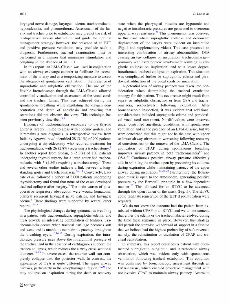
Fig. 4 This photo was taken through the LMA-Classic after the
thyroidectomy using a bronchoscope. With spontaneous ventilation, it
shows virtually complete supraglottic obstruction, worse on inspira-
tion. The vocal cords cannot be seen on this image as they are
obscured by the supraglottic edema. The vocal cords can be seen in
the accompanying video showing paradoxical movement
Fig. 2 This photo is a view of the retrosternal trachea through the
bronchoscope, showing lateral compression of the trachea prior to
intubation and thyroidectomy. The minimal diameter was 0.9 cm
Tracheomalacia and supraglottic obstruction 1031
123
laryngeal nerve damage, laryngeal edema, tracheomalacia,
hypocalcemia, and pneumothorax. Assessment of the lar-
ynx and trachea prior to extubation may predict the risk of
postoperative airway obstruction and guide the optimal
management strategy; however, the presence of an ETT
and positive pressure ventilation may preclude such a
diagnosis. Furthermore, tracheal examination must be
performed in a manner that minimizes stimulation and
coughing in the absence of an ETT.
In this report, an LMA-Classic was used in conjunction
with an airway exchange catheter to facilitate the assess-
ment of the airway and as a temporizing measure to assess
the adequacy of spontaneous ventilation in the presence of
supraglottic and subglottic obstruction. The use of the
flexible bronchoscope through the LMA-Classic allowed
controlled visualization and assessment of the vocal cords
and the tracheal lumen. This was achieved during the
spontaneous breathing while regulating the oxygen con-
centration and depth of anesthesia and ensuring that
secretions did not obscure the view. This technique has
been previously described.8,9
Evidence of tracheomalacia secondary to the thyroid
goiter is largely limited to areas with endemic goiters, and
it remains a rare diagnosis. A retrospective review from
India by Agarwal et al. identified 28 (3.1%) of 900 patients
undergoing a thyroidectomy who required treatment for
tracheomalacia, with 26 (2.8%) receiving a tracheostomy.1
In another report from Sudan, 6 (5.8%) of 103 patients
undergoing thyroid surgery for a large goiter had tracheo-
malacia, with 5 (4.8%) requiring a tracheostomy.2 These
and several other studies indicate a link between a long-
standing goiter and tracheomalacia.7,9-11 Conversely, Lac-
oste et al. followed a cohort of 3,008 patients undergoing
thyroidectomy and found that none of the cases developed
tracheal collapse after surgery.3 The main causes of post-
operative respiratory obstruction were wound hematoma,
bilateral recurrent laryngeal nerve palsies, and laryngeal
edema.3 These findings were supported by several other
reports.12-15
The physiological changes during spontaneous breathing
in a patient with tracheomalacia, supraglottic edema, and
OSA provide an interesting combination of features. Tra-
cheomalacia occurs when tracheal cartilage becomes soft
and weak and is unable to maintain its patency throughout
the breathing cycle.10,16,17 During expiration, the intra-
thoracic pressure rises above the intraluminal pressure of
the trachea, and in the absence of cartilaginous support, the
trachea collapses, which reduces the airway cross-sectional
diameter.16-18 In severe cases, the anterior wall can com-
pletely collapse onto the posterior wall. In contrast, the
appearance of OSA is quite different. The upper airway
narrows, particularly in the velopharyngeal region,19,20 and
may collapse on inspiration during the sleep or recovery
state when the pharyngeal muscles are hypotonic and
negative intrathoracic pressures are generated to overcome
upper airway resistance.21 This phenomenon was observed
in this case where supraglottic collapse and downward
displacement of the larynx were evident on inspiration
(Fig. 4 and supplementary video). This case presented an
interesting combination of airway abnormalities: OSA
causing airway collapse on inspiration; tracheomalacia—
primarily with extrathoracic involvement resulting in sub-
glottic collapse on inspiration; and to a lesser degree,
intrathoracic tracheal collapse on expiration. This situation
was complicated further by supraglottic edema and para-
doxical adduction of the vocal cords on inspiration.
A potential loss of airway patency was taken into con-
sideration when determining the tracheal extubation
strategy for this patient. This occurrence might result from
supra- or subglottic obstruction or from OSA and trache-
omalacia, respectively, following extubation. After
bronchoscopic inspection, it was evident that additional
considerations included supraglottic edema and paradoxi-
cal vocal cord movement. No difficulties were observed
under controlled anesthetic conditions with spontaneous
ventilation and in the presence of an LMA-Classic, but we
were concerned that this might not be the case with upper
or lower airway obstruction worsening following recovery
of consciousness or the removal of the LMA-Classic. The
application of CPAP during spontaneous breathing
improves airway patency in both tracheomalacia17 and
OSA.20 Continuous positive airway pressure effectively
aids in splinting the trachea open by preventing its collapse
during expiration while maintaining patency of the upper
airway during inspiration.17,20-22 Furthermore, the Boussi-
gnac mask is open to the atmosphere, generating positive
pressure by the Bernoulli principle in a flow-dependent
manner.23 This allowed for an ETVC to be advanced
through the open lumen of the mask (Fig. 5). The ETVC
could facilitate reinsertion of the ETT if re-intubation were
required.
We do not know the outcome had the patient been ex-
tubated without CPAP or an ETVC, and we do not contend
that either the edema or the tracheomalacia resolved during
the time these remained in place. However, this strategy
did permit the stepwise withdrawal of support in a fashion
that we believe had the highest probability of safe reversal,
namely, the reinstitution or escalation of CPAP and tra-
cheal reintubation.
In summary, this report describes a patient with docu-
mented supraglottic, subglottic, and intrathoracic airway
obstruction, which was evident only with spontaneous
ventilation following tracheal extubation. This condition
was confirmed by bronchoscopic assessment through an
LMA-Classic, which enabled proactive management with
noninvasive CPAP to maintain airway patency. Access to
1032 C. Lee et al.
123
the trachea was maintained with an ETVC left in situ to
facilitate reintubation.
Funding Funding for this publication was entirely with institutional
resources.
Conflict of interest There are no pertinent conflicts of interest
relating to this report.
References
1. Agarwal A, Mishra AK, Gupta SK, Arshad F, Tripathi M, SinghPK. High incidence of tracheomalacia in longstanding goiters:
experience from an endemic goiter region. World J Surg 2007;
31: 832-7.
2. Abdel Rahim AA, Ahmed ME, Hassan MA. Respiratory compli-
cations after thyroidectomy and the need for tracheostomy in
patients with a large goitre. Br J Surg 1999; 86: 88-90.
3. Lacoste L, Gineste D, Karayan J, et al. Airway complications in
thyroid surgery. Ann Otol Rhinol Laryngol 1993; 102: 441-6.
4. Findlay JM, Sadler GP, Bridge H, Mihai R. Post-thyroidectomy
tracheomalacia: minimal risk despite significant tracheal com-
pression. Br J Anaesth 2011; 106: 903-6.
5. Bennett AM, Hashmi SM, Premachandra DJ, Wright MM. The
myth of tracheomalacia and difficult intubation in cases of ret-
rosternal goitre. J Laryngol Otol 2004; 118: 778-80.
6. Nair I, Bailey PM. Use of the laryngeal mask for airway main-
tenance following tracheal extubation. Anaesthesia 1995; 50:
174-5.
7. Rahman G. Possible risk factors for respiratory complications
after thyroidectomy: an observational study. Ear Nose Throat J
2009; 88: 890-2.
8. Palazzo FF, Allen JG, Greatorex RA. Laryngeal mask airway and
fibre-optic tracheal inspection in thyroid surgery: a method for
timely identification of tracheomalacia requiring tracheostomy.
Ann R Coll Surg Engl 2000; 82: 141-2.
9. Maroof M, Siddique M, Khan RM. Post-thyroidectomy vocal cord
examination by fibreoscopy aided by the laryngeal mask airway.
Anaesthesia 1992; 47: 445.
10. Tripathi D, Kumari I. Tracheomalacia: A Rare complication after
thyroi dectomy. Indian J Anaesth 2008; 52: 328-30.
11. Geelhoed GW. Tracheomalacia from compressing goiter: man-
agement after thyroidectomy. Surgery 1988; 104: 1100-8.
12. Wade JS. Cecil Joll Lecture, 1979. Respiratory obstruction in
thyroid surgery. Ann R Coll Surg Engl 1980; 62: 15-24.
13. Gyoh SK, Emery JG. Coping with respiratory obstruction after
thyroidectomy for giant goitres in northern Nigeria. Ann R Coll
Surg Engl 1988; 70: 99-104.
14. McHenry CR, Piotrowski JJ. Thyroidectomy in patients with
marked thyroid enlargement: airway management, morbidity, and
outcome. Am Surg 1994; 60: 586-91.
15. Rosato L, Avenia N, Bernante P, et al. Complications of thyroid
surgery: analysis of a multicentric study on 14,934 patients
operated on in Italy over 5 years. World J Surg 2004; 28: 271-6.
16. Sahin SH, Kaya G, Oresin Z. Anesthesia management of a patient
with acquired tracheomalacia. Internet J Anesthesiol 2008; 15.
17. Carden KA, Boiselle PM, Waltz DA, Ernst A. Tracheomalacia and
tracheobronchomalacia in children and adults: an in-depth
review. Chest 2005; 127: 984-1005.
18. Lee KS, Sun MR, Ernst A, Feller-Kopman D, Majid A, BoisellePM. Comparison of dynamic expiratory CT With bronchoscopy
for diagnosing airway malacia: a pilot evaluation. Chest 2007;
131: 758-64.
19. Aquino SL, Shepard JA, Ginns LC, et al. Acquired tracheoma-
lacia: detection by expiratory CT scan. J Comput Assist Tomogr
2001; 25: 394-9.
20. Hillman DR, Platt PR, Eastwood PR. The upper airway during
anaesthesia. Br J Anaesth 2003; 91: 31-9.
21. Seet E, Chung F. Management of sleep apnea in adults—func-
tional algorithms for the perioperative period: Continuing
Professional Development. Can J Anesth 2010; 57: 849-64.
22. Calverley P, Miserocchi G, Puente Maestu L, et al. The changing
face of respiratory physiology: 20 years of progress within the
ERS: Clinical Physiology and Integrative Biology Assembly
contribution to the celebration of 20 years of the ERS. Eur Respir
J 2010; 35: 945-8.
23. Neligan PJ, Malhotra G, Fraser M, et al. Continuous positive
airway pressure via the Boussignac system immediately after
extubation improves lung function in morbidly obese patients
with obstructive sleep apnea undergoing laparoscopic bariatric
surgery. Anesthesiology 2009; 110: 878-84.
Fig. 5 Following tracheal extubation, CPAP is applied using a
Boussignac mask with an O2 flow of 15 L�min-1 providing a pressure
of approximately 5 cm H2O. An endotracheal ventilation catheter
(ETVC) has been passed through the open mask to maintain access to
the airway in order to facilitate reintubation if required
Tracheomalacia and supraglottic obstruction 1033
123