management and prognosis of severe traumatic brain injury
TRANSCRIPT

MANAGEMENT AND
PROGNOSIS OF
SEVERE TRAUMATIC
BRAIN INJURY
Part I: Guidelines for the Management of SevereTraumatic Brain Injury
Part II: Early Indicators of Prognosis in SevereTraumatic Brain Injury
A joint project of the
Brain Trauma Foundation
American Association of Neurological Surgeons,Joint Section on Neurotrauma and Critical Care
©2000, Brain Trauma FoundationISBN 0-9703144-0-X


MANAGEMENT AND
PROGNOSIS OF
SEVERE TRAUMATIC
BRAIN INJURY
DISCLAIMER OF LIABILITY
The information contained in the Management and Prognosis of Severe Traumatic Brain Injury (Part I and II) reflects the cur-rent state of knowledge at the time of publication, February 2000.The information is designed to provide accurate and authoritativeinformation in regard to the subject matter covered. In view of thefact that there will be future developments in scientific informa-tion and technology, it is anticipated that there will be periodic re-view and updating of these guidelines. These guidelines are dis-tributed with the understanding that the Brain Trauma Founda-tion, the American Association of Neurological Surgeons, and theother organizations that have collaborated in the development ofthese g␣ uidelines are not engaged in rendering professional medi-cal services. If medical advice or assistance is required, the servicesof a competent physician should be sought. The recommendationscontained in these guidelines may not be appropriate for use in allcircumstances. The decision to adopt any particular recommen-dation contained in the Management and Prognosis of Severe Trau-matic Brain Injury (Part I and II) must be made by a treating physi-cian in the light of all the facts and circumstances surrounding eachparticular case and on the basis of the available resources.


PART I: GUIDELINES FOR
THE MANAGEMENT OF
SEVERE TRAUMATIC
BRAIN INJURY

©2000, Brain Trauma Foundation

Table of Contents
Introduction ..................................................................................................................... 7
Trauma Systems .............................................................................................................. 11
Initial Management ........................................................................................................ 21
Resuscitation of Blood Pressure and Oxygenation ........................................................ 33
Indications for Intracranial Pressure Monitoring .......................................................... 47
Intracranial Pressure Treatment Threshold ................................................................... 71
Recommendations for Intracranial Pressure Monitoring Technology ......................... 75
Guidelines for Cerebral Perfusion Pressure ................................................................... 91
Hyperventilation .......................................................................................................... 101
The Use of Mannitol ..................................................................................................... 115
The Use of Barbiturates in the Control of Intracranial Hypertension ........................ 125
The Role of Steroids ..................................................................................................... 131
Critical Pathway for the Treatment of Established Intracranial Hypertension ........... 139
Nutrition ....................................................................................................................... 143
The Role of Antiseizure Prophylaxis Following Head Injury ...................................... 159
PART I: GUIDELINES FOR THE MANAGEMENT OFSEVERE TRAUMATIC BRAIN INJURY


5Early Indicators of Prognosis
Authors:
M. Ross Bullock, M.D., Ph.D.Reynolds Professor, Division of Neurogurgery & AnesthesiologyMedical College of Virginia/Virginia Commonwealth University
Randall M. Chesnut, M.D.Associate Professor of NeurosurgeryOregon Health Sciences University
Guy L. Clifton, M.D.Professor & Chairman, Department of NeurosurgeryThe University of Texas Houston Health Science Center
Jamshid Ghajar, M.D., Ph.D.Clinical Associate Professor of NeurosurgeryCornell University Medical College
Donald W. Marion, M.D.Professor of NeurosurgeryUniversity of Pittsburgh Medical Center
Raj K. Narayan, M.D.Professor & Chairman of NeurosurgeryTemple University School of Medicine
David W. Newell, M.D.Associate Professor of NeurosurgeryUniversity of Washington Medical School
Lawrence H. Pitts, M.D.Professor of NeurosurgeryUniversity of California Medical Center, San Francisco
Michael J. Rosner, M.D.Private PracticeHendersonville, North Carolina
Beverly C. Walters, M.D.Chairman, AANS Guidelines Committee, Associate Professor of Clinical NeurosciencesBrown University School of Medicine
Jack E. Wilberger, M.D.Professor of Neurosurgery and Chairman, Department of Neurological SurgeryAllegheny General Hospital
GUIDELINES FOR THE MANAGEMENTOF SEVERE TRUAMATIC BRAIN INJURY

6 Guidelines for the Management of Severe Traumatic Brain Injury
Participants:
Robert Florin, M.D.Chairman, AANS Guidelines & Outcomes Committee
Andrew Jagoda, M.D.Representative, American College of Emergency Physicians
James P. Kelly, M.D.Representative, American Academy of Neurology
Anthony Marmarou, Ph.DProfessor & Vice Chairman, Division of Neurosurgery,Medical College of Virginia/Virginia Commonwealth University
Peter C. QuinnExecutive Director, The Brain Trauma Foundation
Jay Rosenberg, M.D.Chairman, Quality Standards Subcommittee, American Academy of Neurology
Alex B. Valadka, M.D.Assistant Professor of Neurosurgery, Baylor College of Medicine
European Advisory Committee:
Mark Dearden, M.D.Leeds General Infirmary, U.K.
Andrew I.R. Mass, M.D.Dijkzigt Hospital Rotterdam, the Netherlands
J. Douglas Miller, M.D.†
The Western General Hospital, Edinburgh, Scotland
Franco Servadei, M.D.WHO Neurotrauma Collaborating Center, Ospedale M. Bufalini, Cesena, Italy
Nino Stocchetti, M.D.Ospedale di Parma, Italy
Graham M. Teasdale, M.D.Institute of Neurological Sciences, The Southern General Hospital, Glasgow, Scotland
Andreas Unterberg, M.D.Virchow-Klinikum, Humboldt Universitat zu Berlin, Germany
†Deceased

7Early Indicators of Prognosis
Traumatic Brain Injury (TBI), a clinical problem treated frequently by neurosurgeons, is amajor cause of disability, death, and economic cost to our society. In the past two decades,
we have increased remarkably our understanding of the pathophysiology of TBI. One of thecentral concepts that emerged from clinical and laboratory research is that all neurologicaldamage does not occur at the moment of impact, but evolves over the ensuing hours and days.Furthermore, we now recognize the deleterious effects of these various delayed insults to theinjured brain at the clinical and biochemical levels. This has led to an interest in developingbetter monitoring and treatment methods as well as the development of new pharmaceuticals,all of which show great promise in improving the outcome for patients who have suffered abrain injury.
Past efforts to develop guidelines for the management of patients with severe TBI relied onauthors’ expert opinion and practice experience and, therefore, had an element of subjectivity.Recently, with the advent of a methodology to develop guideline documents based on scientificmethod, there has been a dramatic increase in clinical practice guidelines with subsequent reportsshowing improvement in patient care and a reduction in medical time and cost.1 The interest indeveloping guidelines for TBI intensified after a national study documented considerablevariability in the management of patients with severe TBI.2
The task force authors developing the Guidelines for the Management of Severe Traumatic Brain Injuryused a meticulous process relying on scientific evidence rather than expert opinion. In addition, thetask force authors actively involved representatives of national and international medical societiesand individuals with demonstrated expertise and interest in the care of patients with severe TBI.
These guidelines address key issues relating to the management of severe TBI in adultpatients with a Glasgow Coma Scale score of 3-8. They are by no means an exhaustive treatiseon severe TBI. Because of the enormous effort required to develop evidence-based guidelines,the task force authors selected topics that were deemed to have an impact on outcomes inpatients with severe TBI. Other important aspects of patient management that were not coveredin the present effort will be considered for study in subsequent editions of this document.Examples of such topics include indications for neurosurgical intervention, specialconsideration in pediatric head injury, the management of penetrating head injury, sedationand paralysis in the TBI patient, and the economics of TBI. We intend that these guidelines willbe continually improved in response to new scientific evidence.
Our intent is that the Guidelines for the Management of Severe Traumatic Brain Injury will clearlystate the current scientific basis for our clinical practice. For most clinical practice parameters,
INTRODUCTION

8 Guidelines for the Management of Severe Traumatic Brain Injury
scientific evidence is insufficient for standards of care, as is generally the case in most of currentmedical practice. Upgrading clinical practice parameters from option to guideline to standardwill require focused, well-designed, and carefully implemented clinical research trials.
Process Used in Development of These GuidelinesThese guidelines are comprised of fourteen topics ranging from trauma systems and prehospitalresuscitation to monitoring and treatment of intracranial hypertension and intensive care. In1993, a head injury guidelines task force was formed and supported by the Brain TraumaFoundation (BTF). Members of the task force were selected based on their academic expertisein head injury. BTF is a nonprofit organization dedicated to improving the outcome of braintrauma patients through education and clinical research. BTF financially supports andmaintains these guidelines in a cooperative agreement with the American Association ofNeurological Surgeons (AANS).
Initially, each author on the task force was assigned a topic and conducted a MEDLINE search,reviewed and graded clinical articles pertinent to the topic, then wrote a report. These reports werereviewed, critiqued, and revised by the entire task force and by representatives of various medicalsocieties, individuals with expertise in head injury care, and members of the AANS Guidelines andOutcomes Committee. The document was critiqued in detail by a group of European neurosurgeonswith expertise in neurotrauma (see European Advisory Committee listing).
In April 1995, the document was reviewed and approved by the AANS Guidelines andOutcomes Committee and the AANS Board of Directors. The guidelines were also reviewed bythe American Academy of Neurology, the American College of Surgeons, the American Collegeof Emergency Physicians, the American Society of Neuroradiology, the Society for Critical CareMedicine, the American Association of Neuroscience Nurses, and the American Academy ofPhysical Medicine and Rehabilitation. In 1998 the task force authors met to review the 1995version of the guidelines and to update the scientific evidence and make other necessarychanges. The term Head Injury as used in the original guidelines title was removed in favor ofTraumatic Brain Injury, which reflected a more prevalent usage in the literature reviews.
Degrees of CertaintyIn assessing the degree of certainty associated with a particular recommendation, the followingterminology is the most widely accepted and is used in this document:
Standards: represent accepted principles of patient management that reflect a highdegree of clinical certainty.
Guidelines: represent a particular strategy or range of management strategies thatreflect a moderate degree of clinical certainty.
Options: are the remaining strategies for patient management for which there isunclear clinical certainty.
Note that the term “guideline” is used both in a global sense, i.e., clinical practice guidelines, aswell as in a more specific sense, as noted above.

9Early Indicators of Prognosis
Classification of EvidenceWhen assessing the value of therapies or interventions, the available data is classified into one ofthree categories according to the following criteria*:
Class I evidence: Prospective, randomized, controlled trials (PRCT)—the gold standardof clinical trials. However, some may be poorly designed, lacksufficient patient numbers, or suffer from other methodologicalinadequacies.
Class II evidence: Clinical studies in which the data was collected prospectively, andretrospective analyses that were based on clearly reliable data. Types ofstudies so classified include: observational studies, cohort studies,prevalence studies, and case control studies.
Class III evidence: Most studies based on retrospectively collected data. Evidence used inthis class indicates clinical series, databases or registries, case reviews,case reports, and expert opinion with some support from animalstudies.
Technology Assessment: The assessment of technology, such as intracranial pressuremonitoring devices, does not lend itself to classification in the above-mentioned format. Thus, for technology assessment the devices wereevaluated in terms of their accuracy, reliability, therapeutic potential,and cost effectiveness.
Correlation Between Evidence and RecommendationsStandards are generally based on Class I evidence. However, strong Class II evidence may formthe basis for a standard, especially if the issue does not lend itself to testing in a randomizedformat. Conversely, weak or contradictory Class I evidence may not be able to support astandard.
Guidelines are usually based on Class II evidence or a preponderance of Class III evidence.
Options are usually based on Class III evidence and are clearly much less useful except foreducational purposes and in guiding future studies.
Attributes of Clinical Practice GuidelinesTo ensure the development of scientifically sound, clinically relevant guidelines that areapplicable to the day-to-day practice of medicine, the American Medical Association (AMA)developed a list of attributes that are listed here in an abbreviated form.3
*A single study may be of a different class depending on the parameter in each topic.

10 Guidelines for the Management of Severe Traumatic Brain Injury
Attribute I Practice guidelines should be developed by or in conjunction withphysician organizations and should be characterized by• scientific and clinical expertise in the content areas of the
parameters.• broad-base representation of physicians likely to be affected by the
parameters.
Attribute II Relevant scientific literature and expert clinical opinion should bereviewed as evidenced by• a description of the process of the review.• a description of the evidence reviewed.• the specialty affiliations and other credentials of the physician
organizations, groups, and individuals conducting the review.• a description of the methods used to evaluate the scientific
literature and other appropriate research findings.• the rationale for including or excluding studies is noted.• the process for selection of clinical experts/reviewers is noted or
available on request.• at least two-thirds of clinical experts/reviewers were actively
involved in clinical practice in relevant clinical areas.• the clinical experts/reviewers thoroughly reviewed and assessed
the scientific literature.
Attribute III Practice parameters should be as comprehensive and specific aspossible.
Attribute IV Practice parameters should be based on current information. Thereshould be provisions for periodic reviews and revisions, whenappropriate.
Attribute V The guidelines should be widely disseminated.
Every effort has been made in the formulation of the Guidelines for the Management of SevereTraumatic Brain Injury to achieve these ideals.
References1. Woolf SH: Practice guidelines: a new reality in medicine. Arch Intern Med 1993; 153: 2646-
2655.2. Ghajar J, Hariri RJ, Narayan RK, et al.: Survey of critical care management of comatose,
head-injured patients in the United States. Crit Care Med 1995; 23: 560-567.3. AMA, Office of Quality Insurance & Health Care Organizations’ Attributes to Guideline
Development of Practice Parameters. AMA; Chicago, IL 1990.
11Trauma Systems
I. RecommendationsA. Standards
There are insufficient data to support a treatment standard for this topic.B. Guidelines
All regions should have an organized trauma care system.C. Options
As delineated in the American College of Surgeons Committee on Trauma Resourcesfor Optimal Care of the Injured Patient: 1999,1 neurosurgeons should have an organizedand responsive system of care for patients with neurotrauma. They should initiateneurotrauma care planning including prehospital management and triage, directtrauma center transport, maintain appropriate call schedules, review trauma carerecords for quality improvement, and participate in trauma education programs.
Trauma facilities treating patients with severe or moderate head injury must have aneurosurgery service, an in-house trauma surgeon, a neurosurgeon promptlyavailable, and a continuously staffed and available operating room, intensive careunit, and laboratory with proper equipment for treating neurotrauma patients. A CTscanner must be immediately available at all times.
In rural or occasionally weather-bound communities without a neurosurgeon, asurgeon should be trained to perform accurate neurological assessment and toinitiate immediate neurotrauma care. Such a surgeon also should be trained toperform life-saving surgical treatment of an extracerebral hematoma in adeteriorating patient.
II. OverviewTrauma causes about 150,000 deaths in the United States each year, about one-third are due tofatal head injuries.23 One million American traumatic brain injury (TBI) victims are treated andreleased from hospital emergency departments annually3 and 230,000 of these survivors requireinpatient care.4 Every year another 10,000 persons sustain spinal cord injuries; some 200,000people in the United States live with the disabilities caused by these injuries. While there is noway to adequately characterize the human costs, the total (direct and indirect) costs of TBI is
TRAUMA SYSTEMS

12 Guidelines for the Management of Severe Traumatic Brain Injury
estimated at $37.8 billion in 1985 dollars.8 Thus, trauma, including neurotrauma, is a seriouspublic health problem requiring continuing improvement in the care of injured patients.Trauma system development and organization and better injury prevention appear to belowering death and disability from intentional and unintentional injury, and should be availableto all people in the United States and other countries.
III. ProcessA MEDLINE search from 1966 to 1998 identified articles with the key words “trauma systems”and “outcome.” Twenty-three relevant manuscripts were used as a basis to assess the value oftrauma systems. The guideline and options listed are derived from studies in trauma andneurotrauma care from a variety of peer-reviewed and other articles. Resources for Optimal Careof the Injured Patient: 1999, 1 published by the American College of Surgeons Committee onTrauma, provides the basis for most recommendations regarding trauma hospital organization.This document, originally published in 1976, is written, reviewed, and revised regularly byhighly recognized North American trauma surgeons. Revision of the next document begins assoon as the latest version is completed; the 1999 version was employed here.
IV. Scientific FoundationSince the late 1970s, various investigators have tried to demonstrate the efficacy of traumasystems. Early studies generally attempted to show that excessive, “preventable” trauma deathsoccurred in regions without organized trauma care 2,7,21 but this methodology was criticized asbeing too subjective.22 Additional studies relied on series of patients treated at one or moretrauma centers and compared them with those treated within a region18 or across the UnitedStates8 using prospectively collected, standardized data for severity and outcome. In allcomparisons between organized and non-organized trauma systems, patient outcome wasworse without organization. Implementing a trauma system in Quebec reduced mortality by50%16, and reduced mortality of TBI patients in Oregon by 20%.10 In the rural setting, ACSLevel II trauma center guideline implementation more than doubled survival in head-injuredpatients.11 A number of studies and their methodologies have been summarized.9,14 There areno published data suggesting that unorganized trauma care is superior to organized systems.Published reports indicate that centers treating larger volumes of trauma have better patientoutcomes than centers with fewer injured patient encounters.19 However, morbidity, mortality,and length of stay does not seem to vary significantly with individual trauma surgeon casevolume.13 One report states that organized Level II trauma centers with attending traumasurgeons who are available but not “in-house” have outcomes as good as those with surgeonspresent in the hospital at all times.20 However, in-house attending surgeons at another centerachieved better than expected survival in patients who had blunt or penetrating trauma treatedwithin 20 minutes of hospital arrival15 (both of these studies examined data prospectivelycollected at their center against data collected prospectively at many trauma centers across theUnited States). Treatment of severely injured patients at a local hospital with subsequenttransfer to a trauma center nearly doubles mortality in both the adult, 18 and pediatricpopulations.19

13Trauma Systems
Organization of Neurotrauma CareSeveral kinds of arrangements can provide optimal management of trauma, includingneurotrauma, and depend on the presence and interest of the local neurosurgeon, traumasurgeon, emergency physician, and critical care specialist. The injured patient, particularly thepatient with injury to several body regions, must have a surgeon available for overallmanagement. A trauma surgeon or an appropriately qualified neurosurgeon may fill this role incollaboration with the Trauma Service. He or she most often assumes overall responsibility inpatients with isolated head or spinal cord injuries, and in multitrauma patients after their otherinjuries have stabilized and when management of neurotrauma is the most pressing problem.When multiple organ injuries require active treatment, appropriate consultants may be calledon to deliver care for respiratory, nutritional, infectious, and hematological needs.1,12
The surgeon qualified for the care of trauma patients is defined as a board certified,Advanced Trauma Life Support (ATLS) certified surgeon with active trauma clinicalinvolvement, continuing medical education, and participation in national or regional traumaorganizations.1 The Resources for Optimal Care document further directs the surgeon’s practice inthe following areas: emergency intervention, critical care, acute care, and discharge planning.1
That same document1 also directs neurosurgical involvement in the care of the injuredpatient. Neurosurgeons should participate in defining prehospital care in their region includingon-site resuscitation and trauma center referral criteria, and in training emergency medicalproviders in the early management of neurotrauma. It is imperative that neurosurgeons defineand maintain on-call schedules and formulate trauma center bypass procedures when aneurosurgeon is unavailable to treat injured patients, and be available when called to providetrauma care. They must ensure that the trauma facility has adequate computed tomographyscanning capabilities, and operating room and intensive care resources for patients to be treatedoptimally. Neurosurgeons also should participate in the trauma system’s review, qualityimprovement, and teaching efforts within their hospital and trauma system.
Prehospital care and emergency department treatment of patients with neurotrauma mayhave profound importance in their ultimate morbidity and mortality. Many key individualsprovide critically important patient care in the early minutes and hours after trauma, includingappropriately credentialed emergency physicians, anesthesiologists, emergency medicaltechnicians and paramedics, and emergency department and operating room nurses, amongothers, whose skills and training are essential in the management of these critically injuredpatients. Because treatment of nervous system injury must be done correctly, involvement byneurosurgeons in the planning and implementation of treatment protocols is extremelyimportant, along with input from other trauma specialists. Reviews of specific treatments aregiven in the following sections in these neurotrauma guidelines.

14 Guidelines for the Management of Severe Traumatic Brain Injury
V. SummaryPublished case series and cohort comparison studies of patients treated in regions where plannedtrauma systems are in place compared to regions without trauma systems, or before and afterinstituting a trauma system, conclude that mortality is reduced after major trauma in patients treatedin a trauma system. For optimal care of neurotrauma, neurosurgeons should be involved in theplanning and implementation of trauma systems and in support of a system once it is in place.
VI. Key Issues for Future InvestigationIn order to establish trauma system development as a standard for treatment, a prospectivestudy would have to compare the outcome of treatment of patients randomly taken to hospitalswithin and without a planned trauma system. This would be required both for trauma patientsin general, and for neurotrauma patients in particular. Given the preponderance of datasupporting trauma systems, such studies are unlikely to be undertaken.
VII. Evidentiary TableACS-COT,1 1999
Description of Study: Guidelines for organization of trauma centers and trauma personnel.Defined by expert opinion and supported by published data where possible.
Classification: Class III Study
Conclusions: Trauma is a surgical disease, and neurotrauma care should be planned andmanaged by neurosurgeons in concert with other trauma surgeons. Trauma systems andhospitals should be defined and maintained according to these guidelines.
Campbell,2 1989
Description of Study: Retrospective case series in an undesignated trauma system showing23% “preventable” deaths other than head injury judged by group review. (n = 452)
Classification: Class III Study
Conclusions: Study demonstrates that a self-designation system without regulatory controlresults in a high percentage of preventable trauma deaths.
Hoyt,5 1989
Description of Study: Retrospective analysis of indications for operating room (OR)resuscitation of trauma patients with cardiac arrest, persistent hypotension despite resuscitation,or uncontrolled external hemorrhage. (n = 323)
Classification: Class III Study
Conclusions: No patients survived after blunt trauma and cardiopulmonary arrest. Patients withblunt trauma who have persistent hypotension rarely have surgery started within 20 minutes ofinjury. They can be resuscitated in the emergency department. Only patients with penetratingchest and abdominal injuries who have persistent hypotension after resuscitation may benefitfrom OR resuscitation.

15Early Indicators of Prognosis
Johnson,6 1995
Description of Study: This study compared the mortality of 98 children who sustained severehead injury and were transported directly to a pediatric trauma center, with those who were firsttaken to the closest hospital and later transferred.
Classification: Class III Study
Conclusions: Mortality for children taken directly to the pediatric trauma center was 27%; forthose taken to the closest hospital first it was 50%.
Kreis,7 1986
Description of Study: Retrospective case series in an undesignated trauma system showing21% “preventable” non-CNS deaths judged by group review. A Level I trauma center had a 12%preventable mortality rate compared with 21% in planned Level II centers and 30% at 16 otherhospitals. (n = 1,201)
Classification: Class III Study
Conclusions: The authors concluded that severely injured patients should be triaged and takento trauma centers and that there is a need for an organized trauma system.
Mendelhoff,9 1991
Description of Study: Review of trauma system studies and implications for public policy.
Classification: Review
Conclusions: Evidence suggests that the introduction of trauma systems in urban areas canprevent deaths at a relatively low cost. The federal government should require states or regionalorganizations to designate appropriate hospitals as trauma centers and to mandate thedevelopment of transfer agreements among hospitals.
Mullins,10 1996
Description of Study: Evaluated the influence of implementing the Oregon state-wide traumasystem on admission distribution and risk of death using a before and after comparison.
Classification: Class III Study
Conclusions: The Oregon trauma system resulted in reduction in risk of trauma-related death.
VII. Evidentiary Table (continued)

16 Guidelines for the Management of Severe Traumatic Brain Injury
Norwood,11 1995
Description of Study: This study compared outcome of trauma patients before and after arural hospital implemented Level II trauma center guidelines using as a study group of patientswith a calculated survival of 25%.
Classification: Class III Study
Conclusions: Survival of patients before meeting trauma center criteria was 13% and after thesurvival increased to 30%.
Pitts,12 1987
Description of Study: Editorial comment on the need for neurosurgeon involvement inneurotrauma care and planning.
Classification: Class III Study
Conclusions: It is essential for neurosurgeons to take an active role in defining triage schemes forneurotrauma, in helping establish the needed hospital organization for neurotrauma care, inmaintaining appropriate call schedules, and in helping in trauma education and qualityassurance.
Roy,14 1987
Description of Study: Review of published literature on the value of local and regional traumacare systems, emphasizing study methodology. Evidence in the reports includes case seriesreports, before and after studies, and intersystem comparisons.
Classification: Review
Conclusions: The literature overwhelmingly suggests that the main determinants of survival arethe adequacy of resuscitation and the early recognition of serious injuries.
Sampalis,16 1995
Description of Study: The study evaluated the impact of trauma center development anddesignation on mortality in Quebec, Canada, comparing mortality before and after the traumasystem was implemented.
Classification: Class III Study
Conclusions: There was a significant reduction in trauma-related mortality after implementinga trauma system.
VII. Evidentiary Table (continued)

17Early Indicators of Prognosis
Sampalis,15 1997
Description of Study: The outcome of severely injured patients (including head trauma) whowere transported directly to trauma centers was compared to patients of similar injury severitywho were transferred to a trauma center after first being transported to a less specialized, localinstitution (n = 1,608)
Classification: Class III Study
Conclusions: This study showed that severely head-injured patients transported directly fromthe scene to Level 1 trauma centers is associated with a significant reduction in mortality.
Shackford,17 1987
Description of Study: Analysis of patients admitted after traumatic injury, of whom 283 wereseverely injured (trauma score < 8). Of those who had sufficient data (n=189) to compare with anational cohort study that provided a model for predicting survival in patients, actual survivalwas 29% whereas predicted survival (Ps) was 18%. In patients with penetrating injury, Ps was 8%and actual survival was 20%. (n = 3393)
Classification: Class II Study
Conclusions: The improved survival was attributed to the integration of prehospital andhospital care and expeditious surgery.
Smith J,18 1990
Description of Study: Analysis of data abstracted from computerized discharge informationabout patients with femoral shaft fractures requiring operation over a one-year period in twostates. (n=1,332)
Classification: Class II Study
Conclusions: Patients treated in trauma care centers had significantly fewer deaths andcomplications than in non-trauma centers.
Smith R,19 1990
Description of Study: A cohort analysis was performed on data from severely injured patientsusing three statistical methods to determine the relationship between trauma center volume andmortality. (n = 1,643)
Classification: Class II Study
Conclusions: Low-volume trauma centers (fewer than 140 patients annually) had significantlyhigher mortality when adjusted for head injury than did high-volume trauma centers (more than200 patients annually) (p < .04).
VII. Evidentiary Table (continued)

18 Guidelines for the Management of Severe Traumatic Brain Injury
Thompson,20 1992
Description of Study: Cohort analysis of trauma admissions at a Level II trauma centershowed no difference between survival in that center and survival among patients in the MajorTrauma Outcome Study (n > 15,000). Whether the trauma surgeon was on call out of thehospital did not adversely affect survival in patients with severe thoracoabdominal injury,compared with the trauma surgeon available in-house. (n = 3,689)
Classification: Class II Study
Conclusions: Level II trauma centers can achieve mortality rates equal to those shown in a largemulticenter trauma study, and trauma surgeons promptly available from outside a hospital canproduce mortality rates equal to in-house trauma surgeons.
West,21 1979
Description of Study: Retrospective case series of motor-vehicle trauma victims in twoCalifornia counties, one with a trauma system (n = 92) and another without (n = 90). About two-thirds of the non-CNS deaths and one-third of the CNS deaths in the county with no traumasystem were judged by the authors to be potentially preventable. Only one death in the countywith a trauma system was judged to be potentially preventable.
Classification: Class III Study
Conclusions: The authors suggested that survival rates for major trauma can be improved by anorganized system of trauma care.
Wilson,22 1992
Description of Study: Compared three methods by which a panel identified preventabletrauma deaths other than from head injury, showing different rates of preventable deaths amongthe three methods.
Classification: Class II Study
Conclusions: Precise determination of preventable deaths is difficult and should not be used tomeasure institutional quality of care. The authors recommended that assessment of performanceshould be based on the study of patient population outcomes, rather than on subjective methodsin which individual cases are reviewed.
VIII. References1. ACS-COT. American College of Surgeons Committee on Trauma. Resources for Optimal Care
of the Injured Patient: 1999. Chicago: American College of Surgeons, 1998.2. Campbell S, Watkins G, Kreis D: Preventable deaths in a self-designated trauma system. Am
Surg 55:478-80, 1989.3. Data from the National Hospital Ambulatory Medical Care Survey, 1995-1996, of the
National Center for Health Statistics. Described in Guerrero JL, Thurman DJ, Sneizek JE:Emergency department visits associated with traumatic brain injury: United States, 1995-1996. In preparation.
VII. Evidentiary Table (continued)

19Trauma Systems
4. Data from the National Hospital Discharge Survey, 1996, of the National Center for HealthStatistics. Described in Thurman DJ, Guerrero JL: Trends in hospitalization associated withtraumatic brain injury—United States, 1980-1995. Submitted for publication.
5. Hoyt D, Shackford R, McGill T, et al.: The impact of in-house surgeons and operating roomresuscitation on outcome of traumatic injuries. Arch Surg 124:906-10, 1989.
6. Johnson DL, Krishnamurthy S: Send severely injured children to a pediatric trauma center.Pediat Neurosurg 25:309-314, 1996.
7. Kreis D Jr, Plasencia G, Augenstein D, et al.: Preventable trauma deaths: Dade County,Florida. J Trauma 26:649-54, 1986.
8. Max W, MacKenzie EJ, Rice DP: Head injuries: costs and consequences. J Head TraumaRehabilitation 6:76-91, 1991.
9. Mendeloff JM, Cayten CG: Trauma systems and public policy. Annual Rev Public Health12:401-424, 1991.
10. Mullens R, Venum-Stone J, Hedges JR, et al.: Influence of a statewide trauma system on thelocation of hospitalization and outcome of injured patients. J Trauma 40:536-545, 1996.
11. Norwood S, Fernandez L, England J: The early effect of implementing American College ofSurgeons Level II criteria on transfer and survival rates at a rurally-based communityhospital. J Trauma 39:240-245, 1995.
12. Pitts L, Ojemann R, Quest D: Neurotrauma care and the neurosurgeon: a statement fromthe Joint Section on Trauma of the AANS and CNS. J Neurosurg 67:783-785, 1987.
13. Richardson JD, Schmieg R, Boaz P, et al.: Impact of trauma attending surgeon case volumeon outcome: is more better? J Trauma 44:266-272, 1998.
14. Roy P: The value of trauma centres: a methodologic review. Can J Surg 30:7-22, 1987.15. Sampalis JS, Denis R, Frechette P, et al.: Direct transport to tertiary trauma centers versus
transfer from lower level facilities: impact on mortality and morbidity among patients withmajor trauma. J Trauma 43:288-296, 1997.
16. Sampalis JS, Lavoie A, Boukas S, et al.: Trauma center designation: initial impact on trauma-related mortality. J Trauma 39:232-239, 1995.
17. Shackford S, Mackersie R, Hoyt D, et al.: Impact of a trauma system on outcome of severelyinjured patients. Arch Surg 122:523-527, 1987.
18. Smith J Jr, Martin L, Young W, et al.: Do trauma centers improve outcome over non-traumacenters: the evaluation of regional trauma care using discharge abstract data and patientmanagement categories. J Trauma 30:1533-1538, 1990.
19. Smith R, Frateschi L, Sloan E, et al.: The impact of volume on outcome in seriously injuredtrauma patients: two years’ experience of the Chicago Trauma System. J Trauma 30:1066-1076, 1990.
20. Thompson C, Bickell W, Siemens R, et al.: Community hospital Level II trauma centeroutcome. J Trauma 32:336-343, 1992.
21. West J, Trunkey D, Lim R: Systems of trauma care. A study of two counties. Arch Surg114:455-460, 1979.
22. Wilson D, McElligott J, Fielding L: Identification of preventable trauma deaths: confoundedinquiries? J Trauma 32:45-51, 1992.
23. Unpublished data from Multiple Cause of Death Public Use Data from the National Centerfor Health Statistics, 1996. Methods are described in Sosin DM, Sniezek JE, Waxweiler RJ:Trends in death associated with traumatic brain injury, 1979-1992. JAMA 273: 1778-1780,1995.


21Initial Management
I. RecommendationsA. Standards
There are insufficient data to support a treatment standard for this topic.B. Guidelines
There are insufficient data to support a treatment guideline for this topic.C. Options
The first priority for the head-injured patient is complete and rapid physiologicresuscitation. No specific treatment should be directed at intracranial hypertensionin the absence of signs of transtentorial herniation or progressive neurologicdeterioration not attributable to extracranial explanations. When either signs oftranstentorial herniation or progressive neurologic deterioration not attributable toextracranial explanations are present, however, the physician should assume thatintracranial hypertension is present and treat it aggressively. Hyperventilation shouldbe rapidly established. The administration of mannitol is desirable but only underconditions of adequate volume resuscitation.
Sedation and neuromuscular blockade can be useful in optimizing transport of thehead injury patient. However, both treatments interfere with the neurologicalexamination. In the absence of outcome-based studies, the choice of sedative is left tothe physician. Neuromuscular blockade should be employed when sedation aloneproves inadequate and short-acting agents should be used when possible.
II. OverviewAlthough there is no present technology for its quantification, intracranial hypertension has thepotential to exert a detrimental influence on outcome during the period between injury andinsertion of an intracranial pressure (ICP) monitoring device. Unfortunately, not only do alltreatment modalities for intracranial hypertension have serious potential complications, butmany of them can directly interfere with resuscitation procedures (e.g., use of diuretics). Theefficacy of cardiopulmonary resuscitation in improving survival from trauma in general is wellaccepted. In addition, the acknowledged negative influence of secondary insults such ashypotension and hypoxia on outcome from severe head injury establishes systemic
INITIAL MANAGEMENT
22 Guidelines for the Management of Severe Traumatic Brain Injury
resuscitation as the critical foundation upon which treatment of intracranial hypertension mustbe based. Therefore, in the absence of obvious evidence of elevated ICP, any presumptive orprophylactic treatment must be consistent with optimal systemic resuscitation.
Alternatively, signs of transtentorial herniation are strong evidence of intracranialhypertension and should initiate rapid treatment to lower ICP. Under such circumstances, it isnecessary to reassess the balance of cerebral and systemic priorities for the individual situation.
III. ProcessThe process leading to this section differs from that of the other chapters in this document inthat many of the conclusions have been derived from analyses outlined in those other sections.In particular, material from the sections on hyperventilation, mannitol, and management ofblood pressure and oxygenation were incorporated. The summary sections from these chaptersare reproduced here and the relevant articles included in the evidentiary table.
For the subject of sedation, a MEDLINE search from 1966 to 1998 was undertaken usingthe following key words: “head injury,” “sedation,” and “human subjects.” This produced 45references that were reviewed for clinical relevance and outcome orientation. No articles metthese criteria.
For the subject of neuromuscular blockade, a MEDLINE search from 1966 to 1998 wasundertaken using the following key words: “head injury” (and “neuromuscular blockade” or“pharmacologic paralysis” or “relaxation”) and “human subjects.” This produced 15 referencesthat were reviewed for clinical relevance and outcome orientation. One article met thesecriteria.
IV. Scientific FoundationThere is a dearth of data focused on the efficacy of head-injury specific resuscitation therapywith respect to either the subsequent in-hospital neurologic course or outcome. Therefore, alltherapeutic conclusions regarding protocols must remain at the level of treatment options.
SedationApproaches to sedation and neuromuscular blockade in the severely head-injured patient varywidely and there is evidence that both sedation and pharmacologic relaxation influence theinitial evaluation and treatment of the neurotrauma patient.10 Unfortunately, there have been nostudies on the influence of sedation on outcome from severe head injury.5 Therefore, decisionsabout the use of sedation and the choice of agents are left to the practitioner to make based onindividual circumstances.
Neuromuscular BlockadeThere has been only one study (Class II) of the influence of neuromuscular blockade onoutcome from severe head injury. Hsiang, et al., studied the effect on outcome in 514 severehead injuries entered into the Traumatic Coma Data Bank of prophylactic neuromuscularblockade (i.e., pharmacologic paralysis beginning early in the patient’s course and lasting atleast 12 hours not administered for control of intracranial hypertension).9 They reported thatsuch use of neuromuscular blockade was associated with a longer intensive care unit course, ahigher incidence of pneumonia, and a trend toward more frequent sepsis without providing animprovement in outcome. They suggested that neuromuscular blockade should be reserved for

23Initial Management
specific indications (e.g. intracranial hypertension, transport, etc.) rather than be routinelyadministered to severe head injury patients.
Blood Pressure and OxygenationEarly, post-injury episodes of hypotension or hypoxia greatly increase the morbidity andmortality from severe head injury. The literature contains no adequate definition of their actualphysiologic values. However, there is abundant Class II evidence suggesting that earlyhypotension, defined as a single observation of a systolic blood pressure of less than 90 mm Hg,or hypoxia, defined as apnea or cyanosis in the field, or a PaO
2 less than 60 mm Hg by arterial
blood gas analysis, are associated with increased mortality and morbidity.4, 6, 14 With respect tothe efficacy of early treatment, there is now evidence from post-hoc (Class II) analysis of datafrom a prospective, randomized, controlled trial that enhanced blood pressure resuscitationimproves outcome from severe head injury.19
A recent single-center, prospective, randomized, controlled trial suggested that delayedresuscitation was more beneficial than immediate resuscitation in improving outcome frompenetrating torso injuries.1 Notably, head injury patients were specifically excluded from thistrial. Therefore, the concept of delayed resuscitation cannot be considered applicable in headinjury.
MannitolThere are two Class I studies16, 18 and one Class II study7 that can be used to support mannitol inICP control (see mannitol chapter).
HyperventilationHyperventilation provides a reduction in ICP by causing cerebral vasoconstriction and asubsequent reduction in cerebral blood flow (CBF). Research conducted over the past 20 yearsclearly demonstrates that CBF during the first day after injury is less than half that of normalindividuals2, 3, 11 and that there is a risk of causing cerebral ischemia when aggressivehyperventilation is employed.13 These findings are corroborated by arteriovenous oxygencontent difference and jugular venous saturation measurements.15, 17 Aggressivehyperventilation (arterial PaCO
2 < 30 mm Hg) will reduce CBF values even further but will not
consistently cause a reduction of ICP and may cause loss of autoregulation.13 While the CBFlevel at which irreversible ischemia occurs has not been clearly established, ischemic cellchanges are seen in 90% of those who die following severe head injury.8 A recent, prospective,randomized study found improved outcome at 3 and 6 months when prophylactichyperventilation was not used as compared to when it was.12 Thus, limiting the use ofhyperventilation following severe head injury may help improve neurologic recovery followinginjury or, at least, avoid iatrogenic cerebral ischemia.
Committee ConsensusConsistent with the analyses outlined above and discussed elsewhere in this document, therecommended management approach (Class III—treatment option) is that the management ofthe severe head injury patient prior to ICP monitoring be predicated on clinical evidence ofintracranial hypertension as manifest by signs of herniation. These signs include unilateral orbilateral pupillary dilatation, asymmetric pupillary reactivity, motor posturing, or other

24 Guidelines for the Management of Severe Traumatic Brain Injury
evidence of deterioration of the neurologic examination. The most convincing evidence of thedevelopment of intracranial hypertension is the witnessed evolution of one or more of thesesigns.
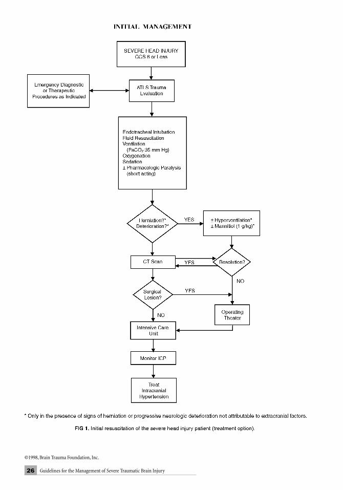
Successful systemic resuscitation is fundamental to maintaining the possibility ofsatisfactory neurologic recovery. Therefore, the Advanced Trauma Life Support (ATLS)evaluation remains the first priority. The considerations contained in this chapter are to beapplied within the framework of the ATLS approach. An algorithm describing an approach tothe resuscitation of the severe head injury patient is presented in Figure 1 (page 26).
Management in the Absence of Clinical Signs ofHerniationIn the absence of clinical evidence of transtentorial herniation, sedation and pharmacologicrelaxation should be used when indicated for safe and efficient patient transport. Theconfusion and agitation frequently attendant to head injury often makes sedation desirable.Pharmacologic relaxation, however, has the undesirable effect of limiting the neurologicexam to the pupils and, on arrival at the hospital, the CT scan. Therefore, its use in theabsence of evidence of herniation should be limited to situations where sedation alone is notsufficient to optimize safe and efficient patient transport and resuscitation. When used, short-acting agents are strongly preferred.
This protocol opinion does not support the “prophylactic” administration of mannitol dueto its volume-depleting diuretic effect. In addition, although it might be desirable toapproximate the lower end of the normal range of PaCO
2 during transport of a suspected brain
injury, the risk of exacerbating early ischemia (see hyperventilation chapter) outweighs thequestionable benefit in the patient without evidence of herniation. Therefore, the protocoloption derived here recommends ventilatory parameters consistent with optimal oxygenationand “normal” ventilation.
Management in the Presence of Clinical Signs ofHerniationWhen there is evidence of transtentorial herniation (or progressive neurologic deterioration notattributable to extracranial explanations), aggressive treatment of suspected intracranialhypertension is indicated. Hyperventilation is easily accomplished by increasing the ventilatoryrate and does not depend on or interfere with successful volume resuscitation. Becausehypotension can produce both neurologic deterioration and intracranial hypertension, the useof mannitol is less desirable unless adequate volume resuscitation has been accomplished (seemannitol chapter). If complete volume resuscitation has been attained, however, mannitolshould be administered by bolus infusion. Under these circumstances, it is critical that thepatient be transported to the hospital with utmost haste.

25Initial Management
V. SummaryThe fundamental goals of resuscitation of the head-injured patient are the restoration ofcirculating volume, blood pressure, oxygenation, and ventilation. The physician should initiatemaneuvers that serve to lower ICP and do not interfere with these aims as early as possibleduring resuscitation of any patient with a head injury. Treatment modalities such ashyperventilation and mannitol administration that have the potential of exacerbatingintracranial ischemia or interfering with resuscitation should be reserved for patients who showsigns of intracranial hypertension such as evidence of herniation or neurologic deterioration.
VI. Key Issues for Future InvestigationThe key issues discussed in all the chapters relevant to this section are germane to thisdiscussion. Specific to this section is the question of combining these modalities into a protocoland testing the efficacy of that protocol in optimizing resuscitation and improving outcomefrom severe head injury. The “prophylactic” treatment of intracranial hypertension in patientssuspected of severe head injury is of particular interest and would lend itself to a prospective,randomized trial.
26 Guidelines for the Management of Severe Traumatic Brain Injury
©1998, Brain Trauma Foundation, Inc.
27Initial Management
VII. Evidentiary Table: The Integration of Brain-SpecificTreatments Into the Initial Resuscitation of theSevere Head Injury Patient
Bickell,1 1994
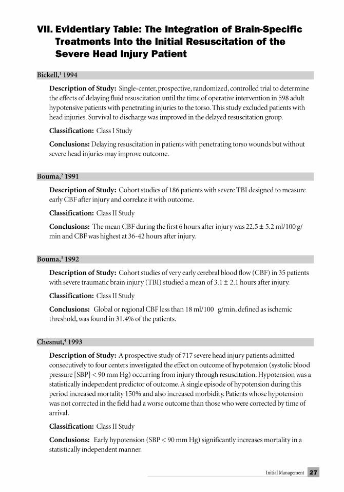
Description of Study: Single-center, prospective, randomized, controlled trial to determinethe effects of delaying fluid resuscitation until the time of operative intervention in 598 adulthypotensive patients with penetrating injuries to the torso. This study excluded patients withhead injuries. Survival to discharge was improved in the delayed resuscitation group.
Classification: Class I Study
Conclusions: Delaying resuscitation in patients with penetrating torso wounds but withoutsevere head injuries may improve outcome.
Bouma,2 1991
Description of Study: Cohort studies of 186 patients with severe TBI designed to measureearly CBF after injury and correlate it with outcome.
Classification: Class II Study
Conclusions: The mean CBF during the first 6 hours after injury was 22.5 ± 5.2 ml/100 g/min and CBF was highest at 36-42 hours after injury.
Bouma,3 1992
Description of Study: Cohort studies of very early cerebral blood flow (CBF) in 35 patientswith severe traumatic brain injury (TBI) studied a mean of 3.1 ± 2.1 hours after injury.
Classification: Class II Study
Conclusions: Global or regional CBF less than 18 ml/100 g/min, defined as ischemicthreshold, was found in 31.4% of the patients.
Chesnut,4 1993
Description of Study: A prospective study of 717 severe head injury patients admittedconsecutively to four centers investigated the effect on outcome of hypotension (systolic bloodpressure [SBP] < 90 mm Hg) occurring from injury through resuscitation. Hypotension was astatistically independent predictor of outcome. A single episode of hypotension during thisperiod increased mortality 150% and also increased morbidity. Patients whose hypotensionwas not corrected in the field had a worse outcome than those who were corrected by time ofarrival.
Classification: Class II Study
Conclusions: Early hypotension (SBP < 90 mm Hg) significantly increases mortality in astatistically independent manner.

28 Guidelines for the Management of Severe Traumatic Brain Injury
Fearnside,6 1993
Description of Study: A prospective study of 315 severe head injury patients admittedconsecutively to a single center with respect to prehospital and in-hospital predictors of outcome.Hypotension (SBP < 90 mm Hg) was an independent predictor of increased mortality andmorbidity.
Classification: Class II Study
Conclusions: Hypotension (SBP < 90 mm Hg) occurring at any time during a patient’s courseindependently predicts worse outcome.
Gaab,7 1990
Title of Study: A Comparative Analysis of THAM (Tris-buffer) in Traumatic Brain Edema.(n = 21 patients, not randomized.)
Classification: Class II Study
Conclusions: Mannitol boluses produced a 32% reduction in ICP and the effect was seen for 60minutes. THAM was “at least as effective as mannitol.”
Graham,8 1988
Description of Study: Histologic study of 71 victims of fatal severe TBI who had no premortemevidence (clinical, radiologic, or pathologic) of elevated ICP.
Classification: Class II Study
Conclusions: Ischemic cell changes were found in 70% of the brains.
Hsiang,9 1994
Description of Study: A prospective study of 514 severe head injury patients admittedconsecutively to four centers investigated the effect on outcome of prophylactic neuromuscularblockade (i.e., pharmacologic paralysis beginning early in the patient’s course and lasting at least12 hours not administered for control of intracranial hypertension). Such use of neuromuscularblockade was associated with a longer intensive care unit course, a higher incidence ofpneumonia, and a trend toward more frequent sepsis without providing an improvement inoutcome.
Classification: Class I Study
Conclusions: Neuromuscular blockade should be reserved for specific indications (e.g.,intracranial hypertension, transport, etc.) rather than be routinely administered to severe headinjury patients.
VII. Evidentiary Table (continued)

29Initial Management
Marion,11 1991
Description of Study: Cohort study of 32 patients with severe TBI aimed at defining temporalchanges in CBF that occur during the first 5 days after injury.
Classification: Class II Study
Conclusions: Mean CBF in the first 1-4 hours after injury was 27 ml/100 g/min, and CBF wasalways lowest during the first 12-24 hours after injury. Regional CBF was substantially hetero-geneous.
Muizelaar,12 1991
Description of Study: Prospective, randomized clinical trial of 77 patients with severe TBIcomparing clinical outcome for a group hyperventilated to a PaCO
2 of 25 ± 2 mm Hg for 5 days
after injury and a group with a PaCO2 kept at 35 ± 2 mm Hg during that period.
Classification: Class I Study
Conclusions: At 3 and 6 months after injury, the patient with an initial Glasgow Coma Scale(GCS) motor score of 4-5 had a significantly better outcome if they were not hyperventilated.
Obrist,13 1984
Description of Study: Cohort study of 31 patients with severe TBI in whom the effect ofaggressive hyperventilation on ICP, CBF, and arteriovenous difference in oxygen content(AVdO
2) was examined.
Classification: Class II Study
Conclusions: Hyperventilation had a much more direct effect on CBF reduction (29 of 31patients) than it did on ICP reduction (15 of 31 patients). Aggressive hyperventilation in 10patients (PaCO
2 of 23.2 ± 2.8 mm Hg) led to AVdO
2 values of 10.5 ± 0.7 vol% and CBF values of
18.6 ± 4.4 ml/100 g/min.
Pigula,14 1993
Description of Study: Fifty-eight children (< 17 years) with severe head injuries wereprospectively studied for the effect of hypotension (SBP < 90 mm Hg) on outcome. An episodeof hypotension decreased survival fourfold. This finding was confirmed in a concomitantanalysis of the effect of hypotension on outcome in 509 patients in the National Pediatric TraumaRegistry. Hypotension appeared to eliminate any neuroprotective mechanisms normally affordedby age.
Classification: Class II Study
Conclusions: The detrimental effects of hypotension (SBP < 90 mm Hg) on outcome appearto extend to children.
VII. Evidentiary Table (continued)

30 Guidelines for the Management of Severe Traumatic Brain Injury
Robertson,15 1992
Description of Study: Cohort study of 102 patients with severe head injury examining thetime course and relationship of AVdO
2, CBF, and ICP.
Classification: Class II Study
Conclusions: AVdO2 values were always widest during the first 24 hours after injury.
Schwartz,16 1984
Title of Study: The University of Toronto Head Injury Treatment Study: A Prospective,Randomized Comparison on Pentobarbital and Mannitol.
Classification: Class I Study
Conclusions: Prospective, randomized comparison of mannitol vs barbiturates for ICP control.Crossover permitted. Sequential analysis (n=59). Pentobarbital was not significantly better thanmannitol. Mannitol group had better outcome mortality, 41% vs 77%. Cerebral perfusionpressure (CPP) was much better with mannitol than barbiturates (75 mm Hg vs 45 mm Hg).
Sheinberg,17 1992
Description of Study: Cohort study of jugular venous Oxygen (O2) saturation in 45 patients
with severe head injury monitored for 1-8 days.
Classification: Class II Study
Conclusions: Hyperventilation was the second most common identifiable cause of jugularvenous desaturations (O
2 saturation < 50%), and was the cause for desaturation in 10 of 33 cases.
Smith,18 1986
Title of Study: Comparison of Two Mannitol Regimens in Patients with Severe Head Injury,Undergoing Intracranial Pressure Monitoring: Effect of Bolus Mannitol Given Only When ICP >25 mm Hg, versus “Empirical Mannitol” (every 2 hours until serum osmolarity (OSM) 310, orneurodeterioration).
Classification: Class I Study
Conclusions: No difference between ICP-directed and empiric mannitol use. ICP smootherand lower in empiric group. (Power too low to detect an effect (n = 8), randomized.)
VII. Evidentiary Table (continued)

31Initial Management
Vassar,19 1993
Description of Study: Prospective, randomized, controlled, multicenter trial comparing theefficacy of administering 250 ml of hypertonic saline vs normal saline as the initial resuscitationfluid in facilitating the resuscitation and improving the outcome of hypotensive trauma patients.In this trial, the hypertonic saline group had significantly improved blood pressure responses anddecreased overall fluid requirements. Although there was an associated improvement in survivalfor the overall group, it did not reach statistical significance. Post-hoc analysis of the severe headinjury group (Class II analysis), however, revealed that the hypertonic saline group had astatistically significant improvement in survival-to-discharge.
Classification: Class II Study
Conclusions: Raising the blood pressure in hypotensive, severe head injury patients improvesoutcome in proportion to the efficacy of the resuscitation.
VIII. References1. Bickell WH, Wall MJ, Pepe PE, et al.: Immediate versus delayed fluid resuscitation for
hypotensive patients with penetrating torso injuries. New England Journal of Medicine331:1105-9, 1994.
2. Bouma GJ, Muizelaar JP, Choi SC, et al.: Cerebral circulation and metabolism after severetraumatic brain injury: the elusive role of ischemia. J Neurosurg 75:685-693, 1991.
3. Bouma GJ, Muizelaar JP, Stringer WA, et al.: Ultra-early evaluation of regional cerebralblood flow in severely head-injured patients using xenon-enhanced computerizedtomography. J Neurosurg 77:360-368, 1992.
4. Chesnut RM, Marshall LF, Klauber MR, et al.: The role of secondary brain injury indetermining outcome from severe head injury. J Trauma 34:216-222, 1993.
5. Chiolero RL: Sedatives and antagonists in the management of severely head-injuredpatients. [Review]. Acta Neurochirurgica Supplementum 55:43-46, 1992.
6. Fearnside MR, Cook RJ, McDougall P, et al.: The Westmead Head Injury Project outcome insevere head injury. A comparative analysis of pre-hospital, clinical and CT variables. Br JNeurosurg 7:267-279, 1993.
7. Gaab MR, Seegers K, Smedema RJ, et al.: A comparative analysis of THAM (Tris-buffer) intraumatic brain edema. Acta Neurochirurgica Supplementum 51:320-323, 1990.
8. Graham DI, Lawrence AE, Adams JH, et al.: Brain damage in fatal non-missile head injurywithout high intracranial pressure. J Clin Pathol 41:34-37, 1988.
9. Hsiang JK, Chesnut RM, Crisp CB, et al.: Early, routine paralysis for intracranial pressurecontrol in severe head injury: is it necessary? Critical Care Medicine 22:1471-1476, 1994.
10. Marion DW, Carlier PM: Problems with initial Glasgow Coma Scale assessment caused byprehospital treatment of patients with head injuries: results of a national survey. J Trauma36:89-95, 1994.
11. Marion DW, Darby J, Yonas H: Acute regional cerebral blood flow changes caused by severehead injuries. J Neurosurg 74:407-414, 1991.
VII. Evidentiary Table (continued)

32 Guidelines for the Management of Severe Traumatic Brain Injury
12. Muizelaar JP, Marmarou A, Ward JD, et al.: Adverse effects of prolonged hyperventilation inpatients with severe head injury: a randomized clinical trial. J Neurosurg 75:731-739, 1991.
13. Obrist WD, Langfitt TW, Jaggi JL, et al.: Cerebral blood flow and metabolism in comatosepatients with acute head injury. Relationship to intracranial hypertension. J Neurosurg61:241-253, 1984.
14. Pigula FA, Wald SL, Shackford SR, et al.: The effect of hypotension and hypoxia on childrenwith severe head injuries. J Pediatr Surg 28:310-316, 1993.
15. Robertson PA, Ryan MD: Neurological deterioration after reduction of cervicalsubluxation. Mechanical compression by disc tissue. J Bone Joint Surg [Br] 74:224-227, 1992.
16. Schwartz ML, Tator CH, Rowed DW, et al.: The University of Toronto Head InjuryTreatment Study: a prospective, randomized comparison of pentobarbital and mannitol.Can J Neurol Sci 11:434-440, 1984.
17. Sheinberg M, Kanter MJ, Robertson CS, et al.: Continuous monitoring of jugular venousoxygen saturation in head-injured patients. J Neurosurg 76:212-217, 1992.
18. Smith HP, Kelly D Jr, McWhorter JM, et al.: Comparison of mannitol regimens in patientswith severe head injury undergoing intracranial monitoring. J Neurosurg 65:820-824, 1986.
19. Vassar MJ, Fischer RP, O’Brien PE, et al.: A multicenter trial for resuscitation of injuredpatients with 7.5% sodium chloride. The effect of added dextran 70. The Multicenter Groupfor the Study of Hypertonic Saline in Trauma Patients. Archives of Surgery 128:1003-1011,1993.
33Resuscitation of Blood Pressure and Oxygenation
I. RecommendationsA. Standards
There are insufficient data to support a treatment standard for this topic.B. Guidelines
Hypotension (systolic blood pressure [SBP] < 90 mm Hg) or hypoxia (apnea,cyanosis, or an Oxygen (O
2)
saturation < 90% in the field or a PaO
2 < 60 mm Hg)
must be monitored and scrupulously avoided, if possible, or corrected immediatelyin severe traumatic brain injury (TBI) patients.
C. OptionsThe mean arterial blood pressure should be maintained above 90 mm Hg throughthe infusion of fluids throughout the patient’s course to attempt to maintain cerebralperfusion pressure (CPP) greater than 70 mm Hg. Patients with a Glasgow ComaScale (GCS) score less than 9, who are unable to maintain their airway or whoremain hypoxemic despite supplemental O
2, require that their airway be secured,
preferably by endotracheal intubation.
II. OverviewFor ethical reasons, a prospective, controlled study concerning the effects of hypotension orhypoxia on outcome from severe head injury has never been done. Nevertheless, there is agrowing body of evidence that secondary insults occur frequently and exert a profound,adverse influence on outcome from severe head injury. This effect appears to be more profoundthan results when hypoxic or hypotensive episodes of similar magnitude occur in traumapatients without neurologic involvement. Therefore, we need to determine if there is any strongevidence that suggests threshold values for oxygenation and blood pressure support.
III. ProcessA MEDLINE search from 1966 to 1998 was undertaken using the following key words: “headinjury” (and “hypoxia” or “hypotension”) and “human subject”; and “head injury” (and “field”or “pre-hospital” or “prehospital”) and (“treatment” or “management” or “resuscitation”). Theresultant references found to be directly relevant regarding outcome analysis and clinicalorientation were individually reviewed for design, content, and relevance. The results of thisreview were then incorporated into the analysis presented here.
RESUSCITATION OF BLOOD PRESSUREAND OXYGENATION

34 Guidelines for the Management of Severe Traumatic Brain Injury
IV. Scientific FoundationHypoxemiaIn head-injured patients, significant secondary brain injury results from systemic hypotensionand hypoxemia.25 An English study revealed that 44% of TBI victims were hypoxemic in thefield or ambulance, with documented O
2 saturations between 75%-90% in 28% of the patients
and O2 saturations less than 75% in 16% of the patients.24 A study in Ireland documented
hypoxemia in 27% of TBI patients on arrival to the closest emergency department.4 These adultpopulation findings are similar to those in a retrospective, pediatric, severe TBI study, where13% of the patients were hypoxemic and 6% were hypercarbic.12 These hypoxemic episodeshave also been associated with statistically significant worse outcomes in the patients. In Italy,55% of helicopter transported TBI patients were hypoxemic prior to intubation.25 Of thehypoxemic patients, 46% did not have concomitant hypotension. In non-hypoxemic patients,mortality was 14.3% with a 4.8% rate of severe disability. However, in patients with documentedO
2 saturations less than 60%, the mortality rate was 50% and all of the survivors were severely
disabled.
HypotensionThe deleterious influence of hypoxemia and hypotension on the outcome of severe head injurywas also demonstrated by the analysis of a large (717 patients), prospectively collected data setfrom the Traumatic Coma Data Bank (TCDB; Class II studies).3,13 Hypoxemia and hypotensioneach occurred in over one-third of severe head injury patients. The TCDB study demonstratedthat prehospital hypotension (a single observation of a SBP < 90 mm Hg) or hypoxia (apnea/cyanosis in the field or a PaO
2 < 60 mm Hg by arterial blood gas analysis) were among the five
most powerful predictors of outcome. These predictors were statistically independent of theother major predictors such as age, admission Glasgow Coma Scale (GCS) score, admissionGCS motor score, intracranial diagnosis, and pupillary status. A single episode of hypotensionwas associated with increased morbidity and a doubling of mortality as compared with amatched group of patients without hypotension. 3 N.B.
A Class II study from Australia supports the above findings, particularly regarding theeffects of hypotension on outcome.5 A retrospective review of prospectively collected data inchildren less than 17 years of age also corroborated these results.19 Here, hypotension markedlyincreased morbidity and mortality independently of other predictors of outcome, eliminatingthe improvement in survival generally afforded by youth. These data validate similarretrospectively analyzed Class II and III reports published previously.6,8,10,11,15-18,20,23
N.B. The question of the influence of hypoxia and hypotension on outcome is not subjectable to manipulative investigation. In addition, noprospective studies with concomitant cohort controls have been performed or are likely to be undertaken due to ethical considerations. Therefore,the large, prospectively collected, observational data set from the TCDB is the best information on the subject that can be expected to be available.Given the size and nature of this study and the unequivocal nature of the results, the avoidance of hypotension (SBP ≤ 90 mm Hg) and hypoxia(PaO
2 ≤ 60 mm Hg) during the early post-injury period can be supported at the level of a guideline, if not a treatment standard.

35Resuscitation of Blood Pressure and Oxygenation
Airway ManagementThe role of active airway management including endotracheal intubation (ETI) for TBI patientshas not been well studied. A prehospital study9 to investigate the relationship between GCSscore and the need for intubation within 30 minutes of Emergency Department (ED) arrivaldetermined that 100% of TBI patients with a GCS score of 3-5 required ETI (27% in the field,72% in the ED). Additionally, 72% of TBI patients with a GCS score of 6-7 required ETI (27% inthe field, 36% in the ED), and 61% of TBI patients with a GCS score of 8-9 required ETI (8% inthe field, 53% in the ED). Prehospital intubation is associated with significantly enhancedsurvival in TBI patients. A retrospective, case control study with severe trauma victims(including head injury), compared patients who underwent prehospital intubation with thosewho did not.31 Mortality was significantly reduced in intubated patients, particularly in thelowest GCS score and isolated head injury subsets.26-30
Resuscitation FluidsA Class I study has never directly addressed the efficacy of preventing or correcting earlyhypotension to improve outcome. The American College of Surgeons advocates the rapidinfusion of two liters of Ringer’s lactate or normal saline as an initial resuscitative crystalloidbolus.1 However, resuscitation prior to definitive surgical hemostasis may cause displacement ofhemostatic clots, hemodilution, and worsen secondary blood loss and mortality in penetratingtorso trauma.2 The Advanced Trauma Life Support course and most textbooks advise thejudicious use of fluid in treating TBI patients from concerns that fluid may augment cerebraledema and intracranial pressure (ICP). However, in multitrauma patients with head injury,Scalea demonstrated a lack of relationship between amount of fluid or blood infused and ICP.22
Hypertonic saline and mannitol have been advocated as resuscitation fluids in addition to thereduction of intracranial hypertension.
Clinically, mannitol is routinely used to reduce ICP in TBI patients with intracranialhypertension. However, mannitol’s osmotic diuresis may cause volume deficits, hypotension,and subsequent secondary brain injury. The prehospital administration of mannitol versusplacebo in TBI patients showed no difference in mortality; however, in the treatment group SBPfell significantly two hours after hospital arrival, but comparing all time periods there was nosubstantial difference.21
Hypertonic saline has been demonstrated to reduce ICP in patients with TBI andintracranial hypertension.7 Subgroup, post-hoc analysis of severe TBI patients in a prospective,randomized, placebo-controlled, multicenter trial demonstrated both a higher SBP andenhanced survival in trauma patients resuscitated with hypertonic saline instead ofcrystalloid.14 This data strongly suggests that elevating the blood pressure in hypotensive, severehead injury patients improves outcome. Meta-analysis of TBI patients who received hypertonicsaline/dextran are about twice as likely to survive as those who receive standard therapy.30 Otherstudies show either no difference or improved survival utilizing hypertonic saline with orwithout dextran over isotonic saline for fluid resuscitation, with most benefit in the subgroup ofpatients with an initial GCS score less than 9.27-29

36 Guidelines for the Management of Severe Traumatic Brain Injury
Resuscitation End-PointsThe value of 90 mm Hg as a systolic pressure threshold for hypotension has arisen in a ratherarbitrary fashion and is more of a statistical than a physiologic parameter. Given the evidenceon the influence of cerebral perfusion pressure (CPP) on outcome, it is possible that systolicpressures significantly higher than 90 mm Hg would be desirable during the prehospital andresuscitation phase, but no studies have been performed thus far to corroborate this. Theimportance of mean arterial pressure, as opposed to systolic pressure, should also be stressed,not only because of its role in calculating CPP, but because the lack of a consistent relationshipbetween systolic and mean pressures makes calculations based on systolic values unreliable. Itmay be valuable to maintain mean arterial pressures considerably above those represented bysystolic pressures of 90 mm Hg throughout the patient’s course. However, once ICP monitoringhas been established, manipulation of blood pressure should be guided by CPP management.
V. SummaryEarly post-injury episodes of hypotension or hypoxia greatly increase morbidity and mortalityfrom severe head injury. At present, the defining level of hypotension and hypoxia is unclear inthese patients. However, ample Class II evidence exists regarding hypotension, defined as asingle observation of an SBP of less than 90 mm Hg, or hypoxia, defined as apnea/cyanosis inthe field or a PaO
2 less than 60 mm Hg by arterial blood gas analysis, to warrant the formation
of guidelines stating that these values must be avoided, if possible, or rapidly corrected in severehead injury patients.1,5,19 A significant proportion of adult and pediatric TBI patients arediscovered to be hypoxemic or hypotensive in the prehospital setting. Patients with severe headinjury that are intubated in the prehospital setting appear to have better outcomes. Strong ClassII evidence suggests that raising the blood pressure in hypotensive, severe head injury patientsimproves outcome in proportion to the efficacy of the resuscitation.17,26
VI. Key Issues for Future InvestigationThe major questions for resuscitating the severe head injury patient are the critical values forduration and magnitude of hypotensive episodes and the optimal resuscitation protocol(hypertonic or isotonic solutions, colloids, route of administration, etc.) affecting neurologicaloutcome. The former question is not a subject for a controlled trial for ethical reasons and,therefore, would be best addressed using a prospective data collection study with highresolution collection of prehospital blood pressure data, correlating this with outcome. Thelatter question can be studied in prospective, randomized investigations, several of which arepresently underway. Finally, because the actual parameter of interest is CPP, a simple,non-invasive method of determining ICP in the field warrants development.
37Resuscitation of Blood Pressure and Oxygenation
VII. Evidentiary Table: Resuscitation of BloodPressure and Oxygenation
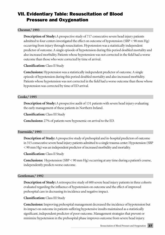
Chesnut,3 1993
Description of Study: A prospective study of 717 consecutive severe head injury patientsadmitted to four centers investigated the effect on outcome of hypotension (SBP < 90 mm Hg)occurring from injury through resuscitation. Hypotension was a statistically independentpredictor of outcome. A single episode of hypotension during this period doubled mortality andalso increased morbidity. Patients whose hypotension was not corrected in the field had a worseoutcome than those who were corrected by time of arrival.
Classification: Class II Study
Conclusions: Hypotension was a statistically independent predictor of outcome. A singleepisode of hypotension during this period doubled mortality and also increased morbidity.Patients whose hypotension was not corrected in the field had a worse outcome than those whosehypotension was corrected by time of ED arrival.
Cooke,4 1995
Description of Study: A prospective audit of 131 patients with severe head injury evaluatingthe early management of these patients in Northern Ireland.
Classification: Class III Study
Conclusions: 27% of patients were hypoxemic on arrival to the ED.
Fearnside,5 1993
Description of Study: A prospective study of prehospital and in-hospital predictors of outcomein 315 consecutive severe head injury patients admitted to a single trauma center. Hypotension (SBP< 90 mm Hg) was an independent predictor of increased morbidity and mortality.
Classification: Class II Study
Conclusions: Hypotension (SBP < 90 mm Hg) occurring at any time during a patient’s course,independently predicts worse outcome.
Gentleman,6 1992
Description of Study: A retrospective study of 600 severe head injury patients in three cohortsevaluated regarding the influence of hypotension on outcome and the effect of improvedprehospital care in decreasing its incidence and negative impact.
Classification: Class III Study
Conclusions: Improving prehospital management decreased the incidence of hypotension butits impact on outcome in patients suffering hypotensive insults maintained as a statisticallysignificant, independent predictor of poor outcome. Management strategies that prevent orminimize hypotension in the prehospital phase improves outcome from severe head injury.

38 Guidelines for the Management of Severe Traumatic Brain Injury
Hill,8 1993
Description of Study: A retrospective study of prehospital and ED resuscitative managementof 40 consecutive, multitrauma patients. Hypotension (SBP ≤ 80 mm Hg) correlated stronglywith fatal outcomes. Hemorrhagic hypovolemia was the major etiology of hypotension.
Classification: Class III Study
Conclusions: Improving the management of hypovolemic hypotension is a major potentialmechanism for improving the outcome from severe head injury.
Hsiao,9 1993
Description of Study: A retrospective trauma registry–based study of 120 patients with a GCSless than 14 that evaluated the need for emergency intubation in the field or ED and evaluated CTscan findings.
Classification: Class III Study
Conclusions: Patients with a GCS score of 3-5 were all intubated and 73% had abnormal CT scans;73% of patients with a GCS score of 6-7 were intubated and 36% had CT scan abnormalities; 62%of patients with a GCS score of 8-9 were intubated and 62% had CT scan abnormalities; 20% ofpatients with a GCS score of 10-13 required intubation and 23% had abnormal CT scans.
Jeffreys,10 1981
Description of Study: A retrospective review of hospital records in 190 head injury patientswho died after admission. Hypotension was one of the four most common avoidable factorscorrelated with death.
Classification: Class III Study
Conclusions: Early hypotension appears to be a common and avoidable cause of death in severehead injury patients.
Kohi,11 1984
Description of Study: A retrospective evaluation of 67 severe head injury patients seen over asix-month period were correlated with 6-month outcome. For a given GCS score, the presence ofhypotension resulted in a worse outcome than would have been predicted.
Classification: Class III Study
Conclusions: Early hypotension increases the mortality and worsens the prognosis of survivorsin severe head injury.
VII. Evidentiary Table (continued)

39Resuscitation of Blood Pressure and Oxygenation
Kokoska,12 1998
Description of Study: A retrospective chart review of 72 pediatric patients admitted to a singlecenter with a GCS score of 6-8; primarily evaluated morbidity from hypotension with a briefmention of hypoxemia.
Classification: Class III Study
Conclusions: Thirteen percent of patients had a documented hypoxic episode, and 6% hadhypercarbia. The exact location (prehospital, ED, OR, PICU) of these episodes was not given.Hypoxemia and hypercarbia could not be related to outcome.
Marmarou,13 1991
Description of Study: From a prospectively collected database of 1,030 severe head injurypatients; all 428 patients who met ICU monitoring criteria were analyzed for monitoringparameters that determined outcome and their threshold values. The two most critical valueswere the proportion of hourly ICP readings greater than 20 mm Hg and the proportion of hourlySBP readings less than 80 mm Hg.
Classification: Class II Study
Conclusions: The incidence of morbidity and mortality resulting from severe head injury isstrongly related to ICP and hypotension measured during the course of ICP management.
Miller,15 1982
Description of Study: A prospective study of 225 severely head-injured patients regarding theinfluence of secondary insults on outcome. Hypotension (SBP < 95 mm Hg) was significantlyassociated with increased morbidity and mortality. The predictive independence of hypotensionin comparison to other associated factors, however, was not investigated.
Classification: Class II Study
Conclusions: A strong statistical relationship exists between early hypotension and increasedmorbidity and mortality in patients with severe head injury.
Miller,16 1978
Description of Study: One hundred consecutive severe head injury patients were prospectivelystudied regarding the influence of secondary insults on outcome (report of first 100 patients insubsequent report of 225 patients [vide supra]). Hypotension (SBP < 95 mm Hg) associatedwith a trend (not statistically significant) toward worse outcome in entire cohort. This trend metstatistical significance for patients without mass lesions. Seminal report relating earlyhypotension to increased morbidity and mortality. Influence of hypotension on outcome notanalyzed independently from other associated factors.
Classification: Class II Study
Conclusions: This is the first prospective report implicating early hypotension as a majorpredictor of increased morbidity and mortality from severe head injury.
VII. Evidentiary Table (continued)

40 Guidelines for the Management of Severe Traumatic Brain Injury
Narayan,17 1982
Description of Study: Retrospective analysis of 207 consecutively admitted severe head injurypatients. Management included aggressive attempts to control ICP using a threshold of 20 mmHg. Outcome was significantly correlated with the ability to control ICP.
Classification: Class III Study
Conclusions: ICP control using a threshold of 20 mm Hg as a part of an overall aggressivetreatment approach to severe head injury may be associated with improved outcome.
Pietropaoli,18 1992
Description of Study: A retrospective review of the impact of hypotension (SBP < 90 mm Hg)on 53 otherwise normotensive severe head injury patients who required early surgery (within 72hours of injury). The mortality rate was 82% in the group with hypotension and 25% in thenormotensive group (p < 0.001). The duration of intraoperative hypotension was inverselycorrelated with Glasgow Outcome Scale score using linear regression (R = -0.30, p = 0.02).
Classification: Class III Study
Conclusions: Early surgery with intraoperative hypotension is significantly correlated withincreased mortality from severe head injury in a duration-dependent fashion.
Pigula,19 1993
Description of Study: Fifty-eight children (< 17 years old) with severe head injuries wereprospectively studied for the effect of hypotension (SBP < 90 mm Hg) on outcome. An episode ofhypotension decreased survival fourfold. This finding was confirmed in a concomitant analysis ofthe effect of hypotension on outcome in 509 patients in the National Pediatric Trauma Registry.Hypotension appeared to eliminate any neuroprotective mechanisms normally afforded by age.
Classification: Class II Study
Conclusions: The detrimental effects of hypotension (SBP < 90 mm Hg) on outcome appear toextend to children.
Rose,20 1977
Description of Study: A retrospective review of hospital and necropsy records of 116 headinjury patients who were known to have talked before dying. Hypotension was a major avoidablefactor related to the increased mortality in this group.
Classification: Class III Study
Conclusions: Hypotension is a major avoidable cause of increased mortality in patients withmoderate head injury.
VII. Evidentiary Table (continued)

41Resuscitation of Blood Pressure and Oxygenation
Sayre,21 1996
Description of Study: A prospective, randomized, double-blind, placebo-controlled clinicaltrial of 41 patients over a one-year period at a university-based Level I trauma center. Allpatients were endotracheally-intubated head trauma victims with a GCS score less than 12evaluated within 6 hours of injury.
Classification: Class II Study
Conclusions: Out-of-hospital administration of mannitol did not significantly change systolicblood pressure in this group of head-injured, multiple trauma patients. There is an insufficientnumber of patients in this pilot study to assess whether out-of-hospital administration ofmannitol to head-injured patients is beneficial overall.
Seelig,23 1986
Description of Study: A study of all patients (n = 160) with an ICP of 30 mm Hg during thefirst 72 hours after injury from a prospectively collected database of severe head injury patients(n = 348). The incidence and severity of intracranial hypertension and increased overallmortality were significantly correlated with systemic hypotension. The statisticalindependence of hypotension as a predictor was not evaluated.
Classification: Class II Study
Conclusions: Early hypotension is significantly correlated with increased incidence and severityof intracranial hypertension and increased mortality.
Silverston,24 1989
Description of Study: A study of 25 consecutive trauma patients, including head injury, whichevaluated the use of non-invasive pulse oximetry in the field and in a moving ambulance.
Classification: Class III Study
Conclusions: 16% of patients had an SaO2 less than 75%, and an additional 28% were between
75% and 90%. There were no demonstrated difficulties in using the pulse oximeter in the field orambulance.
Stocchetti,25 1996
Description of Study: A cohort study of 50 trauma patients transported from the scene byhelicopter, which evaluated the incidence and effect of hypoxemia and hypotension on outcome.
Classification: Class III Study
Conclusions: Fifty-five percent of patients were hypoxic (SaO2 < 90%) and 24% were
hypotensive. Both hypoxemia and hypotension negatively affected outcome, however, the degreeto which each independently affected the outcome was not studied.
VII. Evidentiary Table (continued)

42 Guidelines for the Management of Severe Traumatic Brain Injury
Vassar,26 1990
Description of Study: A prospective, randomized, double-blind, clinical trial of 106 patientsover an 8-month period. Intracranial hemorrhage was present in 28 (26%) patients.
Classification: Class II Study
Conclusions: No adverse effects of rapid infusion of 7.5% NaCl or 7.5% NaCl/6% dextran 70were noted. Nor were any beneficial effects noted. There was no evidence of potentiatingintracranial bleeding. There were no cases of central pontine myelinolysis; however, patients withsevere pre-existing disease were excluded from the study.
Vassar,27 1991
Description of Study: A prospective, randomized, double-blind, multicenter clinical trial of166 hypotensive patients over a 44-month period. Fifty-three of these patients (32%) had a severehead injury (defined as an AIS score for the head of 4, 5, or 6).
Classification: Class II Study
Conclusions: Survival was not significantly different in the total patient group. The survival rateof severely head-injured patients to hospital discharge was significantly higher for those whoreceived hypertonic saline/dextran (HSD) (32% of patients with HSD vs 16% in patients withLR) when using logistic regression analysis.
Vassar,28 1993
Description of Study: A prospective, randomized, double-blind, multicenter trial comparingthe efficacy of administering 250 ml of hypertonic saline vs normal saline as the initialresuscitation fluid in 194 hypotensive trauma patients over a 15-month period. 144 of thesepatients (74%) had a severe brain injury (defined as an abbreviated injury score [AIS] for thehead of 4, 5, or 6). Here, hypertonic saline significantly increased blood pressure and decreasedoverall fluid requirements. Post-hoc analysis of the severe head injury group (Class II analysis)revealed that the hypertonic saline group had a statistically significant improvement in survival-to-discharge. However, the improvement in overall survival was not statistically significant.
Classification: Class II Study
Conclusions: Raising the blood pressure in the hypotensive, severe head injury patientimproves outcome in proportion to the efficacy of the resuscitation. Prehospital administrationof 7.5% sodium chloride to hypotensive trauma patients was associated with a significantincrease in blood pressure compared with infusion of Lactated Ringer’s (LR) solution. Thesurvivors in the LR and hypertonic saline (HS) groups had significantly higher blood pressuresthan the non-survivors. There was no significant increase in the overall survival of patients withsevere brain injuries, however, the survival rate in the HS group was higher than that in the LRgroup for the cohort with a baseline GCS score of 8 or less.
VII. Evidentiary Table (continued)

43Resuscitation of Blood Pressure and Oxygenation
Vassar,29 1993
Description of Study: Prospective, randomized, double-blind, controlled clinical trial of 258hypotensive patients over 31 months at a university-based trauma center. Twenty-seven of thesepatients (10%) had a severe head injury (defined as an AIS score for the head of 4, 5, or 6 only foranatomic lesions).
Classification: Class I Study
Conclusions: The administration of 7.5% NaCl (HS) and 7.5% NaCl/6% dextran 70 (HSD)caused no neurologic abnormalities. On the contrary, their use was associated with improvementin survival (as compared with predicted survival) in the patients with low initial GCS score (< 8)and in patients with anatomic confirmation of severe cerebral damage. It appeared that thedextran added little to improvement in survival when compared with HS alone. HS solutions didincrease the blood pressure response in all patients.
Wade,30 1997
Description of Study: Cohort analysis of individual patient data from a previously publishedprospective, randomized, double-blind trial of HSD in patients with TBI and hypotension. TBIwas defined as an AIS score for the head of 4 or greater. Hypotension was defined as an SBP of90␣ mm Hg or less. 1,395 data records were analyzed from six separate studies; 233 patients werethen included in this review. Eighty patients were treated in the ED and 143 were treated in theprehospital phase.
Classification: Class II Study
Conclusions: There was no statistically significant difference in overall survival when HS wasretrospectively compared with normal saline. Logistic regression analysis was performed onpatients with TBI showing an odds ratio of 1:92 for 24-hour survival, and 2:12 for survival untildischarge. Thus, patients with TBI in the presence of hypotension who received HSD wereapproximately twice as likely to survive as those who received saline. This was statisticallysignificant (p␣ =␣ 0.048).
Winchell,31 1997
Description of Study: A retrospective, case control study of patients with severe head injuryand a field GCS score less than 9 and head or neck AIS score greater than 4. This study comparedpatients who underwent prehospital endotracheal intubation with those who did not.
Classification: Class III Study
Conclusions: Prehospital endotracheal intubation was associated with a statistically significantimproved survival.
VII. Evidentiary Table (continued)

44 Guidelines for the Management of Severe Traumatic Brain Injury
VIII. References1. American College of Surgeons: Advanced Trauma Life Support Instructor’s Manual.
Chicago, Illinois 1996.2. Bickell WH, Wall Jr MJ, Pepe PE, et al.: Immediate versus delayed fluid resuscitation for
hypotensive patients with penetrating torso injuries. N Engl J Med; 331:1105, 1994.3. Chesnut RM, Marshall LF, Klauber MR, et al.: The role of secondary brain injury in
determining outcome from severe head injury. J Trauma 34:216-222, 1993.4. Cooke RS, McNicholl BP, Byrnes DP: Early management of severe head injury in Northern
Ireland. Injury; 26: 395-397, 1995.5. Fearnside MR, Cook RJ, McDougall P, et al.: The Westmead Head Injury Project outcome in
severe head injury. A comparative analysis of pre-hospital, clinical, and CT variables. Br JNeurosurg 7:267-279, 1993.
6. Gentleman D: Causes and effects of systemic complications among severely head-injuredpatients transferred to a neurosurgical unit. Int Surg 77:297-302, 1992.
7. Hartl R, Ghajar J, Hochleuthner H, et al.: Hypertonic/hyperoncotic saline reliably reducesICP in severely head-injured patients with intracranial hypertension. Acta Neurochirugia;70:126-129, 1997.
8. Hill DA, Abraham KJ, West RH: Factors affecting outcome in the resuscitation of severelyinjured patients. Aust N Z J Surg 63:604-609, 1993.
9. Hsiao AK, Michaelson SP, Hedges JR: Emergent intubation and CT scan pathology of blunttrauma patients with Glasgow Coma Scale scores of 3-13. Prehospital Disast Med; 32: 26-32,1998.
10. Jeffreys RV, Jones JJ: Avoidable factors contributing to the death of head injury patients ingeneral hospitals in Mersey Region. Lancet 2:459-461, 1981.
11. Kohi YM, Mendelow AD, Teasdale GM, et al.: Extracranial insults and outcome in patientswith acute head injury—relationship to the Glasgow Coma Scale. Injury 16:25-29, 1984.
12. Kokoska ER, Smith GS, Pittman T, et al.: Early hypotension worsens neurological outcomein pediatric patients with moderately severe head trauma. J Pediatric Surg; 33: 333-338, 1998.
13. Marmarou A, Anderson RL, Ward JD, et al.: Impact of ICP instability and hypotension onoutcome in patients with severe head trauma. J Neurosurg 75:159-166, 1991.
14. Mattox KL, Maningas PA, Moore EE, et al.: Prehospital hypertonic saline/dextran infusionfor post-traumatic hypotension. Ann Surg; 213:482-491, 1991.
15. Miller JD, Becker DP: Secondary insults to the injured brain. J Royal Coll Surg (Edinburgh)27:292-298, 1982.
16. Miller JD, Sweet RC, Narayan R, et al.: Early insults to the injured brain. AMA 240:439-442,1978.
17. Narayan R, Kishore P, Becker D, et al.: Intracranial pressure: to monitor or not to monitor?A review of our experience with head injury. J Neurosurg 56:650-659, 1982.
18. Pietropaoli JA, Rogers FB, Shackford SR, et al.: The deleterious effects of intraoperativehypotension on outcome in patients with severe head injuries. J Trauma 33:403-407, 1992.
19. Pigula FA, Wald SL, Shackford SR, et al.: The effect of hypotension and hypoxia on childrenwith severe head injuries. J Pediatr Surg 28:310-316, 1993.
20. Rose J, Valtonen S, Jennett B: Avoidable factors contributing to death after head injury. BrMed J 2:615-618, 1977.
45Resuscitation of Blood Pressure and Oxygenation
21. Sayre MR, Daily SW, Stern SA, et al.: Out-of-hospital administration of mannitol to head-injured patients does not change systolic blood pressure. Acad Emer Med 1996; 3:840-848,1996.
22. Scalea TM, Maltz S, Yelon J, et al.: Resuscitation of multiple trauma and head injury: role ofcrystalloid fluids and inotropes. Crit Care Med; 22:1610-1615, 1994.
23. Seelig JM, Klauber MR, Toole BM, et al.: Increased ICP and systemic hypotension duringthe first 72 hours following severe head injury. In: Miller JD, Teasdale GM, Rowan JO, et al.(eds): Intracranial Pressure VI. Springer-Verlag, Berlin:675-679, 1986.
24. Silverston P: Pulse oximetry at the roadside: a study of pulse oximetry in immediate care.BMJ; 298: 711-713, 1989.
25. Stochetti N, Furlan A, Volta F: Hypoxemia and arterial hypotention at the accident scene inhead injury. J Trauma; 40: 764-767, 1996.
26. Vassar MJ, Perry CA, Holcroft JW: Analysis of potential risks associated with 7.5% sodiumchloride resuscitation of traumatic shock. Arch Surg; 125:1309-1315, 1990.
27. Vassar MJ, Perry CA, Gannaway WL, et al.: 7.5% sodium chloride/dextran for resuscitationof trauma patients undergoing helicopter transport. Arch Surg; 126:1065-1072, 1991.
28. Vassar MJ, Fischer RP, O’Brien PE, et al.: A multicenter trial for resuscitation of injuredpatients with 7.5% sodium chloride. The effect of added dextran 70. The Multicenter Groupfor the Study of Hypertonic Saline in Trauma Patients. Arch Surg 128:1003-1011, 1993(II-with respect to analysis of the outcome of the severe head injury group).
29. Vassar MJ, Perry CA, Holcroft JW: Prehospital resuscitation of hypotensive trauma patientswith 7.5% NaCl versus 7.5% NaCl with added dextran: a controlled trial. J␣ Trauma; 34:622-632, 1993.
30. Wade CE, Grady JJ, Kramer GC, et al.: Individual patient cohort analysis of the efficacy ofhypertonic saline/dextran in patients with traumatic brain injury and hypotension. JTrauma; 42:561-565, 1997.
31. Winchell RJ, Hoyt DB: Endotracheal intubation in the field improves survival in patientswith severe head injury. Arch Surg; 132: 592-597, 1997.


47Indications for Intracranial Pressure Monitoring
I. RecommendationsA. Standards
There are insufficient data to support a treatment standard for this topic.B. Guidelines
Intracranial pressure (ICP) monitoring is appropriate in patients with severe headinjury with an abnormal admission CT scan. Severe head injury is defined as aGlasgow Coma Scale (GCS) score of 3-8 after cardiopulmonary resuscitation. Anabnormal CT scan of the head is one that reveals hematomas, contusions, edema, orcompressed basal cisterns.
ICP monitoring is appropriate in patients with severe head injury with a normal CTscan if two or more of the following features are noted at admission: age over 40 years,unilateral or bilateral motor posturing, systolic blood pressure (SBP) < 90 mm Hg.
ICP monitoring is not routinely indicated in patients with mild or moderate headinjury. However, a physician may choose to monitor ICP in certain conscious patientswith traumatic mass lesions.
II. OverviewIt is now clear that only part of the damage to the brain during head trauma occurs at themoment of impact. Numerous secondary insults compound the initial damage in the ensuinghours and days. A large body of published data since the late 1970s reports that significantreductions in mortality and morbidity can be achieved in patients with severe head injury byusing intensive management protocols.2,32,38 These protocols emphasize early intubation, rapidtransportation to an appropriate trauma care facility, prompt resuscitation, early CT scanning,and immediate evacuation of intracranial mass lesions, followed by meticulous management inan Intensive Care Unit (ICU) setting. Compared to earlier studies that had reported a mortalityrate of approximately 50%,21 these efforts resulted in a 36% mortality rate from severe headinjury in the Traumatic Coma Data Bank (TCDB).30
Normal ICP is between 0-10 mm Hg (0-136 mm water). Different authors have used 15, 20,or 25 mm Hg as the arbitrary upper limit, beyond which treatment is initiated. Most centers use
INDICATIONS FOR INTRACRANIALPRESSURE MONITORING

48 Guidelines for the Management of Severe Traumatic Brain Injury
20 mm Hg as the upper limit. However, an adequate cerebral perfusion pressure (CPP) isprobably more important than the ICP per se. (CPP = mean arterial blood pressure – ICP.)
The main objective of intensive monitoring is to help the physician maintain adequatecerebral perfusion and oxygenation and avoid medical and surgical complications while thebrain recovers.1 Strong evidence links systemic hypotension with poorer outcomes in head-injured patients.8 The basis of this association appears to be adequate cerebral perfusion.6,33,46
While earlier severe head injury studies concentrated on the importance of ICP elevations perse,37,42 current evidence emphasizes the importance of the CPP.46 Thus, in a hypotensive patient,even a marginally elevated ICP could be harmful. Conversely, a somewhat elevated bloodpresure (BP) could protect against brain ischemia in a patient with high ICP.
The only way to reliably determine the CPP is to continuously monitor ICP and BP. WhileBP monitoring is standard practice in the ICU setting, ICP monitoring is not yet universallyused. The technique has some associated risks and requires a certain commitment of time,personnel, training, and expense. However, virtually all major head injury centers use ICPmonitoring in guiding management, and it is an integral part of intensive care in these centers.It has therefore become difficult, if not impossible, in most centers, to perform a study thatwould single out ICP monitoring as the sole intervention to be tested. This chapter seeks todefine the body of evidence available on this subject and to arrive at conclusions based on thisevidence.
This section addresses three key questions related to ICP monitoring of patients with headinjury:1. Which patients are at high risk for ICP elevation?2. How does ICP data help in patient management?3. Does ICP monitoring improve outcome?
III. ProcessWe conducted a MEDLINE search from 1966 to 1998 using the following key terms: “headinjury,” “intracranial pressure,” “intracranial hypertension,” and “intracranial pressuremonitoring.”
Only English language literature was reviewed. A search of “head injury and intracranialpressure” resulted in 753 articles that were cited on MEDLINE. “Head injury and intracranialhypertension” resulted in 146 articles cited. We narrowed the list down to papers that dealtspecifically with clinical ICP monitoring, using the following terms: “head injury” and“intracranial pressure monitoring” (41 articles); “intracranial pressure” and “monitoring” and“indications” (27 articles). We reviewed these articles and included the relevant ones in thisanalysis, along with certain other articles identified from other sources. We chose papers thatreported outcome and excluded those in which ICP was only incidentally relevant. No articleswere excluded because of their conclusion alone.

49Indications for Intracranial Pressure Monitoring
IV. Scientific Foundation
Which Patients Are at High Risk for ICP Elevation? (Table 1)
Mild and Moderate Head InjuryIt is generally believed that head injury patients who are following commands (GCS 9-15) are atrelatively low risk for intracranial hypertension (ICH) and may be followed with sequentialneurological examinations. Less than 3% of patients with mild head injury (GCS 14 and 15)and about 10%-20% of those with moderate head injury (GCS 9-13) will deteriorate into comaand be classified with severe head injury. Thus, routine ICP monitoring is not indicated inpatients with mild or moderate head injury. However, the treating physician may elect tomonitor ICP in certain conscious patients with traumatic mass lesions.
Severe Head InjuryThe correlation between high ICP and a poorer outcome in patients with severe head injury hasbeen amply demonstrated by several groups.2,22,27,29,30,38,41,51 An extensive body of clinicalexperience now indicates that lowering elevated ICP reduces the risk of herniation and ensuresadequate cerebral perfusion, thus maximizing the likelihood of recovery.11,38,42
Given that placement of an ICP monitor is associated with a small risk of complications(see ICP monitoring technology chapter),36,42 it is reasonable to try to limit its use to patientswho are most at risk of ICH.
Patients with a GCS score of 8 or less (comatose patients) are the high-risk group.29,42
However, even within this group, some patients are more likely to suffer ICH than others. In1982, Narayan, et al., reported a prospective studied series of patients with severe closed headinjury and found that in comatose head injury patients with an abnormal CT scan, theincidence of ICH was between 53%-63%.42 In contrast, patients with a normal CT scan atadmission had a relatively low incidence of ICP elevation (13%). However, within the normalCT group, if patients demonstrated at least two of three adverse features (age over 40 years,unilateral or bilateral motor posturing, or SBP < 90 mm Hg), their risk of ICH was similar tothat of patients with abnormal CT scans.
In 1994, O’Sullivan, et al., reported on 8 patients with severe head injury whose admissionCT scan did not show a mass lesion, midline shift, or effaced basal cisterns, and yet elevated ICP(> 20 mm Hg for 5 minutes or more) was recorded in 7 of the 8 patients.44 It seems reasonableto conclude that the more carefully one looks for episodes of ICH, the more likely one is to findthem. However, the majority of the patients at increased risk may be identifiable by thepreviously listed features.13,40
In 1979, Marshall, et al., reported the results of standardized aggressive treatment of 100consecutive patients with severe head injury.32 ICH (ICP > 15 mm Hg) was present in 55% ofthe patients. Based on this high incidence of ICH, these authors recommended continuousmonitoring of ICP in patients with severe head injury.
In 1986, Lobato, et al., reported on 46 severe head injury patients who had completelynormal CT scans during days 1 to 7 after head trauma.25 These patients representedapproximately 10% of a series of 448 cases. These authors reported that “sustained elevation ofthe ICP was not seen in these patients, indicating that ICP monitoring may be omitted in caseswith a normal scan.” However, because one-third of the patients with a normal admission scan

50 Guidelines for the Management of Severe Traumatic Brain Injury
developed new pathology within the first few days of injury, the authors recommended astrategy for follow-up scanning.
In 1990, Eisenberg, et al., reporting on the TCDB study, also concluded that “severely head-injured patients whose initial CT scan does not show a mass lesion, midline shift, or abnormalcisterns, have a 10%-15% chance of developing elevated pressure.”11 This study analyzed the CTscans of 753 prospectively studied patients treated in a uniform fashion at four major headinjury research centers in the United States between 1984 and 1987.
In 1991, Marmarou, et al., reported on the relationship between ICP and blood pressure indetermining outcome following severe head injury.29 In the TCDB database of 1,030 patients,428 patients who underwent ICP monitoring from within 18 hours post-injury to at least 60hours post-injury were analyzed. In addition to the factors of age, admission motor score, andadmission pupillary response, the factor most predictive of outcome was the proportion of ICPmeasurements greater than 20 mm Hg. The next most significant factor was the proportion ofmean blood pressure (BP) measurements less than 80 mm Hg.
In 1993, Gopinath, et al., studied the relationship between physiological thresholds andoutcome.17 In a large cohort of uniformly managed patients on whom data was prospectivelycollected, separate analyses revealed that outcome was adversely affected when ICP was over 25mm Hg, mean arterial blood pressure under 80 mm Hg, and CPP under 60 mm Hg.
Thus, ICP monitoring is justified in all comatose head injury patients. However, patientswith normal CT scans are at lower risk for ICH if they have only one or none of the factors citedabove.
How Does ICP Data Influence Patient Management? (Table 2)Although ventriculostomies can serve as a therapeutic tool for raised ICP by allowing CSFdrainage, ICP monitoring is primarily a means for guiding therapy.12,26 Just as it is impossible toachieve optimal control of blood pressure or blood sugar without appropriate monitoring, it isnot possible to treat ICP accurately without knowing what it is. It has been known for severaldecades that one cannot ascertain ICP simply by observing clinical signs such as pupillary sizeand reactivity or motor response until the patient has herniated.4,26
ICP data allow a clinician to manage the head-injured patient based on objective data.There is good evidence that virtually all therapies used to control ICP are double-edged swords.Severe prolonged hyperventilation has been conclusively shown in a prospective randomizedstudy to worsen outcome in severe head-injured patients.39 Recent evidence based on jugularvenous oxygen saturation monitoring (SjvO
2) indicates that severe hyperventilation (PaCO
2 <
25) causes ischemic episodes probably by constricting the cerebral vasculature.18,45,50 Yethyperventilation is still widely used in head injury patients to reduce ICP without monitoringICP.15
The response of ICP to mannitol is unpredictable in a given patient—both in extent andduration.31,34 Furthermore, there is evidence that cumulative doses of mannitol can exacerbatebrain edema by leaking into the interstitium.23 However, mannitol is empirically used in head-injured patients with periodic serum osmolality measurements often used as the sole limitingcriterion.
Sedation, analgesia, and chemical paralysis are now used routinely in the acutemanagement of severe head injury. These interventions prevent a patient from hurting him/herself externally, and even more importantly, prevent blood pressure elevations and associated

51Indications for Intracranial Pressure Monitoring
ICP surges. However, these agents make it difficult, if not impossible, to interpret the clinicalexam. ICP data are valuable in this setting for the early detection of developing brain swelling ormass lesions. However, paralysis should not be used if sedation and analgesia are adequate.7
An extreme example of sedation/paralysis is the use of barbiturate coma for ICP control.ICP monitoring is critical in this instance, both to decide when this risky therapy needs to beinitiated and when it can be tapered off. There is Class I evidence showing that barbiturates areeffective in reducing ICH when all other measures have failed. Furthermore, patients in whomthe ICP could be thus controlled had a significantly better outcome than those in whom it couldnot.10
ICP monitoring has achieved widespread use in nontraumatic neurological disorders suchas subarachnoid hemorrhage and hydrocephalus, and to a lesser extent in brain tumors,infarctions, intracerebral hemorrhages, and infections (encephalitis, meningitis, cysticercosis).In Reye’s syndrome, which is characterized by brain swelling, there is Class II evidence that ICPmonitoring and a management protocol similar to that used for severe head injury can reducemortality and morbidity.20
Finally, the value of ICP data is not confined to guiding therapy. ICP data are strongpredictors of outcome.32,38,41 Patients with normal ICP have the best prognosis, whereas thosewith controllable ICH do less well and those with uncontrollable pressures do the worst.41
Strong evidence indicates that ICP data can increase the confidence of outcome predictions thatare based on the clinical examination alone.29,41 Thus, ICP data can help the clinician to predictoutcome with greater certainty and thus counsel the patients’ families more accurately. ICP datamay also allow more appropriate allocation of resources.
Does ICP Monitoring Improve Outcome? (Table 3)
Proving the Effectiveness of an InterventionTo prove that any therapy or intervention (such as ICP monitoring) improves mortality fromhead injury from 35% to 25% with an alpha of 5% (p < 0.05) and a beta of 20% (power 80%)requires a sample size of 349 in each treatment arm.5 In other words, to prove that ICPmonitoring per se improves mortality would require a prospective, randomized study with[698 + 10%] = 768 patients. Because most busy trauma centers can enter only about 50 severehead injury patients annually into such studies, this would require a multicenter design and take4-5 years to complete. The cost of such an undertaking would be over $5 million, based on pastexperience.
A study of this nature has not been performed for the following reasons: 1) ICP monitoringhas become an integral part of the management of severe head injury in virtually all head injuryresearch centers. Hence, it would be difficult to design a study in which a group of patientswould not be monitored, or would be treated according to an empiric protocol independent ofthe ICP; 2) the ethical basis for such a study may be questionable; 3) with several promisingdrugs becoming available for clinical trials, enthusiasm is limited for embarking on a study of atechnique that is considered to be indispensable by most experts in the field; 4) such a study wasproposed by the centers that comprised the TCDB but was not funded by the National Institutesof Health (NIH).
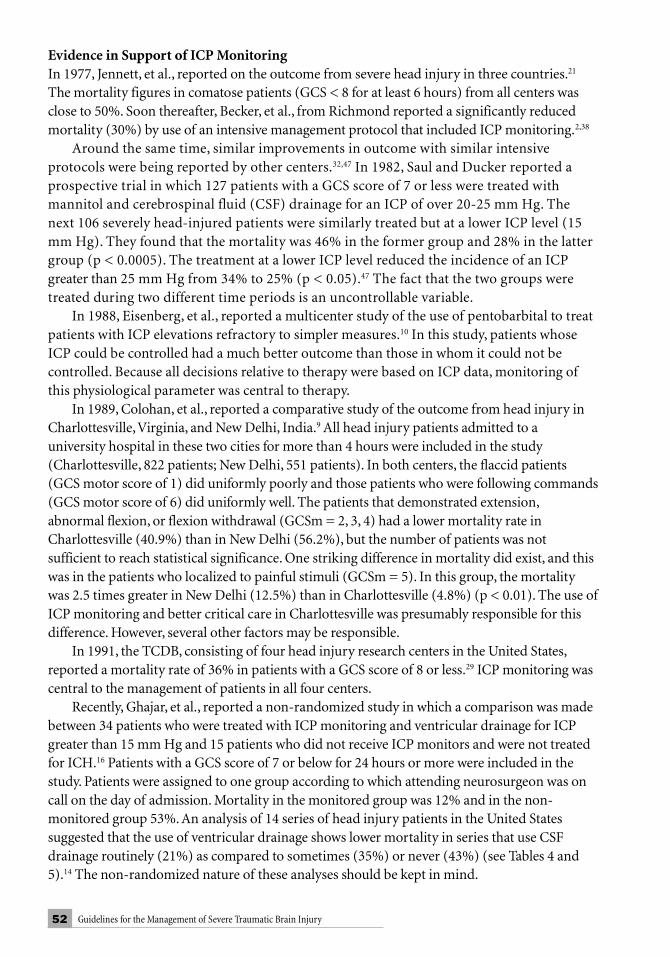
52 Guidelines for the Management of Severe Traumatic Brain Injury
Evidence in Support of ICP MonitoringIn 1977, Jennett, et al., reported on the outcome from severe head injury in three countries.21
The mortality figures in comatose patients (GCS < 8 for at least 6 hours) from all centers wasclose to 50%. Soon thereafter, Becker, et al., from Richmond reported a significantly reducedmortality (30%) by use of an intensive management protocol that included ICP monitoring.2,38
Around the same time, similar improvements in outcome with similar intensiveprotocols were being reported by other centers.32,47 In 1982, Saul and Ducker reported aprospective trial in which 127 patients with a GCS score of 7 or less were treated withmannitol and cerebrospinal fluid (CSF) drainage for an ICP of over 20-25 mm Hg. Thenext 106 severely head-injured patients were similarly treated but at a lower ICP level (15mm Hg). They found that the mortality was 46% in the former group and 28% in the lattergroup (p < 0.0005). The treatment at a lower ICP level reduced the incidence of an ICPgreater than 25 mm Hg from 34% to 25% (p < 0.05).47 The fact that the two groups weretreated during two different time periods is an uncontrollable variable.
In 1988, Eisenberg, et al., reported a multicenter study of the use of pentobarbital to treatpatients with ICP elevations refractory to simpler measures.10 In this study, patients whoseICP could be controlled had a much better outcome than those in whom it could not becontrolled. Because all decisions relative to therapy were based on ICP data, monitoring ofthis physiological parameter was central to therapy.
In 1989, Colohan, et al., reported a comparative study of the outcome from head injury inCharlottesville, Virginia, and New Delhi, India.9 All head injury patients admitted to auniversity hospital in these two cities for more than 4 hours were included in the study(Charlottesville, 822 patients; New Delhi, 551 patients). In both centers, the flaccid patients(GCS motor score of 1) did uniformly poorly and those patients who were following commands(GCS motor score of 6) did uniformly well. The patients that demonstrated extension,abnormal flexion, or flexion withdrawal (GCSm = 2, 3, 4) had a lower mortality rate inCharlottesville (40.9%) than in New Delhi (56.2%), but the number of patients was notsufficient to reach statistical significance. One striking difference in mortality did exist, and thiswas in the patients who localized to painful stimuli (GCSm = 5). In this group, the mortalitywas 2.5 times greater in New Delhi (12.5%) than in Charlottesville (4.8%) (p < 0.01). The use ofICP monitoring and better critical care in Charlottesville was presumably responsible for thisdifference. However, several other factors may be responsible.
In 1991, the TCDB, consisting of four head injury research centers in the United States,reported a mortality rate of 36% in patients with a GCS score of 8 or less.29 ICP monitoring wascentral to the management of patients in all four centers.
Recently, Ghajar, et al., reported a non-randomized study in which a comparison was madebetween 34 patients who were treated with ICP monitoring and ventricular drainage for ICPgreater than 15 mm Hg and 15 patients who did not receive ICP monitors and were not treatedfor ICH.16 Patients with a GCS score of 7 or below for 24 hours or more were included in thestudy. Patients were assigned to one group according to which attending neurosurgeon was oncall on the day of admission. Mortality in the monitored group was 12% and in the non-monitored group 53%. An analysis of 14 series of head injury patients in the United Statessuggested that the use of ventricular drainage shows lower mortality in series that use CSFdrainage routinely (21%) as compared to sometimes (35%) or never (43%) (see Tables 4 and5).14 The non-randomized nature of these analyses should be kept in mind.
53Indications for Intracranial Pressure Monitoring
Evidence Against the Value of ICP MonitoringIn 1983, Stuart, et al., reported a prospective series of 100 consecutive patients with severe headinjury treated in Queensland, Australia, between 1979 and 1981.49 These patients were comatosefor at least 6 hours post-injury. ICP monitoring was not carried out in this series and assistedventilation was used in 43 patients. The incidence of surgical hematomas was 52%. Themortality rate in this series was 34%, with 49% of patients achieving a good or moderatelydisabled outcome. While this report seems to throw into question the value of intensivetherapies if comparable results can be achieved without them, it should be noted that this studyhighlights the difficulties inherent in comparing disparate patient groups. Fifty-five percent ofthe patients in this series were transferred from facilities a great distance away from theneurosurgical center (11 km to over 300 km). The authors of this study note that “mortalityrates decreased with increasing distance of transfer, and this probably represents both naturaland medical selection. Many of the most severely injured do not survive long enough to reach amajor center, and medical officers are reluctant to send a patient a long distance if the prognosisis “hopeless.” The selection bias of this local circumstance undermines the comparability of thisstudy to the predominantly urban series that are in the literature.
In 1986, Smith, et al., reported a prospective, randomized study of 80 patients with severehead injury (GCS ≤ 8).48 All patients were intubated and moderately hyperventilated, ICP wasmonitored with bolts, and CT of the head was obtained every 2 to 3 days. Group I receivedmannitol for ICP greater than 25 mm Hg and pentobarbital for ICP greater than 35 mm Hg.Group II empirically received 0.25 gm/kg/2 hr. The mortality in the specifically treated groupwas 35%, while in the empirically treated group it was 42%. Although suggesting a betteroutcome in Group I, the difference was not statistically significant. This study was limited by itssample size. As noted earlier, it would have taken 349 patients in each group (rather than about40) to demonstrate a 10% improvement in mortality.
V. SummaryICP monitoring per se has never been subjected to a prospective, randomized clinical trial(PRCT) to establish its efficacy (or lack thereof) in improving outcome from severe head injury.Hence, there are insufficient data to support its use as a standard. However, there is a large bodyof published clinical experience that indicates that ICP monitoring: 1) helps in the earlierdetection of intracranial mass lesions, 2) can limit the indiscriminate use of therapies to controlICP, which themselves can be potentially harmful, 3) can reduce ICP by CSF drainage and thusimprove cerebral perfusion, 4) helps in determining prognosis, and 5) may improve outcome.ICP monitoring is therefore used by most head injury experts in the United States and isaccepted as a relatively low-risk, high-yield, modest-cost intervention. Comatose head injurypatients (GCS 3-8) with abnormal CT scans should undergo ICP monitoring. Comatosepatients with normal CT scans have a much lower incidence of ICH unless they have two ormore of the following features at admission: age over 40, unilateral or bilateral motor posturing,or a systolic blood pressure (SBP) of less than 90 mm Hg. ICP monitoring in patients with anormal CT scan with two or more of these risk factors is suggested as a guideline. Routine ICPmonitoring is not indicated in patients with mild or moderate head injury. However, it may beundertaken in certain conscious patients with traumatic mass lesions at the discretion of thetreating physician.

54 Guidelines for the Management of Severe Traumatic Brain Injury
VI. Key Issues for Future InvestigationA prospective, randomized, clinical trial of ICP monitoring would be extremely useful inestablishing the value of this technique. However, it is not clear that such a trial will ever beperformed. Most head injury experts consider ICP or CPP to be the primary basis for ICUmanagement decisions in the care of the severe head-injured patient. However, someneurosurgeons do not routinely monitor ICP in severe head injury patients, demand proof thatit makes a difference, and believe that the risks do not justify the benefits. A PRCT wouldrequire approximately 700 patients with severe head injury to demonstrate a 10% difference inmortality. One would need a multicentered trial at a cost of at least $5 million. The managementof the non-monitored group would remain problematic. Such a trial was proposed to the NIH afew years ago but was not funded. There is a NIH-funded trial currently underway comparingtraditional ICP management to management aimed at maintaining a certain CPP in severe headinjury patients. This study may provide the type of Class I data that is required withoutrandomizing one group of patients to a non-monitored arm.
VII. Evidentiary TablesTable 1: Which Patients Are at High Risk for ICP Elevation?Eisenberg,10 1988
Description of Study: In a multicenter study, 73 patients with severe head injury and elevatedICP refractory to all standard measures were prospectively randomized to receive either aregimen that included high-dose pentobarbital or one that was similar but did not includepentobarbital.
Classification: Class I Study
Conclusions: Patients whose ICP could be controlled with pentobarbital had a much betteroutcome than those in whom it could not be controlled. At 1 month, 92% of the patients whoresponded to treatment survived and 83% who did not respond had died.
Eisenberg,11 1990
Description of Study: Prospective multicenter study (TCDB) in which the authors examinedthe CT scans of 753 patients with severe head injury who were treated in a consistent fashion.
Classification: Class II Study
Conclusions: Severely head-injured patients whose initial CT scan does not show a mass lesion,midline shift, or abnormal cisterns have a 10-15% chance of developing elevated pressure.
Feldman,13 1994
Description of Study: Letter to the editor.
Classification: Class III Study
Conclusions: Response to O’Sullivan paper.
Abbreviations: GR, good recovery; MD, moderate disability; SD, severe disability; PVS, persistent vegetative state; D, dead

55Indications for Intracranial Pressure Monitoring
Gopinath,17 1993
Description of Study: ICP, MABP, CPP, ETCO2, SjvO
2, and SaO
2 were continuously monitored
in 163 severely head-injured patients to determine the critical values for these parameters and torelate them to outcome.
Classification: Class II Study
Conclusions: It was determined in separate analyses that outcome was adversely affected whenICP was greater than 25 mm Hg, MABP less than 80 mm Hg, and CPP less than 60 mm Hg.
Johnston,22 1970
Description of Study: Observational study of 32 patients with severe head injury. All patientshad continuous ICP monitoring to observe the effects of elevated ICP vs normal ICP.
Classification: Class II Study
Conclusions: In Group I (ICP < 20 mm Hg) 5 of 9 patients died; in Group II (ICP = 20-40 mmHg) 6 of 11 patients died; in Group III (ICP > 40 mm Hg) 8 of 12 patients died and 3 remainedin coma.
Lobato,25 1986
Description of Study: Study of 46 severe head injury patients who had normal CT scans days 1through 7 post-injury.
Classification: Class II Study
Conclusions: “A sustained elevation of ICP was not seen in these patients, indicating that ICPmonitoring may be omitted in cases with a normal scan.” However, a strategy for controlledscanning was recommended because 1 of 3 patients with a normal admission scan developednew pathology within the first few days of injury.
Lundberg,27 1965
Description of Study: Thirty patients underwent ICP monitoring to examine the variations ofICP during the acute post-injury stage.
Classification: Class II Study
Conclusions: Uncontrollable ICP greater than 40 mm Hg usually led to a poor outcome.
VII. Evidentiary Table 1 (continued)

56 Guidelines for the Management of Severe Traumatic Brain Injury
Marmarou,29 1991
Description of Study: A study of 428 severely head-injured patients from the TCDB. Describedthe relationship between raised ICP (> 20 mm Hg), hypotension, and outcome.
Classification: Class II Study
Conclusions: The proportion of ICP measurements greater than 20 mm Hg was highly significantin explaining outcome (p < 0.0001). As ICP > 20 increased, more favorable outcomes (G/MD)became less likely while worse outcomes (V/D) became more likely. The next most significant factorin predicting outcome was the proportion of mean blood pressure (BP) measurements less than 80mm Hg. Patients with a GCS less than 8 are at high risk of developing ICH.
Marshall,30 1991
Description of Study: The outcome of severe head injury was prospectively studied in 746patients enrolled in the TCDB. All patients were managed by a relatively “homogeneous” protocolthat included ICP monitoring.
Classification: Class II Study
Conclusions: Among the patients with nonsurgical lesions, the mortality rate was higher inthose having an increased likelihood of elevated ICP greater than 25 mm Hg (diffuse injury IIIand IV).
Marshall,32 1979
Description of Study: Results of a study in which 100 consecutive patients with severe headinjury were managed using a standardized intensive protocol that included ICP monitoring.
Classification: Class II Study
Conclusions: ICH (ICP > 15 mm Hg) was present in 55% of the patients. ICP monitoring tocontrol ICH was recommended in patients with severe head injury.
Miller,36 1987
Description of Study: Review of ICP monitoring.
Classification: Class III Study
Conclusions: ICP monitoring is associated with an infection rate of about 6% and a majorhemorrhage rate of 1-2%.
VII. Evidentiary Table 1 (continued)
Abbreviations: GR, good recovery; MD, moderate disability; SD, severe disability; PVS, persistent vegetative state; D, dead

57Indications for Intracranial Pressure Monitoring
Miller,37 1977
Description of Study: Series of 160 consecutive patients with severe head injury whose ICP wascontinuously monitored to determine the frequency, extent, and significance of ICH.
Classification: Class II Study
Conclusions: 82% of patients had elevated ICP on admission, (i.e., ICP > 20 mm Hg in 44%and > 40 mm Hg in 10%). Patients with an ICP greater than 40 mm Hg and those with diffusebrain injury and an ICP greater than 10 mm Hg on admission had a poor outcome. It wasrecommended that ICP monitoring be included in the management of severely head-injuredpatients.
Miller,38 1981
Description of Study: Series of 225 prospective, consecutive patients with severe head injurymanaged by a uniform and intensive protocol in an effort to relate outcome to several clinicalvariables.
Classification: Class II Study
Conclusions: Factors important in predicting a poor outcome included: presence of intracranialhematoma; increasing age; abnormal motor responses; impaired or absent eye movements orpupil light reflexes; early hypotension, hypoxemia, or hypercarbia; and elevation of ICP greaterthan 20 mm Hg despite artificial ventilation.
Narayan,40 1994
Description of Study: Letter to the editor.
Classification: Class III Study
Conclusions: Response to O’Sullivan paper.
Narayan,41 1981
Description of Study: Clinical signs, multimodality evoked potentials, CT scan, and ICP datawere prospectively recorded and analyzed in 133 severely head-injured patients to ascertain theiraccuracy and relative value in predicting one of two categories of outcome.
Classification: Class II Study
Conclusions: An ICP greater than 20 mm Hg that required treatment was associated with asignificantly poorer prognosis (36% G/MD) than if the ICP was less than 20 mm Hg (80% G/MD).
VII. Evidentiary Table 1 (continued)
Abbreviations: GR, good recovery; MD, moderate disability; SD, severe disability; PVS, persistent vegetative state; D, dead
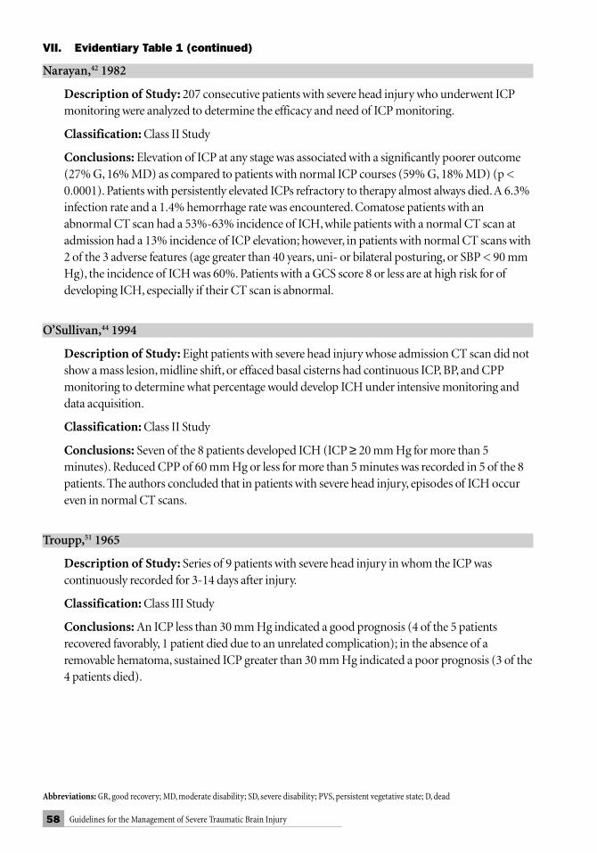
58 Guidelines for the Management of Severe Traumatic Brain Injury
Narayan,42 1982
Description of Study: 207 consecutive patients with severe head injury who underwent ICPmonitoring were analyzed to determine the efficacy and need of ICP monitoring.
Classification: Class II Study
Conclusions: Elevation of ICP at any stage was associated with a significantly poorer outcome(27% G, 16% MD) as compared to patients with normal ICP courses (59% G, 18% MD) (p <0.0001). Patients with persistently elevated ICPs refractory to therapy almost always died. A 6.3%infection rate and a 1.4% hemorrhage rate was encountered. Comatose patients with anabnormal CT scan had a 53%-63% incidence of ICH, while patients with a normal CT scan atadmission had a 13% incidence of ICP elevation; however, in patients with normal CT scans with2 of the 3 adverse features (age greater than 40 years, uni- or bilateral posturing, or SBP < 90 mmHg), the incidence of ICH was 60%. Patients with a GCS score 8 or less are at high risk for ofdeveloping ICH, especially if their CT scan is abnormal.
O’Sullivan,44 1994
Description of Study: Eight patients with severe head injury whose admission CT scan did notshow a mass lesion, midline shift, or effaced basal cisterns had continuous ICP, BP, and CPPmonitoring to determine what percentage would develop ICH under intensive monitoring anddata acquisition.
Classification: Class II Study
Conclusions: Seven of the 8 patients developed ICH (ICP ≥ 20 mm Hg for more than 5minutes). Reduced CPP of 60 mm Hg or less for more than 5 minutes was recorded in 5 of the 8patients. The authors concluded that in patients with severe head injury, episodes of ICH occureven in normal CT scans.
Troupp,51 1965
Description of Study: Series of 9 patients with severe head injury in whom the ICP wascontinuously recorded for 3-14 days after injury.
Classification: Class III Study
Conclusions: An ICP less than 30 mm Hg indicated a good prognosis (4 of the 5 patientsrecovered favorably, 1 patient died due to an unrelated complication); in the absence of aremovable hematoma, sustained ICP greater than 30 mm Hg indicated a poor prognosis (3 of the4 patients died).
VII. Evidentiary Table 1 (continued)
Abbreviations: GR, good recovery; MD, moderate disability; SD, severe disability; PVS, persistent vegetative state; D, dead

59Indications for Intracranial Pressure Monitoring
Table 2: How Does ICP Data Influence PatientManagement?Browder,4 1936
Description of Study: Case review study of 23 cases of head injury to ascertain the relationshipof BP, pulse rate, and CSF pressure for diagnosis, prognosis, and treatment in severe head injury.
Classification: Class III Study
Conclusions: The “classical pattern of signs,” consisting of steady rise above normal levels ofblood pressure, steady fall in pulse rate, decrease in respiratory rate, stupor, coma, vomiting, etc.,which had been held to indicate that ICP was increasing, was not seen in this series.
Chesnut,7 1994
Description of Study: Retrospective review of data from 514 TCDB patients to investigate theefficacy of early, routine use of neuromuscular blocking agents for ICP management: Group 1(n = 239) patients were pharmacologically paralyzed; Group 2 (n=275) patients were notpharmacologically paralyzed.
Classification: Class II Study
Conclusions: The final GCS scores for the two groups were not significantly different in those G/MD survivors. Group 2 had a higher mortality rate (39%) than Group 1 (24%), however, therewere more SD/V survivors in Group 1 (21% vs 13% and 8% vs 4%, respectively). The authorsrecommended that routine early management of head-injured patients in the ICU should beaccomplished using sedation alone, and neuromuscular blockade should be generally reservedfor patients with ICH who require escalation of treatment intensity.
Eisenberg,10 1988
Description of Study: In a multicenter study, 73 patients with severe head injury and refractoryelevation of ICP were prospectively randomized to receive either a regimen that included high-dose pentobarbital or one that was similar but did not include pentobarbital.
Classification: Class I Study
Conclusions: Patients whose ICP could be controlled with pentobarbital had a much betteroutcome than those in whom it could not be controlled; at 1 month, 92% of the patients whoresponded to treatment survived and 83% who did not respond had died.
Feldman,12 1993
Description of Study: Review of ICP monitoring.
Classification: Class III Study
Conclusions: Although ventriculostomies can serve as a therapeutic tool for raised ICP byallowing cerebrospinal fluid drainage, ICP monitoring is primarily a means for guiding therapy.
Abbreviations: GR, good recovery; MD, moderate disability; SD, severe disability; PVS, persistent vegetative state; D, dead
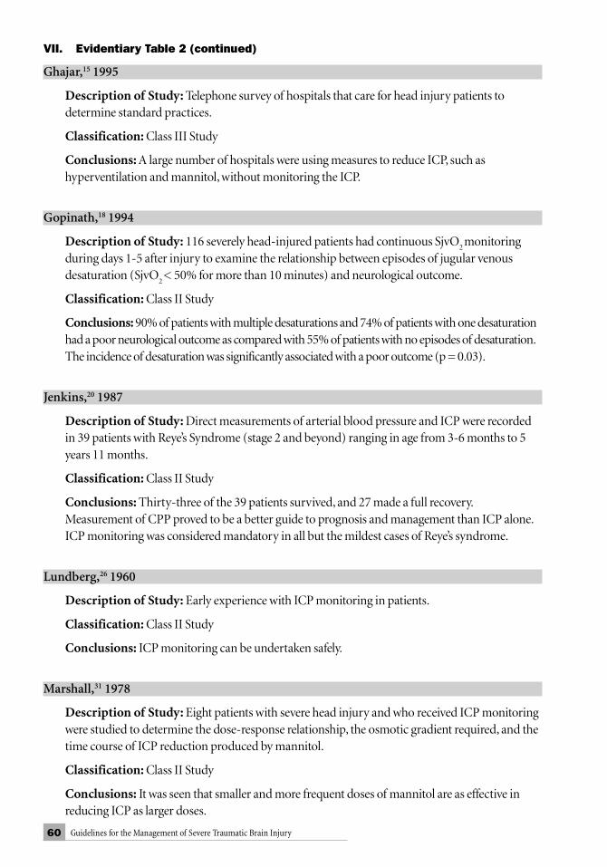
60 Guidelines for the Management of Severe Traumatic Brain Injury
Ghajar,15 1995
Description of Study: Telephone survey of hospitals that care for head injury patients todetermine standard practices.
Classification: Class III Study
Conclusions: A large number of hospitals were using measures to reduce ICP, such ashyperventilation and mannitol, without monitoring the ICP.
Gopinath,18 1994
Description of Study: 116 severely head-injured patients had continuous SjvO2
monitoringduring days 1-5 after injury to examine the relationship between episodes of jugular venousdesaturation (SjvO
2 < 50% for more than 10 minutes) and neurological outcome.
Classification: Class II Study
Conclusions: 90% of patients with multiple desaturations and 74% of patients with one desaturationhad a poor neurological outcome as compared with 55% of patients with no episodes of desaturation.The incidence of desaturation was significantly associated with a poor outcome (p = 0.03).
Jenkins,20 1987
Description of Study: Direct measurements of arterial blood pressure and ICP were recordedin 39 patients with Reye’s Syndrome (stage 2 and beyond) ranging in age from 3-6 months to 5years 11 months.
Classification: Class II Study
Conclusions: Thirty-three of the 39 patients survived, and 27 made a full recovery.Measurement of CPP proved to be a better guide to prognosis and management than ICP alone.ICP monitoring was considered mandatory in all but the mildest cases of Reye’s syndrome.
Lundberg,26 1960
Description of Study: Early experience with ICP monitoring in patients.
Classification: Class II Study
Conclusions: ICP monitoring can be undertaken safely.
Marshall,31 1978
Description of Study: Eight patients with severe head injury and who received ICP monitoringwere studied to determine the dose-response relationship, the osmotic gradient required, and thetime course of ICP reduction produced by mannitol.
Classification: Class II Study
Conclusions: It was seen that smaller and more frequent doses of mannitol are as effective inreducing ICP as larger doses.
VII. Evidentiary Table 2 (continued)

61Indications for Intracranial Pressure Monitoring
Marshall,32 1979
Description of Study: Results of a study in which 100 consecutive patients with severe headinjury were managed using a standardized intensive protocol that included ICP monitoring.
Classification: Class II Study
Conclusions: ICH (ICP > 15 mm Hg) was present in 55% of the patients. ICP monitoring tocontrol ICH was recommended in patients with severe head injury.
Mendelow,34 1985
Description of Study: The effect of mannitol on CPP, ICP, and CBF was studied in 55 patientswith severe head injury.
Classification: Class II Study
Conclusions: Mannitol consistently reduced ICP and increased CPP and CBF 10-20 minutes afterinfusion.
Miller,38 1981
Description of Study: Clinical series of 225 prospective, consecutive patients with severe headinjury managed with a uniform and intensive protocol in order to detect a link between outcomeand several clinical variables.
Classification: Class II Study
Conclusions: Factors important in predicting a poor outcome included: presence of intracranialhematoma; increasing age; abnormal motor responses; impaired or absent eye movements or pupillight reflexes; early hypotension, hypoxemia, or hypercarbia; elevation of ICP (> 20 mm Hg) despiteartificial ventilation.
Muizelaar,39 1991
Description of Study: A prospective, randomized trial on 113 severely head-injured patients,using three groups: control, hyperventilated, and hyperventilated and THAM.
Classification: Class I Study
Conclusions: Prophylactic hyperventilation proved harmful in head-injured patients withmotor scores of 4-5; when sustained hyperventilation was necessary for ICP control, its harmfuleffects might have been partially overcome by THAM.
VII. Evidentiary Table 2 (continued)

62 Guidelines for the Management of Severe Traumatic Brain Injury
Narayan,41 1981
Description of Study: Clinical signs, MEPs, CT scans, and ICP data were prospectivelyrecorded and analyzed in 133 severely head-injured patients to ascertain their accuracy andrelative value, either individually or in various combinations, in predicting one of two categoriesof outcome.
Classification: Class II Study
Conclusions: An ICP greater than 20 mm Hg that required treatment was associated with asignificantly poorer prognosis (36% G/MD) than if the ICP was less than 20 mm Hg (80% G/MD).
Robertson,45 1992
Description of Study: CBF and other physiological variables were measured repeatedly for upto 10 days after severe head injury in 102 patients, and CBF levels were related to outcome.
Classification: Class II Study
Conclusions: A reduced CBF was significantly associated with an unfavorable outcome.
Sheinberg,50 1992
Description of Study: Continuous measurement of SjvO2 with a fiberoptic catheter was
performed on 45 patients with severe head injury as a method of detecting cerebral ischemia.
Classification: Class II Study
Conclusions: Hypocarbia (PaCO2
< 28 mm Hg) was one cause of 10 jugular venous oxygendesaturation episodes (SjvO
2 < 50% for more than 15 minutes).
Table 3: Does ICP Monitoring Improve Outcome?Becker,2 1977
Description of Study:160 patients with severe head injury were managed with a standardizedprotocol that emphasized early diagnosis and evacuation of mass lesions, artificial ventilation,control of ICH by monitoring ICP, and aggressive medical therapy.
Classification: Class II Study
Conclusions: A 30% mortality rate was reported by this study as compared to the 50% mortalityrate reported from the three centers in Jennett, et al., 1977.
VII. Evidentiary Table 2 (continued)

63Indications for Intracranial Pressure Monitoring
Colohan,9 1989
Description of Study: A comparative prospective study of 551 patients with head injury in NewDelhi, India, and 822 patients in Charlottesville, Virginia.
Classification: Class II Study
Conclusions: In patients who localized to painful stimuli (GCS motor=5), the mortality was 2.5times greater in New Delhi (12.5%) than in Charlottesville (4.8%) (p < 0.01). The use of ICPmonitoring and better critical care were among the factors believed to be responsible for thisdifference.
Eisenberg,10 1988
Description of Study: In a multicenter study, 73 patients with severe head injury and elevatedICP were prospectively randomized to receive either a regimen that included high-dosepentobarbital or one that was similar but did not include pentobarbital.
Classification: Class I Study
Conclusions: Because all decisions relative to therapy were based on ICP data, ICP monitoringwas pertinent to therapy. Patients whose ICP could be controlled with pentobarbital had a muchbetter outcome than those in whom it could not be controlled; at 1 month 92% of the patientswho responded to treatment survived and 83% who did not respond had died.
Ghajar,14 1995
Description of Study: A meta analysis of 14 U.S. series of head injury that report the use ofventricular drainage for therapeutic measures.
Classification: Class III Study
Conclusions: A 28% mortality was found in series that use CSF drainage routinely as comparedto 34% mortality that use it sometimes and 44% that never use CSF drainage.
Ghajar,16 1993
Description of Study: Prospective, non-randomized cohort study of 49 consecutive severely head-injured patients: Group 1 (34 patients) received ICP monitoring and CSF drainage when ICP was greaterthan 15 mm Hg; Group 2 (15 patients) received no ICP monitoring and were not treated for ICH.
Classification: Class II Study
Conclusions: Mortality in Group 1 was 12% and in Group 2 was 53%.
VII. Evidentiary Table 3 (continued)

64 Guidelines for the Management of Severe Traumatic Brain Injury
Jennett,21 1977
Description of Study: Early characteristics and late outcome after severe head injury werereported in 700 cases in three countries (Scotland, The Netherlands, and United States). ICPmonitoring was not routinely used.
Classification: Class II Study
Conclusions: Mortality figures in comatose patients (GCS ≤ 8 or less for at least 6 hours) fromall centers was close to 50%.
Marmarou,29 1991
Description of Study: A study of 428 severely head-injured patients form the TCDB describingthe relationship between raised ICP (> 20 mm Hg), hypotension, and outcome.
Classification: Class II Study
Conclusions: A mortality rate of 36% was reported. ICP management was central to this study.
Marshall,30 1991
Description of Study: The outcome of severe head injury was prospectively studied in 746patients enrolled in the TCDB. All patients were managed by a relatively “homogeneous” protocolthat included ICP monitoring.
Classification: Class II Study
Conclusions: A mortality rate of 36% was reported as compared to the 50% mortality ratereported in Jennett, et al., 1977.
Marshall,32 1979
Description of Study: Results of a study in which 100 consecutive patients with severe headinjury were managed using a standardized intensive protocol that included ICP monitoring.
Classification: Class II Study
Conclusions: A mortality rate of 28% was reported as compared to the 50% mortality ratereported in Jennett, et al., 1977.
Miller,38 1981
Description of Study: Clinical series of 225 prospective, consecutive patients with severe headinjury managed with a uniform and intensive protocol in order to relate outcome to severalclinical variables.
Classification: Class II Study
Conclusions: This study reported a 34% mortality rate, an improvement compared to the 1977Jennett, et al., report of a 50% mortality from three centers. This series met the 6 -hour requirementused in the Jennett report.
VII. Evidentiary Table 3 (continued)

65Indications for Intracranial Pressure Monitoring
Saul,47 1982
Description of Study: Prospective study of 127 severely head-injured patients who were treatedwith mannitol and CSF drainage for an ICP at 20 to 25 mm Hg, and 106 patients who weretreated similarly except at a lower ICP (>15 mm Hg) level.
Classification: Class II Study
Conclusions: Mortality was 46% in the 127 patients treated for an ICP at 20-25 mm Hg andonly 28% in the 106 patients treated at an ICP threshold of 15 mm Hg.
Smith,48 1986
Description of Study: A prospective, randomized study of 80 patients with severe head injuryto assess the benefit of ICP monitoring with two regimens of mannitol administration: Group Ireceived mannitol for an ICP greater than 25 mm Hg and Group II received empirical mannitoltherapy irrespective of ICP.
Classification: Class II Study
Conclusions: No statistically significant differences in mortality rate or neurological outcomewere demonstrated between the two groups. This study was limited by its sample size, an n = 349.Patients in each group would have been necessary to demonstrate a 10% improvement inmortality.
Stuart,49 1983
Description of Study: A retrospective analysis of 100 cases of severe head injury inQueensland, Australia, where ICP was not monitored. Patients who did not survivetransportation to the tertiary neurosurgical center (often several hundred miles away) were notincluded in the analysis.
Classification: Class II Study
Conclusions: The mortality rate was 34%, and 49% of patients achieved a good or moderatelydisabled outcome. It is suggested that the outcome compares favorably with series in which ICPwas monitored. However, the selection bias of this study undermines its comparability to thepredominantly urban series that are in the literature.
VII. Evidentiary Table 3 (continued)

66 Guidelines for the Management of Severe Traumatic Brain Injury
Table 4: Comparison of Outcome: Fourteen U.S. Studies
First number GCS ICP Monitor* CSF Drain Rx at ICP OUTCOMEAuthor of patients (mm Hg) G/MD Dead
(%) (%)
Jaggi19 64 ≤ 9 bolt 0 > 20 47% 53%Colohan9 122 ≤ 8 bolt 0 > 20 — 41Smith48 37 ≤ 8 bolt 0 > 25 54 35Wald52 170 ≤ 8 epidural 0 > 20 48 41Saul I47 127 ≤ 7 ventric, bolt +/0 > 25 — 46Saul II47 106 ≤ 7 ventric, bolt +/0 > 15 54 28Bowers3 200 ≤ 7 ventric, bolt† +/0 > 20/25 52 36Becker2 160 ≤ 9 ventric, bolt +/0 > 25/40 60 30Miller37 225 ≤ 9 ventric, bolt +/0 > 25 56 34Muizelaar39 113 ≤ 8 ventric, bolt +/0 > 25 39 34Marion28 68 ≤ 8 ventric + ‡ 51 18Narayan42 207 ≤ 9 ventric + > 25 57 34Rosner46 34 ≤ 7 ventric + ‡ 68 21Ghajar16 34 ≤ 7 ventric + 15 59 12
GCS = Glasgow Coma Score; ICP = intracranial pressure; CSF = cerebrospinal fluid;GOS = Glasgow Outcome Scale; G = good recovery; MD = moderate disability*ICP monitor deemed ventric if ventriculostomy was used in more than 90% of patients†␣ only 41% of patients were monitored‡ cerebral perfusion pressure maintained at 70-80 mm Hg0 = not used; + = used— = data unavailable
Table 5: Average Outcomes from Studies Based on CSFDrainage Category
CSF Drainage Category Independent (%) Dead (%)
Never (0) 49.7% 43.0%Sometimes (+/0) 52.2 34.7Routinely (+) 58.8 21.3
Adapted from Ghajar14, 1996.
67Indications for Intracranial Pressure Monitoring
VIII. References1. Becker DP: Common themes in head injury. In: Textbook of Head Injury. Becker DP
and Gudeman SK, eds. WB Saunders, Philadelphia, 1989: 1-22.2. Becker DP, Miller JD, Ward JD, et al.: The outcome from severe head injury with early
diagnosis and intensive management. J Neurosurg 47:491-502, 1977.3. Bowers SA, Marshall LF: Outcome in 200 cases of severe head injury treated in San
Diego County: a prospective analysis. Neurosurg 6:237-242, 1980.4. Browder J, Meyers R: Observations on behavior of the systemic blood pressure, pulse,
and spinal fluid pressure following craniocerebral injury. Am J Surg 31:403-426, 1936.5. Casagrande JT, Pike MC, Smith PG: An improved approximate formula for calculating
sample sizes for comparing to binomial distributions. Biometrics 34:483-486, 1978.6. Changaris DG, McGraw CP, Richardson JD, et al.: Correlation of cerebral perfusion
pressure and Glasgow Coma Scale to outcome. J Trauma 27:1007-1013, 1987.7. Chesnut RM, Crisp CB, Klauber MR, et al.: Early, routine paralysis for intracranial
pressure control in severe head injury: is it necessary? Critical Care Medicine 22:1471-1476, 1994.
8. Chesnut RM, Marshall LF, Klauber MR: The role of secondary brain injury indetermining outcome from severe head injury. J Trauma 34:216-222, 1993.
9. Colohan AR, Alves WM, Gross CR, et al. Head injury mortality in two centers withdifferent emergency medical services and intensive care. J Neurosurg 71:202-207, 1989.
10. Eisenberg HM, Frankowski RF, Contant CF, et al.: High-dose barbiturates controlelevated intracranial pressure in patients with severe head injury. J Neurosurg 69:15-23,1988.
11. Eisenberg HM, Gary HE Jr, Aldrich EF, et al.: Initial CT findings in 753 patients withsevere head injury. A report from the NIH Traumatic Coma Data Bank. J Neurosurg 73:688-698, 1990.
12. Feldman Z and Narayan RK: Intracranial pressure monitoring: techniques andpitfalls. In: Head Injury, 3rd ed. Cooper PR, ed. Williams and Wilkins, Baltimore:1993.
13. Feldman Z and Reichenthal E. Intracranial pressure monitoring (letter). J Neurosurg81:329, 1994.
14. Ghajar JB. Variability of neurotrauma care in the U.S. In: Neurotrauma. Narayan RK,Wilberger JE, and Povlishock JT, eds. McGraw Hill, Inc., New York: 1996.
15. Ghajar JB, Hariri R, Narayan RK, et al.: Survey of critical care management ofcomatose head-injured patients in the United States. Critical Care Medicine 23: 560-567,1995.
16. Ghajar JB, Hariri RJ, Patterson RH. Improved outcome from traumatic coma usingonly ventricular CSF drainage for ICP control. Advances in Neurosurg 21:173-177, 1993.
17. Gopinath SP, Contant CF, Robertson CS, et al.: Critical thresholds for physiologicalparameters in patients with severe head injury. Congress of Neurological Surgeons.Vancouver: 1993.
18. Gopinath SP, Robertson CS, Contant CF, et al.: Jugular venous desaturation andoutcome after head injury. J Neurol Neurosurg Psychiat 57: 717-723, 1994.
19. Jaggi JL, Obrist WD, Gennarelli TA, et al.: Relationship of early cerebral blood flowand metabolism to outcome in acute head injury. J Neurosurg 72:176-182, 1990.

68 Guidelines for the Management of Severe Traumatic Brain Injury
20. Jenkins JG, Glasgow JFT, Black GW, et al.: Reye’s Syndrome: assessment of intracranialmonitoring. Br Med J 294:337-338, 1987.
21. Jennett B, Teasdale G, Galbraith S, et al.: Severe head injury in three countries. J NeurolNeurosurg Psychiat 40: 291-295, 1977.
22. Johnston IH, Johnston JA, Jennett WB: Intracranial pressure following head injury. Lancet2: 433-436, 1970.
23. Kaufmann AM and Cardoso ER: Aggravation of vasogenic cerebral edema by multiple dosemannitol. J Neurosurg 77:584-489, 1992.
24. Lam AM, Winn HR, Cullen BF, et al.: Hyperglycemia and neurological outcome in patientswith head injury. J Neurosurg 75:545-551, 1991.
25. Lobato RD, Sarabia R, Rivas JJ, et al.: Normal CT scans in severe head injury. J Neurosurg 65:784-789, 1986.
26. Lundberg N. Continuous recording and control of ventricular fluid pressure in neurosurgicalpractice. Acta Psychiat Neurol Scand (suppl. 149) 36:1-193, 1960.
27. Lundberg N, Troupp H, Lorin H: Continuous recording of the ventricular fluid pressure inpatients with severe acute traumatic brain injury. J Neurosurg 22:581-590, 1965.
28. Marion D, Obrist WD, Penrod LE, et al.: Treatment of cerebral ischemia improves outcomefollowing severe traumatic brain injury. American Association of Neurological Surgeons,Boston: 1993.
29. Marmarou A, Anderson RL, Ward JD, et al.: Impact of ICP instability and hypotension onoutcome in patients with severe head trauma. J Neurosurg 75: s59-s66, 1991.
30. Marshall LF, Gautille T, Klauber MR, et al.: The outcome of severe closed head injury. JNeurosurg 75: s28-s36, 1991.
31. Marshall LF, Smith RW, Ranscher LA, et al.: Mannitol dose requirements in brain-injuredpatients. J Neurosurg 48:169-172, 1978.
32. Marshall LF, Smith RW, Shapiro HM.: The outcome with aggressive treatment in severehead injuries. Part I: The significance of intracranial pressure monitoring. J Neurosurg 50:20-25, 1979.
33. McGraw CP: A cerebral perfusion pressure greater than 80 mm Hg is more beneficial. In:ICP VII. Hoff JT, Betz AL, eds. Springer-Verlag: Berlin, 1989: 839-841.
34. Mendelow AD, Teasdale GM, Russell T, et al.: Effects of mannitol on cerebral blood flow andcerebral perfusion pressure in human head injury. J Neurosurg 63: 43-48, 1985.
35. Miller JD: Changing patterns in acute management of head injury. J Neurol Sci 103: s33-s37,1991.
36. Miller JD: ICP monitoring—current status and future directions. Acta Neurochir 85: 80-86,1987.
37. Miller JD, Becker DP, Ward JD, et al.: Significance of intracranial hypertension in severehead injury. J Neurosurg 47:503-576, 1977.
38. Miller JD, Butterworth JF, Gudeman SK, et al.: Further experience in the management ofsevere head injury. J Neurosurg 54:289-299, 1981.
39. Muizelaar JP, Marmarou A, Ward JD, et al.: Adverse effects of prolonged hyperventilation inpatients with severe head injury: a randomized clinical trial. J Neurosurg 75:731-739, 1991.

69Indications for Intracranial Pressure Monitoring
40. Narayan RK. (letter). J Neurosurg 81:810-811, 1994.41. Narayan RK, Greenberg RP, Miller JD, et al.: Improved confidence of outcome prediction in
severe head injury: a comparative analysis of the clinical examination, multimodalityevoked potentials, CT scanning, and intracranial pressure. J Neurosurg 54:751-762, 1981.
42. Narayan RK, Kishore PR, Becker DP, et al.: Intracranial pressure: to monitor or not tomonitor? A review of our experience with severe head injury. J Neurosurg 56: 650-659, 1982.
43. Obrist WD, Langfitt TW, Jaggi JL, et al.: Cerebral blood flow and metabolism in comatosepatients with acute head injury. J Neurosurg 61:241-253, 1984.
44. O’Sullivan MG, Statham PF, Jones PA, et al.: Role of intracranial pressure monitoring inseverely head-injured patients without signs of intracranial hypertension on initial CT. JNeurosurg 80:46-50, 1994.
45. Robertson CS, Contant CF, Gokaslan ZL, et al.: Cerebral blood flow, arteriovenous oxygendifference, and outcome in head-injured patients. J Neurol Neurosurg Psychiat 55: 594-603,1992.
46. Rosner MJ, Daughton S: Cerebral perfusion pressure management in head injury. J Trauma30:933-941, 1990.
47. Saul TG, Ducker TB: Effect of intracranial pressure monitoring and aggressive treatment onmortality in severe head injury. J Neurosurg 56:498-503, 1982.
48. Smith HP, Kelly DL Jr, McWhorter JM, et al.: Comparison of mannitol regimens in patientswith severe head injury undergoing intracranial pressure monitoring. J Neurosurg 65:820-824, 1986.
49. Stuart GG, Merry GS, Smith JA, et al.: Severe head injury managed without intracranialpressure monitoring. J Neurosurg 59:601-605, 1983.
50. Sheinberg M, Kanter MJ, Robertson CS, et al.: Continuous monitoring of jugular venousoxygen saturation in head-injured patients. J Neurosurg 76:212-217, 1992.
51. Troupp H: Intraventricular pressure in patients with severe brain injuries. J Trauma 5:373-378, 1965.
52. Wald SL, Shackford SR, Fenwick J: The effect of secondary insults on mortality and long-term disability after severe head injury in a rural region without a trauma system. J Trauma34:377-382, 1993.


71Intracranial Pressure Treatment Threshold
I. RecommendationsA. Standards
There are insufficient data to support a treatment standard for this topic.B. Guidelines
Intracranial pressure (ICP) treatment should be initiated at an upper threshold of20-25 mm Hg.
C. OptionsInterpretation and treatment of ICP based on any threshold should be corroboratedby frequent clinical examination and cerebral perfusion pressure (CPP) data.
II. OverviewQuantitative guidelines are needed for ICP management. The impact of ICP on outcome fromsevere head injury appears to lie in its role 1) in determining CPP, and 2) as an indicator of masseffect. Because CPP can be managed by manipulation of arterial pressure to a great extent, theissue of herniation is more determinant of the ICP threshold. The goal is to balance the risks ofherniation against the iatrogenic risks of overtreatment.
III. ProcessA MEDLINE search back to 1966 was undertaken using the following query: “intracranialhypertension” or “ICP” or “intracranial pressure” and “head injury” and “treatment” or“management” or “resuscitation” and “threshold” or “level” and “human subject”. This produced146 references. Of these, 46 were found to be directly relevant to clinical orientation and theissue of relating ICP treatment threshold to outcome. These references were individuallyreviewed for design, content, and relevance. The results of this review were then incorporatedinto the analysis presented here.
IV. Scientific FoundationNo prospective, randomized trials compare ICP treatment thresholds. The largest study usingprospectively collected, observational data, controlling for a large number of confoundingprognostic variables, analyzed the mean ICP in 5 mm Hg steps against outcome in a logisticregression model and found 20 mm Hg to be the optimal predictive value. 3
INTRACRANIAL PRESSURE TREATMENTTHRESHOLD

72 Guidelines for the Management of Severe Traumatic Brain Injury
These values are in keeping with small, non-controlled reports suggesting a range of 15-25mm Hg.4, 6, 7 The report by Saul and Ducker changed the ICP threshold from 25 mm Hg to 15mm Hg in two sequentially treated groups of patients and found an associated decrease inmortality from 46% to 28%. 7 However, differences in protocols between the first and secondtreatment periods confound the determination of the independent influence of lowering theICP treatment threshold on outcome.
The study by Eisenberg, et al., is the only prospective, double-blind, placebo-controlled studydemonstrating improved outcome attributable to lowering ICP. 2 Their lowest ICP thresholds were25 mm Hg in patients without craniectomy and 15 mm Hg in patients following craniectomy.However, they defined additional ICP thresholds at higher pressures and shorter durations (seebarbiturates chapter for details) and they did not stratify outcome by threshold.
Patients can herniate at ICPs less than 20-25 mm Hg. The likelihood of herniation depends onthe location of an intracranial mass lesion.1, 5 In the report by Marshall, et al., pupillary abnormalitiesoccurred with ICP values as low as 18 mm Hg. Therefore, at all points, any chosen threshold must beclosely and repeatedly corroborated with the clinical exam and CT imaging in an individual patient.
Adequate CPP values can generally be maintained with ICPs of greater than 20-25 mm Hg.In addition, the ICPs at which patients begin to show signs of herniation can also occasionallybe greater than 20-25 mm Hg. Therefore, in select cases, a higher limit of acceptable ICPs maybe chosen as long as an adequate CPP can be maintained.
V. SummaryAn absolute ICP threshold that is uniformly applicable is unlikely to exist. Current data,however, support 20-25 mm Hg as an upper threshold above which treatment to lower ICPshould generally be initiated.2,3,7
VI. Key Issues for Future InvestigationThe critical value of ICP and its interaction with CPP is the major unanswered question. As werecognize the importance of CPP and improve our ability to safely maintain an adequate CPPsomewhat independently of ICP, the issue of an absolute value for ICP appears to be most closelyrelated to the risk of herniation that seems to vary between patients and within patients over thecourse of their therapy. If a method to estimate this “herniation pressure” can be developed andthe range of values wherein CPP is independent of mean arterial and intracranial pressures can bedetermined, more concrete treatment thresholds for ICP and CPP will be forthcoming.
VII. Evidentiary TableAndrews,1 1988
Description of Study: Retrospective review of the clinical course and CT scans of 45 patientswith supratentorial intracerebral hematomas to determine the effect of hematoma location onclinical course and outcome. Signs of herniation were significantly more common with temporalor temporoparietal lesions. Clot size of 30 cc was the threshold value for increased incidence ofherniation.
Classification: Class III Study
Conclusions: Factors other than ICP (such as location of mass lesion) must be considered inguiding treatment.
73Intracranial Pressure Treatment Threshold
Eisenberg,2 1998
Description of Study: Prospective, multicenter study wherein 73 severe head injury patients,whose ICP was not controllable using “conventional therapy,” were randomly assigned to a high-dose pentobarbital vs placebo-control regimen. Dependent variable was ability to control ICPbelow 20 mm Hg. The outcome for study patients whose ICP could be kept below 20 mm Hgusing either regimen was significantly better than those whose ICP could not be controlled.
Classification: Class II Study (with respect to ICP threshold)
Conclusions: Improved outcome when ICP could be controlled using a threshold of 20 mm Hg.
Marmarou,3 1991
Description of Study: From a prospectively collected database of 1,030 severe head injurypatients, all 428 patients who met ICU monitoring criteria were analyzed for monitoring parametersthat determined outcome and their threshold values. The ICP threshold most predictive of 6-monthoutcome was determined using logistic regression, evaluating values from 0-80 mm Hg inincrements of 5 mm Hg. The threshold value of 20 mm Hg was found to best correlate withoutcome. The proportion of hourly ICP reading greater then 20 mm Hg was a significant andpowerful independent determinant of outcome. Notably, the four centers used ICP treatmentthresholds of 20-25 mm Hg. The degree by which this confounds the regression statistics is unclear.
Classification: Class II Study
Conclusions: The incidence of morbiditly and mortality resulting from severe head injury isstrongly related to ICP control wherein 20 mm Hg is the most predictive threshold.
Marshall,4 1979
Description of Study: Retrospective review of 100 consecutively admitted severe head injurypatients. Patients managed employing a regimen including ICP monitoring using a threshold of15 mm Hg as a part of an aggressive approach to treatment had improved outcome compared topublished reports using less ICP-intensive therapy.
Data Class: Class III Study
Conclusions: ICP control using a threshold of 15 mm Hg as a part of an overall aggressivetreatment approach to severe had injury may be associated with improved outcome.
Marshall,5 1983
Description of Study: Reported 14 patients with oval pupils felt to be due to elevated ICP.Associated ICP values ranged from 18-38 mm Hg. The pupil normalized in 9 of these patientswhen the ICP was reduced.
Classification: Class III Study
Conclusions: Signs of herniation due to intracranial hypertension can occur associated with awide range of ICP values. ICP treatment thresholds should be tempered using other clinicalvalues in individual patients.
VII. Evidentiary Table (continued)

74 Guidelines for the Management of Severe Traumatic Brain Injury
Narayan,6 1982
Description of Study: Retrospective analysis of the courses of 207 consecutively admittedsevere head injury patients. Management included aggressive attempts to control ICP using athreshold of 20 mm Hg. Outcome was significantly correlated with the ability to control ICP.
Classification: Class III Study
Conclusions: ICP control using a threshold of 20 mm Hg as a part of an overall aggressivetreatment approach to severe head injury may be associated with improved outcome.
Saul,7 1982
Description of Study: A series of 127 severe head injury patients whose ICP treatment wasinitiated at 20-25 mm Hg, not using a strict treatment protocol, was compared with a subsequentgroup of 106 patients with similar injury characteristics who received treatment under a strictprotocol at an ICP threshold of 15 mm Hg. Other treatment aspects had not been changed. The46% mortality in the first group was significantly greater then the 28% mortality in the secondgroup.
Classification: Class III Study
Conclusions: Suggests an increase in mortality if ICP maintains above a threshold between 15and 25 mm Hg.
VIII. References1. Andrews BT, Chiles BW, Olsen WL, et al.: The effect of intracerebral hematoma location on
the risk of brain-stem compression and on clinical outcome. J Neurosurg 69:518-522, 1988.2. Eisenberg H, Frankowski R, Contant C, et al.: High-dose barbiturate control of elevated
intracranial pressure in patients with severe head injury. J Neurosurg 69:15-23, 1988.3. Marmarou A, Anderson RL, Ward JD, et al.: Impact of ICP instability and hypotension on
outcome in patients with severe head trauma. J Neurosurg 75:S159-166, 1991.4. Marshall L, Smith R, Shapiro H: The outcome with aggressive treatment in severe head injuries.
Part I. The significance of intracranial pressure monitoring. J Neurosurg 50:20-25, 1979.5. Marshall LF, Barba D, Toole BM, et al.: The oval pupil: clinical significance and relationship
to intracranial hypertension. J Neurosurg 58:566-568, 1983.6. Narayan R, Kishore P, Becker D, et al.: Intracranial pressure: to monitor or not to monitor?
A review of our experience with head injury. J Neurosurg 56:650-659, 1982.7. Saul TG, Ducker TB: Effects of intracranial pressure monitoring and aggressive treatment
on mortality in severe head injury. J Neurosurg 56:498-503, 198.
VII. Evidentiary Table (continued)

75Recommendations for Intracranial Pressure Monitoring Technology
I. Recommendations*
In the current state of technology the ventricular catheter connected to an external strain gaugeis the most accurate, low-cost, and reliable method of monitoring intracranial pressure (ICP). Italso allows therapeutic cerebrospinal fluid drainage. ICP transduction via fiberoptic or straingauge devices placed in ventricular catheters provide similar benefits, but at a higher cost.
Parenchymal ICP monitoring with fiberoptic or strain gauge catheter tip transduction issimilar to ventricular ICP monitoring but has the potential for measurement drift.
Subarachnoid, subdural, and epidural monitors (fluid coupled or pneumatic) are currentlyless accurate.
II. OverviewIn patients for whom ICP monitoring is indicated, a decision as to what type of monitoringdevice to use must be made. The optimal ICP monitoring device is one that is accurate, reliable,cost effective, and causes minimal patient morbidity. We reviewed the scientific literature onICP monitoring and propose a ranking based on the currently available technology.
III. ProcessA computerized literature search of MEDLINE from 1975 to January 1998 using the searchwords “monitor” and “intracranial pressure” found 4,290 articles, of which 1,000 articles werefound to be pertinent to data in humans. Scientific publications on ICP monitoring devicesused clinically and reporting accuracy or complications were reviewed in depth. Case reportswere excluded.
RECOMMENDATIONS FOR INTRACRANIALPRESSURE MONITORING TECHNOLOGY
*The assessment of ICP monitoring technology does not lend itself to classification of evidence as in other guideline sections. Thus, the ICPdevices were evaluated in terms of their accuracy, reliability, therapeutic potential, and cost effectiveness.

76 Guidelines for the Management of Severe Traumatic Brain Injury
IV. Scientific FoundationThe scientific discussion of ICP monitoring technology is divided into the following sections:
A. ICP monitoring device accuracy and stabilityB. Optimal intracranial location of monitorC. ComplicationsD. Cost
A. ICP monitoring device accuracy and stability (Table 1)The Association for the Advancement of Medical Instrumentation (AAMI) hasdeveloped the American National Standard for Intracranial Pressure MonitoringDevices in association with a Neurosurgery committee.4
The purpose of this standard is to provide labeling, safety, and performancerequirements, and to test methods that will help enssure a reasonable level of safetyand effectiveness of devices intended for use in the measurement of ICP.
According to AAMI’s standard, an ICP device should have the followingspecifications:
• Pressure range 0-100 mm Hg• Accuracy ± 2 mm Hg in range of 0-20 mm Hg• Maximum error 10% in range of 20-100 mm Hg
Current ICP monitors allow pressure transduction by external strain gauge, cathetertip strain gauge, or catheter tip fiberoptic technology. External strain gaugetransducers are coupled to the patient’s intracranial space via fluid filled lines,whereas catheter tip transducer technologies are placed intracranially. External straingauge transducers are accurate17 and can be recalibrated, but obstruction of the fluidcouple can cause inaccuracy. In addition, the external transducer must beconsistently maintained at a fixed reference point relative to the patient’s head toavoid measurement error.
Catheter tip strain gauge or fiberoptic devices are calibrated prior to intracranialinsertion and cannot be recalibrated once inserted (without an associated ventricularcatheter). Consequently, if the device measurement drifts and is not recalibrated,there is potential for an inaccurate measurement, especially if the ICP monitor is usedfor several days.
There is potential for significant ICP measurement drift with fiberoptic pressuretransduction1,6,31,41 and strain gauge pressure transduction34,36 in the parenchymalspace. However, other studies of catheter tip strain gauge ICP devices havedemonstrated low or negligible drift over 5 days.6,18 The accuracy of a pressuretransduction device can be assessed by placing the device within the lumen of aventricular catheter and comparing the fluid coupled ventricular pressure reading to

77Recommendations for Intracranial Pressure Monitoring Technology
the device being tested. Catheter tip fiberoptic and strain gauge devices tested in thismanner show differences (greater than ± 2mm Hg) compared to ventricular ICPreadings.9,10,15,16,18 This method of pressure transduction comparison may beerroneous when the ventricular catheter is misplaced or occluded.38
B. Optimal intracranial location of monitorA pressure transduction device for ICP monitoring can be placed in the epidural,subdural, subarachnoid, parenchymal, or ventricular location.
Historically, ventricular ICP is used as the reference standard in comparing theaccuracy of ICP monitors in other intracranial compartments. It also has thetherapeutic benefit of draining cerebrospinal fluid (CSF) in the event of intracranialhypertension. The potential risks of catheter misplacement, infection, hemorrhage,and obstruction have lead to alternative intracranial sites for ICP monitoring.
The ensuing statements can be generated from reviewing the pertinent literature:
• Ventricular pressure measurement is the reference standard for ICP monitoring.4,9,10,12, 15, 16, 18, 25, 27, 28, 34, 36, 37, 39, 40, 45, 48
• ICP measurement by parenchymal catheter tip strain gauge pressuretransduction34, 36, 41 or subdural catheter fluid coupled device is similar toventricular ICP. However, some investigators have found that subdural andparenchymal fiberoptic catheter tip pressure monitoring does not alwayscorrelate well with ventricular ICP.9, 15, 16, 39
• Fluid coupled epidural devices or subarachnoid bolts4, 8, 13, 25, 27, 28, 45 and pneumaticepidural devices1,2, 37,40 are less accurate than ventricular ICP monitors. Significantdifferences in readings have been demonstrated between catheter tip strain gaugeICP devices that are placed in the parenchyma versus in the subdural space.19
C. ComplicationsICP monitoring complications include infection, hemorrhage, malfunction,obstruction, or malposition. While these complications rarely produce long-termmorbidity in patients, they can increase costs by requiring replacement of themonitor, and they can give inaccurate ICP readings.
Most studies define infection as a positive CSF culture in ventricular and subarachnoidbolt monitors, or a positive culture of the intracranial device. A better definitionwould be bacterial colonization of the device rather than infection, because therehave been no reports in large prospective studies of clinically significant intracranialinfections associated with ICP monitoring devices.26, 29 Colonization of the ICPdevices increases significantly after five days of implantation26, 33 and, when detected,is treated by removal of the device. Irrigation of fluid coupled ICP devicessignificantly increases bacterial colonization leading, in one study,2 to an increasefrom 6% to 19% (the higher rate was excluded in the subsequent analysis of ICPdevice complications). ICP catheter infection reductions have been reported with

78 Guidelines for the Management of Severe Traumatic Brain Injury
changes in insertion techniques, antibiotic prophylaxis, maintainence, and CSFsampling methods.3, 23, 35 However these observational studies are limited byinsufficient or unreported numbers of trauma patients.
In reviewing the literature on ICP device colonization, there were 13 articlespertinent to ventriculostomy,11, 14, 18, 20, 22, 26, 29, 30, 33, 43, 44, 46, 48 three for subarachnoidbolt,9, 29, 30 6 for subdural catheters,11, 19, 22, 28, 30, 47 and 3 for parenchymal strain gauge andfiberoptic catheter tip devices.1, 6, 34 The average rate of bacterial colonization was 5%for ventricular (range: 0-9.5%), 5% for subarachnoid (range: 0-10%), 5% forsubdural (range:1-13%), and 14% in parenchymally placed catheter tip strain gaugeor fiberoptic devices. Even though these studies document increasing bacterialcolonization of all ICP devices over time, clinically significant intracranial infectionsare uncommon.
Hemorrhage associated with an ICP device is not defined in the majority of reportsreviewed in terms of volume of hematoma on head CT or morbidity. In order toassess the incidence of hematomas, case report articles were excluded. There were fivearticles on ventriculostomy associated hematomas14,29, 30, 33, 41 reporting an averageincidence of 1.1% versus one article each on subarachnoid bolts (no hematomas),30
subdural catheters (no hematomas),30 catheter tip strain gauge devices (nohematomas),34 and three in parenchymal fiberoptic catheter tip devices31, 48 that wereassociated with an average of 2.8% hematomas. Overall, the incidence of hematomaswith all ICP devices is 1.4%. Significant hematomas requiring surgical evacuationoccurred in 0.5% of patients in published reports with more than 200 patientsrequiring ICP monitoring.29, 33, 41
Malfunction or obstruction in fluid coupled ventricular catheters, subarachnoid bolts,or subdural catheters has been reported as 6.3%, 16%, and 10.5%, respectively.4, 5, 30
With ICP measurements greater than 50 mm Hg, higher rates of obstruction and lossof signal are noted.24 In reports of ventricular catheter malposition, 3% of patientsneeded operative revision.32, 33, 42 Malfunction has been reported in parenchymal andventricular pressure fiberoptic catheter tip transducer devices from 9% to 40%,6, 9, 10, 21,
41, 48 requiring reinsertion of a new fiberoptic device.
As delineated above, each type of pressure transduction system and intracraniallocation of the monitor has a profile of potential complications. Calibration,monitoring for infection, and checking fluid coupled devices for obstruction arenecessary tasks in maintaining an optimal ICP monitoring system. Table 2summarizes each type of ICP monitor by the parameters discussed above.
D. CostA cost analysis of the various ICP devices was done using currently available prices(Tables 3 and 4). The average cost of the intracranial, extracranial, and pressuretransducer disposable components for ICP monitors is shown in Table 2. Thenondisposable hardware costs that need to be purchased with fiberoptic and strain

79Recommendations for Intracranial Pressure Monitoring Technology
gauge catheter tip ICP devices range from $820 to $6,235 per bed (Table 3).Generally, fluid coupled ICP systems were less than half the cost of other systems.
V. Ranking of ICP Monitoring TechnologyICP monitoring devices have been ranked based on their accuracy, stability, and ability to drainCSF.
1. Intraventricular devices—fluid coupled catheter with an external strain gauge orcatheter tip pressure transducer
2. Parenchymal catheter tip pressure transducer devices3. Subdural devices—catheter tip pressure transducer or fluid coupled catheter with an
external strain gauge4. Subarachnoid fluid coupled device with an external strain gauge5. Epidural devices
VI. SummaryIn patients who require ICP monitoring, a ventricular catheter connected to an external straingauge transducer or catheter tip pressure transducer device is the most accurate and reliablemethod of monitoring ICP and enables therapeutic CSF drainage. Clinically significantinfections or hemorrhage associated with ICP devices causing patient morbidity are rare andshould not deter the decision to monitor ICP.
Parenchymal catheter tip pressure transducer devices measure ICP similar to ventricularICP pressure but have the potential for significant measurement differences and drift due to theinability to recalibrate. These devices are advantageous when ventricular ICP is not obtained orif there is obstruction in the fluid couple. Subarachnoid or subdural fluid coupled devices andepidural ICP devices are currently less accurate.
VII. Key Issues for Further Investigation■ The specifications standard for ICP monitoring should include in vivo clinical ICP drift
measurement. In vitro testing of devices does not necessarily reflect clinical performance.Specifications for ICP devices should be reviewed in the context of what data is useful in themanagement of patients that require ICP monitoring.
■ Is there normally a difference in pressure between ventricular and parenchymal ICP?Studies measuring both simultaneously report both positive and negative differences. Thesestudies may be difficult to interpret if the ICP device is inaccurate. A study of parenchymaland ventricular ICP measurements using an accurate catheter tip transducer device wouldbe useful.
■ Does parenchymal monitoring in or near a contusion site provide ICP data that improvesICP management and outcome compared to other sites of ICP monitoring?
■ Recommendations for the use of prophylactic antibiotics, surgical technique, ICP datacollection, monitoring for complications, and timing for removal of ICP monitoringdevices need to be developed.

80 Guidelines for the Management of Severe Traumatic Brain Injury
VIII. Evidentiary Tables:Table 1: ICP Monitoring Device Accuracy and StabilityArtru,1 1992
Description of Study: A prospective study of parenchymal fiberoptic catheter tip ICP monitorsin 100 patients.
Conclusions: Daily baseline drift of 0.3 mm Hg.
Barlow,4 1985
Description of Study: Simultaneous recording of ventricular fluid coupled ICP compared to asubdural fluid coupled catheter in 10 patients and a subdural catheter tip pressure transducerdevice in another 10 patients.
Conclusions: Compared to ventricular ICP, 44% of the subdural fluid coupled devicemeasurements and 72% of the subdural catheter tip pressure transducer devices were within a 10mm Hg range.
Bavetta,6 1997
Description of Study: A prospective study of 101 fiberoptic pressure transducers (52 subduraland 42 ventricular) in 86 patients.
Conclusion: An average of -3.3 mm Hg zero drift was noted each day up to 5 days afterinsertion. 10% of devices had functional failure.
Bruder,8 1995
Description of Study: Comparison of an epidural ICP monitor and a parenchymal fiberopticcatheter tip ICP monitor in 10 severe head injury patients.
Conclusions: There was a lack of measurement agreement with the epidural ICP on average 9mm Hg higher (range: 10-28 mm Hg) than parenchymal ICP.
Chambers,10 1993
Description of Study: Simultaneous recording of ventricular fluid coupled ICP compared to afiberoptic catheter tip pressure transducer device at the tip of the ventricular catheter in 10patients.
Conclusions: 60% of the ICP readings with the fiberoptic device were within 2 mm Hg of theventricular fluid coupled ICP readings.
■ Further improvement in ICP monitoring technology should focus on developing an ICPdevice that can provide ventricular CSF drainage and parenchymal ICP measurementsimultaneously. This would allow in situ recalibration and give accurate ICP measurementsin case of fluid obstruction or when CSF is actively drained.
81Recommendations for Intracranial Pressure Monitoring Technology
VIII. Evidentiary Table (continued)
Chambers,9 1990
Description of Study: ICP recordings between a ventricular fluid coupled system in 10 patientscompared to a subdural fiberoptic catheter tip pressure transducer and the same device situatedin the ventricular catheter in another 10 patients.
Conclusions: 54% and 74% of the fiberoptic subdural and fiberoptic ventricular ICP readings,respectively, were within 5 mm Hg of the ventricular fluid coupled ICP measurements.
Czech,12 1993
Description of Study: Comparison of simultaneous ICP recordings in 15 patients using aventricular fluid coupled ICP monitoring system and an epidural pneumatic ICP monitoringdevice.
Conclusions: In the majority of comparisons the epidural device ICP measurements weredifferent from ventricular ICP recordings with deviations between -20 and +12 mm Hg.
Dearden,13 1984
Description of Study: Assessment of ICP measurement accuracy in a subarachnoid/subduralfluid coupled bolt device using an infusion test in 18 patients.
Conclusions: Device read ICP accurately according to infusion test 48% of the time.
Gambardella,15 1992
Description of Study: Comparison of a parenchymal fiberoptic catheter tip pressuretransduction device to ventricular fluid coupled ICP readings in 18 adults patients.
Conclusions: 55% of parenchymal fiberoptic ICP readings were 5 mm Hg higher or lower thanventricular ICP measurements.
Gambardella,16 1993
Description of Study: Comparison of parenchymal fiberoptic catheter tip pressure transductiondevice to ventricular fluid coupled ICP readings in 12 pediatric patients.
Conclusions: ICP values obtained by the parenchymal fiberoptic device were 3 ± 2 mm Hglower than ventricular ICP readings.
Gopinath,18 1995
Description of Study: Evaluation of the measurement accuracy and drift of a new catheter tipstrain gauge ICP device. The device was placed in the lumen of a ventricular catheter in 25patients.
Conclusions: No significant measurement drift was noted over an average of four days. Thedevice was 63% accurate (within 2 mm Hg) compared to ventricular ICP recordings.
82 Guidelines for the Management of Severe Traumatic Brain Injury
Gray,19 1996
Description of Study: Comparison of ICP readings in 15 patients using catheter tip straingauge devices simultaneously in parenchymal and subdural locations.
Conclusions: ICP measurement differences of greater than 4 mm Hg were noted in 30% of thereadings. Daily baseline drift of 0.3 mm Hg in parenchymal location.
Holloway,20 1996
Description of Study: A retrospective analysis of 584 patients in head injury databanks withregard to ventriculostomy infection rates and duration of monitoring.
Conclusions: Infection rate peaks within 10 days for an overall rate of 10.4% with a significantassociation with septicemia, pneumonia, craniotomy, IVH, and operated depressed skull fractures. Theaverage duration of ICP monitoring was 6.7 days and the average time to onset of infection was 6.8 days.
Jensen,21 1997
Description of Study: Parenchymal fiberoptic catheter tip pressure studied in 98 children(average age of 9 years old).
Conclusions: 13% functional device failure rate and 7% catheter tip cultures positive. Nopositive cultures in devices placed in the operating room.
Mendelow,27 1983
Description of Study: Simultaneous recordings of ICP using two types of subdural fluidcoupled bolt devices and a ventricular catheter fluid coupled system in 31 patients.
Conclusions: ICP recordings were within 10 mm Hg of ventricular ICP in 41% of therecordings using one type of bolt and 58% using the other kind.
Mollman,28 1988
Description of Study: Simultaneous recordings of ICP using a subdural/subarachnoid fluidcoupled catheter and a ventricular fluid coupled catheter in 31 patients.
Conclusions: The difference between the ICP readings was -0.12 mm Hg with a standarddeviation of 5.29 mm Hg.
Ostrup,31 1987
Description of Study: Comparison of ICP readings between a parenchymal fiberoptic cathetertip pressure transducer device and ventricular fluid coupled catheter or subarachnoid bolt in 15adults and 5 children.
Conclusions: Measurement drift up to 1 mm Hg per day. Parenchymal ICP readings weregenerally within 2-5 mm Hg of ventricular or subarachnoid ICP measurements.
VIII. Evidentiary Table (continued)

83Recommendations for Intracranial Pressure Monitoring Technology
Piek and Bock,34 1990
Description of Study: In a series of 100 patients, 13 had simultaneous ICP recordings from aparenchymal strain gauge catheter tip pressure transducer device and a ventricular fluid coupledcatheter.
Conclusions: An initial drift up to 4 mm Hg in the first day. Parenchymal ICP measurementswere generally 4-8 mm Hg below ventricular ICP.
Piek,36 1987
Description of Study: Simultaneous recording of ICP using a parenchymal strain gaugecatheter tip pressure transducer device and a ventricular fluid coupled catheter in 7 patients.
Conclusions: Parenchymal ICP was 4-12 mm Hg lower than ventricular ICP but parallelchanges in pressure were noted.
Piek and Raes,35 1996
Description of Study: Clinical experience with a new combined ventricular catheter and built-in strain gauge sensor at tip in 13 patients (3 head injury).
Conclusions: Initial drift less than 4 mm Hg over 4 days and pressure difference less than 5 mmHg.
Powell and Crockard,37 1985
Description of Study: Simultaneous recording of ICP using an epidural pneumatic pressuretransducer and a ventricular fluid coupled catheter in 17 patients.
Conclusions: Marked differences in pressure up to 30 mm Hg were recorded.
Schickner and Young,39 1992
Description of Study: Comparison of ICP readings between a parenchymal fiberoptic cathetertip pressure transducer device and ventricular fluid coupled catheter in 10 patients.
Conclusions: 66% of the parenchymal fiberoptic measurements exceeded ventricular ICP and21% were lower. Absolute pressure differences of up to 40 mm Hg were recorded.
Schwarz,40 1992
Description of Study: Comparison of ICP readings between an epidural pneumatic pressuretransducer device and a subdural strain gauge, subdural fiberoptic, or ventricular fluid coupledcatheter in 6 patients.
Conclusions: ICP readings from the epidural device correlated with the other device readings in onlyone case.
VIII. Evidentiary Table (continued)
84 Guidelines for the Management of Severe Traumatic Brain Injury
Shapiro,41 1996
Description of Study: Review of clinical performance of parenchymal fiberoptic catheter tipICP monitors in 244 patients (180 head injury) of which 51 also had ventricular catheterplacement.
Conclusions: A strong correlation was found between initial parenchymal and ventricularmeasurements. Fiberoptic breakage and malfunction was seen in 17% and 14% of patients,respectively. The mean length of monitoring was 7 days.
Weaver,45 1982
Description of Study: Comparison of ICP measurements between two subarachnoid fluidcoupled pressure transducers in the same patient. Twenty patients were studied, 4 of them hadunilateral mass lesions.
Conclusions: More than 50% of patients demonstrated significant differences in ICP. Patientsharboring intracranial mass lesions showing clear differences.
VIII. Evidentiary Table (continued)

85Recommendations for Intracranial Pressure Monitoring Technology
Table 2: Ranking and Evidenciary Table for ICP MonitoringTechnologies
Device Method of CSF Accuracy Recalibration (1998) ComplicationsLocation Pressure Drainage Cost (mean %) Infection,
Transduction (in dollars) Hemorrhage,Malfunction
Ventricular 1 FC external + + + $212 5.411, 14, 18, 20, 22, 26, 29, 30,
strain gauge 33, 43, 44, 46, 48,1.114, 29, 30, 33, 44,54, 30, 32, 33, 42
2 FC strain gauge + + + 420 318,, 018, 018
catheter tip3 FC fiberoptic + + + 375 n/a23, n/a, 24.59,10
catheter tipParenchymal 4 Strain gauge – – – 305 1519, 034, n/a
catheter tip5 Fiberoptic – – – 345 9.421, 2.81,31,48,
catheter tip 15.6 6, 10, 21, 41,48
Subarachnoid6 FC external – – + 91 511,29,30, 030, 1630
strain gaugeSubdural 7 Strain gauge – – – 305 1319, n/a, 304
catheter tip8 Fiberoptic – – – 355 n/a, n/a, 165
catheter tip9 FC external – – + 101 3.811,22,28,30,47, 030,
strain gauge 10.54,30
Epidural 10 FC external – – + n/a n/a, n/a, 3325
strain gauge11 Pneumatic – – + n/a n/a, n/a, 7.112
*See Table 3 for device manufacturers
n/a Data not available
FC = Fluid Coupled

86 Guidelines for the Management of Severe Traumatic Brain Injury
Table 3: Cost (1998) of ICP Monitoring Devices
Device Method of Product Description Disposable ReusableLocation Pressure & Catalog Number Pressure Display Monitor
Transduction Transducer and/orCost Calibration(in dollars) Device (in dollars)
Ventricular 1 FC external Ventricular Cathetera $212 –strain gauge External CSF Drainage Baga
Sorenson Transpac IV (Abbott #42584-05)2 FC strain gauge External CSF Drainage Bag 420 $ 820
catheter tip MicroSensor Ventricular Kit(Codman #82-6633)
3 FC strain gauge Ventcontrol MTC, Draeger Medical Germany 375 820catheter tip (HD480122042)
4 FC fiberoptic External CSF Drainage Baga 433 6,250catheter tip Micro Ventricular Bolt Pressure
Monitoring Kit (Camino #110-4HM))Parenchymal 5 Strain gauge MicroSensor Skull Bolt Kit 305d 820
catheter tip (Codman #82-6632)Sensodyn (B. Braun AG) n/a n/aGermany
6 Fiberoptic OLM Intracranial Pressure 345 6,250catheter tip Monitoring System (Camino #110-4B)
Subarachnoid 7 FC external Disposable Bolt System (Codman #80-1198) 91d –strain gauge Sorenson Transpac IV (Abbott #42584-05)
Subdural 8 Strain gauge MicroSensor Basic Kit 305 820catheter tip (Codman #82-6631)
MMI•Gaeltec Model ICT/b Intracranial 1,705Catheter Tip Pressure Transducer 355(Gaeltec #ICT/b)
9 Fiberoptic Post Craniotomy Subdural Pressure 6,250catheter tip Monitoring Kit (Camino #110-4G)
10 FC external Ventricular Catheter and – 101strain gauge Sorenson Transpac IV (Abbott #42584-05)
Epidural 11 FC external Plastimed Transducer, France n/a n/astrain gauge
12 Pneumatic Speigelberg Transducer, Germany n/a n/aLadd/Steritek ICP Monitoring System(Ladd # P1500) n/a n/a
aGeneric
bAverage Price (see Table 2)
cn/a, Data not available
dEstimate
eFor 61/10 uses
Manufacture names may have changed due to business changes.

87Recommendations for Intracranial Pressure Monitoring Technology
Table 4: Average Cost (1998) of Ventricular Catheters andCSF Drainage Systems
Product Manufacturer Product Description & Unit AverageCatalog Number Price Price
Ventricular Cordis Intraventricular MonitoringCatheter (Electa) Catheter (#910-130A) $ 73
P.S. Medical EDM Ventricular Catheter (#46118) 75(Medtronics)Clinical Neuro Ventricular Catheter 37Systems (#10-400)Camino Ventricular Catheter 55Heyer-Schulte (#060)NeuroCare L.P.
$60External CSF Cordis External Drainage Set 130Drainage System (Electa) (#910-112A)
P.S. Medical EDMS-II External Drain 175(Medtronics) without Catheter (#46128)Codman External Drainage II 99Johnson & Johnson (#82-1721)Professional, Inc.Camino Ventricular Drainage System 110Heyer-Schulte (NL850-8300N)NeuroCare L.P.Clinical Neuro MoniTorr ICP External 40Systems Drainage Bag System (#10-150)Phoenix Medical Ventricular Drainage System 110
PFV-1111

88 Guidelines for the Management of Severe Traumatic Brain Injury
IX. References1. Artru F, Terrier A, Gibert I, et al.: Monitoring of intracranial pressure with
intraparenchymal fiberoptic transducer. Technical aspects and clinical reliability. Ann FrAnesth Reanim (France) 11:424-429, 1992.
2. Aucoin PJ, Kotilainen HR, Gantz NM, et al.: Intracranial pressure monitors. Epidemiologicstudy of risk factors and infections. Am J Med 80:369-376, 1986.
3. Bader MK, Littlejohns L, Palmer S: Ventriculostomy and intracranial pressure monitoring:in search of a 0% infection rate. Heart & Lung 24: 166-72, 1995.
4. Barlow P, Mendelow AD, Lawrence AE, et al.: Clinical evaluation of two methods ofsubdural pressure monitoring. J Neurosurg 63:578-582, 1985.
5. Bavetta S, Sutcliffe JC, Sparrow OCE, et al.: A prospective comparison of fibre-optic andfluid-filled single lumen bolt subdural pressure transducers in ventilated neurosurgicalpatients. Brit J Neurosurg 10:279-284, 1996.
6. Bavetta S, Norris JS, Wyatt M, et al.: Prospective study of zero drift in fiberoptic pressuremonitors used in clinical practice. J Neurosurg 86: 927-930, 1997.
7. Brown E.: Intracranial Pressure Monitoring Devices, Association for the Advancement ofMedical Instrumentation, 3330 Washington Boulevard, Suite 400, Arlington, VA22201-4598, 1988.
8. Bruder N, N’Zoghe P, Graziani N, et al.: A comparison of extradural and intraparenchymatousintracranial pressure in head injured patients. Intensive Care Med 21:850-852, 1995.
9. Chambers IR, Mendelow AD, Sinar EJ, et al.: A clinical evaluation of the Camino subduralscrew and ventricular monitoring kits. Neurosurg 26:421-423, 1990.
10. Chambers IR, Kane PJ, Choksey MS, et al.: An evaluation of the Camino ventricular boltsystem in clinical practice. Neurosurg 33:866-868, 1993.
11. Clark WC, Muhlbauer MS, Lowrey R, et al.: Complications of intracranial pressuremonitoring in trauma patients. Neurosurg 25:20-24, 1989.
12. Czech T, Korn Am, Reinprecht A, et al.: Clinical evaluation of a new epidural pressuremonitor. Acta Neurochir (Wein) 125:169-172, 1993.
13. Dearden NM, McDowall DG, Gibson RM: Assessment of Leeds device for monitoringintracranial pressure. J Neurosurg 60:123-129, 1984.
14. Friedman WA, Vries JK: Percutaneous tunnel ventriculostomy. J Neurosurg 53:662, 1980.15. Gambardella G, d’Avella D, Tomasello F: Monitoring of brain tissue pressure with a
fiberoptic device. Neurosurg 31:918-921, 1992.16. Gambardella G, Zaccone D, Cardia E, et al.: Intracranial pressure monitoring in children:
comparison of external ventricular device with the fiberoptic system. Childs Nerv Syst(Germany) 9:470-473, 1993.
17. Gardner RM: Accuracy and reliability of disposable pressure transducers coupled withmodern pressure monitors. Apparatus and Techniques 24:879-882, 1996.
18. Gopinath SP, Robertson CS, Contant CF, et al.: Clinical evaluation of a miniature strain-gauge transducer for monitoring intracranial pressure. Neurosurg 36:1137-1141, 1995.
19. Gray WP, Palmer JD, Gill J, et al.: A clinical study of parenchymal and subdural miniaturestrain-gauge transducers for monitoring intracranial pressure. Neurosurg 39: 927-1214, 1996.
20. Holloway KL, Barnes T, Choi S, et al.: Ventriculostomy infections: the effect of monitoringduration and catheter exchange in 584 patients. J Neurosurg 85:419-424, 1996.

89Recommendations for Intracranial Pressure Monitoring Technology
21. Jensen RL, Hahn YS, Ciro E, et al.: Risk factors of intracranial pressure monitoring inchildren with fiberoptic devices: a critical review. Surg Neurol 47:16-22, 1997.
22. Kanter RK, Weiner LB, Patti AM, et al.: Infectious complications and duration ofintracranial pressure monitoring. Crit Care Med 13:837-839, 1985.
23. Kapadia F, Rodrigues C, Jha AN: A simple technique to limit ICP catheter infection. Brit JNeurosurg 11: 335-336, 1997.
24. Khanna RK, Rosenblum ML, Rock JP, et al.: Prolonged external ventricular drainage withpercutaneous long-tunnel ventriculostomies. J Neurosurg 83:791-794, 1995.
25. Kosteljanetz M, Borgesen SE, Stjernholm P, et al.: Clinical evaluation of a simple epiduralpressure sensor. Acta Neurochir (Wein) 83:108-111, 1986.
26. Mayall CG, Archer NH, Lamb VA, et al.: Ventriculostomy-related infections. A prospectiveepidemiologic study. New Engl J Med 310:553-559, 1984.
27. Mendelow AD, Rowan JO, Murray L, et al.: A clinical comparison of subdural screwpressure measurements with ventricular pressure. J Neurosurg 58:45-50, 1983.
28. Mollman HD, Rockswold GL, Ford SE: A clinical comparison of subarachnoid catheters toventriculostomy and subarachnoid bolts: a prospective study. J Neurosurg 68: 737-741, 1988.
29. Narayan RK, Kishore PRS, Becker DP, et al.: Intracranial pressure: to monitor or not tomonitor? J Neurosurg 56:650-659, 1982.
30. North B, Reilly P: Comparison among three methods of intracranial pressure recording.Neurosurg 18:730, 1986.
31. Ostrup RC, Luerssen TG, Marshall LF, et al.:Continuous monitoring of intracranial pressurewith a miniaturized fiberoptic device. J Neurosurg 67:206-209, 1987.
32. Pang D, Grabb PA: Accurate placement of coronal ventricular catheter using stereotacticcoordinate-guided free-hand passage. J Neurosurg 80:750-755, 1994.
33. Paramore CG, Turner DA: Relative risks of ventriculostomy infection and morbidity. ActaNeurochir (Wien) 127:79-84, 1994.
34. Piek J, Bock WJ: Continuous monitoring of cerebral tissue pressure in neurosurgicalpractice—experiences with 100 patients. Intensive Care Med 16:184-188, 1990.
35. Piek J, Raes P: Pressure-controlled drainage of cerebrospinal fluid: clinical experience witha new type of ventricular catheter (ventcontrol MTC) and an integrated piezo-resistivesensor at its tip: technical note. Neurosurg 38:216-218, 1996.
36. Piek J, Kosub B, Kuch F, et al.: A practical technique for continuous monitoring of cerebraltissue pressure in neurosurgical patients. Preliminary results. Acta Neurochir (Wien) 87:144-9, 1987.
37. Powell MP, Crockard HA: Behavior of an extradural pressure monitor in clinical use.J Neurosurg 63:745-749, 1985.
38. Raabe A, Stockel R, Hohrein D, et al.: Reliability of intraventricular pressure measurement withfiberoptic or solid-state transducers: avoidance of a methodological error. Neurosurg 42:74-80,1998.
39. Schickner DJ, Young RF: Intracranial pressure monitoring: fiberoptic monitor comparedwith the ventricular catheter. Surg Neurol 7:251-254, 1992.
40. Schwarz N, Matuschka H, Meznik A:The Spiegelberg device for epidural registration of theICP. Unfallchirurg 95:113-117, 1992.
41. Shapiro S, Bowman R, Callahan J, et al.: The fiberoptic intraparenchymal cerebral pressuremonitor in 244 patients. Neurol 45:278-82, 1996.

90 Guidelines for the Management of Severe Traumatic Brain Injury
42. Shults WT, Hamby S, Corbett JJ: Neuro-Ophthalmic complications of intracranial catheters.Neurosurg 33:135-138, 1993.
43. Smith RW, Alksne JF: Infections complicating the use of external ventriculostomy. J Neurosurg44:567-665, 1976.
44. Sundbarg G, Nordstrom CH, Soderstrom S: Complications due to prolonged ventricularfluid pressure recording. Br J Neurosurg 2:485-495, 1988.
45. Weaver DD, Winn HR, Jane JA: Differential intracranial pressure in patients with unilateralmass lesions. J Neurosurg 56:660-665, 1982.
46. Winfield JA, Rosenthal P, Kanter RK, et al.: Duration of intracranial pressure monitoringdoes not predict daily risk of infectious complications. Neurosurg 33:424-430, 1993.
47. Winn HR, Dacey RG, Jane JA: Intracranial subarachnoid pressure recording: experiencewith 650 patients. Surg Neurol 8:41-47, 1977.
48. Yablon JS, Lantner HJ, McCormack TM, et al.: Clinical experience with a fiberopticintracranial pressure monitor. J Clin Monit 9:171-175, 1993.

91Guidelines for Cerebral Perfusion Pressure
I. Recommendations1. Standards
There are insufficient data to support treatment standards for this topic.2. Guidelines
There are insufficient data to support treatment guidelines for this topic.3. Options
Cerebral perfusion pressure (CPP) should be maintained at a minimum of70 mm Hg.
II. OverviewCerebral ischemia may be the single most important secondary event affecting outcomefollowing severe traumatic brain injury (TBI).29 The CPP, defined as the mean arterial bloodpressure (MABP) minus intracranial pressure (ICP), is the physiologic variable that defines thepressure gradient driving cerebral blood flow (CBF) and metabolic delivery and is, therefore,closely related to ischemia. Based on previous studies that document a significant incidence ofpost-traumatic vasospasm, as well as changes in pressure and metabolic autoregulation, it isclear that cerebral vascular resistance is altered (often increased) by trauma. A low CPP mayjeopardize regions of the brain with pre-existing ischemia, and enhancing intravascularhydrostatic pressure by increasing CPP can help to improve cerebral perfusion. In most cases,CPP is amendable to clinical manipulation, and enhancement of CPP may help to avoid bothglobal and regional ischemia.
III. ProcessA MEDLINE search for the headings of “cerebral perfusion pressure” and “brain injury” wasperformed for the period of 1970-1998; 266 references were generated and 44 dealt with clinicalbrain injury—17 of these studies provided outcome data. One of the clinical studiesprospectively randomized patients into groups treated at different CPP levels. Several of thestudies were randomized, prospective trials of other therapies in conjunction with CPPmanagement and involved sequential and prospective accumulation of physiologic data.
GUIDELINES FOR CEREBRALPERFUSION PRESSURE

92 Guidelines for the Management of Severe Traumatic Brain Injury
IV. Scientific FoundationThe rationale for attempting to optimize CPP arises from the increasing evidence that CBF istypically very low following TBI and, in many cases, may be near the ischemic threshold.4,5,17,21,33
CBF in the vicinity of post-traumatic contusions and subdural hematomas is reduced evenfurther than global CBF.28,39 Low CBF values may be caused by compression of cerebral vesselsfrom mass lesions but also may be related to reduced cerebral metabolism in comatosepatients31 or to post-traumatic vasospasm, as has been documented in as many as 40% of thesepatients.40 Although there is debate about the absolute CBF value below which irreversibleischemia will occur, it is apparent from histologic analysis of the brains of those who diefollowing TBI that ischemia is very common.11,16,38
The adverse consequences of failure to maintain an adequate CPP following severe TBI arewell described. In a prospective study of 21 patients with severe TBI in whom brain tissue pO
2
(tipO2) was monitored, ischemic episodes defined as a tipO
2 less than 10 mm Hg for greater
than 15 minutes during the first week after injury were associated with unfavorable neurologicoutcomes.19 Elevation of the CPP from 32 ± 2 to 67± 4 mm Hg improved tipO
2 by 62%. Raising
CPP above 68 did not cause a further increase in tipO2, however. In their analysis of multiple
physiologic variables monitored for these patients, a CPP greater than 60 mm Hg emerged asthe most important factor determining a sufficient brain tissue pO
2. Several studies document
worsened clinical outcomes in TBI patients who have had hypotensive episodes (systolic bloodpressure [SBP] <90 mm Hg) during the first several hours or days after their injury.8,18,32 Asignificant inverse relationship between outcome and elevated ICP has been reported1,25 andhypotension has been shown to cause an increase in ICP in those with intact cerebral vascularautoregulation.2,3 There is experimental evidence that a decline in blood pressure is responsiblefor a sudden increase in ICP (plateau waves) and that such waves can be aborted by increasingthe blood pressure.35 There also is evidence that autoregulatory vasodilation in response tohypotension may be as high as 65% above baseline vessel diameters.20
In a prospective study of 11 patients with severe TBI, changes in CPP resulting fromspontaneous fluctuations in MABP and ICP induced highly significant alterations in SjO
2 in all
patients and at all periods after trauma.30 Such changes in the SjO2 were thought to be caused by
changes in CBF induced by alterations in CPP. However, in a separate prospective study of 66patients with severe TBI, a multivariate statistical analysis of these and other physiologicvariables found no correlation between CPP and CBF, CPP and AVdO
2, or CPP and CMRO
2, for
CPPs ranging from 60-130 mm Hg.13
It has been argued that the hypertensive therapy needed in some head-injured patientsto maintain an adequate CPP can cause an increase in ICP and poor outcome.26,27 The effectof artifical blood pressure elevation on ICP and CBF has been systematically studied inpatients with severe TBI. Bauma and Muizelaar studied 35 patients and found thatelevation of the MABP from 92 ± 10 mm Hg to 123 ± 8 mm Hg led to only a slight(insignificant) increase in ICP in those patients with intact autoregulation (less than 1%change in CBF).2 In the group with defective autoregulation, as defined by a 53% ± 20%increase in CBF, there was actually a significant decrease in the mean ICP. In 14 patientswith severe TBI, Bruce, et al., found that artificially increasing the SBP by 30 mm Hgcaused an average increase in ICP of only 4 mm Hg, and in 3 cases the ICP actuallydecreased.6 In a subgroup of these patients with defective autoregulation, as defined by anincrease of CBF of 7 ml/100 g/min or more with the increased blood pressure, ICP

93Guidelines for Cerebral Perfusion Pressure
increased by only 3 mm Hg or less in 4 patients, although it increased by 13 and 27 mm Hgin the other 2 patients.
These studies clearly demonstrate that ICP usually changes very little when blood pressureis increased by as much as 30 mm Hg in head-injured patients, and this is true regardless of thestatus of autoregulation. Because loss of autoregulation is defined as an increase in CBF whenthe blood pressure is increased, there is no direct relationship between CBF and ICP. Thus, amoderate increase in blood pressure, as might be induced to maintain an adequate CPP, shouldnot be expected to cause an increase in ICP in most TBI patients.
When there is loss of autoregulation, the reason for a minimal increase, or even decrease, inICP despite a substantial increase in CBF is not intuitively obvious. In some cases, crystalloid isused to enhance intravascular volume and increase the blood pressure, and it is possible that thesubsequent hemodilution and decreased blood viscosity lead to a decreased transit time ofblood through the brain. Under these circumstances, cerebral blood volume could decrease outof proportion to an increase in CBF.2 In the studies cited, however, blood pressure was increasedwith the use of vasopressor agents, not volume. An alternative explanation is that thephysiologic characteristics of autoregulation are altered by severe brain injury, butautoregulation usually is not lost.36 Those patients thought to have lost autoregulation based onthe criterion of Bauma and Muizelaar2 or Bruce, et al.,6 may in fact have had a trauma-inducedincrease in blood pressure at which autoregulation vasoconstriction begins (a shift of theautoregulatory curve to the right).14,15
The level at which CPP is best maintained is not entirely clear, but several clinical studiessuggest that 70-80 mm Hg may be the critical threshold. Perhaps the largest prospective cohortseries of patients managed with the intention of keeping the CPP above 70 mm Hg is that ofRosner.37 He described the outcomes for 158 patients in whom the CPP was kept at least 70 mmHg at all times. The mean CPP in the group was in fact 83 ± 14 mm Hg. The ICP averaged 27 ±12 mm Hg in these patients. Outcomes at 10.5 months after injury were mortality of 29%,moderate disability of 20%, and good recovery of 39%. Rosner found an 80% favorablerecovery rate for the 71% of his patients who survived and suggested that these resultscompared quite favorably with those of the Traumatic Coma Data Bank (TCDB).
McGraw developed a model relating outcome to CPP.27 When CPP was greater than 80 mmHg, mortality was 35%-40%. When CPP was decreased below this level, mortality progressivelyincreased by 20% for each 10 mm Hg epoch, such that when CPP was less than 60 mm Hg,mortality was about 95%. Morbidity and neurologic deterioration were reliably (p < 0.02)associated with decrementing CPP. In a subsequent retrospective analysis of 136 patients withsevere TBI, this group reported that all patients whose average CPP fell below 60 mm Hg formore than 33% of the hourly measurements obtained on the second day following injury died.7
However, these reports were retrospective observations in which there had been no effort tomanage patients according to CPP. The primary management goal was to maintain ICP lessthan 22 mm Hg, patients were treated with dehydration and aggressive hyperventilation, andoverall mortality was 54%.
Outcomes have been reported for prospective TBI studies in which CPP was activelymaintained at about 70 mm Hg.10,15,22,36,41 The mortality documented in these studies rangedfrom 5%-35%, with a mean of 21% for the patients with a Glasgow Coma Scale (GCS) score of3-7. This was a substantial reduction from the 40% mortality rate reported for the TCDBpatients within the same GCS score range.24 When morbidity was considered, the percent ofgood recoveries and moderate disabilities was 54%, as opposed to 37% in the TCDB database.

94 Guidelines for the Management of Severe Traumatic Brain Injury
Thus, there did not appear to be adverse clinical outcomes when CPP was actively maintained.However, it could not be concluded that the improved outcomes in these studies were a result ofCPP management because none were randomized, prospective trials of this treatment. In someof the reports, another treatment was actually the focus of the study, and maintenance of CPPabove 70 mm Hg was only a part of routine management.10,22
Several studies suggest that monitoring and management of CPP alone may not be enoughto improve outcomes. In a prospective, interventional study of 353 patients with severe TBI,Cruz found that outcome at 6 months after injury was significantly better in the 178 patientswho had monitoring and management of cerebral extraction of oxygen along with CPP ascompared with 175 patients undergoing monitoring and management of CPP alone.12
Robertson, et al., completed a prospective, controlled, randomized trial of 189 adult patientswith a GCS score ± 5 within 12 hours of head injury (closed and gun shot wound [GSW]).34
Patients were randomized to an “ICP-targeted protocol” where CPP was kept above 50 mm Hg,or to a “CBF-targeted protocol” where CPP was kept above 70 mm Hg. They found nosignificant difference in 3/6 month Glasgow Outcome Scale (GOS) or Disability Rating Scale(DRS) scores, or in ICP. There was a significant increase in the incidence of adult respiratorydistress syndrome (ARDS) in the “CBF-targeted” group.
Most studies have shown a strong inverse relationship between outcome and ICP,independent of other clinical variables.1,9,23,25 It could be argued that the poor outcomesobserved in the McGraw study27 were in fact due to high ICP, as ICP varied to a much greaterextent than the MABP. To establish the significance of CPP management, a prospective clinicaltrial will be needed to demonstrate that outcomes are improved when CPP is kept above 70-80mm Hg even when ICP increases to levels well above the previously reported critical thresholdsof 20-25 mm Hg.
V. SummaryMaintenance of a CPP greater than 70 mm Hg is a therapeutic option that may be associatedwith a substantial reduction in mortality and improvement in quality of survival, and is likely toenhance perfusion to ischemic regions of the brain following severe TBI. No study hasdemonstrated that the incidence of intracranial hypertension, morbidity, or mortality isincreased by the active maintenance of CPP above 70 mm Hg, even if this means normalizingthe intravascular volume or inducing systemic hypertension.
VI. Key Issues for Future InvestigationControlled, prospective, randomized studies comparing CPP-based management vs ICP-basedmanagement of head-injured patients will be needed to determine if the former will lead toimproved outcomes. Such studies should attempt to determine at which level CPP should beoptimally maintained, and for which types of brain injury. A recent prospective study of CBF-based management has contributed to but not directly addressed this question because PaCO
2
also was an independent variable in that study design.
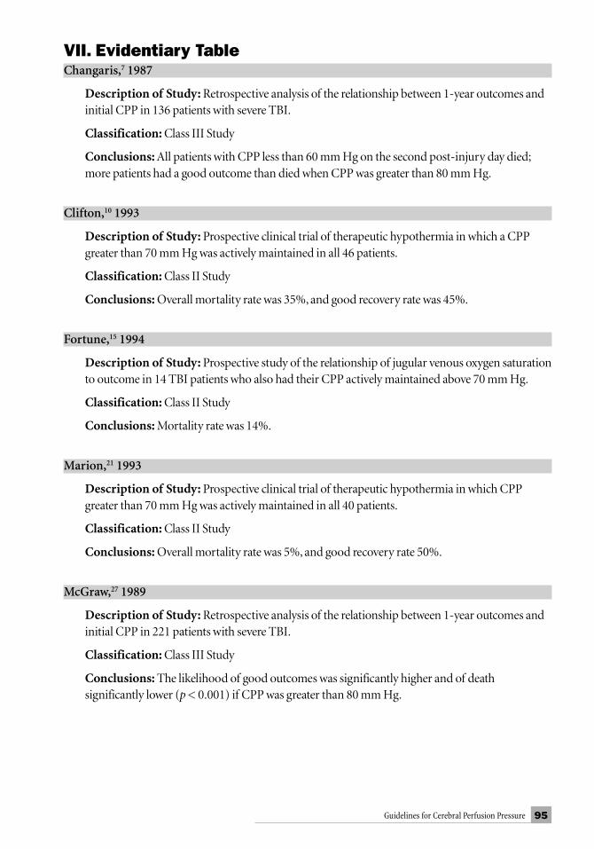
95Guidelines for Cerebral Perfusion Pressure
VII. Evidentiary TableChangaris,7 1987
Description of Study: Retrospective analysis of the relationship between 1-year outcomes andinitial CPP in 136 patients with severe TBI.
Classification: Class III Study
Conclusions: All patients with CPP less than 60 mm Hg on the second post-injury day died;more patients had a good outcome than died when CPP was greater than 80 mm Hg.
Clifton,10 1993
Description of Study: Prospective clinical trial of therapeutic hypothermia in which a CPPgreater than 70 mm Hg was actively maintained in all 46 patients.
Classification: Class II Study
Conclusions: Overall mortality rate was 35%, and good recovery rate was 45%.
Fortune,15 1994
Description of Study: Prospective study of the relationship of jugular venous oxygen saturationto outcome in 14 TBI patients who also had their CPP actively maintained above 70 mm Hg.
Classification: Class II Study
Conclusions: Mortality rate was 14%.
Marion,21 1993
Description of Study: Prospective clinical trial of therapeutic hypothermia in which CPPgreater than 70 mm Hg was actively maintained in all 40 patients.
Classification: Class II Study
Conclusions: Overall mortality rate was 5%, and good recovery rate 50%.
McGraw,27 1989
Description of Study: Retrospective analysis of the relationship between 1-year outcomes andinitial CPP in 221 patients with severe TBI.
Classification: Class III Study
Conclusions: The likelihood of good outcomes was significantly higher and of deathsignificantly lower (p < 0.001) if CPP was greater than 80 mm Hg.
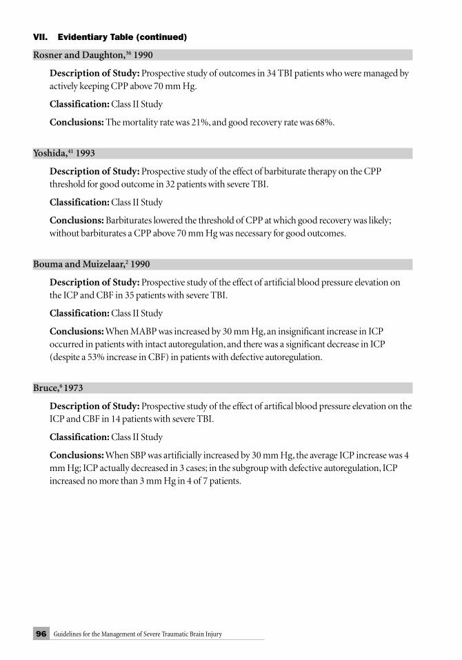
96 Guidelines for the Management of Severe Traumatic Brain Injury
Rosner and Daughton,36 1990
Description of Study: Prospective study of outcomes in 34 TBI patients who were managed byactively keeping CPP above 70 mm Hg.
Classification: Class II Study
Conclusions: The mortality rate was 21%, and good recovery rate was 68%.
Yoshida,41 1993
Description of Study: Prospective study of the effect of barbiturate therapy on the CPPthreshold for good outcome in 32 patients with severe TBI.
Classification: Class II Study
Conclusions: Barbiturates lowered the threshold of CPP at which good recovery was likely;without barbiturates a CPP above 70 mm Hg was necessary for good outcomes.
Bouma and Muizelaar,2 1990
Description of Study: Prospective study of the effect of artificial blood pressure elevation onthe ICP and CBF in 35 patients with severe TBI.
Classification: Class II Study
Conclusions: When MABP was increased by 30 mm Hg, an insignificant increase in ICPoccurred in patients with intact autoregulation, and there was a significant decrease in ICP(despite a 53% increase in CBF) in patients with defective autoregulation.
Bruce,6 1973
Description of Study: Prospective study of the effect of artifical blood pressure elevation on theICP and CBF in 14 patients with severe TBI.
Classification: Class II Study
Conclusions: When SBP was artificially increased by 30 mm Hg, the average ICP increase was 4mm Hg; ICP actually decreased in 3 cases; in the subgroup with defective autoregulation, ICPincreased no more than 3 mm Hg in 4 of 7 patients.
VII. Evidentiary Table (continued)

97Guidelines for Cerebral Perfusion Pressure
Robertson,34 1998
Description of Study: Prospective, randomized clinical trial of 189 patients with severe TBI(15% with GSW) comparing ICP-targeted therapy with CBF-targeted therapy. CPP wasmaintained above 50 mm Hg in the former group, and above 70 mm Hg in the latter group.
Classification: Class I Study
Conclusions: No significant difference in 3/6 month GOS or DRS between the two groups. Theincidence of ARDS was significantly higher in CBF-targeted groups.
Kiening,19 1997
Description of Study: Prospective cohort study of 21 patients with severe TBI in which theeffect of CPP on brain tissue pO
2 was determined.
Classification: Class II Study
Conclusions: Elevation of CPP from 32-67 mm Hg significantly improved brain tissue pO2.
Further increases in the CPP did not alter brain tissue pO2.
Cruz,12 1998
Description of Study: Prospective cohort study of 353 patients with severe TBI in which 178underwent continuous monitoring and management of cerebral O
2 extraction and CPP, and 175
patients had only monitoring and management of CPP.
Classification: Class II Study
Conclusions: Outcome at 6 months after injury was significantly better in those who hadmonitoring and management of cerebral extraction of O
2 as well as CPP.
VIII. References1. Becker DP, Miller JD, Ward JD, et al.: The outcome from severe head injury with early
diagnosis and intensive management. J Neurosurg 47:491-502, 1977.2. Bouma GJ, Muizelaar JP: Relationship between cardiac output and cerebral blood flow in
patients with intact and with impaired autoregulation. J Neurosurg 73:368-374, 1990.3. Bouma GJ, Muizelaar JP, Bandoh K, et al.: Blood pressure and intracranial pressure-volume
dynamics in severe head injury: relationship with cerebral blood flow. J Neurosurg 77:15-19,1992.
4. Bouma GJ, Muizelaar JP, Choi SC, et al.: Cerebral circulation and metabolism after severetraumatic brain injury: the elusive role of ischemia. J Neurosurg 75:685-693, 1991.
5. Bouma GJ, Muizelaar JP, Stringer WA, et al.: Ultra early evaluation of regional cerebralblood flow in severely head-injured patients using xenon enhanced computed tomography.J Neurosurg 77:360-368, 1992.
6. Bruce DA, Langfitt TW, Miller JD, et al.: Regional cerebral blood flow, intracranial pressure,and brain metabolism in comatose patients. J Neurosurg 38:131-144, 1973.
VII. Evidentiary Table (continued)

98 Guidelines for the Management of Severe Traumatic Brain Injury
7. Changaris DG, McGraw CP, Richardson JD, et al.: Correlation of cerebral perfusionpressure and Glasgow Coma Scale to outcome. J Trauma 27:1007-1013, 1987.
8. Chesnut RM, Marshall SB, Piek J, et al.: Early and late systemic hypotension as a frequentand fundamental source of cerebral ischemia following severe brain injury in the TraumaticComa Data Bank. Acta Neurochir Suppl (Wien) 59:121-125, 1993.
9. Choi SC, Narayan RK, Anderson RL, et al.: Enhanced specificity of prognosis in severe headinjury. J Neurosurg 69:381-385, 1988.
10. Clifton GL, Allen S, Barrodale P, et al.: A phase II study of moderate hypothermia in severebrain injury. J Neurotrauma 10:263-271, 1993.
11. Cruz J: Low clinical ischemic threshold for cerebral blood flow in severe acute brain trauma.Case report. J Neurosurg 80:143-147, 1994.
12. Cruz J: The first Decade of continuous monitoring of jugular bulb oxyhemoglobin saturation:management strategies and clinical outcome. Crit Care Med 26:344-351, 1998.
13. Cruz J, Jaggi JL, Hoffstad OJ: Cerebral blood flow, vascular resistance, and oxygenmetabolism in acute brain trauma: redefining the role of cerebral perfusion pressure? CritCare Med 23:1412-1417, 1995.
14. El-Adawy Y, Rosner MJ: Cerebral perfusion pressure, autoregulation and the PVI reflectionpoint. Intracranial Pressure VII. Hoff JT, et al. (eds) Springer-Verlag: Berlin, 829-833, 1988.
15. Fortune JB, Feustel PJ, Weigle CGM, et al.: Continuous measurement of jugular venousoxygen saturation in response to transient elevations of blood presure in head-injuredpatients. J Neurosurg 80:461-468, 1994.
16. Graham DI, Ford I, Adama JH, et al.: Ischaemic brain damage is still common in fatalnon-missile head injury. J Neurol Neurosurg Psychiatry 52:346-350, 1989.
17. Jaggi JL, Obrist WD, Gennarelli TA, et al.: Relationship of early cerebral blood flow andmetabolism to outcome in acute head injury. J Neurosurg 72:176-182, 1990.
18. Jones PA, Andrews PJ, Midgley S, et al.: Measuring the burden of secondary insults inhead-injured patients during intensive care. J Neurosurg Anesthesiol 6:4-14, 1994.
19. Kiening KL, Hartl R, Unterberg AW, et al.: Brain tissue pO2-monitoring in comatose
patients: implications for therapy. Neurol Res 19:233-240, 1997.20. Kontos HA, Wei EP, Navari RM, et al.: Responses of cerebral arteries and arterioles to acute
hypotension and hypertension. Am J Physiol 234:H371-H383, 1978.21. Marion DW, Darby J, Yonas H: Acute regional cerebral blood flow changes caused by severe
head injuries. J Neurosurg 74:407-414, 1991.22. Marion DW, Penrod LE, Kelsey SF, et al.: Treatment of traumatic brain injury with moderate
hypothermia. N Engl J Med 336:540-546, 1997.23. Marmarou A, Anderson RL, Ward JD, et al.: Impact of ICP instability and hypotension on
outcome in patients with severe head trauma. J Neurosurg 75:S59-S66, 1991.24. Marshall LF, Gautille T, Klauber MR, et al.: The outcome of severe closed head injury. J
Neurosurg 75:S28-S36, 1991.25. Marshall LF, Smith RW, Shapiro HM: The outcome with aggressive treatment in severe head
injuries. I. The significance of intracranial pressure monitoring. J Neurosurg 50:20-25, 1979.26. Marshall WJS, Jackson JLF, Langfitt TW: Brain swelling caused by trauma and arterial
hypertension. Arch Neurol 21:545-553, 1969.

99Guidelines for Cerebral Perfusion Pressure
27. McGraw CP: A cerebral perfusion pressure greater than 80 mm Hg is more beneficial:Intracranial pressure VII. Hoff JT, Betz AL (eds), Springer-Verlag: Berlin, 839-841, 1989.
28. McLaughlin MR, Marion DW: Cerebral blood flow and vasoresponsivity within andaround cerebral contusions. J Neurosurg 85:871-876, 1996.
29. Miller JD: Head injury and brain ischaemia—implications for therapy. Br J Anaesth57:120-130, 1985.
30. Murr R, Schurer L: Correlation of jugular venous oxygen saturation to spontaneousfluctuations of cerebral perfusion pressure in patients with severe head injury. Neurol Res17:329-333, 1995.
31. Obrist WD, Langfitt TW, Jaggi JL, et al.: Cerebral blood flow and metabolism in comatosepatients with acute head injury. J Neurosurg 61:241-253, 1984.
32. Pietropaoli JA, Rogers FB, Shackford SR, et al.: The deleterious effects of intraoperativehypotension on outcome in patients with severe head injuries. J Trauma 33:403-407, 1992.
33. Robertson CS, Contant CF, Narayan RK, et al.: Cerebral blood flow, AVdO2, and neurologic
outcome in head-injured patients. J Neurotrauma 9:S349-S358, 1992.34. Robertson CS, Valadka AB, Hannay HJ, et al.: Prevention of secondary ischemic insults after
severe head injury. Crit Care Med 27:2086-2095, 1999.35. Rosner MJ, Becker DP: Origin and evolution of plateau waves: experimental observations
and a theoretical model. J Neurosurg 60:312-324, 1984.36. Rosner MJ, Daughton S: Cerebral perfusion pressure management in head injury. J Trauma
30:933-941, 1990.37. Rosner MJ, Rosner SD, Johnson AH: Cerebral perfusion pressure: management protocol
and clinical results. J Neurosurg 83:949-962, 1995.38. Ross DT, Graham DI, Adams JH: Selective loss of neurons from the thalamic reticular
nucleus following severe human head injury. J Neurotrauma 10:151-165, 199339. Salvant JB, Muizelaar JP: Changes in cerebral blood flow and metabolism related to the
presence of subdural hematoma. Neurosurg 33:387-393, 1993.40. Weber M, Grolimund P, Seiler RW: Evaluation of post-traumatic cerebral blood flow
velocities by transcranial Doppler ultrasonograpy. Neurosurg 27:106-112, 1990.41. Yoshida A, Shima T, Okada Y, et al.: Outcome of patients with severe head injury—
evaluation by cerebral perfusion pressure. In: Nakamura N, Hashimoto T, Yasue M (eds):Springer-Verlag: Hong Kong, 309-312, 1993.


101Hyperventilation
I. RecommendationsA. Standards
In the absence of increased intracranial pressure (ICP), chronic prolongedhyperventilation therapy (PaCO
2 ≤ 25 mm Hg) should be avoided after severe
traumatic brain injury (TBI).B. Guidelines
The use of prophylactic hyperventilation (PaCO2 ≤ 35 mm Hg) therapy during the
first 24 hours after severe TBI should be avoided because it can compromise cerebralperfusion during a time when cerebral blood flow (CBF) is reduced.
C. OptionsHyperventilation therapy may be necessary for brief periods when there is acuteneurologic deterioration, or for longer periods if there is intracranial hypertensionrefractory to sedation, paralysis, cerebrospinal fluid (CSF) drainage, and osmoticdiuretics.
Jugular venous oxygen saturation (SjO2), arterial jugular venous oxygen
(AVdO2) content differences, brain tissue oxygen monitoring, and CBF monitoring
may help to identify cerebral ischemia if hyperventilation, resulting in PaCO2 values
less than 30 mm Hg, is necessary.
II. OverviewAggressive hyperventilation (arterial PaCO
2 ≤ 25 mm Hg) has been a cornerstone in the
management of severe TBI for more than 20 years because it can cause a rapid reduction ofICP. Brain swelling and elevated ICP develop in 40% of patients with severe TBI,32 and highor uncontrolled ICP is one of the most common causes of death and neurologic disabilityafter TBI.1,30,36 Therefore, most clinicians have assumed that hyperventilation benefits allpatients with severe TBI.
However, hyperventilation reduces ICP by causing cerebral vasoconstriction and asubsequent reduction in CBF.40 Research conducted over the past 20 years clearly demonstratesthat CBF during the first day after injury is less than half that of normal individuals,4, 5, 11, 17, 25, 29, 33,
42, 45, 47 and that there is a risk of causing cerebral ischemia with aggressive hyperventilation.Histologic evidence of cerebral ischemia has been found in most victims of severe TBI who
HYPERVENTILATION

102 Guidelines for the Management of Severe Traumatic Brain Injury
die.21, 22, 44 A prospective, randomized study found improved outcome at 3 and 6 months whenprophylactic hyperventilation was not used as compared to when it was.34
Thus, limiting the use of hyperventilation following severe TBI may help improveneurologic recovery following injury, or at least avoid iatrogenic cerebral ischemia.
III. ProcessThe development of these guidelines began with an extensive review of all of the pertinentliterature published during the past 25 years. Approximately 600 citations were acquired bycomputerized search of the National Library of Medicine using the following MeSH headings incombination with “head injuries”: “ischemia,” “jugular vein,” “regional blood flow,” “perfusion,”and “hyperventilation.” Abstracts from the publications were reviewed and relevant articlesselected to develop the guidelines. We focused on four specific areas: cerebral blood flow (30articles reviewed); arterial jugular venous oxygen content differences (AVdO
2*)(7 articles
reviewed); jugular venous oxygen saturation analysis (SjO2*)(6 articles reviewed); and
hyperventilation (16 articles reviewed). All of these articles were cohort studies of more than 8patients (Class II) and were published in peer reviewed journals except for 5:—1 was acontrolled, randomized prospective clinical trial34 (Class I) and 4 were case reports or reviews11,
40, 41, 47 (Class III).
IV. Scientific Foundation
Post-traumatic Cerebral PhysiologyCBF is lowest during the first 24 hours after injury, and increases for at least three daysthereafter except in patients who have uncontrollable ICP and die.4, 11, 25, 29, 33, 42, 45 CBF is typicallyless than 30 cc/100 g/min during the first 8 hours after injury, and may be less than 20 cc/100 g/min during the first four hours after injury in patients with the worst injuries.4, 5, 11, 17, 45, 47
The CBF threshold for irreversible ischemia or infarction in TBI is not clearly established.Obrist has suggested that TBI causes a depression of cerebral metabolism, and that the reducedCBF that occurs following TBI may in many cases be appropriate for the metabolic needs of thebrain.26 However, in a positron emission tomography (PET) study of 16 patients with clinicaland CT evidence of hemispheric stroke, studied at a mean of 23 hours after the onset ofsymptoms, Heiss found that the mean CBF was 16.7 ± 7.95 cc/100 g/min at the center of theinfarct and 31.0 ± 10.65 cc/100 g/min in the zone immediately adjacent to the infarct.24
In severe TBI, CBF is lowest in patients with subdural hematomas, diffuse injuries, andhypotension, and highest in those with epidural hematomas or normal CT scans.5, 29, 38, 45, 47
There is a direct correlation between CBF and Glasgow Coma Scale (GCS) score or outcome,but only during the first 24 hours after injury.4, 29 A change in CBF does not necessarily correlatewith a change in ICP; in some cases, an increase in CBF actually causes a decrease in ICP.3, 39
There is an inverse relationship between AVdO2 values and CBF values during the first 24
hours after injury, except in patients with subdural hematomas.4, 6, 33, 39, 43 AVdO2 values
exceeding 9 vol% probably indicate cerebral ischemia.20, 39
Jugular venous oxygen saturation is normally greater than 50%, and values less than 50%are considered desaturations. Profound or prolonged episodes of desaturation are associatedwith a poor outcome.10, 48 Desaturations are most common with low CBF.48 Hypocapnia is
* See Glossary on page 114.

103Hyperventilation
associated with desaturations: in 6 patients Cruz found that the mean SjO2 was 45 ± 8% when
the arterial PaCO2 was less than 22 mm Hg; and when the pCO
2 was increased by 10 mm Hg in
these patients, the SjO2 was 59 ± 3.2%.13 Sheinberg and colleagues identified an arterial PaCO
2
less than 28 mm Hg as the cause for desaturation in 10 of 33 patients.48
Recent studies continue to demonstrate decreased CBF and widened AVdO2 after severe
TBI, as well as a low CMRO2 and abnormal CO
2 vasoresponsivity.15, 31, 37 Several studies conclude
that there is narrowed microcirculation and persistent vasoconstriction and that post-traumaticvasospasm may occur in as many as 43% of patients who suffer a severe TBI.23, 27, 35, 54 Widevariability of CO
2 vasoresponsivity surrounding contusions has been documented.31 PET studies
have shown an increase in the cerebral metabolic rate for glucose surrounding contusions orunderlying subdural hematomas in some patients.36,53
HyperventilationThe normal response to hyperventilation was studied by Raichle in a group of healthyvolunteers: 40% decrease in CBF 30 minutes after decreasing the PaCO
2 by 15-20 mm Hg; 4
hours later there was an increase in CBF to 90% of baseline; when the original PaCO2 was
restored there was an overshoot in CBF of 31%.41
A study of cerebral blood volume, measured with the 99M-tc-labeled red blood cells(RBC), and simultaneous CBF measurements using carotid duplex scanning found that areduction of the PaCO
2 to 26 mm Hg led to a 7.2% decrease in cerebral blood volume, but a
30.7% decrease in CBF.18 Thus, the change in CBF was far greater than the change in cerebralblood volume caused by hyperventilation. This study is particularly important because of theestablished interrelationships between ICP, CBF, and cerebral blood volume, andhyperventilation. As a static measurement, cerebral blood volume is directly related to ICP. Thedynamic parameter of CBF is not directly related to ICP and yet it is the physiologic parameterdirectly affected by hyperventilation. In global terms, at least, hyperventilation will usuallycause a reduction in CBF, thereby potentially limiting flow of blood to ischemic areas of thebrain. However, CBF changes are not necessarily related to changes in cerebral blood volume.
The relative CO2 vasoresponsivity in severe TBI is a 3% change in CBF per torr change in
PaCO2, but is lower with a lower CBF.6, 7, 39 A low CO
2 vasoresponsivity is associated with a poor
outcome.28, 39, 51 Local CO2 vasoresponsivity differs from global values by more than 50% in a
substantial number of patients.28 In some patients, cerebral autoregulation is preserved withnormocapnia, and lost with hypocapnia.8 In some cases, hyperventilation can actually cause anincrease in ICP: Crockard found an increase in ICP in 4 of 14 patients associated with adecrease in PaCO
2 to 25-30 mm Hg,9 and Obrist found a decrease in ICP with hyperventilation
in only 15 of 31 patients, but there was a decrease in CBF in 29 of them.39 Aggressivehyperventilation can cause AVdO
2 and CBF values that most consider at or near the ischemic
thresholds: in 10 patients with a PaCO2 of 23.2 ± 2.8, Obrist found that the AVdO
2 was 10.5 ±
0.7 and CBF was 18.6 ± 4.4.39
During the past four years (1994 to 1998) blood flow studies using both the xenontechniques as well as the local thermodiffusion technique have continued to demonstrate asignificant decrease in CBF associated with hyperventilation.14, 19, 49, 50 In a recent xenon CT CBFstudy, the incidence of 2 centimeter in diameter regions of CBF with blood flows less than 18ml/100 g/min increased from 28.9% to 73.1% when the PaCO
2 was decreased from 35 mm Hg
to 25 mm Hg.50 Several studies also document an increased incidence of jugular venous

104 Guidelines for the Management of Severe Traumatic Brain Injury
desaturations (SjO2 < 50%) associated with the use of hyperventilation.46 Recent studies of the
use of brain tissue oxygen levels have shown that hyperventilation is frequently associated witha significant decrease in brain tissue oxygen.16, 26, 52
One study has suggested that hyperventilation can “improve cerebral glucose utilizationfollowing trauma.” In that study, jugular bulb monitoring was used to demonstrate an increasein cerebral glucose extraction when the PaCO
2 was lowered to below 25 mm Hg in some
patients.12 However, there is no evidence that this is desirable and certainly no evidence that thechange in glucose extraction is associated with an improved outcome following severe TBI.Because of substantial variability in the metabolic values measured between the right and leftjugular venous samples, and because jugular venous samples measure global cerebralmetabolism in the face of significant regional metabolic heterogeneity, this observation is notthought to provide a rationale for changing the recommendations regarding the use ofhyperventilation following severe TBI.
In 1991, Muizelaar, et al.,34 published the results of a prospective, randomized clinical studyin which 77 patients with severe TBI were randomized to a group treated with chronicprophylactic hyperventilation for 5 days after injury (PaCO
2 of 25 ± 2 mm Hg), or to a group
kept relatively normocapneic during that time (PaCO2 of 35 ± 2 mm Hg). At 3 and 6 months
after injury, patients with an initial GCS motor score of 4-5 who were in the hyperventilationgroup had a significantly worse outcome than did those in the normocapneic group.Statistically significant differences between the two groups were not found at one year afterinjury. However, this lack of significance can be attributed to a type II statistical error becausethere were substantially fewer patients available for follow-up one year after injury.
V. SummaryChronic prophylactic hyperventilation therapy should be avoided during the first 5 days aftersevere TBI, and particularly during the first 24 hours. CBF measurements in patients withsevere TBI demonstrate that blood flow early after injury is low, and strongly suggest that in thefirst few hours after injury the absolute values approach those consistent with ischemia. Thesefindings are corroborated by AVdO
2 and SjO
2 and brain tissue O
2 measurements.
Hyperventilation will reduce CBF values even further, but will not consistently cause areduction of ICP and may cause loss of autoregulation. The cerebral vascular response tohypocapnia is reduced in those with the most severe injuries (subdural hematomas and diffusecontusions), and there is substantial local variability in perfusion. While the CBF level at whichirreversible ischemia occurs has not been clearly established, ischemic cell change has beendemonstrated in 90% of those who die following TBI, and there is PET evidence that suchdamage is likely to occur when CBF drops below 15-20 cc/100 g/min. A prospective,randomized clinical trial has determined that outcomes are worse when TBI patients are treatedwith chronic prophylactic hyperventilation therapy.
Within the standard, guideline, and options, specific PaCO2 thresholds have been described
that are different for each of the three parameters. These individual thresholds were selectedbased on the preponderance of literature supporting those thresholds in the contexts of thestatements that included them. With the exception of the threshold included for the standard inthis guideline, it is emphasized that the PaCO
2 threshold is not as important as the general
concept of hyperventilation. The preponderance of the physiologic literature concludes that

105Hyperventilation
hyperventilation during the first few days following severe TBI, whatever the threshold, ispotentially deleterious in that it can promote cerebral ischemia.
VI. Key Issues for Future InvestigationProspective, randomized, clinical trials are needed to establish if short-term hyperventilationduring the first 24 hours after injury is deleterious. In addition, such trials should determine ifsystemic neuromuscular paralysis and controlled mechanical ventilation is beneficial for thosewho spontaneously hyperventilate following severe TBI.
VII. Evidentiary TablesTable 1: Clinical CBF, Metabolic, and Physiologic Measurementsfollowing Severe TBIFieschi,17 1974
Description of Study: Cohort study of CBF in 12 patients with severe TBI designed to describethe temporal changes in CBF during the first few days after injury.
Classification: Class II Study
Conclusions: CBF was always lowest during first 12 hours after injury, with mean values of 17and 28 ml/100 g/min, respectively, for those who died and who survived.
Bouma,5 1992
Description of Study: Cohort study of very early CBF in 35 patients with severe TBI studied amean of 3.1 ± 2.1 hours after injury.
Classification: Class II Study
Conclusions: Global or regional, CBF of less than 18 ml/100 g/min, defined as ischemicthreshold, was found in 31.4% of the patients.
Muizelaar,33 1989
Description of Study: Cohort of 32 children with severe TBI in which CBF was determinedover time during first several days after injury.
Classification: Class II Study
Conclusions: CBF was lowest during the first 24 hours after injury.
Marion,29 1991
Description of Study: Cohort study of 32 patients with severe TBI aimed at defining temporalchanges in CBF that occur during the first 5 days after injury.
Classification: Class II Study
Conclusions: Mean CBF in the first 1-4 hours after injury was 27 ml/100 g/min, and CBF wasalways lowest during the first 12-24 hours after injury. Regional CBF was substantiallyheterogeneous.

106 Guidelines for the Management of Severe Traumatic Brain Injury
Bouma,4 1991
Description of Study: Cohort study of 186 patients with severe TBI designed to measure earlyCBF after injury and correlate it with outcome.
Classification: Class II Study
Conclusions: The mean CBF during the first 6 hours after injury was 22.5 ± 5.2 ml/100 g/min,and CBF was highest at 36-42 hours after injury.
Salvant and Muizelaar,45 1993
Description of Study: Cohort study of 54 patients with severe TBI and subdural hematoma,designed to define temporal changes in CBF and the effect of the hematoma on regional CBF.
Classification: Class II Study
Conclusions: The lowest CBF was always seen during the first 24 hours after injury. In thehemisphere with the subdural hematoma, 9% of patients had a CBF less than 18 ml/100 g/min.
Newell,37 1997
Description of Study: Cohort study of 22 patients designed to measure CO2 vasoresponsivity
using transcranial Doppler within 48 hours of the injury.
Classification: Class II Study
Conclusions: There was diminished CO2 reactivity in most patients within 48 hours after injury.
Hadani,23 1997
Description of Study: Cohort study of 32 patients studied with TCD to determine theincidence of vasospasm.
Classification: Class II Study
Conclusions: Blood flow velocities compatible with vasospasm were found in 43% of thepatients in either the basilar artery, the middle cerebral artery, or both. The peak of vasospasmwas at 4 to 5 days after injury.
Muizelaar,35 1997
Description of Study: Cohort study of 71 patients with severe TBI. Mean transit time wasstudied using rapid sequence CT scanning after a bolus infusion of intravenous contrast solution.
Classification: Class II Study
Conclusions: The mean transit time was significantly prolonged after TBI and suggested anincrease in cerebrovascular resistance due to narrowing of the microvasculature, consistent withearly cerebral ischemia
VII. Evidentiary Table 1 (continued)

107Hyperventilation
Yoshihara,54 1995
Description of Study: Cohort study of 49 patients with severe TBI in whom the pressurevolume index and ICP were studied to determine the resistance of cerebrovasculature.
Classification: Class II Study
Conclusions: Resistance vessels in the brain are in a state of persistent vasoconstrictionfollowing TBI.
Bergsneider,2 1997
Description of Study: Cohort study of 28 patients with severe TBI using PET scanning todetermine regional cerebral glucose metabolism.
Classification: Class II Study
Conclusions: Increased glucose metabolism was documented surrounding cerebral contusionsand underlying subdural hematomas in several patients with severe TBI.
Table 2: Histologic Evidence for Ischemia Following TBI andEvidence of Widened Arterial Jugular Venous Oxygen ContentDifferences (AVdO2) Early After InjuryGraham,22 1988
Description of Study: Histologic study of 71 victims of fatal severe TBI who had no pre-mortem evidence (clinical, radiologic, or pathologic) of elevated ICP.
Classification: Class II Study
Conclusions: Ischemic cell changes were found in 70% of the brains.
Ross,44 1993
Description of Study: Histologic study of 37 victims of fatal severe TBI to determine theincidence of ischemic cell change in the basal ganglia.
Classification: Class II Study
Conclusions: Loss of thalamic reticular neurons was found in 89% of cases.
Bouma,4 1991
Description of Study: Cohort study of 186 patients with severe TBI to determine the timecourse of CBF and AVdO
2 changes early after injury.
Classification: Class II Study
Conclusions: AVdO2 values were widest (7.1 ± 1.5 vol%) during the first 4-6 hours after injury
and decreased to 4.2 ± 1.7 vol% at 36-42 hours
VII. Evidentiary Table 1 (continued)

108 Guidelines for the Management of Severe Traumatic Brain Injury
Robertson,43 1992
Description of Study: Cohort study of 102 patients with severe TBI examining the time courseand relationship of AVdO
2, CBF, and ICP.
Classification: Class II Study
Conclusions: AVdO2 values were always widest during the first 24 hours after injury.
Table 3: Effects of Hyperventilation on CBF, AVdO2, SjO2, BrainTissue pO2, and Clinical OutcomeObrist,39 1984
Description of Study: Cohort study of 31 patients with severe TBI in whom the effect ofaggressive hyperventilation on ICP, CBF, and AVdO
2 was examined.
Classification: Class II Study
Conclusions: Hyperventilation had a much more direct effect on CBF reduction (29 of 31patients) than it did on ICP reduction (15 of 31 patients). Aggressive hyperventilation in 10patients (PaCO
2 -23.2 ± 2.8 mm Hg) led to AVdO
2 values of 10.5 ± 0.7 vol% and CBF values of
18.6 ± 4.4 ml/100 g/min.
Sheinberg,48 1992
Description of Study: Cohort study of Sj O2 in 45 patients with severe TBI monitored for 1 to 8
days.
Classification: Class II Study
Conclusions: Hyperventilation was the second most common identifiable cause for jugularvenous desaturations (O
2 sat. < 50%), and was the cause for desaturations in 10 of 33 cases.
Cruz,13 1991
Description of Study: Cohort study of 6 patients with severe TBI undergoing continuous SjO2
monitoring for 48 hours after injury.
Classification: Class II Study
Conclusions: Jugular venous O2 desaturations (45.5 ± 8%) were observed in all 6 patients
during aggressive hyperventilation, and the O2 saturation improved to greater than 50% in all
cases with withdrawal of hyperventilation.
VII. Evidentiary Table 2 (continued)

109Hyperventilation
Muizelaar,34 1991
Description of Study: Prospective, randomized, clinical trial of 77 patients with severe TBIcomparing clinical outcome for a group hyperventilated to a PaCO
2 of 25 ± 2 mm Hg for 5 days
after injury, and a group with PaCO2 kept at 35 ± 2 mm Hg during that time.
Classification: Class II Study
Conclusions: At 3 and 6 months after injury, the patients with an initial GCS motor score of 4 to5 had a significantly better outcome if they were not hyperventilated.
Fortune,18 1995
Description of Study: Cohort study of 8 normal individuals. Cerebral blood volume wasmeasured with 99M-tc-labeled RBC and CBF was measured with carotid duplex scanning.
Classification: Class II Study
Conclusions: Decreasing the PaCO2 to 26 mm Hg caused a 7.2% decrease in cerebral blood
volume, but a 30.7% decrease in cerebral blood flow. Thus, changes in cerebral blood flow weremuch greater than changes in cerebral blood volume.
Skippen,50 1997
Description of Study: Cohort study of 23 children with severe TBI studied using xenon CTCBF techniques and SjO
2 to correlate cerebral oxygen consumption with changes in CBF.
Classification: Class II Study
Conclusions: Cerebral oxygen consumption decreased out of proportion to CBF following TBI.Frequency of one or more 2 cm regions of low CBF (CBF < 18 ml/100 g/min) when the pCO
2
was greater than 35 mm Hg was 28.9%. If the PaCO2 was lowered to 25 mm Hg, the frequency
was 73.1%.
Dings,16 1996
Description of Study: Cohort study of 22 patients with severe TBI studied using a brain tissueoxygen sensor to determine the effect of hyperventilation on brain tissue pO
2 .
Classification: Class II Study
Conclusions: Six of 22 patients had a reduction of brain tissue oxygen into ischemic rangesfollowing hyperventilation.
VII. Evidentiary Table 3 (continued)
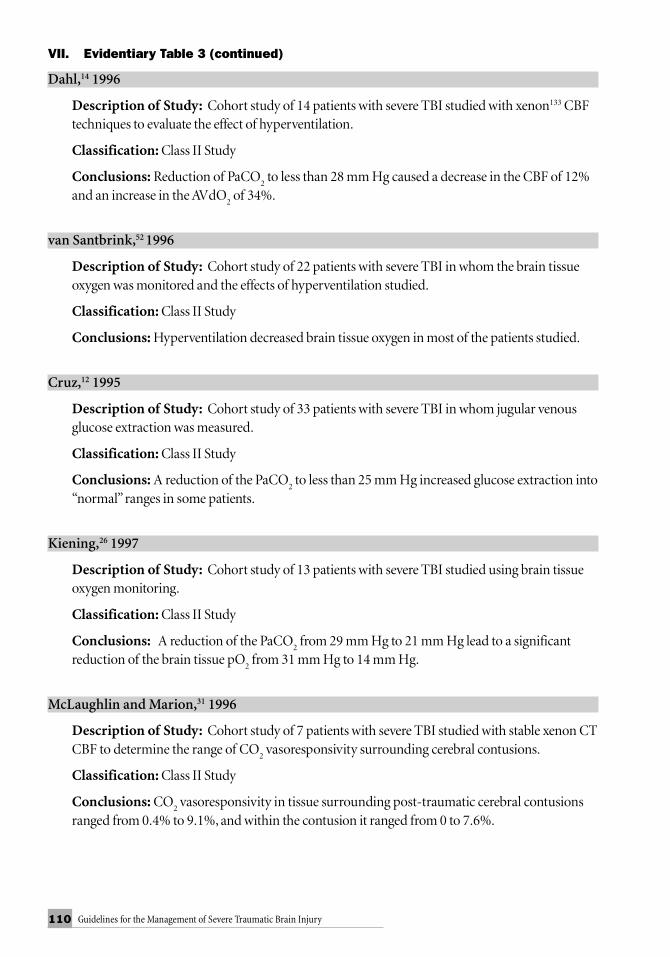
110 Guidelines for the Management of Severe Traumatic Brain Injury
Dahl,14 1996
Description of Study: Cohort study of 14 patients with severe TBI studied with xenon133 CBFtechniques to evaluate the effect of hyperventilation.
Classification: Class II Study
Conclusions: Reduction of PaCO2 to less than 28 mm Hg caused a decrease in the CBF of 12%
and an increase in the AVdO2 of 34%.
van Santbrink,52 1996
Description of Study: Cohort study of 22 patients with severe TBI in whom the brain tissueoxygen was monitored and the effects of hyperventilation studied.
Classification: Class II Study
Conclusions: Hyperventilation decreased brain tissue oxygen in most of the patients studied.
Cruz,12 1995
Description of Study: Cohort study of 33 patients with severe TBI in whom jugular venousglucose extraction was measured.
Classification: Class II Study
Conclusions: A reduction of the PaCO2 to less than 25 mm Hg increased glucose extraction into
“normal” ranges in some patients.
Kiening,26 1997
Description of Study: Cohort study of 13 patients with severe TBI studied using brain tissueoxygen monitoring.
Classification: Class II Study
Conclusions: A reduction of the PaCO2 from 29 mm Hg to 21 mm Hg lead to a significant
reduction of the brain tissue pO2 from 31 mm Hg to 14 mm Hg.
McLaughlin and Marion,31 1996
Description of Study: Cohort study of 7 patients with severe TBI studied with stable xenon CTCBF to determine the range of CO
2 vasoresponsivity surrounding cerebral contusions.
Classification: Class II Study
Conclusions: CO2 vasoresponsivity in tissue surrounding post-traumatic cerebral contusions
ranged from 0.4% to 9.1%, and within the contusion it ranged from 0 to 7.6%.
VII. Evidentiary Table 3 (continued)

111Hyperventilation
Schneider,46 1995
Description of Study: Cohort study of 54 patients with severe TBI; SjO2 was measured to
determine the incidence and causes of jugular venous oxygen desaturations.
Classification: Class II Study
Conclusions: Hyperventilation was the second most common identifiable cause for jugularvenous oxygen desaturations.
Sioutos,49 1995
Description of Study: Thirty-seven patients with severe TBI studied with thermodiffusionCBF monitoring to determine the effects of hyperventilation on local CBF.
Classification: Class II Study
Conclusions: One-third of the patients had a CBF of less than 18 ml/100 g/min following TBIand hyperventilation decreased CBF even further in those patients.
VIII. References1. Becker DP, Miller JD, Ward JD, et al.: The outcome from severe head injury with early
diagnosis and intensive management. J Neurosurg 47:491-502, 1977.2. Bergsneider M, Hovda DA, Shalmon E, et al.: Cerebral hyperglycolysis following severe
traumatic brain injury in humans: a positron emission tomography study. J Neurosurgery86:241-251, 1997.
3. Bouma GJ and Muizelaar JP: Relationship between cardiac output and cerebral blood flowin patients with intact and with impaired autoregulation. J Neurosurg 73:368-374, 1990.
4. Bouma GJ, Muizelaar JP, Choi SC, et al.: Cerebral circulation and metabolism after severetraumatic brain injury: the elusive role of ischemia. J Neurosurg 75:685-693, 1991.
5. Bouma GJ, Muizelaar JP, Stringer WA, et al.: Ultra early evaluation of regional cerebralblood flow in severely head-injured patients using xenon enhanced computed tomography.J Neurosurg 77:360-368, 1992.
6. Cold GE: The relationship between cerebral metabolic rate of oxygen and cerebral bloodflow in the acute phase of head injury. Acta Anaesthesiol Scand 30:453-457, 1986.
7. Cold GE: Measurements of CO2 reactivity and barbiturate reactivity in patients with severe
head injury. Acta Neurochir (Wien) 98:153-163, 1989.8. Cold GE, Christensen MS, Schmidt, K: Effect of two levels of induced hypocapnia on
cerebral autoregulation in the acute phase of head injury coma. Acta Anaesthesiol Scand25:397-401, 1981.
9. Crockard HA, Coppel DL, Morrow WF: Evaluation of hyperventilation in treatment ofhead injuries. Br Med J 4:634-640, 1973.
10. Cruz J: On-line monitoring of global cerebral hypoxia in acute brain injury. Relationship tointracranial hypertension. J Neurosurg 79:228-233, 1993.
11. Cruz J. Low clinical ischemic threshold for cerebral blood flow in severe acute brain trauma.Case report. J Neurosurg 80:143-147, 1994.
VII. Evidentiary Table 3 (continued)

112 Guidelines for the Management of Severe Traumatic Brain Injury
12. Cruz J: An additional therapeutic effect of adequate hyperventilation in severe acute braintrauma: normalization of cerebral glucose uptake. J Neurosurg 82:379-385, 1995.
13. Cruz J, Miner ME, Allen SJ, et al.: Continuous monitoring of cerebral oxygenation in acutebrain injury: assessment of cerebral hemodynamic reserve. Neurosurg 29:743-749, 1991.
14. Dahl B, Bergholt B, Cold GE, et al.: CO2 and indomethacin vasoreactivity in patients with
head injury. Acta Neurochir (Wien) 138:265-273, 1996.15. Della Corte F, Giordano A, Pennisi MA, et al.: Quantitative cerebral blood flow and
metabolism determination in the first 48 hours after severe head injury with a new dynamicSPECT device. Acta Neurochir (Wien) 139:636-642, 1997.
16. Dings J, Meixensberger J, Amschler J, et al.: Continuous monitoring of brain tissue PO2: a
new tool to minimize the risk of ischemia caused by hyperventilation therapy. ZentralblNeurochir 57:177-183, 1996.
17. Fieschi C, Battistini N, Beduschi A, et al. Regional cerebral blood flow and intraventricularpressure in acute head injuries. J Neurol Neurosurg Psychiatry 37:1378-1388, 1974.
18. Fortune JB, Feustel PJ, deLuna C, et al.: Cerebral blood flow and blood volume in responseto O
2 and CO
2 changes in normal humans. J Trauma 39:463-472, 1995.
19. Fortune JB, Feustel PJ, Graca L, et al.: Effect of hyperventilation, mannitol, andventriculostomy drainage on cerebral blood flow after head injury. J Trauma 39:1091-1099,1995.
20. Gotoh F, Meyer JS, Takagi Y: Cerebral effects of hyperventilation in man. Arch Neurol12:410-423, 1965.
21. Graham DI, Adams JH: Ischaemic brain damage in fatal head injuries. Lancet 1:265-266,1971.
22. Graham DI, Lawrence AE, Adams JH, et al.: Brain damage in fatal non-missile head injurywithout high intracranial pressure. J Clin Pathol 41:34-37, 1988.
23. Hadani M, Bruk B, Ram Z, et al.: Transiently increased basilar artery flow velocity followingsevere head injury: a time course transcranial Doppler study. J Neurotrauma 14:629-636,1997.
24. Heiss WD, Huber M, Fink GR, et al.: Progressive derangement of periinfarct viable tissue inischemic stroke. J Cereb Blood Flow Metab 12:193-203, 1992.
25. Jaggi JL, Obrist WD, Gennarelli TA, et al.: Relationship of early cerebral blood flow andmetabolism to outcome in acute head injury. J Neurosurg 72:176-182, 1990.
26. Kiening KL, Hartl R, Unterberg AW, et al.: Brain tissue pO2-monitoring in comatose
patients: implications for therapy. Neurol Res 19:233-240, 1997.27. Lee JH, Martin NA, Alsina G, et al.: Hemodynamically significant cerebral vasospasm and
outcome after head injury: a prospective study. J Neurosurg 87:221-233, 1997.28. Marion DW, Bouma GJ: The use of stable xenon-enhanced computed tomographic studies
of cerebral blood flow to define changes in cerebral carbon dioxide vasoresponsivity causedby a severe head injury. Neurosurg 29:869-873, 1991.
29. Marion DW, Darby J, Yonas H: Acute regional cerebral blood flow changes caused by severehead injuries. J Neurosurg 74:407-414, 1991.
30. Marshall LF, Smith RW, Shapiro HM: The outcome with aggressive treatment in severe headinjuries. I. The significance of intracranial pressure monitoring. J Neurosurg 50:20-25, 1979.

113Hyperventilation
31. McLaughlin MR, Marion DW: Cerebral blood flow and vasoresponsivity within andaround cerebral contusions. J Neurosurg 85:871-876, 1996.
32. Miller JD, Becker DP, Ward JD, et al.: Significance of intracranial hypertension in severehead injury. J Neurosurg 47:503-510, 1977.
33. Muizelaar JP, Marmarou A, DeSalles AA, et al.: Cerebral blood flow and metabolism inseverely head-injured children. Part 1: Relationship with GCS score, outcome, ICP, and PVI.J Neurosurg 71:63-71, 1989.
34. Muizelaar JP, Marmarou A, Ward JD, et al.: Adverse effects of prolonged hyperventilation inpatients with severe head injury: a randomized clinical trial. J Neurosurg 75:731-739, 1991.
35. Muizelaar JP, Fatouros PP, Schroder ML: A new method for quantitative regional cerebralblood volume measurements using computed tomography. Stroke 28:1998-2005, 1997.
36. Narayan RK, Kishore PRS, Becker DP, et al.: Intracranial pressure: to monitor or not tomonitor. J Neurosurg 56:650-659, 1982.
37. Newell DW, Aaslid R, Stooss R, et al.: Evaluation of hemodynamic responses in head injurypatients with transcranial Doppler monitoring. Acta Neurochir (Wien) 139:804-817, 1997.
38. Obrist WD, Gennarelli TA, Segawa H, et al.: Relation of cerebral blood flow to neurologicalstatus and outcome in head-injured patients. J Neurosurg 51:292-300, 1979.
39. Obrist WD, Langfitt TW, Jaggi JL, et al.: Cerebral blood flow and metabolism in comatosepatients with acute head injury. J Neurosurg 61:241-253, 1984.
40. Raichle ME, Plum F: Hyperventilation and cerebral blood flow. Stroke 3:566-575, 1972.41. Raichle ME, Posner JB, Plum F: Cerebral blood flow during and after hyperventilation. Arch
Neurol 23:394-403, 1970.42. Robertson CS, Clifton GL, Grossman RG, et al.: Alterations in cerebral availability of
metabolic substrates after severe head injury. J Trauma 28:1523-1532, 1988.43. Robertson CS, Contant CF, Gokaslan ZL, et al.: Cerebral blood flow, arteriovenous oxygen
difference, and outcome in head injured patients. J Neurol Neurosurg Psychiatry 55:594-603,1992.
44. Ross DT, Graham DI, Adams JH: Selective loss of neurons from the thalamic reticularnucleus following severe human head injury. J Neurotrauma 10:151-165, 1993.
45. Salvant JB, Muizelaar JP: Changes in cerebral blood flow and metabolism related to thepresence of subdural hematoma. Neurosurg 33:387-393, 1993.
46. Schneider GH, von Helden A, Lanksch WR, et al.: Continuous monitoring of jugular bulboxygen saturation in comatose patients—therapeutic implications. Acta Neurochir (Wien)134:71-75, 1995.
47. Schroder ML, Muizelaar JP, Kuta AJ: Documented reversal of global ischemia immediatelyafter removal of an acute subdural hematoma. Neurosurg 80:324-327, 1994.
48. Sheinberg M, Kanter MJ, Robertson CS, et al.: Continuous monitoring of jugular venousoxygen saturation in head-injured patients. J Neurosurg 76:212-217, 1992.
49. Sioutos PJ, Orozco JA, Carter LP, et al.: Continuous regional cerebral cortical blood flowmonitoring in head-injured patients. Neurosurg 36:943-950, 1995.
50. Skippen P, Seear M, Poskitt K, et al.: Effect of hyperventilation on regional cerebral bloodflow in head-injured children. Crit Care Med 25:1402-1409, 1997.
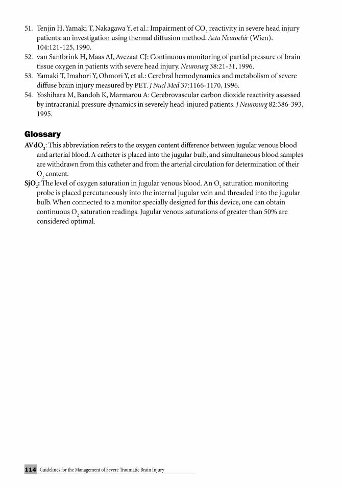
114 Guidelines for the Management of Severe Traumatic Brain Injury
51. Tenjin H, Yamaki T, Nakagawa Y, et al.: Impairment of CO2 reactivity in severe head injury
patients: an investigation using thermal diffusion method. Acta Neurochir (Wien).104:121-125, 1990.
52. van Santbrink H, Maas AI, Avezaat CJ: Continuous monitoring of partial pressure of braintissue oxygen in patients with severe head injury. Neurosurg 38:21-31, 1996.
53. Yamaki T, Imahori Y, Ohmori Y, et al.: Cerebral hemodynamics and metabolism of severediffuse brain injury measured by PET. J Nucl Med 37:1166-1170, 1996.
54. Yoshihara M, Bandoh K, Marmarou A: Cerebrovascular carbon dioxide reactivity assessedby intracranial pressure dynamics in severely head-injured patients. J Neurosurg 82:386-393,1995.
GlossaryAVdO
2: This abbreviation refers to the oxygen content difference between jugular venous blood
and arterial blood. A catheter is placed into the jugular bulb, and simultaneous blood samplesare withdrawn from this catheter and from the arterial circulation for determination of theirO
2 content.
SjO2: The level of oxygen saturation in jugular venous blood. An O
2 saturation monitoring
probe is placed percutaneously into the internal jugular vein and threaded into the jugularbulb. When connected to a monitor specially designed for this device, one can obtaincontinuous O
2 saturation readings. Jugular venous saturations of greater than 50% are
considered optimal.

115The Use of Mannitol
I. RecommendationsA. Standards
There are insufficient data to support a treatment standard for this topic.B. Guidelines
Mannitol is effective for control of raised intracranial pressure (ICP) after severe headinjury. Effective doses range from 0.25 g/kg body weight to 1 gm/kg body weight.
C. Options1. The indications for the use of mannitol prior to ICP monitoring are signs of
transtentorial herniation or progressive neurological deterioration notattributable to extracranial explanations. However, hypovolemia should beavoided by fluid replacement.
2. Serum osmolarity should be kept below 320 mOsm because of concern for renalfailure.
3. Euvolemia should be maintained by adequate fluid replacement. A Foley catheteris essential in these patients.
4. Intermittent boluses may be more effective than continuous infusion.
II. OverviewThe administration of mannitol has become a cornerstone of management of head-injuredpatients, particularly in the acute phase when suspected or actual raised ICP may be present.However, it has never been subjected to a controlled clinical trial against placebo. Althoughthere is much data regarding its mechanism of action, there are few studies that validatedifferent regimens of mannitol usage. Recently, mannitol has been advocated as a “small volumeresuscitation fluid” for the acute phase resuscitation of patients with hypotension andconcomitant brain injury.
III. Process“Mannitol” was listed 147 times associated with “brain trauma” in MEDLINE in a search of thepast 25 years’ literature, to January 1998. The majority of these citations were descriptive,discussing the use of mannitol, among other modalities, in head injury management or
THE USE OF MANNITOL

116 Guidelines for the Management of Severe Traumatic Brain Injury
emergency trauma care. Only 4 of these citations were comparative studies or claimed an effectfor mannitol alone on outcome. Forty-one citations were selected for review because theyfocused on the mechanism of action of mannitol or its effect on outcome or critically reviewedits role in head trauma management. The most important of these are summarized in theevidentiary table.
IV. Scientific FoundationOver the past 20 years, mannitol has replaced other osmotic diuretics.1,2,5,7,8,16,17,22,24 Its beneficialeffects on ICP, cerebral perfusion pressure (CPP), cerebral blood flow (CBF), brain metabolism,and its short-term beneficial effect on neurological outcome, are widely accepted as a result ofmany mechanistic studies performed in humans and animal models.5,25,30,31,33 There is stillcontroversy regarding the exact mechanism or mechanisms by which it exerts its beneficialeffect, and it is probable that it has two distinct effects in the brain that include:13,29
1) an immediate plasma expanding effect that reduces the hematocrit, reduces blood viscosity,increases cerebral blood flow, and increases cerebral oxygen delivery.1,4,14,18,25,29,30,31,35 Theserheological effects probably explain why mannitol reduces ICP within a few minutes ofits administration, and why its effect on ICP is most marked in patients with low CPP(< 70).24,29,30,35 This plasma expanding effect is best accomplished with bolus administration.
2) the osmotic effect of mannitol is delayed for 15-30 minutes while gradients are establishedbetween plasma and cells.1 Its effects persist for a variable period—90 minutes to 6 or morehours, depending on the clinical conditions.2,4,7,13,23,24,40 Mannitol is excreted entirely in theurine, and a significant risk of acute renal failure (acute tubular necrosis) exists if mannitolis administered in large doses, particularly if serum osmolarity exceeds 320 mOsm.2, 9
Patients may be more prone to renal failure if other potentially nephrotoxic drugs are beingadministered, or in the presence of sepsis, or pre-existing renal disease.2, 9, 22, 25 Mannitolmarkedly raises the urinary osmolarity and specific gravity so that these cannot beused to diagnose diabetes insipidus in patients on large doses of mannitol. Mannitol, incommon with other osmotics, is known to cause “opening” of the blood-brain barrier,meaning that both mannitol and other small molecules concurrently in the circulation maypass into brain.3,13,15,19 This effect becomes harmful after many doses have been given,because mannitol may accumulate in the brain causing a reverse osmotic shift and raisingbrain osmolarity, thus theoretically exacerbating ICP by increasing brain cellular swelling.2,
3, 6, 19, 38 The accumulation of mannitol in the brain is most marked when mannitol is in thecirculation for long periods, as occurs with continuous infusion administration.1, 2, 38, 39
Thus, it is recommended that mannitol should be administered as repeated boluses, ratherthan continuous infusion.6, 16, 17, 25, 26, 27
Recently, mannitol has become popular as “a small volume resuscitation fluid,” and hasoften been compared with hypertonic saline (7.5%). Mannitol has particularly beenadvocated in this role for patients with coexistent shock due to systemic injuries and headinjury.14,18,38,40,43 The theoretical concern that mannitol might jeopardize myocardial functionand reduce cardiac output during shock has not been seen in studies in animal models, and ithas been recommended by some as a first stage resuscitation fluid, along with colloids, inpatients with head injury10, 11, 20, 38, 42, 43 and coexistent hypovolemia.

117The Use of Mannitol
The use of concomitant diuretics such as furosemide, along with mannitol boluses, hasbeen advocated, but few data exist to support this.36, 41
On theoretical grounds mannitol may be “neuroprotective” in circumstances of borderlineischemia, because it increases CBF and CPP. However, in animal studies, no consistent effect hasbeen found in reducing lesion size, in infarct, or in trauma models.21,44
The administration of mannitol has become common practice in the management of headinjured patients with suspected or actual raised ICP, but it has never been subjected to acontrolled clinical trial against placebo. A single, well-conducted randomized trial has beenperformed in Canada to compare mannitol against barbiturates for control of high ICP afterhead injury.37 Mannitol was superior to barbiturates, improving CPP, ICP, and outcome. Only 59eligible patients were studied, precluding a statistically significant effect on outcome. Smith, etal., also compared bolus mannitol therapy given whenever ICP exceeded 25 mm Hg, versusempirical small bolus administration every 2 hours.39 This was, in fact, a trial of ICP-directedtherapy versus non–ICP driven management. Unfortunately, no conclusions can be drawnregarding the effectiveness of mannitol, because it was given to both groups. Fortune, et al.,recently studied 22 patients who received ventriculostomy drainage, mannitol, orhyperventilation, to control raised ICP. The effect on ICP, CPP, and jugular vein oxygensaturation was measured 20 minutes after each therapy. Mannitol was the most effective inlowering ICP and improving jugular oxygen saturation (SjO
2). ICP fell by 7.4 ± 0.7 mm Hg, in
90% of observations.12 Mannitol volume, administered per day, is an important component ofthe “therapy intensity level,” or “TIL”, and is thus an indicator of the usefulness of othertherapies in control of ICP.8, 32
V. SummaryThere are two Class I studies (Schwartz et al.37, and Smith et al.39) and one Class II study (Gaab,et al.13), and a large body of “Class III” data (see table) that can be used to support mannitol.The evidence supporting use of mannitol for ICP control is sufficiently strong to warrantguideline status.
Mannitol is effective in reducing ICP and its use is recommended as a guideline in themanagement of traumatic intracranial hypertension. Serum osmolalities greater than 320mOsm and hypovolemia should be avoided. There is some data to suggest that bolusadministration is preferable to continuous infusion.

118 Guidelines for the Management of Severe Traumatic Brain Injury
VI. Evidentiary TableBecker and Vries,2 1972
Description of Study: The alleviation of increased ICP by chronic administration of osmoticagents. Retrospective analysis over an epoch of ICU care.
Data Class: Class III Study
Conclusions: Continuous infusion of mannitol offers no advantage over bolus use. Mannitoloften causes renal failure when continued if serum osmolarity exceeds 320 mOSm.
Cold,6 1990
Description of Study: Cerebral blood flow in acute head injury. Review of mechanistic animalstudies and CBF/CMRO
2/autoregulatory effects in human head injury.
Data Class: N/A REVIEW
Conclusions: Mannitol increases blood flow, CVP, cardiac output, and CMRO2; it decreases
hematocrit, blood viscosity, CBF, and ICP, especially when ICP is high. Reverse osmotic effectssuggest single bolus use is best.
Cruz,7 1990
Description of Study: Continuous monitoring of cerebral oxygenation in acute brain injury:injection of mannitol during hyperventilation. (n=10 adults)
Data Class: Class III Study
Conclusions: Mannitol restores jugular vein O2 saturation to normal levels when profound
hyperventilation makes the brain ischemic.
Eisenberg,8 1988
Description of Study: High-dose barbiturate control of elevated ICP in patients with severehead injury. A trial of barbiturates in patients who fail ICP control with conventional measures(n=925).
Data Class: Class II Study
Conclusions: By inference, mannitol, hyperventilation, and CSF drainage are effective for ICPcontrol in 78% of severe head injury patients.
Feldman and Fish10, 1991
Description of Study: Resuscitation fluids for a patient with head injury and hypovolemicshock. Review of animal and human data.
Data Class: N/A REVIEW
Conclusions: Mannitol plus blood/colloid is effective for resuscitation of shocked head injurypatients.

119The Use of Mannitol
Freshman,11 1993
Description of Study: Hypertonic saline (7.5%) versus mannitol: a comparison for treatmentof acute head injuries. Balloon model of high ICP using sheep.
Data Class: Class III Study
Conclusions: ICP reduction and brain water content was the same with mannitol (20%) andhypertonic saline.
Fortune,11 1995
Description of Study: Comparison between ventricular drain, mannitol, and hyperventilationin 22 severe TBI patients: effect on ICP, SjO
2, AVO
2.
Data Class: Class II Study
Conclusions: Mannitol “increased the CBF/CMRO2 ratio 5 times more than ventricular
drainage,” and lowered ICP 7.4 ± 0.7 mm Hg.
Gaab,13 1990
Description of Study: A comparative analysis of tromethamine (THAM) in traumatic brainedema. (n=21 patients, not randomized).
Data Class: Class II Study
Conclusions: Mannitol boluses produced a 32% reduction in ICP, and the effect was seen for 69minutes. THAM was “at least as effective as Mannitol.”
Israel,14 1988
Description of Study: Hemodynamic effect of mannitol in a canine model of concomitantincreased ICP and shock. Hemorrhagic hypertension model in dogs.
Data Class: Class III Study
Conclusions: Mannitol had no harmful effects on CPP, mean arterial blood pressure, or cardiacoutput when given in shocked dogs with high ICP; its beneficial effect on ICP was marked.
James,16 1980
Description of Study: Methodology for the control of ICP with hypertonic mannitol.Retrospective study based on ICU usage patterns.
Data Class: Class III Study
Conclusions: Effect becomes less after multiple doses—especially greater than 3-4 doses/24hours. Hyperventilation initially avoids any risk of ICP “spike” in first minutes.
VI. Evidentiary Table (continued)

120 Guidelines for the Management of Severe Traumatic Brain Injury
Kuroda,21 1994
Description of Study: Effect of NMDA antagonists upon raised ICP: Studies in the rat acutesubdural hematoma model. Rat neuroprotection study.
Data Class: Class III Study
Conclusions: Mannitol had no effect on ischemic brain damage after subdural hematoma.
Marshall,23 1978
Description of Study: Mannitol dose requirements in brain-injured patients. Uncontrolled,mechanistic study.
Data Class: Class III Study
Conclusions: An osmotic gradient of 10 mOSm or more is effective in lowering ICP. Fast i.v.infusion of 0.5-1 gm/kg is best; effect begins at 2 minutes, lasts 6-8 hours, or more.
Mendelow,25 1985
Description of Study: Effect of mannitol on cerebral blood flow and cerebral perfusionpressure in human head injury. Retrospective, mechanistic, multi-endpoint study.
Data Class: Class III Study
Conclusions: Mannitol consistently improved mean arterial blood pressure, CPP, and CBF, andlowered ICP by 10-20 minutes after infusion; the effect was greater with diffuse injury and innormal hemisphere. CBF increase was greatest when CPP was less than 50 mm Hg. (Rheologiceffect is important.)
Miller,27 1975
Description of Study: Effect of mannitol and steroid therapy on intracranial volume-pressurerelationships in patients. Observations in an ICU head injury population, using pressure/volumeindex, etc., as endpoint.
Data Class: Class III Study
Conclusions: Brain compliance and V/P response improves rapidly after mannitol infusion;probably a rheological effect.
VI. Evidentiary Table (continued)

121The Use of Mannitol
Miller,28 1993
Description of Study: Management of intracranial hypertension in head injury: Matchingtreatment with cause. Uncontrolled, unmatched cohorts, crossover allowed.
Data Class: Class II Study
Conclusions: Mannitol is the “single agent of choice” for ICP control after severe head injury. Itwas effective alone in 25% of cases with high ICP (in a series of 208 patients). Hypnotic drugsmay work better for a small subgroup; ± 5% with “milder injury” plus vascular engorgement.
Muizelaar,31 1984
Description of Study: Effect of mannitol on ICP and CBF and correlation with pressureautoregulation in severely head-injured patients. Uncontrolled, mechanistic study.
Data Class: Class III Study
Conclusions: Mannitol works best on ICP when autoregulation is intact; suggests rheologiceffect is more important than osmotic effect.
Nath and Galbraith,33 1986
Description of Study: The effect of mannitol on cerebral white matter water content. (n=8patients) Mechanistic study.
Data Class: Class III Study
Conclusions: Brain water in damaged white matter fell by 6% over 15 minutes after low-dosemannitol (0.28 gm/Kg).
Rosner,35 1987
Description of Study: Cerebral perfusion pressure: A hemodynamic mechanism of mannitoland post-mannitol hemograms. Prospective, mechanistic study.
Data Class: Class II Study
Conclusions: Mannitol works best when CPP is less than 70 mm Hg, suggesting its rheologicaleffect is more important and more active when cerebral microvessels are dilated maximally.
Schwatz,37 1984
Description of Study: Prospective, randomized comparison of mannitol vs. barbiturates forICP control. Crossover permitted. Sequential analysis n=59.
Data Class: Class I Study
Conclusions: “Pentobarbital was not significantly better than mannitol.” Mannitol group hadbetter outcome mortality-41% vs. 77%. CPP was better maintained with mannitol thanbarbiturates (75 mm Hg vs. 45 mm Hg).
VI. Evidentiary Table (continued)

122 Guidelines for the Management of Severe Traumatic Brain Injury
Smith,39 1984
Description of Study: Comparison of two mannitol regimens in patients with severe headinjury, undergoing intracranial pressure monitoring: effect of bolus mannitol given only whenICP > 25 mm Hg, versus “empirical mannitol” (every 2 hours until serum osmolarity >310mOSm/liter or neurodeterioration).
Data Class: Class I Study
Conclusions: No difference between ICP-directed and empiric mannitol use. ICP was smootherand lower in empiric group. (Power too low to detect an effect, n=80, randomized.)
VII. References1. Barry KG, Berman AR: Mannitol infusion. Part III. The acute effect of the intravenous
infusion of mannitol on blood and plasma volume. N Eng J Med 264:1085-1088, 1961.2. Becker DP, Vries JK: The alleviation of increased intracranial pressure by the chronic
administration of osmotic agents. In: Intracranial Pressure. Brock M, Dietz H(eds).Springer (Berlin) 309-315, 1972.
3. Bell BA, Smith MA, Keen DM, et al.: Brain water measured by magnetic resonance imaging:correlation with direct estimation and change following mannitol and dexamethasone.Lancet 1:66-69, 1987.
4. Brown FD, Johns L, Jafar JJ, et al.: Detailed monitoring of the effects of mannitol followingexperimental head injury. J Neurosurg 50:423-432, 1979.
5. Bullock R, Teasdale GM: Head Injuries in ABC of Major Trauma. In: Skinner, O’Driscolland Erlam (eds), BMJ Medical Publ, 1991.
6. Cold GE: Cerebral blood flow in acute head injury. Acta Neurochir (Suppl) 49:18-21, 1990.7. Cruz J, Miner ME, Allen SJ, et al.: Continuous monitoring of cerebral oxygenation in acute
brain injury: injection of mannitol during hyperventilation. J Neurosurg 73:725-730, 1990.8. Eisenberg HM, Frankowski RF, Contant C, et al., and the Comprehensive Central Nervous
System Trauma Centers: High-dose barbiturate control of elevated intracranial pressure inpatients with severe head injury. J Neurosurg 69:15-23, 1988.
9. Feig PU, McCurdy DK: The hypertonic state. New Eng J Med 297:1449, 1977.10. Feldman JA, Fish S: Resuscitation fluid for a patient with head injury and hypovolemic
shock. J Emerg Med 9:465-468, 1991.11. Freshman SP, Battistella FD, Matteucci M, et al.: Hypertonic saline (7.5%) versus mannitol:
a comparison for treatment of acute head injuries. J Trauma 35:344-348, 1993.12. Fortune JB, Feustel PJ, Graca L, et al.: Effect of hyperventilation, mannitol and
ventriculostomy drainage on cerebral blood flow after head injury. J Trauma: 39:1091-1099,1995.
13. Gaab MR, Seegers K, Smedema RJ, et al.: A comparative analysis of THAM in traumaticbrain edema. Acta Neurochir (Suppl) 51:320-323, 1990.
14. Israel RS, Marks JA, Moore EE, et al.: Hemodynamic effect of mannitol in a canine model ofconcomitant increased intracranial pressure and hemorrhagic shock. Ann Emerg Med17:560-566, 1988.
VI. Evidentiary Table (continued)

123The Use of Mannitol
15. James HE, Lankford TW, Kumar VS, et al.: Treatment of intracranial hypertension. Analysisof 105 consecutive continuous recordings of intracranial pressure. Acta Neurochir 36:189-200, 1977.
16. James HE: Methodology for the control of intracranial pressure with hypertonic mannitol.Acta Neurochir 51:161-172, 1980.
17. Jennett B, Teasdale GM: Management of head injuries. FA Davis, 1982, 240-241,Philadelphia, PA.
18. Kassel NF, Baumann KW, Hitchon PW, et al.: The effect of high-dose mannitol on cerebralblood flow in dogs with normal intracranial pressure. Stroke 13:59-61, 1982.
19. Kaufman AM, Cardozo E: Aggravation of vasogenic cerebral edema by multiple dosemannitol. J Neurosurg 77:584-589, 1992.
20. Koch-Weser J: Influence of osmolarity of perfusate on contractility of mammalianmyocardium. M J Physiol 204:957-962, 1963.
21. Kuroda Y, Fujisawa H, Streibel S, et al.: Effective neuroprotective N-methyl-D-aspartateantagonists on increased intracranial pressure: studies in the rat acute subdural hematomamodel. Neurosurg 35:106-112, 1994.
22. Loughhead MG: Brain resuscitation and protection. Med J Aust 148:458-466, 1988.23. Marshall LF, Smith RW, Rauscher LA: Mannitol dose requirements in brain-injured
patients. J Neurosurg 48:169-172, 1978.24. McGraw CP, Howard G: The effect of mannitol on increased intracranial pressure. Neurosurg
13:269-271, 1983.25. Mendellow AD, Teasdale GM, Russell T, et al.: Effect of mannitol on cerebral blood flow and
cerebral perfusion pressure in human head injury. J Neurosurg 63:43-48, 1985.26. Miller JD: Clinical management of cerebral edema. Brit J Hosp Med 20:152-166, 1979.27. Miller JD, Leach PJ: Assessing the effects of mannitol and steroid therapy on intracranial
volume/pressure relationships. J Neurosurg 42:274-281, 1975.28. Miller JD, Piper IR, Dearden NM: Management of intracranial hypertension in head injury:
matching treatment with cause. Acta Neurochir (Suppl) 57:152-159, 1993.29. Muizelaar JP, Lutz HA, Becker DP: Effect of mannitol on ICP and CBF and correlation with
pressure autoregulation in severely head-injured patients. J Neurosurg 61:700-706, 1984.30. Muizelaar JP, Vanderpoel HG, Li Z, et al.: Pial arteriolar diameter and CO
2 reactivity during
prolonged hyperventilation in the rabbit. J Neurosurg 69:923-927, 1988.31. Muizelaar JP, Wei EP, Kontos HA, et al.: Mannitol causes compensatory cerebral
vasoconstriction and vasodilatation to blood viscosity changes. J Neurosurg 59:822-828, 1983.32. Muizelaar JP, Marmarou A, Young HF, et al.: Improving the outcome of severe head injury
with the oxygen radical scavenger, polyethylene glycol conjugated superoxide dismutase: aPhase II trial. J Neurosurg 78:375-382, 1993.
33. Nath F, Galbraith S: The effect of mannitol on cerebral white matter water content. JNeurosurg 65:41-43, 1986.
34. Pitts LH, Wagner FC: Craniospinal trauma. pp. 104-105. In: Blaisdell F.W. and Trunkey D.D., Trauma Management 5, Thieme Verlag, New York, 1990.
35. Rosner MJ, Coley I: Cerebral perfusion pressure: a hemodynamic mechanism of mannitoland the pre-mannitol hemogram. Neurosurg 21:147-156, 1987.

124 Guidelines for the Management of Severe Traumatic Brain Injury
36. Schettini A, Stahurski B, Young HF: Osmotic and osmotic-loop diuresis in brain surgery.Effects on plasma and CSF electrolytes and ion excretion. J Neurosurg 56:679-684, 1982.
37. Schwartz ML, Tator CH, Rowed DW: The University of Toronto Head Injury TreatmentStudy: A prospective randomized comparison of pentobarbital and mannitol. Can J NeurolSci 11:434-440, 1984.
38. Shackford S, Norton C, Todd M: Renal, cerebral, and pulmonary effects of hypertonicresuscitation in a porcine model of hemorrhagic shock. Surg 104:553-557, 1988.
39. Smith HP, Kelly DL, McWhorter JM, et al.: Comparison of mannitol regimens in patientswith severe head injury undergoing intracranial monitoring. J Neurosurg 65:820-824, 1986.
40. Takagi H, Saito T, Kitahara T, et al.: The mechanism of the ICP reducing effect of mannitol.In: Intracranial Pressure V. Ishii S, Nagai H, Brock N. (eds) SpringerVerlag, Berlin, 729-733,1983.
41. Tornheim PA, McLaurin RL, Sawaya R: Effect of furosemide on experimental traumaticcerebral edema. Neurosurg 4:48-51, 1979.
42. Willerson JT, Curry GC, Atkins JM: Influence of hypertonic mannitol on ventricularperformance and coronary blood flow in patients. Circ 51:1095-1100, 1975.
43. Wisner DH, Busche F, Sturm J: Traumatic shock and head injury: effects of fluidresuscitation on the brain. J Surg Res 46:49-52, 1989.
44. Yoshimoto T, Sakamoto T, Watanabe T, et al.: Experimental cerebral infarction. Part 3:Protective effect of mannitol in thalamic infarction in dogs. Stroke 9:217-218, 1978.

125The Use of Barbiturates in the Control of Intracranial Hypertension
I. RecommendationsA. Standard
There are insufficient data to support a treatment standard for this topic.B. Guideline
High-dose barbiturate therapy may be considered in hemodynamically stablesalvageable severe head injury patients with intracranial hypertension refractory tomaximal medical and surgical intracranial pressure (ICP) lowering therapy.
II. OverviewCurrently, it is estimated that 10%-15% of patients admitted with severe head injury willultimately manifest medically and surgically intractable elevated ICP with an associatedmortality of 84%-100%.7,11,12
High-dose barbiturates have been known since the 1930s to have ICP lowering effects.5
However, their well-known risks and complications have limited their applications to the mostextreme of clinical situations.
Recently Goodman, et al., have used microdialysis to study the neurochemical alterations inpatients under barbiturate coma.4 These authors found that indication of thiopental coma tomanage ICPs above 40 mm Hg in patients was associated with a 37% reduction in lactate, a 59%reduction in glutamate, and a 66% reduction in aspartate in the brain’s extracellular space.
Barbiturates appear to exert their cerebral protective and ICP lowering effects throughseveral distinct mechanisms: alterations in vascular tone, suppression of metabolism, andinhibition of free radical mediated lipid peroxidation.2,6 The most important effect may relate tocoupling of cerebral blood flow to regional metabolic demands such that the lower themetabolic requirements, the less the cerebral blood flow and related cerebral blood volume withsubsequent beneficial effects on ICP and global cerebral perfusion.
Cruz recently reported a potential adverse effect of pentobarbital coma on globalcerebrovenous oxygenation in outcome in a group of 151 patients prospectively studiedutilizing jugular bulb oxyhemoglobin saturations (SjO
2).1 All patients were placed on
pentobarbital for ICPs greater than 40 mm Hg. Outcomes were significantly worse (p < 0.0001)in patients who developed decreases in SjO
2 to levels below 45% than in those whose SjO
2
remained at or above 45%:31% vs 70% good recovery/moderate disability; 40% vs 16.5% severedisability; 29% vs 13.5% vegetative/dead. These findings would suggest that in certain patients
THE USE OF BARBITURATES IN THECONTROL OF INTRACRANIALHYPERTENSION
126 Guidelines for the Management of Severe Traumatic Brain Injury
pentobarbital may induce oligemic hypoxia and that simultaneous monitoring of arteriovenoussaturation may be considered when this therapeutic modality is utilized.
A number of barbiturates have been studied with the most information available onpentobarbital. All will suppress metabolism. However, little is known about comparativeefficacy to recommend one agent over another except in relationship to their particularpharmacologic properties. Considerably more, however, is known about the potentialcomplications of the use of a therapy that is essentially the institution of a general anesthetic ina non–operating room environment.
The use of barbiturates is based on two postulates: 1) that barbiturates can effect long-termICP control when other treatments have failed, and 2) that absolute ICP control improvesultimate outcome.
III. ProcessA second MEDLINE search from 1994 to January 1998 was undertaken using the following keyterms: “barbiturates,” “etomidate,” “head injury,” “ICP treatment,” “pentobarbital,” an “thiopental.”This resulted in 406 citations. The abstracts of all citations were reviewed yielding four clinicallypertinent articles. One was a Class I study, with the remaining three Class II.
IV. Scientific FoundationA number of case series and case reports document the ability of barbiturates to lower ICPwhen other treatments have failed. The first such report was by Shapiro, et al., in 1979.17 In 1979,Marshall, et al., were the first to report not only ICP control but also improved outcome with theuse of barbiturates. In this case series, 25 patients with an ICP greater than 40 mm Hg weretreated. Of the 19 patients in whom ICP control was effected, 50% had a good recovery, while83% of those in whom barbiturates failed to control ICP died.10 In a subsequent report of 15additional patients, identical results were obtained.15 Similarly, Rea’s case series of 27 patientsdemonstrated that, when ICP can be controlled by barbiturates, the mortality rate was only 33%compared to 75% when control could not be accomplished.14
Prophylactic Use of BarbituratesOf the three barbiturate randomized controlled trials (RCTs), two examined early prophylacticadministration and neither demonstrated significant clinical benefits. In 1984, Schwartz, et al.,compared barbiturates to mannitol as the initial therapy for ICP elevations and found noimprovement in outcome, noting that when diffuse injury was present, barbiturate-treatedpatients faired much worse.16 Patients with ICPs greater than 25 mm Hg for more than 15minutes were randomly assigned to a pentobarbital or mannitol treatment group. In patientswho underwent evacuation of hematomas, mortalities were 40% and 43%, respectively, for thepentobarbital and mannitol treatment groups. However, in patients with diffuse injury, therewas a 77% mortality in those receiving pentobarbital compared to 41% with mannitol.Additionally, these authors noted a significant decrement in cerebral perfusion pressure (CPP)in the pentobarbital-treated group.
In 1985, Ward, et al., reported on a RCT of pentobarbital in 53 consecutive head-injuredpatients who had an acute intradural hematoma or whose best motor response was abnormalflexion or extension. There was no significant difference in one-year Glasgow Outcome Scale

127The Use of Barbiturates in the Control of Intracranial Hypertension
(GOS) scores between treated and untreated patients, while 6 in each group died fromuncontrollable ICP. The highly undesirable side effect of hypotension (systolic blood pressure[SBP] < 80 mm Hg) occurred in 54% of the barbiturate-treated patients compared to 7% of thecontrols.18
Refractory Intracranial HypertensionIn 1988, Eisenberg, et al. reported the results of a five-center, RCT of high-dose barbituratetherapy for intractable ICP elevation in patients with Glasgow Coma Scale (GCS) scores 4 to 83.The control of ICP was the primary outcome measure, although mortality was also assessed.The patients were randomly allocated to barbiturate treatment when standard conventionaltherapy failed.
Patients in the control group were electively “crossed over” to barbiturate treatment atspecific “ICP treatment failure” levels. There were 36 controls and 37 treated patients;however, 32 of the controls ultimately “crossed over” to barbiturate treatment. The odds ofICP control were two times greater with barbiturate treatment and four times greater whenadjusted for cardiovascular “complications.” The likelihood of survival for barbiturateresponders was 92% at one month compared to 17% for non-responders. Of all deaths, 80%were due to uncontrollable ICP. At six months, 36% of responders and 90% of non-responders were vegetative or had died. Due to the study design, the effects of barbituratetreatment on outcome other than mortality cannot be conclusively determined.
Pre-randomization cardiac “complications” were carefully evaluated and appeared to showa possible important interaction with barbiturate therapy. The primary cardiovascularcomplication reported was hypotension. In those patients with pre-randomization hypotension,control of ICP with either barbiturate treatment or conventional treatment had a similar chanceof success (24% vs 29%).3 A summary figure of efficacy of barbiturates and ICP control andoutcome is presented in Table 1.
Therapeutic RegimensWhile a number of agents have proven efficacious in lowering ICP, pentobarbital has been mostoften used clinically. A study by Levy, et al., (1995) randomized 7 patients to treatment witheither pentobarbital or etomidate when the ICP was greater than 20 mm Hg for more than 20minutes despite maximal medical treatment.8 There was no significant difference in ICPlowering effect between the two drugs. With etomidate the SBP did drop to a mean of 103.9compared to 92.9 mm Hg with pentobarbital. Likewise, cardiac output dropped to 4.5 liters perminute with etomidate as compared to 4.0 liters per minute with pentobarbital. Neither of thesecardiovascular changes were statistically significant. Additionally, all three of the patients whoreceived etomidate developed significant renal compromise. This was felt to be due to a toxicaccumulation of the carrier agent propylene glycol. A number of therapeutic regimens usingpentobarbital have been applied, all requiring a loading dose followed by a maintenanceinfusion. The Eisenberg RCT used the following protocol3:
Loading dose: 10 mgm/kg over 30 minutes5 mgm/kg every hour x 3 doses
Maintenance: 1 mgm/kg/hr

128 Guidelines for the Management of Severe Traumatic Brain Injury
Even though a goal of therapy is to establish serum pentobarbital levels in the range of 3-4mgm%, available literature suggests a poor correlation among serum level, therapeutic benefit,and systemic complications. A more reliable form of monitoring is the electroencephalographicpattern of burst suppression. Near maximal reductions in cerebral metabolism and cerebralblood flow (CBF) occur when burst suppression is induced.
Table 1: Barbiturate - ICP Control versus Outcome
Series Number of Mortality Mortality ClassPatients Rate Rate Non-
Responders Responders
Marshall (1979) 25 21% 82% IIIEisenberg (1988) 37 8 83 ILobato (1988) 47 69 100 IIINordstrom (1988) 19 25 63 III
V. SummaryHigh-dose barbiturate therapy is efficacious in lowering ICP and decreasing mortality in thesetting of uncontrollable ICP refractory to all other conventional medical and surgical ICP-lowering treatments. Utilization of barbiturates for the prophylactic treatment of ICP is notindicated. The potential complications attendant on this form of therapy mandate that its use belimited to critical care providers and that appropriate systemic monitoring be undertaken toavoid or treat any hemodynamic instability.
When barbiturate coma is utilized, consideration should also be given to monitoringarteriovenous oxygen saturation as some patients treated in this fashion may develop oligemiccerebral hypoxia.
VI. Key Issues for Future InvestigationSubsequent studies have attempted to identify certain subsets of head-injured patients who
might respond favorably to barbiturate treatment. Lobato, et al., based on his experience with 55patients, and suggested that barbiturates increase the chance for survival in the setting of post-traumatic unilateral hemispheric swelling. Survival was seen only in those patients whose ICPresponded to barbiturates.9 Nordstrom, et al., demonstrated a correlation in 19 patients betweencerebral vasoreactivity, the beneficial effects of barbiturate therapy, and clinical outcome in 19patients. The only patients to respond favorably to barbiturate ICP control (50% good recovery,25% mortality) were those who exhibited a retained cerebrovascular autoregulatory response.Of those with an impaired response, ICP was not controlled with barbiturates with a resultant64% mortality.13
It thus remains to be determined if various subsets of head-injured patients might benefitfrom the early administration of barbiturates.
The effects of barbiturate-medicated ICP control on quality of survival after severe headinjury remain, for the most part, unknown. Further studies will be required to adequatelyaddress outcomes utilizing the Glasgow Outcome Scale (GOS) score, Disability Rating Scale,Functional Independence Measures, and Neuropsychological Testing.
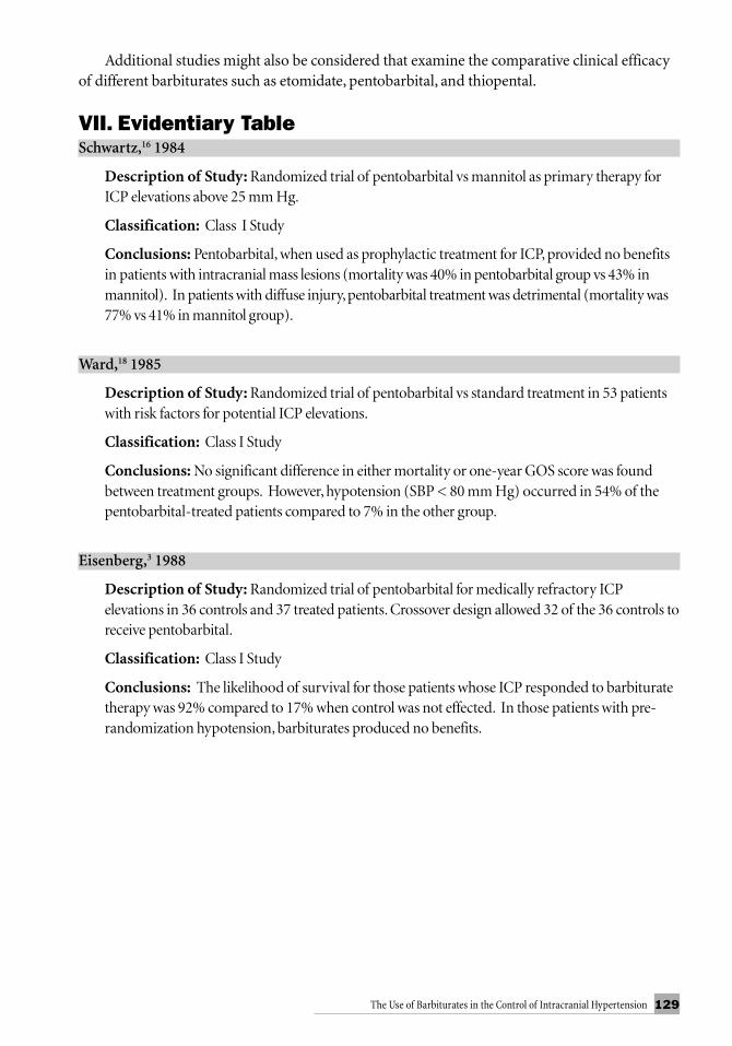
129The Use of Barbiturates in the Control of Intracranial Hypertension
Additional studies might also be considered that examine the comparative clinical efficacyof different barbiturates such as etomidate, pentobarbital, and thiopental.
VII. Evidentiary TableSchwartz,16 1984
Description of Study: Randomized trial of pentobarbital vs mannitol as primary therapy forICP elevations above 25 mm Hg.
Classification: Class I Study
Conclusions: Pentobarbital, when used as prophylactic treatment for ICP, provided no benefitsin patients with intracranial mass lesions (mortality was 40% in pentobarbital group vs 43% inmannitol). In patients with diffuse injury, pentobarbital treatment was detrimental (mortality was77% vs 41% in mannitol group).
Ward,18 1985
Description of Study: Randomized trial of pentobarbital vs standard treatment in 53 patientswith risk factors for potential ICP elevations.
Classification: Class I Study
Conclusions: No significant difference in either mortality or one-year GOS score was foundbetween treatment groups. However, hypotension (SBP < 80 mm Hg) occurred in 54% of thepentobarbital-treated patients compared to 7% in the other group.
Eisenberg,3 1988
Description of Study: Randomized trial of pentobarbital for medically refractory ICPelevations in 36 controls and 37 treated patients. Crossover design allowed 32 of the 36 controls toreceive pentobarbital.
Classification: Class I Study
Conclusions: The likelihood of survival for those patients whose ICP responded to barbituratetherapy was 92% compared to 17% when control was not effected. In those patients with pre-randomization hypotension, barbiturates produced no benefits.

130 Guidelines for the Management of Severe Traumatic Brain Injury
VIII. References1 Cruz J. Adverse effects of pentobarbital on cerebral venous oxygenation of comatose
patients with acute traumatic brain swelling: relationship to outcome. J Neurosurg 85: 758-761, 1996.
2. Demopoulous HB, Flamm ES, Pietronigro DD, et al: The free radical pathology and themicrocirculation in the major central nervous system. Acta Physiol Scand (Suppl) 492:91-119,1980.
3. Eisenberg HM, Frankowski RF, Contant CF, et al.: High-dose barbiturate control of elevatedintracranial pressure in patients with severe head injury. J Neurosurg 69:15-23, 1988.
4. Goodman JC, Valadka AB, Gopinath SP, et al.: Lactate and excitatory amino acids measuredby microdialysis are decreased by pentobarbital coma in head-injured patients. J Neurotrauma13:549-556, 1996.
5. Horsley JS: The intracranial pressure during barbital narcosis. Lancet 1:141-143, 1937.6. Kassell NF, Hitchon PW, Gerk MK, et al.: Alterations in cerebral blood flow, oxygen
metabolism, and electrical activity produced by high-dose thiopental. Neurosurg 7:598-603,1980.
7. Langfitt TW, Gennarelli TA: Can the outcome from head injury be improved? J Neurosurg56:19-25, 1982.
8. Levy ML, Aranda M, Zelman V, et al.: Propylene glycol toxicity following continuousetomidate infusion for the control of refractory cerebral edema. Neurosurg 37:363-371, 1995.
9. Lobato RD, Sarabia R, Cordobes C, et al.: Post-traumatic cerebral hemispheric swelling.Analysis of 55 cases studied by CT. J Neurosurg 68:417-423, 1988.
10. Marshall LF, Smith RW, Shapiro HM: The outcome with aggressive treatment in severe headinjuries. J Neurosurg 50:26-30, 1979.
11. Miller JD, Butterworth JF, Gudeman SK, et al.: Further experience for the management ofsevere head injury. J Neurosurg 54:289-299, 1981.
12. Narayan RK, Kishoure PRS, Becker DP, et al.: Intracranial pressure: to monitor or not tomonitor. A review of our experience with severe head injury. J Neurosurg 56:650-659, 1982.
13. Nordstrom GH, Messeter K, Sundberg B, et al.: Cerebral blood flow, vasoreactivity, andoxygen consumption during barbiturate therapy in severe traumatic brain lesions. JNeurosurg 68:424-431, 1988.
14. Rea GL, Rockswold GL: Barbiturate therapy in uncontrolled intracranial hypertension.Neurosurg 12:401-405, 1983.
15. Rockoff MA, Marshall LF, Shapiro HM: High-dose barbiturate therapy in humans: a clinicalreview of 60 patients. Ann Neurol 6:194-199, 1979.
16. Schwartz M, Tator C, Towed D, et al.: The University of Toronto Head Injury TreatmentStudy: a prospective, randomized comparison of pentobarbital and mannitol. Can J NeurolSci 11:434-440, 1984.
17. Shapiro HM, Wyte SR, Loeser J: Barbiturate augmented hypothermia for reduction ofpersistent intracranial hypertension. J Neurosurg 40:90-100, 1979.
18. Ward JD, Becker DP, Miller JD, et al.: Failure of prophylactic barbiturate coma in thetreatment of severe head injury. J Neurosurg 62:383-388, 1985.

131The Role of Steroids
I. RecommendationsA. Standards
The use of steroids is not recommended for improving outcome or reducingintracranial pressure (ICP) in patients with severe head injury.
B. GuidelinesNone
C. OptionsNone
II. OverviewSteroids were introduced in the early 1960s as a treatment for brain edema. Experimentalevidence accumulated that steroids were useful in the restoration of altered vascularpermeability in experimental brain edema,20 reduction of cerebrospinal fluid production,24
attenuation of free radical production, and other beneficial effects in experimentalmodels.3,4,15,17,20,21 The administration of glucocorticoids to patients with brain tumors oftenresulted in marked clinical improvement, and glucocorticoids were found to be beneficial whenadministered in the perioperative period to patients undergoing brain tumor surgery. Frenchand Galicich reported a strong clinical benefit of glucocorticoids in cases of brain edema andfound glucocorticoids especially beneficial in patients with brain tumors.9 Renaudin, et al., in1973 reported a beneficial effect of high-dose glucocorticoids in patients with brain tumor whowere refractory to conventional doses.22
Glucocorticoids became commonly administered to patients undergoing a variety ofneurosurgical procedures and became commonplace in the treatment of severe head injury.Gobiet, et al., in 1976 compared low-dose and high-dose Decadron to a previous control groupof severely head-injured patients and reported it to be of benefit in the high-dose group.12
Faupel, et al., in 1976 performed a double-blind trial and reported a favorable dose-relatedeffect on mortality in head-injured patients using glucocorticoid treatment.8 Subsequently, sixmajor studies of glucocorticoid in severe head injury were conducted that evaluated clinicaloutcome ICP or both. None of these studies showed a substantial benefit of glucocorticoidtherapy in these patients.2,5,6,11,14,23 More recently, trials in head-injured patients have beencompleted using the synthetic glucocorticoid, triamcinolone,13 the 21-aminosteroid tirilazad,7,19
and also a trial using ultra-high-dose dexamethasone.10 None of these trials have indicated an
THE ROLE OF STEROIDS

132 Guidelines for the Management of Severe Traumatic Brain Injury
overall beneficial effect of steroids on outcome. Moreover a recent meta-analysis of trials ofsteroids in head injury has revealed no overall beneficial effect of steroids on outcome.1
III. ProcessA computer search was performed using MEDLINE for the period from 1966 to 1998 by usingthe key terms “head injury” and “steroids.” A total of 60 documents were found. In addition,reference lists from the major clinical trials of steroid treatment in head injury and a recentmeta-analysis of trials of steroids in head injury were examined. All clinical studies of steroidsand head injury in humans were examined and reviewed in detail.
IV. Scientific FoundationGudeman, et al., reported the effects of high-dose methylprednisolone on ICP and volume-pressure response in 20 patients with severe head injury.14 Patients were given 40 mg ofmethylprednisolone every 6 hours for the first 24 hours and the dose was increased to 2 gloading dose and 500 mg methylprednisolone every 6 hours for the following 24 hours. Therewas no significant change in the ICP or volume-pressure response between the two intervals.There was a 50% incidence of gastrointestinal bleeding and an 85% incidence of hyperglycemiain the treated group.
Cooper, et al., in 1979 reported a prospective, double-blind study of dexamethasone inpatients with severe head injury.5 Ninety-seven patients were stratified for severity and treatedwith placebo, low-dose dexamethasone 60 mg/day, or high-dose dexamethasone 96 mg/day.Seventy-six patients were available for clinical follow-up and ICP was measured in 51 patients.The results showed no difference in outcome, ICP, or serial neurologic examinations among thegroups.
Saul, et al., in 1981 reported a prospective, randomized clinical trial in 100 patients. Onegroup received methylprednisolone 5 mg/kg/day versus a control group that received no drug.23
There was no statistically significant difference in outcome between the treated and non-treatedgroups at 6 months. This study reports a benefit of steroids in a small group of patients basedon a subgroup analysis. This analysis indicated that in the subgroup of patients who improvedduring the first 3 days after head injury, the steroid-treated group fared better when comparedwith the placebo group.
Braakman, et al., reported the results of a large prospective, double-blind trial on the effectof dexamethasone on severely head-injured patients in 1983.2 A total of 161 patients wererandomized to placebo or high-dose dexamethasone (100 mg/day) followed by a tapering dose.There was no difference in 1 month survival or 6-month outcome between the two groups.
Giannotta, et al., in 1984 reported a prospective, double-blind clinical trial of 88 patientscomparing placebo, low-dose methylprednisolone 1.5 mg/kg loading, followed by a tapering dose,with high-dose methylprednisolone 30 mg/kg loading, followed by a tapering dose.14 The data didnot show a beneficial effect of either low-dose or high-dose methylprednisolone compared withplacebo. Subgroup analysis revealed an increased survival and improved speech function in patientsunder age 40 when the high dose was compared against the low dose and placebo groups combined.
Dearden, et al., in 1986, reported the results of a prospective, double-blind study on theeffect of high-dose dexamethasone on outcome and ICP in 130 severely head-injured patients.6
Patients were randomized to receive drug or placebo. No differences in ICP trends or 6-monthoutcome were seen.
133The Role of Steroids
Gaab, et al., in 1994, reported the results of a prospective, randomized, double-blindmulticenter trial on the efficacy and safety of ultra-high-dose dexamethasone on outcome inpatients with moderate and severe head injury.10 The trial enrolled 300 patients, randomized toplacebo or dexamethasone: 500 mg within 3 hours of injury, followed by 200 mg after 3 hours,then 200 mg every 6 hours for 8 doses for a total dexamethasone dose of 2.3 g, given within 51hours. Glasgow Outcome Scale (GOS) score at 10-14 months following injury, and also timefrom injury until Glasgow Coma Scale (GCS) score reached 8 or greater, were used as primaryendpoints. The results of the trial revealed no differences between placebo and drug-treatedpatients in either primary endpoints. This trial has the advantages of having a large number ofpatients who were treated early following injury, and with very high doses of medication.
Grumme, et al., in 1995, reported the results of a prospective, controlled, multicenter trial ofthe synthetic corticosteroid, triamcinolone on outcome in severely head-injured patients.13 Thistrial randomized 396 patients to either placebo or steroids using triamcinolone 200 mg within 4hours of injury followed by 40 mg every 8 hours for 4 days, followed by 20 mg every 8 hours for4 days. The primary outcome measures were GOS at the time of discharge and at 12 monthsfollowing injury. There was a trend toward better outcome in the steroid treated group, whichwas not statistically significant. Subgroup analysis revealed a better outcome in patients withfocal lesions who had GCS scores of less than 8. The improvement in outcome in this subgroupwas reported as significant. Multiple subgroups were analyzed, however, and if the results arecorrected for multiple comparisons, then the difference in outcome in this subgroup is notstatistically significant.
Marshall, et al., in 1998,19 and Kassell, et al., in 199618, reported the results of two largeprospective, randomized, controlled trials of the synthetic 21-amino steroid, tirilazad mesylate,on outcome in severely head-injured patients, one in North America and the other in Europeand Australia.7 There is experimental evidence that this compound may be more effective thanglucocorticoids against specific mechanisms that occur in brain injury, and higher doses can beused without glucocorticoid side effects.15,16 The North American trial enrolled 1,170 patientsand the European trial enrolled 1,128 patients. No overall benefit on outcome in head-injuredpatients was detected in either trial. Subgroup analysis in the European trial indicated that malepatients with subarachnoid hemorrhage on their initial CT scan, who were on active drug, had abetter outcome.
Alderson, et al., in 1997, reported the results of a systematic review of randomizedcontrolled trials of corticosteroids in acute traumatic brain injury (TBI).1 Many of the trialsmentioned above, as well as additional unpublished data, were included in this analysis. Thedata presented indicate that there was no evidence for a beneficial effect of steroids to improveoutcome in head-injured patients. Analysis of the trials with the best blinding of groupsrevealed the summary odds ratio for death was 1.04 (0.83 to 1.30), and for death and disabilitywas 0.97 (0.77 to 1.23). The authors stated that a lack of benefit from steroids remaineduncertain, and recommended that a larger trial of greater than 20,000 patients to detect apossible beneficial effect of steroids be conducted.

134 Guidelines for the Management of Severe Traumatic Brain Injury
V. SummaryThe majority of available evidence indicates that steroids do not improve outcome or lower ICPin severely head-injured patients. The routine use of steroids is not recommended for thesepurposes.
VI. Key Issues for Future InvestigationData analysis from the trials of the 21-aminosteroid tirilizad is ongoing. It is possible that basedon the results of subgroup analysis from these trials further studies, such as in patients withtraumatic subarachnoid hemorrhage, may be proposed. A future trial in patients with moresevere head injuries with focal lesions may determine if this subgroup of patients may benefitfrom triamcinolone.
VII. Evidentiary TableBraakman,2 1983
Description of Study: Prospective, double-blind study, randomized 161 patients with severehead injury to placebo or high-dose dexamethasone (100 mg day followed by tapering dose).
Classification: Class I Study
Conclusions: No significant difference in 1-month survival of 6-month outcome.
Active Treatment PlaceboDead 54 % 58%Good recovery 17.3 15
Cooper,5 1979
Description of Study: Prospective, double-blind study of 97 patients with severe head injury,stratified for severity, and treated with placebo 60 mg/day or 96 mg/day of dexamethasone; 76patients available for follow-up at 6 months.
Classification: Class I Study
Conclusions: No significant difference was seen in 6-month outcome, serial neurological exams,or ICP.
Low Dose High Dose PlaceboBad outcome 56% 71% 63%(SD, PVD, D)Good outcome 44 29 37(GR, MD)
Abbreviations: GR, good recovery; MD, moderate disability; SD, severe disability; PVS, persistent vegetative state; D, dead

135The Role of Steroids
Dearden, 6 1986
Description of Study: Prospective, double-blind study of 130 patients with severe head injury,randomized to high-dose dexamethasone vs placebo. All patients were followed and outcomewas analyzed. ICP trends in the two groups were also analyzed.
Classification: Class I Study
Conclusions: No significant difference in ICP trends or 6-month outcome between control andtreated groups was found.
Active Treatment PlaceboDead 49% 35.5%Good Recovery 32 35.5
Faupel, 8 1976
Description of Study: Prospective, double-blind trial of dexamethasone vs placebo in 95patients with severe head injury.
Classification: Class I Study
Conclusions: Significant improvement in mortality in steroid-treated group; however, overalloutcome was not improved. Of the active treatment groups, 25.4% were vegetative and 11.9%were severely disabled vs 3.6% and 7.1% in the control group.
Active Treatment PlaceboGood Outcome 39% 32%(GR, MD, SD)Dead 24 57Dead or Vegetative 49 61
Gaab,10 1994
Description of Study: Prospective, randomized, double-blind, multicenter trial of ultra-high-dose dexamethasone on outcome in 300 patients with moderate and severe head injury,randomized to placebo or dexamethasone: 500 mg within 3 hours of injury, followed by 200 mgafter 3 hours, then 200 mg every 6 hours for 8 doses for a total dexamethasone dose of 2.3 g, givenwithin 51 hours.
Classification: Class I Study
Conclusions: No significant difference in 12-month outcome or in time to improvement to GCSscore greater than or equal to 8 in treatment group compared with placebo.
Active Treatment PlaceboDead 14.3% 15.4%Good Recovery 61.7 57.4
VII. Evidentiary Table (continued)

136 Guidelines for the Management of Severe Traumatic Brain Injury
Grumme,13 1995
Description of Study: Prospective, controlled, multicenter trial randomized 396 severely head-injured patients to either placebo or steroids using triamcinolone 200 mg within 4 hours of injuryfollowed by 40 mg every 8 hours for 4 days, followed by 20 mg every 8 hours for 4 days.
Classification: Class I Study
Conclusions: A trend toward better outcome in treated group, but no significant difference in12-month outcome or at the time of discharge from the hospital in treatment group comparedwith placebo.
At discharge Active treatment PlaceboDead 16 % 21 %Good recovery 49.2 40.7
Giannotta,11 1984
Description of Study: Prospective, double-blind study of 88 patients with severe head injury.Patients randomized to placebo, low-dose methylprednisolone (30 mg/kg/day) or high-dosemethylprednisolone (100 mg/kg/day).
Classification: Class I Study
Conclusions: No significant difference in 6-month outcome in treatment groups compared withplacebo. Subgroup analysis showed improved survival and speech function in patients under age40 when high-dose group was compared to low-dose and placebo groups combined.
Low Dose High Dose PlaceboDead 55.8% 39.4% 43.7%Good Recovery 14.7 18.4 25
Gobiet,12 1976
Description of Study: Cohort study of 93 head-injured patients. Compared low-dose andhigh-dose dexamethasone to cohort retrospectively.
Classification: Class II Study
Conclusions: Reported significant benefit in high-dose group on mortality.
Low Dose High Dose ControlDead 41.5% 23% 45.5%
VII. Evidentiary Table (continued)

137The Role of Steroids
Gudeman,14 1979
Description of Study: Examined the effect of two doses of methylprednisolone on ICPparameters only in 20 patients with severe head injury. Patients served as their own controls andtwo separate intervals were analyzed.
Classification: Class II Study
Conclusions: No significant difference in ICP or volume-pressure response was seen during aninterval when the methylprednisolone dose was increased.
Marshall, 19 1998 and Kassell,18 1996
Description of Study: Two large prospective, randomized, controlled trials of the synthetic 21-amino steroid, tirilizad mesylate, on outcome in severely head-injured patients, one in NorthAmerica (1,120 patients) and the other in Europe and Australia (1,023 patients).
Classification: Class I Study
Conclusions: No overall benefit on outcome in head-injured patients was detected in either trial.Subgroup analysis in the European trial indicated that male patients with subarachnoid hemorrhageon their initial CT scan, who were on active drug, had a better outcome. Final data analysis is pending.
Saul,23 1981
Description of Study: Prospective, double-blind study of 100 patients with severe head injury,randomized to placebo or methylprednisolone 5 mg/kg/day.
Classification: Class I Study
Conclusions: No significant difference in outcome at 6 months. A subgroup of responders wasidentified who did better with active treatment; however, the trial was not designed to examinethis group.
Active Treatment PlaceboDead or 38% 48%VegetativeGood Recovery 62 52 or Disabled (GR, MD, SD)
VIII. References1. Alderson P, Roberts I: Corticosteroids in acute traumatic brain injury: systematic review of
randomised controlled trials. Brit Med J 314:1855-1859, 1997.2. Braakman R, Schouten HJA, Blaauw-van DM, et al.: Megadose steroids in severe head
injury. J Neurosurg 58:326-330, 1983.3. Bracken MB, Shepard MJ, Collins WF, et al.: A randomized, controlled trial of methyl-
prednisolone or naloxone in the treatment of acute spinal-cord injury. Results of theNational Acute Spinal Cord Injury Study. J Neurosurg 63:704-713, 1985.
VII. Evidentiary Table (continued)

138 Guidelines for the Management of Severe Traumatic Brain Injury
4. Bracken MB, Shepard MJ, Collins WF, et al.: A randomized, controlled trial of methyl-prednisolone or naloxone in the treatment of acute spinal-cord injury. Results of the SecondNational Acute Spinal Cord Injury Study. N Engl J Med 322:1405-1411, 1990.
5. Cooper PR, Moody S, Clark WK, et al.: Dexamethasone and severe head injury. Aprospective double-blind study. J Neurosurg 51:307-316, 1979.
6. Dearden NM, Gibson JS, McDowall DG, et al.: Effect of high-dose dexamethasone onoutcome from severe head injury. J Neurosurg 64:81-88, 1986.
7. Doppenberg EMR, Bullock R: Clinical neuro-protection trials in severe traumatic braininjury: lessons from previous studies. J Neurotrauma 14:71-80, 1997.
8. Faupel G, Reulen HJ, Muller D, et al.: Double-blind study on the effects of steroids on severeclosed head injury. In: Pappius HM, Feindel W (eds): Dynamics of Brain Edema Springer-Verlag:New York, 337-343, 1976.
9. French LA, Galicich JH: The use of steroids for control of cerebral edema. Clin Neurosurg10:212-223, 1964.
10. Gaab MR, Trost HA, Alcantara A, et al.: “Ultrahigh” dexamethasone in acute brain injury. Resultsfrom a prospective randomized double-blind multicenter trial (GUDHIS). German UltrahighDexamethasone Head Injury Study Group. Zentralblatt Fur Neurochirurgie 55:135-143, 1994.
11. Giannotta SL, Weiss MH, Apuzzo MLJ, et al.: High-dose glucocorticoids in the managementof severe head injury. Neurosurg 15:497-501, 1984.
12. Gobiet W, Bock WJ, Liesgang J, et al.: Treatment of acute cerebral edema with high dose ofdexamethasone. In: Beks JWF, Bosch DA, Brock M (eds): Intracranial Pressure III Springer-Verlag: New York, 231-235, 1976.
13. Grumme T, Baethmann A, Kolodziejczyk D, et al.: Treatment of patients with severe headinjury by triamcinolone: a prospective, controlled multicenter clinical trial of 396 cases.Research in Experimental Medicine (Berlin) 195:217-229, 1995.
14. Gudeman SK, Miller JD, Becker DP: Failure of high-dose steroid therapy to influenceintracranial pressure in patients with severe head injury. J Neurosurg 51:301-306, 1979.
15. Hall ED: The neuroprotective pharmacology of methylprednisolone. J Neurosurg 76:13-22, 1992.16. Hall ED, Wolf DL, Braughler JM: Effects of a single large dose of methylprednisolone
sodium succinate on experimental post-traumatic spinal cord ischemia. Dose-response andtime-action analysis. J Neurosurg 61:124-130, 1984.
17. Hall ED, Yonkers PA, McCall JM, et al.: Effects of the 21-aminosteroid U74006F onexperimental head injury in mice. J Neurosurg 68:456-461, 1988.
18. Kassell NF, Haley EC, et al.: Randomized, double-blind, vehicle-controlled trial of tirilazadmesylate in patients with aneurysmal subarachnoid hemorrhage: a cooperateve study inEurope, Australia, and New Zealand. J Neurosurg 84:221-228, 1996.
19. Marshall LF, Maas AL, Marshall SB, et al.: A multicenter trial on the efficacy of usingtirilazad mesylate in cases of head injury. J Neurosurg 89: 519-525, 1998.
20. Maxwell RE, Long DM, French LA: The effects of glucosteroids on experimental cold-induced brain edema: gross morphological alterations and vascular permeability changes. JNeurosurg 34:477-487, 1971.
21. Pappius HM, McCann WP: Effects of steroids on cerebral edema in cats. Arch Neurol 20:207-216, 1969.
22. Renaudin J, Fewer D, Wilson CB, et al.: Dose dependency of Decadron in patients withpartially excised brain tumors. J Neurosurg 39:302-305, 1973.
23. Saul TG, Ducker TB, Salcman M, et al.: Steroids in severe head injury. A prospective,randomized clinical trial. J Neurosurg 54:596-600, 1981.
24. Weiss MH, Nulsen FE: The effect of glucocorticoids on CSF flow in dogs. J Neurosurg32:452-458, 1970.

139Critical Pathway for the Treatment of Established Intracranial Hypertension
I. IntroductionA critical pathway, developed by consensus, is presented in Figure 1. We developed a treatmentalgorithm for established intracranial hypertension wherein the order of steps is determined bythe risk/benefit ratio of individual treatment maneuvers. The considerations involved areoutlined in the chapter specific to each step.
As discussed in the section on intracranial pressure (ICP) treatment threshold, the absolutevalue defining unacceptable intracranial hypertension is unclear. Although a general thresholdof 20-25 mm Hg has been presented, there will be situations where such pressures are too highas well as instances where higher ICP values are acceptable. These considerations are relevant tothe decision to pursue any step in the escalated treatment of ICP.
This critical pathway is a committee consensus and, therefore, must be viewed as Class III(“expert opinion”) evidence. As such, it should be interpreted as a framework that may be usefulin guiding an approach to treating intracranial hypertension. It can and should be modified inan individual case by any circumstances unique to the patient as well as by the response of theICP to individual treatment steps.
II. Critical PathwayA number of general maneuvers may be applied together early during the treatment ofintracranial hypertension. These include control of body temperature, seizure prophylaxis,elevation of the head of the bed, avoidance of jugular venous outflow obstruction, sedation ±pharmacologic paralysis, maintenance of adequate arterial oxygenation, and complete volumeresuscitation to a cerebral perfusion pressure (CPP) of 70 mm Hg or more.
When a ventricular catheter is being used for ICP monitoring, cerebrospinal fluid (CSF)drainage should be used first for ICP elevations. Ventilation may be adjusted to establish aPaCO
2 at the low end of eucapnea (35 mm Hg).
When intracranial hypertension is maintained despite initiation of these generalmaneuvers, treatment modalities with a lower ratio of risk to benefit may be considered. If CSFdrainage is ineffective in controlling ICP or is not available, the level of ventilation may beincreased to obtain PaCO
2 levels of 30-35 mm Hg (0-5 mm Hg below the lower threshold of
eucapnea). If available, measurement of cerebral blood flow or jugular venous saturation shouldbe considered when hyperventilation is further increased. If mild hypocapnia is ineffective incontrolling ICP, mannitol can be employed, limited by serum osmolarity levels of 320 mOsm/l.
CRITICAL PATHWAY FOR THETREATMENT OF ESTABLISHEDINTRACRANIAL HYPERTENSION

140 Guidelines for the Management of Severe Traumatic Brain Injury
The patient’s volume status should be closely observed during mannitol administration andeuvolemia to mild hypervolemia maintained by careful fluid replacement. At all times duringthe treatment of intracranial hypertension, the possibility of a surgical mass or an intracraniallesion should be considered. Therefore, under conditions of intractability or loss of ICP controlor when second tier therapy is being contemplated, consideration should be given to repeating aCT scan.
For intracranial hypertension refractory to the above techniques, second tier therapiesshould be considered when it is the physician’s opinion that the patient may benefit if ICPcontrol can be accomplished. Second tier therapy includes both treatment modalities that havebeen proven effective in improving outcome, but have very significant complication rates (e.g.,barbiturates). Additional second tier therapies are those that appear to effectively lower ICP, butremain unproven in their influence on outcome or the exact magnitude of their risk/benefitratio. These latter modalities include hyperventilation to PaCO
2 less than 30 mm Hg,
decompressive craniectomy, and hypertensive therapy. Barbiturates, the most thoroughlystudied second tier approach, are covered here in a separate chapter. Ventilation to low PaCO
2
values is addressed in the hyperventilation section. The precise indications and implementationof second tier therapies in an individual patient are left to the discretion of the managingphysician.

141Critical Pathway for the Treatment of Established Intracranial Hypertension
© Brain Trauma Foundation, Inc.


143Nutrition
I. RecommendationsA. Standards
There are insufficient data to support a treatment standard for this topic.B. Guidelines
Replace 140% of resting metabolism expenditure in non-paralyzed patients and100% of resting metabolism expenditure in paralyzed patients using enteral orparenteral formulas containing at least 15% of calories as protein by the seventh dayafter injury.
C. OptionsThe preferable option is use of jejunal feeding by gastrojejunostomy due to ease ofuse and avoidance of gastric intolerance.
II. OverviewThe metabolic response to severe brain injury was systematically documented beginning in theearly 1980s. There were occasional case reports of hypermetabolism in brain injury in prioryears (Class III data). Up to this time a casual attitude toward nutritional replacement was usualin clinical practice. This was based on the assumption that, due to coma, metabolicrequirements were reduced. Most of the research done beginning in 1980 consisted ofmeasurements of metabolic expenditure, nitrogen loss, and cardiovascular parameters. Thesevalues were compared to well-established normative data and to the patterns documented inother injured patient groups (Class II data). Hypermetabolism and nitrogen wasting were welldocumented. At least 12 Class I studies have been completed. Nine Class I studies examined theeffect of amount of feeding, type of feeding, route of feeding, and steroids on nitrogen balanceand serum biochemistries. These studies made no statements regarding patient outcome(deleted phrase).
One Class I study examined the effect of IGF-1 on the catabolic state and on outcome.18
Two Class I studies examined the effect of the extent of nutritional replacement on patientoutcome.31,39 These showed that with nearly equivalent quantities of feeding, the mode ofadministration (parenteral or enteral) had no effect on neurologic outcome. Conflicting resultswere found with infection rate and nitrogen retention. One study reported that malnutritionincreased mortality rate in head-injured patients. This study’s methodology can be questioneddue to an unexpectedly low mortality rate in the higher nutrition group and an unexpectedly
NUTRITION

144 Guidelines for the Management of Severe Traumatic Brain Injury
high mortality rate in the low nutrition group. It was judged that this study did not have theforce to establish a standard.
III. ProcessA MEDLINE search of the categories “brain injury” and “nutrition” was conducted for the years1975 through 1997, and all publications published in English were reviewed. Only articlesdiscussing nutritional data in patients with head injury were referenced in evidentiary tablesand used as the primary source for conclusions. The methodology, results, and conclusions ofeach of these 29 references were studied.
IV. Scientific Foundation1) The physician should aim for replacement of 140% of resting metabolism expenditure in
non-paralyzed patients and 100% resting metabolism expenditure in paralyzed patients.A number of publications have dealt with energy requirements after head injury.
2,4,6,8,9,10,13,14,17,25,27,33,34,35,38,41,43,47,49 The technique for measurement is indirect calorimetry,which measures the rate of oxygen utilization and gives energy expenditure by the knowncaloric yield of one liter of oxygen. Because caloric expenditure varies with age, sex, andbody surface area, metabolic expenditure is expressed as a percent of normal at rest for agiven patient. This value for each patient can be found in standard tables. Data from mostinvestigators measuring metabolic expenditure in rested comatose patients with isolatedhead injury yielded a mean increase of approximately 140% of the expected metabolicexpenditure with variations from 120%-250% of that expected. Of importance, only thedata of Young, et al., 47 and Deutschman, et al., 9 are of nonsteroid-treated patients, andthose agree with other calorimetric data in steroid-treated, head-injured patients.
Researchers found that, in head-injured patients, paralysis with pancuronium bromide orbarbiturate coma decreased metabolic expenditure from a mean of 160% of that expected to100%-120%. This finding suggests that a major part of the increased metabolic expenditure isrelated to muscle tone. Even with paralysis, energy expenditure remained elevated by 20%-30% in some patients.4 In the first two weeks after injury, energy expenditure seems to riseregardless of neurological course. The duration of hypermetabolism beyond the first twoweeks is not known.
At some point, the pathologically increased caloric requirements fueled by increasedmuscle tone and an altered hormonal milieu are replaced by the requirements that wouldnormally accompany increased activity as the patient improves. Routine use of calorimetryhas been recommended because of the high variability among patients and the relativelypoor ability of predictive formulas to guide acute nutritional requirements.43 The method iscumbersome and is rarely used in routine clinical practice. The resting metabolicexpenditure for a 70 kg, 25-year-old male is 1,700 kcal/24 hr and for a 50 kg, 50-year-oldfemale is 1,200 kcal/24 hr. Projected caloric needs would, therefore, be 2,400 kcal/24 hr for a70 kg male.
Three randomized (Class I) studies have evaluated the relationship of level of caloricintake to patient outcomes.16,36,48 Rapp showed that the consequence of severe undernutritionfor a two-week period after injury was an increased mortality rate as compared to fullreplacement of measured calories by seven days.36 In a subsequent study of brain injuredpatients, Young, et al., showed that with full replacement at three days after injury in the early
145Nutrition
group (fed parenterally) as opposed to nine days after injury in the late feeding group (fedenterally), there were no changes in morbidity. 48 Patient outcome was better at three monthsbut not at six months. Hadley randomized 45 patients with severe head injury to receiveenteral or parenteral nutrition. While caloric intake between groups was not significantlydifferent, the parenteral group had significantly better nitrogen intake.16
To achieve full caloric replacement by 7 days after injury, nutritional replacement isusually begun no later than 72 hours after injury. This is so because 2-3 days are required togradually increase feedings to full replacement whether feeding is by jejunal or gastric route.5,8,15,37,48 Intravenous hyperalimentation must also be started at levels below restingmetabolism expenditure and advanced over 3 days. Whichever method is used in order toachieve full replacement, feedings are usually begun within 72 hours of injury.
2) Use an enteral or parenteral formula containing 15% of calories as protein. Nitrogenbalance is defined as the difference between nitrogen intake and nitrogen excretion. Foreach gram of nitrogen measured in the urine (plus fecal loss), 6.25 g of protein have beencatabolized. In normal and injured man, optimal protein utilization has been found to beheavily dependent upon the adequacy of caloric intake. Catabolism of protein, which yieldsonly 4 kcal/g (as opposed to fat, which yields 8 kcal/g), makes up 10% or less of consumedcalories in normal man.12 After severe brain injury, not only do energy requirements risegreatly, but nitrogen excretion does also. The contribution of protein to consumed caloriesafter head injury rises to levels as high as 30% (given 10 g/day nitrogen intake and fullcaloric replacement).12 In a fasting normal human, nitrogen catabolism drops to levels aslow as 3-5 gN/day. Fasting patients with severe brain injury continue to lose 14-25 gN/day,however.14 The peak in nitrogen excretion appears to occur in the second week withimprovement in nitrogen retention by the third week.
It is only after the third week that nitrogen balance can be achieved. Two publicationsaddress the question of steroid effect on nitrogen excretion and report that increasednitrogen excretion after brain injury is not the result of steroid administration.37,47
One can estimate the potential sequelae of these levels of nitrogen loss from thefollowing facts. A 30% preoperative weight loss increased the morbidity and mortality ofgastric surgery by tenfold.41 It is, therefore, generally assumed that a 10%-15% weight lossin a bedfast patient is of little consequence, but that a 30% weight loss is potentially verydeleterious. The average nitrogen loss of the fasting head-injured patient is 0.2 gN/kg/day(14 gN in 70 kg male), about double or triple the loss in the normal person, with values offasting nitrogen loss of up to 25 gN/day frequently being seen. This level of nitrogen losswill produce a 10% decrease in lean mass in seven days; hence, underfeeding for a two-to-three week period could result in a 30% weight loss.
The desired level of reduction of nitrogen loss has not been quantified but is the subjectof Class I studies.5,11,15,19,22 These studies have not, however, examined patient outcome. Therelative gains in nitrogen balance achieved with high protein feeding can be illustrated. Twomatched groups of comatose head-injured patients were treated by caloric replacementwith intakes of 17.6 ± 3.6 gN/day and 29.0 ± 5.3 gN/day, respectively, at 140% replacementof expended calories.5 Data are from 7-day balance periods within the first two weeks afterinjury. A nitrogen balance of -9.2 ± 6.7 g/day was found in the lower protein group and abalance of -5.3 ± 5.0 g/day in the higher protein group. These data suggest that at a high

146 Guidelines for the Management of Severe Traumatic Brain Injury
range of nitrogen intake (> 17 g/day), less than 50% of administered nitrogen is retainedafter head injury. Therefore, the level of nitrogen intake that generally results in less than 10 gnitrogen loss per day is 15-17 gN/day or 0.3-0.5 gN/kg/day. As it turns out, this value isabout 20% of the caloric composition of a 50 kcal/kg/day feeding protocol. Twenty percentis the maximal protein content of most enteral feedings designed for the hypermetabolicpatient. Twenty percent is the maximal amino acid content of most parenteral formulationsfor trauma patients, which generally contain more than 15% protein calories.
3) Jejunal feeding by gastrojejunostomy avoids gastric intolerance found in gastric feeding andthe use of intravenous catheters required in total parenteral nutrition.
There are three options for the method of feeding. Some reports indicate that jejunaland parenteral replacement produce better nitrogen retention than gastric feeding. 15,16,19,48
Gastric alimentation has been used by some investigators.5,37,48 Others have found alteredgastric emptying or lower esophageal sphincter dysfunction to complicate gastricfeeding.27,28,29,40 There has been one Class I report and one Class II report indicating bettertolerance of enteral feeding with jejunal rather than gastric administration.5,22 In studies ofboth gastric and jejunal administration, it has been possible to achieve full caloric feedingin most patients by seven days after injury.5,15,22,48
Jejunal alimentation by endoscopic or fluroscopic, not blind, placement has practicaladvantages over gastric feeding. A higher percentage of patients tolerates jejunal rather thangastric feeding early after injury and less risk of aspiration is reported (presumably afterextubation).15,26,29,40 Increasingly parenteral nutrition is started early after injury until eithergastric feedings are tolerated or a jejunal feeding tube can be placed or a procedure team isused to effect very early jejunal tube placement.1,29
Three potential advantages of enteral feeding are:1. Less risk of hyperglycemia than with parenteral feeding2. Lower theoretical risk of infection3. Lower cost
Hyperglycemia has been shown to aggravate hypoxic ischemic brain injury in anextensive experimental literature. One study in experimental cortical contusion injury hasshown that hyperglycemia exacerbates cortical contusion injury with superimposedischemia.3 In two Class II studies, hyperglycemia has been associated with worsenedoutcome. 24,46 Parenteral nutrition has not, however, been shown to aggravatehyperglycemia, though more insulin is required to maintain normoglycemia withparenteral nutrition than with enteral nutrition.42
The risk of infection has not been shown to be increased with parenteral nutrition ascompared to enteral nutrition in brain injured patients.1,46 Literature about patients withother kinds of trauma, however, suggests that use of enteral feedings reduces septiccomplications related to gut mucousal integrity and may decrease hypermetabolism. 20,21,23
Class I data in head injury patients indicates less insulin dependence and earlierimprovement in visceral proteins with enteral nutrition than with parenteral nutrition.42
Another Class I study, however, has shown no difference in recovery of visceral proteins. 1
The primary advantage of parenteral nutrition is that it is well tolerated. While in
147Nutrition
laboratory animals parenteral nutrition may aggravate brain swelling, this has not been aclinical problem.7,44,46 No clearly superior method of feeding has been demonstrated eitherin terms of nitrogen retention, complications, or outcome.
The daily cost of parenteral nutrition is higher than enteral nutrition.1,29 The overallcost of care has not, however, been increased by use of parenteral nutrition.1, 29 Thisdiscrepancy could be related to increased costs associated with problems associated withplacement or replacement of enteral feeding tubes.29
4) New data in a Class I study of 33 patients has shown achievement of positive nitrogenbalance and some indication of improved outcome with administration of IGF-1. Furthertesting of neurotrophic factors in patients with brain injury will probably result from thisstudy.
V. SummaryData show that starved head-injured patients lose sufficient nitrogen to reduce weight by 15%per week. Class II data show that 100%-140% replacement of resting metabolism expenditurewith 15%-20% nitrogen calories reduces nitrogen loss. Data in non–head-injured patients showthat a 30% weight loss increased mortality rate. Class I data suggests that non-feeding of head-injured patients by the first week increases mortality rate. The data strongly support feeding atleast by the end of the first week. It has not been established that any method of feeding is betterthan another or that early feeding prior to seven days improves outcome. Based on the level ofnitrogen wasting documented in head-injured patients and the nitrogen sparing effect offeeding, it is a guideline that full nutritional replacement be instituted by the seventh day.
VI. Key Issues for Future InvestigationThe effect found with IGF-1 in 33 patients could be definitively tested in a multicenter,prospective, randomized trial with a large sample size.
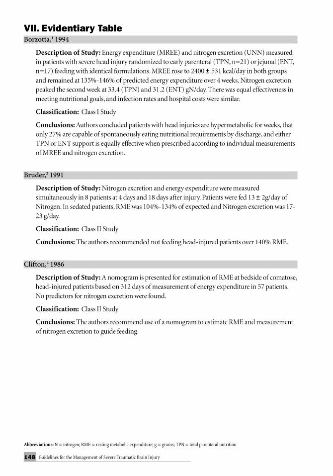
148 Guidelines for the Management of Severe Traumatic Brain Injury
VII. Evidentiary TableBorzotta,1 1994
Description of Study: Energy expenditure (MREE) and nitrogen excretion (UNN) measuredin patients with severe head injury randomized to early parenteral (TPN, n=21) or jejunal (ENT,n=17) feeding with identical formulations. MREE rose to 2400 ± 531 kcal/day in both groupsand remained at 135%-146% of predicted energy expenditure over 4 weeks. Nitrogen excretionpeaked the second week at 33.4 (TPN) and 31.2 (ENT) gN/day. There was equal effectiveness inmeeting nutritional goals, and infection rates and hospital costs were similar.
Classification: Class I Study
Conclusions: Authors concluded patients with head injuries are hypermetabolic for weeks, thatonly 27% are capable of spontaneously eating nutritional requirements by discharge, and eitherTPN or ENT support is equally effective when prescribed according to individual measurementsof MREE and nitrogen excretion.
Bruder,2 1991
Description of Study: Nitrogen excretion and energy expenditure were measuredsimultaneously in 8 patients at 4 days and 18 days after injury. Patients were fed 13 ± 2g/day ofNitrogen. In sedated patients, RME was 104%-134% of expected and Nitrogen excretion was 17-23 g/day.
Classification: Class II Study
Conclusions: The authors recommended not feeding head-injured patients over 140% RME.
Clifton,4 1986
Description of Study: A nomogram is presented for estimation of RME at bedside of comatose,head-injured patients based on 312 days of measurement of energy expenditure in 57 patients.No predictors for nitrogen excretion were found.
Classification: Class II Study
Conclusions: The authors recommend use of a nomogram to estimate RME and measurementof nitrogen excretion to guide feeding.
Abbreviations: N = nitrogen; RME = resting metabolic expenditure; g = grams; TPN = total parenteral nutrition

149Nutrition
Clifton,5 1985
Description of Study: Twenty patients with severe brain injury were randomized to feedingwith an enteral formula containing 14% protein calories or 22% protein calories. Patients were fedat 140% RME. Nitrogen balance was -9.2 ± 6.7 g/24 hr in the group fed 14%, and -5.3 ± 5.0 g/24hr in the higher protein group. Patients were treated with steroids.
Classification: Class I Study
Conclusions: The authors conclude that high protein and caloric feedings can be deliveredenterally. Increasing protein content may improve Nitrogen balance but not eliminate Nitrogenexcretion.
Clifton,6 1984
Description of Study: Caloric expenditure and Nitrogen balance were measured in 14 steroid-treated, comatose, head-injured patients acutely and for 28 days after injury. Nitrogen intake was:8.0 ± 6.1 g (days 1-3), 13.0 ± 7.3 g (days 4-6), and 11.2 ± 6.9 g (days 7-9). Nitrogen loss was 18.6± 6.4 g (days 1-3), 20.3 ± 6.5g (days 4-6), and 22.1 ± 6.0 g (days 7-9). Average RME was 138 and37% of expected patients were steroid treated.
Classification: Class II Study
Conclusions: The authors recommended feeding 0.24 g/kg/day of Nitrogen.
Deutschman,9 1986
Description of Study: Ten patients with severe brain injury underwent measurements ofoxygen consumption by arterial-venous oxygen measurement for the first 7 days after injury.Patients were not fed. Patients were found to be hypermetabolic initially but with resolution by 7days. Nitrogen excretion was 12-14 g/day of Nitrogen. Steroids were administered.
Classification: Class II Study
Conclusions: The authors recommended 1.5-2.0 g protein/kg/24 hr tapered to 1.2-1.4 gprotein/kg/24 hr by 7 days.
Dickerson,10 1990
Description of Study: In six patients with acute head injury, RME and Nitrogen excretion weremeasured acutely after injury. All patients received dexamethasone. Mean RME was 90 ± 31%.Urinary Nitrogen excretion was 16.5 ± 5.8 g/day.
Classification: Class II Study
Conclusions: Protein requirements are accentuated in excess of calorie needs in head-injuredpatients.
VII. Evidentiary Table (continued)
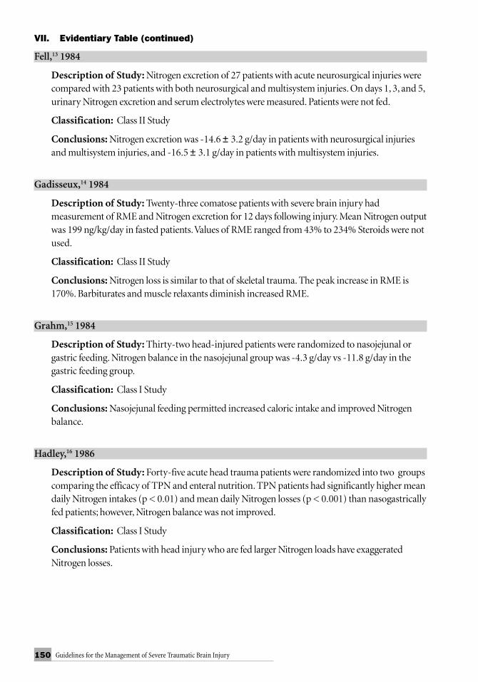
150 Guidelines for the Management of Severe Traumatic Brain Injury
Fell,13 1984
Description of Study: Nitrogen excretion of 27 patients with acute neurosurgical injuries werecompared with 23 patients with both neurosurgical and multisystem injuries. On days 1, 3, and 5,urinary Nitrogen excretion and serum electrolytes were measured. Patients were not fed.
Classification: Class II Study
Conclusions: Nitrogen excretion was -14.6 ± 3.2 g/day in patients with neurosurgical injuriesand multisystem injuries, and -16.5 ± 3.1 g/day in patients with multisystem injuries.
Gadisseux,14 1984
Description of Study: Twenty-three comatose patients with severe brain injury hadmeasurement of RME and Nitrogen excretion for 12 days following injury. Mean Nitrogen outputwas 199 ng/kg/day in fasted patients. Values of RME ranged from 43% to 234% Steroids were notused.
Classification: Class II Study
Conclusions: Nitrogen loss is similar to that of skeletal trauma. The peak increase in RME is170%. Barbiturates and muscle relaxants diminish increased RME.
Grahm,15 1984
Description of Study: Thirty-two head-injured patients were randomized to nasojejunal orgastric feeding. Nitrogen balance in the nasojejunal group was -4.3 g/day vs -11.8 g/day in thegastric feeding group.
Classification: Class I Study
Conclusions: Nasojejunal feeding permitted increased caloric intake and improved Nitrogenbalance.
Hadley,16 1986
Description of Study: Forty-five acute head trauma patients were randomized into two groupscomparing the efficacy of TPN and enteral nutrition. TPN patients had significantly higher meandaily Nitrogen intakes (p < 0.01) and mean daily Nitrogen losses (p < 0.001) than nasogastricallyfed patients; however, Nitrogen balance was not improved.
Classification: Class I Study
Conclusions: Patients with head injury who are fed larger Nitrogen loads have exaggeratedNitrogen losses.
VII. Evidentiary Table (continued)
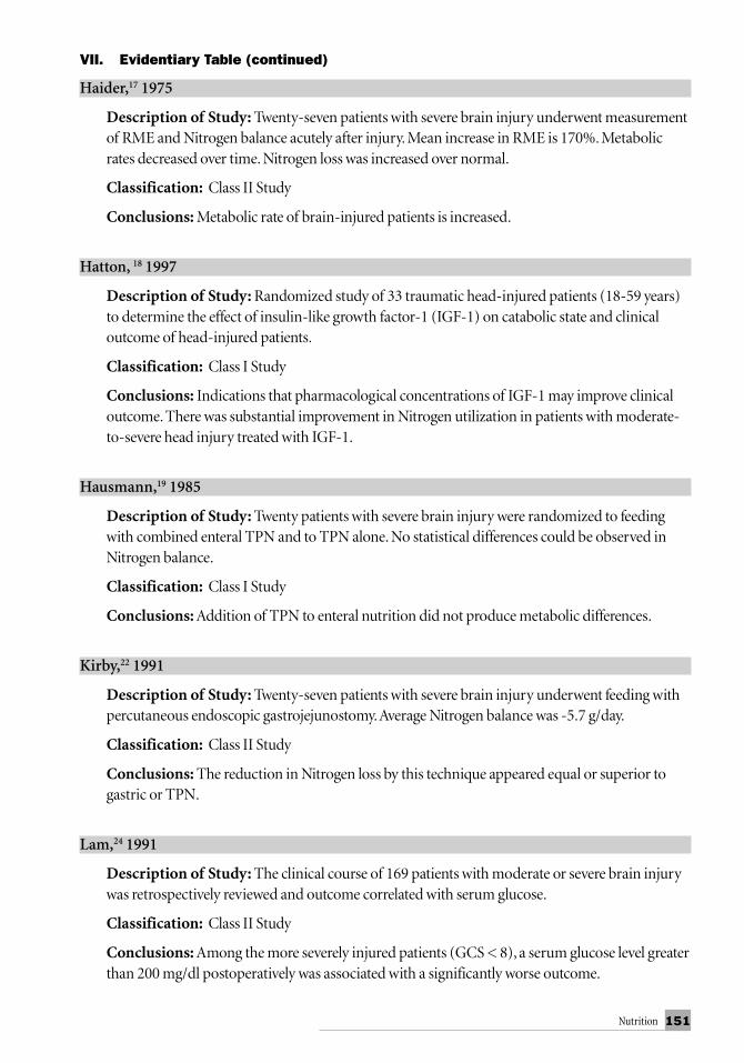
151Nutrition
Haider,17 1975
Description of Study: Twenty-seven patients with severe brain injury underwent measurementof RME and Nitrogen balance acutely after injury. Mean increase in RME is 170%. Metabolicrates decreased over time. Nitrogen loss was increased over normal.
Classification: Class II Study
Conclusions: Metabolic rate of brain-injured patients is increased.
Hatton, 18 1997
Description of Study: Randomized study of 33 traumatic head-injured patients (18-59 years)to determine the effect of insulin-like growth factor-1 (IGF-1) on catabolic state and clinicaloutcome of head-injured patients.
Classification: Class I Study
Conclusions: Indications that pharmacological concentrations of IGF-1 may improve clinicaloutcome. There was substantial improvement in Nitrogen utilization in patients with moderate-to-severe head injury treated with IGF-1.
Hausmann,19 1985
Description of Study: Twenty patients with severe brain injury were randomized to feedingwith combined enteral TPN and to TPN alone. No statistical differences could be observed inNitrogen balance.
Classification: Class I Study
Conclusions: Addition of TPN to enteral nutrition did not produce metabolic differences.
Kirby,22 1991
Description of Study: Twenty-seven patients with severe brain injury underwent feeding withpercutaneous endoscopic gastrojejunostomy. Average Nitrogen balance was -5.7 g/day.
Classification: Class II Study
Conclusions: The reduction in Nitrogen loss by this technique appeared equal or superior togastric or TPN.
Lam,24 1991
Description of Study: The clinical course of 169 patients with moderate or severe brain injurywas retrospectively reviewed and outcome correlated with serum glucose.
Classification: Class II Study
Conclusions: Among the more severely injured patients (GCS < 8), a serum glucose level greaterthan 200 mg/dl postoperatively was associated with a significantly worse outcome.
VII. Evidentiary Table (continued)

152 Guidelines for the Management of Severe Traumatic Brain Injury
Long,25 1979
Description of Study: Metabolic expenditure and urinary Nitrogen loss was measured inpatients after elective surgery, skeletal trauma, blunt trauma, head trauma with steroids, sepsis,burns, and normals.
Classification: Class II Study
Conclusions: Patients with head trauma and steroids had 0.34 ± .1 g/kg/day of nitrogen loss and60.8 ± 6 % increase in RME.
Moore,27 1989
Description of Study: RME of 20 patients with severe brain injury was measured within 48hours of admission.
Classification: Class II Study
Conclusions: RME was 160 ± 37% of expected.
Norton,28 1988
Description of Study: Twenty-three brain-injured patients were fed by gastric tube. The timefrom injury to initiation of full feeding was 11.5 days; 7 patients tolerated feeding within 7 days, 4patients from days 7-10, and 12 patients after 10 days.
Classification: Class II Study
Conclusions: Tolerance of enteral feeding is inversely related to increased ICP and severity ofbrain injury.
Ott,29 1999
Description of Study: A retrospective analysis of early enteral feedings by endoscopic, blind,and percutaneous endoscopic gastrostomy jejunostomy (PEG/J) placement of small bowelfeeding tubes in 57 patients.
Classification: Class II Study
Conclusions: Blind transpyloric feeding tube placement is rarely possible in head injurypatients. Endoscopic access to small bowel permits tolerance of enteral feedings by most patientsand is cheaper than parenteral nutrition.
VII. Evidentiary Table (continued)

153Nutrition
Ott,31 1991
Description of Study: Liquid gastric emptying was measured during the first three weeks in 12patients with severe head injury.
Classification: Class II Study
Conclusions: Delayed or abnormal gastric emptying was observed in most patients in the firsttwo weeks after injury.
Ott,32 1988
Description of Study: Twenty severely brain-injured patients with GCS scores 4-9 wereprospectively randomized to receive one of two standard amino acids formulas, starting with thefirst day of hospital admission up to day 14 post-injury. Nitrogen balance was -8 ± 2.1 g/day vs1.8 ± 1.2 g/day.
Classification: Class I Study
Conclusions: The amino acid formula with increased leucine, isoleucine, valine tyrosine, andphenylalanine resulted in improved N balance.
Phillips,33 1987
Description of Study: Energy expenditures, Nitrogen excretion, and serum protein levels werestudied from the time of hospital admission until two weeks after severe head injury in 8adolescents and 4 children with GCS scores 3-8.
Classification: Class II Study
Conclusions: Head injury in the child and adolescent induced a metabolic response thatincludes increased energy expenditure and decreased serum albumin levels similar to those seenin adult head injuries.
Piek,35 1985
Description of Study: Fourteen patients suffering from severe head injury were followed forchanges in amino acid and protein metabolism during the first 8 days after trauma. Patients werefed 15.7 g/day of Nitrogen and had an Nitrogen balance of -9.7 g/day on the eighth day.
Classification: Class II Study
Conclusions: Protein metabolism is increased after brain injury.
VII. Evidentiary Table (continued)

154 Guidelines for the Management of Severe Traumatic Brain Injury
Rapp,36 1983
Description of Study: Thirty-eight head-injured patients were randomly assigned to receiveTPN or enteral nutrition. There were no significant differences in severity of head injury by GCSscore or other variables that influence outcome. Mean intake for the TPN group was 1,750calories and 10.2 g/day of Nitrogen for the first 18 days. The TPN group got full nutritionalreplacement within 7 days of injury. The enteral group achieved 1,600 calories replacement by 14days after injury. For the enteral nutrition group mean intake in the same period was 685 caloriesand 4.0 g/day of N. There were 8 deaths in the enteral nutrition group and none in the parenteralnutrition group in the first 18 days (p < 0.001).
Classification: Class I Study
Conclusions: Early feeding reduces the mortality rate from head injury.
Robertson,37 1985
Description of Study: The effect of steroid administration on metabolic rate and Nitrogenexcretion was examined in 20 head-injured patients alternately assigned to receive eithermethylprednisolone for 14 days or no steroid treatment. All patients had an increase in Nitrogenexcretion through the second peak at day 11. The patients who received steroids had a 30% higherexcretion of Nitrogen in the first 6 days, and had lower lymphocyte count and higher infection rates.
Classification: Class I Study
Conclusions: Exogenesis steroids increase Nitrogen excretion in head-injured patients.
Robertson,38 1984
Description of Study: Factors that influenced RME and the cardiovascular response associatedwith elevated RME were examined in 55 patients with penetrating and closed head injuries whowere kept normovolemic and hyperalimented in the acute phase of injury.
Classification: Class II Study
Conclusions: Sedatives and muscle relaxants decreased RME. The lower the GCS score thehigher the RME.
Robertson,39 1991
Description of Study: The role of intravenous infusion of glucose in limiting ketogenesis andthe effect of glucose in cerebral metabolism were studied in 21 comatose patients.
Classification: Class II Study
Conclusions: Administration of glucose during the early recovery period of severe head injury isa major cause of ketogenesis and may increase production of lactic acid by the brain.
VII. Evidentiary Table (continued)

155Nutrition
Saxe,40 1994
Description of Study: Study designed to identify role of lower esophageal sphincter (LES)function in gastric feeding complications of vomiting and aspiration pneumonitis in 16 head-injured patients (GCS < 12) within 72 hours of admission.
Classification: Class II Study
Conclusions: There is evidence that LES dysfunction accompanies acute head injury andcontributes to aspiration pneumonitis after early gastric feeding. Authors recommend parenteralor jejunal feeding in patients with low GCS scores.
Suchner, 42 1996
Description of Study: Thirty-four patients after emergency craniotomy were randomized toTPN versus enteral nutrition. The effects on nutritional status, gastrointestinal absorptivefunctional substrate tolerance was studied.
Classification: Class I Study
Conclusions: Enteral nutrition following neurosurgical procedures was associated withaccelerated normalization of nutritional status and improved substrate tolerance. Enteralnutrition opposes early postoperative absorption disturbances of the small intestine.
Sunderland and Heilbrun,43 1992
Description of Study: 385 measurements were obtained in 102 patients with severe head injuryand were compared with three predictive formulas. The best prediction when compared withmeasured RME was able to capture values of 25%-125% of predicted RME in only 56% ofmeasurements.
Classification: Class II Study
Conclusions: The routine use of indirect calorimetry to guide caloric supplementation inpatients with traumatic brain injury is warranted.
Young,45 1989
Description of Study: Serum glucose levels were followed in 59 consecutive brain-injuredpatients for up to 18 days after injury and correlated with outcome.
Classification: Class II Study
Conclusions: The patients with the highest peak admission 24-hour glucose levels had the worst18-day neurologic outcome.
VII. Evidentiary Table (continued)

156 Guidelines for the Management of Severe Traumatic Brain Injury
Young,46 1987
Description of Study: Fifty-one brain-injured patients with admission GCS scores 4-10 wererandomized to receive TPN or enteral nutrition. The TPN group received higher cumulativeintake of protein than the enteral nutrition group (8.75 vs 5.7 g/day of Nitrogen ). Nitrogenbalance was higher in the TPN group in the first week after injury. Caloric balance was higher inthe TPN group (75% vs 59%). Infections, lymphocyte counts, and albumin levels were the samein both groups as was outcome. At 3 months the TPN group had a significantly more favorableoutcome but at 6 months and 1 year the differences were not significant.
Classification: Class I Study
Conclusions: Neurological recovery from head injury occurs more rapidly in patients withbetter early nutritional support.
Young,47 1985
Description of Study: Energy production, substrate oxidation, serum protein levels, and weightchange were studied in 16 non–steroid-treated patients with severe head injury. Mean energyexpenditure was 140% of expected. Patients were given 1.5 g protein/day and in only 2 was therepositive Nitrogen balance.
Classification: Class II Study
Conclusions: Head injury induces a profound traumatic response with increased energyexpenditure, negative Nitrogen balance, weight loss, hypoalbuminemia, and altered substrateoxidation.
Young,48 1987
Description of Study: Ninety-six patients with severe brain injury were randomly assigned toTPN or enteral nutrition. The incidence of increased ICP was measured in both groups for aperiod of 18 days.
Classification: Class I Study
Conclusions: There was no difference in rate of increased ICP in the two groups.
VIII. References1. Borzotta AO, Pennings J, Papasadero B, et al.: Enteral versus parenteral nutrition after severe
closed head injury. J Trauma 37:459-468, 1994.2. Bruder N, Dumont JC, Francois G: Evolution of energy expenditure and nitrogen excretion
in severe head-injured patients. Crit Care Med19:43-48, 1991.3. Cherian L, Goodman JC, Robertson CS.: Hyperglycemia increases brain injury caused by
secondary ischemia after cortical impact injury in rats. Crit Care Med 25:1378-1383,1997.4. Clifton GL, Robertson CS, Choi SC: Assessment of nutritional requirements of head-
injured patients. J Neurosurg 64:895-901, 1986.5. Clifton GL, Robertson CS, Contant DF: Enteral hyperalimentation in head injury. J
Neurosurg 62:186-193, 1985.
VII. Evidentiary Table (continued)

157Nutrition
6. Clifton GL, Robertson CS, Hodge S, et al.: The metabolic response to severe head injury. JNeurosurg 60:687-696, 1984.
7. Combs DJ, Ott L, McAninch PS, et al.: The effect of total parenteral nutrition on vasogenicedema development following cold injury in rats. J Neurosurg 70:623-627, 1989.
8. Deitrick JE, Whedon GD, Shorr E: Effects of immobilization upon various metabolic andphysiologic functions of normal men. Am J Med 4:3-36, 1948.
9. Deutschman CS, Konstantinides FN, Raup S, et al.: Physiological and metabolic response toisolated closed head injury. J Neurosurg 64:89-98, 1986.
10. Dickerson RN, Guenter PA, Gennarelli TA, et al.: Increased contribution of proteinoxidation to energy expenditure in head-injured patients. J Am Coll Nutr 9:86-88, 1990.
11. Dominioni L, Trocki O, Mochizuki H, et al.: Prevention of severe postburn hypermetabolismand catabolism by immediate intragastric feeding. J Burn Care Rehab 5:106-112, 1984.
12. Duke JH Jr, Jorgensen SD, Broell JR. Contribution of protein to caloric expenditurefollowing injury. Surgery 68:168-174, 1970.
13. Fell D, Benner B, Billings A, et al.: Metabolic profiles in patients with acute neurosurgicalinjuries. Crit Care Med 12:649-652, 1984.
14. Gadisseux P, Ward JD, Young HF, et al.: Nutrition and the neurosurgical patient. JNeurosurgery 60:219-232, 1984.
15. Grahm TW, Zadrozny DB, Harrington T: Benefits of early jejunal hyperalimentation in thehead-injured patient. Neurosurg 25:729-735, 1989.
16. Hadley MN, Grahm TW, Harrington T, et al.: Nutritional support and neurotrauma: A criticalreview of early nutrition in forty-five acute head injury patients. Neurosurg 19:367-373, 1986.
17. Haider W, Lackner F, Schlick W, et al.: Metabolic changes in the course of severe acute braindamage. Eur J Intens Care Med 1:19-26, 1975.
18. Hatton J, Pharm D, Rapp RP, et al.: Intravenous insulin-like growth factor-I (IGF-I) in moderate-to-severe head injury: a Phase II safety and efficacy trial. J Neurosurg 86:779-786, 1997.
19. Hausmann D, Mosebach KO, Caspari R: Combined enteral-parenteral nutrition versus totalparenteral nutrition in brain-injured patients. A comparative study. Intens Care Med 11:80-84, 1985.
20. Inoue S, Epstein MD, Alexander JW: Prevention of yeast translocation across the gut by asingle enteral feeding after burn surgery. JPEN 13A:565-571, 1989.
21. Jenkins M, Gottschlich M, Alexander JW, et al.: Effect of immediate enteral feeding on thehypermetabolic response following severe burn injury. JPEN 13(Suppl 1):128, 1989.
22. Kirby DF, Clifton GL, Turner H, et al.: Early enteral nutrition after brain injury bypercutaneous endoscopic gastrojejunostomy. JPEN 15:298-302, 1991.
23. Kudsk KA, Stone JM, Carpenter G, et al.: Enteral and parenteral feeding influence mortalityafter hemoglobin-E coli peritonitis in normal rats. J Trauma 23:605-609, 1983.
24. Lam AM, Winn RH, Cullen BF, et al.: Hyperglycemia and neurological outcome in patientswith head injury. J Neurosurg 75:545-551, 1991.
25. Long CL, Schaffel N, Geiger JW: Metabolic response to injury and protein needs fromindirect calorimetry and nitrogen balance. JPEN 3: 452-456, 1979.
26. McGonigal MD, Lucas CE, Ledgerwood AM: Feeding jejunostomy in patients who arecritically ill. Surg Gynecol Obstet Mar 168:275-277, 1989.
27. Moore R, Najarian MP, Konvolinka CW: Measured energy expenditure in severe headtrauma. J Trauma 29:1633-1663, 1989.

158 Guidelines for the Management of Severe Traumatic Brain Injury
28. Norton JA, Ott LG, McClain C, et al.: Intolerance to enteral feeding in the brain-injuredpatient. J Neurosurg 68:62-66, 1988.
29. Ott L, Annis K, Magnuson B, et al.: Postpyloric enteral feeding costs for patients with severehead injury: endoscopy, blind placement, and PEG/J vs TPN. J Neurotrauma 16: 233-242, 1999.
30. Ott L, Young B, McClain C. The metabolic response to brain injury. JPEN 11:488-493, 1987.31. Ott L, Young B, Phillips R, et al.: Altered gastric emptying in the head-injured patient
relationship to feeding intolerance. J Neurosurg 74:738-742, 1991.32. Ott LG, Schmidt JJ, Young AB, et al.: Comparison of administration of two standard
intravenous amino acid formulas to severely brain-injured patients. Drug Intell Clin Pharm22:763-768, 1988.
33. Phillips R, Ott L, Young B, et al.: Nutritional support and measured energy expenditure ofthe child and adolescent with head injury. J Neurosurg 67:846-851, 1987.
34. Piek J, Brachwitz K, Bock WJ. The energy consumption of patients with craniocerebraltrauma and spontaneous intracranial hemorrhage in the early postoperative post-traumaticphase. Anasth Intensivther Notfallmed 23:325-329, 1988.
35. Piek J, Lumenta CB, Bock WJ. Protein and amino acid metabolism after severe cerebraltrauma. Intensive Care Med 11:192-198, 1985.
36. Rapp RP, Pharm D, Young B, et al.: The favorable effect of early parenteral feeding onsurvival in head injured patients. J Neurosurg 58:906-912, 1983.
37. Robertson CS, Clifton GL, Goodman JC: Steroid administration and nitrogen excretion inthe head injured patient. J Neurosurg 63:714-718, 1985.
38. Robertson CS, Clifton GL, Grossman RG. Oxygen utilization and cardiovascular functionin head injured patients. Neurosur 15:307-314, 1984.
39. Robertson CS:The effect of glucose administration on carbohydrate metabolism after headinjury. JNeurosurg 74:43-50, 1991.
40. Saxe JM, Ledgerwood AM, Lucas CE, et al.: Lower esophageal sphincter dysfunctionprecludes safe gastric feeding after head injury. J Trauma 37:581-586, 1994.
41. Studley HO: Percentage of weight loss: A basic indicator of surgical risk in patients withchronic peptic ulcer. JAMA 106:458-460, 1936.
42. Suchner U, Senftleben U, Eckart T, et al.: Enteral versus parenteral nutrition: effects ongastrointestinal function and metabolism. Nutrition 12:13-22, 1996.
43. Sunderland PM, Heilbrun MP.: Estimating energy expenditure in traumatic brain injury:comparison of indirect calorimetry with predictive formulas. Neurosurg 31:146-253, 1992.
44. Waters DC, Hoff JT, Black KL: Effect of parenteral nutrition on cold-induced vasogenicedema in cats. J Neurosurg 64:460-465, 1986.
45. Young B, Ott L, Dempsey R, et al.: Relationship between admission hyperglycemia andneurologic outcome of severely brain-injured patients. Ann Surg 210:466-473, 1989.
46. Young B, Ott L, Haack D, et al.: Effect of total parenteral nutrition upon intracranialpressure in severe head injury. J Neurosurg 67:76-80, 1987.
47. Young B, Ott L, Norton J, et al.: Metabolic and nutritional sequelae in the non–steroidtreated head injury patient. Neurosurg 17:784-791, 1985.
48. Young B, Ott L, Twyman D, et al.: The effect of nutritional support on outcome from severehead injury. J Neurosurg 67:668-676, 1987.
49. Young B, Ott L, Yingling B, et al.: Nutrition and brain injury. J Neurotrauma 9 (Suppl 1)375-383, 1992.

159The Role of Antiseizure Prophylaxis Following Head Injury
I. RecommendationsA. Standards
Prophylactic use of phenytoin, carbamazepine, phenobarbital or valproateis not recommended for preventing late post-traumatic seizures.
B. GuidelinesNone
C. OptionsIt is recommended as a treatment option that anticonvulsants may be used to preventearly post-traumatic seizures in patients at high risk for seizures following headinjury. Phenytoin and carbamazepine have been demonstrated to be effective inpreventing early post-traumatic seizures. However, the available evidence does notindicate that prevention of early post-traumatic seizures improves outcome followinghead injury.
II. OverviewPost-traumatic seizures are classified as early, occurring within 7 days of injury, or late,occurring after 7 days following injury.11,13 It is desirable to prevent both early and late post-traumatic seizures. However, it is also desirable to avoid neurobehavioral and other side effectsof medications that are ineffective in preventing seizures. Prophylaxis for PTS refers to thepractice of administering anticonvulsants to patients following head injury to prevent theoccurrence of seizures. The rationale for routine seizure prophyaxis is as follows. There is arelatively high incidence of post-traumatic seizures in head-injured patients, and there arepotential benefits to preventing seizures following head injury.11,13 The incidence of seizuresfollowing penetrating injuries is about 50% in patients followed for 15 years.11 In civilian headinjury studies that followed high-risk patients up to 36 months, the incidence of early post-traumatic seizures varied between 4% and 25%, and the incidence of late post-traumaticseizures varied between 9% and 42% in untreated patients.2,6,11 In the acute period, seizures mayprecipitate adverse events in the injured brain because of elevations in intracranial pressure(ICP), blood pressure changes, changes in oxygen delivery, and also excess neurotransmitterrelease. The occurrence of seizures may also be associated with accidental injury, psychologicaleffects, and loss of driving privileges. There has been a belief that prevention of early seizuresmay prevent the development of chronic epilepsy.11,13 Experimental studies have supported the
THE ROLE OF ANTISEIZUREPROPHYLAXIS FOLLOWING HEAD INJURY

160 Guidelines for the Management of Severe Traumatic Brain Injury
idea that initial seizures may initiate kindling, which then may generate a permanent seizurefocus. On the other hand, anticonvulsants have been associated with adverse side effectsincluding rashes, Stevens-Johnson syndrome, hematologic abnormalities, ataxia, andneurobehavioral side effects.2,11,13 It is therefore important to evaluate the efficacy and overallbenefit of anticonvulsants used for the prevention of post-traumatic seizures. Certain riskfactors have been identified that place head injured patients at increased risk for developingpost-traumatic seizures.10,13 These risk factors include:
■ Glasgow Coma Scale (GCS) score less than 10■ Cortical contusion■ Depressed skull fracture■ Subdural hematoma■ Epidural hematoma■ Intracerebral hematoma■ Penetrating head wound■ Seizure within 24 hours of injury
Eight well-controlled studies of prophylaxis for PTS have been found. The scientificevidence supports the use of prophylactic anticonvulsants to prevent early PTS and does notsupport the use of those anticonvulsants studied thus far in preventing late PTS.
III. ProcessA MEDLINE computer search using the key words “seizure” and “head injury” between 1966and 1998, was performed. A total of 95 documents were found. In addition, the results of otherNational Institutes of Health–funded studies that have not been published and other clinicalstudies referred to in major review articles on post-traumatic seizures prophylaxis werereviewed. All clinical studies of seizure prophylaxis in head-injured patients were reviewed.
IV. Scientific FoundationEarly retrospective studies indicated that phenytoin was effective for the prevention of post-traumatic seizures.12,14 A practice survey among U.S. neurosurgeons in late 1973 indicated that60% used seizure prophylaxis for head-injured patients.8 Subsequent prospective, double-blindtrials, with one exception, failed to show a beneficial effect of phenytoin, or phenytoincombined with phenobarbital, in reducing the incidence of PTS.11,13 Penry, et al., reported theresults of a trial that enrolled 125 high-risk head injury patients randomized to placebo or acombination of phenytoin and phenobarbital.7 Patients were treated for 18 months and thenfollowed for an additional 18 months. The 36-month cumulative seizure rates were notsignificantly different (23% in the active group and 13% in the placebo group, p is not significant).
Young, et al., conducted a prospective, randomized, double-blind study of 244 head-injuredpatients and reported that phenytoin was not effective in preventing early or late post-traumaticseizures.15,16 The incidence of early post-traumatic seizures was low in the placebo andtreatment groups, however, which may have influenced the lack of protective effect of treatmenton early post-traumatic seizures. No patient with a phenytoin plasma concentration of 12 ug/mlor higher had a seizure, however, and, therefore, the possibility remained that higher levels mayhave been more effective in preventing late post-traumatic seizures.

161The Role of Antiseizure Prophylaxis Following Head Injury
McQueen, et al., conducted a prospective, randomized, double-blind study of 164 patientsreceiving phenytoin or placebo for the prevention of late post-traumatic seizures.5 Nosignificant reduction in late post-traumatic seizures was found in the treatment group.
Glotzner, et al., evaluated the effect of carbamazepine in preventing early and late PTS in aprospective, randomized, double-blind study of 139 patients.2 There was a significant reductionin the number of early post-traumatic seizures in the treated group, and no significantreduction in late post-traumatic seizures with treatment.
Pechadre, et al., conducted a prospective, randomized study of phenytoin in 86 patients for earlyand late post-traumatic seizures that was neither blinded nor placebo controlled.6 There was asignificant reduction in early post-traumatic seizures and also a significant reduction in late post-traumatic seizures in the acitve treatment groups. The incidence of late post-traumatic seizures washigher than in any of the similar trials, but the number of patients in the study was small.
Temkin, et al., reported the results of the largest prospective, randomized, double-blind,placebo-controlled trial to date, which randomized 404 patients to evaluate the effect ofphenytoin on early and late post-traumatic seizures.10 This trial was unique in that serum levelswere independently monitored and dosages were adjusted so that therapeutic levels weremaintained in at least 70% of the patients. Moreover, three-quarters of the patients who hadlevels monitored on the day of their first late seizure had therapeutic levels. There was asignificant reduction in the incidence of early post-traumatic seizures in the treated group. Therewas no significant reduction in the incidence of late post-traumatic seizures in the treated group.The survival curves for the placebo and active treatment groups showed no significantdifference. The neurobehavioral effect of phenytoin was also examined in this trial.
A secondary analysis has recently been performed on the data from the trial reported byTemkin, et al., to determine if treatment for early post-traumatic seizures was associated withsignificant drug-related adverse side effects. The occurrence of adverse drug effects during thefirst two weeks of treatment was low and not significantly different between the treated andplacebo groups. Hypersensitivity reactions occurred in 0.6% of the phenytoin-treated groupversus 0% in the placebo group (p = 1.0) during week 1, and 2.5% of phenytoin-treated patientsversus 0% of placebo-treated patients (p = 0.12) for the first two weeks of treatment. Mortalitywas also similar in both groups. The results of the study indicate that the incidence of early PTScan be effectively reduced by prophylactic administration of phenytoin for one or two weekswithout a significant increase in serious drug-related side effects.3
In another secondary analysis of the same trial, Dikmen, et al., found significantly impairedperformance on neuropsychologic tests at one month after injury in severely head-injuredpatients maintained on phenytoin. However, the difference was not apparent at one yearfollowing injury.1
Manaka conducted a prospective, randomized, double-blind study of 126 patients receivingplacebo or phenobarbital for the prevention of late post-traumatic seizures.4 There was nosignificant reduction in late PTS in the active treatment group.
An additional prospective, randomized, double-blind study has been recently completedthat evaluated the effect of valproate to reduce the incidence of early and late post-traumaticseizures.9 The trial compared phenytoin to valproate for the prevention of early post-traumaticseizures, and valproate to placebo for the prevention of late post-traumatic seizures. Theincidence of early post-traumatic seizures was similar in patients treated with either valproate orphenytoin. The incidence of late post-traumatic seizures was similar in patients treated with

162 Guidelines for the Management of Severe Traumatic Brain Injury
phenytoin for one week and then placebo, or patients treated with valproate for either onemonth then placebo, or with valproate for six months. There was a trend toward a highermortality in patients treated with valproate.
The majority of studies, therefore, indicate that anticonvulsants administered prophylacticallyreduce the incidence of early post-traumatic seizures but do not significantly reduce the incidenceof late post-traumatic seizures.
V. SummaryThe majority of studies do not support the use of the prophylactic anticonvulsants studied thusfar for the prevention of late post-traumatic seizures. Routine seizure prophylaxis later than oneweek following head injury is, therefore, not recommended. If late post-traumatic seizures occur,patients should be managed in accordance with standard approaches to patients with new onsetseizures. Phenytoin and carbamazepine have been shown to reduce the incidence of early post-traumatic seizures. Valproate may also have a comparable effect to phenytoin on reducing earlyPTS but may also be associated with a higher mortality. It is, therefore, an option to usephenytoin or carbamazepine to prevent the occurrence of seizures in high-risk patients duringthe first week following head injury.
VI. Key Issues for Future InvestigationAdditional studies may be needed to determine if reduction in early post traumatic seizures hasan effect on outcome. Future trials of neuroprotectant agents that have antiepileptic activity,such as magnesium sulphate and other NMDA receptor antagonists, may futher reduce theincidence of post-traumatic seizures.
VII. Evidentiary TableGlotzner,2 1983
Description of Study: Prospective, randomized, double-blind study of 139 patients treatedwith placebo or carbamazepine, and evaluated for early and late post-traumatic seizures.Theraputic levels were maintained in the majority of patients.
Classification: Class I Study
Conclusions: Showed significant reduction in early post-traumatic seizures and no significanteffect on late post-traumatic seizures using therapeutic levels of carbamazepine
Seizure RatesEarly Late
% Active % Placebo p % Active % Placebo p10% 25% 0.016 27% 21% ns

163The Role of Antiseizure Prophylaxis Following Head Injury
Manaka, 4 1992
Description of Study: Prospective, randomized, double-blind study of 126 patients receivingplacebo or phenobarbital for effect on late post-traumatic seizures. Treatment was started onemonth following head injury.
Classification: Class I Study
Conclusions: Showed no significant effect of phenobarbital on late post-traumatic seizures.Seizure Rates
Early Late% Active % Placebo p % Active % Placebo p
(not studied) 16% 11% ns
McQueen,5 1983
Description of Study: Prospective, randomized, double-blind study of 164 patients receivingphenytoin or placebo for late post-traumatic seizures.
Classification: Class I Study
Conclusions: Showed no significant effect of phenytoin on late post-traumatic seizures.
Seizure RatesEarly Late
% Active % Placebo p % Active % Placebo p(not studied) 10% 9% ns
Pechadre, 6 1991
Description of Study: Prospective, randomized study (not blinded or placebo controlled) ofphenytoin or no drug in 86 patients for early and late post-traumatic seizures.
Classification: Class I Study
Conclusions: Showed significant reduction in early and late post-traumatic seizures byphenytoin.
Seizure RatesEarly Late
% Active % No Drug p % Active % No Drug p6% 24% 0.05 6% 42% 0.001
VII. Evidentiary Table (continued)
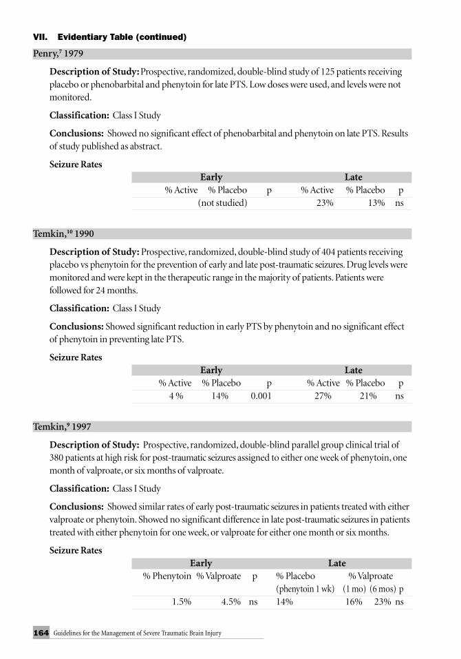
164 Guidelines for the Management of Severe Traumatic Brain Injury
Penry,7 1979
Description of Study: Prospective, randomized, double-blind study of 125 patients receivingplacebo or phenobarbital and phenytoin for late PTS. Low doses were used, and levels were notmonitored.
Classification: Class I Study
Conclusions: Showed no significant effect of phenobarbital and phenytoin on late PTS. Resultsof study published as abstract.
Seizure RatesEarly Late
% Active % Placebo p % Active % Placebo p(not studied) 23% 13% ns
Temkin,10 1990
Description of Study: Prospective, randomized, double-blind study of 404 patients receivingplacebo vs phenytoin for the prevention of early and late post-traumatic seizures. Drug levels weremonitored and were kept in the therapeutic range in the majority of patients. Patients werefollowed for 24 months.
Classification: Class I Study
Conclusions: Showed significant reduction in early PTS by phenytoin and no significant effectof phenytoin in preventing late PTS.
Seizure RatesEarly Late
% Active % Placebo p % Active % Placebo p4 % 14% 0.001 27% 21% ns
Temkin,9 1997
Description of Study: Prospective, randomized, double-blind parallel group clinical trial of380 patients at high risk for post-traumatic seizures assigned to either one week of phenytoin, onemonth of valproate, or six months of valproate.
Classification: Class I Study
Conclusions: Showed similar rates of early post-traumatic seizures in patients treated with eithervalproate or phenytoin. Showed no significant difference in late post-traumatic seizures in patientstreated with either phenytoin for one week, or valproate for either one month or six months.
Seizure RatesEarly Late
% Phenytoin % Valproate p % Placebo % Valproate(phenytoin 1 wk) (1 mo) (6 mos) p
1.5% 4.5% ns 14% 16% 23% ns
VII. Evidentiary Table (continued)

165The Role of Antiseizure Prophylaxis Following Head Injury
Young,15,16 1983
Description of Study: Prospective, randomized, double-blind study of 244 patients receivingplacebo vs phenytoin for the prevention of early and late PTS. Drug levels monitored.
Classification: Class I Study
Conclusions: Showed no significant effect of phenytoin on early or late PTS.
Seizure RatesEarly Late
% Active % Placebo p % Active % Placebo p4 % 4% ns 12% 11% ns
VIII. References1. Dikmen SS, Temkin NR, Miller B, et al.: Neurobehavioral effects of phenytoin prophylaxis
of post-traumatic seizures. JAMA 265:1271-1277, 1991.2. Glotzner FL, Haubitz I, Miltner F, et al.: Anfallsprophylaze mit carbamazepin nach schweren
schadelhirnverletzungen. Neurochirurgia Stuttg 26:66-79, 1983.3. Haltiner AM, Newell DW, Temkin NR, et al.: Side effects and mortality associated with use
of phenytoin for early post-traumatic seizure prophylaxis. J Neurosurg 91:588-592, 1999.4. Manaka S: Cooperative prospective study on post-traumatic epilepsy: risk factors and the
effect of prophylactic anticonvulsant. The Japanese Journal Psychiatry and Neurology 46:311-315, 1992.
5. McQueen JK, Blackwood DHR, Harris P, et al.: Low risk of late post-traumatic seizuresfollowing severe head injury: implications for clinical trials of prophylaxis. J NeurolNeurosurg Psychiatry 46:899-904, 1983.
6. Pechadre JC, Lauxerois M, Colnet G, et al.: Prevention de l’epelepsie posttraumatiquetardive par phenytoine dans les traumatismes carniens graves: suivi durant 2 ans. Presse Med20:841-845, 1991.
7. Penry JK, White BG, Brackett CE: A controlled prospective study of the pharmacologicprophylaxis of post-traumatic epilepsy. Neurology 29:600, 1979.
8. Rapport RL, Penry JK: A survey of attitudes toward the pharmacologic prophylaxis of post-traumatic epilepsy. J Neurosurg 38:159-166, 1973.
9. Temkin NR, Dikmen SS, Anderson GD, et al.: Valproate therapy for prevention of post-traumatic seizures: a randomized trial. J Neurosurg 91: 593-600, 1999.
10. Temkin NR, Dikmen SS, Wilensky AJ, et al.: A randomized, double-blind study ofphenytoin for the prevention of post-traumatic seizures. New Engl J Med 323:497-502, 1990.
11. Temkin NR, Dikmen SS, Winn HR: Post-traumatic seizures. In Eisenberg HM, Aldrich EF(eds): Management of Head Injury. W. B. Saunders:Philadelphia 425-435, 1991.
12. Wohns RNW, Wyler AR: Prophylactic phenytoin in severe head injuries. J Neurosurg 51:507-509,1979.
13. Yablon SA: Post-traumatic seizures. Arch Phys Med Rehabil 74:983-1001, 1993.14. Young B, Rapp RP, Brooks W, et al.: Post-traumatic epilepsy prophylaxis. Epilepsia 20:671-681,
1979.
VII. Evidentiary Table (continued)

PART II: EARLY
INDICATORS
OF PROGNOSIS IN
SEVERE TRAUMATIC
BRAIN INJURY


3Early Indicators of Prognosis
Table of Contents
I. Introduction ......................................................................................................... 7
II. Methodology ...................................................................................................... 11
III. Glasgow Coma Scale Score ................................................................................ 15
IV. Age ..................................................................................................................... 27
V. Pupillary Diameter & Light Reflex .................................................................... 41
VI. Hypotension ...................................................................................................... 55
VII. CT Scan Features ............................................................................................... 65
PART II: EARLY INDICATORS OFPROGNOSIS IN SEVERE TRAUMATICBRAIN INJURY


5Early Indicators of Prognosis
Authors:
Randall M. Chesnut, M.D.Associate Professor of NeurosurgeryOregon Health Sciences University
Jamshid Ghajar, M.D.Clinical Associate Professor of NeurosurgeryCornell University Medical College
Andrew I.R. Maas, M.D.Associate Professor of NeurosurgeryUniversity Hospital Rotterdam, The Netherlands
Donald W. Marion, M.D.Professor of NeurosurgeryUniversity of Pittsburgh Medical Center
Franco Servadei, M.D.Associate Professor of NeurosurgeryOspedale M Bufalini, Cesena, Italy
Graham M. Teasdale, M.D.Professor of NeurosurgeryThe Southern General Hospital, Glasgow, Scotland
Andreas Unterberg, M.D.Professor of NeurosurgeryHumboldt University, Berlin, Germany
Hans von Holst, M.D.Professor of NeurosurgeryKarolinska Institute, Stockholm, Sweden
Beverly C. Walters, M.D.Chairman, AANS Guidelines Committee, Associate Professor of Clinical NeurosciencesBrown University School of Medicine
EARLY INDICATORS OF PROGNOSIS INSEVERE TRAUMATIC BRAIN INJURY

6 Early Indicators of Prognosis
Participants:
Charles Contant, Ph.D.Baylor University
Robert Florin, M.D.Chairman, AANS Guidelines and Outcomes Committee
Peter C. QuinnExecutive Director, Brain Trauma Foundation
George A. Zitney, Ph.DChairman, Neurotrauma Committee, World Health OrganizationDirector, John Jane Brain Injury Center

7Early Indicators of Prognosis
The uncertainty that exists about the likely outcome after traumatic brain injury (TBI) isencapsulated in the Hippocratic aphorism: “No head injury is so serious that it should be
despaired of nor so trivial that it can be ignored.” Today, physicians’ estimates of prognosis arestill often unduly optimistic, unnecessarily pessimistic, or inappropriately ambiguous. 1,4,6,12 Itstill remains impossible to say with certainty what will be the future course of events in anindividual patient, but intensive research in the last two decades has made it possible to be muchmore confident about what is likely to happen, and to consider prognosis in terms ofprobabilities rather than prophecies.
Prediction of outcome involves making probability statements that depend on a logicalrelationship between outcome and features encapsulated in antecedent, early data. The advancesin prognosis reflect the establishment of methods for categorizing outcome9 and early injuryseverity.18 These became widely accepted13 and led to multinational, multicenter studies14 thatidentified the features about the patient, the injury, and the early clinical course with adistinctive, consistent relationship to outcome.10,15 The subsequent chapters consider thesevarious features, first with respect to the existence of a relationship to outcome, then thestrength of the effect or interaction with outcome, and finally, the extent to which the effect isunique to the feature in question (almost never so) or how far there is interaction(interdependence) with other prognostic features.
Although clinicians usually attempt to take a wide range of factors into account whenmaking clinical decisions and assessing prognosis, there is probably a redundancy in this effortto be complete. In practice, relatively few features have been found to contain most of theprognostic information,3,10,17 These include the patient age, clinical indices indicating theseverity of brain injury (e.g., the depth and duration of coma and other neurologicalabnormalities), and the results of investigation and imaging studies, particularly intracranialpressure (ICP) and computed tomography (CT) scanning, which disclose the nature of braininjury and its effects on intracranial dynamics. Even though there is little doubt regarding theimportance of these features from clinical experience, there are still debates about the precisenature of their relationships and about exactly how the different features should be assessed,categorized, and—most importantly—utilized.
The identification of powerful, single prognostic factors is only one step toward a usefulstatement about prognosis. Unique relationships between the findings of a feature and outcomemay apply only for the most extreme abnormalities found only in a tiny minority of patients. Tobe useful, prognostic statements need to be applicable across all severities of injury and capable
INTRODUCTION

8 Early Indicators of Prognosis
of being expressed in a way that indicates the likelihood of an individual patient achievingdifferent outcomes at some future time. This depends on combining the information on thedifferent individual prognostic features. Although a wide number of statistical approaches havebeen described, there is little difference in practice between the results that they produce. Alsothe details of calculation are much less important than the data that are employed.17,19 Likewise,the results of prognostic calculations can be expressed in a number of ways that includemathematical probability,8,10 graphical presentations,2,5 and the methodology used in thisdocument. The merits of different methods have not been established.
The utility of prognostic probabilities can be assessed by various criteria. One is“separation,” in which a particular outcome is emphasized (e.g., by being assigned a very highprobability). Separation has the benefit of conveying discrimination and a high degree ofcertainty; however, it runs the risk of being excessively confident and leading to extremes ofprognosis—either falsely optimistic or falsely pessimistic. Perhaps a more desirable attribute is“faithfulness,” that is, that the probabilites expressed relate reliably to what is likely to happen.Thus, the figure calculated should reflect the distribution of outcomes that occur in a series ofpatients allocated the respective probabilities of different outcomes. To do this effectively wouldrequire a large data set collected prospectively and with relevant patient follow-up.
Clearly, this type of approach cannot be used when reviewing published reports. Themethodology described in the next section has been devised to fit the task of literature reviewwhile adhering to clinical epidemiological principles. Information about prognosis andpredictive statements can be useful in a number of ways. From the start, concern about outcomeis often foremost in the mind of the relative of severely brain-injured victims and realisticcounseling is preferable to over pessimism—characterized as “hanging crepe”—or the raisingof false hopes. An assessment of prognosis is crucial in research studies, both in determining theappropriate target population and in deciding if a given intervention has produced an outcomedifferent from that which would have been expected. The place of prognosis in makingdecisions about the management of individual patients remains controversial. While manyneurosurgeons acknowledge that it is an important factor in decision making,2 others relegateprognosis to a minor or even nonexistent role, reflecting a range of attitudes arising fromcultural and ethical differences as much as clinical convictions.
Although there are concerns that estimation of prognosis may be used to allocate (and inparticular to withdraw) resources, and that this might worsen the outcome in some cases, thiswas not substantiated in a formal study.16 In a large prospective trial, doctors, nurses, and otherstaff providing acute care for severe brain injuries were provided with predictions of theoutcome in individual cases. Compared with control periods without predictions, there was nolessening of the use of intensive care resources nor an increase in the rate of decisions to limittreatment. Instead, there was a shift in the employment of aspects of intensive care from patientswith a calculated high likelihood of poor outcome to those with a greater prospect of recovery,without an adverse affect to outcome in the former group.
The purpose of this exercise is to identify from the published medical literature those earlyclinical factors that may be prognostic for outcome. This will then suggest which early factorsshould be focused on in prospective database research in patients with TBI.
An estimate of a patient’s prognosis should never be the only factor, and only rarely the mainfactor, in influencing clinical decisions. Instead, prognosis is simply one of the many factors thatneed to be considered in the clinical management of a severely brain-injured patient.

9Early Indicators of Prognosis
References1. Barlow P, Teasdale G: Prediction of outcome and the management of severe head injuries:
the attitudes of neurosurgeons. Neurosurg 19:989-991; 1986.2. Barlow P, Murray L, Teasdale G: Outcome after severe head injury—the Glasgow model. In:
Corbett WA, et al. Medical Applications of Microcomputers. New York: Wiley 105-106,1987.
3. Braakman R, Gelpke GJ, Habbema JDF, et al.: Systematic selection of prognostic features inpatients with severe head injury. Neurosurg 6:362-370, 1980.
4. Chang RWS, Lee B, Jacobs S: Accuracy of decisions to withdraw therapy in critically illpatients: clinical judgment versus a computer model. Crit Care Med 17:1091-97, 1989.
5. Choi SC, Ward JD, Becker DP: Chart for outcome prediction in severe head injury. JNeurosurg 59: 294-297, 1983.
6. Dawes RM, Faust D, Meehl RE: Clinical versus acturial judgment. Science 243:1668-74, 1989.7. Foulkes MA, Eisenberg HM, Jane JA, et al.: The Traumatic Coma Data Bank—design,
methods, and baseline characteristics. J Neurosurg 75 (Suppl) 8-13, 1991.8. Gibson RM, Stephensen GC: Aggressive management of severe closed head trauma: time
for reappraisal. Lancet 2:369-371, 1989.9. Jennett B, Bono M: Assessment of outcome after severe brain damage. A practical scale.
Lancet 1: 480-484, 1975.10. Jennett B, Teasdale G, Braakman R, et al.: Prognosis of patients with severe head injury.
Neurosurg 4:283-289, 1979.11. Jennett B, Teasdale G, Galbraith S, et al.: Severe head injuries in three countries. JNNP
40:291-298, 1977.12. Kaufmann MA, Buchmann B, Scheidegger D, et al.: Severe head injury: should expected
outcome influence resuscitation and first day decisions? Resuscitation 23:199-206, 1992.13. Langfitt TW: Measuring the outcome from head injuries. J Neurosurg 48:673-678, 1978.14. Marshall LF, Becker DP, Bowers SA, et al.: The national Traumatic Coma Data Bank Part 1:
design, purpose, goals, and results. J Neurosurg 59 276-284, 1983.15. Marshall LF, Gautille T, Klauber M, et al.: The outcome of severe closed head injury. J
Neurosurg (Suppl) 75:28-36, 1991.16. Murray LS, Teasdale GM, Murray GD, et al.: Does prediction of outcome alter patient
management? Lancet 341; 1487-91, 1993.17. Stablein DM, Miller JD, Choi SC: Statistical methods for determining prognosis in severe
head injury. Neurosurg 6:243-248, 1980.18. Teasdale G, Jennet B. Assessment of coma and impaired consciousness. Lancet 2:81-84,
1974.19. Titterington DM, Murray GD, Murray LS, et al.: Comparison of discrimination techniques
applied to a complex data set of head injured patients. J of the Royal Statistical Society Series A144: 145-175, 1981.

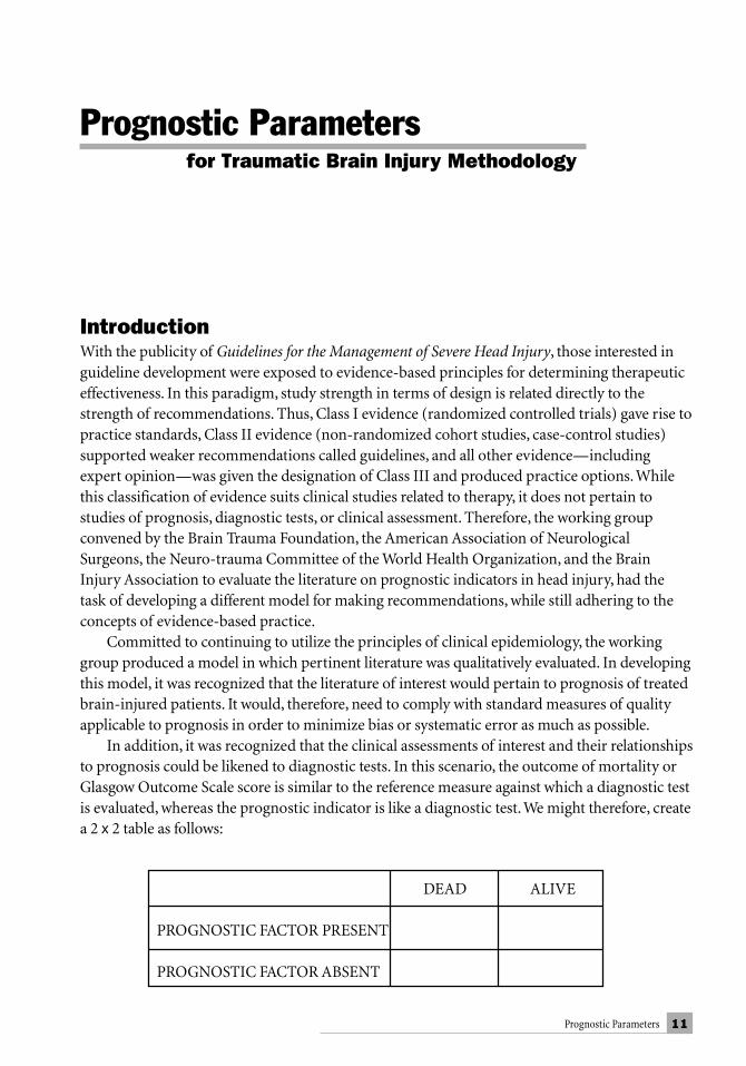
11Prognostic Parameters
IntroductionWith the publicity of Guidelines for the Management of Severe Head Injury, those interested inguideline development were exposed to evidence-based principles for determining therapeuticeffectiveness. In this paradigm, study strength in terms of design is related directly to thestrength of recommendations. Thus, Class I evidence (randomized controlled trials) gave rise topractice standards, Class II evidence (non-randomized cohort studies, case-control studies)supported weaker recommendations called guidelines, and all other evidence—includingexpert opinion—was given the designation of Class III and produced practice options. Whilethis classification of evidence suits clinical studies related to therapy, it does not pertain tostudies of prognosis, diagnostic tests, or clinical assessment. Therefore, the working groupconvened by the Brain Trauma Foundation, the American Association of NeurologicalSurgeons, the Neuro-trauma Committee of the World Health Organization, and the BrainInjury Association to evaluate the literature on prognostic indicators in head injury, had thetask of developing a different model for making recommendations, while still adhering to theconcepts of evidence-based practice.
Committed to continuing to utilize the principles of clinical epidemiology, the workinggroup produced a model in which pertinent literature was qualitatively evaluated. In developingthis model, it was recognized that the literature of interest would pertain to prognosis of treatedbrain-injured patients. It would, therefore, need to comply with standard measures of qualityapplicable to prognosis in order to minimize bias or systematic error as much as possible.
In addition, it was recognized that the clinical assessments of interest and their relationshipsto prognosis could be likened to diagnostic tests. In this scenario, the outcome of mortality orGlasgow Outcome Scale score is similar to the reference measure against which a diagnostic testis evaluated, whereas the prognostic indicator is like a diagnostic test. We might therefore, createa 2 x 2 table as follows:
Prognostic Parametersfor Traumatic Brain Injury Methodology
DEAD ALIVE
PROGNOSTIC FACTOR PRESENT
PROGNOSTIC FACTOR ABSENT
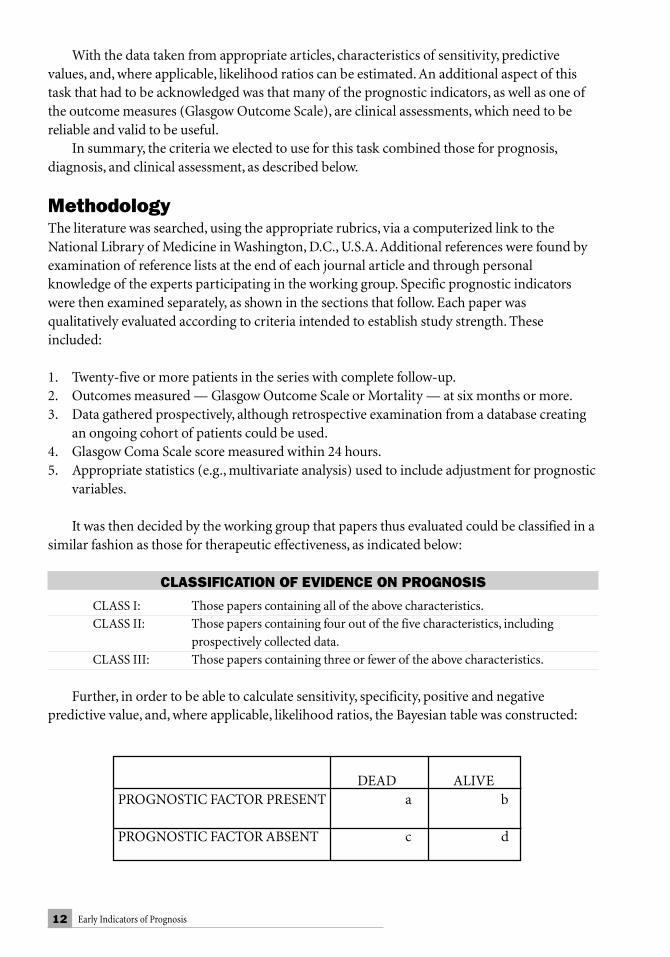
12 Early Indicators of Prognosis
With the data taken from appropriate articles, characteristics of sensitivity, predictivevalues, and, where applicable, likelihood ratios can be estimated. An additional aspect of thistask that had to be acknowledged was that many of the prognostic indicators, as well as one ofthe outcome measures (Glasgow Outcome Scale), are clinical assessments, which need to bereliable and valid to be useful.
In summary, the criteria we elected to use for this task combined those for prognosis,diagnosis, and clinical assessment, as described below.
MethodologyThe literature was searched, using the appropriate rubrics, via a computerized link to theNational Library of Medicine in Washington, D.C., U.S.A. Additional references were found byexamination of reference lists at the end of each journal article and through personalknowledge of the experts participating in the working group. Specific prognostic indicatorswere then examined separately, as shown in the sections that follow. Each paper wasqualitatively evaluated according to criteria intended to establish study strength. Theseincluded:
1. Twenty-five or more patients in the series with complete follow-up.2. Outcomes measured — Glasgow Outcome Scale or Mortality — at six months or more.3. Data gathered prospectively, although retrospective examination from a database creating
an ongoing cohort of patients could be used.4. Glasgow Coma Scale score measured within 24 hours.5. Appropriate statistics (e.g., multivariate analysis) used to include adjustment for prognostic
variables.
It was then decided by the working group that papers thus evaluated could be classified in asimilar fashion as those for therapeutic effectiveness, as indicated below:
CLASSIFICATION OF EVIDENCE ON PROGNOSIS
CLASS I: Those papers containing all of the above characteristics.CLASS II: Those papers containing four out of the five characteristics, including
prospectively collected data.CLASS III: Those papers containing three or fewer of the above characteristics.
Further, in order to be able to calculate sensitivity, specificity, positive and negativepredictive value, and, where applicable, likelihood ratios, the Bayesian table was constructed:
DEAD ALIVEPROGNOSTIC FACTOR PRESENT a b
PROGNOSTIC FACTOR ABSENT c d

13Prognostic Parameters
CLINICAL QUESTION PROGNOSTIC ATTRIBUTE CALCULATION
If a trauma patient reaches a Sensitivity acertain outcome, how likely is a + cshe or he to have had a givenprognostic indicator?
If a trauma patient does not reach Specificity da certain outcome, how likely is b + dshe or he to have not had a givenprognostic indicator?
If a trauma patient has a given Postive predictive value aprognostic indicator, how likely a + bis she or he to reach a certain outcome?
If a trauma patient does not have Negative predictive value da given prognostic indicator, how c + dlikely is she or he to reach a certainoutcome?


15Glasgow Coma Scale Score
GLASGOW COMA SCALE SCORE
I. ConclusionsA. Which feature of the parameter is supported by Class I evidence and has at least a 70%
positive predictive value? There is an increasing probability of poor outcome with adecreasing Glasgow Coma Scale (GCS) score in a continuous, stepwise manner.
B. Parameter measurements:1. How should it be measured?
■ It should be measured in a standardized way.■ It must be obtained through interaction with the patient (e.g., application of
a painful stimulus for patients unable to follow commands).2. When should it be measured for prognostic purposes?
■ Only after pulmonary and hemodynamic resuscitation.■ After pharmacologic sedation or paralytic agents are metabolized.
3. Who should measure it?■ The GCS can be fairly reliably measured by trained medical personal.
II. OverviewThe GCS was developed by Teasdale and Jennett in 1974 as an objective measure of the level ofconsciousness.31 It has since become the most widely used clinical measure of the severity ofinjury in patients with severe traumatic brain injuries (TBIs). A number of studies haveconfirmed a fairly high degree of inter- and intra-rater reliability of the scale across observerswith a wide variety of experience.5,11,24,32
III. Search ProcessThe titles and abstracts of approximately 500 journal articles were retrieved using a computerizedsearch of the National Library of Medicine. The MESH heading “Glasgow Coma Scale” was usedto search for articles published since the GCS was developed in 1974. The abstracts of all articleswere reviewed and those articles that focused on the correlation between the acute GCS score(obtained within the first 24 hours) and outcome in patients with severe closed head injuries wereselected for review of the entire article. This left 20 articles that dealt primarily with correlation ofthe GCS score and outcome, 8 articles that focused on the use of the initial GCS score to predictoutcome, and 6 articles describing the reliability of the GCS score.
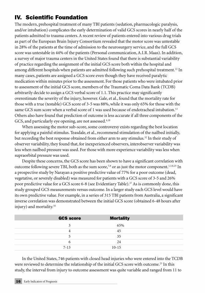
16 Early Indicators of Prognosis
IV. Scientific FoundationThe modern, prehospital treatment of many TBI patients (sedation, pharmacologic paralysis,and/or intubation) complicates the early determination of valid GCS scores in nearly half of thepatients admitted to trauma centers. A recent review of patients entered into various drug trialsas part of the European Brain Injury Consortium revealed that the motor score was untestablein 28% of the patients at the time of admission to the neurosurgery service, and the full GCSscore was untestable in 44% of the patients (Personal communication, A.I.R. Maas). In addition,a survey of major trauma centers in the United States found that there is substantial variabilityof practice regarding the assignment of the initial GCS score both within the hospital andamong different hospitals when patients are admitted following such prehospital treatment.22 Inmany cases, patients are assigned a GCS score even though they have received paralyticmedication within minutes prior to the assessment. For those patients who were intubated priorto assessment of the initial GCS score, members of the Traumatic Coma Data Bank (TCDB)arbitrarily decide to assign a GCS verbal score of 1.1. This practice may significantlyoverestimate the severity of the injury, however. Gale, et al., found that the mortality rate forthose with a true (testable) GCS score of 3-5 was 88%, while it was only 65% for those with thesame GCS sum score when a verbal score of 1 was used because of endotracheal intubation.12
Others also have found that prediction of outcome is less accurate if all three components of theGCS, and particularly eye opening, are not assessed.4,18
When assessing the motor sub-score, some controversy exists regarding the best locationfor applying a painful stimulus. Teasdale, et al., recommend stimulation of the nailbed initially,but recording the best response obtained from either arm to any stimulus.32 In their study ofobserver variability, they found that, for inexperienced observers, interobserver variability wasless when nailbed pressure was used. For those with more experience variability was less whensupraorbital pressure was used.
Despite these concerns, the GCS score has been shown to have a significant correlation withoutcome following severe TBI, both as the sum score,7,8 or as just the motor component.1,3,8,25 Ina prospective study by Narayan a positive predictive value of 77% for a poor outcome (dead,vegetative, or severely disabled) was measured for patients with a GCS score of 3-5 and 26%poor predictive value for a GCS score 6-8 (see Evidentiary Table).27 As is commonly done, thisstudy grouped GCS measurements versus outcome. In a larger study each GCS level would haveits own predictive value. For example, in a series of 315 TBI patients from Australia, a significantinverse correlation was demonstrated between the initial GCS score (obtained 6-48 hours afterinjury) and mortality.10
GCS score Mortality
3 65%4 455 356 24
7-13 10-15
In the United States, 746 patients with closed head injuries who were entered into the TCDBwere reviewed to determine the relationship of the initial GCS score with outcome.23 In thisstudy, the interval from injury to outcome assessment was quite variable and ranged from 11 to

17Glasgow Coma Scale Score
1199 days, with a median of 674 days. The morality rate for those with an initial post-traumaticGCS score of 3 was 78.4%; initial GCS score of 4, 55.9%; and initial GCS score of 5, 40.2%. Ofnote, however, is that 4.1%, 6.3%, and 12.2% of the three groups, respectively, had a goodoutcome.
In a large study of 46,977 head-injured patients the relationship between GCS scores 3-15and mortality was investigated.13 A sharp progressive increase in mortality was noted in patientswho presented to the Emergency Room with a GCS score of 3-8.
In 109 adults with acute subdural hematomas, Phuenpathom also showed a significantinverse relationship with GCS score (best score within 24 hours) and mortality.28
GCS score Mortality Number of patients
3 100% (37)4 90 (9/10)5 63 (5/8)6 33 (2/6)7 22 (2/9)
8-15 0 (39)
In a series of 115 patients with epidural hematomas, Kuday found that the initial GCS scorewas the single most important factor affecting outcome (p<0.00001).21
Because of the strong association with the initial GCS score and outcomes, a number ofinvestigators have studied the predictive value of the initial GCS score using various logisticregression techniques.2,16,20,30,33 Thatcher, et al., used multimodal statistical models to study the abilityof the initial GCS score or the GCS score obtained at a mean of 7.5 days after the injury to predictoutcome at one year after injury for 162 patients with TBI.33 When based on the initial GCS score,only 68.6% of those predicted to have a good outcome and 76.5% of those predicted to have a pooroutcome actually had such outcomes at one year. If the later GCS was used for predictions, there wasa significant increase in the rate of correct predictions for a good outcome (80.6%), but the rate ofcorrect predictions for a poor outcome remained essentially unchanged (78.6%).
Kaufman described the accuracy of outcome predictions of an experienced neurosurgeonfor 100 patients with severe TBI.20 Outcomes were categorized as dead/vegetative, severelydisabled, or capable of independent survival, and were predicted based on the best GCS scoresobtained within 24 hours after injury. Age, pupils, blood pressure, heart rate, laboratory values,and initial computed tomograpy (CT) scans were also considered. Correct prognosis wasestimated in only 56% of the cases.
GOS Predicted/Actual Outcomes
Dead/Vegetative 35/24Severe Disability 23/11Independent 42/65
The table reveals that predictions were best for very bad or very good outcomes. Inaddition, poor outcomes were overestimated by 32%-52%, while good outcomes wereunderestimated by 35%. In a study of 254 patients with severe TBI, Benzer used logisticregression methods to predict patient outcome based on the immediate post-traumatic GCS

18 Early Indicators of Prognosis
score, and made correct predictions 82.68% of the time.2
It should be emphasized that most of these studies looked at the least discriminate scenario(e.g., reduction of potential outcomes to two or at most three groups). When attempts weremade to predict more precisely into one of the five categories of the Glasgow Outcome Scale(GOS), the predictive accuracy of the initial GCS score was poor.19
V. SummaryWhen considering the use of the initial GCS score for prognosis, the two most importantproblems are the reliability of the initial measurement, and its lack of precision for prediction ofa good outcome if the initial GCS score is low. If the initial GCS score is reliably obtained andnot tainted by prehospital medications or intubation, approximately 20% of the patients withthe worst initial GCS score will survive and 8%-10% will have a functional survival (GOS 4-5).
VI. Key Issues for Further InvestigationA. The optimal time after injury for determining the initial GCSB. When to assess the GCS score for those who have received paralytic or sedative medicationC Reliability of the prehospital GCS score
VII. Evidentiary TableFearnside,10 1998
Description of Study: Prospective study of 315 consecutive patients of all ages with severe TBIto identify factors responsible for morbidity and mortality.
Classification: Class II Study
Conclusions: GOSGCS 1
3 65%4 455 356 24
7-13 10-15
Marshall,23 1991
Description of Study: Prospective study of 746 consecutive patients with severe TBI to gatherdemographic and outcome data; adults.
Classification: Class II Study
Conclusions: GOSGCS 1 4, 53 78.4% 7.2%4 55.9 14.45 40.2 29.36 21.2 50.57 17.6 68.98 11.3 77.4

19Glasgow Coma Scale Score
Narayan,27 1989
Description of Study: Prospective study of 133 consecutive patients with severe TBI to identifyfactors responsible for morbidity and mortality; all ages.
Classification: Class I Study
Conclusions: GOSGCS 1 2, 3 4, 53-5 62% 15% 23% +PPV=77%6-8 20 749-11 18 76
Beca,1 1995
Description of Study: Prospective study of 109 children with severe TBI to compare outcomeprediction of somatosensory evoked potentials (SEPs) with GCS.
Classification: Class III Study
Conclusions: GOSGCS Motor 4, 5
1 0%2-3 41-474-5 75-78
Braakman,6 1980
Description of Study: Prospective study of 305 consecutive patients with severe TBI studied toidentify prognostic indicators; all ages.
Classification: Class I Study
Conclusions: GOSGCS 1
3 100%4 805 686 517 278 22
9-15 15
VII. Evidentiary Table (continued)

20 Early Indicators of Prognosis
Phuenpathom,28 1993
Description of Study: Retrospective study of 109 patients with acute subdural hematomas todetermine outcome; all ages.
Classification: Class II Study
Conclusions: GOSGCS 1
3 100%4 905 636 337 22
8-15
Wilberger,34 1990
Description of Study: Retrospective study of 101 adult patients with severe TBI who also hadacute subdural hematomas.
Classification: Class III Study
Conclusions: GOSGCS 1 4, 5
3 90% 5%4 76 105 62 18
6-7 51 44
Rivas,29 1988
Description of Study: Retrospective study of 66 patients with severe TBI who also had epiduralhematomas; all ages.
Classification: Class III Study
Conclusions: GOSGCS Motor 1 4, 5
1 80% 20%2-3 24 734-5 5 90
VII. Evidentiary Table (continued)

21Glasgow Coma Scale Score
Colohan,9 1989
Description of Study: Prospective comparison of outcomes for 551 patients from New Delhiand 822 patients from Charlottesville with severe TBI; all ages.
Classification: Class II Study
Conclusions:
New Delhi GOSGCS Motor 1
1 81.3%2-4 40.9
5 4.86 0.2
Charlottesville GOSGCS Motor 1
1 88.9%2-4 56.2
5 12.56 0.4
Miller,26 1981
Description of Study: Prospective study of 225 patients with severe TBI to analyze factorsrelated to outcome; all ages.
Classification: Class II Study
Conclusions: GOSGCS Motor 1 4, 5
3-4 71% 16%5-7 30 59
8-15 13 79
Young,35 1981
Description of Study: Prospective study of outcomes at one year following severe TBI in 94patients; all ages.
Classification: Class II Study
Conclusions: GOSGCS Motor 1 4, 53-4 90% 5%5-7 33 49
VII. Evidentiary Table (continued)
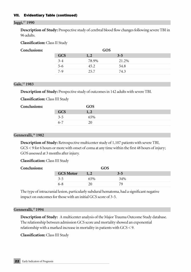
22 Early Indicators of Prognosis
Jaggi,17 1990
Description of Study: Prospective study of cerebral blood flow changes following severe TBI in96 adults.
Classification: Class II Study
Conclusions: GOSGCS 1, 2 3-53-4 78.9% 21.2%5-6 45.2 54.87-9 25.7 74.3
Gale,12 1983
Description of Study: Prospective study of outcomes in 142 adults with severe TBI.
Classification: Class III Study
Conclusions: GOSGCS 1, 23-5 65%6-7 20
Genneralli,14 1982
Description of Study: Retrospective multicenter study of 1,107 patients with severe TBI,GCS < 9 for 6 hours or more with onset of coma at any time within the first 48 hours of injury;GOS assessed at 3 months after injury.
Classification: Class III Study
Conclusions: GOSGCS Motor 1, 2 3-53-5 65% 34%6-8 20 79
The type of intracranial lesion, particularly subdural hematoma, had a significant negativeimpact on outcomes for those with an initial GCS score of 3-5.
Genneralli,13 1994
Description of Study: A multicenter analysis of the Major Trauma Outcome Study database.The relationship between admission GCS score and mortality showed an exponentialrelationship with a marked increase in mortality in patients with GCS < 9.
Classification: Class III Study
VII. Evidentiary Table (continued)
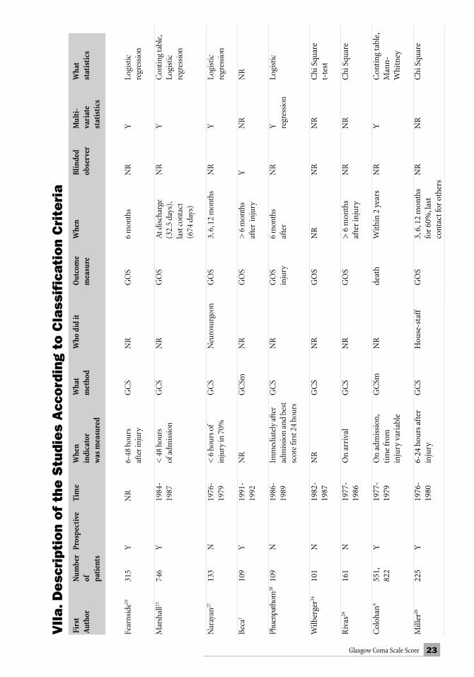
23Glasgow Coma Scale Score
Fir
stN
umbe
rPr
ospe
ctiv
eTi
me
Whe
nW
hat
Who
did
itO
utco
me
Whe
nBl
inde
dM
ulti
-W
hat
Aut
hor
ofin
dica
tor
met
hod
mea
sure
obse
rver
vari
ate
stat
isti
cspa
tien
tsw
as m
easu
red
stat
isti
cs
Fear
nsid
e1031
5Y
NR
6-48
hou
rsG
CS
NR
GO
S6
mon
ths
NR
YLo
gist
icaf
ter i
njur
yre
gres
sion
Mar
shal
l2374
6Y
1984
-<
48
hour
sG
CS
NR
GO
SA
t dis
char
geN
RY
Con
ting
tabl
e,19
87of
adm
issi
on(3
2.5
days
),Lo
gist
icla
st c
onta
ctre
gres
sion
(674
day
s)
Nar
ayan
2713
3N
1976
-<
6 h
ours
of
GC
SN
euro
surg
eon
GO
S3,
6, 1
2 m
onth
sN
RY
Logi
stic
1979
inju
ry in
70%
regr
essi
on
Bec
a110
9Y
1991
-N
RG
CSm
NR
GO
S>
6 m
onth
sY
NR
NR
1992
afte
r in
jury
Phue
npat
hom
2810
9N
1986
-Im
med
iate
ly a
fter
GC
SN
RG
OS
6 m
onth
sN
RY
Logi
stic
1989
adm
issi
on a
nd b
est
inju
ryaf
ter
regr
essi
onsc
ore
first
24
hour
s
Wilb
erge
r3410
1N
1982
-N
RG
CS
NR
GO
SN
RN
RN
RC
hi S
quar
e19
87t-
test
Riv
as29
161
N19
77-
On
arri
val
GC
SN
RG
OS
> 6
mon
ths
NR
NR
Chi
Squ
are
1986
afte
r in
jury
Col
ohan
955
1,Y
1977
-O
n ad
mis
sion
,G
CSm
NR
deat
hW
ithi
n 2
year
sN
RY
Con
ting
tabl
e,82
219
79ti
me
from
Man
n-in
jury
var
iabl
eW
hitn
ey
Mill
er26
225
Y19
76-
6-24
hou
rs a
fter
GC
SH
ouse
-sta
ffG
OS
3, 6
, 12
mon
ths
NR
NR
Chi
Squ
are
1980
inju
ryfo
r 60
%, l
ast
cont
act f
or o
ther
s
VII
a.D
esc
ript
ion
of
the
Stu
dies
Acc
ord
ing
to C
lass
ific
atio
n C
rite
ria

24 Early Indicators of Prognosis
VII
a.D
esc
ript
ion
of
the
Stu
dies
Acc
ord
ing
to C
lass
ific
atio
n C
rite
ria
(co
ntin
ue
d)
Fir
stN
umbe
rPr
ospe
ctiv
eTi
me
Whe
nW
hat
Who
did
itO
utco
me
Whe
nB
linde
dM
ulti
-W
hat
Aut
hor
ofin
dica
tor
met
hod
mea
sure
obse
rver
vari
ate
stat
isti
cspa
tien
tsw
as m
easu
red
stat
isti
cs
Youn
g3517
0Y
NR
Dur
ing
firs
tG
CS
NR
GO
S1
year
NR
YM
ulti
ple
wee
klo
gist
icre
gres
sion
Jagg
i1796
YN
R<
96
hour
sG
CS
NR
GO
S6
mon
ths
YY
2 w
ayA
NO
VA,
logi
stic
regr
essi
on
Gal
e1245
1Y
1980
-N
RG
CS
NR
Dea
thN
RN
RN
RN
R19
81
Bra
akm
an6
305
Y19
73-
24 h
ours
GC
S N
RG
OS
1, 3
, 6, 1
2 m
onth
sN
RY
Step
wis
e19
78lo
gist
icre
gres
sion
Gen
nare
lli13
1,10
7 N
NR
Wit
hin
6-48
hou
rsG
CS
NR
GO
S3
mon
ths
NR
NN
R
Gen
nare
lli14
46,9
77Y
1982
-O
n ad
mis
sion
GC
S N
RD
eath
in-h
ospi
tal
NR
NE
xpon
enti
al19
89m
odel
25Glasgow Coma Scale Score
VIII. References1. Beca J, Cox PN, Taylor MJ, et al.: Somatosensory evoked potentials for prediction of
outcome in acute severe brain injury. J Pediat 126:44-49, 1995.2. Benzer A, Traweger C, Ofner D, et al.: Statistical modelling in analysis of outcome after
trauma: Glasgow Coma Scale and Innsbruck Coma Scale. Anasthesiol Intensivmed NotfallmedSchmerzther 30:231-235, 1995.
3. Bhatty GB, Kapoor N: The Glasgow Coma Scale: a mathematical critique. Acta Neurochir120:132-135, 1993.
4. Braakman R: Interactions between factors determining prognosis in populations of patientswith severe head injury. In Frowein RA, Wilcke O, Karimi-Nejad A, et al. Advances inNeurosurgery: Head Injuries-Tumors of the Cerebellar Region. Springer-Verlag, Berlin: 12-15,1978.
5. Braakman R, Avezaat CJ, Maas AI, et al.: Interobserver agreement in the assessment of themotor response of the Glasgow Coma Scale. Clin Neurol Neurosurg 80:100-106, 1977.
6. Braakman R, Gelpke GJ, Habbema JD, et al.: Systematic selection of prognostic features inpatients with severe head injury. Neurosurg 6:362-370, 1980.
7. Choi SC, Barnes TY, Bullock R, et al.: Temporal profile of outcomes in severe head injury. JNeurosurg 81:169-173, 1994.
8. Choi SC, Narayan RK, Anderson RL, et al.: Enhanced specificity of prognosis in severe headinjury. J Neurosurg 69:381-385, 1988.
9. Colohan ART, Alves WM, Gross CR, et al.: Head injury mortality in two centers withdifferent emergency medical services and intensive care. J Neurosurg 71:202-207, 1989.
10. Fearnside MR, Cook RJ, McDougall P, et al.: The Westmead Head Injury Project outcome insevere head injury. A comparative analysis of pre-hospital, clinical, and CT variables. Br JNeurosurg 7:267-279, 1993.
11. Fielding K, Rowley G: Reliability of assessments by skilled observers using the GlasgowComa Scale. Aust J Adv Nurs 7:13-21, 1990.
12. Gale JL, Dikmen S, Wyler A, et al.: Head injury in the Pacific Northwest. Neurosurg12:487-491, 1983.
13. Gennarelli TA, Champion, HR, et al.: Comparison of mortality, morbidity, and severity of59,713 head injured patients with 114, 447 patients with extracranial injuries. J Trauma37:962-968, 1994.
14. Gennarelli TA, Spielman GM, Langfitt TW, et al.: Influence of the type of intracranial lesionon outcome from severe head injury. J Neurosurg 56:26-32, 1982.
15. Gorke Schmidt U: Non-target visual event-related potentials in evaluation of children withminor head trauma. Neuropediatrics 22:79-84, 1991.
16. Hartley C, Cozens A, Mendelow AD, et al.: The Apache II scoring system in neurosurgicalpatients: a comparison with simple Glasgow coma scoring. Br J Neurosurg 9:179-187, 1995.
17. Jaggi JL, Obrist WD, Gennarelli TA, et al.: Relationship of early cerebral blood flow andmetabolism to outcome in acute head injury. J Neurosurg 72:176-182, 1990.
18. Jennett B, Teasdale G: Aspects of coma after severe head injury. Lancet 1:878-881, 1977.19. Jennett B, Teasdale G, Braakman R, et al.: Predicting outcome in individual patients after
severe head injury. Lancet 1:1031-1034, 1976.20. Kaufmann MA, Buchmann B, Scheidegger D, et al.: Severe head injury: should expected
outcome influence resuscitation and first-day decisions. Resuscitation 23:199-206, 1992.

26 Early Indicators of Prognosis
21. Kuday C, Uzan M, Hanci M: Statistical analysis of the factors affecting the outcome ofextradural haematomas: 115 cases. Acta Neurochir 131:203-206, 1994.
22. Marion DW, Carlier PM: Problems with initial Glasgow Coma Score assessment caused bythe prehospital treatment of head-injured patients: results of a national survey. J Trauma36:89-95, 1994.
23. Marshall LF, Gautille T, Klauber MR, et al.: The outcome of severe closed head injury. JNeurosurg (Suppl) 75:28-36, 1991.
24. Menegazzi JJ, Davis EA, Sucov AN, et al.: Reliability of the Glasgow Coma Scale when usedby emergency physicians and paramedics. J Trauma 34:46-48, 1993.
25. Michaud LJ, Rivara FP, Grady MS, et al.: Predictors of survival and severity of disabilityafter severe brain injury in children. Neurosurg 31:254-264, 1992.
26. Miller JD, Butterworth JF, Gudeman SK, et al.: Further experience in the management ofsevere head injury. J Neurosurg 54:289-299, 1981.
27. Narayan RK, Greenberg RP, Miller JD, et al.: Improved confidence of outcome prediction insevere head injury. J Neurosurg 54:751-762, 1981.
28. Phuenpathom N, Choomuang M, Ratanalert S: Outcome and outcome prediction in acutesubdural hematoma. Surg Neurol 40:22-25, 1993.
29. Rivas JJ, Lobato RD, Sarabia R, et al.: Extradural hematoma: analysis of factors influencingthe course of 161 patients. Neurosurg 23:44-51, 1988.
30. Stablein DM, Miller JD, Choi SC, et al.: Statistical methods for determining prognosis insevere head injury. Neurosurg 6:243-248, 1980.
31. Teasdale G, Jennett B: Assessment of coma and impaired consciousness. A practical scale.Lancet 2:81-84, 1974.
32. Teasdale G, Knill-Jones R, Van Der Sande J: Observer variability in assessing impairedconsciousness and coma. J Neurol Neurosurg Psychiatry 41:603-610, 1978.
33. Thatcher RW, Cantor DS, McAlaster R, et al.: Comprehensive predictions of outcome inclosed head-injured patients. The development of prognostic equations. Ann N Y Acad Sci620:82-101, 1991.
34. Wilberger JE, Jr., Harris M, Diamond DL: Acute subdural hematoma: morbidity andmortality related to timing of operative intervention. J Trauma 30:733-736, 1990.
35. Young B, Rapp RP, Norton JA, et al.: Early prediction of outcome in head-injured patients. JNeurosurg 54:300-303, 1981.

27Age
AGE
I. ConclusionsA. Which feature of the parameter is supported by Class I evidence and has at least a
70% positive predictive value? There is an increasing probability of poor outcomewith increasing age, in a stepwise manner.
B. Parameter measurement for prognosis:Age is not subject to observer measurement variability. Age should be obtained onadmission, preferably with documentation.
II. OverviewThe prognosis for recovery from trauma as one ages is a function not only of the aged brain, butthe type of injury that occurs frequently in each age group. In addition, a decline in health asone ages may predispose the aged to systemic complications after head injury.
An examination of injury type with respect to age demonstrates an increasing proportionof injuries secondary to falls and pedestrian accidents with advancing age2, 20, 24, 34, 42. In thisprospective study of the Traumatic Coma Data Bank (TCDB), motor-vehicle crashes were thecause of injury in 55% of patients ages 15–25, whereas only about 5% suffered falls. However, inthe age range above 55, 45% suffered falls and only about 15% were in motor-vehicle crashes.However, falls as a mode of injury did not appear as an independent predictor of poor outcome.Old patients had a poor outcome compared to younger patients, regardless of the cause ofinjury.
In the TCDB study, a marked increase in pre-existing systemic disease was found withincreasing age. There was a significantly increased percentage of poor outcomes (death andvegetative) in those patients with prior systemic disease in ages above 56 (86% vs 50%);however, this correlation was not found in younger age groups. In addition, multiple systemicinjuries were less likely in the older age group thus emphasizing the role of the severity of braininjury in determining outcome.
The reaction of the aged brain to trauma may be apparent in the head computedtomography (CT) scans of patients. In the above TCDB study there was an age-related trendtoward increasing intracranial hematomas with the largest intracerebral hematomas observedin the oldest groups. The chances of survival in patients with intracranial hematomas decreasewith advancing age2, 4, 9, 15, 29, 34, 39, 42, 43. A significant correlation was noted in the TCDB studybetween a poor outcome and those patients who had intracerebral or extracerebral hematomas

28 Early Indicators of Prognosis
greater than 15 cc, subarachnoid hemorrhage, midline shift, compressed cisterns, or shift, whichall increased with age (except for compressed cisterns). Unfortunately there were too few olderpatients without mass lesion to critically evaluate the effect independent of age.
A multivariate logistic regression analysis was done of the TCDB to evaluate the independenteffect of age on outcome from severe head injury. Age was found to be an independent predictorafter other factors were excluded. One explanation for this is that the brain has a decreasedcapacity for repair as it ages. This has some support in that the proportion of survivors in GlasgowOutcome Scale scores of good recovery (GCS scores 5, 4, and 3) all declined with age.
III. ProcessA MEDLINE search was performed between 1966 and 1995 exploring the following subjects:1) age, 2) human head injury, and 3) prognosis. The search resulted in 44 references thatwere individually reviewed and classified.
IV. Scientific FoundationIn the last few decades, several authors have identified age as a strong prognostic indicatorfollowing injury to the brain6, 14, 20, 22, 31, 41. Most investigations have stressed that youngerindividuals do better than adults. A remarkably low mortality rate among children was noted asearly as 1973.20 Later studies described similar results, and revealed that a higher proportion ofchildren achieved a lower incidence of mortality and better outcomes than adults.1, 3, 5, 8, 17, 26
There are discrepancies in the literature when defining the age point where prognosissignificantly worsens. For example, there has been disagreement regarding the pediatric agegroup. One group of reports has indicated that outcome tends to be better in children under tenyears of age9, 13, 28,44, while others report that children under five have a higher mortalityrate6, 18, 22, 25, 27, 32, 35, 36. Several large pediatric head injury series have reported that children have alower mortality than adults, while others report that the primary mortality rate does not differbetween children and adults. Additionally, some investigations reported better outcomes belowthe age range of 40-50 years6, 7, 12, 16, 19, 21, 36, while other studies reported outcome as a continuousfunction of age without threshold values 4, 10, 11, 17, 22, 26, 30, 31, 32, 37, 40. These discrepancies appear to berelated to variations in the definitions of age groups.
A prospective investigation of 372 TBI patients in the UK with a GCS score less than 13 orISS greater than 16 and age above 14 years showed no prognostic effect of age to 50 years.38 Atthis point, age became an independent predictor of mortality, and GCS and ISS added highmortality significance when individually added to this model.
A prospective study of age and outcome from the TCDB revealed that patients older than60 had a significantly worse outcome. Six months after severe head injury, 92% were dead,vegetative, or severely disabled. Four Class I studies demonstrated a mortality of greater than75% in severely brain injured patients older than 60.5, 25, 31, 33 The critical age threshold forworsening prognosis appears to be above 60 in a review of Class I and II studies. However, thismay be an artifact of the age grouping used by various authors in converting continuous datainto categorical data.

29Age
The following chart summarizes the Class I papers with regard to age threshold and pooroutcome:
First Author Age Threshold Poor Outcome Age Range Poor Outcome
Vollmer,42 1991 > 55 92% (GOS 1, 2, 3) 46-55 78% (GOS 1, 2, 3)80% (GOS 1) 49% (GOS 1)
Braakman,6 1980 > 51 75% (GOS 1) 41-50 49% (GOS 1)> 61 77% (GOS 1)
Teasdale,40 1979 > 60 87% (GOS 1, 2) 40-60 56% (GOS 1)Narayan,31 1981 > 60 78% 41-60 57% (GOS 1, 2, 3)
46% (GOS 1)Signorini,38 1999 ≥ 50 Linear decline 14-49 No significant
in probability effectof survival
V. SummaryAge is a strong factor influencing both mortality and morbidity. Despite some contradictions,most literature supports children faring better than adults who have severe brain injury. Thesignificant influence of age on outcome is not explained by the increased frequency of systemiccomplications or intracerebral hematomas with age. Increasing age is a strong independentfactor in prognosis with a significant increase in poor outcome above 60 years of age.
VI. Key Issues for Future InvestigationFuture studies should record age as a continuous variable in their study designs. Furthermore,potentially confounding variables such as pre-existing medical conditions should be recordedand analyzed. The biology of the aging brain and its vulnerability to injury should beinvestigated.

30 Early Indicators of Prognosis
VII. Evidentiary Table for Age and OutcomeAlberico,1 1987 + + + + -
Description of Study: Prospective analysis of a consecutive series of 330 severely head-injuredpediatric and adult patients treated with the same protocol, by the same physicians and staff in theIntensive Care Unit. The pediatric patients had a significantly higher percentage of good outcomesthan the adult patients. They also had a significantly lower mortality rate than the adult patients.
Classification: Class II Study
Conclusions: GOSAge 1 51-4 (N=6) 17% 17%5-9 (N=18) 22 6110-14 (N=20) 20 4015-19 (N=56) 25 4021-40 35 3341-60 55 1561-80 80 5
Amacher,2 1987 + - - + -
Description of Study: Retrospective analysis of 56 patients 80 or more years of age. Even if asignificant proportion (60%) of old people may make a full recovery from head injury, themortality rate is high even in those with good admissions.
Classification: Class III Study
Conclusions: GOSAge 1 2 3 4 5≥ 80 (N=10) 80% 0% 10% 10%
Berger,5 1985 + + + + -
Description of Study: Retrospective analysis of a consecutive series of 37 children with severe headinjury. The data confirm that morbidity and mortality are lower in children than in adults: 51% of theseyoung patients had a good recovery or moderate disability after 6 months. The mortality rate of 33% ishigher than in some reports but probably more closely approximates the death rate from these injuries.
Classification: Class II Study
Conclusions: GOSAge 1 2 3 4 50-5 (N=16) 26% 0% 12% 12% 50%6-10 (N=7) 43 0 0 14 4311-17 (N=14) 36 7 21 29 7
+, - refers to whether methodology parameters were met or not. (See description of studies and methodology section. Page 11)

31Age
Braakman,6 1980 + + + + +
Description of Study: Retrospective analysis of 305 consecutive head-injured Dutch patients. Therelationship between age and mortality after 6 months shows an increasing mortality rate withincreasing age.
Classification: Class I Study
Conclusions: GOSAge 10-10 (N=40) 35%11-20 (N=85) 3321-30 (N=46) 3731-40 (N=38) 4441-50 (N=29) 5551-60 (N=20) 7561-70 (N=26) 77≥ 70 (N=21) 100
Bricolo,7 1977 + - - + +
Description of Study: Retrospective analysis of 800 patients with severe head injuries with andwithout decerebrate rigidity. In patients of all ages without decerebrate rigidity, the mortality rateprogressively increases with age whereas the mortality rate in decerebrate patients is constant andindependent of age. Of the survivors, three-fourths of those with good recovery were under 40years of age.
Classification: Class III Study
Conclusions: GOSAge (N=800) 1
5 1%10 4 20 8 30 15 40 22 50 29 60 35 70 45 80 55
VII. Evidentiary Table (continued)

32 Early Indicators of Prognosis
Bruce,8 1978 + + - + -
Description of Study: Retrospective analysis of the outcome in 53 children following severehead injury. 90% of the patients made a good recovery or were moderately disabled, and 8% diedor were left vegetative.
Classification: Class III Study
Conclusions: GOSAge 1 2-3 4-50-5 (N=21) 5% 5% 90%6-10 (N=19) 5 5 9010-17 (N=13) 8 0 92
Choi,11 1983 + + - + +
Description of Study: Retrospective analysis of 264 patients with severe head injury. Acombination of the Glasgow Coma Scale score, oculocephalic response, and age can provide asimple but reliable prediction of outcome in severe head injury.
Classification: Class II Study
Conclusions: Age not assessed as an independent predictor of outcome.
Edna,14 1983 + - + + -
Description of Study: Prospective analysis including 1,120 head-injured patients between 1979and 1980. In addition to the level of unconsciousness at admission, age, pupillary light reactions,intracranial hematoma, associated extracranial injuries, and skull fractures seem to be important forpredicting the outcome.
Classification: Class III Study
Conclusions: GOSAge 1-20-39 (N=38) 24%≥ 40 (N=18) 33
Gordon,16 1995 + + - + +
Description of Study: Retrospective analysis of 2,298 head-injured patients. Outcomesignificantly correlates to age and type and severity of lesion. No table of age versus outcome inGCS less than or equal to 8.
Classification: Class II Study
VII. Evidentiary Table (continued)

33Age
Heiskanen,19 1970 + + - + -
Description of Study: Retrospective analysis of 204 patients with severe head injury. Inpatients over 60, no special or heroic methods of treatment are indicated, but in children andadolescents every effort should be made as long as there has not been respiratory arrest andcerebral death.
Classification: Class III Study
Conclusions: GOSAge 10-20 (N=62) 32%21-40 (N=62) 4841-60 (N=53) 59≥ 60 (N=27) 78
Jennett,22 1979 + + + + -
Description of Study: Retrospective analysis of the relationship between clinical features ofbrain dysfunction in the first week after severe head injury and outcome 6 months later for 1,000patients. Depth of coma, pupil reactions, eye movements, motor response pattern, and patient’sage proved to be the most reliable predictors of outcome.
Classification: Class II Study
Conclusions: GOSAge 1-2< 20 (N=320) 33%20-39 (N=284) 4740-59 (N=245) 56> 60 (N=151) 87
Leurssen,26 1988 + - + + -
Description of Study: Prospective analysis of a series of 8,814 head-injured patients admittedto 41 hospitals in three separate metropolitan areas. The pediatric patients exhibited asignificantly lower mortality rate compared to the adults indicating that age itself, even within thepediatric age range, is a major independent factor affecting the mortality rate in head-injuredpatients.
Classification: Class III Study
Conclusions: GOSAge 10-14 (N=95) 28%≥ 15 (N=681) 48
VII. Evidentiary Table (continued)

34 Early Indicators of Prognosis
Miller,30 1981 + + + + -
Description of Study: Prospective analysis of 225 patients with severe head injury who weremanaged in a uniform way and analyzed to relate outcome to several clinical variables. Factorsimportant in predicting a poor outcome include the presence of an intracranial hematoma,increasing age, abnormal motor responses, impaired or absent eye movements or pupillaryreflexes, early hypotension, hypoxemia, or hypercarbia, and elevation of intracranial pressureover 20 mm Hg, despite artificial ventilation.
Classification: Class II Study
Conclusions: GOSAge 1 2-3 4-5
0-20 19% 11% 70%21-40 34 10 5641-60 44 12 4461-90 71 23 6
(NT
= 225)
Narayan,31 1981 + + + + +
Description of Study: Prospective analysis of 133 severely head-injured patients in predictingoutcome. A combination of clinical data including age, GCS score, pupillary response, presenceof surgical mass lesions, extra-ocular motility, and motor posturing predicts outcome with 82%accuracy, 43% with over 90% confidence.
Classification: Class I Study
Conclusions: GOSAge 1 2, 3 4, 50-20 (N=46) 17% 11% 72%21-40 (N=50) 28 6 6641-60 (N=28) 46 11 43≥ 61 (N=9) 78 0 23
Overgaard,32 1973 + + + - +
Description of Study: Prospective analysis of 201 patients injured in road-traffic accidents inan attempt to ascertain clinical factors of prognostic significance after traumatic head injury.Increasing age and post-traumatic hypotension were both related to poor recovery, while majorintracranial and extracranial surgical complications were associated with poor functionalrecovery and increased mortality, respectively.
Classification: Class II Study
VII. Evidentiary Table (continued)
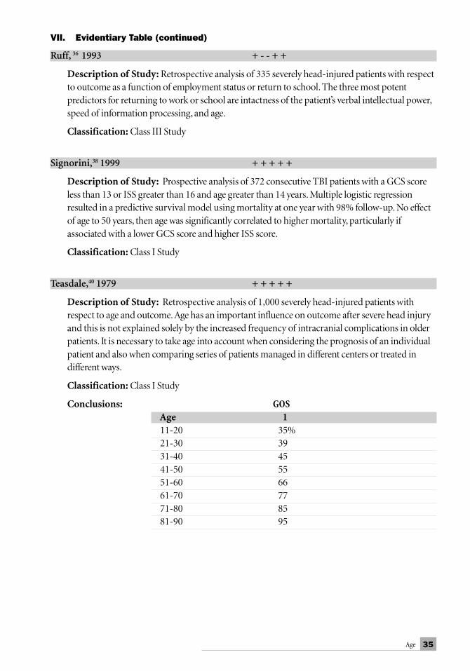
35Age
Ruff, 36␣ 1993 + - - + +
Description of Study: Retrospective analysis of 335 severely head-injured patients with respectto outcome as a function of employment status or return to school. The three most potentpredictors for returning to work or school are intactness of the patient’s verbal intellectual power,speed of information processing, and age.
Classification: Class III Study
Signorini,38 1999 + + + + +
Description of Study: Prospective analysis of 372 consecutive TBI patients with a GCS scoreless than 13 or ISS greater than 16 and age greater than 14 years. Multiple logistic regressionresulted in a predictive survival model using mortality at one year with 98% follow-up. No effectof age to 50 years, then age was significantly correlated to higher mortality, particularly ifassociated with a lower GCS score and higher ISS score.
Classification: Class I Study
Teasdale,40 1979 + + + + +
Description of Study: Retrospective analysis of 1,000 severely head-injured patients withrespect to age and outcome. Age has an important influence on outcome after severe head injuryand this is not explained solely by the increased frequency of intracranial complications in olderpatients. It is necessary to take age into account when considering the prognosis of an individualpatient and also when comparing series of patients managed in different centers or treated indifferent ways.
Classification: Class I Study
Conclusions: GOSAge 111-20 35%21-30 3931-40 4541-50 5551-60 6661-70 7771-80 8581-90 95
VII. Evidentiary Table (continued)

36 Early Indicators of Prognosis
Vollmer,42 1991 + + + + +
Description of Study: Prospective analysis of age and clinical outcome following traumaticcoma, age 15 years or older. The effect of age and outcome following head injury is dependent onan alteration in the pathophysiological response of the aging central nervous system to severetrauma and not on an increased incidence of non-neurological complications or other clinicalparameters.
Classification: Class II Study
Conclusions: GOSAge 1 2 3 4 515-25 (N=311) 31% 4% 16% 16% 33%26-35 (N=151) 29 7 16 21 2836-45 (N=83) 41 7 18 17 1746-55 (N=45) 49 9 20 11 11≥ 56 (N=71) 80 3 9 9 0
VII. Evidentiary Table (continued)

37Age
Fir
stN
umbe
rPr
ospe
c-Ti
me
Whe
nW
hat
Who
did
itO
utco
me
Whe
nB
linde
dM
ulti
-W
hat s
tati
stic
s A
utho
rof
tive
indi
cato
rm
etho
dm
easu
reob
serv
erva
riat
e sta
tist
.pa
tien
tsw
as m
easu
red
at o
utco
me
Y=
yes
ND
=no
t don
e
Alb
eric
o133
0Y
1976
-198
4O
n ad
miss
ion
Age
Neu
rosu
rgG
OS
3 m
onth
s, 8
year
sN
RN
DD
escr
iptiv
eA
mac
her2
56N
1981
-198
5O
n ad
miss
ion
Age
NR
Exce
llent
-N
RN
RN
DD
escr
iptiv
epo
or g
radi
ngB
ecke
r416
0N
1972
-197
6O
n ad
miss
ion
Age
Neu
rosu
rgG
OS
> 3
mon
ths
NR
ND
Chi
Squ
are
Ber
ger5
37Y
1972
-198
2O
n ad
miss
ion
Age
NR
GO
S>
6 m
onth
sN
RN
DD
escr
iptiv
eBr
aakm
an6
305
Y19
72-1
978
On
adm
issio
nA
geN
RG
OS
6 m
onth
sN
RY
Mul
tivar
iant
Bric
olo7
800
N19
73-1
975
On
adm
issio
nA
geN
euro
surg
Mot
or p
atte
rn<
48
mon
ths
NR
ND
Des
crip
tive
Bruc
e853
N19
75-1
977
On
adm
issio
nA
geN
RG
OS
6 m
onth
sN
RN
DD
escr
iptiv
eC
hoi11
264
N19
76-1
981
On
adm
issio
nA
geN
RG
OS
6 m
onth
sN
RY
Logi
stic
regr
essio
nEd
na14
1,12
0Y
1979
-198
8O
n ad
miss
ion
Age
NR
GO
SA
t dis
char
geN
RN
DC
hi S
quar
eG
ordo
n162,
298
N19
68-1
988
On
adm
issio
nA
geN
RG
OS
3, 6
, 12
mon
ths
NR
ND
Chi
Squ
are
Hei
skan
en19
204
N19
62-1
965
On
adm
issio
nA
geN
RD
eath
3-5
year
sN
RN
DSt
uden
t´s t
-tes
tJe
nnet
t221,
000
N19
68-1
976
On
adm
issio
nA
geN
RG
OS
6 m
onth
sN
RN
DD
escr
iptiv
eLu
erss
en26
8,81
4N
1980
-198
1O
n ad
miss
ion
Age
NR
Dea
thN
RN
RY
Chi
Squ
are,
Logi
stic
regr
essio
nan
alys
isM
iller
3022
5Y
1976
-198
0O
n ad
miss
ion
Age
NR
GO
S3,
6, 1
2 m
onth
sN
RN
DC
hi S
quar
eN
aray
an31
133
Y19
76-1
979
On
adm
issio
nA
geN
RG
OS
3, 6
, 12
mon
ths
NR
YLo
gist
ic re
gres
sion
anal
ysis
Ove
rgaa
rd32
201
Y19
68-1
969
On
adm
issio
nA
geN
RN
euro
l.24
, 36
mon
ths
NR
ND
Chi
Squ
are
func
. exa
min
.R
uff36
93N
4 ye
ars
On
adm
issio
nA
geN
REm
ploy
men
t,6,
12
mon
ths
NR
YLo
gist
ic re
gres
sion
retu
rn to
scho
olan
alys
isSi
gnor
ini38
372
Y19
89-1
991
On
adm
issio
nA
geN
RD
eath
12 m
onth
sN
RY
Logi
stic
regr
essio
nan
alys
isTe
asda
le40
1,00
0Y
1968
-197
6O
n ad
miss
ion
Age
NR
GO
S6
mon
ths
NR
ND
Des
crip
tive
Vollm
er42
661
Y19
84-1
987
On
adm
issio
nA
geN
RG
OS
6 m
onth
sN
RY
Chi
Squ
are,
Logi
stic
regr
essio
nan
alys
is
NR
=no
t rec
orde
d; G
OS=
Gla
sgow
Out
com
e Sc
ale;
ND
= n
ot d
one
Evi
dent
ary
Tabl
e f
or
Age
and
Outc
om
e

38 Early Indicators of Prognosis
VIII. References1. Alberico AM, Ward JD, Choi SC, et al.:Outcome after severe head injury. Relationship to
mass lesions, diffuse injury, and ICP course in pediatric and adult patients. J Neurosurg67:648-656, 1987.
2. Amacher A, Bybee DE: Toleration of head injury by the elderly. Neurosurg 20:954, 1987.3. Andrews B, Pitts LH: Functional recovery after traumatic transtentorial herniation.
Neurosurg 2:227-231, 1991.4. Becker DP, Miller JD, Ward JD, et al.:The outcome from severe head injury with early
diagnosis and intensive management. J Neurosurg 47:491-502, 1977.5. Berger MS, Pitts LH, Lovely M, et al.: Outcome from a severe head injury in children and
adolescents. J Neurosurg 62:194-199, 1985.6. Braakman R, Glepke GJ, Habberna JDF, et al.:Systematic selection of prognostic features in
patients with severe head injury. Neurosurg 6:362-370, 1980.7. Bricolo A, Turazzi S, Alexander A, et al.: Decerebrate rigidity in acute head injury.
J Neurosurg 47:680-698, 1977.8. Bruce DA, Schut L, Bruno LA, et al.: Outcome following severe head injuries in children.
J Neurosurg 48:679-688, 1978.9. Carlsson CA, von Essen C, Löfgren J: Factors effecting the clinical course of patients with
severe head injuries. Part 1: Influence of biological factors. Part 2: Significance of post-traumatic coma. J Neurosurg 29:242-251, 1968.
10. Caruselli G, Luongo A: A prognosis of traumatic decerebrated rigidity. J Neurosurg Sci18:124-132, 1974.
11. Choi SC, Ward JD, Becker DP: Chart for outcome prediction in severe head injury.J Neurosurg 59:294-297, 1983.
12. Cifu DX, Kreutzer JS, Marwitz JH, et al.: Functional outcomes of older adults withtraumatic brain injury: a prospective multicenter analysis. Archives of Physical Medicine andRehabilitation 77:883-888, 1996.
13. Comninos SC: Early prognosis of severe head injuries in children. Acta Neurochir Suppl28:144-147, 1979.
14. Edna TH: Risk factors in traumatic head injury. Acta Neurochir 69:15-21, 1983.15. Fell DA, Fitzgerald S, Hoiel R, et al.: Acute subdural hematomas. Review of 144 cases.
J Neurosurg 42:37-42, 1975.16. Gordon E, von Holst H, Rudehill A: Outcome of head injury in 2,298 patients treated in a
single clinic during a 21 year period. J Neurosurg Anesth, Vol. 7, No 4:235-247, 1995.17. Gruszkiewicz J, Doron Y, Peyser E: Recovery from severe craniocerebral injury with
brainstem lesions in childhood. Surg Neurol 1:197-201, 1973.18. Heiden J, Small R, Caton W, et al.: Severe head injury. Clinical assessment and outcome.
Physical Therapy 63: 1946-1951, 1983.19. Heiskanen O, Sipponen P: Prognosis of severe brain injury. Acta Neurol Scand 46:343-348, 1970.20. Hernesniemi J. Outcome following head injuries in the aged. Neurochir 49:67-79, 1979.21. Hoppe E, Christensen L, Christensen KN: The clinical outcome of patients with severe head
injuries treated with high-dose dexamethazone, hyperventilation, and barbiturates.Neurochir 24: 17-20, January 1989.
22. Jennett B, Teasdale G: Prognosis after severe head injury. In: Management of Head Injuries.Davis:Philadelphia, 317, 1981.
39Age
23. Jennett B, Teasdale G, Braakman R, et al.: Prognosis of patients with severe head injury.Neurosurg 4:283-289, 1979.
24. Klauber MR, Barrett-Connor E, Marshall LF, et al.: The epidemiology of head injury. Am JEpidemiol 113:500-509, 1981.
25. Lavati A, Farina ML, Vecchi G, et al.: Prognosis of severe head injuries. J Neurosurg 57:779-783, 1982.
26. Leurssen TG, Klauber MR, Marshall LF: Outcome from head injury related to patient’s age.A longitudinal prospective study of adult and pediatric head injury. J Neurosurg 68:409-416,1988.
27. Mahoney WJ, D´Souza BJ, Haller JA, et al.: Long-term outcome of children with severehead trauma and prolonged coma. Pediatrics 71:756-762, 1983.
28. Mazza C, Pasqualin A, Feriotti G, et al.: Traumatic extradural hematomas in children:experience with 62 cases. Acta Neurochir 65:6780, 1982.
29. McKissock W, Richardson A, Bloom WH: Subdural hematoma: a review of 389 cases. Lancet1:1365-1369, 1960.
30. Miller JD, Butterworth JF, Gudeman SK: Further experience in the management of severehead injury. J Neurosurg 54:289-299, 1981.
31. Narayan RK, Greenberg RP, Miller JD, et al.: Improved confidence of outcome prediction insevere head injury in the clinical examination, multimodality evoked potentials, CTscanning, and intracranial pressure. J Neurosurg 54:751-762, 1981.
32. Overgaard J, Hvid-Hansen O, Land AM, et al.: Prognosis after head injury based on earlyclinical examination. Lancet 2:631-635, 1973.
33. Pazzagllia P, Frank G, Frank F, et al.: Clinical course and prognosis of acute post-traumaticcoma. J Neurol Neurosurg Psych 38:149-154, 1975.
34. Pentland B, Jones PA, Roy CW, et al.: Head injury in the elderly. Age and Ageing 15:193-202, 1986.35. Raimondi AJ, Hirschauer J: Head injury in the infant and toddler. Coma scoring and
outcome scale. Child’s Brain 11:12-35, 1984.36. Ruff RM, Marshall L, Crouch I, et al.:Predictors of outcome following head trauma. Brain
Injury 2:101-111, 1993.37. Sakas D, Ross Bullock M, Teasdale G: One-year outcome following craniotomy for
traumatic hematoma in patients with fixed dilated pupils. J Neurosurg 82:961-965, 1995.38. Signorini DF, Andrews PJ, Jones PA, et al.: Predicting survival using simple clinical
variables; a case study in traumatic brain injury. J Neurol Neurosurg Psychiatry 60:20-25, 1999.39. Talalla A, Morin MA: Acute traumatic subdural hematoma: a review of one hundred
consecutive cases. J Trauma 11:771-776, 1971.40. Teasdale G, Skene A, Parker L, et al.: Age and outcome of severe head injury. Acta Neurochir
(Suppl) 28:140-143, 1979.41. Teasdale G, Skene A, Spiegelhalter D, et al.: Age, severity, and outcome of head injury. In:
Grossman RG, Gildenberg PL (ed.): Head Injury: Basic and Clinical Aspects. New York:Raven Press, 213-220, 1982.
42. Vollmer DG, Torner JC, Eisenberg HM, et al.: Age and outcome following traumatic comas:why do older patients fare worse? J Neurosurg (Suppl) 5:37-49, 1991.
43. Wilberger J, Harris M, Diamond D: Acute subdural hematoma: morbidity, mortality, andoperative timing. J Neurosurg 74:212-218, 1991.
44. Zuccarello M, Facco E, Zampieri P, et al.: Severe head injuries in children: early prognosisand outcome. Childs Nerv Syst I:158-172, 1985.


41Pupillary Diameter and Light Reflex
I. ConclusionsA. Which feature of the parameter is supported by Class I evidence and has at least a
70% positive predictive value? Bilaterally absent pupillary light reflex.B. Recommendations for parameter measurement for prognosis:
1. How should it be measured?■ A measurement difference of 1mm or more is defined as asymmetry.■ A fixed pupil shows no response (< 1mm) to bright light.■ A pupillary size of > 4mm is recommended as the measure for a dilated
pupil.■ The duration of pupillary dilation and fixation should be recorded.
The following pupillary exam should be noted with L (left) or R (right)distinction and duration:■ Evidence of direct orbital trauma■ Asymmetrical response to light■ Asymmetry at rest■ Fixed pupil (one or both)■ Dilated pupil (one or both)■ Fixed and dilated pupils (one or both)
2. When should it be measured?■ After pulmonary and hemodynamic resuscitation
3. Who should measure it?■ Trained medical personnel
II. OverviewThe parasympathetic, pupilloconstrictor, light reflex pathway mediated by the third cranialnerve is anatomically adjacent to brainstem areas controlling consciousness and the medialtemporal lobe. Therefore, damage to the midbrain third nucleus or the efferent third nerve bytemporal lobe compression produces dilation of the pupil. If the damage or compression issignificant, the pupil will be unresponsive (fixed) to a light stimulus. This pupillary light reflexand the size of the pupil has traditionally been used as a clinical parameter in assessingtranstentorial herniation and as a prognostic indicator. The pupillary light reflex and size
PUPILLARY DIAMETERAND LIGHT REFLEX

42 Early Indicators of Prognosis
equality of pupils has a high interobserver reliability.21 The use of the pupillary size and lightreflex are, therefore, indirect measures of dysfunction to pathways subserving consciousnessand, thus, an important clinical parameter in assessing outcome from traumatic coma. Directorbital trauma can damage the third nerve leading to a dilated and/or a fixed pupil and beindependent of intracranial hypertension. Direct oculomotor trauma should be excludedbefore pupillary reactivity or size is used as a prognostic indicator.
III. Search ProcessA MEDLINE search for the period 1980-1995 was done using the key words “pupils,” “pupilsand prognosis,” and “pupils and trauma.” This resulted in the critical review of 19 articles.
IV. Scientific FoundationThe pupillary light reflex pathways are adjacent to brain structures essential for cognitive functionand the temporal lobe. Increased intracranial pressure resulting in uncal herniation compressesthe third cranial nerve resulting in a reduction in parasympathetic tone to the pupillaryconstrictor fibers and therefore results in a dilated pupil. Similarly, destruction of the third nerveparasympathetic brainstem pathway also results in a dilated and fixed to light pupil. Therefore,the pupillary light reflex is an indirect measure of herniation and brainstem injury. Generally,dilation and fixation of one pupil signifies herniation, whereas the appearance of bilaterally dilatedand fixed pupils is consistent with irreversible brainstem injury in a fully resuscitated patient. Alimitation in terms of prognosis is a dilated and nonreactive pupil due to direct orbital traumawithout brainstem or intracranial third nerve compression. The “blown pupil” is important in thecontext of a decreased level of consciousness. This measurement of pupil function must beassessed for outcome with the level of consciousness or intracranial pathology.
Clinical studies investigating the prognostic weight of the pupillary light reflex haveexamined this parameter in a variety of methodologies. Few studies have rigorously measuredthe size and reaction of the pupil to light.18 The vast majority label pupils as dilated withoutgiving the size and do not state whether the pupils are fixed to light even though it is implied.
The incidence of pupillary abnormalities (%) within 24 hours, post-resuscitation, inpatients with severe head injury is shown in the following table:
First Author Both Reactive One Reactive One or Both Unreactive Both Unreactive
Jennett,8 1976 78% —% —% 22%†
Jennett,9 1979 — — — 19*, 29, 32Braakman,4 1980 62 12 — 26Miller,13 1981 77 — — 23Narayan,14 1981 65 — — 35Heiden,7 1983 68 — — 32Van Dongen,22 1983 47 — 53 —Levin,10 1990†† 64 — 36 —Marshall,12 1991 56 11 — 33Average 65 12 — 28*Represents % from Glasgow, Netherlands, and Los Angeles, respectively.†Earlier series from Glasgow and Netherlands††Traumatic Coma Data Bank (TCDB) study

43Pupillary Diameter and Light Reflex
On average 65% of patients with severe head injury have normally reactive pupils afterresuscitation, 12% have one abnormal pupil, and 28% have bilateral pupillary nonreactivity.
There is significant interaction between pupillary reactivity and other early indicators ofprognosis; Glasgow Coma Scale (GCS) score, 4 hypotension,1 and CT basal cisterns,22 as seen inthe following table:
GCS 3-5 GCS 6-7 SBP < 60 SBP 60-90 Cisterns Partly Open Cisterns Closed
UnreactivePupils (%) 56 20 65 53 15* 38**Includes bilateral and unilateral unreactive pupils
In reviewing large studies (> 200 patients), there was a strong association betweenbilaterally unreactive pupils and poor outcome as shown in the following table:
% Vegetative/Dead (Glasgow Outcome Scale Score [GOS] 1, 2)First Author # of Patients Bilateral Reactive Pupils Unilateral Unreactive Bilateral Unreactive
Jennett,8 1976 600 42% —% 95%Braakman,4 1980 305 29 54 90Heiden,7 1983 213 36 — 91Marshall,12 1991 746 32 34 74
Average 35 44 88
In two Class I studies, bilaterally absent pupil reaction had a greater than 70% positive predictivevalue for a poor outcome. In a prospective study of 133 patients with severe head injury, bilaterallyabsent pupillary light reflex was noted in 35%; a poor outcome (dead, vegetative, or severely disabled)was found in 70% of these patients.14 Similarly, in a larger study of 305 patients with regard toprognostic features, bilaterally absent pupillary light reflex was associated with a 90% mortality (seeEvidentiary Table).4
In large series, patients who had bilaterally reactive pupils made a significantly betteroutcome as seen below:
% Good Recovery/Moderate Disability (GOS 4, 5)First Author # of Patients Bilateral Reactive Pupils Unilateral Reactive Bilateral Unreactive
Jennett,8 1976 600 50% —% 5%Heiden,7 1983 213 49 — 3Levin,10 1990 259 53 17* —
Average 51 4*One or both pupils unreactive.
The outcome from bilaterally unreactive pupils is influenced by the underlying pathologyand timing of surgical evaluation of significant hematomas. In patients who are comatose fromepidural hematomas, the mortality with bilateral fixed pupils is only 56% compared to anaverage of 88% in patients with subdural hematomas.15, 16 In another study of patients who wereoperated on for epidural hematomas, with bilaterally fixed pupils, only 18% had a poor

44 Early Indicators of Prognosis
outcome (GOS 1-2) compared to 64% poor outcome in those patients who were operated onfor subdural hematomas and had bilaterally fixed pupils.17, 18 In this same study a delay ofgreater than three hours in evacuating a traumatic intracranial hematoma increased the chanceof a poor outcome with bilateral fixed pupils from 40% to 63%.
In conclusion, pupil reactivity to light can prognosticate outcome. However, direct orbitaltrauma should be excluded as a causative agent, hypotension should be reversed prior toassessment of pupils, and repeat examination after evacuation of intracranial hematomasshould be performed.
V. SummaryThe pupillary diameter and the pupilloconstrictor light reflex are the two parameters that havebeen studied extensively in relation to prognosis. Accurate measurement of pupil diameter orthe constrictor response or the duration of the response has not been performed in studies ontraumatic brain-injured individuals—for lack of a standardized measuring procedure. Thefollowing is recommended:
1. Pupillary light reflex for each eye should be used as a prognostic parameter.2. The duration of pupillary dilation and fixation should be documented.3. A pupillary size greater than 4 mm is recommended as the measure for a dilated pupil.17
4. A fixed pupil should be defined as no constrictor response to bright light.5. Right or left distinction should be made when the pupils are asymmetric.6. Hypotension and hypoxia should be corrected before assessing pupils for prognosis.7. Direct orbital trauma should be excluded.8. Pupils should be reassessed after surgical evacuation of intracranial hematomas.
VI. Key Issues for Further InvestigationFuture studies should dissect the prognostic value of each of the recommended measurementsto discern the least number of pupillary size and light reflex measurements necessary to reliablyprognosticate outcome. Also, a standardized method of measuring pupil size and reactivity tolight would decrease interobserver variability.

45Pupillary Diameter and Light Reflex
VII. Evidentiary TableAndrews and Pitts,2 1991
Description of Study: Retrospective study of 153 consecutive patients presenting withtranstentorial herniation—altered level of consciousness, anisocoria or pupillaryunresponsiveness, and abnormal motor findings; age range 2-83 years.
Classification: Class III Study
Conclusions: GOS1-3 4 5
One Pupil Fixed 72% 13% 15%Both Pupils Fixed 96 4 0
Braakman,4 1980
Description of Study: Review of the International Databank with reference to 305 comatosehead-injured patients’ prognostic parameters measured with 24 hours of admission and (GOS)evaluated at 6 months.
Classification: Class I Study
Conclusions: GOS1
Both Pupils Reactive 29%One Pupil Fixed 54Both Pupils Fixed 91 + PPV = 91%
Choi,5 1988
Description of Study: A review of 523 severely head-injured patients analyzing significantprognostic parameters to predict outcome into four GOS categories.
Classification: Class III Study
Conclusions: Pupillary response to light was a significant (p < .001) factor in determining outcome.
Cordobes,6 1981
Description of Study: Retrospective analysis of 82 patients with regard to mortality before andafter instituting a computed tomography (CT) scanner.
Classification: Class III Study
Conclusions: GOS1
One Pupil Fixed 18%Both Pupils Fixed 100

46 Early Indicators of Prognosis
Heiden,7 1983
Description of Study: Prospective study of 213 patients of all ages with severe head injury toidentify favorable and unfavorable clinical factors.
Classification: Class II Study
Conclusions: GOS1, 2 3 4, 5
Both Pupils Reactive 36% 15% 49%Both Pupils Fixed 91 6 3
Jennett,8 1976
Description of Study: Prospective study in 600 severe head injury patients from Glasgow andthe Netherlands.
Classification: Class II Study
Conclusions: GOS1, 2 4, 5
Both Pupils Reactive 42% 50%Both Pupils Fixed 95 5
Jennett,9 1979
Description of Study: Expanded patient enrollment from 1976 publication with 1,000 patientsfrom Glasgow, Netherlands, and Los Angeles.
Classification: Class I Study
Conclusions: GOS1, 2 4, 5
Both Pupils Reactive 39% 50%Both Pupils Fixed 91 4
VII. Evidentiary Table (continued)
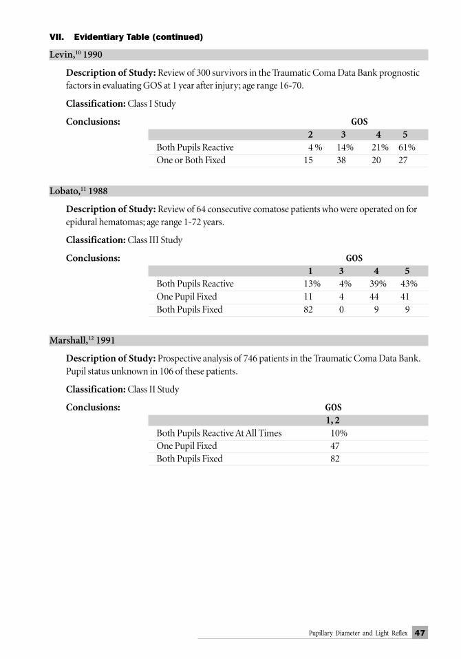
47Pupillary Diameter and Light Reflex
Levin,10 1990
Description of Study: Review of 300 survivors in the Traumatic Coma Data Bank prognosticfactors in evaluating GOS at 1 year after injury; age range 16-70.
Classification: Class I Study
Conclusions: GOS2 3 4 5
Both Pupils Reactive 4 % 14% 21% 61%One or Both Fixed 15 38 20 27
Lobato,11 1988
Description of Study: Review of 64 consecutive comatose patients who were operated on forepidural hematomas; age range 1-72 years.
Classification: Class III Study
Conclusions: GOS1 3 4 5
Both Pupils Reactive 13% 4% 39% 43%One Pupil Fixed 11 4 44 41Both Pupils Fixed 82 0 9 9
Marshall,12 1991
Description of Study: Prospective analysis of 746 patients in the Traumatic Coma Data Bank.Pupil status unknown in 106 of these patients.
Classification: Class II Study
Conclusions: GOS1, 2
Both Pupils Reactive At All Times 10%One Pupil Fixed 47Both Pupils Fixed 82
VII. Evidentiary Table (continued)

48 Early Indicators of Prognosis
Miller,13 1981
Description of Study: A prospective study of 225 consecutive severe head injury patients inregard to outcome. 41% and 10% of the surgical and nonsurgical cases, respectively, had bilateralfixed pupils; age range 2-89.
Classification: Class III Study
Conclusions: GOS1 2, 3 4, 5
Surgical: Both Pupils Fixed 77% 12% 11%Nonsurgical: Both Pupils Fixed 71 11 18
Narayan,14 1981
Description of Study: A study of 133 consecutive patients with severe head injury; age range 0-61+ years.
Classification: Class I Study
Conclusions: GOS1 2-3 4-5
Both Pupils Reactive 16% 8% 76%Both Pupils Unreactive 61 9 30positive productive value (PPV) = 70%
Phonprasert,15 1980
Description of Study: An analysis of 138 consecutive patients who were operated on forepidural hematomas with regard to factors influencing mortality; age range 3 to 71 years.
Classification: Class III Study
Conclusions: GOS1
One Pupil Fixed 15%Both Pupils Fixed 56
VII. Evidentiary Table (continued)

49Pupillary Diameter and Light Reflex
Phuenpathom,16 1993
Description of Study: A retrospective outcome prediction study of 109 consecutive with a GCSscore of 3-15 who presented with an acute subdural hematoma. 83 patients had clot removal. Agerange was 6 months to 79 years old.
Classification: Class III Study
Conclusions: GOS1
Both Pupils Reactive 16%One Pupil Fixed 48Both Pupils Fixed 88
Rivas,17 1988
Description of Study: A series of 161 consecutive patients with a GCS score of 3-15 were operatedon for epidural hematomas with regard to prognostic factors; age range 3 days to 78 years old.
Classification: Class II Study
Conclusions: GOS1 3 4 5
One Pupil Fixed 14% 4% 43% 39%Both Pupils Fixed 82 0 9 9
Sakas,18 1995
Description of Study: One-year outcome analysis of 40 consecutive patients who underwentcraniotomy for traumatic hematoma at various times after developing bilaterally fixed and dilatedpupils (> 4 mm); age range 6-75 years old.
Classification: Class II Study
Conclusions: GOS1, 2 3 4, 5
Patients Operated On Within ThreeHours of Both Pupils Fixed 40% 30% 30%Patients Operated On AfterThree Hours of Both Pupils Fixed 63 12 25Patients Operated On for EpiduralHematomas and Both Pupils Fixed 18 27 55Patients Operated On for SubduralHematomas and Both Pupils Fixed 64 23 13
VII. Evidentiary Table␣ (continued)

50 Early Indicators of Prognosis
Stone,19 1983
Description of Study: Review of 206 patients who were operated on for acute subduralhematomas; age range 3-88 years old.
Classification: Class III Study
Conclusions: GOS4, 5
One Pupil Fixed 25%Both Pupils Fixed 25
Suddaby,20 1987
Description of Study: Review of 49 cases of civilian gunshot wounds to the brain with a GCSscore of 3-15; age range 8-92 years old.
Classification: Class III Study
Conclusions: GOS1-3 4, 5
Both Pupils Reactive 28% 72%Both Pupils Fixed 100 0
Wilberger,23 1991
Description of Study: Review of 115 severely head injured patients (GCS ≤ 7) with subduralhematoma analyzing morbidity, mortality, and operative timing.
Classification: Class III Study
Conclusions: GOS1
Both Pupils Fixed 88%
VII. Evidentiary Table (continued)
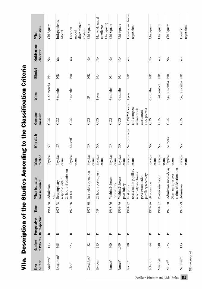
51Pupillary Diameter and Light Reflex
VII
a.
Desc
ript
ion
of
the
Stu
die
s A
cco
rdin
g to
the
Cla
ssif
icat
ion
Cri
teri
a
Firs
tN
umbe
rPr
ospe
ctiv
e/Ti
me
Whe
n in
dica
tor
Wha
tW
ho d
id it
Out
com
eW
hen
Blin
ded
Mul
tiva
riat
eW
hat
Aut
hor
of P
atie
nts
Ret
rosp
ecti
vew
as m
easu
red
met
hod
mea
sure
obse
rver
Stat
isti
cs
And
rew
s215
3R
1981
-88
Adm
issi
onPh
ysic
alN
RG
OS
1-37
mon
ths
No
No
Chi
Squ
are
exam
Braa
kman
430
5R
1973
-78
Bes
t pup
illar
yPh
ysic
alN
RG
OS
6 m
onth
sN
RYe
sIn
depe
nden
cere
spon
se w
ithin
exam
Mod
el24
hou
rs o
f adm
issi
onC
hoi5
523
R19
76-8
6In
ER
Phys
ical
ER st
aff
GO
S6
mon
ths
NR
Yes
Loca
tion
exam
mod
eldi
scri
min
ant
anal
ysis
Cor
dobe
s682
R19
73-8
0Ju
st b
efor
e op
erat
ion
Phys
ical
NR
GO
SN
RN
RN
oC
hi S
quar
eex
amH
eide
n721
3P
NR
24 h
ours
pos
t-in
jury
Phys
ical
NR
GO
S1
year
No
No
Man
tel-
Hae
nzel
exam
(sim
ilar t
oC
hi S
quar
e)Je
nnet
t860
0P
1968
-76
With
in 2
4 ho
urs
Phys
ical
NR
GO
S6
mon
ths
No
No
Chi
Squ
are
pos
t-in
jury
exam
Jenn
ett9
1,00
0P
1968
-76
With
in 2
4 ho
urs
Phys
ical
NR
GO
S6
mon
ths
No
No
Chi
Squ
are
post
-inj
ury
exam
Levi
n1030
0P
1984
-87
Firs
t pos
t-Ph
ysic
alN
euro
surg
eon
GO
S (2
63 p
oint
s)1
year
NR
Yes
Logi
stic
and
line
arre
susc
itatio
n pu
pilla
ryex
aman
d co
mpl
ete
regr
essi
onre
activ
ity a
nd lo
wes
tne
uro-
psyc
h.po
st-r
esus
cita
tion
asse
ssm
ent
pupi
llary
reac
tivity
(127
poi
nts)
Loba
to11
64R
1977
-86
At o
pera
tion
Phys
ical
NR
GO
S6
mon
ths
NR
No
Chi
Squ
are
exam
Mar
shal
l1264
0P
1984
-87
Post
-res
usci
tatio
nPh
ysic
alN
RG
OS
Last
con
tact
NR
Yes
Chi
Squ
are
exam
Mill
er13
225
P19
76-8
0A
dmis
sion
, mea
n de
lay
Phys
ical
Aut
hors
GO
S3,
6, 1
2 m
onth
sN
RN
oC
hi S
quar
e3
hrs.
s/p
inju
ry/o
rex
amat
tim
e of
det
erio
ratio
nN
aray
an14
133
P19
76-7
9A
dmis
sion
Phys
ical
NR
GO
S3,
6, 1
2 m
onth
sN
RYe
sLo
gist
icex
amre
gres
sion
NR
=no
t rep
orte
d

52 Early Indicators of Prognosis
VII
a.
Desc
ript
ion
of
the
Stu
die
s A
cco
rdin
g to
the
Cla
ssif
icat
ion
Cri
teri
a (c
ont
inu
ed)
Firs
t#
ofPr
ospe
ctiv
e/Ti
me
Whe
n in
dica
tor
Wha
tW
ho d
id it
Out
com
eW
hen
Blin
ded
Mul
tiva
riat
eW
hat
Aut
hor
Pati
ents
Ret
rosp
ecti
vew
as m
easu
red
met
hod
mea
sure
obse
rver
Stat
isti
cs
Phon
pras
ert15
138
P19
71-7
8N
RN
RN
RM
orta
lity
NR
NR
No
Chi
Squ
are
Phue
npat
hom
1610
9R
1/86
-A
dmis
sion
and
Ref
erra
lN
RG
OS
6 m
onth
sN
oYe
sLo
gist
ic re
gres
sion
12/8
924
hou
r pos
t-ad
miss
ion
note
s/pa
tient
reco
rds
Riv
as17
161
P19
77-8
6A
dmis
sion
Phys
ical
NR
GO
S—6
mon
ths
No
No
Chi
Squ
are
exam
good
reco
very
,Sa
kas18
40P
& R
1985
-88
Adm
issi
onPh
ysic
alA
utho
rs,
GO
S6
mon
ths
NR
No
Chi
Squ
are
exam
, CT
resi
dent
san
d 1
year
findi
ngs,
& n
urse
s p
ost-
opch
art r
evie
wSt
one19
206
R1/
69 -
Pre-
oper
ativ
eV
isua
lN
RG
OS—
6 m
onth
sN
oN
oC
hi S
quar
e6/
81in
spec
tion
Func
tiona
l/c
hart
revi
ewre
cove
rySu
ddab
y2049
R19
75-8
5A
dmis
sion
Cha
rtA
utho
rsG
OS
Dis
char
geN
oN
RN
Rre
view
Wilb
erge
r2310
1R
1982
-87
Earl
y op
erat
ive
NR
NR
GO
S—18
mon
ths
No
No
Chi
Squ
are/
at ti
me
of o
pera
tion/
Func
tiona
lSt
uden
t pai
red
Inte
rven
tion
< 4
hou
rsre
cove
ry o
r poo
led
t-te
stpo
st-t
raum
a

53Pupillary Diameter and Light Reflex
VIII. References1. Andrews BT, Levy ML, Pitts LH: Implications of systemic hypotension for the
neurological examination in patients with severe head injury. Surg Neurol 28:419-22, 1987.2. Andrews BT, Pitts LH: Functional recovery after traumatic transtentorial herniation.
Neurosurg 29:227-31, 1991.3. Braakman R: Interactions between factors determining prognosis in populations of
patients with severe head injury. Adv Neurosurg 5:12-15, 1998.4. Braakman R, Gelpke, GJ, Habbema JDF, et al.: Systematic selection of prognostic features
in patients with severe head injury. Neurosurg 6:362-70, 1980.5. Choi SC, Narayan RK, Anderson RL, et al.: Enhanced specificity of prognosis in severe
head injury. J Neurosurg 69:381-85, 1988.6. Cordobes F, Lobato RD, Rivas JJ, et al.: Observations on 82 patients with extradural
hematoma. J Neurosurg 54:179-186, 1981.7. Heiden JS, Small R, Caton W, et al.: Severe head injury clinical assessment and outcome.
Physical Therapy 63:1946-51, 1983.8. Jennett B, Teasdale G, Braakman R, et al.: Predicting outcome in individual patients after
severe head injury. Lancet 1:1031-1034, 1976.9. Jennett B, Teasdale G, Braakman R, et al.: Prognosis of patients with severe head injury.
Neurosurg 4:283-289, 1979.10. Levin HS, Gary HE, Eisenberg HM, et al.: Neurobehavioral outcome 1 year after severe
head injury. J Neurosurg 73:699-709, 1990.11. Lobato RD, Rivas JJ, Cordobes J, et al.: Acute epidural hematoma: an analysis of factors
influencing the outcome of patients undergoing surgery in coma. J Neurosurg 68:48-57,1988.
12. Marshall LF, Gautille T, Klauber M, et al.: The outcome of severe closed head injury.J Neurosurg (Suppl) 75:28-36, 1991.
13. Miller JD, Butterworth JF, Gudeman SK, et al.: Further experience in the management ofsevere head injury. J Neurosurg 54:289-299, 1981.
14. Narayan RK, Greenberg RP: Improved confidence of outcome prediction in severe headinjury. J Neurosurg 54:751-762, 198.
15. Phonprasert C, Suwanwela C, Hongsaprabhas C: Extradural hematoma: analysis of 138cases. J Trauma 20: 679-683, 1980.
16. Phuenpathom N, Choomuang M, Ratanalert S: Outcome and outcome prediction in acutesubdural hematoma. Surg Neurol 40:22-5, 1993.
17. Rivas J, Lobato R, Sarabia R, et al.: Extradural hematoma: analysis of factors influencingthe courses of 161 patients. Neurosurg 23:44-51, 1988.
18. Sakas DE, Bullock R, Teasdale G: One-year outcome following craniotomy for traumatichematoma in patients with fixed dilated pupils. J Neurosurg 82:961- 965, 1995.
19. Stone JL, Rifai MH, Sugar O, et al.: Acute subdural hematoma: progress in definition,clinical pathology, and therapy. Surg Neurol 19:216-31, 1983.
20. Suddaby L, Weir B, Forsyth C: The management of .22 caliber gunshot wounds of thebrain: a review of 49 cases. The Canadian Journal of Neurological Sciences 14:268-272, 1987.

54 Early Indicators of Prognosis
21. Van den Berge JH, Schouten HJA: Interobserver agreement in assessment of ocular signs incoma. J Neurol 42:1163-1168, 1979.
22. Van Dongen KJ, Braakman R: The prognostic value of computerized tomography incomatose head injured patients. J Neurosurg 59:951-957, 1983.
23. Wilberger JE, Harris M, Diamond DL: Acute subdural hematoma: morbidity, mortality, andoperative timing. J Neurosurg 74:212-218, 1991.

55Hypotension
I. ConclusionsA. Which feature of the parameter is supported by Class I evidence and has at least a 70%
positive predictive value (PPV)? A systolic blood pressure less than 90 mm Hg was foundto have a 67% PPV for poor outcome and, when combined with hypoxia, a 79% PPV.
B. Parameter measurement:1. How should it be measured?
Systolic and diastolic blood pressure should be measured using the most accuratesystem available under the circumstances. Monitoring by arterial line, when freeof signal artifact, provides data that is both accurate and continuous and is themethod of choice. Methods that do not determine the mean arterial pressure areless valuable.
2. When should it be measured?Blood pressures should be measured as frequently as possible. The incidence andduration of hypotension (systolic blood pressure < 90 mm Hg) should bedocumented by direct blood pressure values.
3. Who should measure it?Blood pressure should be measured by trained medical personnel.
II. OverviewSecondary brain insults are defined as post-traumatic insults to the brain arising from extracranialsources and intracranial hypertension. They are generally ischemic and include hypotension, hypoxia,anemia, infection, etc. There is a growing body of evidence that secondary insults to the injured brain arecommon and can powerfully influence recovery. The most detrimental and best studied of these ishypotension. Because hypotension is amenable to therapeutic manipulation, an understanding of itsinfluence on prognosis is useful for both prediction of outcome at present and optimization of recoveryin the future.
III. ProcessA MEDLINE search back to 1966 was undertaken using the following key words: “head injuryor brain injury” and “secondary insult or hypotension” and “outcome or prognosis” and “humansubject.” This produced 70 references that were individually reviewed for design, content, andrelevance. The results of this review were then incorporated into analysis presented here.
HYPOTENSION

56 Early Indicators of Prognosis
IV. Scientific FoundationIn the scientific literature published to date, the definition of hypotension has been acceptedfrom the literature on systemic insults. Despite the necessity for redefining this term in the braininjury literature, for the purposes of clarity in the following discussion, this entity is defined as:
• Hypotension = Systolic blood pressure < 90 mm Hg
The major secondary brain insults that have been studied with respect to their influence onoutcome from severe brain injury are hypotension and hypoxia. Seminal studies by Miller, et al.,established the importance of these secondary insults as outcome determinants, but did notstudy their independence with respect to other predictive factors.6, 7
The largest and most definitive study of the influence of secondary brain insults on outcomecomes from a Class I analysis of a large (717 patients), prospectively collected data set from theTraumatic Coma Data Bank (TCDB).2 Hypotension was defined as a single measurement of a systolicblood pressure less than 90 mm Hg. The occurrence of one or more episodes of hypotension duringthe period from injury through resuscitation or during the shorter period of resuscitation only wasassociated with a doubling of mortality and a marked increase in morbidity (see Evidentiary Table).2
Hypotension was found to be a statistically significant predictor of outcome and statisticallyindependent of other major predictors of outcome, including age, hypoxia, and the presence orabsence of severe trauma to one or more extracranial organ systems. When the influence ofhypotension on outcome was controlled separately, the statistical significance of severe trauma to oneor more extracranial organ systems as a predictor of outcome was eliminated, suggesting that theinfluence of systemic multiple trauma on the outcome of severe head injury patients is primarilymediated through hypotension.
The analysis of outcome from severe head injury in the TCDB revealed that the five mostpowerful predictors occurring from injury through resuscitation were age, intracranial(computed tomographic) diagnosis, pupillary reactivity, post-resuscitation Glasgow ComaScale (GCS) score, and presence or absence of hypotension. Notably, of these five majorpredictors, only the occurrence and severity of hypotension is amenable to medicalmanipulation.
The analysis of a smaller, prospectively collected database from Australia corroborated theabove findings.4 This study found early and late hypotension to be statistically significant,independent predictors of outcome, both for mortality and for dichotomized quality ofoutcome (good or moderate-to-severe deficits versus vegetative survival or death). In this study,hypotension was again the only predictor amenable to medical modification.
Further support of the strong association between early hypotension and outcome comesfrom a study of the influence of various resuscitation fluids on the outcome of hypotensivemultiple system trauma patients.12 The subgroup of patients with severe head injuries had anoverall mortality of 74%, with those being treated using conventional means of resuscitationhaving an 88% mortality rate.
A Class II report has recently extended the above findings to the pediatric population (ageless than 17 years).10 In this study, both hypoxia and hypotension had deleterious influences onoutcome with hypotension being significantly more powerful in independently determiningrecovery. In this study, an episode of hypotension appeared to eliminate the generally morefavorable outcome afforded by youth.

57Hypotension
Although the above studies firmly establish an association between secondary brain insults(particularly hypotension) and outcome, they do not address the issue of whether preventing ortreating such insults during this period improves recovery. With respect to secondary insults ingeneral (hypotension, hypoxia, hypercapnia, and anemia), a recent Class III study addressed theability of on-site, physician-directed resuscitation to decrease the incidence of secondary braininsults and improve outcome.1 Patients whose secondary brain insults were reversed in the fieldhad a 42% decrease in the frequency of poor outcomes (death, vegetative survival, or severedeficits) at three-month follow-up. Unfortunately, this study did not control for manyconfounding factors. Nevertheless, it does suggest that patients with secondary brain insultsthat respond to treatment have improved outcome when compared to those that are refractoryto correction.
With respect to hypotension in particular, Class II results from a recent Class I studystrongly suggest that reversing or preventing hypotension in the field improves outcome. Arecent prospective, randomized, placebo-controlled, multicenter trial examined the efficacy ofadministering 250 cc’s of hypertonic (7.5%) saline versus normal saline as the initialresuscitation fluid in hypotensive, multiple-trauma patients. For the group as a whole, there wasno statistically significant difference in outcome between the two groups. The hypertonic salinegroup did have improved blood pressure responses, decreased overall fluid requirements, and atrend toward improvement in survival. A retrospective, subgroup analysis of those patients withsevere head injury, however, revealed that those patients in this group that received hypertonicsaline as their initial bolus had a statistically significant improvement in survival measured atthe time of discharge.12 Although such a retrospective, subgroup analysis renders this a Class IIresult from a Class I study, it strongly suggests that the correction of hypotension in the fieldimproves outcome from severe head injury.
The occurrence of early secondary brain insults also appears to be correlated with thesubsequent appearance of other factors that are strongly associated with prognosis. Inparticular, early systemic hypotension appears to exacerbate the subsequent development ofintracranial hypertension in terms of both frequency of occurrence and magnitude.5, 8, 11
Unfortunately, at present, data regarding the strength of these associations and theirindependent utility as prognostic indicators are unavailable.
The improvement in outcome from severe head injury that would result if hypotension waseliminated as a secondary insult has been modelled.3 The interaction of secondary brain insultsoccurring during the early (injury through resuscitation) and late (intensive care unit) periodswas evaluated in 493 patients from the TCDB who survived at least nine hours in the intensivecare unit. Although the definitions of early and late insults used in this study were somewhatdifferent from previous TCDB investigations, the frequency of secondary insults remained high,with early hypotension occurring in 14% of patients and late hypotension in 32%. Of note, latehypotension was the only hypotensive insult in 24%. The percent outcome of vegetative survivalor death was 17% for patients without hypotensive episodes, 47% for those with earlyhypotension, 66% for those with late hypotension, and 77% for those with both insults. Bothearly and late hypotension were significant, independent predictors of outcome in thesepatients, controlling for age, sex, mechanism of injury, GCS score, and intracranial diagnosis.Logistic regression modeling revealed that early hypotension was responsible for a 15-foldexcess mortality and late hypotension for an 11-fold excess mortality, these two factorsindividually being the two most responsible for excess risk of any analyzed variables.

58 Early Indicators of Prognosis
The influence on outcome of iatrogenic hypotensive episodes was reported in a Class IIIstudy that examined the influence of intraoperative hypotension on outcome in patients withsevere head injury who had not otherwise been hypotensive.9 All procedures were performedwithin 72 hours of admission. Patients with intraoperative hypotension had significantly worseneurologic outcomes than those without. Additionally, outcome was inversely correlated withduration of intraoperative hypotension. This study suggests that the potential benefits oftherapeutic procedures can be reversed if there is concomitant hypotension. Therefore, theperformance of these procedures either has to be predicated on strict avoidance of hypotensiveepisodes or consideration be given to delaying them.
V. SummaryHypotension, occurring at any time from injury through the acute intensive care course, hasbeen found to be a primary predictor of outcome from severe head injury for the health caredelivery systems within which prognostic variables have been best studied. Hypotension isrepeatedly found to be one of the five most powerful predictors of outcome and is generally theonly one of these five that is amenable to therapeutic modification. A single recording of ahypotensive episode is generally associated with a doubling of mortality and a marked increasein morbidity from a given head injury. The estimated reduction in unfavorable outcome thatwould result from the elimination of hypotensive secondary brain insults is profound.
VI. Key Issues for Future InvestigationAlthough the impact on outcome of hypotension as a secondary brain insult is well established,there are only very preliminary studies on how it can be eliminated or minimized, on what theeffective mechanisms are for doing so, and on what the specific influences are on outcome ofsuch protocols. There is also little known as to the “critical values” of magnitude and durationfor hypotension following brain injury. Future investigations must prospectively collect accurateand frequent physiologic data on the occurrence of hypotension (systolic blood pressure < 90mm Hg) as well as the actual blood pressure values throughout resuscitation. Criticalphysiologic threshold values and the efficacy of various therapeutic manipulations indecreasing secondary brain insults and improving outcome must be derived from such datausing statistical methods that control for factor-factor interactions as well as the magnitude ofeffect attributable to individual factors. Given the magnitude of influence on outcome attributedto secondary insults in predictive models, investigations into their prevention or eliminationmight well represent the area of early brain injury treatment with the greatest potential forimproving outcome.

59Hypotension
VII. Evidentiary Table for Resuscitation of Blood Pressureand Oxygenation
Carrel,1 1994
Description of Study: Retrospective study of 51 consecutive patients with non-penetratingsevere head injury treated with physician-directed aggressive advanced traumatic life support inthe field. They assessed the effect of secondary insults on 3-month outcome. The secondaryinsults studied were anemia (hematocrit ≤ 30%), hypotension (systolic arterial pressure ≤ 95 mmHg), hypercapnia (PaCO
2 ≥ 45 mm Hg), and hypoxemia (PaO
2 ≤ 65 mm Hg).
Classification: Class III Study
Conclusions:GOS 1, 2, 3 GOS 4, 5
No Secondary Insults 42% 58%Secondary Insults 72 28
Chesnut,2 1993
Description of Study: A prospective study of 717 severe head injury patients admittedconsecutively to four centers investigated the effect on outcome of hypotension (systolic bloodpressure [SBP] < 90 mm Hg) occurring from injury through resuscitation.
Classification: Class I Study
Conclusions:GOS 1 GOS 2, 3 GOS 4, 5
Neither 27% 19% 54%Hypoxia 28 22 50Hypotension 50% + PPV=67% 17 33Both 57% + PPV=79% 22 20
Chesnut,3 1993
Description of Study: A prospective study of 717 severe head injury patients admittedconsecutively to four centers investigated the effect on outcome of hypotension (SBP < 90 mmHg) occurring from injury through resuscitation (early hypotension; N = 717) or in the IntensiveCare Unit (ICU) (late hypotension; N = 493).
Classification: Class I Study
Conclusions:GOS 1, 2 Relative Risk of Mortality
No Hypotension 17%Early Hypotension 47 15-fold (p < 0.001)Late Hypotension 66 11-fold (p < 0.001)Early & Late Hypotension 77

60 Early Indicators of Prognosis
Fearnside,4 1993
Description of Study: A prospective study of 315 severe head injury patients admittedconsecutively to a single center investigated prehospital and in-hospital predictors of outcome.
Classification: Class I Study
Conclusions:GOS 1
No Hypotension 27%Hypotension 42
Miller,6 1982
Description of Study: 225 severe head injury patients were prospectively studied with respectto the influence of secondary insults on outcome. The predictive independence of hypotension incomparison to other associated factors, however, was not investigated.
Classification: Class II Study
Conclusions:GOS 1 GOS 2, 3 GOS 4, 5
Neither 24% 12% 64%Hypoxia* 50 9 41Hypotension*† 53 12 35Anaemia* 52 9 38*Secondary insults not mutually exclusive.†Hypotension = systolic blood pressure < 95 mm Hg
VII. Evidentiary Table (continued)

61Hypotension
Miller,7 1978
Description of Study: 100 consecutive severe head injury patients were prospectively studiedwith respect to the influence of secondary insults on outcome (report of first 100 patients insubsequent report of 225 patients [vide supra]). Hypotension (SBP < 95 mm Hg) associated witha trend (not statistically significant) toward worse outcome in entire cohort; trend met statisticalsignificance for patients without mass lesions. Influence of hypotension on outcome not analyzedindependently from other associated factors.
Classification: Class III Study
Conclusions:
GOS 1, 3 GOS 4, 5Patients with Mass Lesions
No Insults 50% 50%Systemic Insults* 75 25
Patients without Mass Lesions†
No Insults 12 88Systemic Insults* 36 64
*Systemic insults = hypoxia, hypotension, anaemia, hypercarbia†Statistically significant
Pietropaoli,9 1992
Description of Study: Retrospective review of the impact of intraoperative hypotension (SBP< 90 mm Hg) on 53 otherwise normotensive severe head injury patients who required earlysurgery (within 72 hours of injury).
Classification: Class III Study
Conclusions:IntraoperativeHypotension GOS 1 GOS 2, 3 GOS 4, 5 No 25% 17% 58% Yes 82 2 6The inverse correlation of intraoperative hypotension with outcome wasduration dependent.
VII. Evidentiary Table (continued)

62 Early Indicators of Prognosis
Pigula,10 1993
Description of Study: 58 children (< 17 years old) with severe head injuries were prospectivelystudied for the effect of hypotension (SBP < 90 mm Hg) on outcome.
Classification: Class II Study
Conclusions:
MortalityChildren Adults
No Hypotension or Hypoxia 16% 42%Hypotension or Hypoxia 67 66
Vassar,12 1993
Description of Study: Prospective, randomized, controlled, multicenter trial comparing theefficacy of administering 250 ml of hypertonic saline with or without dextran 70 vs normal salineas the initial resuscitation fluid in facilitating the resuscitation and improving the outcome ofhypotensive trauma patients. In this trial, the hypertonic saline group had significantly improvedblood pressure responses and decreased overall fluid requirements. Although there was anassociated improvement in survival for the overall group, it did not reach statistical significance.Post-hoc analysis of the severe head injury group (Class II analysis) revealed that the hypertonicsaline group had a statistically significant improvement in survival-to-discharge vs that predictedby the Major Trauma Outcome Study (MTOS).*
Classification: Class II Study
Conclusions:LR HS HS-6% HS-12%
Predicted GOS 1 (MTOS) 86% 87% 84% 86%Actual GOS 1 88 66 73 70P (Actual vs Predicted) NS < .001 < .05 < .005
Abbreviations:LR=Lactated Ringer’sHS=Hypertonic (7.5%) salineHS-6%=Hypertonic saline with 6% dextran 70HS-12%=Hypertonic saline with 12% dextran 70NS=Not significant
* This study was a prospective, randomized, placebo-controlled trial for hypotensive trauma victims in general. The analysis with respect to severehead injury patients was post-hoc so that, although data collection was prospective, randomization of the subgroup was not in a strict sense.Therefore, with respect to the group of severe head injury patients, this is a Class II study. See text for details.
VII. Evidentiary Table (continued)

63Hypotension
VIII. References1. Carrel M, Moeschler O, Ravussin P, et al.: Medicalisation prehospitaliere heliportee et
agressions cerebrales secondaires d’origine systemique chez les traumatisescraniocerebraux graves. Annales Francaises d Anesthesie et de Reanimation 13:326-35, 1994.
2. Chesnut RM, Marshall LF, Klauber MR, et al.: The role of secondary brain injury indetermining outcome from severe head injury. J Trauma 34:216-22, 1993.
3. Chesnut RM, Marshall SB, Piek J, et al.: Early and late systemic hypotension as a frequentand fundamental source of cerebral ischemia following severe brain injury in the TraumaticComa Data Bank. Acta Neurochirurgica (Suppl) 59:121-5, 1993.
4. Fearnside MR, Cook RJ, McDougall P, et al.: The Westmead Head Injury Project outcome insevere head injury. A comparative analysis of prehospital, clinical, and CT variables. BJNeurosurg 7:267-79, 1993.
5. Lobato RD, Sarabia R, Cordobes F, et al.: Post-traumatic cerebral hemispheric swelling.Analysis of 55 cases studied with computerized tomography. J Neurosurg 68:417-23, 1988.
6. Miller JD, Becker DP: Secondary insults to the injured brain. J Royal Coll Surg (Edinburgh)27:292-298, 1982.
7. Miller JD, Sweet RC, Narayan R, et al.: Early insults to the injured brain. JAMA 240:439-42,1978.
8. Narayan RK, Kishore PR, Becker DP, et al.: Intracranial pressure: to monitor or not tomonitor? A review of our experience with severe head injury. J Neurosurg 56:650-9, 1982.
9. Pietropaoli JA, Rogers FB, Shackford SR, et al.: The deleterious effects of intraoperativehypotension on outcome in patients with severe head injuries. J Trauma 33:403-7, 1992.
10. Pigula FA, Wald SL, Shackford SR, et al.: The effect of hypotension and hypoxia on childrenwith severe head injuries. J Pediatr Surg 28:310-4, 1993.
11. Seelig JM, Klauber MR, Toole BM, et al.: Increased ICP and systemic hypotension duringthe first 72 hours following severe head injury: In: Miller JD, Teasdale GM, Rowan JO, et al.(eds): Intracranial Pressure VI. Springer-Verlag: Berlin, 675-679, 1986.
12. Vassar MJ, Fischer RP, O’Brien PE, et al.: A multicenter trial for resuscitation of injuredpatients with 7.5% sodium chloride. The effect of added dextran 70. The Multicenter Groupfor the Study of Hypertonic Saline in Trauma Patients. Arch Surg 128:1003-11, 1993.


65CT Scan Features
I. ConclusionsA. Which feature of the parameter is supported by Class I and strong Class II evidence
and has at least a 70% positive predictive value (PPV) in severe head injury?a. Presence of abnormalities on initial computed tomography (CT) examinationb. CT classificationc. Compressed or absent basal cisternsd. Traumatic subarachnoid hemorrhage (tSAH):
■ Blood in the basal cisterns■ Extensive tSAH
B. Parameter measurement:1. How should it be measured?
■ Compressed or absent basal cisterns measured at the midbrain level.■ tSAH should be noted in the basal cisterns or over the convexity.■ Midline shift should be measured at the level of the septum pellucidum.
2. When should it be measured?■ Within 12 hours of injury■ The full extent of intracranial pathology, however, may not be disclosed on
early CT examination.3. Who should measure it?
■ A neuroradiologist or other qualified physician, experienced in reading CT-scans of the brain
II. OverviewThe classical clinical features with prognostic significance in patients with severe traumaticbrain injury (TBI) include age, Glasgow Coma Scale (GCS) score, pupil reactivity, brainstemreflexes, and the presence of post-traumatic hypotension. Many patients today arrive in thehospital already intubated, paralyzed, and ventilated. An accurate estimation of the GCS scoreand changes in the GCS score in the initial hours after trauma are therefore often difficult toobtain. In a recent survey on patients with severe and moderate head injury, conducted by theEuropean Brain Injury Consortium, the full GCS score was only testable in 56% of patients onadmission to neurosurgery (Murray, et al.,2 1998). Prognostic features based on the results oftechnical examinations are therefore needed. CT scanning is routinely performed in all patients
CT SCAN FEATURES
66 Early Indicators of Prognosis
with severe TBI and provides information with important therapeutic implications foroperative intervention or indications for intracranial pressure (ICP) monitoring, and mayprovide information concerning prognostic significance.
III. Search ProcessA MEDLINE search from 1976 through mid-1998 was undertaken using the following keywords: “head injury,” “computerized tomography,” “prognosis,” and “outcome”. A search on“head injury,” “CT scan,” and “prognosis” resulted in 27 articles, and a search on “head injury,”“CT scan,” and “outcome” in 55 articles. Only English-language literature and papers reportingon adult head injury were reviewed. In total, 31 manuscripts relevant to the prognostic value ofthe CT scan in the acute stage of adult head injury were identified. Individual CT characteristicsfound to be particularly relevant in terms of prognosis were:
a. Status of basal cisternsb. tSAHc. Presence and degree of midline shiftd. Presence and type of intracranial lesions
These subheadings, including “intraventricular hemorrhage,” “intracranial lesions,” “normalCT,” “epidural hematoma,” and “subdural hematoma” were then subjected to a second search,combining these with “head injury,” “brain injury,” “prognosis,” and “outcome.” This searchyielded an additional 18 manuscripts. Cross referencing and expertise available amongstauthors added an additional 14 manuscripts.
IV. Scientific FoundationTopics analyzed for prognostic significance were:
A. Abnormalities on CTB. CT classificationC. Individual CT characteristics:
■ Status of basal cisterns■ tSAH■ Midline shift■ Presence or absence of intracranial mass lesions
If data permitted, the prognostic value of each feature was analyzed with respect to theGlasgow Outcome Scale (GOS)1, dichotomized into unfavorable (dead, vegetative, severe,disabled) and favorable outcome (moderate disability/good recovery). If such analysis was notpossible on data reported, features were related to mortality.
References:1. Jennett B, Bond M: Assessment of outcome after severe brain damage. A practical scale.
Lancet I:480-484, 1975.2. Murray GD, Teasdale GM, Braakman R, et al.: The European Brain Injury Consortium
Survey of head injuries in “neuro”-units. Acta Neurochir 14: 223-236, 1999.

67CT Scan Features
A. Abnormal CT ScanDescription of parameter: Any abnormality noted on CT examination, consistent with TBI.
Reliability of ScoringNo reports concerning the intraobserver reliability in scoring presence or absence ofabnormalities on CT examination after head injury are reported. The incidence of occurrenceof abnormalities on CT varies in two reports of the Traumatic Coma Data Bank (TCDB). In theinitial report by Eisenberg, et al. (1990), describing the CT scan in 753 patients, a normal CTwas observed in 45 patients2; in the subsequent report by Marshall, et al. (1991), despite aslightly lower number of patients reported in the study (746) an incidence of 52 cases isdescribed. Whether this discrepancy is caused by observer variation or due to methodologicalinconsistencies is unclear.
Incidence of Abnormal CTThe reported incidence of abnormalities on CT scan in patients with severe traumatic braininjury (TBI) varies between 68%-94%. Data are summarized in Table 1.
Table 1 — Incidence of Abnormal CT Scans in Severe TBI PatientsFirst Author Study Population Incidence of Abnormal CT
Sweet,10 1978 140 patients GCS ≤ 8 114/140 81.5%Narayan,8 1981 133 severely head-injured patients 91/133 68.5Holliday,4 1982 160 patients with closed head 143/160 89
injury who had a CT scan within24 hours of admission, in whomICP was monitored
Van Dongen,11 1983 GCS ≤ 8 102/116 88Lobato,5 1986 GCS ≤ 8 402/448 90Eisenberg,2 1990 GCS ≤ 8 within 48 hours 708/753 94Marshall,6 1991 GCS ≤ 8 694/746 93
Traumatic Coma Data BankSelladurai,9 1992 GCS ≤ 8 101/109 93Fearnside,3 1993 GCS ≤ 8 275/315 87EuropeanNimodipine trial,1 1994 GCS ≤ 8 754/819 92EBIC survey,7 1998 GCS ≤ 12 862/983 87
Prognostic ValueClass I and Class II studies show presence of abnormalities on CT to have a positive predictivevalue of 77%-78% with respect to unfavorable outcome in series of patients with severe headinjury as defined by a GCS score of 8 or less. However, both studies already have an incidencejust over 70% of unfavorable outcome in the overall population. The predictive value ofpresence of abnormalities on initial CT examination is therefore limited. The negativepredictive value, that is, the relation between absence of abnormalities and favorable outcome, isof much greater importance and significance. Prognosis in patients without abnormalities on
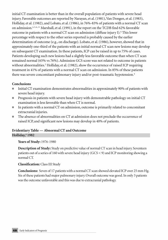
68 Early Indicators of Prognosis
initial CT examination is better than in the overall population of patients with severe headinjury. Favorable outcomes are reported by Narayan, et al. (1981), Van Dongen, et al. (1983),Holliday, et al. (1982), and Lobato, et al. (1986), in 76%-83% of patients with a normal CT scanon admission.4, 5, 6, 11 Marshall, et al. (1991), in the report on the TCDB find 62% favorableoutcome in patients with a normal CT scan on admission (diffuse injury I).6 This lowerpercentage with respect to the other series reported is probably caused by the earlierdetermination of outcome (e.g., on discharge). Lobato, et al. (1986), however, showed that inapproximately one-third of the patients with an initial normal CT scan new lesions may developon subsequent CT examination. In these patients, ICP can be raised in up to 75% of cases.Patients developing such new lesions had a slightly less favorable outcome than when CT scanremained normal (65% vs 76%). Admission GCS score was not related to outcome in patientswithout abnormalities.5 Holliday, et al. (1982), show the occurrence of raised ICP requiringtreatment in 41% of patients with a normal CT scan on admission. In 85% of these patientsthere was severe concomitant pulmonary injury and/or post-traumatic hypotension.4
Conclusions■ Initial CT examination demonstrates abnormalities in approximately 90% of patients with
severe head injury.■ Prognosis in patients with severe head injury with demonstrable pathology on initial CT
examination is less favorable than when CT is normal.■ In patients with a normal CT on admission, outcome is primarily related to concomitant
extracranial injuries.■ The absence of abnormalities on CT at admission does not preclude the occurrence of
raised ICP, and significant new lesions may develop in 40% of patients.
Evidentiary Table — Abnormal CT and OutcomeHolliday,4 1982
Years of Study: 1976-1980
Description of Study: Study on predictive value of normal CT scan in head injury. Seventeenpatients out of a series of 160 with severe head injury (GCS < 9) and ICP monitoring showing anormal CT.
Classification: Class III Study
Conclusions: Seven of 17 patients with a normal CT scan showed elevated ICP over 25 mm Hg.Six of these patients had major pulmonary injury. Overall outcome was good. In only 3 patientswas the outcome unfavorable and this was due to extracranial pathology.

69CT Scan Features
Van Dongen,11 1983
Years of Study: 1977-1979
Description of Study: Prospective, consecutive series examining prognostic value of CT in 121patients with severe head injury.
Classification: Class I Study
Conclusions: Normal CT scan was noted in 14 patients (12%). Outcome in this small groupwas favorable (78.5%).
Outcome at 1 Year: GOS 1, 2, 3 GOS 4, 5Normal Scan 3% 11%Abnormal Scan 76 1PPV*: 78%*Positive predictive value
Lobato,5 1986
Years of Study: 1977-1985
Description of Study: Forty-six patients out of a total series of 448 severe head injury withGCS score of 8 or less for at least 6 hours after injury showing no abnormalities on CT.
Classification: Class II Study
Conclusions: No abnormalities were noted in 10.2% of patients with severe head injury. Absenceof abnormalities showed a PPV of 76% to favorable outcome. Twenty-four of the 46 patients with anormal CT scan on admission developed new lesions on subsequent examinations. 71% of thesepatients had a raised ICP. A moderate degree of raised ICP was only seen in 4 patients (8.5%) whenthe CT remained normal. Outcome was more unfavorable when new lesions developed.
Outcome at 6 Months: Unfavorable FavorableGCS 3-4 4 11GCS 5-8 7 24
Marshall,6 1991
Years of Study: 1984-1987
Description of Study: Prospective study of 746 patients with severe head injury.
Classification: Class II Study
Conclusions: Incidence of normal CT scan in this prospective series was 6.9%. There wasfavorable outcome on discharge in 61% of these patients.
Outcome at Discharge: Unfavorable FavorableNormal CT Scan 20 32(Diffuse Injury IAbnormal CT Scan 522 155PPV: 77%
Evidentiary Table — Abnormal CT and Outcome

70 Early Indicators of Prognosis
Evidence for ClassificationFirst Author > 25 GOS 6 Months Prospective Indicator 24 hours Statistics Class
Lobato5 + + + + - IIHolliday4 - - - + - IIIVan Dongen11 + + + + + IMarshall6 + - + + - II
References1. Anonymous: A multicenter trial of the efficacy of Nimodipine on outcome after severe head
injury. The European Study Group on Nimodipine in severe head injury. J Neurosurg 80:797-804, 1994.
2. Eisenberg HM, Gary HE, Aldrich EF, et al.: Initial CT findings in 753 patients with severehead injury. A report from the NIH Traumatic Coma Data Bank. J Neurosurg 73:688-698,1990.
3. Fearnside MR, Cook RJ, McDougall P, et al.: The Westmead Head Injury Project outcome insevere head injury. A comparative analysis of prehospital, clinical, and CT variables. Br JNeurosurg 7:267-279, 1993.
4. Holliday PO, Kelly DL Jr., Ball M: Normal computed tomograms in acute head injury:correlation of intracranial pressure, ventricular size, and outcome. Neurosurg 10:25-28, 1982.
5. Lobato RD, Sarabia R, Rivas JJ, et al.: Normal computerized tomography scans in severehead injury. Prognostic and clinical management implications. J Neurosurg 65:784-789,1986.
6. Marshall LF, Gantille T, Klauber MR, et al.: The outcome of severe closed head injury.J Neurosurg (Suppl)75:28-36, 1991.
7. Murray GD, Teasdale GM, Braakman R, et al.: The European Brain Injury ConsortiumSurvey of head injuries in “neuro”-units. Acta Neurochir 141:223-236, 1999.
8. Narayan RK, Greenberg RP, Miller JD, et al.: Improved confidence of outcome prediction insevere head injury. A comparative analysis of the clinical examination, multimodalityevoked potentials, CT scanning, and intracranial pressure. J Neurosurg 54:751-762, 1981.
9. Selladurai BM, Jayakumar R, Tan YY, et al.: Outcome prediction in early management ofsevere head injury: an experience in Malaysia. Br J Neurosurg 6:549-557, 1992.
10. Sweet RC, Miller JD, Lipper M, et al.: Significance of bilateral abnormalities on the CT scanin patients with severe head injury. Neurosurg 3:16-21, 1978.
11. Van Dongen KJ, Braakman R, Gelpke GJ: The prognostic value of computerizedtomography in comatose head-injured patients. J Neurosurg 59:951-957, 1983.
B. CT Classification of Head Injury and Its Prognostic SignificanceConventional classification of CT findings in severely head-injured patients differentiatesbetween focal (extradural and subdural hematomas, as well as intracerebral hematomas andspace occupying contusions) and diffuse head injuries (Gennarelli, et al., 1982).3 Diffuseinjuries according to this classification are defined by the absence of mass lesions, althoughsmall contusions without mass effect may be present. In terms of outcome, patients with diffuseinjuries were found to have an intermediate prognosis when compared to patients with epidural

71CT Scan Features
or subdural hematomas. While acute subdural hematomas with low GCS scores had a highmortality, diffuse injuries with higher GCS scores showed a low mortality and a high incidenceof good recovery.
In practice, some confusion exists between this category of patients with diffuse lesions andthe more neuropathologically oriented entity of diffuse axonal injury (DAI). DAI is basedprimarily on neuropathological hallmarks, characterized by wide-spread tearing of axones and/or small blood vessels. Radiologic criteria for diagnosis of DAI are small hemorrhagic lesions atthe cortico-medullary junction, in the corpus collosum, in the midbrain, and in the brain stem,sometimes in conjunction with some intraventricular bleeding. DAI can sometimes besuperimposed by generalized brain swelling (Adams, et al., 1982; Zimmerman, et al., 1978).
Lobato, et al. (1983), have expanded on the anatomical patterns of the conventional CTclassification, outlining eight categories of injury, mainly subdividing patients with focal lesions(Table 1). This classification was shown to have a stronger predictive value than theconventional categorization. Outcome was significantly better in extradural hematoma withoutconcomitant brain swelling, simple brain contusion, generalized swelling, and in the absence oflesions.
Table 1Classification of CT Lesions and Outcome (Lobato,4 1983)
CT Findings Number of Patients Unfavorable Outcome (%)
No Lesions 28 32%Extracerebral Hematoma 19 15Extracerebral Hematoma and Swelling 27 100Bilateral Swelling 42 12Single Brain Contusion 45 22Multiple Unilateral Contusion 32 84Multiple Bilateral Contusion 42 54Diffuse Axonal Injury 43 86
Marshall, et al. (1991), in the publication on the Traumatic Coma Data Bank, propose a newclassification in which the category of diffuse injury is further expanded, taking into accountsigns of raised ICP (i.e., compressed or absent basal cisterns), midline shift, and the presence ofmass lesions (Table 2).

72 Early Indicators of Prognosis
Table 2CT Classification TCDB
Category Definition
Diffuse Injury I (no visible pathology) No visible intracranial pathology seen on CT scan.Diffuse Injury II Cisterns are present with midline shift 0-5 mm and/
or lesions densities present, no high or mixed densitylesion > 25 cc, may include bone fragments andforeign bodies.
Diffuse Injury III (swelling) Cisterns compressed or absent with midline shift 0-5mm, no high or mixed density lesion > 25 cc.
Diffuse Injury IV (shift) Midline shift > 5 mm, no high or mixed densitylesion > 25 cc.
Evacuated Mass Lesion Any lesion surgically evacuated.Non-Evacuated Mass Lesion High or mixed density lesion > 25 cc, not surgically
evacuated.
The frequency of occurrence of the various CT categories according to this classification inthree large series of head injury patients is shown in Table 3.
Table 3Incidence of CT Categories in Head Injury (Marshall,5 1991)
TCDB European Nimodipine Trial EBIC Surveyn = 746 n = 819 n = 983
Diffuse Injury I 52 7% 69 8.4% 121 12%Diffuse Injury II 177 23.7 270 32.9 273 28Diffuse Injury III(swelling) 153 20.5 89 10.9 101 10Diffuse Injury IV(shift) 32 4.2 31 3.8 21 2Evacuated Mass Lesion 276 37 314 38.3 467 48Non-EvacuatedMass Lesion 36 4.8 36 4.4
A clear correlation between CT classification and outcome was shown on analysis of the TCDB(Table 4).

73CT Scan Features
Table 4CT Classification and Outcome on Discharge (Marshall,5 1991)
Number of Patients Unfavorable Outcome Favorable Outcome(D, VS, SD) (MD + GR)
Diffuse Injury I 52 38% 62%Diffuse Injury II 177 65 35Diffuse Injury III 153 84 16Diffuse Injury IV 32 94 6Evacuated Mass Lesion 276 77 23Non-Evacuated Mass Lesion 36 89 11
Walder, et al. (1995), have compared the predictive value of the TCDB classification to the worstapplicable severity code from the Abbreviated Injury Score (AIS). A high correlation was foundbetween AIS and outcome at six months, the TCDB classification and outcome as well asbetween GCS score and outcome. The predictive power for favorable outcome was shown to begreater for the AIS score than for the TCDB classification (Table 5), with a PPV of 95% towardfavorable outcome in the AIS scores 0-3. Conversely an AIS score of 5 was shown to have a PPVof 71% toward the outcome categories dead or vegetative.
Table 5Predictive Power of AIS, TCDB, CT Classification, and GCS score (Walder7, 1995)
Statistical Value of PredictionSensitivity Specificity Positive Likelihood Ratio Percentage
Predictive CorrectValue Predictions
AIS 0-3 40% 98% 95% 25:1 73%TCDBClassification I-IV 51 86 75 3:6:1 66GCS score 6-8 57 69 59 1:9:1 64
Conclusions■ A strong correlation exists between the worst intracranial AIS severity code of the initial CT
in severe head injury and outcome at six months.■ The TCDB CT classification is strongly correlated to outcome.
Recommendations for Further Research■ Investigation of interobserver reliability in classifying severe head injury according to CT
scan.■ There should be further investigation concerning predictive power of the intracranial AIS
severity code of the initial CT.

74 Early Indicators of Prognosis
Evidentiary Table — CT Classification and Outcome Gennarelli,3 1982
Description: Retrospective analysis of 1,107 patients with severe head injury from seven centersanalyzing outcome and type of CT lesion.
Classification: Class III Study
Conclusions: Differentiation of focal versus diffuse injuries being split into two categories ofseverity: marked heterogeneity of outcome; type of lesion as important on outcome as GCS score.Rank order of prognosis: subdural hematoma < diffuse injuries < extradural hematoma.
Lobato,4 1983
Years of Study: 1977-1982
Description: Study of a consecutive series of 277 severely head-injured patients.
Classification: Class II/III Study
Conclusions: Patients with pure extracerebral hematoma, single brain contusion, generalizedbrain swelling, and normal CT scans had a significantly better outcome than patients developingacute hemispheric swelling after operation for a large extracerebral hematoma or patients withmultiple brain contusion, either unilateral or bilateral, and patients with DAI.
Marshall,5 1991
Years of Study:1984-1987
Description: Prospective study of a consecutive series of 746 severely head- injured patients infour centers (TCDB).
Classification: Class II Study
Conclusions: CT classification has clear prognostic value.
Walder,7 1995
Years of Study: 1986-1988
Description: Prospective series of 109 severely head injured patients (GCS ≤ 8) evaluatingpredictive value of worst applicable intracranial severity score from the AIS and CT classificationaccording to the TCDB.
Classification: Class I Study
Conclusions: The AIS based on initial CT scan provides useful prognostic information inpatients with severe head injury. The predictive value of an AIS 0-3 for favorable outcome ishigher than the TCDB classification.

75CT Scan Features
Evidence for Classification
Author > 25 patients GOS/ Prospective Indicator Statistics Classmortality within 24 hours6 months
Gennarelli3 + - - - IIILobato4 + + + - - II/IIIMarshall5 + - + + - IIWalder7 + + + + + I
References1. Adams JH, Graham DI, Murray S, et al.: Diffuse axonal injury due to nonmissile head
injury in humans. An analysis of 45 cases. Annals of Neurology 12:557-563, 1982.2. Anonymous: A multicenter trial of the efficacy of Nimodipine on outcome after severe head
injury. The European Study Group on Nimodipine in severe head injury. J Neurosurg 80:797-804, 1994.
3. Gennarelli TA, Spielman GM, Langfitt TW, et al.: Influence of the type of intracranial lesionon outcome from severe head injury. J Neurosurg 56:26-32, 1982.
4. Lobato RD, Cordobes F, Rivas JJ, et al.: Outcome from severe head injury related to the typeof intracranial lesion: a computerized tomography study. J Neurosurg 59:762-774, 1983.
5. Marshall LF, Gantille T, Klauber MR, et al.: The outcome of severe closed head injury. JNeurosurg (Suppl) 75: 28-36, 1991.
6. Murray GD, Teasdale GM, Braakman R, et al.: The European Brain Injury Consortium Surveyof head injuries in “neuro”-units. Acta Neurochirurgica 141:223-236, 1999.
7. Walder AD, Yeoman PM, Turnbull A: The abbreviated injury scale as a predictor of outcomeof severe head injury. Intensive Care Medicine 21:606-609, 1995.
8. Zimmerman RA, Bilaniuk LT, Genneralli T: Computerized tomography of shear injury ofthe cerebral white matter. Radiology 127:393-396, 1978.
C. Individual CT Scan Characteristics
Definition of parameter: Although the status of the basal cisterns is one of the best studied CTparameters of prognostic significance, it remains ill defined. Most studies concerning the stateof the basal cisterns focus on the perimesencephalic cisterns. Authors describe absent,compressed, or open cisterns. Only two authors give definitions of the parameter studied.11, 15
■ Partial obliteration: Cisterns visible as hypodense slits, usually in one hemisphere.■ Complete obliteration: Cisterns no longer visible as CSF (cerebrospinal fluid) spaces.
Liu, et al. (1995), suggest adding aspects concerning density and deformation of the brainstem in a grading system to the basal cisterns. They describe a good correlation between theirproposed grading scale (Grades 0-5) and outcome.9

76 Early Indicators of Prognosis
Reliability of Scoring Basal CisternsNo interobserver variation studies have been reported concerning the reliability of scoring ofthe basal cisterns.
IncidenceAn overview of the incidence of compressed or absent basal cisterns in reported series ofpatients with severe head injury is shown in Table 1.
Table 1Incidence of Compressed or Absent Basal CisternsFirst Author Patient Population Incidence
Van Dongen,15 1983 GCS ≤ 8 80/116 68.9%Teasdale,12 1984 diffuse head injury in coma 19/37 51Toutant,14 1984 GCS ≤ 8 118/218 54Cordobes,4 1986 78 patients with diffuse axonal injury 59/78 76Colquhoun,3 1989 comatose head injury 49/60 81.6Eisenberg,5 1990 GCS ≤ 8 within 48 hours 413/753 58Selladurai,11 1992 GCS ≤ 8 74/109 67.8European Nimodipine Trial1 GCS ≤ 8 472/819 57.6
Association with Other Prognostic IndicatorsTwo studies describe a strong association between status of the basal cisterns and pupilreactivity.12, 15 Other authors report an association with the GCS score2, 14 and with the presenceof focal lesions2 or with a history of early hypoxic or hypotensive insults.5
Table 2Pupil Reactivity and Status of Basal Cisterns
Status of Cisterns Pupil ReactivityOne or Both Reacting Neither Reacting
Cisterns Present 17% 1%Cisterns Absent 12 7
(Teasdale, 12 1984)
Status of Cisterns Pupil ReactivityBoth Reacting One or None
Partially or Completely Open 40% 17%Completely Obliterated 14 44
(Van Dongen,15 1983)
1. Status of Basal Cisterns and Other CT Indicators of Raised ICPCompression or absence of the basal cisterns on CT scan is considered one of the indicators ofraised intracranial pressure (ICP).7, 10, 13 Other signs of raised ICP include obliteration of thethird ventricle and the presence of small ventricles, often considered indicative of diffuse brainswelling in the absence of midline shift. Some authors combine the status of the third ventricleand that of the basal cisterns in evaluating prognostic significance. Teasdale, et al. report that

77CT Scan Features
the third ventricle usually becomes obliterated before the basal cisterns.13 In the study by Lang,et al. (1994), on 118 patients with diffuse traumatic brain swelling, however, no direct relationwas seen between the status of the third ventricle and that of the basal cisterns. When the thirdventricle was not visible the basal cisterns remained present in more than half of the patients.8
The assessment of the lateral ventricles as being “slitlike” is debatable and in the absence ofknowledge of the pretraumatic size of the ventricles it is difficult to attach too much importanceto the size of the ventricles. Especially in children the size of the lateral ventricles may normallybe small.15 The main CT parameters indicating raised ICP, therefore, are the status of the thirdventricle and that of the basal cisterns. This is confirmed by two Class II studies5, 12 and oneClass III study.3
Prognostic ValueOne Class I,15 four Class II, 4, 5, 11, 12 and various Class III studies2, 3, 14, 16 describe an associationbetween compression or absence of basal cisterns and unfavorable outcome. Van Dongen, et al.(1983), in a series of 116 comatose head injured patients in whom CT was performed, showed a97% positive predictive value for unfavorable outcome when the cisterns were completelyobliterated.15 In a stepwise forward selection of features using the multinominal independencemodel, the state of the basal cisterns together with lesions of the brain parenchyma emerge as apowerful combination of predictors. Sharp predictions based on these two variables could bemade in 30% of cases, all predictions related to the probability of death. The predictiveperformance of a set of four common CT combinations, using the state of the basal cisterns asthe basic discriminative feature was remarkable, allowing predictions in 63% of cases of which93% were accurate. However, when combining a set of CT features with clinical featuresincluding pupil reactivity, best motor response, and age, the state of the basal cisterns was notselected as a discriminating parameter. This was caused by overlap in prognostic information inrelation to the pupil reactivity.
In the preliminary report on CT features in the national pilot TCDB, the ominous value ofcompressed or absent basal cisterns in severe head injury was further demonstrated.14 Mortality ratewhen cisterns were absent was 77%; 39% when cisterns were compressed; and only 22% whencisterns were open. A relatively greater importance of cisterns in the risk of poor outcome was shownamong patients with a GCS score of 6 to 8. These data were confirmed in the report by Eisenberg, etal. (1990), on the initial CT findings from the NIH TCDB.5 In this study the risk of dying in severelyhead-injured patients was increased twofold if the mesencephalic cisterns were compressed orobliterated. The risk of elevated ICP for those patients with abnormal cisterns was increasedthreefold compared to patients with normal cisterns. The value of the status of the basal cisterns asan indicator for presence of increased ICP has been confirmed in many other studies. Cordobes, etal. (1986), in a small study of 78 patients with post-traumatic diffuse axonal injury, showed increasedICP to be present in 50% of the patients with CT scan evidence of ventriculocisternal collapse andthis phenomenon was also associated with an unfavorable outcome.4 Similar conclusions weredrawn by Colquhoun, et al. (1989), and Teasdale, et al. (1984).3, 12
Yanaka, et al. (1993), in a retrospective study on 170 patients with acute subdural hematomaalso show in these patients a positive predictive value of 77% to unfavorable outcome in thepresence of compressed basal cisterns.16

78 Early Indicators of Prognosis
Conclusions■ Compressed or absent basal cisterns indicate a threefold risk of raised ICP.■ Status of basal cisterns is related to outcome.■ Mortality is increased two- to threefold in the presence of compressed or absent basal cisterns.■ Strong association exists between the status of the basal cisterns and pupil reactivity.■ Some association of the status of the basal cisterns is reported with GCS score, presence of
focal lesions, or early hypoxic and hypotensive insults.
Recommendation for Future Research■ Define and test better definition of open, partially compressed, or absent basal cisterns.■ Determine of observer reliability.■ Need to further investigate the independent value of the status of basal cisterns as
predictive parameter.■ Need to further investigate the relative value of status of basal cisterns and compression of
third ventricle as indicator of raised ICP and CT-predictor.
Evidentiary Table: Basal Cisterns and OutcomeVan Dongen,15 1983
Years of Study: 1977-1979
Description: Prospective consecutive series examining prognostic value of CT in 121 patientswith severe head injury.
Classification: Class I Study
Conclusions: Status of the basal cisterns was shown to be a powerful prognostic indicator, but isstrongly related to pupil reactivity. Based on status of the basal cisterns and the presence orabsence of lesions in the brain parenchyma, sharp predictions were possible in 30% of cases.Adding CT features to clinical features increased the rate of sharp predictions from 48% to 62%.
Outcome 12 Months:Unfavorable Favorable
Open Cisterns 12 19Compressed Cisterns 10 11Absent Cisterns 57 2 PPV = 84% (67/80)

79CT Scan Features
Teasdale,12 1984
Description: Prospective analysis of 37 patients with severe diffuse injury.
Classification: Class II Study
Conclusions: Compression of third ventricle and basal cisterns closely correlated with increasedICP and worse prognosis. Association between pupil reactivity and status of the basal cisterns.
Status Basal Cisterns/Third Ventricle:Unfavorable Outcome Favorable Outcome
Present 9 9Absent 15 4
Toutant,141984
Years of Study: 1981-1982
Description: Prospective study of 218 patients with severe head injury (GCS < 8) from the pilotphase of the National Traumatic Coma Data Bank analyzing prognostic importance of basalcisterns.
Classification: Class III Study
Conclusions: Mortality was doubled when basal cisterns were compressed and increasedthreefold when absent. Prognostic value remained strong after adjusting for GCS score. Status ofthe cisterns was more important in patients with higher GCS scores.
Unfavorable Outcome Favorable OutcomeOpen Cisterns (n = 82) 44 56Compressed Cisterns (n = 70) 64 46Absent Cisterns (n = 48) 85 15
Cordobes,4 1986
Years of Study: 1977-1984
Description: Selected series of 78 patients with diffuse axonal injury.
Classification: Class II Study
Conclusions: Collapsed or absent basal cisterns present in 59 out of 78 (75%) patients.Compression or absence of basal cisterns is correlated to unfavorable outcome
Outcome at 6 Months:Unfavorable Favorable
Open Cisterns 12 7Compressed Cisterns 20 7Absent Cisterns 29 PPV = 87%
Evidentiary Table: Basal Cisterns and Outcome (continued)

80 Early Indicators of Prognosis
Colquhoun,3 1989
Years of Study: 1985-1986
Description: Retrospective study on prognostic significance of third ventricle and basal cisternsin 60 patients whose CT scan showed evidence of primary brain injury.
Classification: Class III Study
Conclusions: Compression and obliteration of the third ventricle and basal cisterns were shownto have a close correlation with raised ICP and poor prognosis.
Outcome at 6 Months:Unfavorable Favorable
Normal Third Ventricle and Basal Cisterns 2 9One or Both Compressed 10 8One or Both Absent 26 5
Eisenberg,51990
Years of Study: 1984-1987
Description: CT features studied in National Traumatic Coma Data Bank.
Classification: Class II Study
Conclusions: Compressed cisterns were noted in 58% of patients. Abnormal cisterns indicate athreefold risk of abnormal ICP and a threefold increase in mortality. An association existsbetween diffuse swelling as defined by abnormal cisterns and/or small ventricles and earlyhypoxia/hypotension.
Selladurai,11 1992
Years of Study: 1989-1991
Description: Prospective consecutive series of 109 patients with severe head injury studiedwithin 48 hours of injury.
Classification: Class II Study
Conclusions: Status of basal cisterns strongly correlated to outcome. Complete obliteration ofbasal cisterns doubles unfavorable outcome (34% to 81%)
Outcome at 6 Months:Unfavorable Favorable
Patent Basal Cisterns 13 22Partial Obliteration 16 10Complete Obliteration 38 10PPV = 73%
Evidentiary Table: Basal Cisterns and Outcome (continued)

81CT Scan Features
Athiappan,2 1993
Years of Study: 1990-1992
Description: Study of 107 patients with moderate and severe head injury (GCS < 11). CTexamination within 24 hours.
Classification: Class III Study
Conclusions: Obliteration of basal cisterns increases mortality threefold (27% to 76%).Correlation between status of the basal cisterns and type of pathology and GCS score.
Outcome at 3 Months:Dead Alive
Normal Cisterns 17 45Obliterated Cisterns 35 10
Yanaka,16 1993
Years of Study: 1985-1992
Description: Retrospective study of 170 patients with acute subdural hematoma, identifyingclinical and radiologic prognostic variables.
Classification: Class III Study
Conclusions: Obliteration of cisterns indicates a poorer prognosis. Rating of prognosticeffectiveness:1. Pupils2. Obliteration ambient cistern3. Midline shift4. Age5. GCS score
Prognostic equations including the status of the ambient cisterns were formulated; associationexisted between the status of the basal cisterns and presence of contusions.
Status of Basal Cisterns:Poor Outcome Functional Outcome (3 Month)
Basal Cisterns Open 12 63Basal Cisterns Compressed 73 22 PPV = 77%
Evidentiary Table: Basal Cisterns and Outcome (continued)

82 Early Indicators of Prognosis
Liu,10 1995
Years of Study: 1985-1987
Description: Retrospective study on 334 consecutive cases of head injury evaluating gradingsystem of status of basal cisterns and brainstem changes versus outcome.
Classification: Class III Study
Conclusions: Good correlation between proposed grading system and outcome.Outcome 12 Months:
Unfavorable FavorableGrade 0 9 25Grade 1 6 7Grade 2 6 8Grade 3 17 8Grade 4 4 1Grade 5 48 4
References:1. Anonymous: A multicenter trial of the efficacy of Nimodipine on outcome after severe head
injury. The European Study Group on Nimodipine in severe head injury. J Neurosurg 80:797-804, 1994.
2. Athiappan S, Muthukumar N, Srinivasan US: Influence of basal cisterns, midline shift andpathology on outcome in head injury. Ann Acad Med Singapore 22:452-455, 1993.
3. Colquhoun IR and Burrows EH: The prognostic significance of the third ventricle andbasal cisterns in severe closed head injury. Clin Radiol 40: 13-16, 1989.
4. Cordobes F, Lobato RD, Rivas JJ, et al.: Post-traumatic diffuse axonal brain injury. Analysisof 78 patients studied with computed tomography. Acta Neurochir (Wien) 81:27-35, 1986.
5. Eisenberg HM, Gary HE, Aldrich EF: Initial CT findings in 753 patients with severe headinjury. A report from the NIH Traumatic Coma Data Bank. J Neurosurg 73:688-698, 1990.
6. Grant R, Condon B, Lawrence A, et al.: Human cranial CSF volumes measured by MRI, sexand age influences. Magnetic Reson Imaging 5:465-468, 1987.
7. Klauber MR, Toutant, Marshall LF: A model for predicting delayed intracranialhypertension following severe head injury. J Neurosurg 61:695-699, 1984.
8. Lang DA, Teasdale GM, Macpherson P, et al.: Diffuse brain swelling after head injury: moreoften malignant in adults than children? J Neurosurg 80:675-680, 1994.
9. Liu HM, Tu YK, Su CT: Changes of brainstem and perimesencephalic cistern: dynampredictor of outcome in severe head injury. J Trauma 38:330-333, 1995.
10. Murphy A, Teasdale E, Matheson M, et al.: Relationship between CT indices of brainswelling and intracranial pressure after head injury. In: Intracranial Pressure V. Ishii S, NagaiH, and Brock M (eds). Springer-Verlag, Berlin, Heidelberg, New York, pp. 562-565, 1983.
11. Selladurai BM, Jayakumar R, Tan YY, et al.: Outcome prediction in early management ofsevere head injury: an experience in Malaysia. Br J Neurosurgery 6:549-557, 1992.
Evidentiary Table: Basal Cisterns and Outcome (continued)

83CT Scan Features
12. Teasdale E, Cardoso E, Galbraith S, et al.: CT scan in diffuse head injury: physiological andclinical correlations. J Neurol Neurosurg, and Psych 47:600-603, 1984.
13. Teasdale G, Teasdale E , Hadley D: Computed tomographic and magnetic resonanceimaging classification of head injury. J Neurotrauma 9, Suppl. 1:249-257, 1992.
14. Toutant SM, Klauber MR, Marshall LF, et al.: Absent or compressed basal cisterns on firstCT scan: ominous predictors of outcome in severe head injury. J Neurosurg 61:691-694,1984.
15. Van Dongen KJ, Braakman R , Gelpke GJ: The prognostic value of computerizedtomography in comatose head injured patients. J Neurosurg 59:951-957, 1983.
16. Yanaka K, Kamezaki T, Yamada T, et al.: Acute subdural hematoma—prediction of outcomewith a linear discriminant function. Neurol Med Chir (Tokyo)33:552-558, 1993.
2. Traumatic Subarachnoid HemorrhageDefinition of parameter: presence of blood in the subarachnoid space, either over the convexityor in the basal cisterns.
Reliability of ScoringNo formal investigation has been performed concerning the reliability of scoring thisparameter. In the European Nimodipine trial a difference of opinion concerning the presence orabsence of traumatic subarachnoid hemorrhage (tSAH) was reported between the reviewcommittee and investigators in “a number of cases.” In the paper by Harders, et al. (1996), ontreatment of tSAH with Nimodipine, in patients of varying clinical severity the reviewcommittee could not confirm the presence of subarachnoid blood on the initial CT scan in 26of the 123 patients (21%) included in the study.7 Kakarieka (1997) in his monograph on tSAHconcludes that the CT findings of tSAH do not have a high reliability.8 This conclusion issupported by El Tabou, et al. (1995).16 Greene, et al. (1995), however, although not directlyreporting results, describe in the presence of tSAH a 94% interobserver reliability in grading thedegree of tSAH.6
Incidence and GradingAn overview of the reported incidence of tSAH in patients with head injury of varying severityis shown in Table 1.

84 Early Indicators of Prognosis
Table 1Incidence of Subarachnoid HemorrhageFirst Author Patient Population Incidence tSAH Remarks
Eisenberg,3 1990 GCS ≤ 8 40 % No absolute numbers(within 48 hours reportedn = 753
Selladurai,15 1992 GCS ≤ 8 32/109 29.4Vollmer,19 1991 GCS ≤ 8 237/588 40 TCDBKakarieka,10 1994 Severe head injury 268/819 33 Population
in adults (GCS ≤ 8) EuropeanNimodipine Trial
Lang,11 1994 Head injury with 46/118 39 tSAH +diffuse brain swelling intraventricular(children and adults) hemorrhage
Gaetani,6 1995 Head injury GCS 3-15 148/515 28.7Greene,7 1995 GCS 3-15 355/3157 11 Higher incidence
GCS 3-9 178/704 26.6 of tSAHin moresevere injuries
Taneda,18 1996 Head injury GCS 3-15 130/883 14.7Murray,13 1998 EBIC* survey GCS 3-12 385/953 40 EBIC SurveyMarshall,12 1998 GCS 4-12 568/1067 53 International
Tirilazad Trial
*EBIC = European Brain Injury Consortium
A few investigations have been performed concerning the degree and localization of blood inthe subarachnoid space. Most investigators studying the influence of the extent of SAH use thegrading system proposed by Fisher et al. for patients with spontaneous SAH. Greene, et al.propose a different grading system, specific for trauma patients (Table 2).
Table 2Grading Systens for Subarachnoid HemorrhageFisher Greene
Grading of SAH: Proposed grading system for scoring tSAH:Group 1: no blood Grade 1: thin (≤ 5 mm)Group 2: layer < 1 mm thick Grade 2: thick (> 5 mm)Group 3: layer > 1 mm thick Grade 3: thin (≤ 5 mm) with mass lesionGroup 4: ventricular involvement Grade 4: thick (> 5 mm) with mass lesion
There is no consistent reporting on the location of subarachnoid blood after trauma. Someauthors describe the location in various basal cisterns, in the fissures, on the tentorium, or overthe convexity,10 others only differentiate between the presence of blood in basal cisterns, overthe convexity, or a combination of the two.6 The most frequent location is over the convexity,followed by the fissures and basal cisterns. Location of tSAH in the Sylvian fissure has beenreported to be indicative of the development of local contusions.16

85CT Scan Features
Associations with Other Lesions and/or Prognostic VariablesPatients with tSAH have a higher incidence of contusions, acute subdural hematomas,intraventricular hemorrhage, and increased ICP signs. Kakarieka reports contusions asassociated lesions in 77% of patients with tSAH and acute subdural hematoma in 44% ofpatients. Gaetani, et al. (1995), report an association of contusions or other intracranial lesionsin 63% of patients.6 Both Gaetani, et al. (1995), and Greene, et al. (1995), report an associationbetween admission GCS score and CT grade of tSAH.6, 7
Traumatic SAH as Prognostic VariableThere is Class I evidence supporting a 72% PPV for unfavorable outcome in patients with CTscans showing tSAH in the suprasellar or ambient cisterns. A 78% PPV of an unfavorableoutcome is associated with Fisher’s Grade 4 tSAH.
Although attention was already called to the presence of SAH in severely head-injuredpatients as an important risk factor in the Japanese literature in 1983,14 it is only recently thatthis aspect has gained attention in the international literature. Takaneka, et al. (1990), describedthe poor prognostic significance of the presence of tSAH, especially in the perimesencephalicregion in a limited series of 30 patients with severe shearing injuries.17 Among 17 patients withtSAH there were 11 cases with perimesencephalic hemorrhage of which 10 died. Selladurai, etal. (1992), in a consecutive series of 109 patients with severe head injury also report a significantcorrelation between the presence of CT visible subarachnoid blood and poor outcome (p =0.002).15 The presence of tSAH is correlated to the occurrence of secondary deterioration11;degree and location of tSAH have been reported to be an indication of delayed ischemicsymptoms, caused by vasospasm (Table 3).
Table 3tSAH and Delayed Ischemia: Relation Between Degree of tSAH and Occurrence of DelayedIschemia (Taneda,18 1996)
Delayed IschemiaDegree of tSAH Number yes no
small (< 1 mm) 101 3 98extensive (> 1 mm) 29 7 22
130 10 120
In the population of the U.S. Traumatic Coma Data Bank an incidence of tSAH of 40% wasreported. A twofold increase in the risk of dying was noted in the group with subarachnoidblood. The presence of subarachnoid blood also appeared to predict an abnormal ICP and thepredictive value of tSAH was shown to be additive to other CT scan parameters, such as thepresence of abnormal cisterns, mass lesion, and midline shift. Eisenberg, et al., showed CT scanparameters to be of greater prognostic significance than clinical variables, such as age and post-resuscitation GCS score, when employing a predictive model including CT scan features andclinical variables.3 Traumatic SAH rated second to effacement of the basal cisterns. Thecalculated odds ratios were 2.13 for tSAH, versus 1.03 for age and 0.71 for post-resuscitationGCS score. Upon analyzing the relative predictive value of CT parameters alone, tSAH alsorated second to effacement of the basal cisterns. In the report on the European trial onNimodipine in severe head injury, a trend toward a favorable effect in the Nimodipine-treated

86 Early Indicators of Prognosis
group was seen in patients exhibiting tSAH (1994). The clinical significance of the finding ofsubarachnoid blood on the CT scan in this series has been further analyzed and reported byKakarieka, et al. (1994).9 The outcome of patients with traumatic SAH was significantly worsethan that of patients whose first CT scan did not show subarachnoid blood. The outcome wasunfavorable in 60% of tSAH patients compared to 30% in patients without SAH (p < 0.01).Logistic regression analysis showed the presence of subarachnoid blood to be one of the mostimportant factors of independent prognostic significance (odds ratio 0.29). The presence oftSAH was shown to have a PPV of 60% for an unfavorable outcome. When differentiated to thelocation of tSAH, the presence of blood in the various basal cisterns demonstrated a PPV of69% to 72% and blood over the convexity had a PPV of 61% (Table 4). Gaetani, et al. (1995)also show the presence of tSAH in the basal cisterns to be more indicative of unfavorableoutcome than tSAH over the convexity.6
A larger extent of SAH is related to poorer outcome.8, 9, 18 Kakarieka shows in Fisher’s Grade3 a PPV of 62% and in Fisher’s Grade 4 a PPV of 79% toward unfavorable outcome (Table 5);Harder’s study (1996) shows a PPV of 78% to unfavorable outcome in Fisher’s Grades 3 and 4.The independent predictive value of tSAH has also been found by Greene, et al. (1995), with a78% PPV for the presence of tSAH and poor outcome in patients with severe head injury.7
However, in patients with mild and moderate head injury the adverse influence of tSAH onoutcome was much less pronounced. In patients with acute subdural hematomas, Domenicucci,et al. (1998), report a PPV of 86% to mortality in the presence of tSAH.2
Table 4Distribution of tSAH and Outcome (Kakarieka,9 1997)
Distribution of tSAH Unfavorable Favorable PPV
No tSAH 87 193 31%Convexity 56 38 60Interhemispheric Fissure 38 17 69Lateral Sylvian Fissure 46 22 68Suprasellar Cisterns 21 8 72Ambient Cisterns 26 10 72Quadrigeminal Cisterns 9 4 69(n= 409)
Table 5Relation Between Degree of tSAH and Outcome (Kakarieka,9 1997)
Fisher Grade Unfavorable Favorable
1 87 1932 23 263 29 184 26 7(n= 409)

87CT Scan Features
Conclusions■ tSAH is a frequent occurrence in severe head injury (26%-53%).■ Most frequent location is over the convexity.■ Mortality is increased twofold in the presence of tSAH.■ Presence of blood in the basal cisterns carries a PPV to unfavorable outcome of
approximately 70%.■ Extent of tSAH is related to outcome.■ tSAH is a significant independent prognostic indicator.
Recommendations for Future Research■ There should be further development of a grading system for tSAH, specific to head injury■ There should be observer reliability studies using grading systems■ There should be identification of the relative prognostic value of grading and location of
tSAH
Evidentiary Table: Traumatic Subarachnoid Hemorrhage and OutcomeShigemori,16 1990
Year of Study: Unknown
Description: Study of 20 patients with tSAH; GCS scores 3-15.
Classification: Class III Study
Conclusions: Most frequent location is the Sylvian fissure; this location may be indicative ofdevelopment of local contusions. Extensive hemorrhage in basal cisterns indicates a pooreroutcome.
Takaneka,17 1990
Year of Study: Unknown
Description: Retrospective study on 30 patients with shearing injury.
Classification: Class III Study
Conclusions: Presence of tSAH indicates a poor prognosis.Outcome Unfavorable FavorabletSAH+ 16 1tSAH- 4 9
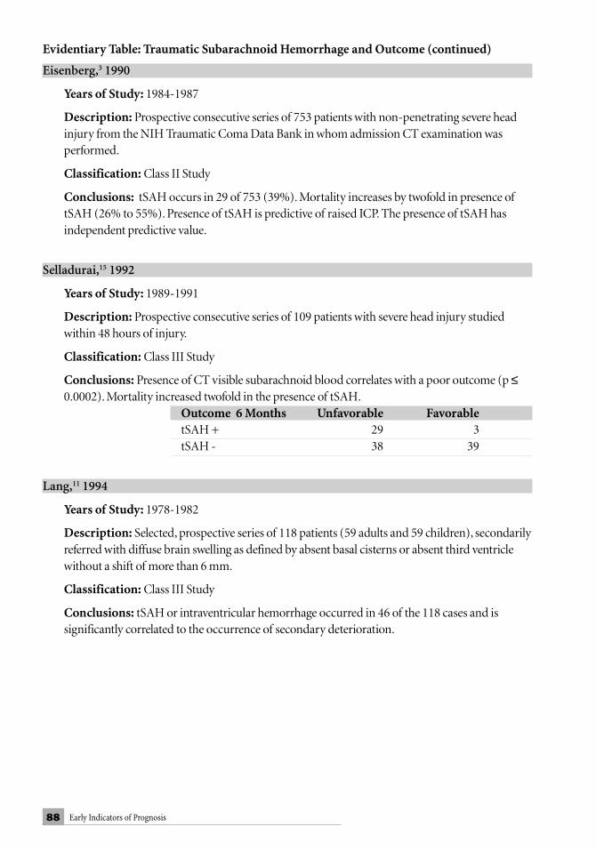
88 Early Indicators of Prognosis
Eisenberg,3 1990
Years of Study: 1984-1987
Description: Prospective consecutive series of 753 patients with non-penetrating severe headinjury from the NIH Traumatic Coma Data Bank in whom admission CT examination wasperformed.
Classification: Class II Study
Conclusions: tSAH occurs in 29 of 753 (39%). Mortality increases by twofold in presence oftSAH (26% to 55%). Presence of tSAH is predictive of raised ICP. The presence of tSAH hasindependent predictive value.
Selladurai,15 1992
Years of Study: 1989-1991
Description: Prospective consecutive series of 109 patients with severe head injury studiedwithin 48 hours of injury.
Classification: Class III Study
Conclusions: Presence of CT visible subarachnoid blood correlates with a poor outcome (p ≤0.0002). Mortality increased twofold in the presence of tSAH.
Outcome 6 Months Unfavorable FavorabletSAH + 29 3tSAH - 38 39
Lang,11 1994
Years of Study: 1978-1982
Description: Selected, prospective series of 118 patients (59 adults and 59 children), secondarilyreferred with diffuse brain swelling as defined by absent basal cisterns or absent third ventriclewithout a shift of more than 6 mm.
Classification: Class III Study
Conclusions: tSAH or intraventricular hemorrhage occurred in 46 of the 118 cases and issignificantly correlated to the occurrence of secondary deterioration.
Evidentiary Table: Traumatic Subarachnoid Hemorrhage and Outcome (continued)

89CT Scan Features
Kakarieka,10 1994
Years of Study: 1989-1991
Description: Population consisted of patients with severe, non-penetrating head injury enrolledin the randomized, prospective, double-blind study on the effect of Nimodipine in severe headinjury. Prognostic evaluation on basis of 414 placebo-treated patients.
Classification: Class I Study
Conclusions: Incidence of tSAH was 33% and adversely influenced outcome. Unfavorableoutcome in the presence of tSAH was doubled (30% to 60%). Logistic regression analysis showedtSAH to be one of the most important prognostic factors. The number of hypotensive episodeswas higher in patients with tSAH.
Outcome at 6 Months Unfavorable FavorabletSAH+ 87 58tSAH- 81 188
Gaetani,6 1995
Years of Study: 1992-1994
Description: Retrospective series of 148 patients with head injury of varying degrees (mild,moderate, and severe) with demonstrated presence of subarachnoid blood on CT examination.Evaluation of clinical significance of degree and extent of SAH was according to Fisher’s grade.
Classification: Class III Study
Conclusions: Degree of tSAH is significantly related to GCS score on admission and tooutcome. Distribution of tSAH is also of prognostic significance. Patients with blood both overthe convexity and in the basal cisterns have worse outcomes.
Outcome at 6 MonthsDistribution of tSAH Unfavorable FavorableSpaces Over Convexity 37 54Basal Cisterns 17 19Mixed 17 4
Evidentiary Table: Traumatic Subarachnoid Hemorrhage and Outcome (continued)

90 Early Indicators of Prognosis
Greene,7 1995
Years of Study: 1988-1991
Description: Retrospective cohort study of 252 patients with head injury of variable degreesshowing CT evidence of tSAH.
Classification: Class III Study
Conclusions: Degree of SAH as defined by proposed grading system is related to admissionGCS score and outcome at discharge. Stepwise regression analysis confirmed the independentpredictive value of the presence of tSAH.
CT Grade of tSAHOutcome at Discharge Outcome at DischargeSevere Head Injury Mild and ModerateGCS 3-9 GCS 10-12 Unfavorable Favorable Unfavorable Favorable
Grade 1 11 7 1 34Grade 2 8 3 0 6Grade 3 45 16 7 35Grade 4 48 6 4 21 PPV = 78% (112/144)
Harders,8 1996
Year of Study: 1994
Description: Prospective, randomized trial on the effect of Nimodipine in 61 patients withtSAH of varying severity (n = 61).
Classification: Class II Study
Conclusions: Most frequent location of blood in the subarachnoid space is over the convexity(67%); less frequently in the basal cisterns (40%). Some relationship between the presence ofsubarachnoid blood and contusions or acute subdural hematoma. The amount of blood asgraded by the Fisher system was related to poorer outcome.
Relation Between Degree of tSAH and Outcome:Outcome at 6 Months:Fisher Grade Unfavorable Favorable
1 0 142 6 133 15 64 7 0
Evidentiary Table: Traumatic Subarachnoid Hemorrhage and Outcome (continued)

91CT Scan Features
Taneda,18 1996
Years of Study: 10-year period
Description: Prospective study of 130 patients with head injury of varying severity (mild,moderate, severe) with CT evidence of subarachnoid blood on admission.
Classification: Class III Study
Conclusions: Ten patients in this series developed delayed ischemic symptoms. The degree andlocation of tSAH was a predictive indicator of delayed ischemic symptoms. In the patients withsymptoms of delayed ischemia, vasospasm was angiographically proven. Mortality wassignificantly higher in the presence of more subarachnoid blood.
Number Outcome at 3 MonthstSAH Dead AliveSmall (< 1 mm) 101 27 74Extensive (> 1 mm) 29 17 12
Domenicucci,2 1998
Years of Study: 1993 and 1994
Description: Retrospective study of 31 patients with severe head injury and ASDH. Analysis ofsubarachnoid spaces and shift.
Classification: Class III Study
Conclusions: Overall mortality was 68%; in the presence of tSAH; 86%.Outcome at 6 Months
Dead AliveAbsence of tSAH or Undetectable Subarachnoid Space 9 8tSAH+ 12 2
References1. Anonymous. A multicenter trial of the efficacy of Nimodipine on outcome after severe head
injury. The European Study Group on Nimodipine in severe head injury. J Neurosurg 80:797-804, 1994.
2. Domenicucci M, Strzelecki JW, Delfini R: Acute post-traumatic subdural hematomas:“intradural” computed tomographic appearance as a favorable prognostic factor. Neursurg42:51-55, 1998.
3. Eisenberg HM, Gary HE, Aldrich EF, et al.: Initial CT findings in 753 patients with severehead injury. A report from the NIH Traumatic Coma Data Bank. J Neurosurg 73:688-698,1990.
4. El Tabou M, Knill Jones RP, Teasdale GM, et al.: Reliability of interpretation of CT scan ofhead injured patients. J Neurotrauma 12:488, 1995.
Evidentiary Table: Traumatic Subarachnoid Hemorrhage and Outcome (continued)

92 Early Indicators of Prognosis
5. Fisher, Kistler JP, Davis JM: Relation of cerebral vasospasm to subarachnoid hemorrhagevisualized by computerized tomographic scanning. Neurosurg 6:1-9, 1980.
6. Gaetani P, Tancioni F, Tartara F, et al.: Prognostic value of the amount of post-traumaticsubarachnoid hemorrhage in a six months follow up period. J Neurology Neurosurg Psych59:635-637, 1995.
7. Greene KA, Marciano FF, Johnson BA, et al.: Impact of traumatic subarachnoidhemorrhage on outcome in non-penetrating head injury. Part I: A proposed computerizedtomography grading scale. J Neurosurg 83:445-452, 1995.
8. Harders A, Kakarieka A, Braakman R, et al.: Traumatic subarachnoid hemorrhage and itstreatment with Nimodipine. German tSAH Study Group. J Neurosurg 85:82-89, 1996.
9. Kakarieka A: Traumatic subarachnoid hemorrhage. Springer-Verlag, Berlin, 1997.10. Kakarieka A, Braakman R, Schakel EH, et al.: Clinical significance of the finding of
subarachnoid blood on CT scan after head injury. Acta Neurochirurgica (Wien) 129:1-5,1994.
11. Lang DA, Teasdale GM, Macpherson P, et al.: Diffuse brain swelling after head injury: moreoften malignant in adults than children? J Neurosurg 80:675-680, 1994.
12. Marshall LF, Maas AIR, Bowers Marshall S: A multicenter trial on the efficacy of usingTirilazad Mesylate in cases of head injury. J Neurosurg 89:519-525, 1998.
13. Murray GD, Teasdale GM, Braakman R, et al.: The European Brain Injury ConsortiumSurvey of head injuries. Acta Neurochirurgica 141:223-236, 1999.
14. Ono J, Yamaura A, Horie T, et al.: CT scan in severe head injury with special reference toGlasgow Coma Scale (in Japanese). No Shinkei Geka 11:379-387, 1983.
15. Selladurai BM, Jayakumar R, Tan YY, et al.: Outcome prediction in early management ofsevere head injury: an experience in Malaysia. Br J Neurosurg 6:549-557, 1992.
16. Shigemori M, Tokutomi T, Hirohata M, et al.: Clinical significance of traumaticsubarachnoid hemorrhage. Neurol Med Chir (Tokyo) 30:396-400, 1990.
17. Takaneka N, Mine T, Suga S, et al.: Interpeduncular high-density spot in severe shearinginjury. Surg Neuro 34:30-38, 1990.
18. Taneda M, Kataoka K, Akai F, et al.: Traumatic subarachnoid hemorrhage and its treatmentwith Nimodipine. J Neurosurg 85:82-89, 1996.
19. Vollmer DG, Torner JC, Jane JA, et al.: Age and outcome following traumatic coma: why doolder patients fare worse? J Neurosurg 75: (Suppl), 37-49, 1991.

93CT Scan Features
Table 2Description of Studies
First > 25 GOS/ Prospective Indicator Measured Statistics Class Author patients Mortality within 24 Hours
6 Months
Lang,11 1994 + - + ? - IIIEisenberg,3 1990 + - + + + IISelladurai,15 1992 + + + - - IIIGaetani,6 1995 + + - +? - IIITaneda,18 1996 + - + + - IIIShigemori,15 1990 - - - + - IIIGreene,7 1995 + - - + + IIIHarders,8 1996 + + + + - IIKakarieka,10 1994 + + + + + ITakaneka,17 1990 + - - + - IIIDomenicucci,2 1998 + - - + - III
3. Midline shiftDefinition of Parameter:The presence of midline shift is defined as the absolute distance (inmm) that midline structures of the brain are displaced in respect to the midline determined byaveraging the distance between the inner tables of the skull. Most authors describe the degree ofdisplacement of the septum pellucidum relative to the midline. Ross, et al. (1989) furtherexamined shift of the pineal gland and of the aquaduct.10
Reliability of Scoring Midline ShiftNo formal observer reliability studies concerning the scoring of midline shift were found. It isremarkable that in three manuscripts authors quantify the degree of midline shift down to.1 mm.5, 6, 16 At the same time, Young states that two observers would agree down to the limitof 1 mm.5
The degree of midline shift and quantification of this is highly variable in the various reports. Anoverview of the various classifications, as mentioned by the different authors, is given in Table 1.
Table 1Variable Classification of Shift Between Authors
First Author Classification of shift
Kotwica and Brzezinski,5 1993 < 1.5 mm, 1.5-3 mm, ≥ 3 mmYoung,16 1981 < 4.1 mm, ≥ 4.1 mmLobato,8 1991 ≤ 5 mm, 6-15 mm, ≥ 15 mmQuattrochi,9 1991 Absent / PresentLipper,6 1985 < 3.8 mm, ≥ 3.8 mmFearnside,4 1993 < 5 mm, 5-10 mm, >10 mmSelladurai,12 1992 < 5 mm, 5-10 mm. > 10 mmEisenberg,3 1990 ≤ 3 mm, > 3 mmVollmer,19 1991 ≤ 5 mm, > 5 mm

94 Early Indicators of Prognosis
IncidenceMidline shift is a relatively frequent occurrence in series of patients with severe head injury. Anoverview of the reported incidence is shown in Table 2.
Table 2Incidence of Midline Shift
First Author Patient Population Incidence Remarks
Young,16 170 head-injured patients with 69/170 40.6% All shift1981 focal neurological deficit or 25/170 14.7 Shift ≥ 4.1 mm
unconsciousness for 6 hoursLipper,6 128 patients with severe 46/128 36 Shift ≥ 3.8 mm1985 head injuryEisenberg,3 GCS ≤ 8 within 48 hours 255/753 34 Shift ≥ 3 mm1990Lobato,8 211 patients who talk 89/211 42.2 Shift 6-15 mm1991 and deteriorate 46/211 21.8 Shift > 15 mmQuattrocchi,9 56 patients with intracranial 28/75 37.31991 hematoma and 19 patients
with normal CTSelladurai,12 109 patients with severe head injury 27/109 24.7 Shift 5-101992 17/109 15.6 Shift > 10 mmAthiappan,1 107 patients with moderate and 35/107 32.71993 severe head injuryKotwica,5 1993 200 adult patients with acute 96/200 48 Shift 1.5-3 mm
subdural hematoma 63/200 31.5 Shift ≥ 3 mmVollmer,14 661 patients with severe head injury 176/597 29 Shift > 5 mm1991 (GCS ≤ 8) in whom CT was available
Association with Other Prognostic VariablesA few studies describe the relative importance of the degree of midline shift in respect to otherprognostic CT variables. A relation to the presence or absence of focal lesions5 and the GCSscore16 is described. Athiappan, et al. (1993), found the prognostic value of midline shift moreimportant in patients with single contusions or intracerebral hematoma than for those withmultiple lesions and extraaxial or subdural hematoma.1 They conclude that the presence ofmidline shift is better correlated with the type of pathology and GCS score, rather than that thedegree of midline shift can be taken alone.
Prognostic Value of Midline ShiftClass I and Class II evidence demonstrate the prognostic significance of both presence orabsence as well as degree of midline shift in patients with severe head injury3, 4, 6, 14 ; Class IIevidence supports the greatest prognostic value of brain shift in patients with GCS scores 5-7.16
In the study by Fearnside, et al. (1993), midline shift and other CT parameters were third instrength (after age and motor score) in a logistic regression analysis of the relative importanceof prognostic variables.4 Lobato, et al. (1991), showed in patients with secondary deteriorationto coma that the degree of midline shift rates third after GCS score and highest mean ICP.8

95CT Scan Features
Other authors, however, have not been able to show such prognostic significance; Selladurai, etal. (1992), describing a poor outcome in the majority of patients with a midline shift greaterthan 10 mm, could not show overall statistical significance in the total population of 109patients with severe head injury.12 The limited prognostic value of midline shift in this studycould in part be explained by the presence of diffuse axonal injury and bilateral hemorrhagiclesions in the significant proportion of patients with midline shift less than 10 mm. In patientscomatose due to acute epidural hematoma, Seelig, et al. (1984), found no correlation betweenthe degree of midline shift and outcome.11
In patients with subdural hematoma some authors report a good correlation betweenmidline shift and outcome, others a less evident relation: Kotwica and Brzezinski showed 42%favorable outcomes and a mortality of 39% when the shift was below 1.5 cm, and a 76%mortality when shift exceeded 3 cm.5 Yanaka reports a mean midline shift of 2.9 mm in thosepatients with a functional recovery and 12.8 mm in those with a poor outcome.15 Lobato, et al.(1991), only found a relation at extreme values of shift comparing a midline shift less than 4mm versus a shift of more than 12 mm.8 The outcome in the intermediate values did not differ.Domenicucci, et al. (1998), describe slightly larger average shift in patients dying with acutesubdural hematoma than in survivors, but these results are not statistically significant.2
Zumkeller, et al. (1996), also reporting on acute subdural hematomata, describe a decrease insurvival density curves at shifts greater than 12 mm, but 50% survival occurs at a shift of 20mm. A PPV of 70% to mortality can be calculated at a shift of approximately 23-24 mm.17
In the initial report on the Traumatic Coma Data Bank (TCDB),3 a midline shift of 3 mm ormore was noted in 34% of patients. In contrast to other studies they find a midline shift,regardless of the underlying pathology, a very strong predictor of abnormal ICP. The risk ofdying was proportional to the degree of midline shift. From the published best fit curvebetween degree of midline shift and outcome, it can be inferred that a PPV of 70% for mortalitycan be calculated at a midline shift of 1.5 cm or more. This is in agreement with the PPV of 68%to fatal outcome reported by Lobato, et al. (1991), when the shift exceeds 1.5 cm.8 From the datapresented by Vollmer, et al. (1991), on a more definitive analysis of the TCDB, including six-month outcome, a PPV of 78% for poor outcome, as defined by the Glasgow Outcome Scale(GOS) categories dead and vegetative, can be calculated in the presence of a shift of 5 mm orgreater in patients over 45 years of age. The relation between shift and poor outcome is,however, more evident in younger patients, in whom poor outcome is doubled. Because of thebetter, age-dependent prognosis in these patients, the PPV of this parameter at ages below 45 isonly 53%.14 Young, et al. (1981), however, report a PPV of 68% versus unfavorable outcomealready at shifts of more than 4.1 mm.16
Quattrocchi, et al. (1991), in a retrospective study of 75 consecutive patients with headinjury, also found a prognostic significance of the presence or absence of midline shift on theadmission CT. The presence of midline shift was associated with a poor outcome in 50% ofcases, whereas the absence of midline shift was associated with a poor outcome in only 14% ofcases (p < 0.05). Significant predictive factors for poor outcome in this study were the presenceof intracranial hemorrhage (34%), intracranial hemorrhage with midline shift (61%), andmidline shift out of proportion to the extent of intracranial hemorrhage (88%).9

96 Early Indicators of Prognosis
Conclusions■ Presence of midline shift is inversely related to prognosis; however, interaction with the
presence of intracranial lesions and other CT parameters exists.■ Class I evidence shows a PPV of 78% to poor outcome in the presence of shift greater than
5 mm in patients over 45 years of age.■ Class II evidence shows a PPV of 70% to unfavorable outcome at midline shift greater than
1.5 cm.■ Presence of midline shift is indicative of increased intracranial pressure. The degree of
midline shift has not been well studied and authors report widely differing values.■ The value of shift seems less important than other CT parameters, because the degree of
shift is also influenced by the location of intracerebral lesions and the presence of bilateralabnormalities. Moreover, the presence and degree of midline shift as seen on the admissionCT scan can be significantly altered on subsequent investigations, following the evacuationof mass lesions.
Recommendations for Future Research■ Further observer reliability studies.■ Further uniform classification of degree of shift.■ Further investigations concerning association with other prognostic variables.■ Further investigation of independent predictive value of midline shift as CT predictor.
Evidentiary Table: Midline Shift and OutcomeDomenicucci,2 1998
Years of Study: 1993-1994
Description: Retrospective study of 31 patients with severe head injury and acute subduralhematoma; analysis of subarachnoid spaces and shift.
Classification: Class III Study
Conclusions: Average shift higher in patients dying (10.7 mm) than in survivors (8.9 mm), butno significant difference.
Eisenberg,3 1990
Years of Study: 1984-1987
Description: Prospective study of early head CT in 753 patients with severe head injury.Analysis of shift at the level of the septum pellucidum and mortality examined as one subset.
Classification: Class II Study
Conclusions: At 1.5 cm shift or greater there was a 70% or higher percent of patients dying.Other parameters such as basal cisterns, mass lesions, tSAH, and ICP were examined.

97CT Scan Features
Fearnside,4 1993
Years of Study: Study duration 2-year period
Description: Prospective series of 315 consecutive patients with a GCS score less than 8 ordeterioration to this level within 48 hours of injury. Analysis of prognostic significance of clinicaland CT variables; outcome 6 months after injury.
Classification: Class II Study
Conclusions: CT parameters with predictive value concerning mortality were: cerebral edema,compressed/absent basal cisterns, shift, and presence of intraventricular hemorrhage. Cerebraledema was considered present in three of the following four variables:1. Loss of grey/white matter differentiation2. Compressed ventricles3. Effaced sulci4. Effaced or compressed perimesencephalic cisterns.
Degrees of shift defined were as follows: none, less than 5 mm, 5-10 mm, and greater than 15 mm.Logistic regression analysis concerning clinical and CT variables showed predictors of mortalityto be: 1) age, 2) motor score, and 3) any of the 3 CT parameters. Predictors of disability insurvivors were different: 1) hypotension, 2) abnormal motor response, 3) tSAH, and 4)intracerebral contusion or hemorrhage.
Kotwica and Brzezinski,5 1993
Years of Study: 1982-1990
Description: Consecutive series of 200 adult patients operated on for acute subdural hematomawith a GCS score less than 10 prior to operation. Analysis of relationship between age, GCS score,operative timing, concomitant presence of focal lesions, shift, and outcome at 3 months.
Classification: Class III Study
Conclusions: Significant correlation between midline shift, presence of contusions, andoutcome.
Outcome at 3 MonthsShift Unfavorable Favorable< 1.5 24 171.5-3 72 24> 3 58 5
Evidentiary Table: Midline Shift and Outcome (continued)
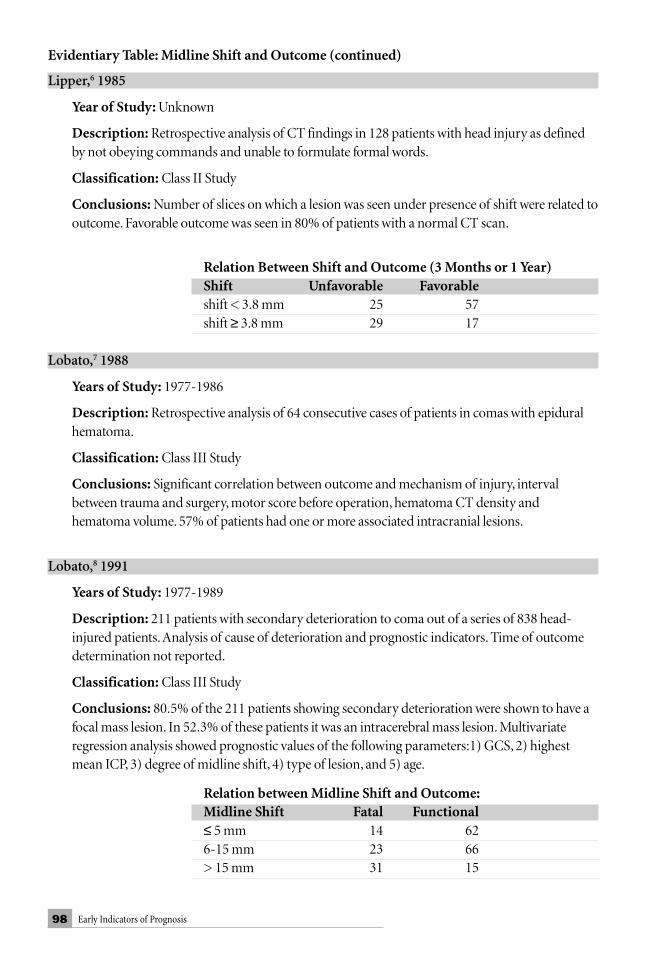
98 Early Indicators of Prognosis
Lipper,6 1985
Year of Study: Unknown
Description: Retrospective analysis of CT findings in 128 patients with head injury as definedby not obeying commands and unable to formulate formal words.
Classification: Class II Study
Conclusions: Number of slices on which a lesion was seen under presence of shift were related tooutcome. Favorable outcome was seen in 80% of patients with a normal CT scan.
Relation Between Shift and Outcome (3 Months or 1 Year)Shift Unfavorable Favorableshift < 3.8 mm 25 57shift ≥ 3.8 mm 29 17
Lobato,7 1988
Years of Study: 1977-1986
Description: Retrospective analysis of 64 consecutive cases of patients in comas with epiduralhematoma.
Classification: Class III Study
Conclusions: Significant correlation between outcome and mechanism of injury, intervalbetween trauma and surgery, motor score before operation, hematoma CT density andhematoma volume. 57% of patients had one or more associated intracranial lesions.
Lobato,8 1991
Years of Study: 1977-1989
Description: 211 patients with secondary deterioration to coma out of a series of 838 head-injured patients. Analysis of cause of deterioration and prognostic indicators. Time of outcomedetermination not reported.
Classification: Class III Study
Conclusions: 80.5% of the 211 patients showing secondary deterioration were shown to have afocal mass lesion. In 52.3% of these patients it was an intracerebral mass lesion. Multivariateregression analysis showed prognostic values of the following parameters:1) GCS, 2) highestmean ICP, 3) degree of midline shift, 4) type of lesion, and 5) age.
Relation between Midline Shift and Outcome:Midline Shift Fatal Functional≤ 5 mm 14 626-15 mm 23 66> 15 mm 31 15
Evidentiary Table: Midline Shift and Outcome (continued)

99CT Scan Features
Quattrocchi,9 1991
Year of Study: 1987
Description: Retrospective study of 56 patients with head injury (GCS 3-12) with intracranialhematoma. Data were compared to a randomly selected series of 19 patients with normal CTscans. Purpose of the study was to determine specific CT criteria for predicting outcome. GOSscore determined at 6 months and 1 year.
Classification: Class III Study
Conclusions: Significant factors for poor outcome were intracranial hemorrhage, intracranialhemorrhage plus shift, and shift out of proportion to intracranial hemorrhage.
Relationship Between Presence and Absence of Shift and Outcome:Shift Unfavorable Favorableshift - 4 24shift + 14 14
Relationship Between Degree of Shift and Outcome:Shift Unfavorable Favorableshift = mass 8 12shift > mass 7 1PPV ≥ 70% for mortality
Ross,10 1989
Year of Study: No year of study reported.
Description: Prospective blinded trial on 46 patients with acute post-traumatic intracerebralhematoma; the relation between the degree of midline shift, GCS score and outcome at 3 monthswas investigated. The study included 19 patients with acute subdural hematoma, 14 withintracerebral hematoma, and 13 with epidural hematoma.
Classification: Class III Study
Conclusions: Significant relation between level of consciousness and lateral pineal or septalshift. Significant correlation between outcome (3 months) and septal shift, but not betweenoutcome and pineal or aquaductal shift. No difference in shift between patients with acutesubdural hematoma, epidural hematoma, or intracerebral hematoma.
Relation Between GCS, Shift, and Outcome:Outcome at Lateral Pineal Lateral Septal 3 Months Shift Shift
GCS 3-5 Poor 6.8 mm 12.6 mmAlert 6.0 10.5
GCS 6-8 Poor 8.2 13.4Alert 5.2 9.4
Evidentiary Table: Midline Shift and Outcome (continued)

100 Early Indicators of Prognosis
Seelig,11 1984
Years of Study: 1980-1982
Description: Prospective series of 51 patients, comatose with epidural hematoma. Studypopulation formed part of the pilot National Traumatic Coma Data Bank (581 patients). Analysisof clinical and CT variables.
Classification: Class III Study
Conclusions: Motor score before operation was most powerful predictor. No relationshipbetween presence or absence of contusions, location of contusion, and midline shift to outcome.Time of outcome determination and tables not given in manuscript.
Servadei,13 1988
Years of Study: 1980-1986
Description: Out of 158 patients examined in CT era with epidural hematoma, 87 werecomatose. Separate analysis of these patients. Study of changing characteristics in connectionwith increased availability of CT scanners.
Classification: Class III Study
Vollmer,14 1991
Year of Study: 1984-1987
Description: Prospective study on 661 patients from the Traumatic Coma Data Bank in whomCT examination was performed.
Classification: Class I Study
Conclusions: Primary focus of this report is on the relationship between age and outcome.Older patients had a greater frequency of shift greater than 5 mm than younger patient groups.Shift of midline, ventricular asymmetry, and effacement of the mesencephalic cisterns was closelycorrelated with higher rates of poor outcome (vegetative or dead).
Age ≤ 45 Age > 45Dead/Veg SD/M/GR Dead/Veg SD/M/GR
Shift > 5 mm 67 59 39 11Shift ≤ 5 mm 97 275 32 17 PPV: 53% PPV: 78%
Evidentiary Table: Midline Shift and Outcome (continued)

101CT Scan Features
Young,16 1981
Year of Study: Unknown
Description: Prospective series of 170 patients with head injury; severity defined as majorneurological deficits and/or unconsciousness at 6 hours after injury. Study population includespatients with missile wounds. Analysis of predictive value of GCS score, age, and shift. Data arerelated to 1-year outcome. The value of shift as measured on CT scan was separately studied in 69patients with GCS scores 5-7.
Classification: Class II Study
Conclusions: In patients with a GCS score less than 5 or greater than 7, there was a highpredictive value of GCS score. Strong relationship between presence or absence of shift more than4.1 mm and outcome. However, outcome prediction was not significantly improved when addingdata concerning shift to the GCS score. Adding shift to GCS score later than 24 hours afteradmission, however, did improve predictive value.
Relation Between Midline Shift and Outcome at 1 Year:Midline shift Unfavorable Favorable< 4.1 mm 17 27≥ 4.1 mm 17 8
Zumkeller,17 1996
Years of Study: 10-year period
Description: Retrospective study of 174 patients with isolated severe head injury and unilateralacute subdural hematoma; analysis of shift and hematoma thickness.
Classification: Class III Study
Conclusions: Survival density decreases markedly at shift greater than 12 mm. Survival rate of50% at shift is 20 mm. Shift exceeding hematoma thickness is unfavorable sign.
Evidentiary Table: Midline Shift and Outcome (continued)

102 Early Indicators of Prognosis
Description of StudiesFirst Author > 25 GOS 6 Month Prospective Indicator < 24 Hours Statistics Class
Ross10 + - + + - IIISeelig11 + - + + - IIIKotwica5 + - + + - IIIYoung16 + + + + +/- IILobato,8 1991 + ? + - + IIILobato,7 1988 + + - + - IIIFearnside4 + + + - + IIQuattrocchi9 + + - - - IIILipper6 + + - ? + IIServadei13 + + - + - IIIEisenberg3 + - + + + IIDomenicucci2 + - - + - IIIZumkeller17 + - - + - IIIVollmer14 + + + + + I
References:1. Athiappan S, Muthukumar N, Srinivasan US: Influence of basal cisterns, midline shift and
pathology on outcome in head injury. Ann Acad Med Singapore 22:452-455, 1993.2. Domenicucci M, Strzelecki JW, Delfini R: Acute post-traumatic subdural hematomas:
“intracerebral” computed tomographic appearance as a favorable prognostic factor.Neurosurg 42:51-55, 1998.
3. Eisenberg HM, Gary HE Jr, Aldrich EF, et al.: Initial CT findings in 753 patients with severehead injury. A report from the NIH Traumatic Coma Data Bank. J Neurosurg 73:688-698,1990.
4. Fearnside MR, Cook RJ, McDougall P, et al.: The Westmead Head Injury Project outcome insevere head injury. A comparative analysis of prehospital, clinical, and CT variables. Br JNeurosurg 7:267-279, 1993.
5. Kotwica Z and Brzezinski J: Acute subdural haematoma in adults: an analysis of outcome incomatose patients. Acta Neurochirurgica (Wien) 121:95-99, 1993.
6. Lipper MH, Kishore PR, Enas GG, et al.: Computed tomography in the prediction ofoutcome in head injury. Am J Roentgenol 144:483-486, 1985.
7. Lobato RD, Sarabia R, Cordobes F, et al.: Post-traumatic cerebral hemispheric swelling.Analysis of 55 cases studied with computerized tomography. J Neurosurg 68:417-423, 1988.
8. Lobato RD, Rivas JJ, Gomez PA, et al.: Head-injured patients who talk and deteriorate intocoma. Analysis of 211 cases studied with computerized tomography. J Neurosurg 75:256-261,1991.
9. Quattrocchi KB, Prasad P, Willits NH, et al.: Quantification of midline shift as a predictor ofpoor outcome following head injury. Surg Neurology 35:183-188, 1991.
10. Ross DA, Olsen WL, Ross AM, et al.: Brain shift, level of consciousness, and restoration ofconsciousness in patients with acute intracranial hematoma. J Neurosurg 71:498-502, 1989.
11. Seelig JM, Marshall LF, Toutant SM, et al.: Traumatic acute epidural hematoma:unrecognized high lethality in comatose patients. Neurosurgery 15:617-620, 1984.

103CT Scan Features
12. Selladurai BM, Jayakumar R, Tan YY, et al.: Outcome prediction in early management ofsevere head injury: an experience in Malaysia. Br J Neurosurg 6:549-557, 1992.
13. Servadei F, Piazza G, Seracchioli A, et al.: Extradural haematomas: an analysis of thechanging characteristics of patients admitted from 1980 to 1986. Diagnostic andtherapeutic implications in 158 cases. Brain Injury 2:87-100, 1988.
14. Vollmer DG, Torner JC, Jane JA, et al.: Age and outcome following traumatic coma: why doolder patients fare worse? J Neurosurg 75: S37-S49, 1991.
15. Yanaka K, Kamezaki T, Yamada T, et al.:Acute subdural hematoma - prediction of outcomewith a linear discriminant function. Neurol Med Chir (Tokyo) 33:552-558, 1993.
16. Young B, Rapp P, Norton JA, et al.: Early prediction of outcome in head-injured patients. JNeurosurg 54:300-303, 1981.
17. Zumkeller M, Behrmann R, Heissler HE, et al.: Computed tomographic criteria andsurvival rate for patients with acute subdural hematoma. Neurosurg 39:708-713, 1996.
4. Intracranial LesionsIntracranial lesions are differentiated into extracerebral and intracerebral lesions; extracerebrallesions in the acute phase after head injury consist of epidural and acute subdural hematomas.Identifying such lesions is important for purposes of management, but at the same time allowsquantifying severity of primary damage by determining number of lesions, type of lesions, theirsizes, location, and mass effect.
Definitions: Epidural hematoma: high/or mixed density blood collection, between dura andskull.Acute subdural hematoma: high/or mixed density blood collection in the subdural or“intradural” space. Parenchymal lesions: Intraparenchymal lesions are ill defined in the literatureand definitions inconsistently applied. Intraparenchymal lesions may be differentiated in low-density, mixed-density, and high-density lesions. High-density lesions may be small, located inthe subcortical white matter, basal ganglia or brain stem and then form part of so-called “diffuseaxonal injury.”49 Other lesions, of variable density, may be larger and cause mass effect. There isno sharp demarcation between contusions of a hemorrhagic nature and intracerebralhematoma.
Reliability of ScoringNo observer reliability studies concerning the scoring of intracranial lesions were found.

104 Early Indicators of Prognosis
Incidence of Intercranial Lesions
First Author Sweet,43 Kobayashi,17 Gennarelli,10 Marshall,22 European1978; 1983; 1982; 1991; Nimodipinen = 140 n = 138 n = 107 n = 746 Trial,1 1994;
evacuated n = 819lesions
EpiduralHematoma 5 (25%) 23 (16%) 96 (9%) 45 (6%) 134 (16%)Acute SubduralHematoma 32 (23%) 45 (33%) 319 (29%) 159 (21%) 248 (30%)Contusions 32 (23%) 33 (38%) 134 (12%) 501 (61%)Contusions 71 (6%) 96 (12%)(mass lesion)Intracerebral 32 (23%) 71 (9.5%) 70 (8.5%)Hemorrhage
Association with Other Lesions and/or Prognostic VariablesIntracerebral lesions occur frequently in patients with epidural hematoma.11, 14, 15, 19, 21, 29, 36 Inpatients with acute subdural hematoma, intracerebral lesions are common.8, 18, 46, 48 Haselsberger,et al. (1988), demonstrated that patients with “pure” epidural hematomas had a good outcomein 70% of cases as compared to 44% of those with associated intracerebral lesions.11
Jamjoom, et al. (1992), compared two series of patients with epidural hematoma with andwithout intracerebral lesions and showed with statistical significance that patients withintracerebral lesions were older, had more falls as mechanism of injury, had a lower GCS scoreat the time of treatment, and presented with more extracranial injuries.14 The correlationbetween age and outcome in patients with epidural hematoma can be explained in part by theincidence of associated intracerebral lesions. Only 20% of patients aged 20 or younger hadassociated intracerebral lesions, whereas such lesions were present in 80% of patients over theage of 60. In patients with acute subdural hematomas, the presence of associated intracerebrallesions is negatively related to outcome. Kotwica and Brzezinski (1993) describe a mortality of85% for an acute subdural hematoma with an associated unilateral contusion, and 17% whenno such lesion exists.18 In the study by Wilberger, et al. (1991), patients with acute subduralhematomas had a mortality rate of 72% when associated with contusions versus 52% of thosewithout contusions.46
This difference was related to the highest postoperative increased ICP. Seelig, et al. (1981),as well as Domenicucci, et al.(1998), however, showed no significant difference in outcome forpatients with acute subdural hematoma with and without associated contusions.8, 32 Similarly,Seelig, et al. (1984), and Servadei, et al. (1988), could not confirm the relationship betweenintracerebral associated lesions and outcome in patients with epidural hematoma.33, 35
Hemorrhagic contusions occur more frequently in the elderly, where falls are the common mostcause of head injury. Intraparenchymal hemorrhage is more frequent in patients with alcoholuse.7, 31

105CT Scan Features
Predictive ValueThe analysis of the predictive value of the presence or absence of intracranial lesions in patientswith severe head injury is complicated by the fact that many studies reporting on such lesionsinclude patients with injuries of less severity (i.e., GCS > 8). Time to operation and widelyvarying indications for operation, particularly concerning intracranial lesions, are factorspossibly influencing treatment results and prognosis. The mortality of comatose patients withepidural hematoma is lower than in patients with acute subdural hematoma (Table 1). A higherpercentage of favorable outcome is described in patients with severe head injury and anepidural hematoma, and a lower percentage of favorable outcome in patients with acutesubdural hematoma is described in comparison to patients with diffuse lesions.10 Class IIevidence shows a PPV of 77% for unfavorable outcome in severely head-injured patients inwhom mass lesions were present and evacuated, and a PPV of 89% when mass lesions were notevacuated.22 Class I evidence shows a PPV of 67% to unfavorable outcome in the presence of acombination of high-density intracerebral and extracerebral lesions.24 Other Class I evidenceshows a PPV of 79% to poor outcome (dead/vegetative) in the presence of lesions greater than15 ml in patients over 45 years of age.45
Table 1Mortality Reported in Series of Patients with Epidural or Acute Subdural Hematoma
Epidural Hematoma Acute Subdural HematomaFirst Author % Mortality First Author % Mortality
Phonprasert,25 1980 24% Seelig,32 1981 57%Cordobes,4 1981 26 Gennarelli,10 1982 74
(GCS 3-5)Gennarelli,10 1982 36 Gennarelli,10 1982 36(GCS 3-5) (GCS 6-8)Gennarelli,10 1982 9 Klun,16 1984 79(GCS 6-8)Bricolo,2 1984 14 Stone,42 1986 59Seelig,33 1984 41 Stening,40 1986 76Reale,28 1984 27 Haselsberger,11 1988 57Dan,6 1986 59 Marshall,22 1991 50Haselsberger,11 1988 38 Wilberger,46 1991 66Lobato,21 1988 28 Phuenpathom,26 1993 74Servadei,34 1988 27 Hatashita,12 1993 55Marshall,22 1991 18 Kotwica,18 1993 55
No correlation is found between hematoma localization and outcome in patients with epiduralhematoma.19, 21, 29, 35 Hematoma volume in epidural hematoma,21, 29, 35 subdural hematoma,13, 42, 48,
50 as well as in intraparenchymal lesions correlates well with outcome.20
Lobato, et al. (1988), show poor outcomes in only 20% of patients with epidural clots less than150 cc versus 58% when the clot volume is greater than 150 cc.21 Yanaka, et al. (1993), shows inpatients with acute subdural hematoma that the mean hematoma volume was 31 cc for patientswith functional recovery and 104 cc for those patients with an unfavorable outcome.48 Stone, et al.(1983), demonstrates that patients with an acute subdural hematoma volume of less than 100 cchad a mortality rate of 51% and those with hematoma over 100 cc had a 79% mortality. Zumkeller,

106 Early Indicators of Prognosis
et al. (1996), on analysis of patients comatose with an acute subdural hematoma, found a 50%survival rate at a hematoma thickness of 18 mm or less. A PPV toward mortality of 70% can beinferred to occur at a hematoma thicknesses of approximately 23 mm. A PPV of 75% for mortalitywas demonstrated when midline shift exceeded hematoma thickness by more than 5 mm.50
Lipper, et al. (1985), in a retrospective analysis of 128 patients with severe head injury, developed aprognostic equation based on the number of slices of the CT scan on which hemorrhagic lesionswere visible. The equation allowed accurate prediction in 69% of cases. The model was lessaccurate in extra-axial lesions.20 The presence of visible subarachnoid spaces without signs oftSAH has been shown to be indicative of favorable outcome in patients with acute subduralhematoma.
In patients with intraparenchymal lesions, the presence of multiple lesions is associatedwith a poorer outcome. Sweet, et al. (1978), describe in 52 patients with bilateral lesions anassociation between high-density lesions, higher ICP, and worse prognosis.43 Patients with low-density lesions and small ventricles, however, generally show lower ICPs and a better prognosis.Chocksey, et al. (1993), in a retospective study of 202 patients, describe a direct relationshipbetween the number of intracerebral lesions and outcome. In patients with a single hematoma,58% have a favorable outcome, in patients with two clots, 20%, while no patients with three ormore hematomas have a favorable outcome.3 Quattrocchi, et al. (1991), describe worse outcomewhen intracranial hemorrhage is associated with midline shift and especially when the midlineshift is out of proportion to the extent of intracranial hemorrhage, 88% of the patients in thisgroup showing a poor outcome.27 Cordobes, et al. (1986), presenting results on 78 patients withdiffuse axonal injury show a poorer outcome when intraparenchymal hemorrhage is associatedwith intraventricular hemorrhage or global brain swelling.5 In the series reported from theTraumatic Coma Data Bank, 71 patients were operated on for an intracerebral hematoma.Nineteen (26.8%) died, while unfavorable outcome on discharge came to a total of 52 (74%).22
Eide and Tysnes (1992), describe a poorer outcome at three months in patients with multifocalcontusions when compared to those with focal contusions.9
CT examination yields by definition momentary information. When determiningprognostic significance of lesions on the CT scan, the time elapsed between injury and CTexamination must be taken into account. Various authors have addressed the issue of changeson CT appearance over time. Kobayashi, et al. (1983), describing a series of 138 patients, notednew lesions developing in 60 patients. In these patients outcome was favorable in only 12, whilea favorable outcome was seen in 60 of the 78 patients not developing a new lesion.17 Sweet, et al.(1978), in a series of 143 patients, show that 13 of the initial 75 patients with unilateral lesionson admission develop controlateral lesions during the first week.43 Tseng (1992) describes 32patients with delayed traumatic intracerebral hematoma. This delayed hemorrhage was foundafter a time interval varying from 7 hours to 10 days. Seventy-five percent of these patients hada favorable outcome; poor prognosis was associated with an earlier occurrence, largerhematoma, low GCS score, clinical deterioration, and obliteration of the supra chiasmaticcistern. In the majority of these patients, contusions were present on the initial CT scan. Thedelayed lesion was diagnosed between 12 hours and 6 days after trauma.44 Soloniuk, et al.(1986), describe 35 patients with delayed traumatic intracerebral hematoma. In 20% of thesepatients, the diagnosis was made within 3 hours, in 6% between 3 and 6 hours, in 29% between6 and 24 hours, and in 46% more than 24 hours after injury. Half of these patients were notcomatose at the time of admission.37 Yamaki, et al. (1990), shows the development of traumatic

107CT Scan Features
intracranial hematoma from brain contusions in 48 patients. In 56% of these patients, the lesiondeveloped within 6 hours, in 81% within 12 hours, and in all patients within 24 hours afterinjury. Stein, et al. (1992), describe new lesions developing on the CT scan in 123 patients out ofa series of 253 patients with head injury. Coagulation disturbances were present in the majority(55%) of these patients.39 In a recent study by Servadei, et al. (1995), 37 patients are described,from a series of 412, developing new lesions within a 12-hour period from time of admission. In22 patients, these hematomas evolved toward surgical removal. Lesions most prone to enlargewere epidural hematomas and intracerebral hemorrhages.35
When describing outcome results and prognosis in patients with demonstrable lesions onthe CT scan, it may be worthwhile to include results of subsequent CT examinations and toreport the “full extent” of such lesions, i.e., the “worst CT” in addition to the initial CT scanresults.
Conclusions■ Extracerebral and intracerebral lesions occur frequently in comatose patients with head
injury.■ Presence of mass lesions has a PPV of 78% to unfavorable outcome (Class II).■ Presence of mass lesions in patients over 45 years of age carries a PPV of 79% to poor
outcome as defined by the categories dead and vegetative.■ Mortality is higher in acute subdural hematoma than in extradural hematoma.■ Outcome is more favorable in patients with severe head injury and an epidural hematoma and
less favorable in acute subdural hematoma in comparison to patients with diffuse injuries.■ Hematoma volume is correlated to outcome.■ Intraparenchymal lesions are ill defined.
Recommendations for Future Research■ There is a need for improved definition for intraparenchymal lesions.■ A more detailed recording of surgical indications is required in future studies. Standardized
reporting of indications for surgery (clinical, such as occurrence of deterioration, CT,results of ICP monitoring), time to operation, and involving lesions are a prerequisite forcomparison of different series and determination of prognostic value. Also reasons for notoperating, i.e., poor prognosis or local “conservative” policy, should be explicitly stated.

108 Early Indicators of Prognosis
Evidentiary Table: Intracranial Lesions and OutcomeEide and Tysnes,9 1992
Years of Study: 1984-1989
Description: Evaluation of outcome in 143 patients admitted with cerebral contusions, definedas non-homogeneous area of low- and high-attenuation values.
Classification: Class III Study
Conclusions: Outcome at 3 months was poorer in patients with multifocal contusions than infocal contusions. Longer-term evaluation did show increased occurrence of post-traumaticmental disturbances, also in patients with focal contusions.
Outcome in Patients with Brain ContusionUnfavorable Favorable
Focal Contusion 0 57Multifocal Unilateral 17 19Multifocal Bilateral 31 19
Gennarelli,10 1982
Description: Retrospective analysis of 1,107 patients with severe head injury from seven centersanalyzing outcome and type of CT lesion.
Classification: Class III Study
Conclusions: Differentiation of focal versus diffuse injuries being split into two categories ofseverity: market heterogeneity of outcome; type of lesion as important on outcome as GCS score.Rank order of prognosis: subdural hematoma < diffuse injuries < extradural hematoma.
Number of patients Unfavorable FavorableDiffuse Injury 487 48% 52%Focal Injury 620 67 33Epidural Hematoma 96 37 63Acute Subdural Hematoma 319 77 22Other Focal Lesions 61 39

109CT Scan Features
Kobayashi,17 1983
Years of Study: 1977-1981
Description: Analysis of serial CT scans performed in 138 patients with severe head injury.
Classification: Class III Study
Conclusions: New findings were visible on follow-up CTs in 91 of the 138 patients. Significantcorrelation was demonstrated between favorable outcome and absence of new lesions, andbetween poor outcome and development of new lesions.
Relationship Between Progression of Lesions and OutcomeUnfavorable Favorable
No New Lesion 18 60New Lesions 48 12
Lipper,20 1985
Year of Study: Not Reported
Description: Retrospective analysis on 128 randomly selected patients with severe head injury.Evaluation of predictive significance of extent of hemorrhagic lesions. Outcome determinationsat 3 months and 1 year.
Classification: Class II Study
Conclusions: Based on the number of slices (each 1 cm thick) of the CT scan on whichhemorrhagic lesions were visible. A prognostic equation was developed, providing accurateprediction in 69.7% of cases. Model is less accurate in extra-axonal lesions.
Relationship Between Extent of Lesion and OutcomeNumber of Slices withHemorrhagic Lesions Unfavorable FavorableNone 14 551 or 2 3 53 or 4 12 125 or 6 15 97 or 8 2 1
Lobato,21 1988
Year of Study: Not Reported
Description: Analysis of 55 patients out of a series of 520 patients with severe head injury,showing post-traumatic hemispheric swelling.
Classification: Class III Study
Conclusions: Highest mortality (87%) in this category of patients with severe head injury.Strong association with the presence of acute subdural hematoma (85%) or epidural hematoma(9%). Also relation to arterial hypotension and/or hypoxia on admission.
Evidentiary Table: Intracranial Lesions and Outcome (continued)

110 Early Indicators of Prognosis
Marshall,22 1991
Description: Prospective study of a consecutive series of 746 severely head injured patients infour centers Traumatic Coma Data Bank (TCDB).
Classification: Class II Study
Conclusions: Outcome more unfavorable in patients with evacuated or non-evacuated masslesions. Presence of mass lesions carries a PPV of 78% to unfavorable outcome.
Outcome at DischargeUnfavorable Favorable
Diffuse Injury 294 120Evacuated Mass Lesion 213 63Non-Evacuated Mass Lesion 32 4
Miller,23 1979
Year of Study: Not Reported
Description: Study of 74 patients with severe head injury (GCS ≤ 9) investigating relationshipbetween CT scan, GCS score, and ICP versus outcome. Patients with epidural hematoma orsubdural hematoma were excluded.
Classification: Class III Study
Conclusions: In the presence of high-density intraparenchymal lesions there was a 50% changeof raised ICP and a strong correlation with poor outcome. In patients with cerebral contusion,admission GCS score was unrelated to outcome.
Focal Lesions and Increased ICPICP Normal ICP Raised
CT Normal/Diffuse Swelling 34 7CT Contusion or Mixed Lesions 14 19(n = 74)
Relationship Between CT Scan, Coma Scale, and OutcomeGCS score CT Normal/ CT Contusion oron Admission Diffuse Swelling Mixed Lesion
Unfavorable Favorable Unfavorable Favorable3-4 3 0 8 15-7 3 18 8 88-10 9 15 3 411 9 2 1 0
Evidentiary Table: Intracranial Lesions and Outcome (continued)
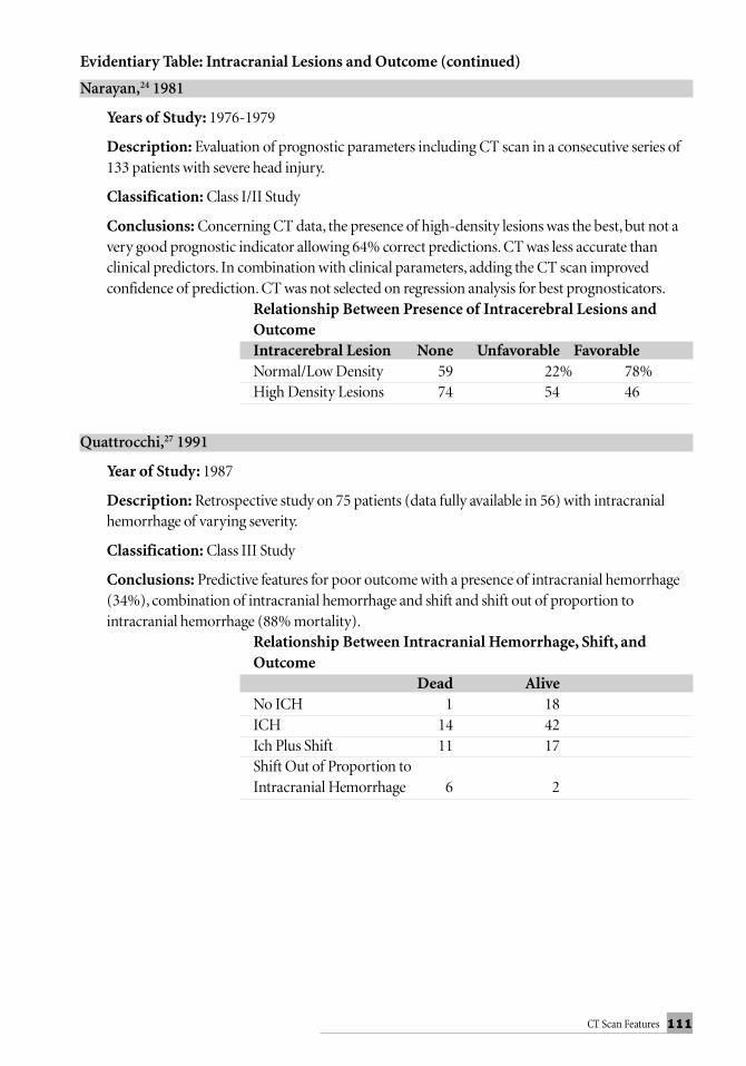
111CT Scan Features
Narayan,24 1981
Years of Study: 1976-1979
Description: Evaluation of prognostic parameters including CT scan in a consecutive series of133 patients with severe head injury.
Classification: Class I/II Study
Conclusions: Concerning CT data, the presence of high-density lesions was the best, but not avery good prognostic indicator allowing 64% correct predictions. CT was less accurate thanclinical predictors. In combination with clinical parameters, adding the CT scan improvedconfidence of prediction. CT was not selected on regression analysis for best prognosticators.
Relationship Between Presence of Intracerebral Lesions andOutcomeIntracerebral Lesion None Unfavorable FavorableNormal/Low Density 59 22% 78%High Density Lesions 74 54 46
Quattrocchi,27 1991
Year of Study: 1987
Description: Retrospective study on 75 patients (data fully available in 56) with intracranialhemorrhage of varying severity.
Classification: Class III Study
Conclusions: Predictive features for poor outcome with a presence of intracranial hemorrhage(34%), combination of intracranial hemorrhage and shift and shift out of proportion tointracranial hemorrhage (88% mortality).
Relationship Between Intracranial Hemorrhage, Shift, andOutcome
Dead AliveNo ICH 1 18ICH 14 42Ich Plus Shift 11 17Shift Out of Proportion toIntracranial Hemorrhage 6 2
Evidentiary Table: Intracranial Lesions and Outcome (continued)

112 Early Indicators of Prognosis
Rudnik,30 1992
Years of Study: 1980-1986
Description: Evaluation of prognostic value of clinical and CT parameters in 146 patients withsevere head injury (GCS ≤ 8).
Classification: Class III Study
Conclusions: GCS had the greatest prognostic significance, followed by degree of intracranialmass lesions. Isolated subdural hematoma or epidural hematoma were indicative of a goodprognosis; the combination with multiple contusions or intracerebral hemorrhage indicated apoor prognosis.
Seelig,33 1984
Years of Study: 1980-1982
Description: Description of treatment results in 51% operated on for epidural hematoma incomatose condition; part of 581 patients in a national pilot Traumatic Coma Data Bank.
Classification: Class III Study
Conclusions: Overall mortality 41%, in 50% of the cases association with intracerebralcontusions. Motor score before operation was the most powerful prognostic indicator.
Servadei,35 1995
Years of Study: 1990-1994
Description: Retrospective review of 37 patients out of a series of 412 showing changing lesionswithin 12 hours of admission.
Classification: Class III Study
Conclusions: Of the 37 patients showing changes, 15 cases evolved toward reabsorption and in22 cases lesion size increased. Indications for control CT detecting these lesions were a raisingICP in 5 patients, clinical deterioration in 10, and scheduled controlled CT in 13 patients.
Stein,38 1993
Years of Study: 1986-1989
Description: Retrospective review of 337 patients with moderate and severe head injury, whohad follow-up CT within 72 hours.
Classification: Class III Study
Conclusions: 149 patients (44.5%) showed new lesions. Highly significant association betweenappearance of delayed insults and severity of initial injury, hypotension, pulmonary injury,coagulopathy or subdural hematoma on initial CT. Appearance of new lesions was stronglyrelated to outcome and was shown to be of independent predictive value.
Evidentiary Table: Intracranial Lesions and Outcome (continued)

113CT Scan Features
Sweet,43 1978
Years of Study: 1976-1977
Description: Analysis of serial CT in 140 head-injured patients evaluating progression of lesionand relationship between intraparenchymal abnormalities and outcome.
Classification: Class III Study
Conclusions: Admission CT showed unilateral lesions in 75 patients and 39 bilateral lesions.Subsequent CT during the first week showed progression in 13 of the unilateral lesions, totaling52 bilateral lesions. Bilateral increased density lesions were associated with a poorer motor score,a higher ICP, and worse outcome.
Type of Lesion versus OutcomeCT lesion Unfavorable FavorableNormal 5 21Unilateral 21 41Bilateral Swelling 8 21Bilateral Hypodense 17 6
Tseng,44 1992
Years of Study: 1987-1989
Description: Report on 32 patients with delayed traumatic intracranial hematoma (DTICH).
Classification: Class III Study
Conclusions: Incidence of DTICH was 5.9% of patients admitted with neurological signs orabnormal CT scan. Reason for control CT scan was clinical deterioration in 10 patients andfailure to recover in 22 patients.
Vollmer,45 1991
Description: Prospective analysis of 661 patients aged 15 years and older in realation to clinicaloutcome.
Classification: Class II Study
Conclusions: The proportion of patients with intracranial hematomas increases with age.Analysis based on the presence and evacuation of a large lesion showed that the increasing agewas associated with poorer outcome in each subgroup.
Age < 45 Age > 45Dead/Veg SD/M/GR Dead/Veg SD/M/GR
Lesion ≥ 15 ml 55 65 57 15No lesion orlesion < 15 122 292 31 16
PPV: 46% PPV: -79%SD= severe disability; M=moderate disability; GR=good recovery
Evidentiary Table: Intracranial Lesions and Outcome (continued)

114 Early Indicators of Prognosis
Description of StudiesFirst Author >25 GOS 6 Month Prospective Indicator within 24 Hours Statistics Class
Lobato21 + ? - + - IIILipper20 + + - + + IIEide9 + - - + - IIIKobayashi17 + - - - - IIISeelig33 + - + + - IIITseng44 + - - - - IIISweet43 + + + - - IIIStein38 + ? - - - IIIServadei35 + ? - + - IIIQuattrocchi27 + + - + - IIIMiller23 + + - + - IIINarayan24 + + + +? + I/IIRudnik30 + ? ? ? + IIIGennarelli10 + - - - IIIMarshall22 + - + + - II
References:1. Anonymous. A multicenter trial of the efficacy of Nimodipine on outcome after severe head
injury. The European Study Group on Nimodipine in severe head injury. J Neurosurg 80:797-804, 1994.
2. Bricolo AP, Pasut ML: Extradural hematoma: toward zero mortality: a prospective study.Neurosurg 14:8-11, 1984.
3. Choksey M, Crockard HA, Sandilands M: Acute traumatic intracerebral haematomas:determinants of outcome in a retrospective series of 202 cases. Br J Neurosurg 7:611-622,1993.
4. Cordobes F, Lobato RD, Rivas JJ, et al.: Observations on 82 patients with extraduralhematoma: comparison of results before and after the advent of computerized tomography.J Neurosurg 54:179-186, 1981.
5. Cordobes F, Lobato RD, Rivas JJ, et al.: Post-traumatic diffuse axonal brain injury. Analysisof 78 patients studied with computed tomography. Acta Neurochirurgica (Wien) 81:27-35,1986.
6. Dan NG, Berry G, Kwok B, et al.: Experience with extradural haematomas in New SouthWales. Aust N Z J Surg 56:535-541, 1986.
7. Djindjian M, Nguyen JP, Lepresle E, et al.: Traumatologie cranienne. Donnees statistiquesrecentes Presse Med 16:991-994, 1987.
8. Domenicucci M, Strzelecki JW, Delfini R: Acute post-traumatic subdural hematomas:“intradural” computed tomographic appearance as a favorable prognostic factor. Neurosurg42: 51-55, 1998.
9. Eide PK, Tysnes O-B: Early and late outcome in head injury patients with radiologicalevidence of brain damage. Acta Neurologica Scandinavia 86:194-198, 1992.
10. Gennarelli TA, Spielman GM, Langfitt TW, et al.: Influence of the type of intracranial lesionon outcome from severe head injury. A multicenter study using a new classification system.J Neurosurg 56:26-32, 1982.

115CT Scan Features
11. Haselsberger K, Pucher R, Auer LM: Prognosis after acute subdural or epiduralhaemorrhage. Acta Neurochirurgica (Wien) 90:111-116, 1988.
12. Hatashita S, Koga N, Hosaka Y, et al.: Acute subdural hematoma: severity of injury, surgicalintervention, and mortality. Neurol Med Chi (Tokyo) 33:13-18, 1993.
13. Howard MA, Gross AS, Dacey RJ, et al.: Acute subdural hematomas: an age-dependentclinical entity. J Neurosurg 71:858-863, 1989.
14. Janjoom A: The influence of concomitant intradural pathology on the presentation andoutcome of patients with acute traumatic extradural haematoma. Acta Neurochirurgica(Wien) 115:86-89, 1992.
15. Jones NR, Molloy CJ, Kloeden CN, et al.: Extradural haematoma: trends in outcome over 35years. Br J Neurosurg 7:465-471, 1993.
16. Klun B, Fettich M: Factors influencing the outcome in acute subdural haematoma. A reviewof 330 cases. Acta Neurochirurgica (Wien) 71:171-178, 1984.
17. Kobayashi S, Nakazawa S, Otsuka T: Clinical value of serial computed tomography withsevere head injury. Surg Neurology 20:25-29, 1983.
18. Kotwica Z, Brzezinski J: Acute subdural haematoma in adults: an analysis of outcome incomatose patients. Acta Neurochirurgica (Wien) 121:95-99, 1993.
19. Kuday C, Uzan M, Hanci M: Statistical analysis of the factors affecting the outcome ofextradural haematomas: 115 cases. Acta Neurochirurgica (Wien) 131:203-206, 1994.
20. Lipper MH, Kishore PRS, Enas GG, et al.: Computed tomography in the prediction ofoutcome in head injury. Am J Roentgenol 144:483-486, 1985.
21. Lobato RD, Rivas JJ, Cordobes F, et al.: Acute epidural hematoma: an analysis of factorsinfluencing the outcome of patients undergoing surgery in coma. J Neurosurg 68:48-57,1988.
22. Marshall LF, Gautille T, Klauber MR, et al.: The outcome of severe closed head injury.J Neurosurg (Suppl) 75:28-36, 1991.
23. Miller JD, Gudeman SK, Kishore PRS, et al.: CT scan, ICP, and early neurological evaluationin the prognosis of severe head injury. Acta Neurochirurgica (Suppl) 28:86-88, 1979.
24. Narayan RK, Greenberg RP, Miller JD, et al.: Improved confidence of outcome prediction insevere head injury. A comparative analysis of the clinical examination, multimodalityevoked potentials, CT scanning, and intracranial pressure. J Neurosurg 54:751-762, 1981.
25. Phonprasert C, Suwanwela C, Hongsaprabhas C, et al.: Extradural hematoma: analysis of138 cases. J Trauma 20:679-683, 1980.
26. Phuenpathom N, Choomuang M, Ratanalert S: Outcome and outcome prediction in acutesubdural hematoma. Surg Neurology 40:22-25, 1993.
27. Quattrocchi KB, Prasad P, Willits NH, et al.: Quantification of midline shift as a predictor ofpoor outcome following head injury. Surg Neurology 35:183-188, 1991.
28. Reale F, Delfini R, Mencattini G: Epidural hematomas. J Neurosurg Sci 28:9-16, 1984.29. Rivas JJ, Lobato RD, Sarabia R, et al.: Extradural hematoma: analysis of factors influencing
the courses of 161 patients. Neurosurg 23:44-51, 1988.30. Rudnik A, Wojtacha M, Wencel T, et al.: The prognostic value of some clinical and
diagnostic factors in traumatic intracranial haematoma. Acta Neurochirurgica (Suppl) 55:33-36, 1992.

116 Early Indicators of Prognosis
31. Schynoll W, Overton D, Krome R, et al.: A prospective study to identify high-yield criteriaassociated with acute intracranial computed tomography findings in head-injured patients.Am J Emerg Med 11: 321-326, 1993.
32. Seelig JM, Greenberg RP, Becker DP, et al.: Reversible brain-stem dysfunction followingacute traumatic subdural hematoma. A clinical and electrophysiological study. J Neurosurg55:516-523, 1981.
33. Seelig JM, Marshall LF, Toutant SM: Traumatic acute epidural hematoma: unrecognizedhigh lethality in comatose patients. Neurosurg 15:617-620, 1984.
34. Servadei F, Nanni A, Nasi MT, et al.: Evolving brain lesions in the first 12 hours after headinjury: analysis of 37 comatose patients. Neurosurg 37:1-9, 1995.
35. Servadei F, Piazza G, Serrachioli A, et al.: Extradural haematomas: an analysis of thechanging characteristics of patients admitted from 1980 to 1986: diagnostic and therapeuticimplications in 158 cases. Brain Injury 2:87-100, 1988.
36. Simpson DA, Heyworth JS, McLean AJ, et al.: Extradural haemorrhage: strategies formanagement in remote places. Injury 19:307-312, 1988.
37. Soloniuk D, Pitts LH, Lovely M, et al.: Traumatic intracerebral hematomas: timing ofappearance and indications for operative removal. J Trauma 26:787-794, 1986.
38. Stein SC, Spettell C, Young G, et al.: Delayed and progressive brain injury in closed-headtrauma: radiological demonstration. Neurosurg 32:25-31, 1993.
39. Stein SC, Talucci RC, Young GS, et al.: Delayed brain injury after head trauma: significanceof coagulopathy. Neurosurg 30: 160-165, 1992.
40. Stening WA, Berry G, Dan NG, et al.: Experience with acute subdural haematomas in NewSouth Wales. Aust NZ J Surg 56:549-556, 1986.
41. Stone JL, Lowe RJ, Jonasson O, et al.: Acute subdural hematoma: direct admission to atrauma center yields improved results. J Trauma 26:445-450, 1986.
42. Stone JL, Rifai MHS, Sugar O, et al.: Subdural hematomas. 1. Acute subdural hematoma:progress in definition, clinical pathology, and therapy. Surg Neurology 19:216-231, 1983.
43. Sweet RC, Miller JD, Lipper M, et al.: Significance of bilateral abnormalities on the CT scanin patients with severe head injury. Neurosurg 3:16-21, 1978.
44. Tseng SH. Delayed traumatic intracerebral hemorrhage: a study of prognostic factors.J Formosan Med Assoc 91:585-589, 1992.
45. Vollmer DG, Torner JC, Jane JA, et al.: Age and outcome following traumatic coma: why doolder patients fare worse? J Neurosurg (Suppl)75:37-49, 1991.
46. Wilberger JE, Harris M, Diamond DL: Acute subdural hematoma: morbidity, mortality, andoperative timing. J Neurosurg 74:212-218, 1991.
47. Yamaki T, Kirakawa K, Ueguchi T, et al.: Chronological evaluation of acute traumaticintracerebral haematoma. Acta Neurochirurgica (Wien) 103:112-115, 1990.
48. Yanaka K, Kamezaki T, Yamada T, et al.: Acute subdural hematoma: prediction of outcomewith linear discriminant function. Neurol Med Chir (Tokyo) 33:552-558, 1993.
49. Zimmerman RA, Bilaniuk LT, Gennarelli T: Computerized tomography of shear injury ofthe cerebral white matter. Radiology 127:393-396, 1978.
50. Zumkeller M, Behrmann R, Heissler HE, et al.: Computed tomographic criteria andsurvival rate for patients with acute subdural hematoma. Neurosurg 39:708-713, 1996.