management and classification of type ii congenital portosystemic shunts
TRANSCRIPT

www.elsevier.com/locate/jpedsurg
Journal of Pediatric Surgery (2011) 46, 308–314
Management and classification of type II congenitalportosystemic shunts☆
Timothy B. Lautz, Niramol Tantemsapya, Erin Rowell, Riccardo A. Superina⁎
Department of Surgery, Children's Memorial Hospital, Feinberg School of Medicine of Northwestern University, Box 57,Chicago, IL 60614, USA
Received 22 October 2010; accepted 4 November 2010
0d
Key words:Abernethy malformation;Congenital portosystemicshunt;
Congenital portocavalshunt
AbstractBackground: Congenital portosystemic shunts (PSS) with preserved intrahepatic portal flow (type II)present with a range of clinical signs. The indications for and benefits of repair of PSS remainincompletely understood. A more comprehensive classification may also benefit comparative analysesfrom different institutions.Methods: All children treated at our institution for type II congenital PSS from 1999 through 2009 werereviewed for presentation, treatment, and outcome.Results: Ten children (7 boys) with type II PSS were identified at a median age of 5.5 years.Hyperammonemia with varying degrees of neurocognitive dysfunction occurred in 80%. The shuntarose from a branch of the portal vein (type IIa; n = 2), from the main portal vein (type IIb; n = 7), orfrom a splenic or mesenteric vein (type IIc; n = 1). Management included operative ligation (n = 6),endovascular occlusion (n = 3), or a combined approach (n = 1). Shunt occlusion was successful in allcases. Serum ammonia decreased from 130 ± 115 μmol/L preoperatively to 31 ± 15 μmol/Lpostoperatively (P = .03). Additional benefits included resolution of neurocognitive dysfunction (n = 3),liver nodules (n = 1), and vaginal bleeding (n = 1).Conclusion: Correction of type II PSS relieves a wide array of symptoms. Surgery is indicated forpatients with clinically significant shunting. A refined classification system will permit futurecomparison of patients with similar physiology.© 2011 Elsevier Inc. All rights reserved.
Congenital absence of the portal vein with an end-to-sideportocaval shunt was first described by John Abernethy in1793 [1]. Many variants of congenital portosystemic shunts(PSS) have been subsequently described, and in 1994,Morgan and Superina [2] introduced a classification systembased on whether the portal vein, often hypoplastic, was
☆ There are no disclaimers or sources of funding or support to report.⁎ Corresponding author. Tel.: +1 773 883 6187; fax: +1 773 975 8534.E-mail address: [email protected] (R.A. Superina).
022-3468/$ – see front matter © 2011 Elsevier Inc. All rights reserved.oi:10.1016/j.jpedsurg.2010.11.009
present and whether the liver was perfused with blood fromthe mesenteric venous system. In a type I shunt, there is acomplete end-to-side portocaval fistula with no discernableportal flow to the liver. Type II shunts occur as a side-to-sideportocaval fistula or as any number of other PSS includinggastrorenal, splenorenal, and portorenal shunts. The keyfeature of type II shunts is preservation of at least somehepatic portal flow. In 1997, Howard and Davenport [3]applied the Abernethy eponym to portocaval shunts and,again, recognized the type I and type II variants. The many

309Management and classification of congenital portosystemic shunts
anatomic variations of these shunts have been recentlydetailed [4].
Congenital absence of the portal vein (type I Abernethymalformation) occurs more frequently in girls and isassociated with other congenital anomalies [3,4]. Livertransplantation, which offers the only option for cure, isreserved for patients with refractory symptoms despitemedical management. Type II PSS, on the other hand,have considerably more variability for anatomy, clinicalfeatures, and treatment options. Hyperammonemia, which isdue to shunting of blood away from the portal circulation,has been the most commonly reported problem [5,6]. Subtlesymptoms of encephalopathy may not manifest untiladulthood [5,7]. Regenerative liver lesions and pulmonaryhypertension have also been associated with type II shunts[3,4,7-9]. Operative ligation and/or endovascular coiling ofthese fistulas have been described in case reports and smallseries, but the risks vs benefits of intervention remain to bedetermined [6,8]. We report the benefits and potential pitfallsof operative and endovascular intervention in our experiencein managing 10 patients with type II congenital PSS. Arefinement of the classification for type II shunts is proposedto facilitate clinically relevant discussion of the anatomy,physiology, and management.
1. Methods
All patients treated for type II congenital PSS atChildren's Memorial Hospital (Chicago, IL) from 1998through 2009 were retrospectively reviewed. Inpatient andoutpatient medical records were queried to determinedemographic information, presenting symptoms, associatedmedical problems, operative course, and clinical outcome.The study was approved by the hospital's institutional reviewboard. Continuous variables are reported as mean ± SD aswell as median values. Comparison of continuous variablesfrom before and after surgery was performed using pairedt test, and P b .05 was considered significant.
Shunt anatomy was classified according to the origin ofthe fistula, regardless of whether intrahepatic or extrahepatic(Table 1). A shunt arising from a branch of the portal vein
Table 1 Classification of congenital PSS
Type Description
I No intrahepatic portal flow (CAPV or type IAbernethy malformation)
II Partial shunt with preserved hepatic portal flow(type II Abernethy malformation)
IIa Arising from left or right portal vein (includes PDV)IIb Arising from main portal vein (including its
bifurcation or splenomesenteric confluence)IIc Arising from the mesenteric, gastric, or splenic veins
CAPV indicates congenital absence of the portal vein.
was classified as type IIa. This includes the patent ductusvenosus (PDV), which connects the left portal vein to theleft hepatic vein near its entry into the inferior vena cava(IVC). Shunts arising from the main portal vein or itsbifurcation were classified as type IIb, whereas thosearising from the mesenteric, gastric, or splenic veins wereclassified as type IIc.
1.1. Operative technique
The endovascular techniques used at our institution forclosure of appropriate PSS have been previously described[9]. The operative algorithm for patients requiringlaparotomy includes the following steps. At the time ofinitial exploration, a catheter is introduced into the portalsystem via a small jejunal branch to transduce mesentericvenous pressure. The shunt is then isolated and temporarilyoccluded. The change in venous pressure before and aftershunt occlusion is noted. In addition, a Doppler flow probeis used whenever possible to measure portal vein bloodflow before and after occlusion. Finally, an intraoperativeportal venogram is performed to confirm the filling ofintrahepatic portal venous branches. If the rise in portalpressure is deemed acceptable (b25 mm Hg absolutepressure), the appearance of the bowel is not worrisome,and there is an accompanying increase in portal venousflow, then the abnormal venous fistula may be permanentlyoccluded. Otherwise, a doubly looped vessel loop securedby a clip is left in place to partially or completely occludethe shunt, and the patient is transferred to the intensivecare unit with a temporary abdominal closure and thevenous catheter for portal pressure transduction left in situ.The patient is placed on heparin drip to avoid thrombosisin the low-flow mesenteric venous system. During theensuing days, portal pressure is monitored continuously,while the intrahepatic portal system is allowed to expandand accommodate the increase in pressure. TransabdominalDoppler ultrasonography can be performed as needed tomonitor portal flow. After 3 to 5 days, a portal venogramis performed via the existing catheter to confirm expansionof the intrahepatic portal system. If the intrahepatic portalveins have dilated sufficiently and portal pressures aretolerable, the shunt is permanently ligated. Otherwise, thefistula is left partially banded with a vessel loop secured toachieve maximal shunt constriction without increasingportal pressure beyond 20 mm Hg.
2. Results
Ten children (7 boys) with type II congenital PSS wereidentified (Table 2). The median age at presentation was5.5 years (range, 0.5-16 years). Hyperammonemia was themost frequent problem (cases 1-4, 7, 8, and 10), and themean preoperative ammonia was 130 ± 115 μmol/L

Table 2 Clinical features, shunt classification, management, and outcome of children with congenital PSS
Caseno.
Age(y)/sex
Presenting symptoms Ammonia(μmol/L)
Fistulaclassificationand anatomy
Procedure(s) Outcome Follow-upduration
Pre | Post
1 1/M Jaundice andhyperammonemia
102 | 48 Type IIb(portocaval)
Operative (initialligation)
Bilirubin and ammonianormalized
10 mo
2 10/M
Coagulopathy withhematuria and liverlesions
86 | 9 Type IIb(portocaval)
Operative (initialligation)
Liver lesions, coagulopathy,and ammonia resolved
36 mo
3 0.5/M
Coagulopathy andhyperammonemia
84 | 26 Type IIb(portocaval)
Operative (stagedligation)
Coagulopathy and ammonianormalized
19 mo
4 8/M Neurocognitivedysfunction and seizures
145 | 33 Type IIb(portocaval)
Operative (initialligation)
Seizures resolved and subjectiveimprovement in neurocognitivefunction
20 mo
5 7/F Vaginal bleeding 38 | Type IIc(mesoiliac)
Hybrid (ligationfollowed byembolizations)
Vaginal bleeding resolved 10 mo
6 8/F Neurocognitivedysfunction and seizures
| 27 Type IIb (PV toRA)
Endovascular(closure device)
Subjective improvement inneurocognitive function
14 mo
7 16/F
Neurocognitivedysfunction andregenerative livernodules
110 | 61 Type IIa (PDV andsmall additionalshunts)
Endovascular(multipleembolizations)
Small residual shunt andobjective improvement inneurocognitive function
36 mo
8 3/M Protein-losinggastropathy
73 | 13 Type IIb(portocaval)
Operative (stagedligation)
Ammonia normalized 10 mo
9 4/M Neurocognitivedysfunction
Type IIa (PDV) Endovascular(concentric stents)
Objective improvement inneurocognitive function
24 mo
10 2.5/M
Hyperammonemia 400 | 45 Type IIb(portocaval)
Operative (partialbanding)
Ammonia normalized 5 mo
M indicates male; F, female; PV, portal vein; RA, right atrium.
310 T.B. Lautz et al.
(median, 94 μmol/L; reference range, 11-35 μmol/L).Treatment of hyperammonemia with severe restriction ofdietary protein consumption, lactulose, or gut antibioticadministration had been attempted in most cases. Fourpatients had documented neurocognitive dysfunction (cases4, 6, 7, and 9). Other associated symptoms includedseizure activity (cases 4 and 6), coagulopathy (cases 2 and3), failure to thrive (case 3), and regenerative liver nodules(cases 2 and 7). One girl with a mesoiliac fistula presentedwith vaginal bleeding (case 5). The fistula arose from abranch of the portal vein (type IIa) in 2 patients (cases 7and 9), from the main portal vein or its bifurcation (typeIIb) in 7 patients (cases 1-4, 6, 8, and 10), and from amesenteric vein (type IIc) in 1 patient (case 5).
Preoperative evaluation included ultrasound in 7 patients,computed tomographic (CT) angiogram in 7 patients, andmagnetic resonance in 1 patient. Preoperative venogram wasperformed in 6 patients (cases 1, 2, 4, 6, 9, and 10).Measurement of portal pressure with trial shunt occlusion(either endovascular or intraoperative) was performed in all9 patients with portocaval fistulas. Portal pressure measure-ment was not indicated in the patient (case 5) with a mesoiliacfistula. Of 9 patients, 5 had an acute rise in portal pressure(N20 mmHg) with temporary shunt occlusion (cases 3 and 7-10) and, therefore, required staged procedures (2 endovas-
cular and 3 open). Case 4 had an acute rise in portal pressure,which stabilized after a short period of observation in theoperating room, and he tolerated definitive shunt ligation atthe initial operation. Cases 1 and 2 tolerated immediate shuntligation, and case 6 tolerated shunt embolization with no risein portal pressure.
Final management was therefore operative (n = 6),endovascular (n = 3), or combined (n = 1). Among the3 patients who underwent definitive endovascular proce-dures, 1 tolerated immediate placement of an occlusiondevice in a long narrow shunt from the portal vein to the rightatrium (case 6). The other 2 patients had PDV (cases 7 and 9).One required staged embolizations because of elevated portalpressure. She also needed additional endovascular proceduresover the subsequent 2 years to obtain occlusion of the ductusvenosus and to manage additional collateral shunts, whichdeveloped. Sequential, concentric endovascular stents wereused to gradually close the PDV in the other patient. Acombined approach was used in a girl with vaginalbleeding that persisted after open ligation of a dominantfistula between the IMV and an iliac branch (case 5).Vaginal bleeding stopped after additional mesoiliac fistulaswere coiled.
Perioperative complications occurred in 2 patients. Case 4developed IVC stenosis, which was successfully managed

311Management and classification of congenital portosystemic shunts
with placement of an expandable covered stent in the IVC aswell as a portal vein thrombus, which resolved with systemicanticoagulation. Case 8 had a large portocaval fistula and athreadlike extrahepatic and intrahepatic portal venoussystem. He underwent staged shunt closure over multipleoperations with continuous portal pressure measurement butdeveloped portal vein stasis and partial thrombotic occlusionof the superior mesenteric vein. Catheter-directed thrombo-lysis with tissue plasminogen activator in addition tosystemic heparinization was successful at restoring portalflow. However, the boy developed a limited motor deficitfrom an intracranial bleed, requiring cessation of allantithrombotic measures. The focal motor deficit improvedsignificantly over the course of his hospitalization. Hisintrahepatic portal system progressively dilated over thecourse of several weeks. He is doing well with minimalresidual neurologic deficit 10 months later.
Shunt occlusion with symptom resolution was ultimatelyobtained in all 10 patients. Serum ammonia decreased from130 ± 115 μmol/L preoperatively to 27 ± 15 μmol/L (median,26) postoperatively (P = .03). Dietary restrictions to reduceserum ammonia levels were lifted in all patients. Theobjective improvement in neurocognitive function in the2 patients with PDV who underwent endovascular shuntclosure has previously been reported (cases 7 and 9) [9].Learning difficulties improved subjectively in 2 patients(cases 4 and 6), and seizure activity resolved in 1 patient(case 4). The liver lesion regressed in the patient with evidenceof regenerative nodules preoperatively (case 2) (Fig. 1).Vaginal bleeding ceased in case 5. Follow-up imaging showedimproved intrahepatic flow in all patients. Median postoper-ative follow-up was for 17 months (5-36 months).
3. Discussion
Review of our experience in managing children with typeII congenital PSS illustrates the benefits of occluding theabnormal communication. Shunt anatomy is highly variablewith resultant differences in clinical presentation and optimaloperative approach. Successful shunt closure can alleviatesymptoms of hyperammonemia and related neurologic
Fig. 1 Regenerative liver lesion (arrowheads) (A) in a child with
symptoms, result in resolution of regenerative liver nodules,reverse growth impairment, and improve coagulopathy inaffected patients.
It is increasingly recognized that the difference betweentype I and type II shunts is really more of a continuum thanan absolute distinction [4]. Our experience would indicatethat intrahepatic portal flow can be restored in children withall but the most profoundly hypoplastic veins. We havedeveloped a management strategy for children with largeshunts arising from the main portal vein, its bifurcation, or atthe confluence of the splenic and superior mesenteric veins,as occurred in 6 patients in this study. Many of these patientshave extremely hypoplastic portal veins distal to the shuntthat are sometimes difficult to visualize with conventionalCT angiography. In these patients, direct catheterization ofthe shunt by interventional radiology is essential to study theanatomy of the shunt and, whenever possible, conduct a trialocclusion of the shunt with pressure measurements in theproximal mesenteric veins to assess the physiologicconsequences on the mesenteric venous pressure of suddencessation of flow through the shunt. This knowledge is usefulin planning operative strategy and aids in discussing thetreatment pathway with the family of the child. It appearsfrom our experience that there is a direct correlation betweenthe severity of the hypoplasia of the portal vein and theposition of the fistula along the mesenteric venous system:more proximally located shunts produce more severehypoplasia of the portal vein and its intrahepatic branchesand a more complete diversion of mesenteric venous bloodinto the systemic circulation.
The operative algorithm described herein has allowed forsuccessful closure of portocaval shunts in children withextremely diminutive intrahepatic portal veins. Temporaryshunt occlusion is very useful in distinguishing betweenpatients who can tolerate immediate shunt ligation and thosewho develop severe portal hypertension and require a stagedapproach. We and others have had success using partialshunt banding to allow gradual expansion of the intrahepaticportal system while avoiding catastrophic portal hyperten-sion. After the intrahepatic portal veins have dilatedsufficiently and the portal pressure normalized, the residualshunt may close spontaneously, as in one of our cases (case
a portosystemic shunt, which resolved after shunt closure (B).
312 T.B. Lautz et al.
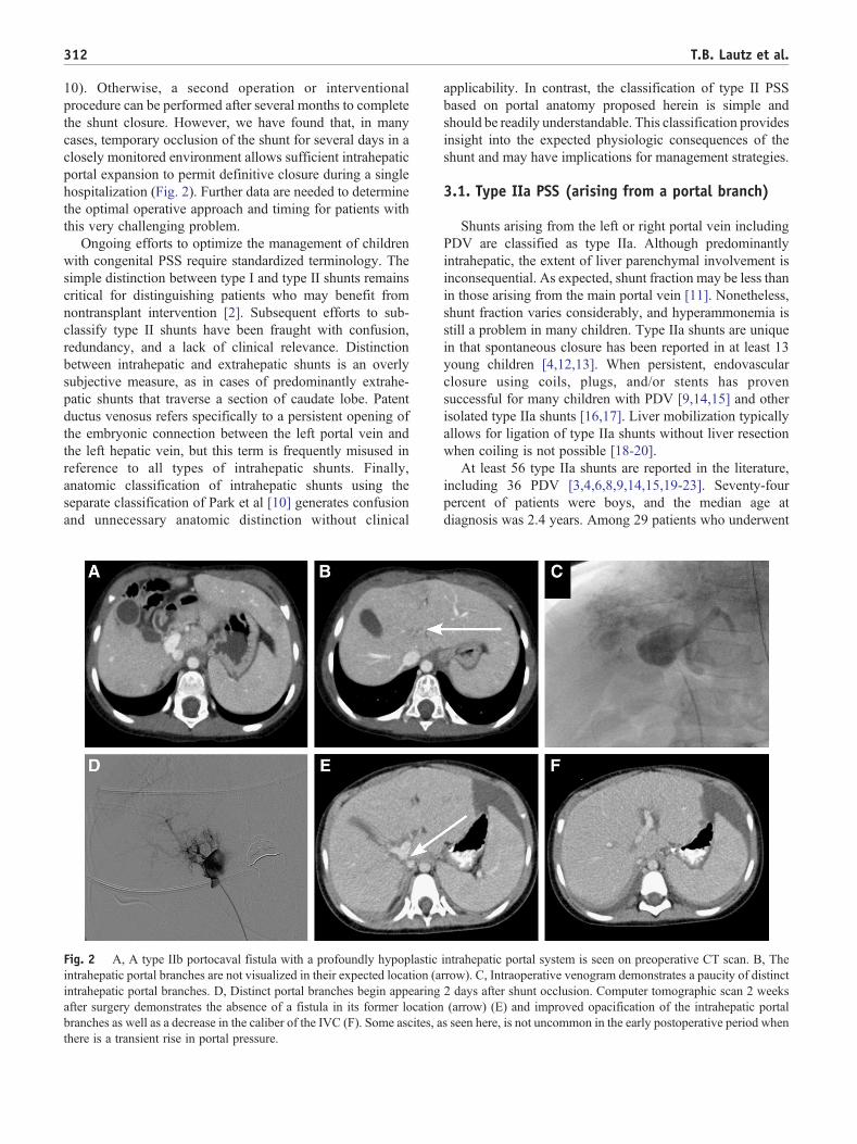
10). Otherwise, a second operation or interventionalprocedure can be performed after several months to completethe shunt closure. However, we have found that, in manycases, temporary occlusion of the shunt for several days in aclosely monitored environment allows sufficient intrahepaticportal expansion to permit definitive closure during a singlehospitalization (Fig. 2). Further data are needed to determinethe optimal operative approach and timing for patients withthis very challenging problem.
Ongoing efforts to optimize the management of childrenwith congenital PSS require standardized terminology. Thesimple distinction between type I and type II shunts remainscritical for distinguishing patients who may benefit fromnontransplant intervention [2]. Subsequent efforts to sub-classify type II shunts have been fraught with confusion,redundancy, and a lack of clinical relevance. Distinctionbetween intrahepatic and extrahepatic shunts is an overlysubjective measure, as in cases of predominantly extrahe-patic shunts that traverse a section of caudate lobe. Patentductus venosus refers specifically to a persistent opening ofthe embryonic connection between the left portal vein andthe left hepatic vein, but this term is frequently misused inreference to all types of intrahepatic shunts. Finally,anatomic classification of intrahepatic shunts using theseparate classification of Park et al [10] generates confusionand unnecessary anatomic distinction without clinical
Fig. 2 A, A type IIb portocaval fistula with a profoundly hypoplasticintrahepatic portal branches are not visualized in their expected location (aintrahepatic portal branches. D, Distinct portal branches begin appearingafter surgery demonstrates the absence of a fistula in its former locationbranches as well as a decrease in the caliber of the IVC (F). Some ascites, athere is a transient rise in portal pressure.
applicability. In contrast, the classification of type II PSSbased on portal anatomy proposed herein is simple andshould be readily understandable. This classification providesinsight into the expected physiologic consequences of theshunt and may have implications for management strategies.
3.1. Type IIa PSS (arising from a portal branch)
Shunts arising from the left or right portal vein includingPDV are classified as type IIa. Although predominantlyintrahepatic, the extent of liver parenchymal involvement isinconsequential. As expected, shunt fraction may be less thanin those arising from the main portal vein [11]. Nonetheless,shunt fraction varies considerably, and hyperammonemia isstill a problem in many children. Type IIa shunts are uniquein that spontaneous closure has been reported in at least 13young children [4,12,13]. When persistent, endovascularclosure using coils, plugs, and/or stents has provensuccessful for many children with PDV [9,14,15] and otherisolated type IIa shunts [16,17]. Liver mobilization typicallyallows for ligation of type IIa shunts without liver resectionwhen coiling is not possible [18-20].
At least 56 type IIa shunts are reported in the literature,including 36 PDV [3,4,6,8,9,14,15,19-23]. Seventy-fourpercent of patients were boys, and the median age atdiagnosis was 2.4 years. Among 29 patients who underwent
intrahepatic portal system is seen on preoperative CT scan. B, Therrow). C, Intraoperative venogram demonstrates a paucity of distinct2 days after shunt occlusion. Computer tomographic scan 2 weeks(arrow) (E) and improved opacification of the intrahepatic portals seen here, is not uncommon in the early postoperative period when
313Management and classification of congenital portosystemic shunts
operative (n = 15) or endovascular (n = 14) closure, 13 of 13with encephalopathy or hyperammonemia improved aftersurgery, and 10 of 12 with cardiopulmonary symptomsimproved after surgery. Further efforts are needed todetermine at which age spontaneous closure of type IIashunts becomes unlikely and which patients benefit fromoperative vs endovascular closure.
3.2. Type IIb PSS (arising from main portal vein, itsbifurcation, or the splenomesenteric confluence)
Shunts arising from a position between the bifurcation ofthe portal vein and the splenomesenteric confluence can beclassified as type IIb. These include main portocaval,portoatrial, and portorenal shunts. Although predominantlyextrahepatic, these shunts may traverse a short segment ofliver parenchyma. These abnormalities may be limited to asingle communication from the main portal vein to the IVC.However, they may also compose a large, complex, andhard-to-delineate venous confluence of mesenteric andsystemic veins encompassing the splenic, superior mesen-teric, portal, and renal veins and the IVC. Hyperammonemiaand other metabolic consequences are frequent. Type IIbshunts are typically short and wide in diameter, makingsuccessful endovascular management unlikely. Nonetheless,successful endovascular closure has been described in rarecases [6,16,24], including our patient with a long fistuladraining into the right atrium. These patients usually requirestaged operative closure using the above-described tech-nique. The degree of difficulty in managing these patients isdirectly related to the extent of portal vein hypoplasia and thelocation of the shunt. More proximal shunts (at or near thesplenomesenteric confluence) often result in a long segmentof hypoplastic extrahepatic portal vein and greater difficultyin restoring intrahepatic portal flow without causingexcessive portal hypertension or stasis.
Including our patients, at least 26 cases of type IIb shuntshave been reported [3,4,6,7,16,25-31]. Median age atdiagnosis was 2.8 years, and 16 (62%) were boys. Shuntanatomywas portocaval in 10, portorenal in 5, and portoatrialin 1. Correction was attempted in 15 (3 endovascular, 10open, and 1 laparoscopic) and was successful in 13. One childwas intolerant of shunt banding, and another developed a newPSS after initial shunt ligation. All but 1 patient whounderwent surgery had hyperammonemia with or withoutencephalopathy, and this resolved in all patients withsuccessful shunt closure. In addition, liver lesions regressedin 2 patients, and cardiopulmonary symptoms improved in2 patients after successful surgery.
3.3. Type IIc PSS (shunt arising from mesenteric,gastric, or splenic veins)
Awide variety of peripheral PSS can be grouped together astype IIc [8,28,29,32-35]. These shunts arise from the mesen-
teric, gastric, or splenic veins and empty into the renal vein,azygos vein, iliac veins, or their branches. The most frequentanatomic variants in children are gastrorenal (4 reported cases)and splenorenal (4 reported cases). Although more peripheral,encephalopathy or subclinical hyperammonemia nonethelessoccurred in 10 of 14 reported cases. Cardiopulmonarysymptoms occurred in 6 cases, and isolated cases of liverlesions and failure to thrivewere reported. A shunt between theIMV and a branch of the iliac vein caused recurrent vaginalbleeding in 1 patient from our series. Some degree of portalvein hypoplasia can occur but usually to a lesser extent thanin type IIb shunts. Therefore, immediate ligation or occlusionof type IIc shunts can typically be performed without causingsevere portal hypertension or other adverse sequelae.Furthermore, these shunts may be approachable by minimal-ly invasive techniques. Among the reported cases, 3 endovas-cular, 3 laparoscopic, 3 open, and 1 combined approaches wereall successful.
In conclusion, symptoms caused by type II PSS aregenerally reversible with shunt ligation or embolization,and an aggressive approach aimed at elimination of theabnormal portosystemic communication should be taken.The approach for occlusion of the shunt must be based onthe individual characteristics of the patient's shunt anatomyand position. Shunt closure is ultimately possible andtolerated even in patients with profoundly hypoplasticintrahepatic and extrahepatic portal veins using a stagedprocedure described herein. A refined classification basedon portal anatomy is proposed, which will allow futurestudies to make meaningful comparisons between patientswith similar anatomy and physiology.
References
[1] Abernethy J. Account of two instances of uncommon formation in theviscera of the human body. Phil Trans R Soc 1793;83:59-66.
[2] Morgan G, Superina R. Congenital absence of the portal vein: twocases and a proposed classification system for portasystemic vascularanomalies. J Pediatr Surg 1994;29:1239-41.
[3] Howard ER, Davenport M. Congenital extrahepatic portocaval shunts—the Abernethy malformation. J Pediatr Surg 1997;32:494-7.
[4] Stringer MD. The clinical anatomy of congenital portosystemic venousshunts. Clin Anat 2008;21:147-57.
[5] Akahoshi T, Nishizaki T, Wakasugi K, et al. Portal-systemicencephalopathy due to a congenital extrahepatic portosystemicshunt: three cases and literature review. Hepatogastroenterology2000;47:1113-6.
[6] Ikeda S, Sera Y, Ohshiro H, et al. Surgical indications for patients withhyperammonemia. J Pediatr Surg 1999;34:1012-5.
[7] Witters P, Maleux G, George C, et al. Congenital veno-venousmalformations of the liver: widely variable clinical presentations.J Gastroenterol Hepatol 2008;23:e390-4.
[8] Ohno T, Muneuchi J, Ihara K, et al. Pulmonary hypertension inpatients with congenital portosystemic venous shunt: a previouslyunrecognized association. Pediatrics 2008;121:e892-9.
[9] Eroglu Y, Donaldson J, Sorensen LG, et al. Improved neurocognitivefunction after radiologic closure of congenital portosystemic shunts.J Pediatr Gastroenterol Nutr 2004;39:410-7.
314 T.B. Lautz et al.
[10] Park JH, Cha SH, Han JK, et al. Intrahepatic portosystemic venousshunt. AJR Am J Roentgenol 1990;155:527-8.
[11] Uchino T, Matsuda I, Endo F. The long-term prognosis of congenitalportosystemic venous shunt. J Pediatr 1999;135:254-6.
[12] Ono H, Mawatari H, Mizoguchi N, et al. Clinical features and outcomeof eight infants with intrahepatic porto-venous shunts detected inneonatal screening for galactosaemia. Acta Paediatr 1998;87:631-4.
[13] Valls E, Ceres L, Urbaneja A, et al. Color Doppler sonography in thediagnosis of neonatal intrahepatic portosystemic shunts. J ClinUltrasound 2000;28:42-6.
[14] Cho YK, Chang NK, Ma JS. Successful transcatheter closure of a largepatent ductus venosus with the Amplatzer vascular plug II. PediatrCardiol 2009;30:540-2.
[15] Schwartz YM, Berkowitz D, Lorber A. Transvenous coil embolizationof a patent ductus venosus in a 2-month-old child. Pediatrics 1999;103:1045-7.
[16] Hoover W, Ackerman V, Schamberger M, et al. The congenitalporto-caval fistula: a unique presentation and novel intervention.Pediatr Pulmonol 2008;43:196-9.
[17] Takayama Y, Moriura S, Nagata J, et al. Embolization of the left portalvein to inferior vena cava shunts for chronic recurrent hepaticencephalopathy via the mesenteric vein. J Gastroenterol Hepatol2001;16:1425-8.
[18] Kamata S, Kitayama Y, Usui N, et al. Patent ductus venosus with ahypoplastic intrahepatic portal system presenting intrapulmonaryshunt: a case treated with banding of the ductus venosus. J PediatrSurg 2000;35:655-7.
[19] Uchino T, Endo F, Ikeda S, et al. Three brothers with progressivehepatic dysfunction and severe hepatic steatosis due to a patent ductusvenosus. Gastroenterology 1996;110:1964-8.
[20] Yoshimoto Y, Shimizu R, Saeki T, et al. Patent ductus venosus inchildren: a case report and review of the literature. J Pediatr Surg2004;39:E1-5.
[21] Alomari AI, Chaudry G, Fox VL, et al. Atypical manifestation ofpatent ductus venosus in a child: intervening against a paradoxicalpresentation. J Vasc Interv Radiol 2009;20:537-42.
[22] Araki T, Kamada M, Okamoto Y, et al. Coil embolization of a patentductus venosus in a 52-day-old girl with congenital heart disease. AnnThorac Surg 2003;75:273-5.
[23] Ferrero GB, Porta F, Biamino E, et al. Remittent hyperammonemia incongenital portosystemic shunt. Eur J Pediatr 2010;169:369-72.
[24] Chiu SN, Chien YH, Wu MH, et al. Transcatheter closure of portal-systemic shunt combining congenital double extrahepatic inferior venacava with vascular plug. J Pediatr 2008;153:723.
[25] Badler R, Price AP, Moy L, et al. Congenital portacaval shunt: CTdemonstration. Pediatr Radiol 2002;32:28-30.
[26] Kanamori Y, Hashizume K, Kitano Y, et al. Congenital extrahepaticportocaval shunt (Abernethy type 2), huge liver mass, and patentductus arteriosus—a case report of its rare clinical presentation in ayoung girl. J Pediatr Surg 2003;38:E15.
[27] Mboyo A, Lemouel A, Sohm O, et al. Congenital extra-hepaticportocaval shunt. Concerning a case of antenatal diagnosis. Eur JPediatr Surg 1995;5:243-5.
[28] Mizoguchi N, Sakura N, Ono H, et al. Congenital porto-left renalvenous shunt as a cause of galactosaemia. J Inherit Metab Dis 2001;24:72-8.
[29] Nii A, Takehara HO, Kuyama H, et al. Successful preemptive surgicaldivision of type 2-congenital extrahepatic portosystemic shunt inchildren. J Med Invest 2009;56:49-54.
[30] Tercier S, Delarue A, Rouault F, et al. Congenital portocaval fistulaassociated with hepatopulmonary syndrome: ligation vs liver trans-plantation. J Pediatr Surg 2006;41:e1-3.
[31] Yonemitsu H, Mori H, Kimura T, et al. Congenital extrahepaticportocaval shunt associated with hepatic hyperplastic nodules in apatient with Dubin-Johnson syndrome. Abdom Imaging 2000;25:572-5.
[32] Kimura T, Soh H, Hasegawa T, et al. Laparoscopic correction ofcongenital portosystemic shunt in children. Surg Laparosc EndoscPercutan Tech 2004;14:285-8.
[33] MorikawaN, Honna T, Kuroda T, et al. Resolution of hepatopulmonarysyndrome after ligation of a portosystemic shunt in a pediatric patientwith an Abernethy malformation. J Pediatr Surg 2008;43:e35-8.
[34] Spencer LT, LanghamMR, Hoyer MH, et al. Resolution of hypoxemiain a liver transplant recipient after ligation of a portosystemic shunt.J Pediatr 2000;137:575-7.
[35] Yamagami T, Yoshimatsu R, Matsumoto T, et al. Successfulembolization using interlocking detachable coils for a congenitalextrahepatic portosystemic venous shunt in a child. J Pediatr Surg2007;42:1949-52.