mammalian homologs of drosophila elav localized to a ... · pdf filethe journal of...
TRANSCRIPT

The Journal of Neuroscience, April 1994, 14(4): 1943-1952
Mammalian Homologs of Drosophila ELAV Localized to a Neuronal Subset Can Bind in vitro to the 3’ UTR of mRNA Encoding the Id Transcriptional Repressor
Peter H. King,’ Todd D. Levine,’ Robert T. Fremeau, Jr.,’ and Jack D. Keenel
Departments of ‘MIcrobiology and ?Pharmacology, Duke University MedIcal Center, Durham, North Carolina 27710
Mammalian cDNAs encoding a rat (Rel-Nl) and a human (Hel-Nl) neuronal RNA-binding protein have been cloned and characterized with respect to tissue specificity, neu- roanatomical localization, and RNA binding specificity. Both proteins are highly similar to the product of the Drosophila elav gene, which is expressed in all neurons of the f ly and is required for development of the nervous system. However, in situ hybridization of rat tissues demonstrated more re- stricted expression of Rel-Nl mRNA within a subset of neu- rons of the hippocampus, cortex, and other regions of the gray matter, but not in glial cells or white matter. In vitro RNA binding experiments demonstrated that Hel-Nl can bind to the 3’ untranslated region (3’ UTR) of Id mRNA, a transcript that encodes a helix-loop-helix transcriptional repressor that is abundantly expressed in undifferentiated neural precur- sors. Sequences characterized for Hel-Nl binding were also abundantly present in the 3’ UTR of the Drosophila extra- macrochaetae mRNA, which encodes an Id homolog. Thus, we have identified a potential link between a neuronal 3’ UTR RNA-binding protein and regulatory transcription factors in- volved in neural development. These findings are interpreted in light of recent studies in which mRNA 3’ UTRs were found to be important for the regulation of cell growth and differ- entiation.
/Key words: RNA-binding proteins, RNA recognition motif, helix-loop-helix, inhibitor of DNA binding, paraneoplastic diseases, 3’ untranslated region, differentiation, autoimmu- nify]
The Drosophila rlav gene locus encodes a protein of vital im- portance in the early development of the CNS (Campos et al.. 1985: Jiminez and Campos-Ortega. 1987). R4utations of this gene locus gi\-e rise to a lethal phenotype with numerous struc- tural defects and hypotrophy of the CNS. Its role in neuronal grolvth and diffcrcntiation of the f1rosopll//u nervous system is also underscored bq the appearance of&z,, transcripts following the differentiation of neuroblasts into primitive neurons (Ro- binow and White. 1988). Another Dmsophilu protein termed
Received June 16, 1993: revised ?iug 30. 1993; accepted Sept. 14, 1993. This work was supported by research grants from the NatlanaI Institutes of
Health to J.D.K. P.H.K. was supported b) Ncurobehavioral Trainmg Grant MH Ii 177. and T.1I.L. was supportrd b) the Four Schools Ph>srcran-Scientist I’rogram
(‘orrespondence should be addressed to Jack II. Keene. Hex 3020, 406 Jones Bulldlng. Duke LJni\ersity Medlcal Center. Durham. NC 277 IO.
Present addrw: I~epartment of Neurology. Irnlvcrs~t) of Alabama MedIcal Center. Birmingham. AI_ 35233.
Coplrlghr c 1993 Soclet? for Newowcncc 07~0.6171 9-l I3 1943. I0$05.00,0
rhl19 is also similar to eluv but mutants have not been charac- tcrixd (Kim and Baker, 199313).
The cDNA cloning of ciuv revealed that it possesses three potential KNA binding domains (Robinow et al.. 1988) based upon the presence of three sets of two conserl-ed core elements. the RNP 1 and RNP 2 consensus sequences (Adam et al., 1986: Dre>.fuss et al.. 1988). These elements are part of a common RN.4 recognition motif (termed RRM) that was shown to be encompassed within a functionally defined RN.&binding do- main (Query et al.. 1989: rcxieaed in Kenan et al., 1991).
In the present study, we have used degenerate probes and PCR to isolate human and rat cDNA homologs of the Dro- mphtlu ELAV protein. The human counterpart is designated Hel-Nl for human eluv-like ncuronal protein I and the rat counterpart as Rel-Nl for rat clue-like neuronal protein 1. We found that the mRNA of Rel-N 1 is expressed only in neuronal tissue and in a subset of CNS neurons in the adult rat. It was not detcctcd in glial cells or white matter and \vas found abun- dantly in portions of hippocampus. cerebral cortex. and brain- stem. Sequcncc comparisons of Dtmophilu rlul, and 15~~9 with Hel-N 1 and Kcl-N I suggest the existence of a subfamily of e/a\,- lihe genes. All of the ELAV-like proteins are highly homologous in their three RRhls, suggesting that the) may have similar RNA-binding targets.
Here we demonstrate that Hcl-Nl binds 1~1 virm to mRNA encoding a transcriptional repressor protein termed “inhibitor of DNA binding” or abbreviated Id (Benczra ct al., 1990). Id has been implicated in muscle cell development of mice because of its interactions with the transcription factor MyoD (Benezra ct al.. 1990). Expression of Id and a human homolog. Id2. were rccentl>. reported to occur during earl! development of neural tissue (Duncan et al.. 1992: Biggs et al.. 1992). WC report here that binding of Hel-Nl to Id mRNA leas dependent upon the integrity of sequences in the 3’ untranslated region (3’ UTR) of the Id transcript. Interestingly. Id mRNA was found to contain short stretches of uridylate residues in the 3’ I!TR that resemble sequences present in growth factor mRNAs (Shaw and Kamen, 1986) that are known to interact with Hel-Nl (Levine et al.. 1993). These data raise the possibility that H&N1 is involved in modulating the expression of transcriptional regulators of the helix-loop-helix family that arc known to play a role in neural and muscle cell growth and differentiation.
Materials and Methods C‘ionrt7~ IIf/-.VI iq, I’C‘R. Degenzratc polymrrasc chain reaclion (PCR) primers nerc s!nthesired based on the first sex-en amino acids of the RNPl conscns~~s sequence in the first RRM (Fig. I./f. amino acids 203-

1944 King et al. * Neuronal Protein Binding of mRNA of Helix-Loop-Helix Protein Id
ELAV
Hal-N1
K3 VW)
ELAV
Hel-Nl
K3 (W)
ELAV
H&N1
K3 (W’)
ELAV
Hsl.Nl
K3 VW’)
ELAV
Hot-N1
K3 VW)
ELAV
Hd-Ni
K3 (W)
ELAV
H&N1
K3 W’PW
ELAV
W-N1
K3 (W)
ELAV
H&N1
K3 (W)
AP”QNhAAYAAAAQLQQQQ”QQAILQ”QQQQTQQA”AAAAAA”~Q 82
- _ NCPT------------TIN--------NNcss--P”Dscyy-- 31
I I 1 I 1 I I I
QTNCGTTATTTAAAOhDSTTNAA”=QA*ANNAASNNNNNNNN~NN ?O
QLQQQQQA""AQQh""QQQQQQAAA""QQAA"QQA""PQPQQAQP 127
_ _ _ - - - - _ - _ - - - -E-DS--------------------------- 3, I I
NNNNN*T*NNNNNNePDp--------------------------- 108
C E I E S ” K L I R D K S Q ” Y I D P I N P Q A P
I I I I I I I I I I CEIESCKLVRDKIT-----------
I I I I I I I I I I I CEVESCKLIRDKVT-----------
LASGPCGRYP--------
SPITNX-AK-- 483 I I , I I I I SFKTNKTHKA- 359 I I I I I I I SFKTNX-HKQT 4.4
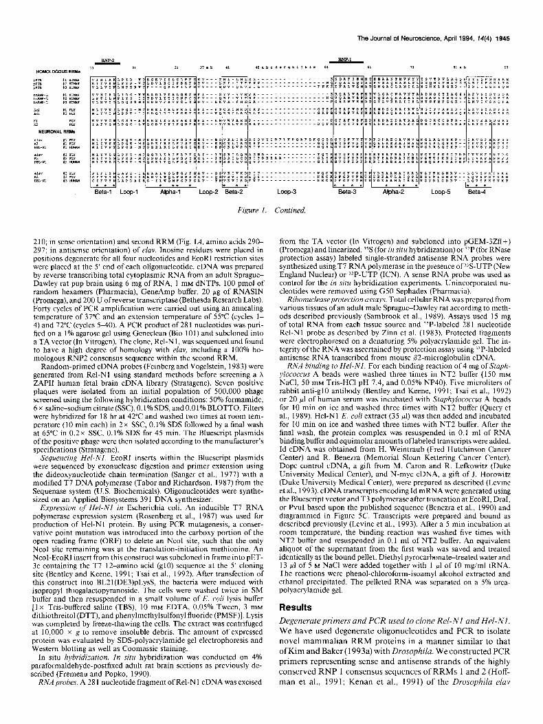
Figure 1. A, Amino acid and nucleotide sequence of Hel-Nl in comparison to Drosophila proteins ELAV and rbp9. Boxes represent the RNPl octamer sequence (shaded boxes) and the RNP2 hexamer sequence (unshaded boxes) of each of the three RNA recognition motifs. B, Comparison of the amino acid sequences in the RNA recognition motif of Hel-Nl with those in the Drosophila proteins ELAV and rbp9 according to the alignment of Kenan et al. (199 1) and Hoffman et al. (199 1).
The Journal of Neuroscience, April 1994, 74(4) 1945
L I l&H-l l Loop-l
l
Alph*all l I
Loop-2 *Beta*-2 * I
Loop-3
Figure 1. Contined.
2 10; in sense orientation) and second RRM (Fig. 1 A, amino acids 290- 297; in antisense orientation) of elav. Inosine residues were placed in positions degenerate for all four nucleotides and EcoRI restriction sites were placed at the 5’ end of each oligonucleotide. cDNA was prepared by reverse transcribing total cytoplasmic RNA from an adult Sprague- Dawley rat pup brain using 6 mg of RNA, 1 mM dNTPs, 100 pmol of random hexamers (Pharmacia), GeneAmp buffer, 20 pg of RNASIN (Promega), and 200 U ofreverse transcriptase (Bethesda Research Labs). Forty cycles of PCR amplification were carried out using an annealing temperature of 37°C and an extension temperature of 55°C (cycles l- 4) and 72°C (cycles S-40). A PCR product of 28 1 nucleotides was puri- fied on a 1% agarose gel using Geneclean (Bio 10 1) and subcloned into a TA vector (In Vitrogen). The clone, Rel-N 1, was sequenced and found to have a high degree of homology with eluv, including a 100% ho- mologous RNP2 consensus sequence within the second RRM.
from the TA vector (In Vitrogen) and subcloned into pGEM-3Zf(+) (Promega) and linearized. YS (for in situ hybridization) or ?IP (for RNase protection assay) labeled single-stranded antisense RNA probes were synthesized using T7 RNA polymerase in the presence of )S-UTP (New England Nuclear) or 32P-UTP (ICN). A sense RNA probe was used as control for the in situ hybridization experiments. Unincorporated nu- cleotides were removed using G50 Sephadex (Pharmacia).
Ribonucleaseprotection assays. Total cellular RNA was prepared from various tissues of an adult male Sprague-Dawley rat according to meth- ods described previously (Sambrook et al., 1989). Assays used 15 mg of total RNA from each tissue source and ‘*P-labeled 281 nucleotide Rel-Nl probe as described by Zinn et al. (1983). Protected fragments were electrophoresed on a denaturing 5% polyacrylamide gel. The in- tegrity ofthe RNA was ascertained by protection assay using “P-labeled antisense RNA transcribed from mouse @2-microglobulin cDNA.
Random-primed cDNA probes (Feinberg and Vogelstein, 1983) were generated from Rel-Nl using standard methods before screening a X ZAP11 human fetal brain cDNA library (Stratagene). Seven positive plaques were isolated from an initial population of 500,000 phage screened using the following hybridization conditions: 50% formamide, 6 x saline-sodium citrate (SSC), 0.1% SDS, and 0.01% BLOTTO. Filters were hybridized for 18 hr at 42°C and washed two times at room tem- perature (10 min each) in 2 x SSC, 0.1% SDS followed by a final wash at 65°C in 0.2 x SSC, 0.1% SDS for 45 min. The Bluescript plasmids of the positive phage were then isolated according to the manufacturer’s specifications (Stratagene).
Sequencing Hel-NI. EcoRI inserts within the Bluescript plasmids were sequenced by exonuclease digestion and primer extension using the dideoxynucleotide chain termination (Sanger et al., 1977) with a modified T7 DNA polymerase (Tabor and Richardson, 1987) from the Sequenase system (U.S. Biochemicals). Oligonucleotides were synthe- sized on an Applied Biosystems 391 DNA synthesizer.
Expression of Hel-Nl in Escherichia coli. An inducible T7 RNA polymerase expression system (Rosenberg et al., 1987) was used for production of Hel-Nl protein. By using PCR mutagenesis, a conser- vative point mutation was introduced into the carboxy portion of the open reading frame (ORF) to delete an NcoI site, such that the only NcoI site remaining was at the translation-initiation methionine. An NcoI-EcoRI insert from this construct was subcloned in frame into PET- 3c containing the T7 12-amino acid (g10) sequence at the 5’ cloning site (Bentley and Keene, 199 1; Tsai et al., 1992). After transfection of this construct into BL21(DE3)pLysS, the bacteria were induced with isopropyl thiogalactopyranoside. The cells were washed twice in SM buffer and then resuspended in a small volume of E. co/i lysis buffer [l x Tris-buffered saline (TBS), 10 mM EDTA, 0.05% Tween, 3 mM dithiothreitol (DTT), and phenylmethylsulfonyl fluoride (PMSF)]. Lysis was completed by freeze-thawing the cells. The extract was centrifuged at 10,000 x g to remove insoluble debris. The amount of expressed protein was evaluated by SDS-polyacrylamide gel electrophoresis and Western blotting as well as Coomassie staining.
RNA binding to Hel-NI. For each binding reaction of 4 mg of Staph- y/ococcus A beads were washed three times in NT2 buffer (150 mM NaCl, 50 mM Tris-HCl pH 7.4, and 0.05% NP40). Five microliters of rabbit anti-g10 antibody (Bentley and Keene, 1991; Tsai et al., 1992) or 20 ~1 of human serum was incubated with Staphylococcus A beads for 10 min on ice and washed three times with NT2 buffer (Query et al., 1989). Hel-Nl E. co/i extract (35 ~1) was then added and incubated for 10 min on ice and washed three times with NT2 buffer. After the final wash, the protein complex was resuspended in 0.1 ml of RNA binding buffer and equimolar amounts oflabeled transcripts were added. Id cDNA was obtained from H. Weintraub (Fred Hutchinson Cancer Center) and R. Benezra (Memorial Sloan Kettering Cancer Center). Dope control cDNA, a gift from M. Caron and R. Lefkowitz (Duke University Medical Center), and N-myc cDNA, a gift of J. Horowitz (Duke University Medical Center), were prepared as described (Levine et al., 1993). cDNA transcripts encoding Id mRNA were generated using the Bluescript vector and T3 polymerase after truncation at EcoRI, DraI, or PvuI based upon the published sequence (Benezra et al., 1990) and diagrammed in Figure 5C. Transcripts were prepared and bound as described previously (Levine et al., 1993). After a 5 min incubation at room temperature, the binding reaction was washed five times with NT2 buffer and resuspended in 0.1 ml of NT2 buffer. An equivalent aliquot of the supematant from the first wash was saved and treated identically as the bound pellet. Diethyl pyrocarbonate-treated water and 13 ~1 of 5 M NaCl were added together with 1 ~1 of 10 mg/ml tRNA. The reactions were phenol-chloroform-isoamyl alcohol extracted and ethanol precipitated. The pelleted RNA was separated on a 5% urea- polyacrylamide gel.
Results
In situ hybridization. In situ hybridization was conducted on 4% paraformaldehyde-postfixed adult rat brain sections as previously de- scribed (Fremeau and Popko, 1990).
RNA probes. A 28 1 nucleotide fragment of Rel-Nl cDNA was excised
Degenerate primers and PCR used to clone Rel-NI and Hel-Nl. We have used degenerate oligonucleotides and PCR to isolate novel mammalian RRM proteins in a manner similar to that ofKim and Baker (1993a) with Drosophila. We constructed PCR primers representing sense and antisense strands of the highly conserved RNP 1 consensus sequences of RRMs 1 and 2 (Hoff- man et al., 1991; Kenan et al., 1991) of the Drosophila elav
****I *I * * * Beta-3 iphal2* Loop-5 Beta-4
1946 King et al. l Neuronal Protein Binding of mRNA of Helix-Loop-Hehx Protein Id
300bp -
240bp -
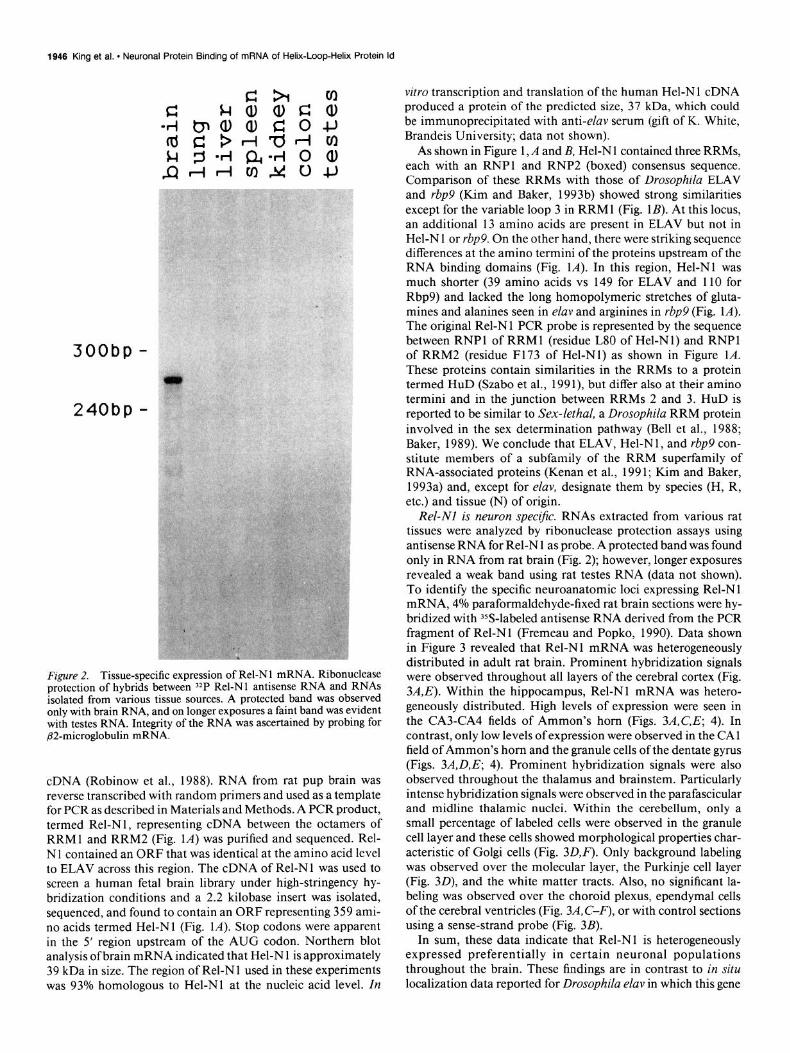
Figure 2. Tissue-specific expression of Rel-Nl mRNA. Ribonuclease protection of hybrids between ‘*P Rel-Nl antisense RNA and RNAs isolated from various tissue sources. A protected band was observed only with brain RNA, and on longer exposures a faint band was evident with testes RNA. Integrity of the RNA was ascertained by probing for @2-microglobulin mRNA.
cDNA (Robinow et al., 1988). RNA from rat pup brain was reverse transcribed with random primers and used as a template for PCR as described in Materials and Methods. A PCR product, termed Rel-N 1, representing cDNA between the octamers of RRMI and RRM2 (Fig. IA) was purified and sequenced. Rel- Nl contained an ORF that was identical at the amino acid level to ELAV across this region. The cDNA of Rel-Nl was used to screen a human fetal brain library under high-stringency hy- bridization conditions and a 2.2 kilobase insert was isolated, sequenced, and found to contain an ORF representing 359 ami- no acids termed Hel-Nl (Fig. 1A). Stop codons were apparent in the 5’ region upstream of the AUG codon. Northern blot analysis of brain mRNA indicated that Hel-N 1 is approximately 39 kDa in size. The region of Rel-N 1 used in these experiments was 93% homologous to Hel-Nl at the nucleic acid level. In
vitro transcription and translation of the human Hel-N 1 cDNA produced a protein of the predicted size, 37 kDa, which could be immunoprecipitated with anti-elav serum (gift of K. White, Brandeis University; data not shown).
As shown in Figure 1, A and B, Hel-N 1 contained three RRMs, each with an RNPl and RNP2 (boxed) consensus sequence. Comparison of these RRMs with those of Drosophila ELAV and rbp9 (Kim and Baker, 1993b) showed strong similarities except for the variable loop 3 in RRM 1 (Fig. 1 B). At this locus, an additional 13 amino acids are present in ELAV but not in Hel-N 1 or rbp9. On the other hand, there were striking sequence differences at the amino termini of the proteins upstream of the RNA binding domains (Fig. 1A). In this region, Hel-Nl was much shorter (39 amino acids vs 149 for ELAV and 110 for Rbp9) and lacked the long homopolymeric stretches of gluta- mines and alanines seen in elav and arginines in rbp9 (Fig. 1A). The original Rel-N 1 PCR probe is represented by the sequence between RNPl of RRMl (residue L80 of Hel-Nl) and RNPl of RRM2 (residue F173 of Hel-Nl) as shown in Figure 1A. These proteins contain similarities in the RRMs to a protein termed HUD (Szabo et al., 199 l), but differ also at their amino termini and in the junction between RRMs 2 and 3. HUD is reported to be similar to Sex-lethal, a Drosophila RRM protein involved in the sex determination pathway (Bell et al., 1988; Baker, 1989). We conclude that ELAV, Hel-NI, and rbp9 con- stitute members of a subfamily of the RRM superfamily of RNA-associated proteins (Kenan et al., 199 1; Kim and Baker, 1993a) and, except for elav, designate them by species (H, R, etc.) and tissue (N) of origin.
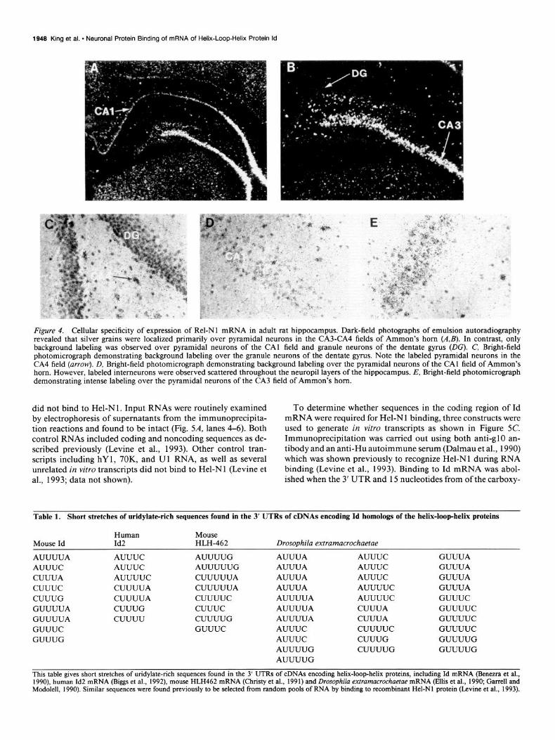
Rel-Nl is neuron specific. RNAs extracted from various rat tissues were analyzed by ribonuclease protection assays using antisense RNA for Rel-N 1 as probe. A protected band was found only in RNA from rat brain (Fig. 2); however, longer exposures revealed a weak band using rat testes RNA (data not shown). To identify the specific neuroanatomic loci expressing Rel-Nl mRNA, 4% paraformaldehyde-fixed rat brain sections were hy- bridized with YS-labeled antisense RNA derived from the PCR fragment of Rel-Nl (Fremeau and Popko, 1990). Data shown in Figure 3 revealed that Rel-Nl mRNA was heterogeneously distributed in adult rat brain. Prominent hybridization signals were observed throughout all layers of the cerebral cortex (Fig. 3A,E). Within the hippocampus, Rel-Nl mRNA was hetero- geneously distributed. High levels of expression were seen in the CA3-CA4 fields of Ammon’s horn (Figs. 3A,C,E; 4). In contrast, only low levels of expression were observed in the CA 1 field of Ammon’s horn and the granule cells of the dentate gyrus (Figs. 3A,D,E, 4). Prominent hybridization signals were also observed throughout the thalamus and brainstem. Particularly intense hybridization signals were observed in the parafascicular and midline thalamic nuclei. Within the cerebellum, only a small percentage of labeled cells were observed in the granule cell layer and these cells showed morphological properties char- acteristic of Golgi cells (Fig. 3DJ’). Only background labeling was observed over the molecular layer, the Purkinje cell layer (Fig. 3D), and the white matter tracts. Also, no significant la- beling was observed over the choroid plexus, ependymal cells of the cerebral ventricles (Fig. 3A, C-F), or with control sections using a sense-strand probe (Fig. 3B).
In sum, these data indicate that Rel-Nl is heterogeneously expressed preferentially in certain neuronal populations throughout the brain. These findings are in contrast to in situ localization data reported for Drosophila elav in which this gene

The Journal of Neuroscience, April 1994, 74(4) 1947
Figure 3. Localization of Rel-Nl RNA in the rat CNS using in situ hybridization. Coronal (A, C, D, F) and horizontal (E) sections of adult rat brain were hybridized with YS-labeled antisense Rel-Nl cRNA. B, Coronal section adjacent to that in A hybridized with a sense-strand control probe. F, Bright-field photomicrograph of the cerebellar cortex (Cer) from D. Note that a small percentage of specifically labeled cells were observed in the granule cell layer of the cerebellum (Gr). In contrast, the Purkinje cells (Pk) were unlabeled. Ctx, cerebral cortex; CA3, field CA3 of Ammon’s horn; CM, central median thalamic nucleus; PC, paracentral thalamic nucleus; PJ, parafascicular nucleus of the thalamus; CAI, field CA1 of Ammon’s horn; DC, granule cells of the dentate gyrus; CPU, caudate-putamen; Gp, globus pallidus.
was found to be expressed in all neurons (Robinow and White, 1988).
Hel-Nl Binds to the 3’end of Id mRNA. Despite the presence of three identifiable RRMs, target RNA molecules have not yet been identified for ELAV or rbp9. Our search for potential li- gands for Hel-N 1 was directed by results of in vitro randomized RNA selection experiments using Hel-Nl (Levine et al., 1993; Tsai et al., 199 l), which showed preferences for binding to short uridylate-rich regions in RNA. Based on the known role of elav in Drosophila, we examined sequences of RNAs involved in neural development. Id mRNAs were found to contain short
stretches of uridylates in the 3’ UTRs (Table 1) that are similar to those shown to bind Hel-Nl in vitro (Levine et al., 1993). To test for binding to Id mRNA, an in vitro transcript of Id mRNA along with Dope and N-myc control mRNAs were each incubated with recombinant epitope-tagged Hel-Nl using stan- dard RNA binding methods (Query et al., 1989; Bentley and Keene, 199 1). Following immunoprecipitation with the epitope- specific antibody (anti-glO), bound products were analyzed on a denaturing acrylamide gel. As shown in Figure 5A, lane 1, Id mRNA terminating at a DraI truncation site in the middle of the 3’ UTR (Fig. 5C) was able to bind, while the control mRNAs
1949 King et al. l Neuronal Protein Binding of mRNA of Helix-Loop-Helix Protein Id
Figure 4. Cellular specificity of expression of Rel-Nl mRNA in adult rat hippocampus. Dark-field photographs of emulsion autoradiography revealed that silver grains were localized primarily over pyramidal neurons in the CA3-CA4 fields of Ammon’s horn (A,&. In contrast, only background labeling was observed over pyramidal neurons of the CA1 field and granule neurons of the dentate gyrus (DG). C, Bright-field photomicrograph demonstrating background labeling over the granule neurons of the dentate gyms. Note the labeled pyramidal neurons in the CA4 field (arrow). D, Bright-field photomicrograph demonstrating background labeling over the pyramidal neurons of the CA1 field of Ammon’s horn. However, labeled interneurons were observed scattered throughout the neuropil layers of the hippocampus. E, Bright-field photomicrograph demonstrating intense labeling over the pyramidal neurons of the CA3 field of Ammon’s horn.
did not bind to Hel-N 1. Input RNAs were routinely examined by electrophoresis of supematants from the immunoprecipita- tion reactions and found to be intact (Fig. 54, lanes 4-6). Both control RNAs included coding and noncoding sequences as de- scribed previously (Levine et al., 1993). Other control tran- scripts including hY 1, 70K, and U 1 RNA, as well as several unrelated in vitro transcripts did not bind to Hel-N 1 (Levine et al., 1993; data not shown).
To determine whether sequences in the coding region of Id mRNA were required for Hel-Nl binding, three constructs were used to generate in vitro transcripts as shown in Figure 5C. Immunoprecipitation was carried out using both anti-g10 an- tibody and an anti-Hu autoimmune serum (Dalmau et al., 1990) which was shown previously to recognize Hel-Nl during RNA binding (Levine et al., 1993). Binding to Id mRNA was abol- ished when the 3’ UTR and 15 nucleotides from of the carboxy-
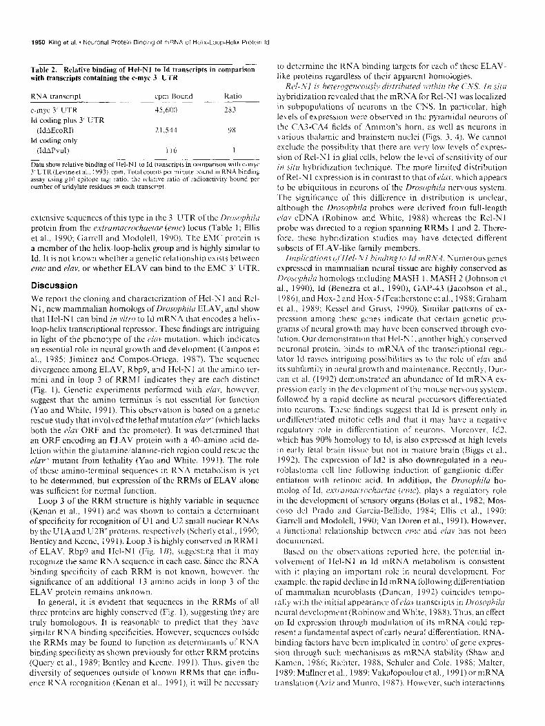
Table 1. Short stretches of uridylate-rich sequences found in the 3’ UTRs of cDNAs encoding id homologs of the helix-loop-helix proteins
Mouse Id Human Mouse Id2 HLH-462 Drosouhila extramacrochaetae
AUUUUA AUUUC CUUUA cuuuc CUUUG GUUUUA GUUUUA GUUUC GUUUG
AUUUC AUUUUG AUUUC AUUUUUG AUUUUC CUUUUUA CUUUUA CUUUUUA CUUUUA cuuuuc CUUUG cuuuc cuuuu CUUUUG
GUUUC
AUUUA AUUUA AUUUA AUUUA AUUUUA AUUUUA AUUUUA AUUUC AUUUC AUUUUG AUUUUG
AUUUC AUUUC AUUUC AUUUUC AUUUUC CUUUA CUUUA cuuuuc CUUUG CUUUUG
GUUUA GUUUA GUUUA GUUUA GUUUC GUUUUC GUUUUC GUUUUC GUUUUG GUUUUG
This table gives short stretches of uridylate-rich sequences found in the 3’ UTRs of cDNAs encoding helix-loop-helix proteins, including Id mRNA (Benezra et al., 1990), human Id2 mRNA (B&s et al., 1992), mouse HLH462 mRNA (Christy et al., 1991) and Drosophila extramacrochaetae mRNA (Ellis et al., 1990, Garrell and Modolell, 1990). Similar sequences were found previously to be selected from random pools of RNA by binding to recombinant Hel-Nl protein (Levine et al., 1993).

The Journal of Neuroscience, April 1994, 74(4) 1949
A B 123 456 123 456
-933 t 933
Ii& t789 C 789
t521
C Transcripts of Id mRNA
Pvu 1 Dral Eco Rl
Id ADra I 5’ I1
5’
Id APVU I
terminal coding region were deleted by cleavage with PvuI (Fig. 5B, lanes 1,4). This transcript was shown to remain intact when supernatants of the binding reaction were examined (data not shown). Binding to Id mRNA was retained, however, when either the DraI (Fig. 5B, lanes 2, 5) or the EcoRI truncated transcript (Fig. 5B, lanes 3,6) were bound. The EcoRI transcript represented the entire 3’ UTR sequence, but was consistently observed as a smeared band by acrylamide gel electrophoresis. PEM autoimmune serum was as effective as anti-g10 serum in the immunoprecipitation/RNA-binding experiments, indicat- ing that anti-HUD autoantibodies can bind to Hel-Nl without interfering with its binding to the Id mRNA. These data indicate that the sequence extending from the DraI site to the PvuI site and perhaps other sequences in the 3’ UTR are essential for binding to Hel-N 1. Table 2 confirms this conclusion by showing
Figure 5. A, Binding of in vitro tran- scripts of Id and control mRNAs to g 1 O-tagged Hel-N 1 protein following immunoprecipitation with anti-g10 an- tibody. Lanes 1-3, pellets after binding, immunoprecipitation and washing; lanes 4-6, supematants from each im- munoprecipitation. Lanes I and 4, transcript of Id mRNA from DraI tnm- cated template (IdADraI); lanes 2 and 5, control transcript of Dope mRNA; lanes 3 and 6, control transcript of N-myc mRNA. B, Binding of truncated transcripts of Id mRNA to glO-tagged Hel-Nl protein following immunopre- cipitation with either serum from a PEM patient (lanes 1-3) or anti-g 10 antibody (lanes 4-6). Transcripts were generated from truncated constructs of the Id cDNA as depicted in C. Lanes 1 and 4, IdAPvul; lanes 2 and 5, IdADraI; and lanes 3 and 6, IdAEcoRI mRNA. Sam- ples were prepared and analyzed as de- scribed in Materials and Methods and in Levine et al. (1993). C, Truncated transcripts designated IdAPvuI, Id- ADraI, and EcoRI were generated by restriction digestion and in vitro syn- thesis using T3 polymerase. The Id- ADraI transcript contains the 3’ UTR sequences cuuuc, CUUUG, GUUUUA, GUUUC, and GUUUG. The EcoRI transcript encompasses the entire Id mRNA and was consistently observed as a smeared band.
radioactive counts in a typical binding experiment in compar- ison with the c-myc 3’ UTR (Levine et al., 1993). As expected, the IdAPvuI transcript was not able to bind to Hel-N 1. Whether Hel-Nl directly binds to a primary sequence in the 3’ UTR of Id mRNA or recognizes a secondary structure in that region is not known.
It is interesting to note that the 3’ UTR of Id and other helix- loop-helix proteins (Table 1) contain short stretches of uridylate- rich sequences (Benezra et al., 1990; Christy et al., 199 1) similar to those identified previously as important for Hel-Nl binding (Levine et al., 1993). The function of these sequences in the 3’ UTR is not known but they may play a role in mRNA stability, localization, or translatability. Increasing evidence suggests an important regulatory role for various mRNA 3’ UTRs (Rasti- nejad and Blau, 1993). Equally interesting was the finding of
1950 King et al. * Neuronal Protein Binding of mRNA of Helrx-Loop-Helix Protein Id
Table 2. Relative binding of Hel-Nl to Id transcripts in comparison with transcripts containing the c-myc 3’ UTR
RNA transcript
c-mqc 3’ UTR Id coding plus 3’ UTR
cpm Round Ratlo
45,600 283
(IdAEcoRI) Id coding only
21.544 9x
(IdAPvuI) 116 1
Data show relative binding ofHel-NI to Id transcripts in comparison with c-my 3’ UTR (Levine et al.. 1993). cpm, Total counts per minute hound m RNA binding assay usmg g10 epitope tag; ratio, the relative ratio of radioactwity bound per number of uridylate residues in each transcript.
extensive sequences of this tl pe in the 3’ UTR of the Ikw~philm protein from the extramacrochactar (rmc) locus (Table 1; Ellis et al., 1990; Garrcll and Modolell. 1990). The EMC protein is a member of the helix-loop-helix group and is highly similar to Id. It is not known whether a genetic relationship exists between emc and rluv, or whether ELAV can bind to the EMC 3’ LJTR.
Discussion We report the cloning and characterization of Hel-N 1 and Rel- Nl, new mammalian homologs of Drosophda ELAV, and show that Hel-N 1 can bind in vitro to Id mRN,4 that encodes a helix- loop-helix transcriptional repressor. These findings arc intriguing in light of the phenotype of the elav mutation, which indicates an essential role in neural growth and development (Campos et al., 1985; Jiminez and Compos-Ortega. 1987). The sequence divergence among ELAV, Rbp9, and Hel-Nl at the amino ter- mini and in loop 3 of RRMl indicates they are each distinct (Fig. I). Genetic experiments performed with eluv, however, suggest that the amino terminus is not essential for function (Yao and White, 199 1). This observation is based on a genetic rescue study that involved the lethal mutation e/aP (which lacks both the e/u)! ORF and the promoter). It was determined that an ORF encoding an ELAV protein with a 40-amino acid de- letion within the glutamineialanine-rich region could rescue the elaF mutant from lethality (Yao and White, 199 1). The role of these amino-terminal sequences in RNA metabolism is )et to be determined, but expression of the RRMs of ELAV alone was sufficient for normal function.
Loop 3 of the RRM structure is highly variable in sequence (Kenan et al.. 1991) and was shown to contain a determinant of specificity for recognition of U 1 and U2 small nuclear RNAs bl the U 1A and U2B” proteins, respective11 (Scherlvet al.. 1990; Bentley and Keene, 199 1). Loop 3 is highly conserved in RRM 1 of EL.r\V, Rbp9 and Hel-Nl (Fig. IB). suggesting that it ma) recognize the same RNA sequence in each case. Since the RNA binding specificity of each RRM is not known, however, the significance of an additional 13 amino acids in loop 3 of the ELAV protein remains unknown.
In general, it is evident that sequences in the RRMs of all three proteins are highly conserved (Fig. 1). suggesting they arc truly homologous. It is reasonable to predict that they have similar RNA binding specificities. However. sequences outside the RRMs may be found to function as determinants of RNA binding specificity as shown previously for other RRM proteins (Query et al.. 1989; Bentley and Keene, 199 1). Thus, given the diversity of sequences outside of known RRMs that can influ- ence RNA recognition (Kenan et al.. 199 I), it will be neccssarq
to determine the RNA binding targets for each of these ELAV- like proteins regardless of their apparent homologies.
Rel-.VI is heterogeneously distributed w/thin the CIVS. In .situ hybridization revealed that the mRNA for Rel-Nl was localized in subpopulations of neurons in the CNS. In particular. high lek-els of expression were observed in the pyramidal neurons of the C.43-CA4 fields of Ammon’s horn. as well as neurons in various thalamic and brainstem nuclei (Figs. 3, 4). We cannot exclude the possibility that there are very low levels of expres- sion of Rel-Nl in glial cells, below the level of sensitivity of our in situ hybridization technique. The more limited distribution of Rel-N 1 expression is in contrast to that of elan. which appears to be ubiquitous in neurons of the Drosophila nervous system. The significance of this difference in distribution is unclear. although the Drosophilu probes were derived from full-length c~/av cDNA (Robinow and White, 1988) whereas the Rel-Nl probe was directed to a region spanning RRMs 1 and 2. There- fore. these hybridization studies may have detected different subsets of ELAV-like family members.
Implications qf/lel-Xl billding to Id mRN‘4. Numerous genes expressed in mammalian neural tissue arc highly conserved as Drosophila homologs including MASH 1. MASH 2 (Johnson et al., 1990), Id (Benezra ct al., 1990), GAP-43 (Jacobson et al., 1986). and Hox-2 and Hox-5 (Featherstone et al.. 1988; Graham et al., 1989; Kessel and Gruss, 1990). Similar patterns of ex- pression among these genes indicate that certain genetic pro- grams of neural growth may have been conserved through cvo- lution. Our demonstration that Hcl-N 1, another highly conserved neuronal protein, binds to mRNA of the transcriptional regu- lator Id raises intriguing possibilities as to the role of elan and its subfamily in neural growth and maintenance. Recently, Dun- can et al. (1992) demonstrated an abundance of Id mRNA ex- pression early in the development ofthc mouse nervous system. followed by a rapid decline as neural precursors dilferentiated into neurons. These findings suggest that Id is present only in undifferentiated mitotic cells and that it may have a negative regulator)’ role in differentiation of neurons. Moreover, Id2. which has 90% homology to Id, is also expressed at high levels in early fetal brain tissue but not in mature brain (Biggs et al.. 1992). The expression of Id2 is also downregulated in a neu- roblastoma cell line following induction of ganglionic differ- entiation with retinoic acid. In addition, the Drosophila ho- molog of Id. e,Ytl.ut)2u(,/.[)Chuetae (cmc), plays a regulatory role in the development of sensory organs (Botas et al., 1982; Mos- coso dcl Prado and Garcia-Bellido, 1984; Ellis et al., 1990: Garrcll and Modolcll, 1990; Van Doren et al., 199 1). However, a functional relationship bet\veen em and ciao has not been documented.
Based on the observations reported here, the potential in- volvemcnt of Hel-Nl in Id mRNA metabolism is consistent \vith it playing an important role in neural development. For example, the rapid decline in Id mRNA following differentiation of mammalian neuroblasts (Duncan. 1992) coincides tempo- rally with the initial appearance ofcluv transcripts in Drosophila neural dcvelopmcnt (Robino\r and White. 1988). Thus. an effect on Id expression through modulation of its mRNA could rep- resent a fundamental aspect of earl) neural differentiation. RNA- binding factors have been implicated in control of gene expres- sion through such mechanisms as mRNA stability (Shaw and Kamcn. 1986: Richter, 1988: Schuler and Cole, 1988; Malter, 1989; Mullncr et al.. 1989; Vakalopoulou et al.. 1991) or mRNA translation (Ayiz and Munro, 1987). However, such interactions

The Journal of Neuroscience, April 1994, 74(4) 1951
have not been documented to occur in the CNS. It will be important to determine whether Hel-Nl or other members of the eluv subfamily participate directly in the regulation of ex- pression of Id and other helix-loop-helix proteins.
Binding of Hel-N 1 to the 3’ UTR of Id mRNA has particular relevance because of recent work demonstrating the importance of 3’ UTRs in growth and differentiation. Through genetic com- plementation using a differentiation-defective myoblast mutant, Rastinejad and Blau (1993) demonstrated that the 3’ UTRs of certain muscle genes can play a regulatory role in cell division and differentiation. Whether interactions between Hel-N 1 and the 3’ UTR of Id mRNA can affect neural functions is not yet known, however. This hypothesis is consistent with the genetic phenotype and neuronal characteristics associated with elav in Drosophila. Findings reported here and interactions demon- strated previously between Hel-Nl and sequences in the 3’ UTRs of growth factor mRNAs (Levine et al., 1993) are consistent with this hypothesis because it is necessary that cell proliferation cease prior to entry into a program of differentiation (Cham- berlain et al., 1985). Whether Hel-Nl represents a neural-spe- cific truns-acting RNA-binding protein that affects these devel- opmental processes is yet to be determined.
References Adam SA, Nakagawa TY, Swanson MS, Woodruff T (1986) mRNA
polyadenylate-binding protein: gene isolation and sequencing and identification of a ribonucleoprotein consensus sequence. Mol Cell Biol 62932-2943.
Aziz N, Munro HN (1987) Iron regulates fenitin mRNA translation through a segment of its 5’ untranslated region. Proc Nat1 Acad Sci USA 84:8478-8482.
Baker BS (1989) Sex in flies: the splice of life. Nature 340:521-524. Bell L, Maine EM, Schedl P, Cline TW (1988) Sex-lethal, a Drosophila
sex determination switch gene, exhibits sex-specific RNA splicing and seauence similaritv to RNA binding. uroteins. Cell 55:1037-1046.
Benezra R, Davis RL, Lockshon D, Turner DL, Weintraub H (1990) The protein Id: a negative regulator of helix-loop-helix DNA binding proteins. Cell 6 1:49-59.
Bentley RC, Keene JD (199 1) Recognition of Ul and U2 small nuclear RNAs can be altered by a 5-amino-acid segment in the U2 small nuclear ribonucleoprotein particle (snRNP) and through interactions with U2 snRNP-A’ protein. Mol Cell Biol 11: 1829-l 839.
Biggs J, Murphy EV, Israel MA (1992) A human Id-like helix-loop- helix protein expressed during early development. Proc Nat1 Acad Sci USA 89:1512-1517.
Botas J, Moscoso de1 Prado J, Garcia-Bellido A (1982) Gene-dose titration analysis in the search of trans-regulatory genes in Drosophila EMBO J 1:307-3 10.
Campos AR, Grossman D, White K (1985) Mutant alleles at the locus elav in Drosophila melanogaster lead to nervous system defects. A developmental-genetic analysis. J Neurogenet 2: 197-2 18.
Chamberlain JS, Jaynes JB, Hauschka SD (1985) Regulation of cre- atine kinase induction in differentiating mouse myoblasts. Mol Cell Biol 5:484-492.
Christy BA, Sanders LK, Lau LF, Copeland NG, Jenkins NA, Nathans D (199 1) An Id-related helix-loop-helix protein encoded by a growth factor-inducible gene. Proc Nat1 Acad Sci USA 88: 18 15-l 8 19.
Dalmau J, Fumeaux HM, Gralla RJ, Kris MG, Posner JB (1990) Detection of the anti-Hu antibody in the serum of patients with small cell lung cancer-a quantitative Western blot analysis. Ann Neurol 27544-552.
Dreyfuss G, Swanson MS, Pinol-Roma S (1988) Heterogeneous nu- clear ribonucleoprotein particles and the pathway of mRNA. Trends Biochem Sci 13:86-9 1.
Duncan M, DiCicco-Bloom EM, Xiang X, Benezra R, Chada K (1992) The gene for the helix-loop-helix protein, Id, is specifically expressed in neural precursors. Dev Biol 154: l-10.
Ellis HM, Spann DR, Posakony JW (1990) extramacrochaetae, a neg- ative regulator of sensory organ development in Drosophila, defines a new class of helix-loop-helix proteins. Cell 61:27-38.
Featherstone MS, Baron A, Gaunt SJ, Mattei M, Duboule D (1988) Hox-5. I defines a homeobox-containing gene locus on mouse chro: mosome 2. Proc Nat1 Acad Sci USA 85:4760-4765.
Feinberg A, Vogelstein B (1983) A technique for radiolabeling DNA restriction endonuclease fragments to high specific activity. Anal Bio- them 132:6-l 3.
Fremeau RT, Popko B (1990) In situ analysis of myelin basic protein gene expression in myelin-deficient oligodendrocytes: antisense hnRNA and readthrouah transcriution. EMBO J 9:3533-3538.
Garrell J, Modolell J (1996) The Drosophila extramacrochaetae locus, an antagonist ofproneural genes that, like these genes, encodes a helix- loop-helix protein. Cell 61:39-48.
Graham A, Papalopulu N, Krumlauf R (1989) The murine and Dro- sophila homeobox gene complexes have common features of orga- nization and expression. Cell 57:367-378.
Hoffman DW, Query CC, Golden BL, White SW, Keene JD (1991) Structural analysis of the RNA binding domain of the U 1 snRNP-A protein using nuclear magnetic resonance spectroscopy reveals sim- ilarity to ribosomal proteins. Proc Nat1 Acad Sci USA 88:2495-2499.
Jacobson RD, Virag I, Skene JHP (1986) A protein associated with axon growth, GAP-43, is widely distributed and developmentally regulated in rat CNS. J Neurosci 6: 1843-1855.
Jimmez F, Campos-Ortega JA (1987) Genes in subdivision 1 B of the Drosophila melanogaster X-chromosome and their influence on neu- ral development. J Neurosenet 4: 179-200.
Johnson JE,-Birren SJ, And&son DJ (1990) Two rat homologues of Drosophila achaete-scute specifically expressed in neuronal precur- sors. Nature 346:858-860.
Kenan DJ, Query CC, Keene JD (1991) RNA recognition: towards identifying determinants of specificity. Trends Biochem Sci 16:2 14- 220.
Kessel M, Gruss P (1990) Murine developmental control genes. Sci- ence 2491374-379.
Kim YJ, Baker BS (1993a) Isolation of RRM-type RNA-binding pro- tein genes and the analysis of their relatedness by using a numerical approach. Mol Cell Bid1 13: 174-183.
Kim YJ. Baker BS C 1993b) The Drosoohila aene rbo9 encodes a orotein that is a member of aconserved group-of putative RNA binding proteins that are nervous system-specific in both flies and humans. J Neurosci 13:1045-1056.
Levine TD, Gao F-B, King PH, Andrews LG, Keene JD (1993) Hel- Nl: an autoimmune RNA-binding protein with specificity for 3’ uri- dylate-rich untranslated regions of growth factor mRNAs. Mol Cell Biol 13:3494-3504.
Malter JS (1989) Identification of an AUUUA-specific messenger RNA binding protein. Science 246~664-666.
Moscoso de1 Prado J, Garcia-Bellido A (1984) Genetic regulation of the Achaete-scute complex of Drosophila melanogaster. Rouxs Arch Dev Biol 193~242-245.
Mullner EW, Neupert B, Kuhn LC (1989) A specific mRNA binding factor regulates the iron-dependent stability of cytoplasmic transfenin receptor mRNA. Cell 58:373-382.
Query CC, Bentley RC, Keene JD (1989) A common RNA recognition motif identified within a defined Ul RNA binding domain of the 70K Ul snRNP protein. Cell 57:89-101.
Rastinejad F, Blau HM (1993) Genetic complementation reveals a novel regulatory role of 3’ untranslated regions in growth and differ- entiation. Cell 72:903-9 17.
Richter JD (1988) Information relay from RNA to protein: the mRNP connection. Trends Biochem Sci 13:483486.
Robinow S, White K (1988) The locus elav ofDrosophila melanogaster is expressed in neurons at all developmental stages. Dev Biol 126: 294-303.
Robinow S, Campos A, Yao K, White K (1988) The eluv gene product of Drosophila, required in neurons, has three RNP consensus motifs. Science 242: 1570-l 572.
Rosenberg AH, Lade BN, Chui DS, Dunn JJ, Studier FW (1987) Vec- tors used for selective expression of cloned DNAs by T7 polymerase. Gene 56:125-135.
Sambrook J, Fritsch EF, Maniatis T (1989) Molecular cloning: a lab- oratory manual, 2d ed. Cold Spring Harbor, NY: Cold Spring Harbor Laboratory.
Sanger F, Nicklen S, Coulson AR (1977) DNA sequencing with chain terminating inhibitors. Proc Nat1 Acad Sci USA 7415463-5467.
Scherly D, Dathan NA, Boelens W, van Venrooij WJ, Mattaj IW (1990)

1952 King et al. * Neuronal Protein BindIng of mRNA of HelwLoop-Helix Protein Id
The LJ2B” RNA motif as a site of protein-protein interaction. EMBO J 9:3675-368 1.
Schuler GD, Cole MD (1988) GM-CSF and oncogene mRNA stabil- ities are independently regulated in tram in a mouse monocytic tumor. Cell 55:1155-l 122.
Shaw G, Kamen R (1986) A conserved AU sequence from the 3’ untranslated region of GM-CSF mRNA mediates selective mRNA degradation. Cell 46:659-667
Szabo A, Dalmau J, Manley G, Rosenfeld M, Wong E, Henson J, Posner JB, Furneaux HM (199 1) HUD a paraneoplastic encephalomyelitis antigen contains RNA binding domains and is homologous to EL-l I’ andsex-lerhal. Cell 67~325-333.
Tabor S. Richardson CC (1987) DNA seauence analvsis with a mod- ified bacteriophage T7 DNA ‘polymerase. Proc Nai Acad Sci USA 8414167477 1.
Tsai DE, Harper DS, Kcene JD (1991) Ul-snRNP A protein selects a ten nucleotide consensus sequence from a degenerate RNA pool presented in various structural contexts. Nucleic Acids Rcs 19:493 I- 4936.
Tsai DE, Kenan DJ, Keene JD (I 992) In v&o selection of an RNA epitope immunologically crossreactive with a peptide. Proc Nat1 Acad Sci USA 89:8864-8868.
Vakalopoulou E, Schaak J, Shenk T (1991) A 32 kilodalton protem binds to AU-rich domains in the 3’ untranslated regions of rapidly degraded mRNAs. Mol Cell Biol 11:3355-3364.
Van Doren M, Ellis HM, Posakony JW (199 1) The Drmophila extra- macrochaetur protein antagonizes sequence-specific DNA binding by dau,Shterlr.s.s/uchaete-scut~~ protein complexes. Development 113:245- 255.
Yao K, White K (199 1) Organizational analysis of elac gene and func- tional analysis of ELAV protein ofDrosophila rnelanog&terand Dro- sonhila viniis. Mel Cell Biol 1 1:2994-3000.
Zinn K, Dimaio T, Maniatis T (1983) Identification of two distinct regulatory regions adjacent to the human o-interferon gene. Cell 34: 865-879.