malnutrition and amino acid metabolism · 2017-10-27 · skeletal wasting and stunting of marasmus...
TRANSCRIPT

The Malnourished Child, edited by RobertM. Suskind and Leslie Lewinter-Suskind.Nestlg Nutrition Workshop Series, Vol. 19.Nestec Ltd., Vevey/Raven Press, Ltd.,New York © 1990.
Malnutrition and Amino Acid Metabolism
Alan A. Jackson and R. F. Grimble
Department of Human Nutrition, University of Southampton, Bassett Crescent East,Southampton SO9 3TU, England
Severe childhood malnutrition embraces a spectrum of clinical presentations andpathologic disorders. Across the entire spectrum, there is clear evidence of majorchanges in the state of amino acid and whole body protein metabolism, from theskeletal wasting and stunting of marasmus to the varied multisystem changes char-acterized by the kwashiorkor syndrome. For this reason, amino acid and protein me-tabolism have commonly been held to lie at the center of the metabolic complexitiesof severe undernutrition. Indeed, the use of the name protein-energy malnutrition(PEM) implies an etiologic role for protein, although this may not be fully justified.When Cecily Williams (1) first described kwashiorkor in 1933, she suggested a pri-mary protein deficiency as the cause of the disease, rather than an infection or vita-min deficiency. Subsequent research has shown that interactions with deficiencies ofspecific micronutrients and infection probably play an important part in the develop-ment of the disease and may account for the wide pattern of clinical presentation.Certainly, any model that seeks to explain the cause of the disease has to account fora bewildering spectrum of pathophysiologic changes (2).
In 1963, Holt and co-workers (3) explored the possibility that kwashiorkor wasthe result of a specific deficiency of one or a small number of essential amino acids.The rationale for their study was that the deficient amino acid ' 'would be revealedby the pattern of free amino acids in the blood plasma.'' There was a striking simi-larity in the fasting amino acid profile in the plasma of 64 children with kwashiorkorof varying severity from nine countries around the world, despite diverse sources ofdietary protein. The conclusion reached by Holt was similar to that of Williams 18years earlier, that "the limiting nitrogenous component in a kwashiorkor-produc-ing diet . . . is not an essential amino acid but nitrogen, either essential or non-essential." The pattern of the amino acid concentrations revealed a reduction of allthe essential, indispensable amino acids, with some of the nonessential, dispensableamino acids having reduced or variable concentrations. Of the indispensable aminoacids, valine, leucine, and isoleucine showed the largest reduction, while phenylala-nine and lysine were less affected. Of the dispensable amino acids, tyrosine, argi-nine, and citrulline were reduced in concentration, while glycine, serine, proline,and histidine were found to be raised, lowered, or normal.
73
74 MALNUTRITION AND AMINO ACID METABOLISM
The battle to explain and understand the distorted pattern of amino acids seen inkwashiorkor was joined in many parts of the world. It provided a stimulus for thedevelopment of biochemical methods for use in the detection of subclinical forms ofPEM. Different groups have adopted different approaches to the problem, but mosthave been stimulated by the idea articulated by Waterlow et al. (4), that deathfrom severe malnutrition is a consequence of the failure of a protein-dependentfunction(s).
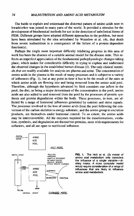
Perhaps the single most important difficulty inhibiting progress in this area ofwork has been the absence of a suitable animal model for the disease state. This re-flects an imperfect appreciation of the fundamental pathophysiologic changes takingplace, which makes for considerable difficulty in trying to explain and understandthe observed changes in the established human disease (5). The only clinical materi-als that are readily available for analysis are plasma and urine. The concentration ofamino acids in the plasma is the result of many processes and is subject to a varietyof influences (Fig. 1), but at any point in time it has to be the result of the rates atwhich amino acids are flowing into and being removed from the amino acid pool.Therefore, although the hypothesis advanced by Holt considers one inflow to thepool, the diet, as being a major determinant of the concentration in the pool, aminoacids are also added to and removed from the pool by the processes of protein syn-thesis and protein degradation within the body. These processes, in turn, are af-fected by a range of hormonal influences generated by nutrient and stress signals.The processes involved in the loss of amino acids from the pool following the con-version of the carbon skeleton to energy substrate, and the amino group to excretionproducts, are themselves under hormonal control. To an extent, the amino acidsmay be interconvertible. All the enzymes required for the transformations, oxida-tion, synthesis, and degradation are themselves proteins, most with requirements forcofactors, and all are open to nutritional influence.
PROTEIN INTAKE
rh
HOLT MODEL
INFECTIONHORMONESADAPTIVE RESPONSESMETABOLIC BLOCKSUNUSUAL DEMANDS/TISSUE SPECIFIC CHANGES)
FIG. 1. The Holt et al. (3) model ofamino acid metabolism only considersthe influence of a single variable—di-etary intake. A more complete modelwould require consideration of a range ofinfluences that are likely to influenceamino acid metabolism.
UNUSUAL LOSSES EXPANDED MODEL

MALNUTRITION AND AMINO ACID METABOLISM 75
DIETARY EVIDENCE
The implication in much of the literature is that kwashiorkor is more common inareas where dietary protein tends to be deficient (6,7). This apparent association hasbeen elevated to a causal relationship and is the main basis for implicating proteindeficiency in the etiology of the disease. Therefore, one of the first points to clarifyis the extent to which the evidence supports the thesis of dietary protein inadequacy.Landman and Jackson (8) collated data from dietary surveys carried out in sevencountries over the course of 30 years in communities where kwashiorkor was a ma-jor problem. In general the mean energy intakes were inadequate, 22 to 48% of therecommended daily allowance (RDA) (9), except in healthy Jamaican children andIndian toddlers. By contrast, mean protein intakes frequently met or exceeded theRDA. When mean protein intakes were inadequate or marginal, mean energy in-takes were so low that the protein would have been utilized inefficiently. For practi-cal and ethical reasons, these data rarely represent the actual intake of children in theprocess of developing kwashiorkor. However, extensive clinical experience hasshown that on a dietary protein intake of only 0.6 g/kgday, about 50% of the RDA,children with kwashiorkor lose their edema (10). Therefore, a protein intake of 0.6g/kg-day is sufficient to "maintain the edema-free state," and to promote correctionof pathologic processes seen in kwashiorkor. This figure can be used as a minimalintake necessary to prevent the development of the kwashiorkor syndrome. Fromthis perspective, the protein content of all the diets studied is abundant. Overall, thedata support the contention that "there is no situation in which the child is adequatewith regard to calories and deficient with regard to protein alone. . . . The majorbottleneck is calories and not protein" (11).
Nevertheless, this conclusion presumes that the requirements for protein are notdifferent from those of a normal, healthy population; that there are no abnormal orunusual losses; and that intercurrent infections, an everyday reality in populations atrisk from severe undernutrition, play no part at all (12).
BODY COMPOSITION
Although all tissues are affected by an inadequate dietary intake, the weight lostduring the development of malnutrition is not evenly shared by all tissues (13).There is always a significant reduction in lean tissue mass, although there is a rela-tive sparing of visceral tissues, with more obvious and extensive loss of muscle(14). Even within the visceral tissues, some functions are spared more obviously atthe expense of others. Hypoalbuminemia, a consequence of decreased albumin syn-thesis by the liver, is representative of the situation that occurs with other exportproteins that carry out carrier functions in the circulation, e.g., transferrin and reti-nol binding protein (15,16). The development of fatty liver is in part a consequenceof inadequate synthesis of the carrier apolipoprotein (17). The evidence suggeststhat the decrease in specific protein synthesis may represent a redistribution of

76 MALNUTRITION AND AMINO ACID METABOLISM
MACROPHAQE
FIG- 2. It is thought that a large part ofthe effect of infection on amino acid and
FEVER protein metabolism is mediated throughthe effects of cytokines released from
MUSCLE SKIN-1 . rL»^n« . , . . _ . , . . . .PROTEIN SYNTHESIS | [ IRON, ZINC 1 leukocytes. Cytokines induce an orches-
trated sequence of metabolic changes,which involve a range of tissues andfunctions.
SERUM ALBUMIN METALLOTHIONEIN fTRANSFERRIN J '
ACUTE PHASE 4PROTEINS
amino acids to the synthesis of acute phase reactants. The specific mechanisms thatcontrol this redistribution of amino acids are not altogether clear. One important fac-tor is almost certainly infection. In response to an infection, macrophages elicit cy-tokines, such as interleukin-1 and tumor necrosis factor, which have the ability toinhibit the synthesis of albumin in the liver and to elevate the production of acutephase proteins (Fig. 2) (18). However, other causes cannot be excluded; for exam-ple, potassium deficiency is invariably present in severe undernutrition, as either abalanced deficiency or a specific depletion (13). It is not possible to retain intracellu-lar protein in the face of a marked potassium deficit, although it has been suggestedthat loss of potassium from muscle represents a mechanism for retaining basicamino acids.
HORMONES
The concentration of amino acids in plasma is the result of metabolism in visceraland peripheral tissues, under the influence of a range of hormones. Insulin, thyroidhormones, growth hormone, somatomedin, glucagon, and glucocorticoids have allbeen shown to exert an influence by affecting uptake into protein in many sites anddispersal into degradative pathways in the liver. A hormone may act directly on aprocess but may also influence the sensitivity of target tissues to other hormones.For example, growth hormone and glucocorticoids may produce insulin insensitiv-ity. Even within a carefully controlled laboratory experiment, it is difficult to predictthe effect that a particular hormone might have on any aspect of protein metabolism.Therefore, with the limited control possible in metabolic or field studies on childrenexposed to varying degrees of infection and malnutrition, interpretations of the spe-cific effect of individual hormones must be guarded.
In an elegant series of animal studies, Mill ward et al. (19) examined the meta-bolic effects of hormones on protein synthesis in skeletal muscle (Table 1). They

MALNUTRITION AND AMINO ACID METABOLISM 77
TABLE 1. Effect of hormones on protein synthesis and degradation in skeletal muscle
Hormone Protein synthesis Protein degradation Net effect
InsulinGrowth hormone and
somatomedinT3: normal levels
hyperthyroid levelsGlucocorticoids: fed
fasted
tt
T TTii
INo effect
Tt T
t
AnabolicAnabolic
AnabolicCatabolicCatabolicCatabolic
In malnutrition: Insulin I | . Growth hormone f , somatomedin J,, T3 | , glucocorticoids f I •1, increase; ! , decrease.From ref. 19.
concluded that both insulin and T3 were independently important for maintainingprotein synthesis. Either hormone could maintain synthesis when the other waspresent in low concentrations. They confirmed the suppressive effect of glucocorti-coids on synthesis and demonstrated that they could override the stimulatory effectof insulin. Thus, insulin and thyroid hormones would promote net uptake of aminoacids by muscle, and glucocorticoids would cause outflow from muscle. Soma-tomedin, the effector of growth hormone, would stimulate muscle protein synthesis.
Many studies have shown that the concentrations and production rates of hor-mones are changed in malnourished children. Thyroid hormone, glucagon, andsomatomedin are depressed, while growth hormone is elevated. Insulin and gluco-corticoids have been found in normal, reduced, or elevated concentrations, with asignificant reduction in the affinity of insulin for its receptor (20-27).
Lunn et al. (28) attempted to obtain a prospective picture of changes in insulin,cortisol, and growth hormone as children moved from a marginally nourished to amalnourished state. They concluded that an early rise in insulin concentration fell tolower levels once a malnourished state had become established, at which timegrowth hormone concentration rose. Cortisol concentrations were normal in margin-ally malnourished children but became elevated as malnutrition developed. Thus, inmarginally nourished children, the outflow of amino acids from muscle for glucone-ogenesis might be suppressed, with a reversal of this condition in the malnourishedstate, particularly with a decrease in thyroid hormone and elevated cortisol. A com-parative study on the hormonal profile of children in Uganda and the Gambia illus-trates the importance of the relative balance between hormones in their effect onmuscle protein (29). Marginally malnourished children in the Gambia had a higherratio of cortisol to insulin, with greater depletion of muscle than marginally nour-ished Ugandan children. Further studies on the Gambian children showed a negativecorrelation between growth and the cortisol/insulin ratio (30).
The influence of the intensity of muscle-protein turnover on the amino acid con-centration in plasma is illustrated by studies in goitrous patients (31), which showed

78 MALNUTRITION AND AMINO ACID METABOLISM
a marked fall in the concentration of all essential amino acids with the exception ofmethionine, and slightly less intense reductions in the nonessentials. A reduction inthe excretion of 3-methyl-histidine was interpreted as a diminished rate of muscleprotein degradation.
METABOLIC ADAPTATION
Whatever other features may be present as a component of severe undemutrition,a negative energy balance with the concomitant loss of tissue, and hence weight, isinvariable. As a consequence, the adaptive responses of the body are brought intoplay, although they may be overshadowed by other responses, such as that to infec-tion. Classic balance studies can yield information on the overall status of nitrogenmetabolism in the body, and in general, these show very efficient utilization of di-etary nitrogen, with no evidence of poor absorption. There is appropriate retentionof nitrogen, so that balance is defended down to very low levels of intake (32-34).In some studies, there is evidence of a protein-losing enteropathy, but the extent towhich this might contribute to a negative nitrogen balance is poorly documented.
The evidence suggests that a significant proportion of basal energy expendituresupports the physiologic processes associated with protein turnover. The physio-logic cost to the body of protein turnover is estimated to be 7.3 kJ/g protein, whichmay represent as much as 30% of resting energy expenditure in the adult (35). Dur-ing malnutrition, protein synthesis is significantly reduced to 4 g/kg-day, comparedwith 6.3 g/kg-day after recovery (36), a theoretical saving of 16.8 kJ/kg-day. Inboth malnourished and recovered children, the dietary protein intake only representsa relatively small proportion of overall protein turnover, with protein synthesis be-ing 90% of the total. Malnutrition is characterized by intermittent periods of growthfaltering and catch-up. During catch-up growth, there is an increase in the intensityof protein synthesis to 9.7 g/kg-day, with only 3% of flux coming from the dietaryintake (36). In children of normal weight, the response to infection is a marked in-crease in the rates of both protein synthesis and breakdown, and as breakdown ex-ceeds synthesis, there is a negative nitrogen balance. In malnourished children,there may be a doubling of synthesis and breakdown in the face of infection, bring-ing turnover to the level seen in a normal, uninfected child. However, the rates ofsynthesis and breakdown remain similar, with the consequence that nitrogen bal-ance is maintained (Fig. 3) (37). It has been suggested that one of the importantfunctions of protein turnover is to give the organism a capability to respond tochange. A reduced turnover would, therefore, imply a measure of compromise inthe ability of the organism to withstand environmental variation.
It is not clear to what extent the body is able to accommodate changes in energyintake without sacrificing function. The evidence shows that with progressive reduc-tions in energy intake from 100, to 90, 80, and 70 kcal/kgday, young children de-fend nitrogen balance very effectively, and measurements of protein turnover madeover 18 hr show little change in the overall rate of protein degradation, with a pro-

MALNUTRITION AND AMINO ACID METABOLISM 79
Protein Turnover g/kg.9h6r
RecoveringInfection
Infected
.Malnourished
Synthesis Degradation
FIG. 3. Changes in protein turnovercan be seen as a part of the response ofnormal and malnourished children to in-fection. The changes in protein synthesisand degradation are modified by nutri-tional state (37).
FIG. 4. There is an adaptive re-sponse seen in young children to de-fend nitrogen balance as the energymetabolizable intake is reduced pro-gressively from 100 to 70 kcal/kg-day(33,34).
NITROGEN RETENTION mg/kgd
100
50
100 90 80 70
ENERGY INTAKE Kcal /kg.d
gressive fall in protein synthesis (Fig. 4) (33,34). This remarkable ability to pre-serve nitrogen balance was associated with a significant fall in stool frequency andhence fecal nitrogen at the lowest levels of energy intake (34). In the circumstancesin which metabolic studies are conducted, the absorption of nitrogen is of the orderof 90% or more, with little variation in fecal nitrogen (32). However, there is evi-dence that in children with diarrheal disease, there is a significant protein-losing en-teropathy, which should give rise to a more variable fecal nitrogen, due to increasedendogenous losses rather than to any change in adsorption.
DIARRHEAL DISEASE
Diarrheal disease and varying degrees of malabsorption are an almost invariableaccompaniment of severe undernutrition. Although it is impossible to determine theextent to which these represent cause and effect, it is quite clear that the loss of spe-cific nutrients in the stool can contribute to a compromise of function. Diarrhea hasbeen associated with small intestinal overgrowth, which gives rise to bile salt decon-jugation and bile salt malabsorption (38,39). There is a marked reduction in the sizeof the bile salt pool in severe malnutrition, which is even more marked in the pres-

80 MALNUTRITION AND AMINO ACID METABOLISM
ence of diarrhea (40). Bile salts are conjugates of either taurine or glycine. Hence,bile salt malabsorption will give rise to a drain on the pool of glycine/serine and sul-fur amino acids (12). This specific fecal loss, which is associated with severe gastro-intestinal dysfunction, may lead to increased fecal losses of total nitrogen, fat,fat-soluble vitamins, and trace elements.
The metabolic activity of the lower bowel probably plays a far more importantrole in the intermediary metabolism of amino acids than has been appreciated. Thenormal response to a relative reduction in nitrogen intake is a fall in the activity ofthe urea cycle enzymes, with a shift of amino acids from catabolic to anabolic path-ways (41). Under normal circumstance, 70% of the urea produced in the body isexcreted in the urine, with 30% being recycled through the lower bowel. The ureanitrogen that is recycled through the lower bowel is a small part, 15 to 20%, of amuch larger enterohepatic pool of metabolic nitrogen, with a flux equivalent to 1.3g protein per kg-day in the adult (42). On a low-protein diet, only 30% of the ureaproduced is excreted in urine, with the greater proportion being retained within themetabolically active pool (43). This function is closely associated with the meta-bolic needs of the gastrointestinal microflora, which are responsive to the dietary in-take of both protein and fiber (Jackson et al., unpublished data) (Fig. 5). Theevidence is clear that the microflora can make both nonessential and essential aminoacids available to the host, and the implication is that this might be particularly im-portant on low protein intakes (44). The recent evidence that shows that protein in-fused into the colon can be utilized by the growing infant is important evidence infavor of these arguments (45).
Evidence suggests that the metabolic activity of the lower bowel may be of partic-ular importance in the metabolic response/adaptation to undernutrition, reflecting aninteraction with dietary nutrients. It is well recognized by pediatricians that manyyoung children develop a sympathetic "diarrhea" as a nonspecific response to a va-riety of infective insults. Infective diarrhea produced by specific pathogens repre-sents an enormous load of morbidity and mortality throughout the world. There isevidence to show that deficiencies of specific nutrients, such as zinc, can of them-selves produce diarrhea (46). Therefore, the demonstration of a metabolic interac-tion of nutritional significance between the host and his gastrointestinal microflora
%70
50
30
FAT/10 FAT/8 CHO/10 CHO/8
FIG. 5. One part of the adaptation to areduced protein intake is a decrease inurea excretion, with a greater proportionof the urea that is produced being sal-vaged in the bowel. In young childrengrowing rapidly, the proportion of ureaproduced that is available for anabolicpathways is affected by the source of en-ergy (diets rich in either fat or carbohy-drate) and the level of protein, proteinenergy ratio (PER) of —10% and —8%.

MALNUTRITION AND AMINO ACID METABOLISM 81
becomes of increasing importance. We are only just beginning to appreciate the ex-tent to which the lower bowel may be important for general homeostasis. As ourknowledge of this area of metabolic control increases over the next decade, it islikely that we shall develop a much clearer appreciation of the metabolic interactionbetween energy and protein in health and disease.
REQUIREMENTS FOR SPECIFIC AMINO ACIDS
Since the early definition of Rose (47), amino acids have been categorized in twogroups: those that have to be provided in the diet to maintain normal weight gain andnitrogen balance (essential or indispensable amino acids) and a group that can bemade in sufficient amounts in the body to satisfy normal metabolic requirements(nonessential or dispensable amino acids). As understanding grows, this simple dis-tinction is becoming increasingly difficult to maintain (44,48,49).
Leucine, Isoleucine, and Valine
The specific way in which the branched chain amino acids are handled has beenstudied extensively. Dietary branched chain amino acids are taken up by liver onlyto a limited extent and pass directly to the periphery where they are taken up bymuscle tissue in particular. Leucine has been credited with exerting a specific con-trol on protein turnover in muscle. The extent to which this may be effective or maybe modulated in malnutrition is not known. Given the reduction in turnover, achange in leucine kinetics is to be expected, but no data are available for severe mal-nutrition. The branched chain amino acids are deaminated in the periphery, theamino group being carried to the liver as either alanine or glutamine. The carbonskeleton may be oxidized locally or at a more distant site such as liver. The plasmaconcentration of branched chain amino acids may play an important role in the rateat which tryptophan and phenylalanine, large neutral amino acids that are precursorsfor neurotransmitters, are taken up by the brain.
Lysine
The specific metabolism of lysine in the human is poorly understood. There is arelatively large free lysine pool. There is evidence for a disturbance in the catabolicpathways in some malnourished children. The presence of a blue spot on amino-grams of urine was identified as an unusual breakdown product of lysine, whichsuggested a metabolic block on the catabolic pathway (50). Post-translational modi-fication of lysine residues takes place to form trimethyl-lysine. Protein degradationmakes trimethyl-lysine available for the endogenous synthesis of carnitine, requiredfor the oxidation of free fatty acids. There is evidence of carnitine deficiency in mal-

82 MALNUTRITION AND AMINO ACID METABOLISM
nutrition (51). There are specific interactions of lysine with copper and ascorbic acidin desmosine formation.
Phenylalanine and Tyrosine
The increased phenylalanine/tyrosine ratio implies a metabolic block at the levelof phenylalanine hydroxylase, a step that requires a normal iron status. Tyrosine isa precursor for neurotransmitters, epinephrine, norepinephrine, dopamine, and thy-roid hormones. It is not known to what extent the limited availability of tyrosinemight affect metabolic activity of these compounds.
Tryptophan
Tryptophan, not measured on a routine aminogram, is important as a precursorfor the neurotransmitter serotonin. This amino acid was originally implicated as aspecific cause of malnutrition, because of its metabolic conversion to niacin and itspresumed causal role in the pellagra/pellagroid skin rash. Tryptophan is carried inblood bound to plasma albumin, from which it is readily displaced by free fattyacids. Therefore, it is difficult to identify an active plasma pool. It competes fortransport into brain with other large neutral amino acids and has been associated atsome time with the neurologic, affective changes seen in malnutrition and withchanges in appetite.
Threonine, Glycine, and Serine
Serine and glycine can be derived from threonine. Glycine and serine are inter-convertible, and the interconversion is absolutely necessary for the remethylation ofhomocysteine to methione to make the methyl group of folk acid available. Threo-nine, serine, and glycine comprise a large component of the amino acid residues ina number of extracellular proteins, proteoglycans, glycoproteins, collagen, elastin,and others. There is an absolute requirement for glycine in a range of metabolic pro-cesses other than protein synthesis, e.g., nucleotides, porphyrins, creatine, gluta-thione, bile salts. All these compounds are functionally end products of glycinemetabolism and, therefore, represent a net drain on the glycine pool.
Glycine and serine are conditionally essential, especially during periods of stressor rapid growth (52). There is direct evidence to show that the endogenous synthesisof glycine is limited in normal man (53), and the relative availability of glycine canbe determined indirectly by measuring the urinary excretion of 5-oxoproline (52).The excretion of 5-oxoproline is increased in malnutrition. The carbon skeleton ofglycine, glyoxilic acid, is toxic and not normally found free in the body. In the most

MALNUTRITION AND AM1NO ACID METABOLISM 83
severely malnourished children, there may be a significant increase in plasma glyox-ylate concentrations (12).
Methionine, Cysteine, and Taurine
Cysteine is often not determined on a routine aminogram. Sulfur amino acids areoften the first limiting amino acids on many diets. Irreversible loss from the bodytakes place in a number of forms such as proteins in desquamated skin or shed hairand through the bowel or urine as bile salts or in a conjugated form with a range ofendogenous metabolites or xenobiotics. Methionine is required for the normal me-tabolism of methyl groups, for example, in the production of choline (an importantcomponent of the lipoproteins in membranes), in the synthesis of nucleotides, car-nitine, and creatinine. Cysteine may be derived from methionine and serine and hasparticular functional significance because of the reactivity of the sulfhydryl groupas, for example, in the structure and function of proteins, be they enzymes, carrierproteins, or receptor molecules. Moreover, cysteine is a constituent of the tripeptideglutathione, which participates in the control of many cellular functions: the mainte-nance of the oxido-reductive state, protection from oxidative stress, and the excre-tion of xenobiotics.
Glutathione acts as an intracellular storage form of highly reactive cysteine. Fattyliver is associated with a limited availability of methyl groups, a low level of cys-teine, and free radical—induced cell damage. The specific pathway to taurine synthe-sis is not clear. Taurine has a large muscle pool, and it may be a conditionallyessential amino acid in the infant (54). The only specific function identified is in bilesalt formation. However, it is also considered to have membrane-stabilizing func-tion in excitable tissue and a central function in the control of body temperature.
Alanine, Glutamate, Aspartate, Proline, and Glutamine
Alanine is the main common final pathway for carrying amino groups to the liverfor gluconeogenesis, a function maintained in severe malnutrition (55), and for se-cretion as urea. Glutamine has an important role as ammoniagenic precursor in kid-ney, and a renal acidifying defect may be associated with phosphate deficiency.There is an absolute requirement for glutamine for cellular multiplication, andhence, there is a specific requirement by rapidly dividing cells, notably in the gas-trointestinal tract and in immunologically active tissues (56,57). The specific path-ways for the formation of glutamine in the periphery are not clear, but muscle andbrain are important in making glutamine available to visceral tissues. Whole bodyglutamine flux is little altered from normal by acute or chronic acidosis and in thefed or fasted state (Jahoor, Golden, and Jackson, unpublished observations). How-ever, infection is thought to bring about a massive efflux of glutamine from muscle,
84 MALNUTRITION AND AMINO ACID METABOLISM
which may contribute to the increased demands of the immune system for hyperpla-sia and gluconeogenesis. It has been proposed, therefore, that a specific relationshipmay exist between muscle wasting and the requirements for glutamine to maintainvisceral function.
Histidine and Arginine
These two amino acids are conditionally essential in the infant. There is a largemuscle pool of histidine that may be depleted in severe wasting. A specific defi-ciency has been associated with impaired hemoglobin formation. There is no directevidence in favor of a limited availability of arginine from the urea cycle; however,there are well-recognized lysine/arginine antagonisms.
Carnitine and Creatine
The formation of carnitine and creatine represents a net drain on the amino acidpool of significant proportions, and both function as essential components in normalenergy exchange. Creatine is synthesized in kidney and liver from arginine, glycine,and methionine. There is a marked reduction in the pool size in malnutrition (14).Carnitine is synthesized from lysine and methionine, and there is evidence that itsavailability is limiting in malnutrition (51).
Glutathione
Glutathione is a tripeptide of the three nonessential amino acids, glutamic acid,cysteine, and glycine. There is a significant reduction in the concentration of gluta-thione in the blood of children with nutritional edema (58). The reduction is mostsevere in the sickest children, and the levels approach normal with clinical improve-ment. In the rat, a reduced dietary cysteine is associated with a low hepatic glutathi-one and fatty liver (59). In malnourished children, there is increased excretion ofglutathione conjugates such as mercapturic acid (60).
INFECTION AND PLASMA AMINO ACIDS
High levels of infection and infestation are characteristics of environments wheremalnutrition is prevalent. Infection and infestation bring about an activation of theimmune system during which macrophage populations around the body are stimu-lated to produce a range of cytokines. The circulating levels of cytokines may be re-duced in malnutrition. Cytokines, of which interleukin 1 (IL-1) and tumor necrosisfactor (TNF)/cachectin are examples, bring about an enhancement of the immune
MALNUTRITION AND AMINO ACID METABOLISM 85
response (61,62). In animal models, this involves a radical shift in the intensity anddirection of protein metabolism, resulting in negative nitrogen balance (Fig. 2).Muscle and skin protein becomes depleted, and the amino acids released as a conse-quence are used by the liver for the synthesis of acute phase proteins, and by the im-mune system as substrates for gluconeogenesis. The profile of the secretory proteinsproduced by the liver changes. Serum albumin synthesis is curtailed, and there is in-creased synthesis of acute phase proteins such as C-reactive protein, fibrinogen, andthe zinc-binding protein metallothionein (63).
There appears to be a particular need for enhanced gluconeogenesis during infec-tions. It is not clear whether this is a specific need to support glycolysis or the activ-ity of the hexose monophosphate shunt or simply a response to anorexia.
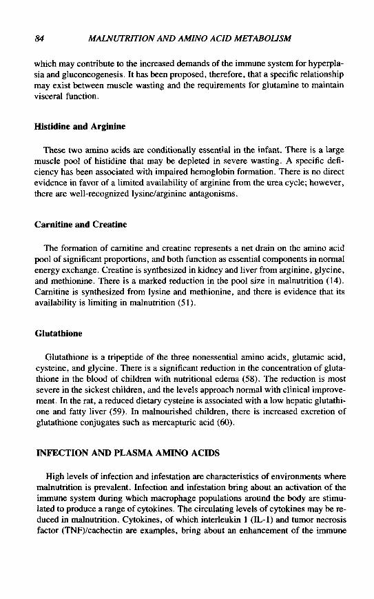
The pattern of amino acids in the plasma of subjects with malnutrition reflects thesuperimposed effects of infection and loss of appetite. It may be possible, therefore,to differentiate the relative contribution of each influence to the pattern observed(Table 2). Studies carried out by Wannemacher et al. (64,65) on well nourished, in-fected subjects and by Young and Scrimshaw (66) on healthy subjects fed protein-deficient diets, together with various studies in animals, help in this respect.Wannemacher found that in healthy adults during experimentally induced sandflyand typhoid fever, the plasma concentrations of phenylalanine and tryptophan be-came raised, while valine, isoleucine, leucine, glycine, and alanine were reduced.In contrast, simple starvation of healthy subjects brought about a response in whichplasma alanine was reduced, while the concentrations of valine, isoleucine, leucine,and glycine became elevated, phenylalanine was unaffected, and tryptophan fell.Thus, there is a contrast in the pattern of change with infection compared with a re-duced food intake for the plasma concentrations of phenylalanine, tryptophan, va-line, leucine, isoleucine, and glycine. When protein-deficient diets were fed tohealthy young subjects, there was an elevation in glycine, alanine, and serine anda reduction in valine, isoleucine, and tyrosine, while phenylalanine was unaffected.Thus, infection and a protein-deficient diet affect the branched chain amino acids inthe same way but have opposite effects on glycine and alanine. Whereas a protein-
TABLE 2. Effect of starvation, a protein-free diet, infection, or severe malnutrition on theplasma concentration of selected amino acids
ValineLeucineIsoleucinePhenylalanineAlanineGlycine
Starvation
trTiT
Protein-free
iIi
tT
Infection
IiiTii
Malnutrition
1IiIII
t , increase; 1 , decrease.From refs. 64-66.

86 MALNUTRITION AND AMINO ACID METABOLISM
deficient diet has no effect on phenylalanine, infection brought about an increase.However, both infection and protein deficiency resulted in a reduced concentrationof tyrosine. Experiments carried out with animals reinforce these conclusions tosome extent. Following enteric viral infections in well-nourished pigs, there weredecreases in the branched chain amino acids, as well as glycine, alanine, and tyro-sine. However, neither phenylalanine nor tryptophan became elevated. Protein-deficient pigs responded to the infection in the same manner. Since proteindeficiency alone had caused elevations of glycine and alanine, the changes broughtabout by infection were of greater magnitude than in the well-nourished animals(67).
What interpretations can be given to the relative pattern of response in the plasmaamino acids to infection, starvation, and protein deficiency? Alanine and glutaminemake up 60% of the amino acids released from skeletal muscle during infection.The glutamine, in addition to being a possible substrate for macrophage metabo-lism, is also a substrate for alanine production by the intestine (57). Long et al. (68)have shown that alanine is a major precursor for the increased glucose formationthat occurs in the liver during sepsis. In experimental situations where low-proteindiets are fed to humans or animals, gluconeogenesis is curtailed, resulting in an ele-vation in plasma alanine concentrations. If, however, infection and its accompany-ing anorexia are superimposed on these dietary situations, then a large fall inconcentration would be expected due to enhanced gluconeogenesis. It has been sug-gested that the decreased concentration of branched chain amino acids in plasma isa consequence of the increased requirement for alanine synthesis in muscle. Wan-nemacher (64) concluded from studies in humans and rats that decreased outputfrom muscle occurred because of the conversion of branched chain amino acidswithin the intracellular pool into alanine and glutamine, the keto acids so formed be-ing utilized as metabolic fuel within the muscle. Thus, although muscle proteinbreakdown is enhanced by infection and reflected in increased output of phenylala-nine and tryptophan, there is no equivalent increase in the outflow of branched chainamino acids.
The reductions brought about by feeding healthy experimental animals and hu-man subjects an inadequate protein diet are due to a quite different effect. In this sit-uation, branched chain amino acids released from muscle are reduced due todecreased muscle protein loss. Once the rate of output from muscle is exceeded bythe demands for protein synthesis in other tissues, such as the liver, then the concen-tration in the blood will fall. This is illustrated in studies by Grimble and Whitehead(69,70) on children recovering from malnutrition and pigs fed a range of proteinintakes. At intakes where growth ceases and serum albumin concentrations areaffected, blood valine concentrations start to fall. The reduced concentrations ofbranched chain amino acids in the blood of children who have been exposed to mul-tiple infections and have been consuming an inadequate diet in terms of protein andenergy content might be due in varying degrees to either of these metabolic sce-narios.
An examination of data from a study of severely malnourished Peruvian children
MALNUTRITION AND AMINO ACID METABOLISM 87
02
0-1
•
a
tHI
oo
o
o
8o0
I
08
06
04
02
•
•
1
NI
8
84-
<§>8
I
•
1NI
zo
oo
|
o
oI
VALINEpmol/ml
GLYCINEpmol/ml
VAL/GLY
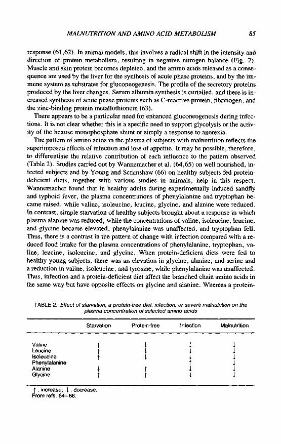
FIG. 6. In malnourished children, thevaline/glycine ratio in plasma is signifi-cantly higher in infected, I, than in nonin-fected, NI, individuals (p<0.05). Thedifference can be accounted for by an in-crease in valine concentration in the in-fected children, rather than by anychange in glycine concentration.
conducted by Baertl et al. (71) supports this concept. In this study, information wascollected on the presence or absence of infection, anthropometry, and clinical signsassociated with malnutrition, in addition to measuring the serum amino acids. Thedata show the ratio of valine to glycine in plasma. If valine is affected by both mal-nutrition and infection, with glycine being affected by infection alone, then a higherratio might be expected in children suffering from malnutrition and infection. Whena comparison is made among children with a length age greater than 6 months, the12 subjects who were uninfected had a ratio of 0.22 ±0.16, whereas those whowere infected had a ratio of 0.33 ± 0.16, p<0.05 (Fig. 6). Care has to be taken inthe interpretation of such data, given the great difficulty in identifying infection dur-ing malnutrition. In this series, there was a strong correlation between the plasmaconcentration of most amino acids and serum albumin, with the exception of taurineand glycine. While anorexia might play some part in the low level of amino acidsobserved in the circulation of infected, malnourished subjects, it would not seem tobe a determinant of low glycine levels, since starvation per se raises glycine concen-trations. The low levels may be accounted for by the metabolic conditions createdby infection and by the multiple metabolic pathways in which glycine participates.
CONCLUSION
The early workers in the field of severe childhood malnutrition were convincedthat protein deficiency, or derangements in protein metabolism, represented one ofthe most important features of the kwashiorkor syndrome. It is difficult to find con-clusive evidence to support the concept of a simple dietary deficiency of protein,especially as, under normal circumstances, the body is very efficient at retaining ni-trogen when necessary (72). Therefore, this concept has fallen into disrepute.
However, there are a number of considerations that might cause us to reflect onthis perspective. First, malnourished children are exposed to a heavy load of infec-tion, which on the one hand, alters the demand for specific nutrients, and on the

88 MALNUTRITION AND AMINO ACID METABOLISM
other, produces unbalanced losses from the body (12). In this situation, a diet thatmay have been adequate to satisfy normal requirements is very likely to become in-adequate for one or more nutrients in the presence of an increased demand. Second,diarrheal disease disturbs the delicate ecological balance between the host and hisgastrointestinal microflora. A disturbance of this kind is likely to lead to a loss ofspecific nutrients, as well as to an alteration in the metabolic exchange across thelower bowel. Third, our ideas about the relationships between individual aminoacids, our concepts of essentiality, and our appreciation of the specificity of the met-abolic flow of individual amino acids have developed rapidly over the past decade.In each of these areas, the advances in understanding have been associated with thedevelopment of powerful new tools for investigations in vivo.
It is self-evident that there are major changes in protein and amino acid metabo-lism in malnourished individuals. There is a need to characterize these changesmore specifically.
REFERENCES
1. Williams CD. A nutritional disease of childhood associated with a maize diet. Arch Dis Child1933;8:423-33.
2. Jackson AA, Golden MHN. Severe malnutrition. In: Weatherall DJ, Ledingham JGG, WarrellDA, eds. Oxford textbook of medicine, 2nd ed. Oxford: Oxford University Press, 1987;812-28.
3. Holt LE, Snyderman SE, Norton PM, et al. The plasma aminogram of kwashiorkor. Lancet1963;ii:1343-8.
4. Waterlow JC, Cravioto J, Stephen JML. Protein malnutrition in man. Adv Protein Chem 1960;15:131-238.
5. Golden MHN. The consequences of protein deficiency in man and its relationship to the features ofkwashiorkor. In: Blaxter K, Waterlow JC, eds. Nutritional adaptation in man. London: John Lib-bey, 1985;169-87.
6. AnnegersJF. Ecology of food and nutrition. 1973;2:225.7. Guraey JM. In: Jelliffe DB, Jelliffe EFP, eds. Nutrition and growth. New York and London: Ple-
num Press, 1979;197.8. Landman J, Jackson AA. The role of protein deficiency in the aetiology of kwashiorkor. West In-
dian MedJ 1980;29:229-38.9. FAO/WHO Energy and protein requirements. FAO Nutrition Meetings Report Series, No 37. WHO
Technical Report Series, no. 301, 1973.10. Golden MHN, Golden BE, Jackson A A. Albumin and nutritional oedema. Lancet 1980;i: 114-6.11. Gopalan C. Protein versus calories in the treatment of protein-calorie malnutrition: metabolic and
population studies in India. In: Olson RE, ed. Protein-calorie malnutrition. New York: AcademicPress, 1975;329-41.
12. Jackson AA. Severe undemutrition in Jamaica. Acta Paediatr Scand (Suppl). 1986;323:43-51.13. Alleyne GAO, Hay RW, Picou D, et al. Protein-energy malnutrition. London: Edward Arnold,
1977.14. Reeds PJ, Jackson AA, Picou D, Poulter N. Muscle mass and composition in malnourished infants
and children and changes seen after recovery. Pediatr Res 1978;12:613-8.15. James WPT, Hay AM. Albumin metabolism: effect of the nutritional state and the dietary protein
intake. J Clin Invest 1968;47:1958-72.16. Sherry PS, Watrasiewicz KE, Jung RT, James WPT. Rapid turnover transport proteins: an index of
subclinical protein-energy malnutrition. Lancet 1979;ii:230-2.17. Flores H, Seakins A, Monckeberg F. Mechanism of fatty liver in infantile malnutrition. In: Galli C,
Jacini G, Pecile A, eds. Dietary lipid and postnatal development. New York: Raven Press, 1973;115-25.
18. Perlmutter DH, Dinarello CA, Punsal PI, Colten HR. Cachectin/tumour necrosis factor regulateshepatic acute phase gene expression. J Clin Invest 1986;78:1349-54.

MALNUTRITION AND AMINO ACID METABOLISM 89
19. Millward DJ, Brown JG, Odedra D. Protein turnover in individual tissues with special emphasis onmuscle. In: Waterlow JC, Stephen JML, eds. Nitrogen metabolism in man. Essex: Applied SciencePublishers, 1981;475-94.
20. Brasel JA. Endocrine adaptation to malnutrition. Pediatr Res 1980; 14:1299-303.21. Milner RDG. Metabolic and hormonal responses to glucose and glucagon in patients with infantile
malnutrition. Pediatr Res 1971;5:33-9.22. Payne Robinson HM, Seakins A. Fasting pancreatic glucagon in Jamaican children during malnutri-
tion and subsequent recovery. Pediatr Res 1982; 16:1011-5.23. Ingenbleek Y. Thyroid dysfunction in protein-calorie malnutrition. Nutr Res 1986;44:253—63.24. Graham GG, Nakashima J, Thompson RG, Blizzard RM. Metabolic and hormonal responses to a
protein-glucose meal in normal infants and in marasmus and marasmic-kwashiorkor. Pediatr Res1976;10:832-43.
25. Payne Robinson HM, Betton H, Jackson AA. Free and total triiodothyronine and thyroxine in mal-nourished Jamaican infants. Hum Nutr Clin Nutr 1985;39C:245-9.
26. Hadden DR. Glucose, free fatty acid and insulin interrelations in kwashiorkor and marasmus. Lan-cet 1967;ii:589-92.
27. Payne Robinson HM, Coore HG, Golden MHN. Red cell insulin binding in Jamaican malnourishedchildren. Clin Res 1986; :
28. Lunn PG, Whitehead RG, Hay RW, Baker BA. Progressive changes in serum cortisol, insulin andgrowth hormone concentrations and their relationship to the development of kwashiorkor. Br J Nutr1973;29:399-422.
29. Whitehead RG, Lunn PG. Endocrines in protein energy malnutrition. Proc Nutr Soc 1979;38:69-76.
30. Lunn PG, Whitehead RG, Cole TJ, Austin S. The relationship between hormonal balance andgrowth in malnourished children and rats. Br J Nutr 1979;41:73-84.
31. Ingenbleek Y, Barclay D, Dirren H. Nutritional significance of alterations in serum amino acid pat-terns in goitrous patients. Am J Clin Nutr 1986;43:310-9.
32. Waterlow JC, Wills VG. Balance studies in malnourished Jamaican infants. I. Absorption and re-tention of nitrogen and phosphorus. Br J Nutr 1960:14:183-98.
33. Jackson AA, Golden MHN, Byfield R, et al. Whole body protein turnover and nitrogen balance inyoung children at intakes of protein and energy in the region of maintenance. Hum Nutr Clin Nutr1983;37C:433-46.
34. Kennedy N, Badaloo V, Jackson A A. Metabolic adaptation to a marginal energy intake. Proc NutrSoc 1987;46:85A.
35. Badaloo A, Jackson AA, Jahoor F. Whole body protein turnover and resting metabolic rate in ho-mozygous sickle cell disease. Clin Sci 1989;77:93-7.
36. Golden MHN, Waterlow JC, Picou D. Protein turnover, synthesis and breakdown before and afterrecovery from protein energy malnutrition. Clin Sci 1977;53:473-7.
37. Tomkins AM, Garlick PJ, Schofield WN, Waterlow JC. The combined effects of infection and mal-nutrition on protein metabolism in children. Clin Sci 1983;65:313—24.
38. Gracey MS. Nutrition, bacteria and the gut. BrMedBull 1981;37:71-5.39. Jackson AA, Golden MHN. The human rumen. Lancet 1978;ii:764.40. Schneider RE, Viteri FE. Luminal events of lipid absorption in protein calorie malnourished chil-
dren; relationship with nutritional recovery and diarrhoea. I. Capacity of the duodenal content toachieve micellar solubilisation of lipids. Am J Clin Nutr 1974;27:777-87.
41. Stephen JML, Waterlow JC. Effect of malnutrition on activity of two enzymes concerned withamino acid metabolism in human liver. Lancet 1968;i:l 18-9.
42. Jackson A A, Picou D, Landman JP. The non-invasive measurement of urea kinetics in normal manby a constant infusion of 15N-urea. Hum Nutr Clin Nutr 1984;38C:339-54.
43. Picou D, Phillips M. Urea metabolism in malnourished and recovered children receiving a high orlow protein diet. Am J Clin Nutr 1972;25:1261-6.
44. Jackson AA. Amino acids: essential and non-essential? Lancet 1983;i:1034—7.45. Heine W, Wutzke KD, Richter I, et al. Evidence for colonic absorption of protein nitrogen in in-
fants. Acta Paediatr Scand 1987;76:741-4.46. Golden MHN, Golden B, Bennett FI. Relationship of trace element deficiencies to malnutrition. In:
Chandra RK, ed. Trace elements in nutrition of children. (Nestle Nutrition Workshop Series) NewYork: Raven Press, 1985;185-207.
47. Rose WC. The amino acid requirements of adult man. Nutr Abstr Rev 1957;27:631-47.48. Blackburn GL, Grant JP, Young VR. Amino acids: metabolism and medical applications. Boston:
John Wright, 1983.
90 MALNUTRITION AND AMINO ACID METABOLISM
49. Laidlaw SA, Kopple JD. Newer concepts of the indispensable amino acids. AmJ ClinNutr 1987;46:593-605.
50. Crowne RS, Donninger C, Grimble RF, Whitehead RG. The isolation and identification of piperi-dine-2-carboxylic acid in the urine of protein malnourished rats. Clin Sci 1971;41:535—43.
51. Khan L, Bamji MS. Plasma camitine levels in children with protein-calorie malnutrition before andafter rehabilitation. Clin ChimActa 1977;75:163-6.
52. Jackson AA, Badaloo AV, Forrester T, et al. Urinary excretion of 5-oxoproline (pyroglutamic aci-duria) as an index of glycine insufficiency in normal man. BrJNutr 1987;58:207-14.
53. Yu YM, Yong RD, Matthews DE, et al. Quantitative aspects of glycine and alanine nitrogen metab-olism in postabsorptive young men: effects of level of nitrogen and dispensable amino acid intake.JNutr 1985; 115:399-410.
54. Gaull GE, Rassin DK. Taurine in development and nutrition. In: Sulphur in biology. CIBA Founda-tion Symposium 72, 1980;271-88.
55. KerrDS, Stevens MCG, PicouDIM. Fasting metabolism in infants. II. Effect of severe undernutri-tion and infusion of alanine on glucose production estimated with U-13C-glucose. Metabolism1978; :831-4.
56. Windmueller HG. Glutamine utilisation by the small intestine. Adv Enzymol 1982;53:201-37.57. Newsholme EA, Crabtree B, Ardawi MSM. Glutamine metabolism in lymphocytes; its biochemi-
cal, physiological and clinical importance. Q J Exp Physiol 1985;70:473-89.58. Jackson AA. Blood glutathione in severe malnutrition in childhood. Trans R Soc Trop Med Hyg
1986;80:911-3.59. Davies E, Hibbert JM, Jackson AA. The relationship between hepatic glutathione and fatty liver in
weanling rats on a low protein diet. Nutr Rep lnt 1988;37:847-56.60. Ramdath DD, Golden MHN. Urinary mercapturic acid outputs of severely malnourished children.
Proc Nutr Soc 1988;47:7A.61. Dinarello CA. Interleukin 1 and the pathogenesis of the acute phase response. N Engl J Med, 1984;
311:413-8.62. Beutler B, Cerami A. Cachectin and tumour necrosis factor as two sides of the same biological coin.
Nature 1986;320:584-8.63. Dinarello CA. Multiple biological properties of recombinant human interleukin 1 (beta). Immunobi-
ology 1986;172:301-15.64. Wannemacher RW. Key roles of various individual amino acids in host responses to infection. Am
J Clin Nutr 1977;30:1269-79.65. Wannemacher RW, Pakarek RS, Bartelloni PJ, et al. Changes in individual amino acids following
experimentally induced sandfly fever virus infection. Metabolism 1972;21:67-76.66. Young VR, Scrimshaw NS. Endogenous nitrogen metabolism and plasma free amino acids in young
adults given a protein free diet. Br J Nutr 1968;22:9-20.67. Isoun T, Whitehair CK, Bergen WG. Influence of viral enteric infection on the aminogram and pa-
thology of protein-calorie malnutrition in the pig. Am J Clin Nutr 1973;26:835-44.68. Long CL, Schiller WR, Blakemore WS, et al. Muscle protein catabolism in the septic patient as
measured by 3-methylhistidine excretion. Am J Clin Nutr 1977;30:1349-52.69. Grimble RF, Whitehead RG. Fasting serum amino acid patterns in kwashiorkor and after adminis-
tration of different levels of protein. Lancet 1970;i:918-21.70. Grimble RF, Whitehead RG. Changes in the concentration of specific amino acids in the serum of
experimentally malnourished pigs. Br J Nutr 1970;24:557-64.71. Baertl JM, Placko RP, Graham GG. Serum protein and free amino acids in severe malnutrition. Am
J Clin Nutr 1974;27:733-42.72. Waterlow JC. Metabolic adaptation to low intakes of energy and protein. Annu Rev Nutr 1986;6:
495-526.
DISCUSSION
Dr. Worrier: Do selenium and zinc deficiency affect the activity of superoxide dismutaseor a glutathione peroxidase?
Dr. Jackson: Golden and Ramdath (1) have measured the level of ferritin on admission and
MALNUTRITION AND AMINO ACID METABOLISM 91
related it to the activity of red cell glutathione peroxidase (GPx), an index of selenium status.They found that the association of a high ferritin with low GPx was predictive of those chil-dren who were going to die. They have suggested that increased free iron may increase therisk of free radical-induced damage in a situation where the mechanisms to protect againstsuch damage are impaired. This is such an important defense system that if one is short of asingle nutrient or component, there are backup systems to give an alternative source of pro-tection. In order to get expression of disease, one might need a major failure in more than onesystem at the same time. Depending on the particular pattern of deficiency, one might expectto get different patterns of disease.
Studies in Chile (2,3) have demonstrated that supplementation of all malnourished childrenwith appropriate quantities of zinc and copper leads to a better quality of rehabilitation.
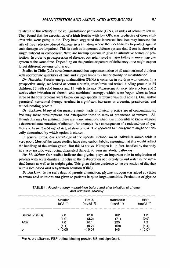
Dr. Haschke: Protein-energy malnutrition (PEM) is common in children with cancer. In aprospective study, we looked at serum albumin, transferrin and retinol-binding protein in 25children, 12 with solid tumors and 13 with leukemia. Measurements were taken before and 2weeks after initiation of chemo- and nutritional therapy, which were begun when at leastthree of the four proteins were below our age-specific reference values (Table 1). Oral and/orparenteral nutritional therapy resulted in significant increases in albumin, prealbumin, andretinol-binding protein.
Dr. Jackson: Many of the measurements made in clinical practice are of concentrations.We may make presumptions and extrapolate these to rates of production or removal. Al-though this may be justified, there are many situations when it is impossible to know whethera decreased concentration of albumin, for example, is a consequence of a reduced rate of syn-thesis or an increased rate of degradation or loss. The approach to management might be criti-cally determined by which option is chosen.
In general terms, our knowledge of the specific metabolism of individual amino acids isvery poor. Most of the tracer studies have used carbon labels, assuming that this would reflectthe handling of the amino group. But this is not so. Nitrogen is, in fact, handled by the bodyin a very specific way, being channeled through its own metabolic pathways.
Dr. M. Mehta: Our studies indicate that glycine plays an important role in rehydration ofpatients with acute diarrhea. It helps in the reabsorption of electrolytes and water in the intes-tinal lumen as well as in weight gain. This gives further credence to the prevention of diarrheawith a rice-based oral rehydration solution (ORS).
Dr. Jackson: In the early days of parenteral nutrition, glycine nitrogen was added as a fillerto amino acid solutions and given to patients in quite large quantities. Production of glycine
TABLE 1. Protein-energy malnutrition before and after initiation of chemo-and nutritional therapy
Albumin Pre-A transferrin RBP(g/dl-1) (mg/dr1) (mg/dl"1) (mg/dl"')
Before x (SD)
After
Pre-A, pre-albumin; RBP, retinal-binding protein; NS, not significant.
2.6(10)4.0(1.1)
<0.05
10.0(3.2)26.1(9.7)
<0.01
162(71)220(58)NS
1.8(0.9)4.2(1.4)
<0.01
92 MALNUTRITION AND AMINO ACID METABOLISM
by the body is tightly controlled; excessive glycine can have deleterious effects. This may bean indication of the role that this group of amino acids plays in the body. Although they canbe produced by the body, there is an upper limit to the production. Without further informa-tion, I would be cautious about saying that we need more.
The problem in management is to appreciate the relatively narrow range of therapeutic ef-fectiveness, particularly in malnourished children, where the degradative pathways are com-promised. One can easily move from a position of deficiency to one of excess. Caution,therefore, is needed when considering supplementation.
Dr. Guesry: Why have you included taurine in the list of essential amino acids? It is notincorporated with protein synthesis and there is no evidence of taurine deficiency in normalinfants. In addition, all rehabilitation programs for malnourished children carried out withcow's milk not containing taurine have been successful.
Dr. Jackson: My reason for listing taurine as one of the conditionally essential amino acidsis that as far as we know, taurine is synthesized from cysteine. The de novo synthesis of cys-teine is through the trans-sulfuration pathway, requiring methionine and serine as precursors.Glycine and serine are metabolically interchangeable. Therefore, it seems that the carbonchain and the amino group of glycine, cysteine, and taurine are all derived from serine. So,if I make the statement that glycine and serine are conditionally essential amino acids, then Ifeel obligated to consider that the derivatives may also be conditionally essential.
Dr. Suskind: Could you comment on the impact of amino acid metabolism on neurotrans-mitter synthesis in the malnourished state and the effect of malnutrition in generally loweringessential amino acids? Dr. Robert Olson, several years ago, looked at the rate of protein syn-thesis in malnourished children. He proposed that there was a hierarchy of visceral proteinsynthesis and that those proteins having the smallest pool size and the fastest turnover ratewere the most sensitive markers of visceral protein synthesis. He referred to such proteins asretinol-binding protein and pre-albumin as being sensitive markers.
Dr. Jackson: There have been many suggestions that the availability of specific aminoacids may modulate either the synthesis or function of neurotransmitters. I am not aware ifthere is direct evidence, although one is particularly concerned about the role played by tryp-tophan. One difficulty in studying the metabolism of tryptophan is to define the active circu-lating pool, since it is bound to plasma albumin and can be displaced by free fatty acids, forexample.
Regarding the sensitivity of proteins in relation to the rate at which they turn over, if thereis a general depression of protein turnover, it will become obvious in the fast-tuming-overproteins first. However, the control in individual proteins is more specific, and there is sel-dom a general depression of turnover.
A clear definition of the metabolic state is important in understanding the processes that aretaking place. I suggested that classifications might be based on the extent to which the energyrequirements of an individual were being satisfied by dietary intake. The extent to which nu-trient supplements can be utilized by the body is determined, in part, by the energy availableto the body. If the requirement for energy is not satisfied, energy is made available throughnet catabolism. In the process of catabolism, nutrients are wasted. If, however, the require-ments for energy are covered, then the next requirement is to replete specific nutrients and tosatisfy other metabolic requirements. Therefore, in considering whether to supplement withamino acids or other specific nutrients, one has to consider whether the individual has alreadysatisfied his energy needs. I do not think it is possible effectively to retain supplements if theenergy requirements of the body are not being satisfied.
Dr. Suskind: It is essential to satisfy both protein and energy needs. In Chiang Mai, we

MALNUTRITION AND AMINO ACID METABOLISM 93
tried to ascertain the optimal nutritional support for malnourished children by putting them onvarying protein and calorie ratios. The children received on a per kg basis 100 calories/1 gramof protein, 100 calories/4 grams of protein, 175 calories/1 gram of protein, or 175 calories/4grams of protein. At 175 calories and 1 gram of protein there was a decrease in visceral pro-tein synthesis. This study emphasized a need to be concerned about the protein as well as theenergy needs of malnourished children during rehabilitation.
Dr. Jackson: I am not clear about the overall metabolic state of the children you described.Because maintenance requirements are about 100 kcal/kg-day, a child given 150 kcal/kg-daymust handle an additional load of 50 kcal/kg-day. This may be deposited as tissue, but withthe existence of specific deficiencies, there is a limit to which this is possible.
When I described the acute management of severely ill children, it was to suggest givingenergy levels sufficient to cover energy expenditure. Protein needs can be met by giving themaintenance requirements of 0.6 g/kg-day until specific nutrient deficiencies are repaired.Until repair, it is not possible to synthesize balanced new tissue, and, more important, therewill be continued anorexia. This loss of appetite appears to be an effective defense againstinappropriate nutrient intake in relation to the body's demands.
The problem that occurs in infection, particularly with diarrheal disease, is that there areunbalanced losses of nutrients. In consequence, the usual dietary intake may no longer be ap-propriate to provide the particular balance of nutrients required to replenish the specificlosses. An excellent example is the increased loss of potassium from diarrheal disease, whichmay lead to a significant depletion of total body potassium, perhaps one of the more impor-tant factors in the genesis of nutritional edema. Although normal potassium intake is suffi-cient for a child in normal health, it is insufficient to replete these unusual losses. Generoussupplements of potassium are, therefore, critical to successful management. This highlightsthe need to be careful, when discussing supplementation, of being clear about the metabolicbackground against which the supplementation is to be provided.
One of the most important considerations for successful management is the ability to con-trol carefully the energy intake to a level that simply covers the need for maintenance. Re-ports in the literature (4) describe the adverse effects of excessive energy intake during theearly period of rehabilitation, reinforcing the idea of the sensitivity of the adapted body to thelevel of energy intake.
Dr. Suskind: Our study found that these malnourished children, within 7 days, were doingvery well on 175 calories/4 grams of protein/kg-day. When a second group was placed on 175calories/1 gram of protein/kg-day, plus other nutrients, there was no anorexia. In fact, thechildren took the formula as easily as those on 175 calories and 4 grams of protein/kg-day.
I do concur, however, that we must be concerned about the infant's being able to differen-tiate an appropriate from an inappropriate intake ratio.
Dr. Jackson: I agree. We must begin to recognize responses such as anorexia as being pro-tective and to learn to be very cautious about overriding such protective mechanisms.
Dr. Guesry: Intensive discussions are taking place regarding the special need for branchedchain amino acids (BCAA) in stressed patients. Dr. Keusch has suggested that stress may bean important factor in PEM. Do you think, therefore, that malnourished children would havespecial requirements for BCAA?
Dr. Jackson: Although there is considerable literature on BCAA and the specific role theymay play in catabolic states, the conclusions are not clear. One of the most recent suggestionshas been that the BCAA act as important precursors for glutamine synthesis in muscle. Gluta-mine may have a particular role in activation of the immune system and protection of the in-tegrity of the gastrointestinal tract.

94 MALNUTRITION AND AMINO ACID METABOLISM
Dr. Truswell: About 5 years ago, Dr. Jackson (5) wrote in the Lancet that carbohydratewas used by the colonic bacteria for their energy, and fat was not available as an energysource. However, in one slide, you showed that at a single protein level, there was actuallybetter utilization of fat as compared with carbohydrate.
Dr. Jackson: I might have misled you by the rather brief description I gave of the diets.There were complex carbohydrates in all the diets received by the children. The diets wereenriched in energy with the addition of either arachis oil or cornstarch. It is generally consid-ered that fats act as a poor substrate for. the colonic microflora because of the predominantlyanaerobic environment.
REFERENCES
1. Golden MH, Ramdath D. Free radicals in the pathogenesis of kwashiorkor. Proc Nutr Soc1987;46:53-68.
2. Castillo-Duran C, et al. Controlled trial of zinc supplementation during recovery from malnutrition:effects on growth and immune function. Am J Clin Nutr 1987;45:602-8.
3. Castillo-Duran C, et al. Controlled trial of copper supplementation during the recovery from maras-mus. Am J Clin Nutr 1983;37:898-903.
4. Patrick J. Death during recovery from severe malnutrition and its possible relationship to sodiumpump activity in the leukocyte. BrhtedJ 1977;l:1051-4.
5. Jackson AA. Amino acids: essential and non-essential. Lancet 1983;i: 1034-7.