malaria in children - wordpress.com filelp if indicated blood group &cross matching blood...
TRANSCRIPT

Malaria,
Leshmaniasis
&Schistosomiasis in
Children Dr.Mai Mohamed Elhassan
Assistant Professor
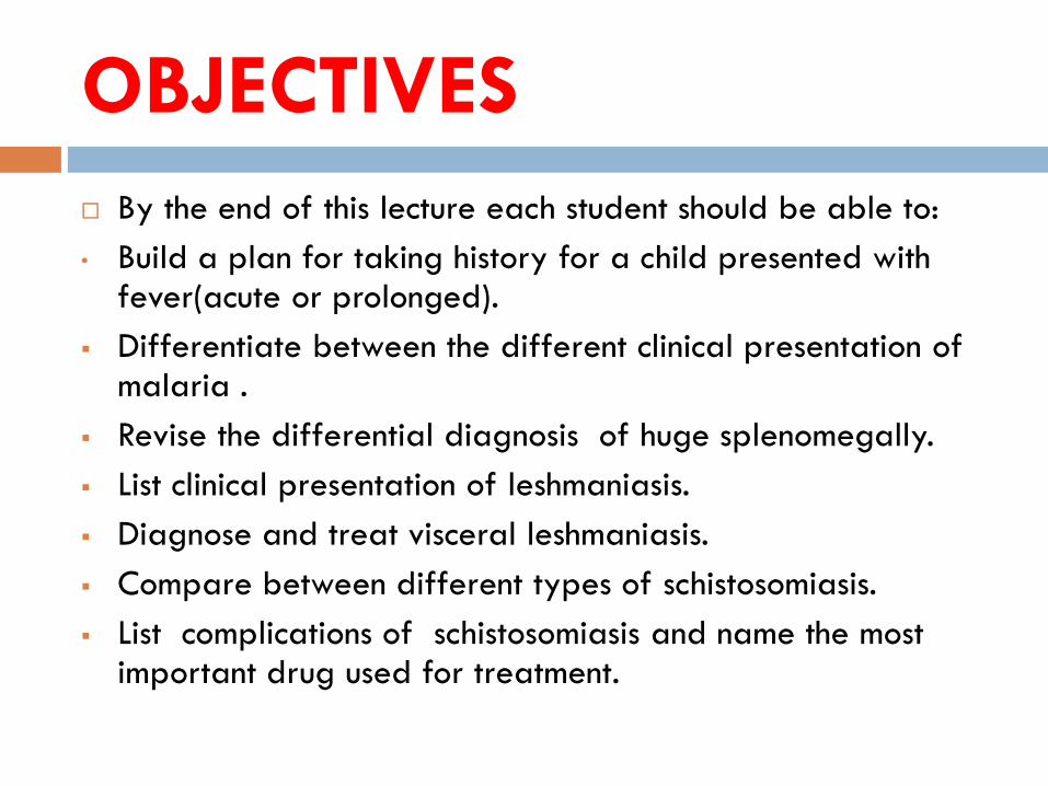
OBJECTIVES
By the end of this lecture each student should be able to:
• Build a plan for taking history for a child presented with fever(acute or prolonged).
Differentiate between the different clinical presentation of malaria .
Revise the differential diagnosis of huge splenomegally.
List clinical presentation of leshmaniasis.
Diagnose and treat visceral leshmaniasis.
Compare between different types of schistosomiasis.
List complications of schistosomiasis and name the most important drug used for treatment.

MALARIA -----EPIDEMIOLOGY
Acute and chronic problem
Mode of transmission
Female anopheles mosquitoes
Blood transfusion
Contaminated needles
Transplacental
Organ transplantation

How does infection develop?

Severity of disease and host
factors
several host factors determine the outcome of
exposure to malaria:
• Naturally-acquired immunity.
• Red cell and haemoglobin variants.
• Foetal haemoglobin (HbF)
• Duffy blood group

MALARIA----PATHOPHYSIOLOGY
This results from the:
1. Destruction of the RBC. Anemia
2. The liberation of parasites and RBC materials into
the circulation, Fever
3. Host reaction to these events (Immunopathologic
events)

Malaria---Clinical Presentation
Asymptomatic during IP
Classical presentation– simple malaria
Severe Malaria
Cerebral malaria
Long term relapse ----- P. vivax and P. ovale
Hyper reactive malaria splenomegally.(chronic
malaria)

A. Asymptomatic parasitaemia
natural immunity / high malaria endemicity
B. Simple, uncomplicated malaria/ some degree of immunity to
malaria/no life-threatening disease.
Fever is the most constant symptom of malaria.
Clinical Course Of P.falciparum

Clinical features--Simple malaria
Paroxysm of fever (rigors,
sweating)
Headache
Myalgia Back pain
Abdominal pain
Nausea, vomiting , diarrhea
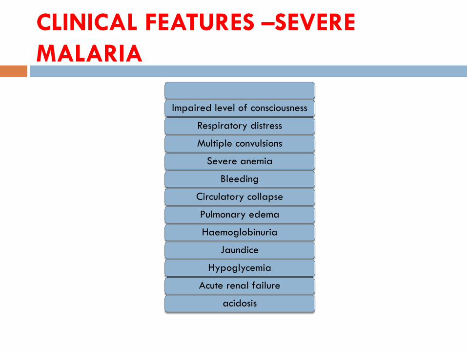
CLINICAL FEATURES –SEVERE
MALARIA
Impaired level of consciousness
Respiratory distress
Multiple convulsions
Severe anemia
Bleeding
Circulatory collapse
Pulmonary edema
Haemoglobinuria
Jaundice
Hypoglycemia
Acute renal failure
acidosis

Severe malaria --investigations
Hb PCV
Glucose Blood Gases
LP if indicated blood Group &cross matching
Blood culture Urea& Electrolytes
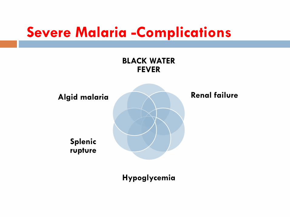
Severe Malaria -Complications
BLACK WATER FEVER
Renal failure
Hypoglycemia
Splenic rupture
Algid malaria

D:CEREBRAL MALARIA
Caused by P.falciparum.
MOST COMMON AMONG CHILDREN
Rapidly developing encephalopathy(un arousable
coma)
Develop over several days or suddenly
20-40% fatality rate if not treated appropriately.
> in parasitemia>5%

CEREBRAL MALARIA
CLINICAL PRESENTATION
Always decreased level of consciousness
Drowsiness
Headache
Confusion
Delirium
Hallucinations
Deep coma
Seizures
Muscle twitching
Contracted or unequal pupils
Retinal hemorrhage
Hemiplegia
Absent tendon reflexes
Positive Babinski sign
opisthotonus (mention 2 differential diagnosis)


Malaria----Diagnosis
1.A good history
Residence or a recent visit (in the preceding 3 months) to a malaria endemic area
The diagnosis of malaria should be considered in any unwell person who has been in a malarias area recently
2.Physical examination
Identify signs consistent with malaria: fever, pallor, jaundice, splenomegaly
3.LAB diagnosis

MALARIA– LAB DIAGNOSIS
Microscopy :Thick film for parasite identification &
Thin blood film :used for species identification and
quantification.

Other methods of diagnosis of
malaria
These are not routinely used in clinical practice. They include :
a) Antigen capture kits.
b) PCR
c) Fluorescent techniques.
d) Serologic tests.

P.Falciparum Malaria--Diagnosis
Criteria that suggest falciparum malaria
Symptoms < 1month after return from endemic
area
Parasitemia > 2%
Rings form with double chromatin dots
Erythrocytes infected with more than one parasite

Ring forms or
trophozoites; many red
cells infected – some
with more than one
parasite
Appearance of P. falciparum in thin
blood films

TREATMENT ---UNCOMLICATED
MALARIA
First –line treatment: chloroquine/ARTUSENATE
For P.vivax and P.ovale infection plus primaquine.
2. Second-line treatment :Fansidar
3. Third-line treatment :
Mefloquine
Quinine

Cerebral malaria and severe Malaria
--Treatment
1) Supportive treatment
2) Treatment of complications ?
3) Antimalarial treatment
• Quinine infusion
• Artemether injections

SCHISTOSOMIASIS
"bilharziasis" after Theodor Bilharz who
identified the parasite first in 1852.

World Wide Distribution of Schistosomiasis

Pathophysiology
The adult worms migrate against portal blood flow
The mesenteric venules of the small intestine — S. japonicum
The mesenteric venules of the colon — S. mansoni and S. intercalatum
The vesical venous plexus — S. haematobium

Clinical Features
Acute infection — Acute symptoms may present as
swimmer's itch or Katayama fever.
Intestinal schistosomiasis
Hepatic schistosomiasis
Urinary schistosomiasis –terminal hematuria
Neurologic complications –transverse Myelitis

Hepatic Schistosomiasis
1.Inflammatory hepatic schistosomiasis causes
hepatomegaly and splenomegaly in children
2.Chronic hepatic schistosomiasis :
splenomegaly ,portal hypertension, ascites and
hematemesis from esophageal varices .

Diagnosis
Demonstration of parasite eggs in stool or urine /multiple samples.
Serology
Abdominal U/S for periportal fibrosis, measure portal vein pressure.
Upper GI endoscopy in cases of suspected esophageal varices

Treatment of schistosomiasis
Praziquantel

Leishmaniasis
Group of diseases caused by infection with
one of the protozoan parasites of the genus
Leishmania
Designated one of the five most important
diseases worldwide by the World Health
Organization
20 million people infected worldwide

It is known in the Kingdom back to 1950.
Ministry of Health has established the
leishmaniasis unit in the 1980 Under The
precautionary medicine to follow-up the
disease in the Saudi cities

Riyadh
Northern
Al-jouf Tabouk
Medina
Makkah
Baha
Aseer
Jazan
Eastern
Najran
Affected area

LESHMANIASIS--ETIOLOGY
(1) Cutaneous Leishmaniasis: caused by L. tropica ,L. major ,L. ethiopica , L.mexicana, L. braziliensis
(2) Mucocutaneous Leishmaniasis:- caused by L.etheopica and L.braziliensis
(3) Visceral Leishmaniasis:-caused L. donovani.

1
5
4
3
2
6
7
8 Sand fly takes a blood meal (Injects promastigote
stage into the tissue)
Promastigotes are
Phagocytized by
macrophages
Promastigotes transfer
into amastigotes inside
macrophages
Amastigotes multiply in cells
(Including macrophages) of
Various tissues Sand fly takes
a blood meal (ingest macrophages
Infected with amastigotes )
Ingestion of
Parasitized cell
Amastigotes transform
Into promastigote
stage in midgut
Divide in midgut and
migrate to proboscis
i
d
i
d
Infective stage
Diagnostic stage

Cutaneous leshmaniasis –clinical
presentation
Exposed skin (face & extremities)
One or more lesion
Papular, nodular, plaque like or ulcerative lesion
Non tender ulcer surrounded by a sharp indurated
erythematous margin
No drainage ,unless infected by bacteria
Ended with residual scar


Diagnosis
is mainly clinical in and the golden role is:-any
boil >l month duration is leishmaniasis until prove
other wise
Direct smear
Culture in NNN media.
Leishmanin skin test
immunological tests
Polymerase chain reaction

Treatment
LOCAL:-
- Chemical treatment mainly Na stibogluconate (pentostam)
Physical treatment: as Cryotherapy, infra red therapy, Excision or cautery

Visceral leishmaniasis
There are geographical variations.
The diseases is called kala-azar
Leishmania infantum mainly affect children
Leishmania donovani mainly affects adults
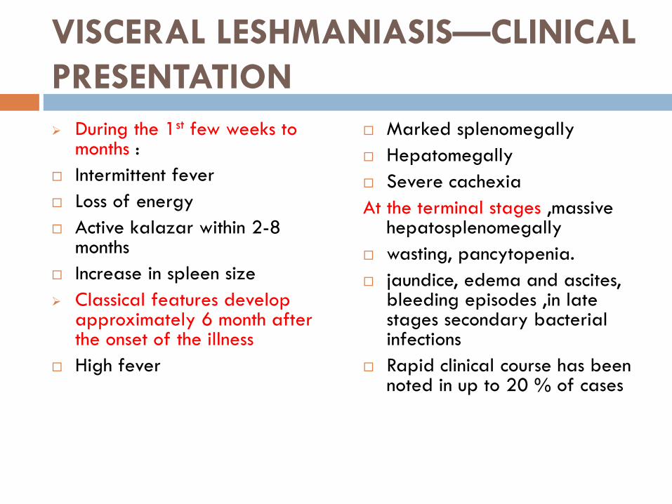
VISCERAL LESHMANIASIS—CLINICAL
PRESENTATION
During the 1st few weeks to months :
Intermittent fever
Loss of energy
Active kalazar within 2-8 months
Increase in spleen size
Classical features develop approximately 6 month after the onset of the illness
High fever
Marked splenomegally
Hepatomegally
Severe cachexia
At the terminal stages ,massive hepatosplenomegally
wasting, pancytopenia.
jaundice, edema and ascites, bleeding episodes ,in late stages secondary bacterial infections
Rapid clinical course has been noted in up to 20 % of cases


What are the differential diagnoses of huge
splenomegally?

Visceral leishmaniasis--diagnosis
1.CBC .anemia, thrombocytopenia, leukopenia
2.High ESR
3.Elevated hepatic transaminasis
4.Hyperglobulinemia—IgG
(1) Parasitological diagnosis:
Bone marrow aspirate 1. microscopy
Lymph node
liver biopsy
Splenic aspirate

LD bodies in bone marrow

(2) Immunological Diagnosis:
Specific Direct Agglutination Test (DAT)
Skin test (leishmanin test) . Non specific
Non specific detection (formal-gel) test or by
electrophoresis.

Treatment:
Pentavalent antimony- sodium stibogluconate
(Pentostam)
Amphotericin B
Treatment of complications:
Anemia
Bleeding
Infections

Any
questions?