making glaucoma care the big easy
TRANSCRIPT

©2021 American Academy of Ophthalmology. All rights reserved. No portion may be reproduced without express written consent of the American Academy of Ophthalmology.
2021 Glaucoma Planning GroupBrian A Francis MD Program Director
Kelly W Muir MDProgram Director
Salwa Abdel-Aziz MDDonald L Budenz MD MPHTeresa C Chen MDIan Patrick Conner MD PhDBabak Eliassi-Rad MDRonald Leigh Fellman MD OCS Davinder S Grover MD MPHLily T Im MDChristine LeeAnn Larsen MDJohn T Lind MD
Former Program Directors2020 Eydie Miller-Ellis MD Brian A Francis MD 2019 JoAnn Giaconi MD Eydie Miller-Ellis MD2018 Shan C Lin MD JoAnn Giaconi MD2017 Jody R Piltz-Seymour MD Shan C Lin MD2016 Joel S Schuman MD Jody R Piltz-Seymour MD2015 James D Brandt MD Joel S Schuman MD 2014 David S Friedman MD MPH PhD James D Brandt MD
2013 Thomas W Samuelson MD David S Friedman MD MPH PhD2012 Wallace L M Alward MD Thomas W Samuelson MD2011 Leon W Herndon MD Wallace LM Alward MD2010 Rohit Varma MD MPH Leon W Herndon MD2009 Donald L Budenz MD MPH Rohit Varma MD MPH2008 Henry D Jampel MD MHS Donald L Budenz MD MPH2007 Anne Louise Coleman MD PhD Henry D Jampel MD MHS2006 Christopher A Girkin MD Anne Louise Coleman MD PhD2005 Claude F Burgoyne MD Christopher A Girkin MD2004 David S Greenfield MD Claude F Burgoyne MD2003 Kuldev Singh MD MPH David S Greenfield MD2002 Theodore Krupin MD Kuldev Singh MD MPH2001 Robert D Fechtner MD Theodore Krupin MD2000 Jeffrey M Liebmann MD Robert D Fechtner MD1999 Robert N Weinreb MD Jeffrey M Liebmann MD1998 George A Cioffi MD Robert N Weinreb MD
1997 Richard A Lewis MD George A Cioffi MD1996 M Bruce Shields MD E Michael Van Buskirk MD1995 Reay H Brown MD Mary Gerard Lynch MD1994 Richard A Lewis MD
Subspecialty Day Advisory CommitteeR Michael Siatkowski MD Chair
Bonnie An Henderson MD Michael S Lee MD Jennifer Irene Lim MD Shahzad I Mian MD Jody R Piltz MD
Maria M Aaron MD Secretary for Annual Meeting
StaffMelanie R Rafaty CMP, Director, Scientific
Meetings Ann L’Estrange, Subspecialty Day ManagerDebra Rosencrance CMP CAE, Vice
President, Meetings & ExhibitsPatricia Heinicke Jr, Copy EditorMark Ong, DesignerGina Comaduran, Cover Design
Glaucoma 2021Making Glaucoma Care the Big Easy Under Pressure®
Program DirectorsBrian A Francis MD and Kelly W Muir MD
In conjunction with the American Glaucoma Society
Ernest N Morial Convention CenterNew Orleans, LouisianaFriday, Nov. 12, 2021
Presented by:The American Academy of Ophthalmology
Supported by an unrestricted educational grant from Aerie Pharmaceuticals, Inc. and Santen, Inc.
Cover photo courtesy of Ian P Conner MD PhD
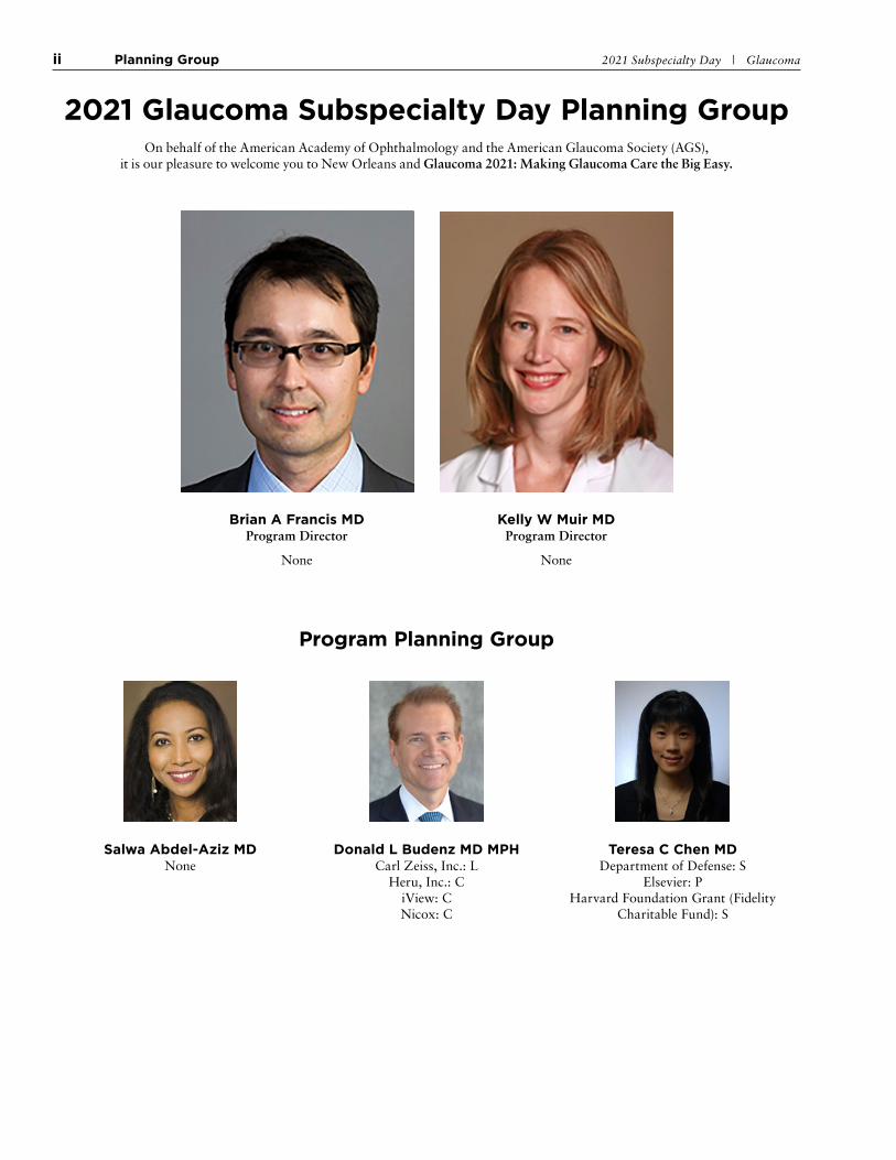
2021 Glaucoma Subspecialty Day Planning GroupOn behalf of the American Academy of Ophthalmology and the American Glaucoma Society (AGS),
it is our pleasure to welcome you to New Orleans and Glaucoma 2021: Making Glaucoma Care the Big Easy.
Brian A Francis MD Program Director
None
Kelly W Muir MD Program Director
None
ii Planning Group 2021 Subspecialty Day | Glaucoma
Program Planning Group
Salwa Abdel-Aziz MDNone
Donald L Budenz MD MPHCarl Zeiss, Inc.: L
Heru, Inc.: CiView: CNicox: C
Teresa C Chen MDDepartment of Defense: S
Elsevier: PHarvard Foundation Grant (Fidelity
Charitable Fund): S

2021 Subspecialty Day | Glaucoma Planning Group iii
Ian Patrick Conner MD PhD Ivantis: C
Ocugenix: C,O,P
Babak Eliassi-Rad MDNone
Ronald Leigh Fellman MD OCS Aerie Pharmaceuticals, Inc.: L
Alcon Laboratories, Inc.: C Bausch + Lomb: L
Endo Optiks, Inc.: C InnFocus: S
Davinder S Grover MD MPHAerie Pharmaceuticals, Inc.: L
Allergan: C,L,S Bausch + Lomb: C,L
Glaukos Corp.: C MicroOptx: C
New World Medical, Inc: C,L Nova Eye Medical: L
Olleyes: O,C,L Reichert, Inc.: C,L
Sanoculis: C Santen, Inc.: C
Surgical Specialties: L
No photo available
Lily T Im MD None
Christine LeeAnn Larsen MDNone
John T Lind MDHeru: SNicox: S
iv Planning Group 2021 Subspecialty Day | Glaucoma
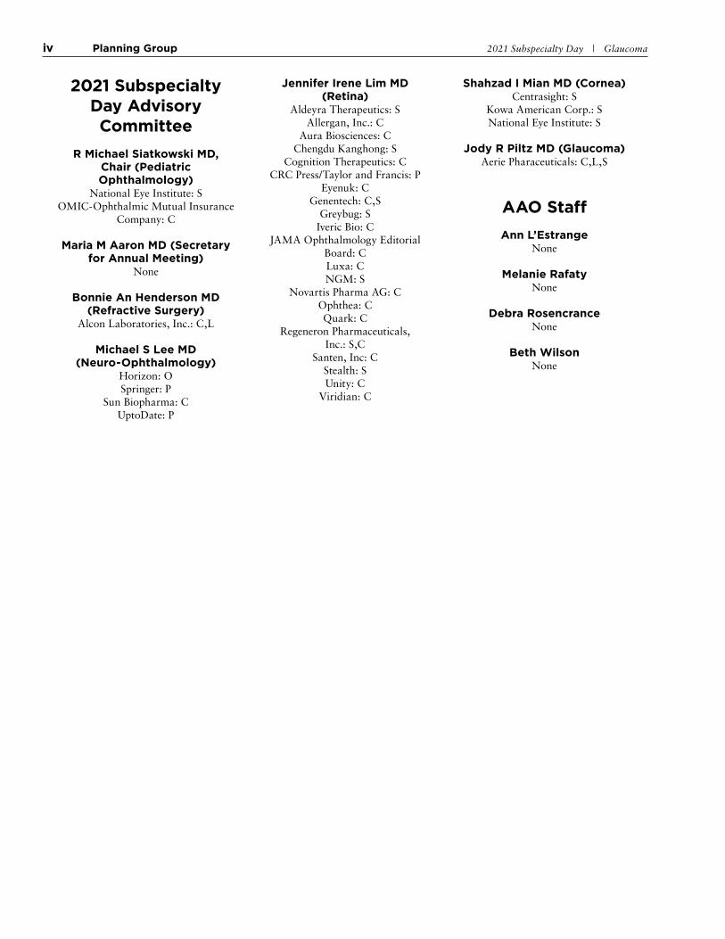
2021 Subspecialty Day Advisory Committee
R Michael Siatkowski MD, Chair (Pediatric Ophthalmology)
National Eye Institute: S OMIC-Ophthalmic Mutual Insurance
Company: C
Maria M Aaron MD (Secretary for Annual Meeting)
None
Bonnie An Henderson MD (Refractive Surgery)
Alcon Laboratories, Inc.: C,L
Michael S Lee MD (Neuro-Ophthalmology)
Horizon: O Springer: P
Sun Biopharma: C UptoDate: P
Jennifer Irene Lim MD (Retina)
Aldeyra Therapeutics: S Allergan, Inc.: C
Aura Biosciences: C Chengdu Kanghong: S
Cognition Therapeutics: C CRC Press/Taylor and Francis: P
Eyenuk: C Genentech: C,S
Greybug: S Iveric Bio: C
JAMA Ophthalmology Editorial Board: C Luxa: C NGM: S
Novartis Pharma AG: C Ophthea: C Quark: C
Regeneron Pharmaceuticals, Inc.: S,C
Santen, Inc: CStealth: SUnity: C
Viridian: C
Shahzad I Mian MD (Cornea) Centrasight: S
Kowa American Corp.: S National Eye Institute: S
Jody R Piltz MD (Glaucoma) Aerie Pharaceuticals: C,L,S
AAO StaffAnn L’Estrange
None
Melanie Rafaty None
Debra Rosencrance None
Beth Wilson None
2021 Subspecialty Day | Glaucoma Contents v
Glaucoma 2021 Contents
Glaucoma 2021 Subspecialty Day Planning Group ii
CME vi
The American Glaucoma Society Subspecialty Day Lecture viii
Faculty Listing ix
How to Use the Audience Interaction Application xiv
Program Schedule xv
Section I: Diagnostics: OCT and Visual Fields 1
Section II: MIGS Cases 9
Section III: Medication and Lasers 15
In These Unprecedented Times . . . 22
The American Glaucoma Society (AGS) Subspecialty Day Lecture: The Use of Mitomycin C in Traditional and Novel Glaucoma Surgeries 24
Section IV: Glaucoma in the Digital Age 25
Section V: Journal Club/Late Breaking 32
Section VI: Lens and Glaucoma 42
Section VII: Surgery Videos Intraoperative Challenges 51
Faculty Financial Disclosure 59
Presenter Index 63

vi CME 2021 Subspecialty Day | Glaucoma
CME Credit
The Academy’s CME Mission Statement
The purpose of the American Academy of Ophthalmology’s Continuing Medical Education (CME) program is to present ophthalmologists with the highest quality lifelong learning opportunities that promote improvement and change in physi-cian practices, performance, or competence, thus enabling such physicians to maintain or improve the competence and profes-sional performance needed to provide the best possible eye care for their patients.
2021 Glaucoma Subspecialty Day Meeting Learning Objectives
Upon completion of this activity, participants should be able to:
■ Demonstrate familiarity with controversial management issues and current gaps in evidence-based glaucoma care
■ Evaluate the current status of optic disc and retinal nerve fiber layer imaging and interpretation, as well as their role in diagnosing and managing glaucoma
■ Demonstrate familiarity with current issues in medical and surgical therapy for glaucoma and how these thera-pies affect other eye disease
■ Recognize factors that complicate care of the glaucoma patient
2021 Glaucoma Subspecialty Day Meeting Target Audience
This activity has been designed to meet the educational needs of general ophthalmologists, glaucoma specialists and other oph-thalmologic subspecialists, and allied health personnel who are involved in the management of glaucoma patients.
Teaching at a Live Activity
Teaching instruction courses or delivering a scientific paper or poster is not an AMA PRA Category 1 Credit™ activity and should not be included when calculating your total AMA PRA Category 1 Credits™. Presenters may claim AMA PRA Category 1 Credits™ through the American Medical Associa-tion. To obtain an application form, please contact the AMA at www.ama-assn.org.
Scientific Integrity and Disclosure of Conflicts of Interest
The American Academy of Ophthalmology is committed to ensuring that all CME information is based on the application of research findings and the implementation of evidence-based medicine. It seeks to promote balance, objectivity, and absence of commercial bias in its content. All persons in a position to control the content of this activity must disclose any and all financial interests. The Academy has mechanisms in place to resolve all conflicts of interest prior to an educational activity being delivered to the learners.
Control of Content
The American Academy of Ophthalmology considers present-ing authors, not coauthors, to be in control of the educational content. It is Academy policy and traditional scientific publish-ing and professional courtesy to acknowledge all people con-tributing to the research, regardless of CME control of the live presentation of that content. This acknowledgement is made in a similar way in other Academy CME activities. Though coau-thors are acknowledged, they do not have control of the CME content, and their disclosures are not published or resolved.
2021 Glaucoma Subspecialty Day CME Credit
The American Academy of Ophthalmology is accredited by the Accreditation Council for Continuing Medical Education (ACCME) to provide continuing medical education for physi-cians.
Friday Subspecialty Day Activity: Glaucoma, Neuro-Ophthalmology, Pediatric Ophthalmology, Refractive Surgery, and Retina (Day 1)The American Academy of Ophthalmology designates this Other (blended live and enduring material) activity for a maxi-mum of 12 AMA PRA Category 1 Credits™. Physicians should claim only the credit commensurate with the extent of their par-ticipation in the activity.
Saturday Subspecialty Day Activity: Cornea, Oculofacial Plastic Surgery, and Retina (Day 2)The American Academy of Ophthalmology designates this Other (blended live and enduring material) activity for a maxi-mum of 12 AMA PRA Category 1 Credits™. Physicians should claim only the credit commensurate with the extent of their par-ticipation in the activity.
Physicians registered as In Person and Virtual are eligible to claim the above CME credit.
How to Claim CME
Attendees can claim credits online.For AAO 2021, you can claim CME credit multiple times,
up to the 50-credit maximum, through Aug. 1, 2022. You can claim some in 2021 and some in 2022, or all in the same year.
For 2021 Subspecialty Day, you can claim CME credit mul-tiple times, up to the 12-credit maximum per day, through Aug. 1, 2022. You can claim some in 2021 and some in 2022, or all in the same year.
You do not need to track which sessions you attend, just the total number of hours you spend in sessions for each claim.
Academy MembersCME transcripts that include AAOE Half-Day Coding Sessions, Subspecialty Day and/or AAO 2021 credits will be available to Academy members through the Academy’s CME Central web page.

2021 Subspecialty Day | Glaucoma CME vii
The Academy transcript cannot list individual course atten-dance. It will list only the overall credits claimed for educational activities at AAOE Half-Day Coding Sessions, Subspecialty Day and/or AAO 2021.
NonmembersThe Academy provides nonmembers with verification of credits earned and reported for a single Academy-sponsored CME activity.
Proof of Attendance
You will be able to obtain a CME credit reporting/ proof-of-attendance letter for reimbursement or hospital privileges, or for nonmembers who need it to report CME credit:
Academy MembersWhen you claim CME credits and complete the evaluation, you will be able to print a certificate/proof of attendance letter from your transcript page. Your certificate will also be emailed to you.
NonmembersWhen you claim CME credits and complete the evaluation, a new browser window will open with a PDF of your certificate. Please disable your pop-up blocker. Your certificate will also be emailed to you.
CME Questions
Send your questions about CME credit reporting to cme@aao .org.
For Continuing Certification questions, contact the Ameri-can Board of Ophthalmology at [email protected].

viii The AGS Subspecialty Day Lecture 2021 Subspecialty Day | Glaucoma
The American Glaucoma Society (AGS) Subspecialty Day Lecture
The Use of Mitomycin C in Traditional and Novel Glaucoma Surgeries
Friday, Nov. 12, 202111:19 AM – 11:49 AM
Michele C Lim MD
Michele C Lim MD was born in Torrance, California, and raised in sunny San Diego, which she identifies as her home-town. She graduated from Cornell University with a B.S. degree in Animal Science and spent a year studying abroad at the University of London. She then matriculated as a veterinary student at the UC Davis School of Veterinary Medicine but then switched over to taking care of two-legged patients. She received her medical degree from the University of California, Los Angeles, and completed her residency at the Jules Stein Eye Institute before undertaking a glaucoma fellowship at the Bas-com Palmer Eye Institute, University of Miami.
In 2000, Dr. Lim joined the Department of Ophthalmology at the University of California, Davis, where her clinical prac-tice focuses exclusively on glaucoma. She became the vice chair and medical director in 2008. Her research interests include health information technology, and she has published numer-ous papers on the adoption and use of electronic health record (EHR) systems, national policy regarding the use of health tech-nology in ophthalmology, and financial and clinical impacts of EHR. Over a 10-year period, she served as a member and as co-chair of the American Academy of Ophthalmology’s Medical
Information Technology Committee, which has provided edu-cation about EHR to the Academy’s membership and has driven policy and evolution of EHR use in our field. Dr. Lim has also published papers in the area of personality type and glaucoma and treatment adherence, as well as on a novel application of antimetabolites in glaucoma surgery.
Dr. Lim has served as a member of the Academy’s Preferred Practice Patterns writing committee (Glaucoma), and she is an examiner for the American Board of Ophthalmology oral examinations. She is also a member of the American Glaucoma Society, for which she has served as co-chair of the Annual Meeting, co-chair of Surgery Day at the Annual Meeting, and member of the Patient Care Subcommittee. She has given numerous invited lectures as visiting professor and as a speaker at national and international ophthalmology meetings.
Dr. Lim serves as co-director of the Paul Hom Asian Eye Clinic, a free clinic that provides care to an underserved popula-tion in the Sacramento, California, region. She resides in Sac-ramento with her husband and two children, and her favorite activities are watching her kids play sports and road-biking. She is also a skiing addict.
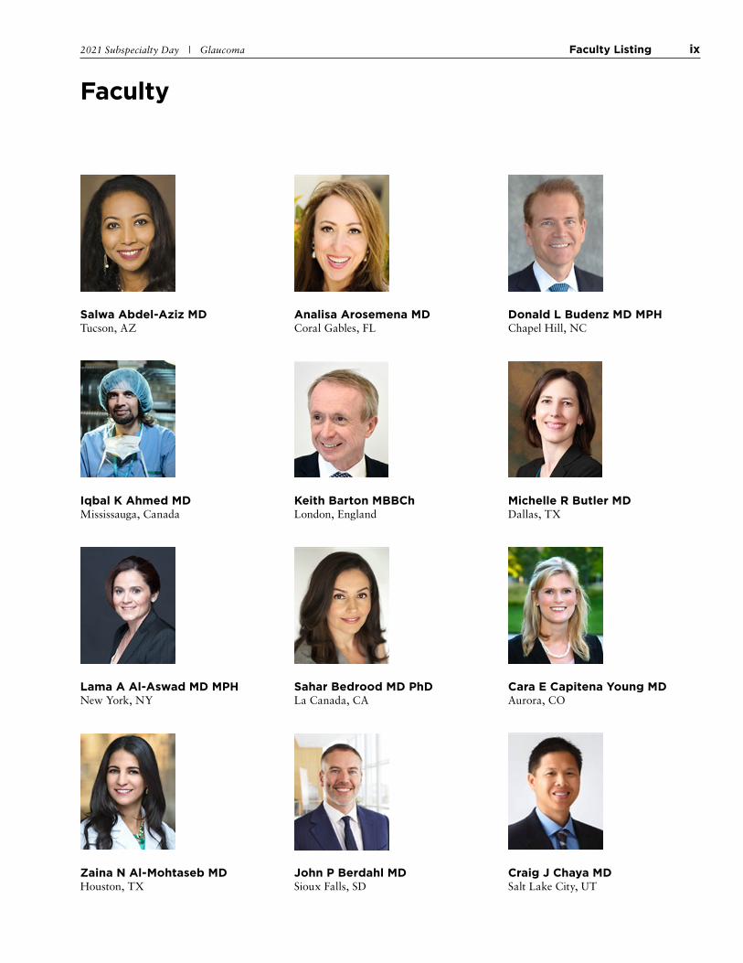
2021 Subspecialty Day | Glaucoma Faculty Listing ix
Salwa Abdel-Aziz MDTucson, AZ
Iqbal K Ahmed MDMississauga, Canada
Lama A Al-Aswad MD MPHNew York, NY
Zaina N Al-Mohtaseb MDHouston, TX
Analisa Arosemena MDCoral Gables, FL
Keith Barton MBBChLondon, England
Sahar Bedrood MD PhDLa Canada, CA
John P Berdahl MDSioux Falls, SD
Donald L Budenz MD MPHChapel Hill, NC
Michelle R Butler MDDallas, TX
Cara E Capitena Young MDAurora, CO
Craig J Chaya MDSalt Lake City, UT
Faculty

x Faculty Listing 2021 Subspecialty Day | Glaucoma
No photo available
Jenny Chen MDSacramento, CA
Teresa C Chen MDBoston, MA
Garry P Condon MDSarasota, FL
Ian P Conner MD PhDPittsburgh, PA
Qi N Cui MDPhiladelphia, PA
Kendall E Donaldson MDPlantation, FL
Angela R Elam MDYpsilanti, MI
Babak Eliassi-Rad MDBoston, MA
Matthew E Emanuel MDDallas, TX
Ronald Leigh Fellman MD OCSDallas, TX
Brian A Francis MDPasadena, CA
No photo available
Mark J Gallardo MDEl Paso, TX
2021 Subspecialty Day | Glaucoma Faculty Listing xi
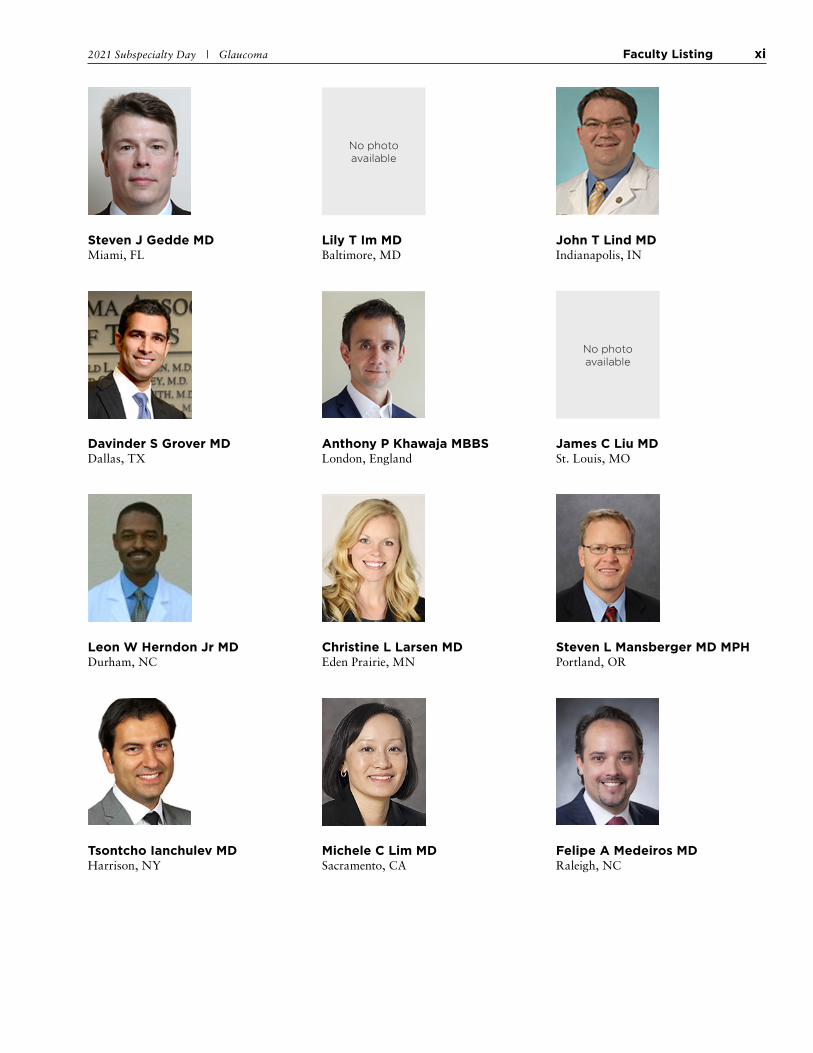
Steven J Gedde MDMiami, FL
Davinder S Grover MDDallas, TX
Leon W Herndon Jr MDDurham, NC
Tsontcho Ianchulev MDHarrison, NY
No photo available
Lily T Im MDBaltimore, MD
Anthony P Khawaja MBBSLondon, England
Christine L Larsen MDEden Prairie, MN
Michele C Lim MDSacramento, CA
John T Lind MDIndianapolis, IN
No photo available
James C Liu MDSt. Louis, MO
Steven L Mansberger MD MPHPortland, OR
Felipe A Medeiros MDRaleigh, NC

xii Faculty Listing 2021 Subspecialty Day | Glaucoma
Lilian Nguyen MDSan Antonio, TX
Yvonne Ou MDSan Francisco, CA
Paul F Palmberg MD PhDMiami, FL
Douglas J Rhee MDCleveland, OH
No photo available
Grace Marie Richter MD MPHLos Angeles, CA
Jullia A Rosdahl MD PhDChapel Hill, NC
Ahmara V Ross MDPhiladelphia, PA
Thomas W Samuelson MDMinneapolis, MN
Terry L Schwartz MDCincinnati, OH
Leonard K Seibold MDAurora, CO
Arsham Sheybani MDSaint Louis, MO
Aakriti Garg Shukla MDPhiladelphia, PA

2021 Subspecialty Day | Glaucoma Faculty Listing xiii
No photo available
David A Sola-Del Valle MDBoston, MA
Jeffrey R SooHoo MDAurora, CO
Thasarat S Vajaranant MD MHAChicago, IL
Sarah Van Tassel MDNew York, NY
Kateki Vinod MDNew York, NY
David S Walton MDBoston, MA
Kelly Walton Muir MDDurham, NC
Andrew M Williams MDPittsburgh, PA
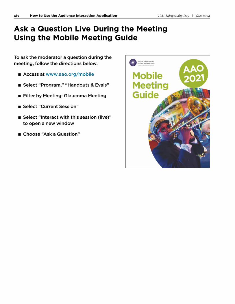
Ask a Question Live During the MeetingUsing the Mobile Meeting Guide
To ask the moderator a question during the meeting, follow the directions below.
■ Access at www.aao.org/mobile
■ Select “Program,” “Handouts & Evals”
■ Filter by Meeting: Glaucoma Meeting
■ Select “Current Session”
■ Select “Interact with this session (live)” to open a new window
■ Choose “Ask a Question”
xiv How to Use the Audience Interaction Application 2021 Subspecialty Day | Glaucoma

2021 Subspecialty Day | Glaucoma Program Schedule xv
Glaucoma Subspecialty Day 2021: Making Glaucoma Care the Big Easy
FRIDAY, NOV. 12, 2021
7:00 AM CONTINENTAL BREAKFAST
8:00 AM Welcome and Introductions Brian A Francis MD
8:02 AM American Glaucoma Society Introduction Ronald Leigh Fellman MD OCS*
8:04 AM AGS Cares Ronald Leigh Fellman MD OCS*
8:09 AM Announcements Kelly Walton Muir MD
Section I: Diagnostics: OCT and Visual Fields
Moderators: Teresa C Chen MD* and John T Lind MD
Virtual Moderator: Aakriti Garg Shukla MD
8:11 AM OCT Interpretation: Basics and Pearls Thasarat S Vajaranant MD 1
8:19 AM Innovations in Visual Field Testing Steven L Mansberger MD MPH* 3
8:27 AM OCT–Visual Field Mismatch: OCT Misinterpreting Grace Marie Richter MPH* 4
8:35 AM OCT–Visual Field Mismatch: VF Misinterpreting Donald L Budenz MD MPH* 5
8:43 AM OCT Progression Jullia A Rosdahl MD PhD* 6
8:51 AM Visual Field Progression Steven J Gedde MD 7
Section II: MIGS Case-Based Section
Moderators: Davinder S Grover MD* and Christine L Larsen MD
Virtual Moderator: Aakriti Garg Shukla MD
9:00 AM iStent: Ideal Patient, Key Pearls for Success, Sahar Bedrood MD PhD* 9 and Why I Didn’t Do the Other MIGS
9:06 AM Hydrus: Ideal Patient, Key Pearls for Success, Craig J Chaya MD 10 and Why I Didn’t Do the Other MIGS
9:12 AM Goniotomy: Ideal Patient, Key Pearls for Success, Leonard K Seibold MD* 11 and Why I Didn’t Do the Other MIGS
9:18 AM Viscodilation: Ideal Patient, Key Pearls for Success, Mark J Gallardo MD* 12 and Why I Didn’t Do the Other MIGS
9:24 AM Gonioscopy-Assisted Transluminal Trabeculotomy: Ideal Patient, Matthew E Emanuel MD* 13 Key Pearls for Success, and Why I Didn’t Do the Other MIGS
9:30 AM Xen Gel Stent: Ideal Patient, Key Pearls for Success, Analisa Arosemena MD* 14 and Why I Didn’t Do the Other MIGS
9:36 AM Discussion
9:51 AM REFRESHMENT BREAK
* Indicates that the presenter has financial interest. No asterisk indicates that the presenter has no financial interest.

xvi Program Schedule 2021 Subspecialty Day | Glaucoma
* Indicates that the presenter has financial interest. No asterisk indicates that the presenter has no financial interest.
Section III: Medication and Lasers
Moderators: Salwa Abdel-Aziz MD and John T Lind MD
Virtual Moderator: Aakriti Garg Shukla MD
10:21 AM Are All Ciliary Body Destruction Procedures Created Equal? Jenny Chen MD 15
10:29 AM Mythbusters: Real or Fake Contraindications Kateki Vinod MD 16
10:39 AM Systemic Drugs and Glaucoma: The Effect of Various Systemic Cara E Capitena MD 17 Medications on Open-Angle and Closed-Angle Glaucoma
10:47 AM Targets of the Medication Pipeline: New and Emerging Treatments David A Sola-Del Valle MD* 18
10:55 AM Alternative Therapeutic Treatments for Glaucoma Angela R Elam MD 19
11:03 AM Emerging Technologies in the Treatment of Glaucoma Ahmara Ross MD PhD* 20
11:12 AM In These Unprecedented Times . . . Donald L Budenz MD MPH* 22
The American Glaucoma Society Subspecialty Day Lecture
Virtual Moderator: Aakriti Garg Shukla MD
11:17 AM Introduction of the Lecturer Donald L Budenz MD MPH*
11:19 AM The Use of Mitomycin C in Traditional and Novel Glaucoma Surgeries Michele C Lim MD* 24
11:49 AM Presentation of the Award Donald L Budenz MD MPH*
11:50 AM LUNCH
Section IV: Glaucoma in the Digital Age
Moderators: Ian P Conner MD PhD* and Babak Eliassi-Rad MD
Virtual Moderator: Andrew M Williams MD
1:05 PM Home Tonometry Jeffrey R SooHoo MD 25
1:12 PM Virtual Reality Visual Fields Yvonne Ou MD* 26
1:19 PM Teleglaucoma Lama A Al-Aswad MD MPH* 27
1:26 PM App-Based Visual Aids Terry L Schwartz MD 28
1:33 PM Deep Learning/Artificial Intelligence Anthony P Khawaja MBBS* 30
1:40 PM Discussion
Section V: Journal Club/Late Breaking
Moderators: Ian P Conner MD PhD* and Kelly Walton Muir MD
Virtual Moderator: Andrew M Williams MD
1:55 PM Introduction Ian P Conner MD PhD*
1:57 PM Case Presentation James C Liu MD 32
2:01 PM Clinical Trial Update for Bimatoprost Implant Felipe A Medeiros MD* 33
2:09 PM Potential Scientific Basis for Sustained Response to Implant Douglas J Rhee MD* 35
2:17 PM Synthesizing the Clinical and Basic Science Information for This Patient Qi N Cui MD* 36
2:23 PM Best of AGS: Surgery in the Advanced Angle-Closure Patient Sarah Van Tassel MD* 37
2:30 PM Best of AGS: Surgery in the Advanced Uveitic Glaucoma Patient Keith Barton MBBCh 39
2021 Subspecialty Day | Glaucoma Program Schedule xvii
* Indicates that the presenter has financial interest. No asterisk indicates that the presenter has no financial interest.
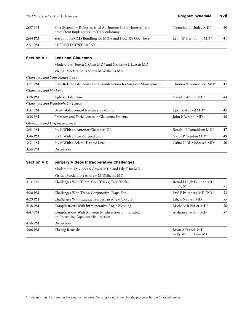
2:37 PM First System for Robot-assisted Ab-Interno Gonio-Intervention: Tsontcho Ianchulev MD* 40 From Stent Implantation to Trabeculotomy
2:44 PM Status of the CMS Bundling for MIGS and How We Got There Leon W Herndon Jr MD* 41
2:51 PM REFRESHMENT BREAK
Section VI: Lens and Glaucoma
Moderators: Teresa C Chen MD* and Christine L Larsen MD
Virtual Moderator: Andrew M Williams MD
Glaucoma and Your Native Lens
3:21 PM Lens-Related Glaucoma and Considerations for Surgical Management Thomas W Samuelson MD* 42
Glaucoma and No Lens
3:26 PM Aphakic Glaucoma David S Walton MD* 44
Glaucoma and Pseudophakic Lenses
3:31 PM Uveitis-Glaucoma-Hyphema Syndrome Iqbal K Ahmed MD* 45
3:36 PM Premium and Toric Lenses in Glaucoma Patients John P Berdahl MD* 46
Glaucoma and Displaced Lenses
3:41 PM Fix It With an Anterior Chamber IOL Kendall E Donaldson MD* 47
3:46 PM Fix It With an Iris-Sutured Lens Garry P Condon MD* 48
3:51 PM Fix It With a Scleral-Fixated Lens Zaina N Al-Mohtaseb MD* 50
3:56 PM Discussion
Section VII: Surgery Videos Intraoperative Challenges
Moderators: Davinder S Grover MD* and Lily T Im MD
Virtual Moderator: Andrew M Williams MD
4:11 PM Challenges With Tubes: Conj Tricks, Tube Tricks Ronald Leigh Fellman MD OCS* 51
4:20 PM Challenges With Trabs: Conjunctiva, Flaps, Etc. Paul F Palmberg MD PhD* 53
4:29 PM Challenges With Cataract Surgery in Angle Closure Lilian Nguyen MD 55
4:38 PM Complications With Intraoperative Angle Bleeding Michelle R Butler MD* 56
4:47 PM Complications With Aqueous Misdirection on the Table; Arsham Sheybani MD 57 or, Preventing Aqueous Misdirection
4:56 PM Discussion
5:06 PM Closing Remarks Brian A Francis MD Kelly Walton Muir MD

2021 Subspecialty Day | Glaucoma Section I: Diagnostics: OCT and Visual Fields 1
OCT Interpretation: Basics and PearlsThasarat Sutabutr Vajaranant MD MHA
Optical coherence tomography (OCT), an imaging technique based on interferometry to reconstruct 3-D cross-sectional images of the optic nerve and macula, has become the standard of care for glaucoma. Low-quality scans can negatively impact the interpretations and lead to mismanagement of glaucoma; hence it is imperative for clinicians to recognize its limitations and common artifacts. This section will provide basics and pearls for OCT interpretations.
OCT Basics
Different scanning protocols 1. Raster cube scan of the optic nerve and the macula: An
area of interest is scanned from side to side in lines from top to bottom.
2. Circular scan of the optic nerve: A circular scan, approxi-mately 3.45 mm in diameter around the optic nerve, captures the retinal ganglion cell axons as they travel through the retinal nerve fiber layer from the entire retina toward the optic nerve head.
3. Radial scan and radial-concentric scan of the optic nerve: A spoke-like scan, centered around the optic nerve (may be combined with concentric scan).
4. Wide-field scan of the optic nerve and macula: A set of wide raster scans that captures both the optic nerve and macula.
OCT analysis 1. Retinal nerve fiber layer parameters 2. Optic disc parameters 3. Macular parameters 4. OCT progression analysis
Essential Pearls for Interpretations
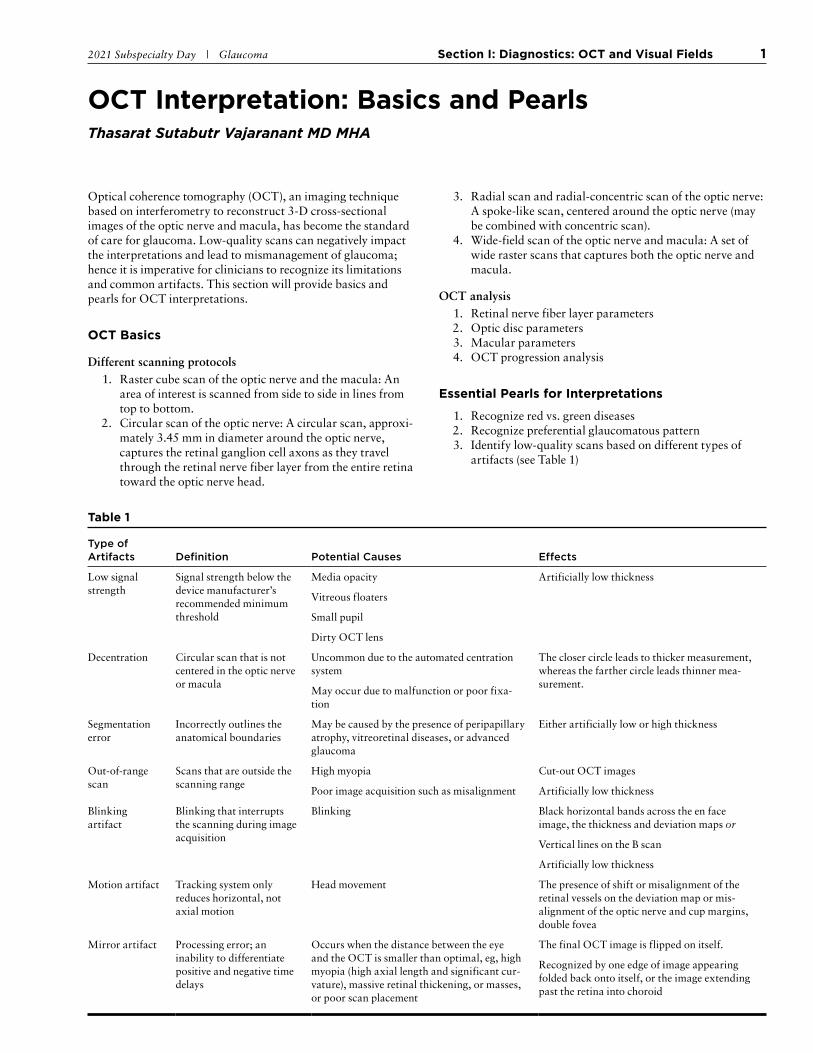
1. Recognize red vs. green diseases 2. Recognize preferential glaucomatous pattern 3. Identify low-quality scans based on different types of
artifacts (see Table 1)
Table 1
Type of Artifacts
Definition
Potential Causes
Effects
Low signal strength
Signal strength below the device manufacturer’s recommended minimum threshold
Media opacity Artificially low thickness
Vitreous floaters
Small pupil
Dirty OCT lens
Decentration Circular scan that is not centered in the optic nerve or macula
Uncommon due to the automated centration system
The closer circle leads to thicker measurement, whereas the farther circle leads thinner mea-surement.
May occur due to malfunction or poor fixa-tion
Segmentation error
Incorrectly outlines the anatomical boundaries
May be caused by the presence of peripapillary atrophy, vitreoretinal diseases, or advanced glaucoma
Either artificially low or high thickness
Out-of-range scan
Scans that are outside the scanning range
High myopia Cut-out OCT images
Poor image acquisition such as misalignment Artificially low thickness
Blinking artifact
Blinking that interrupts the scanning during image acquisition
Blinking Black horizontal bands across the en face image, the thickness and deviation maps or
Vertical lines on the B scan
Artificially low thickness
Motion artifact Tracking system only reduces horizontal, not axial motion
Head movement The presence of shift or misalignment of the retinal vessels on the deviation map or mis-alignment of the optic nerve and cup margins, double fovea
Mirror artifact Processing error; an inability to differentiate positive and negative time delays
Occurs when the distance between the eye and the OCT is smaller than optimal, eg, high myopia (high axial length and significant cur-vature), massive retinal thickening, or masses, or poor scan placement
The final OCT image is flipped on itself.
Recognized by one edge of image appearing folded back onto itself, or the image extending past the retina into choroid

2 Section I: Diagnostics: OCT and Visual Fields 2021 Subspecialty Day | Glaucoma
Selected Readings 1. Budenz D, ed. Atlas of Optical Coherence Tomography for Glau-
coma. Springer; 2020.
2. Varma R, Xu BY, Rihter GM, Reznik A, eds. Advances in Ocular Imaging in Glaucoma. Springer; 2020.
3. Hajizadeh F, ed. Atlas of Ocular Optical Coherence Tomography. Springer; 2018.

2021 Subspecialty Day | Glaucoma Section I: Diagnostics: OCT and Visual Fields 3
Innovations in Visual Field TestingAdvances in PerimetrySteven L Mansberger MD MPH
I. Background
Glaucoma progression can lead to visual disability even if treated.
A. In a retrospective study of 295 treated patients with newly diagnosed open-angle glaucoma in Olmsted County, Minnesota, whose IOP was not appropri-ately controlled1
B. Probability of blindness after 20 years:
1. 27% in 1 eye
2. 9% in both eyes
C. Of 114 patients initially treated for ocular hyper-tension, probability of blindness after 20 years:
1. 14% in 1 eye
2. 4% in both eyes
II. Importance of Detecting Slope of Progressive Glaucoma
Single-field analysis with glaucoma progression analy-sis (GPA) results
A. GPA printout is the preferred method for event analysis.
B. Technician must set up single-page printout for GPA.
C. Clinician must choose baseline fields to be used.
III. Rate-Based Change Glaucoma Change Analysis
A. What rate of loss is significant?2
B. 402,357 anonymized VFs from 75,857 patients recorded between 1989 and 2012
C. Median life expectancies, based on age and sex, from UK Office for National Statistics
D. 7.5% had a rate worse than −1 dB/y.
E. 3.0% of eyes progressed at faster than −1.5 dB/y.
F. But 33.3% had positive MD rates.
G. 90.7% of blindness cases were < −6 dB at baseline in 1 eye.
H. 5%-7.2% blind over their lifetime
IV. What Rate of Loss Is Significant?
A. Early Manifest Glaucoma Trial: 1.08 dB/yr
B. Rosetti L: 1.1 ± 3.5 dB/yr
C. DeMoraes (10-2 visual fields): 1.0 dB/yr
D. Visual Field Index: 2.5%/yr
V. What to Do With Poor Sensitivity <19 dB?
VI. When to Check 10-2 Visual Fields?
A. 10-2: 2 degree grid (vs. 6 degree), 68 points in cen-tral 10 degrees, 44 in central 8 degrees vs. 4 points with 30-2/24-2
B. 12% of patients with normal 30-2 VFs1
C. 50% with mild to moderate glaucoma have repeat-able 10-2 loss.2
VII. Research and Future Applications of Perimetry: Benefits and Disadvantages
A. 24-2c (SITA Faster)
B. Portable perimetry
1. Virtual reality
2. Tablet-based computer approach
C. Cluster perimetry
D. Real-life situation (driving, ambulation) perimetry
E. Data analysis methods: Application of artificial intelligence for visual field testing
F. Combining structure and function perimetry
References 1. Hattenhauer MG, Johnson DG, Ing HH, et al. The probability
of blindness from open-angle glaucoma. Ophthalmology 1998; 105(11):2099-2104.
2. Saunders LJ, Russell RA, Kirwan JF, McNaught AI, Crabb DP. Examining visual field loss in patients in glaucoma clinics during their predicted remaining lifetime. Invest Ophthalmol Vis Sci. 2014; 55(1):102-109.
3. Rossetti L, Digiuni M, Centofanti M, et al . Blindness and glau-coma: a multicenter data review from 7 academic eye clinics. PLoS One. 2015; 10(8):e0136632.
4. de Moraes CG, Song C, Liebmann JM, Simonson JL, Furlanetto RL, Ritch R. Defining 10-2 visual field progression criteria: exploratory and confirmatory factor analysis using pointwise lin-ear regression. Ophthalmology 2014; 121(3):741-749.
5. Gardiner SK, Swanson WH, Goren D, Mansberger SL, Demirel S. Assessment of the reliability of standard automated peri metry in regions of glaucomatous damage. Ophthalmology 2014; 121(7):1359-1369.
6. Langerhorst CT, Carenini LL, Bakker D, De Bie-Raakman MAC. Measurements for description of very early glaucomatous field defects. In: Wall M, Heijl A, eds. Perimetry Update 1996/1997. New York: Kugler 53 Publications; 1997:67-73.
7. Schiefer U, Papageorgiou E, Sample PA, et al. Spatial pattern of glaucomatous visual field loss obtained with regionally con-densed stimulus arrangements. Invest Ophthalmol Vis Sci. 2010; 51:5685-5689.

4 Section I: Diagnostics: OCT and Visual Fields 2021 Subspecialty Day | Glaucoma
OCT–Visual Field Mismatch: OCT MisinterpretingGrace Marie Richter MD MPH
NOTES

2021 Subspecialty Day | Glaucoma Section I: Diagnostics: OCT and Visual Fields 5
OCT–Visual Field Mismatch: VF MisinterpretingDonald L Budenz MD MPH
Introduction
Misinterpretation of a visual field can result in a mismatch with OCT diagnosis in numerous ways. These fall into 3 broad cat-egories: (1) falsely abnormal visual field with normal OCT, (2) falsely normal visual field with abnormal OCT, and (3) OCT makes correct diagnosis but visual field interpretation results in a different and incorrect diagnosis. One of the reasons it is so helpful to have both diagnostic tools is that one can serve as a reality check for the other.
Falsely Abnormal Visual Field With Normal OCT
In this situation, the OCT appears normal and the visual field is abnormal in a glaucomatous pattern. We see this most often in the workup of the glaucoma suspect. A commonly seen phenomenon called “pattern reversal” occurs when the Total Deviation Plot is normal but an early glaucomatous visual field defect is seen in the Pattern Deviation Plot. This is due to a slightly better than average performance by the patient com-pared to age-matched controls and the General Height Adjust-ment causing a depression, rather than elevation, in the entire visual field. Careful examination of the Total Deviation Plot reveals multiple positive integers, enough to cause the General Height Adjustment to do the opposite of what was intended. It is critical to look at the Total Deviation Plot before interpret-ing the Pattern Deviation Plot. If the Total Deviation Plot is Normal, then the patient is normal and you should stop and not even look at the Pattern Deviation Plot.
One of the problems with the Humphrey GPA overview printout is that to save space, only the Total Deviation Plot is displayed. One needs to print out the entire day’s visual field to make sure this phenomenon is not occurring because it will be missed in the GPA printout alone. In the age of EHRs, there is no reason not to refer to the original printout since we are not killing trees or making paper charts thicker!
A variety of commonly seen false positive artifacts in visual fields have been well described, and their recognition and proper interpretation can avoid misdiagnosis. Using the OCT as a reality check is very helpful in all of these situations. These include patient inexperience/learning effect, eyelid and brow artifacts, lens rim artifacts, inattentive patient, fatigue, incor-rect fixation, incorrect trial frame correction, and incorrect date of birth entered. Having a Normal OCT for patients in these situations is very helpful in pointing out a mismatch, and pat-tern recognition of these false positive visual field results can prevent misinterpretation and misdiagnosis.
Falsely Normal Visual Field With Abnormal OCT
A visual field that is normal when the patient has glaucoma by clinical examination and OCT is less common, although we all accept that OCT and structural changes occur before visual fields become abnormal. In a phenomenon called “progres-sion in the green,” the OCT is normal to begin with and slowly worsens due to glaucoma, but the wide genetic variability in retinal nerve fiber layer thickness and other OCT parameters makes it difficult to diagnose early glaucoma. The white-on-white and sometimes even blue-on-yellow visual fields remain normal during this period because they are not sensitive enough to pick up this early damage. Trigger-happy patients cause a high false-positive reliability index and an artifactually normal visual field that can mask glaucoma, so having an abnormal OCT can be helpful in pointing one in the right direction. Poor fixation, when the patient looks at the stimuli being presented instead of the central fixation light, can cause an artifactually good visual field in the face of clear OCT and optic disc abnor-mality.
OCT Correct Diagnosis, Visual Field Different and Incorrect Diagnosis
Nonglaucomatous optic neuropathies, cerebrovascular acci-dents, and retinal disease can cause abnormal visual fields in our glaucoma patients that simulate glaucoma or glaucoma pro-gression. The OCT (as well as careful fundus examination and neuroimaging when indicated) can be very helpful in sorting out these mismatches.
Selected Readings 1. Budenz DL. Atlas of Visual Fields. Philadelphia: Lippincott-
Raven; 1997: chapter 2.
2. Heijl A, Patella VM, Bengtsson B. Excellent Perimetry: The Visual Field Analyzer Primer. 5th ed. Carl Zeiss Meditec; 2021: chapter 12.
3. Budenz DL. Atlas of Optical Coherence Tomography for Glau-coma. Switzerland: Springer Nature; 2020: chapters 8 and 9.
4. Greenfield DS, Siatkowski RM, Glaser JS, et al. The cupped disc. who needs neuroimaging? Ophthalmology 1998; 105(10):1866-1874.
6 Section I: Diagnostics: OCT and Visual Fields 2021 Subspecialty Day | Glaucoma
OCT ProgressionJullia A Rosdahl MD PhD
I. Illustrative Case Example
Glaucoma is a progressive optic neuropathy character-ized by optic nerve thinning with corresponding visual field defects. OCT is a powerful technology enabling quantitative and qualitative evaluation of the optic nerve and subsequent changes over time that would indicate glaucomatous progression.
II. When OCT Is Most/More Useful for Determining Progression
Both OCT and visual field testing are used to assess for disease progression in glaucoma patients. Often, OCT is more useful in earlier stages of glaucoma; and visual field testing, in later stages. In later stages of glaucoma, macular OCT in particular can be helpful.
III. What Constitutes “Real” Progression?
The “rule of 5” is commonly used to identify changes in OCT measurements that are likely to be clinically significant. Trend-based analyses may be better for detecting glaucomatous progression.
IV. Beware of False “Progression”
Just as artifacts and masqueraders can affect the diag-nostic capabilities of OCT, so also can they affect its ability to detect progression. Pathologies of the vitre-ous and macula can affect OCT measurements of the retinal nerve fiber layer and macula. Evaluation of the OCT scans in addition to the thickness maps and pro-gression analyses can help mitigate clinical misjudg-ments.
Selected Readings 1. Zhang X, Dastiridou A, Francis BA, et al.; Advanced Imaging for
Glaucoma Study Group. Baseline Fourier-domain OCT structural risk factors for visual field progression in the Advanced Imaging for Glaucoma Study. Am J Ophthalmol. 2016; 172:94-103.
2. Abe RY, Diniz-Filho A, Zangwill LM, et al. The relative odds of progressing by structural and functional tests in glaucoma. Invest Ophthalmol Vis Sci. 2016; 57:OCT421-OCT428.
3. Tatham AJ, Medeiros FA. Detecting structural progression in glaucoma with optical coherence tomography. Ophthalmology 2017; 124(12 suppl): S57-S65.
4. Zhang X, Dastiridou A, Francis BA, et al.; Advanced Imaging for Glaucoma Study Group. Comparison of glaucoma progression detection by optical coherence tomography and visual field. Am J Ophthalmol. 2017; 184:63-74.
5. Thompson AC, Jammal AA, Berchuck SA, et al. Comparing the rule of 5 to trend-based analysis: detecting glaucoma progression on OCT. Ophthalmol Glaucoma. 2020; 3(6):414-420.
6. Schuman JS, Kostanyan T, Bussel I. Review of longitudinal glau-coma progression: 5 years after the Shaffer Lecture. Ophthalmol Glaucoma. 2020; 3(2):158-166.
7. Thompson AC, Jammal AA, Medeiros FA. A review of deep learning for screening, diagnosis, and detection of glaucoma pro-gression. Trans Vis Sci Tech. 2020; 9(2):42.

2021 Subspecialty Day | Glaucoma Section I: Diagnostics: OCT and Visual Fields 7
Visual Field ProgressionSteven J Gedde MD
Introduction
Perimetry plays an important role in the diagnosis and manage-ment of glaucoma. Visual field (VF) changes that are statisti-cally and clinically significant can provide a basis for adjust-ments in treatment. Ocular imaging of the optic disc, retinal nerve fiber layer, and ganglion cells provides valuable informa-tion that compliments but does not replace VF testing. Notably, a floor effect with OCT measurements makes it impossible to detect further deterioration with this technology in eyes with advanced disease.
A paradigm shift in glaucoma management has occurred over the past decade. Clinicians previously were mainly focused on whether or not VF progression had occurred, and they are now interested in determining the rate of progression. The goal of glaucoma treatment is to prevent loss of visual function, espe-cially as it relates to quality of life.
Selecting a Test Strategy
Selecting the best VF test strategy for an individual patient can increase the likelihood of detecting glaucomatous progression. The most commonly used is a 24-2 test pattern with a size III stimulus, consisting of 54 test points spaced 6 degrees apart. The 24-2 test pattern has gradually replaced the 30-2 test pat-tern because little diagnostic information is lost and test time is reduced.1 The Swedish Interactive Thresholding Algorithm (SITA) has supplanted the older full-threshold strategy and includes SITA Standard, SITA Fast, and SITA Faster. A 10-2 test covers the area within 10 degrees of fixation with a grid of test points 2 degrees apart. This testing strategy may be preferred in glaucoma patients with advanced VF constriction, or in those with scotomas close to fixation at any stage of disease. A study found 61.5% of eyes with glaucoma and 39.5% with a suspicion of glaucoma had VF defects on 10-2 testing that were missed with a 24-2 strategy.2
One shortcoming of 10-2 VFs is the lack of a reference database for progression analysis. However, an event-based algorithm similar to Guided Progression Analysis (GPA) for 24-2 and 30-2 SITA tests was recently developed.3 Using a size V stimulus with a 24-2 or 10-2 test pattern is another option for patients with advanced glaucoma or media opacities. The larger stimulus will extend the available range of sensitivities to monitor for progression. Unfortunately, nonstandard stimulus sizes cannot be used with the SITA testing strategies and require a more time-consuming algorithm (Fastpac or full threshold). Furthermore, the benefit of a normative database or GPA is not available with a size V stimulus.
Repeat Testing
Repeat VF testing should be performed soon after a patient is diagnosed with glaucoma because obtaining 2 similar and representative baseline tests is foundational to future manage-ment decisions. Development of a new VF defect or worsening of existing ones should prompt repeat testing. Clinical trials
have highlighted the importance of repeat testing to confirm or refute progression. Stricter endpoint criteria that included addi-tional confirmatory VFs were adopted during the course of the Collaborative Normal Tension Glaucoma Study, and this proto-col change reduced false calls of progression from 57% to 2%.4 In the Ocular Hypertension Treatment Study, 85.9% of new VF defects were not confirmed on repeat testing.5 However, high test-retest variability is characteristic of areas of VFs affected by glaucoma. Variable sensitivity measurements occurring in the same area, but not always in the same location, commonly pre-cede definite glaucomatous VF progression.
At least 5 threshold VFs are generally needed to quantify how rapidly a patient with glaucoma may be progressing. It’s particularly important for clinicians to identify patients who are experiencing rapid rates of progression that could result in visual disability. A study found the time to detect rapid progres-sion (defined as mean deviation change of 2 dB/year) was 1.7 years with triannual testing compared with 5 years with annual testing.6 Therefore, it has been recommended that 3 VFs per year (including baseline tests) be obtained during the first 2 years of follow-up for newly diagnosed patients with glaucoma-tous VF loss. More frequent VF testing should be performed in glaucoma patients with field loss until they have been shown to be stable or progressing at an acceptable rate.
Guided Progression Analysis (GPA)
The Humphrey perimeter’s GPA offers both event and trend analysis.7 Follow-up VF tests are compared to baseline VFs to quantify the amount and rate of change. Baseline tests should define the patient’s status at a particular time, such as when therapy was started or significantly modified. GPA has been programmed to select by default the first 2 VFs as baseline. However, the clinician may choose other VFs to serve as the baseline tests, and GPA will remember these in subsequent follow-up examinations. The SITA testing strategies (SITA Standard, SITA Fast, and SITA Faster) may be freely intermixed in the upgraded GPA program.
Event analysisThe goal of event analysis is to determine whether there has been any statistically significant worsening in the VF. The GPA’s Glaucoma Change Probability Map highlights test points on 24-2 and 30-2 VFs in which pattern deviation values have deteriorated from baseline by more than the expected range of testing variability found in glaucoma patients. Open, half black, and filled-in black triangular symbols indicate test points show-ing deterioration from baseline that is statistically significant at the P < .05 level on 1, 2, and 3 or more consecutive VFs, respectively. Test points that fall outside the range that can be analyzed for statistically significant change are marked with an “X.” The GPA Alert posts a message based upon the criteria used for progression in the Early Manifest Glaucoma Trial.8 “Possible Progression” is displayed when the same 3 or more test points have shown statistically significant deterioration on

8 Section I: Diagnostics: OCT and Visual Fields 2021 Subspecialty Day | Glaucoma
2 consecutive follow-up examinations, and “Likely Progres-sion” is shown when this deterioration is seen on 3 or more con-secutive follow-up tests.
Trend analysis
The aim of trend analysis is to quantify the rate of VF progres-sion to help clinicians evaluate the risk of future visual impair-ment. The GPA trend analysis estimates the rate of progression using linear regression analysis of the Visual Field Index (VFI) over time. The VFI parameter summarizes a patient’s VF status as a percentage of normal age-corrected sensitivity, with 100% being a completely normal VF and 0% representing perimetric blindness. The GPA trend analysis is automatically calculated when 5 or more eligible VFs are available. A projection of the linear regression line into the future is provided by GPA, if 5 or more VFs covering at least 2 years’ area are available and if the width of the calculated 95% confidence interval for VFI slope is not greater than a VFI value of ±2.5%.
Conclusions
Clinical trials have shown that many treated patients with glaucoma will progress, which is evident if perimetric testing is done regularly for multiple years.8-10 Selecting the best VF test strategy and establishing a baseline of VFs will assist clinicians in the detection of glaucomatous progression. If a VF change is suspected, repeat testing should be performed to confirm or refute progression. Humphrey’s GPA can assist in identifying and quantifying VF progression. Event analysis is an effective method for finding statistically significant perimetric glaucoma progression events, especially in the setting of clinical trials.
In clinical practice, statistically significant changes on event analysis can prompt examination of a patient’s trend analysis to determine whether clinically significant changes may be occur-ring. Perimetric progression rates vary widely among glaucoma patients. While some patients progress slowly and need little if any change in treatment, an important minority will progress at rates that lead to functional impairment if appropriate treat-ment is not implemented. More frequent VF testing for newly diagnosed patients with glaucomatous VF loss serves to identify rapid progressors.
References 1. Khoury JM, Donahue SP, Lavin PJ, Tsai JC. Comparison of 24-2
and 30-2 perimetry in glaucomatous and nonglaucomatous optic neuropathies. J Neuroophthalmol. 1999; 19:100-108.
2. De Moraes CG, Hood DC, Thenappan A, et al. 24-2 visual fields miss central defects shown on 10-2 tests in glaucoma suspects, ocular hypertensives, and early glaucoma. Ophthalmology 2017; 124:1449-1456.
3. De Moraes CG, Paula JS, Blumberg DM, et al. Detection of pro-gression with 10-2 standard automated perimetry: development and validation of an event-based algorithm. Am J Ophthalmol. 2020; 216:37-43.
4. Schulzer M; Normal-Tension Glaucoma Study Group. Errors in the diagnosis of visual field progression in normal-tension glau-coma. Ophthalmology 1994; 101:1589-1595.
5. Keltner JL, Johnson CA, Quigg JM, et al. Confirmation of visual field abnormalities in the Ocular Hypertension Treatment Study. Arch Ophthalmol. 2000; 118:1187-1194.
6. Chauhan BC, Garway-Heath DR, Foni FJ, et al. Practical recom-mendations for measuring rates of visual field change in glau-coma. Br J Ophthalmol. 2008; 92:569-573.
7. Heijl A, Patella VM, Bengtsson B. The Field Analyzer: Excellent Perimetry. 5th ed. Jena, Germany: Carl Zeiss Meditec; 2021.
8. Heijl A, Leske MC, Bengtsson B, et al. Reduction of intraocular pressure and glaucoma progression: results from the Early Mani-fest Glaucoma Trial. Arch Ophthalmol. 2002; 120:1268-1279.
9. Collaborative Normal-Tension Glaucoma Study Group. Com-parison of glaucomatous progression between untreated patients with normal-tension glaucoma and patients with therapeutically reduced intraocular pressures. Am J Ophthalmol. 1998; 126:487-497.
10. Garway-Heath DF, Crabb DP, Bunce C, et al. Latanoprost for open-angle glaucoma (UKGTS): a randomised, multicentre, placebo-controlled trial. Lancet 2015; 385:1295-1304.
2021 Subspecialty Day | Glaucoma Section II: MIGS Case-Based Section 9
iStent: Ideal Patient, Key Pearls for Success, and Why I Didn’t Do the Other MIGSSahar Bedrood MD PhD
I. Introduction of iStent Inject
II. FDA-Approved Indications for Use of iStent/iStent Inject
III. The Ideal Patient
A. Patient with primary open-angle glaucoma in the mild to moderate stage on at least 1 IOP-lowering drop undergoing cataract surgery
B. Case presentation of ideal patient
IV. Pearls for Success
Video montage/presentation pearls
V. Real-World Data on Patient Outcomes
VI. Personal Real-World Data for iStent Inject and IOP Lowering
VII. Summary
Why I wouldn’t use any other MIGS in these patients
Selected Readings 1. Craven ER, Katz LJ, Wells JM, Giamporcaro JE; iStent Study
Group. Cataract surgery with trabecular micro-bypass stent implantation in patients with mild-to-moderate open-angle glau-coma and cataract: two-year follow-up. J Cataract Refract Surg. 2012; 38(8):1339-1345.
2. Samuelson TW, Sarkisian SR, Lubeck DM, et al. Prospective, randomized, controlled pivotal trial of an ab interno implanted trabecular micro-bypass in primary open-angle glaucoma and cataract. Ophthalmology 2019; 126(6):811-821.
3. Rosenquist R, Epstein D, Melamed S, et al. Outflow resistance of enucleated human eyes at two different perfusion pressures and different extents of trabeculotomy. Curr Eye Res. 1989; 8:1233-1240.

10 Section II: MIGS Case-Based Section 2021 Subspecialty Day | Glaucoma
Hydrus: Ideal Patient, Key Pearls for Success, and Why I Didn’t Do the Other MIGSCraig J Chaya MD
NOTES
2021 Subspecialty Day | Glaucoma Section II: MIGS Case-Based Section 11
Goniotomy: Ideal Patient, Key Pearls for Success, and Why I Didn’t Do the Other MIGSLeonard K Seibold MD
I. Goniotomy Overview
A. Long-standing procedure of choice in children
B. Targets site of greatest aqueous outflow resistance in most cases
C. Novel devices allow easier, more complete treat-ment of angle in adults
1. Kahook Dual Blade
2. Trabectome/TrabEx
3. OMNI
D. Excisional vs. incisional goniotomy
E. Excellent IOP and medication reduction while maintaining safety
F. Supreme versatility applicable in a wide range of patients
II. Ideal Patient
A. Mild to moderate open-angle glaucoma, including pseudoexfoliation and pigmentary
B. Coexisting cataract; can be performed in phakic or pseudophakic patients
C. Open angle with well-delineated trabecular mesh-work (TM) and other angle structures
D. Compliant, cooperative patient, able to remain still
E. No significant blood thinner usage
F. Preop IOP of upper teens or higher on 1 or more medications
G. Treatment goals
1. Medication reduction of 1 or more
2. IOP reduction to low to mid teens
III. Pearls for Success
A. Practice on MIGS model eyes before first cases.
B. Ensure sound intraoperative gonioscopy skills.
1. Deepen anterior chamber with cohesive visco-elastic.
2. Optimize head and microscope rotation to ensure “en face” view.
3. Mag up.
C. Use trypan blue or look for blood reflux to help visualize TM.
D. Avoid limbal vessels during wound construction.
E. Don’t treat what you can’t see.
F. Kahook Dual Blade/Trabectome
1. Start with blade angled 10-15 degrees up.
2. Initially apply some pressure to ensure footplate is well seated in canal.
3. Relax hand once seated to allow device to glide in the canal.
4. Too much pressure will rotate the eye.
5. Too little pressure will only scrape/incise super-ficial TM.
G. OMNI
1. Ensure cannula tip is through TM before deploying cannula; consider small blade/needle incision first.
2. Confirm correct placement of catheter in canal before goniotomy.
3. Retract catheter as cannula is withdrawn along angle.
H. Set postoperative recovery expectations preop.
I. Consider continuing 1 medication postop until after steroid taper.
IV. Why I Didn’t Do the Other MIGS
A. No implant left behind to worry about as with iStent/Hydrus/Xen
B. Optimizes natural outflow pathway rather than fistulous pathway with Xen
C. More durable response with removal of tissue com-pared to temporary viscodilation of canal
D. Avoids bleb and bleb-related complications of Xen
E. Better IOP and medication reduction compared to iStent
F. Greatest versatility to treat a wide variety of eyes
1. Phakic or pseudophakic
2. Mild, moderate, or severe disease
3. Can be used in some angle closure patients as well
G. Established procedure code without strict labelling limitations of iStent/Hydrus
H. Much lower rate of hyphema than gonioscopy-assisted transluminal trabeculotomy with similar efficacy

12 Section II: MIGS Case-Based Section 2021 Subspecialty Day | Glaucoma
Viscodilation: Ideal Patient, Key Pearls for Success, and Why I Didn’t Do the Other MIGSMark J Gallardo MD
NOTES

2021 Subspecialty Day | Glaucoma Section II: MIGS Case-Based Section 13
Gonioscopy-Assisted Transluminal Trabeculotomy: Ideal Patient, Key Pearls for Success, and Why I Didn’t Do the Other MIGSMatthew E Emanuel MD
Minimally invasive glaucoma surgeries (MIGS) have offered ophthalmologists an alternative to traditional glaucoma surger-ies such as trabeculectomies and glaucoma drainage implants. Rates of traditional surgeries have been declining since the advent of MIGS, and in 2017 nearly 75% of all glaucoma sur-geries completed in the United States were MIGS.1 In general, MIGS offer IOP reduction with a higher safety profile and quicker recovery. Over the last decade, various MIGS surgeries have been developed, most of which aim to bypass the trabecu-lar meshwork via either a small intraocular implant or by excis-ing the trabecular meshwork.
Gonioscopy-assisted transluminal trabeculotomy (GATT), initially presented in the literature in 2014, was the first tech-nique described to cannulate the Schlemm canal and cleave the trabecular meshwork 360 degrees via a conjunctival-sparing ab interno approach.2 GATT has a strong safety profile and can significantly lower IOP in various forms of glaucoma, ranging from primary and secondary open-angle glaucoma to traumatic and juvenile glaucoma.3
While the ideal patient for any glaucoma surgery may be difficult to identify, there are several factors that may increase the chance of success, particularly for a novice surgeon. Patients with obvious pathology in the trabecular meshwork are likely best suited for GATT. A number of the secondary open-angle glaucomas may be ideal first cases—specifically, pseudoexfolia-tion glaucoma and pigment dispersion glaucoma. These glauco-mas are believed to be almost exclusively due to accumulation of deposits in the trabecular meshwork and have been shown to respond well to GATT. Additionally, since identifying angle landmarks on gonioscopy is so critical for a successful GATT surgery, these glaucomas typically have significant trabecular meshwork pigmentation and improve the surgeon’s ability to successfully cannulate the Schlemm canal.
Another benefit of GATT is that it may be performed with or without cataract surgery. Nearly all patients having under-gone GATT have intraoperative bleeding and a postoperative hyphema, though those having undergone concomitant cataract surgery tend to have less bleeding. Therefore, combining GATT with cataract surgery may be beneficial for first-time surgeons.
GATT is an effective and relatively safe MIGS that can be considered for a wide range of glaucoma patients. When ini-tially performing GATT surgeries, ophthalmologists should take into account a few simple considerations to improve their chances of success.
References 1. Ma AK, Lee JH, Warren JL, Teng CC. GlaucoMap – distribution
of glaucoma surgical procedures in the United States. Clin Oph-thalmol. 2020; 14:2551-2560.
2. Grover DS, Godfrey DG, Smith O, Feuer WJ, Montes de Oca I, Fellman RL. Gonioscopy-assisted transluminal trabeculotomy, ab interno trabeculotomy: technique report and preliminary results. Ophthalmology 2014; 121(4):855-861.
3. Grover DS, Smith O, Fellman RL, et al. Gonioscopy-assisted transluminal trabeculotomy: an ab interno circumferential trabec-ulotomy: 24 months follow-up. J Glaucoma. 2018; 27(5):393-401.

14 Section II: MIGS Case-Based Section 2021 Subspecialty Day | Glaucoma
Xen Gel Stent: Ideal Patient, Key Pearls for Success, and Why I Didn’t Do the Other MIGSAnalisa Arosemena MD
With glaucoma being one of leading causes of blindness in the USA, navigating the treatment options hand in hand with the patient is crucial. Once surgery is indicated, achieving the best outcome depends on matching the ideal surgery with the ideal patient.
The Xen Gel Stent is a porcine gel stent that bypasses the trabecular meshwork to drain the aqueous from the anterior chamber to the subconjunctival space. Placement of the implant is the key to success. Care should be taken to avoid intratenon placement, and depending on the patient, different implant approaches can be used. In the internal approach, patients need to have a healthy superonasal conjunctiva and a deep chamber, and we should avoid prominent cheek bones or deep-set eyes. The external approach (Xen-Ex) can be performed with open or closed conjunctiva. The closed conjunctiva is great for patients with previous glaucoma surgery since we can place the implant in the healthy conjunctiva between other surgeries. The open approach is best for patients with high risk of scarring since it can be combined with tenonectomy. Patients with poor visibility due to cornea issues also benefit from the external placement. With the use of mitomycin C and steroids we can help prevent bleb scarring and decrease the need for needling of the bleb.
There is a high motivation to prevent complications from tra-ditional glaucoma surgery, hypotony, expulsive choroidal hem-orrhages, and bleb leaks, among others. Having a minimally invasive subconjunctival surgery allows us to reduce the risk of these complications.
The ideal patient for Xen gel stent is a patient with low risk of scarring, a deep angle without synechiae, and motile con-junctiva at the location of the implant placement. A healthy conjunctiva is ideal, and the patient should not have neovascu-larization or inflammation in the eye.
I choose Xen on patients who have to return to work and resume physical activity early in the postop period, since the post-Xen visual disturbance is minimal; it is my go-to surgery in patients with previous multifocal or toric IOLs—also in patients with previous glaucoma or retinal surgery, with limited conjunctival real estate, and patients that are anticoagulated, since it decreases the risk of hemorrhages and using the external closed approach minimizes the trauma to the eye.
Minimally invasive glaucoma surgery has revolutionized glaucoma management, allowing for faster recovery of vision and functionality, minimizing patient risk, and resulting in a high success rate. For most patients who require better IOP con-trol you can offer this versus a trabeculectomy or a glaucoma drainage device.
Selected Readings 1. Grover DS, Flynn WJ, Bashford KP, et al. Performance and safety
of a new ab interno gelatin stent in refractory glaucoma at 12 months. Am J Ophthalmol. 2017; 183:25-36.
2. Fea AM, Durr GM, Marolo P, Malinverni L, Economou MA, Ahmed I. XEN® Gel Stent: a comprehensive review on its use as a treatment option for refractory glaucoma. Clin Ophthalmol. 2020; 14:1805-1832.
3. Ayyala RS, Arosemena A, Jewelewicz D, et al. Xen Gel Stent early results: safety and efficacy in the short term. Paper presented at: American Glaucoma Society Annual Meeting; March 1-4, 2018; New York, NY.
4. Panarelli JF, Yan DB, Francis B, Craven ER. XEN Gel Stent open conjunctiva technique: a practical approach paper. Adv Ther. 2020; 37(5):2538-2549.
5. Hong CH, Arosemena A, Zurakowski D, Ayyala RS. Glaucoma drainage devices: a systematic literature review and current con-troversies [review]. Surv Ophthalmol. 2005; 50(1):48-60.
6. Sheybani A, Lindfield D, Arosemena A, Bourne C, Shah M. XEN needling case examples investigating real-world successes and fail-ures. Glaucoma Today, March/April 2019, supplement.

2021 Subspecialty Day | Glaucoma Section III: Medication and Lasers 15
Are All Ciliary Body Destruction Procedures Created Equal?Jenny Chen MD
I. Understanding Patient Visual Goals
II. Understanding IOL Options
A. Presbyopic
B. Toric
C. Light-adjustable lens
III. Special Considerations for Glaucoma Patients
A. Contrast sensitivity
B. Ocular surface disease
C. Zonular stability
D. Progression of glaucoma
IV. Summary

16 Section III: Medication and Lasers 2021 Subspecialty Day | Glaucoma
MythBusters: Real or Fake Indications and ContraindicationsKateki Vinod MD
I. MYTH: Prostaglandin analogues are contraindicated during the perioperative period of cataract surgery.
A. Background
1. Incidence and risk factors for pseudophakic cys-toid macular edema (CME)
2. Proposed mechanism for prostaglandin ana-logue (PGA)-mediated pseudophakic CME
B. What does the evidence show?
1. Preoperative vs. continuous vs. postoperative PGA use and risk of pseudophakic CME
2. Role of topical nonsteroidal anti-inflammatory drugs
C. Conclusions and recommendations
II. MYTH: Laser peripheral iridotomy should be performed in every patient with a narrow angle.
A. Background
1. Modern classification of the narrow angle
2. Natural history of untreated primary angle-closure suspects
B. What does the evidence show?
1. Efficacy of laser peripheral iridotomy
2. Role of lens extraction
C. Conclusions and recommendations
III. MYTH: Selective laser trabeculoplasty (SLT) is a repeatable procedure.
A. Background
1. Role of SLT in management of open-angle glau-coma
2. Efficacy of initial SLT
B. What does the evidence show?
1. Efficacy of repeat SLT vs. initial SLT
2. Safety considerations
C. Conclusions and recommendations
Selected Readings 1. Hernstadt DJ, Husain R. Effect of prostaglandin analogue use on
the development of cystoid macular edema after phacoemulsifica-tion using STROBE statement methodology. J Cataract Refract Surg. 2017; 43(4):564-569.
2. Fakhraie G, Mirghorbani M, Katz LJ, et al. Cystoid macular edema with prostaglandin analogue use after uneventful cataract surgery in glaucoma patients. J Cataract Refract Surg. 2019; 45(10):1436-1445.
3. Niyadurupola N, Brodie J, Patel T, et al. Topical prostaglandin analogue use and cystoid macular oedema following uneventful cataract surgery: a randomised control trial. Br J Ophthalmol. Epub ahead of print 2021 May 27. doi:10.1136/bjophthal-mol-2021-319149.
4. Gedde SJ, Chen PP, Muir KW, et al.; American Academy of Oph-thalmology Preferred Practice Pattern Glaucoma Panel. Primary Angle-Closure Disease Preferred Practice Pattern®. Ophthalmol-ogy 2021; 128(1):30-70.
5. He M, Jiang Y, Huang S, et al. Laser peripheral iridotomy for the prevention of angle closure: a single-centre randomised controlled trial. Lancet 2019; 393(10181):1609-1618.
6. Le JT, Rouse B, Gazzard G. Iridotomy to slow progression of visual field loss in angle-closure glaucoma. Cochrane Database Syst Rev. 2018; 6:CD012270.
7. Radhakrishnan S, Chen PP, Junk AK, Nouri-Mahdavi K, Chen TC. Laser peripheral iridotomy in primary angle closure: a report by the American Academy of Ophthalmology. Ophthalmology 2018; 125(7):1110-1120.
8. Azuara-Blanco A, Burr J, Ramsay C, et al.; EAGLE Study Group. Effectiveness of early lens extraction for the treatment of primary angle-closure glaucoma (EAGLE): a randomised controlled trial. Lancet 2016; 388(10052):1389-1397.
9. Ong AY, Ng SM, Vedula SS, Friedman DS. Lens extraction for chronic angle-closure glaucoma. Cochrane Database Syst Rev. 2021; 3(3):CD005555.
10. Garg A, Vickerstaff V, Nathwani N, et al.; Laser in Glaucoma and Ocular Hypertension Trial Study Group. Efficacy of repeat selective laser trabeculoplasty in medication-naive open-angle glaucoma and ocular hypertension during the LiGHT trial. Oph-thalmology 2020; 127(4):467-476.
11. Samples JR, Singh K, Lin SC, et al. Laser trabeculoplasty for open-angle glaucoma: a report by the American Academy of Oph-thalmology. Ophthalmology 2011; 118(11):2296-302.
2021 Subspecialty Day | Glaucoma Section III: Medication and Lasers 17
Systemic Drugs and Glaucoma: The Effect of Various Systemic Medication on Open-Angle and Closed-Angle GlaucomaCara Capitena Young MD
I. Open-Angle Glaucoma: Corticosteroids
A. Mechanism of action: Increased resistance of aque-ous outflow through the trabecular meshwork
B. Incidence: True incidence is unknown; likely 25%-33% of the general population.
C. Time to onset
1. Varies based on potency of steroid
2. Most studies quote 3-6 weeks, but there are documented cases as early as 1 week.
D. Culprits: Any and all steroids, though systemic use is less likely than topical formulations to cause IOP elevation
E. Treatment
1. Cessation of steroid treatment or alternative formulation ± topical and/or oral IOP-lowering therapy.
2. Some cases are refractory to medical treatment and require surgical intervention.
F. High-risk populations
1. Patients with known history of steroid-induced ocular hypertension
2. Patients with history of primary open-angle glaucoma or a first-degree relative with open-angle glaucoma
3. Age: older adults and children
G. What to tell your patients
1. Known steroid responders and those at high risk: Educate them, consider IOP check 2-6 weeks after starting any new steroid.
2. General patient population: Educate them that steroids can cause elevated IOP. Communicate with your eye care provider when you start a new one and call urgently for any eye pain or changes in vision after starting one.
II. Closed-Angle Glaucoma
A. Classes of medications and more common offend-ers
1. Sulfa derivatives: acetazolamide, hydrochloro-thiazide, topiramate
2. Adrenergics: nasal ephedrine, phenylephrine, epinephrine, salbutamol
3. Anti-cholinergics: ipratropium bromide, anti-histamines (eg, promethazine), TCA antidepres-sants (eg, imipramine), SSRI antidepressants (eg, fluoxetine), botulinum toxin
4. Anticoagulants: heparin, warfarin, clopidogrel
5. Monoclonal antibody: daratumumab
B. Mechanism of action and typical associated medi-cations
1. Pupillary block
a. Adrenergics
b. Anticholinergics: Classically cough and cold medications, antidepressants, and some inhalers
2. Anterior dislocation of the lens-iris diaphragm
a. Sulfa derivatives: Classic example is topira-mate
b. Anticoagulants
c. Monoclonal antibody/chemotherapeutic agent
C. Treatment: Depends on the etiology
1. Pupillary block
a. Peripheral iridotomy
b. IOP-lowering meds as needed
2. Anterior shifting of the lens-iris diaphragm
a. Cessation of medication
b. IOP-lowering medications and/or surgery if required
c. Iridotomy is not effective in these cases.
D. What to tell your patients
1. Discuss risks of these common medications with any patient with narrow angles, a history of angle closure, hyperopia.
2. Discuss risk with patients on topiramate and anti-depressants. This commonly occurs within weeks of starting the medication, therefore it is important to discuss with your primary care, neurology, and psychiatry colleagues as well.

18 Section III: Medication and Lasers 2021 Subspecialty Day | Glaucoma
Targets of the Medication Pipeline: New and Emerging TreatmentsDavid A Sola-Del Valle MD
NOTES

2021 Subspecialty Day | Glaucoma Section III: Medication and Lasers 19
Alternative Therapeutic Treatments for GlaucomaAngela R Elam MD
I. Introduction to Alternative Treatments for Glaucoma
A. Nutraceuticals
Morrone LA, Rombola L, Adornetto A, Corasaniti MT, Russo R. Rational basis for nutraceuticals in the treatment of glaucoma. Curr Neuropharmacol. 2018; 16(7):1004-1017. doi: 10.2174/1570159X15666171109124520. PMID: 29119928; PMCID: PMC6120110.
B. Exercise
Zhu MM, Lai JSM, Choy BNK, et al. Physical exercise and glaucoma: a review on the roles of physical exercise on intraocular pressure control, ocular blood flow regulation, neuroprotection and glaucoma-related mental health. Acta Ophthalmol. 2018; 96(6):e676-e691. doi: 10.1111/aos.13661. Epub 2018 Jan 16. PMID: 29338126.
C. Meditation
Dada T, Bhai N, Midha N, Shakrawal J, et al. Effect of mindfulness meditation on intraocular pressure and trabecular meshwork gene expres-sion: a randomized controlled trial. Am J Oph-thalmol. 2021; 223:308-321. doi: 10.1016/j.ajo.2020.10.012. Epub 2020 Oct 22. PMID: 33393484.
D. Cannabis
Merritt JC, Crawford WJ, Alexander PC, Anduze AL, Gelbart SS. Effect of marihuana on intraocular and blood pressure in glaucoma. Ophthalmol-ogy 1980; 87(3):222-228. doi: 10.1016/s0161-6420(80)35258-5. PMID: 7053160.
E. Antioxidants
Garcia-Medina JJ, Rubio-Velazquez E, Lopez-Ber-nal MD, et al. Glaucoma and antioxidants: review and update. Antioxidants (Basel) 2020; 9(11):1031. doi: 10.3390/antiox9111031. PMID: 33105786; PMCID: PMC7690615.
F. Mitochondria
Yang XJ, Ge J, Zhuo YH. Role of mitochondria in the pathogenesis and treatment of glaucoma. Chin Med J (Engl). 2013; 126(22):4358-4365. PMID: 24238529.
II. Recommendations for Patient Care
20 Section III: Medication and Lasers 2021 Subspecialty Day | Glaucoma
Emerging Technologies in the Treatment of GlaucomaAhmara Gibbons Ross MD
Introduction
Each cell in our body contains inherited genetic material called deoxyribonucleic acid (DNA). This material contains informa-tion on how our bodies will function. Genes are made up of DNA and contain the “critical code” for building enzymes or proteins that will perform these essential bodily functions. Gene mutations can be inherited or can occur as cells age and are damaged.
What is gene- and cell-based therapy?
Gene therapy is the introduction, removal, or alteration of genetic material in a patient’s cells or organ. This transfer can repair a gene itself or compensate for a loss of gene function to treat a specific disease. Once our gene of interest, or target gene, is inside the cell, the therapeutic intervention will correct the disease phenotype by (1) reducing the levels of disease-causing proteins, (2) increasing production of disease-fighting proteins, or (3) producing new or modified proteins. It is important to acknowledge that most gene therapy or gene editing is targeted on monogenetic disease to correct a known mutation, making glaucoma difficult to address with this type of therapy.
How does gene therapy work?
Mainstream and scientific literature describes the aim of gene therapy as addressing human disease in 4 major ways:
1. Gene replacement: The target gene replaces a gene that does not work with a healthy functional one. This mecha-nism is often referred to as “loss of function.”
2. Gene silencing: The target gene inactivates a gene that has become toxic to cells. This mechanism is often referred to as “gain of function.”
3. Gene editing: Permanent manipulation of a patient’s genome
4. Gene addition: The target gene is overexpressed to impact a disease state.
The target gene is introduced into the patient’s cell using a vector to carry the genetic material. Thus far, the most promis-ing vectors are those derived from viruses because of their abil-ity to enter cells efficiently and with minimal damage. When viral vectors are used, all the genes from that virus are removed and replaced by engineered genes and consist of just the viral protein coats.1,2
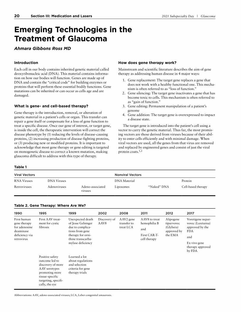
Table 1
Viral Vectors Nonviral Vectors
RNA Viruses DNA Viruses DNA Material Protein
Retroviruses Adenoviruses Adeno-associated viruses
Liposomes “Naked” DNA Cell-based therapy
Table 2. Gene Therapy: Where Are We?
1990 1995 1999 2002 2008 2011 2012 2017
First human gene therapy for adenosine deaminase deficiency via retrovirus
First AAV treat-ment for cystic fibrosis
Unexpected death of Jesse Gelsinger due to complica-tions from gene therapy for orni-thine transcarba-mylase deficiency
Discovery of AAV8
AAV2 gene transfer to treat LCA
AAV8 to treat hemophilia B
and
First CAR-T-cell therapy
Alipogene tiparvovec (Glybera) approved by the EMA
Voretigene nepar-vovec (Luxturna) approved by the FDA
and
Ex vivo gene therapy approved by FDA
Positive safety outcome led to discovery of more AAV serotypes promoting more tissue-specific targeting, specifi-cally, the eye
Learned a lot about regulations and selection criteria for gene therapy trials
Abbreviations: AAV, adeno-associated viruses; LCA, Leber congenital amaurosis.

2021 Subspecialty Day | Glaucoma Section III: Medication and Lasers 21
Where are we in the development of ocular gene therapies?
Adeno-associated viruses (AAV) are believed to be the future of gene therapy and have driven eye-related gene therapy treat-ment.3 As of 2019, there were 145 registered trials categorized on the basis of AAV capsid serotype. In these subsets of trials, over 25%-30% were designed for use in the eye. Most of these were identified as Phase 1 and 2 clinical trials.4
Two widely reported and concluded gene therapy trials for optic nerve disease are GenSight’s RESCUE and REVERSE trials. These trials are separate Phase 3 trials evaluating the efficacy of a single intravitreal injection of GS010 in patients that sustained vision loss due to the 11778 mutation in the ND4 gene.5 These trials have paved the way for many of trials for optic nerve disease, more specifically glaucoma, in the form of gene- and cell-based therapy.6
References 1. Wang D, Tai PWL, Gao G. Adeno-associated virus vector as a
platform for gene therapy delivery. Nat Rev Drug Discov. 2019; 18(5):358-378.
2. Yin H, Kanasty RL, Eltoukhy AA, Vegas AJ, Dorkin JR, Ander-son DG. Non-viral vectors for gene-based therapy. Nat Rev Genet. 2014; 15(8):541-555.
3. Al-Saikhan FI. The gene therapy revolution in ophthalmology. Saudi J Ophthalmol. 2013; 27(2):107-111.
4. Wang D, Tai PWL, Gao G. Adeno-associated virus vector as a platform for gene therapy delivery. Nat Rev Drug Discov. 2019; 18(5):358-378.
5. Newman NJ, Yu-Wai-Man P, Carelli V, et al. Efficacy and safety of intravitreal gene therapy for Leber hereditary optic neuropathy treated within 6 months of disease onset. Ophthalmology 2021; 128(5):649-660.
6. Information regarding active gene therapy trials can be found at the American Society of Gene + Cell Therapy’s Clinical Trials Finder: https://www.asgct.org/clinicaltrials.
Table 3
Trials
Eye-Specific Diagnosis
Location
Trial Summary
Phase and Trial Endpoints
Dual Intravitreal Implantation of NT-501 Encapsulated Cell Therapy for Glaucoma22
Glaucoma Stanford University To determine the safety and efficacy of dual NT-501 CNTF encapsulated cell therapy (ECT) on visual impair-ment related to glaucoma
Phase 2
Endpoints: visual fields, structure measurement of GC-IPL and RNFL
Study of NT-501 Encapsulated Cell Therapy for Glaucoma Neuroprotection and Vision Restoration
Glaucoma Stanford University To determine efficacy of NT-501 CNTF encapsulated cell therapy on visual impairment from glaucoma
Phase 2
Endpoints: visual fields, structure measurement of GC-IPL and RNFL
Safety Assessment of Intravit-real Mesenchymal Stem Cells for Acute Non-arteritic Ante-rior Ischemic Optic Neuropathy
Ischemic optic neuropathy
Instituto Universitario de Oftalmobiología
To evaluate the safety of cell therapy as a new treatment for patients who suffer from acute non-arteritic anterior ischemic optic neuropathy (NAION)
Phase 2
Absence of ocular inflammatory reaction, adverse events that are procedure-related
Efficacy Study of Gene Therapy for the Treatment of Acute Leber’s Hereditary Optic Neu-ropathy (LHON) onset within 3 months
Leber congenital optic neuropathy
To study the efficacy of rAAV2-ND4 (NADH dehydrogenase sub-unit 4) for the treatment of acute LHON onset within 3 months
Unassigned phase
BCVA and visual fields
RESCUE and REVERSE Long-term Follow-up
Leber congenital optic neuropathy
GenSight Biologics To assess the long-term and efficacy of GS010 and assess the quality of life in subjects with LHON due to the G11778A mitochondrial muta-tion in patients 5 years post treat-ment
Phase 3
Adverse events, BCVA, and HVF
A Single Intravitreal Injection of rAAV2-ND4 for the Treat-ment of Leber’s Hereditary Optic Neuropathy
Leber congenital optic neuropathy
Huazhong University of Science and Tech-nology
This study is meant to evaluate the safety and efficacy of rAAV2-ND4 treatment for Leber hereditary optic neuropathy with the G11778A mutation in mitochondrial DNA.
Phase 2/3
BCVA, VF, VEP, struc-ture measurement of RNFL
Safety Study of an Adeno- associated Virus Vector for Gene Therapy of Leber’s Hereditary Optic Neuropathy
Leber congenital optic neuropathy
National Eye Institute To study the potentially toxic effects of scAAV2-P1ND4v2 in patient with LHON and the G11778A mitochondrial gene mutation
Phase 1
Assessment of toxicity
Abbreviations: CNTF, ciliary neurotrophic factor; HVF, Humphrey visual field; VF, visual fields; VEP, visual evoked potentials.

22 In These Unprecedented Times . . . 2021 Subspecialty Day | Glaucoma
In These Unprecedented Times . . . 2021 Glaucoma Subspecialty DayDonald L Budenz MD MPH
The COVID-19 pandemic has impacted us in many ways, including our ability to effectively raise critical funds used to protect sight and empower lives. This objective requires active participation and commitment to advocacy from every ophthal-mologist. Contributions to the following three critical funds are a part of that commitment:
■ OPHTHPAC® ■ Surgical Scope Fund (SSF) ■ State Eye PAC
During AAO 2021 in New Orleans, invest in OPHTHPAC and Surgical Scope Fund at one of our two booths in the con-vention center or online. You may also invest via phone by tex-ting MDEYE to 41444 for OPHTHPAC and SCOPE to 51555 for the Surgical Scope Fund.
We also encourage you to stop by our booth in the Hall B Lobby to learn more about OPHTHPAC Direct, a unique pro-gram that lets you decide who receives your political support.
Please help us in these unprecedented times to continue to protect quality patient eye care for everybody. Two Academy committees made up of your ophthalmology colleagues are working hard on your behalf to ensure this outcome. The OPHTHPAC Committee continues to identify Congressional Advocates in each state to maintain close relationships with federal legislators to advance ophthalmology and patient causes. The Surgical Scope Fund Committee is raising funds to be used to protect Surgery by Surgeons during scope battles at the state level.
Our mission of “protecting sight and empowering lives” requires robust funding of both OPHTHPAC and the Surgical Scope Fund. Each of us has a responsibility to ensure that these funds are strong so that ophthalmology continues to strive, especially in these unprecedented times.
OPHTHPAC®
OPHTHPAC represents the profession of ophthalmology to the U.S. Congress. OPHTHPAC’s most recent victories include the following:
Physician Relief✓ Securing access to COVID-19 relief, including Provider
Relief Funds and forgivable small business loans✓ Pushing Congress to enact a provider-friendly “surprise”
medical billing law
Medicare Payment✓ Mitigating drastic Medicare cuts ✓ Obtaining a one-year moratorium extension on the 2%
Medicare budget sequestration cut
Research & Relationships✓ Increasing vision research funding by $11.6 million✓ Helping get three new physicians elected to Congress,
including an ophthalmologist
However, facing ophthalmology’s federal issues is a continu-ous battle, and OPHTHPAC is always under pressure to ensure we have strong political connections in place to help protect ophthalmology, its members, and their patients.
The support OPHTHPAC receives from invested U.S. Acad-emy members helps build the federal relationships that advance ophthalmology’s agenda on Capitol Hill. These relationships allow us to have a seat at the table with legislators willing to work on issues important to us and our patients. We also use these congressional relationships to help shape the rules and regulations being developed by federal health agencies.
Get engaged with OPHTHPAC and help strengthen oph-thalmology’s voice on Capitol Hill as we address the following legislative and regulatory issues this year:
■ Improving Medicare physician payments ■ Fighting optometric scope expansion in the Veterans’
Health Administration ■ Obtaining relief from prior authorization and step ther-
apy requirements that delay patient care ■ Seeking solutions for rising drug prices and access to
drugs in shortage ■ Ensuring fair reimbursements for Part B drugs
At the Academy’s annual Congressional Advocacy Day, the Academy and the American Glaucoma Society (AGS) ensure a strong presence of glaucoma specialists to support ophthal-mology’s priorities. AGS also supports participation of young ophthalmologists via the Academy’s Advocacy Ambassador Program. Ophthalmologists visit members of Congress and their key health staff to discuss ophthalmology priorities as part of Congressional Advocacy Day. The AGS remains a crucial partner with the Academy in its ongoing federal and state advo-cacy initiatives.
Surgical Scope Fund (SSF)
The Surgical Scope Fund (SSF) provides grants to state ophthal-mology societies to support their efforts to protect patient safety from dangerous optometric surgery proposals. Since its incep-tion, the Surgery by Surgeons campaign and the SSF, in partner-ship with state ophthalmology societies, has helped 41 state/territorial ophthalmology societies reject optometric scope-of-practice expansions into surgery.
If you already have made a SSF contribution, please go to safesurgerycoalition.org to see the impact of your gift.
Dollars from the SSF are critical to building complete, cutting-edge political campaigns, including media efforts (TV, radio, and social media), educating and building relationships with legislators, and educating the voting public to contact their legislators. These political campaigns help the SSF to protect patient safety by defeating optometry’s surgical initiatives.
Each of these endeavors is very expensive, and no one state has the critical resources to battle big optometry on their own. Ophthalmologists must join together and donate to the SSF and to fight for patient safety.

2021 Subspecialty Day | Glaucoma In These Unprecedented Times . . . 23
The Secretariat for State Affairs thanks the American Glau-coma Society, who has joined state ophthalmology societies in the past in contributing to the SSF, and looks forward to its 2021 contribution. These ophthalmic organizations complete the necessary SSF support structure for the protection of our patients’ sight.
State Eye PAC
It is increasingly important for all ophthalmologists to support their respective State Eye PACs because campaign contribu-tions to legislators at the state level must come from individual ophthalmologists and cannot come from the Academy, OPH-THPAC, or the Surgical Scope Fund. The presence of a strong State Eye PAC providing financial support for campaign con-tributions and legislative education to elect ophthalmology-friendly candidates to the state legislature is critical, as scope-of-practice battles and many regulatory issues are all fought on the state level.
ACTION REQUESTED: Support ophthalmology’s advocacy efforts
Academy Surgical Scope Fund contributions are used to sup-port the infrastructure necessary in state legislative/regulatory battles and for public education. State PAC and OPHTHPAC contributions are necessary at the state and federal level, respec-tively, to help elect officials who will support the interests of our patients. Contributions to each of these three funds are neces-sary and help us protect sight and empower lives. Surgical Scope Fund contributions are completely confidential and may be made with corporate checks or credit cards. PAC contributions may be subject to reporting requirements.
Please respond to your Academy colleagues and be part of the community that contributes to OPHTHPAC, the Surgical Scope Fund, and your State Eye PAC. Please be part of the com-munity that ensures ophthalmology has a strong voice in advo-cating for patients.
OPHTHPAC Committee
Jeffrey S Maltzman, MD (AZ)—ChairJanet A Betchkal, MD (FL)Mark J Gallardo MD (TX)Thomas A Graul MD (NE)Sohail J Hasan MD PhD (IL)S Anna Kao MD (GA)Julie S Lee MD (KY)Stephanie J Marioneaux MD (VA)Dorothy M Moore MD (DE)Stephen H Orr MD (OH)Niraj Patel MD (WA)Michelle K Rhee MD (NY)Linda Schumacher-Feero MD (ME)Frank A Scotti MD (CA)Jeffrianne S Young MD (IA)
Ex-Officio Members:Tamara R Fountain MD (IL)David B Glasser MD (MD)David W Parke II MD (CA)Michael X Repka MD MBA (MD)George A Williams MD (MI)
Surgical Scope Fund Committee
Lee A Snyder MD (MD)—ChairVineet (“Nick”) Batra MD (CA)Robert L Bergren MD (PA)Gareth M Lema MD PhD (NY) Darby D Miller MD MPH (FL)Amalia Miranda MD (OK)Christopher C Teng MD (CT)
Ex-Officio Members:John D Peters MD (NE) George A Williams MD (MI)
Surgical Scope Fund OPHTHPAC® State EyePAC
To protect patient safety by defeating opto-metric surgical scope-of-practice initiatives that threaten quality surgical care
Working across the political spectrum to advance ophthalmology and protect its mem-bers and patients at the federal level. Support for candidates for U.S. Congress.
Support for candidates for state House, Sen-ate, and governor
Political grassroots activities, government relations, PR and media campaigns
No funds may be used for campaign contribu-tions or PACs.
Campaign contributions, legislative education Campaign contributions, legislative education
Contributions: Unlimited.
Individual, practice, corporate, and organiza-tion
Contributions: Limited to $5,000
Personal and corporate contributions are accepted.
Contribution limits vary based on state regu-lations.
Contributions are 100% confidential. Contributions $200 and above are on the public record.
Contributions are on the public record depending upon state statutes.

24 The American Glaucoma Society Subspecialty Day Lecture 2021 Subspecialty Day | Glaucoma
“The bleb is dead; long live the bleb!”
Although the minimally invasive glaucoma surgeries (MIGS) dominate much of the conversation about glaucoma surgi-cal management, trabeculectomy surgery still reigns as king when striving for low IOPs. In addition, new “bleb-forming” MIGS are emerging as a tool to fill the glaucoma treatment gap between early and advanced glaucoma, and these surgeries still rely on the antifibrotic agent mitomycin C (MMC). Thus, it is still worthwhile for us to understand how the use of MMC evolved and continues to evolve and how it may affect bleb mor-phology in both traditional and novel glaucoma procedures.
The enemy of incisional glaucoma surgery success is the body’s own propensity to heal an open wound, and glaucoma surgeons have tried for decades to modify bleb morphology and function. The antifibrotic agent MMC has been used in trabeculectomy surgery since 1990, and the traditional method of intraoperative application is with gel-foam sponges. Other methods of MMC application include the use of amniotic mem-brane-soaked tissue and the use of scleral patches or contact lenses soaked in MMC. In the past few years, a newer method of MMC application, by injection, has become widespread among glaucoma surgeons, and many younger ophthalmolo-gists now train with this technique.
This lecture will review the evolution of wound-healing modulation and present current literature on the efficacy and safety of MMC injection. We will pay particular attention to how it may affect bleb morphology in trabeculectomy surgery as well as with the newer bleb-forming MIGS.
MMC and Trabeculectomy
The problem: In trabeculectomy surgery, the ideal bleb is dif-fuse, low profile, and minimally vascular. The scary bleb is one that is thin, cystic, avascular, and prone to leaks. The useless bleb is flat, scarred, and associated with poor IOP control. How do we create the ideal bleb?
A. Review of methods to alter wound healing in trab-eculectomy surgery
B. The goals of wound healing modulation are 3-fold:
1. Retard wound healing
2. Promote ideal bleb morphology
3. Improve IOP control
C. Trabeculectomy: a brief history of MMC use
D. Alternate methods of applying MMC during trab-eculectomy surgery
E. Review of MMC injection for trabeculectomy: goals and published literature1-4
1. Does it allow for better IOP control?
2. Does it promote more diffuse blebs?
3. Does it alter bleb vascularity?
4. Would we avoid focal, cystic blebs?
F. Advantages of MMC injection in comparison to sponge application
G. Preparation of MMC for injection
H. Safety of MMC injection for trabeculectomy sur-gery
MMC and MIGS
A. The bleb-forming MIGS5,6 in the context of other MIGS
B. MMC and clinical outcomes of bleb-forming MIGS
C. Is bleb-formation in a MIGS different than that of a trabeculectomy?
References 1. Lee E, Doyle E, Jenkins C. Trabeculectomy surgery augmented
with intra-Tenon injection of mitomycin C. Acta Ophthalmol. 2008; 86(8):866-870.
2. Pakravan M, Esfandiari H, Yazdani S, et al. Mitomycin C-augmented trabeculectomy: subtenon injection versus soaked sponges: a randomised clinical trial. Br J Ophthalmol. 2017; 101(9):1275-1280.
3. Quist MS, Brown N, Bicket AK, Herndon LW. The short-term effect of subtenon sponge application versus subtenon irrigation of mitomycin-C on the outcomes of trabeculectomy with Ex-PRESS glaucoma filtration device: a randomized trial. J Glaucoma. 2018; 27(2):148-156.
4. Lim MC, Hom B, Watnik MR, et al. A comparison of trabeculec-tomy surgery outcomes with mitomycin-C applied by intra-tenon injection versus sponge. Am J Ophthalmol. 2020; 216:243-256.
5. Grover DS, Flynn WJ, Bashford KP, et al. Performance and safety of a new ab interno gelatin stent in refractory glaucoma at 12 months. Am J Ophthalmol. 2017; 183:25-36.
6. Do AT, Parikh H, Panarelli JF. Subconjunctival microinvasive glaucoma surgeries: an update on the Xen gel stent and the Pre-serFlo MicroShunt. Current Opin Ophthalmol. 2020; 31(2):132-138.
The Use of Mitomycin C in Traditional and Novel Glaucoma SurgeriesMichele C Lim MD
2021 Subspecialty Day | Glaucoma Section IV: Glaucoma in the Digital Age 25
Home TonometryJeffrey R SooHoo MD
I. Why measure tonometry at home?
A. IOP is the only modifiable risk factor in glaucoma.
B. IOP is typically measured for a few seconds a few times a year during office hours.
C. We need to better understand IOP outside of office hours to understand the total range, variation, and peak IOP.
D. IOP is dynamic, with short-, intermediate-, and long-term fluctuations.
E. IOP fluctuations have been correlated with pro-gression in some studies (and not in others).
1. The Advanced Glaucoma Intervention Study (AGIS) and Collaborative Initial Glaucoma Treatment Study (CIGTS) found that fluctua-tion was associated with progression.
2. The Early Manifest Glaucoma Trial (EMGT) and Ocular Hypertension Treatment Study (OHTS) found no relationship between fluctua-tion and progression.
F. Other options for expanding IOP measurements, such as diurnal tension curves or 24-hour IOP monitoring, are time consuming and resource intensive.
II. Home Tonometry
A. iCare home rebound tonometer
1. IOP determined by deceleration and contact time
2. Lightweight probe that does not require corneal anesthesia
B. Studies have shown that use of iCare home is simi-lar to Goldmann applanation.
III. Ideal Patients
A. Pigmentary glaucoma, known fluctuations in IOP
B. Considering surgery to smooth out IOP fluctua-tions, identify IOP rhythm
C. Progressing despite in-office IOP at goal
IV. Practical Considerations
A. Billing: No current billable codes
B. Expensive: Could consider cash pay service (eg, rental fee per week)
1. Limits use to patients with adequate resources or enrolled in a study
2. Opportunity for philanthropy?
C. Not suitable for all patients; not everyone can be trained
D. No nocturnal data
E. Still intermittent and not continuous data
V. Current Practice at the University of Colorado
A. Clinical trials
B. Patient rental
26 Section IV: Glaucoma in the Digital Age 2021 Subspecialty Day | Glaucoma
Virtual Reality Visual FieldsYvonne Ou MD
I. Why monitor visual fields using mobile VR headsets?
A. Low cost
B. Portability
C. Built-in lighting environment
D. No need for highly skilled examiner
E. Potential for testing in a wide variety of contexts
F. Potential for home testing
G. Frequent testing may overcome intertest variability.
H. Advantages/disadvantages compared to other por-table strategies (eg, online testing, iPad, etc.)
II. Indications for VR Visual Fields
A. Glaucoma screening and diagnosis
B. Remote settings
C. Testing bedridden or disabled patients
D. Home testing
E. Glaucoma progression evaluation
F. Teleglaucoma
III. Variety of Platforms Incorporating Different Test Strategies
A. Subjective approaches requiring patient response; concordance with static automated perimetry
B. Objective approaches
IV. Virtual Reality Oculokinetic Perimetry (VR-OKP)
A. Experience with VR-OKP in the clinic setting
1. Test time
2. Test-retest variability
3. Correlation with static automated perimetry
4. Structure-function relationship
B. Real-world experience with VR-OKP in the home setting
1. Patient acceptance and remote training
2. Test-retest variability
3. Longitudinal testing at home
V. The Future of VR Visual Fields
A. Heightened value in the setting of global pandemic
B. Development of improved testing algorithms
C. Prospective studies to determine its ability in moni-toring glaucoma progression

2021 Subspecialty Day | Glaucoma Section IV: Glaucoma in the Digital Age 27
TeleglaucomaGlaucoma in the Digital AgeLama A Al-Aswad MD MPH
I. Introduction
COVID and the digital transformation of the glau-coma practice
II. The Future Glaucoma Practice
A. Practice structure and models
B. Devices and technology
C. Remote monitoring
III. Conclusion
28 Section IV: Glaucoma in the Digital Age 2021 Subspecialty Day | Glaucoma
App-Based Visual AidsApps for Low Vision 2021Terry Schwartz MD
Completing Assignments (note taking, scanning, completing worksheets)
Notability (iOS) ■ Take notes, annotate PDFs, complete worksheets,
markup photos, record lectures, provide audio feedback, and more
■ Take notes directly on PDF slides ■ Teachers email a worksheet, the student completes it and
then returns via email to the teacher. ■ Create folders/dividers to organize and save work.
OneNote (Microsoft, Android) ■ Free app like Notability
Audionote (iOS, Mac, Windows, and Android) ■ Notes linked to voice recordings (instant seek), automati-
cally adapts to room size and volume level. ■ Notes automatically highlight and scroll with audio play-
back. ■ Take notes directly on PDF slides ■ Notes can be organized by subject. ■ Can sync between devices (iCloud, Dropbox)
Joinme (iOS, Android) ■ Screen sharing/meeting app ■ Allows direct access to instruction on a white board or
teacher’s computer onto the student’s tablet ■ Presentation size can be enlarged ■ Screenshots can be kept as notes ■ School buys the “basic” version (about $10/month), and
the student downloads the app (free of charge).
Genius Scan (iOS, Android) ■ Scan single or batch documents with phone/tablet and
create a PDF ■ Take a photo; app automatically recognizes the paper
against the background, crops it, and cleans up the result. Upload PDF document to complete assignments.
Apple accessibility options ■ https://support.apple.com/accessibility ■ www.applevis.com
Chromebook ■ https://www.controlaltachieve.com/2016/10/special-needs
-extensions.html?m=1
Android accessibility options ■ https://support.google.com/accessibility/android/answer
/6006564?hl=en
Books
Bookshare (www.bookshare.org) ■ Huge online library (400,000 digital books) for people
with print disabilities ■ Confirmation of low vision/blindness, physical or learn-
ing disability by a professional (eg, MD, OD, teacher) is required.
■ Various formats are available (eg, text to speech, digital braille, enlarged fonts).
■ Free for U.S. students (funded by Office of Special Ed Programs, U.S. Dept of Ed). Nonstudents pay a nominal fee.
Bard Mobile (iOS, Android) ■ BARD app allows users to download and listen to books
and to read braille books using a Bluetooth-connected braille display.
■ Library includes books, magazines, music instruction, and music scores. New selections added daily.
■ Must enroll in the National Library Service for the Blind and Print Disabled (NLS) at the Library of Congress. To enroll, call 1-888-NLS-READ (1-888-657-7323) and follow the prompts for your state or request enrollment information at https://www.loc.gov/thatallmayread.
Voice Dream Reader and Scanner (iOS, Android) ■ Reading tool with dyslexia friendly font, text and audio
synchronization, customizable font size and color combi-nations, and full VoiceOver support.
■ Options include navigation by sentence, paragraph, page, and chapter. Can add bookmarks, notes, and highlights.
■ Many file formats are supported, including DAISY 3.0 text-only, DAISY 2.02 audio, DRM-free EPUB, PDF, Microsoft Word, Microsoft PowerPoint, HTML, and zipped MP3 files.
■ Integrated with Bookshare and Gutenberg ■ It can load files from Dropbox, Google Drive, or iTunes
via USB or Wi-Fi.
KNFB reader (iOS, Android) ■ Optical character recognition (OCR) reader for auditory
access of printed material
Accessibility: Narrating the Visual Environment (Read short text and cursive, currency, description of scenes, facial recognition, colors)
Seeing AI (iOS) ■ Free app that describes short text, documents, products,
people, currency scenery, colors, handwriting, and light levels
■ Scans barcodes (with the help of sounds to help focus) to describe a product.

2021 Subspecialty Day | Glaucoma Section IV: Glaucoma in the Digital Age 29
■ Description of people (eg, estimates person’s age, facial expression)
■ Available in multiple languages
Lookout by Google (Android) ■ Similar app to Seeing AI—same concept but limited ■ Can read barcodes, currency, objects, short text, docu-
ment scan, and OCR
TapTapSee (iOS) (Android) ■ Uses AI and human to interpret photographed objects/
scenes verbally ■ Double-tap the screen to photograph any 2- or 3-dimen-
sional object at any angle, and have it accurately identi-fied within seconds. VoiceOver then narrates the find-ings.
Dentifi (iOS) ■ Uses AI to recognize virtually any object, brand, color,
facial expression, handwriting, or text, and subsequently deliver an audible description of the image’s contents to the user.
BeSpecular (iOS) (Android) ■ A photo, taken by the user, is sent (along with a voice
message/query) to the BeSpecular community of sighted, who respond with a voice or text message.
■ Response time is within minutes.
ViaOpta Hello (iOS) (Android) (Windows) ■ Face recognition and environment description; identifies
people, items, and scenes ■ Available in 12 languages (English, German, French,
Spanish, Arabic, Japanese, Chinese, Greek, Portuguese, Dutch, Italian, and Hungarian).
Navilens (Android, iOS) ■ QR code reader and creator. Create QR codes for placing
on objects.
Be My Eyes (iOS) (Android) ■ Free app that connects blind and low vision people with
sighted volunteers and company representatives for visual assistance through a live video call
Cinema, TV
Audio description ■ This is a form of narration that supplements movies and
TV. Descriptions are spoken between dialog and perti-nent sound effects, providing content of on-screen action, facial expressions, and other relevant visual elements. Ide-ally, it can enable a totally blind person to understand the overall story as well as individual moments.
■ Netflix and Amazon include audio descriptions on all original programs, as does DirectTV. Every movie theater now has descriptive audio headsets.
Greta (iOS, Android) ■ Enables people with sight or hearing loss to experience
fully accessible cinema (including foreign language sub-titles and audio). It whispers audio descriptions or plays subtitles.
Navigation
Microsoft Soundscape (iOS) ■ A research project exploring the use of audio-based tech-
nology ■ As the user walks, it automatically calls out key points of
interest, including roads and intersections being passed. ■ An audio beacon can be placed on a point of interest that
the user would like to track (eg, the destination, a point to return to, a familiar landmark).
■ It is designed to live in the background and provide the user with ambient awareness and can be used in conjunc-tion with other apps.
BlindSquare (iOS) ■ A GPS app developed for the blind and visually impaired
that describes the environment and announces points of interest and street intersections during travel
Intersection Explorer (Android) ■ Speaks the layout of streets and intersections in neighbor-
hoods by touching and dragging finger around a map to create an understanding of an area before and during walking
Arianna Navigation (iOS, Android) ■ Navigation for indoor environments (eg, airports, muse-
ums, hospitals) and outdoors. High precision localiza-tion services against a simple and cheap infrastructure. Through special vibrational signals the user receives feed-back for correcting their direction.
Lazzus (iOS, Android) ■ Creates an auditory field of vision in real time, pointing
out pedestrian crossings, street intersections, stairs, and businesses
■ Sources Google Places and Open Street Map and searches within a 100-meter radius, highlighting information on things that are near to the user
■ Converts text to speech
30 Section IV: Glaucoma in the Digital Age 2021 Subspecialty Day | Glaucoma
Deep Learning/Artificial IntelligenceAnthony Khawaja MBBS
Introduction
Machine learning has been an established technique for many years, and research applying it to ophthalmology and glaucoma is not new. A PubMed search identifies papers on artificial intel-ligence (AI) and glaucoma from as early as 1985. However, it was the development of a type of machine learning called “deep learning” that provided a step change in the performance of AI algorithms. In particular, deep learning excels at image clas-sification, and it first outperformed humans in 2015. Early deep learning successes in medicine were for classification of skin lesion images as benign or malignant and classification of dia-betic retinopathy status from fundus photos. In last few years, the number of deep learning studies in ophthalmology and glaucoma has risen exponentially, opening up possibilities for improved and more efficient care of our patients.
How Might AI Transform Glaucoma Care?
There are many potential uses for AI across the spectrum of glaucoma management, from community to specialist care. Given the irreversible nature of glaucoma, early detection is important for preventing blindness, yet in most health systems, general population screening for glaucoma is not recommended. This is in part due to the poor performance of tests when applied to populations with a relatively low disease prevalence (too many false positives).
AI may help identify a high-risk subset of the general popula-tion who should be screened through automated image analysis, or help refine or triage referrals to reduce false positives and specialist service burden. AI may also help with specialist moni-toring of low-risk glaucoma patients. With the aging popula-tion, the number of glaucoma patients is growing rapidly and many are low-risk, diverting attention/resources from those who need it most. In the future, AI may in part automate or support follow-up decisions for low-risk patients and help risk stratify patients.
Highlights of AI Successes in Glaucoma
Given the large volume of recent high-quality glaucoma-related AI research, I will present a nonexhaustive selection that aims to highlight the breadth of progress. There has been notable success in AI algorithms that can accurately classify fundus/disc photos for “referrable glaucoma” (this term is used because a firm diagnosis cannot be made from photographs alone in the majority of cases). For example, an AI algorithm trained and validated by Google Health performed extremely well in independent datasets where the ground truth of “referrable glaucoma” was graded by multiple glaucoma experts. In fact, the AI algorithm outperformed any single glaucoma specialist, suggesting it can perform at a level approaching a consensus expert decision. Of course, this is an unfair contest, given that physicians usually have much more information before making a diagnosis, including history, fields, and other imaging.
There have also been successful demonstrations of classify-ing visual field and OCT data for glaucoma, though many of these studies are smaller and the validation was not done in a completely independent population. Notable studies include demonstration of superior diagnostic accuracy of an AI algo-rithm with an unsegmented circumpapillary OCT image as input compared to global segmented indices, and the ability to predict OCT indices from a standard fundus photo. Mul-timodal algorithms that incorporate multiple different forms of input data to create one improved classification output are emerging and may ultimately be a useful approach for diagnos-ing glaucoma. Studies have also been conducted to automati-cally classify angle closure from anterior segment OCTs.
Another class of machine learning is a generative adversarial network (GAN). GANs have been trained to predict spectral domain OCT images from time-domain OCT images, suggest-ing future possibilities to extract more meaningful information from noisy data that was not previously possible. A particularly striking finding in the ophthalmic AI literature is not directly glaucoma related: the ability to predict biological sex from just a fundus photograph with a very high degree of accuracy (AUROC 97%). The fact that humans collectively are unable to do this task opens up the possibility of AI algorithms detecting other human-imperceivable patterns that are glaucoma-relevant and can help us better manage our patients’ care.
Challenges in AI Research
There will be multiple challenges to translating AI research findings into deployable tools with proven clinical utility. It is extremely important that algorithms are trained on datasets that are as diverse as possible, so that the algorithm generalizes well and works in a variety of populations. For example, an algorithm largely trained on people of European descent may not work well in non-Europeans, limiting its equitable impact.
Another issue facing the ophthalmic research community is a lack of open standards among imaging device manufacturers, limiting the large-scale analysis required to train AI algorithms. The American Academy of Ophthalmology has recently pub-lished a statement, supported by other organizations such as the UK Royal College of Ophthalmologists, calling for device manufactures to become compliant with Digital Imaging and Communications in Medicine standards.
Selected Readings 1. Mursch-Edlmayr AS, Ng WS, Diniz-Filho A, et al. Artificial intel-
ligence algorithms to diagnose glaucoma and detect glaucoma progression: translation to clinical practice. Transl Vis Sci Tech-nol. 2020; 9:1-21.
2. Girard MJA, Schmetterer L. Artificial intelligence and deep learn-ing in glaucoma: current state and future prospects. Prog Brain Res. 2020; 257:37-64.

2021 Subspecialty Day | Glaucoma Section IV: Glaucoma in the Digital Age 31
3. Li JPO, Liu H, Ting DSJ, et al. Digital technology, tele-medicine and artificial intelligence in ophthalmology: a global perspective. Prog Retin Eye Res. 2021; 82:100900.
4. Thompson AC, Jammal AA, Medeiros FA. A review of deep learning for screening, diagnosis, and detection of glaucoma pro-gression. Transl Vis Sci Technol. 2020; 9:1-19.
5. Phene S, Dunn RC, Hammel N, et al. Deep learning and glau-coma specialists: the relative importance of optic disc features to predict glaucoma referral in fundus photographs. Ophthalmology 2019; 126:1627-1639.
6. Mayro EL, Wang M, Elze T, Pasquale LR. The impact of artificial intelligence in the diagnosis and management of glaucoma. Eye (Lond). 2020; 34:1-11.
7. Lee AY, Campbell JP, Hwang TS, Lum F, Chew EY; for the American Academy of Ophthalmology. Recommendations for standardization of images in ophthalmology. Ophthalmology. Epub ahead of print 2021 April 5. doi: https://doi.org/10.1016/j .ophtha.2021.03.003.

32 Section V: Journal Club/Late Breaking 2021 Subspecialty Day | Glaucoma
Case PresentationJames C Liu MD
NOTES

2021 Subspecialty Day | Glaucoma Section V: Journal Club/Late Breaking 33
Clinical Trial Update for Bimatoprost ImplantFelipe A Medeiros MD
I. Background on the Bimatoprost Implant
A. The bimatoprost implant is a biodegradable implant that is administered intracamerally with a single-use applicator and releases bimatoprost at a steady rate for 3-4 months.
B. The implant dose strength containing 10 µg bimatoprost is approved by the USFDA for single administration to lower IOP in patients with open-angle glaucoma (OAG) and ocular hypertension (OHT).
C. In vitro assays, aqueous samples taken when the implant was removed from human eyes, and pharmacokinetics studies using dogs predict drug release and intraocular drug bioavailability for 3-4 months after implant administration.1
D. Drug distribution studies in dogs have shown that the implant provides 4400-fold higher concentra-tions of bimatoprost in outflow tissues compared with topical dosing, while minimizing ocular sur-face and periocular tissue drug exposure.2
II. Two identically designed, 20-month, Phase 3 stud-ies compared 10- and 15-µg bimatoprost implants, administered on Day 1, Week 16, and Week 32, to twice-daily timolol eye drops in patients with OAG and OHT.
A. Results of the ARTEMIS 1 study have been reported:1
1. Bimatoprost implant was noninferior to topical timolol in IOP lowering over 12 weeks.
2. Efficacy of the implant was demonstrated in patients regardless of prior treatment with a topical prostaglandin analog or selective laser trabeculoplasty (SLT).
3. An extended duration of IOP-lowering effect was observed in the majority of patients after 3 implant administrations, and most patients required no additional (rescue) IOP-lowering treatment for 1 year after the third administra-tion.
B. The ARTEMIS 2 study was completed more recently; the results were consistent with and cor-roborated those of the ARTEMIS 1 study.
C. In Cox regression analysis to identify factors poten-tially associated with a longer-term IOP response in the pooled ARTEMIS studies, baseline IOP ≤25 mmHg, IOP lowering of >5 mmHg from baseline at the last measurement before the third adminis-tration, no history of SLT, female gender, and pha-kic lens status were significantly associated with a
longer time to rescue after the third administration of the 10-µg bimatoprost implant.3
III. A 24-month ongoing extension study enrolled OAG/OHT patients after their completion of a bimatoprost implant Phase 3 trial.
A. An interim analysis using all data available as of February 2021 evaluated IOP and use of added (rescue) IOP-lowering treatment for patients who were treated with 10- or 15-µg bimatoprost implant in ARTEMIS 1 or 2.
1. Of 200 patients enrolled in the extension study, 69 were unrescued at screening, 54 remained unrescued for ≥2 years, and 18 remained unres-cued for ≥3 years after their last bimatoprost implant administration.
2. The mean IOP in these patients remained con-trolled for 2 and 3 years after the last implant administration (17.3 and 16.6 mmHg, respec-tively).
B. These results suggested that some patients can maintain controlled IOP without additional treat-ment for up to 2-3 years after their last bimatoprost implant administration in a Phase 3 trial.
IV. The proposed mechanism for the long duration of response is matrix metalloproteinase (MMP)-medi-ated durable remodeling of aqueous outflow path-ways.4
A. The mechanism of IOP lowering with prostaglan-din analogs such as bimatoprost involves upregula-tion of MMPs and MMP-mediated turnover of the extracellular matrix, leading to decreased resis-tance to aqueous outflow.
B. Studies using cell cultures have shown that the upregulation of MMPs by bimatoprost is concen-tration dependent.
C. The higher concentrations of bimatoprost produced in target tissues by the implant are proposed to cause enhanced upregulation of MMPs, resulting in more durable tissue remodeling and sustained IOP lowering.
V. In safety evaluations in the ARTEMIS studies, the bimatoprost implant was usually well tolerated; how-ever, corneal adverse events (AEs), mostly corneal endothelial cell loss (CECL) and edema, were reported more frequently in study eyes treated with the implant than in study eyes treated with topical timolol.
A. The corneal AEs generally occurred after repeated administration and were more common with the 15-µg implant, which is 50% larger than the 10-µg implant.

34 Section V: Journal Club/Late Breaking 2021 Subspecialty Day | Glaucoma
B. The rate of implant biodegradation is slow, and with the 4-month fixed interval administra-tion used in the ARTEMIS studies, as many as 3 implants were present in the angle at the same time. Clinically significant implant biodegradation (to ≤25% of the initial size) was observed in the major-ity of patients by 12 months after administration.5
C. The corneal AEs are believed to result from physi-cal interaction between the implant material and the cornea, and so are more likely to occur when multiple implants that have not yet biodegraded to ≤25% of their initial size are present.
VI. A single administration of 10-µg implant is FDA approved because no eyes in the ARTEMIS studies or in a previous Phase 1/2 study6 had ≥20% CECL after a single administration.
VII. Ongoing studies are evaluating the safety and efficacy of as-needed administration of the implant with longer administration intervals.
References 1. Medeiros FA, Walters TR, Kolko M, et al. Phase 3, randomized,
20-month study of bimatoprost implant in open-angle glaucoma and ocular hypertension (ARTEMIS 1). Ophthalmology 2020; 127:1627-1641.
2. Seal JR, Robinson MR, Burke J, Benjanian M, Coote M, Atar M. Intracameral sustained-release bimatoprost implant delivers bima-toprost to target tissues with reduced drug exposure to off-target tissues. J Ocul Pharmacol Ther. 2019; 35:50-57.
3. Khouri AS, et al. Predictive factors for long-term IOP lowering after bimatoprost implant 10 µg administration in Phase 3 ARTE-MIS studies. Presented at the American Academy of Ophthalmol-ogy (AAO) 2020 Virtual Congress; November 13-15, 2020.
4. Weinreb RN, Robinson MR, Dibas M, Stamer WD. Matrix metalloproteinases and glaucoma treatment. J Ocul Pharmacol Ther. 2020; 36:208-228.
5. Weinreb RN, et al. Bimatoprost implant biodegradation in the phase 3 ARTEMIS studies. Presented at the American Glaucoma Society (AGS) 2021 Virtual Congress; March 4-7, 2021.
6. Craven ER, Walters T, Christie WC, et al. 24-month phase I/II clinical trial of bimatoprost sustained-release implant (Bimato-prost SR) in glaucoma patients. Drugs 2020;80:167-179.

2021 Subspecialty Day | Glaucoma Section V: Journal Club/Late Breaking 35
Potential Scientific Basis for Sustained Response to ImplantDouglas J Rhee MD
NOTES

36 Section V: Journal Club/Late Breaking 2021 Subspecialty Day | Glaucoma
Synthesizing the Clinical and Basic Science Information for This PatientQi N Cui MD
Summary: This is the last in a series of talks centered on the bimatoprost implant. It will closely tie-in with the data pre-sented earlier in the same section. Briefly, the presentation will synthesize available scientific and clinical trials data on the bimatoprost intraocular implant to arrive at an informed treat-ment decision for the patient in the case presentation.
I. Summary of Scientific Mechanisms-of-Action Relevant to the Case Presentation
II. Summary of Clinical Trials Data Relevant to the Case Presentation
III. Patient Factors Relevant to Treatment
IV. Recommended Treatment Course
V. Discussion of Pitfalls and Alternatives

2021 Subspecialty Day | Glaucoma Section V: Journal Club/Late Breaking 37
Best of AGS: Surgery in the Advanced Angle-Closure PatientSarah H Van Tassel MD
We all have patients in our practices with chronic angle closure and severe structural and functional loss. The decision about appropriate treatment may differ, depending on whether the patient is phakic with cataract, phakic without cataract, or already pseudophakic.
I. Phakic With Cataract
A. Cataract surgery in the angle-closure patient: In eyes with visually significant cataract and chronic angle-closure glaucoma, the decision to pursue cat-aract surgery is easy, but these cases can be among the toughest. Prepare for challenges:
1. Posterior synechiae: Lift gently rather than sweeping to avoid tearing iris root.
2. Posterior pressure: Pretreatment with acet-azolamide, osmotics, or a Honan balloon can decompress the vitreous and choroid and lessen the posterior pressure.
3. Malyugin ring, iris hooks, or similar for small pupils; keep the iris away from the wounds.
4. Avoid iris prolapse: Longer incision, gentle hydrodissection
5. Avoid chamber shallowing with the help of con-tinuous irrigation and viscoelastic devices.
6. Look out for zonular issues, which are common in angle closure.
7. Review axial length: Some angle-closure eyes are microphthalmic and nanophthalmic, and in a study of such eyes, shorter axial length and IOP 22 or higher were independent risk factors for complications, with the odds of complica-tions rising quickly as axial length shortened.1
B. Real-world IOP lowering from cataract extrac-tion (CE) IOL: Cataract surgery alone can have a substantial IOP-lowering effect. For example, Wang and colleagues2 extracted data from EHRs and demonstrated that narrow- and closed-angle eyes experienced a mean decrease in IOP of about 2 mmHg. However, we’re often targeting a far greater IOP reduction from surgery, and the ques-tion arises, “What else should I do at the time of cataract surgery?”
C. What else should I do at the time of cataract sur-gery?
1. Synechialysis: Mixed results in the literature; synechialysis has been mostly eclipsed by enthu-siasm for minimally invasive glaucoma surgery (MIGS) and can be combined with MIGS.
2. MIGS: Histopathologic work has demonstrated that persistent trabecular-iris contact plays a causal role in the progressive process of Sch-lemm canal endothelial damage, Schlemm canal occlusion, and trabecular cell damage and that gonioscopic evaluation of the extent of periph-eral anterior synechiae may not truly reflect the extent of trabecular damage. Therefore, it’s very intellectually satisfying to pursue excision or cleaving of the trabecular meshwork or dilation of Schlemm canal as strategies targeted at this diseased tissue. While these advanced angle-closure eyes can be exquisitely sensitive to angle surgery, there is little high-quality evidence to demonstrate whether this approach is superior to cataract surgery alone.
3. Endoscopic cyclophotocoagulation (ECP): Tar-gets inflow rather than outflow
4. Traditional surgery: Combining incisional glaucoma surgery with cataract extraction in advanced angle-closure eyes is also an option. Studies investigating this pathway have shown greater angle opening and anterior chamber deepening in cataract surgery alone compared with combined cataract and trabeculectomy.3 Additionally, cataract surgery combined with trabeculectomy has been shown to have more postoperative complications and mixed results as to whether there is a clear IOP or medication advantage.4,5 While I am generally a proponent of combined cataract and glaucoma surgery when appropriate, I find that angle-closure glau-coma is often an exception. These eyes can be exquisitely sensitive to cataract surgery/MIGS even in advanced disease.
5. Important considerations include your access to, comfort with, and results from these surgeries. With ECP in particular, give consideration to the patient’s risk of postoperative inflamma-tion. Consider combining surgical approaches. In some of these eyes, you’ll end up returning to the OR for incisional surgery and discuss-ing that possibility with the patient, as well as understanding that the ease of that in your envi-ronment is important. Finally, more research is needed to enhance our knowledge in this area to determine a more accurate prediction formula for the surgical outcome rather than relying on a best guess.

38 Section V: Journal Club/Late Breaking 2021 Subspecialty Day | Glaucoma
II. Phakic, No Cataract
Controversy arises when the lens is relatively clear without visually significant cataract. Important con-siderations in these eyes include noting whether the patient still accommodates, which may motivate one to leave them phakic. Deciding whether to perform a tube or a trab can be challenging because studies like the Primary Tube vs. Trabeculectomy study excluded eyes with narrow angles.
If you opt for a tube, ensure there is space in the ante-rior chamber for the tube to be distanced from the endothelium. Paying attention to the lens thickness and vault in a particular patient may provide insight into the role of the lens in that person’s disease and further guide decision making. In patients with com-bined exfoliation and angle-closure glaucoma, I have a strong preference for early cataract extraction because the zonulopathy can be tremendously challenging in the dense cataracts that often follow incisional sur-gery.
III. Already Pseudophakic
A. All of the options discussed thus far remain viable, and I typically move forward with incisional sur-gery in the setting of the advanced disease eye.
B. Tube considerations: We now have high-quality evidence demonstrating that sulcus tube placement is better for the corneal endothelium than anterior chamber placement. Sulcus placement in angle clo-sure is also nice because it prevents you from get-ting tangled in high peripheral anterior synechiae as you attempt optimal tube placement.
IV. Summary
The best surgical strategy for advanced angle-closure glaucoma is not clearly defined. If appropriate, remove cataract. Consider nonincisional surgery (ie, ECP, MIGS, synechialysis) options for lowering IOP at time of CE IOL, even in advanced cases. Proceed with incisional surgery as indicated. Consider sulcus tube placement.
References 1. Day AC, MacLaren RE, Bunce C, Stevens JD, Foster PJ. Out-
comes of phacoemulsification and intraocular lens implantation of microphthalmos and nanophthalmos. J Cataract Refract Surg. 2013; 39(1):87-96.
2. Wang SY, Azad AD, Lin SC, Hernandez-Boussard T, Pershing S. Intraocular pressure changes after cataract surgery in patients with and without glaucoma: an informatics-based approach. Ophthalmol Glaucoma. 2020; 3(5):343-349.
3. Tham CCY, Leung DYL, Kwong YYY, et al. Effects of phaco-emulsification versus combined phaco-trabeculectomy on drain-age angle status in primary angle closure glaucoma (PACG). J Glaucoma. 2010; 19(2):119-123.
4. Hansapinyo L, Choyt BNK, Lai JSM, Tham CC. Phacoemulsifi-cation versus phacotrabeculectomy in primary angle-closure glau-coma with cataract: long-term clinical outcomes. J Glaucoma. 2020; 29(1):15-23.
5. El Sayed YM, Elhusseiny AM, Albalkini AS, El Sheikh RH, Osman MA. Mitomycin C-augmented phacotrabeculectomy versus phacoemulsification in primary angle-closure glaucoma: a randomized controlled study. J Glaucoma. 2019; 28(10):911-915.

2021 Subspecialty Day | Glaucoma Section V: Journal Club/Late Breaking 39
Best of AGS: Surgery in the Advanced Uveitic Glaucoma PatientKeith Barton MBBCh
Intraocular pressure (IOP) elevation is a common complication of uveitis, affecting roughly 20% of uveitics. IOP elevation may result from any or several of the following mechanisms: inflam-mation, corticosteroid treatment, chronic synechial angle clo-sure, pupil block from a secluded pupil, or forward movement of the lens-iris diaphragm. The IOP in uveitis may swing widely, from extremely high to very low levels, as a result of reduced aqueous production by a compromised ciliary body in the presence of outflow resistance that is elevated as a result of the above mechanisms. In general, the role of the glaucoma special-ist is to take IOP out of the equation in order to permit the uve-itis specialist to treat the patient’s inflammatory disease to an appropriate degree to prevent further inflammatory damage to the eye without risk of IOP elevation from corticosteroid usage.
While there are many surgical options for the control of IOP—most of which may not be specifically licensed for use in patients with uveitis but nevertheless may have some util-ity in patients with uveitis—patients with advanced uveitic glaucoma, like advanced glaucoma of any type, require defini-tive IOP control as the risk of severe visual loss is high, should any progression of optic neuropathy occur. For that reason, many of the newer procedures, the IOP-lowering benefits of which are largely modest, are not appropriate for patients with advanced uveitic glaucoma, and it is usually necessary to fall back on the more definitive conventional procedures, such as trabeculectomy with mitomycin C and aqueous shunt implants.
Basic Points
I. Control inflammation in preference to withholding steroids to minimize IOP elevation.
II. Identify the patient with advanced glaucoma who is at risk of severe visual loss.
A. Advanced field loss
B. Paracentral field loss
III. Identify mechanism(s) of IOP elevation.
A. Corticosteroid response
B. Inflammation
C. Angle closure
IV. If angle closure, identify type.
A. Pupil block vs.
B. Chronic peripheral anterior synechial closure vs.
C. Forward movement of the lens-iris diaphragm
Also look for other angle abnormalities, such as neo-vascularization and inflammatory nodules.
V. Management
A. Correct pupil block if present.
1. Uveitic pupil block is fundamentally differ-ent from pupil block in primary angle closure (PACG).
a. Absolute (uveitis) vs. relative (PACG)
b. Iris reversibly adherent to trabecular mesh-work due to fibrin and sticky aqueous (uveitis) vs. iris falls away from trabecular meshwork immediately on laser iridotomy (PACG)
2. Laser iridotomy is often ineffective at reversing pupil block in uveitis due to:
a. Loculation of aqueous behind iris, thick iris with adhesions to lens
b. Even if pupil block is relieved by iridotomy, iris remains adherent to trabecular mesh-work and even peripheral cornea.
B. Chronic synechial closure is not specifically treated, as angle surgery is ineffective.
1. Patients are treated similarly to those with sec-ondary open-angle glaucoma.
2. Chronic synechial closure is a predictor of a long-term IOP problem that is unlikely to resolve on steroid withdrawal.
C. Identify the likely IOP level required: In the Advanced Glaucoma Intervention Study, a mean IOP of 12 mmHg resulted in a 13% risk of progres-sion over 8 years.
VI. Surgical Management of IOP Elevation in Association With Uveitis
A. When to consider surgery?
1. Acute angle closure with pupil block and a secluded pupil: Surgical iridectomy with visco-goniosynechiolysis
2. Acute angle closure from forward movement of the lens-iris diaphragm: Treat the cause—poste-rior scleritis, Vogt-Koyanagi-Harada syndrome, iatrogenic after retinal surgery, aqueous misdi-rection, phacomorphic.
3. Uncontrolled chronic secondary synechial angle closure: In advanced glaucoma, trabeculectomy vs. aqueous shunt
4. Uncontrolled secondary open-angle glaucoma: In advanced glaucoma, trabeculectomy vs. aqueous shunt
B. The role of MIGS/angle surgery in advanced uveitic glaucoma

40 Section V: Journal Club/Late Breaking 2021 Subspecialty Day | Glaucoma
First System for Robot-Assisted Ab-Interno Gonio-Intervention: From Stent Implantation to Trabeculotomy Tsontcho Ianchulev MD
NOTES

2021 Subspecialty Day | Glaucoma Section V: Journal Club/Late Breaking 41
Status of the CMS Bundling for MIGS and How We Got There Leon W Herndon Jr MD
NOTES
42 Section VI: Lens and Glaucoma 2021 Subspecialty Day | Glaucoma
Lens-Related Glaucoma and Considerations for Surgical ManagementGlaucoma and the Native LensThomas W Samuelson MD
I. Introduction
The native lens plays a pivotal role in the pathogenesis and management of many adult-onset glaucomas. While the importance of the lens has been long recog-nized in many forms of glaucoma—including phaco-morphic narrow- and closed-angle glaucoma; pupil-lary block glaucomas, including microspherophakia and ectopia lentis; and various lens-related secondary glaucomas, such as phacolytic and phacoantigenic glaucoma—there is a growing body of empiric evi-dence that the native lens may play an important role in adult-onset open-angle glaucoma. While the role of the lens is especially important in the surgical manage-ment of phakic glaucoma, there is growing circum-stantial evidence that the native lens may have impor-tant implications in the pathogenesis of some forms of primary open-angle glaucoma (POAG).
II. Lens-Related Glaucoma
A. Pupillary block
1. Primary phacomorphic narrow-angle glaucoma
2. Microspherophakia
3. Ectopia lentis
B. Lens-related secondary glaucomas
1. Associated with hypermature or Morgagnian cataract with leakage of lens proteins through an intact capsule. Generally associated with an open angle and variable inflammatory anterior chamber reaction. Treatment includes anti-inflammatory medications, IOP-lowering medi-cations, and ultimately surgical removal of the native lens.
2. Lens particle glaucoma
Similar to phacolytic glaucoma in presentation and management, but an important differentiat-ing feature is a disrupted lens capsule, which makes surgical intervention more complex. Identifiable lens material is often visible in the anterior chamber.
3. Phacoantigenic (aka phacoanaphylactic)
Not a true allergic reaction to lens material but rather an immune-mediated inflammatory response to residual lens material. Definitive diagnosis is made with aqueous aspirate and identification of polymorphonuclear leukocytes, but this is rarely needed as removal of the resid-ual lens material is generally the recommended treatment.
C. POAG?
A provocative question to consider . . . Does phaco-morphic open-angle glaucoma exist? The evidence is mostly circumstantial. Consider that POAG and cataract often coexist, the incidence of each condi-tion being more common with aging.
In addition, there is extensive level-one evidence that cataract removal lowers IOP when the base-line IOP is higher than physiological and that this reduction is sustained for at least 4-5 years. Thus, if cataract removal lowers IOP, one must consider that the reverse may be true as well. Specifically, that the native lens may play a causal role in the elevation of IOP in some individuals. The native lens thickness increases with normal aging, and the anteroposterior diameter is directly related to age. In one study, the thickness of the cataractous lens was directly proportional to age, with a mean of 3.78 (±0.21) mm in the 3rd decade and a mean of 5.03 (±0.46) mm in the 10th decade. It is quite plau-sible that the increased anteroposterior diameter of the native lens results in forward rotation of the cili-ary muscle-scleral spur complex, resulting in rela-tive compression of the trabecular meshwork and reduction of conventional outflow, even if the angle remains open. A typical IOL is 1 mm in thickness. Accordingly, cataract removal with placement of an IOL widens the iridocorneal angle, which may favorably influence physiologic outflow by relaxing lens-induced compression of the trabecular mesh-work.
III. Management of Coincident Cataract and Glaucoma
A. Phacoemulsification alone
B. Combined phacoemulsification and canal-based glaucoma surgery
1. Trabecular microbypass with device
2. Incisional goniotomy
3. Excisional goniotomy
4. Viscodilation of Schlemm canal
C. Combined phacoemulsification and transscleral (bleb forming) surgery
1. Gel stenting surgery
2. Traditional trabeculectomy
3. Traditional aqueous drainage devices

2021 Subspecialty Day | Glaucoma Section VI: Lens and Glaucoma 43
D. Combined phacoemulsification and cyclophotoco-agulation (CPC)
1. Transscleral diode CPC
2. Transscleral micropulse CPC
3. Endoscopic cyclophotocoagulation
Selected Readings 1. Mansberger SL. Change in IOP and medications in the treatment
group of the Ocular Hypertension Treatment Study. American Glaucoma Society Virtual Annual Meeting; Mar. 4-7, 2021.
2. Hoffer KJ. Axial dimension of the human cataractous lens. Arch Ophthalmol. 1993; 111(7):914-918.
3. Samuelson TW, Chang DF, Marquis R, et al.; HORIZON Inves-tigators. A Schlemm canal microstent for intraocular pressure reduction in primary open-angle glaucoma and cataract: the HORIZON study. Ophthalmology 2019; 126(1):29-37.
4. Ahmed IK. 5-year results from the Horizon Trial: long term safety and effectiveness of combined phacoemulsification and minimally invasive glaucoma surgery with the Hydrus microstent. American Glaucoma Society Virtual Annual Meeting; Mar. 4-7, 2021.
44 Section VI: Lens and Glaucoma 2021 Subspecialty Day | Glaucoma
Aphakic GlaucomaDavid S Walton MD
NOTES

2021 Subspecialty Day | Glaucoma Section VI: Lens and Glaucoma 45
Uveitis-Glaucoma-Hyphema SyndromeIqbal K Ahmed MD
NOTES

46 Section VI: Lens and Glaucoma 2021 Subspecialty Day | Glaucoma
Premium and Toric Lenses in Glaucoma PatientsJohn P Berdahl MD
I. Understanding Patient Visual Goals
II. Understanding IOL Options
A. Presbyopic
B. Toric
C. Light-adjustable lens
III. Special Considerations for Glaucoma Patients
A. Contrast sensitivity
B. Ocular surface disease
C. Zonular stability
D. Progression of glaucoma
IV. Summary
2021 Subspecialty Day | Glaucoma Section VI: Lens and Glaucoma 47
Fix It With an Anterior Chamber IOLAC-IOLs in Cases That Lack Capsular SupportKendall Donaldson MD
I. Indications: Choosing the Right Patient and Situation
A. Age
B. Comorbid conditions
C. Situation
1. Planned
2. Unplanned
II. Recommended Preoperative Testing for Planned AC-IOL
A. Specular microscopy
B. Measurement of AC depth: Pentacam, ultrasound, or anterior segment OCT
III. Technique
A. IOL calculations
B. Surgical technique
IV. Postoperative Course and Recommended Follow-up
A. Drops
B. Specular microscopy or pachymetry and macular OCT
V. Outcomes: The Literature
A. Visual outcomes
B. Complications
C. Comparisons with other options for lack of capsu-lar support

48 Section VI: Lens and Glaucoma 2021 Subspecialty Day | Glaucoma
Fix It With an Iris-Sutured LensGarry P Condon MD
Simplified Scleral Fixation for Late In-Bag IOL Subluxation
The vast majority of these cases can be managed with scleral fixation of the bag/IOL complex regardless of whether or not a capsular tension ring (CTR) is present. There are various well-described techniques for placing 2 scleral lasso sutures 180 degrees apart that incorporate needle passage through the cap-sular bag. Most are ab-externo and require only microincisions that minimize intraoperative risks. In my experience, with even the most dramatic subluxation or dislocation, the existing IOL can be retained while avoiding more invasive IOL exchange.1 Microforceps and small-gauge vitrectomy instrumentation make this all the more possible.
I have found, however, that even with a CTR in the bag, it is easier and more secure to pass the suture through the bag between the optic and the haptic close to what I call the IOL “armpit.” The bag is often thin and fragile more peripher-ally, risking tearing along the ring when the suture is barely tensioned. The capsule is generally more robust centrally, and a square-edge haptic fibrosed in the bag affords great support for the suture near this haptic/IOL junction. Rotating the bag/IOL complex to the favored orientation is fairly easy in these pseudoexfoliation cases with minimal residual intact zonule. A poorly dilating pupil makes the more central portion of the bag/IOL complex easier to visualize and work on, as opposed to the more peripheral ring.
Rhexis-to-Iris Fixation for Late In-Bag IOL Subluxation
While a number of techniques have been described utilizing a lasso technique to fixate an in-the-bag IOL to the sclera,2-4 I suggest considering a modified McCannel suture to fix the superior capsulorrhexis margin to the posterior aspect of the superior iris. Using only a clear corneal approach, this simple form of fixation can be effective for managing early IOL sub-luxation associated with exfoliation.5
Preoperative tropicamide 1% typically produces a mid-dilated pupil in these pseudoexfoliation patients. Under topical tetracaine and intracameral nonpreserved lidocaine, paracente-sis tracks are made in clear cornea at about the 10:00 and 2:00 meridians. A cohesive viscoelastic is injected into the anterior chamber and behind upper iris to stabilize the anterior cham-ber, create space behind the superior iris, and tamponade the vitreous face where the zonule is likely deficient. The fibrotic and phimotic capsulorrhexis edge is key in applying this tech-nique. A small-gauge microforceps is placed through the nasal paracentesis. The edge of the capsulorrhexis is grasped while
a 10-0 polypropylene suture on a long curved needle is guided through the temporal paracentesis and directed down through midperipheral iris a clock hour or two away from the 12:00 meridian. Once the iris is pierced, the tip of the needle is rotated and passed through the capsular bag just underneath the edge of the capsulorrhexis while the bag is stabilized with the micro-forceps (Figure 1). The capsulorrhexis edge is then released from the forceps, and the needle is turned back superiorly so the tip of the needle passes over the same edge of the capsulorrhexis (Figure 2) and is then directed up through mid-peripheral iris 2 or 3 clock hours nasal to the first iris entry point. Once the needle passes up through this location in iris, it is pushed out through clear cornea (Figure 3). The result is a suture pass that extends through superotemporal clear cornea, down through mid-peripheral iris, through capsular bag beneath the fibrotic capsulorrhexis edge, back around and over the capsulorrhexis edge, up through superonasal midperipheral iris, and out through clear cornea.
A sliding Siepser knot technique is used to gently secure the edge of the capsulorrhexis to the posterior aspect of the iris. The sliding knot is initiated by retrieving a loop of the distal suture segment and bringing it out through the superotemporal entry paracentesis. This suture typically produces slight peaking of the pupillary margin (Figure 4). The viscoelastic is gently irri-gated from the chamber. In these cases, I recommend a small peripheral iridectomy to avoid postoperative pupillary block.
Figure 1. The needle is passed through cornea, mid iris, and the edge of the capsulorrhexis, which is held with a microforceps.

2021 Subspecialty Day | Glaucoma Section VI: Lens and Glaucoma 49
Figure 2. Once through the edge of the capsulorrhexis, the needle is rotated back superiorly under the nasal iris.
Figure 3. Passing the needle up through the nasal iris and out through adjacent cornea completes the suture pass.
Figure 4. The finished suture fixes the capsulorrhexis edge to the poste-rior surface of the iris.
References 1. Kirk TQ, Condon GP. Simplified ab externo scleral fixation for
late in-the-bag intraocular lens dislocation. J Cataract Refract Surg. 2012; 38:1711-1715 (erratum, 2013; 39:489).
2. Chan C, Crandall A, Ahmed IK. Ab externo scleral suture loop fixation for posterior chamber intraocular lens decentration: clini-cal results. J Cataract Refract Surg. 2006; 32:121.
3. Hoffman RS, Fine IH, Packer M. Scleral fixation without con-junctival dissection. J Cataract Refract Surg. 2006; 32:1907-1912.
4. Kirk TQ, Condon GP. Simplified ab externo scleral fixation for late in-the-bag intraocular lens dislocation. J Cataract Refract Surg. 2012; 38:1711-1715 (erratum, 2013; 39:489).
5. Siegal MJ, Condon GP. Single suture iris-to-capsulorhexis fixa-tion for in-the-bag intraocular lens subluxation. J Cataract Refract Surg. 2015; 41:2347-52.
50 Section VI: Lens and Glaucoma 2021 Subspecialty Day | Glaucoma
Fix It With a Scleral-Fixated Lens Zaina Al-Mohtaseb MD
IOL Placement in Absence of Capsular Support: Scleral fixation is the way to go.
Secondary IOLs are implanted during IOL exchange surgery or to treat aphakia. The most common indications include IOL dislocation, uveitis-glaucoma-hyphema (UGH) syndrome, incorrect IOL power, IOL opacification, and dissatisfaction with a multifocal IOL. The ideal position for placement of a secondary IOL is in the capsular bag; however, patients requir-ing a secondary IOL often have a history of trauma, previous complicated surgery, or other ocular comorbidity resulting in inadequate capsule support. Alternate positions including scleral fixation present distinct challenges and advantages. I will be discussing scleral fixation in absence of capsular support and concentrate on the Yamane technique specifically. I will discuss the details of the procedure, the necessary instrumentation, refractive outcomes, potential complications, and combination procedures.

2021 Subspecialty Day | Glaucoma Section VII: Surgery Videos Intraoperative Challenges 51
Challenges With Tubes: Conj Tricks, Tube TricksRonald Leigh Fellman MD OCS
Most short-term problems with nonvalved devices are related to ligatures, rip cords, and managing postoperative IOP. The hypertensive phase is a problem with all devices, especially valved devices. Most long-term complications of all glaucoma drainage devices (GDDs) are caused by either tube erosion or location. Once tube erosion is repaired primarily, it may recur in 40% of cases. Tube erosion may lead to endophthalmitis. Many re-erosions are due to scarring or a shortage of conjunc-tiva. The use of a pedicle flap is emphasized. Special manage-ment of the Tenon capsule during GDD surgery may decrease exposure problems.
1. Tube Length, 1Usually a valved device is used for neovascular glaucoma (NVG); leave the tube considerably longer in the anterior cham-ber (AC) with NVG. This will help prevent blockage of the tip of the tube by a blood clot that is universally seen on postopera-tive day 1. Also, leave the tube slightly longer in the AC with secondary glaucomas, iridocorneal endothelial syndromes, and epithelial downgrowth to prevent occult blockage.
2. Tube Length, 2Leave extra length of tube (which should also be covered with a patch graft) on the surface of the eye (eg, a curve in tube sutured to sclera), in pediatric glaucomas, for as the eye grows, the tube length in the AC becomes less and less and may reach a critical point. Easier to revise if extra tube length on surface of eye to work with than using a tube extender.
3. Separate Closure of Tenon Capsule by Advancement (Sclerolimbal Tenoncapsulopexy)Gently separate underlying Tenon capsule from conjunctiva at edge of fornix-based incision for 3-4 mm posteriorly. Advance Tenon separately over the plate and tube and secure near the limbus on both sides of the patch (sclerolimbal tenoncapsulo-pexy), especially when the overlying conjunctiva needs to be stretched to cover the hardware.
4. Closure of Conjunctiva Suture the radial wings together as usual but in addition, suture edge of conjunctiva to limbus (limbal conjunctivopexy – hori-zontal mattress suture × 2). This reduces retraction of conjunc-tiva and decreases likelihood of bacteria reaching the plate.
5. Tube Erosion When faced with a tube erosion, use a cotton swab to assess the mobility of conjunctiva over an exposed tube in order to decide on revision technique. (1% to 4% per year is rate of primary erosion.)
6. Tube Re-erosion Re-erosion rate after primary repair, up to 40%, definitive management of tube erosion when there is a shortage of con-junctiva (prior buckle, prior mitomycin C trabeculectomy, etc.) interpolated forniceal-conjunctival pedicle flap, likelihood of re-erosion is less than 5%.
7. Double Patch Graft Initially, double patch all tubes in eyes receiving bevacizumab for AMD.
8. Check Flow to Plate When repositioning a tube at a later date, always check flow to plate before reinserting tube. Blue dye is very helpful in order to make sure there is flow to plate and to identify location of plate.
9. Tube Ligation and Rip Cord When one must control timing of flow to plate with nonvalved GDD (due to high risk of complications from hypotony), ligate tube with nonabsorbable suture with additional rip cord in tube for optimal control of removing rip cord in clinic.
10. Always suspect occult blockage of any device, tube, or sclerostomy. Any device or sclerostomy opening in the eye that is a conduit for the exit of aqueous may become visibly blocked by iris, vitreous, or fibrous tissue, or may be blocked by imperceptible (invisible) tissue. Slit-lamp exam looks normal, but tip of tube may be blocked by hidden debris. This may occur decades after tube insertion.Laser the tip of the GDD with the YAG laser, even if it looks unblocked (capsulotomy settings), especially if it worked for years. As an aside, this also applies to Xen45, Ex-Press shunt, or YAG sclerostomy if prior trabeculectomy (gonioscopy key), or YAG laser to trabeculo-Descemet membrane (TDM) if prior canaloplasty.
11. Consider 2-stage drainage device. Use a 2-stage GDD approach for aphakic vitrectomized eyes and possibly for high-risk patients on anticoagulants. The plate encapsulates by 6 weeks, and the tube may then be inserted along with viscoelastic. This avoids hypotony but requires 2 separate trips to the operating room.
12. Maintain AC when having to remove tube/plate.First, before removing tube, ligate tube a couple of mm poste-rior to limbal insertion site, cut tube posterior to ligature, leave segment of ligated tube in sclerostomy to maintain AC. Remove or replace GDD as needed. Replace the ligated tube stump with new tube if entry site is desirable.
13. Use pericardium to patch abandoned tube entry site to avoid astigmatism.
14. Chronic Hypotony ProblemConsider suture in the tube, but bury the tip of the suture in the wall of the tube near its beveled tip so suture will not fall into AC. This avoids a knot that may change the direction of the tube and avoids removing the tube from the AC to ligate.

52 Section VII: Surgery Videos Intraoperative Challenges 2021 Subspecialty Day | Glaucoma
15. History of UveitisPerform a peripheral iridectomy when there is a history of uve-itis and there is an absolute need to avoid uveal tissue contact. Insert the tip of the tube into the iridectomy site, free of uveal tissue.
16. Fibrous Tissue When fibrous tissue invades the AC and pupillary area, usually secondary to retinal disease, the AC is usually shallow, and tube is encased in fibrous tissue. Remove the IOL and create a uni-chambered eye. At the time of IOL removal, reposition the tube in the vitreous cavity to prevent fibrous tissue from blocking tube (very helpful in eyes with KPro).
17. Secondary glaucomas do best with GDD.When downgrowth covers the cornea, a penetrating kerato-plasty (PK) has a reasonable chance of clarity for years due to contact inhibition of healthy endothelium, which prevents epi-thelium from growing on it, granted the IOP is controlled with a tube and there is no rejection of PK.
18. Reposition a tube posteriorly when too close to cornea.
19. Cover Tube Decide on best tissue or method (scleral flap? long track?) to cover tube (phakic vs. pseudophakic). Cornea, sclera, or peri-cardium.
20. Tube InsertionDecide on best method to insert tube into the eye (angle needle?).
21. Aqueous SuppressantsUse aqueous suppressants aggressively postoperatively in valved devices, low IOP decreases hypertensive phase; very early on, even as early as end of first postoperative week, when IOP is 10 mmHg. (Choroidal effusion is rare.)
22. MMCConsider MMC injection over plate to inhibit hypertensive phase in valved implants: 3 injections, total 75 micrograms (25 plus 25 plus 25).
23. Uveitic Eye or Eye With Aggressive Prior CyclophotocoagulationPlan for a smaller plate area to avoid hypotony.
24. Bury plate suture knots.Always take the time to ensure the knots that are used to secure the plate to the globe are buried below the surface of the device.
25. Postop DiplopiaIs there evidence for less postoperative diplopia with certain GDDs?
26. GDD ExchangeConsider supratenons placement when exchanging a GDD.
27. Failed GDDConsider angle surgery in patients with failed GDD; surpris-ingly, it oftentimes works.
28. Smaller Implants Smaller implants that do not involve muscle isolation (Clear-Path250, FP7, Molteno) may be done with topical anesthesia. A smaller plate size may decrease risk of double vision.
2021 Subspecialty Day | Glaucoma Section VII: Surgery Videos Intraoperative Challenges 53
Challenges With Trabs: Conjunctiva, Flaps, Etc.Paul Palmberg MD PhD
Reconsidering Anatomy in Glaucoma Surgery—Avoiding Leaks and Bleeding
The key information for making a conjunctival-Tenon capsule flap is illustrated with a video and slides, showing how to avoid bleeding into the tissue and how to get a watertight seal at the limbus.
Think of conjunctiva as a thin layer of relatively water-tight cellophane and the Tenon capsule as a sponge. Each has its own blood vessels and attachments at the limbus and posteriorly. The secret to avoiding bleeding into the conjunctival-Tenon flap is to only cut these tissues at their insertions anteriorly, except for an initial radial relaxing incision done straight temporally. This approach allows one to evert the edge of the developing flap and to enter the potential space under the Tenon capsule, allowing one to lift the flap away from the sclera, slide the scis-sors underneath, slide forward to the limbus, and then with one blade under the flap and one over the cornea to sever the conjunctiva and Tenon capsule at their insertions. In this way bleeding within the sponge of Tenon capsule is avoided, as well as any cutting of the vessels on the surface of the sclera that run radially from the superior rectus to enter the sclera about 3 mm behind the limbus at 12 o’clock. It is not important to make a small conjunctiva-Tenon capsule flap; on the contrary, the most scarring will take place where the conjunctiva-Tenon flap tissues were cut and where sclera is cut, as Tenon tissue will attempt to heal to scleral cuts.
The key to a watertight closure is to use “square-wave” bites in the sclera at the limbus. One (1) enters the sclera just anterior to the scleral flap, with the needle passing straight down into the tissue to half depth, (2) turns to be parallel to the limbus, and then (3) attempts to turn the needle as straight back up to the surface as possible, then (4) up through the conjunctiva-Tenon capsule for the same length as the scleral pass, and (5) ties a square knot with the knot brought under the edge of conjunctiva-Tenon capsule by pulling the suture ends away from each other horizontally across the cornea. This results in a bur-ied knot that is not irritating or tugged on by upper lid action. The flap tissue is then pulled firmly toward anchoring sutures at 3 and 9 o’clock, tied with a single throw, the relaxing incision closed with running closure, and then the needle passed back through the apex of the relaxing incision and underneath the closure back to the limbus, where a square knot is tied, pulling the ends of the suture superiorly and inferiorly along the surface of the cornea so that the knot slides under the closure. During this last tying, the superior hand in each case pulls more tightly, so that the knot slides superiorly under the cut edge of the flap. The square wave bites in sclera to avoid gradual tearing of the scleral anchoring that can occur with the use of skimming scleral bites over the first few days after surgery. With skimming scleral bites one may have a water-tight closure on the table, but then find a leakage developing at the limbus after a few days postoperatively as the compression of a loosening suture is lost.
The use of a wide conjunctival flap with an anchoring suture at 12 o’clock and tight closure along the limbus to the 3 and 9 o’clock positions allows one to avoid leaking at the limbus, but
also, very importantly, to avoid any compressing tension over the site of intended filtration, allowing the sponge of Tenon cap-sule to be fully hydrated by aqueous, inflating like a spinnaker sail, and not to be compressed, which would increase resistance to aqueous flow posteriorly though the Tenon capsule.
Safety-Valve Trabeculectomy—A Technique for Avoiding Hypotony and Flat Chambers
This is the technique I began using in 1988 as a strategy to avoid hypotony when using antimetabolites (5-fluorouracil [5-FU] and later mitomycin C [MMC]) in trabeculectomy. I began using 5-FU on May 11, 1982, with this initial case of its use being suggested by Mark Blumenkranz to Richard Par-rish, after tissue culture studies in my laboratory performed for Dr Blumenkranz by Anthony Hajek and studies in rabbits and monkeys performed by Dr Parrish. The initial trabeculec-tomies were performed with the usual scleral flaps and sutures providing the scleral resistance, and they were performed only in repeat operations in which some conjunctiva-Tenon capsule scarring was already present, so that postoperative hypotony was not common.
However, we heard from Richard Simmons of Boston and others of late hypotony developing when the nylon sutures in scleral flaps would dissolve a few years postoperatively and realized that we needed a new strategy to avoid hypotony. All hypotony cases after trabeculectomy have an insufficient scleral flap resistance (in the absence of choroidal detachment or cyclo-dialysis), so it occurred to me that for primary surgery I needed a new strategy to create some permanent scleral flap resistance through a new valve-like architecture. What I developed was a 3-mm wide scleral and peripheral corneal tunnel incision (about 1 mm of each, for a 2-mm length) with removal of tissue in the anterior base of the tunnel using a Kelly Descemet punch, such that there was a canal that reached within about a half millime-ter from the scleral exit that had an opening pressure of about 4-6 mmHg. It is essential to estimate the IOP at equilibrium flow, using a 30-gauge cannula to press on the cornea to be sure that no more than the intended resistance is present before going on to place scleral tunnel mouth sutures to adjust the pres-sure at equilibrium flow up to the ideal 8-12 mmHg. It probably would be much more difficult to estimate the pressure gradient established with a large scleral flap, as the opening and closing pressures may be quite different, with a resulting intermittent flow. However, with the short tunnel filter the flow is steady at equilibrium, as illustrated in the video. Indeed, the average IOP on the first postoperative day in 212 cases was 10 mmHg, as it also was on day 7 and at 1 month, and the average IOP was 11 mm Hg thereafter for more than a decade.
The use of 0.5 mg/mL MMC for 5 minutes had indeed greatly retarded the formation of additional resistance to aque-ous flow in the 65% of cases functioning at 10 years, which I presented in the Shaffer Lecture in 2005 . All subjects remained in the analysis, even if they required further surgery to revise the procedure, as happened in one-third of cases by a decade, often as part of a subsequent cataract surgery. The results after

54 Section VII: Surgery Videos Intraoperative Challenges 2021 Subspecialty Day | Glaucoma
2 years and the technique were published;1,2 we reported a 4% rate of hypotony maculopathy in primary trabeculectomy, quite low considering the very high dose of MMC used to obtain optimal efficacy. We also reported that nearly all cases of hypotony maculopathy could be reversed, with visual recovery and elimination of metamorphopsia, with the use of 2 sets of sutures.
Transcorneal Needling of Blebs at the Slit Lamp
A 30-gauge half-inch needle mounted on a 1-mm syringe and bent into a bayonet shape can be used at the slit lamp to needle the external scleral flap of a trabeculectomy to lyse adhesions of Tenon capsule to the sclera, the usual site of failure of trabecu-lectomies. Using a self-sealing entry through cornea (requiring the needle to pass with the bevel parallel to the surface) and passing out the ostium of the trabeculectomy, with some cutting of the scar tissue with the point and then the side of the needle tip, reduces the scleral flap resistance without disturbing the margins of the bleb (so not creating hypotony) and not creating a hole in the conjunctiva (so not risking infection or hypotony). The procedure is best performed at the slit lamp and is preceded by placement of a solid-bladed speculum and applying propara-caine for corneal anesthesia, 5% povidone iodide for antisepsis, 1% apraclonidine for vasoconstriction, and 3.5% xylocaine gel for scleral anesthesia.3
Keys to Successful Surgery With the PreserFlo MicroShunt and the Xen Gel Stent
My observations as a consultant, trainer, and medical monitor for the new microshunts and discussion with those with the most experience in using them have led me to recognize 4 key aspects of the surgery for optimal results:
1. A higher dose of MMC is required to achieve IOPs similar to those achieved with trabeculectomy and optimal for visual field preservation in advanced dam-age patients. Comparative results find a 1.5-mmHg lower IOP with 0.4 mg/mL for 3 minutes than for 0.2 mg/mL for 2 minutes. Average values of 11-12 mmHg can then be achieved. Injected doses of 60 μg MMC can also achieve such IOPs.
Why is the higher dose of MMC needed, compared to what might be used with a trabeculectomy? At normal aqueous flow, the PreserFlow MicroShunt produces a 4-6 mmHg pressure gradient from the anterior cham-ber to the subconjunctival-Tenon space. While that greatly reduces the risk of hypotony, it also means that one wishes to strongly inhibit the formation of addi-tional resistance in the conjunctiva-Tenon tissues.
2. The tip of the microshunt must be either under (or over) the Tenon tissue. The ideal location for the Pre-serFlo is in the potential space under the Tenon cap-sule. Why does that matter? If the tip is within Tenon tissue, rather than under the Tenon, the mechanical rubbing of the tip can stimulate the fibroblasts to pro-duce a fibrous cap at the tip. MMC-treated fibroblasts are not dead, they just have their DNA crosslinked,
which prevents cell division but does not prevent tran-scription and production of proteins like collagen. The careful dissection to enter the sub-Tenon space was covered above. Results by those surgeons who use a relaxing incision to reach the sub-Tenon space (ie, Juan Batlle, Ike Ahmed) have been superior to those of sur-geons who just blindly open a small conjunctiva-Tenon flap at 12 o’clock, and actually cut inside the Tenon, placing the tube tip inside Tenon.
Some have advocated placing the Xen just under the conjunctiva and over Tenon, which can reduce the chance of forming a fibrotic cap at the tube tip; how-ever, that does increase the risk of tube exposure and infection.
3. The tube tip should be placed about 1½ clock hours nasal or temporal to the 12 o’clock position. While it is essential to have the tube tip exit the sclera 3 mm behind the limbus and thus behind the excursion of the superior lid in order to avoid tube exposures (as shown by Felix Gil Carrasco with the Ahmed Implant), the Tenon is thicker at 12 o’clock, and about a 1-mm lower IOP was observed for tubes placed at 10:30 or 1:30 o’clock compared to those placed at 12 o’clock.
4. One must prime the PreserFlo implant and check that the IOP at equilibrium flow is about 4-6 mmHg before closing the conjunctiva-Tenon flap. The device is hydrophobic, and the resistance to initial flow is much greater than the resistance once primed. Additionally, when the device is primed with the cannula that fits over the tip outside the eye, one could unintention-ally jam the device further into the insertion track, which can result in the tube being pinched and the resistance to flow being increased, resulting in IOPs at equilibrium well above that desired. So it is essential to estimate the IOP at equilibrium flow (as illustrated above for the valve-like trabeculectomy operation) and if the IOP estimated with a 30-gauge cannula press-ing on the cornea is above the desired range, pull back slightly on the implant and thus release the pinching effect, and test again. This problem is well illustrated by a case videoed by Ike Ahmed in which he detected and easily corrected the problem.
You may contact me to obtain a copy on Dropbox of any of the videos used.
References 1. Scott IU, Greenfield DS, Schiffman J, et al. Outcomes of primary
trabeculectomy with the use of adjunctive mitomycin. Ophthal-mology 1998; 116:286-291.
2. Suñer J, Greenfield DS, Miller MP, Nicolela MT, Palmberg PF. Hypotony maculopathy after filtering surgery with mitomycin C: incidence and treatment. Ophthalmology 1997; 104:207-214.
3. Palmberg PF. Ab interno bleb needling. In: Aref AA and Varma R, eds. Advanced Glaucoma Surgery. Springer; 2015: chapter 13, pp. 113-116.

2021 Subspecialty Day | Glaucoma Section VII: Surgery Videos Intraoperative Challenges 55
Challenges With Cataract Surgery in Angle ClosureLilian Nguyen MD
I. IOL Power Calculation in Short Eyes
II. Dealing With Shallow Anterior Chambers
A. Wound construction
B. Viscoelastic
C. Pupil expansion devices
III. Complications More Common in Angle Closure Eyes
Aqueous misdirection: Irido-zonulo-hyaloidectomy
IV. Management of Angle-Closure Glaucoma
A. Goniosynechialysis ± goniotomy
B. Endoscopic cyclophotocoagulation
56 Section VII: Surgery Videos Intraoperative Challenges 2021 Subspecialty Day | Glaucoma
Complications With Intraoperative Angle BleedingMichelle Butler MD
Performing angle-based glaucoma surgery frequently causes a transient hyphema as blood refluxes into the anterior chamber with a lower IOP. The extent of the hyphema is variable but gen-erally minimal with canal stents (Glaukos iStent inject W and Ivantis Hydrus Microstent), slightly more with partial trabecu-lar stripping procedures (New World Medical Kahook Dual Blade, MST TrabEx, or MST Trabectome), and greater with 180-360 degree canal cleavage (GATT or Sight Science OMNI). Postoperatively, these are usually managed conservatively and will resolve without additional intervention. While a small amount of blood reflux may be reassuring as it indicates that the surgeon is working in the correct space, significant intraop-erative bleeding can make the surgery much more challenging. This video presentation will review responses to intraoperative hyphemas, including patient positioning, use of viscoelastic and irrigation to wash out the anterior chamber.

2021 Subspecialty Day | Glaucoma Section VII: Surgery Videos Intraoperative Challenges 57
Complications With Aqueous Misdirection on the Table; or, Preventing Aqueous MisdirectionArsham Sheybani MD
NOTES

2021 Subspecialty Day | Glaucoma Financial Disclosure 59
Financial Disclosure
The American Academy of Ophthalmology has a profound duty to its members, the larger medical community and the public to ensure the integrity of all of its scientific, educational, advo-cacy and consumer information activities and materials. Thus each Academy Trustee, Secretary, committee Chair, committee member, taskforce chair, taskforce member, councilor, and representative to other organizations (“Academy Leader”), as well as the Academy staff and those responsible for organizing and presenting CME activities, must disclose interactions with Companies and manage conflicts of interest or the appearance of conflicts of interest that affect this integrity. Where such conflicts or perceived conflicts exist, they must be appropriately and fully disclosed and resolved.
All contributors to Academy educational and leadership activities must disclose all financial relationships (defined below) to the Academy annually. The ACCME requires the Academy to disclose the following to participants prior to the activity:
■ All financial relationships with Commercial Companies that contributors and their immediate family have had within the previous 12 months. A commercial company is any entity producing, marketing, re-selling or distributing health care goods or services consumed by, or used on, patients.
■ Meeting presenters, authors, contributors or reviewers who report they have no known financial relationships to disclose.
The Academy will request disclosure information from meeting presenters, authors, contributors or reviewers, com-mittee members, Board of Trustees and others involved in Academy leadership activities (“Contributors”) annually. Disclosure information will be kept on file and used during the calendar year in which it was collected for all Academy activities. Updates to the disclosure information file should be made whenever there is a change. At the time of submission of a Journal article or materials for an educational activity or nomi-nation to a leadership position, each Contributor should specifi-cally review his/her statement on file and notify the Academy of any changes to his/her financial disclosures. These requirements apply to relationships that are in place at the time of or were in place 12 months preceding the presentation, publication sub-mission or nomination to a leadership position. Any financial relationship that may constitute a conflict of interest will be resolved prior to the delivery of the activity.
Visit www.aao.org/about/policies for the Academy’s policy on identifying and resolving conflicts of interest.
Financial Relationship Disclosure
For purposes of this disclosure, a known financial relationship is defined as any financial gain or expectancy of financial gain brought to the Contributor or the Contributor’s immediate fam-ily (defined as spouse, domestic partner, parent, child or spouse of child, or sibling or spouse of sibling of the Contributor) by:
■ Direct or indirect compensation; ■ Ownership of stock in the producing company; ■ Stock options and/or warrants in the producing company,
even if they have not been exercised or they are not cur-rently exercisable;
■ Financial support or funding to the investigator, includ-ing research support from government agencies (e.g., NIH), device manufacturers and\or pharmaceutical com-panies; or
■ Involvement with any for-profit corporation that is likely to become involved in activities directly impacting the Academy where the Contributor or the Contributor’s family is a director or recipient.
Description of Financial Interests
Category Code Description
Consultant / Advisor C Consultant fee, paid advisory boards or fees for attending a meeting
Employee E Employed by a commercial company
Lecture Fees L Lecture and speakers bureau fees (honoraria), travel fees or reimbursements when speaking at the invitation of a commer-cial company
Equity Owner O Equity ownership/stock options (publicly or privately traded firms, excluding mutual funds)
Patents / Royalty P Patents and/or royalties that might be viewed as creating a potential conflict of interest
Grant Support S Grant support from all sources
60 Financial Disclosure 2021 Subspecialty Day | Glaucoma
Faculty Financial Disclosures
Salwa Abdel-Aziz MDNone
Iqbal K Ahmed MDAcucela: C Aequus: C Aerie Pharmaceuticals: C Alcon Laboratories, Inc.: C,L,S Allergan, Inc.: C,L,S ArcScan: C Bausch + Lomb: C Beaver-Visitec International, Inc.: C Beyeonics: C Camras Vision: C Carl Zeiss Meditec: C,L,S CorNeat Vision: C Ellex: L ELT Sight: C Elutimed: C Envisia: C Equinox: C ForSight Labs: C Glaukos Corp.: C,O,S Gore: C Graybug Vision: C Iantech: C Iantrek: C InjectSense: C Iridex: C iStar: C Ivantis: C,S Johnson & Johnson Vision: C,S KeLoTec: C LayerBio: C Leica: C MicroOptx: C New World Medical, Inc.: C,S Ocular Instruments: C Omega Ophthalmics: C PolyActiva: C Sanoculis: C Santen: C ScienceBased Health: C Sight Sciences: C Stroma: C Transcend Medical: C TrueVision: C ViaLase: C Vizzario: C,S
Lama A Al-Aswad MD MPHAerie Pharmaceuticals, Inc.: C AI Optics: O GlobeChek: O New World Medical, Inc.: S Save Vision Foundation: S Topcon Medical Systems, Inc.: S
Zaina N Al-Mohtaseb MDAlcon: C Bausch + Lomb: C Carl Zeiss, Inc.: C CorneaGen: C
Analisa Arosemena MDAerie Pharmaceuticals, Inc.: C,L Allergan, Inc.: C Iridex: L Ivantis: L
Keith Barton MBBChAdvanced Ophthalmic Innovations: C,P Alcon Laboratories, Inc.: C,S Alimera Sciences, Inc.: C Allergan: C,L,S C-Mer Holdings: C Carl Zeiss Meditec: C ELT Sight: C EyeD Pharma: C Glaukos Corp.: C International Glaucoma Surgery
Registry Ltd.: O iStar: C Ivantis: C JamJoom Pharmaceuticals: L MedEther Ophthalmology Ltd.: O PhPharma: C Santen, Inc.: C Sun Pharmaceutical Industries Ltd.: L Thea: C,S Vision Futures (UK) Ltd.: O Vision Medical Events Ltd.: O
Sahar Bedrood MD PhDAllergan, Inc.: C,L Glaukos Corp.: L,C Santen, Inc.: CVisus: C
John P Berdahl MDAerie Pharmaceuticals, Inc.: C Aerpio: C Alcon Laboratories, Inc.: C Allergan: C,L Aurea: C,O Avedro: C Bausch + Lomb: C Calhoun Vision, Inc.: C Carl Zeiss, Inc.: C Equinox: C,O Glaukos Corp.: C,O Iacta Pharmaceuticals: C Imprimis: C,P iRenix: C Johnson & Johnson: C Kala: C Kedalion: C MELT Pharmaceuticals: C MicroOptx: C New World Medical, Inc.: C Ocular Surgical Data: C,O Ocular Theraputix: C Omega Ophthalmic: C,O Orasis: C Oyster Point: C,O Rx Sight: C Sight Sciences, Inc.: C Surface, Inc.: C,O Tarsus: C Tear Clear: C Verana Health: O Vertex Ventures: C ViaLase: C Visionary Ventures: C Vittamed: C
Donald L Budenz MD MPHCarl Zeiss, Inc.: L Heru, Inc.: C iView: C Nicox: C
Michelle R Butler MDAllergan, Inc.: C Bausch + Lomb: C Santen, Inc.: C
Cara E Capitena Young MDNone
Disclosures current as of 10/29/21. Check the Mobile Meeting Guide for the most up-to-date financial disclosures.

2021 Subspecialty Day | Glaucoma Financial Disclosure 61
Disclosures current as of 10/29/21. Check the Mobile Meeting Guide for the most up-to-date financial disclosures.
Craig J Chaya MDNone
Jenny Chen MDNone
Teresa C Chen MDDepartment of Defense: S Elsevier: P Harvard Foundation Grant (Fidelity
Charitable Fund): S
Garry P Condon MDAlcon Laboratories, Inc.: C MicroSurgical Technology: C
Ian P Conner MD PhDIvantis: C Ocugenix: C,O,P
Qi N Cui MDPenn Center for Innovation: P
Kendall E Donaldson MDAlcon Laboratories, Inc.: C,L Allergan: C Avellino Labs: C Carl Zeiss Meditec: C Eyevance: C Johnson & Johnson: C Kala: C Lensar: C Lumenis Vision: C Novartis Pharma AG: C Omeros: C ScienceBased Health: C Shire: C SUN: C Tissue Tech: C
Angela R Elam MDNone
Babak Eliassi-Rad MDNone
Matthew E Emanuel MDAerie Pharmaceuticals, Inc.: L
Ronald Leigh Fellman MD OCSAerie Pharmaceuticals, Inc.: L Alcon Laboratories, Inc.: C Bausch + Lomb: L Endo Optiks, Inc.: C InnFocus: S
Brian A Francis MDNone
Mark J Gallardo MDAerie Pharmaceuticals, Inc.: L Allergan, Inc.: S,C,L Bausch + Lomb: LEllex: C,L,SGlaukos Corporation: C,L,S Ivantis: C,LSight Sciences, Inc.: C,L,S
Steven J Gedde MDNone
Davinder S Grover MDAerie Pharmaceuticals, Inc.: L Allergan: C,L,S Bausch + Lomb: C,L Glaukos Corp.: C MicroOptx: C New World Medical, Inc.: C,L Nova Eye Medical: L Olleyes: O,C,L Reichert, Inc.: C,L Sanoculis: C Santen, Inc.: C Surgical Specialties: L
Leon Herndon Jr MDAlcon Laboratories, Inc.: C Allergan, Inc.: C,L GENENTECH: C Glaukos Corporation: C,L New World Medical, Inc.: C,L Ocular Therapeutix: S,C Sight Sciences, Inc.: C
Tsontcho Ianchulev MDAeye: O Eyenovia: E,O Iantrek: O,P Preceyes: C Private Medical Equity: O
Lily T Im MDNone
Anthony P Khawaja MBBSAerie Pharmaceuticals, Inc.: C Allergan: C,L Google Health: C Novartis Pharmaceuticals Corp.: C Reichert, Inc.: C Santen, Inc.: C,L Thea: S
Christine L Larsen MDNone
Michele C Lim MDSanten, Inc.: C,S
John T Lind MDHeru: SNicox: S
James C Liu MDNone
Steven L Mansberger MD MPHAllergan: S National Eye Institute: S Nicox: C THEA: C
Felipe A Medeiros MDAerie Pharmaceuticals: C Allergan, an AbbVie company: C Annexon: C Apple, Inc.: L Biogen: C Carl Zeiss, Inc.: S Carl Zeiss Meditec: C Galimedix: C Google: S Heidelberg Engineering, Inc.: S NGoggle, Inc.: P Novartis: C Reichert: C,S Stealth BioTherapeutics: C Stuart Therapeutics: C
Lilian Nguyen MDNone
Yvonne Ou MDAstellas: S Zordera: C
Paul F Palmberg MD PhDAbbott Medical Optics: S Aurolab (unpaid consultant): C InnFocus, Inc.: C,L Santen, Inc.: C,L
Douglas J Rhee MDAerie: C,L,O Alcon Laboratories, Inc.: C Allergan, Inc.: C,S Bausch + Lomb: L Glaukos Corp.: S Ivantis: C,L,S Ocular Therapeutix: C
Grace Marie Richter MD MPHCarl Zeiss Meditec: SNational Eye Institute: S

62 Financial Disclosure 2021 Subspecialty Day | Glaucoma
Disclosures current as of 10/29/21. Check the Mobile Meeting Guide for the most up-to-date financial disclosures.
Jullia A Rosdahl MD PhDLocus Biosciences: E
Ahmara V Ross MDNoveome: P,C,S
Thomas W Samuelson MDAerie Pharmaceuticals: C Alcon Surgical: C Allergan: C Bausch + Lomb/Valeant: C Belkin Laser, Inc.: C,O ELT Sight: C Equinox: C,O Glaukos Corp.: C Imprimis: C Ivantis: C,O Johnson & Johnson Vision: C MicroOptx: C New World Medical: C Ocuphire: C,O PolyActiva: C Ripple Therapeutics: C Santen: C Sight Sciences, Inc.: C,O TearClear: C,O Vialase: C,O Zeiss Meditec: C
Terry L Schwartz MDNone
Leonard K Seibold MDNew World Medical, Inc.: C
Arsham Sheybani MDAllergan, Inc.: CIvantis: CNew World Medical Inc: CSanten, Inc.: C
Aakriti Garg Shukla MDAllergan, Inc.: S
David A Sola-Del Valle MDAllergan, Inc.: L
Jeffrey R SooHoo MDNone
Thasarat S Vajaranant MD MHANone
Sarah Van Tassel MDAerie Pharmaceuticals, Inc.: C Allergan, Inc.: C Equinox: C New World Medical, Inc.: C Zeiss: C
Kateki Vinod MDNone
David S Walton MDNone
Kelly Walton Muir MDNone
Andrew M Williams MDNone

2021 Subspecialty Day | Glaucoma Index 63
Presenter Index
Ahmed*, Iqbal K 45Al-Aswad*, Lama A 27Al-Mohtaseb*, Zaina N 50Arosemena*, Analisa 14Barton*, Keith 39Bedrood*, Sahar 9Berdahl*, John P 46Budenz*, Donald L 5, 22Butler*, Michelle R 56Capitena Young , Cara E 17Chaya, Craig J 10Chen, Jenny 15Condon*, Garry P 48Cui*, Qi N 36Donaldson*, Kendall 47Elam, Angela R 19Emanuel*, Matthew E 13Fellman*, Ronald Leigh 51Gallardo*, Mark J 12Gedde, Steven 7Herndon*, Leon W 41Ianchulev*, Tsontcho 40Khawaja*, Anthony P 30Lim*, Michele C 24Liu, James C 32Mansberger*, Steven L 3Medeiros*, Felipe A 33Nguyen, Lilian 55Ou*, Yvonne 26Palmberg*, Paul F 53Rhee*, Douglas J 35Richter*, Grace Marie 4Rosdahl*, Jullia A 6Ross*, Ahmara V 20Samuelson*, Thomas W 42Schwartz, Terry L 28Seibold*, Leonard K 11Sheybani, Arsham 57Sola-Del Valle, David A 18SooHoo, Jeffrey R 25Vajaranant, Thasarat S 1Van Tassel*, Sarah 37Vinod, Kateki 16Walton, David S 44
* Indicates that the presenter has financial interest. No asterisk indicates that the presenter has no financial interest.