magnetic resonance in condensed mattersfm.asm.md/cfm2007/mrcmabstracts.pdfmagnetic resonance in...
TRANSCRIPT

INTERNATIONAL SYMPOSIUM
MAGNETIC
RESONANCE IN
CONDENSED MATTER
A B S T R A C T S
CHISINAU 11-12 OCTOBER 2007
Dedicated to the 70
th anniversary of the
corresponding member of the Academy of Sciences
of Moldova Professor Ion I Geru - pioneer of the
EPR investigations in Republic of Moldova (1961)
MAGNETIC
RESONANCE IN
CONDENSED MATTER
ABSTRACTS OF THE
INTERNATIONAL SYMPOSIUM
Editor
ION I GERU
CHISINAU 11-12 OCTOBER 2007
Prof Ion I Geru
Center for Metrology and
Analytical Methods of Research
Academy of Sciences of Moldova
Phone +(373 22) 735417
E-mail iongeruyahoocom
This work is subject to copyright
All rights are reserved whether the whole or part of the material is concerned
specifically those of translation reprinting re-use of illustrations broadcasting
reproduction by photocopying machines or similar means and storage in data
banks
copy 2007 Center for Metrology and Analytical Methods of Research Chisinau
Printed in Republic of Moldova
INTERNATIONAL ADVISORY BOARD
Vadim A Atsarkin (Moscow Russia)
Robert Blinc (Ljubljana Slovenia)
Naresh Dalal (Tallahassee USA)
Richard Ernst (Zurich Switzerland)
Voicu Grecu (Bucharest Romania)
Stefan Iurga (Poznan Poland)
Boris I Kochelaev (Kazan Russia)
Dieter Michel (Leipzig Germany)
Beat Meier (Zurich Switzerland)
Sergei M Ryabchenko (Kiev Ukraine)
Kev M Salikhov (Kazan Russia)
Boris Tsukerblat (Beersheba Israel)
Yurii Yablokov (Poznan Poland)
ORGANIZING COMMITTEE
Ion I Geru - Chairman
Pavel F Vlad - Deputy chairman
Alex N Savin - Scientific secretary
PROGRAM COMMITTEE
Ion I Geru Co-chairman (Moldova)
Kev M Sailkov Co-chairman (Russia)
Linar K Aminov (Russia)
Alex N Barba (Moldova)
Calin Deleanu (Romania)
Maya D Glincuk (Ukraine)
Aurelian P Gulea (Moldova)
Stanislav S Ischenko (Ukraine)
Boris Z Malkin (Russia)
Stefan F Manole (Moldova)
Ghenadie Novitsky (Moldova)
Igor V Ovchinnikov (Russia)
Aurel V Pop (Romania)
Nina A Popenko (Ukraine)
Bella D Shanina (Ukraine)
Vladimir V Trachevsky (Ukraine)
EXECUTIVE COMMITTEE
AA Avdeev
OS Bietsky
PI Blaja
SN Garaeva
T Mitina
VG Onica
TV Scurtu
NC Secara
IS Siretsanu
IE Tamazlacaru
VP Thore
ADDRESS FOR CORRESPONDENCE
The International Symposium
ldquoMagnetic Resonance in Condensed Matterrdquo
Center for Metrology and Analytical Methods of Research Academy of Sciences
of Moldova 32 Academiei str MD-2028 Chisinau Moldova
E-mail iongeruyahoocom
Phone +(373 22) 735417
Fax +(373 22) 737411
ORGANIZERS
Center for Metrology and Analytical Methods of Research of the Academy of
Sciences of Moldova
Moldavian Physical Society
Zavoisky Physical-Technical Institute of Kazan Scientific Center of the Russian
Academy of Sciences
FINANCIAL SUPPORT BY
Bruker BioSpin GmbH Elttingen Germany
CONTENTS
Plenary Lectures
VA Atsarkin N Noginova T Weaver EP Giannelis AB Bourlinos and VV Demidov BETWEEN FERRO- AND PARAMAGNETISM MAGNETIC RESONANCE IN
NANOPARTICLES 2
BD Shanina A Veynger AM Danishevskii and SK Gordeev MAGNETIC PROPERTIES OF TRANSITION METAL ATOMS
ENCAPSULATED IN PORES OF NANOPOROUS CARBON3
V Bratusrsquo ORIGIN OF PARAMAGNETIC DEFECTS IN SILICON-BASED
STRUCTURES WITH NANOCRYSTALLITES 5
A Gulea F Riblet Gh Novitchi L Helm and AE Merbach 19F NMR SPECTROSCOPY VARIABLE PRESSURE (VP) AND VARIABLE
TEMPERATURE (VT) INVESTIGATION 6
U Eichhoff D Gross K Zick NMR MICROSCOPY PRINCIPLE AND APPLICATIONS 8
U Eichhoff P Houmlfer RECENT DEVELOPMENT IN SPIN LABEL EPR9
NM Sergheev WATER SPIN ISOMERS 12
II Geru ELECTRON SPIN DYNAMICS IN QUANTUM DOTS 13
Oral Talks
AA Konovalov VF Tarasov STRUCTURE OF CHROMIUM IMPURITY CENTERS IN SYNTHETIC
FORSTERITE STUDY BY TUNABLE-FREQUENCY EPR SPECTROSCOPY 16
TA Ivanova IV Ovchinnikov AR Mustafina TB Makeeva VA Mamedov AN Turanov STRUCTURAL AND MAGNETIC PROPERTIES OF
3-ACETYLQUINOXALIN-2(1H)-ON WITH Cu(II) 17
A Kovalenko M Bulaniy S Omelchenko ODMR-INVESTIGATIONS OF RESONANCE ENERGY TRANSMISSIONS
TO LUMINESCENCE Mn2+ CENTERS IN ZnS CRYSTALS 18
SN Podyachev SN Sudakova TA Ivanova OA Turanova THE COMPLEX Fe (III) WITH
4-TERT-UTYLPHENOXYACETYLHYDRAZONE OF
PYRIDINE-2-CARBALDEHYDE SYNTHESIS AND MAGNETIC
PROPERTIES19
KG Dergachev MI Kobets EN Khatsko EPR SPECTRUM AND SPIN DYNAMICS IN QUASI-TWO-DIMENSIONAL
ANTIFERROMAGNET Mn[C10H6(OH)(COO-)2times2H2O 20
S Omelchenko O Khmelenko A Kovalenko L Shevchenko ESR-INVESTIGATIONS INFLUENCE OF ELECTRIC FIELDS OF
DISLOCATIONS ON ENERGY STATE OF IONS Mn2+ IN ZnSe CRYSTALS21
VV Trachevsky SV Zymina AP Shpak NMR STUDY OF OUTER-SPHERE INTERACTIONS22
I Geru V Arion S Shova V Olednic I Siretseanut V Shofransky C Turta EPR SPECTRA STRUCTURE AND SYNTHESIS OF
[CuBa(SalH)4(DMAA)4(H2O)] COMPLEX 23
S Manole M Cocu E Rybak-Akimova 1H NMR APLICATION FOR THE SYNTHESIS CONDUCT OF
3D-ELEMENTS COMPLEXES BASED ON
S-ALKYLISOTHIOSEMICARBAZIDE24
K Omelchenko M Volnyansky ESR OF Mn CENTERS IN LiNaGe4O9 CRYSTALS 26
V Bayev N Lapchuk
RESONATOR QUALITY CHANGING AT THE P1-CENTRE IN-PHASE ESR
SIGNAL INVERSION IN DIAMOND AND POLYCRYSTALLINE DIAMONDS
FILMS 27
G Malovichko M Munro V Grachev V Bratusrsquo S Okulov Ed Kokanyan AXIAL AND LOW-SYMMETRY Nd3+ AND Yb3+ CENTERS IN CONGRUENT
AND NEARLY STOICHIOMETRIC LiNbO3 29
S Omelchenko A Gorban M Bulaniy USE OF ESR METHOD FOR DISLOCATION ELECTRIC PROPERTIES IN
ZnS CRYSTALS 31
A Nicolescu M Balan and C Deleanu HMBC AND HSQC SPECTRA AS TOOLS IN PROVING STRUCTURE OF
COMPOUNDS CONTAINING SElarrN INTRAMOLECULAR BONDING32
U Eichhoff D Muumlller MODERN NMR METHODS IN POLYMERS AND MATERIAL SCIENCES 33
SA Baranov FERROMAGNETIC RESONANCE IN AMORPHOUS MICROWIRE 35
YuF Oprunenko DYu Mityuk AI Rebrov MM Stern and IP Gloriozov MULTINUCLEAR SOLID STATE CP MAS AND HIGH RESOLUTION NMR
INVESTIGATION OF TRANSITION METAL COMPLEXES 37
N Zamstein A Tarantul B Tsukerblat MAGNETIC EXCITATIONS AND EPR OF Cu6 HEXAGONS EMBEDDED IN
D3d-SYMMETRIC POLYOXOTUGSTATES38
SA Zvyagin E Čižmaacuter M Ozerov O Ignatchik TP Papageorgiou J Wosnitza J Krzystek Z Zhou CP Landee BR Landry JL Wikaira and MM Turnbull ELECTRON SPIN RESONANCE AND MAGNETIC PROPERTIES OF THE
NEW S=1 HALDANE-GAP MATERIAL NENB 40
AA Prokhorov MT Borowiec VP Dyakonov VI Kamenev AD Prokhorov P Aleshkevych T Zayarnyuk H Szymczak THE GROUND STATE AND EPR SPECTRUM IN MONOCLINIC
KY(WO4)2Nd3+ SINGLE CRYSTAL 41
Poster Session
V Bayev METHOD OF THE MAGNETIC COMPONENT MAGNITUDE OF THE
MICROWAVE FIELD IN ELECTRON SPIN RESONANCE SPECTROMETER
DETERMINATION 44
YuS Gromovoj SV Plyatsko FF Sizov EPR INVESTIGATION OF Mn2+ AND Eu2+ IN PbTe THIN FILMS 46
D Gross K Zick V Lehmann 3D VELOCITY MAPPING WITH RHEO-NMR ACCESSORY47
D Gross K Zick V Lehmann NEW SIGNAL-TO-NOISE HORIZONS IN MR IMAGING 48
S Klokishner S Ostrovsky O Reu A Palii B Tsukerblat K Dunbar THEORETICAL MODELING OF SINGLE MOLECULE MAGNETS AND
SINGLE CHAIN MAGNETS CONTAINING METAL IONS WITH
UNQUENCHED ORBITAL ANGULAR MOMENTA49
IgV Ovchinnikov TA Ivanova AN Turanov VlO Volotskoy THE FEATURES OF EPR SPECTRA OF SOME MESOGENIC SPIN
CROSSOVER COMPLEXES50
SV Plyatsko FF Sizov YuS Gromovoj EI Slynrsquoko SK Kadyshev THE ESR OF PbGeTe SOLID SOLUTIONS DOPED WITH MANGANESE
UNDER THE IR-LASER RADIATION TREATMENT 51
AV Pop LV Giurgiu Al Darabont and II Geru ELECTRON SPIN RESONANCE mdash METHOD FOR MEASUREMENT OF THE
EFFECTIVE SUSCEPTIBILITY AT THE SURFACE OF Y123
SUPERCONDUCTOR52
O Reu S Ostrovsky S Klokishner A Palii A Fishman Ph Tregenna-Piggott MAGNETIC ANISOTROPY OF 3D-4F CLUSTERS EXHIBITING SINGLE
MOLECULE MAGNET BEHAVIOR53
VA Sevryugin NE Zhuravleva VV Loskutov THE ANALYTICAL DESCRIBING OF DIFFUSION DECAYS ON THE
INTERNAL MAGNETIC FIELD GRADIENT IN POROUS MEDIUM 54
E Skuratoskaya M Bulaniy A Kovalenko S Omelchenko THE ESR OF PHOTOSENSITIVE PARAMAGNETIC CENTERS IN ZnSLi
SINGLE CRYSTALS55
V Timar R Ciceo-Lucacel O Hulpus and I Ardelean EPR INVESTIGATIONS OF CuO-B2O3-PbO-Ag2O GLASSES 56
VV Trachevsky SV Zymina RADIOSPECTROSCOPY OF METAL THIOSULFATES57
GL Ailiesei and V Barboiu STUDY OF POLY(N-VINYLCARBAZOLE) CHAIN MICROSTRUCTURE BY
NMR 58
PT Levkowski AP Levkowski VF Lapshin RESEARCH OF N-InSb EXPOSED TO γndashRADIATION BY MEANS OF
HELICON RESONANCE59
PLENARY LECTURES
2
BETWEEN FERRO- AND PARAMAGNETISM
MAGNETIC RESONANCE IN NANOPARTICLES
VA Atsarkin1 N Noginova2 T Weaver2 EP Giannelis3 AB Bourlinos4 and VV Demidov1
1Institute of Radio Engineering and Electronics RAS Moscow Russia
2Norfolk State University Norfolk VA USA 3Cornell University Ithaca NY USA
4Institute of Materials Science NCSR Demokritos Athens Greece
Nanometer-scale magnetic objects are at the interface between quantum dy-
namics of several interacting spins and classical thermodynamics of multi-particle systems Recent years typical quantum effects were found in molecular magnets whereas larger objects such as nanoscale ferro-(ferri-) magnetic particles are mainly considered from purely classical point of view In alternative (quantal) approach a nanoparticle can be considered as a giant exchange-coupled cluster with the ground spin multiplet S~103 Recently we demonstrated [1] that this approach allows one to describe major features of the electron magnetic resonance (EMR) in superparamag-netic nanoparticles In the present study we report observation of multiple-quantum resonances which can be considered as one more evidence for the quantal behavior
The EMR studies were performed at 98 GHz on the γ-Fe2O3 nanoparticles The mean particle diameter was 48 nm the surfaces were treated with an organic corona [2] Both the concentrated ferrofluid and samples diluted in solid and liquid non-magnetic matrices were studied Along with the major spectral feature observed around g=2 the additional spectral lines have been found at the fields B0k = B0k where B0 is the resonance field of the main resonance and k = 2 3 and 4 These lines (denoted below as kQ) correspond to the transitions at the double triple etc resonance frequencies Their intensities decrease sharply with k increasing the rela-tive magnitudes being independent on the microwave power
In the terms of quantal (ldquoparamagneticrdquo) interpretation the observed kQ sig-nals correspond to the transitions with Δm = plusmnk allowed in the higher orders of the perturbation theory Specifically they are due to the non-secular spin operators ari-sing from the single-particle magnetic anisotropy orand inter-particle dipole-dipole interactions For the both mechanisms the theoretical expressions are given for the relative multiple-quantum intensity The predicted temperature dependence tends to a constant at high temperatures (paramagnetic limit) passes through a maximum in the superparamagnetic range and decreases to zero at low temperatures (ferromagnetic limit) The calculated intensity and shape of the 2Q line are consistent with the ex-periment at realistic values of the anisotropy field and interparticle distance [1] N Noginova et al Archiv cond-mat0610383 J Phys Cond Matter in press [2] AB Bourlinos et al Adv Mater 17 234 (2005)
3
MAGNETIC PROPERTIES OF TRANSITION METAL
ATOMS ENCAPSULATED IN PORES OF
NANOPOROUS CARBON
BD Shanina1 A Veynger2 AM Danishevskii2 and SK Gordeev3 1Institute of Semiconductor Physics NASU 45 Nauky ave 03028 Kyiv Ukraine
e-mail shanina_belaramblerru tel (380)(44)525-83-45 2AF Ioffe PhysampTech Institute of Russian Academy of Sciences
26 Politechnicheskaya str 194021 St-Petersburg Russia
e-mail alexdioffemailru 3 Central Scientific Research Institute of Materials 191014 St-Petersburg Russia
Using the electron spin resonance (ESR) method conductance measure-
ments and method of small angle X-ray scattering (SAXS) properties of nanopo-rous carbon (NPC) free of metal atoms and NPC containing atoms of Ni Co Pd in the pores are studied The size of micropores in the carbon skeleton of C(SiC) obtained using small angle X-ray scattering (about 08-2 nm [1]) is comparable with the extension of the exchange interaction work between magnetic atoms This fact suggests that if atoms of the transition metals are introduced into the micro-pores they could form small ferromagnetic areas with autolocalization of charge carriers which opens interesting applications for magnetic storage devices What is the role of the carbon-metal bond energy is an important question With the aim to answer this question we study nanoporous material with pores filled by different transition metal
Hall effect measurements show that the dominating charge carriers in metal free NPC are holes [2] The asymmetrical line shape of ESR points out also that charge carriers are responsible for the resonance spectrum The resonance spect-rum for NPC solid state plates is described by a combination of two resonance lines with so called ldquoDyson line shaperdquo and the next parameters [3] line 1 with 20016 le g1 le 20038 and line 2 with 20024le g2 le 20064 in dependence on heat treatment temperature The line width of both of lines is equal to ΔН1 2 = 15 divide 20 Oe Line 2 is related with the electron states in the area of sample mainly with sp2 carbon-carbon bonds
Although the amount of Ni introduced in nanopores is small the NPC pro-perties change significantly The ESR spectra show that at low temperatures the charge carriers are captured at the localized states The bulk ferromagnetism is not observed at temperatures as low as 32 K Magnetic properties are strongly tem-perature-dependent and are not limited by the paramagnetism of free charge carri-ers The temperature dependence of the ESR resonance field indicates the influence of ferromagnetic inclusions The ferromagnetic shift of the resonance fields is ob-served up to a temperature of 80 K The temperature dependence of the ESR integ-
4
ral intensity and conductance reveals an exponential growth with the same activa-tion energy
Unlike from case of the NPC with nickel integral intensities of the reso-nance signals in the NPCCo decrease when temperature increases which is ex-pected for the samples containing magnetic moments with the constant concentration Co atoms in pores of NPC cause the formation of new material namely a disordered ferromagnetic medium with some peculiarities of Co atom distribution
Magnetic resonance spectrum in NPCPd consists of three signals for a case of NPC based on B4C carbide and four signals when NPC is formed from SiC car-bide Ferromagnetic signal is not observed in former case and is surely observed in the last case Three paramagnetic signals belong to the carbon dangle bonds of sp3 kind (narrow line) and of sp2 kind (like signal 2 for metal free NPC) and to the superparamagnetic clusters of Pd atoms The obtained results are discussed
5
ORIGIN OF PARAMAGNETIC DEFECTS IN
SILICON-BASED STRUCTURES WITH
NANOCRYSTALLITES
V Bratusrsquo
Institute of Semiconductors Physics NASU 45 Nauky ave 03028 Kyiv Ukraine
Starting with observation of strong visible photoluminescence (PL) from sili-
con layers with high porosity there has been considerable research activity in manu-facturing of luminescent structures as well as in understanding of the origin of various PL bands A quantity of methods has been developed for fabrication of semi-conductor nanocrystallites embedded in semiconducting or dielectric matrix Among them there are high dose ion implantation of Si C or Ge atoms into SiO2 matrix di-rect and oblique thermal vacuum evaporation of SiO pulsed laser ablation of Si tar-gets in inert buffer gases and spark erosion of crystalline Si in gas atmosphere This contribution presents a review of original electron paramagnetic resonance (EPR) studies on the samples obtained by above-mentioned methods The role of paramag-netic defects in PL is briefly discussed
Distinct oxygen-vacancy associated defects and nonbridging oxygen hole centers in SiO2 were identified in Si+-implanted SiSiO2 structures after their soft post-implantation anneal As a rule these defects disappeared after 15 min anneal at 700 degC By contrast with C+ ions co-implantation a defect with g = 20024 and linewidth ΔHpp = 045 mT was detected even after 10 h anneal at 1100 degC This de-fect was attributed to dangling bonds of carbon or silicon carbide precipitates
Skew-Gaussian and box profiles were formed by germanium single and mul-tiple implantations of SiSiO2 structures Oxygen-vacancy associated Ersquoγ centers in SiO2 and related to Ge introduction into SiO2 network the Ge Ersquo Ge(2) and germa-nium peroxy radical Ge PR centers were identified by EPR Paramagnetic defect number was found an order of magnitude smaller in the box-profile samples as com-pared with the skew-Gaussian ones This aspect has been tentatively explained by dynamic annealing of defects at the time of multiple implantation processing
With EPR monitoring the structural changes and formation of Si nanocrystal-lites were found in SiOx films prepared by thermal evaporation of SiO in vacuum The appearance of features at g asymp 2002 and g asymp 2008 being characteristic of the Pb centers was observed supporting Raman results that amorphous silicon precipitates transform into crystalline state after being annealed at 1100 degC In the case of oblique evaporation of SiO the EX-like defect was revealed as thermally stable
Various types of paramagnetic defects were found in Si films obtained by la-ser ablation and spark-processed Si depending on formation conditions The strong influence of processing conditions on the origin of defects in Si-based structures with nanocrystallites is discussed in detail
6
19F NMR SPECTROSCOPY VARIABLE PRESSURE (VP)
AND VARIABLE TEMPERATURE (VT) INVESTIGATION
A Gulea1 F Riblet2 Gh Novitchi3 L Helm2 and AE Merbach2
1Moldova State University 60 A Mateevici str MD-2009
Chisinau Republic of Moldova 2Ecole Polytechnique Federale de Lausanne EPFL-ISIC-LCIB BCH
3Institute of Chemistry Academy of Sciences of Moldova 3 Academiei str
MD-2028 Chisinau Republic of Moldova
The understanding of isomerization processses in labile divalent 3d-metal complexes are still open to many questions Though dynamic NMR spectroscopy in the conditions of VP and VT is a well-known tool to investigate rather inert complexes it has also been successfully employed in stereochemical studies of labile cobalt (II) complexes in solution Providing that the electron spin relaxation is at its optimum NMR spectroscopy can be used to investigate equilibria and ki-netics of isomeric forms of paramagnetic complexes in solution It has been estab-lished that the dipolar pseudocontact component is the predominant factor for paramagnetic NMR chemical shift in Co(II) (4T1g) coordination compounds The observed chemical shifts are important in respect to the broadening of the reso-nance lines leading to well resolved spectra
Octahedral cobalt (II) complexes usually undergo substitution reactions ac-cording to dissociative pathways By analogy two isomerization patterns involving
only single step dissociative mechanisms (pentacoordinated trigonal bipyramidal intermediate) can be suggested
Monodentate dissociative
mechanism dissociative Pic boundfree ligand exchange may proceed with isomerization or not
Bidentate bond-rupture
mechanism it involves the breaking of the weakest CondashO bond with the TUF ligand (closest to the CF3 withdrawing group) The reformation of the bond may lead to isomerization or not This mechanism is not purely dissociative since TUF remains partially bonded to the metal centre in the intermediate Hence this is a bond-rupture mechanism
7
It can be shown that among the ten possible isomerization reactions only six can directly occur for a single step dissociative mechanism These six possible routes are shown
Among those six routes four (IIlt=gtIV IIlt=gtI IIlt=gtV IIlt=gtIV amp IIIlt=gtV) are common for both isomerization patterns suggested above However the route Ilt=gtIII can occur for the monodentate dissociative mechanism but not for the bidentate bond-rupture mechanism In contrary the route IIIlt=gtIV can oc-cur for the bidentate case but not for the monodentate one Over the whole range of temperature studied a very good agreement between experimental and fitted data was obtained when the route Ilt=gtIII was neglected (see the excellent agreement between experimental and calculated spectra) while neglecting IIIlt=gtIV did not lead to an acceptable fit of the spectra On the basis on these observations the bond rupture mechanism is suggested for the isomerization of compound 1 in CD2Cl2 The five possible isomers of the octahedral adduct [Co(TUF)2(Pic)2] were observed and identified by 19F NMR in CD2Cl2 The thermodynamic parameters (∆Hdeg ∆Sdeg) between all the isomers for the isomerization equilibria were determined The line-shape analysis of the spectra allowed to identify the isomerization pathways Among the two possible single step dissociative mechanisms the bidentate single bond rupture gave the most satisfying fit over the whole temperature range
8
NMR MICROSCOPY PRINCIPLE AND APPLICATIONS
U Eichhoff D Gross K Zick
Bruker BioSpin GmbH Silberstreifen D-76287 Rheinstetten Germany
The achievements of MR Imaging as a diagnostic tool in medicine are well
known and recognized The same methods can be used with all analytical NMR spectrometers if a probehead with three orthogonal gradients a corresponding gra-dient power supply and imaging software are available This is just an accessory to a normal NMR spectrometer The magnet bores of analytical spectrometers allow to study only small objects and therefore this field of application is called Micro-Imaging or NMR microscopy The necessary gradients are proportional to the natu-ral line width which in solids may be 10000 times higher than in liquids and tis-sues Even the best spectrometer and probehead design cannot fulfill directly these requirements STRAFI (stray field imaging) CTI (Constant Time Imaging) and line narrowing techniques are promising approaches and will be discussed Ano-ther problem is sensitivity because measurement time for the same signal-to-noise-ratio for a 3D image increases with the 6th power of the resolution Nevertheless a spatial resolution below 10 micron has been obtained In contrast to a light micros-copy NMR is a nondestructive 3D method Additionally selective chemical infor-mation is available and local NMR spectra can be measured Applications in biomedicine pharmaceutical industry food processing material science studies of diffusion and rheological processes will be discussed
9
RECENT DEVELOPMENT IN SPIN LABEL EPR
U Eichhoff P Houmlfer
Bruker BioSpin GmbH Silberstreifen D-76287 Rheinstetten Germany
EPR-Techniques have experienced some kind of revival and increased in-
terest especially in connection with biological and biomedical applications of EPR The main aim is to get information on the localization of the spin label and its interaction with the substrate molecule
CW-saturation experiments as well as pulse techniques like Saturation-recovery-ELDOR and Dead-Time-free Double Electron-Electron Resonance can be used for this purpose All these techniques require some special hardware de-velopment in addition to our routine and research EPR spectrometers The neces-sary power for saturation must be available and the conversion factor of the resonator must allow the effective utilization of the microwave power to saturate the sample The power leading to saturation of the signal to half of its maximum value must be evaluated for various resonator types In general smaller resonators have a higher conversion factor and therefore show better saturation behaviour Often the sample quantities are very small and must be measured in aqueous solu-tion In these cases high sensitivity must be combined with high B1 for saturation and good B1 homogeneity For exact saturation results all paramagnetic impurities especially O2 must be removed from the cavity O2 leads to increased relaxation and obscures the saturation and relaxation behaviour of the spin label Autotune capability is important for automatic saturation experiments
The resonators must be matched to the corresponding application For ge-neral cw-EPR a standard resonator with a Q-factor of 2500 and sample volume of 30microl H20 may be used The dielectric resonator has a high Q of 4000 and a very good conversion factor The high conversion factor leads to saturation at lower microwave power and therefore this resonator is ideal for cw-saturation experi-ments It also allows to measure small sample volumes of 3microl in aqueous solution Splitring resonators have a good conversion factor small sample volume and low Q-factor The high conversion factor allows convenient saturation measurements and the small Q-factor resulting in short dead times makes them ideal for transient EPR In the High-Q dielectric resonator with is much better conversion factor the DPPH-sample is saturated at 25 times less microwave power than in the standard resonator At the right we show that the same applies for a spin label Tempol in aqueous solution A microwave power of 10mW is sufficient to saturate the samp-le Power saturation is a simple cw-experiment but gives only qualitative results The measured quantity is related to radicT1T2 So no real physical quantity can be determined from this experiment which could be compared to a calculated value
10
Pulsed EPR measurements enable direct determination of relaxation times T1 and T2 and many more parameters which are not accessible by cw-methods But Pulse methods require special high sophisticated hardware We have develo-ped a stand-alone Pulsed EPR and Pulse accessory to our X-band EPR spectrome-ter Both instruments can be upgraded to Q-band and W-band operation All digital pulse generation and signal detection from X-band is preserved in Q- and W-band
The important items for a Pulse Spectrometer are Pulse generator (Pattern jet) time resolution 4 or 2 nsec Fast digitiser and signal averager (Specjet) Pulse signals are often too small to be seen directly and therefore tuning may be difficult The Specjet has a sampling rate of 250MHz (2nsec) Fast signals can be digitised and 1 million shots can be averaged in one second Signal is accumulated and dis-played real time in second time interval Furthermore we need a resonator with short dead time and high bandwidth and quadrature receiver with high gain and bandwidth All pulse sequences can be easily edited and visualized on the control monitor The parameters can be set and the effect on the sequency will be immedi-ately visible The actual quadrature signal is shown on the SpecJet monitor
Furthermore the spectrometer can be extended to Pulsed ELDOR and ENDOR operation Electron-Nuclear-Electron-Triple Resonance involves 3 fre-quencies two microwave frequencies and one rf frequency The allowed transition 1 is observed and the allowed transition 2 is pumped and both transitions are con-nected by an rf pulse The microwave pump frequency and the rf-frequency are swept The rf sweep gives the ENDOR spectrum and the sweep of the microwave pump frequency gives the hyperfine couplings for the ENDOR lines
ELDOR is a quite old method but its implementation in cw spectrometers was very difficult and technically imperfect This is the reason why its use was quite limited Pulse techniques are much easier and avoid the previous limitations which were mainly due to the limited bandwidth of the cavity An large variety of ELDOR-based methods have evolved immediately like Saturation Recovery ELDOR Double Electron-Electron-Resonance (DEER) ELDOR detected NMR and Electron-Nuclear-Electron Triple Resonance
Saturation-Revovery Electron-Electron Double Resonance is a method to determine T1 in cases where T2 is too short to obtain an FID or Spin-Echo The EPR-signal is inverted or saturated by a strong microwave pulse and detected di-rectly without modulation in a low microwave field with the Quad-receiver The preparation and detection frequencies can be identical or different In the first case we measure T1 and in the second case we have additional information on electron-electron double resonance The pulse sequence looks very simple but this experi-ment is very difficult to perform We need high pulse power and the low Q-resonator has to be critically coupled Since signal is detected without modula-tion stability of the experimental conditions is very important Because of the di-rect detection any instability will immediately disturb the measurements The experiment can be set up on a special pulse panel screen
11
The standard methods for distance determinations in molecules are X-ray-diffraction and NMR NMR does not need single crystals which in many cases cannot be obtained from biological macromolecules like proteins Another limita-tion for NMR is the molecular weight Up to now structures could be determined for up to 50KDalton The NOE allows to see distances up to 5 Angstroem EPR also does not need single crystals but has no limitation for molecular weight and can see much larger distances in the range from 5 up to 80 Angstroem
Various EPR methods can probe various distances Cw-EPR can probe dis-tances up to about 10 Angstroem ENDOR and ESEEM probe nuclear-electrom interactions and ELDOR electron-electron interactions Since the dipolar interac-tions are proportional to the magnetic moment of the involved particles ELDOR is the most sensitive method and can detect dipolar interactions at larger distances This is especially important for site directed Spin labeling where labels can be placed at various residues of a protein and dipolar interaction can probe the dis-tance between the labeling sites
In DEER (3-pulse-ELDOR) the signal is detected by an echo The sequence consists of a 2-pulse SE-subsequence with fixed interpulse delay τ between the two mw-pulses at the observer frequency The pump pulse at MW2 inverts the upper transition changing the local field at the other spin species due to electron-electron-coupling ωee At the time of echo formation the magnetization is out of phase by a ωeet By shifting the MW2-pulse electron-electron-coupling can be determined by observing the echo amplitude as a function of t
This experiment needs also a large bandwidth resonator high B1-values (high conversion factor) large bandwidth of the ELDOR source large bandwidth of the power amplifier
In EPR tomography the relationships between FOV resolution pixel band-width and gradient strength for EPRI are the same as for NMR In practice how-ever there is a large difference regarding the pixel bandwidth In proton imaging at low to medium field the gradient strength is often large enough to ensure that no chemical shift distortion appears in the image In EPRI to suppress the appearance of hyperfine interaction in any image would require enormous gradients Instead the spectral distortion is tackled in the data processing
For tomographic investigations in material science a gradient accessory has to be added to a standard EPR spectrometer Biological objects due to their greater size and high water content are better investigated in the L-band with a special large bore magnet and gradient assembly For an EPR line width of 100mG a resolution of 25μ can be achieved
One of the most promising applications of EPR imaging is oximetry The width of the EPR line depends on the partial oxygen pressure as The line width can be calibrated against the partial oxygen pressure and then the partial oxygen pressure calculated from the EPR line width is displayed in a colour code The image therefore reflects the oxygen concentration in the sample
12
WATER SPIN ISOMERS
NM Sergheev
Moscow State University ldquoMV Lomonosovrdquo Chair of chemistry Moscow
Russian Federation e-mail sergeyevnmrchemmsusu
The sensational report of Tikhonov and Volkov in 2002 (V Tikhonov and
A Volkov Science 2002 296 2363 [1]) regarding the possibility to separate or-tho- and para- spin isomers (ortho-H2O the total proton spin I=1 and para-H2O I=0) with the help of selective absorption using frontal chromatography caused a burst of interest in the scientific community Not only have Tikhonov and Volkov separated the two isomers they have also measured the lifetimes (or the conversion times) of the spin isomers These lifetimes appeared quite long in the case of ice and steam (days) and a bit shorter (minutes) in the case of liquid water The possi-bility to obtain pure spin isomers of water puts forward several very interesting perspectives for the MR-tomography This is due to the fact that para-isomers of water (just like para-hidrogen) shouldnrsquot produce any signals in the NMR of pro-tons The report will contain a critical analysis of experimental and theoretical re-searches in this area It will be shown that long lifetimes for pure spin isomers in a non-equilibrated ratio (ie different from 31) in case of room temperature can be obtained only in special conditions (gas at low pressures or diluted water solutions in hydrophobic solvents) The report will present an alternative path of chemical synthesis of para-water using catalytic hydration of compounds containing semi-polar links such as X rarr O (eg NO2 N-oxides) with para-hydrogen on rhodium iridium or palladium complexes
We express our gratitude to EC NEST programme ProposalContract no5032 2005 and to the Russian Foundation of Basic Research (grant 06-03-32995) for their financial support
13
ELECTRON SPIN DYNAMICS IN QUANTUM DOTS
II Geru Center for Metrology and Analytical Methods of Research Academy of Sciences of
Moldova 32 Academiei str MD-2028 Chisinau Moldova
E-mail iongeruyahoocom
Electron spins in semiconductor quantum dots (QDs) are promising candi-
dates for quantum computation [1] It is due to the slower spin-dephasing expected upon electron confinement In QDs dephasing mechanisms involving spin-orbit coupling are believed to be suppressed making electron-nuclear hyperfine coup-ling the dominant source of spin relaxation [2 3]
Recently relatively long room-temperature spin-dephasing times )190(
2nsT le have been measured for bulk and epitaxial n-type ZnO at the room-
temperature time-resolved Faraday rotation spectroscopy [4] in ZnO nanostructures
has been reported In Ref [5] the
2T times measured at the same temperature for
freestanding ZnO QDs containing between 1 and 6 additional conduction band electrons has been presented Values up to 25 ns or ~ 125 times longer than in bulk or thin-films ZnO were obtained The hypothesis that electron-nuclear hyper-fine interaction dominate spin-dephasing dynamics was confirmed directly by variation of the 67Zn (I = 52) content
In GaAs InAs and CdSe QDs spin dephasing has been associated also with electron-nuclear hyperfine interaction [2 3] which is large in these lattices
because many of their ions have nuclear spin Following Ref [6]
2T in this sce-
nario depends on the strength and number of hyperfine interactions in the QDs ac-cording to the formula
( )( )
12
32
2
sum +
=
j
jjj
L
AIIn
NT h
(1)
where NL is the total number of ions in the QD n is the number of ions in the unit cell I j is the nuclear spin of the j-th ion Aj is the hyperfine coupling constant at the j-th ion and the sum is over all ions in the unit cell
In contrast with GaAs InAs or CdSe the vast majority of cations and ani-ons in ZnO have I = 0 Only 67Zn (I = 52 41 natural abundance) my contribute significantly to spin dephasing via the hyperfine interaction The longest room-
temperature spin-dephasing time observed experimentally is
2T = 25 ns for a sin-
gle electron in the d = 46 nm ZnO QDs with natural abundance lt67Zngt=41 and
A (67Zn)=517μeV [6] With NL asymp 4300 Eq (1) yields
2T = 60 ns that is a fac-
tor of 4 smaller than the experimental value
14
In summary chemical preparation of freestanding charged QDs has been shown to offer rich opportunities for exploration of spin dynamics in semiconduc-tor nanostructures related to quantum computation and spintronics [1] V Cerletti WA Coish O Gywat and D Loss Nanotechnology 16 R27
(2005) [2] IA Merkulov AL Efros and M Rosen Phys Rev B 65 205309 (2002) [3] PF Braun X Marie L Lombez et al Phys Rev Lett 94 116601 (2005) [4] S Ghosh V Sih WH Lau et al Appl Phys Lett 86 232507 (2005) [5] WK Liu KN Whitaker AL Smith et al Phys Rev Lett 98 186804
(2007) [6] JE Wertz and JR Bolton Electron Spin Resonance McGraw-Hill NY
1972
15
ORAL TALKS
16
STRUCTURE OF CHROMIUM IMPURITY CENTERS IN
SYNTHETIC FORSTERITE
STUDY BY TUNABLE-FREQUENCY EPR SPECTROSCOPY
AA Konovalov VF Tarasov
Zavoisky Physical Technical Institute of the Russian Academy of Sciences
107 Sibirskii trakt 420029 Kazan Russia
Chromium doped forsterite is well known as material for efficient tunable
laser generation in 1236-1300 nm range Recently tunable laser generation in a range of 1030 ndash 1180 nm was obtained on forsterite co-doped by chromium and lithium [1] We used tunable high-frequency EPR spectroscopy [2] in the fre-quency range 60 - 230 GHz to study structure of Cr3+ paramagnetic centers in syn-thetic forsterite co-doped by chromium and lithium and influence of lithium on relative concentration of divalent and trivalent chromium ions in this material
Samples had the cubic form with dimensions 3times3times3 мм3 and were cut from the crystals grown by the Czochralski method in slightly oxidizing atmosphere (Ar + 0001 O2) at various concentration of lithium in the melt (from 0 up to 037 weight ) and at approximately constant concentration of chromium (006 weight )
It was confirmed unambiguously that Cr3+-Cr3+ dimers substituting Mg2+ ions in the M1 sites are formed in forsterite grown without Li impurity but their concentration diminishes rapidly with introduction of the Li impurity At the same time concentrations of two other structure non-equivalent Cr3+ ions are increased
For the ions of the divalent chromium replacing magnesium in the М1 and М2 positions reduction of concentration of both centers approximately in 6 times is found due to increasing of lithium concentration in the melt from 0 up to 037 weight
The work was supported the RFFI grant 06-02-16662 and partially by the Russian President grant NSh-621320062 [1] AV Gaister EV Zharikov VF Lebedev AS Podstsvkin SYu Tenyakov
AV Shestakov IA Shcherbakov Quantum Electronics 34 8 693-694 (2004)
[2] VF Tarasov GS Shakurov Appl Magn Reson 2 571-576 (1991)
17
STRUCTURAL AND MAGNETIC PROPERTIES OF
3-ACETYLQUINOXALIN-2(1H)-ON WITH Cu(II)
TA Ivanova1 IV Ovchinnikov1 AR Mustafina2 TB Makeeva2 VA Mamedov2 AN Turanov1
1Zavoisky Physical-Technical Institute RAS Kazan Russia
2AE Arbuzov Institute of Organic and Physical Chemistry RAS Kazan Russia
A quinoxalins derivatives are perspective ligands to metalcontaining supra-molecular compounds formation as they have some potential bonding centers of metal ion The quinoxalins ability to two Cu(II) ions coordination and the polymeric chains formation has been established on the basis of EPR and magnetochemistry data for complexes CuHL(NO3)2sdotH2O where HL=3-acetyl-quinoxalin-2(1H)-оn These complexes have been synthesized and investigated for the first time
The obtained materials content is in concordance with the molar proportion of copper ion and HL 11 This conclusion has been made on the basis of electron spect-roscopy data The copper complexes have distorted octahedron coordination The electronic absorption band 7995 nanometers has been found in the compound DMSO solution The atoms groups which coordinated with a metal ion have been identified by IR spectroscopy The possibility of plane 5-component chelate cycle formation at bidentate bound of Cu(II) ion by nitrogen and oxygen atoms of neighbouring carbonyl group has been shown It has been assumed that the poly-meric structure of a substance [CuHL(NO3)2sdotH2O]n has been formed in investigated material Every ligand molecule is the bridge between two copper ions
The EPR spectrum of solid state compounds is the exchange narrowed line with g=2147 The line width ∆H is decreased linearly from 510 Oe to 140 Oe at the sample cooling from 300 K to 11 K The linear temperature dependence of line width correlate with the theoretical analysis results of exchange narrowed line in a one-dimensional magnetic in case of symmetric anisotropic exchange interaction It is concluded that polymeric chains exist in the investigated structure in these the Cu(II) complexes are bonded by strong exchange interactions The EPR spectrum of Cu(II) complexes frozen in DMSO solution (1500) has been investigated also It consists of both copper spectrum with parameters g1=2055 g2=2165 g3=2350 and not re-solved hyperfine structure and the spectrum of the exchange-connected pairs Cu(II) ions (S=1) with splitting parameter ~1000 Oe in a zero field Relative dimers con-tents is about 15 according to our estimations The temperature dependence of effective magnetic moment μeff indicate on antiferromagnetic exchange interactions between metal ions The assumption about the existence of chains fragments and copper ions dimers allow to describe the experimental dependence μeff(T) and to es-timate of exchange interaction parameters The possible model is discussed where dimers are dendrit structure centers on which linear polymers grows on
18
ODMR-INVESTIGATIONS OF RESONANCE ENERGY
TRANSMISSIONS TO LUMINESCENCE Mn2+
CENTERS IN
ZnS CRYSTALS
A Kovalenko M Bulaniy S Omelchenko
Department of Physics Electronics and Computer Systems Dnipropetrovsk
National University 13 Nauchnaya str 49050 Dnipropetrovsk Ukraine
E-mail somelchmailru
Now the excitation of Mn2+ ions in zinc sulphide during a luminescence is
left under discussion The question is what mechanism takes place during excita-tion energy transmission from centers of sensibility a resonance one or a percus-sive one It is know that the resonance energy transmission arises when conditions for quantomechanical interaction are fulfilled Such conditions are an overlap of sensibilizator emitted spectrum with absorption spectrum of activator and the dis-tance between these centers should be small enough for interaction
The emission spectra of self-activating zinc sulphide crystals annealed in zinc fumes contain bands with maximums in λ=465 nm and λ=530 nm during pho-toluminescence which coincide with the absorption bands The PL bands with λ=480 nm and λ=504 nm partially overlap with absorption band at λ=496 nm Overlapping of the manganese ion absorption spectra with sensibilizator emission spectra shows that in the investigated samples the first condition of resonance in-teraction is also met
The experiments we carried out showed that during the excitation by UV light (λ=365 nm ndash area of impurity absorption) the photoluminescence of the ini-tial ZnSMn crystals is due to a mechanism of the resonance excitation of the Mn2+ ions in zinc sulphide Considering that the manganese centers are distributed evenly throughout the crystal and donrsquot group around any places in lattice it was determined that the distance between Mn2+ ions and sensibilizator centers ie cen-ters of blue and green zinc sulphide luminescence approximately equal to 1 nm that in the investigated samples the second condition of resonance interaction is also met
The most interesting result we discovered during the analysis of ODMR spectra was that during ODMR signal detection in the blue region of PL spectra of ZnSMn crystals a structure of six lines was observed corresponding to the su-perthin structure of Mn2+ ions The same structure is also specific for the EPR spectra of Mn2+ ions The obtained result directly testifies a presence of resonance interaction between blue PL centers and Mn2+ ions in ZnS
19
THE COMPLEX Fe (III) WITH
4-TERT-BUTYLPHENOXYACETYLHYDRAZONE OF
PYRIDINE-2-CARBALDEHYDE
SYNTHESIS AND MAGNETIC PROPERTIES
SN Podyachev1 SN Sudakova1 TA Ivanova2 OA Turanova2
1AE Arbuzov Institute of Organic and Physical Chemistry of
Russian Academy of Sciences Kazan Russia 2Zavoisky Physical-Technical Institute of Russian Academy of Sciences
Kazan Russia e-mail turanovamailkncru
The Fe (III) complex with 4-tert-butylphenoxyacetylhydrazone of pyridine-2-
carbaldehyde has been synthesized with aim of searching for new materials with per-spective electromagnetic properties The choice ligand has been caused by presence of
several bonding centers of various nature in it This is the necessary condition for effec-tive and selective transition metals (3d-) ions bonding This substance can be used as the structural block with threedentant N N O coordi-nation unit in a calix[4]arens and complexes synthesis It was the second reason of our interest to this compound The ligand (L) 4-tert-butylphenoxyacetylhydrazone
has been obtained by interac-tion 4-tert-butylphenoxyacetylhydrazide with pyridine-2-carbaldehyde The contain and structure of the received compound have been fixed by data of the element analy-sis IR and 1H NMR spectroscopy
The magnetic properties of material have been investigated by EPR spectros-copy The symmetric line has been observed in EPR spectra of a coordination complex powder at temperature from 10 until 300 K The line parameters are gasymp2 and width ΔH=800 Oe It is the evidence of strong exchange interaction between substance mole-cules This compound demonstrated high spin (S=52) state with parameter of thin structure Dgtgt03 in methanol frozen solutions The EPR line integrated intensity of Fe(III) complex dependend from temperature in accordance with Curie-Weiss law
This work was carried out with the financial support of RFBR (grant no 07-03-00325a)
O
O NH
NH2
t Bu
N O
EtOHreflux
But
O
O NH
N
N
Fe Cl3
MeOHFeL2
1L
20
EPR SPECTRUM AND SPIN DYNAMICS IN
QUASI-TWO-DIMENSIONAL ANTIFERROMAGNET
Mn[C10H6(OH)(COO-)2times2H2O
KG Dergachev MI Kobets EN Khatsko
Department of Magnetism B Verkin Institute for Low Temperature Physics and
Engineering of the National Academy of Sciences of Ukraine
47 Lenin ave 61103 Kharkov Ukraine
The EPR spectrum of a low dimensional antiferromagnet
Mn[C10H6(OH)(COO-)]2times2H2O investigation was performed in the frequency range 96 GHz In H||h polarisation in ab plane along with a basic magnetic reso-nance line in field H0 an additional absorption line with of relatively low intensity in resonant field Н02 The peak intensity of the observed line is of the order of 1800 less then the peak intensity of the basic EPR line in Hperph polarisation The availability of an additional absorption line is due to low dimensionality of the sys-tem
The angular and frequency dependences of the fundamental absorption line in the EPR spectrum of the antiferromagnet Mn[C10H6(OH)(COO-)]2times2H2O was studied The EPR spectrum behaviour at high temperatures is described by the dif-fusion model for quasi-two-dimensional magnetic structures The diffusion cons-tant D =62times1011 sec-1 the starting time
1τ sim15times10-12 sec and the end time of the
diffusion process 2
τ sim88times10-10 sec was estimated A non diffuse contribution in the line width of EPR spectrum was determinate
21
ESR-INVESTIGATIONS INFLUENCE OF ELECTRIC
FIELDS OF DISLOCATIONS ON ENERGY STATE OF
IONS Mn2+
IN ZnSe CRYSTALS
S Omelchenko1 O Khmelenko2 A Kovalenko L Shevchenko
Department of Physics Electronics and Computer Systems Dnipropetrovsk
National University 13 Nauchnaya str 49050 Dnipropetrovsk Ukraine
E-mail somelchmailru1 khmelenkoukrnet2
The physical properties of real ZnSe crystals determine of their structures
defects This is a subject of many researches In this work were investigated ZnSe and ZnS singlendashcrystals grown from the melt Investigations of ESR spectra changes after plastic deformation are showing that as against ZnS crystals in ZnSe ones the quantity of manganese impurity ions which are in twice ionized state de-pends of concentration of dislocations Deformation of crystals decreases the quan-tity of Mn2+ ions In spectra of manganese luminescence excitation there are not two maximums which are corresponding to levels of upper excited states in ZnSе crystals Such situation is possible if these levels are located in a conduction band In this case they are detached from energy levels of a conduction band by high po-tential energy barriers Electrons from levels of this upper excited states of manga-nese make tunnel transitions through barriers into conduction band and vice versa with equal probability The part of Mn2+ ions get into zone of action of electric fields of negatively charged of dislocations The charge state of these Mn2+ ions will be changed in result of preferred tunneling of electrons from levels of two up-per excited states
4T2 (4G) and 4T1 (4P) to conduction band For testing this guess
the action of dislocations was modeling by application to undeformed crystals of an external electric field It turned out that effect of electric field is similar to effect of deformation In crystals ZnS similar effects is absent It is naturally so in these crystals all levels of excited states of manganese are located into forbidden energy gap and tunneling of electrons from them does not occur or more weak It is inter-estingly that in ZnSе crystals in situ absentee maximums in spectrums of exciting we observed increase of a photoconduction
22
NMR STUDY OF OUTER-SPHERE INTERACTIONS
VV Trachevsky SV Zymina AP Shpak
Technical Centre of the National Academy of Sciences of Ukraine
13 Pokrovskaya str 03070 Kyiv Ukraine e-mail trachevimpkievua
The directed formation of outer-sphere complexes is a method of molecular
system control Therefore it is of importance to develop the methodology of syn-thesis of coordination compounds as well as to access the data on optimal condi-tions of their complexation composition structure and properties
In this work the associates with different types of outer-sphere coordination (anionic cationic and molecular) were studied
The systematical NMR investigations of systems [CoLnL3-n]mplusmn ndash MXp ndash H2O (D2O) (where L L ndash ethylenediamine (en) monoethanolamine (etm) ali-phatic aminoacids М ndash Li+ Na+ Cs+ tetraalkylammonium cations Со2+ Ni2+ Zn2+Cd2+ Pb2+ X ndash F- Cl- Br- I- SO4
2- PO43- NO3
- CH3COO- ethilenediamine-tetraacetate ndash edta) was carried out 59Со NMR spectroscopy used in the current work was the most sensitive method for studying the changes in the electronic environment caused by the nature and numbers of donor atoms as well as the fea-tures of outer-sphere interactions
[Coen3]3+ + nCat+X- rarr[Coen]Xn(n-3)- +nCat+ n=1divide12
[Coen3]3+ H2edta2- + nCat+X-rarr Catn[Coen3]H2edtaXn(n-1)-
n=1divide8 The use of [Coen3][B(Phenyl)4]3 and water-organic solvents allows to pro-
pose an appropriate model of set of indicated equilibrium processes For molecular complexes whose coordination sphere is consisted of bifunc-
tional ligands the outer-sphere origin of cations as well as anions was revealed 2[Co(etm)3]+ Mn+Xn-rarr [Co(etm)3]Mmnm++[Co(etm)3]Xmnn-
n=1 2 3 m=1 2 In complexes with polydentate ligands the dynamics of functional group ex-
change in coordination sphere is initiated by outer-sphere cations (Li+ K+ NR4+
etc) MLHn-x+mCatOHrarr[MLHn-x-m]Catm+mH2O
The existence of the series of complexes was established by the 35Cl 23Na 59Co NMR spectroscopy in the solid state In addition the temperature-dependent heteronuclear NMR measurements have been performed in solution Inner-sphere effects of magnetic field of paramagnetic ions were revealed for the heteronuclear complexes
23
EPR SPECTRA STRUCTURE AND SYNTHESIS OF
[CuBa(SalH)4(DMAA)4(H2O)] COMPLEX
I Geru1 V Arion2 S Shova3 V Olednic4 I Siretanu1 V Shofransky 4 C Turta 4
1Center of Metrology and Analytical Methods of Research
Academy of Sciences of Moldova Chisinau Moldova 2Institute of Inorganic Chemistry University of Vienna Vienna Austria
3Institute of Applied Physics Academy of Sciences of Moldova Chisinau Moldova 4Institute of Chemistry of the Academy of Sciences of Moldova Chisinau Moldova
By the reaction of copper nitrate with barium salycilate in methanol at cons-tant stirring and room temperature the blue powder was obtained By recrystalliza-tion of this product from the mixture of solvents dimethylacetamide (DMAA) and tetrahydrofuran (THF) the crystals of new complex of the composition [CuBa(SalH)4(DMAA)4(H2O)] (1) of intense blue colour were obtained
The X-ray investigation of monocrystal of this substance has demonstrated that this complex has a dimer structure of ldquolanternrdquo type Four bidentate carboxylate bridged groups link copper and barium atoms in dimer The coordination number of Cu is five (4+1) and Ba is 8 (Fig 1) In the crystal by hydrogen bonds dimmer comp-lex forms the polymer structure
2400 2600 2800 3000 3200 3400 3600
-600
-400
-200
0
200
400
600
800
Inte
nsity o
f E
PR
lin
e
rela
tive
un
its
Magnetic field Gs
Fig1 The molecular structure of 1 Fig2 The EPR spectrum of a single
crystal 1 at Hσ and T = 300K
The EPR spectrum of 1 corresponds to his molecular structure presented on Fig 1 It is caused by Cu2+ ions (the electron configuration 3d9 and spin S=12) being components of Cu2+-Ba2+ dimmers (the ion Ba2+ havenrsquot a spin) Fig 2 shown the room-temperature EPR spectrum at Hperpσ (σ is the big side of the crystal) with the well solved hyperfine structure (I=32 for bots 63Cu and 65Cu isotopes with 6909 and 30091 natural abundance correspondingly) At Hσ the EPR spectrum it is transformed into a single line without hyperfine structure In summary it is the new dimmer compound which contain copper (II) and s-element
24
1H NMR APLICATION FOR THE SYNTHESIS CONDUCT
OF 3D-ELEMENTS COMPLEXES BASED ON
S-ALKYLISOTHIOSEMICARBAZIDE
S Manole1 M Cocu1 E Rybak-Akimova2
1Institute of Chemistry Academy of Sciences of Moldova 3 Academiei str
MD-2028 Chisinau Republic of Moldova 2Tufts University Medford USA
The application of the 1H NMR method for the synthesis conduct of
3d-elements coordination compounds obtained via acetyl(benzoyl)acetone S-alkylisothiosemicarbazone template assembly involving oxycarbonilic ligands consists in the fact that these complexes promote the biosynthesis process of the fungus [1] they have an intense vivid colour and are thermostable what permits to exploit such coordination compounds as thermoplastic dyes by using it in very small quantities (05 mg ndash 1 g) [2]
The participation of many ligands in a template synthesis leads to the formation of diverse ranges of substances (fig)
OOH
N
N
NH2
R
OH
SR1
M
N
O
N
N
R
O
SR1
CH3OH
NNN
SR1
N NN
SR
M
R
R
+M2+
M = Ni Co Cu VO
R = CH3 C
6H5
R1 = CH3 C
2H5
(I)
(II)
Due to the fact that two or more substances in which ligands combine with
the central atom by means of different atoms (NNNN or NNOO) are formed the nonequivalent protons of the alkyl groups possess diverse ecranisations and thus as a results separate signals (for R=R1 = CH3 M = Ni (I) δ = 2 22 2 34 2 64 and (II) δ = 2 05 2 26 2 42 ppm) are registered in the 1H NMR spectra Their integration permits to determine the quantitative ratio molar of the obtained sub-stances The 1H RMN spectroscopy can be used for the synthesis conditions opti-mization in order to obtain a high yield of necessary substances
25
Acknowledgements
The authors gratefully acknowledge MRDACRDF grant (MTFP-1018A)
References
[1] MA Cocu S Clapco NV Garbalau et al Synthesis and biological study of Ni(II) Cu(II) and VO(II) [N2O2] Schiff base complexes derived from S-methylisothiosemicarbazide Romanian International Conference on Chemistry and Chemical Engineering - RICCCE XIV Bucharest Romania September 22-24 S01-51 - S01-53 (2005)
[2] M Cocu N Garbalau S Manole J Gradinaru A Forni Compusi complecsi triazamacrociclici ai nichelului(II) si aplicarea lor icircn calitate de coloranti pentru polimerii termoplastici Brevet МD 2881 BOPI nr 10 33 (2005)
26
ESR OF Mn CENTERS IN LiNaGe4O9 CRYSTALS
K Omelchenko M Volnyansky
Department of Physics Electronics and Computer Systems Dnipropetrovsk
National University 13 Nauchnaya str 49050 Dnipropetrovsk Ukraine
E-mail omelchenkouafm
The ESR spectra of Mn ions in LiNaGe4O9 crystals which were grown by
Chokhralsky method are investigation in this paper As well know that such com-pounds are in paraelectric phase at room temperature and have orthorhombic crys-tal cell (Pcca- D8
2h) Transition in ferroelectric phase take place at Tc = 1127 K The ESR spectra has been studied at Т=300 К in Х-band There are consist
from three hyperfine structure groups lines of Mn ions sufficiently intensity This groups have similar angle dependences which are well describe by orthorhombic symmetry spin-hamiltonian with S = frac12 The main value of g- factors for one of centers are gxx = 17444 gyy = 22948 gzz = 25631 Hyperfine structure constants have strong anisotropy It was discovered that local symmetry paramagnetic cen-ters at the low temperature (below of point of ferroelastic phase transition) no changes In this case lines of several groups of ESR spectra are just broader and some other lines groups are became double This fact is testify about this lines group it may concern to centers of different nature
Discussion of the occasion of charge state Mn centers are going in this pa-per
As resume ndash the measuring in K-band is necessary
27
RESONATOR QUALITY CHANGING AT THE P1-CENTRE
IN-PHASE ESR SIGNAL INVERSION IN DIAMOND AND
POLYCRYSTALLINE DIAMONDS FILMS
V Bayev N Lapchuk
Physics Department Belarussian State University 220050 Minsk Belarus
The in-phase ESR signal inversion of P1 center (an isolated impurity nitro-
gen atom substituted for carbon in the diamond lattice) was earlier observed in the polycrystalline diamond films (thickness till 05 mm) grown in methanehydrogen mixtures by chemical vapor deposition (CVD) in microwave plasma and annealed at temperature 1500 degC during 13 hours [1] The CW ESR experiments were made at X band with 100-kHz modulation frequency under the room temperature without illumination The same phenomenon was observed for P1 center in the thermobaric processed monocrystals of synthetic diamond grown by the temperature gradient method in the Ni-Fe-C and Fe-Co-C systems using ldquosplit-sphererdquo high-pressure apparatus [2 3] In all cases the inversion of in-phase ESR signal occurs when the microwave power P rises above ldquocritical levelrdquo which differs for each sample Moreover the amplitude of the inverted signal grows with the microwave power increase It should be noted that earlier the in-phase ESR signal inversion was ob-served for paramagnetic center (nitrogen atom + carbon vacancy) in diamond crys-tals of Ib type with illumination (photon energy was less then gap energy) [4 5]
In order to show that the observed in-phase ESR signal inversion of P1 cen-ter isnrsquot a passage effect or so called ldquoapparatus effectrdquo the processes of absorption and radiation inside the resonator at the resonance moment were studied The stu-dies were made by means of exterior spin-label 1 (an irradiated point (02 mm) crystal of natural diamond) with g-factor close to that of P1 center and unsaturated signal in all used power range (the maximum microwave power Pm = 70 mW) The processes of absorption and radiation inside the resonator were controlled by moni-toring the resonator quality changing ΔQ at different microwave powers at the ESR of P1 center for diamonds investigated in [2]
The resonator quality changes were determined by measuring the in-phase ESR signal difference of spin-label at and without presence of studied diamonds in resonator (fig 1) For original non-thermobaric processed monocrystal of synthetic diamond (the inversion isnrsquot observed) ΔQ rises with the power as one should ex-pect for microwave energy absorption of the investigated object increases and the resonator quality decreases (fig 1 a) As for the thermobaric processed monocrys-tal of synthetic diamond (the in-phase ESR signal is inverted at the microwave power sim 2 mW) the resonator quality changing increases with the power until the inversion and then decreases (fig 1 b) Thus the resonator quality improves the amplitude of the inverted in-phase ESR signal increasing Also the crystal of ruby
28
(Al2O3Cr) was used as a spin-label 2 It was oriented in the resonator so that g-factors of ruby and that of the investigated thermobaric processed monocrystal of synthetic diamond were different As the spin-label ESR signals were detected without P1 center inversion the microwave power dependence of the resonator quality changing should be constant (as itrsquos shown on fig 1 c) The same results were obtained for CVD diamond films with the inverted in-phase ESR signal These experiments prove the paramagnetic nature of the P1 center in-phase ESR signal inversion
0 02 04 06 08 1
0
50
100
150
200
b
c
aΔQ
mPP
Fig 1 The microwave power dependence of H102 resonator quality chan-ging ΔQ for a ndash original non-thermobaric processed monocrystal of synthetic dia-mond (the inversion isnrsquot observed) and spin-label 1 b ndash thermobaric processed monocrystal of synthetic diamond (the in-phase ESR signal is inverted at the mi-crowave power sim 2 mW) and spin-label 1 c ndash thermobaric processed monocrystal of synthetic diamond and spin-label 2 [1] VG Bayev Theses of reports of XII Republican scientific conference of post-
graduate students undergraduates and students Grodno 121 (2004) [2] NA Poklonski TM Lapchuk VG Bayev GA Gusakov JAS 73 9
(2006) [3] NA Poklonski NM Lapchuk TM Lapchuk JETP Lett 80 748 (2004) [4] JHN Loubser JA van Wyk Rep Prog Phys 41 1201 (1978) [5] J Harrison MJ Sellars NB Manson J Lumin 107 245 (2004)
29
100 150 200 250 300
dχd
B
Magnetic field (mT)
Yb1
isotopes
with I=0
173
Yb
Yb4
Yb2+Yb
3Yb
1
171
Yb
171
Yb
d
c
b
aB||z
Congr
B||y
B||z
B||x
AXIAL AND LOW-SYMMETRY Nd3+
AND Yb3+
CENTERS IN
CONGRUENT AND NEARLY STOICHIOMETRIC LiNbO3
G Malovichko1 M Munro1 V Grachev1 V Bratusrsquo2 S Okulov2 Ed Kokanyan3
1Physics Department Montana State University MT 59717 Bozeman USA
2Institute of Semiconductors Physics NASU 45 Nauky ave 03028 Kyiv Ukraine 3Institute of Physical Researches 378410 Ashtarak Armenia
Lithium niobate is of great interest for both fundamental science and appli-
cations due to unusual richness of its ferro- pyro- and piezoelectric properties Conventional crystals grown from a congruent melt with lithium deficiency (Xmelt = XCrystal = 484 where X = [Li]([Li]+[Nb]) contain some percent of intrinsic (nonstoichiometric) defects and have strong structural disorder Crystals grown under special conditions from melts to which potassium has been added have ext-remely low intrinsic defect concentrations These samples called stoichiometric have significantly decreased widths of spectral lines This leads to the increased resolution of both optical electron paramagnetic resonance (EPR) and electron nuclear double resonance (ENDOR) spectra
Fig 1 The assignment of the lines of different NEC (Yb1 ndash Yb4) and HFS for Yb3+ in nearly
stoichiometric (a-c) and con-gruent (d) sam-ples of LiNbO3
Lithium niobate crystals doped with neodymium andor ytterbium were studied by EPR and ENDOR in the temperature range of 42-50 K with Bruker ELEXSYS 560 spectrometer operating in X- and Q- bands (98 and 344-350 GHz) Tremendous narrowing of magnetic resonance lines in nearly
30
stoichiometric samples as compared with congruent ones (Fig 1) allowed us to distinguish four different non-equivalent centers (NEC) of Nd3+ and five NEC of Yb3+ as well as to observe hyperfine structure (HFS) for magnetic isotopes 143Nd and 145Nd (171Yb and 173Yb) It was shown that some centers have axial C3 symmet-ry whereas all others have lowest C1 symmetry due to the presence of intrinsic orand charge compensation defects in the near neighborhood of Nd3+ and Yb3+ The possible models for these centers are considered
31
USE OF ESR METHOD FOR DISLOCATION ELECTRIC
PROPERTIES IN ZnS CRYSTALS
S Omelchenko A Gorban M Bulaniy
Department of Physics Electronics and Computer Systems
Dnipropetrovsk National University 13 Nauchnaya str 49050 Dnipropetrovsk
Ukraine e-mail somelchmailru
The dislocations in II-VI compounds have electric charge Its value deter-mines a degree of influence of an electric field of dislocations on properties of ma-terials
We studied ZnS single crystals grown from melt We observed ESR spect-rum of centers Cr+ in ZnS crystals without UV excitation after their annealing in steams of Zn and their subsequent fast cooling We guess that it happens as a re-sult of fast diffusion of zinc atoms along dislocation tubes around of growth dislo-cations Thus zinc atoms enter in composition of Cottrellrsquos atmospheres of dislocations Being by more deep donors than zinc the Cr2+ centers which were within of Cottrellrsquos atmospheres are filling by electrons and convert into Cr+ cent-res But inside of Ridrsquos cylinders the electric fields of dislocations ionize these ions up to Cr2+ state Thus between exterior boundaries of Ridrsquos cylinders and Cottrellrsquos atmospheres a charge state of ions of chrome - Cr+ and outside Cottrellrsquos atmos-pheres - again Cr2+ Thus the intensity of an ESR spectral lines of ions Cr+ are proportional to volume of areas of Cottrellrsquos atmospheres which are outside an operation of electric fields of dislocations
It was discovered intensity of ESR spectral lines of Cr2+centers diminishes after plastic deformation of crystals It is possible to explain this fact so The growth dislocations originated at high temperatures in favourable conditions for processes of diffusion therefore they are surrounded by a heavy-bodied cloud of the impurity ionized centres which do compensation their charge That is why ra-dius of Ridrsquos cylinders of growth dislocations is very much small and density of Cr+ centres inside of Cottrellrsquos atmospheres is maximal In process of deformation the dislocations are moving from a starting place and shifting out from this com-pensation clouds Radius of them Ridrsquos cylinders becoming more and density of centres Cr+ are diminishing
32
HMBC AND HSQC SPECTRA AS TOOLS IN PROVING
STRUCTURE OF COMPOUNDS CONTAINING SElarrN
INTRAMOLECULAR BONDING
A Nicolescu1 M Balan1 and C Deleanu12
1Petru Poni Institute of Macromolecular Chemistry
41-A Grigore Ghica Voda str Iasi Romania 2CD Nenitescu Institute of Organic Chemistry
202-B Spl Independentei str Bucharest Romania
The organoselenium (II) derivatives are used in organic synthesis enzyme
mimics and as chemotherapeutic agents or for the potential of 2-(NN-dimethylaminomethyl)phenyl and related derivatives to stabilize exotic species such as the covalent azide [2-(Me2NCH2)C6H4]SeN3 [1] or the trihalide [2-(Me2NCH2)C6H4]SeCl3 [2 3]
Due to these properties the chemistry of organoselenium (II) compounds containing an intramolecular NrarrSe interaction has attracted considerable interest in recent years [4-7]
We report here on the characterization of new hypervalent organoselenium compounds containing 2-X(CH2CH2)2NCH2C6H4 moieties (X = O NMe)
The paper describes the use of decoupled HSQC spectra for assigning 1H and 13C signals and undecoupled HSQC spectra for revealing 1H signal multipli-cities in crowded regions of the NMR spectrum [8]
The H Se-HMBC correlations provided evidence for the presence of the in-tramolecular NrarrSe interaction in solution for the investigated organoselenium compounds [8] [1] M Klapoumltke B Krumm and K Polborn J Am Chem Soc 126 710
(2004) [2] M Kulcsar A Silvestru C Silvestru JE Drake CLB Macdonald ME
Hursthouse and ME Light J Organometal Chem 690 3217 (2005) [3] C Deleanu JE Drake MB Hursthouse M Kulcsar ME Light and
A Silvestru Appl Organometal Chem 16 727 (2002) [4] G Mugesh and HB Singh Chem Soc Rev 29 347 (2000) [5] G Mugesh and HB Singh Acc Chem Res 35 226 (2002) [6] K Kandasamy S Kumar HB Singh RJ Butcher and KT Holman Eur J
Inorg Chem 1014 (2004) [7] S Kumar K Kandasamy HB Singh G Wolmershauser and RJ Butcher
Organometallics 23 4199 (2004) [8] M Kulcsar A Beleaga C Silvestru A Nicolescu C Deleanu C Todasca
and A Silvestru J Chem Soc Dalton Trans 2187 2007
33
MODERN NMR METHODS IN POLYMERS AND
MATERIAL SCIENCES
U Eichhoff D Muumlller
Bruker BioSpin GmbH Silberstreifen D-76287 Rheinstetten Germany
In the early days of NMR until 1965 NMR in polymers was almost re-
stricted to 1H wide line NMR and the analysis of line width second moment and relaxation times and their temperature dependence The introduction of Fourier transform added the investigation of tacticity and copolymerisation by means of 13C high resolution NMR in solution But polymers are solids and therefore proton enhanced MAS was a major step to extend structural studies to the solid state
Finally the appearance of multidimensional NMR created almost unlimited possibilities for NMR in polymers Most of the ingenious methods have been de-veloped in the Max Planck Institute of Polymer Research in Mainz by the group of H Spiess It was the prince of multidimensional NMR that brought the sleeping beauty polymer-NMR to life
The various interactions in the spin Hamiltonian can be manipulated during the evolution mixing and detection periods by keeping the sample static or rotating at the magic angle by changing the rotation angle by changing the synchroniza-tion between MAS and signal detection by introducing line narrowing techniques cross polarization and homo- and hetero-decoupling by simple waiting for the mo-lecular motions to develop and by changing the macroscopic sample orientation With these methods chemical shift anisotropy can be separated from isotropic chemical shift 1H wide lines can be assigned to isotropic and anisotropic 13C chemical shifts spin diffusion can be studied heteronuclear correlation of isotropic chemical shifts can be determined molecular motions can be detected by exchange spectroscopy in crystalline as well as in amorphous regions orientation distribu-tions can be evaluated
A completely different field for NMR in polymers is the application of sim-ple NMR analysers based on FID and spin echo measurements for routine analysis of crystallinity degree of polymerisation and content of low molecular compounds and elastomers based on their relaxation time differences
The achievements of MR Imaging as a diagnostic tool in medicine are well known and recognized The same methods can be used with all analytical NMR spectrometers if a probehead with three orthogonal gradients a corresponding gra-dient power supply and imaging software are available This is just an accessory to a normal NMR spectrometer The magnet bores of analytical spectrometers allow to study only small objects and therefore this field of application is called Micro-Imaging or NMR microscopy The necessary gradients are proportional to the natu-ral line width which in solids may be 10000 times higher than in liquids and tis-
34
sues Even the best spectrometer and probehead design cannot fulfill directly these requirements STRAFI (stray field imaging) CTI (Constant Time Imaging) and line narrowing techniques are promising approaches and will be discussed An-other problem is sensitivity because measurement time for the same signal-to-noise-ratio for a 3D image increases with the 6th power of the resolution Neverthe-less a spatial resolution below 10 micron has been obtained In contrast to a light microscopy NMR is a nondestructive 3D method Additionally selective chemical information is available and local NMR spectra can be measured Applications in biomedicine pharmaceutical industry food processing material science studies of diffusion and rheological processes will be discussed
35
FERROMAGNETIC RESONANCE IN AMORPHOUS
MICROWIRE
SA Baranov
Institute of Applied Physics Academy of Sciences of Moldova 5 Academiei str
MD-2028 Chisinau Republic of Moldova
E-mail dikusarphysasmmd
The use of high and super-high frequency fields (HFF) in engineering has
led to the need to create protective screens As there are not the publications on protective screens on the basis of cast amorphous microwire we will be limited of the analysis of an opportunity of application of these materials
The radio absorption properties of these cast amorphous microwire (pro-duced by Ulitovsky-Taylor method) appear around the frequency of natural ferro-magnetic resonance in the range of 1 divide 12 GHz (see in [1-7])
Now we know experimental data on absorption of electromagnetic waves only in case Natural ferromagnetic resonance becomes apparent by abnormal ab-sorption of electromagnetic wave energy and is caused by interaction of substance and electromagnetic radiation field
We can obtain (Ώγ)sup2= (Ha + Hb + 4πM)(Ha + Hb)
where Ω is frequency of natural ferromagnetic resonance γ=2πmiddot28 МHzOe (gy-romagnetic ratio) Ha is inside magnetization field Hb is the deformation anisot-ropy field which is proportional to effective residual tensing and magnetostriction M is magnetization of cast microwire (see in [4-8])
Radio absorption properties of glass-coated microwires embedded in sui-table polymeric matrix have been investigated experimentally Fe-base microwires with large and positive magnetostriction exhibit outstanding radio absorption in the microwave range from 8 to 10 GHz (see in [1]) Optimum results of resonant ab-sorption are obtained for microwires taken as dipoles of length L = 1 divide 3 mm with a nucleus diameter of 1 divide 3 μm
We can change magnetostriction and chemical structure of a microwire ac-cordingly to change 1) Resonant frequency in a range from 1 up to 12 GHz 2) Maximum of an imaginary part of relative magnetic permeability 3) Width of a resonant curve (or more than 2 GHz or about 05 GHz
This research received support from RFFI ndash Moldova grant No 0611 CRF
36
References
[1] SA Baranov ldquoUse of microconductor with natural ferromagnetic resonance for radio-absorbing materialsrdquo Technical physics letters 24 7 549-550 (1998)
[2] SA Baranov ldquoMagnetic properties of an amorphous microconductor in mi-crowave rangerdquo Technical physics 43 1 122-123 (1998)
[3] SA Baranov ldquoPermeability of an amorphous microwire in the Microwave bandrdquo Journal of Communications Technology and Electronics 48 2 226-228 (2003)
[4] SA Baranov VS Larin AV Torkunov A Zhukov and M Vazquez ldquoMagnetic properties of glass insulated amorphous microwiresrdquo in ldquoNanostruc-tured amp non Crystalline Materialsrdquo Ed M Vazquez and A Hernando (World Scientific Singapore) 567-571 1995
[5] SA Baranov VN Berzhanski SK Zotov VL Kokoz VS Larin and A Torkunov ldquoFerromagnetic resonance in amorphous magnetic microwiresrdquo Fiz Met Metalloved 67 1 73 ndash 78 (1989)
[6] SA Baranov SK Zotov VS Larin and AV Torkunov ldquoSpecific natural ferromagnetic resonance in amorphous microwirerdquo Fiz Met Metalloved 69 12 172 ndash 174 (1991)
[7] AN Antonenko SA Baranov VS Larin and AV Torcunov ldquoNatural fer-romagnetic resonance in cast amorphous microwire covered by glass insula-tionrdquo in ldquoRapidly quenched amp metastable materialsrdquo Supplements to Journal ldquoMaterials Science and Engineeringrdquo A 248 ndash250 (1997)
[8] SA Baranov VS Larin AV Torcunov and AN Antonenko ldquoSpecific natural ferromagnetic resonance in amorphous microwire covered by glass in-sulationrdquo J Phys France 8 Pr2-265 ndash Pr2-268 (1998)
37
MULTINUCLEAR SOLID STATE CP MAS AND
HIGH RESOLUTION NMR INVESTIGATION OF
TRANSITION METAL COMPLEXES YuF Oprunenko DYu Mityuk AI Rebrov MM Stern and IP Gloriozov
NMR Laboratory Chemistry Department Moscow State University
Vorobrsquoevy Gory 119899 Moscow Russia
Number of transition metal complexes (Ru Pt Mo V W Pd etc) with he-
tero- and carbopolyaromatic ligands (naphthalene biphenylene biphenyl fluorene carbazole indole etc) [1] were investigated by means of multinuclear high resolu-tion and solid state NMR with cross polarization and magic angle spinning (CP MAS) Such complexes are quite prospective for the search of effective catalysts for different industrially important reactions metal-containing pharmaceuticals contrasting reagents in NMR imaging and new materials for nanotechnology As well these complexes have very interesting structural and dynamic features which can be revealed by means of modern NMR techniques
All complexes were investigated by high resolution NMR in liquid phase and their structures in solution were established by the use of modern 2D NMR (1H 13C 15N 31P 19F 2D) techniques (COSY NOESY HETCOR HMBC COLOC etc) CP MAS (13C 15N 7Li) and CRAMS (1H and 19F) were used for the investigation of the structure and dynamic processes in solid state For some comp-lexes activation barriers of different isomerizations (haptotropic rearrangements [1 5]- and [1 2]-sigmatropic hydride shifts with the participation of transition me-tals flip-flop reorientations organometallic group rotations) were measured [2]
For some complexes supported on inorganic oxides (SiO2 Al2O3) and zeo-lites high resolution MAS 1H and 13C (line widths less than 10 Hz) were measured Such approach gives possibility to reveal valuable information about complex structures dynamic processes and reactions which occur on the surface
For the most of the complexes structural data were confirmed by X-ray analysis and DFT calculations of geometric parameters activation barriers and 1H and 13C NMR shifts [3] [1] YuF Oprunenko Russian Chemical Reviews 69 744 (2000) [2] YuF Oprunenko Russian Chemical Bulletin 51 6 907-929 (2002) [3] Yu Oprunenko IP Gloriozov Ch Elschenbroich et al J Organometal
Chem 656 27 (2002)
38
MAGNETIC EXCITATIONS AND EPR OF Cu6 HEXAGONS
EMBEDDED IN D3d-SYMMETRIC POLYOXOTUGSTATES
N Zamstein A Tarantul B Tsukerblat
Department of Chemistry Ben-Gurion University of the Negev 84105
Beer-Sheva Israel
We critically reconsider the analysis (performed within the Kambersquos model
in [1]) of the magnetic measurements for the two novel polyoxotungstates (n-BuNH3)12[(CuCl)6-(AsW9O33)2]6H2O and (n-BuNH3)12[(MnCl)6(SbW9O33)2]6H2O [1] Analysis of the magnetic susceptibility and magnetization for +12
6Cu and +12
6Mn
hexagons based on the exact diagonalization of isotropic exchange Hamiltonian supplemented by anisotropic terms (based on Magpack software [2]) shows that the best fit first-neighbor coupling parameters are J = 35 cm-1 and 055 cm-1 for
+12
6Cu and +12
6Mn clusters We got also perfect fit to the experimental data for the
field dependence of magnetiza-tion at 18 K
We discuss also the sym-metry assignments of exchange multiplets to the exact ΓS terms (full spin S and irreducible repre-sentation Γ of the point group) and correlate the results with the selection rules for the anisotropic magnetic contributions [3] The antisymmetric (AS) exchange is shown to appear in orbitally de-generate multiplets as a first or-der perturbation and gives rise to an easy axis of magnetization along C6 axis EPR spectra of the lowest lying orbital doublet (5E) are simulated with the use of the pseudo-angular momentum rep-resentation which determines the angular dependence selection rules and the intensities of the transitions It is shown that the AS exchange introduces strong
39
magnetic anisotropy ie dependence on the orientation of the field with respect to the C6 axis (q) of the hexagon In parallel field the levels exhibit linear field de-pendence increase of the angle q mixes the levels and finally at q=p2 they become degenerate (figure) Strong field in the plane (hard axes) fully reduces the AS ex-change (dashed lines)
[1] T Yamase K Fukaya H Nojiri Y Ohshima Inorg Chem 45 7698-7704 (2006)
[2] (a) JJ Borraacutes-Almenar JM Clemente-Juan E Coronado BS Tsukerblat J Comp Chem 22 985-991 (2001) (b) JJ Borraacutes-Almenar JM Clemente-Juan E Coronado BS Tsukerblat Inorg Chem 38 6081-6088 (1999)
[3] N Zamstein A Tarantul and B Tsukerblat Inorganic chemistry 2007 (in press)
40
ELECTRON SPIN RESONANCE AND MAGNETIC
PROPERTIES OF THE NEW S=1 HALDANE-GAP
MATERIAL NENB
SA Zvyagin E Čižmaacuter M Ozerov O Ignatchik TP Papageorgiou J Wosnitza J Krzystek Z Zhou CP Landee BR Landry
JL Wikaira and MM Turnbull
Dresden High Magnetic Field Laboratory (HLD) Research Center
Dresden - Rossendorf (FZD) 01314 Dresden Germany
Results of high-field ESR and magnetization studies of the new spin-1
Haldane-chain material [Ni(C2H8N2)2NO2](BF4) (NENB) are reported A definite signature of the Haldane state in NENB was obtained From the analysis of the frequency-field dependence of magnetic excitations measured in fields up to 16 T and in a frequency range of 150 - 700 GHz the spin-Hamiltonian parameters were calculated yielding Δ = 174 K g = 214 D = 75 K and |E| = 07 K for the Haldane gap g factor and the crystal-field anisotropy constants respectively The presence of fractional spin-12 chain-end states revealed by ESR and magnetiza-tion measurements is found to be responsible for spin-glass freezing effects In addition extra states in the excitation spectrum of NENB have been observed in the vicinity of the Haldane gap which origin is discussed
41
THE GROUND STATE AND EPR SPECTRUM IN
MONOCLINIC KY(WO4)2Nd3+
SINGLE CRYSTAL
AA Prokhorov1 MT Borowiec2 VP Dyakonov12 VI Kamenev1 AD Prokhorov1 P Aleshkevych2 T Zayarnyuk2 H Szymczak2
1AAGalkin Physico-Technical Institute 83114 Donetsk Ukraine
2Institute of Physics Polish Academy of Sciences 3246 Lotnikoacutew ave
Pl 02-668 Warsaw Poland
The rare earth double tungstates are investigated recently by different ex-perimental techniques because they are very perspective materials for the stimu-lated light generation in over a wide range of frequencies The common scheme of the laser generation channels for the majority rare earth ions in tungstates matrix was presented in work [1] The α-КRE(WO4)2 tungstate has also the sufficiently large optical nonlinearity to be used in the devices working on the basis of the Ra-man effect [2 3]
The double tungstate reveal an interesting properties at low temperature For the first time in so low symmetry crystals the structural ordering phase transi-tions connected with cooperative Jahn-Teller effect were observed [4 5] Until now the EPR spectra of the Gd3+ [6] Dy3+ [7] Er3+ [8] Nd3+ [9] and Yb3+ [10] ions in the magnetically diluted and concentrated double tungstate single crystals have been investigated
1200 1600 2000 2400 2800
z-orientation
B Gs
Nd145
Nd143
Fig1 EPR spectrum of the Nd3+ ion in KY(WO4)2 for the z-orientation (В||z) The experimental spectrum is shown on the top of the figure
42
The electron paramagnetic resonance (EPR) of Nd3+ ion in KY(WO4)2 sing-le crystal was investigated at T=42 K using X-band spectrometer The resonance absorption observed on the lowest Kramers doublet represents the complex super-position of three spectra that correspond to the neodymium isotopes with different nuclear moments Thus in the monoclinic doped by Nd3+ ion KY(WO4)2 single crystal strong magnetic anisotropy is observed Its manifests itself in significant difference of both principal values of the g-tensor and hyperfine splitting tensor as well as in the nonequdistance of the hyperfine structure lines which is strongly pronounced in the x and y orientations The parameters of the spin Hamiltonian and directions of the main axes of the g-tensor with respect to the crystallographic axes were determined
The broadening of the resonance lines with increasing temperature is con-nected with short times of the spin-lattice relaxation caused by processes of the resonance fluorescence of the phonons (the Orbach-Aminov process)
The low temperature dependence of the g-factor is caused by the presence of the strong spin-orbital and orbital-lattice couplings This phenomena is charac-teristic for ions with unquenched orbital moment [1] AA Kaminskii AF Konstantinova VP Orekhova AV Butashin
RF Klevtsova AA Pavlyuk Crystallography Reports 46 733 (2001) [2] AA Kaminskii 70 years researches of Raman scattering Moscow FizIn-
tRAN 1998 [3] AA Lagatsky A Abdolvand NV Kuleshov Opt Lett 25 616 (2000) [4] MT Borowiec V Dyakonov A Nabialek A Pavlyuk S Piechota
A Prokhorov H Szymczak Physica B 240 21 (1997) [5] MT Borowiec V Dyakonov A Jedrzejczak V Markovich A Pavlyuk
H Szymczak EZubov MZaleski Phys Lett A 243 85 (1998) [6] MT Borowiec V Dyakonov V Kamenev I Krygin S Piechota
A Prokhorov H Szymczak Phys Stat Sol(b) 209 443 (1998) [7] MT Borowiec V Dyakonov A Prokhorov H Szymczak PhysRev B 62
5838 (2000) [8] MT Borowiec AA Prokhorov AD Prokhorov VP Dyakonov
H Szymczak J PhysCondens Matter 15 5113 (2003) [9] MT Borowiec AD Prokhorov IM Krygin VP Dyakonov K Woźniak
Ł Dobrzycki T Zayarnyuk M Barański W Domuchowski H Szymczak Physica B 371 205 (2006)
[10] AD Prokhorov MT Borowiec MC Pujo IM Krygin AA Prokhorov VP Dyakonov P Aleshkevych T Zayarnyuk and H Szymczak Eur Phys J B 55 389ndash395 (2007)
43
POSTER SESSION
44
METHOD OF THE MAGNETIC COMPONENT MAGNITUDE
OF THE MICROWAVE FIELD IN ELECTRON SPIN
RESONANCE SPECTROMETER DETERMINATION
V Bayev
Physics Department Belarussian State University 220050 Minsk Belarus
In CW ESR experiments the microwave field magnetic component magni-
tude B1 is used for the relaxation times and passage case determinations and for spectrum simulations Usually to vary it the microwave attenuator is used To cali-brate the attenuator in terms of the rotating component of the microwave field one should determine magnitude B1m at 0 dB attenuation Very often it is determined by special formulas for example in a typical X-band TE102 rectangular resonator B1 is
calculated as QP6
1063minus
sdot where Q ndash loaded resonator quality factor and P-microwave power in resonator [1] However such approach demands the deter-mination of Q that is a separate and difficult task and usually estimated value is taken Also B1 can be determined by comparing measured signal amplitude for the DPPH (g = 20036 plusmn 00003) as a function of the microwave field magnetic com-ponent magnitude and calculated of that from Bloch equations [2] taking T1 = T2 = 6middot10-8 s [3 4] In this case the microwave power is to be enough to observe the saturation of the paramagnetic centers In other words the B1 dependence of ESR amplitude is to be nonlinear
0 02 04 06 08 10
02
04
06
08
1
B1m
= 55sdot10-2
mT
AA
m
B1B
1m
0 02 04 06 08 10
10
11
12
13
14
B1m
= 57sdot10-2
mT
1
(B1B
1m)2
1s
Fig 1 Experimental and calcu-
lated B1 component dependence of ESR signal amplitude for the DPPH in H102 resonator
Fig 2 Microwave power (P prop B1
2) dependence of inverted satu-ration factor 1s for DPPH in H102 resonator
45
Another method of determination of the microwave field magnetic compo-nent magnitude in CW ESR spectrometer was proposed This method is based on
microwave power dependence of saturation factor 1
21
2
1
2 )1( minus
γ+= TTBs and doesnrsquot require the satisfaction of the paramagnetic centers saturation condition and more simple in realization
Two methods of determination of the microwave field magnetic component magnitude in CW ESR spectrometer are realized by means of the DPPH powder saturation curves (Fig 1) and the saturation factor (Fig 2) and compared Both methods do not require the determination of the loaded resonator quality factor and microwave power in resonator give almost the same value of B1 differing for 6
[1] CP Poole Electron Spin resonance New York 1967 [2] F Bloch Phys Rev 70 460 (1946) [3] JP Lloyd GE Pake Phys Rev 92 1576 (1953) [4] RC Barklie J Guven J Phys C Solid State Phys 14 3621 (1981)
46
EPR INVESTIGATION OF Mn2+
AND Eu2+
IN
PbTe THIN FILMS
YuS Gromovoj SV Plyatsko FF Sizov
Institute of Physics of Semiconductor 45 Nauki ave 03028 Kyiv Ukraine
The basic structural electrical and electron paramagnetic resonance (EPR)
of Mn2+ and Eu2+ in films and structures grown by laser-assisted deposition tech-nique LAD technique on different substrates at relatively low temperatures (T = 293-573 K) are analyzed LAD is well-known as the method of obtaining high quality IV-VI and II-VI semiconductor thin layers and low-dimensional structures with abrupt metallurgical boundary and homogeneous impurity distribution in the layers The electron paramagnetic resonance spectra investigations (Varian E-12 spectrometer 3 cm wave length region temperature range 20-300 K) were indi-cated to verify the crystalline quality of IV-VI layers grown by LAD and also for studying the state of paramagnetic impurities in the lattice of these of these layers In the layers deposited from the PbTeMn and PbTeEu targets measurements of the EPR spectra with the orientation of the magnetic field perpendicular to the layer surface exhibited satellite lines near each HFS line (ΔH cong 45 Oe) caused by superhyperfine interaction (SHFI) of Mn2+ 3d5 and Eu2+ 4f7 electrons with the nuc-lear moments of 125Te and 153Te in the first coordination shell In this case the SH parameters are the next
PbTeMn - g = 19975 plusmn 00005 A = (599 plusmn 02)times10-4 cm-1 and aTe=(158 plusmn02) times10-4 cm-1 PbTeEu ndash g = 1997510 plusmn 00055 b4 = (401 plusmn 045)times10-4 cm-1 b6 = (-063 plusmn 045)times10-4 cm-1 A=(270 plusmn 045)times10-4 cm-1 aTe=(118 plusmn 045)times10-4 cm-1 for the 151Eu isotope A = (119 plusmn 045)times10-4 cm-1 for the 153Eu isotope where aTe is SHFI constant
Due to the heavy overlap of the EPR lines the value of the SHFI constant for the 153Eu isotope cold not be determined The ratio of the HFI constants A(151Eu)A(153Eu) obtained from the experimental data is equal to the ratio of the nuclear magnetic moments for these isotopes
The SHFS-to-HFS line intensity ratio (ISHFS IHFSISHFS) exhibited a strong dependence on the experimental conditions (UHV power level and temperature) and therefore did not always agree with the theoretical ratio 141
Distinct from the EPR spectra of Mn2+ for PbTeMn films no line broade-ning in the EPR spectra of PbTeEu was observed when the magnetic field devia-ted from the normal to the film surface This can possibly be explained by a much smaller effect of residual stress arising in the films due to the difference in lattice constant between the film and the substrate material and on the deeper europium 4f7 levels compared to the manganese 3d5 levels
47
3D VELOCITY MAPPING WITH RHEO-NMR ACCESSORY
D Gross K Zick V Lehmann
Bruker BioSpin GmbH Silberstreifen D-76287 Rheinstetten Germany
Bruker BioSpin GmbH offers the RheoNMR accessory enabling full
RheoNMR measurements made with any compatible Bruker BioSpin NMR spect-rometer The system allows a sample to be placed under rheological stress inside the RF coil in an NMR magnet A wide variety of shearing and extensional flow cells featuring different geometries are available The cells are driven by a specia-lized motor and drive shaft assembly that is controlled by a dedicated hardware interface unit
The RheoNMR accessory is available for wide bore (89 mm) and super-widebore (150 mm) magnets It consists of a stepper motor extended shaft and one or more rheometric cells for various shear rates These parts are mounted into a wide bore shim system and adapted to a Micro 25 mm imaging probe with gradi-ent system and a 25 mm ID exchangeable resonator
RheoNMR is making a major impact on studies of mechanical properties of complex fluids including polymer melts and solutions lyotropic and thermotropic liquid crystals micellar surfactant phases colloidal suspensions and emulsions biological fluids foods and many fluids important in industrial processing and engineering
Modification or replacement of the rheo cells by other mechanical structures allow investigations for the characterization of flow and mixing behaviour in a given stirring design especially of optically opaque disperse media The typical velocity range is between 01 mmsec and 1000 mmsec Mixing and demixing for instance sedimentation can be observed and the temporal evolution of chemi-calphysical reactions in process engineering can be analyzed eg mash process or fermentation
48
NEW SIGNAL-TO-NOISE HORIZONS IN MR IMAGING
D Gross K Zick V Lehmann
Bruker BioSpin GmbH Silberstreifen D-76287 Rheinstetten Germany
The innovative series of MRI CryoCoils deliver ultra-high spatial resolution
in vivo showing brain structures of single cortical layers bringing you closer to the molecular level research They feature very low temperature closed cycle cooled RF-coils and preamplifiers offering an increase in signal-to-noise ratio (SNR) by a factor of 2 or more over standard room temperature RF-coils in routine MRI appli-cations
Animal handling and supervision of the MRI CryoiProbes is very similar to standard room temperature RF-coils CryoProbe cooling can be accomplished out-side the magnet ensuring optimum usage of the scanner time
Extending NMR cryogenically-cooled probe technology to MRI micros-copy leads to new and exciting opportunities in the micro-imaging field Micro-imaging techniques for small sample sizes up to 5mm benefit from new MRI CryoProbe products in form of improved image quality increased spatial resolu-tion andor reduced scan times due to the improvements in signal-to-noise up to a factor of 4
The probes feature a temperature range from 0 to 80degC and are used with a Bruker BioSpin Micro 25 gradient system in vertical magnets between 400 and 600MHz with bore sizes of 89mm and above Next to 1H micro-imaging Cryo-Probes now also a dual 1H13C CryoProbe is available
The key applications are investigations on plants insects and other small animals embryos and histological tissue samples Porous and inhomogeneous ob-jects can be studied with highest sensitivity at intermediate field strengths allow-ing to maintain a minimum of susceptibility distortions
49
THEORETICAL MODELING OF SINGLE MOLECULE
MAGNETS AND SINGLE CHAIN MAGNETS CONTAINING
METAL IONS WITH
UNQUENCHED ORBITAL ANGULAR MOMENTA
S Klokishner1 S Ostrovsky
1 O Reu
1 A Palii
1 B Tsukerblat
2 K Dunbar
3
1Institute of Applied Physics Academy of Sciences of Moldova 5 Academy str
MD-2028 Chisinau Republic of Moldova 2Chemistry Department Ben-Gurion University of the Negev
84105 Beer-Sheva Israel 3Department of Chemistry Texas AampM University PO Box 30012
77842-3012 Texas USA
Recently several examples of Single Molecule Magnets (SMMs) and Single Chain Magnets (SCMs) containing metal ions with unquenched orbital angular mo-menta have been reported The conventional theoretical approaches fail to explain the formation of the magnetization reversal barrier in such molecules In this contribu-tion we present more comprehensive models of SMMs and SCMs which properly take into account the orbital effects
First we examine the cyano-bridged trigonal bipyramidal cluster [MnIII(CN)6]2[MnII(tmphen)2]3 exhibiting SMM properties The developed model takes into account the spin-orbit interaction and the trigonal crystal field acting on the 3T1 term of each Mn(III) ion as well as the isotropic exchange interaction between Mn(III) and Mn(II) ions This model explains both the existence of the barrier and the observed magnetic data
Then we discuss the origin of SMM behavior of the [MnCl]4[Re(triphos)(CN)3]4 molecular cube slightly compressed along one of the C3 axes We have demonstrated that the magnetic exchange between Re and Mn ions can be approximately described by the Ising-like pseudo-spin-12 Hamiltonian This pseudo-spin-12 formalism is applied to the explanation of the magnetic behavior of the Re(II)4Mn(II)4 cube and to the elucidation of the conditions favoring the exis-tence of the barrier
Finally the SCM behavior of the Co(II) disphosphonate compound Co(H2L)(H2O) with antiferromagnetic exchange is discussed We show that two fac-tors are responsible for SCM behavior of this compound namely the strong single-ion anisotropy of the Co(II) ion and the spin-canting arising from the different orien-tations of the principal axes of g-tensors for neighboring Co(II) ions The latter gives rise to an uncompensated magnetic moment along the chain in spite of the fact that the exchange coupling between Co(II) ions is antiferromagnetic
Financial support of the Supreme Council for Science and Technical Deve-lopment of Moldova is highly appreciated
50
THE FEATURES OF EPR SPECTRA OF
SOME MESOGENIC SPIN CROSSOVER COMPLEXES
IgV Ovchinnikov TA Ivanova AN Turanov VlO Volotskoy
Kazan Physical-Technical Institute Russia Academy of Sciences Kazan Russia
Possible changes of the spin state (spin-crossover phenomena) of a system under the action of weak external factors (temperature pressure illumination etc) may be used in molecular electronics The EPR investigation of liquid crystalline (mesogenic) complexes Fe (III) of Shiff base with outsphere PF6 [1] and SCN ani-ons show the next features ndash the signal EPR only from high-spin (HS) fraction (PF6 anion) or only from
low-spin (LS) fraction (SCN anion) is detected The difference of the value of crystal field splitting 2T2g term explain this feature
ndash the jump of EPR line intensities at the crystal-liquid crystal phase transition which then leads to unusual spin-crossover hysteresis [2] The competition be-tween the changing of HS and LS fraction orientational ordering and stabiliza-tion of non-equilibrium HS+LS state by the magnetic field are responsible for this anomaly observed
ndash the influence of not detected LS complexes (PF6) reveals itself in the unusual HS-compoundrsquos line broadening when decreasing the temperature (through the dipolar field of LS ions) The EPR signal from HS complexes (SCN) can not be observed because of dipolar field of LS ions
ndash the symmetrization of the crystal field around Fe(III) ion under first heating of a polycrystalline sample from the room temperature was observed in the spect-ra of HS complexes (PF6) The line narrowing (from 45 to 15 mT) under the crystal-smectic phase transition is explained by the strengthening of intermo-lecular exchange interaction as a result of the structural reorganization of lay-ers in the smectic phase
[1] Yu Galyametdinov V Ksenofontov A Prosvirin I Ovchinnikov
G Ivanova P Gutlich W Haase Angew Chem Int Ed 40 4269-4271 (2001)
[2] I Ovchinnikov T Ivanova V Petrashenrsquo Yu Galyametdinov G Ivanova Appl Magn Reson 29 325-334 (2005)
51
THE ESR OF PbGeTe SOLID SOLUTIONS DOPED WITH
MANGANESE UNDER THE IR-LASER RADIATION
TREATMENT
SV Plyatsko1 FF Sizov
1 YuS Gromovoj
1 EI Slynrsquoko
2 SK Kadyshev
3
1V Lashkaryov Institute of Semiconductor Physics NAS of Ukraine 45 Nauky ave
03028 Kyiv Ukraine 2Institute of Material Science NAS of Ukraine Chernivtsy Ukraine 3J Balasagyn Kyrgyz National University named after J Balasagyn
547 Frunze str 720033 Bishkek Kyrgyz Republic
In the present paper we report the results of an experimental study of the elect-
rophysical properties and ESR of the Pb1-xGexTe (x=003) doped with manganese and of laserinduced transformations of intrinsic and impurity defects under the influence of infrared laser light
The power density (W) of the laser radiation is always below threshold at which the crystal would melt The temperature of the samples in the course of the illumination was held well below the temperatures required for the thermal transformation of de-fects Applying the IR laser radiation it is possible to order the native and impurities defects and destroy the enriched regions in the lattices of the bulk crystals The laser - stimulated changes of the samples are of a bulk nature that was proved by investigation of optical transmission electrical properties and electron spin rezonans (ESR) before and after applying IR laser treatment with different power densities (lt50 Wcm-2) It was shown that under the IR laser treatment one can not only the carrier concentration in this of crystals but also the type of conductivity in them cause by redistribution of in-trinsic and impurities components in lattices and compensations of electrically active vacancies After the laser treatment the mobility of free carriers was grown up to sev-eral times and was equal to that one observed in perfect single crystals
Observation of hyperfine and superhyperfine interactions in PbGeTe solid solu-tion doped by Mn after laser treatment testify to homogeneous distribution of such kind of impurities in metal sublattice of crystals
The electrical properties of the samples were investigated in temperature region 42divide350 K The ESR of the single crystals doped by Mn was carried out on a VARIAN E -12 apparatus in the 3 cm wavelength range and at temperature of 20divide300 К
In the case of Mn doped crystals which were not exposed to laser radiation the ESR spectra consisted of six isotropic hyperfine structure lines due to the interaction between the 3d5 electrons of Mn2+ with an intrinsic nuclear moment (I=52) of the 55Mn isotope Interaction of laser radiation with crystals resulted eventually in an increase in the integral intensities of isolated hyperfine structure lines in the ESR spectrum
The constants of the spin Hamiltonian of the interstitial and site Mn in the PbGeTe lattice were determined
52
ELECTRON SPIN RESONANCE mdash METHOD FOR
MEASUREMENT OF THE EFFECTIVE SUSCEPTIBILITY
AT THE SURFACE OF Y123 SUPERCONDUCTOR
AV Pop1 LV Giurgiu
2 Al Darabont
1 and II Geru
3
1Physics Department University Babes-Bolyai Cluj-Napoca Romania
2Institute for Research and Development of Molecular Isotopic Technologies
Cluj-Napoca Romania 3Center for Metrology and Analytical Methods of Research Academy of Sciences
of Moldova Chisinau Moldova
ESR method was used for directely measuring the response of the super-
conductor to an external field Hc1 lt Hext lt Hc2 The surface effective susceptibility below TC was determined from ESR measurements of local field intensities ob-tained with two DPPH paramagnetic markers located to the cylindrical surface of Y123 superconductor Our results were explained in relation with the field distri-bution at the surface and by different grain structures The appearance of two re-solved ESR lines at TC = 902 K were correlated by the AC magnetic susceptibility measurements
53
MAGNETIC ANISOTROPY OF 3D-4F CLUSTERS
EXHIBITING SINGLE MOLECULE MAGNET BEHAVIOR
O Reu1 S Ostrovsky
1 S Klokishner
1 A Palii
1
A Fishman2 Ph Tregenna-Piggott
3
1Institute of Applied Physics Academy of Sciences of Moldova 5 Academy str
MD-2028 Chisinau Republic of Moldova 2Institute of Metallurgy Ural Branch of the Russian Academy of Sciences 620016
Ekaterinburg Russia 3ETHZ and Paul Scherrer Institut CH-5232 Villigen Switherland
In the present contribution we report the explanation of the magnetic beha-
vior of two recently reported 3d-4f compounds [NiIILTbIII(hfac)2]2 and [CuIILTbIII(hfac)2]2 [1] exhibiting strong magnetic anisotropy Since the NiII
2TbIII2
system contains the diamagnetic NiII ion and the magnetic interaction between two Tb ions is negligible due to the long Tb-Tb distance the magnetic properties of this compound are solely determined by the TbIII ndashion The parameters of the crystal field that splits the ground 7F6 ndashmultiplet of the TbIII ion have been evaluated with the aid of the exchange charge model of the crystal field (BZ Malkin et al 1987) which takes into account the covalence effects In this way the energies and wave functions of the Stark levels have been calculated and the principal values of the magnetic susceptibility tensor for the NiII
2TbIII2 molecule have been found
The obtained results have been further used for the calculation of the energy pattern and magnetic susceptibility of the CuII
2TbIII2 cluster In this calculation
along with the crystal field acting on each TbIII ion we take into account the mag-netic superexchange between TbIII and CuII ions Two exchange parameters related to two different Tb-Cu distances have been found from the best fit procedure The calculated principal values of the magnetic susceptibility tensor indicate the pre-sence of strong magnetic anisotropy with the easy axis of magnetization for the CuII
2TbIII2 cluster Moreover the calculated low-lying energy levels form the bar-
rier for the magnetization reversal This result is compatible with the observed sin-gle-molecule magnet behavior of the CuII
2TbIII2 molecule
Financial support of the joint RFFI-Moldova grant program (Nr 09R) and
Swiss National Science Foundation (grant IB7320-111004) is highly appreciated [1] S Osa T Kido N Matsumoto et al J Am Chem Soc 126 420-421 (2004)
54
THE ANALYTICAL DESCRIBING OF
DIFFUSION DECAYS ON THE INTERNAL MAGNETIC
FIELD GRADIENT IN POROUS MEDIUM
VA Sevryugin1 NE Zhuravleva
2 VV Loskutov
1
1Physics and Mathematics Department Mari State Pedagogical Institute
424002 Yoshkar-Ola Russia 2Kazan State University Kazan Russia e-mail val_loskramblerru
When the molecular mobility is studied by NMR diffusometry and NMR re-
laxometry in species with a different magnetic susceptibility χΔ between different parts of the system (for instance between porous matrix and liquid) the presence of internal magnetic field gradients
~H χnabla Δ
uur
has to be taken into account to inter-pret the experimental results correctly For porous materials filled by liquids inter-nal magnetic field gradients and their distributions over the sample depend on the external magnetic field 0H
uur
on the morphology of the porous media and on the magnitude of χΔ Thus the internal magnetic field gradients correspond to the geometry and properties of the porous media Effects of these gradients on the re-sult of the NMR experiments are very important topic to study
The question of the inducted in the porous media internal magnetic fields is discussed in the work The possibility of investigation of the diffusion decay in the internal magnetic field gradients (without applied pulse field gradient) is discussed It is shown that information about porous media structure could be obtained by the study of the diffusion decays
( )( ) ( ) ( )2 2 22 exp
3t g r g r L tγ
⎧ ⎫Ψ = minus⎨ ⎬
⎩ ⎭
when ( ) 0g r g=
and with account the time dependence of square diffusion dis-
placements of molecules
( ) ( )( )
( )2 2 20
0 0
0 0
41 exp
eff
L tD DL t L t L t
D Ddα π
infin infin
⎧ ⎫⎛ ⎞ ⎪ ⎪= minus +⎨ ⎬⎜ ⎟
⎝ ⎠ ⎪ ⎪⎩ ⎭
Here t is the time of diffusion process exposition γ - gyromagnetic ratio
0D - the self-diffusion coefficient of the bulk liquid D
infin
- the self-diffusion coeffi-
cient of the liquid in long-time regime ( )trarrinfin effd - the effective pore diameter
α - the coefficient describing the features of a pore space geometry The effect of the internal gradients was studied for a liquid in a porous me-
dia formed by glass spheres various diameters using spectrometer ldquoProton 20Mrdquo (ZAO ldquoChromatecrdquo Yoshkar-Ola Russia)
Work supported by RFBR grant 07-02-96604-r_povoljie_a
55
THE ESR OF PHOTOSENSITIVE PARAMAGNETIC
CENTERS IN ZnSLi SINGLE CRYSTALS
E Skuratoskaya1 M Bulaniy
2 A Kovalenko
3 S Omelchenko
4
Department of Physics Electronics and Computer Systems Dnipropetrovsk
National University 13 Nauchnaya str 49050 Dnipropetrovsk Ukrane
E-mail elenkovalmailru1 somelchmailru2
The object of this work is the analysis of influence of Li impurity on beha-viour of the local photosensitive paramagnetic centers (PPC) Fe3+ Cr+ А-centers in ZnS single crystals
At present it is rather important to have materials as n- and p-type conductiv-ity for creation new optoelectronic devices on the ZnS basis Earlier it had been shown that Li impurity is quite perspectiv for getting of ZnS materials with p-type conductivity In view of the aforesaid the investigations carried out by us will al-low to understand more deeply the processes of formation of the complex associa-tive defects with Li ions introduction in ZnS lattice and also with aftereffects of these processes which should appear in both spectra of ESR and photolumines-cence (PL)
ZnSAl single crystals with Al Fe Cr impurities were used as initial samp-les The Al impurity was introduced into crystals intentionally and Fe Cr are pre-sent at samples as uncontrollable impurities In ESR spectra of initial samples with T = 77 K the lines of absorption of Mn2+ were observed and with UV-light the lines of absorption of PPC - Cr+ Fe3+ and A-centers Doping of samples was made by means of thermal diffusion
In ESR spectra of annealed ZnSAl crystals the intensity of signals PPC of ions Cr+ and A-centres are decreasing The rise of temperature of heating leads to their complete disappearance At the same time intensity of PPC spectra lines of ions Fe3+ are increasing Itrsquos possible to explain the observed changes of ESR spec-tra during a thermal diffusion as follows First of all lithium atoms getting into the ZnS lattice replace zinc vacancies (VZn) The most probable that the vacancies which are being near to impurity centers will be replaced as the lattice space is most deformed in these places As in initial crystals the Al3+ is appearing comp-lexes with VZn These complexes are consisting in A-centres Itrsquos possible to ex-pect that Li replacing such vacancy destroys A-center This supposition is confirmed both ESR and PL data obtained by us Besides as Li ions in ZnS crystals make shallow acceptor levels in valence band it should be expect the occurrence of additional of holes that leads to a recharge of Fe2+ and Cr2+ defects The behaviour of PPC spectra of ions Cr+ and Fe3+ and PL spectra with increase of Li ion concen-tration in ZnS crystals indirectly confirms a role of chrome and iron as centres of nonradiative recombination
56
EPR INVESTIGATIONS OF CuO-B2O3-PbO-Ag2O GLASSES
V Timar R Ciceo-Lucacel O Hulpus and I Ardelean
Faculty of Physics Babes-Bolyai University 400084 Cluj-Napoca Romania
E-mail ardephysubbclujro
Glasses of the xCuOmiddot(100-x)[3B2O3middot09PbOmiddot01Ag2O] system with 05 le x le 10 mol were prepared and investigated by means of electron paramag-netic resonance (EPR) measurements Structural details of different diamagnetic vitreous systems may be revealed by the Cu2+ ions frequently used as paramag-netic probes in EPR experiments The shape and the intensity of the recorded EPR lines are depending on the CuO content The EPR spectra are asymmetric charac-teristics for Cu2+ (3d9) ions in axially distorted octahedral symmetric sites Due to the interaction of the unpaired electron spin (S = 12) with the nuclear one (I = 32) of Cu2+ ions the characteristic hyperfine structure (hfs) is present in both spectra bands (parallel and perpendicular) The increasing of copper content in the samples generates the EPR absorption spectra modification the hfs being resolved in the parallel band of the spectra in whole compositional range while in perpendicular band hfs disappear for x gt 3 mol This fact indicates the structural modification in Cu2+ vicinity and possible magnetic interactions
57
RADIOSPECTROSCOPY OF METAL THIOSULFATES
VV Trachevsky SV Zymina
Technical Centre of the National Academy of Sciences of Ukraine
13 Pokrovskaya str 03070 Kyiv Ukraine e-mail trachevimpkievua
The transformation of electron structure and changes in S2O32- ion reacti-
vity under the influence of different factors are required to be improved in order to control effectively the oxidation-reduction processes with their participation
In this study the S2O32--containing systems Мn+ - Lm- ndash Solv where Мn+ -
Со3+ Fe3+ Cu2+ Zn2+ MoO3+ Cd2+ Hg2+ Lm- ndash F- Cl- Br- NO3- ClO4
- SO42-
NCS- Solv ndash H2O DMFA were investigated using electronic spectroscopy 17О 33S 59Со 67Zn 113Cd 199Нg NMR spectroscopy and EPR spectroscopy
Computer modeling executed (CNDO and MNDO) and parameters of 33S NMR spectra of S2- and SO4
2- ions as well as value of electric-field gradient in the position of sulfur atoms of different origin allowed to estimate the expected line width and place of terminal and central sulfur atoms S2O3
2- in 33S NMR spectra Due to the optimal concentration pH-value and temperature of solution
were chosen the 33S NMR spectrum of thiosulfate-ion was recorded for the first time δ=365 ppm Δν=50 Hz for Scentral and δ=271 ppm Δν=2500 Hz for Ssulfide
This fact opens up the possibility for quantification of S-S bonds It allows to model the electron structure of S2O3
2- ion as well as to forecast its evolution in case of different types of binding The activation of S-S bond is based on interac-tions according to the principle of rigid and mild acidsbases
Based on all results mentioned above the cases where S2O32- ion behaves as
a bifunctional reagent were considered It was revealed that the type of binding between S2O3
2- ion and cations here proposed correspond to the Pearsonrsquos rule Taking into consideration all information on S2O3
2- ion behavior in solution the scheme of oxidation-reduction transformations caused by the homo- and he-terolytic splitting of S-S bonds or electric charge transfer in metal-ligand systems is proposed
58
STUDY OF POLY(N-VINYLCARBAZOLE) CHAIN
MICROSTRUCTURE BY NMR
GL Ailiesei and V Barboiu
1Petru Poni Institute of Macromolecular Chemistry
41-A Grigore Ghica Voda str Iasi Romania
Poly(N-vinylcarbazole) (PNVC) is one of the polymers that have received
considerable attention due to its photoconductive properties It is very important to know and control the molecular structure or chain microstructure of this polymer because its physical and film forming characteristics are highly influenced by such aspects There are several more or less recent articles about PNVC chain micro-structure all based on NMR spectroscopy But some spectral observations are not well assigned yet To clarify them we recorded proton NMR spectra in very polar solvents like DMSO and nitrobenzene and at high temperatures The most interes-ting information was obtained in nitrobenzene The nitrobenzene molecule has got a strong electron acceptor character while the carbazole group is known for its electron donor character So electron donor-acceptor interactions are expected to act in the nitrobenzene solution A preliminary conclusion is that the macromole-cules of PNVC obtained by radical polymerization present two types of micro-structures (i) one that is common to vinyl polymers where the carbazole (side) groups are statistically distributed in iso hetero and syndiotactic triads and (ii) another one which has not been observed up to now Some assumptions about the second microstructure are critically presented together the in progress studies
59
RESEARCH OF N-InSb EXPOSED TO γndashRADIATION BY
MEANS OF HELICON RESONANCE
PT Levkowski AP Levkowski VF Lapshin
National Technical University of Ukrainem ldquoKyiv Polytechnic Instituterdquo
37 Peremogy ave 03056 Kyiv Ukraine
Helicon waves were registered in the solid state plasma in the frequency
range of 1-400 MHz with the magnetic field adjustment within 01 ndash 35 T The monocrystal n-InSb materials exposed to γndashradiation were analysed
Specimens were grown by Chokhralsky method with the size of 5 x 5 x1 mm3 and were cut out in the two perpendicular directions along and across of the crystal growth axe The average carrier density was n = 35 1017 sm-3 The specimens were exposed to γndashradiation 13 MeV The radiation dose was Dγ1 = 200 Mrad and Dγ2 = Dγ1 + 240 = 440 Mrad The measurements were taken within the temperature range of 120 -300 K
It was determined that the resonance relation of the frequencies and mag-netic fields differ considerably as concerning the direction of magnetic field with respect to the crystal growth axe as the radiation dose for the certain specimen This fact leads to significant dependencies of carrier density and carrier mobility on crystal axe orientation nevertheless Fermi surface sphericity in n-InSb should result to equal carrier density for both direction
The diffence is caused by the so-called ldquogrowth layersrdquo which are in semi-conductors and have periodic structure Therefore distribution of the radiation de-fect in semiconductor materials are not statistical but periodical correspond to ldquogrowth layersrdquo

2
BETWEEN FERRO- AND PARAMAGNETISM
MAGNETIC RESONANCE IN NANOPARTICLES
VA Atsarkin1 N Noginova2 T Weaver2 EP Giannelis3 AB Bourlinos4 and VV Demidov1
1Institute of Radio Engineering and Electronics RAS Moscow Russia
2Norfolk State University Norfolk VA USA 3Cornell University Ithaca NY USA
4Institute of Materials Science NCSR Demokritos Athens Greece
Nanometer-scale magnetic objects are at the interface between quantum dy-
namics of several interacting spins and classical thermodynamics of multi-particle systems Recent years typical quantum effects were found in molecular magnets whereas larger objects such as nanoscale ferro-(ferri-) magnetic particles are mainly considered from purely classical point of view In alternative (quantal) approach a nanoparticle can be considered as a giant exchange-coupled cluster with the ground spin multiplet S~103 Recently we demonstrated [1] that this approach allows one to describe major features of the electron magnetic resonance (EMR) in superparamag-netic nanoparticles In the present study we report observation of multiple-quantum resonances which can be considered as one more evidence for the quantal behavior
The EMR studies were performed at 98 GHz on the γ-Fe2O3 nanoparticles The mean particle diameter was 48 nm the surfaces were treated with an organic corona [2] Both the concentrated ferrofluid and samples diluted in solid and liquid non-magnetic matrices were studied Along with the major spectral feature observed around g=2 the additional spectral lines have been found at the fields B0k = B0k where B0 is the resonance field of the main resonance and k = 2 3 and 4 These lines (denoted below as kQ) correspond to the transitions at the double triple etc resonance frequencies Their intensities decrease sharply with k increasing the rela-tive magnitudes being independent on the microwave power
In the terms of quantal (ldquoparamagneticrdquo) interpretation the observed kQ sig-nals correspond to the transitions with Δm = plusmnk allowed in the higher orders of the perturbation theory Specifically they are due to the non-secular spin operators ari-sing from the single-particle magnetic anisotropy orand inter-particle dipole-dipole interactions For the both mechanisms the theoretical expressions are given for the relative multiple-quantum intensity The predicted temperature dependence tends to a constant at high temperatures (paramagnetic limit) passes through a maximum in the superparamagnetic range and decreases to zero at low temperatures (ferromagnetic limit) The calculated intensity and shape of the 2Q line are consistent with the ex-periment at realistic values of the anisotropy field and interparticle distance [1] N Noginova et al Archiv cond-mat0610383 J Phys Cond Matter in press [2] AB Bourlinos et al Adv Mater 17 234 (2005)
3
MAGNETIC PROPERTIES OF TRANSITION METAL
ATOMS ENCAPSULATED IN PORES OF
NANOPOROUS CARBON
BD Shanina1 A Veynger2 AM Danishevskii2 and SK Gordeev3 1Institute of Semiconductor Physics NASU 45 Nauky ave 03028 Kyiv Ukraine
e-mail shanina_belaramblerru tel (380)(44)525-83-45 2AF Ioffe PhysampTech Institute of Russian Academy of Sciences
26 Politechnicheskaya str 194021 St-Petersburg Russia
e-mail alexdioffemailru 3 Central Scientific Research Institute of Materials 191014 St-Petersburg Russia
Using the electron spin resonance (ESR) method conductance measure-
ments and method of small angle X-ray scattering (SAXS) properties of nanopo-rous carbon (NPC) free of metal atoms and NPC containing atoms of Ni Co Pd in the pores are studied The size of micropores in the carbon skeleton of C(SiC) obtained using small angle X-ray scattering (about 08-2 nm [1]) is comparable with the extension of the exchange interaction work between magnetic atoms This fact suggests that if atoms of the transition metals are introduced into the micro-pores they could form small ferromagnetic areas with autolocalization of charge carriers which opens interesting applications for magnetic storage devices What is the role of the carbon-metal bond energy is an important question With the aim to answer this question we study nanoporous material with pores filled by different transition metal
Hall effect measurements show that the dominating charge carriers in metal free NPC are holes [2] The asymmetrical line shape of ESR points out also that charge carriers are responsible for the resonance spectrum The resonance spect-rum for NPC solid state plates is described by a combination of two resonance lines with so called ldquoDyson line shaperdquo and the next parameters [3] line 1 with 20016 le g1 le 20038 and line 2 with 20024le g2 le 20064 in dependence on heat treatment temperature The line width of both of lines is equal to ΔН1 2 = 15 divide 20 Oe Line 2 is related with the electron states in the area of sample mainly with sp2 carbon-carbon bonds
Although the amount of Ni introduced in nanopores is small the NPC pro-perties change significantly The ESR spectra show that at low temperatures the charge carriers are captured at the localized states The bulk ferromagnetism is not observed at temperatures as low as 32 K Magnetic properties are strongly tem-perature-dependent and are not limited by the paramagnetism of free charge carri-ers The temperature dependence of the ESR resonance field indicates the influence of ferromagnetic inclusions The ferromagnetic shift of the resonance fields is ob-served up to a temperature of 80 K The temperature dependence of the ESR integ-
4
ral intensity and conductance reveals an exponential growth with the same activa-tion energy
Unlike from case of the NPC with nickel integral intensities of the reso-nance signals in the NPCCo decrease when temperature increases which is ex-pected for the samples containing magnetic moments with the constant concentration Co atoms in pores of NPC cause the formation of new material namely a disordered ferromagnetic medium with some peculiarities of Co atom distribution
Magnetic resonance spectrum in NPCPd consists of three signals for a case of NPC based on B4C carbide and four signals when NPC is formed from SiC car-bide Ferromagnetic signal is not observed in former case and is surely observed in the last case Three paramagnetic signals belong to the carbon dangle bonds of sp3 kind (narrow line) and of sp2 kind (like signal 2 for metal free NPC) and to the superparamagnetic clusters of Pd atoms The obtained results are discussed
5
ORIGIN OF PARAMAGNETIC DEFECTS IN
SILICON-BASED STRUCTURES WITH
NANOCRYSTALLITES
V Bratusrsquo
Institute of Semiconductors Physics NASU 45 Nauky ave 03028 Kyiv Ukraine
Starting with observation of strong visible photoluminescence (PL) from sili-
con layers with high porosity there has been considerable research activity in manu-facturing of luminescent structures as well as in understanding of the origin of various PL bands A quantity of methods has been developed for fabrication of semi-conductor nanocrystallites embedded in semiconducting or dielectric matrix Among them there are high dose ion implantation of Si C or Ge atoms into SiO2 matrix di-rect and oblique thermal vacuum evaporation of SiO pulsed laser ablation of Si tar-gets in inert buffer gases and spark erosion of crystalline Si in gas atmosphere This contribution presents a review of original electron paramagnetic resonance (EPR) studies on the samples obtained by above-mentioned methods The role of paramag-netic defects in PL is briefly discussed
Distinct oxygen-vacancy associated defects and nonbridging oxygen hole centers in SiO2 were identified in Si+-implanted SiSiO2 structures after their soft post-implantation anneal As a rule these defects disappeared after 15 min anneal at 700 degC By contrast with C+ ions co-implantation a defect with g = 20024 and linewidth ΔHpp = 045 mT was detected even after 10 h anneal at 1100 degC This de-fect was attributed to dangling bonds of carbon or silicon carbide precipitates
Skew-Gaussian and box profiles were formed by germanium single and mul-tiple implantations of SiSiO2 structures Oxygen-vacancy associated Ersquoγ centers in SiO2 and related to Ge introduction into SiO2 network the Ge Ersquo Ge(2) and germa-nium peroxy radical Ge PR centers were identified by EPR Paramagnetic defect number was found an order of magnitude smaller in the box-profile samples as com-pared with the skew-Gaussian ones This aspect has been tentatively explained by dynamic annealing of defects at the time of multiple implantation processing
With EPR monitoring the structural changes and formation of Si nanocrystal-lites were found in SiOx films prepared by thermal evaporation of SiO in vacuum The appearance of features at g asymp 2002 and g asymp 2008 being characteristic of the Pb centers was observed supporting Raman results that amorphous silicon precipitates transform into crystalline state after being annealed at 1100 degC In the case of oblique evaporation of SiO the EX-like defect was revealed as thermally stable
Various types of paramagnetic defects were found in Si films obtained by la-ser ablation and spark-processed Si depending on formation conditions The strong influence of processing conditions on the origin of defects in Si-based structures with nanocrystallites is discussed in detail
6
19F NMR SPECTROSCOPY VARIABLE PRESSURE (VP)
AND VARIABLE TEMPERATURE (VT) INVESTIGATION
A Gulea1 F Riblet2 Gh Novitchi3 L Helm2 and AE Merbach2
1Moldova State University 60 A Mateevici str MD-2009
Chisinau Republic of Moldova 2Ecole Polytechnique Federale de Lausanne EPFL-ISIC-LCIB BCH
3Institute of Chemistry Academy of Sciences of Moldova 3 Academiei str
MD-2028 Chisinau Republic of Moldova
The understanding of isomerization processses in labile divalent 3d-metal complexes are still open to many questions Though dynamic NMR spectroscopy in the conditions of VP and VT is a well-known tool to investigate rather inert complexes it has also been successfully employed in stereochemical studies of labile cobalt (II) complexes in solution Providing that the electron spin relaxation is at its optimum NMR spectroscopy can be used to investigate equilibria and ki-netics of isomeric forms of paramagnetic complexes in solution It has been estab-lished that the dipolar pseudocontact component is the predominant factor for paramagnetic NMR chemical shift in Co(II) (4T1g) coordination compounds The observed chemical shifts are important in respect to the broadening of the reso-nance lines leading to well resolved spectra
Octahedral cobalt (II) complexes usually undergo substitution reactions ac-cording to dissociative pathways By analogy two isomerization patterns involving
only single step dissociative mechanisms (pentacoordinated trigonal bipyramidal intermediate) can be suggested
Monodentate dissociative
mechanism dissociative Pic boundfree ligand exchange may proceed with isomerization or not
Bidentate bond-rupture
mechanism it involves the breaking of the weakest CondashO bond with the TUF ligand (closest to the CF3 withdrawing group) The reformation of the bond may lead to isomerization or not This mechanism is not purely dissociative since TUF remains partially bonded to the metal centre in the intermediate Hence this is a bond-rupture mechanism
7
It can be shown that among the ten possible isomerization reactions only six can directly occur for a single step dissociative mechanism These six possible routes are shown
Among those six routes four (IIlt=gtIV IIlt=gtI IIlt=gtV IIlt=gtIV amp IIIlt=gtV) are common for both isomerization patterns suggested above However the route Ilt=gtIII can occur for the monodentate dissociative mechanism but not for the bidentate bond-rupture mechanism In contrary the route IIIlt=gtIV can oc-cur for the bidentate case but not for the monodentate one Over the whole range of temperature studied a very good agreement between experimental and fitted data was obtained when the route Ilt=gtIII was neglected (see the excellent agreement between experimental and calculated spectra) while neglecting IIIlt=gtIV did not lead to an acceptable fit of the spectra On the basis on these observations the bond rupture mechanism is suggested for the isomerization of compound 1 in CD2Cl2 The five possible isomers of the octahedral adduct [Co(TUF)2(Pic)2] were observed and identified by 19F NMR in CD2Cl2 The thermodynamic parameters (∆Hdeg ∆Sdeg) between all the isomers for the isomerization equilibria were determined The line-shape analysis of the spectra allowed to identify the isomerization pathways Among the two possible single step dissociative mechanisms the bidentate single bond rupture gave the most satisfying fit over the whole temperature range
8
NMR MICROSCOPY PRINCIPLE AND APPLICATIONS
U Eichhoff D Gross K Zick
Bruker BioSpin GmbH Silberstreifen D-76287 Rheinstetten Germany
The achievements of MR Imaging as a diagnostic tool in medicine are well
known and recognized The same methods can be used with all analytical NMR spectrometers if a probehead with three orthogonal gradients a corresponding gra-dient power supply and imaging software are available This is just an accessory to a normal NMR spectrometer The magnet bores of analytical spectrometers allow to study only small objects and therefore this field of application is called Micro-Imaging or NMR microscopy The necessary gradients are proportional to the natu-ral line width which in solids may be 10000 times higher than in liquids and tis-sues Even the best spectrometer and probehead design cannot fulfill directly these requirements STRAFI (stray field imaging) CTI (Constant Time Imaging) and line narrowing techniques are promising approaches and will be discussed Ano-ther problem is sensitivity because measurement time for the same signal-to-noise-ratio for a 3D image increases with the 6th power of the resolution Nevertheless a spatial resolution below 10 micron has been obtained In contrast to a light micros-copy NMR is a nondestructive 3D method Additionally selective chemical infor-mation is available and local NMR spectra can be measured Applications in biomedicine pharmaceutical industry food processing material science studies of diffusion and rheological processes will be discussed
9
RECENT DEVELOPMENT IN SPIN LABEL EPR
U Eichhoff P Houmlfer
Bruker BioSpin GmbH Silberstreifen D-76287 Rheinstetten Germany
EPR-Techniques have experienced some kind of revival and increased in-
terest especially in connection with biological and biomedical applications of EPR The main aim is to get information on the localization of the spin label and its interaction with the substrate molecule
CW-saturation experiments as well as pulse techniques like Saturation-recovery-ELDOR and Dead-Time-free Double Electron-Electron Resonance can be used for this purpose All these techniques require some special hardware de-velopment in addition to our routine and research EPR spectrometers The neces-sary power for saturation must be available and the conversion factor of the resonator must allow the effective utilization of the microwave power to saturate the sample The power leading to saturation of the signal to half of its maximum value must be evaluated for various resonator types In general smaller resonators have a higher conversion factor and therefore show better saturation behaviour Often the sample quantities are very small and must be measured in aqueous solu-tion In these cases high sensitivity must be combined with high B1 for saturation and good B1 homogeneity For exact saturation results all paramagnetic impurities especially O2 must be removed from the cavity O2 leads to increased relaxation and obscures the saturation and relaxation behaviour of the spin label Autotune capability is important for automatic saturation experiments
The resonators must be matched to the corresponding application For ge-neral cw-EPR a standard resonator with a Q-factor of 2500 and sample volume of 30microl H20 may be used The dielectric resonator has a high Q of 4000 and a very good conversion factor The high conversion factor leads to saturation at lower microwave power and therefore this resonator is ideal for cw-saturation experi-ments It also allows to measure small sample volumes of 3microl in aqueous solution Splitring resonators have a good conversion factor small sample volume and low Q-factor The high conversion factor allows convenient saturation measurements and the small Q-factor resulting in short dead times makes them ideal for transient EPR In the High-Q dielectric resonator with is much better conversion factor the DPPH-sample is saturated at 25 times less microwave power than in the standard resonator At the right we show that the same applies for a spin label Tempol in aqueous solution A microwave power of 10mW is sufficient to saturate the samp-le Power saturation is a simple cw-experiment but gives only qualitative results The measured quantity is related to radicT1T2 So no real physical quantity can be determined from this experiment which could be compared to a calculated value
10
Pulsed EPR measurements enable direct determination of relaxation times T1 and T2 and many more parameters which are not accessible by cw-methods But Pulse methods require special high sophisticated hardware We have develo-ped a stand-alone Pulsed EPR and Pulse accessory to our X-band EPR spectrome-ter Both instruments can be upgraded to Q-band and W-band operation All digital pulse generation and signal detection from X-band is preserved in Q- and W-band
The important items for a Pulse Spectrometer are Pulse generator (Pattern jet) time resolution 4 or 2 nsec Fast digitiser and signal averager (Specjet) Pulse signals are often too small to be seen directly and therefore tuning may be difficult The Specjet has a sampling rate of 250MHz (2nsec) Fast signals can be digitised and 1 million shots can be averaged in one second Signal is accumulated and dis-played real time in second time interval Furthermore we need a resonator with short dead time and high bandwidth and quadrature receiver with high gain and bandwidth All pulse sequences can be easily edited and visualized on the control monitor The parameters can be set and the effect on the sequency will be immedi-ately visible The actual quadrature signal is shown on the SpecJet monitor
Furthermore the spectrometer can be extended to Pulsed ELDOR and ENDOR operation Electron-Nuclear-Electron-Triple Resonance involves 3 fre-quencies two microwave frequencies and one rf frequency The allowed transition 1 is observed and the allowed transition 2 is pumped and both transitions are con-nected by an rf pulse The microwave pump frequency and the rf-frequency are swept The rf sweep gives the ENDOR spectrum and the sweep of the microwave pump frequency gives the hyperfine couplings for the ENDOR lines
ELDOR is a quite old method but its implementation in cw spectrometers was very difficult and technically imperfect This is the reason why its use was quite limited Pulse techniques are much easier and avoid the previous limitations which were mainly due to the limited bandwidth of the cavity An large variety of ELDOR-based methods have evolved immediately like Saturation Recovery ELDOR Double Electron-Electron-Resonance (DEER) ELDOR detected NMR and Electron-Nuclear-Electron Triple Resonance
Saturation-Revovery Electron-Electron Double Resonance is a method to determine T1 in cases where T2 is too short to obtain an FID or Spin-Echo The EPR-signal is inverted or saturated by a strong microwave pulse and detected di-rectly without modulation in a low microwave field with the Quad-receiver The preparation and detection frequencies can be identical or different In the first case we measure T1 and in the second case we have additional information on electron-electron double resonance The pulse sequence looks very simple but this experi-ment is very difficult to perform We need high pulse power and the low Q-resonator has to be critically coupled Since signal is detected without modula-tion stability of the experimental conditions is very important Because of the di-rect detection any instability will immediately disturb the measurements The experiment can be set up on a special pulse panel screen
11
The standard methods for distance determinations in molecules are X-ray-diffraction and NMR NMR does not need single crystals which in many cases cannot be obtained from biological macromolecules like proteins Another limita-tion for NMR is the molecular weight Up to now structures could be determined for up to 50KDalton The NOE allows to see distances up to 5 Angstroem EPR also does not need single crystals but has no limitation for molecular weight and can see much larger distances in the range from 5 up to 80 Angstroem
Various EPR methods can probe various distances Cw-EPR can probe dis-tances up to about 10 Angstroem ENDOR and ESEEM probe nuclear-electrom interactions and ELDOR electron-electron interactions Since the dipolar interac-tions are proportional to the magnetic moment of the involved particles ELDOR is the most sensitive method and can detect dipolar interactions at larger distances This is especially important for site directed Spin labeling where labels can be placed at various residues of a protein and dipolar interaction can probe the dis-tance between the labeling sites
In DEER (3-pulse-ELDOR) the signal is detected by an echo The sequence consists of a 2-pulse SE-subsequence with fixed interpulse delay τ between the two mw-pulses at the observer frequency The pump pulse at MW2 inverts the upper transition changing the local field at the other spin species due to electron-electron-coupling ωee At the time of echo formation the magnetization is out of phase by a ωeet By shifting the MW2-pulse electron-electron-coupling can be determined by observing the echo amplitude as a function of t
This experiment needs also a large bandwidth resonator high B1-values (high conversion factor) large bandwidth of the ELDOR source large bandwidth of the power amplifier
In EPR tomography the relationships between FOV resolution pixel band-width and gradient strength for EPRI are the same as for NMR In practice how-ever there is a large difference regarding the pixel bandwidth In proton imaging at low to medium field the gradient strength is often large enough to ensure that no chemical shift distortion appears in the image In EPRI to suppress the appearance of hyperfine interaction in any image would require enormous gradients Instead the spectral distortion is tackled in the data processing
For tomographic investigations in material science a gradient accessory has to be added to a standard EPR spectrometer Biological objects due to their greater size and high water content are better investigated in the L-band with a special large bore magnet and gradient assembly For an EPR line width of 100mG a resolution of 25μ can be achieved
One of the most promising applications of EPR imaging is oximetry The width of the EPR line depends on the partial oxygen pressure as The line width can be calibrated against the partial oxygen pressure and then the partial oxygen pressure calculated from the EPR line width is displayed in a colour code The image therefore reflects the oxygen concentration in the sample
12
WATER SPIN ISOMERS
NM Sergheev
Moscow State University ldquoMV Lomonosovrdquo Chair of chemistry Moscow
Russian Federation e-mail sergeyevnmrchemmsusu
The sensational report of Tikhonov and Volkov in 2002 (V Tikhonov and
A Volkov Science 2002 296 2363 [1]) regarding the possibility to separate or-tho- and para- spin isomers (ortho-H2O the total proton spin I=1 and para-H2O I=0) with the help of selective absorption using frontal chromatography caused a burst of interest in the scientific community Not only have Tikhonov and Volkov separated the two isomers they have also measured the lifetimes (or the conversion times) of the spin isomers These lifetimes appeared quite long in the case of ice and steam (days) and a bit shorter (minutes) in the case of liquid water The possi-bility to obtain pure spin isomers of water puts forward several very interesting perspectives for the MR-tomography This is due to the fact that para-isomers of water (just like para-hidrogen) shouldnrsquot produce any signals in the NMR of pro-tons The report will contain a critical analysis of experimental and theoretical re-searches in this area It will be shown that long lifetimes for pure spin isomers in a non-equilibrated ratio (ie different from 31) in case of room temperature can be obtained only in special conditions (gas at low pressures or diluted water solutions in hydrophobic solvents) The report will present an alternative path of chemical synthesis of para-water using catalytic hydration of compounds containing semi-polar links such as X rarr O (eg NO2 N-oxides) with para-hydrogen on rhodium iridium or palladium complexes
We express our gratitude to EC NEST programme ProposalContract no5032 2005 and to the Russian Foundation of Basic Research (grant 06-03-32995) for their financial support
13
ELECTRON SPIN DYNAMICS IN QUANTUM DOTS
II Geru Center for Metrology and Analytical Methods of Research Academy of Sciences of
Moldova 32 Academiei str MD-2028 Chisinau Moldova
E-mail iongeruyahoocom
Electron spins in semiconductor quantum dots (QDs) are promising candi-
dates for quantum computation [1] It is due to the slower spin-dephasing expected upon electron confinement In QDs dephasing mechanisms involving spin-orbit coupling are believed to be suppressed making electron-nuclear hyperfine coup-ling the dominant source of spin relaxation [2 3]
Recently relatively long room-temperature spin-dephasing times )190(
2nsT le have been measured for bulk and epitaxial n-type ZnO at the room-
temperature time-resolved Faraday rotation spectroscopy [4] in ZnO nanostructures
has been reported In Ref [5] the
2T times measured at the same temperature for
freestanding ZnO QDs containing between 1 and 6 additional conduction band electrons has been presented Values up to 25 ns or ~ 125 times longer than in bulk or thin-films ZnO were obtained The hypothesis that electron-nuclear hyper-fine interaction dominate spin-dephasing dynamics was confirmed directly by variation of the 67Zn (I = 52) content
In GaAs InAs and CdSe QDs spin dephasing has been associated also with electron-nuclear hyperfine interaction [2 3] which is large in these lattices
because many of their ions have nuclear spin Following Ref [6]
2T in this sce-
nario depends on the strength and number of hyperfine interactions in the QDs ac-cording to the formula
( )( )
12
32
2
sum +
=
j
jjj
L
AIIn
NT h
(1)
where NL is the total number of ions in the QD n is the number of ions in the unit cell I j is the nuclear spin of the j-th ion Aj is the hyperfine coupling constant at the j-th ion and the sum is over all ions in the unit cell
In contrast with GaAs InAs or CdSe the vast majority of cations and ani-ons in ZnO have I = 0 Only 67Zn (I = 52 41 natural abundance) my contribute significantly to spin dephasing via the hyperfine interaction The longest room-
temperature spin-dephasing time observed experimentally is
2T = 25 ns for a sin-
gle electron in the d = 46 nm ZnO QDs with natural abundance lt67Zngt=41 and
A (67Zn)=517μeV [6] With NL asymp 4300 Eq (1) yields
2T = 60 ns that is a fac-
tor of 4 smaller than the experimental value
14
In summary chemical preparation of freestanding charged QDs has been shown to offer rich opportunities for exploration of spin dynamics in semiconduc-tor nanostructures related to quantum computation and spintronics [1] V Cerletti WA Coish O Gywat and D Loss Nanotechnology 16 R27
(2005) [2] IA Merkulov AL Efros and M Rosen Phys Rev B 65 205309 (2002) [3] PF Braun X Marie L Lombez et al Phys Rev Lett 94 116601 (2005) [4] S Ghosh V Sih WH Lau et al Appl Phys Lett 86 232507 (2005) [5] WK Liu KN Whitaker AL Smith et al Phys Rev Lett 98 186804
(2007) [6] JE Wertz and JR Bolton Electron Spin Resonance McGraw-Hill NY
1972
15
ORAL TALKS
16
STRUCTURE OF CHROMIUM IMPURITY CENTERS IN
SYNTHETIC FORSTERITE
STUDY BY TUNABLE-FREQUENCY EPR SPECTROSCOPY
AA Konovalov VF Tarasov
Zavoisky Physical Technical Institute of the Russian Academy of Sciences
107 Sibirskii trakt 420029 Kazan Russia
Chromium doped forsterite is well known as material for efficient tunable
laser generation in 1236-1300 nm range Recently tunable laser generation in a range of 1030 ndash 1180 nm was obtained on forsterite co-doped by chromium and lithium [1] We used tunable high-frequency EPR spectroscopy [2] in the fre-quency range 60 - 230 GHz to study structure of Cr3+ paramagnetic centers in syn-thetic forsterite co-doped by chromium and lithium and influence of lithium on relative concentration of divalent and trivalent chromium ions in this material
Samples had the cubic form with dimensions 3times3times3 мм3 and were cut from the crystals grown by the Czochralski method in slightly oxidizing atmosphere (Ar + 0001 O2) at various concentration of lithium in the melt (from 0 up to 037 weight ) and at approximately constant concentration of chromium (006 weight )
It was confirmed unambiguously that Cr3+-Cr3+ dimers substituting Mg2+ ions in the M1 sites are formed in forsterite grown without Li impurity but their concentration diminishes rapidly with introduction of the Li impurity At the same time concentrations of two other structure non-equivalent Cr3+ ions are increased
For the ions of the divalent chromium replacing magnesium in the М1 and М2 positions reduction of concentration of both centers approximately in 6 times is found due to increasing of lithium concentration in the melt from 0 up to 037 weight
The work was supported the RFFI grant 06-02-16662 and partially by the Russian President grant NSh-621320062 [1] AV Gaister EV Zharikov VF Lebedev AS Podstsvkin SYu Tenyakov
AV Shestakov IA Shcherbakov Quantum Electronics 34 8 693-694 (2004)
[2] VF Tarasov GS Shakurov Appl Magn Reson 2 571-576 (1991)
17
STRUCTURAL AND MAGNETIC PROPERTIES OF
3-ACETYLQUINOXALIN-2(1H)-ON WITH Cu(II)
TA Ivanova1 IV Ovchinnikov1 AR Mustafina2 TB Makeeva2 VA Mamedov2 AN Turanov1
1Zavoisky Physical-Technical Institute RAS Kazan Russia
2AE Arbuzov Institute of Organic and Physical Chemistry RAS Kazan Russia
A quinoxalins derivatives are perspective ligands to metalcontaining supra-molecular compounds formation as they have some potential bonding centers of metal ion The quinoxalins ability to two Cu(II) ions coordination and the polymeric chains formation has been established on the basis of EPR and magnetochemistry data for complexes CuHL(NO3)2sdotH2O where HL=3-acetyl-quinoxalin-2(1H)-оn These complexes have been synthesized and investigated for the first time
The obtained materials content is in concordance with the molar proportion of copper ion and HL 11 This conclusion has been made on the basis of electron spect-roscopy data The copper complexes have distorted octahedron coordination The electronic absorption band 7995 nanometers has been found in the compound DMSO solution The atoms groups which coordinated with a metal ion have been identified by IR spectroscopy The possibility of plane 5-component chelate cycle formation at bidentate bound of Cu(II) ion by nitrogen and oxygen atoms of neighbouring carbonyl group has been shown It has been assumed that the poly-meric structure of a substance [CuHL(NO3)2sdotH2O]n has been formed in investigated material Every ligand molecule is the bridge between two copper ions
The EPR spectrum of solid state compounds is the exchange narrowed line with g=2147 The line width ∆H is decreased linearly from 510 Oe to 140 Oe at the sample cooling from 300 K to 11 K The linear temperature dependence of line width correlate with the theoretical analysis results of exchange narrowed line in a one-dimensional magnetic in case of symmetric anisotropic exchange interaction It is concluded that polymeric chains exist in the investigated structure in these the Cu(II) complexes are bonded by strong exchange interactions The EPR spectrum of Cu(II) complexes frozen in DMSO solution (1500) has been investigated also It consists of both copper spectrum with parameters g1=2055 g2=2165 g3=2350 and not re-solved hyperfine structure and the spectrum of the exchange-connected pairs Cu(II) ions (S=1) with splitting parameter ~1000 Oe in a zero field Relative dimers con-tents is about 15 according to our estimations The temperature dependence of effective magnetic moment μeff indicate on antiferromagnetic exchange interactions between metal ions The assumption about the existence of chains fragments and copper ions dimers allow to describe the experimental dependence μeff(T) and to es-timate of exchange interaction parameters The possible model is discussed where dimers are dendrit structure centers on which linear polymers grows on
18
ODMR-INVESTIGATIONS OF RESONANCE ENERGY
TRANSMISSIONS TO LUMINESCENCE Mn2+
CENTERS IN
ZnS CRYSTALS
A Kovalenko M Bulaniy S Omelchenko
Department of Physics Electronics and Computer Systems Dnipropetrovsk
National University 13 Nauchnaya str 49050 Dnipropetrovsk Ukraine
E-mail somelchmailru
Now the excitation of Mn2+ ions in zinc sulphide during a luminescence is
left under discussion The question is what mechanism takes place during excita-tion energy transmission from centers of sensibility a resonance one or a percus-sive one It is know that the resonance energy transmission arises when conditions for quantomechanical interaction are fulfilled Such conditions are an overlap of sensibilizator emitted spectrum with absorption spectrum of activator and the dis-tance between these centers should be small enough for interaction
The emission spectra of self-activating zinc sulphide crystals annealed in zinc fumes contain bands with maximums in λ=465 nm and λ=530 nm during pho-toluminescence which coincide with the absorption bands The PL bands with λ=480 nm and λ=504 nm partially overlap with absorption band at λ=496 nm Overlapping of the manganese ion absorption spectra with sensibilizator emission spectra shows that in the investigated samples the first condition of resonance in-teraction is also met
The experiments we carried out showed that during the excitation by UV light (λ=365 nm ndash area of impurity absorption) the photoluminescence of the ini-tial ZnSMn crystals is due to a mechanism of the resonance excitation of the Mn2+ ions in zinc sulphide Considering that the manganese centers are distributed evenly throughout the crystal and donrsquot group around any places in lattice it was determined that the distance between Mn2+ ions and sensibilizator centers ie cen-ters of blue and green zinc sulphide luminescence approximately equal to 1 nm that in the investigated samples the second condition of resonance interaction is also met
The most interesting result we discovered during the analysis of ODMR spectra was that during ODMR signal detection in the blue region of PL spectra of ZnSMn crystals a structure of six lines was observed corresponding to the su-perthin structure of Mn2+ ions The same structure is also specific for the EPR spectra of Mn2+ ions The obtained result directly testifies a presence of resonance interaction between blue PL centers and Mn2+ ions in ZnS
19
THE COMPLEX Fe (III) WITH
4-TERT-BUTYLPHENOXYACETYLHYDRAZONE OF
PYRIDINE-2-CARBALDEHYDE
SYNTHESIS AND MAGNETIC PROPERTIES
SN Podyachev1 SN Sudakova1 TA Ivanova2 OA Turanova2
1AE Arbuzov Institute of Organic and Physical Chemistry of
Russian Academy of Sciences Kazan Russia 2Zavoisky Physical-Technical Institute of Russian Academy of Sciences
Kazan Russia e-mail turanovamailkncru
The Fe (III) complex with 4-tert-butylphenoxyacetylhydrazone of pyridine-2-
carbaldehyde has been synthesized with aim of searching for new materials with per-spective electromagnetic properties The choice ligand has been caused by presence of
several bonding centers of various nature in it This is the necessary condition for effec-tive and selective transition metals (3d-) ions bonding This substance can be used as the structural block with threedentant N N O coordi-nation unit in a calix[4]arens and complexes synthesis It was the second reason of our interest to this compound The ligand (L) 4-tert-butylphenoxyacetylhydrazone
has been obtained by interac-tion 4-tert-butylphenoxyacetylhydrazide with pyridine-2-carbaldehyde The contain and structure of the received compound have been fixed by data of the element analy-sis IR and 1H NMR spectroscopy
The magnetic properties of material have been investigated by EPR spectros-copy The symmetric line has been observed in EPR spectra of a coordination complex powder at temperature from 10 until 300 K The line parameters are gasymp2 and width ΔH=800 Oe It is the evidence of strong exchange interaction between substance mole-cules This compound demonstrated high spin (S=52) state with parameter of thin structure Dgtgt03 in methanol frozen solutions The EPR line integrated intensity of Fe(III) complex dependend from temperature in accordance with Curie-Weiss law
This work was carried out with the financial support of RFBR (grant no 07-03-00325a)
O
O NH
NH2
t Bu
N O
EtOHreflux
But
O
O NH
N
N
Fe Cl3
MeOHFeL2
1L
20
EPR SPECTRUM AND SPIN DYNAMICS IN
QUASI-TWO-DIMENSIONAL ANTIFERROMAGNET
Mn[C10H6(OH)(COO-)2times2H2O
KG Dergachev MI Kobets EN Khatsko
Department of Magnetism B Verkin Institute for Low Temperature Physics and
Engineering of the National Academy of Sciences of Ukraine
47 Lenin ave 61103 Kharkov Ukraine
The EPR spectrum of a low dimensional antiferromagnet
Mn[C10H6(OH)(COO-)]2times2H2O investigation was performed in the frequency range 96 GHz In H||h polarisation in ab plane along with a basic magnetic reso-nance line in field H0 an additional absorption line with of relatively low intensity in resonant field Н02 The peak intensity of the observed line is of the order of 1800 less then the peak intensity of the basic EPR line in Hperph polarisation The availability of an additional absorption line is due to low dimensionality of the sys-tem
The angular and frequency dependences of the fundamental absorption line in the EPR spectrum of the antiferromagnet Mn[C10H6(OH)(COO-)]2times2H2O was studied The EPR spectrum behaviour at high temperatures is described by the dif-fusion model for quasi-two-dimensional magnetic structures The diffusion cons-tant D =62times1011 sec-1 the starting time
1τ sim15times10-12 sec and the end time of the
diffusion process 2
τ sim88times10-10 sec was estimated A non diffuse contribution in the line width of EPR spectrum was determinate
21
ESR-INVESTIGATIONS INFLUENCE OF ELECTRIC
FIELDS OF DISLOCATIONS ON ENERGY STATE OF
IONS Mn2+
IN ZnSe CRYSTALS
S Omelchenko1 O Khmelenko2 A Kovalenko L Shevchenko
Department of Physics Electronics and Computer Systems Dnipropetrovsk
National University 13 Nauchnaya str 49050 Dnipropetrovsk Ukraine
E-mail somelchmailru1 khmelenkoukrnet2
The physical properties of real ZnSe crystals determine of their structures
defects This is a subject of many researches In this work were investigated ZnSe and ZnS singlendashcrystals grown from the melt Investigations of ESR spectra changes after plastic deformation are showing that as against ZnS crystals in ZnSe ones the quantity of manganese impurity ions which are in twice ionized state de-pends of concentration of dislocations Deformation of crystals decreases the quan-tity of Mn2+ ions In spectra of manganese luminescence excitation there are not two maximums which are corresponding to levels of upper excited states in ZnSе crystals Such situation is possible if these levels are located in a conduction band In this case they are detached from energy levels of a conduction band by high po-tential energy barriers Electrons from levels of this upper excited states of manga-nese make tunnel transitions through barriers into conduction band and vice versa with equal probability The part of Mn2+ ions get into zone of action of electric fields of negatively charged of dislocations The charge state of these Mn2+ ions will be changed in result of preferred tunneling of electrons from levels of two up-per excited states
4T2 (4G) and 4T1 (4P) to conduction band For testing this guess
the action of dislocations was modeling by application to undeformed crystals of an external electric field It turned out that effect of electric field is similar to effect of deformation In crystals ZnS similar effects is absent It is naturally so in these crystals all levels of excited states of manganese are located into forbidden energy gap and tunneling of electrons from them does not occur or more weak It is inter-estingly that in ZnSе crystals in situ absentee maximums in spectrums of exciting we observed increase of a photoconduction
22
NMR STUDY OF OUTER-SPHERE INTERACTIONS
VV Trachevsky SV Zymina AP Shpak
Technical Centre of the National Academy of Sciences of Ukraine
13 Pokrovskaya str 03070 Kyiv Ukraine e-mail trachevimpkievua
The directed formation of outer-sphere complexes is a method of molecular
system control Therefore it is of importance to develop the methodology of syn-thesis of coordination compounds as well as to access the data on optimal condi-tions of their complexation composition structure and properties
In this work the associates with different types of outer-sphere coordination (anionic cationic and molecular) were studied
The systematical NMR investigations of systems [CoLnL3-n]mplusmn ndash MXp ndash H2O (D2O) (where L L ndash ethylenediamine (en) monoethanolamine (etm) ali-phatic aminoacids М ndash Li+ Na+ Cs+ tetraalkylammonium cations Со2+ Ni2+ Zn2+Cd2+ Pb2+ X ndash F- Cl- Br- I- SO4
2- PO43- NO3
- CH3COO- ethilenediamine-tetraacetate ndash edta) was carried out 59Со NMR spectroscopy used in the current work was the most sensitive method for studying the changes in the electronic environment caused by the nature and numbers of donor atoms as well as the fea-tures of outer-sphere interactions
[Coen3]3+ + nCat+X- rarr[Coen]Xn(n-3)- +nCat+ n=1divide12
[Coen3]3+ H2edta2- + nCat+X-rarr Catn[Coen3]H2edtaXn(n-1)-
n=1divide8 The use of [Coen3][B(Phenyl)4]3 and water-organic solvents allows to pro-
pose an appropriate model of set of indicated equilibrium processes For molecular complexes whose coordination sphere is consisted of bifunc-
tional ligands the outer-sphere origin of cations as well as anions was revealed 2[Co(etm)3]+ Mn+Xn-rarr [Co(etm)3]Mmnm++[Co(etm)3]Xmnn-
n=1 2 3 m=1 2 In complexes with polydentate ligands the dynamics of functional group ex-
change in coordination sphere is initiated by outer-sphere cations (Li+ K+ NR4+
etc) MLHn-x+mCatOHrarr[MLHn-x-m]Catm+mH2O
The existence of the series of complexes was established by the 35Cl 23Na 59Co NMR spectroscopy in the solid state In addition the temperature-dependent heteronuclear NMR measurements have been performed in solution Inner-sphere effects of magnetic field of paramagnetic ions were revealed for the heteronuclear complexes
23
EPR SPECTRA STRUCTURE AND SYNTHESIS OF
[CuBa(SalH)4(DMAA)4(H2O)] COMPLEX
I Geru1 V Arion2 S Shova3 V Olednic4 I Siretanu1 V Shofransky 4 C Turta 4
1Center of Metrology and Analytical Methods of Research
Academy of Sciences of Moldova Chisinau Moldova 2Institute of Inorganic Chemistry University of Vienna Vienna Austria
3Institute of Applied Physics Academy of Sciences of Moldova Chisinau Moldova 4Institute of Chemistry of the Academy of Sciences of Moldova Chisinau Moldova
By the reaction of copper nitrate with barium salycilate in methanol at cons-tant stirring and room temperature the blue powder was obtained By recrystalliza-tion of this product from the mixture of solvents dimethylacetamide (DMAA) and tetrahydrofuran (THF) the crystals of new complex of the composition [CuBa(SalH)4(DMAA)4(H2O)] (1) of intense blue colour were obtained
The X-ray investigation of monocrystal of this substance has demonstrated that this complex has a dimer structure of ldquolanternrdquo type Four bidentate carboxylate bridged groups link copper and barium atoms in dimer The coordination number of Cu is five (4+1) and Ba is 8 (Fig 1) In the crystal by hydrogen bonds dimmer comp-lex forms the polymer structure
2400 2600 2800 3000 3200 3400 3600
-600
-400
-200
0
200
400
600
800
Inte
nsity o
f E
PR
lin
e
rela
tive
un
its
Magnetic field Gs
Fig1 The molecular structure of 1 Fig2 The EPR spectrum of a single
crystal 1 at Hσ and T = 300K
The EPR spectrum of 1 corresponds to his molecular structure presented on Fig 1 It is caused by Cu2+ ions (the electron configuration 3d9 and spin S=12) being components of Cu2+-Ba2+ dimmers (the ion Ba2+ havenrsquot a spin) Fig 2 shown the room-temperature EPR spectrum at Hperpσ (σ is the big side of the crystal) with the well solved hyperfine structure (I=32 for bots 63Cu and 65Cu isotopes with 6909 and 30091 natural abundance correspondingly) At Hσ the EPR spectrum it is transformed into a single line without hyperfine structure In summary it is the new dimmer compound which contain copper (II) and s-element
24
1H NMR APLICATION FOR THE SYNTHESIS CONDUCT
OF 3D-ELEMENTS COMPLEXES BASED ON
S-ALKYLISOTHIOSEMICARBAZIDE
S Manole1 M Cocu1 E Rybak-Akimova2
1Institute of Chemistry Academy of Sciences of Moldova 3 Academiei str
MD-2028 Chisinau Republic of Moldova 2Tufts University Medford USA
The application of the 1H NMR method for the synthesis conduct of
3d-elements coordination compounds obtained via acetyl(benzoyl)acetone S-alkylisothiosemicarbazone template assembly involving oxycarbonilic ligands consists in the fact that these complexes promote the biosynthesis process of the fungus [1] they have an intense vivid colour and are thermostable what permits to exploit such coordination compounds as thermoplastic dyes by using it in very small quantities (05 mg ndash 1 g) [2]
The participation of many ligands in a template synthesis leads to the formation of diverse ranges of substances (fig)
OOH
N
N
NH2
R
OH
SR1
M
N
O
N
N
R
O
SR1
CH3OH
NNN
SR1
N NN
SR
M
R
R
+M2+
M = Ni Co Cu VO
R = CH3 C
6H5
R1 = CH3 C
2H5
(I)
(II)
Due to the fact that two or more substances in which ligands combine with
the central atom by means of different atoms (NNNN or NNOO) are formed the nonequivalent protons of the alkyl groups possess diverse ecranisations and thus as a results separate signals (for R=R1 = CH3 M = Ni (I) δ = 2 22 2 34 2 64 and (II) δ = 2 05 2 26 2 42 ppm) are registered in the 1H NMR spectra Their integration permits to determine the quantitative ratio molar of the obtained sub-stances The 1H RMN spectroscopy can be used for the synthesis conditions opti-mization in order to obtain a high yield of necessary substances
25
Acknowledgements
The authors gratefully acknowledge MRDACRDF grant (MTFP-1018A)
References
[1] MA Cocu S Clapco NV Garbalau et al Synthesis and biological study of Ni(II) Cu(II) and VO(II) [N2O2] Schiff base complexes derived from S-methylisothiosemicarbazide Romanian International Conference on Chemistry and Chemical Engineering - RICCCE XIV Bucharest Romania September 22-24 S01-51 - S01-53 (2005)
[2] M Cocu N Garbalau S Manole J Gradinaru A Forni Compusi complecsi triazamacrociclici ai nichelului(II) si aplicarea lor icircn calitate de coloranti pentru polimerii termoplastici Brevet МD 2881 BOPI nr 10 33 (2005)
26
ESR OF Mn CENTERS IN LiNaGe4O9 CRYSTALS
K Omelchenko M Volnyansky
Department of Physics Electronics and Computer Systems Dnipropetrovsk
National University 13 Nauchnaya str 49050 Dnipropetrovsk Ukraine
E-mail omelchenkouafm
The ESR spectra of Mn ions in LiNaGe4O9 crystals which were grown by
Chokhralsky method are investigation in this paper As well know that such com-pounds are in paraelectric phase at room temperature and have orthorhombic crys-tal cell (Pcca- D8
2h) Transition in ferroelectric phase take place at Tc = 1127 K The ESR spectra has been studied at Т=300 К in Х-band There are consist
from three hyperfine structure groups lines of Mn ions sufficiently intensity This groups have similar angle dependences which are well describe by orthorhombic symmetry spin-hamiltonian with S = frac12 The main value of g- factors for one of centers are gxx = 17444 gyy = 22948 gzz = 25631 Hyperfine structure constants have strong anisotropy It was discovered that local symmetry paramagnetic cen-ters at the low temperature (below of point of ferroelastic phase transition) no changes In this case lines of several groups of ESR spectra are just broader and some other lines groups are became double This fact is testify about this lines group it may concern to centers of different nature
Discussion of the occasion of charge state Mn centers are going in this pa-per
As resume ndash the measuring in K-band is necessary
27
RESONATOR QUALITY CHANGING AT THE P1-CENTRE
IN-PHASE ESR SIGNAL INVERSION IN DIAMOND AND
POLYCRYSTALLINE DIAMONDS FILMS
V Bayev N Lapchuk
Physics Department Belarussian State University 220050 Minsk Belarus
The in-phase ESR signal inversion of P1 center (an isolated impurity nitro-
gen atom substituted for carbon in the diamond lattice) was earlier observed in the polycrystalline diamond films (thickness till 05 mm) grown in methanehydrogen mixtures by chemical vapor deposition (CVD) in microwave plasma and annealed at temperature 1500 degC during 13 hours [1] The CW ESR experiments were made at X band with 100-kHz modulation frequency under the room temperature without illumination The same phenomenon was observed for P1 center in the thermobaric processed monocrystals of synthetic diamond grown by the temperature gradient method in the Ni-Fe-C and Fe-Co-C systems using ldquosplit-sphererdquo high-pressure apparatus [2 3] In all cases the inversion of in-phase ESR signal occurs when the microwave power P rises above ldquocritical levelrdquo which differs for each sample Moreover the amplitude of the inverted signal grows with the microwave power increase It should be noted that earlier the in-phase ESR signal inversion was ob-served for paramagnetic center (nitrogen atom + carbon vacancy) in diamond crys-tals of Ib type with illumination (photon energy was less then gap energy) [4 5]
In order to show that the observed in-phase ESR signal inversion of P1 cen-ter isnrsquot a passage effect or so called ldquoapparatus effectrdquo the processes of absorption and radiation inside the resonator at the resonance moment were studied The stu-dies were made by means of exterior spin-label 1 (an irradiated point (02 mm) crystal of natural diamond) with g-factor close to that of P1 center and unsaturated signal in all used power range (the maximum microwave power Pm = 70 mW) The processes of absorption and radiation inside the resonator were controlled by moni-toring the resonator quality changing ΔQ at different microwave powers at the ESR of P1 center for diamonds investigated in [2]
The resonator quality changes were determined by measuring the in-phase ESR signal difference of spin-label at and without presence of studied diamonds in resonator (fig 1) For original non-thermobaric processed monocrystal of synthetic diamond (the inversion isnrsquot observed) ΔQ rises with the power as one should ex-pect for microwave energy absorption of the investigated object increases and the resonator quality decreases (fig 1 a) As for the thermobaric processed monocrys-tal of synthetic diamond (the in-phase ESR signal is inverted at the microwave power sim 2 mW) the resonator quality changing increases with the power until the inversion and then decreases (fig 1 b) Thus the resonator quality improves the amplitude of the inverted in-phase ESR signal increasing Also the crystal of ruby
28
(Al2O3Cr) was used as a spin-label 2 It was oriented in the resonator so that g-factors of ruby and that of the investigated thermobaric processed monocrystal of synthetic diamond were different As the spin-label ESR signals were detected without P1 center inversion the microwave power dependence of the resonator quality changing should be constant (as itrsquos shown on fig 1 c) The same results were obtained for CVD diamond films with the inverted in-phase ESR signal These experiments prove the paramagnetic nature of the P1 center in-phase ESR signal inversion
0 02 04 06 08 1
0
50
100
150
200
b
c
aΔQ
mPP
Fig 1 The microwave power dependence of H102 resonator quality chan-ging ΔQ for a ndash original non-thermobaric processed monocrystal of synthetic dia-mond (the inversion isnrsquot observed) and spin-label 1 b ndash thermobaric processed monocrystal of synthetic diamond (the in-phase ESR signal is inverted at the mi-crowave power sim 2 mW) and spin-label 1 c ndash thermobaric processed monocrystal of synthetic diamond and spin-label 2 [1] VG Bayev Theses of reports of XII Republican scientific conference of post-
graduate students undergraduates and students Grodno 121 (2004) [2] NA Poklonski TM Lapchuk VG Bayev GA Gusakov JAS 73 9
(2006) [3] NA Poklonski NM Lapchuk TM Lapchuk JETP Lett 80 748 (2004) [4] JHN Loubser JA van Wyk Rep Prog Phys 41 1201 (1978) [5] J Harrison MJ Sellars NB Manson J Lumin 107 245 (2004)
29
100 150 200 250 300
dχd
B
Magnetic field (mT)
Yb1
isotopes
with I=0
173
Yb
Yb4
Yb2+Yb
3Yb
1
171
Yb
171
Yb
d
c
b
aB||z
Congr
B||y
B||z
B||x
AXIAL AND LOW-SYMMETRY Nd3+
AND Yb3+
CENTERS IN
CONGRUENT AND NEARLY STOICHIOMETRIC LiNbO3
G Malovichko1 M Munro1 V Grachev1 V Bratusrsquo2 S Okulov2 Ed Kokanyan3
1Physics Department Montana State University MT 59717 Bozeman USA
2Institute of Semiconductors Physics NASU 45 Nauky ave 03028 Kyiv Ukraine 3Institute of Physical Researches 378410 Ashtarak Armenia
Lithium niobate is of great interest for both fundamental science and appli-
cations due to unusual richness of its ferro- pyro- and piezoelectric properties Conventional crystals grown from a congruent melt with lithium deficiency (Xmelt = XCrystal = 484 where X = [Li]([Li]+[Nb]) contain some percent of intrinsic (nonstoichiometric) defects and have strong structural disorder Crystals grown under special conditions from melts to which potassium has been added have ext-remely low intrinsic defect concentrations These samples called stoichiometric have significantly decreased widths of spectral lines This leads to the increased resolution of both optical electron paramagnetic resonance (EPR) and electron nuclear double resonance (ENDOR) spectra
Fig 1 The assignment of the lines of different NEC (Yb1 ndash Yb4) and HFS for Yb3+ in nearly
stoichiometric (a-c) and con-gruent (d) sam-ples of LiNbO3
Lithium niobate crystals doped with neodymium andor ytterbium were studied by EPR and ENDOR in the temperature range of 42-50 K with Bruker ELEXSYS 560 spectrometer operating in X- and Q- bands (98 and 344-350 GHz) Tremendous narrowing of magnetic resonance lines in nearly
30
stoichiometric samples as compared with congruent ones (Fig 1) allowed us to distinguish four different non-equivalent centers (NEC) of Nd3+ and five NEC of Yb3+ as well as to observe hyperfine structure (HFS) for magnetic isotopes 143Nd and 145Nd (171Yb and 173Yb) It was shown that some centers have axial C3 symmet-ry whereas all others have lowest C1 symmetry due to the presence of intrinsic orand charge compensation defects in the near neighborhood of Nd3+ and Yb3+ The possible models for these centers are considered
31
USE OF ESR METHOD FOR DISLOCATION ELECTRIC
PROPERTIES IN ZnS CRYSTALS
S Omelchenko A Gorban M Bulaniy
Department of Physics Electronics and Computer Systems
Dnipropetrovsk National University 13 Nauchnaya str 49050 Dnipropetrovsk
Ukraine e-mail somelchmailru
The dislocations in II-VI compounds have electric charge Its value deter-mines a degree of influence of an electric field of dislocations on properties of ma-terials
We studied ZnS single crystals grown from melt We observed ESR spect-rum of centers Cr+ in ZnS crystals without UV excitation after their annealing in steams of Zn and their subsequent fast cooling We guess that it happens as a re-sult of fast diffusion of zinc atoms along dislocation tubes around of growth dislo-cations Thus zinc atoms enter in composition of Cottrellrsquos atmospheres of dislocations Being by more deep donors than zinc the Cr2+ centers which were within of Cottrellrsquos atmospheres are filling by electrons and convert into Cr+ cent-res But inside of Ridrsquos cylinders the electric fields of dislocations ionize these ions up to Cr2+ state Thus between exterior boundaries of Ridrsquos cylinders and Cottrellrsquos atmospheres a charge state of ions of chrome - Cr+ and outside Cottrellrsquos atmos-pheres - again Cr2+ Thus the intensity of an ESR spectral lines of ions Cr+ are proportional to volume of areas of Cottrellrsquos atmospheres which are outside an operation of electric fields of dislocations
It was discovered intensity of ESR spectral lines of Cr2+centers diminishes after plastic deformation of crystals It is possible to explain this fact so The growth dislocations originated at high temperatures in favourable conditions for processes of diffusion therefore they are surrounded by a heavy-bodied cloud of the impurity ionized centres which do compensation their charge That is why ra-dius of Ridrsquos cylinders of growth dislocations is very much small and density of Cr+ centres inside of Cottrellrsquos atmospheres is maximal In process of deformation the dislocations are moving from a starting place and shifting out from this com-pensation clouds Radius of them Ridrsquos cylinders becoming more and density of centres Cr+ are diminishing
32
HMBC AND HSQC SPECTRA AS TOOLS IN PROVING
STRUCTURE OF COMPOUNDS CONTAINING SElarrN
INTRAMOLECULAR BONDING
A Nicolescu1 M Balan1 and C Deleanu12
1Petru Poni Institute of Macromolecular Chemistry
41-A Grigore Ghica Voda str Iasi Romania 2CD Nenitescu Institute of Organic Chemistry
202-B Spl Independentei str Bucharest Romania
The organoselenium (II) derivatives are used in organic synthesis enzyme
mimics and as chemotherapeutic agents or for the potential of 2-(NN-dimethylaminomethyl)phenyl and related derivatives to stabilize exotic species such as the covalent azide [2-(Me2NCH2)C6H4]SeN3 [1] or the trihalide [2-(Me2NCH2)C6H4]SeCl3 [2 3]
Due to these properties the chemistry of organoselenium (II) compounds containing an intramolecular NrarrSe interaction has attracted considerable interest in recent years [4-7]
We report here on the characterization of new hypervalent organoselenium compounds containing 2-X(CH2CH2)2NCH2C6H4 moieties (X = O NMe)
The paper describes the use of decoupled HSQC spectra for assigning 1H and 13C signals and undecoupled HSQC spectra for revealing 1H signal multipli-cities in crowded regions of the NMR spectrum [8]
The H Se-HMBC correlations provided evidence for the presence of the in-tramolecular NrarrSe interaction in solution for the investigated organoselenium compounds [8] [1] M Klapoumltke B Krumm and K Polborn J Am Chem Soc 126 710
(2004) [2] M Kulcsar A Silvestru C Silvestru JE Drake CLB Macdonald ME
Hursthouse and ME Light J Organometal Chem 690 3217 (2005) [3] C Deleanu JE Drake MB Hursthouse M Kulcsar ME Light and
A Silvestru Appl Organometal Chem 16 727 (2002) [4] G Mugesh and HB Singh Chem Soc Rev 29 347 (2000) [5] G Mugesh and HB Singh Acc Chem Res 35 226 (2002) [6] K Kandasamy S Kumar HB Singh RJ Butcher and KT Holman Eur J
Inorg Chem 1014 (2004) [7] S Kumar K Kandasamy HB Singh G Wolmershauser and RJ Butcher
Organometallics 23 4199 (2004) [8] M Kulcsar A Beleaga C Silvestru A Nicolescu C Deleanu C Todasca
and A Silvestru J Chem Soc Dalton Trans 2187 2007
33
MODERN NMR METHODS IN POLYMERS AND
MATERIAL SCIENCES
U Eichhoff D Muumlller
Bruker BioSpin GmbH Silberstreifen D-76287 Rheinstetten Germany
In the early days of NMR until 1965 NMR in polymers was almost re-
stricted to 1H wide line NMR and the analysis of line width second moment and relaxation times and their temperature dependence The introduction of Fourier transform added the investigation of tacticity and copolymerisation by means of 13C high resolution NMR in solution But polymers are solids and therefore proton enhanced MAS was a major step to extend structural studies to the solid state
Finally the appearance of multidimensional NMR created almost unlimited possibilities for NMR in polymers Most of the ingenious methods have been de-veloped in the Max Planck Institute of Polymer Research in Mainz by the group of H Spiess It was the prince of multidimensional NMR that brought the sleeping beauty polymer-NMR to life
The various interactions in the spin Hamiltonian can be manipulated during the evolution mixing and detection periods by keeping the sample static or rotating at the magic angle by changing the rotation angle by changing the synchroniza-tion between MAS and signal detection by introducing line narrowing techniques cross polarization and homo- and hetero-decoupling by simple waiting for the mo-lecular motions to develop and by changing the macroscopic sample orientation With these methods chemical shift anisotropy can be separated from isotropic chemical shift 1H wide lines can be assigned to isotropic and anisotropic 13C chemical shifts spin diffusion can be studied heteronuclear correlation of isotropic chemical shifts can be determined molecular motions can be detected by exchange spectroscopy in crystalline as well as in amorphous regions orientation distribu-tions can be evaluated
A completely different field for NMR in polymers is the application of sim-ple NMR analysers based on FID and spin echo measurements for routine analysis of crystallinity degree of polymerisation and content of low molecular compounds and elastomers based on their relaxation time differences
The achievements of MR Imaging as a diagnostic tool in medicine are well known and recognized The same methods can be used with all analytical NMR spectrometers if a probehead with three orthogonal gradients a corresponding gra-dient power supply and imaging software are available This is just an accessory to a normal NMR spectrometer The magnet bores of analytical spectrometers allow to study only small objects and therefore this field of application is called Micro-Imaging or NMR microscopy The necessary gradients are proportional to the natu-ral line width which in solids may be 10000 times higher than in liquids and tis-
34
sues Even the best spectrometer and probehead design cannot fulfill directly these requirements STRAFI (stray field imaging) CTI (Constant Time Imaging) and line narrowing techniques are promising approaches and will be discussed An-other problem is sensitivity because measurement time for the same signal-to-noise-ratio for a 3D image increases with the 6th power of the resolution Neverthe-less a spatial resolution below 10 micron has been obtained In contrast to a light microscopy NMR is a nondestructive 3D method Additionally selective chemical information is available and local NMR spectra can be measured Applications in biomedicine pharmaceutical industry food processing material science studies of diffusion and rheological processes will be discussed
35
FERROMAGNETIC RESONANCE IN AMORPHOUS
MICROWIRE
SA Baranov
Institute of Applied Physics Academy of Sciences of Moldova 5 Academiei str
MD-2028 Chisinau Republic of Moldova
E-mail dikusarphysasmmd
The use of high and super-high frequency fields (HFF) in engineering has
led to the need to create protective screens As there are not the publications on protective screens on the basis of cast amorphous microwire we will be limited of the analysis of an opportunity of application of these materials
The radio absorption properties of these cast amorphous microwire (pro-duced by Ulitovsky-Taylor method) appear around the frequency of natural ferro-magnetic resonance in the range of 1 divide 12 GHz (see in [1-7])
Now we know experimental data on absorption of electromagnetic waves only in case Natural ferromagnetic resonance becomes apparent by abnormal ab-sorption of electromagnetic wave energy and is caused by interaction of substance and electromagnetic radiation field
We can obtain (Ώγ)sup2= (Ha + Hb + 4πM)(Ha + Hb)
where Ω is frequency of natural ferromagnetic resonance γ=2πmiddot28 МHzOe (gy-romagnetic ratio) Ha is inside magnetization field Hb is the deformation anisot-ropy field which is proportional to effective residual tensing and magnetostriction M is magnetization of cast microwire (see in [4-8])
Radio absorption properties of glass-coated microwires embedded in sui-table polymeric matrix have been investigated experimentally Fe-base microwires with large and positive magnetostriction exhibit outstanding radio absorption in the microwave range from 8 to 10 GHz (see in [1]) Optimum results of resonant ab-sorption are obtained for microwires taken as dipoles of length L = 1 divide 3 mm with a nucleus diameter of 1 divide 3 μm
We can change magnetostriction and chemical structure of a microwire ac-cordingly to change 1) Resonant frequency in a range from 1 up to 12 GHz 2) Maximum of an imaginary part of relative magnetic permeability 3) Width of a resonant curve (or more than 2 GHz or about 05 GHz
This research received support from RFFI ndash Moldova grant No 0611 CRF
36
References
[1] SA Baranov ldquoUse of microconductor with natural ferromagnetic resonance for radio-absorbing materialsrdquo Technical physics letters 24 7 549-550 (1998)
[2] SA Baranov ldquoMagnetic properties of an amorphous microconductor in mi-crowave rangerdquo Technical physics 43 1 122-123 (1998)
[3] SA Baranov ldquoPermeability of an amorphous microwire in the Microwave bandrdquo Journal of Communications Technology and Electronics 48 2 226-228 (2003)
[4] SA Baranov VS Larin AV Torkunov A Zhukov and M Vazquez ldquoMagnetic properties of glass insulated amorphous microwiresrdquo in ldquoNanostruc-tured amp non Crystalline Materialsrdquo Ed M Vazquez and A Hernando (World Scientific Singapore) 567-571 1995
[5] SA Baranov VN Berzhanski SK Zotov VL Kokoz VS Larin and A Torkunov ldquoFerromagnetic resonance in amorphous magnetic microwiresrdquo Fiz Met Metalloved 67 1 73 ndash 78 (1989)
[6] SA Baranov SK Zotov VS Larin and AV Torkunov ldquoSpecific natural ferromagnetic resonance in amorphous microwirerdquo Fiz Met Metalloved 69 12 172 ndash 174 (1991)
[7] AN Antonenko SA Baranov VS Larin and AV Torcunov ldquoNatural fer-romagnetic resonance in cast amorphous microwire covered by glass insula-tionrdquo in ldquoRapidly quenched amp metastable materialsrdquo Supplements to Journal ldquoMaterials Science and Engineeringrdquo A 248 ndash250 (1997)
[8] SA Baranov VS Larin AV Torcunov and AN Antonenko ldquoSpecific natural ferromagnetic resonance in amorphous microwire covered by glass in-sulationrdquo J Phys France 8 Pr2-265 ndash Pr2-268 (1998)
37
MULTINUCLEAR SOLID STATE CP MAS AND
HIGH RESOLUTION NMR INVESTIGATION OF
TRANSITION METAL COMPLEXES YuF Oprunenko DYu Mityuk AI Rebrov MM Stern and IP Gloriozov
NMR Laboratory Chemistry Department Moscow State University
Vorobrsquoevy Gory 119899 Moscow Russia
Number of transition metal complexes (Ru Pt Mo V W Pd etc) with he-
tero- and carbopolyaromatic ligands (naphthalene biphenylene biphenyl fluorene carbazole indole etc) [1] were investigated by means of multinuclear high resolu-tion and solid state NMR with cross polarization and magic angle spinning (CP MAS) Such complexes are quite prospective for the search of effective catalysts for different industrially important reactions metal-containing pharmaceuticals contrasting reagents in NMR imaging and new materials for nanotechnology As well these complexes have very interesting structural and dynamic features which can be revealed by means of modern NMR techniques
All complexes were investigated by high resolution NMR in liquid phase and their structures in solution were established by the use of modern 2D NMR (1H 13C 15N 31P 19F 2D) techniques (COSY NOESY HETCOR HMBC COLOC etc) CP MAS (13C 15N 7Li) and CRAMS (1H and 19F) were used for the investigation of the structure and dynamic processes in solid state For some comp-lexes activation barriers of different isomerizations (haptotropic rearrangements [1 5]- and [1 2]-sigmatropic hydride shifts with the participation of transition me-tals flip-flop reorientations organometallic group rotations) were measured [2]
For some complexes supported on inorganic oxides (SiO2 Al2O3) and zeo-lites high resolution MAS 1H and 13C (line widths less than 10 Hz) were measured Such approach gives possibility to reveal valuable information about complex structures dynamic processes and reactions which occur on the surface
For the most of the complexes structural data were confirmed by X-ray analysis and DFT calculations of geometric parameters activation barriers and 1H and 13C NMR shifts [3] [1] YuF Oprunenko Russian Chemical Reviews 69 744 (2000) [2] YuF Oprunenko Russian Chemical Bulletin 51 6 907-929 (2002) [3] Yu Oprunenko IP Gloriozov Ch Elschenbroich et al J Organometal
Chem 656 27 (2002)
38
MAGNETIC EXCITATIONS AND EPR OF Cu6 HEXAGONS
EMBEDDED IN D3d-SYMMETRIC POLYOXOTUGSTATES
N Zamstein A Tarantul B Tsukerblat
Department of Chemistry Ben-Gurion University of the Negev 84105
Beer-Sheva Israel
We critically reconsider the analysis (performed within the Kambersquos model
in [1]) of the magnetic measurements for the two novel polyoxotungstates (n-BuNH3)12[(CuCl)6-(AsW9O33)2]6H2O and (n-BuNH3)12[(MnCl)6(SbW9O33)2]6H2O [1] Analysis of the magnetic susceptibility and magnetization for +12
6Cu and +12
6Mn
hexagons based on the exact diagonalization of isotropic exchange Hamiltonian supplemented by anisotropic terms (based on Magpack software [2]) shows that the best fit first-neighbor coupling parameters are J = 35 cm-1 and 055 cm-1 for
+12
6Cu and +12
6Mn clusters We got also perfect fit to the experimental data for the
field dependence of magnetiza-tion at 18 K
We discuss also the sym-metry assignments of exchange multiplets to the exact ΓS terms (full spin S and irreducible repre-sentation Γ of the point group) and correlate the results with the selection rules for the anisotropic magnetic contributions [3] The antisymmetric (AS) exchange is shown to appear in orbitally de-generate multiplets as a first or-der perturbation and gives rise to an easy axis of magnetization along C6 axis EPR spectra of the lowest lying orbital doublet (5E) are simulated with the use of the pseudo-angular momentum rep-resentation which determines the angular dependence selection rules and the intensities of the transitions It is shown that the AS exchange introduces strong
39
magnetic anisotropy ie dependence on the orientation of the field with respect to the C6 axis (q) of the hexagon In parallel field the levels exhibit linear field de-pendence increase of the angle q mixes the levels and finally at q=p2 they become degenerate (figure) Strong field in the plane (hard axes) fully reduces the AS ex-change (dashed lines)
[1] T Yamase K Fukaya H Nojiri Y Ohshima Inorg Chem 45 7698-7704 (2006)
[2] (a) JJ Borraacutes-Almenar JM Clemente-Juan E Coronado BS Tsukerblat J Comp Chem 22 985-991 (2001) (b) JJ Borraacutes-Almenar JM Clemente-Juan E Coronado BS Tsukerblat Inorg Chem 38 6081-6088 (1999)
[3] N Zamstein A Tarantul and B Tsukerblat Inorganic chemistry 2007 (in press)
40
ELECTRON SPIN RESONANCE AND MAGNETIC
PROPERTIES OF THE NEW S=1 HALDANE-GAP
MATERIAL NENB
SA Zvyagin E Čižmaacuter M Ozerov O Ignatchik TP Papageorgiou J Wosnitza J Krzystek Z Zhou CP Landee BR Landry
JL Wikaira and MM Turnbull
Dresden High Magnetic Field Laboratory (HLD) Research Center
Dresden - Rossendorf (FZD) 01314 Dresden Germany
Results of high-field ESR and magnetization studies of the new spin-1
Haldane-chain material [Ni(C2H8N2)2NO2](BF4) (NENB) are reported A definite signature of the Haldane state in NENB was obtained From the analysis of the frequency-field dependence of magnetic excitations measured in fields up to 16 T and in a frequency range of 150 - 700 GHz the spin-Hamiltonian parameters were calculated yielding Δ = 174 K g = 214 D = 75 K and |E| = 07 K for the Haldane gap g factor and the crystal-field anisotropy constants respectively The presence of fractional spin-12 chain-end states revealed by ESR and magnetiza-tion measurements is found to be responsible for spin-glass freezing effects In addition extra states in the excitation spectrum of NENB have been observed in the vicinity of the Haldane gap which origin is discussed
41
THE GROUND STATE AND EPR SPECTRUM IN
MONOCLINIC KY(WO4)2Nd3+
SINGLE CRYSTAL
AA Prokhorov1 MT Borowiec2 VP Dyakonov12 VI Kamenev1 AD Prokhorov1 P Aleshkevych2 T Zayarnyuk2 H Szymczak2
1AAGalkin Physico-Technical Institute 83114 Donetsk Ukraine
2Institute of Physics Polish Academy of Sciences 3246 Lotnikoacutew ave
Pl 02-668 Warsaw Poland
The rare earth double tungstates are investigated recently by different ex-perimental techniques because they are very perspective materials for the stimu-lated light generation in over a wide range of frequencies The common scheme of the laser generation channels for the majority rare earth ions in tungstates matrix was presented in work [1] The α-КRE(WO4)2 tungstate has also the sufficiently large optical nonlinearity to be used in the devices working on the basis of the Ra-man effect [2 3]
The double tungstate reveal an interesting properties at low temperature For the first time in so low symmetry crystals the structural ordering phase transi-tions connected with cooperative Jahn-Teller effect were observed [4 5] Until now the EPR spectra of the Gd3+ [6] Dy3+ [7] Er3+ [8] Nd3+ [9] and Yb3+ [10] ions in the magnetically diluted and concentrated double tungstate single crystals have been investigated
1200 1600 2000 2400 2800
z-orientation
B Gs
Nd145
Nd143
Fig1 EPR spectrum of the Nd3+ ion in KY(WO4)2 for the z-orientation (В||z) The experimental spectrum is shown on the top of the figure
42
The electron paramagnetic resonance (EPR) of Nd3+ ion in KY(WO4)2 sing-le crystal was investigated at T=42 K using X-band spectrometer The resonance absorption observed on the lowest Kramers doublet represents the complex super-position of three spectra that correspond to the neodymium isotopes with different nuclear moments Thus in the monoclinic doped by Nd3+ ion KY(WO4)2 single crystal strong magnetic anisotropy is observed Its manifests itself in significant difference of both principal values of the g-tensor and hyperfine splitting tensor as well as in the nonequdistance of the hyperfine structure lines which is strongly pronounced in the x and y orientations The parameters of the spin Hamiltonian and directions of the main axes of the g-tensor with respect to the crystallographic axes were determined
The broadening of the resonance lines with increasing temperature is con-nected with short times of the spin-lattice relaxation caused by processes of the resonance fluorescence of the phonons (the Orbach-Aminov process)
The low temperature dependence of the g-factor is caused by the presence of the strong spin-orbital and orbital-lattice couplings This phenomena is charac-teristic for ions with unquenched orbital moment [1] AA Kaminskii AF Konstantinova VP Orekhova AV Butashin
RF Klevtsova AA Pavlyuk Crystallography Reports 46 733 (2001) [2] AA Kaminskii 70 years researches of Raman scattering Moscow FizIn-
tRAN 1998 [3] AA Lagatsky A Abdolvand NV Kuleshov Opt Lett 25 616 (2000) [4] MT Borowiec V Dyakonov A Nabialek A Pavlyuk S Piechota
A Prokhorov H Szymczak Physica B 240 21 (1997) [5] MT Borowiec V Dyakonov A Jedrzejczak V Markovich A Pavlyuk
H Szymczak EZubov MZaleski Phys Lett A 243 85 (1998) [6] MT Borowiec V Dyakonov V Kamenev I Krygin S Piechota
A Prokhorov H Szymczak Phys Stat Sol(b) 209 443 (1998) [7] MT Borowiec V Dyakonov A Prokhorov H Szymczak PhysRev B 62
5838 (2000) [8] MT Borowiec AA Prokhorov AD Prokhorov VP Dyakonov
H Szymczak J PhysCondens Matter 15 5113 (2003) [9] MT Borowiec AD Prokhorov IM Krygin VP Dyakonov K Woźniak
Ł Dobrzycki T Zayarnyuk M Barański W Domuchowski H Szymczak Physica B 371 205 (2006)
[10] AD Prokhorov MT Borowiec MC Pujo IM Krygin AA Prokhorov VP Dyakonov P Aleshkevych T Zayarnyuk and H Szymczak Eur Phys J B 55 389ndash395 (2007)
43
POSTER SESSION
44
METHOD OF THE MAGNETIC COMPONENT MAGNITUDE
OF THE MICROWAVE FIELD IN ELECTRON SPIN
RESONANCE SPECTROMETER DETERMINATION
V Bayev
Physics Department Belarussian State University 220050 Minsk Belarus
In CW ESR experiments the microwave field magnetic component magni-
tude B1 is used for the relaxation times and passage case determinations and for spectrum simulations Usually to vary it the microwave attenuator is used To cali-brate the attenuator in terms of the rotating component of the microwave field one should determine magnitude B1m at 0 dB attenuation Very often it is determined by special formulas for example in a typical X-band TE102 rectangular resonator B1 is
calculated as QP6
1063minus
sdot where Q ndash loaded resonator quality factor and P-microwave power in resonator [1] However such approach demands the deter-mination of Q that is a separate and difficult task and usually estimated value is taken Also B1 can be determined by comparing measured signal amplitude for the DPPH (g = 20036 plusmn 00003) as a function of the microwave field magnetic com-ponent magnitude and calculated of that from Bloch equations [2] taking T1 = T2 = 6middot10-8 s [3 4] In this case the microwave power is to be enough to observe the saturation of the paramagnetic centers In other words the B1 dependence of ESR amplitude is to be nonlinear
0 02 04 06 08 10
02
04
06
08
1
B1m
= 55sdot10-2
mT
AA
m
B1B
1m
0 02 04 06 08 10
10
11
12
13
14
B1m
= 57sdot10-2
mT
1
(B1B
1m)2
1s
Fig 1 Experimental and calcu-
lated B1 component dependence of ESR signal amplitude for the DPPH in H102 resonator
Fig 2 Microwave power (P prop B1
2) dependence of inverted satu-ration factor 1s for DPPH in H102 resonator
45
Another method of determination of the microwave field magnetic compo-nent magnitude in CW ESR spectrometer was proposed This method is based on
microwave power dependence of saturation factor 1
21
2
1
2 )1( minus
γ+= TTBs and doesnrsquot require the satisfaction of the paramagnetic centers saturation condition and more simple in realization
Two methods of determination of the microwave field magnetic component magnitude in CW ESR spectrometer are realized by means of the DPPH powder saturation curves (Fig 1) and the saturation factor (Fig 2) and compared Both methods do not require the determination of the loaded resonator quality factor and microwave power in resonator give almost the same value of B1 differing for 6
[1] CP Poole Electron Spin resonance New York 1967 [2] F Bloch Phys Rev 70 460 (1946) [3] JP Lloyd GE Pake Phys Rev 92 1576 (1953) [4] RC Barklie J Guven J Phys C Solid State Phys 14 3621 (1981)
46
EPR INVESTIGATION OF Mn2+
AND Eu2+
IN
PbTe THIN FILMS
YuS Gromovoj SV Plyatsko FF Sizov
Institute of Physics of Semiconductor 45 Nauki ave 03028 Kyiv Ukraine
The basic structural electrical and electron paramagnetic resonance (EPR)
of Mn2+ and Eu2+ in films and structures grown by laser-assisted deposition tech-nique LAD technique on different substrates at relatively low temperatures (T = 293-573 K) are analyzed LAD is well-known as the method of obtaining high quality IV-VI and II-VI semiconductor thin layers and low-dimensional structures with abrupt metallurgical boundary and homogeneous impurity distribution in the layers The electron paramagnetic resonance spectra investigations (Varian E-12 spectrometer 3 cm wave length region temperature range 20-300 K) were indi-cated to verify the crystalline quality of IV-VI layers grown by LAD and also for studying the state of paramagnetic impurities in the lattice of these of these layers In the layers deposited from the PbTeMn and PbTeEu targets measurements of the EPR spectra with the orientation of the magnetic field perpendicular to the layer surface exhibited satellite lines near each HFS line (ΔH cong 45 Oe) caused by superhyperfine interaction (SHFI) of Mn2+ 3d5 and Eu2+ 4f7 electrons with the nuc-lear moments of 125Te and 153Te in the first coordination shell In this case the SH parameters are the next
PbTeMn - g = 19975 plusmn 00005 A = (599 plusmn 02)times10-4 cm-1 and aTe=(158 plusmn02) times10-4 cm-1 PbTeEu ndash g = 1997510 plusmn 00055 b4 = (401 plusmn 045)times10-4 cm-1 b6 = (-063 plusmn 045)times10-4 cm-1 A=(270 plusmn 045)times10-4 cm-1 aTe=(118 plusmn 045)times10-4 cm-1 for the 151Eu isotope A = (119 plusmn 045)times10-4 cm-1 for the 153Eu isotope where aTe is SHFI constant
Due to the heavy overlap of the EPR lines the value of the SHFI constant for the 153Eu isotope cold not be determined The ratio of the HFI constants A(151Eu)A(153Eu) obtained from the experimental data is equal to the ratio of the nuclear magnetic moments for these isotopes
The SHFS-to-HFS line intensity ratio (ISHFS IHFSISHFS) exhibited a strong dependence on the experimental conditions (UHV power level and temperature) and therefore did not always agree with the theoretical ratio 141
Distinct from the EPR spectra of Mn2+ for PbTeMn films no line broade-ning in the EPR spectra of PbTeEu was observed when the magnetic field devia-ted from the normal to the film surface This can possibly be explained by a much smaller effect of residual stress arising in the films due to the difference in lattice constant between the film and the substrate material and on the deeper europium 4f7 levels compared to the manganese 3d5 levels
47
3D VELOCITY MAPPING WITH RHEO-NMR ACCESSORY
D Gross K Zick V Lehmann
Bruker BioSpin GmbH Silberstreifen D-76287 Rheinstetten Germany
Bruker BioSpin GmbH offers the RheoNMR accessory enabling full
RheoNMR measurements made with any compatible Bruker BioSpin NMR spect-rometer The system allows a sample to be placed under rheological stress inside the RF coil in an NMR magnet A wide variety of shearing and extensional flow cells featuring different geometries are available The cells are driven by a specia-lized motor and drive shaft assembly that is controlled by a dedicated hardware interface unit
The RheoNMR accessory is available for wide bore (89 mm) and super-widebore (150 mm) magnets It consists of a stepper motor extended shaft and one or more rheometric cells for various shear rates These parts are mounted into a wide bore shim system and adapted to a Micro 25 mm imaging probe with gradi-ent system and a 25 mm ID exchangeable resonator
RheoNMR is making a major impact on studies of mechanical properties of complex fluids including polymer melts and solutions lyotropic and thermotropic liquid crystals micellar surfactant phases colloidal suspensions and emulsions biological fluids foods and many fluids important in industrial processing and engineering
Modification or replacement of the rheo cells by other mechanical structures allow investigations for the characterization of flow and mixing behaviour in a given stirring design especially of optically opaque disperse media The typical velocity range is between 01 mmsec and 1000 mmsec Mixing and demixing for instance sedimentation can be observed and the temporal evolution of chemi-calphysical reactions in process engineering can be analyzed eg mash process or fermentation
48
NEW SIGNAL-TO-NOISE HORIZONS IN MR IMAGING
D Gross K Zick V Lehmann
Bruker BioSpin GmbH Silberstreifen D-76287 Rheinstetten Germany
The innovative series of MRI CryoCoils deliver ultra-high spatial resolution
in vivo showing brain structures of single cortical layers bringing you closer to the molecular level research They feature very low temperature closed cycle cooled RF-coils and preamplifiers offering an increase in signal-to-noise ratio (SNR) by a factor of 2 or more over standard room temperature RF-coils in routine MRI appli-cations
Animal handling and supervision of the MRI CryoiProbes is very similar to standard room temperature RF-coils CryoProbe cooling can be accomplished out-side the magnet ensuring optimum usage of the scanner time
Extending NMR cryogenically-cooled probe technology to MRI micros-copy leads to new and exciting opportunities in the micro-imaging field Micro-imaging techniques for small sample sizes up to 5mm benefit from new MRI CryoProbe products in form of improved image quality increased spatial resolu-tion andor reduced scan times due to the improvements in signal-to-noise up to a factor of 4
The probes feature a temperature range from 0 to 80degC and are used with a Bruker BioSpin Micro 25 gradient system in vertical magnets between 400 and 600MHz with bore sizes of 89mm and above Next to 1H micro-imaging Cryo-Probes now also a dual 1H13C CryoProbe is available
The key applications are investigations on plants insects and other small animals embryos and histological tissue samples Porous and inhomogeneous ob-jects can be studied with highest sensitivity at intermediate field strengths allow-ing to maintain a minimum of susceptibility distortions
49
THEORETICAL MODELING OF SINGLE MOLECULE
MAGNETS AND SINGLE CHAIN MAGNETS CONTAINING
METAL IONS WITH
UNQUENCHED ORBITAL ANGULAR MOMENTA
S Klokishner1 S Ostrovsky
1 O Reu
1 A Palii
1 B Tsukerblat
2 K Dunbar
3
1Institute of Applied Physics Academy of Sciences of Moldova 5 Academy str
MD-2028 Chisinau Republic of Moldova 2Chemistry Department Ben-Gurion University of the Negev
84105 Beer-Sheva Israel 3Department of Chemistry Texas AampM University PO Box 30012
77842-3012 Texas USA
Recently several examples of Single Molecule Magnets (SMMs) and Single Chain Magnets (SCMs) containing metal ions with unquenched orbital angular mo-menta have been reported The conventional theoretical approaches fail to explain the formation of the magnetization reversal barrier in such molecules In this contribu-tion we present more comprehensive models of SMMs and SCMs which properly take into account the orbital effects
First we examine the cyano-bridged trigonal bipyramidal cluster [MnIII(CN)6]2[MnII(tmphen)2]3 exhibiting SMM properties The developed model takes into account the spin-orbit interaction and the trigonal crystal field acting on the 3T1 term of each Mn(III) ion as well as the isotropic exchange interaction between Mn(III) and Mn(II) ions This model explains both the existence of the barrier and the observed magnetic data
Then we discuss the origin of SMM behavior of the [MnCl]4[Re(triphos)(CN)3]4 molecular cube slightly compressed along one of the C3 axes We have demonstrated that the magnetic exchange between Re and Mn ions can be approximately described by the Ising-like pseudo-spin-12 Hamiltonian This pseudo-spin-12 formalism is applied to the explanation of the magnetic behavior of the Re(II)4Mn(II)4 cube and to the elucidation of the conditions favoring the exis-tence of the barrier
Finally the SCM behavior of the Co(II) disphosphonate compound Co(H2L)(H2O) with antiferromagnetic exchange is discussed We show that two fac-tors are responsible for SCM behavior of this compound namely the strong single-ion anisotropy of the Co(II) ion and the spin-canting arising from the different orien-tations of the principal axes of g-tensors for neighboring Co(II) ions The latter gives rise to an uncompensated magnetic moment along the chain in spite of the fact that the exchange coupling between Co(II) ions is antiferromagnetic
Financial support of the Supreme Council for Science and Technical Deve-lopment of Moldova is highly appreciated
50
THE FEATURES OF EPR SPECTRA OF
SOME MESOGENIC SPIN CROSSOVER COMPLEXES
IgV Ovchinnikov TA Ivanova AN Turanov VlO Volotskoy
Kazan Physical-Technical Institute Russia Academy of Sciences Kazan Russia
Possible changes of the spin state (spin-crossover phenomena) of a system under the action of weak external factors (temperature pressure illumination etc) may be used in molecular electronics The EPR investigation of liquid crystalline (mesogenic) complexes Fe (III) of Shiff base with outsphere PF6 [1] and SCN ani-ons show the next features ndash the signal EPR only from high-spin (HS) fraction (PF6 anion) or only from
low-spin (LS) fraction (SCN anion) is detected The difference of the value of crystal field splitting 2T2g term explain this feature
ndash the jump of EPR line intensities at the crystal-liquid crystal phase transition which then leads to unusual spin-crossover hysteresis [2] The competition be-tween the changing of HS and LS fraction orientational ordering and stabiliza-tion of non-equilibrium HS+LS state by the magnetic field are responsible for this anomaly observed
ndash the influence of not detected LS complexes (PF6) reveals itself in the unusual HS-compoundrsquos line broadening when decreasing the temperature (through the dipolar field of LS ions) The EPR signal from HS complexes (SCN) can not be observed because of dipolar field of LS ions
ndash the symmetrization of the crystal field around Fe(III) ion under first heating of a polycrystalline sample from the room temperature was observed in the spect-ra of HS complexes (PF6) The line narrowing (from 45 to 15 mT) under the crystal-smectic phase transition is explained by the strengthening of intermo-lecular exchange interaction as a result of the structural reorganization of lay-ers in the smectic phase
[1] Yu Galyametdinov V Ksenofontov A Prosvirin I Ovchinnikov
G Ivanova P Gutlich W Haase Angew Chem Int Ed 40 4269-4271 (2001)
[2] I Ovchinnikov T Ivanova V Petrashenrsquo Yu Galyametdinov G Ivanova Appl Magn Reson 29 325-334 (2005)
51
THE ESR OF PbGeTe SOLID SOLUTIONS DOPED WITH
MANGANESE UNDER THE IR-LASER RADIATION
TREATMENT
SV Plyatsko1 FF Sizov
1 YuS Gromovoj
1 EI Slynrsquoko
2 SK Kadyshev
3
1V Lashkaryov Institute of Semiconductor Physics NAS of Ukraine 45 Nauky ave
03028 Kyiv Ukraine 2Institute of Material Science NAS of Ukraine Chernivtsy Ukraine 3J Balasagyn Kyrgyz National University named after J Balasagyn
547 Frunze str 720033 Bishkek Kyrgyz Republic
In the present paper we report the results of an experimental study of the elect-
rophysical properties and ESR of the Pb1-xGexTe (x=003) doped with manganese and of laserinduced transformations of intrinsic and impurity defects under the influence of infrared laser light
The power density (W) of the laser radiation is always below threshold at which the crystal would melt The temperature of the samples in the course of the illumination was held well below the temperatures required for the thermal transformation of de-fects Applying the IR laser radiation it is possible to order the native and impurities defects and destroy the enriched regions in the lattices of the bulk crystals The laser - stimulated changes of the samples are of a bulk nature that was proved by investigation of optical transmission electrical properties and electron spin rezonans (ESR) before and after applying IR laser treatment with different power densities (lt50 Wcm-2) It was shown that under the IR laser treatment one can not only the carrier concentration in this of crystals but also the type of conductivity in them cause by redistribution of in-trinsic and impurities components in lattices and compensations of electrically active vacancies After the laser treatment the mobility of free carriers was grown up to sev-eral times and was equal to that one observed in perfect single crystals
Observation of hyperfine and superhyperfine interactions in PbGeTe solid solu-tion doped by Mn after laser treatment testify to homogeneous distribution of such kind of impurities in metal sublattice of crystals
The electrical properties of the samples were investigated in temperature region 42divide350 K The ESR of the single crystals doped by Mn was carried out on a VARIAN E -12 apparatus in the 3 cm wavelength range and at temperature of 20divide300 К
In the case of Mn doped crystals which were not exposed to laser radiation the ESR spectra consisted of six isotropic hyperfine structure lines due to the interaction between the 3d5 electrons of Mn2+ with an intrinsic nuclear moment (I=52) of the 55Mn isotope Interaction of laser radiation with crystals resulted eventually in an increase in the integral intensities of isolated hyperfine structure lines in the ESR spectrum
The constants of the spin Hamiltonian of the interstitial and site Mn in the PbGeTe lattice were determined
52
ELECTRON SPIN RESONANCE mdash METHOD FOR
MEASUREMENT OF THE EFFECTIVE SUSCEPTIBILITY
AT THE SURFACE OF Y123 SUPERCONDUCTOR
AV Pop1 LV Giurgiu
2 Al Darabont
1 and II Geru
3
1Physics Department University Babes-Bolyai Cluj-Napoca Romania
2Institute for Research and Development of Molecular Isotopic Technologies
Cluj-Napoca Romania 3Center for Metrology and Analytical Methods of Research Academy of Sciences
of Moldova Chisinau Moldova
ESR method was used for directely measuring the response of the super-
conductor to an external field Hc1 lt Hext lt Hc2 The surface effective susceptibility below TC was determined from ESR measurements of local field intensities ob-tained with two DPPH paramagnetic markers located to the cylindrical surface of Y123 superconductor Our results were explained in relation with the field distri-bution at the surface and by different grain structures The appearance of two re-solved ESR lines at TC = 902 K were correlated by the AC magnetic susceptibility measurements
53
MAGNETIC ANISOTROPY OF 3D-4F CLUSTERS
EXHIBITING SINGLE MOLECULE MAGNET BEHAVIOR
O Reu1 S Ostrovsky
1 S Klokishner
1 A Palii
1
A Fishman2 Ph Tregenna-Piggott
3
1Institute of Applied Physics Academy of Sciences of Moldova 5 Academy str
MD-2028 Chisinau Republic of Moldova 2Institute of Metallurgy Ural Branch of the Russian Academy of Sciences 620016
Ekaterinburg Russia 3ETHZ and Paul Scherrer Institut CH-5232 Villigen Switherland
In the present contribution we report the explanation of the magnetic beha-
vior of two recently reported 3d-4f compounds [NiIILTbIII(hfac)2]2 and [CuIILTbIII(hfac)2]2 [1] exhibiting strong magnetic anisotropy Since the NiII
2TbIII2
system contains the diamagnetic NiII ion and the magnetic interaction between two Tb ions is negligible due to the long Tb-Tb distance the magnetic properties of this compound are solely determined by the TbIII ndashion The parameters of the crystal field that splits the ground 7F6 ndashmultiplet of the TbIII ion have been evaluated with the aid of the exchange charge model of the crystal field (BZ Malkin et al 1987) which takes into account the covalence effects In this way the energies and wave functions of the Stark levels have been calculated and the principal values of the magnetic susceptibility tensor for the NiII
2TbIII2 molecule have been found
The obtained results have been further used for the calculation of the energy pattern and magnetic susceptibility of the CuII
2TbIII2 cluster In this calculation
along with the crystal field acting on each TbIII ion we take into account the mag-netic superexchange between TbIII and CuII ions Two exchange parameters related to two different Tb-Cu distances have been found from the best fit procedure The calculated principal values of the magnetic susceptibility tensor indicate the pre-sence of strong magnetic anisotropy with the easy axis of magnetization for the CuII
2TbIII2 cluster Moreover the calculated low-lying energy levels form the bar-
rier for the magnetization reversal This result is compatible with the observed sin-gle-molecule magnet behavior of the CuII
2TbIII2 molecule
Financial support of the joint RFFI-Moldova grant program (Nr 09R) and
Swiss National Science Foundation (grant IB7320-111004) is highly appreciated [1] S Osa T Kido N Matsumoto et al J Am Chem Soc 126 420-421 (2004)
54
THE ANALYTICAL DESCRIBING OF
DIFFUSION DECAYS ON THE INTERNAL MAGNETIC
FIELD GRADIENT IN POROUS MEDIUM
VA Sevryugin1 NE Zhuravleva
2 VV Loskutov
1
1Physics and Mathematics Department Mari State Pedagogical Institute
424002 Yoshkar-Ola Russia 2Kazan State University Kazan Russia e-mail val_loskramblerru
When the molecular mobility is studied by NMR diffusometry and NMR re-
laxometry in species with a different magnetic susceptibility χΔ between different parts of the system (for instance between porous matrix and liquid) the presence of internal magnetic field gradients
~H χnabla Δ
uur
has to be taken into account to inter-pret the experimental results correctly For porous materials filled by liquids inter-nal magnetic field gradients and their distributions over the sample depend on the external magnetic field 0H
uur
on the morphology of the porous media and on the magnitude of χΔ Thus the internal magnetic field gradients correspond to the geometry and properties of the porous media Effects of these gradients on the re-sult of the NMR experiments are very important topic to study
The question of the inducted in the porous media internal magnetic fields is discussed in the work The possibility of investigation of the diffusion decay in the internal magnetic field gradients (without applied pulse field gradient) is discussed It is shown that information about porous media structure could be obtained by the study of the diffusion decays
( )( ) ( ) ( )2 2 22 exp
3t g r g r L tγ
⎧ ⎫Ψ = minus⎨ ⎬
⎩ ⎭
when ( ) 0g r g=
and with account the time dependence of square diffusion dis-
placements of molecules
( ) ( )( )
( )2 2 20
0 0
0 0
41 exp
eff
L tD DL t L t L t
D Ddα π
infin infin
⎧ ⎫⎛ ⎞ ⎪ ⎪= minus +⎨ ⎬⎜ ⎟
⎝ ⎠ ⎪ ⎪⎩ ⎭
Here t is the time of diffusion process exposition γ - gyromagnetic ratio
0D - the self-diffusion coefficient of the bulk liquid D
infin
- the self-diffusion coeffi-
cient of the liquid in long-time regime ( )trarrinfin effd - the effective pore diameter
α - the coefficient describing the features of a pore space geometry The effect of the internal gradients was studied for a liquid in a porous me-
dia formed by glass spheres various diameters using spectrometer ldquoProton 20Mrdquo (ZAO ldquoChromatecrdquo Yoshkar-Ola Russia)
Work supported by RFBR grant 07-02-96604-r_povoljie_a
55
THE ESR OF PHOTOSENSITIVE PARAMAGNETIC
CENTERS IN ZnSLi SINGLE CRYSTALS
E Skuratoskaya1 M Bulaniy
2 A Kovalenko
3 S Omelchenko
4
Department of Physics Electronics and Computer Systems Dnipropetrovsk
National University 13 Nauchnaya str 49050 Dnipropetrovsk Ukrane
E-mail elenkovalmailru1 somelchmailru2
The object of this work is the analysis of influence of Li impurity on beha-viour of the local photosensitive paramagnetic centers (PPC) Fe3+ Cr+ А-centers in ZnS single crystals
At present it is rather important to have materials as n- and p-type conductiv-ity for creation new optoelectronic devices on the ZnS basis Earlier it had been shown that Li impurity is quite perspectiv for getting of ZnS materials with p-type conductivity In view of the aforesaid the investigations carried out by us will al-low to understand more deeply the processes of formation of the complex associa-tive defects with Li ions introduction in ZnS lattice and also with aftereffects of these processes which should appear in both spectra of ESR and photolumines-cence (PL)
ZnSAl single crystals with Al Fe Cr impurities were used as initial samp-les The Al impurity was introduced into crystals intentionally and Fe Cr are pre-sent at samples as uncontrollable impurities In ESR spectra of initial samples with T = 77 K the lines of absorption of Mn2+ were observed and with UV-light the lines of absorption of PPC - Cr+ Fe3+ and A-centers Doping of samples was made by means of thermal diffusion
In ESR spectra of annealed ZnSAl crystals the intensity of signals PPC of ions Cr+ and A-centres are decreasing The rise of temperature of heating leads to their complete disappearance At the same time intensity of PPC spectra lines of ions Fe3+ are increasing Itrsquos possible to explain the observed changes of ESR spec-tra during a thermal diffusion as follows First of all lithium atoms getting into the ZnS lattice replace zinc vacancies (VZn) The most probable that the vacancies which are being near to impurity centers will be replaced as the lattice space is most deformed in these places As in initial crystals the Al3+ is appearing comp-lexes with VZn These complexes are consisting in A-centres Itrsquos possible to ex-pect that Li replacing such vacancy destroys A-center This supposition is confirmed both ESR and PL data obtained by us Besides as Li ions in ZnS crystals make shallow acceptor levels in valence band it should be expect the occurrence of additional of holes that leads to a recharge of Fe2+ and Cr2+ defects The behaviour of PPC spectra of ions Cr+ and Fe3+ and PL spectra with increase of Li ion concen-tration in ZnS crystals indirectly confirms a role of chrome and iron as centres of nonradiative recombination
56
EPR INVESTIGATIONS OF CuO-B2O3-PbO-Ag2O GLASSES
V Timar R Ciceo-Lucacel O Hulpus and I Ardelean
Faculty of Physics Babes-Bolyai University 400084 Cluj-Napoca Romania
E-mail ardephysubbclujro
Glasses of the xCuOmiddot(100-x)[3B2O3middot09PbOmiddot01Ag2O] system with 05 le x le 10 mol were prepared and investigated by means of electron paramag-netic resonance (EPR) measurements Structural details of different diamagnetic vitreous systems may be revealed by the Cu2+ ions frequently used as paramag-netic probes in EPR experiments The shape and the intensity of the recorded EPR lines are depending on the CuO content The EPR spectra are asymmetric charac-teristics for Cu2+ (3d9) ions in axially distorted octahedral symmetric sites Due to the interaction of the unpaired electron spin (S = 12) with the nuclear one (I = 32) of Cu2+ ions the characteristic hyperfine structure (hfs) is present in both spectra bands (parallel and perpendicular) The increasing of copper content in the samples generates the EPR absorption spectra modification the hfs being resolved in the parallel band of the spectra in whole compositional range while in perpendicular band hfs disappear for x gt 3 mol This fact indicates the structural modification in Cu2+ vicinity and possible magnetic interactions
57
RADIOSPECTROSCOPY OF METAL THIOSULFATES
VV Trachevsky SV Zymina
Technical Centre of the National Academy of Sciences of Ukraine
13 Pokrovskaya str 03070 Kyiv Ukraine e-mail trachevimpkievua
The transformation of electron structure and changes in S2O32- ion reacti-
vity under the influence of different factors are required to be improved in order to control effectively the oxidation-reduction processes with their participation
In this study the S2O32--containing systems Мn+ - Lm- ndash Solv where Мn+ -
Со3+ Fe3+ Cu2+ Zn2+ MoO3+ Cd2+ Hg2+ Lm- ndash F- Cl- Br- NO3- ClO4
- SO42-
NCS- Solv ndash H2O DMFA were investigated using electronic spectroscopy 17О 33S 59Со 67Zn 113Cd 199Нg NMR spectroscopy and EPR spectroscopy
Computer modeling executed (CNDO and MNDO) and parameters of 33S NMR spectra of S2- and SO4
2- ions as well as value of electric-field gradient in the position of sulfur atoms of different origin allowed to estimate the expected line width and place of terminal and central sulfur atoms S2O3
2- in 33S NMR spectra Due to the optimal concentration pH-value and temperature of solution
were chosen the 33S NMR spectrum of thiosulfate-ion was recorded for the first time δ=365 ppm Δν=50 Hz for Scentral and δ=271 ppm Δν=2500 Hz for Ssulfide
This fact opens up the possibility for quantification of S-S bonds It allows to model the electron structure of S2O3
2- ion as well as to forecast its evolution in case of different types of binding The activation of S-S bond is based on interac-tions according to the principle of rigid and mild acidsbases
Based on all results mentioned above the cases where S2O32- ion behaves as
a bifunctional reagent were considered It was revealed that the type of binding between S2O3
2- ion and cations here proposed correspond to the Pearsonrsquos rule Taking into consideration all information on S2O3
2- ion behavior in solution the scheme of oxidation-reduction transformations caused by the homo- and he-terolytic splitting of S-S bonds or electric charge transfer in metal-ligand systems is proposed
58
STUDY OF POLY(N-VINYLCARBAZOLE) CHAIN
MICROSTRUCTURE BY NMR
GL Ailiesei and V Barboiu
1Petru Poni Institute of Macromolecular Chemistry
41-A Grigore Ghica Voda str Iasi Romania
Poly(N-vinylcarbazole) (PNVC) is one of the polymers that have received
considerable attention due to its photoconductive properties It is very important to know and control the molecular structure or chain microstructure of this polymer because its physical and film forming characteristics are highly influenced by such aspects There are several more or less recent articles about PNVC chain micro-structure all based on NMR spectroscopy But some spectral observations are not well assigned yet To clarify them we recorded proton NMR spectra in very polar solvents like DMSO and nitrobenzene and at high temperatures The most interes-ting information was obtained in nitrobenzene The nitrobenzene molecule has got a strong electron acceptor character while the carbazole group is known for its electron donor character So electron donor-acceptor interactions are expected to act in the nitrobenzene solution A preliminary conclusion is that the macromole-cules of PNVC obtained by radical polymerization present two types of micro-structures (i) one that is common to vinyl polymers where the carbazole (side) groups are statistically distributed in iso hetero and syndiotactic triads and (ii) another one which has not been observed up to now Some assumptions about the second microstructure are critically presented together the in progress studies
59
RESEARCH OF N-InSb EXPOSED TO γndashRADIATION BY
MEANS OF HELICON RESONANCE
PT Levkowski AP Levkowski VF Lapshin
National Technical University of Ukrainem ldquoKyiv Polytechnic Instituterdquo
37 Peremogy ave 03056 Kyiv Ukraine
Helicon waves were registered in the solid state plasma in the frequency
range of 1-400 MHz with the magnetic field adjustment within 01 ndash 35 T The monocrystal n-InSb materials exposed to γndashradiation were analysed
Specimens were grown by Chokhralsky method with the size of 5 x 5 x1 mm3 and were cut out in the two perpendicular directions along and across of the crystal growth axe The average carrier density was n = 35 1017 sm-3 The specimens were exposed to γndashradiation 13 MeV The radiation dose was Dγ1 = 200 Mrad and Dγ2 = Dγ1 + 240 = 440 Mrad The measurements were taken within the temperature range of 120 -300 K
It was determined that the resonance relation of the frequencies and mag-netic fields differ considerably as concerning the direction of magnetic field with respect to the crystal growth axe as the radiation dose for the certain specimen This fact leads to significant dependencies of carrier density and carrier mobility on crystal axe orientation nevertheless Fermi surface sphericity in n-InSb should result to equal carrier density for both direction
The diffence is caused by the so-called ldquogrowth layersrdquo which are in semi-conductors and have periodic structure Therefore distribution of the radiation de-fect in semiconductor materials are not statistical but periodical correspond to ldquogrowth layersrdquo

3
MAGNETIC PROPERTIES OF TRANSITION METAL
ATOMS ENCAPSULATED IN PORES OF
NANOPOROUS CARBON
BD Shanina1 A Veynger2 AM Danishevskii2 and SK Gordeev3 1Institute of Semiconductor Physics NASU 45 Nauky ave 03028 Kyiv Ukraine
e-mail shanina_belaramblerru tel (380)(44)525-83-45 2AF Ioffe PhysampTech Institute of Russian Academy of Sciences
26 Politechnicheskaya str 194021 St-Petersburg Russia
e-mail alexdioffemailru 3 Central Scientific Research Institute of Materials 191014 St-Petersburg Russia
Using the electron spin resonance (ESR) method conductance measure-
ments and method of small angle X-ray scattering (SAXS) properties of nanopo-rous carbon (NPC) free of metal atoms and NPC containing atoms of Ni Co Pd in the pores are studied The size of micropores in the carbon skeleton of C(SiC) obtained using small angle X-ray scattering (about 08-2 nm [1]) is comparable with the extension of the exchange interaction work between magnetic atoms This fact suggests that if atoms of the transition metals are introduced into the micro-pores they could form small ferromagnetic areas with autolocalization of charge carriers which opens interesting applications for magnetic storage devices What is the role of the carbon-metal bond energy is an important question With the aim to answer this question we study nanoporous material with pores filled by different transition metal
Hall effect measurements show that the dominating charge carriers in metal free NPC are holes [2] The asymmetrical line shape of ESR points out also that charge carriers are responsible for the resonance spectrum The resonance spect-rum for NPC solid state plates is described by a combination of two resonance lines with so called ldquoDyson line shaperdquo and the next parameters [3] line 1 with 20016 le g1 le 20038 and line 2 with 20024le g2 le 20064 in dependence on heat treatment temperature The line width of both of lines is equal to ΔН1 2 = 15 divide 20 Oe Line 2 is related with the electron states in the area of sample mainly with sp2 carbon-carbon bonds
Although the amount of Ni introduced in nanopores is small the NPC pro-perties change significantly The ESR spectra show that at low temperatures the charge carriers are captured at the localized states The bulk ferromagnetism is not observed at temperatures as low as 32 K Magnetic properties are strongly tem-perature-dependent and are not limited by the paramagnetism of free charge carri-ers The temperature dependence of the ESR resonance field indicates the influence of ferromagnetic inclusions The ferromagnetic shift of the resonance fields is ob-served up to a temperature of 80 K The temperature dependence of the ESR integ-
4
ral intensity and conductance reveals an exponential growth with the same activa-tion energy
Unlike from case of the NPC with nickel integral intensities of the reso-nance signals in the NPCCo decrease when temperature increases which is ex-pected for the samples containing magnetic moments with the constant concentration Co atoms in pores of NPC cause the formation of new material namely a disordered ferromagnetic medium with some peculiarities of Co atom distribution
Magnetic resonance spectrum in NPCPd consists of three signals for a case of NPC based on B4C carbide and four signals when NPC is formed from SiC car-bide Ferromagnetic signal is not observed in former case and is surely observed in the last case Three paramagnetic signals belong to the carbon dangle bonds of sp3 kind (narrow line) and of sp2 kind (like signal 2 for metal free NPC) and to the superparamagnetic clusters of Pd atoms The obtained results are discussed
5
ORIGIN OF PARAMAGNETIC DEFECTS IN
SILICON-BASED STRUCTURES WITH
NANOCRYSTALLITES
V Bratusrsquo
Institute of Semiconductors Physics NASU 45 Nauky ave 03028 Kyiv Ukraine
Starting with observation of strong visible photoluminescence (PL) from sili-
con layers with high porosity there has been considerable research activity in manu-facturing of luminescent structures as well as in understanding of the origin of various PL bands A quantity of methods has been developed for fabrication of semi-conductor nanocrystallites embedded in semiconducting or dielectric matrix Among them there are high dose ion implantation of Si C or Ge atoms into SiO2 matrix di-rect and oblique thermal vacuum evaporation of SiO pulsed laser ablation of Si tar-gets in inert buffer gases and spark erosion of crystalline Si in gas atmosphere This contribution presents a review of original electron paramagnetic resonance (EPR) studies on the samples obtained by above-mentioned methods The role of paramag-netic defects in PL is briefly discussed
Distinct oxygen-vacancy associated defects and nonbridging oxygen hole centers in SiO2 were identified in Si+-implanted SiSiO2 structures after their soft post-implantation anneal As a rule these defects disappeared after 15 min anneal at 700 degC By contrast with C+ ions co-implantation a defect with g = 20024 and linewidth ΔHpp = 045 mT was detected even after 10 h anneal at 1100 degC This de-fect was attributed to dangling bonds of carbon or silicon carbide precipitates
Skew-Gaussian and box profiles were formed by germanium single and mul-tiple implantations of SiSiO2 structures Oxygen-vacancy associated Ersquoγ centers in SiO2 and related to Ge introduction into SiO2 network the Ge Ersquo Ge(2) and germa-nium peroxy radical Ge PR centers were identified by EPR Paramagnetic defect number was found an order of magnitude smaller in the box-profile samples as com-pared with the skew-Gaussian ones This aspect has been tentatively explained by dynamic annealing of defects at the time of multiple implantation processing
With EPR monitoring the structural changes and formation of Si nanocrystal-lites were found in SiOx films prepared by thermal evaporation of SiO in vacuum The appearance of features at g asymp 2002 and g asymp 2008 being characteristic of the Pb centers was observed supporting Raman results that amorphous silicon precipitates transform into crystalline state after being annealed at 1100 degC In the case of oblique evaporation of SiO the EX-like defect was revealed as thermally stable
Various types of paramagnetic defects were found in Si films obtained by la-ser ablation and spark-processed Si depending on formation conditions The strong influence of processing conditions on the origin of defects in Si-based structures with nanocrystallites is discussed in detail
6
19F NMR SPECTROSCOPY VARIABLE PRESSURE (VP)
AND VARIABLE TEMPERATURE (VT) INVESTIGATION
A Gulea1 F Riblet2 Gh Novitchi3 L Helm2 and AE Merbach2
1Moldova State University 60 A Mateevici str MD-2009
Chisinau Republic of Moldova 2Ecole Polytechnique Federale de Lausanne EPFL-ISIC-LCIB BCH
3Institute of Chemistry Academy of Sciences of Moldova 3 Academiei str
MD-2028 Chisinau Republic of Moldova
The understanding of isomerization processses in labile divalent 3d-metal complexes are still open to many questions Though dynamic NMR spectroscopy in the conditions of VP and VT is a well-known tool to investigate rather inert complexes it has also been successfully employed in stereochemical studies of labile cobalt (II) complexes in solution Providing that the electron spin relaxation is at its optimum NMR spectroscopy can be used to investigate equilibria and ki-netics of isomeric forms of paramagnetic complexes in solution It has been estab-lished that the dipolar pseudocontact component is the predominant factor for paramagnetic NMR chemical shift in Co(II) (4T1g) coordination compounds The observed chemical shifts are important in respect to the broadening of the reso-nance lines leading to well resolved spectra
Octahedral cobalt (II) complexes usually undergo substitution reactions ac-cording to dissociative pathways By analogy two isomerization patterns involving
only single step dissociative mechanisms (pentacoordinated trigonal bipyramidal intermediate) can be suggested
Monodentate dissociative
mechanism dissociative Pic boundfree ligand exchange may proceed with isomerization or not
Bidentate bond-rupture
mechanism it involves the breaking of the weakest CondashO bond with the TUF ligand (closest to the CF3 withdrawing group) The reformation of the bond may lead to isomerization or not This mechanism is not purely dissociative since TUF remains partially bonded to the metal centre in the intermediate Hence this is a bond-rupture mechanism
7
It can be shown that among the ten possible isomerization reactions only six can directly occur for a single step dissociative mechanism These six possible routes are shown
Among those six routes four (IIlt=gtIV IIlt=gtI IIlt=gtV IIlt=gtIV amp IIIlt=gtV) are common for both isomerization patterns suggested above However the route Ilt=gtIII can occur for the monodentate dissociative mechanism but not for the bidentate bond-rupture mechanism In contrary the route IIIlt=gtIV can oc-cur for the bidentate case but not for the monodentate one Over the whole range of temperature studied a very good agreement between experimental and fitted data was obtained when the route Ilt=gtIII was neglected (see the excellent agreement between experimental and calculated spectra) while neglecting IIIlt=gtIV did not lead to an acceptable fit of the spectra On the basis on these observations the bond rupture mechanism is suggested for the isomerization of compound 1 in CD2Cl2 The five possible isomers of the octahedral adduct [Co(TUF)2(Pic)2] were observed and identified by 19F NMR in CD2Cl2 The thermodynamic parameters (∆Hdeg ∆Sdeg) between all the isomers for the isomerization equilibria were determined The line-shape analysis of the spectra allowed to identify the isomerization pathways Among the two possible single step dissociative mechanisms the bidentate single bond rupture gave the most satisfying fit over the whole temperature range
8
NMR MICROSCOPY PRINCIPLE AND APPLICATIONS
U Eichhoff D Gross K Zick
Bruker BioSpin GmbH Silberstreifen D-76287 Rheinstetten Germany
The achievements of MR Imaging as a diagnostic tool in medicine are well
known and recognized The same methods can be used with all analytical NMR spectrometers if a probehead with three orthogonal gradients a corresponding gra-dient power supply and imaging software are available This is just an accessory to a normal NMR spectrometer The magnet bores of analytical spectrometers allow to study only small objects and therefore this field of application is called Micro-Imaging or NMR microscopy The necessary gradients are proportional to the natu-ral line width which in solids may be 10000 times higher than in liquids and tis-sues Even the best spectrometer and probehead design cannot fulfill directly these requirements STRAFI (stray field imaging) CTI (Constant Time Imaging) and line narrowing techniques are promising approaches and will be discussed Ano-ther problem is sensitivity because measurement time for the same signal-to-noise-ratio for a 3D image increases with the 6th power of the resolution Nevertheless a spatial resolution below 10 micron has been obtained In contrast to a light micros-copy NMR is a nondestructive 3D method Additionally selective chemical infor-mation is available and local NMR spectra can be measured Applications in biomedicine pharmaceutical industry food processing material science studies of diffusion and rheological processes will be discussed
9
RECENT DEVELOPMENT IN SPIN LABEL EPR
U Eichhoff P Houmlfer
Bruker BioSpin GmbH Silberstreifen D-76287 Rheinstetten Germany
EPR-Techniques have experienced some kind of revival and increased in-
terest especially in connection with biological and biomedical applications of EPR The main aim is to get information on the localization of the spin label and its interaction with the substrate molecule
CW-saturation experiments as well as pulse techniques like Saturation-recovery-ELDOR and Dead-Time-free Double Electron-Electron Resonance can be used for this purpose All these techniques require some special hardware de-velopment in addition to our routine and research EPR spectrometers The neces-sary power for saturation must be available and the conversion factor of the resonator must allow the effective utilization of the microwave power to saturate the sample The power leading to saturation of the signal to half of its maximum value must be evaluated for various resonator types In general smaller resonators have a higher conversion factor and therefore show better saturation behaviour Often the sample quantities are very small and must be measured in aqueous solu-tion In these cases high sensitivity must be combined with high B1 for saturation and good B1 homogeneity For exact saturation results all paramagnetic impurities especially O2 must be removed from the cavity O2 leads to increased relaxation and obscures the saturation and relaxation behaviour of the spin label Autotune capability is important for automatic saturation experiments
The resonators must be matched to the corresponding application For ge-neral cw-EPR a standard resonator with a Q-factor of 2500 and sample volume of 30microl H20 may be used The dielectric resonator has a high Q of 4000 and a very good conversion factor The high conversion factor leads to saturation at lower microwave power and therefore this resonator is ideal for cw-saturation experi-ments It also allows to measure small sample volumes of 3microl in aqueous solution Splitring resonators have a good conversion factor small sample volume and low Q-factor The high conversion factor allows convenient saturation measurements and the small Q-factor resulting in short dead times makes them ideal for transient EPR In the High-Q dielectric resonator with is much better conversion factor the DPPH-sample is saturated at 25 times less microwave power than in the standard resonator At the right we show that the same applies for a spin label Tempol in aqueous solution A microwave power of 10mW is sufficient to saturate the samp-le Power saturation is a simple cw-experiment but gives only qualitative results The measured quantity is related to radicT1T2 So no real physical quantity can be determined from this experiment which could be compared to a calculated value
10
Pulsed EPR measurements enable direct determination of relaxation times T1 and T2 and many more parameters which are not accessible by cw-methods But Pulse methods require special high sophisticated hardware We have develo-ped a stand-alone Pulsed EPR and Pulse accessory to our X-band EPR spectrome-ter Both instruments can be upgraded to Q-band and W-band operation All digital pulse generation and signal detection from X-band is preserved in Q- and W-band
The important items for a Pulse Spectrometer are Pulse generator (Pattern jet) time resolution 4 or 2 nsec Fast digitiser and signal averager (Specjet) Pulse signals are often too small to be seen directly and therefore tuning may be difficult The Specjet has a sampling rate of 250MHz (2nsec) Fast signals can be digitised and 1 million shots can be averaged in one second Signal is accumulated and dis-played real time in second time interval Furthermore we need a resonator with short dead time and high bandwidth and quadrature receiver with high gain and bandwidth All pulse sequences can be easily edited and visualized on the control monitor The parameters can be set and the effect on the sequency will be immedi-ately visible The actual quadrature signal is shown on the SpecJet monitor
Furthermore the spectrometer can be extended to Pulsed ELDOR and ENDOR operation Electron-Nuclear-Electron-Triple Resonance involves 3 fre-quencies two microwave frequencies and one rf frequency The allowed transition 1 is observed and the allowed transition 2 is pumped and both transitions are con-nected by an rf pulse The microwave pump frequency and the rf-frequency are swept The rf sweep gives the ENDOR spectrum and the sweep of the microwave pump frequency gives the hyperfine couplings for the ENDOR lines
ELDOR is a quite old method but its implementation in cw spectrometers was very difficult and technically imperfect This is the reason why its use was quite limited Pulse techniques are much easier and avoid the previous limitations which were mainly due to the limited bandwidth of the cavity An large variety of ELDOR-based methods have evolved immediately like Saturation Recovery ELDOR Double Electron-Electron-Resonance (DEER) ELDOR detected NMR and Electron-Nuclear-Electron Triple Resonance
Saturation-Revovery Electron-Electron Double Resonance is a method to determine T1 in cases where T2 is too short to obtain an FID or Spin-Echo The EPR-signal is inverted or saturated by a strong microwave pulse and detected di-rectly without modulation in a low microwave field with the Quad-receiver The preparation and detection frequencies can be identical or different In the first case we measure T1 and in the second case we have additional information on electron-electron double resonance The pulse sequence looks very simple but this experi-ment is very difficult to perform We need high pulse power and the low Q-resonator has to be critically coupled Since signal is detected without modula-tion stability of the experimental conditions is very important Because of the di-rect detection any instability will immediately disturb the measurements The experiment can be set up on a special pulse panel screen
11
The standard methods for distance determinations in molecules are X-ray-diffraction and NMR NMR does not need single crystals which in many cases cannot be obtained from biological macromolecules like proteins Another limita-tion for NMR is the molecular weight Up to now structures could be determined for up to 50KDalton The NOE allows to see distances up to 5 Angstroem EPR also does not need single crystals but has no limitation for molecular weight and can see much larger distances in the range from 5 up to 80 Angstroem
Various EPR methods can probe various distances Cw-EPR can probe dis-tances up to about 10 Angstroem ENDOR and ESEEM probe nuclear-electrom interactions and ELDOR electron-electron interactions Since the dipolar interac-tions are proportional to the magnetic moment of the involved particles ELDOR is the most sensitive method and can detect dipolar interactions at larger distances This is especially important for site directed Spin labeling where labels can be placed at various residues of a protein and dipolar interaction can probe the dis-tance between the labeling sites
In DEER (3-pulse-ELDOR) the signal is detected by an echo The sequence consists of a 2-pulse SE-subsequence with fixed interpulse delay τ between the two mw-pulses at the observer frequency The pump pulse at MW2 inverts the upper transition changing the local field at the other spin species due to electron-electron-coupling ωee At the time of echo formation the magnetization is out of phase by a ωeet By shifting the MW2-pulse electron-electron-coupling can be determined by observing the echo amplitude as a function of t
This experiment needs also a large bandwidth resonator high B1-values (high conversion factor) large bandwidth of the ELDOR source large bandwidth of the power amplifier
In EPR tomography the relationships between FOV resolution pixel band-width and gradient strength for EPRI are the same as for NMR In practice how-ever there is a large difference regarding the pixel bandwidth In proton imaging at low to medium field the gradient strength is often large enough to ensure that no chemical shift distortion appears in the image In EPRI to suppress the appearance of hyperfine interaction in any image would require enormous gradients Instead the spectral distortion is tackled in the data processing
For tomographic investigations in material science a gradient accessory has to be added to a standard EPR spectrometer Biological objects due to their greater size and high water content are better investigated in the L-band with a special large bore magnet and gradient assembly For an EPR line width of 100mG a resolution of 25μ can be achieved
One of the most promising applications of EPR imaging is oximetry The width of the EPR line depends on the partial oxygen pressure as The line width can be calibrated against the partial oxygen pressure and then the partial oxygen pressure calculated from the EPR line width is displayed in a colour code The image therefore reflects the oxygen concentration in the sample
12
WATER SPIN ISOMERS
NM Sergheev
Moscow State University ldquoMV Lomonosovrdquo Chair of chemistry Moscow
Russian Federation e-mail sergeyevnmrchemmsusu
The sensational report of Tikhonov and Volkov in 2002 (V Tikhonov and
A Volkov Science 2002 296 2363 [1]) regarding the possibility to separate or-tho- and para- spin isomers (ortho-H2O the total proton spin I=1 and para-H2O I=0) with the help of selective absorption using frontal chromatography caused a burst of interest in the scientific community Not only have Tikhonov and Volkov separated the two isomers they have also measured the lifetimes (or the conversion times) of the spin isomers These lifetimes appeared quite long in the case of ice and steam (days) and a bit shorter (minutes) in the case of liquid water The possi-bility to obtain pure spin isomers of water puts forward several very interesting perspectives for the MR-tomography This is due to the fact that para-isomers of water (just like para-hidrogen) shouldnrsquot produce any signals in the NMR of pro-tons The report will contain a critical analysis of experimental and theoretical re-searches in this area It will be shown that long lifetimes for pure spin isomers in a non-equilibrated ratio (ie different from 31) in case of room temperature can be obtained only in special conditions (gas at low pressures or diluted water solutions in hydrophobic solvents) The report will present an alternative path of chemical synthesis of para-water using catalytic hydration of compounds containing semi-polar links such as X rarr O (eg NO2 N-oxides) with para-hydrogen on rhodium iridium or palladium complexes
We express our gratitude to EC NEST programme ProposalContract no5032 2005 and to the Russian Foundation of Basic Research (grant 06-03-32995) for their financial support
13
ELECTRON SPIN DYNAMICS IN QUANTUM DOTS
II Geru Center for Metrology and Analytical Methods of Research Academy of Sciences of
Moldova 32 Academiei str MD-2028 Chisinau Moldova
E-mail iongeruyahoocom
Electron spins in semiconductor quantum dots (QDs) are promising candi-
dates for quantum computation [1] It is due to the slower spin-dephasing expected upon electron confinement In QDs dephasing mechanisms involving spin-orbit coupling are believed to be suppressed making electron-nuclear hyperfine coup-ling the dominant source of spin relaxation [2 3]
Recently relatively long room-temperature spin-dephasing times )190(
2nsT le have been measured for bulk and epitaxial n-type ZnO at the room-
temperature time-resolved Faraday rotation spectroscopy [4] in ZnO nanostructures
has been reported In Ref [5] the
2T times measured at the same temperature for
freestanding ZnO QDs containing between 1 and 6 additional conduction band electrons has been presented Values up to 25 ns or ~ 125 times longer than in bulk or thin-films ZnO were obtained The hypothesis that electron-nuclear hyper-fine interaction dominate spin-dephasing dynamics was confirmed directly by variation of the 67Zn (I = 52) content
In GaAs InAs and CdSe QDs spin dephasing has been associated also with electron-nuclear hyperfine interaction [2 3] which is large in these lattices
because many of their ions have nuclear spin Following Ref [6]
2T in this sce-
nario depends on the strength and number of hyperfine interactions in the QDs ac-cording to the formula
( )( )
12
32
2
sum +
=
j
jjj
L
AIIn
NT h
(1)
where NL is the total number of ions in the QD n is the number of ions in the unit cell I j is the nuclear spin of the j-th ion Aj is the hyperfine coupling constant at the j-th ion and the sum is over all ions in the unit cell
In contrast with GaAs InAs or CdSe the vast majority of cations and ani-ons in ZnO have I = 0 Only 67Zn (I = 52 41 natural abundance) my contribute significantly to spin dephasing via the hyperfine interaction The longest room-
temperature spin-dephasing time observed experimentally is
2T = 25 ns for a sin-
gle electron in the d = 46 nm ZnO QDs with natural abundance lt67Zngt=41 and
A (67Zn)=517μeV [6] With NL asymp 4300 Eq (1) yields
2T = 60 ns that is a fac-
tor of 4 smaller than the experimental value
14
In summary chemical preparation of freestanding charged QDs has been shown to offer rich opportunities for exploration of spin dynamics in semiconduc-tor nanostructures related to quantum computation and spintronics [1] V Cerletti WA Coish O Gywat and D Loss Nanotechnology 16 R27
(2005) [2] IA Merkulov AL Efros and M Rosen Phys Rev B 65 205309 (2002) [3] PF Braun X Marie L Lombez et al Phys Rev Lett 94 116601 (2005) [4] S Ghosh V Sih WH Lau et al Appl Phys Lett 86 232507 (2005) [5] WK Liu KN Whitaker AL Smith et al Phys Rev Lett 98 186804
(2007) [6] JE Wertz and JR Bolton Electron Spin Resonance McGraw-Hill NY
1972
15
ORAL TALKS
16
STRUCTURE OF CHROMIUM IMPURITY CENTERS IN
SYNTHETIC FORSTERITE
STUDY BY TUNABLE-FREQUENCY EPR SPECTROSCOPY
AA Konovalov VF Tarasov
Zavoisky Physical Technical Institute of the Russian Academy of Sciences
107 Sibirskii trakt 420029 Kazan Russia
Chromium doped forsterite is well known as material for efficient tunable
laser generation in 1236-1300 nm range Recently tunable laser generation in a range of 1030 ndash 1180 nm was obtained on forsterite co-doped by chromium and lithium [1] We used tunable high-frequency EPR spectroscopy [2] in the fre-quency range 60 - 230 GHz to study structure of Cr3+ paramagnetic centers in syn-thetic forsterite co-doped by chromium and lithium and influence of lithium on relative concentration of divalent and trivalent chromium ions in this material
Samples had the cubic form with dimensions 3times3times3 мм3 and were cut from the crystals grown by the Czochralski method in slightly oxidizing atmosphere (Ar + 0001 O2) at various concentration of lithium in the melt (from 0 up to 037 weight ) and at approximately constant concentration of chromium (006 weight )
It was confirmed unambiguously that Cr3+-Cr3+ dimers substituting Mg2+ ions in the M1 sites are formed in forsterite grown without Li impurity but their concentration diminishes rapidly with introduction of the Li impurity At the same time concentrations of two other structure non-equivalent Cr3+ ions are increased
For the ions of the divalent chromium replacing magnesium in the М1 and М2 positions reduction of concentration of both centers approximately in 6 times is found due to increasing of lithium concentration in the melt from 0 up to 037 weight
The work was supported the RFFI grant 06-02-16662 and partially by the Russian President grant NSh-621320062 [1] AV Gaister EV Zharikov VF Lebedev AS Podstsvkin SYu Tenyakov
AV Shestakov IA Shcherbakov Quantum Electronics 34 8 693-694 (2004)
[2] VF Tarasov GS Shakurov Appl Magn Reson 2 571-576 (1991)
17
STRUCTURAL AND MAGNETIC PROPERTIES OF
3-ACETYLQUINOXALIN-2(1H)-ON WITH Cu(II)
TA Ivanova1 IV Ovchinnikov1 AR Mustafina2 TB Makeeva2 VA Mamedov2 AN Turanov1
1Zavoisky Physical-Technical Institute RAS Kazan Russia
2AE Arbuzov Institute of Organic and Physical Chemistry RAS Kazan Russia
A quinoxalins derivatives are perspective ligands to metalcontaining supra-molecular compounds formation as they have some potential bonding centers of metal ion The quinoxalins ability to two Cu(II) ions coordination and the polymeric chains formation has been established on the basis of EPR and magnetochemistry data for complexes CuHL(NO3)2sdotH2O where HL=3-acetyl-quinoxalin-2(1H)-оn These complexes have been synthesized and investigated for the first time
The obtained materials content is in concordance with the molar proportion of copper ion and HL 11 This conclusion has been made on the basis of electron spect-roscopy data The copper complexes have distorted octahedron coordination The electronic absorption band 7995 nanometers has been found in the compound DMSO solution The atoms groups which coordinated with a metal ion have been identified by IR spectroscopy The possibility of plane 5-component chelate cycle formation at bidentate bound of Cu(II) ion by nitrogen and oxygen atoms of neighbouring carbonyl group has been shown It has been assumed that the poly-meric structure of a substance [CuHL(NO3)2sdotH2O]n has been formed in investigated material Every ligand molecule is the bridge between two copper ions
The EPR spectrum of solid state compounds is the exchange narrowed line with g=2147 The line width ∆H is decreased linearly from 510 Oe to 140 Oe at the sample cooling from 300 K to 11 K The linear temperature dependence of line width correlate with the theoretical analysis results of exchange narrowed line in a one-dimensional magnetic in case of symmetric anisotropic exchange interaction It is concluded that polymeric chains exist in the investigated structure in these the Cu(II) complexes are bonded by strong exchange interactions The EPR spectrum of Cu(II) complexes frozen in DMSO solution (1500) has been investigated also It consists of both copper spectrum with parameters g1=2055 g2=2165 g3=2350 and not re-solved hyperfine structure and the spectrum of the exchange-connected pairs Cu(II) ions (S=1) with splitting parameter ~1000 Oe in a zero field Relative dimers con-tents is about 15 according to our estimations The temperature dependence of effective magnetic moment μeff indicate on antiferromagnetic exchange interactions between metal ions The assumption about the existence of chains fragments and copper ions dimers allow to describe the experimental dependence μeff(T) and to es-timate of exchange interaction parameters The possible model is discussed where dimers are dendrit structure centers on which linear polymers grows on
18
ODMR-INVESTIGATIONS OF RESONANCE ENERGY
TRANSMISSIONS TO LUMINESCENCE Mn2+
CENTERS IN
ZnS CRYSTALS
A Kovalenko M Bulaniy S Omelchenko
Department of Physics Electronics and Computer Systems Dnipropetrovsk
National University 13 Nauchnaya str 49050 Dnipropetrovsk Ukraine
E-mail somelchmailru
Now the excitation of Mn2+ ions in zinc sulphide during a luminescence is
left under discussion The question is what mechanism takes place during excita-tion energy transmission from centers of sensibility a resonance one or a percus-sive one It is know that the resonance energy transmission arises when conditions for quantomechanical interaction are fulfilled Such conditions are an overlap of sensibilizator emitted spectrum with absorption spectrum of activator and the dis-tance between these centers should be small enough for interaction
The emission spectra of self-activating zinc sulphide crystals annealed in zinc fumes contain bands with maximums in λ=465 nm and λ=530 nm during pho-toluminescence which coincide with the absorption bands The PL bands with λ=480 nm and λ=504 nm partially overlap with absorption band at λ=496 nm Overlapping of the manganese ion absorption spectra with sensibilizator emission spectra shows that in the investigated samples the first condition of resonance in-teraction is also met
The experiments we carried out showed that during the excitation by UV light (λ=365 nm ndash area of impurity absorption) the photoluminescence of the ini-tial ZnSMn crystals is due to a mechanism of the resonance excitation of the Mn2+ ions in zinc sulphide Considering that the manganese centers are distributed evenly throughout the crystal and donrsquot group around any places in lattice it was determined that the distance between Mn2+ ions and sensibilizator centers ie cen-ters of blue and green zinc sulphide luminescence approximately equal to 1 nm that in the investigated samples the second condition of resonance interaction is also met
The most interesting result we discovered during the analysis of ODMR spectra was that during ODMR signal detection in the blue region of PL spectra of ZnSMn crystals a structure of six lines was observed corresponding to the su-perthin structure of Mn2+ ions The same structure is also specific for the EPR spectra of Mn2+ ions The obtained result directly testifies a presence of resonance interaction between blue PL centers and Mn2+ ions in ZnS
19
THE COMPLEX Fe (III) WITH
4-TERT-BUTYLPHENOXYACETYLHYDRAZONE OF
PYRIDINE-2-CARBALDEHYDE
SYNTHESIS AND MAGNETIC PROPERTIES
SN Podyachev1 SN Sudakova1 TA Ivanova2 OA Turanova2
1AE Arbuzov Institute of Organic and Physical Chemistry of
Russian Academy of Sciences Kazan Russia 2Zavoisky Physical-Technical Institute of Russian Academy of Sciences
Kazan Russia e-mail turanovamailkncru
The Fe (III) complex with 4-tert-butylphenoxyacetylhydrazone of pyridine-2-
carbaldehyde has been synthesized with aim of searching for new materials with per-spective electromagnetic properties The choice ligand has been caused by presence of
several bonding centers of various nature in it This is the necessary condition for effec-tive and selective transition metals (3d-) ions bonding This substance can be used as the structural block with threedentant N N O coordi-nation unit in a calix[4]arens and complexes synthesis It was the second reason of our interest to this compound The ligand (L) 4-tert-butylphenoxyacetylhydrazone
has been obtained by interac-tion 4-tert-butylphenoxyacetylhydrazide with pyridine-2-carbaldehyde The contain and structure of the received compound have been fixed by data of the element analy-sis IR and 1H NMR spectroscopy
The magnetic properties of material have been investigated by EPR spectros-copy The symmetric line has been observed in EPR spectra of a coordination complex powder at temperature from 10 until 300 K The line parameters are gasymp2 and width ΔH=800 Oe It is the evidence of strong exchange interaction between substance mole-cules This compound demonstrated high spin (S=52) state with parameter of thin structure Dgtgt03 in methanol frozen solutions The EPR line integrated intensity of Fe(III) complex dependend from temperature in accordance with Curie-Weiss law
This work was carried out with the financial support of RFBR (grant no 07-03-00325a)
O
O NH
NH2
t Bu
N O
EtOHreflux
But
O
O NH
N
N
Fe Cl3
MeOHFeL2
1L
20
EPR SPECTRUM AND SPIN DYNAMICS IN
QUASI-TWO-DIMENSIONAL ANTIFERROMAGNET
Mn[C10H6(OH)(COO-)2times2H2O
KG Dergachev MI Kobets EN Khatsko
Department of Magnetism B Verkin Institute for Low Temperature Physics and
Engineering of the National Academy of Sciences of Ukraine
47 Lenin ave 61103 Kharkov Ukraine
The EPR spectrum of a low dimensional antiferromagnet
Mn[C10H6(OH)(COO-)]2times2H2O investigation was performed in the frequency range 96 GHz In H||h polarisation in ab plane along with a basic magnetic reso-nance line in field H0 an additional absorption line with of relatively low intensity in resonant field Н02 The peak intensity of the observed line is of the order of 1800 less then the peak intensity of the basic EPR line in Hperph polarisation The availability of an additional absorption line is due to low dimensionality of the sys-tem
The angular and frequency dependences of the fundamental absorption line in the EPR spectrum of the antiferromagnet Mn[C10H6(OH)(COO-)]2times2H2O was studied The EPR spectrum behaviour at high temperatures is described by the dif-fusion model for quasi-two-dimensional magnetic structures The diffusion cons-tant D =62times1011 sec-1 the starting time
1τ sim15times10-12 sec and the end time of the
diffusion process 2
τ sim88times10-10 sec was estimated A non diffuse contribution in the line width of EPR spectrum was determinate
21
ESR-INVESTIGATIONS INFLUENCE OF ELECTRIC
FIELDS OF DISLOCATIONS ON ENERGY STATE OF
IONS Mn2+
IN ZnSe CRYSTALS
S Omelchenko1 O Khmelenko2 A Kovalenko L Shevchenko
Department of Physics Electronics and Computer Systems Dnipropetrovsk
National University 13 Nauchnaya str 49050 Dnipropetrovsk Ukraine
E-mail somelchmailru1 khmelenkoukrnet2
The physical properties of real ZnSe crystals determine of their structures
defects This is a subject of many researches In this work were investigated ZnSe and ZnS singlendashcrystals grown from the melt Investigations of ESR spectra changes after plastic deformation are showing that as against ZnS crystals in ZnSe ones the quantity of manganese impurity ions which are in twice ionized state de-pends of concentration of dislocations Deformation of crystals decreases the quan-tity of Mn2+ ions In spectra of manganese luminescence excitation there are not two maximums which are corresponding to levels of upper excited states in ZnSе crystals Such situation is possible if these levels are located in a conduction band In this case they are detached from energy levels of a conduction band by high po-tential energy barriers Electrons from levels of this upper excited states of manga-nese make tunnel transitions through barriers into conduction band and vice versa with equal probability The part of Mn2+ ions get into zone of action of electric fields of negatively charged of dislocations The charge state of these Mn2+ ions will be changed in result of preferred tunneling of electrons from levels of two up-per excited states
4T2 (4G) and 4T1 (4P) to conduction band For testing this guess
the action of dislocations was modeling by application to undeformed crystals of an external electric field It turned out that effect of electric field is similar to effect of deformation In crystals ZnS similar effects is absent It is naturally so in these crystals all levels of excited states of manganese are located into forbidden energy gap and tunneling of electrons from them does not occur or more weak It is inter-estingly that in ZnSе crystals in situ absentee maximums in spectrums of exciting we observed increase of a photoconduction
22
NMR STUDY OF OUTER-SPHERE INTERACTIONS
VV Trachevsky SV Zymina AP Shpak
Technical Centre of the National Academy of Sciences of Ukraine
13 Pokrovskaya str 03070 Kyiv Ukraine e-mail trachevimpkievua
The directed formation of outer-sphere complexes is a method of molecular
system control Therefore it is of importance to develop the methodology of syn-thesis of coordination compounds as well as to access the data on optimal condi-tions of their complexation composition structure and properties
In this work the associates with different types of outer-sphere coordination (anionic cationic and molecular) were studied
The systematical NMR investigations of systems [CoLnL3-n]mplusmn ndash MXp ndash H2O (D2O) (where L L ndash ethylenediamine (en) monoethanolamine (etm) ali-phatic aminoacids М ndash Li+ Na+ Cs+ tetraalkylammonium cations Со2+ Ni2+ Zn2+Cd2+ Pb2+ X ndash F- Cl- Br- I- SO4
2- PO43- NO3
- CH3COO- ethilenediamine-tetraacetate ndash edta) was carried out 59Со NMR spectroscopy used in the current work was the most sensitive method for studying the changes in the electronic environment caused by the nature and numbers of donor atoms as well as the fea-tures of outer-sphere interactions
[Coen3]3+ + nCat+X- rarr[Coen]Xn(n-3)- +nCat+ n=1divide12
[Coen3]3+ H2edta2- + nCat+X-rarr Catn[Coen3]H2edtaXn(n-1)-
n=1divide8 The use of [Coen3][B(Phenyl)4]3 and water-organic solvents allows to pro-
pose an appropriate model of set of indicated equilibrium processes For molecular complexes whose coordination sphere is consisted of bifunc-
tional ligands the outer-sphere origin of cations as well as anions was revealed 2[Co(etm)3]+ Mn+Xn-rarr [Co(etm)3]Mmnm++[Co(etm)3]Xmnn-
n=1 2 3 m=1 2 In complexes with polydentate ligands the dynamics of functional group ex-
change in coordination sphere is initiated by outer-sphere cations (Li+ K+ NR4+
etc) MLHn-x+mCatOHrarr[MLHn-x-m]Catm+mH2O
The existence of the series of complexes was established by the 35Cl 23Na 59Co NMR spectroscopy in the solid state In addition the temperature-dependent heteronuclear NMR measurements have been performed in solution Inner-sphere effects of magnetic field of paramagnetic ions were revealed for the heteronuclear complexes
23
EPR SPECTRA STRUCTURE AND SYNTHESIS OF
[CuBa(SalH)4(DMAA)4(H2O)] COMPLEX
I Geru1 V Arion2 S Shova3 V Olednic4 I Siretanu1 V Shofransky 4 C Turta 4
1Center of Metrology and Analytical Methods of Research
Academy of Sciences of Moldova Chisinau Moldova 2Institute of Inorganic Chemistry University of Vienna Vienna Austria
3Institute of Applied Physics Academy of Sciences of Moldova Chisinau Moldova 4Institute of Chemistry of the Academy of Sciences of Moldova Chisinau Moldova
By the reaction of copper nitrate with barium salycilate in methanol at cons-tant stirring and room temperature the blue powder was obtained By recrystalliza-tion of this product from the mixture of solvents dimethylacetamide (DMAA) and tetrahydrofuran (THF) the crystals of new complex of the composition [CuBa(SalH)4(DMAA)4(H2O)] (1) of intense blue colour were obtained
The X-ray investigation of monocrystal of this substance has demonstrated that this complex has a dimer structure of ldquolanternrdquo type Four bidentate carboxylate bridged groups link copper and barium atoms in dimer The coordination number of Cu is five (4+1) and Ba is 8 (Fig 1) In the crystal by hydrogen bonds dimmer comp-lex forms the polymer structure
2400 2600 2800 3000 3200 3400 3600
-600
-400
-200
0
200
400
600
800
Inte
nsity o
f E
PR
lin
e
rela
tive
un
its
Magnetic field Gs
Fig1 The molecular structure of 1 Fig2 The EPR spectrum of a single
crystal 1 at Hσ and T = 300K
The EPR spectrum of 1 corresponds to his molecular structure presented on Fig 1 It is caused by Cu2+ ions (the electron configuration 3d9 and spin S=12) being components of Cu2+-Ba2+ dimmers (the ion Ba2+ havenrsquot a spin) Fig 2 shown the room-temperature EPR spectrum at Hperpσ (σ is the big side of the crystal) with the well solved hyperfine structure (I=32 for bots 63Cu and 65Cu isotopes with 6909 and 30091 natural abundance correspondingly) At Hσ the EPR spectrum it is transformed into a single line without hyperfine structure In summary it is the new dimmer compound which contain copper (II) and s-element
24
1H NMR APLICATION FOR THE SYNTHESIS CONDUCT
OF 3D-ELEMENTS COMPLEXES BASED ON
S-ALKYLISOTHIOSEMICARBAZIDE
S Manole1 M Cocu1 E Rybak-Akimova2
1Institute of Chemistry Academy of Sciences of Moldova 3 Academiei str
MD-2028 Chisinau Republic of Moldova 2Tufts University Medford USA
The application of the 1H NMR method for the synthesis conduct of
3d-elements coordination compounds obtained via acetyl(benzoyl)acetone S-alkylisothiosemicarbazone template assembly involving oxycarbonilic ligands consists in the fact that these complexes promote the biosynthesis process of the fungus [1] they have an intense vivid colour and are thermostable what permits to exploit such coordination compounds as thermoplastic dyes by using it in very small quantities (05 mg ndash 1 g) [2]
The participation of many ligands in a template synthesis leads to the formation of diverse ranges of substances (fig)
OOH
N
N
NH2
R
OH
SR1
M
N
O
N
N
R
O
SR1
CH3OH
NNN
SR1
N NN
SR
M
R
R
+M2+
M = Ni Co Cu VO
R = CH3 C
6H5
R1 = CH3 C
2H5
(I)
(II)
Due to the fact that two or more substances in which ligands combine with
the central atom by means of different atoms (NNNN or NNOO) are formed the nonequivalent protons of the alkyl groups possess diverse ecranisations and thus as a results separate signals (for R=R1 = CH3 M = Ni (I) δ = 2 22 2 34 2 64 and (II) δ = 2 05 2 26 2 42 ppm) are registered in the 1H NMR spectra Their integration permits to determine the quantitative ratio molar of the obtained sub-stances The 1H RMN spectroscopy can be used for the synthesis conditions opti-mization in order to obtain a high yield of necessary substances
25
Acknowledgements
The authors gratefully acknowledge MRDACRDF grant (MTFP-1018A)
References
[1] MA Cocu S Clapco NV Garbalau et al Synthesis and biological study of Ni(II) Cu(II) and VO(II) [N2O2] Schiff base complexes derived from S-methylisothiosemicarbazide Romanian International Conference on Chemistry and Chemical Engineering - RICCCE XIV Bucharest Romania September 22-24 S01-51 - S01-53 (2005)
[2] M Cocu N Garbalau S Manole J Gradinaru A Forni Compusi complecsi triazamacrociclici ai nichelului(II) si aplicarea lor icircn calitate de coloranti pentru polimerii termoplastici Brevet МD 2881 BOPI nr 10 33 (2005)
26
ESR OF Mn CENTERS IN LiNaGe4O9 CRYSTALS
K Omelchenko M Volnyansky
Department of Physics Electronics and Computer Systems Dnipropetrovsk
National University 13 Nauchnaya str 49050 Dnipropetrovsk Ukraine
E-mail omelchenkouafm
The ESR spectra of Mn ions in LiNaGe4O9 crystals which were grown by
Chokhralsky method are investigation in this paper As well know that such com-pounds are in paraelectric phase at room temperature and have orthorhombic crys-tal cell (Pcca- D8
2h) Transition in ferroelectric phase take place at Tc = 1127 K The ESR spectra has been studied at Т=300 К in Х-band There are consist
from three hyperfine structure groups lines of Mn ions sufficiently intensity This groups have similar angle dependences which are well describe by orthorhombic symmetry spin-hamiltonian with S = frac12 The main value of g- factors for one of centers are gxx = 17444 gyy = 22948 gzz = 25631 Hyperfine structure constants have strong anisotropy It was discovered that local symmetry paramagnetic cen-ters at the low temperature (below of point of ferroelastic phase transition) no changes In this case lines of several groups of ESR spectra are just broader and some other lines groups are became double This fact is testify about this lines group it may concern to centers of different nature
Discussion of the occasion of charge state Mn centers are going in this pa-per
As resume ndash the measuring in K-band is necessary
27
RESONATOR QUALITY CHANGING AT THE P1-CENTRE
IN-PHASE ESR SIGNAL INVERSION IN DIAMOND AND
POLYCRYSTALLINE DIAMONDS FILMS
V Bayev N Lapchuk
Physics Department Belarussian State University 220050 Minsk Belarus
The in-phase ESR signal inversion of P1 center (an isolated impurity nitro-
gen atom substituted for carbon in the diamond lattice) was earlier observed in the polycrystalline diamond films (thickness till 05 mm) grown in methanehydrogen mixtures by chemical vapor deposition (CVD) in microwave plasma and annealed at temperature 1500 degC during 13 hours [1] The CW ESR experiments were made at X band with 100-kHz modulation frequency under the room temperature without illumination The same phenomenon was observed for P1 center in the thermobaric processed monocrystals of synthetic diamond grown by the temperature gradient method in the Ni-Fe-C and Fe-Co-C systems using ldquosplit-sphererdquo high-pressure apparatus [2 3] In all cases the inversion of in-phase ESR signal occurs when the microwave power P rises above ldquocritical levelrdquo which differs for each sample Moreover the amplitude of the inverted signal grows with the microwave power increase It should be noted that earlier the in-phase ESR signal inversion was ob-served for paramagnetic center (nitrogen atom + carbon vacancy) in diamond crys-tals of Ib type with illumination (photon energy was less then gap energy) [4 5]
In order to show that the observed in-phase ESR signal inversion of P1 cen-ter isnrsquot a passage effect or so called ldquoapparatus effectrdquo the processes of absorption and radiation inside the resonator at the resonance moment were studied The stu-dies were made by means of exterior spin-label 1 (an irradiated point (02 mm) crystal of natural diamond) with g-factor close to that of P1 center and unsaturated signal in all used power range (the maximum microwave power Pm = 70 mW) The processes of absorption and radiation inside the resonator were controlled by moni-toring the resonator quality changing ΔQ at different microwave powers at the ESR of P1 center for diamonds investigated in [2]
The resonator quality changes were determined by measuring the in-phase ESR signal difference of spin-label at and without presence of studied diamonds in resonator (fig 1) For original non-thermobaric processed monocrystal of synthetic diamond (the inversion isnrsquot observed) ΔQ rises with the power as one should ex-pect for microwave energy absorption of the investigated object increases and the resonator quality decreases (fig 1 a) As for the thermobaric processed monocrys-tal of synthetic diamond (the in-phase ESR signal is inverted at the microwave power sim 2 mW) the resonator quality changing increases with the power until the inversion and then decreases (fig 1 b) Thus the resonator quality improves the amplitude of the inverted in-phase ESR signal increasing Also the crystal of ruby
28
(Al2O3Cr) was used as a spin-label 2 It was oriented in the resonator so that g-factors of ruby and that of the investigated thermobaric processed monocrystal of synthetic diamond were different As the spin-label ESR signals were detected without P1 center inversion the microwave power dependence of the resonator quality changing should be constant (as itrsquos shown on fig 1 c) The same results were obtained for CVD diamond films with the inverted in-phase ESR signal These experiments prove the paramagnetic nature of the P1 center in-phase ESR signal inversion
0 02 04 06 08 1
0
50
100
150
200
b
c
aΔQ
mPP
Fig 1 The microwave power dependence of H102 resonator quality chan-ging ΔQ for a ndash original non-thermobaric processed monocrystal of synthetic dia-mond (the inversion isnrsquot observed) and spin-label 1 b ndash thermobaric processed monocrystal of synthetic diamond (the in-phase ESR signal is inverted at the mi-crowave power sim 2 mW) and spin-label 1 c ndash thermobaric processed monocrystal of synthetic diamond and spin-label 2 [1] VG Bayev Theses of reports of XII Republican scientific conference of post-
graduate students undergraduates and students Grodno 121 (2004) [2] NA Poklonski TM Lapchuk VG Bayev GA Gusakov JAS 73 9
(2006) [3] NA Poklonski NM Lapchuk TM Lapchuk JETP Lett 80 748 (2004) [4] JHN Loubser JA van Wyk Rep Prog Phys 41 1201 (1978) [5] J Harrison MJ Sellars NB Manson J Lumin 107 245 (2004)
29
100 150 200 250 300
dχd
B
Magnetic field (mT)
Yb1
isotopes
with I=0
173
Yb
Yb4
Yb2+Yb
3Yb
1
171
Yb
171
Yb
d
c
b
aB||z
Congr
B||y
B||z
B||x
AXIAL AND LOW-SYMMETRY Nd3+
AND Yb3+
CENTERS IN
CONGRUENT AND NEARLY STOICHIOMETRIC LiNbO3
G Malovichko1 M Munro1 V Grachev1 V Bratusrsquo2 S Okulov2 Ed Kokanyan3
1Physics Department Montana State University MT 59717 Bozeman USA
2Institute of Semiconductors Physics NASU 45 Nauky ave 03028 Kyiv Ukraine 3Institute of Physical Researches 378410 Ashtarak Armenia
Lithium niobate is of great interest for both fundamental science and appli-
cations due to unusual richness of its ferro- pyro- and piezoelectric properties Conventional crystals grown from a congruent melt with lithium deficiency (Xmelt = XCrystal = 484 where X = [Li]([Li]+[Nb]) contain some percent of intrinsic (nonstoichiometric) defects and have strong structural disorder Crystals grown under special conditions from melts to which potassium has been added have ext-remely low intrinsic defect concentrations These samples called stoichiometric have significantly decreased widths of spectral lines This leads to the increased resolution of both optical electron paramagnetic resonance (EPR) and electron nuclear double resonance (ENDOR) spectra
Fig 1 The assignment of the lines of different NEC (Yb1 ndash Yb4) and HFS for Yb3+ in nearly
stoichiometric (a-c) and con-gruent (d) sam-ples of LiNbO3
Lithium niobate crystals doped with neodymium andor ytterbium were studied by EPR and ENDOR in the temperature range of 42-50 K with Bruker ELEXSYS 560 spectrometer operating in X- and Q- bands (98 and 344-350 GHz) Tremendous narrowing of magnetic resonance lines in nearly
30
stoichiometric samples as compared with congruent ones (Fig 1) allowed us to distinguish four different non-equivalent centers (NEC) of Nd3+ and five NEC of Yb3+ as well as to observe hyperfine structure (HFS) for magnetic isotopes 143Nd and 145Nd (171Yb and 173Yb) It was shown that some centers have axial C3 symmet-ry whereas all others have lowest C1 symmetry due to the presence of intrinsic orand charge compensation defects in the near neighborhood of Nd3+ and Yb3+ The possible models for these centers are considered
31
USE OF ESR METHOD FOR DISLOCATION ELECTRIC
PROPERTIES IN ZnS CRYSTALS
S Omelchenko A Gorban M Bulaniy
Department of Physics Electronics and Computer Systems
Dnipropetrovsk National University 13 Nauchnaya str 49050 Dnipropetrovsk
Ukraine e-mail somelchmailru
The dislocations in II-VI compounds have electric charge Its value deter-mines a degree of influence of an electric field of dislocations on properties of ma-terials
We studied ZnS single crystals grown from melt We observed ESR spect-rum of centers Cr+ in ZnS crystals without UV excitation after their annealing in steams of Zn and their subsequent fast cooling We guess that it happens as a re-sult of fast diffusion of zinc atoms along dislocation tubes around of growth dislo-cations Thus zinc atoms enter in composition of Cottrellrsquos atmospheres of dislocations Being by more deep donors than zinc the Cr2+ centers which were within of Cottrellrsquos atmospheres are filling by electrons and convert into Cr+ cent-res But inside of Ridrsquos cylinders the electric fields of dislocations ionize these ions up to Cr2+ state Thus between exterior boundaries of Ridrsquos cylinders and Cottrellrsquos atmospheres a charge state of ions of chrome - Cr+ and outside Cottrellrsquos atmos-pheres - again Cr2+ Thus the intensity of an ESR spectral lines of ions Cr+ are proportional to volume of areas of Cottrellrsquos atmospheres which are outside an operation of electric fields of dislocations
It was discovered intensity of ESR spectral lines of Cr2+centers diminishes after plastic deformation of crystals It is possible to explain this fact so The growth dislocations originated at high temperatures in favourable conditions for processes of diffusion therefore they are surrounded by a heavy-bodied cloud of the impurity ionized centres which do compensation their charge That is why ra-dius of Ridrsquos cylinders of growth dislocations is very much small and density of Cr+ centres inside of Cottrellrsquos atmospheres is maximal In process of deformation the dislocations are moving from a starting place and shifting out from this com-pensation clouds Radius of them Ridrsquos cylinders becoming more and density of centres Cr+ are diminishing
32
HMBC AND HSQC SPECTRA AS TOOLS IN PROVING
STRUCTURE OF COMPOUNDS CONTAINING SElarrN
INTRAMOLECULAR BONDING
A Nicolescu1 M Balan1 and C Deleanu12
1Petru Poni Institute of Macromolecular Chemistry
41-A Grigore Ghica Voda str Iasi Romania 2CD Nenitescu Institute of Organic Chemistry
202-B Spl Independentei str Bucharest Romania
The organoselenium (II) derivatives are used in organic synthesis enzyme
mimics and as chemotherapeutic agents or for the potential of 2-(NN-dimethylaminomethyl)phenyl and related derivatives to stabilize exotic species such as the covalent azide [2-(Me2NCH2)C6H4]SeN3 [1] or the trihalide [2-(Me2NCH2)C6H4]SeCl3 [2 3]
Due to these properties the chemistry of organoselenium (II) compounds containing an intramolecular NrarrSe interaction has attracted considerable interest in recent years [4-7]
We report here on the characterization of new hypervalent organoselenium compounds containing 2-X(CH2CH2)2NCH2C6H4 moieties (X = O NMe)
The paper describes the use of decoupled HSQC spectra for assigning 1H and 13C signals and undecoupled HSQC spectra for revealing 1H signal multipli-cities in crowded regions of the NMR spectrum [8]
The H Se-HMBC correlations provided evidence for the presence of the in-tramolecular NrarrSe interaction in solution for the investigated organoselenium compounds [8] [1] M Klapoumltke B Krumm and K Polborn J Am Chem Soc 126 710
(2004) [2] M Kulcsar A Silvestru C Silvestru JE Drake CLB Macdonald ME
Hursthouse and ME Light J Organometal Chem 690 3217 (2005) [3] C Deleanu JE Drake MB Hursthouse M Kulcsar ME Light and
A Silvestru Appl Organometal Chem 16 727 (2002) [4] G Mugesh and HB Singh Chem Soc Rev 29 347 (2000) [5] G Mugesh and HB Singh Acc Chem Res 35 226 (2002) [6] K Kandasamy S Kumar HB Singh RJ Butcher and KT Holman Eur J
Inorg Chem 1014 (2004) [7] S Kumar K Kandasamy HB Singh G Wolmershauser and RJ Butcher
Organometallics 23 4199 (2004) [8] M Kulcsar A Beleaga C Silvestru A Nicolescu C Deleanu C Todasca
and A Silvestru J Chem Soc Dalton Trans 2187 2007
33
MODERN NMR METHODS IN POLYMERS AND
MATERIAL SCIENCES
U Eichhoff D Muumlller
Bruker BioSpin GmbH Silberstreifen D-76287 Rheinstetten Germany
In the early days of NMR until 1965 NMR in polymers was almost re-
stricted to 1H wide line NMR and the analysis of line width second moment and relaxation times and their temperature dependence The introduction of Fourier transform added the investigation of tacticity and copolymerisation by means of 13C high resolution NMR in solution But polymers are solids and therefore proton enhanced MAS was a major step to extend structural studies to the solid state
Finally the appearance of multidimensional NMR created almost unlimited possibilities for NMR in polymers Most of the ingenious methods have been de-veloped in the Max Planck Institute of Polymer Research in Mainz by the group of H Spiess It was the prince of multidimensional NMR that brought the sleeping beauty polymer-NMR to life
The various interactions in the spin Hamiltonian can be manipulated during the evolution mixing and detection periods by keeping the sample static or rotating at the magic angle by changing the rotation angle by changing the synchroniza-tion between MAS and signal detection by introducing line narrowing techniques cross polarization and homo- and hetero-decoupling by simple waiting for the mo-lecular motions to develop and by changing the macroscopic sample orientation With these methods chemical shift anisotropy can be separated from isotropic chemical shift 1H wide lines can be assigned to isotropic and anisotropic 13C chemical shifts spin diffusion can be studied heteronuclear correlation of isotropic chemical shifts can be determined molecular motions can be detected by exchange spectroscopy in crystalline as well as in amorphous regions orientation distribu-tions can be evaluated
A completely different field for NMR in polymers is the application of sim-ple NMR analysers based on FID and spin echo measurements for routine analysis of crystallinity degree of polymerisation and content of low molecular compounds and elastomers based on their relaxation time differences
The achievements of MR Imaging as a diagnostic tool in medicine are well known and recognized The same methods can be used with all analytical NMR spectrometers if a probehead with three orthogonal gradients a corresponding gra-dient power supply and imaging software are available This is just an accessory to a normal NMR spectrometer The magnet bores of analytical spectrometers allow to study only small objects and therefore this field of application is called Micro-Imaging or NMR microscopy The necessary gradients are proportional to the natu-ral line width which in solids may be 10000 times higher than in liquids and tis-
34
sues Even the best spectrometer and probehead design cannot fulfill directly these requirements STRAFI (stray field imaging) CTI (Constant Time Imaging) and line narrowing techniques are promising approaches and will be discussed An-other problem is sensitivity because measurement time for the same signal-to-noise-ratio for a 3D image increases with the 6th power of the resolution Neverthe-less a spatial resolution below 10 micron has been obtained In contrast to a light microscopy NMR is a nondestructive 3D method Additionally selective chemical information is available and local NMR spectra can be measured Applications in biomedicine pharmaceutical industry food processing material science studies of diffusion and rheological processes will be discussed
35
FERROMAGNETIC RESONANCE IN AMORPHOUS
MICROWIRE
SA Baranov
Institute of Applied Physics Academy of Sciences of Moldova 5 Academiei str
MD-2028 Chisinau Republic of Moldova
E-mail dikusarphysasmmd
The use of high and super-high frequency fields (HFF) in engineering has
led to the need to create protective screens As there are not the publications on protective screens on the basis of cast amorphous microwire we will be limited of the analysis of an opportunity of application of these materials
The radio absorption properties of these cast amorphous microwire (pro-duced by Ulitovsky-Taylor method) appear around the frequency of natural ferro-magnetic resonance in the range of 1 divide 12 GHz (see in [1-7])
Now we know experimental data on absorption of electromagnetic waves only in case Natural ferromagnetic resonance becomes apparent by abnormal ab-sorption of electromagnetic wave energy and is caused by interaction of substance and electromagnetic radiation field
We can obtain (Ώγ)sup2= (Ha + Hb + 4πM)(Ha + Hb)
where Ω is frequency of natural ferromagnetic resonance γ=2πmiddot28 МHzOe (gy-romagnetic ratio) Ha is inside magnetization field Hb is the deformation anisot-ropy field which is proportional to effective residual tensing and magnetostriction M is magnetization of cast microwire (see in [4-8])
Radio absorption properties of glass-coated microwires embedded in sui-table polymeric matrix have been investigated experimentally Fe-base microwires with large and positive magnetostriction exhibit outstanding radio absorption in the microwave range from 8 to 10 GHz (see in [1]) Optimum results of resonant ab-sorption are obtained for microwires taken as dipoles of length L = 1 divide 3 mm with a nucleus diameter of 1 divide 3 μm
We can change magnetostriction and chemical structure of a microwire ac-cordingly to change 1) Resonant frequency in a range from 1 up to 12 GHz 2) Maximum of an imaginary part of relative magnetic permeability 3) Width of a resonant curve (or more than 2 GHz or about 05 GHz
This research received support from RFFI ndash Moldova grant No 0611 CRF
36
References
[1] SA Baranov ldquoUse of microconductor with natural ferromagnetic resonance for radio-absorbing materialsrdquo Technical physics letters 24 7 549-550 (1998)
[2] SA Baranov ldquoMagnetic properties of an amorphous microconductor in mi-crowave rangerdquo Technical physics 43 1 122-123 (1998)
[3] SA Baranov ldquoPermeability of an amorphous microwire in the Microwave bandrdquo Journal of Communications Technology and Electronics 48 2 226-228 (2003)
[4] SA Baranov VS Larin AV Torkunov A Zhukov and M Vazquez ldquoMagnetic properties of glass insulated amorphous microwiresrdquo in ldquoNanostruc-tured amp non Crystalline Materialsrdquo Ed M Vazquez and A Hernando (World Scientific Singapore) 567-571 1995
[5] SA Baranov VN Berzhanski SK Zotov VL Kokoz VS Larin and A Torkunov ldquoFerromagnetic resonance in amorphous magnetic microwiresrdquo Fiz Met Metalloved 67 1 73 ndash 78 (1989)
[6] SA Baranov SK Zotov VS Larin and AV Torkunov ldquoSpecific natural ferromagnetic resonance in amorphous microwirerdquo Fiz Met Metalloved 69 12 172 ndash 174 (1991)
[7] AN Antonenko SA Baranov VS Larin and AV Torcunov ldquoNatural fer-romagnetic resonance in cast amorphous microwire covered by glass insula-tionrdquo in ldquoRapidly quenched amp metastable materialsrdquo Supplements to Journal ldquoMaterials Science and Engineeringrdquo A 248 ndash250 (1997)
[8] SA Baranov VS Larin AV Torcunov and AN Antonenko ldquoSpecific natural ferromagnetic resonance in amorphous microwire covered by glass in-sulationrdquo J Phys France 8 Pr2-265 ndash Pr2-268 (1998)
37
MULTINUCLEAR SOLID STATE CP MAS AND
HIGH RESOLUTION NMR INVESTIGATION OF
TRANSITION METAL COMPLEXES YuF Oprunenko DYu Mityuk AI Rebrov MM Stern and IP Gloriozov
NMR Laboratory Chemistry Department Moscow State University
Vorobrsquoevy Gory 119899 Moscow Russia
Number of transition metal complexes (Ru Pt Mo V W Pd etc) with he-
tero- and carbopolyaromatic ligands (naphthalene biphenylene biphenyl fluorene carbazole indole etc) [1] were investigated by means of multinuclear high resolu-tion and solid state NMR with cross polarization and magic angle spinning (CP MAS) Such complexes are quite prospective for the search of effective catalysts for different industrially important reactions metal-containing pharmaceuticals contrasting reagents in NMR imaging and new materials for nanotechnology As well these complexes have very interesting structural and dynamic features which can be revealed by means of modern NMR techniques
All complexes were investigated by high resolution NMR in liquid phase and their structures in solution were established by the use of modern 2D NMR (1H 13C 15N 31P 19F 2D) techniques (COSY NOESY HETCOR HMBC COLOC etc) CP MAS (13C 15N 7Li) and CRAMS (1H and 19F) were used for the investigation of the structure and dynamic processes in solid state For some comp-lexes activation barriers of different isomerizations (haptotropic rearrangements [1 5]- and [1 2]-sigmatropic hydride shifts with the participation of transition me-tals flip-flop reorientations organometallic group rotations) were measured [2]
For some complexes supported on inorganic oxides (SiO2 Al2O3) and zeo-lites high resolution MAS 1H and 13C (line widths less than 10 Hz) were measured Such approach gives possibility to reveal valuable information about complex structures dynamic processes and reactions which occur on the surface
For the most of the complexes structural data were confirmed by X-ray analysis and DFT calculations of geometric parameters activation barriers and 1H and 13C NMR shifts [3] [1] YuF Oprunenko Russian Chemical Reviews 69 744 (2000) [2] YuF Oprunenko Russian Chemical Bulletin 51 6 907-929 (2002) [3] Yu Oprunenko IP Gloriozov Ch Elschenbroich et al J Organometal
Chem 656 27 (2002)
38
MAGNETIC EXCITATIONS AND EPR OF Cu6 HEXAGONS
EMBEDDED IN D3d-SYMMETRIC POLYOXOTUGSTATES
N Zamstein A Tarantul B Tsukerblat
Department of Chemistry Ben-Gurion University of the Negev 84105
Beer-Sheva Israel
We critically reconsider the analysis (performed within the Kambersquos model
in [1]) of the magnetic measurements for the two novel polyoxotungstates (n-BuNH3)12[(CuCl)6-(AsW9O33)2]6H2O and (n-BuNH3)12[(MnCl)6(SbW9O33)2]6H2O [1] Analysis of the magnetic susceptibility and magnetization for +12
6Cu and +12
6Mn
hexagons based on the exact diagonalization of isotropic exchange Hamiltonian supplemented by anisotropic terms (based on Magpack software [2]) shows that the best fit first-neighbor coupling parameters are J = 35 cm-1 and 055 cm-1 for
+12
6Cu and +12
6Mn clusters We got also perfect fit to the experimental data for the
field dependence of magnetiza-tion at 18 K
We discuss also the sym-metry assignments of exchange multiplets to the exact ΓS terms (full spin S and irreducible repre-sentation Γ of the point group) and correlate the results with the selection rules for the anisotropic magnetic contributions [3] The antisymmetric (AS) exchange is shown to appear in orbitally de-generate multiplets as a first or-der perturbation and gives rise to an easy axis of magnetization along C6 axis EPR spectra of the lowest lying orbital doublet (5E) are simulated with the use of the pseudo-angular momentum rep-resentation which determines the angular dependence selection rules and the intensities of the transitions It is shown that the AS exchange introduces strong
39
magnetic anisotropy ie dependence on the orientation of the field with respect to the C6 axis (q) of the hexagon In parallel field the levels exhibit linear field de-pendence increase of the angle q mixes the levels and finally at q=p2 they become degenerate (figure) Strong field in the plane (hard axes) fully reduces the AS ex-change (dashed lines)
[1] T Yamase K Fukaya H Nojiri Y Ohshima Inorg Chem 45 7698-7704 (2006)
[2] (a) JJ Borraacutes-Almenar JM Clemente-Juan E Coronado BS Tsukerblat J Comp Chem 22 985-991 (2001) (b) JJ Borraacutes-Almenar JM Clemente-Juan E Coronado BS Tsukerblat Inorg Chem 38 6081-6088 (1999)
[3] N Zamstein A Tarantul and B Tsukerblat Inorganic chemistry 2007 (in press)
40
ELECTRON SPIN RESONANCE AND MAGNETIC
PROPERTIES OF THE NEW S=1 HALDANE-GAP
MATERIAL NENB
SA Zvyagin E Čižmaacuter M Ozerov O Ignatchik TP Papageorgiou J Wosnitza J Krzystek Z Zhou CP Landee BR Landry
JL Wikaira and MM Turnbull
Dresden High Magnetic Field Laboratory (HLD) Research Center
Dresden - Rossendorf (FZD) 01314 Dresden Germany
Results of high-field ESR and magnetization studies of the new spin-1
Haldane-chain material [Ni(C2H8N2)2NO2](BF4) (NENB) are reported A definite signature of the Haldane state in NENB was obtained From the analysis of the frequency-field dependence of magnetic excitations measured in fields up to 16 T and in a frequency range of 150 - 700 GHz the spin-Hamiltonian parameters were calculated yielding Δ = 174 K g = 214 D = 75 K and |E| = 07 K for the Haldane gap g factor and the crystal-field anisotropy constants respectively The presence of fractional spin-12 chain-end states revealed by ESR and magnetiza-tion measurements is found to be responsible for spin-glass freezing effects In addition extra states in the excitation spectrum of NENB have been observed in the vicinity of the Haldane gap which origin is discussed
41
THE GROUND STATE AND EPR SPECTRUM IN
MONOCLINIC KY(WO4)2Nd3+
SINGLE CRYSTAL
AA Prokhorov1 MT Borowiec2 VP Dyakonov12 VI Kamenev1 AD Prokhorov1 P Aleshkevych2 T Zayarnyuk2 H Szymczak2
1AAGalkin Physico-Technical Institute 83114 Donetsk Ukraine
2Institute of Physics Polish Academy of Sciences 3246 Lotnikoacutew ave
Pl 02-668 Warsaw Poland
The rare earth double tungstates are investigated recently by different ex-perimental techniques because they are very perspective materials for the stimu-lated light generation in over a wide range of frequencies The common scheme of the laser generation channels for the majority rare earth ions in tungstates matrix was presented in work [1] The α-КRE(WO4)2 tungstate has also the sufficiently large optical nonlinearity to be used in the devices working on the basis of the Ra-man effect [2 3]
The double tungstate reveal an interesting properties at low temperature For the first time in so low symmetry crystals the structural ordering phase transi-tions connected with cooperative Jahn-Teller effect were observed [4 5] Until now the EPR spectra of the Gd3+ [6] Dy3+ [7] Er3+ [8] Nd3+ [9] and Yb3+ [10] ions in the magnetically diluted and concentrated double tungstate single crystals have been investigated
1200 1600 2000 2400 2800
z-orientation
B Gs
Nd145
Nd143
Fig1 EPR spectrum of the Nd3+ ion in KY(WO4)2 for the z-orientation (В||z) The experimental spectrum is shown on the top of the figure
42
The electron paramagnetic resonance (EPR) of Nd3+ ion in KY(WO4)2 sing-le crystal was investigated at T=42 K using X-band spectrometer The resonance absorption observed on the lowest Kramers doublet represents the complex super-position of three spectra that correspond to the neodymium isotopes with different nuclear moments Thus in the monoclinic doped by Nd3+ ion KY(WO4)2 single crystal strong magnetic anisotropy is observed Its manifests itself in significant difference of both principal values of the g-tensor and hyperfine splitting tensor as well as in the nonequdistance of the hyperfine structure lines which is strongly pronounced in the x and y orientations The parameters of the spin Hamiltonian and directions of the main axes of the g-tensor with respect to the crystallographic axes were determined
The broadening of the resonance lines with increasing temperature is con-nected with short times of the spin-lattice relaxation caused by processes of the resonance fluorescence of the phonons (the Orbach-Aminov process)
The low temperature dependence of the g-factor is caused by the presence of the strong spin-orbital and orbital-lattice couplings This phenomena is charac-teristic for ions with unquenched orbital moment [1] AA Kaminskii AF Konstantinova VP Orekhova AV Butashin
RF Klevtsova AA Pavlyuk Crystallography Reports 46 733 (2001) [2] AA Kaminskii 70 years researches of Raman scattering Moscow FizIn-
tRAN 1998 [3] AA Lagatsky A Abdolvand NV Kuleshov Opt Lett 25 616 (2000) [4] MT Borowiec V Dyakonov A Nabialek A Pavlyuk S Piechota
A Prokhorov H Szymczak Physica B 240 21 (1997) [5] MT Borowiec V Dyakonov A Jedrzejczak V Markovich A Pavlyuk
H Szymczak EZubov MZaleski Phys Lett A 243 85 (1998) [6] MT Borowiec V Dyakonov V Kamenev I Krygin S Piechota
A Prokhorov H Szymczak Phys Stat Sol(b) 209 443 (1998) [7] MT Borowiec V Dyakonov A Prokhorov H Szymczak PhysRev B 62
5838 (2000) [8] MT Borowiec AA Prokhorov AD Prokhorov VP Dyakonov
H Szymczak J PhysCondens Matter 15 5113 (2003) [9] MT Borowiec AD Prokhorov IM Krygin VP Dyakonov K Woźniak
Ł Dobrzycki T Zayarnyuk M Barański W Domuchowski H Szymczak Physica B 371 205 (2006)
[10] AD Prokhorov MT Borowiec MC Pujo IM Krygin AA Prokhorov VP Dyakonov P Aleshkevych T Zayarnyuk and H Szymczak Eur Phys J B 55 389ndash395 (2007)
43
POSTER SESSION
44
METHOD OF THE MAGNETIC COMPONENT MAGNITUDE
OF THE MICROWAVE FIELD IN ELECTRON SPIN
RESONANCE SPECTROMETER DETERMINATION
V Bayev
Physics Department Belarussian State University 220050 Minsk Belarus
In CW ESR experiments the microwave field magnetic component magni-
tude B1 is used for the relaxation times and passage case determinations and for spectrum simulations Usually to vary it the microwave attenuator is used To cali-brate the attenuator in terms of the rotating component of the microwave field one should determine magnitude B1m at 0 dB attenuation Very often it is determined by special formulas for example in a typical X-band TE102 rectangular resonator B1 is
calculated as QP6
1063minus
sdot where Q ndash loaded resonator quality factor and P-microwave power in resonator [1] However such approach demands the deter-mination of Q that is a separate and difficult task and usually estimated value is taken Also B1 can be determined by comparing measured signal amplitude for the DPPH (g = 20036 plusmn 00003) as a function of the microwave field magnetic com-ponent magnitude and calculated of that from Bloch equations [2] taking T1 = T2 = 6middot10-8 s [3 4] In this case the microwave power is to be enough to observe the saturation of the paramagnetic centers In other words the B1 dependence of ESR amplitude is to be nonlinear
0 02 04 06 08 10
02
04
06
08
1
B1m
= 55sdot10-2
mT
AA
m
B1B
1m
0 02 04 06 08 10
10
11
12
13
14
B1m
= 57sdot10-2
mT
1
(B1B
1m)2
1s
Fig 1 Experimental and calcu-
lated B1 component dependence of ESR signal amplitude for the DPPH in H102 resonator
Fig 2 Microwave power (P prop B1
2) dependence of inverted satu-ration factor 1s for DPPH in H102 resonator
45
Another method of determination of the microwave field magnetic compo-nent magnitude in CW ESR spectrometer was proposed This method is based on
microwave power dependence of saturation factor 1
21
2
1
2 )1( minus
γ+= TTBs and doesnrsquot require the satisfaction of the paramagnetic centers saturation condition and more simple in realization
Two methods of determination of the microwave field magnetic component magnitude in CW ESR spectrometer are realized by means of the DPPH powder saturation curves (Fig 1) and the saturation factor (Fig 2) and compared Both methods do not require the determination of the loaded resonator quality factor and microwave power in resonator give almost the same value of B1 differing for 6
[1] CP Poole Electron Spin resonance New York 1967 [2] F Bloch Phys Rev 70 460 (1946) [3] JP Lloyd GE Pake Phys Rev 92 1576 (1953) [4] RC Barklie J Guven J Phys C Solid State Phys 14 3621 (1981)
46
EPR INVESTIGATION OF Mn2+
AND Eu2+
IN
PbTe THIN FILMS
YuS Gromovoj SV Plyatsko FF Sizov
Institute of Physics of Semiconductor 45 Nauki ave 03028 Kyiv Ukraine
The basic structural electrical and electron paramagnetic resonance (EPR)
of Mn2+ and Eu2+ in films and structures grown by laser-assisted deposition tech-nique LAD technique on different substrates at relatively low temperatures (T = 293-573 K) are analyzed LAD is well-known as the method of obtaining high quality IV-VI and II-VI semiconductor thin layers and low-dimensional structures with abrupt metallurgical boundary and homogeneous impurity distribution in the layers The electron paramagnetic resonance spectra investigations (Varian E-12 spectrometer 3 cm wave length region temperature range 20-300 K) were indi-cated to verify the crystalline quality of IV-VI layers grown by LAD and also for studying the state of paramagnetic impurities in the lattice of these of these layers In the layers deposited from the PbTeMn and PbTeEu targets measurements of the EPR spectra with the orientation of the magnetic field perpendicular to the layer surface exhibited satellite lines near each HFS line (ΔH cong 45 Oe) caused by superhyperfine interaction (SHFI) of Mn2+ 3d5 and Eu2+ 4f7 electrons with the nuc-lear moments of 125Te and 153Te in the first coordination shell In this case the SH parameters are the next
PbTeMn - g = 19975 plusmn 00005 A = (599 plusmn 02)times10-4 cm-1 and aTe=(158 plusmn02) times10-4 cm-1 PbTeEu ndash g = 1997510 plusmn 00055 b4 = (401 plusmn 045)times10-4 cm-1 b6 = (-063 plusmn 045)times10-4 cm-1 A=(270 plusmn 045)times10-4 cm-1 aTe=(118 plusmn 045)times10-4 cm-1 for the 151Eu isotope A = (119 plusmn 045)times10-4 cm-1 for the 153Eu isotope where aTe is SHFI constant
Due to the heavy overlap of the EPR lines the value of the SHFI constant for the 153Eu isotope cold not be determined The ratio of the HFI constants A(151Eu)A(153Eu) obtained from the experimental data is equal to the ratio of the nuclear magnetic moments for these isotopes
The SHFS-to-HFS line intensity ratio (ISHFS IHFSISHFS) exhibited a strong dependence on the experimental conditions (UHV power level and temperature) and therefore did not always agree with the theoretical ratio 141
Distinct from the EPR spectra of Mn2+ for PbTeMn films no line broade-ning in the EPR spectra of PbTeEu was observed when the magnetic field devia-ted from the normal to the film surface This can possibly be explained by a much smaller effect of residual stress arising in the films due to the difference in lattice constant between the film and the substrate material and on the deeper europium 4f7 levels compared to the manganese 3d5 levels
47
3D VELOCITY MAPPING WITH RHEO-NMR ACCESSORY
D Gross K Zick V Lehmann
Bruker BioSpin GmbH Silberstreifen D-76287 Rheinstetten Germany
Bruker BioSpin GmbH offers the RheoNMR accessory enabling full
RheoNMR measurements made with any compatible Bruker BioSpin NMR spect-rometer The system allows a sample to be placed under rheological stress inside the RF coil in an NMR magnet A wide variety of shearing and extensional flow cells featuring different geometries are available The cells are driven by a specia-lized motor and drive shaft assembly that is controlled by a dedicated hardware interface unit
The RheoNMR accessory is available for wide bore (89 mm) and super-widebore (150 mm) magnets It consists of a stepper motor extended shaft and one or more rheometric cells for various shear rates These parts are mounted into a wide bore shim system and adapted to a Micro 25 mm imaging probe with gradi-ent system and a 25 mm ID exchangeable resonator
RheoNMR is making a major impact on studies of mechanical properties of complex fluids including polymer melts and solutions lyotropic and thermotropic liquid crystals micellar surfactant phases colloidal suspensions and emulsions biological fluids foods and many fluids important in industrial processing and engineering
Modification or replacement of the rheo cells by other mechanical structures allow investigations for the characterization of flow and mixing behaviour in a given stirring design especially of optically opaque disperse media The typical velocity range is between 01 mmsec and 1000 mmsec Mixing and demixing for instance sedimentation can be observed and the temporal evolution of chemi-calphysical reactions in process engineering can be analyzed eg mash process or fermentation
48
NEW SIGNAL-TO-NOISE HORIZONS IN MR IMAGING
D Gross K Zick V Lehmann
Bruker BioSpin GmbH Silberstreifen D-76287 Rheinstetten Germany
The innovative series of MRI CryoCoils deliver ultra-high spatial resolution
in vivo showing brain structures of single cortical layers bringing you closer to the molecular level research They feature very low temperature closed cycle cooled RF-coils and preamplifiers offering an increase in signal-to-noise ratio (SNR) by a factor of 2 or more over standard room temperature RF-coils in routine MRI appli-cations
Animal handling and supervision of the MRI CryoiProbes is very similar to standard room temperature RF-coils CryoProbe cooling can be accomplished out-side the magnet ensuring optimum usage of the scanner time
Extending NMR cryogenically-cooled probe technology to MRI micros-copy leads to new and exciting opportunities in the micro-imaging field Micro-imaging techniques for small sample sizes up to 5mm benefit from new MRI CryoProbe products in form of improved image quality increased spatial resolu-tion andor reduced scan times due to the improvements in signal-to-noise up to a factor of 4
The probes feature a temperature range from 0 to 80degC and are used with a Bruker BioSpin Micro 25 gradient system in vertical magnets between 400 and 600MHz with bore sizes of 89mm and above Next to 1H micro-imaging Cryo-Probes now also a dual 1H13C CryoProbe is available
The key applications are investigations on plants insects and other small animals embryos and histological tissue samples Porous and inhomogeneous ob-jects can be studied with highest sensitivity at intermediate field strengths allow-ing to maintain a minimum of susceptibility distortions
49
THEORETICAL MODELING OF SINGLE MOLECULE
MAGNETS AND SINGLE CHAIN MAGNETS CONTAINING
METAL IONS WITH
UNQUENCHED ORBITAL ANGULAR MOMENTA
S Klokishner1 S Ostrovsky
1 O Reu
1 A Palii
1 B Tsukerblat
2 K Dunbar
3
1Institute of Applied Physics Academy of Sciences of Moldova 5 Academy str
MD-2028 Chisinau Republic of Moldova 2Chemistry Department Ben-Gurion University of the Negev
84105 Beer-Sheva Israel 3Department of Chemistry Texas AampM University PO Box 30012
77842-3012 Texas USA
Recently several examples of Single Molecule Magnets (SMMs) and Single Chain Magnets (SCMs) containing metal ions with unquenched orbital angular mo-menta have been reported The conventional theoretical approaches fail to explain the formation of the magnetization reversal barrier in such molecules In this contribu-tion we present more comprehensive models of SMMs and SCMs which properly take into account the orbital effects
First we examine the cyano-bridged trigonal bipyramidal cluster [MnIII(CN)6]2[MnII(tmphen)2]3 exhibiting SMM properties The developed model takes into account the spin-orbit interaction and the trigonal crystal field acting on the 3T1 term of each Mn(III) ion as well as the isotropic exchange interaction between Mn(III) and Mn(II) ions This model explains both the existence of the barrier and the observed magnetic data
Then we discuss the origin of SMM behavior of the [MnCl]4[Re(triphos)(CN)3]4 molecular cube slightly compressed along one of the C3 axes We have demonstrated that the magnetic exchange between Re and Mn ions can be approximately described by the Ising-like pseudo-spin-12 Hamiltonian This pseudo-spin-12 formalism is applied to the explanation of the magnetic behavior of the Re(II)4Mn(II)4 cube and to the elucidation of the conditions favoring the exis-tence of the barrier
Finally the SCM behavior of the Co(II) disphosphonate compound Co(H2L)(H2O) with antiferromagnetic exchange is discussed We show that two fac-tors are responsible for SCM behavior of this compound namely the strong single-ion anisotropy of the Co(II) ion and the spin-canting arising from the different orien-tations of the principal axes of g-tensors for neighboring Co(II) ions The latter gives rise to an uncompensated magnetic moment along the chain in spite of the fact that the exchange coupling between Co(II) ions is antiferromagnetic
Financial support of the Supreme Council for Science and Technical Deve-lopment of Moldova is highly appreciated
50
THE FEATURES OF EPR SPECTRA OF
SOME MESOGENIC SPIN CROSSOVER COMPLEXES
IgV Ovchinnikov TA Ivanova AN Turanov VlO Volotskoy
Kazan Physical-Technical Institute Russia Academy of Sciences Kazan Russia
Possible changes of the spin state (spin-crossover phenomena) of a system under the action of weak external factors (temperature pressure illumination etc) may be used in molecular electronics The EPR investigation of liquid crystalline (mesogenic) complexes Fe (III) of Shiff base with outsphere PF6 [1] and SCN ani-ons show the next features ndash the signal EPR only from high-spin (HS) fraction (PF6 anion) or only from
low-spin (LS) fraction (SCN anion) is detected The difference of the value of crystal field splitting 2T2g term explain this feature
ndash the jump of EPR line intensities at the crystal-liquid crystal phase transition which then leads to unusual spin-crossover hysteresis [2] The competition be-tween the changing of HS and LS fraction orientational ordering and stabiliza-tion of non-equilibrium HS+LS state by the magnetic field are responsible for this anomaly observed
ndash the influence of not detected LS complexes (PF6) reveals itself in the unusual HS-compoundrsquos line broadening when decreasing the temperature (through the dipolar field of LS ions) The EPR signal from HS complexes (SCN) can not be observed because of dipolar field of LS ions
ndash the symmetrization of the crystal field around Fe(III) ion under first heating of a polycrystalline sample from the room temperature was observed in the spect-ra of HS complexes (PF6) The line narrowing (from 45 to 15 mT) under the crystal-smectic phase transition is explained by the strengthening of intermo-lecular exchange interaction as a result of the structural reorganization of lay-ers in the smectic phase
[1] Yu Galyametdinov V Ksenofontov A Prosvirin I Ovchinnikov
G Ivanova P Gutlich W Haase Angew Chem Int Ed 40 4269-4271 (2001)
[2] I Ovchinnikov T Ivanova V Petrashenrsquo Yu Galyametdinov G Ivanova Appl Magn Reson 29 325-334 (2005)
51
THE ESR OF PbGeTe SOLID SOLUTIONS DOPED WITH
MANGANESE UNDER THE IR-LASER RADIATION
TREATMENT
SV Plyatsko1 FF Sizov
1 YuS Gromovoj
1 EI Slynrsquoko
2 SK Kadyshev
3
1V Lashkaryov Institute of Semiconductor Physics NAS of Ukraine 45 Nauky ave
03028 Kyiv Ukraine 2Institute of Material Science NAS of Ukraine Chernivtsy Ukraine 3J Balasagyn Kyrgyz National University named after J Balasagyn
547 Frunze str 720033 Bishkek Kyrgyz Republic
In the present paper we report the results of an experimental study of the elect-
rophysical properties and ESR of the Pb1-xGexTe (x=003) doped with manganese and of laserinduced transformations of intrinsic and impurity defects under the influence of infrared laser light
The power density (W) of the laser radiation is always below threshold at which the crystal would melt The temperature of the samples in the course of the illumination was held well below the temperatures required for the thermal transformation of de-fects Applying the IR laser radiation it is possible to order the native and impurities defects and destroy the enriched regions in the lattices of the bulk crystals The laser - stimulated changes of the samples are of a bulk nature that was proved by investigation of optical transmission electrical properties and electron spin rezonans (ESR) before and after applying IR laser treatment with different power densities (lt50 Wcm-2) It was shown that under the IR laser treatment one can not only the carrier concentration in this of crystals but also the type of conductivity in them cause by redistribution of in-trinsic and impurities components in lattices and compensations of electrically active vacancies After the laser treatment the mobility of free carriers was grown up to sev-eral times and was equal to that one observed in perfect single crystals
Observation of hyperfine and superhyperfine interactions in PbGeTe solid solu-tion doped by Mn after laser treatment testify to homogeneous distribution of such kind of impurities in metal sublattice of crystals
The electrical properties of the samples were investigated in temperature region 42divide350 K The ESR of the single crystals doped by Mn was carried out on a VARIAN E -12 apparatus in the 3 cm wavelength range and at temperature of 20divide300 К
In the case of Mn doped crystals which were not exposed to laser radiation the ESR spectra consisted of six isotropic hyperfine structure lines due to the interaction between the 3d5 electrons of Mn2+ with an intrinsic nuclear moment (I=52) of the 55Mn isotope Interaction of laser radiation with crystals resulted eventually in an increase in the integral intensities of isolated hyperfine structure lines in the ESR spectrum
The constants of the spin Hamiltonian of the interstitial and site Mn in the PbGeTe lattice were determined
52
ELECTRON SPIN RESONANCE mdash METHOD FOR
MEASUREMENT OF THE EFFECTIVE SUSCEPTIBILITY
AT THE SURFACE OF Y123 SUPERCONDUCTOR
AV Pop1 LV Giurgiu
2 Al Darabont
1 and II Geru
3
1Physics Department University Babes-Bolyai Cluj-Napoca Romania
2Institute for Research and Development of Molecular Isotopic Technologies
Cluj-Napoca Romania 3Center for Metrology and Analytical Methods of Research Academy of Sciences
of Moldova Chisinau Moldova
ESR method was used for directely measuring the response of the super-
conductor to an external field Hc1 lt Hext lt Hc2 The surface effective susceptibility below TC was determined from ESR measurements of local field intensities ob-tained with two DPPH paramagnetic markers located to the cylindrical surface of Y123 superconductor Our results were explained in relation with the field distri-bution at the surface and by different grain structures The appearance of two re-solved ESR lines at TC = 902 K were correlated by the AC magnetic susceptibility measurements
53
MAGNETIC ANISOTROPY OF 3D-4F CLUSTERS
EXHIBITING SINGLE MOLECULE MAGNET BEHAVIOR
O Reu1 S Ostrovsky
1 S Klokishner
1 A Palii
1
A Fishman2 Ph Tregenna-Piggott
3
1Institute of Applied Physics Academy of Sciences of Moldova 5 Academy str
MD-2028 Chisinau Republic of Moldova 2Institute of Metallurgy Ural Branch of the Russian Academy of Sciences 620016
Ekaterinburg Russia 3ETHZ and Paul Scherrer Institut CH-5232 Villigen Switherland
In the present contribution we report the explanation of the magnetic beha-
vior of two recently reported 3d-4f compounds [NiIILTbIII(hfac)2]2 and [CuIILTbIII(hfac)2]2 [1] exhibiting strong magnetic anisotropy Since the NiII
2TbIII2
system contains the diamagnetic NiII ion and the magnetic interaction between two Tb ions is negligible due to the long Tb-Tb distance the magnetic properties of this compound are solely determined by the TbIII ndashion The parameters of the crystal field that splits the ground 7F6 ndashmultiplet of the TbIII ion have been evaluated with the aid of the exchange charge model of the crystal field (BZ Malkin et al 1987) which takes into account the covalence effects In this way the energies and wave functions of the Stark levels have been calculated and the principal values of the magnetic susceptibility tensor for the NiII
2TbIII2 molecule have been found
The obtained results have been further used for the calculation of the energy pattern and magnetic susceptibility of the CuII
2TbIII2 cluster In this calculation
along with the crystal field acting on each TbIII ion we take into account the mag-netic superexchange between TbIII and CuII ions Two exchange parameters related to two different Tb-Cu distances have been found from the best fit procedure The calculated principal values of the magnetic susceptibility tensor indicate the pre-sence of strong magnetic anisotropy with the easy axis of magnetization for the CuII
2TbIII2 cluster Moreover the calculated low-lying energy levels form the bar-
rier for the magnetization reversal This result is compatible with the observed sin-gle-molecule magnet behavior of the CuII
2TbIII2 molecule
Financial support of the joint RFFI-Moldova grant program (Nr 09R) and
Swiss National Science Foundation (grant IB7320-111004) is highly appreciated [1] S Osa T Kido N Matsumoto et al J Am Chem Soc 126 420-421 (2004)
54
THE ANALYTICAL DESCRIBING OF
DIFFUSION DECAYS ON THE INTERNAL MAGNETIC
FIELD GRADIENT IN POROUS MEDIUM
VA Sevryugin1 NE Zhuravleva
2 VV Loskutov
1
1Physics and Mathematics Department Mari State Pedagogical Institute
424002 Yoshkar-Ola Russia 2Kazan State University Kazan Russia e-mail val_loskramblerru
When the molecular mobility is studied by NMR diffusometry and NMR re-
laxometry in species with a different magnetic susceptibility χΔ between different parts of the system (for instance between porous matrix and liquid) the presence of internal magnetic field gradients
~H χnabla Δ
uur
has to be taken into account to inter-pret the experimental results correctly For porous materials filled by liquids inter-nal magnetic field gradients and their distributions over the sample depend on the external magnetic field 0H
uur
on the morphology of the porous media and on the magnitude of χΔ Thus the internal magnetic field gradients correspond to the geometry and properties of the porous media Effects of these gradients on the re-sult of the NMR experiments are very important topic to study
The question of the inducted in the porous media internal magnetic fields is discussed in the work The possibility of investigation of the diffusion decay in the internal magnetic field gradients (without applied pulse field gradient) is discussed It is shown that information about porous media structure could be obtained by the study of the diffusion decays
( )( ) ( ) ( )2 2 22 exp
3t g r g r L tγ
⎧ ⎫Ψ = minus⎨ ⎬
⎩ ⎭
when ( ) 0g r g=
and with account the time dependence of square diffusion dis-
placements of molecules
( ) ( )( )
( )2 2 20
0 0
0 0
41 exp
eff
L tD DL t L t L t
D Ddα π
infin infin
⎧ ⎫⎛ ⎞ ⎪ ⎪= minus +⎨ ⎬⎜ ⎟
⎝ ⎠ ⎪ ⎪⎩ ⎭
Here t is the time of diffusion process exposition γ - gyromagnetic ratio
0D - the self-diffusion coefficient of the bulk liquid D
infin
- the self-diffusion coeffi-
cient of the liquid in long-time regime ( )trarrinfin effd - the effective pore diameter
α - the coefficient describing the features of a pore space geometry The effect of the internal gradients was studied for a liquid in a porous me-
dia formed by glass spheres various diameters using spectrometer ldquoProton 20Mrdquo (ZAO ldquoChromatecrdquo Yoshkar-Ola Russia)
Work supported by RFBR grant 07-02-96604-r_povoljie_a
55
THE ESR OF PHOTOSENSITIVE PARAMAGNETIC
CENTERS IN ZnSLi SINGLE CRYSTALS
E Skuratoskaya1 M Bulaniy
2 A Kovalenko
3 S Omelchenko
4
Department of Physics Electronics and Computer Systems Dnipropetrovsk
National University 13 Nauchnaya str 49050 Dnipropetrovsk Ukrane
E-mail elenkovalmailru1 somelchmailru2
The object of this work is the analysis of influence of Li impurity on beha-viour of the local photosensitive paramagnetic centers (PPC) Fe3+ Cr+ А-centers in ZnS single crystals
At present it is rather important to have materials as n- and p-type conductiv-ity for creation new optoelectronic devices on the ZnS basis Earlier it had been shown that Li impurity is quite perspectiv for getting of ZnS materials with p-type conductivity In view of the aforesaid the investigations carried out by us will al-low to understand more deeply the processes of formation of the complex associa-tive defects with Li ions introduction in ZnS lattice and also with aftereffects of these processes which should appear in both spectra of ESR and photolumines-cence (PL)
ZnSAl single crystals with Al Fe Cr impurities were used as initial samp-les The Al impurity was introduced into crystals intentionally and Fe Cr are pre-sent at samples as uncontrollable impurities In ESR spectra of initial samples with T = 77 K the lines of absorption of Mn2+ were observed and with UV-light the lines of absorption of PPC - Cr+ Fe3+ and A-centers Doping of samples was made by means of thermal diffusion
In ESR spectra of annealed ZnSAl crystals the intensity of signals PPC of ions Cr+ and A-centres are decreasing The rise of temperature of heating leads to their complete disappearance At the same time intensity of PPC spectra lines of ions Fe3+ are increasing Itrsquos possible to explain the observed changes of ESR spec-tra during a thermal diffusion as follows First of all lithium atoms getting into the ZnS lattice replace zinc vacancies (VZn) The most probable that the vacancies which are being near to impurity centers will be replaced as the lattice space is most deformed in these places As in initial crystals the Al3+ is appearing comp-lexes with VZn These complexes are consisting in A-centres Itrsquos possible to ex-pect that Li replacing such vacancy destroys A-center This supposition is confirmed both ESR and PL data obtained by us Besides as Li ions in ZnS crystals make shallow acceptor levels in valence band it should be expect the occurrence of additional of holes that leads to a recharge of Fe2+ and Cr2+ defects The behaviour of PPC spectra of ions Cr+ and Fe3+ and PL spectra with increase of Li ion concen-tration in ZnS crystals indirectly confirms a role of chrome and iron as centres of nonradiative recombination
56
EPR INVESTIGATIONS OF CuO-B2O3-PbO-Ag2O GLASSES
V Timar R Ciceo-Lucacel O Hulpus and I Ardelean
Faculty of Physics Babes-Bolyai University 400084 Cluj-Napoca Romania
E-mail ardephysubbclujro
Glasses of the xCuOmiddot(100-x)[3B2O3middot09PbOmiddot01Ag2O] system with 05 le x le 10 mol were prepared and investigated by means of electron paramag-netic resonance (EPR) measurements Structural details of different diamagnetic vitreous systems may be revealed by the Cu2+ ions frequently used as paramag-netic probes in EPR experiments The shape and the intensity of the recorded EPR lines are depending on the CuO content The EPR spectra are asymmetric charac-teristics for Cu2+ (3d9) ions in axially distorted octahedral symmetric sites Due to the interaction of the unpaired electron spin (S = 12) with the nuclear one (I = 32) of Cu2+ ions the characteristic hyperfine structure (hfs) is present in both spectra bands (parallel and perpendicular) The increasing of copper content in the samples generates the EPR absorption spectra modification the hfs being resolved in the parallel band of the spectra in whole compositional range while in perpendicular band hfs disappear for x gt 3 mol This fact indicates the structural modification in Cu2+ vicinity and possible magnetic interactions
57
RADIOSPECTROSCOPY OF METAL THIOSULFATES
VV Trachevsky SV Zymina
Technical Centre of the National Academy of Sciences of Ukraine
13 Pokrovskaya str 03070 Kyiv Ukraine e-mail trachevimpkievua
The transformation of electron structure and changes in S2O32- ion reacti-
vity under the influence of different factors are required to be improved in order to control effectively the oxidation-reduction processes with their participation
In this study the S2O32--containing systems Мn+ - Lm- ndash Solv where Мn+ -
Со3+ Fe3+ Cu2+ Zn2+ MoO3+ Cd2+ Hg2+ Lm- ndash F- Cl- Br- NO3- ClO4
- SO42-
NCS- Solv ndash H2O DMFA were investigated using electronic spectroscopy 17О 33S 59Со 67Zn 113Cd 199Нg NMR spectroscopy and EPR spectroscopy
Computer modeling executed (CNDO and MNDO) and parameters of 33S NMR spectra of S2- and SO4
2- ions as well as value of electric-field gradient in the position of sulfur atoms of different origin allowed to estimate the expected line width and place of terminal and central sulfur atoms S2O3
2- in 33S NMR spectra Due to the optimal concentration pH-value and temperature of solution
were chosen the 33S NMR spectrum of thiosulfate-ion was recorded for the first time δ=365 ppm Δν=50 Hz for Scentral and δ=271 ppm Δν=2500 Hz for Ssulfide
This fact opens up the possibility for quantification of S-S bonds It allows to model the electron structure of S2O3
2- ion as well as to forecast its evolution in case of different types of binding The activation of S-S bond is based on interac-tions according to the principle of rigid and mild acidsbases
Based on all results mentioned above the cases where S2O32- ion behaves as
a bifunctional reagent were considered It was revealed that the type of binding between S2O3
2- ion and cations here proposed correspond to the Pearsonrsquos rule Taking into consideration all information on S2O3
2- ion behavior in solution the scheme of oxidation-reduction transformations caused by the homo- and he-terolytic splitting of S-S bonds or electric charge transfer in metal-ligand systems is proposed
58
STUDY OF POLY(N-VINYLCARBAZOLE) CHAIN
MICROSTRUCTURE BY NMR
GL Ailiesei and V Barboiu
1Petru Poni Institute of Macromolecular Chemistry
41-A Grigore Ghica Voda str Iasi Romania
Poly(N-vinylcarbazole) (PNVC) is one of the polymers that have received
considerable attention due to its photoconductive properties It is very important to know and control the molecular structure or chain microstructure of this polymer because its physical and film forming characteristics are highly influenced by such aspects There are several more or less recent articles about PNVC chain micro-structure all based on NMR spectroscopy But some spectral observations are not well assigned yet To clarify them we recorded proton NMR spectra in very polar solvents like DMSO and nitrobenzene and at high temperatures The most interes-ting information was obtained in nitrobenzene The nitrobenzene molecule has got a strong electron acceptor character while the carbazole group is known for its electron donor character So electron donor-acceptor interactions are expected to act in the nitrobenzene solution A preliminary conclusion is that the macromole-cules of PNVC obtained by radical polymerization present two types of micro-structures (i) one that is common to vinyl polymers where the carbazole (side) groups are statistically distributed in iso hetero and syndiotactic triads and (ii) another one which has not been observed up to now Some assumptions about the second microstructure are critically presented together the in progress studies
59
RESEARCH OF N-InSb EXPOSED TO γndashRADIATION BY
MEANS OF HELICON RESONANCE
PT Levkowski AP Levkowski VF Lapshin
National Technical University of Ukrainem ldquoKyiv Polytechnic Instituterdquo
37 Peremogy ave 03056 Kyiv Ukraine
Helicon waves were registered in the solid state plasma in the frequency
range of 1-400 MHz with the magnetic field adjustment within 01 ndash 35 T The monocrystal n-InSb materials exposed to γndashradiation were analysed
Specimens were grown by Chokhralsky method with the size of 5 x 5 x1 mm3 and were cut out in the two perpendicular directions along and across of the crystal growth axe The average carrier density was n = 35 1017 sm-3 The specimens were exposed to γndashradiation 13 MeV The radiation dose was Dγ1 = 200 Mrad and Dγ2 = Dγ1 + 240 = 440 Mrad The measurements were taken within the temperature range of 120 -300 K
It was determined that the resonance relation of the frequencies and mag-netic fields differ considerably as concerning the direction of magnetic field with respect to the crystal growth axe as the radiation dose for the certain specimen This fact leads to significant dependencies of carrier density and carrier mobility on crystal axe orientation nevertheless Fermi surface sphericity in n-InSb should result to equal carrier density for both direction
The diffence is caused by the so-called ldquogrowth layersrdquo which are in semi-conductors and have periodic structure Therefore distribution of the radiation de-fect in semiconductor materials are not statistical but periodical correspond to ldquogrowth layersrdquo

4
ral intensity and conductance reveals an exponential growth with the same activa-tion energy
Unlike from case of the NPC with nickel integral intensities of the reso-nance signals in the NPCCo decrease when temperature increases which is ex-pected for the samples containing magnetic moments with the constant concentration Co atoms in pores of NPC cause the formation of new material namely a disordered ferromagnetic medium with some peculiarities of Co atom distribution
Magnetic resonance spectrum in NPCPd consists of three signals for a case of NPC based on B4C carbide and four signals when NPC is formed from SiC car-bide Ferromagnetic signal is not observed in former case and is surely observed in the last case Three paramagnetic signals belong to the carbon dangle bonds of sp3 kind (narrow line) and of sp2 kind (like signal 2 for metal free NPC) and to the superparamagnetic clusters of Pd atoms The obtained results are discussed
5
ORIGIN OF PARAMAGNETIC DEFECTS IN
SILICON-BASED STRUCTURES WITH
NANOCRYSTALLITES
V Bratusrsquo
Institute of Semiconductors Physics NASU 45 Nauky ave 03028 Kyiv Ukraine
Starting with observation of strong visible photoluminescence (PL) from sili-
con layers with high porosity there has been considerable research activity in manu-facturing of luminescent structures as well as in understanding of the origin of various PL bands A quantity of methods has been developed for fabrication of semi-conductor nanocrystallites embedded in semiconducting or dielectric matrix Among them there are high dose ion implantation of Si C or Ge atoms into SiO2 matrix di-rect and oblique thermal vacuum evaporation of SiO pulsed laser ablation of Si tar-gets in inert buffer gases and spark erosion of crystalline Si in gas atmosphere This contribution presents a review of original electron paramagnetic resonance (EPR) studies on the samples obtained by above-mentioned methods The role of paramag-netic defects in PL is briefly discussed
Distinct oxygen-vacancy associated defects and nonbridging oxygen hole centers in SiO2 were identified in Si+-implanted SiSiO2 structures after their soft post-implantation anneal As a rule these defects disappeared after 15 min anneal at 700 degC By contrast with C+ ions co-implantation a defect with g = 20024 and linewidth ΔHpp = 045 mT was detected even after 10 h anneal at 1100 degC This de-fect was attributed to dangling bonds of carbon or silicon carbide precipitates
Skew-Gaussian and box profiles were formed by germanium single and mul-tiple implantations of SiSiO2 structures Oxygen-vacancy associated Ersquoγ centers in SiO2 and related to Ge introduction into SiO2 network the Ge Ersquo Ge(2) and germa-nium peroxy radical Ge PR centers were identified by EPR Paramagnetic defect number was found an order of magnitude smaller in the box-profile samples as com-pared with the skew-Gaussian ones This aspect has been tentatively explained by dynamic annealing of defects at the time of multiple implantation processing
With EPR monitoring the structural changes and formation of Si nanocrystal-lites were found in SiOx films prepared by thermal evaporation of SiO in vacuum The appearance of features at g asymp 2002 and g asymp 2008 being characteristic of the Pb centers was observed supporting Raman results that amorphous silicon precipitates transform into crystalline state after being annealed at 1100 degC In the case of oblique evaporation of SiO the EX-like defect was revealed as thermally stable
Various types of paramagnetic defects were found in Si films obtained by la-ser ablation and spark-processed Si depending on formation conditions The strong influence of processing conditions on the origin of defects in Si-based structures with nanocrystallites is discussed in detail
6
19F NMR SPECTROSCOPY VARIABLE PRESSURE (VP)
AND VARIABLE TEMPERATURE (VT) INVESTIGATION
A Gulea1 F Riblet2 Gh Novitchi3 L Helm2 and AE Merbach2
1Moldova State University 60 A Mateevici str MD-2009
Chisinau Republic of Moldova 2Ecole Polytechnique Federale de Lausanne EPFL-ISIC-LCIB BCH
3Institute of Chemistry Academy of Sciences of Moldova 3 Academiei str
MD-2028 Chisinau Republic of Moldova
The understanding of isomerization processses in labile divalent 3d-metal complexes are still open to many questions Though dynamic NMR spectroscopy in the conditions of VP and VT is a well-known tool to investigate rather inert complexes it has also been successfully employed in stereochemical studies of labile cobalt (II) complexes in solution Providing that the electron spin relaxation is at its optimum NMR spectroscopy can be used to investigate equilibria and ki-netics of isomeric forms of paramagnetic complexes in solution It has been estab-lished that the dipolar pseudocontact component is the predominant factor for paramagnetic NMR chemical shift in Co(II) (4T1g) coordination compounds The observed chemical shifts are important in respect to the broadening of the reso-nance lines leading to well resolved spectra
Octahedral cobalt (II) complexes usually undergo substitution reactions ac-cording to dissociative pathways By analogy two isomerization patterns involving
only single step dissociative mechanisms (pentacoordinated trigonal bipyramidal intermediate) can be suggested
Monodentate dissociative
mechanism dissociative Pic boundfree ligand exchange may proceed with isomerization or not
Bidentate bond-rupture
mechanism it involves the breaking of the weakest CondashO bond with the TUF ligand (closest to the CF3 withdrawing group) The reformation of the bond may lead to isomerization or not This mechanism is not purely dissociative since TUF remains partially bonded to the metal centre in the intermediate Hence this is a bond-rupture mechanism
7
It can be shown that among the ten possible isomerization reactions only six can directly occur for a single step dissociative mechanism These six possible routes are shown
Among those six routes four (IIlt=gtIV IIlt=gtI IIlt=gtV IIlt=gtIV amp IIIlt=gtV) are common for both isomerization patterns suggested above However the route Ilt=gtIII can occur for the monodentate dissociative mechanism but not for the bidentate bond-rupture mechanism In contrary the route IIIlt=gtIV can oc-cur for the bidentate case but not for the monodentate one Over the whole range of temperature studied a very good agreement between experimental and fitted data was obtained when the route Ilt=gtIII was neglected (see the excellent agreement between experimental and calculated spectra) while neglecting IIIlt=gtIV did not lead to an acceptable fit of the spectra On the basis on these observations the bond rupture mechanism is suggested for the isomerization of compound 1 in CD2Cl2 The five possible isomers of the octahedral adduct [Co(TUF)2(Pic)2] were observed and identified by 19F NMR in CD2Cl2 The thermodynamic parameters (∆Hdeg ∆Sdeg) between all the isomers for the isomerization equilibria were determined The line-shape analysis of the spectra allowed to identify the isomerization pathways Among the two possible single step dissociative mechanisms the bidentate single bond rupture gave the most satisfying fit over the whole temperature range
8
NMR MICROSCOPY PRINCIPLE AND APPLICATIONS
U Eichhoff D Gross K Zick
Bruker BioSpin GmbH Silberstreifen D-76287 Rheinstetten Germany
The achievements of MR Imaging as a diagnostic tool in medicine are well
known and recognized The same methods can be used with all analytical NMR spectrometers if a probehead with three orthogonal gradients a corresponding gra-dient power supply and imaging software are available This is just an accessory to a normal NMR spectrometer The magnet bores of analytical spectrometers allow to study only small objects and therefore this field of application is called Micro-Imaging or NMR microscopy The necessary gradients are proportional to the natu-ral line width which in solids may be 10000 times higher than in liquids and tis-sues Even the best spectrometer and probehead design cannot fulfill directly these requirements STRAFI (stray field imaging) CTI (Constant Time Imaging) and line narrowing techniques are promising approaches and will be discussed Ano-ther problem is sensitivity because measurement time for the same signal-to-noise-ratio for a 3D image increases with the 6th power of the resolution Nevertheless a spatial resolution below 10 micron has been obtained In contrast to a light micros-copy NMR is a nondestructive 3D method Additionally selective chemical infor-mation is available and local NMR spectra can be measured Applications in biomedicine pharmaceutical industry food processing material science studies of diffusion and rheological processes will be discussed
9
RECENT DEVELOPMENT IN SPIN LABEL EPR
U Eichhoff P Houmlfer
Bruker BioSpin GmbH Silberstreifen D-76287 Rheinstetten Germany
EPR-Techniques have experienced some kind of revival and increased in-
terest especially in connection with biological and biomedical applications of EPR The main aim is to get information on the localization of the spin label and its interaction with the substrate molecule
CW-saturation experiments as well as pulse techniques like Saturation-recovery-ELDOR and Dead-Time-free Double Electron-Electron Resonance can be used for this purpose All these techniques require some special hardware de-velopment in addition to our routine and research EPR spectrometers The neces-sary power for saturation must be available and the conversion factor of the resonator must allow the effective utilization of the microwave power to saturate the sample The power leading to saturation of the signal to half of its maximum value must be evaluated for various resonator types In general smaller resonators have a higher conversion factor and therefore show better saturation behaviour Often the sample quantities are very small and must be measured in aqueous solu-tion In these cases high sensitivity must be combined with high B1 for saturation and good B1 homogeneity For exact saturation results all paramagnetic impurities especially O2 must be removed from the cavity O2 leads to increased relaxation and obscures the saturation and relaxation behaviour of the spin label Autotune capability is important for automatic saturation experiments
The resonators must be matched to the corresponding application For ge-neral cw-EPR a standard resonator with a Q-factor of 2500 and sample volume of 30microl H20 may be used The dielectric resonator has a high Q of 4000 and a very good conversion factor The high conversion factor leads to saturation at lower microwave power and therefore this resonator is ideal for cw-saturation experi-ments It also allows to measure small sample volumes of 3microl in aqueous solution Splitring resonators have a good conversion factor small sample volume and low Q-factor The high conversion factor allows convenient saturation measurements and the small Q-factor resulting in short dead times makes them ideal for transient EPR In the High-Q dielectric resonator with is much better conversion factor the DPPH-sample is saturated at 25 times less microwave power than in the standard resonator At the right we show that the same applies for a spin label Tempol in aqueous solution A microwave power of 10mW is sufficient to saturate the samp-le Power saturation is a simple cw-experiment but gives only qualitative results The measured quantity is related to radicT1T2 So no real physical quantity can be determined from this experiment which could be compared to a calculated value
10
Pulsed EPR measurements enable direct determination of relaxation times T1 and T2 and many more parameters which are not accessible by cw-methods But Pulse methods require special high sophisticated hardware We have develo-ped a stand-alone Pulsed EPR and Pulse accessory to our X-band EPR spectrome-ter Both instruments can be upgraded to Q-band and W-band operation All digital pulse generation and signal detection from X-band is preserved in Q- and W-band
The important items for a Pulse Spectrometer are Pulse generator (Pattern jet) time resolution 4 or 2 nsec Fast digitiser and signal averager (Specjet) Pulse signals are often too small to be seen directly and therefore tuning may be difficult The Specjet has a sampling rate of 250MHz (2nsec) Fast signals can be digitised and 1 million shots can be averaged in one second Signal is accumulated and dis-played real time in second time interval Furthermore we need a resonator with short dead time and high bandwidth and quadrature receiver with high gain and bandwidth All pulse sequences can be easily edited and visualized on the control monitor The parameters can be set and the effect on the sequency will be immedi-ately visible The actual quadrature signal is shown on the SpecJet monitor
Furthermore the spectrometer can be extended to Pulsed ELDOR and ENDOR operation Electron-Nuclear-Electron-Triple Resonance involves 3 fre-quencies two microwave frequencies and one rf frequency The allowed transition 1 is observed and the allowed transition 2 is pumped and both transitions are con-nected by an rf pulse The microwave pump frequency and the rf-frequency are swept The rf sweep gives the ENDOR spectrum and the sweep of the microwave pump frequency gives the hyperfine couplings for the ENDOR lines
ELDOR is a quite old method but its implementation in cw spectrometers was very difficult and technically imperfect This is the reason why its use was quite limited Pulse techniques are much easier and avoid the previous limitations which were mainly due to the limited bandwidth of the cavity An large variety of ELDOR-based methods have evolved immediately like Saturation Recovery ELDOR Double Electron-Electron-Resonance (DEER) ELDOR detected NMR and Electron-Nuclear-Electron Triple Resonance
Saturation-Revovery Electron-Electron Double Resonance is a method to determine T1 in cases where T2 is too short to obtain an FID or Spin-Echo The EPR-signal is inverted or saturated by a strong microwave pulse and detected di-rectly without modulation in a low microwave field with the Quad-receiver The preparation and detection frequencies can be identical or different In the first case we measure T1 and in the second case we have additional information on electron-electron double resonance The pulse sequence looks very simple but this experi-ment is very difficult to perform We need high pulse power and the low Q-resonator has to be critically coupled Since signal is detected without modula-tion stability of the experimental conditions is very important Because of the di-rect detection any instability will immediately disturb the measurements The experiment can be set up on a special pulse panel screen
11
The standard methods for distance determinations in molecules are X-ray-diffraction and NMR NMR does not need single crystals which in many cases cannot be obtained from biological macromolecules like proteins Another limita-tion for NMR is the molecular weight Up to now structures could be determined for up to 50KDalton The NOE allows to see distances up to 5 Angstroem EPR also does not need single crystals but has no limitation for molecular weight and can see much larger distances in the range from 5 up to 80 Angstroem
Various EPR methods can probe various distances Cw-EPR can probe dis-tances up to about 10 Angstroem ENDOR and ESEEM probe nuclear-electrom interactions and ELDOR electron-electron interactions Since the dipolar interac-tions are proportional to the magnetic moment of the involved particles ELDOR is the most sensitive method and can detect dipolar interactions at larger distances This is especially important for site directed Spin labeling where labels can be placed at various residues of a protein and dipolar interaction can probe the dis-tance between the labeling sites
In DEER (3-pulse-ELDOR) the signal is detected by an echo The sequence consists of a 2-pulse SE-subsequence with fixed interpulse delay τ between the two mw-pulses at the observer frequency The pump pulse at MW2 inverts the upper transition changing the local field at the other spin species due to electron-electron-coupling ωee At the time of echo formation the magnetization is out of phase by a ωeet By shifting the MW2-pulse electron-electron-coupling can be determined by observing the echo amplitude as a function of t
This experiment needs also a large bandwidth resonator high B1-values (high conversion factor) large bandwidth of the ELDOR source large bandwidth of the power amplifier
In EPR tomography the relationships between FOV resolution pixel band-width and gradient strength for EPRI are the same as for NMR In practice how-ever there is a large difference regarding the pixel bandwidth In proton imaging at low to medium field the gradient strength is often large enough to ensure that no chemical shift distortion appears in the image In EPRI to suppress the appearance of hyperfine interaction in any image would require enormous gradients Instead the spectral distortion is tackled in the data processing
For tomographic investigations in material science a gradient accessory has to be added to a standard EPR spectrometer Biological objects due to their greater size and high water content are better investigated in the L-band with a special large bore magnet and gradient assembly For an EPR line width of 100mG a resolution of 25μ can be achieved
One of the most promising applications of EPR imaging is oximetry The width of the EPR line depends on the partial oxygen pressure as The line width can be calibrated against the partial oxygen pressure and then the partial oxygen pressure calculated from the EPR line width is displayed in a colour code The image therefore reflects the oxygen concentration in the sample
12
WATER SPIN ISOMERS
NM Sergheev
Moscow State University ldquoMV Lomonosovrdquo Chair of chemistry Moscow
Russian Federation e-mail sergeyevnmrchemmsusu
The sensational report of Tikhonov and Volkov in 2002 (V Tikhonov and
A Volkov Science 2002 296 2363 [1]) regarding the possibility to separate or-tho- and para- spin isomers (ortho-H2O the total proton spin I=1 and para-H2O I=0) with the help of selective absorption using frontal chromatography caused a burst of interest in the scientific community Not only have Tikhonov and Volkov separated the two isomers they have also measured the lifetimes (or the conversion times) of the spin isomers These lifetimes appeared quite long in the case of ice and steam (days) and a bit shorter (minutes) in the case of liquid water The possi-bility to obtain pure spin isomers of water puts forward several very interesting perspectives for the MR-tomography This is due to the fact that para-isomers of water (just like para-hidrogen) shouldnrsquot produce any signals in the NMR of pro-tons The report will contain a critical analysis of experimental and theoretical re-searches in this area It will be shown that long lifetimes for pure spin isomers in a non-equilibrated ratio (ie different from 31) in case of room temperature can be obtained only in special conditions (gas at low pressures or diluted water solutions in hydrophobic solvents) The report will present an alternative path of chemical synthesis of para-water using catalytic hydration of compounds containing semi-polar links such as X rarr O (eg NO2 N-oxides) with para-hydrogen on rhodium iridium or palladium complexes
We express our gratitude to EC NEST programme ProposalContract no5032 2005 and to the Russian Foundation of Basic Research (grant 06-03-32995) for their financial support
13
ELECTRON SPIN DYNAMICS IN QUANTUM DOTS
II Geru Center for Metrology and Analytical Methods of Research Academy of Sciences of
Moldova 32 Academiei str MD-2028 Chisinau Moldova
E-mail iongeruyahoocom
Electron spins in semiconductor quantum dots (QDs) are promising candi-
dates for quantum computation [1] It is due to the slower spin-dephasing expected upon electron confinement In QDs dephasing mechanisms involving spin-orbit coupling are believed to be suppressed making electron-nuclear hyperfine coup-ling the dominant source of spin relaxation [2 3]
Recently relatively long room-temperature spin-dephasing times )190(
2nsT le have been measured for bulk and epitaxial n-type ZnO at the room-
temperature time-resolved Faraday rotation spectroscopy [4] in ZnO nanostructures
has been reported In Ref [5] the
2T times measured at the same temperature for
freestanding ZnO QDs containing between 1 and 6 additional conduction band electrons has been presented Values up to 25 ns or ~ 125 times longer than in bulk or thin-films ZnO were obtained The hypothesis that electron-nuclear hyper-fine interaction dominate spin-dephasing dynamics was confirmed directly by variation of the 67Zn (I = 52) content
In GaAs InAs and CdSe QDs spin dephasing has been associated also with electron-nuclear hyperfine interaction [2 3] which is large in these lattices
because many of their ions have nuclear spin Following Ref [6]
2T in this sce-
nario depends on the strength and number of hyperfine interactions in the QDs ac-cording to the formula
( )( )
12
32
2
sum +
=
j
jjj
L
AIIn
NT h
(1)
where NL is the total number of ions in the QD n is the number of ions in the unit cell I j is the nuclear spin of the j-th ion Aj is the hyperfine coupling constant at the j-th ion and the sum is over all ions in the unit cell
In contrast with GaAs InAs or CdSe the vast majority of cations and ani-ons in ZnO have I = 0 Only 67Zn (I = 52 41 natural abundance) my contribute significantly to spin dephasing via the hyperfine interaction The longest room-
temperature spin-dephasing time observed experimentally is
2T = 25 ns for a sin-
gle electron in the d = 46 nm ZnO QDs with natural abundance lt67Zngt=41 and
A (67Zn)=517μeV [6] With NL asymp 4300 Eq (1) yields
2T = 60 ns that is a fac-
tor of 4 smaller than the experimental value
14
In summary chemical preparation of freestanding charged QDs has been shown to offer rich opportunities for exploration of spin dynamics in semiconduc-tor nanostructures related to quantum computation and spintronics [1] V Cerletti WA Coish O Gywat and D Loss Nanotechnology 16 R27
(2005) [2] IA Merkulov AL Efros and M Rosen Phys Rev B 65 205309 (2002) [3] PF Braun X Marie L Lombez et al Phys Rev Lett 94 116601 (2005) [4] S Ghosh V Sih WH Lau et al Appl Phys Lett 86 232507 (2005) [5] WK Liu KN Whitaker AL Smith et al Phys Rev Lett 98 186804
(2007) [6] JE Wertz and JR Bolton Electron Spin Resonance McGraw-Hill NY
1972
15
ORAL TALKS
16
STRUCTURE OF CHROMIUM IMPURITY CENTERS IN
SYNTHETIC FORSTERITE
STUDY BY TUNABLE-FREQUENCY EPR SPECTROSCOPY
AA Konovalov VF Tarasov
Zavoisky Physical Technical Institute of the Russian Academy of Sciences
107 Sibirskii trakt 420029 Kazan Russia
Chromium doped forsterite is well known as material for efficient tunable
laser generation in 1236-1300 nm range Recently tunable laser generation in a range of 1030 ndash 1180 nm was obtained on forsterite co-doped by chromium and lithium [1] We used tunable high-frequency EPR spectroscopy [2] in the fre-quency range 60 - 230 GHz to study structure of Cr3+ paramagnetic centers in syn-thetic forsterite co-doped by chromium and lithium and influence of lithium on relative concentration of divalent and trivalent chromium ions in this material
Samples had the cubic form with dimensions 3times3times3 мм3 and were cut from the crystals grown by the Czochralski method in slightly oxidizing atmosphere (Ar + 0001 O2) at various concentration of lithium in the melt (from 0 up to 037 weight ) and at approximately constant concentration of chromium (006 weight )
It was confirmed unambiguously that Cr3+-Cr3+ dimers substituting Mg2+ ions in the M1 sites are formed in forsterite grown without Li impurity but their concentration diminishes rapidly with introduction of the Li impurity At the same time concentrations of two other structure non-equivalent Cr3+ ions are increased
For the ions of the divalent chromium replacing magnesium in the М1 and М2 positions reduction of concentration of both centers approximately in 6 times is found due to increasing of lithium concentration in the melt from 0 up to 037 weight
The work was supported the RFFI grant 06-02-16662 and partially by the Russian President grant NSh-621320062 [1] AV Gaister EV Zharikov VF Lebedev AS Podstsvkin SYu Tenyakov
AV Shestakov IA Shcherbakov Quantum Electronics 34 8 693-694 (2004)
[2] VF Tarasov GS Shakurov Appl Magn Reson 2 571-576 (1991)
17
STRUCTURAL AND MAGNETIC PROPERTIES OF
3-ACETYLQUINOXALIN-2(1H)-ON WITH Cu(II)
TA Ivanova1 IV Ovchinnikov1 AR Mustafina2 TB Makeeva2 VA Mamedov2 AN Turanov1
1Zavoisky Physical-Technical Institute RAS Kazan Russia
2AE Arbuzov Institute of Organic and Physical Chemistry RAS Kazan Russia
A quinoxalins derivatives are perspective ligands to metalcontaining supra-molecular compounds formation as they have some potential bonding centers of metal ion The quinoxalins ability to two Cu(II) ions coordination and the polymeric chains formation has been established on the basis of EPR and magnetochemistry data for complexes CuHL(NO3)2sdotH2O where HL=3-acetyl-quinoxalin-2(1H)-оn These complexes have been synthesized and investigated for the first time
The obtained materials content is in concordance with the molar proportion of copper ion and HL 11 This conclusion has been made on the basis of electron spect-roscopy data The copper complexes have distorted octahedron coordination The electronic absorption band 7995 nanometers has been found in the compound DMSO solution The atoms groups which coordinated with a metal ion have been identified by IR spectroscopy The possibility of plane 5-component chelate cycle formation at bidentate bound of Cu(II) ion by nitrogen and oxygen atoms of neighbouring carbonyl group has been shown It has been assumed that the poly-meric structure of a substance [CuHL(NO3)2sdotH2O]n has been formed in investigated material Every ligand molecule is the bridge between two copper ions
The EPR spectrum of solid state compounds is the exchange narrowed line with g=2147 The line width ∆H is decreased linearly from 510 Oe to 140 Oe at the sample cooling from 300 K to 11 K The linear temperature dependence of line width correlate with the theoretical analysis results of exchange narrowed line in a one-dimensional magnetic in case of symmetric anisotropic exchange interaction It is concluded that polymeric chains exist in the investigated structure in these the Cu(II) complexes are bonded by strong exchange interactions The EPR spectrum of Cu(II) complexes frozen in DMSO solution (1500) has been investigated also It consists of both copper spectrum with parameters g1=2055 g2=2165 g3=2350 and not re-solved hyperfine structure and the spectrum of the exchange-connected pairs Cu(II) ions (S=1) with splitting parameter ~1000 Oe in a zero field Relative dimers con-tents is about 15 according to our estimations The temperature dependence of effective magnetic moment μeff indicate on antiferromagnetic exchange interactions between metal ions The assumption about the existence of chains fragments and copper ions dimers allow to describe the experimental dependence μeff(T) and to es-timate of exchange interaction parameters The possible model is discussed where dimers are dendrit structure centers on which linear polymers grows on
18
ODMR-INVESTIGATIONS OF RESONANCE ENERGY
TRANSMISSIONS TO LUMINESCENCE Mn2+
CENTERS IN
ZnS CRYSTALS
A Kovalenko M Bulaniy S Omelchenko
Department of Physics Electronics and Computer Systems Dnipropetrovsk
National University 13 Nauchnaya str 49050 Dnipropetrovsk Ukraine
E-mail somelchmailru
Now the excitation of Mn2+ ions in zinc sulphide during a luminescence is
left under discussion The question is what mechanism takes place during excita-tion energy transmission from centers of sensibility a resonance one or a percus-sive one It is know that the resonance energy transmission arises when conditions for quantomechanical interaction are fulfilled Such conditions are an overlap of sensibilizator emitted spectrum with absorption spectrum of activator and the dis-tance between these centers should be small enough for interaction
The emission spectra of self-activating zinc sulphide crystals annealed in zinc fumes contain bands with maximums in λ=465 nm and λ=530 nm during pho-toluminescence which coincide with the absorption bands The PL bands with λ=480 nm and λ=504 nm partially overlap with absorption band at λ=496 nm Overlapping of the manganese ion absorption spectra with sensibilizator emission spectra shows that in the investigated samples the first condition of resonance in-teraction is also met
The experiments we carried out showed that during the excitation by UV light (λ=365 nm ndash area of impurity absorption) the photoluminescence of the ini-tial ZnSMn crystals is due to a mechanism of the resonance excitation of the Mn2+ ions in zinc sulphide Considering that the manganese centers are distributed evenly throughout the crystal and donrsquot group around any places in lattice it was determined that the distance between Mn2+ ions and sensibilizator centers ie cen-ters of blue and green zinc sulphide luminescence approximately equal to 1 nm that in the investigated samples the second condition of resonance interaction is also met
The most interesting result we discovered during the analysis of ODMR spectra was that during ODMR signal detection in the blue region of PL spectra of ZnSMn crystals a structure of six lines was observed corresponding to the su-perthin structure of Mn2+ ions The same structure is also specific for the EPR spectra of Mn2+ ions The obtained result directly testifies a presence of resonance interaction between blue PL centers and Mn2+ ions in ZnS
19
THE COMPLEX Fe (III) WITH
4-TERT-BUTYLPHENOXYACETYLHYDRAZONE OF
PYRIDINE-2-CARBALDEHYDE
SYNTHESIS AND MAGNETIC PROPERTIES
SN Podyachev1 SN Sudakova1 TA Ivanova2 OA Turanova2
1AE Arbuzov Institute of Organic and Physical Chemistry of
Russian Academy of Sciences Kazan Russia 2Zavoisky Physical-Technical Institute of Russian Academy of Sciences
Kazan Russia e-mail turanovamailkncru
The Fe (III) complex with 4-tert-butylphenoxyacetylhydrazone of pyridine-2-
carbaldehyde has been synthesized with aim of searching for new materials with per-spective electromagnetic properties The choice ligand has been caused by presence of
several bonding centers of various nature in it This is the necessary condition for effec-tive and selective transition metals (3d-) ions bonding This substance can be used as the structural block with threedentant N N O coordi-nation unit in a calix[4]arens and complexes synthesis It was the second reason of our interest to this compound The ligand (L) 4-tert-butylphenoxyacetylhydrazone
has been obtained by interac-tion 4-tert-butylphenoxyacetylhydrazide with pyridine-2-carbaldehyde The contain and structure of the received compound have been fixed by data of the element analy-sis IR and 1H NMR spectroscopy
The magnetic properties of material have been investigated by EPR spectros-copy The symmetric line has been observed in EPR spectra of a coordination complex powder at temperature from 10 until 300 K The line parameters are gasymp2 and width ΔH=800 Oe It is the evidence of strong exchange interaction between substance mole-cules This compound demonstrated high spin (S=52) state with parameter of thin structure Dgtgt03 in methanol frozen solutions The EPR line integrated intensity of Fe(III) complex dependend from temperature in accordance with Curie-Weiss law
This work was carried out with the financial support of RFBR (grant no 07-03-00325a)
O
O NH
NH2
t Bu
N O
EtOHreflux
But
O
O NH
N
N
Fe Cl3
MeOHFeL2
1L
20
EPR SPECTRUM AND SPIN DYNAMICS IN
QUASI-TWO-DIMENSIONAL ANTIFERROMAGNET
Mn[C10H6(OH)(COO-)2times2H2O
KG Dergachev MI Kobets EN Khatsko
Department of Magnetism B Verkin Institute for Low Temperature Physics and
Engineering of the National Academy of Sciences of Ukraine
47 Lenin ave 61103 Kharkov Ukraine
The EPR spectrum of a low dimensional antiferromagnet
Mn[C10H6(OH)(COO-)]2times2H2O investigation was performed in the frequency range 96 GHz In H||h polarisation in ab plane along with a basic magnetic reso-nance line in field H0 an additional absorption line with of relatively low intensity in resonant field Н02 The peak intensity of the observed line is of the order of 1800 less then the peak intensity of the basic EPR line in Hperph polarisation The availability of an additional absorption line is due to low dimensionality of the sys-tem
The angular and frequency dependences of the fundamental absorption line in the EPR spectrum of the antiferromagnet Mn[C10H6(OH)(COO-)]2times2H2O was studied The EPR spectrum behaviour at high temperatures is described by the dif-fusion model for quasi-two-dimensional magnetic structures The diffusion cons-tant D =62times1011 sec-1 the starting time
1τ sim15times10-12 sec and the end time of the
diffusion process 2
τ sim88times10-10 sec was estimated A non diffuse contribution in the line width of EPR spectrum was determinate
21
ESR-INVESTIGATIONS INFLUENCE OF ELECTRIC
FIELDS OF DISLOCATIONS ON ENERGY STATE OF
IONS Mn2+
IN ZnSe CRYSTALS
S Omelchenko1 O Khmelenko2 A Kovalenko L Shevchenko
Department of Physics Electronics and Computer Systems Dnipropetrovsk
National University 13 Nauchnaya str 49050 Dnipropetrovsk Ukraine
E-mail somelchmailru1 khmelenkoukrnet2
The physical properties of real ZnSe crystals determine of their structures
defects This is a subject of many researches In this work were investigated ZnSe and ZnS singlendashcrystals grown from the melt Investigations of ESR spectra changes after plastic deformation are showing that as against ZnS crystals in ZnSe ones the quantity of manganese impurity ions which are in twice ionized state de-pends of concentration of dislocations Deformation of crystals decreases the quan-tity of Mn2+ ions In spectra of manganese luminescence excitation there are not two maximums which are corresponding to levels of upper excited states in ZnSе crystals Such situation is possible if these levels are located in a conduction band In this case they are detached from energy levels of a conduction band by high po-tential energy barriers Electrons from levels of this upper excited states of manga-nese make tunnel transitions through barriers into conduction band and vice versa with equal probability The part of Mn2+ ions get into zone of action of electric fields of negatively charged of dislocations The charge state of these Mn2+ ions will be changed in result of preferred tunneling of electrons from levels of two up-per excited states
4T2 (4G) and 4T1 (4P) to conduction band For testing this guess
the action of dislocations was modeling by application to undeformed crystals of an external electric field It turned out that effect of electric field is similar to effect of deformation In crystals ZnS similar effects is absent It is naturally so in these crystals all levels of excited states of manganese are located into forbidden energy gap and tunneling of electrons from them does not occur or more weak It is inter-estingly that in ZnSе crystals in situ absentee maximums in spectrums of exciting we observed increase of a photoconduction
22
NMR STUDY OF OUTER-SPHERE INTERACTIONS
VV Trachevsky SV Zymina AP Shpak
Technical Centre of the National Academy of Sciences of Ukraine
13 Pokrovskaya str 03070 Kyiv Ukraine e-mail trachevimpkievua
The directed formation of outer-sphere complexes is a method of molecular
system control Therefore it is of importance to develop the methodology of syn-thesis of coordination compounds as well as to access the data on optimal condi-tions of their complexation composition structure and properties
In this work the associates with different types of outer-sphere coordination (anionic cationic and molecular) were studied
The systematical NMR investigations of systems [CoLnL3-n]mplusmn ndash MXp ndash H2O (D2O) (where L L ndash ethylenediamine (en) monoethanolamine (etm) ali-phatic aminoacids М ndash Li+ Na+ Cs+ tetraalkylammonium cations Со2+ Ni2+ Zn2+Cd2+ Pb2+ X ndash F- Cl- Br- I- SO4
2- PO43- NO3
- CH3COO- ethilenediamine-tetraacetate ndash edta) was carried out 59Со NMR spectroscopy used in the current work was the most sensitive method for studying the changes in the electronic environment caused by the nature and numbers of donor atoms as well as the fea-tures of outer-sphere interactions
[Coen3]3+ + nCat+X- rarr[Coen]Xn(n-3)- +nCat+ n=1divide12
[Coen3]3+ H2edta2- + nCat+X-rarr Catn[Coen3]H2edtaXn(n-1)-
n=1divide8 The use of [Coen3][B(Phenyl)4]3 and water-organic solvents allows to pro-
pose an appropriate model of set of indicated equilibrium processes For molecular complexes whose coordination sphere is consisted of bifunc-
tional ligands the outer-sphere origin of cations as well as anions was revealed 2[Co(etm)3]+ Mn+Xn-rarr [Co(etm)3]Mmnm++[Co(etm)3]Xmnn-
n=1 2 3 m=1 2 In complexes with polydentate ligands the dynamics of functional group ex-
change in coordination sphere is initiated by outer-sphere cations (Li+ K+ NR4+
etc) MLHn-x+mCatOHrarr[MLHn-x-m]Catm+mH2O
The existence of the series of complexes was established by the 35Cl 23Na 59Co NMR spectroscopy in the solid state In addition the temperature-dependent heteronuclear NMR measurements have been performed in solution Inner-sphere effects of magnetic field of paramagnetic ions were revealed for the heteronuclear complexes
23
EPR SPECTRA STRUCTURE AND SYNTHESIS OF
[CuBa(SalH)4(DMAA)4(H2O)] COMPLEX
I Geru1 V Arion2 S Shova3 V Olednic4 I Siretanu1 V Shofransky 4 C Turta 4
1Center of Metrology and Analytical Methods of Research
Academy of Sciences of Moldova Chisinau Moldova 2Institute of Inorganic Chemistry University of Vienna Vienna Austria
3Institute of Applied Physics Academy of Sciences of Moldova Chisinau Moldova 4Institute of Chemistry of the Academy of Sciences of Moldova Chisinau Moldova
By the reaction of copper nitrate with barium salycilate in methanol at cons-tant stirring and room temperature the blue powder was obtained By recrystalliza-tion of this product from the mixture of solvents dimethylacetamide (DMAA) and tetrahydrofuran (THF) the crystals of new complex of the composition [CuBa(SalH)4(DMAA)4(H2O)] (1) of intense blue colour were obtained
The X-ray investigation of monocrystal of this substance has demonstrated that this complex has a dimer structure of ldquolanternrdquo type Four bidentate carboxylate bridged groups link copper and barium atoms in dimer The coordination number of Cu is five (4+1) and Ba is 8 (Fig 1) In the crystal by hydrogen bonds dimmer comp-lex forms the polymer structure
2400 2600 2800 3000 3200 3400 3600
-600
-400
-200
0
200
400
600
800
Inte
nsity o
f E
PR
lin
e
rela
tive
un
its
Magnetic field Gs
Fig1 The molecular structure of 1 Fig2 The EPR spectrum of a single
crystal 1 at Hσ and T = 300K
The EPR spectrum of 1 corresponds to his molecular structure presented on Fig 1 It is caused by Cu2+ ions (the electron configuration 3d9 and spin S=12) being components of Cu2+-Ba2+ dimmers (the ion Ba2+ havenrsquot a spin) Fig 2 shown the room-temperature EPR spectrum at Hperpσ (σ is the big side of the crystal) with the well solved hyperfine structure (I=32 for bots 63Cu and 65Cu isotopes with 6909 and 30091 natural abundance correspondingly) At Hσ the EPR spectrum it is transformed into a single line without hyperfine structure In summary it is the new dimmer compound which contain copper (II) and s-element
24
1H NMR APLICATION FOR THE SYNTHESIS CONDUCT
OF 3D-ELEMENTS COMPLEXES BASED ON
S-ALKYLISOTHIOSEMICARBAZIDE
S Manole1 M Cocu1 E Rybak-Akimova2
1Institute of Chemistry Academy of Sciences of Moldova 3 Academiei str
MD-2028 Chisinau Republic of Moldova 2Tufts University Medford USA
The application of the 1H NMR method for the synthesis conduct of
3d-elements coordination compounds obtained via acetyl(benzoyl)acetone S-alkylisothiosemicarbazone template assembly involving oxycarbonilic ligands consists in the fact that these complexes promote the biosynthesis process of the fungus [1] they have an intense vivid colour and are thermostable what permits to exploit such coordination compounds as thermoplastic dyes by using it in very small quantities (05 mg ndash 1 g) [2]
The participation of many ligands in a template synthesis leads to the formation of diverse ranges of substances (fig)
OOH
N
N
NH2
R
OH
SR1
M
N
O
N
N
R
O
SR1
CH3OH
NNN
SR1
N NN
SR
M
R
R
+M2+
M = Ni Co Cu VO
R = CH3 C
6H5
R1 = CH3 C
2H5
(I)
(II)
Due to the fact that two or more substances in which ligands combine with
the central atom by means of different atoms (NNNN or NNOO) are formed the nonequivalent protons of the alkyl groups possess diverse ecranisations and thus as a results separate signals (for R=R1 = CH3 M = Ni (I) δ = 2 22 2 34 2 64 and (II) δ = 2 05 2 26 2 42 ppm) are registered in the 1H NMR spectra Their integration permits to determine the quantitative ratio molar of the obtained sub-stances The 1H RMN spectroscopy can be used for the synthesis conditions opti-mization in order to obtain a high yield of necessary substances
25
Acknowledgements
The authors gratefully acknowledge MRDACRDF grant (MTFP-1018A)
References
[1] MA Cocu S Clapco NV Garbalau et al Synthesis and biological study of Ni(II) Cu(II) and VO(II) [N2O2] Schiff base complexes derived from S-methylisothiosemicarbazide Romanian International Conference on Chemistry and Chemical Engineering - RICCCE XIV Bucharest Romania September 22-24 S01-51 - S01-53 (2005)
[2] M Cocu N Garbalau S Manole J Gradinaru A Forni Compusi complecsi triazamacrociclici ai nichelului(II) si aplicarea lor icircn calitate de coloranti pentru polimerii termoplastici Brevet МD 2881 BOPI nr 10 33 (2005)
26
ESR OF Mn CENTERS IN LiNaGe4O9 CRYSTALS
K Omelchenko M Volnyansky
Department of Physics Electronics and Computer Systems Dnipropetrovsk
National University 13 Nauchnaya str 49050 Dnipropetrovsk Ukraine
E-mail omelchenkouafm
The ESR spectra of Mn ions in LiNaGe4O9 crystals which were grown by
Chokhralsky method are investigation in this paper As well know that such com-pounds are in paraelectric phase at room temperature and have orthorhombic crys-tal cell (Pcca- D8
2h) Transition in ferroelectric phase take place at Tc = 1127 K The ESR spectra has been studied at Т=300 К in Х-band There are consist
from three hyperfine structure groups lines of Mn ions sufficiently intensity This groups have similar angle dependences which are well describe by orthorhombic symmetry spin-hamiltonian with S = frac12 The main value of g- factors for one of centers are gxx = 17444 gyy = 22948 gzz = 25631 Hyperfine structure constants have strong anisotropy It was discovered that local symmetry paramagnetic cen-ters at the low temperature (below of point of ferroelastic phase transition) no changes In this case lines of several groups of ESR spectra are just broader and some other lines groups are became double This fact is testify about this lines group it may concern to centers of different nature
Discussion of the occasion of charge state Mn centers are going in this pa-per
As resume ndash the measuring in K-band is necessary
27
RESONATOR QUALITY CHANGING AT THE P1-CENTRE
IN-PHASE ESR SIGNAL INVERSION IN DIAMOND AND
POLYCRYSTALLINE DIAMONDS FILMS
V Bayev N Lapchuk
Physics Department Belarussian State University 220050 Minsk Belarus
The in-phase ESR signal inversion of P1 center (an isolated impurity nitro-
gen atom substituted for carbon in the diamond lattice) was earlier observed in the polycrystalline diamond films (thickness till 05 mm) grown in methanehydrogen mixtures by chemical vapor deposition (CVD) in microwave plasma and annealed at temperature 1500 degC during 13 hours [1] The CW ESR experiments were made at X band with 100-kHz modulation frequency under the room temperature without illumination The same phenomenon was observed for P1 center in the thermobaric processed monocrystals of synthetic diamond grown by the temperature gradient method in the Ni-Fe-C and Fe-Co-C systems using ldquosplit-sphererdquo high-pressure apparatus [2 3] In all cases the inversion of in-phase ESR signal occurs when the microwave power P rises above ldquocritical levelrdquo which differs for each sample Moreover the amplitude of the inverted signal grows with the microwave power increase It should be noted that earlier the in-phase ESR signal inversion was ob-served for paramagnetic center (nitrogen atom + carbon vacancy) in diamond crys-tals of Ib type with illumination (photon energy was less then gap energy) [4 5]
In order to show that the observed in-phase ESR signal inversion of P1 cen-ter isnrsquot a passage effect or so called ldquoapparatus effectrdquo the processes of absorption and radiation inside the resonator at the resonance moment were studied The stu-dies were made by means of exterior spin-label 1 (an irradiated point (02 mm) crystal of natural diamond) with g-factor close to that of P1 center and unsaturated signal in all used power range (the maximum microwave power Pm = 70 mW) The processes of absorption and radiation inside the resonator were controlled by moni-toring the resonator quality changing ΔQ at different microwave powers at the ESR of P1 center for diamonds investigated in [2]
The resonator quality changes were determined by measuring the in-phase ESR signal difference of spin-label at and without presence of studied diamonds in resonator (fig 1) For original non-thermobaric processed monocrystal of synthetic diamond (the inversion isnrsquot observed) ΔQ rises with the power as one should ex-pect for microwave energy absorption of the investigated object increases and the resonator quality decreases (fig 1 a) As for the thermobaric processed monocrys-tal of synthetic diamond (the in-phase ESR signal is inverted at the microwave power sim 2 mW) the resonator quality changing increases with the power until the inversion and then decreases (fig 1 b) Thus the resonator quality improves the amplitude of the inverted in-phase ESR signal increasing Also the crystal of ruby
28
(Al2O3Cr) was used as a spin-label 2 It was oriented in the resonator so that g-factors of ruby and that of the investigated thermobaric processed monocrystal of synthetic diamond were different As the spin-label ESR signals were detected without P1 center inversion the microwave power dependence of the resonator quality changing should be constant (as itrsquos shown on fig 1 c) The same results were obtained for CVD diamond films with the inverted in-phase ESR signal These experiments prove the paramagnetic nature of the P1 center in-phase ESR signal inversion
0 02 04 06 08 1
0
50
100
150
200
b
c
aΔQ
mPP
Fig 1 The microwave power dependence of H102 resonator quality chan-ging ΔQ for a ndash original non-thermobaric processed monocrystal of synthetic dia-mond (the inversion isnrsquot observed) and spin-label 1 b ndash thermobaric processed monocrystal of synthetic diamond (the in-phase ESR signal is inverted at the mi-crowave power sim 2 mW) and spin-label 1 c ndash thermobaric processed monocrystal of synthetic diamond and spin-label 2 [1] VG Bayev Theses of reports of XII Republican scientific conference of post-
graduate students undergraduates and students Grodno 121 (2004) [2] NA Poklonski TM Lapchuk VG Bayev GA Gusakov JAS 73 9
(2006) [3] NA Poklonski NM Lapchuk TM Lapchuk JETP Lett 80 748 (2004) [4] JHN Loubser JA van Wyk Rep Prog Phys 41 1201 (1978) [5] J Harrison MJ Sellars NB Manson J Lumin 107 245 (2004)
29
100 150 200 250 300
dχd
B
Magnetic field (mT)
Yb1
isotopes
with I=0
173
Yb
Yb4
Yb2+Yb
3Yb
1
171
Yb
171
Yb
d
c
b
aB||z
Congr
B||y
B||z
B||x
AXIAL AND LOW-SYMMETRY Nd3+
AND Yb3+
CENTERS IN
CONGRUENT AND NEARLY STOICHIOMETRIC LiNbO3
G Malovichko1 M Munro1 V Grachev1 V Bratusrsquo2 S Okulov2 Ed Kokanyan3
1Physics Department Montana State University MT 59717 Bozeman USA
2Institute of Semiconductors Physics NASU 45 Nauky ave 03028 Kyiv Ukraine 3Institute of Physical Researches 378410 Ashtarak Armenia
Lithium niobate is of great interest for both fundamental science and appli-
cations due to unusual richness of its ferro- pyro- and piezoelectric properties Conventional crystals grown from a congruent melt with lithium deficiency (Xmelt = XCrystal = 484 where X = [Li]([Li]+[Nb]) contain some percent of intrinsic (nonstoichiometric) defects and have strong structural disorder Crystals grown under special conditions from melts to which potassium has been added have ext-remely low intrinsic defect concentrations These samples called stoichiometric have significantly decreased widths of spectral lines This leads to the increased resolution of both optical electron paramagnetic resonance (EPR) and electron nuclear double resonance (ENDOR) spectra
Fig 1 The assignment of the lines of different NEC (Yb1 ndash Yb4) and HFS for Yb3+ in nearly
stoichiometric (a-c) and con-gruent (d) sam-ples of LiNbO3
Lithium niobate crystals doped with neodymium andor ytterbium were studied by EPR and ENDOR in the temperature range of 42-50 K with Bruker ELEXSYS 560 spectrometer operating in X- and Q- bands (98 and 344-350 GHz) Tremendous narrowing of magnetic resonance lines in nearly
30
stoichiometric samples as compared with congruent ones (Fig 1) allowed us to distinguish four different non-equivalent centers (NEC) of Nd3+ and five NEC of Yb3+ as well as to observe hyperfine structure (HFS) for magnetic isotopes 143Nd and 145Nd (171Yb and 173Yb) It was shown that some centers have axial C3 symmet-ry whereas all others have lowest C1 symmetry due to the presence of intrinsic orand charge compensation defects in the near neighborhood of Nd3+ and Yb3+ The possible models for these centers are considered
31
USE OF ESR METHOD FOR DISLOCATION ELECTRIC
PROPERTIES IN ZnS CRYSTALS
S Omelchenko A Gorban M Bulaniy
Department of Physics Electronics and Computer Systems
Dnipropetrovsk National University 13 Nauchnaya str 49050 Dnipropetrovsk
Ukraine e-mail somelchmailru
The dislocations in II-VI compounds have electric charge Its value deter-mines a degree of influence of an electric field of dislocations on properties of ma-terials
We studied ZnS single crystals grown from melt We observed ESR spect-rum of centers Cr+ in ZnS crystals without UV excitation after their annealing in steams of Zn and their subsequent fast cooling We guess that it happens as a re-sult of fast diffusion of zinc atoms along dislocation tubes around of growth dislo-cations Thus zinc atoms enter in composition of Cottrellrsquos atmospheres of dislocations Being by more deep donors than zinc the Cr2+ centers which were within of Cottrellrsquos atmospheres are filling by electrons and convert into Cr+ cent-res But inside of Ridrsquos cylinders the electric fields of dislocations ionize these ions up to Cr2+ state Thus between exterior boundaries of Ridrsquos cylinders and Cottrellrsquos atmospheres a charge state of ions of chrome - Cr+ and outside Cottrellrsquos atmos-pheres - again Cr2+ Thus the intensity of an ESR spectral lines of ions Cr+ are proportional to volume of areas of Cottrellrsquos atmospheres which are outside an operation of electric fields of dislocations
It was discovered intensity of ESR spectral lines of Cr2+centers diminishes after plastic deformation of crystals It is possible to explain this fact so The growth dislocations originated at high temperatures in favourable conditions for processes of diffusion therefore they are surrounded by a heavy-bodied cloud of the impurity ionized centres which do compensation their charge That is why ra-dius of Ridrsquos cylinders of growth dislocations is very much small and density of Cr+ centres inside of Cottrellrsquos atmospheres is maximal In process of deformation the dislocations are moving from a starting place and shifting out from this com-pensation clouds Radius of them Ridrsquos cylinders becoming more and density of centres Cr+ are diminishing
32
HMBC AND HSQC SPECTRA AS TOOLS IN PROVING
STRUCTURE OF COMPOUNDS CONTAINING SElarrN
INTRAMOLECULAR BONDING
A Nicolescu1 M Balan1 and C Deleanu12
1Petru Poni Institute of Macromolecular Chemistry
41-A Grigore Ghica Voda str Iasi Romania 2CD Nenitescu Institute of Organic Chemistry
202-B Spl Independentei str Bucharest Romania
The organoselenium (II) derivatives are used in organic synthesis enzyme
mimics and as chemotherapeutic agents or for the potential of 2-(NN-dimethylaminomethyl)phenyl and related derivatives to stabilize exotic species such as the covalent azide [2-(Me2NCH2)C6H4]SeN3 [1] or the trihalide [2-(Me2NCH2)C6H4]SeCl3 [2 3]
Due to these properties the chemistry of organoselenium (II) compounds containing an intramolecular NrarrSe interaction has attracted considerable interest in recent years [4-7]
We report here on the characterization of new hypervalent organoselenium compounds containing 2-X(CH2CH2)2NCH2C6H4 moieties (X = O NMe)
The paper describes the use of decoupled HSQC spectra for assigning 1H and 13C signals and undecoupled HSQC spectra for revealing 1H signal multipli-cities in crowded regions of the NMR spectrum [8]
The H Se-HMBC correlations provided evidence for the presence of the in-tramolecular NrarrSe interaction in solution for the investigated organoselenium compounds [8] [1] M Klapoumltke B Krumm and K Polborn J Am Chem Soc 126 710
(2004) [2] M Kulcsar A Silvestru C Silvestru JE Drake CLB Macdonald ME
Hursthouse and ME Light J Organometal Chem 690 3217 (2005) [3] C Deleanu JE Drake MB Hursthouse M Kulcsar ME Light and
A Silvestru Appl Organometal Chem 16 727 (2002) [4] G Mugesh and HB Singh Chem Soc Rev 29 347 (2000) [5] G Mugesh and HB Singh Acc Chem Res 35 226 (2002) [6] K Kandasamy S Kumar HB Singh RJ Butcher and KT Holman Eur J
Inorg Chem 1014 (2004) [7] S Kumar K Kandasamy HB Singh G Wolmershauser and RJ Butcher
Organometallics 23 4199 (2004) [8] M Kulcsar A Beleaga C Silvestru A Nicolescu C Deleanu C Todasca
and A Silvestru J Chem Soc Dalton Trans 2187 2007
33
MODERN NMR METHODS IN POLYMERS AND
MATERIAL SCIENCES
U Eichhoff D Muumlller
Bruker BioSpin GmbH Silberstreifen D-76287 Rheinstetten Germany
In the early days of NMR until 1965 NMR in polymers was almost re-
stricted to 1H wide line NMR and the analysis of line width second moment and relaxation times and their temperature dependence The introduction of Fourier transform added the investigation of tacticity and copolymerisation by means of 13C high resolution NMR in solution But polymers are solids and therefore proton enhanced MAS was a major step to extend structural studies to the solid state
Finally the appearance of multidimensional NMR created almost unlimited possibilities for NMR in polymers Most of the ingenious methods have been de-veloped in the Max Planck Institute of Polymer Research in Mainz by the group of H Spiess It was the prince of multidimensional NMR that brought the sleeping beauty polymer-NMR to life
The various interactions in the spin Hamiltonian can be manipulated during the evolution mixing and detection periods by keeping the sample static or rotating at the magic angle by changing the rotation angle by changing the synchroniza-tion between MAS and signal detection by introducing line narrowing techniques cross polarization and homo- and hetero-decoupling by simple waiting for the mo-lecular motions to develop and by changing the macroscopic sample orientation With these methods chemical shift anisotropy can be separated from isotropic chemical shift 1H wide lines can be assigned to isotropic and anisotropic 13C chemical shifts spin diffusion can be studied heteronuclear correlation of isotropic chemical shifts can be determined molecular motions can be detected by exchange spectroscopy in crystalline as well as in amorphous regions orientation distribu-tions can be evaluated
A completely different field for NMR in polymers is the application of sim-ple NMR analysers based on FID and spin echo measurements for routine analysis of crystallinity degree of polymerisation and content of low molecular compounds and elastomers based on their relaxation time differences
The achievements of MR Imaging as a diagnostic tool in medicine are well known and recognized The same methods can be used with all analytical NMR spectrometers if a probehead with three orthogonal gradients a corresponding gra-dient power supply and imaging software are available This is just an accessory to a normal NMR spectrometer The magnet bores of analytical spectrometers allow to study only small objects and therefore this field of application is called Micro-Imaging or NMR microscopy The necessary gradients are proportional to the natu-ral line width which in solids may be 10000 times higher than in liquids and tis-
34
sues Even the best spectrometer and probehead design cannot fulfill directly these requirements STRAFI (stray field imaging) CTI (Constant Time Imaging) and line narrowing techniques are promising approaches and will be discussed An-other problem is sensitivity because measurement time for the same signal-to-noise-ratio for a 3D image increases with the 6th power of the resolution Neverthe-less a spatial resolution below 10 micron has been obtained In contrast to a light microscopy NMR is a nondestructive 3D method Additionally selective chemical information is available and local NMR spectra can be measured Applications in biomedicine pharmaceutical industry food processing material science studies of diffusion and rheological processes will be discussed
35
FERROMAGNETIC RESONANCE IN AMORPHOUS
MICROWIRE
SA Baranov
Institute of Applied Physics Academy of Sciences of Moldova 5 Academiei str
MD-2028 Chisinau Republic of Moldova
E-mail dikusarphysasmmd
The use of high and super-high frequency fields (HFF) in engineering has
led to the need to create protective screens As there are not the publications on protective screens on the basis of cast amorphous microwire we will be limited of the analysis of an opportunity of application of these materials
The radio absorption properties of these cast amorphous microwire (pro-duced by Ulitovsky-Taylor method) appear around the frequency of natural ferro-magnetic resonance in the range of 1 divide 12 GHz (see in [1-7])
Now we know experimental data on absorption of electromagnetic waves only in case Natural ferromagnetic resonance becomes apparent by abnormal ab-sorption of electromagnetic wave energy and is caused by interaction of substance and electromagnetic radiation field
We can obtain (Ώγ)sup2= (Ha + Hb + 4πM)(Ha + Hb)
where Ω is frequency of natural ferromagnetic resonance γ=2πmiddot28 МHzOe (gy-romagnetic ratio) Ha is inside magnetization field Hb is the deformation anisot-ropy field which is proportional to effective residual tensing and magnetostriction M is magnetization of cast microwire (see in [4-8])
Radio absorption properties of glass-coated microwires embedded in sui-table polymeric matrix have been investigated experimentally Fe-base microwires with large and positive magnetostriction exhibit outstanding radio absorption in the microwave range from 8 to 10 GHz (see in [1]) Optimum results of resonant ab-sorption are obtained for microwires taken as dipoles of length L = 1 divide 3 mm with a nucleus diameter of 1 divide 3 μm
We can change magnetostriction and chemical structure of a microwire ac-cordingly to change 1) Resonant frequency in a range from 1 up to 12 GHz 2) Maximum of an imaginary part of relative magnetic permeability 3) Width of a resonant curve (or more than 2 GHz or about 05 GHz
This research received support from RFFI ndash Moldova grant No 0611 CRF
36
References
[1] SA Baranov ldquoUse of microconductor with natural ferromagnetic resonance for radio-absorbing materialsrdquo Technical physics letters 24 7 549-550 (1998)
[2] SA Baranov ldquoMagnetic properties of an amorphous microconductor in mi-crowave rangerdquo Technical physics 43 1 122-123 (1998)
[3] SA Baranov ldquoPermeability of an amorphous microwire in the Microwave bandrdquo Journal of Communications Technology and Electronics 48 2 226-228 (2003)
[4] SA Baranov VS Larin AV Torkunov A Zhukov and M Vazquez ldquoMagnetic properties of glass insulated amorphous microwiresrdquo in ldquoNanostruc-tured amp non Crystalline Materialsrdquo Ed M Vazquez and A Hernando (World Scientific Singapore) 567-571 1995
[5] SA Baranov VN Berzhanski SK Zotov VL Kokoz VS Larin and A Torkunov ldquoFerromagnetic resonance in amorphous magnetic microwiresrdquo Fiz Met Metalloved 67 1 73 ndash 78 (1989)
[6] SA Baranov SK Zotov VS Larin and AV Torkunov ldquoSpecific natural ferromagnetic resonance in amorphous microwirerdquo Fiz Met Metalloved 69 12 172 ndash 174 (1991)
[7] AN Antonenko SA Baranov VS Larin and AV Torcunov ldquoNatural fer-romagnetic resonance in cast amorphous microwire covered by glass insula-tionrdquo in ldquoRapidly quenched amp metastable materialsrdquo Supplements to Journal ldquoMaterials Science and Engineeringrdquo A 248 ndash250 (1997)
[8] SA Baranov VS Larin AV Torcunov and AN Antonenko ldquoSpecific natural ferromagnetic resonance in amorphous microwire covered by glass in-sulationrdquo J Phys France 8 Pr2-265 ndash Pr2-268 (1998)
37
MULTINUCLEAR SOLID STATE CP MAS AND
HIGH RESOLUTION NMR INVESTIGATION OF
TRANSITION METAL COMPLEXES YuF Oprunenko DYu Mityuk AI Rebrov MM Stern and IP Gloriozov
NMR Laboratory Chemistry Department Moscow State University
Vorobrsquoevy Gory 119899 Moscow Russia
Number of transition metal complexes (Ru Pt Mo V W Pd etc) with he-
tero- and carbopolyaromatic ligands (naphthalene biphenylene biphenyl fluorene carbazole indole etc) [1] were investigated by means of multinuclear high resolu-tion and solid state NMR with cross polarization and magic angle spinning (CP MAS) Such complexes are quite prospective for the search of effective catalysts for different industrially important reactions metal-containing pharmaceuticals contrasting reagents in NMR imaging and new materials for nanotechnology As well these complexes have very interesting structural and dynamic features which can be revealed by means of modern NMR techniques
All complexes were investigated by high resolution NMR in liquid phase and their structures in solution were established by the use of modern 2D NMR (1H 13C 15N 31P 19F 2D) techniques (COSY NOESY HETCOR HMBC COLOC etc) CP MAS (13C 15N 7Li) and CRAMS (1H and 19F) were used for the investigation of the structure and dynamic processes in solid state For some comp-lexes activation barriers of different isomerizations (haptotropic rearrangements [1 5]- and [1 2]-sigmatropic hydride shifts with the participation of transition me-tals flip-flop reorientations organometallic group rotations) were measured [2]
For some complexes supported on inorganic oxides (SiO2 Al2O3) and zeo-lites high resolution MAS 1H and 13C (line widths less than 10 Hz) were measured Such approach gives possibility to reveal valuable information about complex structures dynamic processes and reactions which occur on the surface
For the most of the complexes structural data were confirmed by X-ray analysis and DFT calculations of geometric parameters activation barriers and 1H and 13C NMR shifts [3] [1] YuF Oprunenko Russian Chemical Reviews 69 744 (2000) [2] YuF Oprunenko Russian Chemical Bulletin 51 6 907-929 (2002) [3] Yu Oprunenko IP Gloriozov Ch Elschenbroich et al J Organometal
Chem 656 27 (2002)
38
MAGNETIC EXCITATIONS AND EPR OF Cu6 HEXAGONS
EMBEDDED IN D3d-SYMMETRIC POLYOXOTUGSTATES
N Zamstein A Tarantul B Tsukerblat
Department of Chemistry Ben-Gurion University of the Negev 84105
Beer-Sheva Israel
We critically reconsider the analysis (performed within the Kambersquos model
in [1]) of the magnetic measurements for the two novel polyoxotungstates (n-BuNH3)12[(CuCl)6-(AsW9O33)2]6H2O and (n-BuNH3)12[(MnCl)6(SbW9O33)2]6H2O [1] Analysis of the magnetic susceptibility and magnetization for +12
6Cu and +12
6Mn
hexagons based on the exact diagonalization of isotropic exchange Hamiltonian supplemented by anisotropic terms (based on Magpack software [2]) shows that the best fit first-neighbor coupling parameters are J = 35 cm-1 and 055 cm-1 for
+12
6Cu and +12
6Mn clusters We got also perfect fit to the experimental data for the
field dependence of magnetiza-tion at 18 K
We discuss also the sym-metry assignments of exchange multiplets to the exact ΓS terms (full spin S and irreducible repre-sentation Γ of the point group) and correlate the results with the selection rules for the anisotropic magnetic contributions [3] The antisymmetric (AS) exchange is shown to appear in orbitally de-generate multiplets as a first or-der perturbation and gives rise to an easy axis of magnetization along C6 axis EPR spectra of the lowest lying orbital doublet (5E) are simulated with the use of the pseudo-angular momentum rep-resentation which determines the angular dependence selection rules and the intensities of the transitions It is shown that the AS exchange introduces strong
39
magnetic anisotropy ie dependence on the orientation of the field with respect to the C6 axis (q) of the hexagon In parallel field the levels exhibit linear field de-pendence increase of the angle q mixes the levels and finally at q=p2 they become degenerate (figure) Strong field in the plane (hard axes) fully reduces the AS ex-change (dashed lines)
[1] T Yamase K Fukaya H Nojiri Y Ohshima Inorg Chem 45 7698-7704 (2006)
[2] (a) JJ Borraacutes-Almenar JM Clemente-Juan E Coronado BS Tsukerblat J Comp Chem 22 985-991 (2001) (b) JJ Borraacutes-Almenar JM Clemente-Juan E Coronado BS Tsukerblat Inorg Chem 38 6081-6088 (1999)
[3] N Zamstein A Tarantul and B Tsukerblat Inorganic chemistry 2007 (in press)
40
ELECTRON SPIN RESONANCE AND MAGNETIC
PROPERTIES OF THE NEW S=1 HALDANE-GAP
MATERIAL NENB
SA Zvyagin E Čižmaacuter M Ozerov O Ignatchik TP Papageorgiou J Wosnitza J Krzystek Z Zhou CP Landee BR Landry
JL Wikaira and MM Turnbull
Dresden High Magnetic Field Laboratory (HLD) Research Center
Dresden - Rossendorf (FZD) 01314 Dresden Germany
Results of high-field ESR and magnetization studies of the new spin-1
Haldane-chain material [Ni(C2H8N2)2NO2](BF4) (NENB) are reported A definite signature of the Haldane state in NENB was obtained From the analysis of the frequency-field dependence of magnetic excitations measured in fields up to 16 T and in a frequency range of 150 - 700 GHz the spin-Hamiltonian parameters were calculated yielding Δ = 174 K g = 214 D = 75 K and |E| = 07 K for the Haldane gap g factor and the crystal-field anisotropy constants respectively The presence of fractional spin-12 chain-end states revealed by ESR and magnetiza-tion measurements is found to be responsible for spin-glass freezing effects In addition extra states in the excitation spectrum of NENB have been observed in the vicinity of the Haldane gap which origin is discussed
41
THE GROUND STATE AND EPR SPECTRUM IN
MONOCLINIC KY(WO4)2Nd3+
SINGLE CRYSTAL
AA Prokhorov1 MT Borowiec2 VP Dyakonov12 VI Kamenev1 AD Prokhorov1 P Aleshkevych2 T Zayarnyuk2 H Szymczak2
1AAGalkin Physico-Technical Institute 83114 Donetsk Ukraine
2Institute of Physics Polish Academy of Sciences 3246 Lotnikoacutew ave
Pl 02-668 Warsaw Poland
The rare earth double tungstates are investigated recently by different ex-perimental techniques because they are very perspective materials for the stimu-lated light generation in over a wide range of frequencies The common scheme of the laser generation channels for the majority rare earth ions in tungstates matrix was presented in work [1] The α-КRE(WO4)2 tungstate has also the sufficiently large optical nonlinearity to be used in the devices working on the basis of the Ra-man effect [2 3]
The double tungstate reveal an interesting properties at low temperature For the first time in so low symmetry crystals the structural ordering phase transi-tions connected with cooperative Jahn-Teller effect were observed [4 5] Until now the EPR spectra of the Gd3+ [6] Dy3+ [7] Er3+ [8] Nd3+ [9] and Yb3+ [10] ions in the magnetically diluted and concentrated double tungstate single crystals have been investigated
1200 1600 2000 2400 2800
z-orientation
B Gs
Nd145
Nd143
Fig1 EPR spectrum of the Nd3+ ion in KY(WO4)2 for the z-orientation (В||z) The experimental spectrum is shown on the top of the figure
42
The electron paramagnetic resonance (EPR) of Nd3+ ion in KY(WO4)2 sing-le crystal was investigated at T=42 K using X-band spectrometer The resonance absorption observed on the lowest Kramers doublet represents the complex super-position of three spectra that correspond to the neodymium isotopes with different nuclear moments Thus in the monoclinic doped by Nd3+ ion KY(WO4)2 single crystal strong magnetic anisotropy is observed Its manifests itself in significant difference of both principal values of the g-tensor and hyperfine splitting tensor as well as in the nonequdistance of the hyperfine structure lines which is strongly pronounced in the x and y orientations The parameters of the spin Hamiltonian and directions of the main axes of the g-tensor with respect to the crystallographic axes were determined
The broadening of the resonance lines with increasing temperature is con-nected with short times of the spin-lattice relaxation caused by processes of the resonance fluorescence of the phonons (the Orbach-Aminov process)
The low temperature dependence of the g-factor is caused by the presence of the strong spin-orbital and orbital-lattice couplings This phenomena is charac-teristic for ions with unquenched orbital moment [1] AA Kaminskii AF Konstantinova VP Orekhova AV Butashin
RF Klevtsova AA Pavlyuk Crystallography Reports 46 733 (2001) [2] AA Kaminskii 70 years researches of Raman scattering Moscow FizIn-
tRAN 1998 [3] AA Lagatsky A Abdolvand NV Kuleshov Opt Lett 25 616 (2000) [4] MT Borowiec V Dyakonov A Nabialek A Pavlyuk S Piechota
A Prokhorov H Szymczak Physica B 240 21 (1997) [5] MT Borowiec V Dyakonov A Jedrzejczak V Markovich A Pavlyuk
H Szymczak EZubov MZaleski Phys Lett A 243 85 (1998) [6] MT Borowiec V Dyakonov V Kamenev I Krygin S Piechota
A Prokhorov H Szymczak Phys Stat Sol(b) 209 443 (1998) [7] MT Borowiec V Dyakonov A Prokhorov H Szymczak PhysRev B 62
5838 (2000) [8] MT Borowiec AA Prokhorov AD Prokhorov VP Dyakonov
H Szymczak J PhysCondens Matter 15 5113 (2003) [9] MT Borowiec AD Prokhorov IM Krygin VP Dyakonov K Woźniak
Ł Dobrzycki T Zayarnyuk M Barański W Domuchowski H Szymczak Physica B 371 205 (2006)
[10] AD Prokhorov MT Borowiec MC Pujo IM Krygin AA Prokhorov VP Dyakonov P Aleshkevych T Zayarnyuk and H Szymczak Eur Phys J B 55 389ndash395 (2007)
43
POSTER SESSION
44
METHOD OF THE MAGNETIC COMPONENT MAGNITUDE
OF THE MICROWAVE FIELD IN ELECTRON SPIN
RESONANCE SPECTROMETER DETERMINATION
V Bayev
Physics Department Belarussian State University 220050 Minsk Belarus
In CW ESR experiments the microwave field magnetic component magni-
tude B1 is used for the relaxation times and passage case determinations and for spectrum simulations Usually to vary it the microwave attenuator is used To cali-brate the attenuator in terms of the rotating component of the microwave field one should determine magnitude B1m at 0 dB attenuation Very often it is determined by special formulas for example in a typical X-band TE102 rectangular resonator B1 is
calculated as QP6
1063minus
sdot where Q ndash loaded resonator quality factor and P-microwave power in resonator [1] However such approach demands the deter-mination of Q that is a separate and difficult task and usually estimated value is taken Also B1 can be determined by comparing measured signal amplitude for the DPPH (g = 20036 plusmn 00003) as a function of the microwave field magnetic com-ponent magnitude and calculated of that from Bloch equations [2] taking T1 = T2 = 6middot10-8 s [3 4] In this case the microwave power is to be enough to observe the saturation of the paramagnetic centers In other words the B1 dependence of ESR amplitude is to be nonlinear
0 02 04 06 08 10
02
04
06
08
1
B1m
= 55sdot10-2
mT
AA
m
B1B
1m
0 02 04 06 08 10
10
11
12
13
14
B1m
= 57sdot10-2
mT
1
(B1B
1m)2
1s
Fig 1 Experimental and calcu-
lated B1 component dependence of ESR signal amplitude for the DPPH in H102 resonator
Fig 2 Microwave power (P prop B1
2) dependence of inverted satu-ration factor 1s for DPPH in H102 resonator
45
Another method of determination of the microwave field magnetic compo-nent magnitude in CW ESR spectrometer was proposed This method is based on
microwave power dependence of saturation factor 1
21
2
1
2 )1( minus
γ+= TTBs and doesnrsquot require the satisfaction of the paramagnetic centers saturation condition and more simple in realization
Two methods of determination of the microwave field magnetic component magnitude in CW ESR spectrometer are realized by means of the DPPH powder saturation curves (Fig 1) and the saturation factor (Fig 2) and compared Both methods do not require the determination of the loaded resonator quality factor and microwave power in resonator give almost the same value of B1 differing for 6
[1] CP Poole Electron Spin resonance New York 1967 [2] F Bloch Phys Rev 70 460 (1946) [3] JP Lloyd GE Pake Phys Rev 92 1576 (1953) [4] RC Barklie J Guven J Phys C Solid State Phys 14 3621 (1981)
46
EPR INVESTIGATION OF Mn2+
AND Eu2+
IN
PbTe THIN FILMS
YuS Gromovoj SV Plyatsko FF Sizov
Institute of Physics of Semiconductor 45 Nauki ave 03028 Kyiv Ukraine
The basic structural electrical and electron paramagnetic resonance (EPR)
of Mn2+ and Eu2+ in films and structures grown by laser-assisted deposition tech-nique LAD technique on different substrates at relatively low temperatures (T = 293-573 K) are analyzed LAD is well-known as the method of obtaining high quality IV-VI and II-VI semiconductor thin layers and low-dimensional structures with abrupt metallurgical boundary and homogeneous impurity distribution in the layers The electron paramagnetic resonance spectra investigations (Varian E-12 spectrometer 3 cm wave length region temperature range 20-300 K) were indi-cated to verify the crystalline quality of IV-VI layers grown by LAD and also for studying the state of paramagnetic impurities in the lattice of these of these layers In the layers deposited from the PbTeMn and PbTeEu targets measurements of the EPR spectra with the orientation of the magnetic field perpendicular to the layer surface exhibited satellite lines near each HFS line (ΔH cong 45 Oe) caused by superhyperfine interaction (SHFI) of Mn2+ 3d5 and Eu2+ 4f7 electrons with the nuc-lear moments of 125Te and 153Te in the first coordination shell In this case the SH parameters are the next
PbTeMn - g = 19975 plusmn 00005 A = (599 plusmn 02)times10-4 cm-1 and aTe=(158 plusmn02) times10-4 cm-1 PbTeEu ndash g = 1997510 plusmn 00055 b4 = (401 plusmn 045)times10-4 cm-1 b6 = (-063 plusmn 045)times10-4 cm-1 A=(270 plusmn 045)times10-4 cm-1 aTe=(118 plusmn 045)times10-4 cm-1 for the 151Eu isotope A = (119 plusmn 045)times10-4 cm-1 for the 153Eu isotope where aTe is SHFI constant
Due to the heavy overlap of the EPR lines the value of the SHFI constant for the 153Eu isotope cold not be determined The ratio of the HFI constants A(151Eu)A(153Eu) obtained from the experimental data is equal to the ratio of the nuclear magnetic moments for these isotopes
The SHFS-to-HFS line intensity ratio (ISHFS IHFSISHFS) exhibited a strong dependence on the experimental conditions (UHV power level and temperature) and therefore did not always agree with the theoretical ratio 141
Distinct from the EPR spectra of Mn2+ for PbTeMn films no line broade-ning in the EPR spectra of PbTeEu was observed when the magnetic field devia-ted from the normal to the film surface This can possibly be explained by a much smaller effect of residual stress arising in the films due to the difference in lattice constant between the film and the substrate material and on the deeper europium 4f7 levels compared to the manganese 3d5 levels
47
3D VELOCITY MAPPING WITH RHEO-NMR ACCESSORY
D Gross K Zick V Lehmann
Bruker BioSpin GmbH Silberstreifen D-76287 Rheinstetten Germany
Bruker BioSpin GmbH offers the RheoNMR accessory enabling full
RheoNMR measurements made with any compatible Bruker BioSpin NMR spect-rometer The system allows a sample to be placed under rheological stress inside the RF coil in an NMR magnet A wide variety of shearing and extensional flow cells featuring different geometries are available The cells are driven by a specia-lized motor and drive shaft assembly that is controlled by a dedicated hardware interface unit
The RheoNMR accessory is available for wide bore (89 mm) and super-widebore (150 mm) magnets It consists of a stepper motor extended shaft and one or more rheometric cells for various shear rates These parts are mounted into a wide bore shim system and adapted to a Micro 25 mm imaging probe with gradi-ent system and a 25 mm ID exchangeable resonator
RheoNMR is making a major impact on studies of mechanical properties of complex fluids including polymer melts and solutions lyotropic and thermotropic liquid crystals micellar surfactant phases colloidal suspensions and emulsions biological fluids foods and many fluids important in industrial processing and engineering
Modification or replacement of the rheo cells by other mechanical structures allow investigations for the characterization of flow and mixing behaviour in a given stirring design especially of optically opaque disperse media The typical velocity range is between 01 mmsec and 1000 mmsec Mixing and demixing for instance sedimentation can be observed and the temporal evolution of chemi-calphysical reactions in process engineering can be analyzed eg mash process or fermentation
48
NEW SIGNAL-TO-NOISE HORIZONS IN MR IMAGING
D Gross K Zick V Lehmann
Bruker BioSpin GmbH Silberstreifen D-76287 Rheinstetten Germany
The innovative series of MRI CryoCoils deliver ultra-high spatial resolution
in vivo showing brain structures of single cortical layers bringing you closer to the molecular level research They feature very low temperature closed cycle cooled RF-coils and preamplifiers offering an increase in signal-to-noise ratio (SNR) by a factor of 2 or more over standard room temperature RF-coils in routine MRI appli-cations
Animal handling and supervision of the MRI CryoiProbes is very similar to standard room temperature RF-coils CryoProbe cooling can be accomplished out-side the magnet ensuring optimum usage of the scanner time
Extending NMR cryogenically-cooled probe technology to MRI micros-copy leads to new and exciting opportunities in the micro-imaging field Micro-imaging techniques for small sample sizes up to 5mm benefit from new MRI CryoProbe products in form of improved image quality increased spatial resolu-tion andor reduced scan times due to the improvements in signal-to-noise up to a factor of 4
The probes feature a temperature range from 0 to 80degC and are used with a Bruker BioSpin Micro 25 gradient system in vertical magnets between 400 and 600MHz with bore sizes of 89mm and above Next to 1H micro-imaging Cryo-Probes now also a dual 1H13C CryoProbe is available
The key applications are investigations on plants insects and other small animals embryos and histological tissue samples Porous and inhomogeneous ob-jects can be studied with highest sensitivity at intermediate field strengths allow-ing to maintain a minimum of susceptibility distortions
49
THEORETICAL MODELING OF SINGLE MOLECULE
MAGNETS AND SINGLE CHAIN MAGNETS CONTAINING
METAL IONS WITH
UNQUENCHED ORBITAL ANGULAR MOMENTA
S Klokishner1 S Ostrovsky
1 O Reu
1 A Palii
1 B Tsukerblat
2 K Dunbar
3
1Institute of Applied Physics Academy of Sciences of Moldova 5 Academy str
MD-2028 Chisinau Republic of Moldova 2Chemistry Department Ben-Gurion University of the Negev
84105 Beer-Sheva Israel 3Department of Chemistry Texas AampM University PO Box 30012
77842-3012 Texas USA
Recently several examples of Single Molecule Magnets (SMMs) and Single Chain Magnets (SCMs) containing metal ions with unquenched orbital angular mo-menta have been reported The conventional theoretical approaches fail to explain the formation of the magnetization reversal barrier in such molecules In this contribu-tion we present more comprehensive models of SMMs and SCMs which properly take into account the orbital effects
First we examine the cyano-bridged trigonal bipyramidal cluster [MnIII(CN)6]2[MnII(tmphen)2]3 exhibiting SMM properties The developed model takes into account the spin-orbit interaction and the trigonal crystal field acting on the 3T1 term of each Mn(III) ion as well as the isotropic exchange interaction between Mn(III) and Mn(II) ions This model explains both the existence of the barrier and the observed magnetic data
Then we discuss the origin of SMM behavior of the [MnCl]4[Re(triphos)(CN)3]4 molecular cube slightly compressed along one of the C3 axes We have demonstrated that the magnetic exchange between Re and Mn ions can be approximately described by the Ising-like pseudo-spin-12 Hamiltonian This pseudo-spin-12 formalism is applied to the explanation of the magnetic behavior of the Re(II)4Mn(II)4 cube and to the elucidation of the conditions favoring the exis-tence of the barrier
Finally the SCM behavior of the Co(II) disphosphonate compound Co(H2L)(H2O) with antiferromagnetic exchange is discussed We show that two fac-tors are responsible for SCM behavior of this compound namely the strong single-ion anisotropy of the Co(II) ion and the spin-canting arising from the different orien-tations of the principal axes of g-tensors for neighboring Co(II) ions The latter gives rise to an uncompensated magnetic moment along the chain in spite of the fact that the exchange coupling between Co(II) ions is antiferromagnetic
Financial support of the Supreme Council for Science and Technical Deve-lopment of Moldova is highly appreciated
50
THE FEATURES OF EPR SPECTRA OF
SOME MESOGENIC SPIN CROSSOVER COMPLEXES
IgV Ovchinnikov TA Ivanova AN Turanov VlO Volotskoy
Kazan Physical-Technical Institute Russia Academy of Sciences Kazan Russia
Possible changes of the spin state (spin-crossover phenomena) of a system under the action of weak external factors (temperature pressure illumination etc) may be used in molecular electronics The EPR investigation of liquid crystalline (mesogenic) complexes Fe (III) of Shiff base with outsphere PF6 [1] and SCN ani-ons show the next features ndash the signal EPR only from high-spin (HS) fraction (PF6 anion) or only from
low-spin (LS) fraction (SCN anion) is detected The difference of the value of crystal field splitting 2T2g term explain this feature
ndash the jump of EPR line intensities at the crystal-liquid crystal phase transition which then leads to unusual spin-crossover hysteresis [2] The competition be-tween the changing of HS and LS fraction orientational ordering and stabiliza-tion of non-equilibrium HS+LS state by the magnetic field are responsible for this anomaly observed
ndash the influence of not detected LS complexes (PF6) reveals itself in the unusual HS-compoundrsquos line broadening when decreasing the temperature (through the dipolar field of LS ions) The EPR signal from HS complexes (SCN) can not be observed because of dipolar field of LS ions
ndash the symmetrization of the crystal field around Fe(III) ion under first heating of a polycrystalline sample from the room temperature was observed in the spect-ra of HS complexes (PF6) The line narrowing (from 45 to 15 mT) under the crystal-smectic phase transition is explained by the strengthening of intermo-lecular exchange interaction as a result of the structural reorganization of lay-ers in the smectic phase
[1] Yu Galyametdinov V Ksenofontov A Prosvirin I Ovchinnikov
G Ivanova P Gutlich W Haase Angew Chem Int Ed 40 4269-4271 (2001)
[2] I Ovchinnikov T Ivanova V Petrashenrsquo Yu Galyametdinov G Ivanova Appl Magn Reson 29 325-334 (2005)
51
THE ESR OF PbGeTe SOLID SOLUTIONS DOPED WITH
MANGANESE UNDER THE IR-LASER RADIATION
TREATMENT
SV Plyatsko1 FF Sizov
1 YuS Gromovoj
1 EI Slynrsquoko
2 SK Kadyshev
3
1V Lashkaryov Institute of Semiconductor Physics NAS of Ukraine 45 Nauky ave
03028 Kyiv Ukraine 2Institute of Material Science NAS of Ukraine Chernivtsy Ukraine 3J Balasagyn Kyrgyz National University named after J Balasagyn
547 Frunze str 720033 Bishkek Kyrgyz Republic
In the present paper we report the results of an experimental study of the elect-
rophysical properties and ESR of the Pb1-xGexTe (x=003) doped with manganese and of laserinduced transformations of intrinsic and impurity defects under the influence of infrared laser light
The power density (W) of the laser radiation is always below threshold at which the crystal would melt The temperature of the samples in the course of the illumination was held well below the temperatures required for the thermal transformation of de-fects Applying the IR laser radiation it is possible to order the native and impurities defects and destroy the enriched regions in the lattices of the bulk crystals The laser - stimulated changes of the samples are of a bulk nature that was proved by investigation of optical transmission electrical properties and electron spin rezonans (ESR) before and after applying IR laser treatment with different power densities (lt50 Wcm-2) It was shown that under the IR laser treatment one can not only the carrier concentration in this of crystals but also the type of conductivity in them cause by redistribution of in-trinsic and impurities components in lattices and compensations of electrically active vacancies After the laser treatment the mobility of free carriers was grown up to sev-eral times and was equal to that one observed in perfect single crystals
Observation of hyperfine and superhyperfine interactions in PbGeTe solid solu-tion doped by Mn after laser treatment testify to homogeneous distribution of such kind of impurities in metal sublattice of crystals
The electrical properties of the samples were investigated in temperature region 42divide350 K The ESR of the single crystals doped by Mn was carried out on a VARIAN E -12 apparatus in the 3 cm wavelength range and at temperature of 20divide300 К
In the case of Mn doped crystals which were not exposed to laser radiation the ESR spectra consisted of six isotropic hyperfine structure lines due to the interaction between the 3d5 electrons of Mn2+ with an intrinsic nuclear moment (I=52) of the 55Mn isotope Interaction of laser radiation with crystals resulted eventually in an increase in the integral intensities of isolated hyperfine structure lines in the ESR spectrum
The constants of the spin Hamiltonian of the interstitial and site Mn in the PbGeTe lattice were determined
52
ELECTRON SPIN RESONANCE mdash METHOD FOR
MEASUREMENT OF THE EFFECTIVE SUSCEPTIBILITY
AT THE SURFACE OF Y123 SUPERCONDUCTOR
AV Pop1 LV Giurgiu
2 Al Darabont
1 and II Geru
3
1Physics Department University Babes-Bolyai Cluj-Napoca Romania
2Institute for Research and Development of Molecular Isotopic Technologies
Cluj-Napoca Romania 3Center for Metrology and Analytical Methods of Research Academy of Sciences
of Moldova Chisinau Moldova
ESR method was used for directely measuring the response of the super-
conductor to an external field Hc1 lt Hext lt Hc2 The surface effective susceptibility below TC was determined from ESR measurements of local field intensities ob-tained with two DPPH paramagnetic markers located to the cylindrical surface of Y123 superconductor Our results were explained in relation with the field distri-bution at the surface and by different grain structures The appearance of two re-solved ESR lines at TC = 902 K were correlated by the AC magnetic susceptibility measurements
53
MAGNETIC ANISOTROPY OF 3D-4F CLUSTERS
EXHIBITING SINGLE MOLECULE MAGNET BEHAVIOR
O Reu1 S Ostrovsky
1 S Klokishner
1 A Palii
1
A Fishman2 Ph Tregenna-Piggott
3
1Institute of Applied Physics Academy of Sciences of Moldova 5 Academy str
MD-2028 Chisinau Republic of Moldova 2Institute of Metallurgy Ural Branch of the Russian Academy of Sciences 620016
Ekaterinburg Russia 3ETHZ and Paul Scherrer Institut CH-5232 Villigen Switherland
In the present contribution we report the explanation of the magnetic beha-
vior of two recently reported 3d-4f compounds [NiIILTbIII(hfac)2]2 and [CuIILTbIII(hfac)2]2 [1] exhibiting strong magnetic anisotropy Since the NiII
2TbIII2
system contains the diamagnetic NiII ion and the magnetic interaction between two Tb ions is negligible due to the long Tb-Tb distance the magnetic properties of this compound are solely determined by the TbIII ndashion The parameters of the crystal field that splits the ground 7F6 ndashmultiplet of the TbIII ion have been evaluated with the aid of the exchange charge model of the crystal field (BZ Malkin et al 1987) which takes into account the covalence effects In this way the energies and wave functions of the Stark levels have been calculated and the principal values of the magnetic susceptibility tensor for the NiII
2TbIII2 molecule have been found
The obtained results have been further used for the calculation of the energy pattern and magnetic susceptibility of the CuII
2TbIII2 cluster In this calculation
along with the crystal field acting on each TbIII ion we take into account the mag-netic superexchange between TbIII and CuII ions Two exchange parameters related to two different Tb-Cu distances have been found from the best fit procedure The calculated principal values of the magnetic susceptibility tensor indicate the pre-sence of strong magnetic anisotropy with the easy axis of magnetization for the CuII
2TbIII2 cluster Moreover the calculated low-lying energy levels form the bar-
rier for the magnetization reversal This result is compatible with the observed sin-gle-molecule magnet behavior of the CuII
2TbIII2 molecule
Financial support of the joint RFFI-Moldova grant program (Nr 09R) and
Swiss National Science Foundation (grant IB7320-111004) is highly appreciated [1] S Osa T Kido N Matsumoto et al J Am Chem Soc 126 420-421 (2004)
54
THE ANALYTICAL DESCRIBING OF
DIFFUSION DECAYS ON THE INTERNAL MAGNETIC
FIELD GRADIENT IN POROUS MEDIUM
VA Sevryugin1 NE Zhuravleva
2 VV Loskutov
1
1Physics and Mathematics Department Mari State Pedagogical Institute
424002 Yoshkar-Ola Russia 2Kazan State University Kazan Russia e-mail val_loskramblerru
When the molecular mobility is studied by NMR diffusometry and NMR re-
laxometry in species with a different magnetic susceptibility χΔ between different parts of the system (for instance between porous matrix and liquid) the presence of internal magnetic field gradients
~H χnabla Δ
uur
has to be taken into account to inter-pret the experimental results correctly For porous materials filled by liquids inter-nal magnetic field gradients and their distributions over the sample depend on the external magnetic field 0H
uur
on the morphology of the porous media and on the magnitude of χΔ Thus the internal magnetic field gradients correspond to the geometry and properties of the porous media Effects of these gradients on the re-sult of the NMR experiments are very important topic to study
The question of the inducted in the porous media internal magnetic fields is discussed in the work The possibility of investigation of the diffusion decay in the internal magnetic field gradients (without applied pulse field gradient) is discussed It is shown that information about porous media structure could be obtained by the study of the diffusion decays
( )( ) ( ) ( )2 2 22 exp
3t g r g r L tγ
⎧ ⎫Ψ = minus⎨ ⎬
⎩ ⎭
when ( ) 0g r g=
and with account the time dependence of square diffusion dis-
placements of molecules
( ) ( )( )
( )2 2 20
0 0
0 0
41 exp
eff
L tD DL t L t L t
D Ddα π
infin infin
⎧ ⎫⎛ ⎞ ⎪ ⎪= minus +⎨ ⎬⎜ ⎟
⎝ ⎠ ⎪ ⎪⎩ ⎭
Here t is the time of diffusion process exposition γ - gyromagnetic ratio
0D - the self-diffusion coefficient of the bulk liquid D
infin
- the self-diffusion coeffi-
cient of the liquid in long-time regime ( )trarrinfin effd - the effective pore diameter
α - the coefficient describing the features of a pore space geometry The effect of the internal gradients was studied for a liquid in a porous me-
dia formed by glass spheres various diameters using spectrometer ldquoProton 20Mrdquo (ZAO ldquoChromatecrdquo Yoshkar-Ola Russia)
Work supported by RFBR grant 07-02-96604-r_povoljie_a
55
THE ESR OF PHOTOSENSITIVE PARAMAGNETIC
CENTERS IN ZnSLi SINGLE CRYSTALS
E Skuratoskaya1 M Bulaniy
2 A Kovalenko
3 S Omelchenko
4
Department of Physics Electronics and Computer Systems Dnipropetrovsk
National University 13 Nauchnaya str 49050 Dnipropetrovsk Ukrane
E-mail elenkovalmailru1 somelchmailru2
The object of this work is the analysis of influence of Li impurity on beha-viour of the local photosensitive paramagnetic centers (PPC) Fe3+ Cr+ А-centers in ZnS single crystals
At present it is rather important to have materials as n- and p-type conductiv-ity for creation new optoelectronic devices on the ZnS basis Earlier it had been shown that Li impurity is quite perspectiv for getting of ZnS materials with p-type conductivity In view of the aforesaid the investigations carried out by us will al-low to understand more deeply the processes of formation of the complex associa-tive defects with Li ions introduction in ZnS lattice and also with aftereffects of these processes which should appear in both spectra of ESR and photolumines-cence (PL)
ZnSAl single crystals with Al Fe Cr impurities were used as initial samp-les The Al impurity was introduced into crystals intentionally and Fe Cr are pre-sent at samples as uncontrollable impurities In ESR spectra of initial samples with T = 77 K the lines of absorption of Mn2+ were observed and with UV-light the lines of absorption of PPC - Cr+ Fe3+ and A-centers Doping of samples was made by means of thermal diffusion
In ESR spectra of annealed ZnSAl crystals the intensity of signals PPC of ions Cr+ and A-centres are decreasing The rise of temperature of heating leads to their complete disappearance At the same time intensity of PPC spectra lines of ions Fe3+ are increasing Itrsquos possible to explain the observed changes of ESR spec-tra during a thermal diffusion as follows First of all lithium atoms getting into the ZnS lattice replace zinc vacancies (VZn) The most probable that the vacancies which are being near to impurity centers will be replaced as the lattice space is most deformed in these places As in initial crystals the Al3+ is appearing comp-lexes with VZn These complexes are consisting in A-centres Itrsquos possible to ex-pect that Li replacing such vacancy destroys A-center This supposition is confirmed both ESR and PL data obtained by us Besides as Li ions in ZnS crystals make shallow acceptor levels in valence band it should be expect the occurrence of additional of holes that leads to a recharge of Fe2+ and Cr2+ defects The behaviour of PPC spectra of ions Cr+ and Fe3+ and PL spectra with increase of Li ion concen-tration in ZnS crystals indirectly confirms a role of chrome and iron as centres of nonradiative recombination
56
EPR INVESTIGATIONS OF CuO-B2O3-PbO-Ag2O GLASSES
V Timar R Ciceo-Lucacel O Hulpus and I Ardelean
Faculty of Physics Babes-Bolyai University 400084 Cluj-Napoca Romania
E-mail ardephysubbclujro
Glasses of the xCuOmiddot(100-x)[3B2O3middot09PbOmiddot01Ag2O] system with 05 le x le 10 mol were prepared and investigated by means of electron paramag-netic resonance (EPR) measurements Structural details of different diamagnetic vitreous systems may be revealed by the Cu2+ ions frequently used as paramag-netic probes in EPR experiments The shape and the intensity of the recorded EPR lines are depending on the CuO content The EPR spectra are asymmetric charac-teristics for Cu2+ (3d9) ions in axially distorted octahedral symmetric sites Due to the interaction of the unpaired electron spin (S = 12) with the nuclear one (I = 32) of Cu2+ ions the characteristic hyperfine structure (hfs) is present in both spectra bands (parallel and perpendicular) The increasing of copper content in the samples generates the EPR absorption spectra modification the hfs being resolved in the parallel band of the spectra in whole compositional range while in perpendicular band hfs disappear for x gt 3 mol This fact indicates the structural modification in Cu2+ vicinity and possible magnetic interactions
57
RADIOSPECTROSCOPY OF METAL THIOSULFATES
VV Trachevsky SV Zymina
Technical Centre of the National Academy of Sciences of Ukraine
13 Pokrovskaya str 03070 Kyiv Ukraine e-mail trachevimpkievua
The transformation of electron structure and changes in S2O32- ion reacti-
vity under the influence of different factors are required to be improved in order to control effectively the oxidation-reduction processes with their participation
In this study the S2O32--containing systems Мn+ - Lm- ndash Solv where Мn+ -
Со3+ Fe3+ Cu2+ Zn2+ MoO3+ Cd2+ Hg2+ Lm- ndash F- Cl- Br- NO3- ClO4
- SO42-
NCS- Solv ndash H2O DMFA were investigated using electronic spectroscopy 17О 33S 59Со 67Zn 113Cd 199Нg NMR spectroscopy and EPR spectroscopy
Computer modeling executed (CNDO and MNDO) and parameters of 33S NMR spectra of S2- and SO4
2- ions as well as value of electric-field gradient in the position of sulfur atoms of different origin allowed to estimate the expected line width and place of terminal and central sulfur atoms S2O3
2- in 33S NMR spectra Due to the optimal concentration pH-value and temperature of solution
were chosen the 33S NMR spectrum of thiosulfate-ion was recorded for the first time δ=365 ppm Δν=50 Hz for Scentral and δ=271 ppm Δν=2500 Hz for Ssulfide
This fact opens up the possibility for quantification of S-S bonds It allows to model the electron structure of S2O3
2- ion as well as to forecast its evolution in case of different types of binding The activation of S-S bond is based on interac-tions according to the principle of rigid and mild acidsbases
Based on all results mentioned above the cases where S2O32- ion behaves as
a bifunctional reagent were considered It was revealed that the type of binding between S2O3
2- ion and cations here proposed correspond to the Pearsonrsquos rule Taking into consideration all information on S2O3
2- ion behavior in solution the scheme of oxidation-reduction transformations caused by the homo- and he-terolytic splitting of S-S bonds or electric charge transfer in metal-ligand systems is proposed
58
STUDY OF POLY(N-VINYLCARBAZOLE) CHAIN
MICROSTRUCTURE BY NMR
GL Ailiesei and V Barboiu
1Petru Poni Institute of Macromolecular Chemistry
41-A Grigore Ghica Voda str Iasi Romania
Poly(N-vinylcarbazole) (PNVC) is one of the polymers that have received
considerable attention due to its photoconductive properties It is very important to know and control the molecular structure or chain microstructure of this polymer because its physical and film forming characteristics are highly influenced by such aspects There are several more or less recent articles about PNVC chain micro-structure all based on NMR spectroscopy But some spectral observations are not well assigned yet To clarify them we recorded proton NMR spectra in very polar solvents like DMSO and nitrobenzene and at high temperatures The most interes-ting information was obtained in nitrobenzene The nitrobenzene molecule has got a strong electron acceptor character while the carbazole group is known for its electron donor character So electron donor-acceptor interactions are expected to act in the nitrobenzene solution A preliminary conclusion is that the macromole-cules of PNVC obtained by radical polymerization present two types of micro-structures (i) one that is common to vinyl polymers where the carbazole (side) groups are statistically distributed in iso hetero and syndiotactic triads and (ii) another one which has not been observed up to now Some assumptions about the second microstructure are critically presented together the in progress studies
59
RESEARCH OF N-InSb EXPOSED TO γndashRADIATION BY
MEANS OF HELICON RESONANCE
PT Levkowski AP Levkowski VF Lapshin
National Technical University of Ukrainem ldquoKyiv Polytechnic Instituterdquo
37 Peremogy ave 03056 Kyiv Ukraine
Helicon waves were registered in the solid state plasma in the frequency
range of 1-400 MHz with the magnetic field adjustment within 01 ndash 35 T The monocrystal n-InSb materials exposed to γndashradiation were analysed
Specimens were grown by Chokhralsky method with the size of 5 x 5 x1 mm3 and were cut out in the two perpendicular directions along and across of the crystal growth axe The average carrier density was n = 35 1017 sm-3 The specimens were exposed to γndashradiation 13 MeV The radiation dose was Dγ1 = 200 Mrad and Dγ2 = Dγ1 + 240 = 440 Mrad The measurements were taken within the temperature range of 120 -300 K
It was determined that the resonance relation of the frequencies and mag-netic fields differ considerably as concerning the direction of magnetic field with respect to the crystal growth axe as the radiation dose for the certain specimen This fact leads to significant dependencies of carrier density and carrier mobility on crystal axe orientation nevertheless Fermi surface sphericity in n-InSb should result to equal carrier density for both direction
The diffence is caused by the so-called ldquogrowth layersrdquo which are in semi-conductors and have periodic structure Therefore distribution of the radiation de-fect in semiconductor materials are not statistical but periodical correspond to ldquogrowth layersrdquo
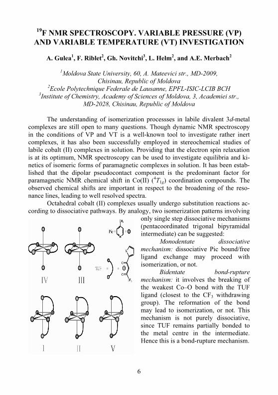
10
Pulsed EPR measurements enable direct determination of relaxation times T1 and T2 and many more parameters which are not accessible by cw-methods But Pulse methods require special high sophisticated hardware We have develo-ped a stand-alone Pulsed EPR and Pulse accessory to our X-band EPR spectrome-ter Both instruments can be upgraded to Q-band and W-band operation All digital pulse generation and signal detection from X-band is preserved in Q- and W-band
The important items for a Pulse Spectrometer are Pulse generator (Pattern jet) time resolution 4 or 2 nsec Fast digitiser and signal averager (Specjet) Pulse signals are often too small to be seen directly and therefore tuning may be difficult The Specjet has a sampling rate of 250MHz (2nsec) Fast signals can be digitised and 1 million shots can be averaged in one second Signal is accumulated and dis-played real time in second time interval Furthermore we need a resonator with short dead time and high bandwidth and quadrature receiver with high gain and bandwidth All pulse sequences can be easily edited and visualized on the control monitor The parameters can be set and the effect on the sequency will be immedi-ately visible The actual quadrature signal is shown on the SpecJet monitor
Furthermore the spectrometer can be extended to Pulsed ELDOR and ENDOR operation Electron-Nuclear-Electron-Triple Resonance involves 3 fre-quencies two microwave frequencies and one rf frequency The allowed transition 1 is observed and the allowed transition 2 is pumped and both transitions are con-nected by an rf pulse The microwave pump frequency and the rf-frequency are swept The rf sweep gives the ENDOR spectrum and the sweep of the microwave pump frequency gives the hyperfine couplings for the ENDOR lines
ELDOR is a quite old method but its implementation in cw spectrometers was very difficult and technically imperfect This is the reason why its use was quite limited Pulse techniques are much easier and avoid the previous limitations which were mainly due to the limited bandwidth of the cavity An large variety of ELDOR-based methods have evolved immediately like Saturation Recovery ELDOR Double Electron-Electron-Resonance (DEER) ELDOR detected NMR and Electron-Nuclear-Electron Triple Resonance
Saturation-Revovery Electron-Electron Double Resonance is a method to determine T1 in cases where T2 is too short to obtain an FID or Spin-Echo The EPR-signal is inverted or saturated by a strong microwave pulse and detected di-rectly without modulation in a low microwave field with the Quad-receiver The preparation and detection frequencies can be identical or different In the first case we measure T1 and in the second case we have additional information on electron-electron double resonance The pulse sequence looks very simple but this experi-ment is very difficult to perform We need high pulse power and the low Q-resonator has to be critically coupled Since signal is detected without modula-tion stability of the experimental conditions is very important Because of the di-rect detection any instability will immediately disturb the measurements The experiment can be set up on a special pulse panel screen
11
The standard methods for distance determinations in molecules are X-ray-diffraction and NMR NMR does not need single crystals which in many cases cannot be obtained from biological macromolecules like proteins Another limita-tion for NMR is the molecular weight Up to now structures could be determined for up to 50KDalton The NOE allows to see distances up to 5 Angstroem EPR also does not need single crystals but has no limitation for molecular weight and can see much larger distances in the range from 5 up to 80 Angstroem
Various EPR methods can probe various distances Cw-EPR can probe dis-tances up to about 10 Angstroem ENDOR and ESEEM probe nuclear-electrom interactions and ELDOR electron-electron interactions Since the dipolar interac-tions are proportional to the magnetic moment of the involved particles ELDOR is the most sensitive method and can detect dipolar interactions at larger distances This is especially important for site directed Spin labeling where labels can be placed at various residues of a protein and dipolar interaction can probe the dis-tance between the labeling sites
In DEER (3-pulse-ELDOR) the signal is detected by an echo The sequence consists of a 2-pulse SE-subsequence with fixed interpulse delay τ between the two mw-pulses at the observer frequency The pump pulse at MW2 inverts the upper transition changing the local field at the other spin species due to electron-electron-coupling ωee At the time of echo formation the magnetization is out of phase by a ωeet By shifting the MW2-pulse electron-electron-coupling can be determined by observing the echo amplitude as a function of t
This experiment needs also a large bandwidth resonator high B1-values (high conversion factor) large bandwidth of the ELDOR source large bandwidth of the power amplifier
In EPR tomography the relationships between FOV resolution pixel band-width and gradient strength for EPRI are the same as for NMR In practice how-ever there is a large difference regarding the pixel bandwidth In proton imaging at low to medium field the gradient strength is often large enough to ensure that no chemical shift distortion appears in the image In EPRI to suppress the appearance of hyperfine interaction in any image would require enormous gradients Instead the spectral distortion is tackled in the data processing
For tomographic investigations in material science a gradient accessory has to be added to a standard EPR spectrometer Biological objects due to their greater size and high water content are better investigated in the L-band with a special large bore magnet and gradient assembly For an EPR line width of 100mG a resolution of 25μ can be achieved
One of the most promising applications of EPR imaging is oximetry The width of the EPR line depends on the partial oxygen pressure as The line width can be calibrated against the partial oxygen pressure and then the partial oxygen pressure calculated from the EPR line width is displayed in a colour code The image therefore reflects the oxygen concentration in the sample
12
WATER SPIN ISOMERS
NM Sergheev
Moscow State University ldquoMV Lomonosovrdquo Chair of chemistry Moscow
Russian Federation e-mail sergeyevnmrchemmsusu
The sensational report of Tikhonov and Volkov in 2002 (V Tikhonov and
A Volkov Science 2002 296 2363 [1]) regarding the possibility to separate or-tho- and para- spin isomers (ortho-H2O the total proton spin I=1 and para-H2O I=0) with the help of selective absorption using frontal chromatography caused a burst of interest in the scientific community Not only have Tikhonov and Volkov separated the two isomers they have also measured the lifetimes (or the conversion times) of the spin isomers These lifetimes appeared quite long in the case of ice and steam (days) and a bit shorter (minutes) in the case of liquid water The possi-bility to obtain pure spin isomers of water puts forward several very interesting perspectives for the MR-tomography This is due to the fact that para-isomers of water (just like para-hidrogen) shouldnrsquot produce any signals in the NMR of pro-tons The report will contain a critical analysis of experimental and theoretical re-searches in this area It will be shown that long lifetimes for pure spin isomers in a non-equilibrated ratio (ie different from 31) in case of room temperature can be obtained only in special conditions (gas at low pressures or diluted water solutions in hydrophobic solvents) The report will present an alternative path of chemical synthesis of para-water using catalytic hydration of compounds containing semi-polar links such as X rarr O (eg NO2 N-oxides) with para-hydrogen on rhodium iridium or palladium complexes
We express our gratitude to EC NEST programme ProposalContract no5032 2005 and to the Russian Foundation of Basic Research (grant 06-03-32995) for their financial support
13
ELECTRON SPIN DYNAMICS IN QUANTUM DOTS
II Geru Center for Metrology and Analytical Methods of Research Academy of Sciences of
Moldova 32 Academiei str MD-2028 Chisinau Moldova
E-mail iongeruyahoocom
Electron spins in semiconductor quantum dots (QDs) are promising candi-
dates for quantum computation [1] It is due to the slower spin-dephasing expected upon electron confinement In QDs dephasing mechanisms involving spin-orbit coupling are believed to be suppressed making electron-nuclear hyperfine coup-ling the dominant source of spin relaxation [2 3]
Recently relatively long room-temperature spin-dephasing times )190(
2nsT le have been measured for bulk and epitaxial n-type ZnO at the room-
temperature time-resolved Faraday rotation spectroscopy [4] in ZnO nanostructures
has been reported In Ref [5] the
2T times measured at the same temperature for
freestanding ZnO QDs containing between 1 and 6 additional conduction band electrons has been presented Values up to 25 ns or ~ 125 times longer than in bulk or thin-films ZnO were obtained The hypothesis that electron-nuclear hyper-fine interaction dominate spin-dephasing dynamics was confirmed directly by variation of the 67Zn (I = 52) content
In GaAs InAs and CdSe QDs spin dephasing has been associated also with electron-nuclear hyperfine interaction [2 3] which is large in these lattices
because many of their ions have nuclear spin Following Ref [6]
2T in this sce-
nario depends on the strength and number of hyperfine interactions in the QDs ac-cording to the formula
( )( )
12
32
2
sum +
=
j
jjj
L
AIIn
NT h
(1)
where NL is the total number of ions in the QD n is the number of ions in the unit cell I j is the nuclear spin of the j-th ion Aj is the hyperfine coupling constant at the j-th ion and the sum is over all ions in the unit cell
In contrast with GaAs InAs or CdSe the vast majority of cations and ani-ons in ZnO have I = 0 Only 67Zn (I = 52 41 natural abundance) my contribute significantly to spin dephasing via the hyperfine interaction The longest room-
temperature spin-dephasing time observed experimentally is
2T = 25 ns for a sin-
gle electron in the d = 46 nm ZnO QDs with natural abundance lt67Zngt=41 and
A (67Zn)=517μeV [6] With NL asymp 4300 Eq (1) yields
2T = 60 ns that is a fac-
tor of 4 smaller than the experimental value
14
In summary chemical preparation of freestanding charged QDs has been shown to offer rich opportunities for exploration of spin dynamics in semiconduc-tor nanostructures related to quantum computation and spintronics [1] V Cerletti WA Coish O Gywat and D Loss Nanotechnology 16 R27
(2005) [2] IA Merkulov AL Efros and M Rosen Phys Rev B 65 205309 (2002) [3] PF Braun X Marie L Lombez et al Phys Rev Lett 94 116601 (2005) [4] S Ghosh V Sih WH Lau et al Appl Phys Lett 86 232507 (2005) [5] WK Liu KN Whitaker AL Smith et al Phys Rev Lett 98 186804
(2007) [6] JE Wertz and JR Bolton Electron Spin Resonance McGraw-Hill NY
1972
15
ORAL TALKS
16
STRUCTURE OF CHROMIUM IMPURITY CENTERS IN
SYNTHETIC FORSTERITE
STUDY BY TUNABLE-FREQUENCY EPR SPECTROSCOPY
AA Konovalov VF Tarasov
Zavoisky Physical Technical Institute of the Russian Academy of Sciences
107 Sibirskii trakt 420029 Kazan Russia
Chromium doped forsterite is well known as material for efficient tunable
laser generation in 1236-1300 nm range Recently tunable laser generation in a range of 1030 ndash 1180 nm was obtained on forsterite co-doped by chromium and lithium [1] We used tunable high-frequency EPR spectroscopy [2] in the fre-quency range 60 - 230 GHz to study structure of Cr3+ paramagnetic centers in syn-thetic forsterite co-doped by chromium and lithium and influence of lithium on relative concentration of divalent and trivalent chromium ions in this material
Samples had the cubic form with dimensions 3times3times3 мм3 and were cut from the crystals grown by the Czochralski method in slightly oxidizing atmosphere (Ar + 0001 O2) at various concentration of lithium in the melt (from 0 up to 037 weight ) and at approximately constant concentration of chromium (006 weight )
It was confirmed unambiguously that Cr3+-Cr3+ dimers substituting Mg2+ ions in the M1 sites are formed in forsterite grown without Li impurity but their concentration diminishes rapidly with introduction of the Li impurity At the same time concentrations of two other structure non-equivalent Cr3+ ions are increased
For the ions of the divalent chromium replacing magnesium in the М1 and М2 positions reduction of concentration of both centers approximately in 6 times is found due to increasing of lithium concentration in the melt from 0 up to 037 weight
The work was supported the RFFI grant 06-02-16662 and partially by the Russian President grant NSh-621320062 [1] AV Gaister EV Zharikov VF Lebedev AS Podstsvkin SYu Tenyakov
AV Shestakov IA Shcherbakov Quantum Electronics 34 8 693-694 (2004)
[2] VF Tarasov GS Shakurov Appl Magn Reson 2 571-576 (1991)
17
STRUCTURAL AND MAGNETIC PROPERTIES OF
3-ACETYLQUINOXALIN-2(1H)-ON WITH Cu(II)
TA Ivanova1 IV Ovchinnikov1 AR Mustafina2 TB Makeeva2 VA Mamedov2 AN Turanov1
1Zavoisky Physical-Technical Institute RAS Kazan Russia
2AE Arbuzov Institute of Organic and Physical Chemistry RAS Kazan Russia
A quinoxalins derivatives are perspective ligands to metalcontaining supra-molecular compounds formation as they have some potential bonding centers of metal ion The quinoxalins ability to two Cu(II) ions coordination and the polymeric chains formation has been established on the basis of EPR and magnetochemistry data for complexes CuHL(NO3)2sdotH2O where HL=3-acetyl-quinoxalin-2(1H)-оn These complexes have been synthesized and investigated for the first time
The obtained materials content is in concordance with the molar proportion of copper ion and HL 11 This conclusion has been made on the basis of electron spect-roscopy data The copper complexes have distorted octahedron coordination The electronic absorption band 7995 nanometers has been found in the compound DMSO solution The atoms groups which coordinated with a metal ion have been identified by IR spectroscopy The possibility of plane 5-component chelate cycle formation at bidentate bound of Cu(II) ion by nitrogen and oxygen atoms of neighbouring carbonyl group has been shown It has been assumed that the poly-meric structure of a substance [CuHL(NO3)2sdotH2O]n has been formed in investigated material Every ligand molecule is the bridge between two copper ions
The EPR spectrum of solid state compounds is the exchange narrowed line with g=2147 The line width ∆H is decreased linearly from 510 Oe to 140 Oe at the sample cooling from 300 K to 11 K The linear temperature dependence of line width correlate with the theoretical analysis results of exchange narrowed line in a one-dimensional magnetic in case of symmetric anisotropic exchange interaction It is concluded that polymeric chains exist in the investigated structure in these the Cu(II) complexes are bonded by strong exchange interactions The EPR spectrum of Cu(II) complexes frozen in DMSO solution (1500) has been investigated also It consists of both copper spectrum with parameters g1=2055 g2=2165 g3=2350 and not re-solved hyperfine structure and the spectrum of the exchange-connected pairs Cu(II) ions (S=1) with splitting parameter ~1000 Oe in a zero field Relative dimers con-tents is about 15 according to our estimations The temperature dependence of effective magnetic moment μeff indicate on antiferromagnetic exchange interactions between metal ions The assumption about the existence of chains fragments and copper ions dimers allow to describe the experimental dependence μeff(T) and to es-timate of exchange interaction parameters The possible model is discussed where dimers are dendrit structure centers on which linear polymers grows on
18
ODMR-INVESTIGATIONS OF RESONANCE ENERGY
TRANSMISSIONS TO LUMINESCENCE Mn2+
CENTERS IN
ZnS CRYSTALS
A Kovalenko M Bulaniy S Omelchenko
Department of Physics Electronics and Computer Systems Dnipropetrovsk
National University 13 Nauchnaya str 49050 Dnipropetrovsk Ukraine
E-mail somelchmailru
Now the excitation of Mn2+ ions in zinc sulphide during a luminescence is
left under discussion The question is what mechanism takes place during excita-tion energy transmission from centers of sensibility a resonance one or a percus-sive one It is know that the resonance energy transmission arises when conditions for quantomechanical interaction are fulfilled Such conditions are an overlap of sensibilizator emitted spectrum with absorption spectrum of activator and the dis-tance between these centers should be small enough for interaction
The emission spectra of self-activating zinc sulphide crystals annealed in zinc fumes contain bands with maximums in λ=465 nm and λ=530 nm during pho-toluminescence which coincide with the absorption bands The PL bands with λ=480 nm and λ=504 nm partially overlap with absorption band at λ=496 nm Overlapping of the manganese ion absorption spectra with sensibilizator emission spectra shows that in the investigated samples the first condition of resonance in-teraction is also met
The experiments we carried out showed that during the excitation by UV light (λ=365 nm ndash area of impurity absorption) the photoluminescence of the ini-tial ZnSMn crystals is due to a mechanism of the resonance excitation of the Mn2+ ions in zinc sulphide Considering that the manganese centers are distributed evenly throughout the crystal and donrsquot group around any places in lattice it was determined that the distance between Mn2+ ions and sensibilizator centers ie cen-ters of blue and green zinc sulphide luminescence approximately equal to 1 nm that in the investigated samples the second condition of resonance interaction is also met
The most interesting result we discovered during the analysis of ODMR spectra was that during ODMR signal detection in the blue region of PL spectra of ZnSMn crystals a structure of six lines was observed corresponding to the su-perthin structure of Mn2+ ions The same structure is also specific for the EPR spectra of Mn2+ ions The obtained result directly testifies a presence of resonance interaction between blue PL centers and Mn2+ ions in ZnS
19
THE COMPLEX Fe (III) WITH
4-TERT-BUTYLPHENOXYACETYLHYDRAZONE OF
PYRIDINE-2-CARBALDEHYDE
SYNTHESIS AND MAGNETIC PROPERTIES
SN Podyachev1 SN Sudakova1 TA Ivanova2 OA Turanova2
1AE Arbuzov Institute of Organic and Physical Chemistry of
Russian Academy of Sciences Kazan Russia 2Zavoisky Physical-Technical Institute of Russian Academy of Sciences
Kazan Russia e-mail turanovamailkncru
The Fe (III) complex with 4-tert-butylphenoxyacetylhydrazone of pyridine-2-
carbaldehyde has been synthesized with aim of searching for new materials with per-spective electromagnetic properties The choice ligand has been caused by presence of
several bonding centers of various nature in it This is the necessary condition for effec-tive and selective transition metals (3d-) ions bonding This substance can be used as the structural block with threedentant N N O coordi-nation unit in a calix[4]arens and complexes synthesis It was the second reason of our interest to this compound The ligand (L) 4-tert-butylphenoxyacetylhydrazone
has been obtained by interac-tion 4-tert-butylphenoxyacetylhydrazide with pyridine-2-carbaldehyde The contain and structure of the received compound have been fixed by data of the element analy-sis IR and 1H NMR spectroscopy
The magnetic properties of material have been investigated by EPR spectros-copy The symmetric line has been observed in EPR spectra of a coordination complex powder at temperature from 10 until 300 K The line parameters are gasymp2 and width ΔH=800 Oe It is the evidence of strong exchange interaction between substance mole-cules This compound demonstrated high spin (S=52) state with parameter of thin structure Dgtgt03 in methanol frozen solutions The EPR line integrated intensity of Fe(III) complex dependend from temperature in accordance with Curie-Weiss law
This work was carried out with the financial support of RFBR (grant no 07-03-00325a)
O
O NH
NH2
t Bu
N O
EtOHreflux
But
O
O NH
N
N
Fe Cl3
MeOHFeL2
1L
20
EPR SPECTRUM AND SPIN DYNAMICS IN
QUASI-TWO-DIMENSIONAL ANTIFERROMAGNET
Mn[C10H6(OH)(COO-)2times2H2O
KG Dergachev MI Kobets EN Khatsko
Department of Magnetism B Verkin Institute for Low Temperature Physics and
Engineering of the National Academy of Sciences of Ukraine
47 Lenin ave 61103 Kharkov Ukraine
The EPR spectrum of a low dimensional antiferromagnet
Mn[C10H6(OH)(COO-)]2times2H2O investigation was performed in the frequency range 96 GHz In H||h polarisation in ab plane along with a basic magnetic reso-nance line in field H0 an additional absorption line with of relatively low intensity in resonant field Н02 The peak intensity of the observed line is of the order of 1800 less then the peak intensity of the basic EPR line in Hperph polarisation The availability of an additional absorption line is due to low dimensionality of the sys-tem
The angular and frequency dependences of the fundamental absorption line in the EPR spectrum of the antiferromagnet Mn[C10H6(OH)(COO-)]2times2H2O was studied The EPR spectrum behaviour at high temperatures is described by the dif-fusion model for quasi-two-dimensional magnetic structures The diffusion cons-tant D =62times1011 sec-1 the starting time
1τ sim15times10-12 sec and the end time of the
diffusion process 2
τ sim88times10-10 sec was estimated A non diffuse contribution in the line width of EPR spectrum was determinate
21
ESR-INVESTIGATIONS INFLUENCE OF ELECTRIC
FIELDS OF DISLOCATIONS ON ENERGY STATE OF
IONS Mn2+
IN ZnSe CRYSTALS
S Omelchenko1 O Khmelenko2 A Kovalenko L Shevchenko
Department of Physics Electronics and Computer Systems Dnipropetrovsk
National University 13 Nauchnaya str 49050 Dnipropetrovsk Ukraine
E-mail somelchmailru1 khmelenkoukrnet2
The physical properties of real ZnSe crystals determine of their structures
defects This is a subject of many researches In this work were investigated ZnSe and ZnS singlendashcrystals grown from the melt Investigations of ESR spectra changes after plastic deformation are showing that as against ZnS crystals in ZnSe ones the quantity of manganese impurity ions which are in twice ionized state de-pends of concentration of dislocations Deformation of crystals decreases the quan-tity of Mn2+ ions In spectra of manganese luminescence excitation there are not two maximums which are corresponding to levels of upper excited states in ZnSе crystals Such situation is possible if these levels are located in a conduction band In this case they are detached from energy levels of a conduction band by high po-tential energy barriers Electrons from levels of this upper excited states of manga-nese make tunnel transitions through barriers into conduction band and vice versa with equal probability The part of Mn2+ ions get into zone of action of electric fields of negatively charged of dislocations The charge state of these Mn2+ ions will be changed in result of preferred tunneling of electrons from levels of two up-per excited states
4T2 (4G) and 4T1 (4P) to conduction band For testing this guess
the action of dislocations was modeling by application to undeformed crystals of an external electric field It turned out that effect of electric field is similar to effect of deformation In crystals ZnS similar effects is absent It is naturally so in these crystals all levels of excited states of manganese are located into forbidden energy gap and tunneling of electrons from them does not occur or more weak It is inter-estingly that in ZnSе crystals in situ absentee maximums in spectrums of exciting we observed increase of a photoconduction
22
NMR STUDY OF OUTER-SPHERE INTERACTIONS
VV Trachevsky SV Zymina AP Shpak
Technical Centre of the National Academy of Sciences of Ukraine
13 Pokrovskaya str 03070 Kyiv Ukraine e-mail trachevimpkievua
The directed formation of outer-sphere complexes is a method of molecular
system control Therefore it is of importance to develop the methodology of syn-thesis of coordination compounds as well as to access the data on optimal condi-tions of their complexation composition structure and properties
In this work the associates with different types of outer-sphere coordination (anionic cationic and molecular) were studied
The systematical NMR investigations of systems [CoLnL3-n]mplusmn ndash MXp ndash H2O (D2O) (where L L ndash ethylenediamine (en) monoethanolamine (etm) ali-phatic aminoacids М ndash Li+ Na+ Cs+ tetraalkylammonium cations Со2+ Ni2+ Zn2+Cd2+ Pb2+ X ndash F- Cl- Br- I- SO4
2- PO43- NO3
- CH3COO- ethilenediamine-tetraacetate ndash edta) was carried out 59Со NMR spectroscopy used in the current work was the most sensitive method for studying the changes in the electronic environment caused by the nature and numbers of donor atoms as well as the fea-tures of outer-sphere interactions
[Coen3]3+ + nCat+X- rarr[Coen]Xn(n-3)- +nCat+ n=1divide12
[Coen3]3+ H2edta2- + nCat+X-rarr Catn[Coen3]H2edtaXn(n-1)-
n=1divide8 The use of [Coen3][B(Phenyl)4]3 and water-organic solvents allows to pro-
pose an appropriate model of set of indicated equilibrium processes For molecular complexes whose coordination sphere is consisted of bifunc-
tional ligands the outer-sphere origin of cations as well as anions was revealed 2[Co(etm)3]+ Mn+Xn-rarr [Co(etm)3]Mmnm++[Co(etm)3]Xmnn-
n=1 2 3 m=1 2 In complexes with polydentate ligands the dynamics of functional group ex-
change in coordination sphere is initiated by outer-sphere cations (Li+ K+ NR4+
etc) MLHn-x+mCatOHrarr[MLHn-x-m]Catm+mH2O
The existence of the series of complexes was established by the 35Cl 23Na 59Co NMR spectroscopy in the solid state In addition the temperature-dependent heteronuclear NMR measurements have been performed in solution Inner-sphere effects of magnetic field of paramagnetic ions were revealed for the heteronuclear complexes
23
EPR SPECTRA STRUCTURE AND SYNTHESIS OF
[CuBa(SalH)4(DMAA)4(H2O)] COMPLEX
I Geru1 V Arion2 S Shova3 V Olednic4 I Siretanu1 V Shofransky 4 C Turta 4
1Center of Metrology and Analytical Methods of Research
Academy of Sciences of Moldova Chisinau Moldova 2Institute of Inorganic Chemistry University of Vienna Vienna Austria
3Institute of Applied Physics Academy of Sciences of Moldova Chisinau Moldova 4Institute of Chemistry of the Academy of Sciences of Moldova Chisinau Moldova
By the reaction of copper nitrate with barium salycilate in methanol at cons-tant stirring and room temperature the blue powder was obtained By recrystalliza-tion of this product from the mixture of solvents dimethylacetamide (DMAA) and tetrahydrofuran (THF) the crystals of new complex of the composition [CuBa(SalH)4(DMAA)4(H2O)] (1) of intense blue colour were obtained
The X-ray investigation of monocrystal of this substance has demonstrated that this complex has a dimer structure of ldquolanternrdquo type Four bidentate carboxylate bridged groups link copper and barium atoms in dimer The coordination number of Cu is five (4+1) and Ba is 8 (Fig 1) In the crystal by hydrogen bonds dimmer comp-lex forms the polymer structure
2400 2600 2800 3000 3200 3400 3600
-600
-400
-200
0
200
400
600
800
Inte
nsity o
f E
PR
lin
e
rela
tive
un
its
Magnetic field Gs
Fig1 The molecular structure of 1 Fig2 The EPR spectrum of a single
crystal 1 at Hσ and T = 300K
The EPR spectrum of 1 corresponds to his molecular structure presented on Fig 1 It is caused by Cu2+ ions (the electron configuration 3d9 and spin S=12) being components of Cu2+-Ba2+ dimmers (the ion Ba2+ havenrsquot a spin) Fig 2 shown the room-temperature EPR spectrum at Hperpσ (σ is the big side of the crystal) with the well solved hyperfine structure (I=32 for bots 63Cu and 65Cu isotopes with 6909 and 30091 natural abundance correspondingly) At Hσ the EPR spectrum it is transformed into a single line without hyperfine structure In summary it is the new dimmer compound which contain copper (II) and s-element
24
1H NMR APLICATION FOR THE SYNTHESIS CONDUCT
OF 3D-ELEMENTS COMPLEXES BASED ON
S-ALKYLISOTHIOSEMICARBAZIDE
S Manole1 M Cocu1 E Rybak-Akimova2
1Institute of Chemistry Academy of Sciences of Moldova 3 Academiei str
MD-2028 Chisinau Republic of Moldova 2Tufts University Medford USA
The application of the 1H NMR method for the synthesis conduct of
3d-elements coordination compounds obtained via acetyl(benzoyl)acetone S-alkylisothiosemicarbazone template assembly involving oxycarbonilic ligands consists in the fact that these complexes promote the biosynthesis process of the fungus [1] they have an intense vivid colour and are thermostable what permits to exploit such coordination compounds as thermoplastic dyes by using it in very small quantities (05 mg ndash 1 g) [2]
The participation of many ligands in a template synthesis leads to the formation of diverse ranges of substances (fig)
OOH
N
N
NH2
R
OH
SR1
M
N
O
N
N
R
O
SR1
CH3OH
NNN
SR1
N NN
SR
M
R
R
+M2+
M = Ni Co Cu VO
R = CH3 C
6H5
R1 = CH3 C
2H5
(I)
(II)
Due to the fact that two or more substances in which ligands combine with
the central atom by means of different atoms (NNNN or NNOO) are formed the nonequivalent protons of the alkyl groups possess diverse ecranisations and thus as a results separate signals (for R=R1 = CH3 M = Ni (I) δ = 2 22 2 34 2 64 and (II) δ = 2 05 2 26 2 42 ppm) are registered in the 1H NMR spectra Their integration permits to determine the quantitative ratio molar of the obtained sub-stances The 1H RMN spectroscopy can be used for the synthesis conditions opti-mization in order to obtain a high yield of necessary substances
25
Acknowledgements
The authors gratefully acknowledge MRDACRDF grant (MTFP-1018A)
References
[1] MA Cocu S Clapco NV Garbalau et al Synthesis and biological study of Ni(II) Cu(II) and VO(II) [N2O2] Schiff base complexes derived from S-methylisothiosemicarbazide Romanian International Conference on Chemistry and Chemical Engineering - RICCCE XIV Bucharest Romania September 22-24 S01-51 - S01-53 (2005)
[2] M Cocu N Garbalau S Manole J Gradinaru A Forni Compusi complecsi triazamacrociclici ai nichelului(II) si aplicarea lor icircn calitate de coloranti pentru polimerii termoplastici Brevet МD 2881 BOPI nr 10 33 (2005)
26
ESR OF Mn CENTERS IN LiNaGe4O9 CRYSTALS
K Omelchenko M Volnyansky
Department of Physics Electronics and Computer Systems Dnipropetrovsk
National University 13 Nauchnaya str 49050 Dnipropetrovsk Ukraine
E-mail omelchenkouafm
The ESR spectra of Mn ions in LiNaGe4O9 crystals which were grown by
Chokhralsky method are investigation in this paper As well know that such com-pounds are in paraelectric phase at room temperature and have orthorhombic crys-tal cell (Pcca- D8
2h) Transition in ferroelectric phase take place at Tc = 1127 K The ESR spectra has been studied at Т=300 К in Х-band There are consist
from three hyperfine structure groups lines of Mn ions sufficiently intensity This groups have similar angle dependences which are well describe by orthorhombic symmetry spin-hamiltonian with S = frac12 The main value of g- factors for one of centers are gxx = 17444 gyy = 22948 gzz = 25631 Hyperfine structure constants have strong anisotropy It was discovered that local symmetry paramagnetic cen-ters at the low temperature (below of point of ferroelastic phase transition) no changes In this case lines of several groups of ESR spectra are just broader and some other lines groups are became double This fact is testify about this lines group it may concern to centers of different nature
Discussion of the occasion of charge state Mn centers are going in this pa-per
As resume ndash the measuring in K-band is necessary
27
RESONATOR QUALITY CHANGING AT THE P1-CENTRE
IN-PHASE ESR SIGNAL INVERSION IN DIAMOND AND
POLYCRYSTALLINE DIAMONDS FILMS
V Bayev N Lapchuk
Physics Department Belarussian State University 220050 Minsk Belarus
The in-phase ESR signal inversion of P1 center (an isolated impurity nitro-
gen atom substituted for carbon in the diamond lattice) was earlier observed in the polycrystalline diamond films (thickness till 05 mm) grown in methanehydrogen mixtures by chemical vapor deposition (CVD) in microwave plasma and annealed at temperature 1500 degC during 13 hours [1] The CW ESR experiments were made at X band with 100-kHz modulation frequency under the room temperature without illumination The same phenomenon was observed for P1 center in the thermobaric processed monocrystals of synthetic diamond grown by the temperature gradient method in the Ni-Fe-C and Fe-Co-C systems using ldquosplit-sphererdquo high-pressure apparatus [2 3] In all cases the inversion of in-phase ESR signal occurs when the microwave power P rises above ldquocritical levelrdquo which differs for each sample Moreover the amplitude of the inverted signal grows with the microwave power increase It should be noted that earlier the in-phase ESR signal inversion was ob-served for paramagnetic center (nitrogen atom + carbon vacancy) in diamond crys-tals of Ib type with illumination (photon energy was less then gap energy) [4 5]
In order to show that the observed in-phase ESR signal inversion of P1 cen-ter isnrsquot a passage effect or so called ldquoapparatus effectrdquo the processes of absorption and radiation inside the resonator at the resonance moment were studied The stu-dies were made by means of exterior spin-label 1 (an irradiated point (02 mm) crystal of natural diamond) with g-factor close to that of P1 center and unsaturated signal in all used power range (the maximum microwave power Pm = 70 mW) The processes of absorption and radiation inside the resonator were controlled by moni-toring the resonator quality changing ΔQ at different microwave powers at the ESR of P1 center for diamonds investigated in [2]
The resonator quality changes were determined by measuring the in-phase ESR signal difference of spin-label at and without presence of studied diamonds in resonator (fig 1) For original non-thermobaric processed monocrystal of synthetic diamond (the inversion isnrsquot observed) ΔQ rises with the power as one should ex-pect for microwave energy absorption of the investigated object increases and the resonator quality decreases (fig 1 a) As for the thermobaric processed monocrys-tal of synthetic diamond (the in-phase ESR signal is inverted at the microwave power sim 2 mW) the resonator quality changing increases with the power until the inversion and then decreases (fig 1 b) Thus the resonator quality improves the amplitude of the inverted in-phase ESR signal increasing Also the crystal of ruby
28
(Al2O3Cr) was used as a spin-label 2 It was oriented in the resonator so that g-factors of ruby and that of the investigated thermobaric processed monocrystal of synthetic diamond were different As the spin-label ESR signals were detected without P1 center inversion the microwave power dependence of the resonator quality changing should be constant (as itrsquos shown on fig 1 c) The same results were obtained for CVD diamond films with the inverted in-phase ESR signal These experiments prove the paramagnetic nature of the P1 center in-phase ESR signal inversion
0 02 04 06 08 1
0
50
100
150
200
b
c
aΔQ
mPP
Fig 1 The microwave power dependence of H102 resonator quality chan-ging ΔQ for a ndash original non-thermobaric processed monocrystal of synthetic dia-mond (the inversion isnrsquot observed) and spin-label 1 b ndash thermobaric processed monocrystal of synthetic diamond (the in-phase ESR signal is inverted at the mi-crowave power sim 2 mW) and spin-label 1 c ndash thermobaric processed monocrystal of synthetic diamond and spin-label 2 [1] VG Bayev Theses of reports of XII Republican scientific conference of post-
graduate students undergraduates and students Grodno 121 (2004) [2] NA Poklonski TM Lapchuk VG Bayev GA Gusakov JAS 73 9
(2006) [3] NA Poklonski NM Lapchuk TM Lapchuk JETP Lett 80 748 (2004) [4] JHN Loubser JA van Wyk Rep Prog Phys 41 1201 (1978) [5] J Harrison MJ Sellars NB Manson J Lumin 107 245 (2004)
29
100 150 200 250 300
dχd
B
Magnetic field (mT)
Yb1
isotopes
with I=0
173
Yb
Yb4
Yb2+Yb
3Yb
1
171
Yb
171
Yb
d
c
b
aB||z
Congr
B||y
B||z
B||x
AXIAL AND LOW-SYMMETRY Nd3+
AND Yb3+
CENTERS IN
CONGRUENT AND NEARLY STOICHIOMETRIC LiNbO3
G Malovichko1 M Munro1 V Grachev1 V Bratusrsquo2 S Okulov2 Ed Kokanyan3
1Physics Department Montana State University MT 59717 Bozeman USA
2Institute of Semiconductors Physics NASU 45 Nauky ave 03028 Kyiv Ukraine 3Institute of Physical Researches 378410 Ashtarak Armenia
Lithium niobate is of great interest for both fundamental science and appli-
cations due to unusual richness of its ferro- pyro- and piezoelectric properties Conventional crystals grown from a congruent melt with lithium deficiency (Xmelt = XCrystal = 484 where X = [Li]([Li]+[Nb]) contain some percent of intrinsic (nonstoichiometric) defects and have strong structural disorder Crystals grown under special conditions from melts to which potassium has been added have ext-remely low intrinsic defect concentrations These samples called stoichiometric have significantly decreased widths of spectral lines This leads to the increased resolution of both optical electron paramagnetic resonance (EPR) and electron nuclear double resonance (ENDOR) spectra
Fig 1 The assignment of the lines of different NEC (Yb1 ndash Yb4) and HFS for Yb3+ in nearly
stoichiometric (a-c) and con-gruent (d) sam-ples of LiNbO3
Lithium niobate crystals doped with neodymium andor ytterbium were studied by EPR and ENDOR in the temperature range of 42-50 K with Bruker ELEXSYS 560 spectrometer operating in X- and Q- bands (98 and 344-350 GHz) Tremendous narrowing of magnetic resonance lines in nearly
30
stoichiometric samples as compared with congruent ones (Fig 1) allowed us to distinguish four different non-equivalent centers (NEC) of Nd3+ and five NEC of Yb3+ as well as to observe hyperfine structure (HFS) for magnetic isotopes 143Nd and 145Nd (171Yb and 173Yb) It was shown that some centers have axial C3 symmet-ry whereas all others have lowest C1 symmetry due to the presence of intrinsic orand charge compensation defects in the near neighborhood of Nd3+ and Yb3+ The possible models for these centers are considered
31
USE OF ESR METHOD FOR DISLOCATION ELECTRIC
PROPERTIES IN ZnS CRYSTALS
S Omelchenko A Gorban M Bulaniy
Department of Physics Electronics and Computer Systems
Dnipropetrovsk National University 13 Nauchnaya str 49050 Dnipropetrovsk
Ukraine e-mail somelchmailru
The dislocations in II-VI compounds have electric charge Its value deter-mines a degree of influence of an electric field of dislocations on properties of ma-terials
We studied ZnS single crystals grown from melt We observed ESR spect-rum of centers Cr+ in ZnS crystals without UV excitation after their annealing in steams of Zn and their subsequent fast cooling We guess that it happens as a re-sult of fast diffusion of zinc atoms along dislocation tubes around of growth dislo-cations Thus zinc atoms enter in composition of Cottrellrsquos atmospheres of dislocations Being by more deep donors than zinc the Cr2+ centers which were within of Cottrellrsquos atmospheres are filling by electrons and convert into Cr+ cent-res But inside of Ridrsquos cylinders the electric fields of dislocations ionize these ions up to Cr2+ state Thus between exterior boundaries of Ridrsquos cylinders and Cottrellrsquos atmospheres a charge state of ions of chrome - Cr+ and outside Cottrellrsquos atmos-pheres - again Cr2+ Thus the intensity of an ESR spectral lines of ions Cr+ are proportional to volume of areas of Cottrellrsquos atmospheres which are outside an operation of electric fields of dislocations
It was discovered intensity of ESR spectral lines of Cr2+centers diminishes after plastic deformation of crystals It is possible to explain this fact so The growth dislocations originated at high temperatures in favourable conditions for processes of diffusion therefore they are surrounded by a heavy-bodied cloud of the impurity ionized centres which do compensation their charge That is why ra-dius of Ridrsquos cylinders of growth dislocations is very much small and density of Cr+ centres inside of Cottrellrsquos atmospheres is maximal In process of deformation the dislocations are moving from a starting place and shifting out from this com-pensation clouds Radius of them Ridrsquos cylinders becoming more and density of centres Cr+ are diminishing
32
HMBC AND HSQC SPECTRA AS TOOLS IN PROVING
STRUCTURE OF COMPOUNDS CONTAINING SElarrN
INTRAMOLECULAR BONDING
A Nicolescu1 M Balan1 and C Deleanu12
1Petru Poni Institute of Macromolecular Chemistry
41-A Grigore Ghica Voda str Iasi Romania 2CD Nenitescu Institute of Organic Chemistry
202-B Spl Independentei str Bucharest Romania
The organoselenium (II) derivatives are used in organic synthesis enzyme
mimics and as chemotherapeutic agents or for the potential of 2-(NN-dimethylaminomethyl)phenyl and related derivatives to stabilize exotic species such as the covalent azide [2-(Me2NCH2)C6H4]SeN3 [1] or the trihalide [2-(Me2NCH2)C6H4]SeCl3 [2 3]
Due to these properties the chemistry of organoselenium (II) compounds containing an intramolecular NrarrSe interaction has attracted considerable interest in recent years [4-7]
We report here on the characterization of new hypervalent organoselenium compounds containing 2-X(CH2CH2)2NCH2C6H4 moieties (X = O NMe)
The paper describes the use of decoupled HSQC spectra for assigning 1H and 13C signals and undecoupled HSQC spectra for revealing 1H signal multipli-cities in crowded regions of the NMR spectrum [8]
The H Se-HMBC correlations provided evidence for the presence of the in-tramolecular NrarrSe interaction in solution for the investigated organoselenium compounds [8] [1] M Klapoumltke B Krumm and K Polborn J Am Chem Soc 126 710
(2004) [2] M Kulcsar A Silvestru C Silvestru JE Drake CLB Macdonald ME
Hursthouse and ME Light J Organometal Chem 690 3217 (2005) [3] C Deleanu JE Drake MB Hursthouse M Kulcsar ME Light and
A Silvestru Appl Organometal Chem 16 727 (2002) [4] G Mugesh and HB Singh Chem Soc Rev 29 347 (2000) [5] G Mugesh and HB Singh Acc Chem Res 35 226 (2002) [6] K Kandasamy S Kumar HB Singh RJ Butcher and KT Holman Eur J
Inorg Chem 1014 (2004) [7] S Kumar K Kandasamy HB Singh G Wolmershauser and RJ Butcher
Organometallics 23 4199 (2004) [8] M Kulcsar A Beleaga C Silvestru A Nicolescu C Deleanu C Todasca
and A Silvestru J Chem Soc Dalton Trans 2187 2007
33
MODERN NMR METHODS IN POLYMERS AND
MATERIAL SCIENCES
U Eichhoff D Muumlller
Bruker BioSpin GmbH Silberstreifen D-76287 Rheinstetten Germany
In the early days of NMR until 1965 NMR in polymers was almost re-
stricted to 1H wide line NMR and the analysis of line width second moment and relaxation times and their temperature dependence The introduction of Fourier transform added the investigation of tacticity and copolymerisation by means of 13C high resolution NMR in solution But polymers are solids and therefore proton enhanced MAS was a major step to extend structural studies to the solid state
Finally the appearance of multidimensional NMR created almost unlimited possibilities for NMR in polymers Most of the ingenious methods have been de-veloped in the Max Planck Institute of Polymer Research in Mainz by the group of H Spiess It was the prince of multidimensional NMR that brought the sleeping beauty polymer-NMR to life
The various interactions in the spin Hamiltonian can be manipulated during the evolution mixing and detection periods by keeping the sample static or rotating at the magic angle by changing the rotation angle by changing the synchroniza-tion between MAS and signal detection by introducing line narrowing techniques cross polarization and homo- and hetero-decoupling by simple waiting for the mo-lecular motions to develop and by changing the macroscopic sample orientation With these methods chemical shift anisotropy can be separated from isotropic chemical shift 1H wide lines can be assigned to isotropic and anisotropic 13C chemical shifts spin diffusion can be studied heteronuclear correlation of isotropic chemical shifts can be determined molecular motions can be detected by exchange spectroscopy in crystalline as well as in amorphous regions orientation distribu-tions can be evaluated
A completely different field for NMR in polymers is the application of sim-ple NMR analysers based on FID and spin echo measurements for routine analysis of crystallinity degree of polymerisation and content of low molecular compounds and elastomers based on their relaxation time differences
The achievements of MR Imaging as a diagnostic tool in medicine are well known and recognized The same methods can be used with all analytical NMR spectrometers if a probehead with three orthogonal gradients a corresponding gra-dient power supply and imaging software are available This is just an accessory to a normal NMR spectrometer The magnet bores of analytical spectrometers allow to study only small objects and therefore this field of application is called Micro-Imaging or NMR microscopy The necessary gradients are proportional to the natu-ral line width which in solids may be 10000 times higher than in liquids and tis-
34
sues Even the best spectrometer and probehead design cannot fulfill directly these requirements STRAFI (stray field imaging) CTI (Constant Time Imaging) and line narrowing techniques are promising approaches and will be discussed An-other problem is sensitivity because measurement time for the same signal-to-noise-ratio for a 3D image increases with the 6th power of the resolution Neverthe-less a spatial resolution below 10 micron has been obtained In contrast to a light microscopy NMR is a nondestructive 3D method Additionally selective chemical information is available and local NMR spectra can be measured Applications in biomedicine pharmaceutical industry food processing material science studies of diffusion and rheological processes will be discussed
35
FERROMAGNETIC RESONANCE IN AMORPHOUS
MICROWIRE
SA Baranov
Institute of Applied Physics Academy of Sciences of Moldova 5 Academiei str
MD-2028 Chisinau Republic of Moldova
E-mail dikusarphysasmmd
The use of high and super-high frequency fields (HFF) in engineering has
led to the need to create protective screens As there are not the publications on protective screens on the basis of cast amorphous microwire we will be limited of the analysis of an opportunity of application of these materials
The radio absorption properties of these cast amorphous microwire (pro-duced by Ulitovsky-Taylor method) appear around the frequency of natural ferro-magnetic resonance in the range of 1 divide 12 GHz (see in [1-7])
Now we know experimental data on absorption of electromagnetic waves only in case Natural ferromagnetic resonance becomes apparent by abnormal ab-sorption of electromagnetic wave energy and is caused by interaction of substance and electromagnetic radiation field
We can obtain (Ώγ)sup2= (Ha + Hb + 4πM)(Ha + Hb)
where Ω is frequency of natural ferromagnetic resonance γ=2πmiddot28 МHzOe (gy-romagnetic ratio) Ha is inside magnetization field Hb is the deformation anisot-ropy field which is proportional to effective residual tensing and magnetostriction M is magnetization of cast microwire (see in [4-8])
Radio absorption properties of glass-coated microwires embedded in sui-table polymeric matrix have been investigated experimentally Fe-base microwires with large and positive magnetostriction exhibit outstanding radio absorption in the microwave range from 8 to 10 GHz (see in [1]) Optimum results of resonant ab-sorption are obtained for microwires taken as dipoles of length L = 1 divide 3 mm with a nucleus diameter of 1 divide 3 μm
We can change magnetostriction and chemical structure of a microwire ac-cordingly to change 1) Resonant frequency in a range from 1 up to 12 GHz 2) Maximum of an imaginary part of relative magnetic permeability 3) Width of a resonant curve (or more than 2 GHz or about 05 GHz
This research received support from RFFI ndash Moldova grant No 0611 CRF
36
References
[1] SA Baranov ldquoUse of microconductor with natural ferromagnetic resonance for radio-absorbing materialsrdquo Technical physics letters 24 7 549-550 (1998)
[2] SA Baranov ldquoMagnetic properties of an amorphous microconductor in mi-crowave rangerdquo Technical physics 43 1 122-123 (1998)
[3] SA Baranov ldquoPermeability of an amorphous microwire in the Microwave bandrdquo Journal of Communications Technology and Electronics 48 2 226-228 (2003)
[4] SA Baranov VS Larin AV Torkunov A Zhukov and M Vazquez ldquoMagnetic properties of glass insulated amorphous microwiresrdquo in ldquoNanostruc-tured amp non Crystalline Materialsrdquo Ed M Vazquez and A Hernando (World Scientific Singapore) 567-571 1995
[5] SA Baranov VN Berzhanski SK Zotov VL Kokoz VS Larin and A Torkunov ldquoFerromagnetic resonance in amorphous magnetic microwiresrdquo Fiz Met Metalloved 67 1 73 ndash 78 (1989)
[6] SA Baranov SK Zotov VS Larin and AV Torkunov ldquoSpecific natural ferromagnetic resonance in amorphous microwirerdquo Fiz Met Metalloved 69 12 172 ndash 174 (1991)
[7] AN Antonenko SA Baranov VS Larin and AV Torcunov ldquoNatural fer-romagnetic resonance in cast amorphous microwire covered by glass insula-tionrdquo in ldquoRapidly quenched amp metastable materialsrdquo Supplements to Journal ldquoMaterials Science and Engineeringrdquo A 248 ndash250 (1997)
[8] SA Baranov VS Larin AV Torcunov and AN Antonenko ldquoSpecific natural ferromagnetic resonance in amorphous microwire covered by glass in-sulationrdquo J Phys France 8 Pr2-265 ndash Pr2-268 (1998)
37
MULTINUCLEAR SOLID STATE CP MAS AND
HIGH RESOLUTION NMR INVESTIGATION OF
TRANSITION METAL COMPLEXES YuF Oprunenko DYu Mityuk AI Rebrov MM Stern and IP Gloriozov
NMR Laboratory Chemistry Department Moscow State University
Vorobrsquoevy Gory 119899 Moscow Russia
Number of transition metal complexes (Ru Pt Mo V W Pd etc) with he-
tero- and carbopolyaromatic ligands (naphthalene biphenylene biphenyl fluorene carbazole indole etc) [1] were investigated by means of multinuclear high resolu-tion and solid state NMR with cross polarization and magic angle spinning (CP MAS) Such complexes are quite prospective for the search of effective catalysts for different industrially important reactions metal-containing pharmaceuticals contrasting reagents in NMR imaging and new materials for nanotechnology As well these complexes have very interesting structural and dynamic features which can be revealed by means of modern NMR techniques
All complexes were investigated by high resolution NMR in liquid phase and their structures in solution were established by the use of modern 2D NMR (1H 13C 15N 31P 19F 2D) techniques (COSY NOESY HETCOR HMBC COLOC etc) CP MAS (13C 15N 7Li) and CRAMS (1H and 19F) were used for the investigation of the structure and dynamic processes in solid state For some comp-lexes activation barriers of different isomerizations (haptotropic rearrangements [1 5]- and [1 2]-sigmatropic hydride shifts with the participation of transition me-tals flip-flop reorientations organometallic group rotations) were measured [2]
For some complexes supported on inorganic oxides (SiO2 Al2O3) and zeo-lites high resolution MAS 1H and 13C (line widths less than 10 Hz) were measured Such approach gives possibility to reveal valuable information about complex structures dynamic processes and reactions which occur on the surface
For the most of the complexes structural data were confirmed by X-ray analysis and DFT calculations of geometric parameters activation barriers and 1H and 13C NMR shifts [3] [1] YuF Oprunenko Russian Chemical Reviews 69 744 (2000) [2] YuF Oprunenko Russian Chemical Bulletin 51 6 907-929 (2002) [3] Yu Oprunenko IP Gloriozov Ch Elschenbroich et al J Organometal
Chem 656 27 (2002)
38
MAGNETIC EXCITATIONS AND EPR OF Cu6 HEXAGONS
EMBEDDED IN D3d-SYMMETRIC POLYOXOTUGSTATES
N Zamstein A Tarantul B Tsukerblat
Department of Chemistry Ben-Gurion University of the Negev 84105
Beer-Sheva Israel
We critically reconsider the analysis (performed within the Kambersquos model
in [1]) of the magnetic measurements for the two novel polyoxotungstates (n-BuNH3)12[(CuCl)6-(AsW9O33)2]6H2O and (n-BuNH3)12[(MnCl)6(SbW9O33)2]6H2O [1] Analysis of the magnetic susceptibility and magnetization for +12
6Cu and +12
6Mn
hexagons based on the exact diagonalization of isotropic exchange Hamiltonian supplemented by anisotropic terms (based on Magpack software [2]) shows that the best fit first-neighbor coupling parameters are J = 35 cm-1 and 055 cm-1 for
+12
6Cu and +12
6Mn clusters We got also perfect fit to the experimental data for the
field dependence of magnetiza-tion at 18 K
We discuss also the sym-metry assignments of exchange multiplets to the exact ΓS terms (full spin S and irreducible repre-sentation Γ of the point group) and correlate the results with the selection rules for the anisotropic magnetic contributions [3] The antisymmetric (AS) exchange is shown to appear in orbitally de-generate multiplets as a first or-der perturbation and gives rise to an easy axis of magnetization along C6 axis EPR spectra of the lowest lying orbital doublet (5E) are simulated with the use of the pseudo-angular momentum rep-resentation which determines the angular dependence selection rules and the intensities of the transitions It is shown that the AS exchange introduces strong
39
magnetic anisotropy ie dependence on the orientation of the field with respect to the C6 axis (q) of the hexagon In parallel field the levels exhibit linear field de-pendence increase of the angle q mixes the levels and finally at q=p2 they become degenerate (figure) Strong field in the plane (hard axes) fully reduces the AS ex-change (dashed lines)
[1] T Yamase K Fukaya H Nojiri Y Ohshima Inorg Chem 45 7698-7704 (2006)
[2] (a) JJ Borraacutes-Almenar JM Clemente-Juan E Coronado BS Tsukerblat J Comp Chem 22 985-991 (2001) (b) JJ Borraacutes-Almenar JM Clemente-Juan E Coronado BS Tsukerblat Inorg Chem 38 6081-6088 (1999)
[3] N Zamstein A Tarantul and B Tsukerblat Inorganic chemistry 2007 (in press)
40
ELECTRON SPIN RESONANCE AND MAGNETIC
PROPERTIES OF THE NEW S=1 HALDANE-GAP
MATERIAL NENB
SA Zvyagin E Čižmaacuter M Ozerov O Ignatchik TP Papageorgiou J Wosnitza J Krzystek Z Zhou CP Landee BR Landry
JL Wikaira and MM Turnbull
Dresden High Magnetic Field Laboratory (HLD) Research Center
Dresden - Rossendorf (FZD) 01314 Dresden Germany
Results of high-field ESR and magnetization studies of the new spin-1
Haldane-chain material [Ni(C2H8N2)2NO2](BF4) (NENB) are reported A definite signature of the Haldane state in NENB was obtained From the analysis of the frequency-field dependence of magnetic excitations measured in fields up to 16 T and in a frequency range of 150 - 700 GHz the spin-Hamiltonian parameters were calculated yielding Δ = 174 K g = 214 D = 75 K and |E| = 07 K for the Haldane gap g factor and the crystal-field anisotropy constants respectively The presence of fractional spin-12 chain-end states revealed by ESR and magnetiza-tion measurements is found to be responsible for spin-glass freezing effects In addition extra states in the excitation spectrum of NENB have been observed in the vicinity of the Haldane gap which origin is discussed
41
THE GROUND STATE AND EPR SPECTRUM IN
MONOCLINIC KY(WO4)2Nd3+
SINGLE CRYSTAL
AA Prokhorov1 MT Borowiec2 VP Dyakonov12 VI Kamenev1 AD Prokhorov1 P Aleshkevych2 T Zayarnyuk2 H Szymczak2
1AAGalkin Physico-Technical Institute 83114 Donetsk Ukraine
2Institute of Physics Polish Academy of Sciences 3246 Lotnikoacutew ave
Pl 02-668 Warsaw Poland
The rare earth double tungstates are investigated recently by different ex-perimental techniques because they are very perspective materials for the stimu-lated light generation in over a wide range of frequencies The common scheme of the laser generation channels for the majority rare earth ions in tungstates matrix was presented in work [1] The α-КRE(WO4)2 tungstate has also the sufficiently large optical nonlinearity to be used in the devices working on the basis of the Ra-man effect [2 3]
The double tungstate reveal an interesting properties at low temperature For the first time in so low symmetry crystals the structural ordering phase transi-tions connected with cooperative Jahn-Teller effect were observed [4 5] Until now the EPR spectra of the Gd3+ [6] Dy3+ [7] Er3+ [8] Nd3+ [9] and Yb3+ [10] ions in the magnetically diluted and concentrated double tungstate single crystals have been investigated
1200 1600 2000 2400 2800
z-orientation
B Gs
Nd145
Nd143
Fig1 EPR spectrum of the Nd3+ ion in KY(WO4)2 for the z-orientation (В||z) The experimental spectrum is shown on the top of the figure
42
The electron paramagnetic resonance (EPR) of Nd3+ ion in KY(WO4)2 sing-le crystal was investigated at T=42 K using X-band spectrometer The resonance absorption observed on the lowest Kramers doublet represents the complex super-position of three spectra that correspond to the neodymium isotopes with different nuclear moments Thus in the monoclinic doped by Nd3+ ion KY(WO4)2 single crystal strong magnetic anisotropy is observed Its manifests itself in significant difference of both principal values of the g-tensor and hyperfine splitting tensor as well as in the nonequdistance of the hyperfine structure lines which is strongly pronounced in the x and y orientations The parameters of the spin Hamiltonian and directions of the main axes of the g-tensor with respect to the crystallographic axes were determined
The broadening of the resonance lines with increasing temperature is con-nected with short times of the spin-lattice relaxation caused by processes of the resonance fluorescence of the phonons (the Orbach-Aminov process)
The low temperature dependence of the g-factor is caused by the presence of the strong spin-orbital and orbital-lattice couplings This phenomena is charac-teristic for ions with unquenched orbital moment [1] AA Kaminskii AF Konstantinova VP Orekhova AV Butashin
RF Klevtsova AA Pavlyuk Crystallography Reports 46 733 (2001) [2] AA Kaminskii 70 years researches of Raman scattering Moscow FizIn-
tRAN 1998 [3] AA Lagatsky A Abdolvand NV Kuleshov Opt Lett 25 616 (2000) [4] MT Borowiec V Dyakonov A Nabialek A Pavlyuk S Piechota
A Prokhorov H Szymczak Physica B 240 21 (1997) [5] MT Borowiec V Dyakonov A Jedrzejczak V Markovich A Pavlyuk
H Szymczak EZubov MZaleski Phys Lett A 243 85 (1998) [6] MT Borowiec V Dyakonov V Kamenev I Krygin S Piechota
A Prokhorov H Szymczak Phys Stat Sol(b) 209 443 (1998) [7] MT Borowiec V Dyakonov A Prokhorov H Szymczak PhysRev B 62
5838 (2000) [8] MT Borowiec AA Prokhorov AD Prokhorov VP Dyakonov
H Szymczak J PhysCondens Matter 15 5113 (2003) [9] MT Borowiec AD Prokhorov IM Krygin VP Dyakonov K Woźniak
Ł Dobrzycki T Zayarnyuk M Barański W Domuchowski H Szymczak Physica B 371 205 (2006)
[10] AD Prokhorov MT Borowiec MC Pujo IM Krygin AA Prokhorov VP Dyakonov P Aleshkevych T Zayarnyuk and H Szymczak Eur Phys J B 55 389ndash395 (2007)
43
POSTER SESSION
44
METHOD OF THE MAGNETIC COMPONENT MAGNITUDE
OF THE MICROWAVE FIELD IN ELECTRON SPIN
RESONANCE SPECTROMETER DETERMINATION
V Bayev
Physics Department Belarussian State University 220050 Minsk Belarus
In CW ESR experiments the microwave field magnetic component magni-
tude B1 is used for the relaxation times and passage case determinations and for spectrum simulations Usually to vary it the microwave attenuator is used To cali-brate the attenuator in terms of the rotating component of the microwave field one should determine magnitude B1m at 0 dB attenuation Very often it is determined by special formulas for example in a typical X-band TE102 rectangular resonator B1 is
calculated as QP6
1063minus
sdot where Q ndash loaded resonator quality factor and P-microwave power in resonator [1] However such approach demands the deter-mination of Q that is a separate and difficult task and usually estimated value is taken Also B1 can be determined by comparing measured signal amplitude for the DPPH (g = 20036 plusmn 00003) as a function of the microwave field magnetic com-ponent magnitude and calculated of that from Bloch equations [2] taking T1 = T2 = 6middot10-8 s [3 4] In this case the microwave power is to be enough to observe the saturation of the paramagnetic centers In other words the B1 dependence of ESR amplitude is to be nonlinear
0 02 04 06 08 10
02
04
06
08
1
B1m
= 55sdot10-2
mT
AA
m
B1B
1m
0 02 04 06 08 10
10
11
12
13
14
B1m
= 57sdot10-2
mT
1
(B1B
1m)2
1s
Fig 1 Experimental and calcu-
lated B1 component dependence of ESR signal amplitude for the DPPH in H102 resonator
Fig 2 Microwave power (P prop B1
2) dependence of inverted satu-ration factor 1s for DPPH in H102 resonator
45
Another method of determination of the microwave field magnetic compo-nent magnitude in CW ESR spectrometer was proposed This method is based on
microwave power dependence of saturation factor 1
21
2
1
2 )1( minus
γ+= TTBs and doesnrsquot require the satisfaction of the paramagnetic centers saturation condition and more simple in realization
Two methods of determination of the microwave field magnetic component magnitude in CW ESR spectrometer are realized by means of the DPPH powder saturation curves (Fig 1) and the saturation factor (Fig 2) and compared Both methods do not require the determination of the loaded resonator quality factor and microwave power in resonator give almost the same value of B1 differing for 6
[1] CP Poole Electron Spin resonance New York 1967 [2] F Bloch Phys Rev 70 460 (1946) [3] JP Lloyd GE Pake Phys Rev 92 1576 (1953) [4] RC Barklie J Guven J Phys C Solid State Phys 14 3621 (1981)
46
EPR INVESTIGATION OF Mn2+
AND Eu2+
IN
PbTe THIN FILMS
YuS Gromovoj SV Plyatsko FF Sizov
Institute of Physics of Semiconductor 45 Nauki ave 03028 Kyiv Ukraine
The basic structural electrical and electron paramagnetic resonance (EPR)
of Mn2+ and Eu2+ in films and structures grown by laser-assisted deposition tech-nique LAD technique on different substrates at relatively low temperatures (T = 293-573 K) are analyzed LAD is well-known as the method of obtaining high quality IV-VI and II-VI semiconductor thin layers and low-dimensional structures with abrupt metallurgical boundary and homogeneous impurity distribution in the layers The electron paramagnetic resonance spectra investigations (Varian E-12 spectrometer 3 cm wave length region temperature range 20-300 K) were indi-cated to verify the crystalline quality of IV-VI layers grown by LAD and also for studying the state of paramagnetic impurities in the lattice of these of these layers In the layers deposited from the PbTeMn and PbTeEu targets measurements of the EPR spectra with the orientation of the magnetic field perpendicular to the layer surface exhibited satellite lines near each HFS line (ΔH cong 45 Oe) caused by superhyperfine interaction (SHFI) of Mn2+ 3d5 and Eu2+ 4f7 electrons with the nuc-lear moments of 125Te and 153Te in the first coordination shell In this case the SH parameters are the next
PbTeMn - g = 19975 plusmn 00005 A = (599 plusmn 02)times10-4 cm-1 and aTe=(158 plusmn02) times10-4 cm-1 PbTeEu ndash g = 1997510 plusmn 00055 b4 = (401 plusmn 045)times10-4 cm-1 b6 = (-063 plusmn 045)times10-4 cm-1 A=(270 plusmn 045)times10-4 cm-1 aTe=(118 plusmn 045)times10-4 cm-1 for the 151Eu isotope A = (119 plusmn 045)times10-4 cm-1 for the 153Eu isotope where aTe is SHFI constant
Due to the heavy overlap of the EPR lines the value of the SHFI constant for the 153Eu isotope cold not be determined The ratio of the HFI constants A(151Eu)A(153Eu) obtained from the experimental data is equal to the ratio of the nuclear magnetic moments for these isotopes
The SHFS-to-HFS line intensity ratio (ISHFS IHFSISHFS) exhibited a strong dependence on the experimental conditions (UHV power level and temperature) and therefore did not always agree with the theoretical ratio 141
Distinct from the EPR spectra of Mn2+ for PbTeMn films no line broade-ning in the EPR spectra of PbTeEu was observed when the magnetic field devia-ted from the normal to the film surface This can possibly be explained by a much smaller effect of residual stress arising in the films due to the difference in lattice constant between the film and the substrate material and on the deeper europium 4f7 levels compared to the manganese 3d5 levels
47
3D VELOCITY MAPPING WITH RHEO-NMR ACCESSORY
D Gross K Zick V Lehmann
Bruker BioSpin GmbH Silberstreifen D-76287 Rheinstetten Germany
Bruker BioSpin GmbH offers the RheoNMR accessory enabling full
RheoNMR measurements made with any compatible Bruker BioSpin NMR spect-rometer The system allows a sample to be placed under rheological stress inside the RF coil in an NMR magnet A wide variety of shearing and extensional flow cells featuring different geometries are available The cells are driven by a specia-lized motor and drive shaft assembly that is controlled by a dedicated hardware interface unit
The RheoNMR accessory is available for wide bore (89 mm) and super-widebore (150 mm) magnets It consists of a stepper motor extended shaft and one or more rheometric cells for various shear rates These parts are mounted into a wide bore shim system and adapted to a Micro 25 mm imaging probe with gradi-ent system and a 25 mm ID exchangeable resonator
RheoNMR is making a major impact on studies of mechanical properties of complex fluids including polymer melts and solutions lyotropic and thermotropic liquid crystals micellar surfactant phases colloidal suspensions and emulsions biological fluids foods and many fluids important in industrial processing and engineering
Modification or replacement of the rheo cells by other mechanical structures allow investigations for the characterization of flow and mixing behaviour in a given stirring design especially of optically opaque disperse media The typical velocity range is between 01 mmsec and 1000 mmsec Mixing and demixing for instance sedimentation can be observed and the temporal evolution of chemi-calphysical reactions in process engineering can be analyzed eg mash process or fermentation
48
NEW SIGNAL-TO-NOISE HORIZONS IN MR IMAGING
D Gross K Zick V Lehmann
Bruker BioSpin GmbH Silberstreifen D-76287 Rheinstetten Germany
The innovative series of MRI CryoCoils deliver ultra-high spatial resolution
in vivo showing brain structures of single cortical layers bringing you closer to the molecular level research They feature very low temperature closed cycle cooled RF-coils and preamplifiers offering an increase in signal-to-noise ratio (SNR) by a factor of 2 or more over standard room temperature RF-coils in routine MRI appli-cations
Animal handling and supervision of the MRI CryoiProbes is very similar to standard room temperature RF-coils CryoProbe cooling can be accomplished out-side the magnet ensuring optimum usage of the scanner time
Extending NMR cryogenically-cooled probe technology to MRI micros-copy leads to new and exciting opportunities in the micro-imaging field Micro-imaging techniques for small sample sizes up to 5mm benefit from new MRI CryoProbe products in form of improved image quality increased spatial resolu-tion andor reduced scan times due to the improvements in signal-to-noise up to a factor of 4
The probes feature a temperature range from 0 to 80degC and are used with a Bruker BioSpin Micro 25 gradient system in vertical magnets between 400 and 600MHz with bore sizes of 89mm and above Next to 1H micro-imaging Cryo-Probes now also a dual 1H13C CryoProbe is available
The key applications are investigations on plants insects and other small animals embryos and histological tissue samples Porous and inhomogeneous ob-jects can be studied with highest sensitivity at intermediate field strengths allow-ing to maintain a minimum of susceptibility distortions
49
THEORETICAL MODELING OF SINGLE MOLECULE
MAGNETS AND SINGLE CHAIN MAGNETS CONTAINING
METAL IONS WITH
UNQUENCHED ORBITAL ANGULAR MOMENTA
S Klokishner1 S Ostrovsky
1 O Reu
1 A Palii
1 B Tsukerblat
2 K Dunbar
3
1Institute of Applied Physics Academy of Sciences of Moldova 5 Academy str
MD-2028 Chisinau Republic of Moldova 2Chemistry Department Ben-Gurion University of the Negev
84105 Beer-Sheva Israel 3Department of Chemistry Texas AampM University PO Box 30012
77842-3012 Texas USA
Recently several examples of Single Molecule Magnets (SMMs) and Single Chain Magnets (SCMs) containing metal ions with unquenched orbital angular mo-menta have been reported The conventional theoretical approaches fail to explain the formation of the magnetization reversal barrier in such molecules In this contribu-tion we present more comprehensive models of SMMs and SCMs which properly take into account the orbital effects
First we examine the cyano-bridged trigonal bipyramidal cluster [MnIII(CN)6]2[MnII(tmphen)2]3 exhibiting SMM properties The developed model takes into account the spin-orbit interaction and the trigonal crystal field acting on the 3T1 term of each Mn(III) ion as well as the isotropic exchange interaction between Mn(III) and Mn(II) ions This model explains both the existence of the barrier and the observed magnetic data
Then we discuss the origin of SMM behavior of the [MnCl]4[Re(triphos)(CN)3]4 molecular cube slightly compressed along one of the C3 axes We have demonstrated that the magnetic exchange between Re and Mn ions can be approximately described by the Ising-like pseudo-spin-12 Hamiltonian This pseudo-spin-12 formalism is applied to the explanation of the magnetic behavior of the Re(II)4Mn(II)4 cube and to the elucidation of the conditions favoring the exis-tence of the barrier
Finally the SCM behavior of the Co(II) disphosphonate compound Co(H2L)(H2O) with antiferromagnetic exchange is discussed We show that two fac-tors are responsible for SCM behavior of this compound namely the strong single-ion anisotropy of the Co(II) ion and the spin-canting arising from the different orien-tations of the principal axes of g-tensors for neighboring Co(II) ions The latter gives rise to an uncompensated magnetic moment along the chain in spite of the fact that the exchange coupling between Co(II) ions is antiferromagnetic
Financial support of the Supreme Council for Science and Technical Deve-lopment of Moldova is highly appreciated
50
THE FEATURES OF EPR SPECTRA OF
SOME MESOGENIC SPIN CROSSOVER COMPLEXES
IgV Ovchinnikov TA Ivanova AN Turanov VlO Volotskoy
Kazan Physical-Technical Institute Russia Academy of Sciences Kazan Russia
Possible changes of the spin state (spin-crossover phenomena) of a system under the action of weak external factors (temperature pressure illumination etc) may be used in molecular electronics The EPR investigation of liquid crystalline (mesogenic) complexes Fe (III) of Shiff base with outsphere PF6 [1] and SCN ani-ons show the next features ndash the signal EPR only from high-spin (HS) fraction (PF6 anion) or only from
low-spin (LS) fraction (SCN anion) is detected The difference of the value of crystal field splitting 2T2g term explain this feature
ndash the jump of EPR line intensities at the crystal-liquid crystal phase transition which then leads to unusual spin-crossover hysteresis [2] The competition be-tween the changing of HS and LS fraction orientational ordering and stabiliza-tion of non-equilibrium HS+LS state by the magnetic field are responsible for this anomaly observed
ndash the influence of not detected LS complexes (PF6) reveals itself in the unusual HS-compoundrsquos line broadening when decreasing the temperature (through the dipolar field of LS ions) The EPR signal from HS complexes (SCN) can not be observed because of dipolar field of LS ions
ndash the symmetrization of the crystal field around Fe(III) ion under first heating of a polycrystalline sample from the room temperature was observed in the spect-ra of HS complexes (PF6) The line narrowing (from 45 to 15 mT) under the crystal-smectic phase transition is explained by the strengthening of intermo-lecular exchange interaction as a result of the structural reorganization of lay-ers in the smectic phase
[1] Yu Galyametdinov V Ksenofontov A Prosvirin I Ovchinnikov
G Ivanova P Gutlich W Haase Angew Chem Int Ed 40 4269-4271 (2001)
[2] I Ovchinnikov T Ivanova V Petrashenrsquo Yu Galyametdinov G Ivanova Appl Magn Reson 29 325-334 (2005)
51
THE ESR OF PbGeTe SOLID SOLUTIONS DOPED WITH
MANGANESE UNDER THE IR-LASER RADIATION
TREATMENT
SV Plyatsko1 FF Sizov
1 YuS Gromovoj
1 EI Slynrsquoko
2 SK Kadyshev
3
1V Lashkaryov Institute of Semiconductor Physics NAS of Ukraine 45 Nauky ave
03028 Kyiv Ukraine 2Institute of Material Science NAS of Ukraine Chernivtsy Ukraine 3J Balasagyn Kyrgyz National University named after J Balasagyn
547 Frunze str 720033 Bishkek Kyrgyz Republic
In the present paper we report the results of an experimental study of the elect-
rophysical properties and ESR of the Pb1-xGexTe (x=003) doped with manganese and of laserinduced transformations of intrinsic and impurity defects under the influence of infrared laser light
The power density (W) of the laser radiation is always below threshold at which the crystal would melt The temperature of the samples in the course of the illumination was held well below the temperatures required for the thermal transformation of de-fects Applying the IR laser radiation it is possible to order the native and impurities defects and destroy the enriched regions in the lattices of the bulk crystals The laser - stimulated changes of the samples are of a bulk nature that was proved by investigation of optical transmission electrical properties and electron spin rezonans (ESR) before and after applying IR laser treatment with different power densities (lt50 Wcm-2) It was shown that under the IR laser treatment one can not only the carrier concentration in this of crystals but also the type of conductivity in them cause by redistribution of in-trinsic and impurities components in lattices and compensations of electrically active vacancies After the laser treatment the mobility of free carriers was grown up to sev-eral times and was equal to that one observed in perfect single crystals
Observation of hyperfine and superhyperfine interactions in PbGeTe solid solu-tion doped by Mn after laser treatment testify to homogeneous distribution of such kind of impurities in metal sublattice of crystals
The electrical properties of the samples were investigated in temperature region 42divide350 K The ESR of the single crystals doped by Mn was carried out on a VARIAN E -12 apparatus in the 3 cm wavelength range and at temperature of 20divide300 К
In the case of Mn doped crystals which were not exposed to laser radiation the ESR spectra consisted of six isotropic hyperfine structure lines due to the interaction between the 3d5 electrons of Mn2+ with an intrinsic nuclear moment (I=52) of the 55Mn isotope Interaction of laser radiation with crystals resulted eventually in an increase in the integral intensities of isolated hyperfine structure lines in the ESR spectrum
The constants of the spin Hamiltonian of the interstitial and site Mn in the PbGeTe lattice were determined
52
ELECTRON SPIN RESONANCE mdash METHOD FOR
MEASUREMENT OF THE EFFECTIVE SUSCEPTIBILITY
AT THE SURFACE OF Y123 SUPERCONDUCTOR
AV Pop1 LV Giurgiu
2 Al Darabont
1 and II Geru
3
1Physics Department University Babes-Bolyai Cluj-Napoca Romania
2Institute for Research and Development of Molecular Isotopic Technologies
Cluj-Napoca Romania 3Center for Metrology and Analytical Methods of Research Academy of Sciences
of Moldova Chisinau Moldova
ESR method was used for directely measuring the response of the super-
conductor to an external field Hc1 lt Hext lt Hc2 The surface effective susceptibility below TC was determined from ESR measurements of local field intensities ob-tained with two DPPH paramagnetic markers located to the cylindrical surface of Y123 superconductor Our results were explained in relation with the field distri-bution at the surface and by different grain structures The appearance of two re-solved ESR lines at TC = 902 K were correlated by the AC magnetic susceptibility measurements
53
MAGNETIC ANISOTROPY OF 3D-4F CLUSTERS
EXHIBITING SINGLE MOLECULE MAGNET BEHAVIOR
O Reu1 S Ostrovsky
1 S Klokishner
1 A Palii
1
A Fishman2 Ph Tregenna-Piggott
3
1Institute of Applied Physics Academy of Sciences of Moldova 5 Academy str
MD-2028 Chisinau Republic of Moldova 2Institute of Metallurgy Ural Branch of the Russian Academy of Sciences 620016
Ekaterinburg Russia 3ETHZ and Paul Scherrer Institut CH-5232 Villigen Switherland
In the present contribution we report the explanation of the magnetic beha-
vior of two recently reported 3d-4f compounds [NiIILTbIII(hfac)2]2 and [CuIILTbIII(hfac)2]2 [1] exhibiting strong magnetic anisotropy Since the NiII
2TbIII2
system contains the diamagnetic NiII ion and the magnetic interaction between two Tb ions is negligible due to the long Tb-Tb distance the magnetic properties of this compound are solely determined by the TbIII ndashion The parameters of the crystal field that splits the ground 7F6 ndashmultiplet of the TbIII ion have been evaluated with the aid of the exchange charge model of the crystal field (BZ Malkin et al 1987) which takes into account the covalence effects In this way the energies and wave functions of the Stark levels have been calculated and the principal values of the magnetic susceptibility tensor for the NiII
2TbIII2 molecule have been found
The obtained results have been further used for the calculation of the energy pattern and magnetic susceptibility of the CuII
2TbIII2 cluster In this calculation
along with the crystal field acting on each TbIII ion we take into account the mag-netic superexchange between TbIII and CuII ions Two exchange parameters related to two different Tb-Cu distances have been found from the best fit procedure The calculated principal values of the magnetic susceptibility tensor indicate the pre-sence of strong magnetic anisotropy with the easy axis of magnetization for the CuII
2TbIII2 cluster Moreover the calculated low-lying energy levels form the bar-
rier for the magnetization reversal This result is compatible with the observed sin-gle-molecule magnet behavior of the CuII
2TbIII2 molecule
Financial support of the joint RFFI-Moldova grant program (Nr 09R) and
Swiss National Science Foundation (grant IB7320-111004) is highly appreciated [1] S Osa T Kido N Matsumoto et al J Am Chem Soc 126 420-421 (2004)
54
THE ANALYTICAL DESCRIBING OF
DIFFUSION DECAYS ON THE INTERNAL MAGNETIC
FIELD GRADIENT IN POROUS MEDIUM
VA Sevryugin1 NE Zhuravleva
2 VV Loskutov
1
1Physics and Mathematics Department Mari State Pedagogical Institute
424002 Yoshkar-Ola Russia 2Kazan State University Kazan Russia e-mail val_loskramblerru
When the molecular mobility is studied by NMR diffusometry and NMR re-
laxometry in species with a different magnetic susceptibility χΔ between different parts of the system (for instance between porous matrix and liquid) the presence of internal magnetic field gradients
~H χnabla Δ
uur
has to be taken into account to inter-pret the experimental results correctly For porous materials filled by liquids inter-nal magnetic field gradients and their distributions over the sample depend on the external magnetic field 0H
uur
on the morphology of the porous media and on the magnitude of χΔ Thus the internal magnetic field gradients correspond to the geometry and properties of the porous media Effects of these gradients on the re-sult of the NMR experiments are very important topic to study
The question of the inducted in the porous media internal magnetic fields is discussed in the work The possibility of investigation of the diffusion decay in the internal magnetic field gradients (without applied pulse field gradient) is discussed It is shown that information about porous media structure could be obtained by the study of the diffusion decays
( )( ) ( ) ( )2 2 22 exp
3t g r g r L tγ
⎧ ⎫Ψ = minus⎨ ⎬
⎩ ⎭
when ( ) 0g r g=
and with account the time dependence of square diffusion dis-
placements of molecules
( ) ( )( )
( )2 2 20
0 0
0 0
41 exp
eff
L tD DL t L t L t
D Ddα π
infin infin
⎧ ⎫⎛ ⎞ ⎪ ⎪= minus +⎨ ⎬⎜ ⎟
⎝ ⎠ ⎪ ⎪⎩ ⎭
Here t is the time of diffusion process exposition γ - gyromagnetic ratio
0D - the self-diffusion coefficient of the bulk liquid D
infin
- the self-diffusion coeffi-
cient of the liquid in long-time regime ( )trarrinfin effd - the effective pore diameter
α - the coefficient describing the features of a pore space geometry The effect of the internal gradients was studied for a liquid in a porous me-
dia formed by glass spheres various diameters using spectrometer ldquoProton 20Mrdquo (ZAO ldquoChromatecrdquo Yoshkar-Ola Russia)
Work supported by RFBR grant 07-02-96604-r_povoljie_a
55
THE ESR OF PHOTOSENSITIVE PARAMAGNETIC
CENTERS IN ZnSLi SINGLE CRYSTALS
E Skuratoskaya1 M Bulaniy
2 A Kovalenko
3 S Omelchenko
4
Department of Physics Electronics and Computer Systems Dnipropetrovsk
National University 13 Nauchnaya str 49050 Dnipropetrovsk Ukrane
E-mail elenkovalmailru1 somelchmailru2
The object of this work is the analysis of influence of Li impurity on beha-viour of the local photosensitive paramagnetic centers (PPC) Fe3+ Cr+ А-centers in ZnS single crystals
At present it is rather important to have materials as n- and p-type conductiv-ity for creation new optoelectronic devices on the ZnS basis Earlier it had been shown that Li impurity is quite perspectiv for getting of ZnS materials with p-type conductivity In view of the aforesaid the investigations carried out by us will al-low to understand more deeply the processes of formation of the complex associa-tive defects with Li ions introduction in ZnS lattice and also with aftereffects of these processes which should appear in both spectra of ESR and photolumines-cence (PL)
ZnSAl single crystals with Al Fe Cr impurities were used as initial samp-les The Al impurity was introduced into crystals intentionally and Fe Cr are pre-sent at samples as uncontrollable impurities In ESR spectra of initial samples with T = 77 K the lines of absorption of Mn2+ were observed and with UV-light the lines of absorption of PPC - Cr+ Fe3+ and A-centers Doping of samples was made by means of thermal diffusion
In ESR spectra of annealed ZnSAl crystals the intensity of signals PPC of ions Cr+ and A-centres are decreasing The rise of temperature of heating leads to their complete disappearance At the same time intensity of PPC spectra lines of ions Fe3+ are increasing Itrsquos possible to explain the observed changes of ESR spec-tra during a thermal diffusion as follows First of all lithium atoms getting into the ZnS lattice replace zinc vacancies (VZn) The most probable that the vacancies which are being near to impurity centers will be replaced as the lattice space is most deformed in these places As in initial crystals the Al3+ is appearing comp-lexes with VZn These complexes are consisting in A-centres Itrsquos possible to ex-pect that Li replacing such vacancy destroys A-center This supposition is confirmed both ESR and PL data obtained by us Besides as Li ions in ZnS crystals make shallow acceptor levels in valence band it should be expect the occurrence of additional of holes that leads to a recharge of Fe2+ and Cr2+ defects The behaviour of PPC spectra of ions Cr+ and Fe3+ and PL spectra with increase of Li ion concen-tration in ZnS crystals indirectly confirms a role of chrome and iron as centres of nonradiative recombination
56
EPR INVESTIGATIONS OF CuO-B2O3-PbO-Ag2O GLASSES
V Timar R Ciceo-Lucacel O Hulpus and I Ardelean
Faculty of Physics Babes-Bolyai University 400084 Cluj-Napoca Romania
E-mail ardephysubbclujro
Glasses of the xCuOmiddot(100-x)[3B2O3middot09PbOmiddot01Ag2O] system with 05 le x le 10 mol were prepared and investigated by means of electron paramag-netic resonance (EPR) measurements Structural details of different diamagnetic vitreous systems may be revealed by the Cu2+ ions frequently used as paramag-netic probes in EPR experiments The shape and the intensity of the recorded EPR lines are depending on the CuO content The EPR spectra are asymmetric charac-teristics for Cu2+ (3d9) ions in axially distorted octahedral symmetric sites Due to the interaction of the unpaired electron spin (S = 12) with the nuclear one (I = 32) of Cu2+ ions the characteristic hyperfine structure (hfs) is present in both spectra bands (parallel and perpendicular) The increasing of copper content in the samples generates the EPR absorption spectra modification the hfs being resolved in the parallel band of the spectra in whole compositional range while in perpendicular band hfs disappear for x gt 3 mol This fact indicates the structural modification in Cu2+ vicinity and possible magnetic interactions
57
RADIOSPECTROSCOPY OF METAL THIOSULFATES
VV Trachevsky SV Zymina
Technical Centre of the National Academy of Sciences of Ukraine
13 Pokrovskaya str 03070 Kyiv Ukraine e-mail trachevimpkievua
The transformation of electron structure and changes in S2O32- ion reacti-
vity under the influence of different factors are required to be improved in order to control effectively the oxidation-reduction processes with their participation
In this study the S2O32--containing systems Мn+ - Lm- ndash Solv where Мn+ -
Со3+ Fe3+ Cu2+ Zn2+ MoO3+ Cd2+ Hg2+ Lm- ndash F- Cl- Br- NO3- ClO4
- SO42-
NCS- Solv ndash H2O DMFA were investigated using electronic spectroscopy 17О 33S 59Со 67Zn 113Cd 199Нg NMR spectroscopy and EPR spectroscopy
Computer modeling executed (CNDO and MNDO) and parameters of 33S NMR spectra of S2- and SO4
2- ions as well as value of electric-field gradient in the position of sulfur atoms of different origin allowed to estimate the expected line width and place of terminal and central sulfur atoms S2O3
2- in 33S NMR spectra Due to the optimal concentration pH-value and temperature of solution
were chosen the 33S NMR spectrum of thiosulfate-ion was recorded for the first time δ=365 ppm Δν=50 Hz for Scentral and δ=271 ppm Δν=2500 Hz for Ssulfide
This fact opens up the possibility for quantification of S-S bonds It allows to model the electron structure of S2O3
2- ion as well as to forecast its evolution in case of different types of binding The activation of S-S bond is based on interac-tions according to the principle of rigid and mild acidsbases
Based on all results mentioned above the cases where S2O32- ion behaves as
a bifunctional reagent were considered It was revealed that the type of binding between S2O3
2- ion and cations here proposed correspond to the Pearsonrsquos rule Taking into consideration all information on S2O3
2- ion behavior in solution the scheme of oxidation-reduction transformations caused by the homo- and he-terolytic splitting of S-S bonds or electric charge transfer in metal-ligand systems is proposed
58
STUDY OF POLY(N-VINYLCARBAZOLE) CHAIN
MICROSTRUCTURE BY NMR
GL Ailiesei and V Barboiu
1Petru Poni Institute of Macromolecular Chemistry
41-A Grigore Ghica Voda str Iasi Romania
Poly(N-vinylcarbazole) (PNVC) is one of the polymers that have received
considerable attention due to its photoconductive properties It is very important to know and control the molecular structure or chain microstructure of this polymer because its physical and film forming characteristics are highly influenced by such aspects There are several more or less recent articles about PNVC chain micro-structure all based on NMR spectroscopy But some spectral observations are not well assigned yet To clarify them we recorded proton NMR spectra in very polar solvents like DMSO and nitrobenzene and at high temperatures The most interes-ting information was obtained in nitrobenzene The nitrobenzene molecule has got a strong electron acceptor character while the carbazole group is known for its electron donor character So electron donor-acceptor interactions are expected to act in the nitrobenzene solution A preliminary conclusion is that the macromole-cules of PNVC obtained by radical polymerization present two types of micro-structures (i) one that is common to vinyl polymers where the carbazole (side) groups are statistically distributed in iso hetero and syndiotactic triads and (ii) another one which has not been observed up to now Some assumptions about the second microstructure are critically presented together the in progress studies
59
RESEARCH OF N-InSb EXPOSED TO γndashRADIATION BY
MEANS OF HELICON RESONANCE
PT Levkowski AP Levkowski VF Lapshin
National Technical University of Ukrainem ldquoKyiv Polytechnic Instituterdquo
37 Peremogy ave 03056 Kyiv Ukraine
Helicon waves were registered in the solid state plasma in the frequency
range of 1-400 MHz with the magnetic field adjustment within 01 ndash 35 T The monocrystal n-InSb materials exposed to γndashradiation were analysed
Specimens were grown by Chokhralsky method with the size of 5 x 5 x1 mm3 and were cut out in the two perpendicular directions along and across of the crystal growth axe The average carrier density was n = 35 1017 sm-3 The specimens were exposed to γndashradiation 13 MeV The radiation dose was Dγ1 = 200 Mrad and Dγ2 = Dγ1 + 240 = 440 Mrad The measurements were taken within the temperature range of 120 -300 K
It was determined that the resonance relation of the frequencies and mag-netic fields differ considerably as concerning the direction of magnetic field with respect to the crystal growth axe as the radiation dose for the certain specimen This fact leads to significant dependencies of carrier density and carrier mobility on crystal axe orientation nevertheless Fermi surface sphericity in n-InSb should result to equal carrier density for both direction
The diffence is caused by the so-called ldquogrowth layersrdquo which are in semi-conductors and have periodic structure Therefore distribution of the radiation de-fect in semiconductor materials are not statistical but periodical correspond to ldquogrowth layersrdquo

11
The standard methods for distance determinations in molecules are X-ray-diffraction and NMR NMR does not need single crystals which in many cases cannot be obtained from biological macromolecules like proteins Another limita-tion for NMR is the molecular weight Up to now structures could be determined for up to 50KDalton The NOE allows to see distances up to 5 Angstroem EPR also does not need single crystals but has no limitation for molecular weight and can see much larger distances in the range from 5 up to 80 Angstroem
Various EPR methods can probe various distances Cw-EPR can probe dis-tances up to about 10 Angstroem ENDOR and ESEEM probe nuclear-electrom interactions and ELDOR electron-electron interactions Since the dipolar interac-tions are proportional to the magnetic moment of the involved particles ELDOR is the most sensitive method and can detect dipolar interactions at larger distances This is especially important for site directed Spin labeling where labels can be placed at various residues of a protein and dipolar interaction can probe the dis-tance between the labeling sites
In DEER (3-pulse-ELDOR) the signal is detected by an echo The sequence consists of a 2-pulse SE-subsequence with fixed interpulse delay τ between the two mw-pulses at the observer frequency The pump pulse at MW2 inverts the upper transition changing the local field at the other spin species due to electron-electron-coupling ωee At the time of echo formation the magnetization is out of phase by a ωeet By shifting the MW2-pulse electron-electron-coupling can be determined by observing the echo amplitude as a function of t
This experiment needs also a large bandwidth resonator high B1-values (high conversion factor) large bandwidth of the ELDOR source large bandwidth of the power amplifier
In EPR tomography the relationships between FOV resolution pixel band-width and gradient strength for EPRI are the same as for NMR In practice how-ever there is a large difference regarding the pixel bandwidth In proton imaging at low to medium field the gradient strength is often large enough to ensure that no chemical shift distortion appears in the image In EPRI to suppress the appearance of hyperfine interaction in any image would require enormous gradients Instead the spectral distortion is tackled in the data processing
For tomographic investigations in material science a gradient accessory has to be added to a standard EPR spectrometer Biological objects due to their greater size and high water content are better investigated in the L-band with a special large bore magnet and gradient assembly For an EPR line width of 100mG a resolution of 25μ can be achieved
One of the most promising applications of EPR imaging is oximetry The width of the EPR line depends on the partial oxygen pressure as The line width can be calibrated against the partial oxygen pressure and then the partial oxygen pressure calculated from the EPR line width is displayed in a colour code The image therefore reflects the oxygen concentration in the sample
12
WATER SPIN ISOMERS
NM Sergheev
Moscow State University ldquoMV Lomonosovrdquo Chair of chemistry Moscow
Russian Federation e-mail sergeyevnmrchemmsusu
The sensational report of Tikhonov and Volkov in 2002 (V Tikhonov and
A Volkov Science 2002 296 2363 [1]) regarding the possibility to separate or-tho- and para- spin isomers (ortho-H2O the total proton spin I=1 and para-H2O I=0) with the help of selective absorption using frontal chromatography caused a burst of interest in the scientific community Not only have Tikhonov and Volkov separated the two isomers they have also measured the lifetimes (or the conversion times) of the spin isomers These lifetimes appeared quite long in the case of ice and steam (days) and a bit shorter (minutes) in the case of liquid water The possi-bility to obtain pure spin isomers of water puts forward several very interesting perspectives for the MR-tomography This is due to the fact that para-isomers of water (just like para-hidrogen) shouldnrsquot produce any signals in the NMR of pro-tons The report will contain a critical analysis of experimental and theoretical re-searches in this area It will be shown that long lifetimes for pure spin isomers in a non-equilibrated ratio (ie different from 31) in case of room temperature can be obtained only in special conditions (gas at low pressures or diluted water solutions in hydrophobic solvents) The report will present an alternative path of chemical synthesis of para-water using catalytic hydration of compounds containing semi-polar links such as X rarr O (eg NO2 N-oxides) with para-hydrogen on rhodium iridium or palladium complexes
We express our gratitude to EC NEST programme ProposalContract no5032 2005 and to the Russian Foundation of Basic Research (grant 06-03-32995) for their financial support
13
ELECTRON SPIN DYNAMICS IN QUANTUM DOTS
II Geru Center for Metrology and Analytical Methods of Research Academy of Sciences of
Moldova 32 Academiei str MD-2028 Chisinau Moldova
E-mail iongeruyahoocom
Electron spins in semiconductor quantum dots (QDs) are promising candi-
dates for quantum computation [1] It is due to the slower spin-dephasing expected upon electron confinement In QDs dephasing mechanisms involving spin-orbit coupling are believed to be suppressed making electron-nuclear hyperfine coup-ling the dominant source of spin relaxation [2 3]
Recently relatively long room-temperature spin-dephasing times )190(
2nsT le have been measured for bulk and epitaxial n-type ZnO at the room-
temperature time-resolved Faraday rotation spectroscopy [4] in ZnO nanostructures
has been reported In Ref [5] the
2T times measured at the same temperature for
freestanding ZnO QDs containing between 1 and 6 additional conduction band electrons has been presented Values up to 25 ns or ~ 125 times longer than in bulk or thin-films ZnO were obtained The hypothesis that electron-nuclear hyper-fine interaction dominate spin-dephasing dynamics was confirmed directly by variation of the 67Zn (I = 52) content
In GaAs InAs and CdSe QDs spin dephasing has been associated also with electron-nuclear hyperfine interaction [2 3] which is large in these lattices
because many of their ions have nuclear spin Following Ref [6]
2T in this sce-
nario depends on the strength and number of hyperfine interactions in the QDs ac-cording to the formula
( )( )
12
32
2
sum +
=
j
jjj
L
AIIn
NT h
(1)
where NL is the total number of ions in the QD n is the number of ions in the unit cell I j is the nuclear spin of the j-th ion Aj is the hyperfine coupling constant at the j-th ion and the sum is over all ions in the unit cell
In contrast with GaAs InAs or CdSe the vast majority of cations and ani-ons in ZnO have I = 0 Only 67Zn (I = 52 41 natural abundance) my contribute significantly to spin dephasing via the hyperfine interaction The longest room-
temperature spin-dephasing time observed experimentally is
2T = 25 ns for a sin-
gle electron in the d = 46 nm ZnO QDs with natural abundance lt67Zngt=41 and
A (67Zn)=517μeV [6] With NL asymp 4300 Eq (1) yields
2T = 60 ns that is a fac-
tor of 4 smaller than the experimental value
14
In summary chemical preparation of freestanding charged QDs has been shown to offer rich opportunities for exploration of spin dynamics in semiconduc-tor nanostructures related to quantum computation and spintronics [1] V Cerletti WA Coish O Gywat and D Loss Nanotechnology 16 R27
(2005) [2] IA Merkulov AL Efros and M Rosen Phys Rev B 65 205309 (2002) [3] PF Braun X Marie L Lombez et al Phys Rev Lett 94 116601 (2005) [4] S Ghosh V Sih WH Lau et al Appl Phys Lett 86 232507 (2005) [5] WK Liu KN Whitaker AL Smith et al Phys Rev Lett 98 186804
(2007) [6] JE Wertz and JR Bolton Electron Spin Resonance McGraw-Hill NY
1972
15
ORAL TALKS
16
STRUCTURE OF CHROMIUM IMPURITY CENTERS IN
SYNTHETIC FORSTERITE
STUDY BY TUNABLE-FREQUENCY EPR SPECTROSCOPY
AA Konovalov VF Tarasov
Zavoisky Physical Technical Institute of the Russian Academy of Sciences
107 Sibirskii trakt 420029 Kazan Russia
Chromium doped forsterite is well known as material for efficient tunable
laser generation in 1236-1300 nm range Recently tunable laser generation in a range of 1030 ndash 1180 nm was obtained on forsterite co-doped by chromium and lithium [1] We used tunable high-frequency EPR spectroscopy [2] in the fre-quency range 60 - 230 GHz to study structure of Cr3+ paramagnetic centers in syn-thetic forsterite co-doped by chromium and lithium and influence of lithium on relative concentration of divalent and trivalent chromium ions in this material
Samples had the cubic form with dimensions 3times3times3 мм3 and were cut from the crystals grown by the Czochralski method in slightly oxidizing atmosphere (Ar + 0001 O2) at various concentration of lithium in the melt (from 0 up to 037 weight ) and at approximately constant concentration of chromium (006 weight )
It was confirmed unambiguously that Cr3+-Cr3+ dimers substituting Mg2+ ions in the M1 sites are formed in forsterite grown without Li impurity but their concentration diminishes rapidly with introduction of the Li impurity At the same time concentrations of two other structure non-equivalent Cr3+ ions are increased
For the ions of the divalent chromium replacing magnesium in the М1 and М2 positions reduction of concentration of both centers approximately in 6 times is found due to increasing of lithium concentration in the melt from 0 up to 037 weight
The work was supported the RFFI grant 06-02-16662 and partially by the Russian President grant NSh-621320062 [1] AV Gaister EV Zharikov VF Lebedev AS Podstsvkin SYu Tenyakov
AV Shestakov IA Shcherbakov Quantum Electronics 34 8 693-694 (2004)
[2] VF Tarasov GS Shakurov Appl Magn Reson 2 571-576 (1991)
17
STRUCTURAL AND MAGNETIC PROPERTIES OF
3-ACETYLQUINOXALIN-2(1H)-ON WITH Cu(II)
TA Ivanova1 IV Ovchinnikov1 AR Mustafina2 TB Makeeva2 VA Mamedov2 AN Turanov1
1Zavoisky Physical-Technical Institute RAS Kazan Russia
2AE Arbuzov Institute of Organic and Physical Chemistry RAS Kazan Russia
A quinoxalins derivatives are perspective ligands to metalcontaining supra-molecular compounds formation as they have some potential bonding centers of metal ion The quinoxalins ability to two Cu(II) ions coordination and the polymeric chains formation has been established on the basis of EPR and magnetochemistry data for complexes CuHL(NO3)2sdotH2O where HL=3-acetyl-quinoxalin-2(1H)-оn These complexes have been synthesized and investigated for the first time
The obtained materials content is in concordance with the molar proportion of copper ion and HL 11 This conclusion has been made on the basis of electron spect-roscopy data The copper complexes have distorted octahedron coordination The electronic absorption band 7995 nanometers has been found in the compound DMSO solution The atoms groups which coordinated with a metal ion have been identified by IR spectroscopy The possibility of plane 5-component chelate cycle formation at bidentate bound of Cu(II) ion by nitrogen and oxygen atoms of neighbouring carbonyl group has been shown It has been assumed that the poly-meric structure of a substance [CuHL(NO3)2sdotH2O]n has been formed in investigated material Every ligand molecule is the bridge between two copper ions
The EPR spectrum of solid state compounds is the exchange narrowed line with g=2147 The line width ∆H is decreased linearly from 510 Oe to 140 Oe at the sample cooling from 300 K to 11 K The linear temperature dependence of line width correlate with the theoretical analysis results of exchange narrowed line in a one-dimensional magnetic in case of symmetric anisotropic exchange interaction It is concluded that polymeric chains exist in the investigated structure in these the Cu(II) complexes are bonded by strong exchange interactions The EPR spectrum of Cu(II) complexes frozen in DMSO solution (1500) has been investigated also It consists of both copper spectrum with parameters g1=2055 g2=2165 g3=2350 and not re-solved hyperfine structure and the spectrum of the exchange-connected pairs Cu(II) ions (S=1) with splitting parameter ~1000 Oe in a zero field Relative dimers con-tents is about 15 according to our estimations The temperature dependence of effective magnetic moment μeff indicate on antiferromagnetic exchange interactions between metal ions The assumption about the existence of chains fragments and copper ions dimers allow to describe the experimental dependence μeff(T) and to es-timate of exchange interaction parameters The possible model is discussed where dimers are dendrit structure centers on which linear polymers grows on
18
ODMR-INVESTIGATIONS OF RESONANCE ENERGY
TRANSMISSIONS TO LUMINESCENCE Mn2+
CENTERS IN
ZnS CRYSTALS
A Kovalenko M Bulaniy S Omelchenko
Department of Physics Electronics and Computer Systems Dnipropetrovsk
National University 13 Nauchnaya str 49050 Dnipropetrovsk Ukraine
E-mail somelchmailru
Now the excitation of Mn2+ ions in zinc sulphide during a luminescence is
left under discussion The question is what mechanism takes place during excita-tion energy transmission from centers of sensibility a resonance one or a percus-sive one It is know that the resonance energy transmission arises when conditions for quantomechanical interaction are fulfilled Such conditions are an overlap of sensibilizator emitted spectrum with absorption spectrum of activator and the dis-tance between these centers should be small enough for interaction
The emission spectra of self-activating zinc sulphide crystals annealed in zinc fumes contain bands with maximums in λ=465 nm and λ=530 nm during pho-toluminescence which coincide with the absorption bands The PL bands with λ=480 nm and λ=504 nm partially overlap with absorption band at λ=496 nm Overlapping of the manganese ion absorption spectra with sensibilizator emission spectra shows that in the investigated samples the first condition of resonance in-teraction is also met
The experiments we carried out showed that during the excitation by UV light (λ=365 nm ndash area of impurity absorption) the photoluminescence of the ini-tial ZnSMn crystals is due to a mechanism of the resonance excitation of the Mn2+ ions in zinc sulphide Considering that the manganese centers are distributed evenly throughout the crystal and donrsquot group around any places in lattice it was determined that the distance between Mn2+ ions and sensibilizator centers ie cen-ters of blue and green zinc sulphide luminescence approximately equal to 1 nm that in the investigated samples the second condition of resonance interaction is also met
The most interesting result we discovered during the analysis of ODMR spectra was that during ODMR signal detection in the blue region of PL spectra of ZnSMn crystals a structure of six lines was observed corresponding to the su-perthin structure of Mn2+ ions The same structure is also specific for the EPR spectra of Mn2+ ions The obtained result directly testifies a presence of resonance interaction between blue PL centers and Mn2+ ions in ZnS
19
THE COMPLEX Fe (III) WITH
4-TERT-BUTYLPHENOXYACETYLHYDRAZONE OF
PYRIDINE-2-CARBALDEHYDE
SYNTHESIS AND MAGNETIC PROPERTIES
SN Podyachev1 SN Sudakova1 TA Ivanova2 OA Turanova2
1AE Arbuzov Institute of Organic and Physical Chemistry of
Russian Academy of Sciences Kazan Russia 2Zavoisky Physical-Technical Institute of Russian Academy of Sciences
Kazan Russia e-mail turanovamailkncru
The Fe (III) complex with 4-tert-butylphenoxyacetylhydrazone of pyridine-2-
carbaldehyde has been synthesized with aim of searching for new materials with per-spective electromagnetic properties The choice ligand has been caused by presence of
several bonding centers of various nature in it This is the necessary condition for effec-tive and selective transition metals (3d-) ions bonding This substance can be used as the structural block with threedentant N N O coordi-nation unit in a calix[4]arens and complexes synthesis It was the second reason of our interest to this compound The ligand (L) 4-tert-butylphenoxyacetylhydrazone
has been obtained by interac-tion 4-tert-butylphenoxyacetylhydrazide with pyridine-2-carbaldehyde The contain and structure of the received compound have been fixed by data of the element analy-sis IR and 1H NMR spectroscopy
The magnetic properties of material have been investigated by EPR spectros-copy The symmetric line has been observed in EPR spectra of a coordination complex powder at temperature from 10 until 300 K The line parameters are gasymp2 and width ΔH=800 Oe It is the evidence of strong exchange interaction between substance mole-cules This compound demonstrated high spin (S=52) state with parameter of thin structure Dgtgt03 in methanol frozen solutions The EPR line integrated intensity of Fe(III) complex dependend from temperature in accordance with Curie-Weiss law
This work was carried out with the financial support of RFBR (grant no 07-03-00325a)
O
O NH
NH2
t Bu
N O
EtOHreflux
But
O
O NH
N
N
Fe Cl3
MeOHFeL2
1L
20
EPR SPECTRUM AND SPIN DYNAMICS IN
QUASI-TWO-DIMENSIONAL ANTIFERROMAGNET
Mn[C10H6(OH)(COO-)2times2H2O
KG Dergachev MI Kobets EN Khatsko
Department of Magnetism B Verkin Institute for Low Temperature Physics and
Engineering of the National Academy of Sciences of Ukraine
47 Lenin ave 61103 Kharkov Ukraine
The EPR spectrum of a low dimensional antiferromagnet
Mn[C10H6(OH)(COO-)]2times2H2O investigation was performed in the frequency range 96 GHz In H||h polarisation in ab plane along with a basic magnetic reso-nance line in field H0 an additional absorption line with of relatively low intensity in resonant field Н02 The peak intensity of the observed line is of the order of 1800 less then the peak intensity of the basic EPR line in Hperph polarisation The availability of an additional absorption line is due to low dimensionality of the sys-tem
The angular and frequency dependences of the fundamental absorption line in the EPR spectrum of the antiferromagnet Mn[C10H6(OH)(COO-)]2times2H2O was studied The EPR spectrum behaviour at high temperatures is described by the dif-fusion model for quasi-two-dimensional magnetic structures The diffusion cons-tant D =62times1011 sec-1 the starting time
1τ sim15times10-12 sec and the end time of the
diffusion process 2
τ sim88times10-10 sec was estimated A non diffuse contribution in the line width of EPR spectrum was determinate
21
ESR-INVESTIGATIONS INFLUENCE OF ELECTRIC
FIELDS OF DISLOCATIONS ON ENERGY STATE OF
IONS Mn2+
IN ZnSe CRYSTALS
S Omelchenko1 O Khmelenko2 A Kovalenko L Shevchenko
Department of Physics Electronics and Computer Systems Dnipropetrovsk
National University 13 Nauchnaya str 49050 Dnipropetrovsk Ukraine
E-mail somelchmailru1 khmelenkoukrnet2
The physical properties of real ZnSe crystals determine of their structures
defects This is a subject of many researches In this work were investigated ZnSe and ZnS singlendashcrystals grown from the melt Investigations of ESR spectra changes after plastic deformation are showing that as against ZnS crystals in ZnSe ones the quantity of manganese impurity ions which are in twice ionized state de-pends of concentration of dislocations Deformation of crystals decreases the quan-tity of Mn2+ ions In spectra of manganese luminescence excitation there are not two maximums which are corresponding to levels of upper excited states in ZnSе crystals Such situation is possible if these levels are located in a conduction band In this case they are detached from energy levels of a conduction band by high po-tential energy barriers Electrons from levels of this upper excited states of manga-nese make tunnel transitions through barriers into conduction band and vice versa with equal probability The part of Mn2+ ions get into zone of action of electric fields of negatively charged of dislocations The charge state of these Mn2+ ions will be changed in result of preferred tunneling of electrons from levels of two up-per excited states
4T2 (4G) and 4T1 (4P) to conduction band For testing this guess
the action of dislocations was modeling by application to undeformed crystals of an external electric field It turned out that effect of electric field is similar to effect of deformation In crystals ZnS similar effects is absent It is naturally so in these crystals all levels of excited states of manganese are located into forbidden energy gap and tunneling of electrons from them does not occur or more weak It is inter-estingly that in ZnSе crystals in situ absentee maximums in spectrums of exciting we observed increase of a photoconduction
22
NMR STUDY OF OUTER-SPHERE INTERACTIONS
VV Trachevsky SV Zymina AP Shpak
Technical Centre of the National Academy of Sciences of Ukraine
13 Pokrovskaya str 03070 Kyiv Ukraine e-mail trachevimpkievua
The directed formation of outer-sphere complexes is a method of molecular
system control Therefore it is of importance to develop the methodology of syn-thesis of coordination compounds as well as to access the data on optimal condi-tions of their complexation composition structure and properties
In this work the associates with different types of outer-sphere coordination (anionic cationic and molecular) were studied
The systematical NMR investigations of systems [CoLnL3-n]mplusmn ndash MXp ndash H2O (D2O) (where L L ndash ethylenediamine (en) monoethanolamine (etm) ali-phatic aminoacids М ndash Li+ Na+ Cs+ tetraalkylammonium cations Со2+ Ni2+ Zn2+Cd2+ Pb2+ X ndash F- Cl- Br- I- SO4
2- PO43- NO3
- CH3COO- ethilenediamine-tetraacetate ndash edta) was carried out 59Со NMR spectroscopy used in the current work was the most sensitive method for studying the changes in the electronic environment caused by the nature and numbers of donor atoms as well as the fea-tures of outer-sphere interactions
[Coen3]3+ + nCat+X- rarr[Coen]Xn(n-3)- +nCat+ n=1divide12
[Coen3]3+ H2edta2- + nCat+X-rarr Catn[Coen3]H2edtaXn(n-1)-
n=1divide8 The use of [Coen3][B(Phenyl)4]3 and water-organic solvents allows to pro-
pose an appropriate model of set of indicated equilibrium processes For molecular complexes whose coordination sphere is consisted of bifunc-
tional ligands the outer-sphere origin of cations as well as anions was revealed 2[Co(etm)3]+ Mn+Xn-rarr [Co(etm)3]Mmnm++[Co(etm)3]Xmnn-
n=1 2 3 m=1 2 In complexes with polydentate ligands the dynamics of functional group ex-
change in coordination sphere is initiated by outer-sphere cations (Li+ K+ NR4+
etc) MLHn-x+mCatOHrarr[MLHn-x-m]Catm+mH2O
The existence of the series of complexes was established by the 35Cl 23Na 59Co NMR spectroscopy in the solid state In addition the temperature-dependent heteronuclear NMR measurements have been performed in solution Inner-sphere effects of magnetic field of paramagnetic ions were revealed for the heteronuclear complexes
23
EPR SPECTRA STRUCTURE AND SYNTHESIS OF
[CuBa(SalH)4(DMAA)4(H2O)] COMPLEX
I Geru1 V Arion2 S Shova3 V Olednic4 I Siretanu1 V Shofransky 4 C Turta 4
1Center of Metrology and Analytical Methods of Research
Academy of Sciences of Moldova Chisinau Moldova 2Institute of Inorganic Chemistry University of Vienna Vienna Austria
3Institute of Applied Physics Academy of Sciences of Moldova Chisinau Moldova 4Institute of Chemistry of the Academy of Sciences of Moldova Chisinau Moldova
By the reaction of copper nitrate with barium salycilate in methanol at cons-tant stirring and room temperature the blue powder was obtained By recrystalliza-tion of this product from the mixture of solvents dimethylacetamide (DMAA) and tetrahydrofuran (THF) the crystals of new complex of the composition [CuBa(SalH)4(DMAA)4(H2O)] (1) of intense blue colour were obtained
The X-ray investigation of monocrystal of this substance has demonstrated that this complex has a dimer structure of ldquolanternrdquo type Four bidentate carboxylate bridged groups link copper and barium atoms in dimer The coordination number of Cu is five (4+1) and Ba is 8 (Fig 1) In the crystal by hydrogen bonds dimmer comp-lex forms the polymer structure
2400 2600 2800 3000 3200 3400 3600
-600
-400
-200
0
200
400
600
800
Inte
nsity o
f E
PR
lin
e
rela
tive
un
its
Magnetic field Gs
Fig1 The molecular structure of 1 Fig2 The EPR spectrum of a single
crystal 1 at Hσ and T = 300K
The EPR spectrum of 1 corresponds to his molecular structure presented on Fig 1 It is caused by Cu2+ ions (the electron configuration 3d9 and spin S=12) being components of Cu2+-Ba2+ dimmers (the ion Ba2+ havenrsquot a spin) Fig 2 shown the room-temperature EPR spectrum at Hperpσ (σ is the big side of the crystal) with the well solved hyperfine structure (I=32 for bots 63Cu and 65Cu isotopes with 6909 and 30091 natural abundance correspondingly) At Hσ the EPR spectrum it is transformed into a single line without hyperfine structure In summary it is the new dimmer compound which contain copper (II) and s-element
24
1H NMR APLICATION FOR THE SYNTHESIS CONDUCT
OF 3D-ELEMENTS COMPLEXES BASED ON
S-ALKYLISOTHIOSEMICARBAZIDE
S Manole1 M Cocu1 E Rybak-Akimova2
1Institute of Chemistry Academy of Sciences of Moldova 3 Academiei str
MD-2028 Chisinau Republic of Moldova 2Tufts University Medford USA
The application of the 1H NMR method for the synthesis conduct of
3d-elements coordination compounds obtained via acetyl(benzoyl)acetone S-alkylisothiosemicarbazone template assembly involving oxycarbonilic ligands consists in the fact that these complexes promote the biosynthesis process of the fungus [1] they have an intense vivid colour and are thermostable what permits to exploit such coordination compounds as thermoplastic dyes by using it in very small quantities (05 mg ndash 1 g) [2]
The participation of many ligands in a template synthesis leads to the formation of diverse ranges of substances (fig)
OOH
N
N
NH2
R
OH
SR1
M
N
O
N
N
R
O
SR1
CH3OH
NNN
SR1
N NN
SR
M
R
R
+M2+
M = Ni Co Cu VO
R = CH3 C
6H5
R1 = CH3 C
2H5
(I)
(II)
Due to the fact that two or more substances in which ligands combine with
the central atom by means of different atoms (NNNN or NNOO) are formed the nonequivalent protons of the alkyl groups possess diverse ecranisations and thus as a results separate signals (for R=R1 = CH3 M = Ni (I) δ = 2 22 2 34 2 64 and (II) δ = 2 05 2 26 2 42 ppm) are registered in the 1H NMR spectra Their integration permits to determine the quantitative ratio molar of the obtained sub-stances The 1H RMN spectroscopy can be used for the synthesis conditions opti-mization in order to obtain a high yield of necessary substances
25
Acknowledgements
The authors gratefully acknowledge MRDACRDF grant (MTFP-1018A)
References
[1] MA Cocu S Clapco NV Garbalau et al Synthesis and biological study of Ni(II) Cu(II) and VO(II) [N2O2] Schiff base complexes derived from S-methylisothiosemicarbazide Romanian International Conference on Chemistry and Chemical Engineering - RICCCE XIV Bucharest Romania September 22-24 S01-51 - S01-53 (2005)
[2] M Cocu N Garbalau S Manole J Gradinaru A Forni Compusi complecsi triazamacrociclici ai nichelului(II) si aplicarea lor icircn calitate de coloranti pentru polimerii termoplastici Brevet МD 2881 BOPI nr 10 33 (2005)
26
ESR OF Mn CENTERS IN LiNaGe4O9 CRYSTALS
K Omelchenko M Volnyansky
Department of Physics Electronics and Computer Systems Dnipropetrovsk
National University 13 Nauchnaya str 49050 Dnipropetrovsk Ukraine
E-mail omelchenkouafm
The ESR spectra of Mn ions in LiNaGe4O9 crystals which were grown by
Chokhralsky method are investigation in this paper As well know that such com-pounds are in paraelectric phase at room temperature and have orthorhombic crys-tal cell (Pcca- D8
2h) Transition in ferroelectric phase take place at Tc = 1127 K The ESR spectra has been studied at Т=300 К in Х-band There are consist
from three hyperfine structure groups lines of Mn ions sufficiently intensity This groups have similar angle dependences which are well describe by orthorhombic symmetry spin-hamiltonian with S = frac12 The main value of g- factors for one of centers are gxx = 17444 gyy = 22948 gzz = 25631 Hyperfine structure constants have strong anisotropy It was discovered that local symmetry paramagnetic cen-ters at the low temperature (below of point of ferroelastic phase transition) no changes In this case lines of several groups of ESR spectra are just broader and some other lines groups are became double This fact is testify about this lines group it may concern to centers of different nature
Discussion of the occasion of charge state Mn centers are going in this pa-per
As resume ndash the measuring in K-band is necessary
27
RESONATOR QUALITY CHANGING AT THE P1-CENTRE
IN-PHASE ESR SIGNAL INVERSION IN DIAMOND AND
POLYCRYSTALLINE DIAMONDS FILMS
V Bayev N Lapchuk
Physics Department Belarussian State University 220050 Minsk Belarus
The in-phase ESR signal inversion of P1 center (an isolated impurity nitro-
gen atom substituted for carbon in the diamond lattice) was earlier observed in the polycrystalline diamond films (thickness till 05 mm) grown in methanehydrogen mixtures by chemical vapor deposition (CVD) in microwave plasma and annealed at temperature 1500 degC during 13 hours [1] The CW ESR experiments were made at X band with 100-kHz modulation frequency under the room temperature without illumination The same phenomenon was observed for P1 center in the thermobaric processed monocrystals of synthetic diamond grown by the temperature gradient method in the Ni-Fe-C and Fe-Co-C systems using ldquosplit-sphererdquo high-pressure apparatus [2 3] In all cases the inversion of in-phase ESR signal occurs when the microwave power P rises above ldquocritical levelrdquo which differs for each sample Moreover the amplitude of the inverted signal grows with the microwave power increase It should be noted that earlier the in-phase ESR signal inversion was ob-served for paramagnetic center (nitrogen atom + carbon vacancy) in diamond crys-tals of Ib type with illumination (photon energy was less then gap energy) [4 5]
In order to show that the observed in-phase ESR signal inversion of P1 cen-ter isnrsquot a passage effect or so called ldquoapparatus effectrdquo the processes of absorption and radiation inside the resonator at the resonance moment were studied The stu-dies were made by means of exterior spin-label 1 (an irradiated point (02 mm) crystal of natural diamond) with g-factor close to that of P1 center and unsaturated signal in all used power range (the maximum microwave power Pm = 70 mW) The processes of absorption and radiation inside the resonator were controlled by moni-toring the resonator quality changing ΔQ at different microwave powers at the ESR of P1 center for diamonds investigated in [2]
The resonator quality changes were determined by measuring the in-phase ESR signal difference of spin-label at and without presence of studied diamonds in resonator (fig 1) For original non-thermobaric processed monocrystal of synthetic diamond (the inversion isnrsquot observed) ΔQ rises with the power as one should ex-pect for microwave energy absorption of the investigated object increases and the resonator quality decreases (fig 1 a) As for the thermobaric processed monocrys-tal of synthetic diamond (the in-phase ESR signal is inverted at the microwave power sim 2 mW) the resonator quality changing increases with the power until the inversion and then decreases (fig 1 b) Thus the resonator quality improves the amplitude of the inverted in-phase ESR signal increasing Also the crystal of ruby
28
(Al2O3Cr) was used as a spin-label 2 It was oriented in the resonator so that g-factors of ruby and that of the investigated thermobaric processed monocrystal of synthetic diamond were different As the spin-label ESR signals were detected without P1 center inversion the microwave power dependence of the resonator quality changing should be constant (as itrsquos shown on fig 1 c) The same results were obtained for CVD diamond films with the inverted in-phase ESR signal These experiments prove the paramagnetic nature of the P1 center in-phase ESR signal inversion
0 02 04 06 08 1
0
50
100
150
200
b
c
aΔQ
mPP
Fig 1 The microwave power dependence of H102 resonator quality chan-ging ΔQ for a ndash original non-thermobaric processed monocrystal of synthetic dia-mond (the inversion isnrsquot observed) and spin-label 1 b ndash thermobaric processed monocrystal of synthetic diamond (the in-phase ESR signal is inverted at the mi-crowave power sim 2 mW) and spin-label 1 c ndash thermobaric processed monocrystal of synthetic diamond and spin-label 2 [1] VG Bayev Theses of reports of XII Republican scientific conference of post-
graduate students undergraduates and students Grodno 121 (2004) [2] NA Poklonski TM Lapchuk VG Bayev GA Gusakov JAS 73 9
(2006) [3] NA Poklonski NM Lapchuk TM Lapchuk JETP Lett 80 748 (2004) [4] JHN Loubser JA van Wyk Rep Prog Phys 41 1201 (1978) [5] J Harrison MJ Sellars NB Manson J Lumin 107 245 (2004)
29
100 150 200 250 300
dχd
B
Magnetic field (mT)
Yb1
isotopes
with I=0
173
Yb
Yb4
Yb2+Yb
3Yb
1
171
Yb
171
Yb
d
c
b
aB||z
Congr
B||y
B||z
B||x
AXIAL AND LOW-SYMMETRY Nd3+
AND Yb3+
CENTERS IN
CONGRUENT AND NEARLY STOICHIOMETRIC LiNbO3
G Malovichko1 M Munro1 V Grachev1 V Bratusrsquo2 S Okulov2 Ed Kokanyan3
1Physics Department Montana State University MT 59717 Bozeman USA
2Institute of Semiconductors Physics NASU 45 Nauky ave 03028 Kyiv Ukraine 3Institute of Physical Researches 378410 Ashtarak Armenia
Lithium niobate is of great interest for both fundamental science and appli-
cations due to unusual richness of its ferro- pyro- and piezoelectric properties Conventional crystals grown from a congruent melt with lithium deficiency (Xmelt = XCrystal = 484 where X = [Li]([Li]+[Nb]) contain some percent of intrinsic (nonstoichiometric) defects and have strong structural disorder Crystals grown under special conditions from melts to which potassium has been added have ext-remely low intrinsic defect concentrations These samples called stoichiometric have significantly decreased widths of spectral lines This leads to the increased resolution of both optical electron paramagnetic resonance (EPR) and electron nuclear double resonance (ENDOR) spectra
Fig 1 The assignment of the lines of different NEC (Yb1 ndash Yb4) and HFS for Yb3+ in nearly
stoichiometric (a-c) and con-gruent (d) sam-ples of LiNbO3
Lithium niobate crystals doped with neodymium andor ytterbium were studied by EPR and ENDOR in the temperature range of 42-50 K with Bruker ELEXSYS 560 spectrometer operating in X- and Q- bands (98 and 344-350 GHz) Tremendous narrowing of magnetic resonance lines in nearly
30
stoichiometric samples as compared with congruent ones (Fig 1) allowed us to distinguish four different non-equivalent centers (NEC) of Nd3+ and five NEC of Yb3+ as well as to observe hyperfine structure (HFS) for magnetic isotopes 143Nd and 145Nd (171Yb and 173Yb) It was shown that some centers have axial C3 symmet-ry whereas all others have lowest C1 symmetry due to the presence of intrinsic orand charge compensation defects in the near neighborhood of Nd3+ and Yb3+ The possible models for these centers are considered
31
USE OF ESR METHOD FOR DISLOCATION ELECTRIC
PROPERTIES IN ZnS CRYSTALS
S Omelchenko A Gorban M Bulaniy
Department of Physics Electronics and Computer Systems
Dnipropetrovsk National University 13 Nauchnaya str 49050 Dnipropetrovsk
Ukraine e-mail somelchmailru
The dislocations in II-VI compounds have electric charge Its value deter-mines a degree of influence of an electric field of dislocations on properties of ma-terials
We studied ZnS single crystals grown from melt We observed ESR spect-rum of centers Cr+ in ZnS crystals without UV excitation after their annealing in steams of Zn and their subsequent fast cooling We guess that it happens as a re-sult of fast diffusion of zinc atoms along dislocation tubes around of growth dislo-cations Thus zinc atoms enter in composition of Cottrellrsquos atmospheres of dislocations Being by more deep donors than zinc the Cr2+ centers which were within of Cottrellrsquos atmospheres are filling by electrons and convert into Cr+ cent-res But inside of Ridrsquos cylinders the electric fields of dislocations ionize these ions up to Cr2+ state Thus between exterior boundaries of Ridrsquos cylinders and Cottrellrsquos atmospheres a charge state of ions of chrome - Cr+ and outside Cottrellrsquos atmos-pheres - again Cr2+ Thus the intensity of an ESR spectral lines of ions Cr+ are proportional to volume of areas of Cottrellrsquos atmospheres which are outside an operation of electric fields of dislocations
It was discovered intensity of ESR spectral lines of Cr2+centers diminishes after plastic deformation of crystals It is possible to explain this fact so The growth dislocations originated at high temperatures in favourable conditions for processes of diffusion therefore they are surrounded by a heavy-bodied cloud of the impurity ionized centres which do compensation their charge That is why ra-dius of Ridrsquos cylinders of growth dislocations is very much small and density of Cr+ centres inside of Cottrellrsquos atmospheres is maximal In process of deformation the dislocations are moving from a starting place and shifting out from this com-pensation clouds Radius of them Ridrsquos cylinders becoming more and density of centres Cr+ are diminishing
32
HMBC AND HSQC SPECTRA AS TOOLS IN PROVING
STRUCTURE OF COMPOUNDS CONTAINING SElarrN
INTRAMOLECULAR BONDING
A Nicolescu1 M Balan1 and C Deleanu12
1Petru Poni Institute of Macromolecular Chemistry
41-A Grigore Ghica Voda str Iasi Romania 2CD Nenitescu Institute of Organic Chemistry
202-B Spl Independentei str Bucharest Romania
The organoselenium (II) derivatives are used in organic synthesis enzyme
mimics and as chemotherapeutic agents or for the potential of 2-(NN-dimethylaminomethyl)phenyl and related derivatives to stabilize exotic species such as the covalent azide [2-(Me2NCH2)C6H4]SeN3 [1] or the trihalide [2-(Me2NCH2)C6H4]SeCl3 [2 3]
Due to these properties the chemistry of organoselenium (II) compounds containing an intramolecular NrarrSe interaction has attracted considerable interest in recent years [4-7]
We report here on the characterization of new hypervalent organoselenium compounds containing 2-X(CH2CH2)2NCH2C6H4 moieties (X = O NMe)
The paper describes the use of decoupled HSQC spectra for assigning 1H and 13C signals and undecoupled HSQC spectra for revealing 1H signal multipli-cities in crowded regions of the NMR spectrum [8]
The H Se-HMBC correlations provided evidence for the presence of the in-tramolecular NrarrSe interaction in solution for the investigated organoselenium compounds [8] [1] M Klapoumltke B Krumm and K Polborn J Am Chem Soc 126 710
(2004) [2] M Kulcsar A Silvestru C Silvestru JE Drake CLB Macdonald ME
Hursthouse and ME Light J Organometal Chem 690 3217 (2005) [3] C Deleanu JE Drake MB Hursthouse M Kulcsar ME Light and
A Silvestru Appl Organometal Chem 16 727 (2002) [4] G Mugesh and HB Singh Chem Soc Rev 29 347 (2000) [5] G Mugesh and HB Singh Acc Chem Res 35 226 (2002) [6] K Kandasamy S Kumar HB Singh RJ Butcher and KT Holman Eur J
Inorg Chem 1014 (2004) [7] S Kumar K Kandasamy HB Singh G Wolmershauser and RJ Butcher
Organometallics 23 4199 (2004) [8] M Kulcsar A Beleaga C Silvestru A Nicolescu C Deleanu C Todasca
and A Silvestru J Chem Soc Dalton Trans 2187 2007
33
MODERN NMR METHODS IN POLYMERS AND
MATERIAL SCIENCES
U Eichhoff D Muumlller
Bruker BioSpin GmbH Silberstreifen D-76287 Rheinstetten Germany
In the early days of NMR until 1965 NMR in polymers was almost re-
stricted to 1H wide line NMR and the analysis of line width second moment and relaxation times and their temperature dependence The introduction of Fourier transform added the investigation of tacticity and copolymerisation by means of 13C high resolution NMR in solution But polymers are solids and therefore proton enhanced MAS was a major step to extend structural studies to the solid state
Finally the appearance of multidimensional NMR created almost unlimited possibilities for NMR in polymers Most of the ingenious methods have been de-veloped in the Max Planck Institute of Polymer Research in Mainz by the group of H Spiess It was the prince of multidimensional NMR that brought the sleeping beauty polymer-NMR to life
The various interactions in the spin Hamiltonian can be manipulated during the evolution mixing and detection periods by keeping the sample static or rotating at the magic angle by changing the rotation angle by changing the synchroniza-tion between MAS and signal detection by introducing line narrowing techniques cross polarization and homo- and hetero-decoupling by simple waiting for the mo-lecular motions to develop and by changing the macroscopic sample orientation With these methods chemical shift anisotropy can be separated from isotropic chemical shift 1H wide lines can be assigned to isotropic and anisotropic 13C chemical shifts spin diffusion can be studied heteronuclear correlation of isotropic chemical shifts can be determined molecular motions can be detected by exchange spectroscopy in crystalline as well as in amorphous regions orientation distribu-tions can be evaluated
A completely different field for NMR in polymers is the application of sim-ple NMR analysers based on FID and spin echo measurements for routine analysis of crystallinity degree of polymerisation and content of low molecular compounds and elastomers based on their relaxation time differences
The achievements of MR Imaging as a diagnostic tool in medicine are well known and recognized The same methods can be used with all analytical NMR spectrometers if a probehead with three orthogonal gradients a corresponding gra-dient power supply and imaging software are available This is just an accessory to a normal NMR spectrometer The magnet bores of analytical spectrometers allow to study only small objects and therefore this field of application is called Micro-Imaging or NMR microscopy The necessary gradients are proportional to the natu-ral line width which in solids may be 10000 times higher than in liquids and tis-
34
sues Even the best spectrometer and probehead design cannot fulfill directly these requirements STRAFI (stray field imaging) CTI (Constant Time Imaging) and line narrowing techniques are promising approaches and will be discussed An-other problem is sensitivity because measurement time for the same signal-to-noise-ratio for a 3D image increases with the 6th power of the resolution Neverthe-less a spatial resolution below 10 micron has been obtained In contrast to a light microscopy NMR is a nondestructive 3D method Additionally selective chemical information is available and local NMR spectra can be measured Applications in biomedicine pharmaceutical industry food processing material science studies of diffusion and rheological processes will be discussed
35
FERROMAGNETIC RESONANCE IN AMORPHOUS
MICROWIRE
SA Baranov
Institute of Applied Physics Academy of Sciences of Moldova 5 Academiei str
MD-2028 Chisinau Republic of Moldova
E-mail dikusarphysasmmd
The use of high and super-high frequency fields (HFF) in engineering has
led to the need to create protective screens As there are not the publications on protective screens on the basis of cast amorphous microwire we will be limited of the analysis of an opportunity of application of these materials
The radio absorption properties of these cast amorphous microwire (pro-duced by Ulitovsky-Taylor method) appear around the frequency of natural ferro-magnetic resonance in the range of 1 divide 12 GHz (see in [1-7])
Now we know experimental data on absorption of electromagnetic waves only in case Natural ferromagnetic resonance becomes apparent by abnormal ab-sorption of electromagnetic wave energy and is caused by interaction of substance and electromagnetic radiation field
We can obtain (Ώγ)sup2= (Ha + Hb + 4πM)(Ha + Hb)
where Ω is frequency of natural ferromagnetic resonance γ=2πmiddot28 МHzOe (gy-romagnetic ratio) Ha is inside magnetization field Hb is the deformation anisot-ropy field which is proportional to effective residual tensing and magnetostriction M is magnetization of cast microwire (see in [4-8])
Radio absorption properties of glass-coated microwires embedded in sui-table polymeric matrix have been investigated experimentally Fe-base microwires with large and positive magnetostriction exhibit outstanding radio absorption in the microwave range from 8 to 10 GHz (see in [1]) Optimum results of resonant ab-sorption are obtained for microwires taken as dipoles of length L = 1 divide 3 mm with a nucleus diameter of 1 divide 3 μm
We can change magnetostriction and chemical structure of a microwire ac-cordingly to change 1) Resonant frequency in a range from 1 up to 12 GHz 2) Maximum of an imaginary part of relative magnetic permeability 3) Width of a resonant curve (or more than 2 GHz or about 05 GHz
This research received support from RFFI ndash Moldova grant No 0611 CRF
36
References
[1] SA Baranov ldquoUse of microconductor with natural ferromagnetic resonance for radio-absorbing materialsrdquo Technical physics letters 24 7 549-550 (1998)
[2] SA Baranov ldquoMagnetic properties of an amorphous microconductor in mi-crowave rangerdquo Technical physics 43 1 122-123 (1998)
[3] SA Baranov ldquoPermeability of an amorphous microwire in the Microwave bandrdquo Journal of Communications Technology and Electronics 48 2 226-228 (2003)
[4] SA Baranov VS Larin AV Torkunov A Zhukov and M Vazquez ldquoMagnetic properties of glass insulated amorphous microwiresrdquo in ldquoNanostruc-tured amp non Crystalline Materialsrdquo Ed M Vazquez and A Hernando (World Scientific Singapore) 567-571 1995
[5] SA Baranov VN Berzhanski SK Zotov VL Kokoz VS Larin and A Torkunov ldquoFerromagnetic resonance in amorphous magnetic microwiresrdquo Fiz Met Metalloved 67 1 73 ndash 78 (1989)
[6] SA Baranov SK Zotov VS Larin and AV Torkunov ldquoSpecific natural ferromagnetic resonance in amorphous microwirerdquo Fiz Met Metalloved 69 12 172 ndash 174 (1991)
[7] AN Antonenko SA Baranov VS Larin and AV Torcunov ldquoNatural fer-romagnetic resonance in cast amorphous microwire covered by glass insula-tionrdquo in ldquoRapidly quenched amp metastable materialsrdquo Supplements to Journal ldquoMaterials Science and Engineeringrdquo A 248 ndash250 (1997)
[8] SA Baranov VS Larin AV Torcunov and AN Antonenko ldquoSpecific natural ferromagnetic resonance in amorphous microwire covered by glass in-sulationrdquo J Phys France 8 Pr2-265 ndash Pr2-268 (1998)
37
MULTINUCLEAR SOLID STATE CP MAS AND
HIGH RESOLUTION NMR INVESTIGATION OF
TRANSITION METAL COMPLEXES YuF Oprunenko DYu Mityuk AI Rebrov MM Stern and IP Gloriozov
NMR Laboratory Chemistry Department Moscow State University
Vorobrsquoevy Gory 119899 Moscow Russia
Number of transition metal complexes (Ru Pt Mo V W Pd etc) with he-
tero- and carbopolyaromatic ligands (naphthalene biphenylene biphenyl fluorene carbazole indole etc) [1] were investigated by means of multinuclear high resolu-tion and solid state NMR with cross polarization and magic angle spinning (CP MAS) Such complexes are quite prospective for the search of effective catalysts for different industrially important reactions metal-containing pharmaceuticals contrasting reagents in NMR imaging and new materials for nanotechnology As well these complexes have very interesting structural and dynamic features which can be revealed by means of modern NMR techniques
All complexes were investigated by high resolution NMR in liquid phase and their structures in solution were established by the use of modern 2D NMR (1H 13C 15N 31P 19F 2D) techniques (COSY NOESY HETCOR HMBC COLOC etc) CP MAS (13C 15N 7Li) and CRAMS (1H and 19F) were used for the investigation of the structure and dynamic processes in solid state For some comp-lexes activation barriers of different isomerizations (haptotropic rearrangements [1 5]- and [1 2]-sigmatropic hydride shifts with the participation of transition me-tals flip-flop reorientations organometallic group rotations) were measured [2]
For some complexes supported on inorganic oxides (SiO2 Al2O3) and zeo-lites high resolution MAS 1H and 13C (line widths less than 10 Hz) were measured Such approach gives possibility to reveal valuable information about complex structures dynamic processes and reactions which occur on the surface
For the most of the complexes structural data were confirmed by X-ray analysis and DFT calculations of geometric parameters activation barriers and 1H and 13C NMR shifts [3] [1] YuF Oprunenko Russian Chemical Reviews 69 744 (2000) [2] YuF Oprunenko Russian Chemical Bulletin 51 6 907-929 (2002) [3] Yu Oprunenko IP Gloriozov Ch Elschenbroich et al J Organometal
Chem 656 27 (2002)
38
MAGNETIC EXCITATIONS AND EPR OF Cu6 HEXAGONS
EMBEDDED IN D3d-SYMMETRIC POLYOXOTUGSTATES
N Zamstein A Tarantul B Tsukerblat
Department of Chemistry Ben-Gurion University of the Negev 84105
Beer-Sheva Israel
We critically reconsider the analysis (performed within the Kambersquos model
in [1]) of the magnetic measurements for the two novel polyoxotungstates (n-BuNH3)12[(CuCl)6-(AsW9O33)2]6H2O and (n-BuNH3)12[(MnCl)6(SbW9O33)2]6H2O [1] Analysis of the magnetic susceptibility and magnetization for +12
6Cu and +12
6Mn
hexagons based on the exact diagonalization of isotropic exchange Hamiltonian supplemented by anisotropic terms (based on Magpack software [2]) shows that the best fit first-neighbor coupling parameters are J = 35 cm-1 and 055 cm-1 for
+12
6Cu and +12
6Mn clusters We got also perfect fit to the experimental data for the
field dependence of magnetiza-tion at 18 K
We discuss also the sym-metry assignments of exchange multiplets to the exact ΓS terms (full spin S and irreducible repre-sentation Γ of the point group) and correlate the results with the selection rules for the anisotropic magnetic contributions [3] The antisymmetric (AS) exchange is shown to appear in orbitally de-generate multiplets as a first or-der perturbation and gives rise to an easy axis of magnetization along C6 axis EPR spectra of the lowest lying orbital doublet (5E) are simulated with the use of the pseudo-angular momentum rep-resentation which determines the angular dependence selection rules and the intensities of the transitions It is shown that the AS exchange introduces strong
39
magnetic anisotropy ie dependence on the orientation of the field with respect to the C6 axis (q) of the hexagon In parallel field the levels exhibit linear field de-pendence increase of the angle q mixes the levels and finally at q=p2 they become degenerate (figure) Strong field in the plane (hard axes) fully reduces the AS ex-change (dashed lines)
[1] T Yamase K Fukaya H Nojiri Y Ohshima Inorg Chem 45 7698-7704 (2006)
[2] (a) JJ Borraacutes-Almenar JM Clemente-Juan E Coronado BS Tsukerblat J Comp Chem 22 985-991 (2001) (b) JJ Borraacutes-Almenar JM Clemente-Juan E Coronado BS Tsukerblat Inorg Chem 38 6081-6088 (1999)
[3] N Zamstein A Tarantul and B Tsukerblat Inorganic chemistry 2007 (in press)
40
ELECTRON SPIN RESONANCE AND MAGNETIC
PROPERTIES OF THE NEW S=1 HALDANE-GAP
MATERIAL NENB
SA Zvyagin E Čižmaacuter M Ozerov O Ignatchik TP Papageorgiou J Wosnitza J Krzystek Z Zhou CP Landee BR Landry
JL Wikaira and MM Turnbull
Dresden High Magnetic Field Laboratory (HLD) Research Center
Dresden - Rossendorf (FZD) 01314 Dresden Germany
Results of high-field ESR and magnetization studies of the new spin-1
Haldane-chain material [Ni(C2H8N2)2NO2](BF4) (NENB) are reported A definite signature of the Haldane state in NENB was obtained From the analysis of the frequency-field dependence of magnetic excitations measured in fields up to 16 T and in a frequency range of 150 - 700 GHz the spin-Hamiltonian parameters were calculated yielding Δ = 174 K g = 214 D = 75 K and |E| = 07 K for the Haldane gap g factor and the crystal-field anisotropy constants respectively The presence of fractional spin-12 chain-end states revealed by ESR and magnetiza-tion measurements is found to be responsible for spin-glass freezing effects In addition extra states in the excitation spectrum of NENB have been observed in the vicinity of the Haldane gap which origin is discussed
41
THE GROUND STATE AND EPR SPECTRUM IN
MONOCLINIC KY(WO4)2Nd3+
SINGLE CRYSTAL
AA Prokhorov1 MT Borowiec2 VP Dyakonov12 VI Kamenev1 AD Prokhorov1 P Aleshkevych2 T Zayarnyuk2 H Szymczak2
1AAGalkin Physico-Technical Institute 83114 Donetsk Ukraine
2Institute of Physics Polish Academy of Sciences 3246 Lotnikoacutew ave
Pl 02-668 Warsaw Poland
The rare earth double tungstates are investigated recently by different ex-perimental techniques because they are very perspective materials for the stimu-lated light generation in over a wide range of frequencies The common scheme of the laser generation channels for the majority rare earth ions in tungstates matrix was presented in work [1] The α-КRE(WO4)2 tungstate has also the sufficiently large optical nonlinearity to be used in the devices working on the basis of the Ra-man effect [2 3]
The double tungstate reveal an interesting properties at low temperature For the first time in so low symmetry crystals the structural ordering phase transi-tions connected with cooperative Jahn-Teller effect were observed [4 5] Until now the EPR spectra of the Gd3+ [6] Dy3+ [7] Er3+ [8] Nd3+ [9] and Yb3+ [10] ions in the magnetically diluted and concentrated double tungstate single crystals have been investigated
1200 1600 2000 2400 2800
z-orientation
B Gs
Nd145
Nd143
Fig1 EPR spectrum of the Nd3+ ion in KY(WO4)2 for the z-orientation (В||z) The experimental spectrum is shown on the top of the figure
42
The electron paramagnetic resonance (EPR) of Nd3+ ion in KY(WO4)2 sing-le crystal was investigated at T=42 K using X-band spectrometer The resonance absorption observed on the lowest Kramers doublet represents the complex super-position of three spectra that correspond to the neodymium isotopes with different nuclear moments Thus in the monoclinic doped by Nd3+ ion KY(WO4)2 single crystal strong magnetic anisotropy is observed Its manifests itself in significant difference of both principal values of the g-tensor and hyperfine splitting tensor as well as in the nonequdistance of the hyperfine structure lines which is strongly pronounced in the x and y orientations The parameters of the spin Hamiltonian and directions of the main axes of the g-tensor with respect to the crystallographic axes were determined
The broadening of the resonance lines with increasing temperature is con-nected with short times of the spin-lattice relaxation caused by processes of the resonance fluorescence of the phonons (the Orbach-Aminov process)
The low temperature dependence of the g-factor is caused by the presence of the strong spin-orbital and orbital-lattice couplings This phenomena is charac-teristic for ions with unquenched orbital moment [1] AA Kaminskii AF Konstantinova VP Orekhova AV Butashin
RF Klevtsova AA Pavlyuk Crystallography Reports 46 733 (2001) [2] AA Kaminskii 70 years researches of Raman scattering Moscow FizIn-
tRAN 1998 [3] AA Lagatsky A Abdolvand NV Kuleshov Opt Lett 25 616 (2000) [4] MT Borowiec V Dyakonov A Nabialek A Pavlyuk S Piechota
A Prokhorov H Szymczak Physica B 240 21 (1997) [5] MT Borowiec V Dyakonov A Jedrzejczak V Markovich A Pavlyuk
H Szymczak EZubov MZaleski Phys Lett A 243 85 (1998) [6] MT Borowiec V Dyakonov V Kamenev I Krygin S Piechota
A Prokhorov H Szymczak Phys Stat Sol(b) 209 443 (1998) [7] MT Borowiec V Dyakonov A Prokhorov H Szymczak PhysRev B 62
5838 (2000) [8] MT Borowiec AA Prokhorov AD Prokhorov VP Dyakonov
H Szymczak J PhysCondens Matter 15 5113 (2003) [9] MT Borowiec AD Prokhorov IM Krygin VP Dyakonov K Woźniak
Ł Dobrzycki T Zayarnyuk M Barański W Domuchowski H Szymczak Physica B 371 205 (2006)
[10] AD Prokhorov MT Borowiec MC Pujo IM Krygin AA Prokhorov VP Dyakonov P Aleshkevych T Zayarnyuk and H Szymczak Eur Phys J B 55 389ndash395 (2007)
43
POSTER SESSION
44
METHOD OF THE MAGNETIC COMPONENT MAGNITUDE
OF THE MICROWAVE FIELD IN ELECTRON SPIN
RESONANCE SPECTROMETER DETERMINATION
V Bayev
Physics Department Belarussian State University 220050 Minsk Belarus
In CW ESR experiments the microwave field magnetic component magni-
tude B1 is used for the relaxation times and passage case determinations and for spectrum simulations Usually to vary it the microwave attenuator is used To cali-brate the attenuator in terms of the rotating component of the microwave field one should determine magnitude B1m at 0 dB attenuation Very often it is determined by special formulas for example in a typical X-band TE102 rectangular resonator B1 is
calculated as QP6
1063minus
sdot where Q ndash loaded resonator quality factor and P-microwave power in resonator [1] However such approach demands the deter-mination of Q that is a separate and difficult task and usually estimated value is taken Also B1 can be determined by comparing measured signal amplitude for the DPPH (g = 20036 plusmn 00003) as a function of the microwave field magnetic com-ponent magnitude and calculated of that from Bloch equations [2] taking T1 = T2 = 6middot10-8 s [3 4] In this case the microwave power is to be enough to observe the saturation of the paramagnetic centers In other words the B1 dependence of ESR amplitude is to be nonlinear
0 02 04 06 08 10
02
04
06
08
1
B1m
= 55sdot10-2
mT
AA
m
B1B
1m
0 02 04 06 08 10
10
11
12
13
14
B1m
= 57sdot10-2
mT
1
(B1B
1m)2
1s
Fig 1 Experimental and calcu-
lated B1 component dependence of ESR signal amplitude for the DPPH in H102 resonator
Fig 2 Microwave power (P prop B1
2) dependence of inverted satu-ration factor 1s for DPPH in H102 resonator
45
Another method of determination of the microwave field magnetic compo-nent magnitude in CW ESR spectrometer was proposed This method is based on
microwave power dependence of saturation factor 1
21
2
1
2 )1( minus
γ+= TTBs and doesnrsquot require the satisfaction of the paramagnetic centers saturation condition and more simple in realization
Two methods of determination of the microwave field magnetic component magnitude in CW ESR spectrometer are realized by means of the DPPH powder saturation curves (Fig 1) and the saturation factor (Fig 2) and compared Both methods do not require the determination of the loaded resonator quality factor and microwave power in resonator give almost the same value of B1 differing for 6
[1] CP Poole Electron Spin resonance New York 1967 [2] F Bloch Phys Rev 70 460 (1946) [3] JP Lloyd GE Pake Phys Rev 92 1576 (1953) [4] RC Barklie J Guven J Phys C Solid State Phys 14 3621 (1981)
46
EPR INVESTIGATION OF Mn2+
AND Eu2+
IN
PbTe THIN FILMS
YuS Gromovoj SV Plyatsko FF Sizov
Institute of Physics of Semiconductor 45 Nauki ave 03028 Kyiv Ukraine
The basic structural electrical and electron paramagnetic resonance (EPR)
of Mn2+ and Eu2+ in films and structures grown by laser-assisted deposition tech-nique LAD technique on different substrates at relatively low temperatures (T = 293-573 K) are analyzed LAD is well-known as the method of obtaining high quality IV-VI and II-VI semiconductor thin layers and low-dimensional structures with abrupt metallurgical boundary and homogeneous impurity distribution in the layers The electron paramagnetic resonance spectra investigations (Varian E-12 spectrometer 3 cm wave length region temperature range 20-300 K) were indi-cated to verify the crystalline quality of IV-VI layers grown by LAD and also for studying the state of paramagnetic impurities in the lattice of these of these layers In the layers deposited from the PbTeMn and PbTeEu targets measurements of the EPR spectra with the orientation of the magnetic field perpendicular to the layer surface exhibited satellite lines near each HFS line (ΔH cong 45 Oe) caused by superhyperfine interaction (SHFI) of Mn2+ 3d5 and Eu2+ 4f7 electrons with the nuc-lear moments of 125Te and 153Te in the first coordination shell In this case the SH parameters are the next
PbTeMn - g = 19975 plusmn 00005 A = (599 plusmn 02)times10-4 cm-1 and aTe=(158 plusmn02) times10-4 cm-1 PbTeEu ndash g = 1997510 plusmn 00055 b4 = (401 plusmn 045)times10-4 cm-1 b6 = (-063 plusmn 045)times10-4 cm-1 A=(270 plusmn 045)times10-4 cm-1 aTe=(118 plusmn 045)times10-4 cm-1 for the 151Eu isotope A = (119 plusmn 045)times10-4 cm-1 for the 153Eu isotope where aTe is SHFI constant
Due to the heavy overlap of the EPR lines the value of the SHFI constant for the 153Eu isotope cold not be determined The ratio of the HFI constants A(151Eu)A(153Eu) obtained from the experimental data is equal to the ratio of the nuclear magnetic moments for these isotopes
The SHFS-to-HFS line intensity ratio (ISHFS IHFSISHFS) exhibited a strong dependence on the experimental conditions (UHV power level and temperature) and therefore did not always agree with the theoretical ratio 141
Distinct from the EPR spectra of Mn2+ for PbTeMn films no line broade-ning in the EPR spectra of PbTeEu was observed when the magnetic field devia-ted from the normal to the film surface This can possibly be explained by a much smaller effect of residual stress arising in the films due to the difference in lattice constant between the film and the substrate material and on the deeper europium 4f7 levels compared to the manganese 3d5 levels
47
3D VELOCITY MAPPING WITH RHEO-NMR ACCESSORY
D Gross K Zick V Lehmann
Bruker BioSpin GmbH Silberstreifen D-76287 Rheinstetten Germany
Bruker BioSpin GmbH offers the RheoNMR accessory enabling full
RheoNMR measurements made with any compatible Bruker BioSpin NMR spect-rometer The system allows a sample to be placed under rheological stress inside the RF coil in an NMR magnet A wide variety of shearing and extensional flow cells featuring different geometries are available The cells are driven by a specia-lized motor and drive shaft assembly that is controlled by a dedicated hardware interface unit
The RheoNMR accessory is available for wide bore (89 mm) and super-widebore (150 mm) magnets It consists of a stepper motor extended shaft and one or more rheometric cells for various shear rates These parts are mounted into a wide bore shim system and adapted to a Micro 25 mm imaging probe with gradi-ent system and a 25 mm ID exchangeable resonator
RheoNMR is making a major impact on studies of mechanical properties of complex fluids including polymer melts and solutions lyotropic and thermotropic liquid crystals micellar surfactant phases colloidal suspensions and emulsions biological fluids foods and many fluids important in industrial processing and engineering
Modification or replacement of the rheo cells by other mechanical structures allow investigations for the characterization of flow and mixing behaviour in a given stirring design especially of optically opaque disperse media The typical velocity range is between 01 mmsec and 1000 mmsec Mixing and demixing for instance sedimentation can be observed and the temporal evolution of chemi-calphysical reactions in process engineering can be analyzed eg mash process or fermentation
48
NEW SIGNAL-TO-NOISE HORIZONS IN MR IMAGING
D Gross K Zick V Lehmann
Bruker BioSpin GmbH Silberstreifen D-76287 Rheinstetten Germany
The innovative series of MRI CryoCoils deliver ultra-high spatial resolution
in vivo showing brain structures of single cortical layers bringing you closer to the molecular level research They feature very low temperature closed cycle cooled RF-coils and preamplifiers offering an increase in signal-to-noise ratio (SNR) by a factor of 2 or more over standard room temperature RF-coils in routine MRI appli-cations
Animal handling and supervision of the MRI CryoiProbes is very similar to standard room temperature RF-coils CryoProbe cooling can be accomplished out-side the magnet ensuring optimum usage of the scanner time
Extending NMR cryogenically-cooled probe technology to MRI micros-copy leads to new and exciting opportunities in the micro-imaging field Micro-imaging techniques for small sample sizes up to 5mm benefit from new MRI CryoProbe products in form of improved image quality increased spatial resolu-tion andor reduced scan times due to the improvements in signal-to-noise up to a factor of 4
The probes feature a temperature range from 0 to 80degC and are used with a Bruker BioSpin Micro 25 gradient system in vertical magnets between 400 and 600MHz with bore sizes of 89mm and above Next to 1H micro-imaging Cryo-Probes now also a dual 1H13C CryoProbe is available
The key applications are investigations on plants insects and other small animals embryos and histological tissue samples Porous and inhomogeneous ob-jects can be studied with highest sensitivity at intermediate field strengths allow-ing to maintain a minimum of susceptibility distortions
49
THEORETICAL MODELING OF SINGLE MOLECULE
MAGNETS AND SINGLE CHAIN MAGNETS CONTAINING
METAL IONS WITH
UNQUENCHED ORBITAL ANGULAR MOMENTA
S Klokishner1 S Ostrovsky
1 O Reu
1 A Palii
1 B Tsukerblat
2 K Dunbar
3
1Institute of Applied Physics Academy of Sciences of Moldova 5 Academy str
MD-2028 Chisinau Republic of Moldova 2Chemistry Department Ben-Gurion University of the Negev
84105 Beer-Sheva Israel 3Department of Chemistry Texas AampM University PO Box 30012
77842-3012 Texas USA
Recently several examples of Single Molecule Magnets (SMMs) and Single Chain Magnets (SCMs) containing metal ions with unquenched orbital angular mo-menta have been reported The conventional theoretical approaches fail to explain the formation of the magnetization reversal barrier in such molecules In this contribu-tion we present more comprehensive models of SMMs and SCMs which properly take into account the orbital effects
First we examine the cyano-bridged trigonal bipyramidal cluster [MnIII(CN)6]2[MnII(tmphen)2]3 exhibiting SMM properties The developed model takes into account the spin-orbit interaction and the trigonal crystal field acting on the 3T1 term of each Mn(III) ion as well as the isotropic exchange interaction between Mn(III) and Mn(II) ions This model explains both the existence of the barrier and the observed magnetic data
Then we discuss the origin of SMM behavior of the [MnCl]4[Re(triphos)(CN)3]4 molecular cube slightly compressed along one of the C3 axes We have demonstrated that the magnetic exchange between Re and Mn ions can be approximately described by the Ising-like pseudo-spin-12 Hamiltonian This pseudo-spin-12 formalism is applied to the explanation of the magnetic behavior of the Re(II)4Mn(II)4 cube and to the elucidation of the conditions favoring the exis-tence of the barrier
Finally the SCM behavior of the Co(II) disphosphonate compound Co(H2L)(H2O) with antiferromagnetic exchange is discussed We show that two fac-tors are responsible for SCM behavior of this compound namely the strong single-ion anisotropy of the Co(II) ion and the spin-canting arising from the different orien-tations of the principal axes of g-tensors for neighboring Co(II) ions The latter gives rise to an uncompensated magnetic moment along the chain in spite of the fact that the exchange coupling between Co(II) ions is antiferromagnetic
Financial support of the Supreme Council for Science and Technical Deve-lopment of Moldova is highly appreciated
50
THE FEATURES OF EPR SPECTRA OF
SOME MESOGENIC SPIN CROSSOVER COMPLEXES
IgV Ovchinnikov TA Ivanova AN Turanov VlO Volotskoy
Kazan Physical-Technical Institute Russia Academy of Sciences Kazan Russia
Possible changes of the spin state (spin-crossover phenomena) of a system under the action of weak external factors (temperature pressure illumination etc) may be used in molecular electronics The EPR investigation of liquid crystalline (mesogenic) complexes Fe (III) of Shiff base with outsphere PF6 [1] and SCN ani-ons show the next features ndash the signal EPR only from high-spin (HS) fraction (PF6 anion) or only from
low-spin (LS) fraction (SCN anion) is detected The difference of the value of crystal field splitting 2T2g term explain this feature
ndash the jump of EPR line intensities at the crystal-liquid crystal phase transition which then leads to unusual spin-crossover hysteresis [2] The competition be-tween the changing of HS and LS fraction orientational ordering and stabiliza-tion of non-equilibrium HS+LS state by the magnetic field are responsible for this anomaly observed
ndash the influence of not detected LS complexes (PF6) reveals itself in the unusual HS-compoundrsquos line broadening when decreasing the temperature (through the dipolar field of LS ions) The EPR signal from HS complexes (SCN) can not be observed because of dipolar field of LS ions
ndash the symmetrization of the crystal field around Fe(III) ion under first heating of a polycrystalline sample from the room temperature was observed in the spect-ra of HS complexes (PF6) The line narrowing (from 45 to 15 mT) under the crystal-smectic phase transition is explained by the strengthening of intermo-lecular exchange interaction as a result of the structural reorganization of lay-ers in the smectic phase
[1] Yu Galyametdinov V Ksenofontov A Prosvirin I Ovchinnikov
G Ivanova P Gutlich W Haase Angew Chem Int Ed 40 4269-4271 (2001)
[2] I Ovchinnikov T Ivanova V Petrashenrsquo Yu Galyametdinov G Ivanova Appl Magn Reson 29 325-334 (2005)
51
THE ESR OF PbGeTe SOLID SOLUTIONS DOPED WITH
MANGANESE UNDER THE IR-LASER RADIATION
TREATMENT
SV Plyatsko1 FF Sizov
1 YuS Gromovoj
1 EI Slynrsquoko
2 SK Kadyshev
3
1V Lashkaryov Institute of Semiconductor Physics NAS of Ukraine 45 Nauky ave
03028 Kyiv Ukraine 2Institute of Material Science NAS of Ukraine Chernivtsy Ukraine 3J Balasagyn Kyrgyz National University named after J Balasagyn
547 Frunze str 720033 Bishkek Kyrgyz Republic
In the present paper we report the results of an experimental study of the elect-
rophysical properties and ESR of the Pb1-xGexTe (x=003) doped with manganese and of laserinduced transformations of intrinsic and impurity defects under the influence of infrared laser light
The power density (W) of the laser radiation is always below threshold at which the crystal would melt The temperature of the samples in the course of the illumination was held well below the temperatures required for the thermal transformation of de-fects Applying the IR laser radiation it is possible to order the native and impurities defects and destroy the enriched regions in the lattices of the bulk crystals The laser - stimulated changes of the samples are of a bulk nature that was proved by investigation of optical transmission electrical properties and electron spin rezonans (ESR) before and after applying IR laser treatment with different power densities (lt50 Wcm-2) It was shown that under the IR laser treatment one can not only the carrier concentration in this of crystals but also the type of conductivity in them cause by redistribution of in-trinsic and impurities components in lattices and compensations of electrically active vacancies After the laser treatment the mobility of free carriers was grown up to sev-eral times and was equal to that one observed in perfect single crystals
Observation of hyperfine and superhyperfine interactions in PbGeTe solid solu-tion doped by Mn after laser treatment testify to homogeneous distribution of such kind of impurities in metal sublattice of crystals
The electrical properties of the samples were investigated in temperature region 42divide350 K The ESR of the single crystals doped by Mn was carried out on a VARIAN E -12 apparatus in the 3 cm wavelength range and at temperature of 20divide300 К
In the case of Mn doped crystals which were not exposed to laser radiation the ESR spectra consisted of six isotropic hyperfine structure lines due to the interaction between the 3d5 electrons of Mn2+ with an intrinsic nuclear moment (I=52) of the 55Mn isotope Interaction of laser radiation with crystals resulted eventually in an increase in the integral intensities of isolated hyperfine structure lines in the ESR spectrum
The constants of the spin Hamiltonian of the interstitial and site Mn in the PbGeTe lattice were determined
52
ELECTRON SPIN RESONANCE mdash METHOD FOR
MEASUREMENT OF THE EFFECTIVE SUSCEPTIBILITY
AT THE SURFACE OF Y123 SUPERCONDUCTOR
AV Pop1 LV Giurgiu
2 Al Darabont
1 and II Geru
3
1Physics Department University Babes-Bolyai Cluj-Napoca Romania
2Institute for Research and Development of Molecular Isotopic Technologies
Cluj-Napoca Romania 3Center for Metrology and Analytical Methods of Research Academy of Sciences
of Moldova Chisinau Moldova
ESR method was used for directely measuring the response of the super-
conductor to an external field Hc1 lt Hext lt Hc2 The surface effective susceptibility below TC was determined from ESR measurements of local field intensities ob-tained with two DPPH paramagnetic markers located to the cylindrical surface of Y123 superconductor Our results were explained in relation with the field distri-bution at the surface and by different grain structures The appearance of two re-solved ESR lines at TC = 902 K were correlated by the AC magnetic susceptibility measurements
53
MAGNETIC ANISOTROPY OF 3D-4F CLUSTERS
EXHIBITING SINGLE MOLECULE MAGNET BEHAVIOR
O Reu1 S Ostrovsky
1 S Klokishner
1 A Palii
1
A Fishman2 Ph Tregenna-Piggott
3
1Institute of Applied Physics Academy of Sciences of Moldova 5 Academy str
MD-2028 Chisinau Republic of Moldova 2Institute of Metallurgy Ural Branch of the Russian Academy of Sciences 620016
Ekaterinburg Russia 3ETHZ and Paul Scherrer Institut CH-5232 Villigen Switherland
In the present contribution we report the explanation of the magnetic beha-
vior of two recently reported 3d-4f compounds [NiIILTbIII(hfac)2]2 and [CuIILTbIII(hfac)2]2 [1] exhibiting strong magnetic anisotropy Since the NiII
2TbIII2
system contains the diamagnetic NiII ion and the magnetic interaction between two Tb ions is negligible due to the long Tb-Tb distance the magnetic properties of this compound are solely determined by the TbIII ndashion The parameters of the crystal field that splits the ground 7F6 ndashmultiplet of the TbIII ion have been evaluated with the aid of the exchange charge model of the crystal field (BZ Malkin et al 1987) which takes into account the covalence effects In this way the energies and wave functions of the Stark levels have been calculated and the principal values of the magnetic susceptibility tensor for the NiII
2TbIII2 molecule have been found
The obtained results have been further used for the calculation of the energy pattern and magnetic susceptibility of the CuII
2TbIII2 cluster In this calculation
along with the crystal field acting on each TbIII ion we take into account the mag-netic superexchange between TbIII and CuII ions Two exchange parameters related to two different Tb-Cu distances have been found from the best fit procedure The calculated principal values of the magnetic susceptibility tensor indicate the pre-sence of strong magnetic anisotropy with the easy axis of magnetization for the CuII
2TbIII2 cluster Moreover the calculated low-lying energy levels form the bar-
rier for the magnetization reversal This result is compatible with the observed sin-gle-molecule magnet behavior of the CuII
2TbIII2 molecule
Financial support of the joint RFFI-Moldova grant program (Nr 09R) and
Swiss National Science Foundation (grant IB7320-111004) is highly appreciated [1] S Osa T Kido N Matsumoto et al J Am Chem Soc 126 420-421 (2004)
54
THE ANALYTICAL DESCRIBING OF
DIFFUSION DECAYS ON THE INTERNAL MAGNETIC
FIELD GRADIENT IN POROUS MEDIUM
VA Sevryugin1 NE Zhuravleva
2 VV Loskutov
1
1Physics and Mathematics Department Mari State Pedagogical Institute
424002 Yoshkar-Ola Russia 2Kazan State University Kazan Russia e-mail val_loskramblerru
When the molecular mobility is studied by NMR diffusometry and NMR re-
laxometry in species with a different magnetic susceptibility χΔ between different parts of the system (for instance between porous matrix and liquid) the presence of internal magnetic field gradients
~H χnabla Δ
uur
has to be taken into account to inter-pret the experimental results correctly For porous materials filled by liquids inter-nal magnetic field gradients and their distributions over the sample depend on the external magnetic field 0H
uur
on the morphology of the porous media and on the magnitude of χΔ Thus the internal magnetic field gradients correspond to the geometry and properties of the porous media Effects of these gradients on the re-sult of the NMR experiments are very important topic to study
The question of the inducted in the porous media internal magnetic fields is discussed in the work The possibility of investigation of the diffusion decay in the internal magnetic field gradients (without applied pulse field gradient) is discussed It is shown that information about porous media structure could be obtained by the study of the diffusion decays
( )( ) ( ) ( )2 2 22 exp
3t g r g r L tγ
⎧ ⎫Ψ = minus⎨ ⎬
⎩ ⎭
when ( ) 0g r g=
and with account the time dependence of square diffusion dis-
placements of molecules
( ) ( )( )
( )2 2 20
0 0
0 0
41 exp
eff
L tD DL t L t L t
D Ddα π
infin infin
⎧ ⎫⎛ ⎞ ⎪ ⎪= minus +⎨ ⎬⎜ ⎟
⎝ ⎠ ⎪ ⎪⎩ ⎭
Here t is the time of diffusion process exposition γ - gyromagnetic ratio
0D - the self-diffusion coefficient of the bulk liquid D
infin
- the self-diffusion coeffi-
cient of the liquid in long-time regime ( )trarrinfin effd - the effective pore diameter
α - the coefficient describing the features of a pore space geometry The effect of the internal gradients was studied for a liquid in a porous me-
dia formed by glass spheres various diameters using spectrometer ldquoProton 20Mrdquo (ZAO ldquoChromatecrdquo Yoshkar-Ola Russia)
Work supported by RFBR grant 07-02-96604-r_povoljie_a
55
THE ESR OF PHOTOSENSITIVE PARAMAGNETIC
CENTERS IN ZnSLi SINGLE CRYSTALS
E Skuratoskaya1 M Bulaniy
2 A Kovalenko
3 S Omelchenko
4
Department of Physics Electronics and Computer Systems Dnipropetrovsk
National University 13 Nauchnaya str 49050 Dnipropetrovsk Ukrane
E-mail elenkovalmailru1 somelchmailru2
The object of this work is the analysis of influence of Li impurity on beha-viour of the local photosensitive paramagnetic centers (PPC) Fe3+ Cr+ А-centers in ZnS single crystals
At present it is rather important to have materials as n- and p-type conductiv-ity for creation new optoelectronic devices on the ZnS basis Earlier it had been shown that Li impurity is quite perspectiv for getting of ZnS materials with p-type conductivity In view of the aforesaid the investigations carried out by us will al-low to understand more deeply the processes of formation of the complex associa-tive defects with Li ions introduction in ZnS lattice and also with aftereffects of these processes which should appear in both spectra of ESR and photolumines-cence (PL)
ZnSAl single crystals with Al Fe Cr impurities were used as initial samp-les The Al impurity was introduced into crystals intentionally and Fe Cr are pre-sent at samples as uncontrollable impurities In ESR spectra of initial samples with T = 77 K the lines of absorption of Mn2+ were observed and with UV-light the lines of absorption of PPC - Cr+ Fe3+ and A-centers Doping of samples was made by means of thermal diffusion
In ESR spectra of annealed ZnSAl crystals the intensity of signals PPC of ions Cr+ and A-centres are decreasing The rise of temperature of heating leads to their complete disappearance At the same time intensity of PPC spectra lines of ions Fe3+ are increasing Itrsquos possible to explain the observed changes of ESR spec-tra during a thermal diffusion as follows First of all lithium atoms getting into the ZnS lattice replace zinc vacancies (VZn) The most probable that the vacancies which are being near to impurity centers will be replaced as the lattice space is most deformed in these places As in initial crystals the Al3+ is appearing comp-lexes with VZn These complexes are consisting in A-centres Itrsquos possible to ex-pect that Li replacing such vacancy destroys A-center This supposition is confirmed both ESR and PL data obtained by us Besides as Li ions in ZnS crystals make shallow acceptor levels in valence band it should be expect the occurrence of additional of holes that leads to a recharge of Fe2+ and Cr2+ defects The behaviour of PPC spectra of ions Cr+ and Fe3+ and PL spectra with increase of Li ion concen-tration in ZnS crystals indirectly confirms a role of chrome and iron as centres of nonradiative recombination
56
EPR INVESTIGATIONS OF CuO-B2O3-PbO-Ag2O GLASSES
V Timar R Ciceo-Lucacel O Hulpus and I Ardelean
Faculty of Physics Babes-Bolyai University 400084 Cluj-Napoca Romania
E-mail ardephysubbclujro
Glasses of the xCuOmiddot(100-x)[3B2O3middot09PbOmiddot01Ag2O] system with 05 le x le 10 mol were prepared and investigated by means of electron paramag-netic resonance (EPR) measurements Structural details of different diamagnetic vitreous systems may be revealed by the Cu2+ ions frequently used as paramag-netic probes in EPR experiments The shape and the intensity of the recorded EPR lines are depending on the CuO content The EPR spectra are asymmetric charac-teristics for Cu2+ (3d9) ions in axially distorted octahedral symmetric sites Due to the interaction of the unpaired electron spin (S = 12) with the nuclear one (I = 32) of Cu2+ ions the characteristic hyperfine structure (hfs) is present in both spectra bands (parallel and perpendicular) The increasing of copper content in the samples generates the EPR absorption spectra modification the hfs being resolved in the parallel band of the spectra in whole compositional range while in perpendicular band hfs disappear for x gt 3 mol This fact indicates the structural modification in Cu2+ vicinity and possible magnetic interactions
57
RADIOSPECTROSCOPY OF METAL THIOSULFATES
VV Trachevsky SV Zymina
Technical Centre of the National Academy of Sciences of Ukraine
13 Pokrovskaya str 03070 Kyiv Ukraine e-mail trachevimpkievua
The transformation of electron structure and changes in S2O32- ion reacti-
vity under the influence of different factors are required to be improved in order to control effectively the oxidation-reduction processes with their participation
In this study the S2O32--containing systems Мn+ - Lm- ndash Solv where Мn+ -
Со3+ Fe3+ Cu2+ Zn2+ MoO3+ Cd2+ Hg2+ Lm- ndash F- Cl- Br- NO3- ClO4
- SO42-
NCS- Solv ndash H2O DMFA were investigated using electronic spectroscopy 17О 33S 59Со 67Zn 113Cd 199Нg NMR spectroscopy and EPR spectroscopy
Computer modeling executed (CNDO and MNDO) and parameters of 33S NMR spectra of S2- and SO4
2- ions as well as value of electric-field gradient in the position of sulfur atoms of different origin allowed to estimate the expected line width and place of terminal and central sulfur atoms S2O3
2- in 33S NMR spectra Due to the optimal concentration pH-value and temperature of solution
were chosen the 33S NMR spectrum of thiosulfate-ion was recorded for the first time δ=365 ppm Δν=50 Hz for Scentral and δ=271 ppm Δν=2500 Hz for Ssulfide
This fact opens up the possibility for quantification of S-S bonds It allows to model the electron structure of S2O3
2- ion as well as to forecast its evolution in case of different types of binding The activation of S-S bond is based on interac-tions according to the principle of rigid and mild acidsbases
Based on all results mentioned above the cases where S2O32- ion behaves as
a bifunctional reagent were considered It was revealed that the type of binding between S2O3
2- ion and cations here proposed correspond to the Pearsonrsquos rule Taking into consideration all information on S2O3
2- ion behavior in solution the scheme of oxidation-reduction transformations caused by the homo- and he-terolytic splitting of S-S bonds or electric charge transfer in metal-ligand systems is proposed
58
STUDY OF POLY(N-VINYLCARBAZOLE) CHAIN
MICROSTRUCTURE BY NMR
GL Ailiesei and V Barboiu
1Petru Poni Institute of Macromolecular Chemistry
41-A Grigore Ghica Voda str Iasi Romania
Poly(N-vinylcarbazole) (PNVC) is one of the polymers that have received
considerable attention due to its photoconductive properties It is very important to know and control the molecular structure or chain microstructure of this polymer because its physical and film forming characteristics are highly influenced by such aspects There are several more or less recent articles about PNVC chain micro-structure all based on NMR spectroscopy But some spectral observations are not well assigned yet To clarify them we recorded proton NMR spectra in very polar solvents like DMSO and nitrobenzene and at high temperatures The most interes-ting information was obtained in nitrobenzene The nitrobenzene molecule has got a strong electron acceptor character while the carbazole group is known for its electron donor character So electron donor-acceptor interactions are expected to act in the nitrobenzene solution A preliminary conclusion is that the macromole-cules of PNVC obtained by radical polymerization present two types of micro-structures (i) one that is common to vinyl polymers where the carbazole (side) groups are statistically distributed in iso hetero and syndiotactic triads and (ii) another one which has not been observed up to now Some assumptions about the second microstructure are critically presented together the in progress studies
59
RESEARCH OF N-InSb EXPOSED TO γndashRADIATION BY
MEANS OF HELICON RESONANCE
PT Levkowski AP Levkowski VF Lapshin
National Technical University of Ukrainem ldquoKyiv Polytechnic Instituterdquo
37 Peremogy ave 03056 Kyiv Ukraine
Helicon waves were registered in the solid state plasma in the frequency
range of 1-400 MHz with the magnetic field adjustment within 01 ndash 35 T The monocrystal n-InSb materials exposed to γndashradiation were analysed
Specimens were grown by Chokhralsky method with the size of 5 x 5 x1 mm3 and were cut out in the two perpendicular directions along and across of the crystal growth axe The average carrier density was n = 35 1017 sm-3 The specimens were exposed to γndashradiation 13 MeV The radiation dose was Dγ1 = 200 Mrad and Dγ2 = Dγ1 + 240 = 440 Mrad The measurements were taken within the temperature range of 120 -300 K
It was determined that the resonance relation of the frequencies and mag-netic fields differ considerably as concerning the direction of magnetic field with respect to the crystal growth axe as the radiation dose for the certain specimen This fact leads to significant dependencies of carrier density and carrier mobility on crystal axe orientation nevertheless Fermi surface sphericity in n-InSb should result to equal carrier density for both direction
The diffence is caused by the so-called ldquogrowth layersrdquo which are in semi-conductors and have periodic structure Therefore distribution of the radiation de-fect in semiconductor materials are not statistical but periodical correspond to ldquogrowth layersrdquo
12
WATER SPIN ISOMERS
NM Sergheev
Moscow State University ldquoMV Lomonosovrdquo Chair of chemistry Moscow
Russian Federation e-mail sergeyevnmrchemmsusu
The sensational report of Tikhonov and Volkov in 2002 (V Tikhonov and
A Volkov Science 2002 296 2363 [1]) regarding the possibility to separate or-tho- and para- spin isomers (ortho-H2O the total proton spin I=1 and para-H2O I=0) with the help of selective absorption using frontal chromatography caused a burst of interest in the scientific community Not only have Tikhonov and Volkov separated the two isomers they have also measured the lifetimes (or the conversion times) of the spin isomers These lifetimes appeared quite long in the case of ice and steam (days) and a bit shorter (minutes) in the case of liquid water The possi-bility to obtain pure spin isomers of water puts forward several very interesting perspectives for the MR-tomography This is due to the fact that para-isomers of water (just like para-hidrogen) shouldnrsquot produce any signals in the NMR of pro-tons The report will contain a critical analysis of experimental and theoretical re-searches in this area It will be shown that long lifetimes for pure spin isomers in a non-equilibrated ratio (ie different from 31) in case of room temperature can be obtained only in special conditions (gas at low pressures or diluted water solutions in hydrophobic solvents) The report will present an alternative path of chemical synthesis of para-water using catalytic hydration of compounds containing semi-polar links such as X rarr O (eg NO2 N-oxides) with para-hydrogen on rhodium iridium or palladium complexes
We express our gratitude to EC NEST programme ProposalContract no5032 2005 and to the Russian Foundation of Basic Research (grant 06-03-32995) for their financial support
13
ELECTRON SPIN DYNAMICS IN QUANTUM DOTS
II Geru Center for Metrology and Analytical Methods of Research Academy of Sciences of
Moldova 32 Academiei str MD-2028 Chisinau Moldova
E-mail iongeruyahoocom
Electron spins in semiconductor quantum dots (QDs) are promising candi-
dates for quantum computation [1] It is due to the slower spin-dephasing expected upon electron confinement In QDs dephasing mechanisms involving spin-orbit coupling are believed to be suppressed making electron-nuclear hyperfine coup-ling the dominant source of spin relaxation [2 3]
Recently relatively long room-temperature spin-dephasing times )190(
2nsT le have been measured for bulk and epitaxial n-type ZnO at the room-
temperature time-resolved Faraday rotation spectroscopy [4] in ZnO nanostructures
has been reported In Ref [5] the
2T times measured at the same temperature for
freestanding ZnO QDs containing between 1 and 6 additional conduction band electrons has been presented Values up to 25 ns or ~ 125 times longer than in bulk or thin-films ZnO were obtained The hypothesis that electron-nuclear hyper-fine interaction dominate spin-dephasing dynamics was confirmed directly by variation of the 67Zn (I = 52) content
In GaAs InAs and CdSe QDs spin dephasing has been associated also with electron-nuclear hyperfine interaction [2 3] which is large in these lattices
because many of their ions have nuclear spin Following Ref [6]
2T in this sce-
nario depends on the strength and number of hyperfine interactions in the QDs ac-cording to the formula
( )( )
12
32
2
sum +
=
j
jjj
L
AIIn
NT h
(1)
where NL is the total number of ions in the QD n is the number of ions in the unit cell I j is the nuclear spin of the j-th ion Aj is the hyperfine coupling constant at the j-th ion and the sum is over all ions in the unit cell
In contrast with GaAs InAs or CdSe the vast majority of cations and ani-ons in ZnO have I = 0 Only 67Zn (I = 52 41 natural abundance) my contribute significantly to spin dephasing via the hyperfine interaction The longest room-
temperature spin-dephasing time observed experimentally is
2T = 25 ns for a sin-
gle electron in the d = 46 nm ZnO QDs with natural abundance lt67Zngt=41 and
A (67Zn)=517μeV [6] With NL asymp 4300 Eq (1) yields
2T = 60 ns that is a fac-
tor of 4 smaller than the experimental value
14
In summary chemical preparation of freestanding charged QDs has been shown to offer rich opportunities for exploration of spin dynamics in semiconduc-tor nanostructures related to quantum computation and spintronics [1] V Cerletti WA Coish O Gywat and D Loss Nanotechnology 16 R27
(2005) [2] IA Merkulov AL Efros and M Rosen Phys Rev B 65 205309 (2002) [3] PF Braun X Marie L Lombez et al Phys Rev Lett 94 116601 (2005) [4] S Ghosh V Sih WH Lau et al Appl Phys Lett 86 232507 (2005) [5] WK Liu KN Whitaker AL Smith et al Phys Rev Lett 98 186804
(2007) [6] JE Wertz and JR Bolton Electron Spin Resonance McGraw-Hill NY
1972
15
ORAL TALKS
16
STRUCTURE OF CHROMIUM IMPURITY CENTERS IN
SYNTHETIC FORSTERITE
STUDY BY TUNABLE-FREQUENCY EPR SPECTROSCOPY
AA Konovalov VF Tarasov
Zavoisky Physical Technical Institute of the Russian Academy of Sciences
107 Sibirskii trakt 420029 Kazan Russia
Chromium doped forsterite is well known as material for efficient tunable
laser generation in 1236-1300 nm range Recently tunable laser generation in a range of 1030 ndash 1180 nm was obtained on forsterite co-doped by chromium and lithium [1] We used tunable high-frequency EPR spectroscopy [2] in the fre-quency range 60 - 230 GHz to study structure of Cr3+ paramagnetic centers in syn-thetic forsterite co-doped by chromium and lithium and influence of lithium on relative concentration of divalent and trivalent chromium ions in this material
Samples had the cubic form with dimensions 3times3times3 мм3 and were cut from the crystals grown by the Czochralski method in slightly oxidizing atmosphere (Ar + 0001 O2) at various concentration of lithium in the melt (from 0 up to 037 weight ) and at approximately constant concentration of chromium (006 weight )
It was confirmed unambiguously that Cr3+-Cr3+ dimers substituting Mg2+ ions in the M1 sites are formed in forsterite grown without Li impurity but their concentration diminishes rapidly with introduction of the Li impurity At the same time concentrations of two other structure non-equivalent Cr3+ ions are increased
For the ions of the divalent chromium replacing magnesium in the М1 and М2 positions reduction of concentration of both centers approximately in 6 times is found due to increasing of lithium concentration in the melt from 0 up to 037 weight
The work was supported the RFFI grant 06-02-16662 and partially by the Russian President grant NSh-621320062 [1] AV Gaister EV Zharikov VF Lebedev AS Podstsvkin SYu Tenyakov
AV Shestakov IA Shcherbakov Quantum Electronics 34 8 693-694 (2004)
[2] VF Tarasov GS Shakurov Appl Magn Reson 2 571-576 (1991)
17
STRUCTURAL AND MAGNETIC PROPERTIES OF
3-ACETYLQUINOXALIN-2(1H)-ON WITH Cu(II)
TA Ivanova1 IV Ovchinnikov1 AR Mustafina2 TB Makeeva2 VA Mamedov2 AN Turanov1
1Zavoisky Physical-Technical Institute RAS Kazan Russia
2AE Arbuzov Institute of Organic and Physical Chemistry RAS Kazan Russia
A quinoxalins derivatives are perspective ligands to metalcontaining supra-molecular compounds formation as they have some potential bonding centers of metal ion The quinoxalins ability to two Cu(II) ions coordination and the polymeric chains formation has been established on the basis of EPR and magnetochemistry data for complexes CuHL(NO3)2sdotH2O where HL=3-acetyl-quinoxalin-2(1H)-оn These complexes have been synthesized and investigated for the first time
The obtained materials content is in concordance with the molar proportion of copper ion and HL 11 This conclusion has been made on the basis of electron spect-roscopy data The copper complexes have distorted octahedron coordination The electronic absorption band 7995 nanometers has been found in the compound DMSO solution The atoms groups which coordinated with a metal ion have been identified by IR spectroscopy The possibility of plane 5-component chelate cycle formation at bidentate bound of Cu(II) ion by nitrogen and oxygen atoms of neighbouring carbonyl group has been shown It has been assumed that the poly-meric structure of a substance [CuHL(NO3)2sdotH2O]n has been formed in investigated material Every ligand molecule is the bridge between two copper ions
The EPR spectrum of solid state compounds is the exchange narrowed line with g=2147 The line width ∆H is decreased linearly from 510 Oe to 140 Oe at the sample cooling from 300 K to 11 K The linear temperature dependence of line width correlate with the theoretical analysis results of exchange narrowed line in a one-dimensional magnetic in case of symmetric anisotropic exchange interaction It is concluded that polymeric chains exist in the investigated structure in these the Cu(II) complexes are bonded by strong exchange interactions The EPR spectrum of Cu(II) complexes frozen in DMSO solution (1500) has been investigated also It consists of both copper spectrum with parameters g1=2055 g2=2165 g3=2350 and not re-solved hyperfine structure and the spectrum of the exchange-connected pairs Cu(II) ions (S=1) with splitting parameter ~1000 Oe in a zero field Relative dimers con-tents is about 15 according to our estimations The temperature dependence of effective magnetic moment μeff indicate on antiferromagnetic exchange interactions between metal ions The assumption about the existence of chains fragments and copper ions dimers allow to describe the experimental dependence μeff(T) and to es-timate of exchange interaction parameters The possible model is discussed where dimers are dendrit structure centers on which linear polymers grows on
18
ODMR-INVESTIGATIONS OF RESONANCE ENERGY
TRANSMISSIONS TO LUMINESCENCE Mn2+
CENTERS IN
ZnS CRYSTALS
A Kovalenko M Bulaniy S Omelchenko
Department of Physics Electronics and Computer Systems Dnipropetrovsk
National University 13 Nauchnaya str 49050 Dnipropetrovsk Ukraine
E-mail somelchmailru
Now the excitation of Mn2+ ions in zinc sulphide during a luminescence is
left under discussion The question is what mechanism takes place during excita-tion energy transmission from centers of sensibility a resonance one or a percus-sive one It is know that the resonance energy transmission arises when conditions for quantomechanical interaction are fulfilled Such conditions are an overlap of sensibilizator emitted spectrum with absorption spectrum of activator and the dis-tance between these centers should be small enough for interaction
The emission spectra of self-activating zinc sulphide crystals annealed in zinc fumes contain bands with maximums in λ=465 nm and λ=530 nm during pho-toluminescence which coincide with the absorption bands The PL bands with λ=480 nm and λ=504 nm partially overlap with absorption band at λ=496 nm Overlapping of the manganese ion absorption spectra with sensibilizator emission spectra shows that in the investigated samples the first condition of resonance in-teraction is also met
The experiments we carried out showed that during the excitation by UV light (λ=365 nm ndash area of impurity absorption) the photoluminescence of the ini-tial ZnSMn crystals is due to a mechanism of the resonance excitation of the Mn2+ ions in zinc sulphide Considering that the manganese centers are distributed evenly throughout the crystal and donrsquot group around any places in lattice it was determined that the distance between Mn2+ ions and sensibilizator centers ie cen-ters of blue and green zinc sulphide luminescence approximately equal to 1 nm that in the investigated samples the second condition of resonance interaction is also met
The most interesting result we discovered during the analysis of ODMR spectra was that during ODMR signal detection in the blue region of PL spectra of ZnSMn crystals a structure of six lines was observed corresponding to the su-perthin structure of Mn2+ ions The same structure is also specific for the EPR spectra of Mn2+ ions The obtained result directly testifies a presence of resonance interaction between blue PL centers and Mn2+ ions in ZnS
19
THE COMPLEX Fe (III) WITH
4-TERT-BUTYLPHENOXYACETYLHYDRAZONE OF
PYRIDINE-2-CARBALDEHYDE
SYNTHESIS AND MAGNETIC PROPERTIES
SN Podyachev1 SN Sudakova1 TA Ivanova2 OA Turanova2
1AE Arbuzov Institute of Organic and Physical Chemistry of
Russian Academy of Sciences Kazan Russia 2Zavoisky Physical-Technical Institute of Russian Academy of Sciences
Kazan Russia e-mail turanovamailkncru
The Fe (III) complex with 4-tert-butylphenoxyacetylhydrazone of pyridine-2-
carbaldehyde has been synthesized with aim of searching for new materials with per-spective electromagnetic properties The choice ligand has been caused by presence of
several bonding centers of various nature in it This is the necessary condition for effec-tive and selective transition metals (3d-) ions bonding This substance can be used as the structural block with threedentant N N O coordi-nation unit in a calix[4]arens and complexes synthesis It was the second reason of our interest to this compound The ligand (L) 4-tert-butylphenoxyacetylhydrazone
has been obtained by interac-tion 4-tert-butylphenoxyacetylhydrazide with pyridine-2-carbaldehyde The contain and structure of the received compound have been fixed by data of the element analy-sis IR and 1H NMR spectroscopy
The magnetic properties of material have been investigated by EPR spectros-copy The symmetric line has been observed in EPR spectra of a coordination complex powder at temperature from 10 until 300 K The line parameters are gasymp2 and width ΔH=800 Oe It is the evidence of strong exchange interaction between substance mole-cules This compound demonstrated high spin (S=52) state with parameter of thin structure Dgtgt03 in methanol frozen solutions The EPR line integrated intensity of Fe(III) complex dependend from temperature in accordance with Curie-Weiss law
This work was carried out with the financial support of RFBR (grant no 07-03-00325a)
O
O NH
NH2
t Bu
N O
EtOHreflux
But
O
O NH
N
N
Fe Cl3
MeOHFeL2
1L
20
EPR SPECTRUM AND SPIN DYNAMICS IN
QUASI-TWO-DIMENSIONAL ANTIFERROMAGNET
Mn[C10H6(OH)(COO-)2times2H2O
KG Dergachev MI Kobets EN Khatsko
Department of Magnetism B Verkin Institute for Low Temperature Physics and
Engineering of the National Academy of Sciences of Ukraine
47 Lenin ave 61103 Kharkov Ukraine
The EPR spectrum of a low dimensional antiferromagnet
Mn[C10H6(OH)(COO-)]2times2H2O investigation was performed in the frequency range 96 GHz In H||h polarisation in ab plane along with a basic magnetic reso-nance line in field H0 an additional absorption line with of relatively low intensity in resonant field Н02 The peak intensity of the observed line is of the order of 1800 less then the peak intensity of the basic EPR line in Hperph polarisation The availability of an additional absorption line is due to low dimensionality of the sys-tem
The angular and frequency dependences of the fundamental absorption line in the EPR spectrum of the antiferromagnet Mn[C10H6(OH)(COO-)]2times2H2O was studied The EPR spectrum behaviour at high temperatures is described by the dif-fusion model for quasi-two-dimensional magnetic structures The diffusion cons-tant D =62times1011 sec-1 the starting time
1τ sim15times10-12 sec and the end time of the
diffusion process 2
τ sim88times10-10 sec was estimated A non diffuse contribution in the line width of EPR spectrum was determinate
21
ESR-INVESTIGATIONS INFLUENCE OF ELECTRIC
FIELDS OF DISLOCATIONS ON ENERGY STATE OF
IONS Mn2+
IN ZnSe CRYSTALS
S Omelchenko1 O Khmelenko2 A Kovalenko L Shevchenko
Department of Physics Electronics and Computer Systems Dnipropetrovsk
National University 13 Nauchnaya str 49050 Dnipropetrovsk Ukraine
E-mail somelchmailru1 khmelenkoukrnet2
The physical properties of real ZnSe crystals determine of their structures
defects This is a subject of many researches In this work were investigated ZnSe and ZnS singlendashcrystals grown from the melt Investigations of ESR spectra changes after plastic deformation are showing that as against ZnS crystals in ZnSe ones the quantity of manganese impurity ions which are in twice ionized state de-pends of concentration of dislocations Deformation of crystals decreases the quan-tity of Mn2+ ions In spectra of manganese luminescence excitation there are not two maximums which are corresponding to levels of upper excited states in ZnSе crystals Such situation is possible if these levels are located in a conduction band In this case they are detached from energy levels of a conduction band by high po-tential energy barriers Electrons from levels of this upper excited states of manga-nese make tunnel transitions through barriers into conduction band and vice versa with equal probability The part of Mn2+ ions get into zone of action of electric fields of negatively charged of dislocations The charge state of these Mn2+ ions will be changed in result of preferred tunneling of electrons from levels of two up-per excited states
4T2 (4G) and 4T1 (4P) to conduction band For testing this guess
the action of dislocations was modeling by application to undeformed crystals of an external electric field It turned out that effect of electric field is similar to effect of deformation In crystals ZnS similar effects is absent It is naturally so in these crystals all levels of excited states of manganese are located into forbidden energy gap and tunneling of electrons from them does not occur or more weak It is inter-estingly that in ZnSе crystals in situ absentee maximums in spectrums of exciting we observed increase of a photoconduction
22
NMR STUDY OF OUTER-SPHERE INTERACTIONS
VV Trachevsky SV Zymina AP Shpak
Technical Centre of the National Academy of Sciences of Ukraine
13 Pokrovskaya str 03070 Kyiv Ukraine e-mail trachevimpkievua
The directed formation of outer-sphere complexes is a method of molecular
system control Therefore it is of importance to develop the methodology of syn-thesis of coordination compounds as well as to access the data on optimal condi-tions of their complexation composition structure and properties
In this work the associates with different types of outer-sphere coordination (anionic cationic and molecular) were studied
The systematical NMR investigations of systems [CoLnL3-n]mplusmn ndash MXp ndash H2O (D2O) (where L L ndash ethylenediamine (en) monoethanolamine (etm) ali-phatic aminoacids М ndash Li+ Na+ Cs+ tetraalkylammonium cations Со2+ Ni2+ Zn2+Cd2+ Pb2+ X ndash F- Cl- Br- I- SO4
2- PO43- NO3
- CH3COO- ethilenediamine-tetraacetate ndash edta) was carried out 59Со NMR spectroscopy used in the current work was the most sensitive method for studying the changes in the electronic environment caused by the nature and numbers of donor atoms as well as the fea-tures of outer-sphere interactions
[Coen3]3+ + nCat+X- rarr[Coen]Xn(n-3)- +nCat+ n=1divide12
[Coen3]3+ H2edta2- + nCat+X-rarr Catn[Coen3]H2edtaXn(n-1)-
n=1divide8 The use of [Coen3][B(Phenyl)4]3 and water-organic solvents allows to pro-
pose an appropriate model of set of indicated equilibrium processes For molecular complexes whose coordination sphere is consisted of bifunc-
tional ligands the outer-sphere origin of cations as well as anions was revealed 2[Co(etm)3]+ Mn+Xn-rarr [Co(etm)3]Mmnm++[Co(etm)3]Xmnn-
n=1 2 3 m=1 2 In complexes with polydentate ligands the dynamics of functional group ex-
change in coordination sphere is initiated by outer-sphere cations (Li+ K+ NR4+
etc) MLHn-x+mCatOHrarr[MLHn-x-m]Catm+mH2O
The existence of the series of complexes was established by the 35Cl 23Na 59Co NMR spectroscopy in the solid state In addition the temperature-dependent heteronuclear NMR measurements have been performed in solution Inner-sphere effects of magnetic field of paramagnetic ions were revealed for the heteronuclear complexes
23
EPR SPECTRA STRUCTURE AND SYNTHESIS OF
[CuBa(SalH)4(DMAA)4(H2O)] COMPLEX
I Geru1 V Arion2 S Shova3 V Olednic4 I Siretanu1 V Shofransky 4 C Turta 4
1Center of Metrology and Analytical Methods of Research
Academy of Sciences of Moldova Chisinau Moldova 2Institute of Inorganic Chemistry University of Vienna Vienna Austria
3Institute of Applied Physics Academy of Sciences of Moldova Chisinau Moldova 4Institute of Chemistry of the Academy of Sciences of Moldova Chisinau Moldova
By the reaction of copper nitrate with barium salycilate in methanol at cons-tant stirring and room temperature the blue powder was obtained By recrystalliza-tion of this product from the mixture of solvents dimethylacetamide (DMAA) and tetrahydrofuran (THF) the crystals of new complex of the composition [CuBa(SalH)4(DMAA)4(H2O)] (1) of intense blue colour were obtained
The X-ray investigation of monocrystal of this substance has demonstrated that this complex has a dimer structure of ldquolanternrdquo type Four bidentate carboxylate bridged groups link copper and barium atoms in dimer The coordination number of Cu is five (4+1) and Ba is 8 (Fig 1) In the crystal by hydrogen bonds dimmer comp-lex forms the polymer structure
2400 2600 2800 3000 3200 3400 3600
-600
-400
-200
0
200
400
600
800
Inte
nsity o
f E
PR
lin
e
rela
tive
un
its
Magnetic field Gs
Fig1 The molecular structure of 1 Fig2 The EPR spectrum of a single
crystal 1 at Hσ and T = 300K
The EPR spectrum of 1 corresponds to his molecular structure presented on Fig 1 It is caused by Cu2+ ions (the electron configuration 3d9 and spin S=12) being components of Cu2+-Ba2+ dimmers (the ion Ba2+ havenrsquot a spin) Fig 2 shown the room-temperature EPR spectrum at Hperpσ (σ is the big side of the crystal) with the well solved hyperfine structure (I=32 for bots 63Cu and 65Cu isotopes with 6909 and 30091 natural abundance correspondingly) At Hσ the EPR spectrum it is transformed into a single line without hyperfine structure In summary it is the new dimmer compound which contain copper (II) and s-element
24
1H NMR APLICATION FOR THE SYNTHESIS CONDUCT
OF 3D-ELEMENTS COMPLEXES BASED ON
S-ALKYLISOTHIOSEMICARBAZIDE
S Manole1 M Cocu1 E Rybak-Akimova2
1Institute of Chemistry Academy of Sciences of Moldova 3 Academiei str
MD-2028 Chisinau Republic of Moldova 2Tufts University Medford USA
The application of the 1H NMR method for the synthesis conduct of
3d-elements coordination compounds obtained via acetyl(benzoyl)acetone S-alkylisothiosemicarbazone template assembly involving oxycarbonilic ligands consists in the fact that these complexes promote the biosynthesis process of the fungus [1] they have an intense vivid colour and are thermostable what permits to exploit such coordination compounds as thermoplastic dyes by using it in very small quantities (05 mg ndash 1 g) [2]
The participation of many ligands in a template synthesis leads to the formation of diverse ranges of substances (fig)
OOH
N
N
NH2
R
OH
SR1
M
N
O
N
N
R
O
SR1
CH3OH
NNN
SR1
N NN
SR
M
R
R
+M2+
M = Ni Co Cu VO
R = CH3 C
6H5
R1 = CH3 C
2H5
(I)
(II)
Due to the fact that two or more substances in which ligands combine with
the central atom by means of different atoms (NNNN or NNOO) are formed the nonequivalent protons of the alkyl groups possess diverse ecranisations and thus as a results separate signals (for R=R1 = CH3 M = Ni (I) δ = 2 22 2 34 2 64 and (II) δ = 2 05 2 26 2 42 ppm) are registered in the 1H NMR spectra Their integration permits to determine the quantitative ratio molar of the obtained sub-stances The 1H RMN spectroscopy can be used for the synthesis conditions opti-mization in order to obtain a high yield of necessary substances
25
Acknowledgements
The authors gratefully acknowledge MRDACRDF grant (MTFP-1018A)
References
[1] MA Cocu S Clapco NV Garbalau et al Synthesis and biological study of Ni(II) Cu(II) and VO(II) [N2O2] Schiff base complexes derived from S-methylisothiosemicarbazide Romanian International Conference on Chemistry and Chemical Engineering - RICCCE XIV Bucharest Romania September 22-24 S01-51 - S01-53 (2005)
[2] M Cocu N Garbalau S Manole J Gradinaru A Forni Compusi complecsi triazamacrociclici ai nichelului(II) si aplicarea lor icircn calitate de coloranti pentru polimerii termoplastici Brevet МD 2881 BOPI nr 10 33 (2005)
26
ESR OF Mn CENTERS IN LiNaGe4O9 CRYSTALS
K Omelchenko M Volnyansky
Department of Physics Electronics and Computer Systems Dnipropetrovsk
National University 13 Nauchnaya str 49050 Dnipropetrovsk Ukraine
E-mail omelchenkouafm
The ESR spectra of Mn ions in LiNaGe4O9 crystals which were grown by
Chokhralsky method are investigation in this paper As well know that such com-pounds are in paraelectric phase at room temperature and have orthorhombic crys-tal cell (Pcca- D8
2h) Transition in ferroelectric phase take place at Tc = 1127 K The ESR spectra has been studied at Т=300 К in Х-band There are consist
from three hyperfine structure groups lines of Mn ions sufficiently intensity This groups have similar angle dependences which are well describe by orthorhombic symmetry spin-hamiltonian with S = frac12 The main value of g- factors for one of centers are gxx = 17444 gyy = 22948 gzz = 25631 Hyperfine structure constants have strong anisotropy It was discovered that local symmetry paramagnetic cen-ters at the low temperature (below of point of ferroelastic phase transition) no changes In this case lines of several groups of ESR spectra are just broader and some other lines groups are became double This fact is testify about this lines group it may concern to centers of different nature
Discussion of the occasion of charge state Mn centers are going in this pa-per
As resume ndash the measuring in K-band is necessary
27
RESONATOR QUALITY CHANGING AT THE P1-CENTRE
IN-PHASE ESR SIGNAL INVERSION IN DIAMOND AND
POLYCRYSTALLINE DIAMONDS FILMS
V Bayev N Lapchuk
Physics Department Belarussian State University 220050 Minsk Belarus
The in-phase ESR signal inversion of P1 center (an isolated impurity nitro-
gen atom substituted for carbon in the diamond lattice) was earlier observed in the polycrystalline diamond films (thickness till 05 mm) grown in methanehydrogen mixtures by chemical vapor deposition (CVD) in microwave plasma and annealed at temperature 1500 degC during 13 hours [1] The CW ESR experiments were made at X band with 100-kHz modulation frequency under the room temperature without illumination The same phenomenon was observed for P1 center in the thermobaric processed monocrystals of synthetic diamond grown by the temperature gradient method in the Ni-Fe-C and Fe-Co-C systems using ldquosplit-sphererdquo high-pressure apparatus [2 3] In all cases the inversion of in-phase ESR signal occurs when the microwave power P rises above ldquocritical levelrdquo which differs for each sample Moreover the amplitude of the inverted signal grows with the microwave power increase It should be noted that earlier the in-phase ESR signal inversion was ob-served for paramagnetic center (nitrogen atom + carbon vacancy) in diamond crys-tals of Ib type with illumination (photon energy was less then gap energy) [4 5]
In order to show that the observed in-phase ESR signal inversion of P1 cen-ter isnrsquot a passage effect or so called ldquoapparatus effectrdquo the processes of absorption and radiation inside the resonator at the resonance moment were studied The stu-dies were made by means of exterior spin-label 1 (an irradiated point (02 mm) crystal of natural diamond) with g-factor close to that of P1 center and unsaturated signal in all used power range (the maximum microwave power Pm = 70 mW) The processes of absorption and radiation inside the resonator were controlled by moni-toring the resonator quality changing ΔQ at different microwave powers at the ESR of P1 center for diamonds investigated in [2]
The resonator quality changes were determined by measuring the in-phase ESR signal difference of spin-label at and without presence of studied diamonds in resonator (fig 1) For original non-thermobaric processed monocrystal of synthetic diamond (the inversion isnrsquot observed) ΔQ rises with the power as one should ex-pect for microwave energy absorption of the investigated object increases and the resonator quality decreases (fig 1 a) As for the thermobaric processed monocrys-tal of synthetic diamond (the in-phase ESR signal is inverted at the microwave power sim 2 mW) the resonator quality changing increases with the power until the inversion and then decreases (fig 1 b) Thus the resonator quality improves the amplitude of the inverted in-phase ESR signal increasing Also the crystal of ruby
28
(Al2O3Cr) was used as a spin-label 2 It was oriented in the resonator so that g-factors of ruby and that of the investigated thermobaric processed monocrystal of synthetic diamond were different As the spin-label ESR signals were detected without P1 center inversion the microwave power dependence of the resonator quality changing should be constant (as itrsquos shown on fig 1 c) The same results were obtained for CVD diamond films with the inverted in-phase ESR signal These experiments prove the paramagnetic nature of the P1 center in-phase ESR signal inversion
0 02 04 06 08 1
0
50
100
150
200
b
c
aΔQ
mPP
Fig 1 The microwave power dependence of H102 resonator quality chan-ging ΔQ for a ndash original non-thermobaric processed monocrystal of synthetic dia-mond (the inversion isnrsquot observed) and spin-label 1 b ndash thermobaric processed monocrystal of synthetic diamond (the in-phase ESR signal is inverted at the mi-crowave power sim 2 mW) and spin-label 1 c ndash thermobaric processed monocrystal of synthetic diamond and spin-label 2 [1] VG Bayev Theses of reports of XII Republican scientific conference of post-
graduate students undergraduates and students Grodno 121 (2004) [2] NA Poklonski TM Lapchuk VG Bayev GA Gusakov JAS 73 9
(2006) [3] NA Poklonski NM Lapchuk TM Lapchuk JETP Lett 80 748 (2004) [4] JHN Loubser JA van Wyk Rep Prog Phys 41 1201 (1978) [5] J Harrison MJ Sellars NB Manson J Lumin 107 245 (2004)
29
100 150 200 250 300
dχd
B
Magnetic field (mT)
Yb1
isotopes
with I=0
173
Yb
Yb4
Yb2+Yb
3Yb
1
171
Yb
171
Yb
d
c
b
aB||z
Congr
B||y
B||z
B||x
AXIAL AND LOW-SYMMETRY Nd3+
AND Yb3+
CENTERS IN
CONGRUENT AND NEARLY STOICHIOMETRIC LiNbO3
G Malovichko1 M Munro1 V Grachev1 V Bratusrsquo2 S Okulov2 Ed Kokanyan3
1Physics Department Montana State University MT 59717 Bozeman USA
2Institute of Semiconductors Physics NASU 45 Nauky ave 03028 Kyiv Ukraine 3Institute of Physical Researches 378410 Ashtarak Armenia
Lithium niobate is of great interest for both fundamental science and appli-
cations due to unusual richness of its ferro- pyro- and piezoelectric properties Conventional crystals grown from a congruent melt with lithium deficiency (Xmelt = XCrystal = 484 where X = [Li]([Li]+[Nb]) contain some percent of intrinsic (nonstoichiometric) defects and have strong structural disorder Crystals grown under special conditions from melts to which potassium has been added have ext-remely low intrinsic defect concentrations These samples called stoichiometric have significantly decreased widths of spectral lines This leads to the increased resolution of both optical electron paramagnetic resonance (EPR) and electron nuclear double resonance (ENDOR) spectra
Fig 1 The assignment of the lines of different NEC (Yb1 ndash Yb4) and HFS for Yb3+ in nearly
stoichiometric (a-c) and con-gruent (d) sam-ples of LiNbO3
Lithium niobate crystals doped with neodymium andor ytterbium were studied by EPR and ENDOR in the temperature range of 42-50 K with Bruker ELEXSYS 560 spectrometer operating in X- and Q- bands (98 and 344-350 GHz) Tremendous narrowing of magnetic resonance lines in nearly
30
stoichiometric samples as compared with congruent ones (Fig 1) allowed us to distinguish four different non-equivalent centers (NEC) of Nd3+ and five NEC of Yb3+ as well as to observe hyperfine structure (HFS) for magnetic isotopes 143Nd and 145Nd (171Yb and 173Yb) It was shown that some centers have axial C3 symmet-ry whereas all others have lowest C1 symmetry due to the presence of intrinsic orand charge compensation defects in the near neighborhood of Nd3+ and Yb3+ The possible models for these centers are considered
31
USE OF ESR METHOD FOR DISLOCATION ELECTRIC
PROPERTIES IN ZnS CRYSTALS
S Omelchenko A Gorban M Bulaniy
Department of Physics Electronics and Computer Systems
Dnipropetrovsk National University 13 Nauchnaya str 49050 Dnipropetrovsk
Ukraine e-mail somelchmailru
The dislocations in II-VI compounds have electric charge Its value deter-mines a degree of influence of an electric field of dislocations on properties of ma-terials
We studied ZnS single crystals grown from melt We observed ESR spect-rum of centers Cr+ in ZnS crystals without UV excitation after their annealing in steams of Zn and their subsequent fast cooling We guess that it happens as a re-sult of fast diffusion of zinc atoms along dislocation tubes around of growth dislo-cations Thus zinc atoms enter in composition of Cottrellrsquos atmospheres of dislocations Being by more deep donors than zinc the Cr2+ centers which were within of Cottrellrsquos atmospheres are filling by electrons and convert into Cr+ cent-res But inside of Ridrsquos cylinders the electric fields of dislocations ionize these ions up to Cr2+ state Thus between exterior boundaries of Ridrsquos cylinders and Cottrellrsquos atmospheres a charge state of ions of chrome - Cr+ and outside Cottrellrsquos atmos-pheres - again Cr2+ Thus the intensity of an ESR spectral lines of ions Cr+ are proportional to volume of areas of Cottrellrsquos atmospheres which are outside an operation of electric fields of dislocations
It was discovered intensity of ESR spectral lines of Cr2+centers diminishes after plastic deformation of crystals It is possible to explain this fact so The growth dislocations originated at high temperatures in favourable conditions for processes of diffusion therefore they are surrounded by a heavy-bodied cloud of the impurity ionized centres which do compensation their charge That is why ra-dius of Ridrsquos cylinders of growth dislocations is very much small and density of Cr+ centres inside of Cottrellrsquos atmospheres is maximal In process of deformation the dislocations are moving from a starting place and shifting out from this com-pensation clouds Radius of them Ridrsquos cylinders becoming more and density of centres Cr+ are diminishing
32
HMBC AND HSQC SPECTRA AS TOOLS IN PROVING
STRUCTURE OF COMPOUNDS CONTAINING SElarrN
INTRAMOLECULAR BONDING
A Nicolescu1 M Balan1 and C Deleanu12
1Petru Poni Institute of Macromolecular Chemistry
41-A Grigore Ghica Voda str Iasi Romania 2CD Nenitescu Institute of Organic Chemistry
202-B Spl Independentei str Bucharest Romania
The organoselenium (II) derivatives are used in organic synthesis enzyme
mimics and as chemotherapeutic agents or for the potential of 2-(NN-dimethylaminomethyl)phenyl and related derivatives to stabilize exotic species such as the covalent azide [2-(Me2NCH2)C6H4]SeN3 [1] or the trihalide [2-(Me2NCH2)C6H4]SeCl3 [2 3]
Due to these properties the chemistry of organoselenium (II) compounds containing an intramolecular NrarrSe interaction has attracted considerable interest in recent years [4-7]
We report here on the characterization of new hypervalent organoselenium compounds containing 2-X(CH2CH2)2NCH2C6H4 moieties (X = O NMe)
The paper describes the use of decoupled HSQC spectra for assigning 1H and 13C signals and undecoupled HSQC spectra for revealing 1H signal multipli-cities in crowded regions of the NMR spectrum [8]
The H Se-HMBC correlations provided evidence for the presence of the in-tramolecular NrarrSe interaction in solution for the investigated organoselenium compounds [8] [1] M Klapoumltke B Krumm and K Polborn J Am Chem Soc 126 710
(2004) [2] M Kulcsar A Silvestru C Silvestru JE Drake CLB Macdonald ME
Hursthouse and ME Light J Organometal Chem 690 3217 (2005) [3] C Deleanu JE Drake MB Hursthouse M Kulcsar ME Light and
A Silvestru Appl Organometal Chem 16 727 (2002) [4] G Mugesh and HB Singh Chem Soc Rev 29 347 (2000) [5] G Mugesh and HB Singh Acc Chem Res 35 226 (2002) [6] K Kandasamy S Kumar HB Singh RJ Butcher and KT Holman Eur J
Inorg Chem 1014 (2004) [7] S Kumar K Kandasamy HB Singh G Wolmershauser and RJ Butcher
Organometallics 23 4199 (2004) [8] M Kulcsar A Beleaga C Silvestru A Nicolescu C Deleanu C Todasca
and A Silvestru J Chem Soc Dalton Trans 2187 2007
33
MODERN NMR METHODS IN POLYMERS AND
MATERIAL SCIENCES
U Eichhoff D Muumlller
Bruker BioSpin GmbH Silberstreifen D-76287 Rheinstetten Germany
In the early days of NMR until 1965 NMR in polymers was almost re-
stricted to 1H wide line NMR and the analysis of line width second moment and relaxation times and their temperature dependence The introduction of Fourier transform added the investigation of tacticity and copolymerisation by means of 13C high resolution NMR in solution But polymers are solids and therefore proton enhanced MAS was a major step to extend structural studies to the solid state
Finally the appearance of multidimensional NMR created almost unlimited possibilities for NMR in polymers Most of the ingenious methods have been de-veloped in the Max Planck Institute of Polymer Research in Mainz by the group of H Spiess It was the prince of multidimensional NMR that brought the sleeping beauty polymer-NMR to life
The various interactions in the spin Hamiltonian can be manipulated during the evolution mixing and detection periods by keeping the sample static or rotating at the magic angle by changing the rotation angle by changing the synchroniza-tion between MAS and signal detection by introducing line narrowing techniques cross polarization and homo- and hetero-decoupling by simple waiting for the mo-lecular motions to develop and by changing the macroscopic sample orientation With these methods chemical shift anisotropy can be separated from isotropic chemical shift 1H wide lines can be assigned to isotropic and anisotropic 13C chemical shifts spin diffusion can be studied heteronuclear correlation of isotropic chemical shifts can be determined molecular motions can be detected by exchange spectroscopy in crystalline as well as in amorphous regions orientation distribu-tions can be evaluated
A completely different field for NMR in polymers is the application of sim-ple NMR analysers based on FID and spin echo measurements for routine analysis of crystallinity degree of polymerisation and content of low molecular compounds and elastomers based on their relaxation time differences
The achievements of MR Imaging as a diagnostic tool in medicine are well known and recognized The same methods can be used with all analytical NMR spectrometers if a probehead with three orthogonal gradients a corresponding gra-dient power supply and imaging software are available This is just an accessory to a normal NMR spectrometer The magnet bores of analytical spectrometers allow to study only small objects and therefore this field of application is called Micro-Imaging or NMR microscopy The necessary gradients are proportional to the natu-ral line width which in solids may be 10000 times higher than in liquids and tis-
34
sues Even the best spectrometer and probehead design cannot fulfill directly these requirements STRAFI (stray field imaging) CTI (Constant Time Imaging) and line narrowing techniques are promising approaches and will be discussed An-other problem is sensitivity because measurement time for the same signal-to-noise-ratio for a 3D image increases with the 6th power of the resolution Neverthe-less a spatial resolution below 10 micron has been obtained In contrast to a light microscopy NMR is a nondestructive 3D method Additionally selective chemical information is available and local NMR spectra can be measured Applications in biomedicine pharmaceutical industry food processing material science studies of diffusion and rheological processes will be discussed
35
FERROMAGNETIC RESONANCE IN AMORPHOUS
MICROWIRE
SA Baranov
Institute of Applied Physics Academy of Sciences of Moldova 5 Academiei str
MD-2028 Chisinau Republic of Moldova
E-mail dikusarphysasmmd
The use of high and super-high frequency fields (HFF) in engineering has
led to the need to create protective screens As there are not the publications on protective screens on the basis of cast amorphous microwire we will be limited of the analysis of an opportunity of application of these materials
The radio absorption properties of these cast amorphous microwire (pro-duced by Ulitovsky-Taylor method) appear around the frequency of natural ferro-magnetic resonance in the range of 1 divide 12 GHz (see in [1-7])
Now we know experimental data on absorption of electromagnetic waves only in case Natural ferromagnetic resonance becomes apparent by abnormal ab-sorption of electromagnetic wave energy and is caused by interaction of substance and electromagnetic radiation field
We can obtain (Ώγ)sup2= (Ha + Hb + 4πM)(Ha + Hb)
where Ω is frequency of natural ferromagnetic resonance γ=2πmiddot28 МHzOe (gy-romagnetic ratio) Ha is inside magnetization field Hb is the deformation anisot-ropy field which is proportional to effective residual tensing and magnetostriction M is magnetization of cast microwire (see in [4-8])
Radio absorption properties of glass-coated microwires embedded in sui-table polymeric matrix have been investigated experimentally Fe-base microwires with large and positive magnetostriction exhibit outstanding radio absorption in the microwave range from 8 to 10 GHz (see in [1]) Optimum results of resonant ab-sorption are obtained for microwires taken as dipoles of length L = 1 divide 3 mm with a nucleus diameter of 1 divide 3 μm
We can change magnetostriction and chemical structure of a microwire ac-cordingly to change 1) Resonant frequency in a range from 1 up to 12 GHz 2) Maximum of an imaginary part of relative magnetic permeability 3) Width of a resonant curve (or more than 2 GHz or about 05 GHz
This research received support from RFFI ndash Moldova grant No 0611 CRF
36
References
[1] SA Baranov ldquoUse of microconductor with natural ferromagnetic resonance for radio-absorbing materialsrdquo Technical physics letters 24 7 549-550 (1998)
[2] SA Baranov ldquoMagnetic properties of an amorphous microconductor in mi-crowave rangerdquo Technical physics 43 1 122-123 (1998)
[3] SA Baranov ldquoPermeability of an amorphous microwire in the Microwave bandrdquo Journal of Communications Technology and Electronics 48 2 226-228 (2003)
[4] SA Baranov VS Larin AV Torkunov A Zhukov and M Vazquez ldquoMagnetic properties of glass insulated amorphous microwiresrdquo in ldquoNanostruc-tured amp non Crystalline Materialsrdquo Ed M Vazquez and A Hernando (World Scientific Singapore) 567-571 1995
[5] SA Baranov VN Berzhanski SK Zotov VL Kokoz VS Larin and A Torkunov ldquoFerromagnetic resonance in amorphous magnetic microwiresrdquo Fiz Met Metalloved 67 1 73 ndash 78 (1989)
[6] SA Baranov SK Zotov VS Larin and AV Torkunov ldquoSpecific natural ferromagnetic resonance in amorphous microwirerdquo Fiz Met Metalloved 69 12 172 ndash 174 (1991)
[7] AN Antonenko SA Baranov VS Larin and AV Torcunov ldquoNatural fer-romagnetic resonance in cast amorphous microwire covered by glass insula-tionrdquo in ldquoRapidly quenched amp metastable materialsrdquo Supplements to Journal ldquoMaterials Science and Engineeringrdquo A 248 ndash250 (1997)
[8] SA Baranov VS Larin AV Torcunov and AN Antonenko ldquoSpecific natural ferromagnetic resonance in amorphous microwire covered by glass in-sulationrdquo J Phys France 8 Pr2-265 ndash Pr2-268 (1998)
37
MULTINUCLEAR SOLID STATE CP MAS AND
HIGH RESOLUTION NMR INVESTIGATION OF
TRANSITION METAL COMPLEXES YuF Oprunenko DYu Mityuk AI Rebrov MM Stern and IP Gloriozov
NMR Laboratory Chemistry Department Moscow State University
Vorobrsquoevy Gory 119899 Moscow Russia
Number of transition metal complexes (Ru Pt Mo V W Pd etc) with he-
tero- and carbopolyaromatic ligands (naphthalene biphenylene biphenyl fluorene carbazole indole etc) [1] were investigated by means of multinuclear high resolu-tion and solid state NMR with cross polarization and magic angle spinning (CP MAS) Such complexes are quite prospective for the search of effective catalysts for different industrially important reactions metal-containing pharmaceuticals contrasting reagents in NMR imaging and new materials for nanotechnology As well these complexes have very interesting structural and dynamic features which can be revealed by means of modern NMR techniques
All complexes were investigated by high resolution NMR in liquid phase and their structures in solution were established by the use of modern 2D NMR (1H 13C 15N 31P 19F 2D) techniques (COSY NOESY HETCOR HMBC COLOC etc) CP MAS (13C 15N 7Li) and CRAMS (1H and 19F) were used for the investigation of the structure and dynamic processes in solid state For some comp-lexes activation barriers of different isomerizations (haptotropic rearrangements [1 5]- and [1 2]-sigmatropic hydride shifts with the participation of transition me-tals flip-flop reorientations organometallic group rotations) were measured [2]
For some complexes supported on inorganic oxides (SiO2 Al2O3) and zeo-lites high resolution MAS 1H and 13C (line widths less than 10 Hz) were measured Such approach gives possibility to reveal valuable information about complex structures dynamic processes and reactions which occur on the surface
For the most of the complexes structural data were confirmed by X-ray analysis and DFT calculations of geometric parameters activation barriers and 1H and 13C NMR shifts [3] [1] YuF Oprunenko Russian Chemical Reviews 69 744 (2000) [2] YuF Oprunenko Russian Chemical Bulletin 51 6 907-929 (2002) [3] Yu Oprunenko IP Gloriozov Ch Elschenbroich et al J Organometal
Chem 656 27 (2002)
38
MAGNETIC EXCITATIONS AND EPR OF Cu6 HEXAGONS
EMBEDDED IN D3d-SYMMETRIC POLYOXOTUGSTATES
N Zamstein A Tarantul B Tsukerblat
Department of Chemistry Ben-Gurion University of the Negev 84105
Beer-Sheva Israel
We critically reconsider the analysis (performed within the Kambersquos model
in [1]) of the magnetic measurements for the two novel polyoxotungstates (n-BuNH3)12[(CuCl)6-(AsW9O33)2]6H2O and (n-BuNH3)12[(MnCl)6(SbW9O33)2]6H2O [1] Analysis of the magnetic susceptibility and magnetization for +12
6Cu and +12
6Mn
hexagons based on the exact diagonalization of isotropic exchange Hamiltonian supplemented by anisotropic terms (based on Magpack software [2]) shows that the best fit first-neighbor coupling parameters are J = 35 cm-1 and 055 cm-1 for
+12
6Cu and +12
6Mn clusters We got also perfect fit to the experimental data for the
field dependence of magnetiza-tion at 18 K
We discuss also the sym-metry assignments of exchange multiplets to the exact ΓS terms (full spin S and irreducible repre-sentation Γ of the point group) and correlate the results with the selection rules for the anisotropic magnetic contributions [3] The antisymmetric (AS) exchange is shown to appear in orbitally de-generate multiplets as a first or-der perturbation and gives rise to an easy axis of magnetization along C6 axis EPR spectra of the lowest lying orbital doublet (5E) are simulated with the use of the pseudo-angular momentum rep-resentation which determines the angular dependence selection rules and the intensities of the transitions It is shown that the AS exchange introduces strong
39
magnetic anisotropy ie dependence on the orientation of the field with respect to the C6 axis (q) of the hexagon In parallel field the levels exhibit linear field de-pendence increase of the angle q mixes the levels and finally at q=p2 they become degenerate (figure) Strong field in the plane (hard axes) fully reduces the AS ex-change (dashed lines)
[1] T Yamase K Fukaya H Nojiri Y Ohshima Inorg Chem 45 7698-7704 (2006)
[2] (a) JJ Borraacutes-Almenar JM Clemente-Juan E Coronado BS Tsukerblat J Comp Chem 22 985-991 (2001) (b) JJ Borraacutes-Almenar JM Clemente-Juan E Coronado BS Tsukerblat Inorg Chem 38 6081-6088 (1999)
[3] N Zamstein A Tarantul and B Tsukerblat Inorganic chemistry 2007 (in press)
40
ELECTRON SPIN RESONANCE AND MAGNETIC
PROPERTIES OF THE NEW S=1 HALDANE-GAP
MATERIAL NENB
SA Zvyagin E Čižmaacuter M Ozerov O Ignatchik TP Papageorgiou J Wosnitza J Krzystek Z Zhou CP Landee BR Landry
JL Wikaira and MM Turnbull
Dresden High Magnetic Field Laboratory (HLD) Research Center
Dresden - Rossendorf (FZD) 01314 Dresden Germany
Results of high-field ESR and magnetization studies of the new spin-1
Haldane-chain material [Ni(C2H8N2)2NO2](BF4) (NENB) are reported A definite signature of the Haldane state in NENB was obtained From the analysis of the frequency-field dependence of magnetic excitations measured in fields up to 16 T and in a frequency range of 150 - 700 GHz the spin-Hamiltonian parameters were calculated yielding Δ = 174 K g = 214 D = 75 K and |E| = 07 K for the Haldane gap g factor and the crystal-field anisotropy constants respectively The presence of fractional spin-12 chain-end states revealed by ESR and magnetiza-tion measurements is found to be responsible for spin-glass freezing effects In addition extra states in the excitation spectrum of NENB have been observed in the vicinity of the Haldane gap which origin is discussed
41
THE GROUND STATE AND EPR SPECTRUM IN
MONOCLINIC KY(WO4)2Nd3+
SINGLE CRYSTAL
AA Prokhorov1 MT Borowiec2 VP Dyakonov12 VI Kamenev1 AD Prokhorov1 P Aleshkevych2 T Zayarnyuk2 H Szymczak2
1AAGalkin Physico-Technical Institute 83114 Donetsk Ukraine
2Institute of Physics Polish Academy of Sciences 3246 Lotnikoacutew ave
Pl 02-668 Warsaw Poland
The rare earth double tungstates are investigated recently by different ex-perimental techniques because they are very perspective materials for the stimu-lated light generation in over a wide range of frequencies The common scheme of the laser generation channels for the majority rare earth ions in tungstates matrix was presented in work [1] The α-КRE(WO4)2 tungstate has also the sufficiently large optical nonlinearity to be used in the devices working on the basis of the Ra-man effect [2 3]
The double tungstate reveal an interesting properties at low temperature For the first time in so low symmetry crystals the structural ordering phase transi-tions connected with cooperative Jahn-Teller effect were observed [4 5] Until now the EPR spectra of the Gd3+ [6] Dy3+ [7] Er3+ [8] Nd3+ [9] and Yb3+ [10] ions in the magnetically diluted and concentrated double tungstate single crystals have been investigated
1200 1600 2000 2400 2800
z-orientation
B Gs
Nd145
Nd143
Fig1 EPR spectrum of the Nd3+ ion in KY(WO4)2 for the z-orientation (В||z) The experimental spectrum is shown on the top of the figure
42
The electron paramagnetic resonance (EPR) of Nd3+ ion in KY(WO4)2 sing-le crystal was investigated at T=42 K using X-band spectrometer The resonance absorption observed on the lowest Kramers doublet represents the complex super-position of three spectra that correspond to the neodymium isotopes with different nuclear moments Thus in the monoclinic doped by Nd3+ ion KY(WO4)2 single crystal strong magnetic anisotropy is observed Its manifests itself in significant difference of both principal values of the g-tensor and hyperfine splitting tensor as well as in the nonequdistance of the hyperfine structure lines which is strongly pronounced in the x and y orientations The parameters of the spin Hamiltonian and directions of the main axes of the g-tensor with respect to the crystallographic axes were determined
The broadening of the resonance lines with increasing temperature is con-nected with short times of the spin-lattice relaxation caused by processes of the resonance fluorescence of the phonons (the Orbach-Aminov process)
The low temperature dependence of the g-factor is caused by the presence of the strong spin-orbital and orbital-lattice couplings This phenomena is charac-teristic for ions with unquenched orbital moment [1] AA Kaminskii AF Konstantinova VP Orekhova AV Butashin
RF Klevtsova AA Pavlyuk Crystallography Reports 46 733 (2001) [2] AA Kaminskii 70 years researches of Raman scattering Moscow FizIn-
tRAN 1998 [3] AA Lagatsky A Abdolvand NV Kuleshov Opt Lett 25 616 (2000) [4] MT Borowiec V Dyakonov A Nabialek A Pavlyuk S Piechota
A Prokhorov H Szymczak Physica B 240 21 (1997) [5] MT Borowiec V Dyakonov A Jedrzejczak V Markovich A Pavlyuk
H Szymczak EZubov MZaleski Phys Lett A 243 85 (1998) [6] MT Borowiec V Dyakonov V Kamenev I Krygin S Piechota
A Prokhorov H Szymczak Phys Stat Sol(b) 209 443 (1998) [7] MT Borowiec V Dyakonov A Prokhorov H Szymczak PhysRev B 62
5838 (2000) [8] MT Borowiec AA Prokhorov AD Prokhorov VP Dyakonov
H Szymczak J PhysCondens Matter 15 5113 (2003) [9] MT Borowiec AD Prokhorov IM Krygin VP Dyakonov K Woźniak
Ł Dobrzycki T Zayarnyuk M Barański W Domuchowski H Szymczak Physica B 371 205 (2006)
[10] AD Prokhorov MT Borowiec MC Pujo IM Krygin AA Prokhorov VP Dyakonov P Aleshkevych T Zayarnyuk and H Szymczak Eur Phys J B 55 389ndash395 (2007)
43
POSTER SESSION
44
METHOD OF THE MAGNETIC COMPONENT MAGNITUDE
OF THE MICROWAVE FIELD IN ELECTRON SPIN
RESONANCE SPECTROMETER DETERMINATION
V Bayev
Physics Department Belarussian State University 220050 Minsk Belarus
In CW ESR experiments the microwave field magnetic component magni-
tude B1 is used for the relaxation times and passage case determinations and for spectrum simulations Usually to vary it the microwave attenuator is used To cali-brate the attenuator in terms of the rotating component of the microwave field one should determine magnitude B1m at 0 dB attenuation Very often it is determined by special formulas for example in a typical X-band TE102 rectangular resonator B1 is
calculated as QP6
1063minus
sdot where Q ndash loaded resonator quality factor and P-microwave power in resonator [1] However such approach demands the deter-mination of Q that is a separate and difficult task and usually estimated value is taken Also B1 can be determined by comparing measured signal amplitude for the DPPH (g = 20036 plusmn 00003) as a function of the microwave field magnetic com-ponent magnitude and calculated of that from Bloch equations [2] taking T1 = T2 = 6middot10-8 s [3 4] In this case the microwave power is to be enough to observe the saturation of the paramagnetic centers In other words the B1 dependence of ESR amplitude is to be nonlinear
0 02 04 06 08 10
02
04
06
08
1
B1m
= 55sdot10-2
mT
AA
m
B1B
1m
0 02 04 06 08 10
10
11
12
13
14
B1m
= 57sdot10-2
mT
1
(B1B
1m)2
1s
Fig 1 Experimental and calcu-
lated B1 component dependence of ESR signal amplitude for the DPPH in H102 resonator
Fig 2 Microwave power (P prop B1
2) dependence of inverted satu-ration factor 1s for DPPH in H102 resonator
45
Another method of determination of the microwave field magnetic compo-nent magnitude in CW ESR spectrometer was proposed This method is based on
microwave power dependence of saturation factor 1
21
2
1
2 )1( minus
γ+= TTBs and doesnrsquot require the satisfaction of the paramagnetic centers saturation condition and more simple in realization
Two methods of determination of the microwave field magnetic component magnitude in CW ESR spectrometer are realized by means of the DPPH powder saturation curves (Fig 1) and the saturation factor (Fig 2) and compared Both methods do not require the determination of the loaded resonator quality factor and microwave power in resonator give almost the same value of B1 differing for 6
[1] CP Poole Electron Spin resonance New York 1967 [2] F Bloch Phys Rev 70 460 (1946) [3] JP Lloyd GE Pake Phys Rev 92 1576 (1953) [4] RC Barklie J Guven J Phys C Solid State Phys 14 3621 (1981)
46
EPR INVESTIGATION OF Mn2+
AND Eu2+
IN
PbTe THIN FILMS
YuS Gromovoj SV Plyatsko FF Sizov
Institute of Physics of Semiconductor 45 Nauki ave 03028 Kyiv Ukraine
The basic structural electrical and electron paramagnetic resonance (EPR)
of Mn2+ and Eu2+ in films and structures grown by laser-assisted deposition tech-nique LAD technique on different substrates at relatively low temperatures (T = 293-573 K) are analyzed LAD is well-known as the method of obtaining high quality IV-VI and II-VI semiconductor thin layers and low-dimensional structures with abrupt metallurgical boundary and homogeneous impurity distribution in the layers The electron paramagnetic resonance spectra investigations (Varian E-12 spectrometer 3 cm wave length region temperature range 20-300 K) were indi-cated to verify the crystalline quality of IV-VI layers grown by LAD and also for studying the state of paramagnetic impurities in the lattice of these of these layers In the layers deposited from the PbTeMn and PbTeEu targets measurements of the EPR spectra with the orientation of the magnetic field perpendicular to the layer surface exhibited satellite lines near each HFS line (ΔH cong 45 Oe) caused by superhyperfine interaction (SHFI) of Mn2+ 3d5 and Eu2+ 4f7 electrons with the nuc-lear moments of 125Te and 153Te in the first coordination shell In this case the SH parameters are the next
PbTeMn - g = 19975 plusmn 00005 A = (599 plusmn 02)times10-4 cm-1 and aTe=(158 plusmn02) times10-4 cm-1 PbTeEu ndash g = 1997510 plusmn 00055 b4 = (401 plusmn 045)times10-4 cm-1 b6 = (-063 plusmn 045)times10-4 cm-1 A=(270 plusmn 045)times10-4 cm-1 aTe=(118 plusmn 045)times10-4 cm-1 for the 151Eu isotope A = (119 plusmn 045)times10-4 cm-1 for the 153Eu isotope where aTe is SHFI constant
Due to the heavy overlap of the EPR lines the value of the SHFI constant for the 153Eu isotope cold not be determined The ratio of the HFI constants A(151Eu)A(153Eu) obtained from the experimental data is equal to the ratio of the nuclear magnetic moments for these isotopes
The SHFS-to-HFS line intensity ratio (ISHFS IHFSISHFS) exhibited a strong dependence on the experimental conditions (UHV power level and temperature) and therefore did not always agree with the theoretical ratio 141
Distinct from the EPR spectra of Mn2+ for PbTeMn films no line broade-ning in the EPR spectra of PbTeEu was observed when the magnetic field devia-ted from the normal to the film surface This can possibly be explained by a much smaller effect of residual stress arising in the films due to the difference in lattice constant between the film and the substrate material and on the deeper europium 4f7 levels compared to the manganese 3d5 levels
47
3D VELOCITY MAPPING WITH RHEO-NMR ACCESSORY
D Gross K Zick V Lehmann
Bruker BioSpin GmbH Silberstreifen D-76287 Rheinstetten Germany
Bruker BioSpin GmbH offers the RheoNMR accessory enabling full
RheoNMR measurements made with any compatible Bruker BioSpin NMR spect-rometer The system allows a sample to be placed under rheological stress inside the RF coil in an NMR magnet A wide variety of shearing and extensional flow cells featuring different geometries are available The cells are driven by a specia-lized motor and drive shaft assembly that is controlled by a dedicated hardware interface unit
The RheoNMR accessory is available for wide bore (89 mm) and super-widebore (150 mm) magnets It consists of a stepper motor extended shaft and one or more rheometric cells for various shear rates These parts are mounted into a wide bore shim system and adapted to a Micro 25 mm imaging probe with gradi-ent system and a 25 mm ID exchangeable resonator
RheoNMR is making a major impact on studies of mechanical properties of complex fluids including polymer melts and solutions lyotropic and thermotropic liquid crystals micellar surfactant phases colloidal suspensions and emulsions biological fluids foods and many fluids important in industrial processing and engineering
Modification or replacement of the rheo cells by other mechanical structures allow investigations for the characterization of flow and mixing behaviour in a given stirring design especially of optically opaque disperse media The typical velocity range is between 01 mmsec and 1000 mmsec Mixing and demixing for instance sedimentation can be observed and the temporal evolution of chemi-calphysical reactions in process engineering can be analyzed eg mash process or fermentation
48
NEW SIGNAL-TO-NOISE HORIZONS IN MR IMAGING
D Gross K Zick V Lehmann
Bruker BioSpin GmbH Silberstreifen D-76287 Rheinstetten Germany
The innovative series of MRI CryoCoils deliver ultra-high spatial resolution
in vivo showing brain structures of single cortical layers bringing you closer to the molecular level research They feature very low temperature closed cycle cooled RF-coils and preamplifiers offering an increase in signal-to-noise ratio (SNR) by a factor of 2 or more over standard room temperature RF-coils in routine MRI appli-cations
Animal handling and supervision of the MRI CryoiProbes is very similar to standard room temperature RF-coils CryoProbe cooling can be accomplished out-side the magnet ensuring optimum usage of the scanner time
Extending NMR cryogenically-cooled probe technology to MRI micros-copy leads to new and exciting opportunities in the micro-imaging field Micro-imaging techniques for small sample sizes up to 5mm benefit from new MRI CryoProbe products in form of improved image quality increased spatial resolu-tion andor reduced scan times due to the improvements in signal-to-noise up to a factor of 4
The probes feature a temperature range from 0 to 80degC and are used with a Bruker BioSpin Micro 25 gradient system in vertical magnets between 400 and 600MHz with bore sizes of 89mm and above Next to 1H micro-imaging Cryo-Probes now also a dual 1H13C CryoProbe is available
The key applications are investigations on plants insects and other small animals embryos and histological tissue samples Porous and inhomogeneous ob-jects can be studied with highest sensitivity at intermediate field strengths allow-ing to maintain a minimum of susceptibility distortions
49
THEORETICAL MODELING OF SINGLE MOLECULE
MAGNETS AND SINGLE CHAIN MAGNETS CONTAINING
METAL IONS WITH
UNQUENCHED ORBITAL ANGULAR MOMENTA
S Klokishner1 S Ostrovsky
1 O Reu
1 A Palii
1 B Tsukerblat
2 K Dunbar
3
1Institute of Applied Physics Academy of Sciences of Moldova 5 Academy str
MD-2028 Chisinau Republic of Moldova 2Chemistry Department Ben-Gurion University of the Negev
84105 Beer-Sheva Israel 3Department of Chemistry Texas AampM University PO Box 30012
77842-3012 Texas USA
Recently several examples of Single Molecule Magnets (SMMs) and Single Chain Magnets (SCMs) containing metal ions with unquenched orbital angular mo-menta have been reported The conventional theoretical approaches fail to explain the formation of the magnetization reversal barrier in such molecules In this contribu-tion we present more comprehensive models of SMMs and SCMs which properly take into account the orbital effects
First we examine the cyano-bridged trigonal bipyramidal cluster [MnIII(CN)6]2[MnII(tmphen)2]3 exhibiting SMM properties The developed model takes into account the spin-orbit interaction and the trigonal crystal field acting on the 3T1 term of each Mn(III) ion as well as the isotropic exchange interaction between Mn(III) and Mn(II) ions This model explains both the existence of the barrier and the observed magnetic data
Then we discuss the origin of SMM behavior of the [MnCl]4[Re(triphos)(CN)3]4 molecular cube slightly compressed along one of the C3 axes We have demonstrated that the magnetic exchange between Re and Mn ions can be approximately described by the Ising-like pseudo-spin-12 Hamiltonian This pseudo-spin-12 formalism is applied to the explanation of the magnetic behavior of the Re(II)4Mn(II)4 cube and to the elucidation of the conditions favoring the exis-tence of the barrier
Finally the SCM behavior of the Co(II) disphosphonate compound Co(H2L)(H2O) with antiferromagnetic exchange is discussed We show that two fac-tors are responsible for SCM behavior of this compound namely the strong single-ion anisotropy of the Co(II) ion and the spin-canting arising from the different orien-tations of the principal axes of g-tensors for neighboring Co(II) ions The latter gives rise to an uncompensated magnetic moment along the chain in spite of the fact that the exchange coupling between Co(II) ions is antiferromagnetic
Financial support of the Supreme Council for Science and Technical Deve-lopment of Moldova is highly appreciated
50
THE FEATURES OF EPR SPECTRA OF
SOME MESOGENIC SPIN CROSSOVER COMPLEXES
IgV Ovchinnikov TA Ivanova AN Turanov VlO Volotskoy
Kazan Physical-Technical Institute Russia Academy of Sciences Kazan Russia
Possible changes of the spin state (spin-crossover phenomena) of a system under the action of weak external factors (temperature pressure illumination etc) may be used in molecular electronics The EPR investigation of liquid crystalline (mesogenic) complexes Fe (III) of Shiff base with outsphere PF6 [1] and SCN ani-ons show the next features ndash the signal EPR only from high-spin (HS) fraction (PF6 anion) or only from
low-spin (LS) fraction (SCN anion) is detected The difference of the value of crystal field splitting 2T2g term explain this feature
ndash the jump of EPR line intensities at the crystal-liquid crystal phase transition which then leads to unusual spin-crossover hysteresis [2] The competition be-tween the changing of HS and LS fraction orientational ordering and stabiliza-tion of non-equilibrium HS+LS state by the magnetic field are responsible for this anomaly observed
ndash the influence of not detected LS complexes (PF6) reveals itself in the unusual HS-compoundrsquos line broadening when decreasing the temperature (through the dipolar field of LS ions) The EPR signal from HS complexes (SCN) can not be observed because of dipolar field of LS ions
ndash the symmetrization of the crystal field around Fe(III) ion under first heating of a polycrystalline sample from the room temperature was observed in the spect-ra of HS complexes (PF6) The line narrowing (from 45 to 15 mT) under the crystal-smectic phase transition is explained by the strengthening of intermo-lecular exchange interaction as a result of the structural reorganization of lay-ers in the smectic phase
[1] Yu Galyametdinov V Ksenofontov A Prosvirin I Ovchinnikov
G Ivanova P Gutlich W Haase Angew Chem Int Ed 40 4269-4271 (2001)
[2] I Ovchinnikov T Ivanova V Petrashenrsquo Yu Galyametdinov G Ivanova Appl Magn Reson 29 325-334 (2005)
51
THE ESR OF PbGeTe SOLID SOLUTIONS DOPED WITH
MANGANESE UNDER THE IR-LASER RADIATION
TREATMENT
SV Plyatsko1 FF Sizov
1 YuS Gromovoj
1 EI Slynrsquoko
2 SK Kadyshev
3
1V Lashkaryov Institute of Semiconductor Physics NAS of Ukraine 45 Nauky ave
03028 Kyiv Ukraine 2Institute of Material Science NAS of Ukraine Chernivtsy Ukraine 3J Balasagyn Kyrgyz National University named after J Balasagyn
547 Frunze str 720033 Bishkek Kyrgyz Republic
In the present paper we report the results of an experimental study of the elect-
rophysical properties and ESR of the Pb1-xGexTe (x=003) doped with manganese and of laserinduced transformations of intrinsic and impurity defects under the influence of infrared laser light
The power density (W) of the laser radiation is always below threshold at which the crystal would melt The temperature of the samples in the course of the illumination was held well below the temperatures required for the thermal transformation of de-fects Applying the IR laser radiation it is possible to order the native and impurities defects and destroy the enriched regions in the lattices of the bulk crystals The laser - stimulated changes of the samples are of a bulk nature that was proved by investigation of optical transmission electrical properties and electron spin rezonans (ESR) before and after applying IR laser treatment with different power densities (lt50 Wcm-2) It was shown that under the IR laser treatment one can not only the carrier concentration in this of crystals but also the type of conductivity in them cause by redistribution of in-trinsic and impurities components in lattices and compensations of electrically active vacancies After the laser treatment the mobility of free carriers was grown up to sev-eral times and was equal to that one observed in perfect single crystals
Observation of hyperfine and superhyperfine interactions in PbGeTe solid solu-tion doped by Mn after laser treatment testify to homogeneous distribution of such kind of impurities in metal sublattice of crystals
The electrical properties of the samples were investigated in temperature region 42divide350 K The ESR of the single crystals doped by Mn was carried out on a VARIAN E -12 apparatus in the 3 cm wavelength range and at temperature of 20divide300 К
In the case of Mn doped crystals which were not exposed to laser radiation the ESR spectra consisted of six isotropic hyperfine structure lines due to the interaction between the 3d5 electrons of Mn2+ with an intrinsic nuclear moment (I=52) of the 55Mn isotope Interaction of laser radiation with crystals resulted eventually in an increase in the integral intensities of isolated hyperfine structure lines in the ESR spectrum
The constants of the spin Hamiltonian of the interstitial and site Mn in the PbGeTe lattice were determined
52
ELECTRON SPIN RESONANCE mdash METHOD FOR
MEASUREMENT OF THE EFFECTIVE SUSCEPTIBILITY
AT THE SURFACE OF Y123 SUPERCONDUCTOR
AV Pop1 LV Giurgiu
2 Al Darabont
1 and II Geru
3
1Physics Department University Babes-Bolyai Cluj-Napoca Romania
2Institute for Research and Development of Molecular Isotopic Technologies
Cluj-Napoca Romania 3Center for Metrology and Analytical Methods of Research Academy of Sciences
of Moldova Chisinau Moldova
ESR method was used for directely measuring the response of the super-
conductor to an external field Hc1 lt Hext lt Hc2 The surface effective susceptibility below TC was determined from ESR measurements of local field intensities ob-tained with two DPPH paramagnetic markers located to the cylindrical surface of Y123 superconductor Our results were explained in relation with the field distri-bution at the surface and by different grain structures The appearance of two re-solved ESR lines at TC = 902 K were correlated by the AC magnetic susceptibility measurements
53
MAGNETIC ANISOTROPY OF 3D-4F CLUSTERS
EXHIBITING SINGLE MOLECULE MAGNET BEHAVIOR
O Reu1 S Ostrovsky
1 S Klokishner
1 A Palii
1
A Fishman2 Ph Tregenna-Piggott
3
1Institute of Applied Physics Academy of Sciences of Moldova 5 Academy str
MD-2028 Chisinau Republic of Moldova 2Institute of Metallurgy Ural Branch of the Russian Academy of Sciences 620016
Ekaterinburg Russia 3ETHZ and Paul Scherrer Institut CH-5232 Villigen Switherland
In the present contribution we report the explanation of the magnetic beha-
vior of two recently reported 3d-4f compounds [NiIILTbIII(hfac)2]2 and [CuIILTbIII(hfac)2]2 [1] exhibiting strong magnetic anisotropy Since the NiII
2TbIII2
system contains the diamagnetic NiII ion and the magnetic interaction between two Tb ions is negligible due to the long Tb-Tb distance the magnetic properties of this compound are solely determined by the TbIII ndashion The parameters of the crystal field that splits the ground 7F6 ndashmultiplet of the TbIII ion have been evaluated with the aid of the exchange charge model of the crystal field (BZ Malkin et al 1987) which takes into account the covalence effects In this way the energies and wave functions of the Stark levels have been calculated and the principal values of the magnetic susceptibility tensor for the NiII
2TbIII2 molecule have been found
The obtained results have been further used for the calculation of the energy pattern and magnetic susceptibility of the CuII
2TbIII2 cluster In this calculation
along with the crystal field acting on each TbIII ion we take into account the mag-netic superexchange between TbIII and CuII ions Two exchange parameters related to two different Tb-Cu distances have been found from the best fit procedure The calculated principal values of the magnetic susceptibility tensor indicate the pre-sence of strong magnetic anisotropy with the easy axis of magnetization for the CuII
2TbIII2 cluster Moreover the calculated low-lying energy levels form the bar-
rier for the magnetization reversal This result is compatible with the observed sin-gle-molecule magnet behavior of the CuII
2TbIII2 molecule
Financial support of the joint RFFI-Moldova grant program (Nr 09R) and
Swiss National Science Foundation (grant IB7320-111004) is highly appreciated [1] S Osa T Kido N Matsumoto et al J Am Chem Soc 126 420-421 (2004)
54
THE ANALYTICAL DESCRIBING OF
DIFFUSION DECAYS ON THE INTERNAL MAGNETIC
FIELD GRADIENT IN POROUS MEDIUM
VA Sevryugin1 NE Zhuravleva
2 VV Loskutov
1
1Physics and Mathematics Department Mari State Pedagogical Institute
424002 Yoshkar-Ola Russia 2Kazan State University Kazan Russia e-mail val_loskramblerru
When the molecular mobility is studied by NMR diffusometry and NMR re-
laxometry in species with a different magnetic susceptibility χΔ between different parts of the system (for instance between porous matrix and liquid) the presence of internal magnetic field gradients
~H χnabla Δ
uur
has to be taken into account to inter-pret the experimental results correctly For porous materials filled by liquids inter-nal magnetic field gradients and their distributions over the sample depend on the external magnetic field 0H
uur
on the morphology of the porous media and on the magnitude of χΔ Thus the internal magnetic field gradients correspond to the geometry and properties of the porous media Effects of these gradients on the re-sult of the NMR experiments are very important topic to study
The question of the inducted in the porous media internal magnetic fields is discussed in the work The possibility of investigation of the diffusion decay in the internal magnetic field gradients (without applied pulse field gradient) is discussed It is shown that information about porous media structure could be obtained by the study of the diffusion decays
( )( ) ( ) ( )2 2 22 exp
3t g r g r L tγ
⎧ ⎫Ψ = minus⎨ ⎬
⎩ ⎭
when ( ) 0g r g=
and with account the time dependence of square diffusion dis-
placements of molecules
( ) ( )( )
( )2 2 20
0 0
0 0
41 exp
eff
L tD DL t L t L t
D Ddα π
infin infin
⎧ ⎫⎛ ⎞ ⎪ ⎪= minus +⎨ ⎬⎜ ⎟
⎝ ⎠ ⎪ ⎪⎩ ⎭
Here t is the time of diffusion process exposition γ - gyromagnetic ratio
0D - the self-diffusion coefficient of the bulk liquid D
infin
- the self-diffusion coeffi-
cient of the liquid in long-time regime ( )trarrinfin effd - the effective pore diameter
α - the coefficient describing the features of a pore space geometry The effect of the internal gradients was studied for a liquid in a porous me-
dia formed by glass spheres various diameters using spectrometer ldquoProton 20Mrdquo (ZAO ldquoChromatecrdquo Yoshkar-Ola Russia)
Work supported by RFBR grant 07-02-96604-r_povoljie_a
55
THE ESR OF PHOTOSENSITIVE PARAMAGNETIC
CENTERS IN ZnSLi SINGLE CRYSTALS
E Skuratoskaya1 M Bulaniy
2 A Kovalenko
3 S Omelchenko
4
Department of Physics Electronics and Computer Systems Dnipropetrovsk
National University 13 Nauchnaya str 49050 Dnipropetrovsk Ukrane
E-mail elenkovalmailru1 somelchmailru2
The object of this work is the analysis of influence of Li impurity on beha-viour of the local photosensitive paramagnetic centers (PPC) Fe3+ Cr+ А-centers in ZnS single crystals
At present it is rather important to have materials as n- and p-type conductiv-ity for creation new optoelectronic devices on the ZnS basis Earlier it had been shown that Li impurity is quite perspectiv for getting of ZnS materials with p-type conductivity In view of the aforesaid the investigations carried out by us will al-low to understand more deeply the processes of formation of the complex associa-tive defects with Li ions introduction in ZnS lattice and also with aftereffects of these processes which should appear in both spectra of ESR and photolumines-cence (PL)
ZnSAl single crystals with Al Fe Cr impurities were used as initial samp-les The Al impurity was introduced into crystals intentionally and Fe Cr are pre-sent at samples as uncontrollable impurities In ESR spectra of initial samples with T = 77 K the lines of absorption of Mn2+ were observed and with UV-light the lines of absorption of PPC - Cr+ Fe3+ and A-centers Doping of samples was made by means of thermal diffusion
In ESR spectra of annealed ZnSAl crystals the intensity of signals PPC of ions Cr+ and A-centres are decreasing The rise of temperature of heating leads to their complete disappearance At the same time intensity of PPC spectra lines of ions Fe3+ are increasing Itrsquos possible to explain the observed changes of ESR spec-tra during a thermal diffusion as follows First of all lithium atoms getting into the ZnS lattice replace zinc vacancies (VZn) The most probable that the vacancies which are being near to impurity centers will be replaced as the lattice space is most deformed in these places As in initial crystals the Al3+ is appearing comp-lexes with VZn These complexes are consisting in A-centres Itrsquos possible to ex-pect that Li replacing such vacancy destroys A-center This supposition is confirmed both ESR and PL data obtained by us Besides as Li ions in ZnS crystals make shallow acceptor levels in valence band it should be expect the occurrence of additional of holes that leads to a recharge of Fe2+ and Cr2+ defects The behaviour of PPC spectra of ions Cr+ and Fe3+ and PL spectra with increase of Li ion concen-tration in ZnS crystals indirectly confirms a role of chrome and iron as centres of nonradiative recombination
56
EPR INVESTIGATIONS OF CuO-B2O3-PbO-Ag2O GLASSES
V Timar R Ciceo-Lucacel O Hulpus and I Ardelean
Faculty of Physics Babes-Bolyai University 400084 Cluj-Napoca Romania
E-mail ardephysubbclujro
Glasses of the xCuOmiddot(100-x)[3B2O3middot09PbOmiddot01Ag2O] system with 05 le x le 10 mol were prepared and investigated by means of electron paramag-netic resonance (EPR) measurements Structural details of different diamagnetic vitreous systems may be revealed by the Cu2+ ions frequently used as paramag-netic probes in EPR experiments The shape and the intensity of the recorded EPR lines are depending on the CuO content The EPR spectra are asymmetric charac-teristics for Cu2+ (3d9) ions in axially distorted octahedral symmetric sites Due to the interaction of the unpaired electron spin (S = 12) with the nuclear one (I = 32) of Cu2+ ions the characteristic hyperfine structure (hfs) is present in both spectra bands (parallel and perpendicular) The increasing of copper content in the samples generates the EPR absorption spectra modification the hfs being resolved in the parallel band of the spectra in whole compositional range while in perpendicular band hfs disappear for x gt 3 mol This fact indicates the structural modification in Cu2+ vicinity and possible magnetic interactions
57
RADIOSPECTROSCOPY OF METAL THIOSULFATES
VV Trachevsky SV Zymina
Technical Centre of the National Academy of Sciences of Ukraine
13 Pokrovskaya str 03070 Kyiv Ukraine e-mail trachevimpkievua
The transformation of electron structure and changes in S2O32- ion reacti-
vity under the influence of different factors are required to be improved in order to control effectively the oxidation-reduction processes with their participation
In this study the S2O32--containing systems Мn+ - Lm- ndash Solv where Мn+ -
Со3+ Fe3+ Cu2+ Zn2+ MoO3+ Cd2+ Hg2+ Lm- ndash F- Cl- Br- NO3- ClO4
- SO42-
NCS- Solv ndash H2O DMFA were investigated using electronic spectroscopy 17О 33S 59Со 67Zn 113Cd 199Нg NMR spectroscopy and EPR spectroscopy
Computer modeling executed (CNDO and MNDO) and parameters of 33S NMR spectra of S2- and SO4
2- ions as well as value of electric-field gradient in the position of sulfur atoms of different origin allowed to estimate the expected line width and place of terminal and central sulfur atoms S2O3
2- in 33S NMR spectra Due to the optimal concentration pH-value and temperature of solution
were chosen the 33S NMR spectrum of thiosulfate-ion was recorded for the first time δ=365 ppm Δν=50 Hz for Scentral and δ=271 ppm Δν=2500 Hz for Ssulfide
This fact opens up the possibility for quantification of S-S bonds It allows to model the electron structure of S2O3
2- ion as well as to forecast its evolution in case of different types of binding The activation of S-S bond is based on interac-tions according to the principle of rigid and mild acidsbases
Based on all results mentioned above the cases where S2O32- ion behaves as
a bifunctional reagent were considered It was revealed that the type of binding between S2O3
2- ion and cations here proposed correspond to the Pearsonrsquos rule Taking into consideration all information on S2O3
2- ion behavior in solution the scheme of oxidation-reduction transformations caused by the homo- and he-terolytic splitting of S-S bonds or electric charge transfer in metal-ligand systems is proposed
58
STUDY OF POLY(N-VINYLCARBAZOLE) CHAIN
MICROSTRUCTURE BY NMR
GL Ailiesei and V Barboiu
1Petru Poni Institute of Macromolecular Chemistry
41-A Grigore Ghica Voda str Iasi Romania
Poly(N-vinylcarbazole) (PNVC) is one of the polymers that have received
considerable attention due to its photoconductive properties It is very important to know and control the molecular structure or chain microstructure of this polymer because its physical and film forming characteristics are highly influenced by such aspects There are several more or less recent articles about PNVC chain micro-structure all based on NMR spectroscopy But some spectral observations are not well assigned yet To clarify them we recorded proton NMR spectra in very polar solvents like DMSO and nitrobenzene and at high temperatures The most interes-ting information was obtained in nitrobenzene The nitrobenzene molecule has got a strong electron acceptor character while the carbazole group is known for its electron donor character So electron donor-acceptor interactions are expected to act in the nitrobenzene solution A preliminary conclusion is that the macromole-cules of PNVC obtained by radical polymerization present two types of micro-structures (i) one that is common to vinyl polymers where the carbazole (side) groups are statistically distributed in iso hetero and syndiotactic triads and (ii) another one which has not been observed up to now Some assumptions about the second microstructure are critically presented together the in progress studies
59
RESEARCH OF N-InSb EXPOSED TO γndashRADIATION BY
MEANS OF HELICON RESONANCE
PT Levkowski AP Levkowski VF Lapshin
National Technical University of Ukrainem ldquoKyiv Polytechnic Instituterdquo
37 Peremogy ave 03056 Kyiv Ukraine
Helicon waves were registered in the solid state plasma in the frequency
range of 1-400 MHz with the magnetic field adjustment within 01 ndash 35 T The monocrystal n-InSb materials exposed to γndashradiation were analysed
Specimens were grown by Chokhralsky method with the size of 5 x 5 x1 mm3 and were cut out in the two perpendicular directions along and across of the crystal growth axe The average carrier density was n = 35 1017 sm-3 The specimens were exposed to γndashradiation 13 MeV The radiation dose was Dγ1 = 200 Mrad and Dγ2 = Dγ1 + 240 = 440 Mrad The measurements were taken within the temperature range of 120 -300 K
It was determined that the resonance relation of the frequencies and mag-netic fields differ considerably as concerning the direction of magnetic field with respect to the crystal growth axe as the radiation dose for the certain specimen This fact leads to significant dependencies of carrier density and carrier mobility on crystal axe orientation nevertheless Fermi surface sphericity in n-InSb should result to equal carrier density for both direction
The diffence is caused by the so-called ldquogrowth layersrdquo which are in semi-conductors and have periodic structure Therefore distribution of the radiation de-fect in semiconductor materials are not statistical but periodical correspond to ldquogrowth layersrdquo
13
ELECTRON SPIN DYNAMICS IN QUANTUM DOTS
II Geru Center for Metrology and Analytical Methods of Research Academy of Sciences of
Moldova 32 Academiei str MD-2028 Chisinau Moldova
E-mail iongeruyahoocom
Electron spins in semiconductor quantum dots (QDs) are promising candi-
dates for quantum computation [1] It is due to the slower spin-dephasing expected upon electron confinement In QDs dephasing mechanisms involving spin-orbit coupling are believed to be suppressed making electron-nuclear hyperfine coup-ling the dominant source of spin relaxation [2 3]
Recently relatively long room-temperature spin-dephasing times )190(
2nsT le have been measured for bulk and epitaxial n-type ZnO at the room-
temperature time-resolved Faraday rotation spectroscopy [4] in ZnO nanostructures
has been reported In Ref [5] the
2T times measured at the same temperature for
freestanding ZnO QDs containing between 1 and 6 additional conduction band electrons has been presented Values up to 25 ns or ~ 125 times longer than in bulk or thin-films ZnO were obtained The hypothesis that electron-nuclear hyper-fine interaction dominate spin-dephasing dynamics was confirmed directly by variation of the 67Zn (I = 52) content
In GaAs InAs and CdSe QDs spin dephasing has been associated also with electron-nuclear hyperfine interaction [2 3] which is large in these lattices
because many of their ions have nuclear spin Following Ref [6]
2T in this sce-
nario depends on the strength and number of hyperfine interactions in the QDs ac-cording to the formula
( )( )
12
32
2
sum +
=
j
jjj
L
AIIn
NT h
(1)
where NL is the total number of ions in the QD n is the number of ions in the unit cell I j is the nuclear spin of the j-th ion Aj is the hyperfine coupling constant at the j-th ion and the sum is over all ions in the unit cell
In contrast with GaAs InAs or CdSe the vast majority of cations and ani-ons in ZnO have I = 0 Only 67Zn (I = 52 41 natural abundance) my contribute significantly to spin dephasing via the hyperfine interaction The longest room-
temperature spin-dephasing time observed experimentally is
2T = 25 ns for a sin-
gle electron in the d = 46 nm ZnO QDs with natural abundance lt67Zngt=41 and
A (67Zn)=517μeV [6] With NL asymp 4300 Eq (1) yields
2T = 60 ns that is a fac-
tor of 4 smaller than the experimental value
14
In summary chemical preparation of freestanding charged QDs has been shown to offer rich opportunities for exploration of spin dynamics in semiconduc-tor nanostructures related to quantum computation and spintronics [1] V Cerletti WA Coish O Gywat and D Loss Nanotechnology 16 R27
(2005) [2] IA Merkulov AL Efros and M Rosen Phys Rev B 65 205309 (2002) [3] PF Braun X Marie L Lombez et al Phys Rev Lett 94 116601 (2005) [4] S Ghosh V Sih WH Lau et al Appl Phys Lett 86 232507 (2005) [5] WK Liu KN Whitaker AL Smith et al Phys Rev Lett 98 186804
(2007) [6] JE Wertz and JR Bolton Electron Spin Resonance McGraw-Hill NY
1972
15
ORAL TALKS
16
STRUCTURE OF CHROMIUM IMPURITY CENTERS IN
SYNTHETIC FORSTERITE
STUDY BY TUNABLE-FREQUENCY EPR SPECTROSCOPY
AA Konovalov VF Tarasov
Zavoisky Physical Technical Institute of the Russian Academy of Sciences
107 Sibirskii trakt 420029 Kazan Russia
Chromium doped forsterite is well known as material for efficient tunable
laser generation in 1236-1300 nm range Recently tunable laser generation in a range of 1030 ndash 1180 nm was obtained on forsterite co-doped by chromium and lithium [1] We used tunable high-frequency EPR spectroscopy [2] in the fre-quency range 60 - 230 GHz to study structure of Cr3+ paramagnetic centers in syn-thetic forsterite co-doped by chromium and lithium and influence of lithium on relative concentration of divalent and trivalent chromium ions in this material
Samples had the cubic form with dimensions 3times3times3 мм3 and were cut from the crystals grown by the Czochralski method in slightly oxidizing atmosphere (Ar + 0001 O2) at various concentration of lithium in the melt (from 0 up to 037 weight ) and at approximately constant concentration of chromium (006 weight )
It was confirmed unambiguously that Cr3+-Cr3+ dimers substituting Mg2+ ions in the M1 sites are formed in forsterite grown without Li impurity but their concentration diminishes rapidly with introduction of the Li impurity At the same time concentrations of two other structure non-equivalent Cr3+ ions are increased
For the ions of the divalent chromium replacing magnesium in the М1 and М2 positions reduction of concentration of both centers approximately in 6 times is found due to increasing of lithium concentration in the melt from 0 up to 037 weight
The work was supported the RFFI grant 06-02-16662 and partially by the Russian President grant NSh-621320062 [1] AV Gaister EV Zharikov VF Lebedev AS Podstsvkin SYu Tenyakov
AV Shestakov IA Shcherbakov Quantum Electronics 34 8 693-694 (2004)
[2] VF Tarasov GS Shakurov Appl Magn Reson 2 571-576 (1991)
17
STRUCTURAL AND MAGNETIC PROPERTIES OF
3-ACETYLQUINOXALIN-2(1H)-ON WITH Cu(II)
TA Ivanova1 IV Ovchinnikov1 AR Mustafina2 TB Makeeva2 VA Mamedov2 AN Turanov1
1Zavoisky Physical-Technical Institute RAS Kazan Russia
2AE Arbuzov Institute of Organic and Physical Chemistry RAS Kazan Russia
A quinoxalins derivatives are perspective ligands to metalcontaining supra-molecular compounds formation as they have some potential bonding centers of metal ion The quinoxalins ability to two Cu(II) ions coordination and the polymeric chains formation has been established on the basis of EPR and magnetochemistry data for complexes CuHL(NO3)2sdotH2O where HL=3-acetyl-quinoxalin-2(1H)-оn These complexes have been synthesized and investigated for the first time
The obtained materials content is in concordance with the molar proportion of copper ion and HL 11 This conclusion has been made on the basis of electron spect-roscopy data The copper complexes have distorted octahedron coordination The electronic absorption band 7995 nanometers has been found in the compound DMSO solution The atoms groups which coordinated with a metal ion have been identified by IR spectroscopy The possibility of plane 5-component chelate cycle formation at bidentate bound of Cu(II) ion by nitrogen and oxygen atoms of neighbouring carbonyl group has been shown It has been assumed that the poly-meric structure of a substance [CuHL(NO3)2sdotH2O]n has been formed in investigated material Every ligand molecule is the bridge between two copper ions
The EPR spectrum of solid state compounds is the exchange narrowed line with g=2147 The line width ∆H is decreased linearly from 510 Oe to 140 Oe at the sample cooling from 300 K to 11 K The linear temperature dependence of line width correlate with the theoretical analysis results of exchange narrowed line in a one-dimensional magnetic in case of symmetric anisotropic exchange interaction It is concluded that polymeric chains exist in the investigated structure in these the Cu(II) complexes are bonded by strong exchange interactions The EPR spectrum of Cu(II) complexes frozen in DMSO solution (1500) has been investigated also It consists of both copper spectrum with parameters g1=2055 g2=2165 g3=2350 and not re-solved hyperfine structure and the spectrum of the exchange-connected pairs Cu(II) ions (S=1) with splitting parameter ~1000 Oe in a zero field Relative dimers con-tents is about 15 according to our estimations The temperature dependence of effective magnetic moment μeff indicate on antiferromagnetic exchange interactions between metal ions The assumption about the existence of chains fragments and copper ions dimers allow to describe the experimental dependence μeff(T) and to es-timate of exchange interaction parameters The possible model is discussed where dimers are dendrit structure centers on which linear polymers grows on
18
ODMR-INVESTIGATIONS OF RESONANCE ENERGY
TRANSMISSIONS TO LUMINESCENCE Mn2+
CENTERS IN
ZnS CRYSTALS
A Kovalenko M Bulaniy S Omelchenko
Department of Physics Electronics and Computer Systems Dnipropetrovsk
National University 13 Nauchnaya str 49050 Dnipropetrovsk Ukraine
E-mail somelchmailru
Now the excitation of Mn2+ ions in zinc sulphide during a luminescence is
left under discussion The question is what mechanism takes place during excita-tion energy transmission from centers of sensibility a resonance one or a percus-sive one It is know that the resonance energy transmission arises when conditions for quantomechanical interaction are fulfilled Such conditions are an overlap of sensibilizator emitted spectrum with absorption spectrum of activator and the dis-tance between these centers should be small enough for interaction
The emission spectra of self-activating zinc sulphide crystals annealed in zinc fumes contain bands with maximums in λ=465 nm and λ=530 nm during pho-toluminescence which coincide with the absorption bands The PL bands with λ=480 nm and λ=504 nm partially overlap with absorption band at λ=496 nm Overlapping of the manganese ion absorption spectra with sensibilizator emission spectra shows that in the investigated samples the first condition of resonance in-teraction is also met
The experiments we carried out showed that during the excitation by UV light (λ=365 nm ndash area of impurity absorption) the photoluminescence of the ini-tial ZnSMn crystals is due to a mechanism of the resonance excitation of the Mn2+ ions in zinc sulphide Considering that the manganese centers are distributed evenly throughout the crystal and donrsquot group around any places in lattice it was determined that the distance between Mn2+ ions and sensibilizator centers ie cen-ters of blue and green zinc sulphide luminescence approximately equal to 1 nm that in the investigated samples the second condition of resonance interaction is also met
The most interesting result we discovered during the analysis of ODMR spectra was that during ODMR signal detection in the blue region of PL spectra of ZnSMn crystals a structure of six lines was observed corresponding to the su-perthin structure of Mn2+ ions The same structure is also specific for the EPR spectra of Mn2+ ions The obtained result directly testifies a presence of resonance interaction between blue PL centers and Mn2+ ions in ZnS
19
THE COMPLEX Fe (III) WITH
4-TERT-BUTYLPHENOXYACETYLHYDRAZONE OF
PYRIDINE-2-CARBALDEHYDE
SYNTHESIS AND MAGNETIC PROPERTIES
SN Podyachev1 SN Sudakova1 TA Ivanova2 OA Turanova2
1AE Arbuzov Institute of Organic and Physical Chemistry of
Russian Academy of Sciences Kazan Russia 2Zavoisky Physical-Technical Institute of Russian Academy of Sciences
Kazan Russia e-mail turanovamailkncru
The Fe (III) complex with 4-tert-butylphenoxyacetylhydrazone of pyridine-2-
carbaldehyde has been synthesized with aim of searching for new materials with per-spective electromagnetic properties The choice ligand has been caused by presence of
several bonding centers of various nature in it This is the necessary condition for effec-tive and selective transition metals (3d-) ions bonding This substance can be used as the structural block with threedentant N N O coordi-nation unit in a calix[4]arens and complexes synthesis It was the second reason of our interest to this compound The ligand (L) 4-tert-butylphenoxyacetylhydrazone
has been obtained by interac-tion 4-tert-butylphenoxyacetylhydrazide with pyridine-2-carbaldehyde The contain and structure of the received compound have been fixed by data of the element analy-sis IR and 1H NMR spectroscopy
The magnetic properties of material have been investigated by EPR spectros-copy The symmetric line has been observed in EPR spectra of a coordination complex powder at temperature from 10 until 300 K The line parameters are gasymp2 and width ΔH=800 Oe It is the evidence of strong exchange interaction between substance mole-cules This compound demonstrated high spin (S=52) state with parameter of thin structure Dgtgt03 in methanol frozen solutions The EPR line integrated intensity of Fe(III) complex dependend from temperature in accordance with Curie-Weiss law
This work was carried out with the financial support of RFBR (grant no 07-03-00325a)
O
O NH
NH2
t Bu
N O
EtOHreflux
But
O
O NH
N
N
Fe Cl3
MeOHFeL2
1L
20
EPR SPECTRUM AND SPIN DYNAMICS IN
QUASI-TWO-DIMENSIONAL ANTIFERROMAGNET
Mn[C10H6(OH)(COO-)2times2H2O
KG Dergachev MI Kobets EN Khatsko
Department of Magnetism B Verkin Institute for Low Temperature Physics and
Engineering of the National Academy of Sciences of Ukraine
47 Lenin ave 61103 Kharkov Ukraine
The EPR spectrum of a low dimensional antiferromagnet
Mn[C10H6(OH)(COO-)]2times2H2O investigation was performed in the frequency range 96 GHz In H||h polarisation in ab plane along with a basic magnetic reso-nance line in field H0 an additional absorption line with of relatively low intensity in resonant field Н02 The peak intensity of the observed line is of the order of 1800 less then the peak intensity of the basic EPR line in Hperph polarisation The availability of an additional absorption line is due to low dimensionality of the sys-tem
The angular and frequency dependences of the fundamental absorption line in the EPR spectrum of the antiferromagnet Mn[C10H6(OH)(COO-)]2times2H2O was studied The EPR spectrum behaviour at high temperatures is described by the dif-fusion model for quasi-two-dimensional magnetic structures The diffusion cons-tant D =62times1011 sec-1 the starting time
1τ sim15times10-12 sec and the end time of the
diffusion process 2
τ sim88times10-10 sec was estimated A non diffuse contribution in the line width of EPR spectrum was determinate
21
ESR-INVESTIGATIONS INFLUENCE OF ELECTRIC
FIELDS OF DISLOCATIONS ON ENERGY STATE OF
IONS Mn2+
IN ZnSe CRYSTALS
S Omelchenko1 O Khmelenko2 A Kovalenko L Shevchenko
Department of Physics Electronics and Computer Systems Dnipropetrovsk
National University 13 Nauchnaya str 49050 Dnipropetrovsk Ukraine
E-mail somelchmailru1 khmelenkoukrnet2
The physical properties of real ZnSe crystals determine of their structures
defects This is a subject of many researches In this work were investigated ZnSe and ZnS singlendashcrystals grown from the melt Investigations of ESR spectra changes after plastic deformation are showing that as against ZnS crystals in ZnSe ones the quantity of manganese impurity ions which are in twice ionized state de-pends of concentration of dislocations Deformation of crystals decreases the quan-tity of Mn2+ ions In spectra of manganese luminescence excitation there are not two maximums which are corresponding to levels of upper excited states in ZnSе crystals Such situation is possible if these levels are located in a conduction band In this case they are detached from energy levels of a conduction band by high po-tential energy barriers Electrons from levels of this upper excited states of manga-nese make tunnel transitions through barriers into conduction band and vice versa with equal probability The part of Mn2+ ions get into zone of action of electric fields of negatively charged of dislocations The charge state of these Mn2+ ions will be changed in result of preferred tunneling of electrons from levels of two up-per excited states
4T2 (4G) and 4T1 (4P) to conduction band For testing this guess
the action of dislocations was modeling by application to undeformed crystals of an external electric field It turned out that effect of electric field is similar to effect of deformation In crystals ZnS similar effects is absent It is naturally so in these crystals all levels of excited states of manganese are located into forbidden energy gap and tunneling of electrons from them does not occur or more weak It is inter-estingly that in ZnSе crystals in situ absentee maximums in spectrums of exciting we observed increase of a photoconduction
22
NMR STUDY OF OUTER-SPHERE INTERACTIONS
VV Trachevsky SV Zymina AP Shpak
Technical Centre of the National Academy of Sciences of Ukraine
13 Pokrovskaya str 03070 Kyiv Ukraine e-mail trachevimpkievua
The directed formation of outer-sphere complexes is a method of molecular
system control Therefore it is of importance to develop the methodology of syn-thesis of coordination compounds as well as to access the data on optimal condi-tions of their complexation composition structure and properties
In this work the associates with different types of outer-sphere coordination (anionic cationic and molecular) were studied
The systematical NMR investigations of systems [CoLnL3-n]mplusmn ndash MXp ndash H2O (D2O) (where L L ndash ethylenediamine (en) monoethanolamine (etm) ali-phatic aminoacids М ndash Li+ Na+ Cs+ tetraalkylammonium cations Со2+ Ni2+ Zn2+Cd2+ Pb2+ X ndash F- Cl- Br- I- SO4
2- PO43- NO3
- CH3COO- ethilenediamine-tetraacetate ndash edta) was carried out 59Со NMR spectroscopy used in the current work was the most sensitive method for studying the changes in the electronic environment caused by the nature and numbers of donor atoms as well as the fea-tures of outer-sphere interactions
[Coen3]3+ + nCat+X- rarr[Coen]Xn(n-3)- +nCat+ n=1divide12
[Coen3]3+ H2edta2- + nCat+X-rarr Catn[Coen3]H2edtaXn(n-1)-
n=1divide8 The use of [Coen3][B(Phenyl)4]3 and water-organic solvents allows to pro-
pose an appropriate model of set of indicated equilibrium processes For molecular complexes whose coordination sphere is consisted of bifunc-
tional ligands the outer-sphere origin of cations as well as anions was revealed 2[Co(etm)3]+ Mn+Xn-rarr [Co(etm)3]Mmnm++[Co(etm)3]Xmnn-
n=1 2 3 m=1 2 In complexes with polydentate ligands the dynamics of functional group ex-
change in coordination sphere is initiated by outer-sphere cations (Li+ K+ NR4+
etc) MLHn-x+mCatOHrarr[MLHn-x-m]Catm+mH2O
The existence of the series of complexes was established by the 35Cl 23Na 59Co NMR spectroscopy in the solid state In addition the temperature-dependent heteronuclear NMR measurements have been performed in solution Inner-sphere effects of magnetic field of paramagnetic ions were revealed for the heteronuclear complexes
23
EPR SPECTRA STRUCTURE AND SYNTHESIS OF
[CuBa(SalH)4(DMAA)4(H2O)] COMPLEX
I Geru1 V Arion2 S Shova3 V Olednic4 I Siretanu1 V Shofransky 4 C Turta 4
1Center of Metrology and Analytical Methods of Research
Academy of Sciences of Moldova Chisinau Moldova 2Institute of Inorganic Chemistry University of Vienna Vienna Austria
3Institute of Applied Physics Academy of Sciences of Moldova Chisinau Moldova 4Institute of Chemistry of the Academy of Sciences of Moldova Chisinau Moldova
By the reaction of copper nitrate with barium salycilate in methanol at cons-tant stirring and room temperature the blue powder was obtained By recrystalliza-tion of this product from the mixture of solvents dimethylacetamide (DMAA) and tetrahydrofuran (THF) the crystals of new complex of the composition [CuBa(SalH)4(DMAA)4(H2O)] (1) of intense blue colour were obtained
The X-ray investigation of monocrystal of this substance has demonstrated that this complex has a dimer structure of ldquolanternrdquo type Four bidentate carboxylate bridged groups link copper and barium atoms in dimer The coordination number of Cu is five (4+1) and Ba is 8 (Fig 1) In the crystal by hydrogen bonds dimmer comp-lex forms the polymer structure
2400 2600 2800 3000 3200 3400 3600
-600
-400
-200
0
200
400
600
800
Inte
nsity o
f E
PR
lin
e
rela
tive
un
its
Magnetic field Gs
Fig1 The molecular structure of 1 Fig2 The EPR spectrum of a single
crystal 1 at Hσ and T = 300K
The EPR spectrum of 1 corresponds to his molecular structure presented on Fig 1 It is caused by Cu2+ ions (the electron configuration 3d9 and spin S=12) being components of Cu2+-Ba2+ dimmers (the ion Ba2+ havenrsquot a spin) Fig 2 shown the room-temperature EPR spectrum at Hperpσ (σ is the big side of the crystal) with the well solved hyperfine structure (I=32 for bots 63Cu and 65Cu isotopes with 6909 and 30091 natural abundance correspondingly) At Hσ the EPR spectrum it is transformed into a single line without hyperfine structure In summary it is the new dimmer compound which contain copper (II) and s-element
24
1H NMR APLICATION FOR THE SYNTHESIS CONDUCT
OF 3D-ELEMENTS COMPLEXES BASED ON
S-ALKYLISOTHIOSEMICARBAZIDE
S Manole1 M Cocu1 E Rybak-Akimova2
1Institute of Chemistry Academy of Sciences of Moldova 3 Academiei str
MD-2028 Chisinau Republic of Moldova 2Tufts University Medford USA
The application of the 1H NMR method for the synthesis conduct of
3d-elements coordination compounds obtained via acetyl(benzoyl)acetone S-alkylisothiosemicarbazone template assembly involving oxycarbonilic ligands consists in the fact that these complexes promote the biosynthesis process of the fungus [1] they have an intense vivid colour and are thermostable what permits to exploit such coordination compounds as thermoplastic dyes by using it in very small quantities (05 mg ndash 1 g) [2]
The participation of many ligands in a template synthesis leads to the formation of diverse ranges of substances (fig)
OOH
N
N
NH2
R
OH
SR1
M
N
O
N
N
R
O
SR1
CH3OH
NNN
SR1
N NN
SR
M
R
R
+M2+
M = Ni Co Cu VO
R = CH3 C
6H5
R1 = CH3 C
2H5
(I)
(II)
Due to the fact that two or more substances in which ligands combine with
the central atom by means of different atoms (NNNN or NNOO) are formed the nonequivalent protons of the alkyl groups possess diverse ecranisations and thus as a results separate signals (for R=R1 = CH3 M = Ni (I) δ = 2 22 2 34 2 64 and (II) δ = 2 05 2 26 2 42 ppm) are registered in the 1H NMR spectra Their integration permits to determine the quantitative ratio molar of the obtained sub-stances The 1H RMN spectroscopy can be used for the synthesis conditions opti-mization in order to obtain a high yield of necessary substances
25
Acknowledgements
The authors gratefully acknowledge MRDACRDF grant (MTFP-1018A)
References
[1] MA Cocu S Clapco NV Garbalau et al Synthesis and biological study of Ni(II) Cu(II) and VO(II) [N2O2] Schiff base complexes derived from S-methylisothiosemicarbazide Romanian International Conference on Chemistry and Chemical Engineering - RICCCE XIV Bucharest Romania September 22-24 S01-51 - S01-53 (2005)
[2] M Cocu N Garbalau S Manole J Gradinaru A Forni Compusi complecsi triazamacrociclici ai nichelului(II) si aplicarea lor icircn calitate de coloranti pentru polimerii termoplastici Brevet МD 2881 BOPI nr 10 33 (2005)
26
ESR OF Mn CENTERS IN LiNaGe4O9 CRYSTALS
K Omelchenko M Volnyansky
Department of Physics Electronics and Computer Systems Dnipropetrovsk
National University 13 Nauchnaya str 49050 Dnipropetrovsk Ukraine
E-mail omelchenkouafm
The ESR spectra of Mn ions in LiNaGe4O9 crystals which were grown by
Chokhralsky method are investigation in this paper As well know that such com-pounds are in paraelectric phase at room temperature and have orthorhombic crys-tal cell (Pcca- D8
2h) Transition in ferroelectric phase take place at Tc = 1127 K The ESR spectra has been studied at Т=300 К in Х-band There are consist
from three hyperfine structure groups lines of Mn ions sufficiently intensity This groups have similar angle dependences which are well describe by orthorhombic symmetry spin-hamiltonian with S = frac12 The main value of g- factors for one of centers are gxx = 17444 gyy = 22948 gzz = 25631 Hyperfine structure constants have strong anisotropy It was discovered that local symmetry paramagnetic cen-ters at the low temperature (below of point of ferroelastic phase transition) no changes In this case lines of several groups of ESR spectra are just broader and some other lines groups are became double This fact is testify about this lines group it may concern to centers of different nature
Discussion of the occasion of charge state Mn centers are going in this pa-per
As resume ndash the measuring in K-band is necessary
27
RESONATOR QUALITY CHANGING AT THE P1-CENTRE
IN-PHASE ESR SIGNAL INVERSION IN DIAMOND AND
POLYCRYSTALLINE DIAMONDS FILMS
V Bayev N Lapchuk
Physics Department Belarussian State University 220050 Minsk Belarus
The in-phase ESR signal inversion of P1 center (an isolated impurity nitro-
gen atom substituted for carbon in the diamond lattice) was earlier observed in the polycrystalline diamond films (thickness till 05 mm) grown in methanehydrogen mixtures by chemical vapor deposition (CVD) in microwave plasma and annealed at temperature 1500 degC during 13 hours [1] The CW ESR experiments were made at X band with 100-kHz modulation frequency under the room temperature without illumination The same phenomenon was observed for P1 center in the thermobaric processed monocrystals of synthetic diamond grown by the temperature gradient method in the Ni-Fe-C and Fe-Co-C systems using ldquosplit-sphererdquo high-pressure apparatus [2 3] In all cases the inversion of in-phase ESR signal occurs when the microwave power P rises above ldquocritical levelrdquo which differs for each sample Moreover the amplitude of the inverted signal grows with the microwave power increase It should be noted that earlier the in-phase ESR signal inversion was ob-served for paramagnetic center (nitrogen atom + carbon vacancy) in diamond crys-tals of Ib type with illumination (photon energy was less then gap energy) [4 5]
In order to show that the observed in-phase ESR signal inversion of P1 cen-ter isnrsquot a passage effect or so called ldquoapparatus effectrdquo the processes of absorption and radiation inside the resonator at the resonance moment were studied The stu-dies were made by means of exterior spin-label 1 (an irradiated point (02 mm) crystal of natural diamond) with g-factor close to that of P1 center and unsaturated signal in all used power range (the maximum microwave power Pm = 70 mW) The processes of absorption and radiation inside the resonator were controlled by moni-toring the resonator quality changing ΔQ at different microwave powers at the ESR of P1 center for diamonds investigated in [2]
The resonator quality changes were determined by measuring the in-phase ESR signal difference of spin-label at and without presence of studied diamonds in resonator (fig 1) For original non-thermobaric processed monocrystal of synthetic diamond (the inversion isnrsquot observed) ΔQ rises with the power as one should ex-pect for microwave energy absorption of the investigated object increases and the resonator quality decreases (fig 1 a) As for the thermobaric processed monocrys-tal of synthetic diamond (the in-phase ESR signal is inverted at the microwave power sim 2 mW) the resonator quality changing increases with the power until the inversion and then decreases (fig 1 b) Thus the resonator quality improves the amplitude of the inverted in-phase ESR signal increasing Also the crystal of ruby
28
(Al2O3Cr) was used as a spin-label 2 It was oriented in the resonator so that g-factors of ruby and that of the investigated thermobaric processed monocrystal of synthetic diamond were different As the spin-label ESR signals were detected without P1 center inversion the microwave power dependence of the resonator quality changing should be constant (as itrsquos shown on fig 1 c) The same results were obtained for CVD diamond films with the inverted in-phase ESR signal These experiments prove the paramagnetic nature of the P1 center in-phase ESR signal inversion
0 02 04 06 08 1
0
50
100
150
200
b
c
aΔQ
mPP
Fig 1 The microwave power dependence of H102 resonator quality chan-ging ΔQ for a ndash original non-thermobaric processed monocrystal of synthetic dia-mond (the inversion isnrsquot observed) and spin-label 1 b ndash thermobaric processed monocrystal of synthetic diamond (the in-phase ESR signal is inverted at the mi-crowave power sim 2 mW) and spin-label 1 c ndash thermobaric processed monocrystal of synthetic diamond and spin-label 2 [1] VG Bayev Theses of reports of XII Republican scientific conference of post-
graduate students undergraduates and students Grodno 121 (2004) [2] NA Poklonski TM Lapchuk VG Bayev GA Gusakov JAS 73 9
(2006) [3] NA Poklonski NM Lapchuk TM Lapchuk JETP Lett 80 748 (2004) [4] JHN Loubser JA van Wyk Rep Prog Phys 41 1201 (1978) [5] J Harrison MJ Sellars NB Manson J Lumin 107 245 (2004)
29
100 150 200 250 300
dχd
B
Magnetic field (mT)
Yb1
isotopes
with I=0
173
Yb
Yb4
Yb2+Yb
3Yb
1
171
Yb
171
Yb
d
c
b
aB||z
Congr
B||y
B||z
B||x
AXIAL AND LOW-SYMMETRY Nd3+
AND Yb3+
CENTERS IN
CONGRUENT AND NEARLY STOICHIOMETRIC LiNbO3
G Malovichko1 M Munro1 V Grachev1 V Bratusrsquo2 S Okulov2 Ed Kokanyan3
1Physics Department Montana State University MT 59717 Bozeman USA
2Institute of Semiconductors Physics NASU 45 Nauky ave 03028 Kyiv Ukraine 3Institute of Physical Researches 378410 Ashtarak Armenia
Lithium niobate is of great interest for both fundamental science and appli-
cations due to unusual richness of its ferro- pyro- and piezoelectric properties Conventional crystals grown from a congruent melt with lithium deficiency (Xmelt = XCrystal = 484 where X = [Li]([Li]+[Nb]) contain some percent of intrinsic (nonstoichiometric) defects and have strong structural disorder Crystals grown under special conditions from melts to which potassium has been added have ext-remely low intrinsic defect concentrations These samples called stoichiometric have significantly decreased widths of spectral lines This leads to the increased resolution of both optical electron paramagnetic resonance (EPR) and electron nuclear double resonance (ENDOR) spectra
Fig 1 The assignment of the lines of different NEC (Yb1 ndash Yb4) and HFS for Yb3+ in nearly
stoichiometric (a-c) and con-gruent (d) sam-ples of LiNbO3
Lithium niobate crystals doped with neodymium andor ytterbium were studied by EPR and ENDOR in the temperature range of 42-50 K with Bruker ELEXSYS 560 spectrometer operating in X- and Q- bands (98 and 344-350 GHz) Tremendous narrowing of magnetic resonance lines in nearly
30
stoichiometric samples as compared with congruent ones (Fig 1) allowed us to distinguish four different non-equivalent centers (NEC) of Nd3+ and five NEC of Yb3+ as well as to observe hyperfine structure (HFS) for magnetic isotopes 143Nd and 145Nd (171Yb and 173Yb) It was shown that some centers have axial C3 symmet-ry whereas all others have lowest C1 symmetry due to the presence of intrinsic orand charge compensation defects in the near neighborhood of Nd3+ and Yb3+ The possible models for these centers are considered
31
USE OF ESR METHOD FOR DISLOCATION ELECTRIC
PROPERTIES IN ZnS CRYSTALS
S Omelchenko A Gorban M Bulaniy
Department of Physics Electronics and Computer Systems
Dnipropetrovsk National University 13 Nauchnaya str 49050 Dnipropetrovsk
Ukraine e-mail somelchmailru
The dislocations in II-VI compounds have electric charge Its value deter-mines a degree of influence of an electric field of dislocations on properties of ma-terials
We studied ZnS single crystals grown from melt We observed ESR spect-rum of centers Cr+ in ZnS crystals without UV excitation after their annealing in steams of Zn and their subsequent fast cooling We guess that it happens as a re-sult of fast diffusion of zinc atoms along dislocation tubes around of growth dislo-cations Thus zinc atoms enter in composition of Cottrellrsquos atmospheres of dislocations Being by more deep donors than zinc the Cr2+ centers which were within of Cottrellrsquos atmospheres are filling by electrons and convert into Cr+ cent-res But inside of Ridrsquos cylinders the electric fields of dislocations ionize these ions up to Cr2+ state Thus between exterior boundaries of Ridrsquos cylinders and Cottrellrsquos atmospheres a charge state of ions of chrome - Cr+ and outside Cottrellrsquos atmos-pheres - again Cr2+ Thus the intensity of an ESR spectral lines of ions Cr+ are proportional to volume of areas of Cottrellrsquos atmospheres which are outside an operation of electric fields of dislocations
It was discovered intensity of ESR spectral lines of Cr2+centers diminishes after plastic deformation of crystals It is possible to explain this fact so The growth dislocations originated at high temperatures in favourable conditions for processes of diffusion therefore they are surrounded by a heavy-bodied cloud of the impurity ionized centres which do compensation their charge That is why ra-dius of Ridrsquos cylinders of growth dislocations is very much small and density of Cr+ centres inside of Cottrellrsquos atmospheres is maximal In process of deformation the dislocations are moving from a starting place and shifting out from this com-pensation clouds Radius of them Ridrsquos cylinders becoming more and density of centres Cr+ are diminishing
32
HMBC AND HSQC SPECTRA AS TOOLS IN PROVING
STRUCTURE OF COMPOUNDS CONTAINING SElarrN
INTRAMOLECULAR BONDING
A Nicolescu1 M Balan1 and C Deleanu12
1Petru Poni Institute of Macromolecular Chemistry
41-A Grigore Ghica Voda str Iasi Romania 2CD Nenitescu Institute of Organic Chemistry
202-B Spl Independentei str Bucharest Romania
The organoselenium (II) derivatives are used in organic synthesis enzyme
mimics and as chemotherapeutic agents or for the potential of 2-(NN-dimethylaminomethyl)phenyl and related derivatives to stabilize exotic species such as the covalent azide [2-(Me2NCH2)C6H4]SeN3 [1] or the trihalide [2-(Me2NCH2)C6H4]SeCl3 [2 3]
Due to these properties the chemistry of organoselenium (II) compounds containing an intramolecular NrarrSe interaction has attracted considerable interest in recent years [4-7]
We report here on the characterization of new hypervalent organoselenium compounds containing 2-X(CH2CH2)2NCH2C6H4 moieties (X = O NMe)
The paper describes the use of decoupled HSQC spectra for assigning 1H and 13C signals and undecoupled HSQC spectra for revealing 1H signal multipli-cities in crowded regions of the NMR spectrum [8]
The H Se-HMBC correlations provided evidence for the presence of the in-tramolecular NrarrSe interaction in solution for the investigated organoselenium compounds [8] [1] M Klapoumltke B Krumm and K Polborn J Am Chem Soc 126 710
(2004) [2] M Kulcsar A Silvestru C Silvestru JE Drake CLB Macdonald ME
Hursthouse and ME Light J Organometal Chem 690 3217 (2005) [3] C Deleanu JE Drake MB Hursthouse M Kulcsar ME Light and
A Silvestru Appl Organometal Chem 16 727 (2002) [4] G Mugesh and HB Singh Chem Soc Rev 29 347 (2000) [5] G Mugesh and HB Singh Acc Chem Res 35 226 (2002) [6] K Kandasamy S Kumar HB Singh RJ Butcher and KT Holman Eur J
Inorg Chem 1014 (2004) [7] S Kumar K Kandasamy HB Singh G Wolmershauser and RJ Butcher
Organometallics 23 4199 (2004) [8] M Kulcsar A Beleaga C Silvestru A Nicolescu C Deleanu C Todasca
and A Silvestru J Chem Soc Dalton Trans 2187 2007
33
MODERN NMR METHODS IN POLYMERS AND
MATERIAL SCIENCES
U Eichhoff D Muumlller
Bruker BioSpin GmbH Silberstreifen D-76287 Rheinstetten Germany
In the early days of NMR until 1965 NMR in polymers was almost re-
stricted to 1H wide line NMR and the analysis of line width second moment and relaxation times and their temperature dependence The introduction of Fourier transform added the investigation of tacticity and copolymerisation by means of 13C high resolution NMR in solution But polymers are solids and therefore proton enhanced MAS was a major step to extend structural studies to the solid state
Finally the appearance of multidimensional NMR created almost unlimited possibilities for NMR in polymers Most of the ingenious methods have been de-veloped in the Max Planck Institute of Polymer Research in Mainz by the group of H Spiess It was the prince of multidimensional NMR that brought the sleeping beauty polymer-NMR to life
The various interactions in the spin Hamiltonian can be manipulated during the evolution mixing and detection periods by keeping the sample static or rotating at the magic angle by changing the rotation angle by changing the synchroniza-tion between MAS and signal detection by introducing line narrowing techniques cross polarization and homo- and hetero-decoupling by simple waiting for the mo-lecular motions to develop and by changing the macroscopic sample orientation With these methods chemical shift anisotropy can be separated from isotropic chemical shift 1H wide lines can be assigned to isotropic and anisotropic 13C chemical shifts spin diffusion can be studied heteronuclear correlation of isotropic chemical shifts can be determined molecular motions can be detected by exchange spectroscopy in crystalline as well as in amorphous regions orientation distribu-tions can be evaluated
A completely different field for NMR in polymers is the application of sim-ple NMR analysers based on FID and spin echo measurements for routine analysis of crystallinity degree of polymerisation and content of low molecular compounds and elastomers based on their relaxation time differences
The achievements of MR Imaging as a diagnostic tool in medicine are well known and recognized The same methods can be used with all analytical NMR spectrometers if a probehead with three orthogonal gradients a corresponding gra-dient power supply and imaging software are available This is just an accessory to a normal NMR spectrometer The magnet bores of analytical spectrometers allow to study only small objects and therefore this field of application is called Micro-Imaging or NMR microscopy The necessary gradients are proportional to the natu-ral line width which in solids may be 10000 times higher than in liquids and tis-
34
sues Even the best spectrometer and probehead design cannot fulfill directly these requirements STRAFI (stray field imaging) CTI (Constant Time Imaging) and line narrowing techniques are promising approaches and will be discussed An-other problem is sensitivity because measurement time for the same signal-to-noise-ratio for a 3D image increases with the 6th power of the resolution Neverthe-less a spatial resolution below 10 micron has been obtained In contrast to a light microscopy NMR is a nondestructive 3D method Additionally selective chemical information is available and local NMR spectra can be measured Applications in biomedicine pharmaceutical industry food processing material science studies of diffusion and rheological processes will be discussed
35
FERROMAGNETIC RESONANCE IN AMORPHOUS
MICROWIRE
SA Baranov
Institute of Applied Physics Academy of Sciences of Moldova 5 Academiei str
MD-2028 Chisinau Republic of Moldova
E-mail dikusarphysasmmd
The use of high and super-high frequency fields (HFF) in engineering has
led to the need to create protective screens As there are not the publications on protective screens on the basis of cast amorphous microwire we will be limited of the analysis of an opportunity of application of these materials
The radio absorption properties of these cast amorphous microwire (pro-duced by Ulitovsky-Taylor method) appear around the frequency of natural ferro-magnetic resonance in the range of 1 divide 12 GHz (see in [1-7])
Now we know experimental data on absorption of electromagnetic waves only in case Natural ferromagnetic resonance becomes apparent by abnormal ab-sorption of electromagnetic wave energy and is caused by interaction of substance and electromagnetic radiation field
We can obtain (Ώγ)sup2= (Ha + Hb + 4πM)(Ha + Hb)
where Ω is frequency of natural ferromagnetic resonance γ=2πmiddot28 МHzOe (gy-romagnetic ratio) Ha is inside magnetization field Hb is the deformation anisot-ropy field which is proportional to effective residual tensing and magnetostriction M is magnetization of cast microwire (see in [4-8])
Radio absorption properties of glass-coated microwires embedded in sui-table polymeric matrix have been investigated experimentally Fe-base microwires with large and positive magnetostriction exhibit outstanding radio absorption in the microwave range from 8 to 10 GHz (see in [1]) Optimum results of resonant ab-sorption are obtained for microwires taken as dipoles of length L = 1 divide 3 mm with a nucleus diameter of 1 divide 3 μm
We can change magnetostriction and chemical structure of a microwire ac-cordingly to change 1) Resonant frequency in a range from 1 up to 12 GHz 2) Maximum of an imaginary part of relative magnetic permeability 3) Width of a resonant curve (or more than 2 GHz or about 05 GHz
This research received support from RFFI ndash Moldova grant No 0611 CRF
36
References
[1] SA Baranov ldquoUse of microconductor with natural ferromagnetic resonance for radio-absorbing materialsrdquo Technical physics letters 24 7 549-550 (1998)
[2] SA Baranov ldquoMagnetic properties of an amorphous microconductor in mi-crowave rangerdquo Technical physics 43 1 122-123 (1998)
[3] SA Baranov ldquoPermeability of an amorphous microwire in the Microwave bandrdquo Journal of Communications Technology and Electronics 48 2 226-228 (2003)
[4] SA Baranov VS Larin AV Torkunov A Zhukov and M Vazquez ldquoMagnetic properties of glass insulated amorphous microwiresrdquo in ldquoNanostruc-tured amp non Crystalline Materialsrdquo Ed M Vazquez and A Hernando (World Scientific Singapore) 567-571 1995
[5] SA Baranov VN Berzhanski SK Zotov VL Kokoz VS Larin and A Torkunov ldquoFerromagnetic resonance in amorphous magnetic microwiresrdquo Fiz Met Metalloved 67 1 73 ndash 78 (1989)
[6] SA Baranov SK Zotov VS Larin and AV Torkunov ldquoSpecific natural ferromagnetic resonance in amorphous microwirerdquo Fiz Met Metalloved 69 12 172 ndash 174 (1991)
[7] AN Antonenko SA Baranov VS Larin and AV Torcunov ldquoNatural fer-romagnetic resonance in cast amorphous microwire covered by glass insula-tionrdquo in ldquoRapidly quenched amp metastable materialsrdquo Supplements to Journal ldquoMaterials Science and Engineeringrdquo A 248 ndash250 (1997)
[8] SA Baranov VS Larin AV Torcunov and AN Antonenko ldquoSpecific natural ferromagnetic resonance in amorphous microwire covered by glass in-sulationrdquo J Phys France 8 Pr2-265 ndash Pr2-268 (1998)
37
MULTINUCLEAR SOLID STATE CP MAS AND
HIGH RESOLUTION NMR INVESTIGATION OF
TRANSITION METAL COMPLEXES YuF Oprunenko DYu Mityuk AI Rebrov MM Stern and IP Gloriozov
NMR Laboratory Chemistry Department Moscow State University
Vorobrsquoevy Gory 119899 Moscow Russia
Number of transition metal complexes (Ru Pt Mo V W Pd etc) with he-
tero- and carbopolyaromatic ligands (naphthalene biphenylene biphenyl fluorene carbazole indole etc) [1] were investigated by means of multinuclear high resolu-tion and solid state NMR with cross polarization and magic angle spinning (CP MAS) Such complexes are quite prospective for the search of effective catalysts for different industrially important reactions metal-containing pharmaceuticals contrasting reagents in NMR imaging and new materials for nanotechnology As well these complexes have very interesting structural and dynamic features which can be revealed by means of modern NMR techniques
All complexes were investigated by high resolution NMR in liquid phase and their structures in solution were established by the use of modern 2D NMR (1H 13C 15N 31P 19F 2D) techniques (COSY NOESY HETCOR HMBC COLOC etc) CP MAS (13C 15N 7Li) and CRAMS (1H and 19F) were used for the investigation of the structure and dynamic processes in solid state For some comp-lexes activation barriers of different isomerizations (haptotropic rearrangements [1 5]- and [1 2]-sigmatropic hydride shifts with the participation of transition me-tals flip-flop reorientations organometallic group rotations) were measured [2]
For some complexes supported on inorganic oxides (SiO2 Al2O3) and zeo-lites high resolution MAS 1H and 13C (line widths less than 10 Hz) were measured Such approach gives possibility to reveal valuable information about complex structures dynamic processes and reactions which occur on the surface
For the most of the complexes structural data were confirmed by X-ray analysis and DFT calculations of geometric parameters activation barriers and 1H and 13C NMR shifts [3] [1] YuF Oprunenko Russian Chemical Reviews 69 744 (2000) [2] YuF Oprunenko Russian Chemical Bulletin 51 6 907-929 (2002) [3] Yu Oprunenko IP Gloriozov Ch Elschenbroich et al J Organometal
Chem 656 27 (2002)
38
MAGNETIC EXCITATIONS AND EPR OF Cu6 HEXAGONS
EMBEDDED IN D3d-SYMMETRIC POLYOXOTUGSTATES
N Zamstein A Tarantul B Tsukerblat
Department of Chemistry Ben-Gurion University of the Negev 84105
Beer-Sheva Israel
We critically reconsider the analysis (performed within the Kambersquos model
in [1]) of the magnetic measurements for the two novel polyoxotungstates (n-BuNH3)12[(CuCl)6-(AsW9O33)2]6H2O and (n-BuNH3)12[(MnCl)6(SbW9O33)2]6H2O [1] Analysis of the magnetic susceptibility and magnetization for +12
6Cu and +12
6Mn
hexagons based on the exact diagonalization of isotropic exchange Hamiltonian supplemented by anisotropic terms (based on Magpack software [2]) shows that the best fit first-neighbor coupling parameters are J = 35 cm-1 and 055 cm-1 for
+12
6Cu and +12
6Mn clusters We got also perfect fit to the experimental data for the
field dependence of magnetiza-tion at 18 K
We discuss also the sym-metry assignments of exchange multiplets to the exact ΓS terms (full spin S and irreducible repre-sentation Γ of the point group) and correlate the results with the selection rules for the anisotropic magnetic contributions [3] The antisymmetric (AS) exchange is shown to appear in orbitally de-generate multiplets as a first or-der perturbation and gives rise to an easy axis of magnetization along C6 axis EPR spectra of the lowest lying orbital doublet (5E) are simulated with the use of the pseudo-angular momentum rep-resentation which determines the angular dependence selection rules and the intensities of the transitions It is shown that the AS exchange introduces strong
39
magnetic anisotropy ie dependence on the orientation of the field with respect to the C6 axis (q) of the hexagon In parallel field the levels exhibit linear field de-pendence increase of the angle q mixes the levels and finally at q=p2 they become degenerate (figure) Strong field in the plane (hard axes) fully reduces the AS ex-change (dashed lines)
[1] T Yamase K Fukaya H Nojiri Y Ohshima Inorg Chem 45 7698-7704 (2006)
[2] (a) JJ Borraacutes-Almenar JM Clemente-Juan E Coronado BS Tsukerblat J Comp Chem 22 985-991 (2001) (b) JJ Borraacutes-Almenar JM Clemente-Juan E Coronado BS Tsukerblat Inorg Chem 38 6081-6088 (1999)
[3] N Zamstein A Tarantul and B Tsukerblat Inorganic chemistry 2007 (in press)
40
ELECTRON SPIN RESONANCE AND MAGNETIC
PROPERTIES OF THE NEW S=1 HALDANE-GAP
MATERIAL NENB
SA Zvyagin E Čižmaacuter M Ozerov O Ignatchik TP Papageorgiou J Wosnitza J Krzystek Z Zhou CP Landee BR Landry
JL Wikaira and MM Turnbull
Dresden High Magnetic Field Laboratory (HLD) Research Center
Dresden - Rossendorf (FZD) 01314 Dresden Germany
Results of high-field ESR and magnetization studies of the new spin-1
Haldane-chain material [Ni(C2H8N2)2NO2](BF4) (NENB) are reported A definite signature of the Haldane state in NENB was obtained From the analysis of the frequency-field dependence of magnetic excitations measured in fields up to 16 T and in a frequency range of 150 - 700 GHz the spin-Hamiltonian parameters were calculated yielding Δ = 174 K g = 214 D = 75 K and |E| = 07 K for the Haldane gap g factor and the crystal-field anisotropy constants respectively The presence of fractional spin-12 chain-end states revealed by ESR and magnetiza-tion measurements is found to be responsible for spin-glass freezing effects In addition extra states in the excitation spectrum of NENB have been observed in the vicinity of the Haldane gap which origin is discussed
41
THE GROUND STATE AND EPR SPECTRUM IN
MONOCLINIC KY(WO4)2Nd3+
SINGLE CRYSTAL
AA Prokhorov1 MT Borowiec2 VP Dyakonov12 VI Kamenev1 AD Prokhorov1 P Aleshkevych2 T Zayarnyuk2 H Szymczak2
1AAGalkin Physico-Technical Institute 83114 Donetsk Ukraine
2Institute of Physics Polish Academy of Sciences 3246 Lotnikoacutew ave
Pl 02-668 Warsaw Poland
The rare earth double tungstates are investigated recently by different ex-perimental techniques because they are very perspective materials for the stimu-lated light generation in over a wide range of frequencies The common scheme of the laser generation channels for the majority rare earth ions in tungstates matrix was presented in work [1] The α-КRE(WO4)2 tungstate has also the sufficiently large optical nonlinearity to be used in the devices working on the basis of the Ra-man effect [2 3]
The double tungstate reveal an interesting properties at low temperature For the first time in so low symmetry crystals the structural ordering phase transi-tions connected with cooperative Jahn-Teller effect were observed [4 5] Until now the EPR spectra of the Gd3+ [6] Dy3+ [7] Er3+ [8] Nd3+ [9] and Yb3+ [10] ions in the magnetically diluted and concentrated double tungstate single crystals have been investigated
1200 1600 2000 2400 2800
z-orientation
B Gs
Nd145
Nd143
Fig1 EPR spectrum of the Nd3+ ion in KY(WO4)2 for the z-orientation (В||z) The experimental spectrum is shown on the top of the figure
42
The electron paramagnetic resonance (EPR) of Nd3+ ion in KY(WO4)2 sing-le crystal was investigated at T=42 K using X-band spectrometer The resonance absorption observed on the lowest Kramers doublet represents the complex super-position of three spectra that correspond to the neodymium isotopes with different nuclear moments Thus in the monoclinic doped by Nd3+ ion KY(WO4)2 single crystal strong magnetic anisotropy is observed Its manifests itself in significant difference of both principal values of the g-tensor and hyperfine splitting tensor as well as in the nonequdistance of the hyperfine structure lines which is strongly pronounced in the x and y orientations The parameters of the spin Hamiltonian and directions of the main axes of the g-tensor with respect to the crystallographic axes were determined
The broadening of the resonance lines with increasing temperature is con-nected with short times of the spin-lattice relaxation caused by processes of the resonance fluorescence of the phonons (the Orbach-Aminov process)
The low temperature dependence of the g-factor is caused by the presence of the strong spin-orbital and orbital-lattice couplings This phenomena is charac-teristic for ions with unquenched orbital moment [1] AA Kaminskii AF Konstantinova VP Orekhova AV Butashin
RF Klevtsova AA Pavlyuk Crystallography Reports 46 733 (2001) [2] AA Kaminskii 70 years researches of Raman scattering Moscow FizIn-
tRAN 1998 [3] AA Lagatsky A Abdolvand NV Kuleshov Opt Lett 25 616 (2000) [4] MT Borowiec V Dyakonov A Nabialek A Pavlyuk S Piechota
A Prokhorov H Szymczak Physica B 240 21 (1997) [5] MT Borowiec V Dyakonov A Jedrzejczak V Markovich A Pavlyuk
H Szymczak EZubov MZaleski Phys Lett A 243 85 (1998) [6] MT Borowiec V Dyakonov V Kamenev I Krygin S Piechota
A Prokhorov H Szymczak Phys Stat Sol(b) 209 443 (1998) [7] MT Borowiec V Dyakonov A Prokhorov H Szymczak PhysRev B 62
5838 (2000) [8] MT Borowiec AA Prokhorov AD Prokhorov VP Dyakonov
H Szymczak J PhysCondens Matter 15 5113 (2003) [9] MT Borowiec AD Prokhorov IM Krygin VP Dyakonov K Woźniak
Ł Dobrzycki T Zayarnyuk M Barański W Domuchowski H Szymczak Physica B 371 205 (2006)
[10] AD Prokhorov MT Borowiec MC Pujo IM Krygin AA Prokhorov VP Dyakonov P Aleshkevych T Zayarnyuk and H Szymczak Eur Phys J B 55 389ndash395 (2007)
43
POSTER SESSION
44
METHOD OF THE MAGNETIC COMPONENT MAGNITUDE
OF THE MICROWAVE FIELD IN ELECTRON SPIN
RESONANCE SPECTROMETER DETERMINATION
V Bayev
Physics Department Belarussian State University 220050 Minsk Belarus
In CW ESR experiments the microwave field magnetic component magni-
tude B1 is used for the relaxation times and passage case determinations and for spectrum simulations Usually to vary it the microwave attenuator is used To cali-brate the attenuator in terms of the rotating component of the microwave field one should determine magnitude B1m at 0 dB attenuation Very often it is determined by special formulas for example in a typical X-band TE102 rectangular resonator B1 is
calculated as QP6
1063minus
sdot where Q ndash loaded resonator quality factor and P-microwave power in resonator [1] However such approach demands the deter-mination of Q that is a separate and difficult task and usually estimated value is taken Also B1 can be determined by comparing measured signal amplitude for the DPPH (g = 20036 plusmn 00003) as a function of the microwave field magnetic com-ponent magnitude and calculated of that from Bloch equations [2] taking T1 = T2 = 6middot10-8 s [3 4] In this case the microwave power is to be enough to observe the saturation of the paramagnetic centers In other words the B1 dependence of ESR amplitude is to be nonlinear
0 02 04 06 08 10
02
04
06
08
1
B1m
= 55sdot10-2
mT
AA
m
B1B
1m
0 02 04 06 08 10
10
11
12
13
14
B1m
= 57sdot10-2
mT
1
(B1B
1m)2
1s
Fig 1 Experimental and calcu-
lated B1 component dependence of ESR signal amplitude for the DPPH in H102 resonator
Fig 2 Microwave power (P prop B1
2) dependence of inverted satu-ration factor 1s for DPPH in H102 resonator
45
Another method of determination of the microwave field magnetic compo-nent magnitude in CW ESR spectrometer was proposed This method is based on
microwave power dependence of saturation factor 1
21
2
1
2 )1( minus
γ+= TTBs and doesnrsquot require the satisfaction of the paramagnetic centers saturation condition and more simple in realization
Two methods of determination of the microwave field magnetic component magnitude in CW ESR spectrometer are realized by means of the DPPH powder saturation curves (Fig 1) and the saturation factor (Fig 2) and compared Both methods do not require the determination of the loaded resonator quality factor and microwave power in resonator give almost the same value of B1 differing for 6
[1] CP Poole Electron Spin resonance New York 1967 [2] F Bloch Phys Rev 70 460 (1946) [3] JP Lloyd GE Pake Phys Rev 92 1576 (1953) [4] RC Barklie J Guven J Phys C Solid State Phys 14 3621 (1981)
46
EPR INVESTIGATION OF Mn2+
AND Eu2+
IN
PbTe THIN FILMS
YuS Gromovoj SV Plyatsko FF Sizov
Institute of Physics of Semiconductor 45 Nauki ave 03028 Kyiv Ukraine
The basic structural electrical and electron paramagnetic resonance (EPR)
of Mn2+ and Eu2+ in films and structures grown by laser-assisted deposition tech-nique LAD technique on different substrates at relatively low temperatures (T = 293-573 K) are analyzed LAD is well-known as the method of obtaining high quality IV-VI and II-VI semiconductor thin layers and low-dimensional structures with abrupt metallurgical boundary and homogeneous impurity distribution in the layers The electron paramagnetic resonance spectra investigations (Varian E-12 spectrometer 3 cm wave length region temperature range 20-300 K) were indi-cated to verify the crystalline quality of IV-VI layers grown by LAD and also for studying the state of paramagnetic impurities in the lattice of these of these layers In the layers deposited from the PbTeMn and PbTeEu targets measurements of the EPR spectra with the orientation of the magnetic field perpendicular to the layer surface exhibited satellite lines near each HFS line (ΔH cong 45 Oe) caused by superhyperfine interaction (SHFI) of Mn2+ 3d5 and Eu2+ 4f7 electrons with the nuc-lear moments of 125Te and 153Te in the first coordination shell In this case the SH parameters are the next
PbTeMn - g = 19975 plusmn 00005 A = (599 plusmn 02)times10-4 cm-1 and aTe=(158 plusmn02) times10-4 cm-1 PbTeEu ndash g = 1997510 plusmn 00055 b4 = (401 plusmn 045)times10-4 cm-1 b6 = (-063 plusmn 045)times10-4 cm-1 A=(270 plusmn 045)times10-4 cm-1 aTe=(118 plusmn 045)times10-4 cm-1 for the 151Eu isotope A = (119 plusmn 045)times10-4 cm-1 for the 153Eu isotope where aTe is SHFI constant
Due to the heavy overlap of the EPR lines the value of the SHFI constant for the 153Eu isotope cold not be determined The ratio of the HFI constants A(151Eu)A(153Eu) obtained from the experimental data is equal to the ratio of the nuclear magnetic moments for these isotopes
The SHFS-to-HFS line intensity ratio (ISHFS IHFSISHFS) exhibited a strong dependence on the experimental conditions (UHV power level and temperature) and therefore did not always agree with the theoretical ratio 141
Distinct from the EPR spectra of Mn2+ for PbTeMn films no line broade-ning in the EPR spectra of PbTeEu was observed when the magnetic field devia-ted from the normal to the film surface This can possibly be explained by a much smaller effect of residual stress arising in the films due to the difference in lattice constant between the film and the substrate material and on the deeper europium 4f7 levels compared to the manganese 3d5 levels
47
3D VELOCITY MAPPING WITH RHEO-NMR ACCESSORY
D Gross K Zick V Lehmann
Bruker BioSpin GmbH Silberstreifen D-76287 Rheinstetten Germany
Bruker BioSpin GmbH offers the RheoNMR accessory enabling full
RheoNMR measurements made with any compatible Bruker BioSpin NMR spect-rometer The system allows a sample to be placed under rheological stress inside the RF coil in an NMR magnet A wide variety of shearing and extensional flow cells featuring different geometries are available The cells are driven by a specia-lized motor and drive shaft assembly that is controlled by a dedicated hardware interface unit
The RheoNMR accessory is available for wide bore (89 mm) and super-widebore (150 mm) magnets It consists of a stepper motor extended shaft and one or more rheometric cells for various shear rates These parts are mounted into a wide bore shim system and adapted to a Micro 25 mm imaging probe with gradi-ent system and a 25 mm ID exchangeable resonator
RheoNMR is making a major impact on studies of mechanical properties of complex fluids including polymer melts and solutions lyotropic and thermotropic liquid crystals micellar surfactant phases colloidal suspensions and emulsions biological fluids foods and many fluids important in industrial processing and engineering
Modification or replacement of the rheo cells by other mechanical structures allow investigations for the characterization of flow and mixing behaviour in a given stirring design especially of optically opaque disperse media The typical velocity range is between 01 mmsec and 1000 mmsec Mixing and demixing for instance sedimentation can be observed and the temporal evolution of chemi-calphysical reactions in process engineering can be analyzed eg mash process or fermentation
48
NEW SIGNAL-TO-NOISE HORIZONS IN MR IMAGING
D Gross K Zick V Lehmann
Bruker BioSpin GmbH Silberstreifen D-76287 Rheinstetten Germany
The innovative series of MRI CryoCoils deliver ultra-high spatial resolution
in vivo showing brain structures of single cortical layers bringing you closer to the molecular level research They feature very low temperature closed cycle cooled RF-coils and preamplifiers offering an increase in signal-to-noise ratio (SNR) by a factor of 2 or more over standard room temperature RF-coils in routine MRI appli-cations
Animal handling and supervision of the MRI CryoiProbes is very similar to standard room temperature RF-coils CryoProbe cooling can be accomplished out-side the magnet ensuring optimum usage of the scanner time
Extending NMR cryogenically-cooled probe technology to MRI micros-copy leads to new and exciting opportunities in the micro-imaging field Micro-imaging techniques for small sample sizes up to 5mm benefit from new MRI CryoProbe products in form of improved image quality increased spatial resolu-tion andor reduced scan times due to the improvements in signal-to-noise up to a factor of 4
The probes feature a temperature range from 0 to 80degC and are used with a Bruker BioSpin Micro 25 gradient system in vertical magnets between 400 and 600MHz with bore sizes of 89mm and above Next to 1H micro-imaging Cryo-Probes now also a dual 1H13C CryoProbe is available
The key applications are investigations on plants insects and other small animals embryos and histological tissue samples Porous and inhomogeneous ob-jects can be studied with highest sensitivity at intermediate field strengths allow-ing to maintain a minimum of susceptibility distortions
49
THEORETICAL MODELING OF SINGLE MOLECULE
MAGNETS AND SINGLE CHAIN MAGNETS CONTAINING
METAL IONS WITH
UNQUENCHED ORBITAL ANGULAR MOMENTA
S Klokishner1 S Ostrovsky
1 O Reu
1 A Palii
1 B Tsukerblat
2 K Dunbar
3
1Institute of Applied Physics Academy of Sciences of Moldova 5 Academy str
MD-2028 Chisinau Republic of Moldova 2Chemistry Department Ben-Gurion University of the Negev
84105 Beer-Sheva Israel 3Department of Chemistry Texas AampM University PO Box 30012
77842-3012 Texas USA
Recently several examples of Single Molecule Magnets (SMMs) and Single Chain Magnets (SCMs) containing metal ions with unquenched orbital angular mo-menta have been reported The conventional theoretical approaches fail to explain the formation of the magnetization reversal barrier in such molecules In this contribu-tion we present more comprehensive models of SMMs and SCMs which properly take into account the orbital effects
First we examine the cyano-bridged trigonal bipyramidal cluster [MnIII(CN)6]2[MnII(tmphen)2]3 exhibiting SMM properties The developed model takes into account the spin-orbit interaction and the trigonal crystal field acting on the 3T1 term of each Mn(III) ion as well as the isotropic exchange interaction between Mn(III) and Mn(II) ions This model explains both the existence of the barrier and the observed magnetic data
Then we discuss the origin of SMM behavior of the [MnCl]4[Re(triphos)(CN)3]4 molecular cube slightly compressed along one of the C3 axes We have demonstrated that the magnetic exchange between Re and Mn ions can be approximately described by the Ising-like pseudo-spin-12 Hamiltonian This pseudo-spin-12 formalism is applied to the explanation of the magnetic behavior of the Re(II)4Mn(II)4 cube and to the elucidation of the conditions favoring the exis-tence of the barrier
Finally the SCM behavior of the Co(II) disphosphonate compound Co(H2L)(H2O) with antiferromagnetic exchange is discussed We show that two fac-tors are responsible for SCM behavior of this compound namely the strong single-ion anisotropy of the Co(II) ion and the spin-canting arising from the different orien-tations of the principal axes of g-tensors for neighboring Co(II) ions The latter gives rise to an uncompensated magnetic moment along the chain in spite of the fact that the exchange coupling between Co(II) ions is antiferromagnetic
Financial support of the Supreme Council for Science and Technical Deve-lopment of Moldova is highly appreciated
50
THE FEATURES OF EPR SPECTRA OF
SOME MESOGENIC SPIN CROSSOVER COMPLEXES
IgV Ovchinnikov TA Ivanova AN Turanov VlO Volotskoy
Kazan Physical-Technical Institute Russia Academy of Sciences Kazan Russia
Possible changes of the spin state (spin-crossover phenomena) of a system under the action of weak external factors (temperature pressure illumination etc) may be used in molecular electronics The EPR investigation of liquid crystalline (mesogenic) complexes Fe (III) of Shiff base with outsphere PF6 [1] and SCN ani-ons show the next features ndash the signal EPR only from high-spin (HS) fraction (PF6 anion) or only from
low-spin (LS) fraction (SCN anion) is detected The difference of the value of crystal field splitting 2T2g term explain this feature
ndash the jump of EPR line intensities at the crystal-liquid crystal phase transition which then leads to unusual spin-crossover hysteresis [2] The competition be-tween the changing of HS and LS fraction orientational ordering and stabiliza-tion of non-equilibrium HS+LS state by the magnetic field are responsible for this anomaly observed
ndash the influence of not detected LS complexes (PF6) reveals itself in the unusual HS-compoundrsquos line broadening when decreasing the temperature (through the dipolar field of LS ions) The EPR signal from HS complexes (SCN) can not be observed because of dipolar field of LS ions
ndash the symmetrization of the crystal field around Fe(III) ion under first heating of a polycrystalline sample from the room temperature was observed in the spect-ra of HS complexes (PF6) The line narrowing (from 45 to 15 mT) under the crystal-smectic phase transition is explained by the strengthening of intermo-lecular exchange interaction as a result of the structural reorganization of lay-ers in the smectic phase
[1] Yu Galyametdinov V Ksenofontov A Prosvirin I Ovchinnikov
G Ivanova P Gutlich W Haase Angew Chem Int Ed 40 4269-4271 (2001)
[2] I Ovchinnikov T Ivanova V Petrashenrsquo Yu Galyametdinov G Ivanova Appl Magn Reson 29 325-334 (2005)
51
THE ESR OF PbGeTe SOLID SOLUTIONS DOPED WITH
MANGANESE UNDER THE IR-LASER RADIATION
TREATMENT
SV Plyatsko1 FF Sizov
1 YuS Gromovoj
1 EI Slynrsquoko
2 SK Kadyshev
3
1V Lashkaryov Institute of Semiconductor Physics NAS of Ukraine 45 Nauky ave
03028 Kyiv Ukraine 2Institute of Material Science NAS of Ukraine Chernivtsy Ukraine 3J Balasagyn Kyrgyz National University named after J Balasagyn
547 Frunze str 720033 Bishkek Kyrgyz Republic
In the present paper we report the results of an experimental study of the elect-
rophysical properties and ESR of the Pb1-xGexTe (x=003) doped with manganese and of laserinduced transformations of intrinsic and impurity defects under the influence of infrared laser light
The power density (W) of the laser radiation is always below threshold at which the crystal would melt The temperature of the samples in the course of the illumination was held well below the temperatures required for the thermal transformation of de-fects Applying the IR laser radiation it is possible to order the native and impurities defects and destroy the enriched regions in the lattices of the bulk crystals The laser - stimulated changes of the samples are of a bulk nature that was proved by investigation of optical transmission electrical properties and electron spin rezonans (ESR) before and after applying IR laser treatment with different power densities (lt50 Wcm-2) It was shown that under the IR laser treatment one can not only the carrier concentration in this of crystals but also the type of conductivity in them cause by redistribution of in-trinsic and impurities components in lattices and compensations of electrically active vacancies After the laser treatment the mobility of free carriers was grown up to sev-eral times and was equal to that one observed in perfect single crystals
Observation of hyperfine and superhyperfine interactions in PbGeTe solid solu-tion doped by Mn after laser treatment testify to homogeneous distribution of such kind of impurities in metal sublattice of crystals
The electrical properties of the samples were investigated in temperature region 42divide350 K The ESR of the single crystals doped by Mn was carried out on a VARIAN E -12 apparatus in the 3 cm wavelength range and at temperature of 20divide300 К
In the case of Mn doped crystals which were not exposed to laser radiation the ESR spectra consisted of six isotropic hyperfine structure lines due to the interaction between the 3d5 electrons of Mn2+ with an intrinsic nuclear moment (I=52) of the 55Mn isotope Interaction of laser radiation with crystals resulted eventually in an increase in the integral intensities of isolated hyperfine structure lines in the ESR spectrum
The constants of the spin Hamiltonian of the interstitial and site Mn in the PbGeTe lattice were determined
52
ELECTRON SPIN RESONANCE mdash METHOD FOR
MEASUREMENT OF THE EFFECTIVE SUSCEPTIBILITY
AT THE SURFACE OF Y123 SUPERCONDUCTOR
AV Pop1 LV Giurgiu
2 Al Darabont
1 and II Geru
3
1Physics Department University Babes-Bolyai Cluj-Napoca Romania
2Institute for Research and Development of Molecular Isotopic Technologies
Cluj-Napoca Romania 3Center for Metrology and Analytical Methods of Research Academy of Sciences
of Moldova Chisinau Moldova
ESR method was used for directely measuring the response of the super-
conductor to an external field Hc1 lt Hext lt Hc2 The surface effective susceptibility below TC was determined from ESR measurements of local field intensities ob-tained with two DPPH paramagnetic markers located to the cylindrical surface of Y123 superconductor Our results were explained in relation with the field distri-bution at the surface and by different grain structures The appearance of two re-solved ESR lines at TC = 902 K were correlated by the AC magnetic susceptibility measurements
53
MAGNETIC ANISOTROPY OF 3D-4F CLUSTERS
EXHIBITING SINGLE MOLECULE MAGNET BEHAVIOR
O Reu1 S Ostrovsky
1 S Klokishner
1 A Palii
1
A Fishman2 Ph Tregenna-Piggott
3
1Institute of Applied Physics Academy of Sciences of Moldova 5 Academy str
MD-2028 Chisinau Republic of Moldova 2Institute of Metallurgy Ural Branch of the Russian Academy of Sciences 620016
Ekaterinburg Russia 3ETHZ and Paul Scherrer Institut CH-5232 Villigen Switherland
In the present contribution we report the explanation of the magnetic beha-
vior of two recently reported 3d-4f compounds [NiIILTbIII(hfac)2]2 and [CuIILTbIII(hfac)2]2 [1] exhibiting strong magnetic anisotropy Since the NiII
2TbIII2
system contains the diamagnetic NiII ion and the magnetic interaction between two Tb ions is negligible due to the long Tb-Tb distance the magnetic properties of this compound are solely determined by the TbIII ndashion The parameters of the crystal field that splits the ground 7F6 ndashmultiplet of the TbIII ion have been evaluated with the aid of the exchange charge model of the crystal field (BZ Malkin et al 1987) which takes into account the covalence effects In this way the energies and wave functions of the Stark levels have been calculated and the principal values of the magnetic susceptibility tensor for the NiII
2TbIII2 molecule have been found
The obtained results have been further used for the calculation of the energy pattern and magnetic susceptibility of the CuII
2TbIII2 cluster In this calculation
along with the crystal field acting on each TbIII ion we take into account the mag-netic superexchange between TbIII and CuII ions Two exchange parameters related to two different Tb-Cu distances have been found from the best fit procedure The calculated principal values of the magnetic susceptibility tensor indicate the pre-sence of strong magnetic anisotropy with the easy axis of magnetization for the CuII
2TbIII2 cluster Moreover the calculated low-lying energy levels form the bar-
rier for the magnetization reversal This result is compatible with the observed sin-gle-molecule magnet behavior of the CuII
2TbIII2 molecule
Financial support of the joint RFFI-Moldova grant program (Nr 09R) and
Swiss National Science Foundation (grant IB7320-111004) is highly appreciated [1] S Osa T Kido N Matsumoto et al J Am Chem Soc 126 420-421 (2004)
54
THE ANALYTICAL DESCRIBING OF
DIFFUSION DECAYS ON THE INTERNAL MAGNETIC
FIELD GRADIENT IN POROUS MEDIUM
VA Sevryugin1 NE Zhuravleva
2 VV Loskutov
1
1Physics and Mathematics Department Mari State Pedagogical Institute
424002 Yoshkar-Ola Russia 2Kazan State University Kazan Russia e-mail val_loskramblerru
When the molecular mobility is studied by NMR diffusometry and NMR re-
laxometry in species with a different magnetic susceptibility χΔ between different parts of the system (for instance between porous matrix and liquid) the presence of internal magnetic field gradients
~H χnabla Δ
uur
has to be taken into account to inter-pret the experimental results correctly For porous materials filled by liquids inter-nal magnetic field gradients and their distributions over the sample depend on the external magnetic field 0H
uur
on the morphology of the porous media and on the magnitude of χΔ Thus the internal magnetic field gradients correspond to the geometry and properties of the porous media Effects of these gradients on the re-sult of the NMR experiments are very important topic to study
The question of the inducted in the porous media internal magnetic fields is discussed in the work The possibility of investigation of the diffusion decay in the internal magnetic field gradients (without applied pulse field gradient) is discussed It is shown that information about porous media structure could be obtained by the study of the diffusion decays
( )( ) ( ) ( )2 2 22 exp
3t g r g r L tγ
⎧ ⎫Ψ = minus⎨ ⎬
⎩ ⎭
when ( ) 0g r g=
and with account the time dependence of square diffusion dis-
placements of molecules
( ) ( )( )
( )2 2 20
0 0
0 0
41 exp
eff
L tD DL t L t L t
D Ddα π
infin infin
⎧ ⎫⎛ ⎞ ⎪ ⎪= minus +⎨ ⎬⎜ ⎟
⎝ ⎠ ⎪ ⎪⎩ ⎭
Here t is the time of diffusion process exposition γ - gyromagnetic ratio
0D - the self-diffusion coefficient of the bulk liquid D
infin
- the self-diffusion coeffi-
cient of the liquid in long-time regime ( )trarrinfin effd - the effective pore diameter
α - the coefficient describing the features of a pore space geometry The effect of the internal gradients was studied for a liquid in a porous me-
dia formed by glass spheres various diameters using spectrometer ldquoProton 20Mrdquo (ZAO ldquoChromatecrdquo Yoshkar-Ola Russia)
Work supported by RFBR grant 07-02-96604-r_povoljie_a
55
THE ESR OF PHOTOSENSITIVE PARAMAGNETIC
CENTERS IN ZnSLi SINGLE CRYSTALS
E Skuratoskaya1 M Bulaniy
2 A Kovalenko
3 S Omelchenko
4
Department of Physics Electronics and Computer Systems Dnipropetrovsk
National University 13 Nauchnaya str 49050 Dnipropetrovsk Ukrane
E-mail elenkovalmailru1 somelchmailru2
The object of this work is the analysis of influence of Li impurity on beha-viour of the local photosensitive paramagnetic centers (PPC) Fe3+ Cr+ А-centers in ZnS single crystals
At present it is rather important to have materials as n- and p-type conductiv-ity for creation new optoelectronic devices on the ZnS basis Earlier it had been shown that Li impurity is quite perspectiv for getting of ZnS materials with p-type conductivity In view of the aforesaid the investigations carried out by us will al-low to understand more deeply the processes of formation of the complex associa-tive defects with Li ions introduction in ZnS lattice and also with aftereffects of these processes which should appear in both spectra of ESR and photolumines-cence (PL)
ZnSAl single crystals with Al Fe Cr impurities were used as initial samp-les The Al impurity was introduced into crystals intentionally and Fe Cr are pre-sent at samples as uncontrollable impurities In ESR spectra of initial samples with T = 77 K the lines of absorption of Mn2+ were observed and with UV-light the lines of absorption of PPC - Cr+ Fe3+ and A-centers Doping of samples was made by means of thermal diffusion
In ESR spectra of annealed ZnSAl crystals the intensity of signals PPC of ions Cr+ and A-centres are decreasing The rise of temperature of heating leads to their complete disappearance At the same time intensity of PPC spectra lines of ions Fe3+ are increasing Itrsquos possible to explain the observed changes of ESR spec-tra during a thermal diffusion as follows First of all lithium atoms getting into the ZnS lattice replace zinc vacancies (VZn) The most probable that the vacancies which are being near to impurity centers will be replaced as the lattice space is most deformed in these places As in initial crystals the Al3+ is appearing comp-lexes with VZn These complexes are consisting in A-centres Itrsquos possible to ex-pect that Li replacing such vacancy destroys A-center This supposition is confirmed both ESR and PL data obtained by us Besides as Li ions in ZnS crystals make shallow acceptor levels in valence band it should be expect the occurrence of additional of holes that leads to a recharge of Fe2+ and Cr2+ defects The behaviour of PPC spectra of ions Cr+ and Fe3+ and PL spectra with increase of Li ion concen-tration in ZnS crystals indirectly confirms a role of chrome and iron as centres of nonradiative recombination
56
EPR INVESTIGATIONS OF CuO-B2O3-PbO-Ag2O GLASSES
V Timar R Ciceo-Lucacel O Hulpus and I Ardelean
Faculty of Physics Babes-Bolyai University 400084 Cluj-Napoca Romania
E-mail ardephysubbclujro
Glasses of the xCuOmiddot(100-x)[3B2O3middot09PbOmiddot01Ag2O] system with 05 le x le 10 mol were prepared and investigated by means of electron paramag-netic resonance (EPR) measurements Structural details of different diamagnetic vitreous systems may be revealed by the Cu2+ ions frequently used as paramag-netic probes in EPR experiments The shape and the intensity of the recorded EPR lines are depending on the CuO content The EPR spectra are asymmetric charac-teristics for Cu2+ (3d9) ions in axially distorted octahedral symmetric sites Due to the interaction of the unpaired electron spin (S = 12) with the nuclear one (I = 32) of Cu2+ ions the characteristic hyperfine structure (hfs) is present in both spectra bands (parallel and perpendicular) The increasing of copper content in the samples generates the EPR absorption spectra modification the hfs being resolved in the parallel band of the spectra in whole compositional range while in perpendicular band hfs disappear for x gt 3 mol This fact indicates the structural modification in Cu2+ vicinity and possible magnetic interactions
57
RADIOSPECTROSCOPY OF METAL THIOSULFATES
VV Trachevsky SV Zymina
Technical Centre of the National Academy of Sciences of Ukraine
13 Pokrovskaya str 03070 Kyiv Ukraine e-mail trachevimpkievua
The transformation of electron structure and changes in S2O32- ion reacti-
vity under the influence of different factors are required to be improved in order to control effectively the oxidation-reduction processes with their participation
In this study the S2O32--containing systems Мn+ - Lm- ndash Solv where Мn+ -
Со3+ Fe3+ Cu2+ Zn2+ MoO3+ Cd2+ Hg2+ Lm- ndash F- Cl- Br- NO3- ClO4
- SO42-
NCS- Solv ndash H2O DMFA were investigated using electronic spectroscopy 17О 33S 59Со 67Zn 113Cd 199Нg NMR spectroscopy and EPR spectroscopy
Computer modeling executed (CNDO and MNDO) and parameters of 33S NMR spectra of S2- and SO4
2- ions as well as value of electric-field gradient in the position of sulfur atoms of different origin allowed to estimate the expected line width and place of terminal and central sulfur atoms S2O3
2- in 33S NMR spectra Due to the optimal concentration pH-value and temperature of solution
were chosen the 33S NMR spectrum of thiosulfate-ion was recorded for the first time δ=365 ppm Δν=50 Hz for Scentral and δ=271 ppm Δν=2500 Hz for Ssulfide
This fact opens up the possibility for quantification of S-S bonds It allows to model the electron structure of S2O3
2- ion as well as to forecast its evolution in case of different types of binding The activation of S-S bond is based on interac-tions according to the principle of rigid and mild acidsbases
Based on all results mentioned above the cases where S2O32- ion behaves as
a bifunctional reagent were considered It was revealed that the type of binding between S2O3
2- ion and cations here proposed correspond to the Pearsonrsquos rule Taking into consideration all information on S2O3
2- ion behavior in solution the scheme of oxidation-reduction transformations caused by the homo- and he-terolytic splitting of S-S bonds or electric charge transfer in metal-ligand systems is proposed
58
STUDY OF POLY(N-VINYLCARBAZOLE) CHAIN
MICROSTRUCTURE BY NMR
GL Ailiesei and V Barboiu
1Petru Poni Institute of Macromolecular Chemistry
41-A Grigore Ghica Voda str Iasi Romania
Poly(N-vinylcarbazole) (PNVC) is one of the polymers that have received
considerable attention due to its photoconductive properties It is very important to know and control the molecular structure or chain microstructure of this polymer because its physical and film forming characteristics are highly influenced by such aspects There are several more or less recent articles about PNVC chain micro-structure all based on NMR spectroscopy But some spectral observations are not well assigned yet To clarify them we recorded proton NMR spectra in very polar solvents like DMSO and nitrobenzene and at high temperatures The most interes-ting information was obtained in nitrobenzene The nitrobenzene molecule has got a strong electron acceptor character while the carbazole group is known for its electron donor character So electron donor-acceptor interactions are expected to act in the nitrobenzene solution A preliminary conclusion is that the macromole-cules of PNVC obtained by radical polymerization present two types of micro-structures (i) one that is common to vinyl polymers where the carbazole (side) groups are statistically distributed in iso hetero and syndiotactic triads and (ii) another one which has not been observed up to now Some assumptions about the second microstructure are critically presented together the in progress studies
59
RESEARCH OF N-InSb EXPOSED TO γndashRADIATION BY
MEANS OF HELICON RESONANCE
PT Levkowski AP Levkowski VF Lapshin
National Technical University of Ukrainem ldquoKyiv Polytechnic Instituterdquo
37 Peremogy ave 03056 Kyiv Ukraine
Helicon waves were registered in the solid state plasma in the frequency
range of 1-400 MHz with the magnetic field adjustment within 01 ndash 35 T The monocrystal n-InSb materials exposed to γndashradiation were analysed
Specimens were grown by Chokhralsky method with the size of 5 x 5 x1 mm3 and were cut out in the two perpendicular directions along and across of the crystal growth axe The average carrier density was n = 35 1017 sm-3 The specimens were exposed to γndashradiation 13 MeV The radiation dose was Dγ1 = 200 Mrad and Dγ2 = Dγ1 + 240 = 440 Mrad The measurements were taken within the temperature range of 120 -300 K
It was determined that the resonance relation of the frequencies and mag-netic fields differ considerably as concerning the direction of magnetic field with respect to the crystal growth axe as the radiation dose for the certain specimen This fact leads to significant dependencies of carrier density and carrier mobility on crystal axe orientation nevertheless Fermi surface sphericity in n-InSb should result to equal carrier density for both direction
The diffence is caused by the so-called ldquogrowth layersrdquo which are in semi-conductors and have periodic structure Therefore distribution of the radiation de-fect in semiconductor materials are not statistical but periodical correspond to ldquogrowth layersrdquo

14
In summary chemical preparation of freestanding charged QDs has been shown to offer rich opportunities for exploration of spin dynamics in semiconduc-tor nanostructures related to quantum computation and spintronics [1] V Cerletti WA Coish O Gywat and D Loss Nanotechnology 16 R27
(2005) [2] IA Merkulov AL Efros and M Rosen Phys Rev B 65 205309 (2002) [3] PF Braun X Marie L Lombez et al Phys Rev Lett 94 116601 (2005) [4] S Ghosh V Sih WH Lau et al Appl Phys Lett 86 232507 (2005) [5] WK Liu KN Whitaker AL Smith et al Phys Rev Lett 98 186804
(2007) [6] JE Wertz and JR Bolton Electron Spin Resonance McGraw-Hill NY
1972
15
ORAL TALKS
16
STRUCTURE OF CHROMIUM IMPURITY CENTERS IN
SYNTHETIC FORSTERITE
STUDY BY TUNABLE-FREQUENCY EPR SPECTROSCOPY
AA Konovalov VF Tarasov
Zavoisky Physical Technical Institute of the Russian Academy of Sciences
107 Sibirskii trakt 420029 Kazan Russia
Chromium doped forsterite is well known as material for efficient tunable
laser generation in 1236-1300 nm range Recently tunable laser generation in a range of 1030 ndash 1180 nm was obtained on forsterite co-doped by chromium and lithium [1] We used tunable high-frequency EPR spectroscopy [2] in the fre-quency range 60 - 230 GHz to study structure of Cr3+ paramagnetic centers in syn-thetic forsterite co-doped by chromium and lithium and influence of lithium on relative concentration of divalent and trivalent chromium ions in this material
Samples had the cubic form with dimensions 3times3times3 мм3 and were cut from the crystals grown by the Czochralski method in slightly oxidizing atmosphere (Ar + 0001 O2) at various concentration of lithium in the melt (from 0 up to 037 weight ) and at approximately constant concentration of chromium (006 weight )
It was confirmed unambiguously that Cr3+-Cr3+ dimers substituting Mg2+ ions in the M1 sites are formed in forsterite grown without Li impurity but their concentration diminishes rapidly with introduction of the Li impurity At the same time concentrations of two other structure non-equivalent Cr3+ ions are increased
For the ions of the divalent chromium replacing magnesium in the М1 and М2 positions reduction of concentration of both centers approximately in 6 times is found due to increasing of lithium concentration in the melt from 0 up to 037 weight
The work was supported the RFFI grant 06-02-16662 and partially by the Russian President grant NSh-621320062 [1] AV Gaister EV Zharikov VF Lebedev AS Podstsvkin SYu Tenyakov
AV Shestakov IA Shcherbakov Quantum Electronics 34 8 693-694 (2004)
[2] VF Tarasov GS Shakurov Appl Magn Reson 2 571-576 (1991)
17
STRUCTURAL AND MAGNETIC PROPERTIES OF
3-ACETYLQUINOXALIN-2(1H)-ON WITH Cu(II)
TA Ivanova1 IV Ovchinnikov1 AR Mustafina2 TB Makeeva2 VA Mamedov2 AN Turanov1
1Zavoisky Physical-Technical Institute RAS Kazan Russia
2AE Arbuzov Institute of Organic and Physical Chemistry RAS Kazan Russia
A quinoxalins derivatives are perspective ligands to metalcontaining supra-molecular compounds formation as they have some potential bonding centers of metal ion The quinoxalins ability to two Cu(II) ions coordination and the polymeric chains formation has been established on the basis of EPR and magnetochemistry data for complexes CuHL(NO3)2sdotH2O where HL=3-acetyl-quinoxalin-2(1H)-оn These complexes have been synthesized and investigated for the first time
The obtained materials content is in concordance with the molar proportion of copper ion and HL 11 This conclusion has been made on the basis of electron spect-roscopy data The copper complexes have distorted octahedron coordination The electronic absorption band 7995 nanometers has been found in the compound DMSO solution The atoms groups which coordinated with a metal ion have been identified by IR spectroscopy The possibility of plane 5-component chelate cycle formation at bidentate bound of Cu(II) ion by nitrogen and oxygen atoms of neighbouring carbonyl group has been shown It has been assumed that the poly-meric structure of a substance [CuHL(NO3)2sdotH2O]n has been formed in investigated material Every ligand molecule is the bridge between two copper ions
The EPR spectrum of solid state compounds is the exchange narrowed line with g=2147 The line width ∆H is decreased linearly from 510 Oe to 140 Oe at the sample cooling from 300 K to 11 K The linear temperature dependence of line width correlate with the theoretical analysis results of exchange narrowed line in a one-dimensional magnetic in case of symmetric anisotropic exchange interaction It is concluded that polymeric chains exist in the investigated structure in these the Cu(II) complexes are bonded by strong exchange interactions The EPR spectrum of Cu(II) complexes frozen in DMSO solution (1500) has been investigated also It consists of both copper spectrum with parameters g1=2055 g2=2165 g3=2350 and not re-solved hyperfine structure and the spectrum of the exchange-connected pairs Cu(II) ions (S=1) with splitting parameter ~1000 Oe in a zero field Relative dimers con-tents is about 15 according to our estimations The temperature dependence of effective magnetic moment μeff indicate on antiferromagnetic exchange interactions between metal ions The assumption about the existence of chains fragments and copper ions dimers allow to describe the experimental dependence μeff(T) and to es-timate of exchange interaction parameters The possible model is discussed where dimers are dendrit structure centers on which linear polymers grows on
18
ODMR-INVESTIGATIONS OF RESONANCE ENERGY
TRANSMISSIONS TO LUMINESCENCE Mn2+
CENTERS IN
ZnS CRYSTALS
A Kovalenko M Bulaniy S Omelchenko
Department of Physics Electronics and Computer Systems Dnipropetrovsk
National University 13 Nauchnaya str 49050 Dnipropetrovsk Ukraine
E-mail somelchmailru
Now the excitation of Mn2+ ions in zinc sulphide during a luminescence is
left under discussion The question is what mechanism takes place during excita-tion energy transmission from centers of sensibility a resonance one or a percus-sive one It is know that the resonance energy transmission arises when conditions for quantomechanical interaction are fulfilled Such conditions are an overlap of sensibilizator emitted spectrum with absorption spectrum of activator and the dis-tance between these centers should be small enough for interaction
The emission spectra of self-activating zinc sulphide crystals annealed in zinc fumes contain bands with maximums in λ=465 nm and λ=530 nm during pho-toluminescence which coincide with the absorption bands The PL bands with λ=480 nm and λ=504 nm partially overlap with absorption band at λ=496 nm Overlapping of the manganese ion absorption spectra with sensibilizator emission spectra shows that in the investigated samples the first condition of resonance in-teraction is also met
The experiments we carried out showed that during the excitation by UV light (λ=365 nm ndash area of impurity absorption) the photoluminescence of the ini-tial ZnSMn crystals is due to a mechanism of the resonance excitation of the Mn2+ ions in zinc sulphide Considering that the manganese centers are distributed evenly throughout the crystal and donrsquot group around any places in lattice it was determined that the distance between Mn2+ ions and sensibilizator centers ie cen-ters of blue and green zinc sulphide luminescence approximately equal to 1 nm that in the investigated samples the second condition of resonance interaction is also met
The most interesting result we discovered during the analysis of ODMR spectra was that during ODMR signal detection in the blue region of PL spectra of ZnSMn crystals a structure of six lines was observed corresponding to the su-perthin structure of Mn2+ ions The same structure is also specific for the EPR spectra of Mn2+ ions The obtained result directly testifies a presence of resonance interaction between blue PL centers and Mn2+ ions in ZnS
19
THE COMPLEX Fe (III) WITH
4-TERT-BUTYLPHENOXYACETYLHYDRAZONE OF
PYRIDINE-2-CARBALDEHYDE
SYNTHESIS AND MAGNETIC PROPERTIES
SN Podyachev1 SN Sudakova1 TA Ivanova2 OA Turanova2
1AE Arbuzov Institute of Organic and Physical Chemistry of
Russian Academy of Sciences Kazan Russia 2Zavoisky Physical-Technical Institute of Russian Academy of Sciences
Kazan Russia e-mail turanovamailkncru
The Fe (III) complex with 4-tert-butylphenoxyacetylhydrazone of pyridine-2-
carbaldehyde has been synthesized with aim of searching for new materials with per-spective electromagnetic properties The choice ligand has been caused by presence of
several bonding centers of various nature in it This is the necessary condition for effec-tive and selective transition metals (3d-) ions bonding This substance can be used as the structural block with threedentant N N O coordi-nation unit in a calix[4]arens and complexes synthesis It was the second reason of our interest to this compound The ligand (L) 4-tert-butylphenoxyacetylhydrazone
has been obtained by interac-tion 4-tert-butylphenoxyacetylhydrazide with pyridine-2-carbaldehyde The contain and structure of the received compound have been fixed by data of the element analy-sis IR and 1H NMR spectroscopy
The magnetic properties of material have been investigated by EPR spectros-copy The symmetric line has been observed in EPR spectra of a coordination complex powder at temperature from 10 until 300 K The line parameters are gasymp2 and width ΔH=800 Oe It is the evidence of strong exchange interaction between substance mole-cules This compound demonstrated high spin (S=52) state with parameter of thin structure Dgtgt03 in methanol frozen solutions The EPR line integrated intensity of Fe(III) complex dependend from temperature in accordance with Curie-Weiss law
This work was carried out with the financial support of RFBR (grant no 07-03-00325a)
O
O NH
NH2
t Bu
N O
EtOHreflux
But
O
O NH
N
N
Fe Cl3
MeOHFeL2
1L
20
EPR SPECTRUM AND SPIN DYNAMICS IN
QUASI-TWO-DIMENSIONAL ANTIFERROMAGNET
Mn[C10H6(OH)(COO-)2times2H2O
KG Dergachev MI Kobets EN Khatsko
Department of Magnetism B Verkin Institute for Low Temperature Physics and
Engineering of the National Academy of Sciences of Ukraine
47 Lenin ave 61103 Kharkov Ukraine
The EPR spectrum of a low dimensional antiferromagnet
Mn[C10H6(OH)(COO-)]2times2H2O investigation was performed in the frequency range 96 GHz In H||h polarisation in ab plane along with a basic magnetic reso-nance line in field H0 an additional absorption line with of relatively low intensity in resonant field Н02 The peak intensity of the observed line is of the order of 1800 less then the peak intensity of the basic EPR line in Hperph polarisation The availability of an additional absorption line is due to low dimensionality of the sys-tem
The angular and frequency dependences of the fundamental absorption line in the EPR spectrum of the antiferromagnet Mn[C10H6(OH)(COO-)]2times2H2O was studied The EPR spectrum behaviour at high temperatures is described by the dif-fusion model for quasi-two-dimensional magnetic structures The diffusion cons-tant D =62times1011 sec-1 the starting time
1τ sim15times10-12 sec and the end time of the
diffusion process 2
τ sim88times10-10 sec was estimated A non diffuse contribution in the line width of EPR spectrum was determinate
21
ESR-INVESTIGATIONS INFLUENCE OF ELECTRIC
FIELDS OF DISLOCATIONS ON ENERGY STATE OF
IONS Mn2+
IN ZnSe CRYSTALS
S Omelchenko1 O Khmelenko2 A Kovalenko L Shevchenko
Department of Physics Electronics and Computer Systems Dnipropetrovsk
National University 13 Nauchnaya str 49050 Dnipropetrovsk Ukraine
E-mail somelchmailru1 khmelenkoukrnet2
The physical properties of real ZnSe crystals determine of their structures
defects This is a subject of many researches In this work were investigated ZnSe and ZnS singlendashcrystals grown from the melt Investigations of ESR spectra changes after plastic deformation are showing that as against ZnS crystals in ZnSe ones the quantity of manganese impurity ions which are in twice ionized state de-pends of concentration of dislocations Deformation of crystals decreases the quan-tity of Mn2+ ions In spectra of manganese luminescence excitation there are not two maximums which are corresponding to levels of upper excited states in ZnSе crystals Such situation is possible if these levels are located in a conduction band In this case they are detached from energy levels of a conduction band by high po-tential energy barriers Electrons from levels of this upper excited states of manga-nese make tunnel transitions through barriers into conduction band and vice versa with equal probability The part of Mn2+ ions get into zone of action of electric fields of negatively charged of dislocations The charge state of these Mn2+ ions will be changed in result of preferred tunneling of electrons from levels of two up-per excited states
4T2 (4G) and 4T1 (4P) to conduction band For testing this guess
the action of dislocations was modeling by application to undeformed crystals of an external electric field It turned out that effect of electric field is similar to effect of deformation In crystals ZnS similar effects is absent It is naturally so in these crystals all levels of excited states of manganese are located into forbidden energy gap and tunneling of electrons from them does not occur or more weak It is inter-estingly that in ZnSе crystals in situ absentee maximums in spectrums of exciting we observed increase of a photoconduction
22
NMR STUDY OF OUTER-SPHERE INTERACTIONS
VV Trachevsky SV Zymina AP Shpak
Technical Centre of the National Academy of Sciences of Ukraine
13 Pokrovskaya str 03070 Kyiv Ukraine e-mail trachevimpkievua
The directed formation of outer-sphere complexes is a method of molecular
system control Therefore it is of importance to develop the methodology of syn-thesis of coordination compounds as well as to access the data on optimal condi-tions of their complexation composition structure and properties
In this work the associates with different types of outer-sphere coordination (anionic cationic and molecular) were studied
The systematical NMR investigations of systems [CoLnL3-n]mplusmn ndash MXp ndash H2O (D2O) (where L L ndash ethylenediamine (en) monoethanolamine (etm) ali-phatic aminoacids М ndash Li+ Na+ Cs+ tetraalkylammonium cations Со2+ Ni2+ Zn2+Cd2+ Pb2+ X ndash F- Cl- Br- I- SO4
2- PO43- NO3
- CH3COO- ethilenediamine-tetraacetate ndash edta) was carried out 59Со NMR spectroscopy used in the current work was the most sensitive method for studying the changes in the electronic environment caused by the nature and numbers of donor atoms as well as the fea-tures of outer-sphere interactions
[Coen3]3+ + nCat+X- rarr[Coen]Xn(n-3)- +nCat+ n=1divide12
[Coen3]3+ H2edta2- + nCat+X-rarr Catn[Coen3]H2edtaXn(n-1)-
n=1divide8 The use of [Coen3][B(Phenyl)4]3 and water-organic solvents allows to pro-
pose an appropriate model of set of indicated equilibrium processes For molecular complexes whose coordination sphere is consisted of bifunc-
tional ligands the outer-sphere origin of cations as well as anions was revealed 2[Co(etm)3]+ Mn+Xn-rarr [Co(etm)3]Mmnm++[Co(etm)3]Xmnn-
n=1 2 3 m=1 2 In complexes with polydentate ligands the dynamics of functional group ex-
change in coordination sphere is initiated by outer-sphere cations (Li+ K+ NR4+
etc) MLHn-x+mCatOHrarr[MLHn-x-m]Catm+mH2O
The existence of the series of complexes was established by the 35Cl 23Na 59Co NMR spectroscopy in the solid state In addition the temperature-dependent heteronuclear NMR measurements have been performed in solution Inner-sphere effects of magnetic field of paramagnetic ions were revealed for the heteronuclear complexes
23
EPR SPECTRA STRUCTURE AND SYNTHESIS OF
[CuBa(SalH)4(DMAA)4(H2O)] COMPLEX
I Geru1 V Arion2 S Shova3 V Olednic4 I Siretanu1 V Shofransky 4 C Turta 4
1Center of Metrology and Analytical Methods of Research
Academy of Sciences of Moldova Chisinau Moldova 2Institute of Inorganic Chemistry University of Vienna Vienna Austria
3Institute of Applied Physics Academy of Sciences of Moldova Chisinau Moldova 4Institute of Chemistry of the Academy of Sciences of Moldova Chisinau Moldova
By the reaction of copper nitrate with barium salycilate in methanol at cons-tant stirring and room temperature the blue powder was obtained By recrystalliza-tion of this product from the mixture of solvents dimethylacetamide (DMAA) and tetrahydrofuran (THF) the crystals of new complex of the composition [CuBa(SalH)4(DMAA)4(H2O)] (1) of intense blue colour were obtained
The X-ray investigation of monocrystal of this substance has demonstrated that this complex has a dimer structure of ldquolanternrdquo type Four bidentate carboxylate bridged groups link copper and barium atoms in dimer The coordination number of Cu is five (4+1) and Ba is 8 (Fig 1) In the crystal by hydrogen bonds dimmer comp-lex forms the polymer structure
2400 2600 2800 3000 3200 3400 3600
-600
-400
-200
0
200
400
600
800
Inte
nsity o
f E
PR
lin
e
rela
tive
un
its
Magnetic field Gs
Fig1 The molecular structure of 1 Fig2 The EPR spectrum of a single
crystal 1 at Hσ and T = 300K
The EPR spectrum of 1 corresponds to his molecular structure presented on Fig 1 It is caused by Cu2+ ions (the electron configuration 3d9 and spin S=12) being components of Cu2+-Ba2+ dimmers (the ion Ba2+ havenrsquot a spin) Fig 2 shown the room-temperature EPR spectrum at Hperpσ (σ is the big side of the crystal) with the well solved hyperfine structure (I=32 for bots 63Cu and 65Cu isotopes with 6909 and 30091 natural abundance correspondingly) At Hσ the EPR spectrum it is transformed into a single line without hyperfine structure In summary it is the new dimmer compound which contain copper (II) and s-element
24
1H NMR APLICATION FOR THE SYNTHESIS CONDUCT
OF 3D-ELEMENTS COMPLEXES BASED ON
S-ALKYLISOTHIOSEMICARBAZIDE
S Manole1 M Cocu1 E Rybak-Akimova2
1Institute of Chemistry Academy of Sciences of Moldova 3 Academiei str
MD-2028 Chisinau Republic of Moldova 2Tufts University Medford USA
The application of the 1H NMR method for the synthesis conduct of
3d-elements coordination compounds obtained via acetyl(benzoyl)acetone S-alkylisothiosemicarbazone template assembly involving oxycarbonilic ligands consists in the fact that these complexes promote the biosynthesis process of the fungus [1] they have an intense vivid colour and are thermostable what permits to exploit such coordination compounds as thermoplastic dyes by using it in very small quantities (05 mg ndash 1 g) [2]
The participation of many ligands in a template synthesis leads to the formation of diverse ranges of substances (fig)
OOH
N
N
NH2
R
OH
SR1
M
N
O
N
N
R
O
SR1
CH3OH
NNN
SR1
N NN
SR
M
R
R
+M2+
M = Ni Co Cu VO
R = CH3 C
6H5
R1 = CH3 C
2H5
(I)
(II)
Due to the fact that two or more substances in which ligands combine with
the central atom by means of different atoms (NNNN or NNOO) are formed the nonequivalent protons of the alkyl groups possess diverse ecranisations and thus as a results separate signals (for R=R1 = CH3 M = Ni (I) δ = 2 22 2 34 2 64 and (II) δ = 2 05 2 26 2 42 ppm) are registered in the 1H NMR spectra Their integration permits to determine the quantitative ratio molar of the obtained sub-stances The 1H RMN spectroscopy can be used for the synthesis conditions opti-mization in order to obtain a high yield of necessary substances
25
Acknowledgements
The authors gratefully acknowledge MRDACRDF grant (MTFP-1018A)
References
[1] MA Cocu S Clapco NV Garbalau et al Synthesis and biological study of Ni(II) Cu(II) and VO(II) [N2O2] Schiff base complexes derived from S-methylisothiosemicarbazide Romanian International Conference on Chemistry and Chemical Engineering - RICCCE XIV Bucharest Romania September 22-24 S01-51 - S01-53 (2005)
[2] M Cocu N Garbalau S Manole J Gradinaru A Forni Compusi complecsi triazamacrociclici ai nichelului(II) si aplicarea lor icircn calitate de coloranti pentru polimerii termoplastici Brevet МD 2881 BOPI nr 10 33 (2005)
26
ESR OF Mn CENTERS IN LiNaGe4O9 CRYSTALS
K Omelchenko M Volnyansky
Department of Physics Electronics and Computer Systems Dnipropetrovsk
National University 13 Nauchnaya str 49050 Dnipropetrovsk Ukraine
E-mail omelchenkouafm
The ESR spectra of Mn ions in LiNaGe4O9 crystals which were grown by
Chokhralsky method are investigation in this paper As well know that such com-pounds are in paraelectric phase at room temperature and have orthorhombic crys-tal cell (Pcca- D8
2h) Transition in ferroelectric phase take place at Tc = 1127 K The ESR spectra has been studied at Т=300 К in Х-band There are consist
from three hyperfine structure groups lines of Mn ions sufficiently intensity This groups have similar angle dependences which are well describe by orthorhombic symmetry spin-hamiltonian with S = frac12 The main value of g- factors for one of centers are gxx = 17444 gyy = 22948 gzz = 25631 Hyperfine structure constants have strong anisotropy It was discovered that local symmetry paramagnetic cen-ters at the low temperature (below of point of ferroelastic phase transition) no changes In this case lines of several groups of ESR spectra are just broader and some other lines groups are became double This fact is testify about this lines group it may concern to centers of different nature
Discussion of the occasion of charge state Mn centers are going in this pa-per
As resume ndash the measuring in K-band is necessary
27
RESONATOR QUALITY CHANGING AT THE P1-CENTRE
IN-PHASE ESR SIGNAL INVERSION IN DIAMOND AND
POLYCRYSTALLINE DIAMONDS FILMS
V Bayev N Lapchuk
Physics Department Belarussian State University 220050 Minsk Belarus
The in-phase ESR signal inversion of P1 center (an isolated impurity nitro-
gen atom substituted for carbon in the diamond lattice) was earlier observed in the polycrystalline diamond films (thickness till 05 mm) grown in methanehydrogen mixtures by chemical vapor deposition (CVD) in microwave plasma and annealed at temperature 1500 degC during 13 hours [1] The CW ESR experiments were made at X band with 100-kHz modulation frequency under the room temperature without illumination The same phenomenon was observed for P1 center in the thermobaric processed monocrystals of synthetic diamond grown by the temperature gradient method in the Ni-Fe-C and Fe-Co-C systems using ldquosplit-sphererdquo high-pressure apparatus [2 3] In all cases the inversion of in-phase ESR signal occurs when the microwave power P rises above ldquocritical levelrdquo which differs for each sample Moreover the amplitude of the inverted signal grows with the microwave power increase It should be noted that earlier the in-phase ESR signal inversion was ob-served for paramagnetic center (nitrogen atom + carbon vacancy) in diamond crys-tals of Ib type with illumination (photon energy was less then gap energy) [4 5]
In order to show that the observed in-phase ESR signal inversion of P1 cen-ter isnrsquot a passage effect or so called ldquoapparatus effectrdquo the processes of absorption and radiation inside the resonator at the resonance moment were studied The stu-dies were made by means of exterior spin-label 1 (an irradiated point (02 mm) crystal of natural diamond) with g-factor close to that of P1 center and unsaturated signal in all used power range (the maximum microwave power Pm = 70 mW) The processes of absorption and radiation inside the resonator were controlled by moni-toring the resonator quality changing ΔQ at different microwave powers at the ESR of P1 center for diamonds investigated in [2]
The resonator quality changes were determined by measuring the in-phase ESR signal difference of spin-label at and without presence of studied diamonds in resonator (fig 1) For original non-thermobaric processed monocrystal of synthetic diamond (the inversion isnrsquot observed) ΔQ rises with the power as one should ex-pect for microwave energy absorption of the investigated object increases and the resonator quality decreases (fig 1 a) As for the thermobaric processed monocrys-tal of synthetic diamond (the in-phase ESR signal is inverted at the microwave power sim 2 mW) the resonator quality changing increases with the power until the inversion and then decreases (fig 1 b) Thus the resonator quality improves the amplitude of the inverted in-phase ESR signal increasing Also the crystal of ruby
28
(Al2O3Cr) was used as a spin-label 2 It was oriented in the resonator so that g-factors of ruby and that of the investigated thermobaric processed monocrystal of synthetic diamond were different As the spin-label ESR signals were detected without P1 center inversion the microwave power dependence of the resonator quality changing should be constant (as itrsquos shown on fig 1 c) The same results were obtained for CVD diamond films with the inverted in-phase ESR signal These experiments prove the paramagnetic nature of the P1 center in-phase ESR signal inversion
0 02 04 06 08 1
0
50
100
150
200
b
c
aΔQ
mPP
Fig 1 The microwave power dependence of H102 resonator quality chan-ging ΔQ for a ndash original non-thermobaric processed monocrystal of synthetic dia-mond (the inversion isnrsquot observed) and spin-label 1 b ndash thermobaric processed monocrystal of synthetic diamond (the in-phase ESR signal is inverted at the mi-crowave power sim 2 mW) and spin-label 1 c ndash thermobaric processed monocrystal of synthetic diamond and spin-label 2 [1] VG Bayev Theses of reports of XII Republican scientific conference of post-
graduate students undergraduates and students Grodno 121 (2004) [2] NA Poklonski TM Lapchuk VG Bayev GA Gusakov JAS 73 9
(2006) [3] NA Poklonski NM Lapchuk TM Lapchuk JETP Lett 80 748 (2004) [4] JHN Loubser JA van Wyk Rep Prog Phys 41 1201 (1978) [5] J Harrison MJ Sellars NB Manson J Lumin 107 245 (2004)
29
100 150 200 250 300
dχd
B
Magnetic field (mT)
Yb1
isotopes
with I=0
173
Yb
Yb4
Yb2+Yb
3Yb
1
171
Yb
171
Yb
d
c
b
aB||z
Congr
B||y
B||z
B||x
AXIAL AND LOW-SYMMETRY Nd3+
AND Yb3+
CENTERS IN
CONGRUENT AND NEARLY STOICHIOMETRIC LiNbO3
G Malovichko1 M Munro1 V Grachev1 V Bratusrsquo2 S Okulov2 Ed Kokanyan3
1Physics Department Montana State University MT 59717 Bozeman USA
2Institute of Semiconductors Physics NASU 45 Nauky ave 03028 Kyiv Ukraine 3Institute of Physical Researches 378410 Ashtarak Armenia
Lithium niobate is of great interest for both fundamental science and appli-
cations due to unusual richness of its ferro- pyro- and piezoelectric properties Conventional crystals grown from a congruent melt with lithium deficiency (Xmelt = XCrystal = 484 where X = [Li]([Li]+[Nb]) contain some percent of intrinsic (nonstoichiometric) defects and have strong structural disorder Crystals grown under special conditions from melts to which potassium has been added have ext-remely low intrinsic defect concentrations These samples called stoichiometric have significantly decreased widths of spectral lines This leads to the increased resolution of both optical electron paramagnetic resonance (EPR) and electron nuclear double resonance (ENDOR) spectra
Fig 1 The assignment of the lines of different NEC (Yb1 ndash Yb4) and HFS for Yb3+ in nearly
stoichiometric (a-c) and con-gruent (d) sam-ples of LiNbO3
Lithium niobate crystals doped with neodymium andor ytterbium were studied by EPR and ENDOR in the temperature range of 42-50 K with Bruker ELEXSYS 560 spectrometer operating in X- and Q- bands (98 and 344-350 GHz) Tremendous narrowing of magnetic resonance lines in nearly
30
stoichiometric samples as compared with congruent ones (Fig 1) allowed us to distinguish four different non-equivalent centers (NEC) of Nd3+ and five NEC of Yb3+ as well as to observe hyperfine structure (HFS) for magnetic isotopes 143Nd and 145Nd (171Yb and 173Yb) It was shown that some centers have axial C3 symmet-ry whereas all others have lowest C1 symmetry due to the presence of intrinsic orand charge compensation defects in the near neighborhood of Nd3+ and Yb3+ The possible models for these centers are considered
31
USE OF ESR METHOD FOR DISLOCATION ELECTRIC
PROPERTIES IN ZnS CRYSTALS
S Omelchenko A Gorban M Bulaniy
Department of Physics Electronics and Computer Systems
Dnipropetrovsk National University 13 Nauchnaya str 49050 Dnipropetrovsk
Ukraine e-mail somelchmailru
The dislocations in II-VI compounds have electric charge Its value deter-mines a degree of influence of an electric field of dislocations on properties of ma-terials
We studied ZnS single crystals grown from melt We observed ESR spect-rum of centers Cr+ in ZnS crystals without UV excitation after their annealing in steams of Zn and their subsequent fast cooling We guess that it happens as a re-sult of fast diffusion of zinc atoms along dislocation tubes around of growth dislo-cations Thus zinc atoms enter in composition of Cottrellrsquos atmospheres of dislocations Being by more deep donors than zinc the Cr2+ centers which were within of Cottrellrsquos atmospheres are filling by electrons and convert into Cr+ cent-res But inside of Ridrsquos cylinders the electric fields of dislocations ionize these ions up to Cr2+ state Thus between exterior boundaries of Ridrsquos cylinders and Cottrellrsquos atmospheres a charge state of ions of chrome - Cr+ and outside Cottrellrsquos atmos-pheres - again Cr2+ Thus the intensity of an ESR spectral lines of ions Cr+ are proportional to volume of areas of Cottrellrsquos atmospheres which are outside an operation of electric fields of dislocations
It was discovered intensity of ESR spectral lines of Cr2+centers diminishes after plastic deformation of crystals It is possible to explain this fact so The growth dislocations originated at high temperatures in favourable conditions for processes of diffusion therefore they are surrounded by a heavy-bodied cloud of the impurity ionized centres which do compensation their charge That is why ra-dius of Ridrsquos cylinders of growth dislocations is very much small and density of Cr+ centres inside of Cottrellrsquos atmospheres is maximal In process of deformation the dislocations are moving from a starting place and shifting out from this com-pensation clouds Radius of them Ridrsquos cylinders becoming more and density of centres Cr+ are diminishing
32
HMBC AND HSQC SPECTRA AS TOOLS IN PROVING
STRUCTURE OF COMPOUNDS CONTAINING SElarrN
INTRAMOLECULAR BONDING
A Nicolescu1 M Balan1 and C Deleanu12
1Petru Poni Institute of Macromolecular Chemistry
41-A Grigore Ghica Voda str Iasi Romania 2CD Nenitescu Institute of Organic Chemistry
202-B Spl Independentei str Bucharest Romania
The organoselenium (II) derivatives are used in organic synthesis enzyme
mimics and as chemotherapeutic agents or for the potential of 2-(NN-dimethylaminomethyl)phenyl and related derivatives to stabilize exotic species such as the covalent azide [2-(Me2NCH2)C6H4]SeN3 [1] or the trihalide [2-(Me2NCH2)C6H4]SeCl3 [2 3]
Due to these properties the chemistry of organoselenium (II) compounds containing an intramolecular NrarrSe interaction has attracted considerable interest in recent years [4-7]
We report here on the characterization of new hypervalent organoselenium compounds containing 2-X(CH2CH2)2NCH2C6H4 moieties (X = O NMe)
The paper describes the use of decoupled HSQC spectra for assigning 1H and 13C signals and undecoupled HSQC spectra for revealing 1H signal multipli-cities in crowded regions of the NMR spectrum [8]
The H Se-HMBC correlations provided evidence for the presence of the in-tramolecular NrarrSe interaction in solution for the investigated organoselenium compounds [8] [1] M Klapoumltke B Krumm and K Polborn J Am Chem Soc 126 710
(2004) [2] M Kulcsar A Silvestru C Silvestru JE Drake CLB Macdonald ME
Hursthouse and ME Light J Organometal Chem 690 3217 (2005) [3] C Deleanu JE Drake MB Hursthouse M Kulcsar ME Light and
A Silvestru Appl Organometal Chem 16 727 (2002) [4] G Mugesh and HB Singh Chem Soc Rev 29 347 (2000) [5] G Mugesh and HB Singh Acc Chem Res 35 226 (2002) [6] K Kandasamy S Kumar HB Singh RJ Butcher and KT Holman Eur J
Inorg Chem 1014 (2004) [7] S Kumar K Kandasamy HB Singh G Wolmershauser and RJ Butcher
Organometallics 23 4199 (2004) [8] M Kulcsar A Beleaga C Silvestru A Nicolescu C Deleanu C Todasca
and A Silvestru J Chem Soc Dalton Trans 2187 2007
33
MODERN NMR METHODS IN POLYMERS AND
MATERIAL SCIENCES
U Eichhoff D Muumlller
Bruker BioSpin GmbH Silberstreifen D-76287 Rheinstetten Germany
In the early days of NMR until 1965 NMR in polymers was almost re-
stricted to 1H wide line NMR and the analysis of line width second moment and relaxation times and their temperature dependence The introduction of Fourier transform added the investigation of tacticity and copolymerisation by means of 13C high resolution NMR in solution But polymers are solids and therefore proton enhanced MAS was a major step to extend structural studies to the solid state
Finally the appearance of multidimensional NMR created almost unlimited possibilities for NMR in polymers Most of the ingenious methods have been de-veloped in the Max Planck Institute of Polymer Research in Mainz by the group of H Spiess It was the prince of multidimensional NMR that brought the sleeping beauty polymer-NMR to life
The various interactions in the spin Hamiltonian can be manipulated during the evolution mixing and detection periods by keeping the sample static or rotating at the magic angle by changing the rotation angle by changing the synchroniza-tion between MAS and signal detection by introducing line narrowing techniques cross polarization and homo- and hetero-decoupling by simple waiting for the mo-lecular motions to develop and by changing the macroscopic sample orientation With these methods chemical shift anisotropy can be separated from isotropic chemical shift 1H wide lines can be assigned to isotropic and anisotropic 13C chemical shifts spin diffusion can be studied heteronuclear correlation of isotropic chemical shifts can be determined molecular motions can be detected by exchange spectroscopy in crystalline as well as in amorphous regions orientation distribu-tions can be evaluated
A completely different field for NMR in polymers is the application of sim-ple NMR analysers based on FID and spin echo measurements for routine analysis of crystallinity degree of polymerisation and content of low molecular compounds and elastomers based on their relaxation time differences
The achievements of MR Imaging as a diagnostic tool in medicine are well known and recognized The same methods can be used with all analytical NMR spectrometers if a probehead with three orthogonal gradients a corresponding gra-dient power supply and imaging software are available This is just an accessory to a normal NMR spectrometer The magnet bores of analytical spectrometers allow to study only small objects and therefore this field of application is called Micro-Imaging or NMR microscopy The necessary gradients are proportional to the natu-ral line width which in solids may be 10000 times higher than in liquids and tis-
34
sues Even the best spectrometer and probehead design cannot fulfill directly these requirements STRAFI (stray field imaging) CTI (Constant Time Imaging) and line narrowing techniques are promising approaches and will be discussed An-other problem is sensitivity because measurement time for the same signal-to-noise-ratio for a 3D image increases with the 6th power of the resolution Neverthe-less a spatial resolution below 10 micron has been obtained In contrast to a light microscopy NMR is a nondestructive 3D method Additionally selective chemical information is available and local NMR spectra can be measured Applications in biomedicine pharmaceutical industry food processing material science studies of diffusion and rheological processes will be discussed
35
FERROMAGNETIC RESONANCE IN AMORPHOUS
MICROWIRE
SA Baranov
Institute of Applied Physics Academy of Sciences of Moldova 5 Academiei str
MD-2028 Chisinau Republic of Moldova
E-mail dikusarphysasmmd
The use of high and super-high frequency fields (HFF) in engineering has
led to the need to create protective screens As there are not the publications on protective screens on the basis of cast amorphous microwire we will be limited of the analysis of an opportunity of application of these materials
The radio absorption properties of these cast amorphous microwire (pro-duced by Ulitovsky-Taylor method) appear around the frequency of natural ferro-magnetic resonance in the range of 1 divide 12 GHz (see in [1-7])
Now we know experimental data on absorption of electromagnetic waves only in case Natural ferromagnetic resonance becomes apparent by abnormal ab-sorption of electromagnetic wave energy and is caused by interaction of substance and electromagnetic radiation field
We can obtain (Ώγ)sup2= (Ha + Hb + 4πM)(Ha + Hb)
where Ω is frequency of natural ferromagnetic resonance γ=2πmiddot28 МHzOe (gy-romagnetic ratio) Ha is inside magnetization field Hb is the deformation anisot-ropy field which is proportional to effective residual tensing and magnetostriction M is magnetization of cast microwire (see in [4-8])
Radio absorption properties of glass-coated microwires embedded in sui-table polymeric matrix have been investigated experimentally Fe-base microwires with large and positive magnetostriction exhibit outstanding radio absorption in the microwave range from 8 to 10 GHz (see in [1]) Optimum results of resonant ab-sorption are obtained for microwires taken as dipoles of length L = 1 divide 3 mm with a nucleus diameter of 1 divide 3 μm
We can change magnetostriction and chemical structure of a microwire ac-cordingly to change 1) Resonant frequency in a range from 1 up to 12 GHz 2) Maximum of an imaginary part of relative magnetic permeability 3) Width of a resonant curve (or more than 2 GHz or about 05 GHz
This research received support from RFFI ndash Moldova grant No 0611 CRF
36
References
[1] SA Baranov ldquoUse of microconductor with natural ferromagnetic resonance for radio-absorbing materialsrdquo Technical physics letters 24 7 549-550 (1998)
[2] SA Baranov ldquoMagnetic properties of an amorphous microconductor in mi-crowave rangerdquo Technical physics 43 1 122-123 (1998)
[3] SA Baranov ldquoPermeability of an amorphous microwire in the Microwave bandrdquo Journal of Communications Technology and Electronics 48 2 226-228 (2003)
[4] SA Baranov VS Larin AV Torkunov A Zhukov and M Vazquez ldquoMagnetic properties of glass insulated amorphous microwiresrdquo in ldquoNanostruc-tured amp non Crystalline Materialsrdquo Ed M Vazquez and A Hernando (World Scientific Singapore) 567-571 1995
[5] SA Baranov VN Berzhanski SK Zotov VL Kokoz VS Larin and A Torkunov ldquoFerromagnetic resonance in amorphous magnetic microwiresrdquo Fiz Met Metalloved 67 1 73 ndash 78 (1989)
[6] SA Baranov SK Zotov VS Larin and AV Torkunov ldquoSpecific natural ferromagnetic resonance in amorphous microwirerdquo Fiz Met Metalloved 69 12 172 ndash 174 (1991)
[7] AN Antonenko SA Baranov VS Larin and AV Torcunov ldquoNatural fer-romagnetic resonance in cast amorphous microwire covered by glass insula-tionrdquo in ldquoRapidly quenched amp metastable materialsrdquo Supplements to Journal ldquoMaterials Science and Engineeringrdquo A 248 ndash250 (1997)
[8] SA Baranov VS Larin AV Torcunov and AN Antonenko ldquoSpecific natural ferromagnetic resonance in amorphous microwire covered by glass in-sulationrdquo J Phys France 8 Pr2-265 ndash Pr2-268 (1998)
37
MULTINUCLEAR SOLID STATE CP MAS AND
HIGH RESOLUTION NMR INVESTIGATION OF
TRANSITION METAL COMPLEXES YuF Oprunenko DYu Mityuk AI Rebrov MM Stern and IP Gloriozov
NMR Laboratory Chemistry Department Moscow State University
Vorobrsquoevy Gory 119899 Moscow Russia
Number of transition metal complexes (Ru Pt Mo V W Pd etc) with he-
tero- and carbopolyaromatic ligands (naphthalene biphenylene biphenyl fluorene carbazole indole etc) [1] were investigated by means of multinuclear high resolu-tion and solid state NMR with cross polarization and magic angle spinning (CP MAS) Such complexes are quite prospective for the search of effective catalysts for different industrially important reactions metal-containing pharmaceuticals contrasting reagents in NMR imaging and new materials for nanotechnology As well these complexes have very interesting structural and dynamic features which can be revealed by means of modern NMR techniques
All complexes were investigated by high resolution NMR in liquid phase and their structures in solution were established by the use of modern 2D NMR (1H 13C 15N 31P 19F 2D) techniques (COSY NOESY HETCOR HMBC COLOC etc) CP MAS (13C 15N 7Li) and CRAMS (1H and 19F) were used for the investigation of the structure and dynamic processes in solid state For some comp-lexes activation barriers of different isomerizations (haptotropic rearrangements [1 5]- and [1 2]-sigmatropic hydride shifts with the participation of transition me-tals flip-flop reorientations organometallic group rotations) were measured [2]
For some complexes supported on inorganic oxides (SiO2 Al2O3) and zeo-lites high resolution MAS 1H and 13C (line widths less than 10 Hz) were measured Such approach gives possibility to reveal valuable information about complex structures dynamic processes and reactions which occur on the surface
For the most of the complexes structural data were confirmed by X-ray analysis and DFT calculations of geometric parameters activation barriers and 1H and 13C NMR shifts [3] [1] YuF Oprunenko Russian Chemical Reviews 69 744 (2000) [2] YuF Oprunenko Russian Chemical Bulletin 51 6 907-929 (2002) [3] Yu Oprunenko IP Gloriozov Ch Elschenbroich et al J Organometal
Chem 656 27 (2002)
38
MAGNETIC EXCITATIONS AND EPR OF Cu6 HEXAGONS
EMBEDDED IN D3d-SYMMETRIC POLYOXOTUGSTATES
N Zamstein A Tarantul B Tsukerblat
Department of Chemistry Ben-Gurion University of the Negev 84105
Beer-Sheva Israel
We critically reconsider the analysis (performed within the Kambersquos model
in [1]) of the magnetic measurements for the two novel polyoxotungstates (n-BuNH3)12[(CuCl)6-(AsW9O33)2]6H2O and (n-BuNH3)12[(MnCl)6(SbW9O33)2]6H2O [1] Analysis of the magnetic susceptibility and magnetization for +12
6Cu and +12
6Mn
hexagons based on the exact diagonalization of isotropic exchange Hamiltonian supplemented by anisotropic terms (based on Magpack software [2]) shows that the best fit first-neighbor coupling parameters are J = 35 cm-1 and 055 cm-1 for
+12
6Cu and +12
6Mn clusters We got also perfect fit to the experimental data for the
field dependence of magnetiza-tion at 18 K
We discuss also the sym-metry assignments of exchange multiplets to the exact ΓS terms (full spin S and irreducible repre-sentation Γ of the point group) and correlate the results with the selection rules for the anisotropic magnetic contributions [3] The antisymmetric (AS) exchange is shown to appear in orbitally de-generate multiplets as a first or-der perturbation and gives rise to an easy axis of magnetization along C6 axis EPR spectra of the lowest lying orbital doublet (5E) are simulated with the use of the pseudo-angular momentum rep-resentation which determines the angular dependence selection rules and the intensities of the transitions It is shown that the AS exchange introduces strong
39
magnetic anisotropy ie dependence on the orientation of the field with respect to the C6 axis (q) of the hexagon In parallel field the levels exhibit linear field de-pendence increase of the angle q mixes the levels and finally at q=p2 they become degenerate (figure) Strong field in the plane (hard axes) fully reduces the AS ex-change (dashed lines)
[1] T Yamase K Fukaya H Nojiri Y Ohshima Inorg Chem 45 7698-7704 (2006)
[2] (a) JJ Borraacutes-Almenar JM Clemente-Juan E Coronado BS Tsukerblat J Comp Chem 22 985-991 (2001) (b) JJ Borraacutes-Almenar JM Clemente-Juan E Coronado BS Tsukerblat Inorg Chem 38 6081-6088 (1999)
[3] N Zamstein A Tarantul and B Tsukerblat Inorganic chemistry 2007 (in press)
40
ELECTRON SPIN RESONANCE AND MAGNETIC
PROPERTIES OF THE NEW S=1 HALDANE-GAP
MATERIAL NENB
SA Zvyagin E Čižmaacuter M Ozerov O Ignatchik TP Papageorgiou J Wosnitza J Krzystek Z Zhou CP Landee BR Landry
JL Wikaira and MM Turnbull
Dresden High Magnetic Field Laboratory (HLD) Research Center
Dresden - Rossendorf (FZD) 01314 Dresden Germany
Results of high-field ESR and magnetization studies of the new spin-1
Haldane-chain material [Ni(C2H8N2)2NO2](BF4) (NENB) are reported A definite signature of the Haldane state in NENB was obtained From the analysis of the frequency-field dependence of magnetic excitations measured in fields up to 16 T and in a frequency range of 150 - 700 GHz the spin-Hamiltonian parameters were calculated yielding Δ = 174 K g = 214 D = 75 K and |E| = 07 K for the Haldane gap g factor and the crystal-field anisotropy constants respectively The presence of fractional spin-12 chain-end states revealed by ESR and magnetiza-tion measurements is found to be responsible for spin-glass freezing effects In addition extra states in the excitation spectrum of NENB have been observed in the vicinity of the Haldane gap which origin is discussed
41
THE GROUND STATE AND EPR SPECTRUM IN
MONOCLINIC KY(WO4)2Nd3+
SINGLE CRYSTAL
AA Prokhorov1 MT Borowiec2 VP Dyakonov12 VI Kamenev1 AD Prokhorov1 P Aleshkevych2 T Zayarnyuk2 H Szymczak2
1AAGalkin Physico-Technical Institute 83114 Donetsk Ukraine
2Institute of Physics Polish Academy of Sciences 3246 Lotnikoacutew ave
Pl 02-668 Warsaw Poland
The rare earth double tungstates are investigated recently by different ex-perimental techniques because they are very perspective materials for the stimu-lated light generation in over a wide range of frequencies The common scheme of the laser generation channels for the majority rare earth ions in tungstates matrix was presented in work [1] The α-КRE(WO4)2 tungstate has also the sufficiently large optical nonlinearity to be used in the devices working on the basis of the Ra-man effect [2 3]
The double tungstate reveal an interesting properties at low temperature For the first time in so low symmetry crystals the structural ordering phase transi-tions connected with cooperative Jahn-Teller effect were observed [4 5] Until now the EPR spectra of the Gd3+ [6] Dy3+ [7] Er3+ [8] Nd3+ [9] and Yb3+ [10] ions in the magnetically diluted and concentrated double tungstate single crystals have been investigated
1200 1600 2000 2400 2800
z-orientation
B Gs
Nd145
Nd143
Fig1 EPR spectrum of the Nd3+ ion in KY(WO4)2 for the z-orientation (В||z) The experimental spectrum is shown on the top of the figure
42
The electron paramagnetic resonance (EPR) of Nd3+ ion in KY(WO4)2 sing-le crystal was investigated at T=42 K using X-band spectrometer The resonance absorption observed on the lowest Kramers doublet represents the complex super-position of three spectra that correspond to the neodymium isotopes with different nuclear moments Thus in the monoclinic doped by Nd3+ ion KY(WO4)2 single crystal strong magnetic anisotropy is observed Its manifests itself in significant difference of both principal values of the g-tensor and hyperfine splitting tensor as well as in the nonequdistance of the hyperfine structure lines which is strongly pronounced in the x and y orientations The parameters of the spin Hamiltonian and directions of the main axes of the g-tensor with respect to the crystallographic axes were determined
The broadening of the resonance lines with increasing temperature is con-nected with short times of the spin-lattice relaxation caused by processes of the resonance fluorescence of the phonons (the Orbach-Aminov process)
The low temperature dependence of the g-factor is caused by the presence of the strong spin-orbital and orbital-lattice couplings This phenomena is charac-teristic for ions with unquenched orbital moment [1] AA Kaminskii AF Konstantinova VP Orekhova AV Butashin
RF Klevtsova AA Pavlyuk Crystallography Reports 46 733 (2001) [2] AA Kaminskii 70 years researches of Raman scattering Moscow FizIn-
tRAN 1998 [3] AA Lagatsky A Abdolvand NV Kuleshov Opt Lett 25 616 (2000) [4] MT Borowiec V Dyakonov A Nabialek A Pavlyuk S Piechota
A Prokhorov H Szymczak Physica B 240 21 (1997) [5] MT Borowiec V Dyakonov A Jedrzejczak V Markovich A Pavlyuk
H Szymczak EZubov MZaleski Phys Lett A 243 85 (1998) [6] MT Borowiec V Dyakonov V Kamenev I Krygin S Piechota
A Prokhorov H Szymczak Phys Stat Sol(b) 209 443 (1998) [7] MT Borowiec V Dyakonov A Prokhorov H Szymczak PhysRev B 62
5838 (2000) [8] MT Borowiec AA Prokhorov AD Prokhorov VP Dyakonov
H Szymczak J PhysCondens Matter 15 5113 (2003) [9] MT Borowiec AD Prokhorov IM Krygin VP Dyakonov K Woźniak
Ł Dobrzycki T Zayarnyuk M Barański W Domuchowski H Szymczak Physica B 371 205 (2006)
[10] AD Prokhorov MT Borowiec MC Pujo IM Krygin AA Prokhorov VP Dyakonov P Aleshkevych T Zayarnyuk and H Szymczak Eur Phys J B 55 389ndash395 (2007)
43
POSTER SESSION
44
METHOD OF THE MAGNETIC COMPONENT MAGNITUDE
OF THE MICROWAVE FIELD IN ELECTRON SPIN
RESONANCE SPECTROMETER DETERMINATION
V Bayev
Physics Department Belarussian State University 220050 Minsk Belarus
In CW ESR experiments the microwave field magnetic component magni-
tude B1 is used for the relaxation times and passage case determinations and for spectrum simulations Usually to vary it the microwave attenuator is used To cali-brate the attenuator in terms of the rotating component of the microwave field one should determine magnitude B1m at 0 dB attenuation Very often it is determined by special formulas for example in a typical X-band TE102 rectangular resonator B1 is
calculated as QP6
1063minus
sdot where Q ndash loaded resonator quality factor and P-microwave power in resonator [1] However such approach demands the deter-mination of Q that is a separate and difficult task and usually estimated value is taken Also B1 can be determined by comparing measured signal amplitude for the DPPH (g = 20036 plusmn 00003) as a function of the microwave field magnetic com-ponent magnitude and calculated of that from Bloch equations [2] taking T1 = T2 = 6middot10-8 s [3 4] In this case the microwave power is to be enough to observe the saturation of the paramagnetic centers In other words the B1 dependence of ESR amplitude is to be nonlinear
0 02 04 06 08 10
02
04
06
08
1
B1m
= 55sdot10-2
mT
AA
m
B1B
1m
0 02 04 06 08 10
10
11
12
13
14
B1m
= 57sdot10-2
mT
1
(B1B
1m)2
1s
Fig 1 Experimental and calcu-
lated B1 component dependence of ESR signal amplitude for the DPPH in H102 resonator
Fig 2 Microwave power (P prop B1
2) dependence of inverted satu-ration factor 1s for DPPH in H102 resonator
45
Another method of determination of the microwave field magnetic compo-nent magnitude in CW ESR spectrometer was proposed This method is based on
microwave power dependence of saturation factor 1
21
2
1
2 )1( minus
γ+= TTBs and doesnrsquot require the satisfaction of the paramagnetic centers saturation condition and more simple in realization
Two methods of determination of the microwave field magnetic component magnitude in CW ESR spectrometer are realized by means of the DPPH powder saturation curves (Fig 1) and the saturation factor (Fig 2) and compared Both methods do not require the determination of the loaded resonator quality factor and microwave power in resonator give almost the same value of B1 differing for 6
[1] CP Poole Electron Spin resonance New York 1967 [2] F Bloch Phys Rev 70 460 (1946) [3] JP Lloyd GE Pake Phys Rev 92 1576 (1953) [4] RC Barklie J Guven J Phys C Solid State Phys 14 3621 (1981)
46
EPR INVESTIGATION OF Mn2+
AND Eu2+
IN
PbTe THIN FILMS
YuS Gromovoj SV Plyatsko FF Sizov
Institute of Physics of Semiconductor 45 Nauki ave 03028 Kyiv Ukraine
The basic structural electrical and electron paramagnetic resonance (EPR)
of Mn2+ and Eu2+ in films and structures grown by laser-assisted deposition tech-nique LAD technique on different substrates at relatively low temperatures (T = 293-573 K) are analyzed LAD is well-known as the method of obtaining high quality IV-VI and II-VI semiconductor thin layers and low-dimensional structures with abrupt metallurgical boundary and homogeneous impurity distribution in the layers The electron paramagnetic resonance spectra investigations (Varian E-12 spectrometer 3 cm wave length region temperature range 20-300 K) were indi-cated to verify the crystalline quality of IV-VI layers grown by LAD and also for studying the state of paramagnetic impurities in the lattice of these of these layers In the layers deposited from the PbTeMn and PbTeEu targets measurements of the EPR spectra with the orientation of the magnetic field perpendicular to the layer surface exhibited satellite lines near each HFS line (ΔH cong 45 Oe) caused by superhyperfine interaction (SHFI) of Mn2+ 3d5 and Eu2+ 4f7 electrons with the nuc-lear moments of 125Te and 153Te in the first coordination shell In this case the SH parameters are the next
PbTeMn - g = 19975 plusmn 00005 A = (599 plusmn 02)times10-4 cm-1 and aTe=(158 plusmn02) times10-4 cm-1 PbTeEu ndash g = 1997510 plusmn 00055 b4 = (401 plusmn 045)times10-4 cm-1 b6 = (-063 plusmn 045)times10-4 cm-1 A=(270 plusmn 045)times10-4 cm-1 aTe=(118 plusmn 045)times10-4 cm-1 for the 151Eu isotope A = (119 plusmn 045)times10-4 cm-1 for the 153Eu isotope where aTe is SHFI constant
Due to the heavy overlap of the EPR lines the value of the SHFI constant for the 153Eu isotope cold not be determined The ratio of the HFI constants A(151Eu)A(153Eu) obtained from the experimental data is equal to the ratio of the nuclear magnetic moments for these isotopes
The SHFS-to-HFS line intensity ratio (ISHFS IHFSISHFS) exhibited a strong dependence on the experimental conditions (UHV power level and temperature) and therefore did not always agree with the theoretical ratio 141
Distinct from the EPR spectra of Mn2+ for PbTeMn films no line broade-ning in the EPR spectra of PbTeEu was observed when the magnetic field devia-ted from the normal to the film surface This can possibly be explained by a much smaller effect of residual stress arising in the films due to the difference in lattice constant between the film and the substrate material and on the deeper europium 4f7 levels compared to the manganese 3d5 levels
47
3D VELOCITY MAPPING WITH RHEO-NMR ACCESSORY
D Gross K Zick V Lehmann
Bruker BioSpin GmbH Silberstreifen D-76287 Rheinstetten Germany
Bruker BioSpin GmbH offers the RheoNMR accessory enabling full
RheoNMR measurements made with any compatible Bruker BioSpin NMR spect-rometer The system allows a sample to be placed under rheological stress inside the RF coil in an NMR magnet A wide variety of shearing and extensional flow cells featuring different geometries are available The cells are driven by a specia-lized motor and drive shaft assembly that is controlled by a dedicated hardware interface unit
The RheoNMR accessory is available for wide bore (89 mm) and super-widebore (150 mm) magnets It consists of a stepper motor extended shaft and one or more rheometric cells for various shear rates These parts are mounted into a wide bore shim system and adapted to a Micro 25 mm imaging probe with gradi-ent system and a 25 mm ID exchangeable resonator
RheoNMR is making a major impact on studies of mechanical properties of complex fluids including polymer melts and solutions lyotropic and thermotropic liquid crystals micellar surfactant phases colloidal suspensions and emulsions biological fluids foods and many fluids important in industrial processing and engineering
Modification or replacement of the rheo cells by other mechanical structures allow investigations for the characterization of flow and mixing behaviour in a given stirring design especially of optically opaque disperse media The typical velocity range is between 01 mmsec and 1000 mmsec Mixing and demixing for instance sedimentation can be observed and the temporal evolution of chemi-calphysical reactions in process engineering can be analyzed eg mash process or fermentation
48
NEW SIGNAL-TO-NOISE HORIZONS IN MR IMAGING
D Gross K Zick V Lehmann
Bruker BioSpin GmbH Silberstreifen D-76287 Rheinstetten Germany
The innovative series of MRI CryoCoils deliver ultra-high spatial resolution
in vivo showing brain structures of single cortical layers bringing you closer to the molecular level research They feature very low temperature closed cycle cooled RF-coils and preamplifiers offering an increase in signal-to-noise ratio (SNR) by a factor of 2 or more over standard room temperature RF-coils in routine MRI appli-cations
Animal handling and supervision of the MRI CryoiProbes is very similar to standard room temperature RF-coils CryoProbe cooling can be accomplished out-side the magnet ensuring optimum usage of the scanner time
Extending NMR cryogenically-cooled probe technology to MRI micros-copy leads to new and exciting opportunities in the micro-imaging field Micro-imaging techniques for small sample sizes up to 5mm benefit from new MRI CryoProbe products in form of improved image quality increased spatial resolu-tion andor reduced scan times due to the improvements in signal-to-noise up to a factor of 4
The probes feature a temperature range from 0 to 80degC and are used with a Bruker BioSpin Micro 25 gradient system in vertical magnets between 400 and 600MHz with bore sizes of 89mm and above Next to 1H micro-imaging Cryo-Probes now also a dual 1H13C CryoProbe is available
The key applications are investigations on plants insects and other small animals embryos and histological tissue samples Porous and inhomogeneous ob-jects can be studied with highest sensitivity at intermediate field strengths allow-ing to maintain a minimum of susceptibility distortions
49
THEORETICAL MODELING OF SINGLE MOLECULE
MAGNETS AND SINGLE CHAIN MAGNETS CONTAINING
METAL IONS WITH
UNQUENCHED ORBITAL ANGULAR MOMENTA
S Klokishner1 S Ostrovsky
1 O Reu
1 A Palii
1 B Tsukerblat
2 K Dunbar
3
1Institute of Applied Physics Academy of Sciences of Moldova 5 Academy str
MD-2028 Chisinau Republic of Moldova 2Chemistry Department Ben-Gurion University of the Negev
84105 Beer-Sheva Israel 3Department of Chemistry Texas AampM University PO Box 30012
77842-3012 Texas USA
Recently several examples of Single Molecule Magnets (SMMs) and Single Chain Magnets (SCMs) containing metal ions with unquenched orbital angular mo-menta have been reported The conventional theoretical approaches fail to explain the formation of the magnetization reversal barrier in such molecules In this contribu-tion we present more comprehensive models of SMMs and SCMs which properly take into account the orbital effects
First we examine the cyano-bridged trigonal bipyramidal cluster [MnIII(CN)6]2[MnII(tmphen)2]3 exhibiting SMM properties The developed model takes into account the spin-orbit interaction and the trigonal crystal field acting on the 3T1 term of each Mn(III) ion as well as the isotropic exchange interaction between Mn(III) and Mn(II) ions This model explains both the existence of the barrier and the observed magnetic data
Then we discuss the origin of SMM behavior of the [MnCl]4[Re(triphos)(CN)3]4 molecular cube slightly compressed along one of the C3 axes We have demonstrated that the magnetic exchange between Re and Mn ions can be approximately described by the Ising-like pseudo-spin-12 Hamiltonian This pseudo-spin-12 formalism is applied to the explanation of the magnetic behavior of the Re(II)4Mn(II)4 cube and to the elucidation of the conditions favoring the exis-tence of the barrier
Finally the SCM behavior of the Co(II) disphosphonate compound Co(H2L)(H2O) with antiferromagnetic exchange is discussed We show that two fac-tors are responsible for SCM behavior of this compound namely the strong single-ion anisotropy of the Co(II) ion and the spin-canting arising from the different orien-tations of the principal axes of g-tensors for neighboring Co(II) ions The latter gives rise to an uncompensated magnetic moment along the chain in spite of the fact that the exchange coupling between Co(II) ions is antiferromagnetic
Financial support of the Supreme Council for Science and Technical Deve-lopment of Moldova is highly appreciated
50
THE FEATURES OF EPR SPECTRA OF
SOME MESOGENIC SPIN CROSSOVER COMPLEXES
IgV Ovchinnikov TA Ivanova AN Turanov VlO Volotskoy
Kazan Physical-Technical Institute Russia Academy of Sciences Kazan Russia
Possible changes of the spin state (spin-crossover phenomena) of a system under the action of weak external factors (temperature pressure illumination etc) may be used in molecular electronics The EPR investigation of liquid crystalline (mesogenic) complexes Fe (III) of Shiff base with outsphere PF6 [1] and SCN ani-ons show the next features ndash the signal EPR only from high-spin (HS) fraction (PF6 anion) or only from
low-spin (LS) fraction (SCN anion) is detected The difference of the value of crystal field splitting 2T2g term explain this feature
ndash the jump of EPR line intensities at the crystal-liquid crystal phase transition which then leads to unusual spin-crossover hysteresis [2] The competition be-tween the changing of HS and LS fraction orientational ordering and stabiliza-tion of non-equilibrium HS+LS state by the magnetic field are responsible for this anomaly observed
ndash the influence of not detected LS complexes (PF6) reveals itself in the unusual HS-compoundrsquos line broadening when decreasing the temperature (through the dipolar field of LS ions) The EPR signal from HS complexes (SCN) can not be observed because of dipolar field of LS ions
ndash the symmetrization of the crystal field around Fe(III) ion under first heating of a polycrystalline sample from the room temperature was observed in the spect-ra of HS complexes (PF6) The line narrowing (from 45 to 15 mT) under the crystal-smectic phase transition is explained by the strengthening of intermo-lecular exchange interaction as a result of the structural reorganization of lay-ers in the smectic phase
[1] Yu Galyametdinov V Ksenofontov A Prosvirin I Ovchinnikov
G Ivanova P Gutlich W Haase Angew Chem Int Ed 40 4269-4271 (2001)
[2] I Ovchinnikov T Ivanova V Petrashenrsquo Yu Galyametdinov G Ivanova Appl Magn Reson 29 325-334 (2005)
51
THE ESR OF PbGeTe SOLID SOLUTIONS DOPED WITH
MANGANESE UNDER THE IR-LASER RADIATION
TREATMENT
SV Plyatsko1 FF Sizov
1 YuS Gromovoj
1 EI Slynrsquoko
2 SK Kadyshev
3
1V Lashkaryov Institute of Semiconductor Physics NAS of Ukraine 45 Nauky ave
03028 Kyiv Ukraine 2Institute of Material Science NAS of Ukraine Chernivtsy Ukraine 3J Balasagyn Kyrgyz National University named after J Balasagyn
547 Frunze str 720033 Bishkek Kyrgyz Republic
In the present paper we report the results of an experimental study of the elect-
rophysical properties and ESR of the Pb1-xGexTe (x=003) doped with manganese and of laserinduced transformations of intrinsic and impurity defects under the influence of infrared laser light
The power density (W) of the laser radiation is always below threshold at which the crystal would melt The temperature of the samples in the course of the illumination was held well below the temperatures required for the thermal transformation of de-fects Applying the IR laser radiation it is possible to order the native and impurities defects and destroy the enriched regions in the lattices of the bulk crystals The laser - stimulated changes of the samples are of a bulk nature that was proved by investigation of optical transmission electrical properties and electron spin rezonans (ESR) before and after applying IR laser treatment with different power densities (lt50 Wcm-2) It was shown that under the IR laser treatment one can not only the carrier concentration in this of crystals but also the type of conductivity in them cause by redistribution of in-trinsic and impurities components in lattices and compensations of electrically active vacancies After the laser treatment the mobility of free carriers was grown up to sev-eral times and was equal to that one observed in perfect single crystals
Observation of hyperfine and superhyperfine interactions in PbGeTe solid solu-tion doped by Mn after laser treatment testify to homogeneous distribution of such kind of impurities in metal sublattice of crystals
The electrical properties of the samples were investigated in temperature region 42divide350 K The ESR of the single crystals doped by Mn was carried out on a VARIAN E -12 apparatus in the 3 cm wavelength range and at temperature of 20divide300 К
In the case of Mn doped crystals which were not exposed to laser radiation the ESR spectra consisted of six isotropic hyperfine structure lines due to the interaction between the 3d5 electrons of Mn2+ with an intrinsic nuclear moment (I=52) of the 55Mn isotope Interaction of laser radiation with crystals resulted eventually in an increase in the integral intensities of isolated hyperfine structure lines in the ESR spectrum
The constants of the spin Hamiltonian of the interstitial and site Mn in the PbGeTe lattice were determined
52
ELECTRON SPIN RESONANCE mdash METHOD FOR
MEASUREMENT OF THE EFFECTIVE SUSCEPTIBILITY
AT THE SURFACE OF Y123 SUPERCONDUCTOR
AV Pop1 LV Giurgiu
2 Al Darabont
1 and II Geru
3
1Physics Department University Babes-Bolyai Cluj-Napoca Romania
2Institute for Research and Development of Molecular Isotopic Technologies
Cluj-Napoca Romania 3Center for Metrology and Analytical Methods of Research Academy of Sciences
of Moldova Chisinau Moldova
ESR method was used for directely measuring the response of the super-
conductor to an external field Hc1 lt Hext lt Hc2 The surface effective susceptibility below TC was determined from ESR measurements of local field intensities ob-tained with two DPPH paramagnetic markers located to the cylindrical surface of Y123 superconductor Our results were explained in relation with the field distri-bution at the surface and by different grain structures The appearance of two re-solved ESR lines at TC = 902 K were correlated by the AC magnetic susceptibility measurements
53
MAGNETIC ANISOTROPY OF 3D-4F CLUSTERS
EXHIBITING SINGLE MOLECULE MAGNET BEHAVIOR
O Reu1 S Ostrovsky
1 S Klokishner
1 A Palii
1
A Fishman2 Ph Tregenna-Piggott
3
1Institute of Applied Physics Academy of Sciences of Moldova 5 Academy str
MD-2028 Chisinau Republic of Moldova 2Institute of Metallurgy Ural Branch of the Russian Academy of Sciences 620016
Ekaterinburg Russia 3ETHZ and Paul Scherrer Institut CH-5232 Villigen Switherland
In the present contribution we report the explanation of the magnetic beha-
vior of two recently reported 3d-4f compounds [NiIILTbIII(hfac)2]2 and [CuIILTbIII(hfac)2]2 [1] exhibiting strong magnetic anisotropy Since the NiII
2TbIII2
system contains the diamagnetic NiII ion and the magnetic interaction between two Tb ions is negligible due to the long Tb-Tb distance the magnetic properties of this compound are solely determined by the TbIII ndashion The parameters of the crystal field that splits the ground 7F6 ndashmultiplet of the TbIII ion have been evaluated with the aid of the exchange charge model of the crystal field (BZ Malkin et al 1987) which takes into account the covalence effects In this way the energies and wave functions of the Stark levels have been calculated and the principal values of the magnetic susceptibility tensor for the NiII
2TbIII2 molecule have been found
The obtained results have been further used for the calculation of the energy pattern and magnetic susceptibility of the CuII
2TbIII2 cluster In this calculation
along with the crystal field acting on each TbIII ion we take into account the mag-netic superexchange between TbIII and CuII ions Two exchange parameters related to two different Tb-Cu distances have been found from the best fit procedure The calculated principal values of the magnetic susceptibility tensor indicate the pre-sence of strong magnetic anisotropy with the easy axis of magnetization for the CuII
2TbIII2 cluster Moreover the calculated low-lying energy levels form the bar-
rier for the magnetization reversal This result is compatible with the observed sin-gle-molecule magnet behavior of the CuII
2TbIII2 molecule
Financial support of the joint RFFI-Moldova grant program (Nr 09R) and
Swiss National Science Foundation (grant IB7320-111004) is highly appreciated [1] S Osa T Kido N Matsumoto et al J Am Chem Soc 126 420-421 (2004)
54
THE ANALYTICAL DESCRIBING OF
DIFFUSION DECAYS ON THE INTERNAL MAGNETIC
FIELD GRADIENT IN POROUS MEDIUM
VA Sevryugin1 NE Zhuravleva
2 VV Loskutov
1
1Physics and Mathematics Department Mari State Pedagogical Institute
424002 Yoshkar-Ola Russia 2Kazan State University Kazan Russia e-mail val_loskramblerru
When the molecular mobility is studied by NMR diffusometry and NMR re-
laxometry in species with a different magnetic susceptibility χΔ between different parts of the system (for instance between porous matrix and liquid) the presence of internal magnetic field gradients
~H χnabla Δ
uur
has to be taken into account to inter-pret the experimental results correctly For porous materials filled by liquids inter-nal magnetic field gradients and their distributions over the sample depend on the external magnetic field 0H
uur
on the morphology of the porous media and on the magnitude of χΔ Thus the internal magnetic field gradients correspond to the geometry and properties of the porous media Effects of these gradients on the re-sult of the NMR experiments are very important topic to study
The question of the inducted in the porous media internal magnetic fields is discussed in the work The possibility of investigation of the diffusion decay in the internal magnetic field gradients (without applied pulse field gradient) is discussed It is shown that information about porous media structure could be obtained by the study of the diffusion decays
( )( ) ( ) ( )2 2 22 exp
3t g r g r L tγ
⎧ ⎫Ψ = minus⎨ ⎬
⎩ ⎭
when ( ) 0g r g=
and with account the time dependence of square diffusion dis-
placements of molecules
( ) ( )( )
( )2 2 20
0 0
0 0
41 exp
eff
L tD DL t L t L t
D Ddα π
infin infin
⎧ ⎫⎛ ⎞ ⎪ ⎪= minus +⎨ ⎬⎜ ⎟
⎝ ⎠ ⎪ ⎪⎩ ⎭
Here t is the time of diffusion process exposition γ - gyromagnetic ratio
0D - the self-diffusion coefficient of the bulk liquid D
infin
- the self-diffusion coeffi-
cient of the liquid in long-time regime ( )trarrinfin effd - the effective pore diameter
α - the coefficient describing the features of a pore space geometry The effect of the internal gradients was studied for a liquid in a porous me-
dia formed by glass spheres various diameters using spectrometer ldquoProton 20Mrdquo (ZAO ldquoChromatecrdquo Yoshkar-Ola Russia)
Work supported by RFBR grant 07-02-96604-r_povoljie_a
55
THE ESR OF PHOTOSENSITIVE PARAMAGNETIC
CENTERS IN ZnSLi SINGLE CRYSTALS
E Skuratoskaya1 M Bulaniy
2 A Kovalenko
3 S Omelchenko
4
Department of Physics Electronics and Computer Systems Dnipropetrovsk
National University 13 Nauchnaya str 49050 Dnipropetrovsk Ukrane
E-mail elenkovalmailru1 somelchmailru2
The object of this work is the analysis of influence of Li impurity on beha-viour of the local photosensitive paramagnetic centers (PPC) Fe3+ Cr+ А-centers in ZnS single crystals
At present it is rather important to have materials as n- and p-type conductiv-ity for creation new optoelectronic devices on the ZnS basis Earlier it had been shown that Li impurity is quite perspectiv for getting of ZnS materials with p-type conductivity In view of the aforesaid the investigations carried out by us will al-low to understand more deeply the processes of formation of the complex associa-tive defects with Li ions introduction in ZnS lattice and also with aftereffects of these processes which should appear in both spectra of ESR and photolumines-cence (PL)
ZnSAl single crystals with Al Fe Cr impurities were used as initial samp-les The Al impurity was introduced into crystals intentionally and Fe Cr are pre-sent at samples as uncontrollable impurities In ESR spectra of initial samples with T = 77 K the lines of absorption of Mn2+ were observed and with UV-light the lines of absorption of PPC - Cr+ Fe3+ and A-centers Doping of samples was made by means of thermal diffusion
In ESR spectra of annealed ZnSAl crystals the intensity of signals PPC of ions Cr+ and A-centres are decreasing The rise of temperature of heating leads to their complete disappearance At the same time intensity of PPC spectra lines of ions Fe3+ are increasing Itrsquos possible to explain the observed changes of ESR spec-tra during a thermal diffusion as follows First of all lithium atoms getting into the ZnS lattice replace zinc vacancies (VZn) The most probable that the vacancies which are being near to impurity centers will be replaced as the lattice space is most deformed in these places As in initial crystals the Al3+ is appearing comp-lexes with VZn These complexes are consisting in A-centres Itrsquos possible to ex-pect that Li replacing such vacancy destroys A-center This supposition is confirmed both ESR and PL data obtained by us Besides as Li ions in ZnS crystals make shallow acceptor levels in valence band it should be expect the occurrence of additional of holes that leads to a recharge of Fe2+ and Cr2+ defects The behaviour of PPC spectra of ions Cr+ and Fe3+ and PL spectra with increase of Li ion concen-tration in ZnS crystals indirectly confirms a role of chrome and iron as centres of nonradiative recombination
56
EPR INVESTIGATIONS OF CuO-B2O3-PbO-Ag2O GLASSES
V Timar R Ciceo-Lucacel O Hulpus and I Ardelean
Faculty of Physics Babes-Bolyai University 400084 Cluj-Napoca Romania
E-mail ardephysubbclujro
Glasses of the xCuOmiddot(100-x)[3B2O3middot09PbOmiddot01Ag2O] system with 05 le x le 10 mol were prepared and investigated by means of electron paramag-netic resonance (EPR) measurements Structural details of different diamagnetic vitreous systems may be revealed by the Cu2+ ions frequently used as paramag-netic probes in EPR experiments The shape and the intensity of the recorded EPR lines are depending on the CuO content The EPR spectra are asymmetric charac-teristics for Cu2+ (3d9) ions in axially distorted octahedral symmetric sites Due to the interaction of the unpaired electron spin (S = 12) with the nuclear one (I = 32) of Cu2+ ions the characteristic hyperfine structure (hfs) is present in both spectra bands (parallel and perpendicular) The increasing of copper content in the samples generates the EPR absorption spectra modification the hfs being resolved in the parallel band of the spectra in whole compositional range while in perpendicular band hfs disappear for x gt 3 mol This fact indicates the structural modification in Cu2+ vicinity and possible magnetic interactions
57
RADIOSPECTROSCOPY OF METAL THIOSULFATES
VV Trachevsky SV Zymina
Technical Centre of the National Academy of Sciences of Ukraine
13 Pokrovskaya str 03070 Kyiv Ukraine e-mail trachevimpkievua
The transformation of electron structure and changes in S2O32- ion reacti-
vity under the influence of different factors are required to be improved in order to control effectively the oxidation-reduction processes with their participation
In this study the S2O32--containing systems Мn+ - Lm- ndash Solv where Мn+ -
Со3+ Fe3+ Cu2+ Zn2+ MoO3+ Cd2+ Hg2+ Lm- ndash F- Cl- Br- NO3- ClO4
- SO42-
NCS- Solv ndash H2O DMFA were investigated using electronic spectroscopy 17О 33S 59Со 67Zn 113Cd 199Нg NMR spectroscopy and EPR spectroscopy
Computer modeling executed (CNDO and MNDO) and parameters of 33S NMR spectra of S2- and SO4
2- ions as well as value of electric-field gradient in the position of sulfur atoms of different origin allowed to estimate the expected line width and place of terminal and central sulfur atoms S2O3
2- in 33S NMR spectra Due to the optimal concentration pH-value and temperature of solution
were chosen the 33S NMR spectrum of thiosulfate-ion was recorded for the first time δ=365 ppm Δν=50 Hz for Scentral and δ=271 ppm Δν=2500 Hz for Ssulfide
This fact opens up the possibility for quantification of S-S bonds It allows to model the electron structure of S2O3
2- ion as well as to forecast its evolution in case of different types of binding The activation of S-S bond is based on interac-tions according to the principle of rigid and mild acidsbases
Based on all results mentioned above the cases where S2O32- ion behaves as
a bifunctional reagent were considered It was revealed that the type of binding between S2O3
2- ion and cations here proposed correspond to the Pearsonrsquos rule Taking into consideration all information on S2O3
2- ion behavior in solution the scheme of oxidation-reduction transformations caused by the homo- and he-terolytic splitting of S-S bonds or electric charge transfer in metal-ligand systems is proposed
58
STUDY OF POLY(N-VINYLCARBAZOLE) CHAIN
MICROSTRUCTURE BY NMR
GL Ailiesei and V Barboiu
1Petru Poni Institute of Macromolecular Chemistry
41-A Grigore Ghica Voda str Iasi Romania
Poly(N-vinylcarbazole) (PNVC) is one of the polymers that have received
considerable attention due to its photoconductive properties It is very important to know and control the molecular structure or chain microstructure of this polymer because its physical and film forming characteristics are highly influenced by such aspects There are several more or less recent articles about PNVC chain micro-structure all based on NMR spectroscopy But some spectral observations are not well assigned yet To clarify them we recorded proton NMR spectra in very polar solvents like DMSO and nitrobenzene and at high temperatures The most interes-ting information was obtained in nitrobenzene The nitrobenzene molecule has got a strong electron acceptor character while the carbazole group is known for its electron donor character So electron donor-acceptor interactions are expected to act in the nitrobenzene solution A preliminary conclusion is that the macromole-cules of PNVC obtained by radical polymerization present two types of micro-structures (i) one that is common to vinyl polymers where the carbazole (side) groups are statistically distributed in iso hetero and syndiotactic triads and (ii) another one which has not been observed up to now Some assumptions about the second microstructure are critically presented together the in progress studies
59
RESEARCH OF N-InSb EXPOSED TO γndashRADIATION BY
MEANS OF HELICON RESONANCE
PT Levkowski AP Levkowski VF Lapshin
National Technical University of Ukrainem ldquoKyiv Polytechnic Instituterdquo
37 Peremogy ave 03056 Kyiv Ukraine
Helicon waves were registered in the solid state plasma in the frequency
range of 1-400 MHz with the magnetic field adjustment within 01 ndash 35 T The monocrystal n-InSb materials exposed to γndashradiation were analysed
Specimens were grown by Chokhralsky method with the size of 5 x 5 x1 mm3 and were cut out in the two perpendicular directions along and across of the crystal growth axe The average carrier density was n = 35 1017 sm-3 The specimens were exposed to γndashradiation 13 MeV The radiation dose was Dγ1 = 200 Mrad and Dγ2 = Dγ1 + 240 = 440 Mrad The measurements were taken within the temperature range of 120 -300 K
It was determined that the resonance relation of the frequencies and mag-netic fields differ considerably as concerning the direction of magnetic field with respect to the crystal growth axe as the radiation dose for the certain specimen This fact leads to significant dependencies of carrier density and carrier mobility on crystal axe orientation nevertheless Fermi surface sphericity in n-InSb should result to equal carrier density for both direction
The diffence is caused by the so-called ldquogrowth layersrdquo which are in semi-conductors and have periodic structure Therefore distribution of the radiation de-fect in semiconductor materials are not statistical but periodical correspond to ldquogrowth layersrdquo
21
ESR-INVESTIGATIONS INFLUENCE OF ELECTRIC
FIELDS OF DISLOCATIONS ON ENERGY STATE OF
IONS Mn2+
IN ZnSe CRYSTALS
S Omelchenko1 O Khmelenko2 A Kovalenko L Shevchenko
Department of Physics Electronics and Computer Systems Dnipropetrovsk
National University 13 Nauchnaya str 49050 Dnipropetrovsk Ukraine
E-mail somelchmailru1 khmelenkoukrnet2
The physical properties of real ZnSe crystals determine of their structures
defects This is a subject of many researches In this work were investigated ZnSe and ZnS singlendashcrystals grown from the melt Investigations of ESR spectra changes after plastic deformation are showing that as against ZnS crystals in ZnSe ones the quantity of manganese impurity ions which are in twice ionized state de-pends of concentration of dislocations Deformation of crystals decreases the quan-tity of Mn2+ ions In spectra of manganese luminescence excitation there are not two maximums which are corresponding to levels of upper excited states in ZnSе crystals Such situation is possible if these levels are located in a conduction band In this case they are detached from energy levels of a conduction band by high po-tential energy barriers Electrons from levels of this upper excited states of manga-nese make tunnel transitions through barriers into conduction band and vice versa with equal probability The part of Mn2+ ions get into zone of action of electric fields of negatively charged of dislocations The charge state of these Mn2+ ions will be changed in result of preferred tunneling of electrons from levels of two up-per excited states
4T2 (4G) and 4T1 (4P) to conduction band For testing this guess
the action of dislocations was modeling by application to undeformed crystals of an external electric field It turned out that effect of electric field is similar to effect of deformation In crystals ZnS similar effects is absent It is naturally so in these crystals all levels of excited states of manganese are located into forbidden energy gap and tunneling of electrons from them does not occur or more weak It is inter-estingly that in ZnSе crystals in situ absentee maximums in spectrums of exciting we observed increase of a photoconduction
22
NMR STUDY OF OUTER-SPHERE INTERACTIONS
VV Trachevsky SV Zymina AP Shpak
Technical Centre of the National Academy of Sciences of Ukraine
13 Pokrovskaya str 03070 Kyiv Ukraine e-mail trachevimpkievua
The directed formation of outer-sphere complexes is a method of molecular
system control Therefore it is of importance to develop the methodology of syn-thesis of coordination compounds as well as to access the data on optimal condi-tions of their complexation composition structure and properties
In this work the associates with different types of outer-sphere coordination (anionic cationic and molecular) were studied
The systematical NMR investigations of systems [CoLnL3-n]mplusmn ndash MXp ndash H2O (D2O) (where L L ndash ethylenediamine (en) monoethanolamine (etm) ali-phatic aminoacids М ndash Li+ Na+ Cs+ tetraalkylammonium cations Со2+ Ni2+ Zn2+Cd2+ Pb2+ X ndash F- Cl- Br- I- SO4
2- PO43- NO3
- CH3COO- ethilenediamine-tetraacetate ndash edta) was carried out 59Со NMR spectroscopy used in the current work was the most sensitive method for studying the changes in the electronic environment caused by the nature and numbers of donor atoms as well as the fea-tures of outer-sphere interactions
[Coen3]3+ + nCat+X- rarr[Coen]Xn(n-3)- +nCat+ n=1divide12
[Coen3]3+ H2edta2- + nCat+X-rarr Catn[Coen3]H2edtaXn(n-1)-
n=1divide8 The use of [Coen3][B(Phenyl)4]3 and water-organic solvents allows to pro-
pose an appropriate model of set of indicated equilibrium processes For molecular complexes whose coordination sphere is consisted of bifunc-
tional ligands the outer-sphere origin of cations as well as anions was revealed 2[Co(etm)3]+ Mn+Xn-rarr [Co(etm)3]Mmnm++[Co(etm)3]Xmnn-
n=1 2 3 m=1 2 In complexes with polydentate ligands the dynamics of functional group ex-
change in coordination sphere is initiated by outer-sphere cations (Li+ K+ NR4+
etc) MLHn-x+mCatOHrarr[MLHn-x-m]Catm+mH2O
The existence of the series of complexes was established by the 35Cl 23Na 59Co NMR spectroscopy in the solid state In addition the temperature-dependent heteronuclear NMR measurements have been performed in solution Inner-sphere effects of magnetic field of paramagnetic ions were revealed for the heteronuclear complexes
23
EPR SPECTRA STRUCTURE AND SYNTHESIS OF
[CuBa(SalH)4(DMAA)4(H2O)] COMPLEX
I Geru1 V Arion2 S Shova3 V Olednic4 I Siretanu1 V Shofransky 4 C Turta 4
1Center of Metrology and Analytical Methods of Research
Academy of Sciences of Moldova Chisinau Moldova 2Institute of Inorganic Chemistry University of Vienna Vienna Austria
3Institute of Applied Physics Academy of Sciences of Moldova Chisinau Moldova 4Institute of Chemistry of the Academy of Sciences of Moldova Chisinau Moldova
By the reaction of copper nitrate with barium salycilate in methanol at cons-tant stirring and room temperature the blue powder was obtained By recrystalliza-tion of this product from the mixture of solvents dimethylacetamide (DMAA) and tetrahydrofuran (THF) the crystals of new complex of the composition [CuBa(SalH)4(DMAA)4(H2O)] (1) of intense blue colour were obtained
The X-ray investigation of monocrystal of this substance has demonstrated that this complex has a dimer structure of ldquolanternrdquo type Four bidentate carboxylate bridged groups link copper and barium atoms in dimer The coordination number of Cu is five (4+1) and Ba is 8 (Fig 1) In the crystal by hydrogen bonds dimmer comp-lex forms the polymer structure
2400 2600 2800 3000 3200 3400 3600
-600
-400
-200
0
200
400
600
800
Inte
nsity o
f E
PR
lin
e
rela
tive
un
its
Magnetic field Gs
Fig1 The molecular structure of 1 Fig2 The EPR spectrum of a single
crystal 1 at Hσ and T = 300K
The EPR spectrum of 1 corresponds to his molecular structure presented on Fig 1 It is caused by Cu2+ ions (the electron configuration 3d9 and spin S=12) being components of Cu2+-Ba2+ dimmers (the ion Ba2+ havenrsquot a spin) Fig 2 shown the room-temperature EPR spectrum at Hperpσ (σ is the big side of the crystal) with the well solved hyperfine structure (I=32 for bots 63Cu and 65Cu isotopes with 6909 and 30091 natural abundance correspondingly) At Hσ the EPR spectrum it is transformed into a single line without hyperfine structure In summary it is the new dimmer compound which contain copper (II) and s-element
24
1H NMR APLICATION FOR THE SYNTHESIS CONDUCT
OF 3D-ELEMENTS COMPLEXES BASED ON
S-ALKYLISOTHIOSEMICARBAZIDE
S Manole1 M Cocu1 E Rybak-Akimova2
1Institute of Chemistry Academy of Sciences of Moldova 3 Academiei str
MD-2028 Chisinau Republic of Moldova 2Tufts University Medford USA
The application of the 1H NMR method for the synthesis conduct of
3d-elements coordination compounds obtained via acetyl(benzoyl)acetone S-alkylisothiosemicarbazone template assembly involving oxycarbonilic ligands consists in the fact that these complexes promote the biosynthesis process of the fungus [1] they have an intense vivid colour and are thermostable what permits to exploit such coordination compounds as thermoplastic dyes by using it in very small quantities (05 mg ndash 1 g) [2]
The participation of many ligands in a template synthesis leads to the formation of diverse ranges of substances (fig)
OOH
N
N
NH2
R
OH
SR1
M
N
O
N
N
R
O
SR1
CH3OH
NNN
SR1
N NN
SR
M
R
R
+M2+
M = Ni Co Cu VO
R = CH3 C
6H5
R1 = CH3 C
2H5
(I)
(II)
Due to the fact that two or more substances in which ligands combine with
the central atom by means of different atoms (NNNN or NNOO) are formed the nonequivalent protons of the alkyl groups possess diverse ecranisations and thus as a results separate signals (for R=R1 = CH3 M = Ni (I) δ = 2 22 2 34 2 64 and (II) δ = 2 05 2 26 2 42 ppm) are registered in the 1H NMR spectra Their integration permits to determine the quantitative ratio molar of the obtained sub-stances The 1H RMN spectroscopy can be used for the synthesis conditions opti-mization in order to obtain a high yield of necessary substances
25
Acknowledgements
The authors gratefully acknowledge MRDACRDF grant (MTFP-1018A)
References
[1] MA Cocu S Clapco NV Garbalau et al Synthesis and biological study of Ni(II) Cu(II) and VO(II) [N2O2] Schiff base complexes derived from S-methylisothiosemicarbazide Romanian International Conference on Chemistry and Chemical Engineering - RICCCE XIV Bucharest Romania September 22-24 S01-51 - S01-53 (2005)
[2] M Cocu N Garbalau S Manole J Gradinaru A Forni Compusi complecsi triazamacrociclici ai nichelului(II) si aplicarea lor icircn calitate de coloranti pentru polimerii termoplastici Brevet МD 2881 BOPI nr 10 33 (2005)
26
ESR OF Mn CENTERS IN LiNaGe4O9 CRYSTALS
K Omelchenko M Volnyansky
Department of Physics Electronics and Computer Systems Dnipropetrovsk
National University 13 Nauchnaya str 49050 Dnipropetrovsk Ukraine
E-mail omelchenkouafm
The ESR spectra of Mn ions in LiNaGe4O9 crystals which were grown by
Chokhralsky method are investigation in this paper As well know that such com-pounds are in paraelectric phase at room temperature and have orthorhombic crys-tal cell (Pcca- D8
2h) Transition in ferroelectric phase take place at Tc = 1127 K The ESR spectra has been studied at Т=300 К in Х-band There are consist
from three hyperfine structure groups lines of Mn ions sufficiently intensity This groups have similar angle dependences which are well describe by orthorhombic symmetry spin-hamiltonian with S = frac12 The main value of g- factors for one of centers are gxx = 17444 gyy = 22948 gzz = 25631 Hyperfine structure constants have strong anisotropy It was discovered that local symmetry paramagnetic cen-ters at the low temperature (below of point of ferroelastic phase transition) no changes In this case lines of several groups of ESR spectra are just broader and some other lines groups are became double This fact is testify about this lines group it may concern to centers of different nature
Discussion of the occasion of charge state Mn centers are going in this pa-per
As resume ndash the measuring in K-band is necessary
27
RESONATOR QUALITY CHANGING AT THE P1-CENTRE
IN-PHASE ESR SIGNAL INVERSION IN DIAMOND AND
POLYCRYSTALLINE DIAMONDS FILMS
V Bayev N Lapchuk
Physics Department Belarussian State University 220050 Minsk Belarus
The in-phase ESR signal inversion of P1 center (an isolated impurity nitro-
gen atom substituted for carbon in the diamond lattice) was earlier observed in the polycrystalline diamond films (thickness till 05 mm) grown in methanehydrogen mixtures by chemical vapor deposition (CVD) in microwave plasma and annealed at temperature 1500 degC during 13 hours [1] The CW ESR experiments were made at X band with 100-kHz modulation frequency under the room temperature without illumination The same phenomenon was observed for P1 center in the thermobaric processed monocrystals of synthetic diamond grown by the temperature gradient method in the Ni-Fe-C and Fe-Co-C systems using ldquosplit-sphererdquo high-pressure apparatus [2 3] In all cases the inversion of in-phase ESR signal occurs when the microwave power P rises above ldquocritical levelrdquo which differs for each sample Moreover the amplitude of the inverted signal grows with the microwave power increase It should be noted that earlier the in-phase ESR signal inversion was ob-served for paramagnetic center (nitrogen atom + carbon vacancy) in diamond crys-tals of Ib type with illumination (photon energy was less then gap energy) [4 5]
In order to show that the observed in-phase ESR signal inversion of P1 cen-ter isnrsquot a passage effect or so called ldquoapparatus effectrdquo the processes of absorption and radiation inside the resonator at the resonance moment were studied The stu-dies were made by means of exterior spin-label 1 (an irradiated point (02 mm) crystal of natural diamond) with g-factor close to that of P1 center and unsaturated signal in all used power range (the maximum microwave power Pm = 70 mW) The processes of absorption and radiation inside the resonator were controlled by moni-toring the resonator quality changing ΔQ at different microwave powers at the ESR of P1 center for diamonds investigated in [2]
The resonator quality changes were determined by measuring the in-phase ESR signal difference of spin-label at and without presence of studied diamonds in resonator (fig 1) For original non-thermobaric processed monocrystal of synthetic diamond (the inversion isnrsquot observed) ΔQ rises with the power as one should ex-pect for microwave energy absorption of the investigated object increases and the resonator quality decreases (fig 1 a) As for the thermobaric processed monocrys-tal of synthetic diamond (the in-phase ESR signal is inverted at the microwave power sim 2 mW) the resonator quality changing increases with the power until the inversion and then decreases (fig 1 b) Thus the resonator quality improves the amplitude of the inverted in-phase ESR signal increasing Also the crystal of ruby
28
(Al2O3Cr) was used as a spin-label 2 It was oriented in the resonator so that g-factors of ruby and that of the investigated thermobaric processed monocrystal of synthetic diamond were different As the spin-label ESR signals were detected without P1 center inversion the microwave power dependence of the resonator quality changing should be constant (as itrsquos shown on fig 1 c) The same results were obtained for CVD diamond films with the inverted in-phase ESR signal These experiments prove the paramagnetic nature of the P1 center in-phase ESR signal inversion
0 02 04 06 08 1
0
50
100
150
200
b
c
aΔQ
mPP
Fig 1 The microwave power dependence of H102 resonator quality chan-ging ΔQ for a ndash original non-thermobaric processed monocrystal of synthetic dia-mond (the inversion isnrsquot observed) and spin-label 1 b ndash thermobaric processed monocrystal of synthetic diamond (the in-phase ESR signal is inverted at the mi-crowave power sim 2 mW) and spin-label 1 c ndash thermobaric processed monocrystal of synthetic diamond and spin-label 2 [1] VG Bayev Theses of reports of XII Republican scientific conference of post-
graduate students undergraduates and students Grodno 121 (2004) [2] NA Poklonski TM Lapchuk VG Bayev GA Gusakov JAS 73 9
(2006) [3] NA Poklonski NM Lapchuk TM Lapchuk JETP Lett 80 748 (2004) [4] JHN Loubser JA van Wyk Rep Prog Phys 41 1201 (1978) [5] J Harrison MJ Sellars NB Manson J Lumin 107 245 (2004)
29
100 150 200 250 300
dχd
B
Magnetic field (mT)
Yb1
isotopes
with I=0
173
Yb
Yb4
Yb2+Yb
3Yb
1
171
Yb
171
Yb
d
c
b
aB||z
Congr
B||y
B||z
B||x
AXIAL AND LOW-SYMMETRY Nd3+
AND Yb3+
CENTERS IN
CONGRUENT AND NEARLY STOICHIOMETRIC LiNbO3
G Malovichko1 M Munro1 V Grachev1 V Bratusrsquo2 S Okulov2 Ed Kokanyan3
1Physics Department Montana State University MT 59717 Bozeman USA
2Institute of Semiconductors Physics NASU 45 Nauky ave 03028 Kyiv Ukraine 3Institute of Physical Researches 378410 Ashtarak Armenia
Lithium niobate is of great interest for both fundamental science and appli-
cations due to unusual richness of its ferro- pyro- and piezoelectric properties Conventional crystals grown from a congruent melt with lithium deficiency (Xmelt = XCrystal = 484 where X = [Li]([Li]+[Nb]) contain some percent of intrinsic (nonstoichiometric) defects and have strong structural disorder Crystals grown under special conditions from melts to which potassium has been added have ext-remely low intrinsic defect concentrations These samples called stoichiometric have significantly decreased widths of spectral lines This leads to the increased resolution of both optical electron paramagnetic resonance (EPR) and electron nuclear double resonance (ENDOR) spectra
Fig 1 The assignment of the lines of different NEC (Yb1 ndash Yb4) and HFS for Yb3+ in nearly
stoichiometric (a-c) and con-gruent (d) sam-ples of LiNbO3
Lithium niobate crystals doped with neodymium andor ytterbium were studied by EPR and ENDOR in the temperature range of 42-50 K with Bruker ELEXSYS 560 spectrometer operating in X- and Q- bands (98 and 344-350 GHz) Tremendous narrowing of magnetic resonance lines in nearly
30
stoichiometric samples as compared with congruent ones (Fig 1) allowed us to distinguish four different non-equivalent centers (NEC) of Nd3+ and five NEC of Yb3+ as well as to observe hyperfine structure (HFS) for magnetic isotopes 143Nd and 145Nd (171Yb and 173Yb) It was shown that some centers have axial C3 symmet-ry whereas all others have lowest C1 symmetry due to the presence of intrinsic orand charge compensation defects in the near neighborhood of Nd3+ and Yb3+ The possible models for these centers are considered
31
USE OF ESR METHOD FOR DISLOCATION ELECTRIC
PROPERTIES IN ZnS CRYSTALS
S Omelchenko A Gorban M Bulaniy
Department of Physics Electronics and Computer Systems
Dnipropetrovsk National University 13 Nauchnaya str 49050 Dnipropetrovsk
Ukraine e-mail somelchmailru
The dislocations in II-VI compounds have electric charge Its value deter-mines a degree of influence of an electric field of dislocations on properties of ma-terials
We studied ZnS single crystals grown from melt We observed ESR spect-rum of centers Cr+ in ZnS crystals without UV excitation after their annealing in steams of Zn and their subsequent fast cooling We guess that it happens as a re-sult of fast diffusion of zinc atoms along dislocation tubes around of growth dislo-cations Thus zinc atoms enter in composition of Cottrellrsquos atmospheres of dislocations Being by more deep donors than zinc the Cr2+ centers which were within of Cottrellrsquos atmospheres are filling by electrons and convert into Cr+ cent-res But inside of Ridrsquos cylinders the electric fields of dislocations ionize these ions up to Cr2+ state Thus between exterior boundaries of Ridrsquos cylinders and Cottrellrsquos atmospheres a charge state of ions of chrome - Cr+ and outside Cottrellrsquos atmos-pheres - again Cr2+ Thus the intensity of an ESR spectral lines of ions Cr+ are proportional to volume of areas of Cottrellrsquos atmospheres which are outside an operation of electric fields of dislocations
It was discovered intensity of ESR spectral lines of Cr2+centers diminishes after plastic deformation of crystals It is possible to explain this fact so The growth dislocations originated at high temperatures in favourable conditions for processes of diffusion therefore they are surrounded by a heavy-bodied cloud of the impurity ionized centres which do compensation their charge That is why ra-dius of Ridrsquos cylinders of growth dislocations is very much small and density of Cr+ centres inside of Cottrellrsquos atmospheres is maximal In process of deformation the dislocations are moving from a starting place and shifting out from this com-pensation clouds Radius of them Ridrsquos cylinders becoming more and density of centres Cr+ are diminishing
32
HMBC AND HSQC SPECTRA AS TOOLS IN PROVING
STRUCTURE OF COMPOUNDS CONTAINING SElarrN
INTRAMOLECULAR BONDING
A Nicolescu1 M Balan1 and C Deleanu12
1Petru Poni Institute of Macromolecular Chemistry
41-A Grigore Ghica Voda str Iasi Romania 2CD Nenitescu Institute of Organic Chemistry
202-B Spl Independentei str Bucharest Romania
The organoselenium (II) derivatives are used in organic synthesis enzyme
mimics and as chemotherapeutic agents or for the potential of 2-(NN-dimethylaminomethyl)phenyl and related derivatives to stabilize exotic species such as the covalent azide [2-(Me2NCH2)C6H4]SeN3 [1] or the trihalide [2-(Me2NCH2)C6H4]SeCl3 [2 3]
Due to these properties the chemistry of organoselenium (II) compounds containing an intramolecular NrarrSe interaction has attracted considerable interest in recent years [4-7]
We report here on the characterization of new hypervalent organoselenium compounds containing 2-X(CH2CH2)2NCH2C6H4 moieties (X = O NMe)
The paper describes the use of decoupled HSQC spectra for assigning 1H and 13C signals and undecoupled HSQC spectra for revealing 1H signal multipli-cities in crowded regions of the NMR spectrum [8]
The H Se-HMBC correlations provided evidence for the presence of the in-tramolecular NrarrSe interaction in solution for the investigated organoselenium compounds [8] [1] M Klapoumltke B Krumm and K Polborn J Am Chem Soc 126 710
(2004) [2] M Kulcsar A Silvestru C Silvestru JE Drake CLB Macdonald ME
Hursthouse and ME Light J Organometal Chem 690 3217 (2005) [3] C Deleanu JE Drake MB Hursthouse M Kulcsar ME Light and
A Silvestru Appl Organometal Chem 16 727 (2002) [4] G Mugesh and HB Singh Chem Soc Rev 29 347 (2000) [5] G Mugesh and HB Singh Acc Chem Res 35 226 (2002) [6] K Kandasamy S Kumar HB Singh RJ Butcher and KT Holman Eur J
Inorg Chem 1014 (2004) [7] S Kumar K Kandasamy HB Singh G Wolmershauser and RJ Butcher
Organometallics 23 4199 (2004) [8] M Kulcsar A Beleaga C Silvestru A Nicolescu C Deleanu C Todasca
and A Silvestru J Chem Soc Dalton Trans 2187 2007
33
MODERN NMR METHODS IN POLYMERS AND
MATERIAL SCIENCES
U Eichhoff D Muumlller
Bruker BioSpin GmbH Silberstreifen D-76287 Rheinstetten Germany
In the early days of NMR until 1965 NMR in polymers was almost re-
stricted to 1H wide line NMR and the analysis of line width second moment and relaxation times and their temperature dependence The introduction of Fourier transform added the investigation of tacticity and copolymerisation by means of 13C high resolution NMR in solution But polymers are solids and therefore proton enhanced MAS was a major step to extend structural studies to the solid state
Finally the appearance of multidimensional NMR created almost unlimited possibilities for NMR in polymers Most of the ingenious methods have been de-veloped in the Max Planck Institute of Polymer Research in Mainz by the group of H Spiess It was the prince of multidimensional NMR that brought the sleeping beauty polymer-NMR to life
The various interactions in the spin Hamiltonian can be manipulated during the evolution mixing and detection periods by keeping the sample static or rotating at the magic angle by changing the rotation angle by changing the synchroniza-tion between MAS and signal detection by introducing line narrowing techniques cross polarization and homo- and hetero-decoupling by simple waiting for the mo-lecular motions to develop and by changing the macroscopic sample orientation With these methods chemical shift anisotropy can be separated from isotropic chemical shift 1H wide lines can be assigned to isotropic and anisotropic 13C chemical shifts spin diffusion can be studied heteronuclear correlation of isotropic chemical shifts can be determined molecular motions can be detected by exchange spectroscopy in crystalline as well as in amorphous regions orientation distribu-tions can be evaluated
A completely different field for NMR in polymers is the application of sim-ple NMR analysers based on FID and spin echo measurements for routine analysis of crystallinity degree of polymerisation and content of low molecular compounds and elastomers based on their relaxation time differences
The achievements of MR Imaging as a diagnostic tool in medicine are well known and recognized The same methods can be used with all analytical NMR spectrometers if a probehead with three orthogonal gradients a corresponding gra-dient power supply and imaging software are available This is just an accessory to a normal NMR spectrometer The magnet bores of analytical spectrometers allow to study only small objects and therefore this field of application is called Micro-Imaging or NMR microscopy The necessary gradients are proportional to the natu-ral line width which in solids may be 10000 times higher than in liquids and tis-
34
sues Even the best spectrometer and probehead design cannot fulfill directly these requirements STRAFI (stray field imaging) CTI (Constant Time Imaging) and line narrowing techniques are promising approaches and will be discussed An-other problem is sensitivity because measurement time for the same signal-to-noise-ratio for a 3D image increases with the 6th power of the resolution Neverthe-less a spatial resolution below 10 micron has been obtained In contrast to a light microscopy NMR is a nondestructive 3D method Additionally selective chemical information is available and local NMR spectra can be measured Applications in biomedicine pharmaceutical industry food processing material science studies of diffusion and rheological processes will be discussed
35
FERROMAGNETIC RESONANCE IN AMORPHOUS
MICROWIRE
SA Baranov
Institute of Applied Physics Academy of Sciences of Moldova 5 Academiei str
MD-2028 Chisinau Republic of Moldova
E-mail dikusarphysasmmd
The use of high and super-high frequency fields (HFF) in engineering has
led to the need to create protective screens As there are not the publications on protective screens on the basis of cast amorphous microwire we will be limited of the analysis of an opportunity of application of these materials
The radio absorption properties of these cast amorphous microwire (pro-duced by Ulitovsky-Taylor method) appear around the frequency of natural ferro-magnetic resonance in the range of 1 divide 12 GHz (see in [1-7])
Now we know experimental data on absorption of electromagnetic waves only in case Natural ferromagnetic resonance becomes apparent by abnormal ab-sorption of electromagnetic wave energy and is caused by interaction of substance and electromagnetic radiation field
We can obtain (Ώγ)sup2= (Ha + Hb + 4πM)(Ha + Hb)
where Ω is frequency of natural ferromagnetic resonance γ=2πmiddot28 МHzOe (gy-romagnetic ratio) Ha is inside magnetization field Hb is the deformation anisot-ropy field which is proportional to effective residual tensing and magnetostriction M is magnetization of cast microwire (see in [4-8])
Radio absorption properties of glass-coated microwires embedded in sui-table polymeric matrix have been investigated experimentally Fe-base microwires with large and positive magnetostriction exhibit outstanding radio absorption in the microwave range from 8 to 10 GHz (see in [1]) Optimum results of resonant ab-sorption are obtained for microwires taken as dipoles of length L = 1 divide 3 mm with a nucleus diameter of 1 divide 3 μm
We can change magnetostriction and chemical structure of a microwire ac-cordingly to change 1) Resonant frequency in a range from 1 up to 12 GHz 2) Maximum of an imaginary part of relative magnetic permeability 3) Width of a resonant curve (or more than 2 GHz or about 05 GHz
This research received support from RFFI ndash Moldova grant No 0611 CRF
36
References
[1] SA Baranov ldquoUse of microconductor with natural ferromagnetic resonance for radio-absorbing materialsrdquo Technical physics letters 24 7 549-550 (1998)
[2] SA Baranov ldquoMagnetic properties of an amorphous microconductor in mi-crowave rangerdquo Technical physics 43 1 122-123 (1998)
[3] SA Baranov ldquoPermeability of an amorphous microwire in the Microwave bandrdquo Journal of Communications Technology and Electronics 48 2 226-228 (2003)
[4] SA Baranov VS Larin AV Torkunov A Zhukov and M Vazquez ldquoMagnetic properties of glass insulated amorphous microwiresrdquo in ldquoNanostruc-tured amp non Crystalline Materialsrdquo Ed M Vazquez and A Hernando (World Scientific Singapore) 567-571 1995
[5] SA Baranov VN Berzhanski SK Zotov VL Kokoz VS Larin and A Torkunov ldquoFerromagnetic resonance in amorphous magnetic microwiresrdquo Fiz Met Metalloved 67 1 73 ndash 78 (1989)
[6] SA Baranov SK Zotov VS Larin and AV Torkunov ldquoSpecific natural ferromagnetic resonance in amorphous microwirerdquo Fiz Met Metalloved 69 12 172 ndash 174 (1991)
[7] AN Antonenko SA Baranov VS Larin and AV Torcunov ldquoNatural fer-romagnetic resonance in cast amorphous microwire covered by glass insula-tionrdquo in ldquoRapidly quenched amp metastable materialsrdquo Supplements to Journal ldquoMaterials Science and Engineeringrdquo A 248 ndash250 (1997)
[8] SA Baranov VS Larin AV Torcunov and AN Antonenko ldquoSpecific natural ferromagnetic resonance in amorphous microwire covered by glass in-sulationrdquo J Phys France 8 Pr2-265 ndash Pr2-268 (1998)
37
MULTINUCLEAR SOLID STATE CP MAS AND
HIGH RESOLUTION NMR INVESTIGATION OF
TRANSITION METAL COMPLEXES YuF Oprunenko DYu Mityuk AI Rebrov MM Stern and IP Gloriozov
NMR Laboratory Chemistry Department Moscow State University
Vorobrsquoevy Gory 119899 Moscow Russia
Number of transition metal complexes (Ru Pt Mo V W Pd etc) with he-
tero- and carbopolyaromatic ligands (naphthalene biphenylene biphenyl fluorene carbazole indole etc) [1] were investigated by means of multinuclear high resolu-tion and solid state NMR with cross polarization and magic angle spinning (CP MAS) Such complexes are quite prospective for the search of effective catalysts for different industrially important reactions metal-containing pharmaceuticals contrasting reagents in NMR imaging and new materials for nanotechnology As well these complexes have very interesting structural and dynamic features which can be revealed by means of modern NMR techniques
All complexes were investigated by high resolution NMR in liquid phase and their structures in solution were established by the use of modern 2D NMR (1H 13C 15N 31P 19F 2D) techniques (COSY NOESY HETCOR HMBC COLOC etc) CP MAS (13C 15N 7Li) and CRAMS (1H and 19F) were used for the investigation of the structure and dynamic processes in solid state For some comp-lexes activation barriers of different isomerizations (haptotropic rearrangements [1 5]- and [1 2]-sigmatropic hydride shifts with the participation of transition me-tals flip-flop reorientations organometallic group rotations) were measured [2]
For some complexes supported on inorganic oxides (SiO2 Al2O3) and zeo-lites high resolution MAS 1H and 13C (line widths less than 10 Hz) were measured Such approach gives possibility to reveal valuable information about complex structures dynamic processes and reactions which occur on the surface
For the most of the complexes structural data were confirmed by X-ray analysis and DFT calculations of geometric parameters activation barriers and 1H and 13C NMR shifts [3] [1] YuF Oprunenko Russian Chemical Reviews 69 744 (2000) [2] YuF Oprunenko Russian Chemical Bulletin 51 6 907-929 (2002) [3] Yu Oprunenko IP Gloriozov Ch Elschenbroich et al J Organometal
Chem 656 27 (2002)
38
MAGNETIC EXCITATIONS AND EPR OF Cu6 HEXAGONS
EMBEDDED IN D3d-SYMMETRIC POLYOXOTUGSTATES
N Zamstein A Tarantul B Tsukerblat
Department of Chemistry Ben-Gurion University of the Negev 84105
Beer-Sheva Israel
We critically reconsider the analysis (performed within the Kambersquos model
in [1]) of the magnetic measurements for the two novel polyoxotungstates (n-BuNH3)12[(CuCl)6-(AsW9O33)2]6H2O and (n-BuNH3)12[(MnCl)6(SbW9O33)2]6H2O [1] Analysis of the magnetic susceptibility and magnetization for +12
6Cu and +12
6Mn
hexagons based on the exact diagonalization of isotropic exchange Hamiltonian supplemented by anisotropic terms (based on Magpack software [2]) shows that the best fit first-neighbor coupling parameters are J = 35 cm-1 and 055 cm-1 for
+12
6Cu and +12
6Mn clusters We got also perfect fit to the experimental data for the
field dependence of magnetiza-tion at 18 K
We discuss also the sym-metry assignments of exchange multiplets to the exact ΓS terms (full spin S and irreducible repre-sentation Γ of the point group) and correlate the results with the selection rules for the anisotropic magnetic contributions [3] The antisymmetric (AS) exchange is shown to appear in orbitally de-generate multiplets as a first or-der perturbation and gives rise to an easy axis of magnetization along C6 axis EPR spectra of the lowest lying orbital doublet (5E) are simulated with the use of the pseudo-angular momentum rep-resentation which determines the angular dependence selection rules and the intensities of the transitions It is shown that the AS exchange introduces strong
39
magnetic anisotropy ie dependence on the orientation of the field with respect to the C6 axis (q) of the hexagon In parallel field the levels exhibit linear field de-pendence increase of the angle q mixes the levels and finally at q=p2 they become degenerate (figure) Strong field in the plane (hard axes) fully reduces the AS ex-change (dashed lines)
[1] T Yamase K Fukaya H Nojiri Y Ohshima Inorg Chem 45 7698-7704 (2006)
[2] (a) JJ Borraacutes-Almenar JM Clemente-Juan E Coronado BS Tsukerblat J Comp Chem 22 985-991 (2001) (b) JJ Borraacutes-Almenar JM Clemente-Juan E Coronado BS Tsukerblat Inorg Chem 38 6081-6088 (1999)
[3] N Zamstein A Tarantul and B Tsukerblat Inorganic chemistry 2007 (in press)
40
ELECTRON SPIN RESONANCE AND MAGNETIC
PROPERTIES OF THE NEW S=1 HALDANE-GAP
MATERIAL NENB
SA Zvyagin E Čižmaacuter M Ozerov O Ignatchik TP Papageorgiou J Wosnitza J Krzystek Z Zhou CP Landee BR Landry
JL Wikaira and MM Turnbull
Dresden High Magnetic Field Laboratory (HLD) Research Center
Dresden - Rossendorf (FZD) 01314 Dresden Germany
Results of high-field ESR and magnetization studies of the new spin-1
Haldane-chain material [Ni(C2H8N2)2NO2](BF4) (NENB) are reported A definite signature of the Haldane state in NENB was obtained From the analysis of the frequency-field dependence of magnetic excitations measured in fields up to 16 T and in a frequency range of 150 - 700 GHz the spin-Hamiltonian parameters were calculated yielding Δ = 174 K g = 214 D = 75 K and |E| = 07 K for the Haldane gap g factor and the crystal-field anisotropy constants respectively The presence of fractional spin-12 chain-end states revealed by ESR and magnetiza-tion measurements is found to be responsible for spin-glass freezing effects In addition extra states in the excitation spectrum of NENB have been observed in the vicinity of the Haldane gap which origin is discussed
41
THE GROUND STATE AND EPR SPECTRUM IN
MONOCLINIC KY(WO4)2Nd3+
SINGLE CRYSTAL
AA Prokhorov1 MT Borowiec2 VP Dyakonov12 VI Kamenev1 AD Prokhorov1 P Aleshkevych2 T Zayarnyuk2 H Szymczak2
1AAGalkin Physico-Technical Institute 83114 Donetsk Ukraine
2Institute of Physics Polish Academy of Sciences 3246 Lotnikoacutew ave
Pl 02-668 Warsaw Poland
The rare earth double tungstates are investigated recently by different ex-perimental techniques because they are very perspective materials for the stimu-lated light generation in over a wide range of frequencies The common scheme of the laser generation channels for the majority rare earth ions in tungstates matrix was presented in work [1] The α-КRE(WO4)2 tungstate has also the sufficiently large optical nonlinearity to be used in the devices working on the basis of the Ra-man effect [2 3]
The double tungstate reveal an interesting properties at low temperature For the first time in so low symmetry crystals the structural ordering phase transi-tions connected with cooperative Jahn-Teller effect were observed [4 5] Until now the EPR spectra of the Gd3+ [6] Dy3+ [7] Er3+ [8] Nd3+ [9] and Yb3+ [10] ions in the magnetically diluted and concentrated double tungstate single crystals have been investigated
1200 1600 2000 2400 2800
z-orientation
B Gs
Nd145
Nd143
Fig1 EPR spectrum of the Nd3+ ion in KY(WO4)2 for the z-orientation (В||z) The experimental spectrum is shown on the top of the figure
42
The electron paramagnetic resonance (EPR) of Nd3+ ion in KY(WO4)2 sing-le crystal was investigated at T=42 K using X-band spectrometer The resonance absorption observed on the lowest Kramers doublet represents the complex super-position of three spectra that correspond to the neodymium isotopes with different nuclear moments Thus in the monoclinic doped by Nd3+ ion KY(WO4)2 single crystal strong magnetic anisotropy is observed Its manifests itself in significant difference of both principal values of the g-tensor and hyperfine splitting tensor as well as in the nonequdistance of the hyperfine structure lines which is strongly pronounced in the x and y orientations The parameters of the spin Hamiltonian and directions of the main axes of the g-tensor with respect to the crystallographic axes were determined
The broadening of the resonance lines with increasing temperature is con-nected with short times of the spin-lattice relaxation caused by processes of the resonance fluorescence of the phonons (the Orbach-Aminov process)
The low temperature dependence of the g-factor is caused by the presence of the strong spin-orbital and orbital-lattice couplings This phenomena is charac-teristic for ions with unquenched orbital moment [1] AA Kaminskii AF Konstantinova VP Orekhova AV Butashin
RF Klevtsova AA Pavlyuk Crystallography Reports 46 733 (2001) [2] AA Kaminskii 70 years researches of Raman scattering Moscow FizIn-
tRAN 1998 [3] AA Lagatsky A Abdolvand NV Kuleshov Opt Lett 25 616 (2000) [4] MT Borowiec V Dyakonov A Nabialek A Pavlyuk S Piechota
A Prokhorov H Szymczak Physica B 240 21 (1997) [5] MT Borowiec V Dyakonov A Jedrzejczak V Markovich A Pavlyuk
H Szymczak EZubov MZaleski Phys Lett A 243 85 (1998) [6] MT Borowiec V Dyakonov V Kamenev I Krygin S Piechota
A Prokhorov H Szymczak Phys Stat Sol(b) 209 443 (1998) [7] MT Borowiec V Dyakonov A Prokhorov H Szymczak PhysRev B 62
5838 (2000) [8] MT Borowiec AA Prokhorov AD Prokhorov VP Dyakonov
H Szymczak J PhysCondens Matter 15 5113 (2003) [9] MT Borowiec AD Prokhorov IM Krygin VP Dyakonov K Woźniak
Ł Dobrzycki T Zayarnyuk M Barański W Domuchowski H Szymczak Physica B 371 205 (2006)
[10] AD Prokhorov MT Borowiec MC Pujo IM Krygin AA Prokhorov VP Dyakonov P Aleshkevych T Zayarnyuk and H Szymczak Eur Phys J B 55 389ndash395 (2007)
43
POSTER SESSION
44
METHOD OF THE MAGNETIC COMPONENT MAGNITUDE
OF THE MICROWAVE FIELD IN ELECTRON SPIN
RESONANCE SPECTROMETER DETERMINATION
V Bayev
Physics Department Belarussian State University 220050 Minsk Belarus
In CW ESR experiments the microwave field magnetic component magni-
tude B1 is used for the relaxation times and passage case determinations and for spectrum simulations Usually to vary it the microwave attenuator is used To cali-brate the attenuator in terms of the rotating component of the microwave field one should determine magnitude B1m at 0 dB attenuation Very often it is determined by special formulas for example in a typical X-band TE102 rectangular resonator B1 is
calculated as QP6
1063minus
sdot where Q ndash loaded resonator quality factor and P-microwave power in resonator [1] However such approach demands the deter-mination of Q that is a separate and difficult task and usually estimated value is taken Also B1 can be determined by comparing measured signal amplitude for the DPPH (g = 20036 plusmn 00003) as a function of the microwave field magnetic com-ponent magnitude and calculated of that from Bloch equations [2] taking T1 = T2 = 6middot10-8 s [3 4] In this case the microwave power is to be enough to observe the saturation of the paramagnetic centers In other words the B1 dependence of ESR amplitude is to be nonlinear
0 02 04 06 08 10
02
04
06
08
1
B1m
= 55sdot10-2
mT
AA
m
B1B
1m
0 02 04 06 08 10
10
11
12
13
14
B1m
= 57sdot10-2
mT
1
(B1B
1m)2
1s
Fig 1 Experimental and calcu-
lated B1 component dependence of ESR signal amplitude for the DPPH in H102 resonator
Fig 2 Microwave power (P prop B1
2) dependence of inverted satu-ration factor 1s for DPPH in H102 resonator
45
Another method of determination of the microwave field magnetic compo-nent magnitude in CW ESR spectrometer was proposed This method is based on
microwave power dependence of saturation factor 1
21
2
1
2 )1( minus
γ+= TTBs and doesnrsquot require the satisfaction of the paramagnetic centers saturation condition and more simple in realization
Two methods of determination of the microwave field magnetic component magnitude in CW ESR spectrometer are realized by means of the DPPH powder saturation curves (Fig 1) and the saturation factor (Fig 2) and compared Both methods do not require the determination of the loaded resonator quality factor and microwave power in resonator give almost the same value of B1 differing for 6
[1] CP Poole Electron Spin resonance New York 1967 [2] F Bloch Phys Rev 70 460 (1946) [3] JP Lloyd GE Pake Phys Rev 92 1576 (1953) [4] RC Barklie J Guven J Phys C Solid State Phys 14 3621 (1981)
46
EPR INVESTIGATION OF Mn2+
AND Eu2+
IN
PbTe THIN FILMS
YuS Gromovoj SV Plyatsko FF Sizov
Institute of Physics of Semiconductor 45 Nauki ave 03028 Kyiv Ukraine
The basic structural electrical and electron paramagnetic resonance (EPR)
of Mn2+ and Eu2+ in films and structures grown by laser-assisted deposition tech-nique LAD technique on different substrates at relatively low temperatures (T = 293-573 K) are analyzed LAD is well-known as the method of obtaining high quality IV-VI and II-VI semiconductor thin layers and low-dimensional structures with abrupt metallurgical boundary and homogeneous impurity distribution in the layers The electron paramagnetic resonance spectra investigations (Varian E-12 spectrometer 3 cm wave length region temperature range 20-300 K) were indi-cated to verify the crystalline quality of IV-VI layers grown by LAD and also for studying the state of paramagnetic impurities in the lattice of these of these layers In the layers deposited from the PbTeMn and PbTeEu targets measurements of the EPR spectra with the orientation of the magnetic field perpendicular to the layer surface exhibited satellite lines near each HFS line (ΔH cong 45 Oe) caused by superhyperfine interaction (SHFI) of Mn2+ 3d5 and Eu2+ 4f7 electrons with the nuc-lear moments of 125Te and 153Te in the first coordination shell In this case the SH parameters are the next
PbTeMn - g = 19975 plusmn 00005 A = (599 plusmn 02)times10-4 cm-1 and aTe=(158 plusmn02) times10-4 cm-1 PbTeEu ndash g = 1997510 plusmn 00055 b4 = (401 plusmn 045)times10-4 cm-1 b6 = (-063 plusmn 045)times10-4 cm-1 A=(270 plusmn 045)times10-4 cm-1 aTe=(118 plusmn 045)times10-4 cm-1 for the 151Eu isotope A = (119 plusmn 045)times10-4 cm-1 for the 153Eu isotope where aTe is SHFI constant
Due to the heavy overlap of the EPR lines the value of the SHFI constant for the 153Eu isotope cold not be determined The ratio of the HFI constants A(151Eu)A(153Eu) obtained from the experimental data is equal to the ratio of the nuclear magnetic moments for these isotopes
The SHFS-to-HFS line intensity ratio (ISHFS IHFSISHFS) exhibited a strong dependence on the experimental conditions (UHV power level and temperature) and therefore did not always agree with the theoretical ratio 141
Distinct from the EPR spectra of Mn2+ for PbTeMn films no line broade-ning in the EPR spectra of PbTeEu was observed when the magnetic field devia-ted from the normal to the film surface This can possibly be explained by a much smaller effect of residual stress arising in the films due to the difference in lattice constant between the film and the substrate material and on the deeper europium 4f7 levels compared to the manganese 3d5 levels
47
3D VELOCITY MAPPING WITH RHEO-NMR ACCESSORY
D Gross K Zick V Lehmann
Bruker BioSpin GmbH Silberstreifen D-76287 Rheinstetten Germany
Bruker BioSpin GmbH offers the RheoNMR accessory enabling full
RheoNMR measurements made with any compatible Bruker BioSpin NMR spect-rometer The system allows a sample to be placed under rheological stress inside the RF coil in an NMR magnet A wide variety of shearing and extensional flow cells featuring different geometries are available The cells are driven by a specia-lized motor and drive shaft assembly that is controlled by a dedicated hardware interface unit
The RheoNMR accessory is available for wide bore (89 mm) and super-widebore (150 mm) magnets It consists of a stepper motor extended shaft and one or more rheometric cells for various shear rates These parts are mounted into a wide bore shim system and adapted to a Micro 25 mm imaging probe with gradi-ent system and a 25 mm ID exchangeable resonator
RheoNMR is making a major impact on studies of mechanical properties of complex fluids including polymer melts and solutions lyotropic and thermotropic liquid crystals micellar surfactant phases colloidal suspensions and emulsions biological fluids foods and many fluids important in industrial processing and engineering
Modification or replacement of the rheo cells by other mechanical structures allow investigations for the characterization of flow and mixing behaviour in a given stirring design especially of optically opaque disperse media The typical velocity range is between 01 mmsec and 1000 mmsec Mixing and demixing for instance sedimentation can be observed and the temporal evolution of chemi-calphysical reactions in process engineering can be analyzed eg mash process or fermentation
48
NEW SIGNAL-TO-NOISE HORIZONS IN MR IMAGING
D Gross K Zick V Lehmann
Bruker BioSpin GmbH Silberstreifen D-76287 Rheinstetten Germany
The innovative series of MRI CryoCoils deliver ultra-high spatial resolution
in vivo showing brain structures of single cortical layers bringing you closer to the molecular level research They feature very low temperature closed cycle cooled RF-coils and preamplifiers offering an increase in signal-to-noise ratio (SNR) by a factor of 2 or more over standard room temperature RF-coils in routine MRI appli-cations
Animal handling and supervision of the MRI CryoiProbes is very similar to standard room temperature RF-coils CryoProbe cooling can be accomplished out-side the magnet ensuring optimum usage of the scanner time
Extending NMR cryogenically-cooled probe technology to MRI micros-copy leads to new and exciting opportunities in the micro-imaging field Micro-imaging techniques for small sample sizes up to 5mm benefit from new MRI CryoProbe products in form of improved image quality increased spatial resolu-tion andor reduced scan times due to the improvements in signal-to-noise up to a factor of 4
The probes feature a temperature range from 0 to 80degC and are used with a Bruker BioSpin Micro 25 gradient system in vertical magnets between 400 and 600MHz with bore sizes of 89mm and above Next to 1H micro-imaging Cryo-Probes now also a dual 1H13C CryoProbe is available
The key applications are investigations on plants insects and other small animals embryos and histological tissue samples Porous and inhomogeneous ob-jects can be studied with highest sensitivity at intermediate field strengths allow-ing to maintain a minimum of susceptibility distortions
49
THEORETICAL MODELING OF SINGLE MOLECULE
MAGNETS AND SINGLE CHAIN MAGNETS CONTAINING
METAL IONS WITH
UNQUENCHED ORBITAL ANGULAR MOMENTA
S Klokishner1 S Ostrovsky
1 O Reu
1 A Palii
1 B Tsukerblat
2 K Dunbar
3
1Institute of Applied Physics Academy of Sciences of Moldova 5 Academy str
MD-2028 Chisinau Republic of Moldova 2Chemistry Department Ben-Gurion University of the Negev
84105 Beer-Sheva Israel 3Department of Chemistry Texas AampM University PO Box 30012
77842-3012 Texas USA
Recently several examples of Single Molecule Magnets (SMMs) and Single Chain Magnets (SCMs) containing metal ions with unquenched orbital angular mo-menta have been reported The conventional theoretical approaches fail to explain the formation of the magnetization reversal barrier in such molecules In this contribu-tion we present more comprehensive models of SMMs and SCMs which properly take into account the orbital effects
First we examine the cyano-bridged trigonal bipyramidal cluster [MnIII(CN)6]2[MnII(tmphen)2]3 exhibiting SMM properties The developed model takes into account the spin-orbit interaction and the trigonal crystal field acting on the 3T1 term of each Mn(III) ion as well as the isotropic exchange interaction between Mn(III) and Mn(II) ions This model explains both the existence of the barrier and the observed magnetic data
Then we discuss the origin of SMM behavior of the [MnCl]4[Re(triphos)(CN)3]4 molecular cube slightly compressed along one of the C3 axes We have demonstrated that the magnetic exchange between Re and Mn ions can be approximately described by the Ising-like pseudo-spin-12 Hamiltonian This pseudo-spin-12 formalism is applied to the explanation of the magnetic behavior of the Re(II)4Mn(II)4 cube and to the elucidation of the conditions favoring the exis-tence of the barrier
Finally the SCM behavior of the Co(II) disphosphonate compound Co(H2L)(H2O) with antiferromagnetic exchange is discussed We show that two fac-tors are responsible for SCM behavior of this compound namely the strong single-ion anisotropy of the Co(II) ion and the spin-canting arising from the different orien-tations of the principal axes of g-tensors for neighboring Co(II) ions The latter gives rise to an uncompensated magnetic moment along the chain in spite of the fact that the exchange coupling between Co(II) ions is antiferromagnetic
Financial support of the Supreme Council for Science and Technical Deve-lopment of Moldova is highly appreciated
50
THE FEATURES OF EPR SPECTRA OF
SOME MESOGENIC SPIN CROSSOVER COMPLEXES
IgV Ovchinnikov TA Ivanova AN Turanov VlO Volotskoy
Kazan Physical-Technical Institute Russia Academy of Sciences Kazan Russia
Possible changes of the spin state (spin-crossover phenomena) of a system under the action of weak external factors (temperature pressure illumination etc) may be used in molecular electronics The EPR investigation of liquid crystalline (mesogenic) complexes Fe (III) of Shiff base with outsphere PF6 [1] and SCN ani-ons show the next features ndash the signal EPR only from high-spin (HS) fraction (PF6 anion) or only from
low-spin (LS) fraction (SCN anion) is detected The difference of the value of crystal field splitting 2T2g term explain this feature
ndash the jump of EPR line intensities at the crystal-liquid crystal phase transition which then leads to unusual spin-crossover hysteresis [2] The competition be-tween the changing of HS and LS fraction orientational ordering and stabiliza-tion of non-equilibrium HS+LS state by the magnetic field are responsible for this anomaly observed
ndash the influence of not detected LS complexes (PF6) reveals itself in the unusual HS-compoundrsquos line broadening when decreasing the temperature (through the dipolar field of LS ions) The EPR signal from HS complexes (SCN) can not be observed because of dipolar field of LS ions
ndash the symmetrization of the crystal field around Fe(III) ion under first heating of a polycrystalline sample from the room temperature was observed in the spect-ra of HS complexes (PF6) The line narrowing (from 45 to 15 mT) under the crystal-smectic phase transition is explained by the strengthening of intermo-lecular exchange interaction as a result of the structural reorganization of lay-ers in the smectic phase
[1] Yu Galyametdinov V Ksenofontov A Prosvirin I Ovchinnikov
G Ivanova P Gutlich W Haase Angew Chem Int Ed 40 4269-4271 (2001)
[2] I Ovchinnikov T Ivanova V Petrashenrsquo Yu Galyametdinov G Ivanova Appl Magn Reson 29 325-334 (2005)
51
THE ESR OF PbGeTe SOLID SOLUTIONS DOPED WITH
MANGANESE UNDER THE IR-LASER RADIATION
TREATMENT
SV Plyatsko1 FF Sizov
1 YuS Gromovoj
1 EI Slynrsquoko
2 SK Kadyshev
3
1V Lashkaryov Institute of Semiconductor Physics NAS of Ukraine 45 Nauky ave
03028 Kyiv Ukraine 2Institute of Material Science NAS of Ukraine Chernivtsy Ukraine 3J Balasagyn Kyrgyz National University named after J Balasagyn
547 Frunze str 720033 Bishkek Kyrgyz Republic
In the present paper we report the results of an experimental study of the elect-
rophysical properties and ESR of the Pb1-xGexTe (x=003) doped with manganese and of laserinduced transformations of intrinsic and impurity defects under the influence of infrared laser light
The power density (W) of the laser radiation is always below threshold at which the crystal would melt The temperature of the samples in the course of the illumination was held well below the temperatures required for the thermal transformation of de-fects Applying the IR laser radiation it is possible to order the native and impurities defects and destroy the enriched regions in the lattices of the bulk crystals The laser - stimulated changes of the samples are of a bulk nature that was proved by investigation of optical transmission electrical properties and electron spin rezonans (ESR) before and after applying IR laser treatment with different power densities (lt50 Wcm-2) It was shown that under the IR laser treatment one can not only the carrier concentration in this of crystals but also the type of conductivity in them cause by redistribution of in-trinsic and impurities components in lattices and compensations of electrically active vacancies After the laser treatment the mobility of free carriers was grown up to sev-eral times and was equal to that one observed in perfect single crystals
Observation of hyperfine and superhyperfine interactions in PbGeTe solid solu-tion doped by Mn after laser treatment testify to homogeneous distribution of such kind of impurities in metal sublattice of crystals
The electrical properties of the samples were investigated in temperature region 42divide350 K The ESR of the single crystals doped by Mn was carried out on a VARIAN E -12 apparatus in the 3 cm wavelength range and at temperature of 20divide300 К
In the case of Mn doped crystals which were not exposed to laser radiation the ESR spectra consisted of six isotropic hyperfine structure lines due to the interaction between the 3d5 electrons of Mn2+ with an intrinsic nuclear moment (I=52) of the 55Mn isotope Interaction of laser radiation with crystals resulted eventually in an increase in the integral intensities of isolated hyperfine structure lines in the ESR spectrum
The constants of the spin Hamiltonian of the interstitial and site Mn in the PbGeTe lattice were determined
52
ELECTRON SPIN RESONANCE mdash METHOD FOR
MEASUREMENT OF THE EFFECTIVE SUSCEPTIBILITY
AT THE SURFACE OF Y123 SUPERCONDUCTOR
AV Pop1 LV Giurgiu
2 Al Darabont
1 and II Geru
3
1Physics Department University Babes-Bolyai Cluj-Napoca Romania
2Institute for Research and Development of Molecular Isotopic Technologies
Cluj-Napoca Romania 3Center for Metrology and Analytical Methods of Research Academy of Sciences
of Moldova Chisinau Moldova
ESR method was used for directely measuring the response of the super-
conductor to an external field Hc1 lt Hext lt Hc2 The surface effective susceptibility below TC was determined from ESR measurements of local field intensities ob-tained with two DPPH paramagnetic markers located to the cylindrical surface of Y123 superconductor Our results were explained in relation with the field distri-bution at the surface and by different grain structures The appearance of two re-solved ESR lines at TC = 902 K were correlated by the AC magnetic susceptibility measurements
53
MAGNETIC ANISOTROPY OF 3D-4F CLUSTERS
EXHIBITING SINGLE MOLECULE MAGNET BEHAVIOR
O Reu1 S Ostrovsky
1 S Klokishner
1 A Palii
1
A Fishman2 Ph Tregenna-Piggott
3
1Institute of Applied Physics Academy of Sciences of Moldova 5 Academy str
MD-2028 Chisinau Republic of Moldova 2Institute of Metallurgy Ural Branch of the Russian Academy of Sciences 620016
Ekaterinburg Russia 3ETHZ and Paul Scherrer Institut CH-5232 Villigen Switherland
In the present contribution we report the explanation of the magnetic beha-
vior of two recently reported 3d-4f compounds [NiIILTbIII(hfac)2]2 and [CuIILTbIII(hfac)2]2 [1] exhibiting strong magnetic anisotropy Since the NiII
2TbIII2
system contains the diamagnetic NiII ion and the magnetic interaction between two Tb ions is negligible due to the long Tb-Tb distance the magnetic properties of this compound are solely determined by the TbIII ndashion The parameters of the crystal field that splits the ground 7F6 ndashmultiplet of the TbIII ion have been evaluated with the aid of the exchange charge model of the crystal field (BZ Malkin et al 1987) which takes into account the covalence effects In this way the energies and wave functions of the Stark levels have been calculated and the principal values of the magnetic susceptibility tensor for the NiII
2TbIII2 molecule have been found
The obtained results have been further used for the calculation of the energy pattern and magnetic susceptibility of the CuII
2TbIII2 cluster In this calculation
along with the crystal field acting on each TbIII ion we take into account the mag-netic superexchange between TbIII and CuII ions Two exchange parameters related to two different Tb-Cu distances have been found from the best fit procedure The calculated principal values of the magnetic susceptibility tensor indicate the pre-sence of strong magnetic anisotropy with the easy axis of magnetization for the CuII
2TbIII2 cluster Moreover the calculated low-lying energy levels form the bar-
rier for the magnetization reversal This result is compatible with the observed sin-gle-molecule magnet behavior of the CuII
2TbIII2 molecule
Financial support of the joint RFFI-Moldova grant program (Nr 09R) and
Swiss National Science Foundation (grant IB7320-111004) is highly appreciated [1] S Osa T Kido N Matsumoto et al J Am Chem Soc 126 420-421 (2004)
54
THE ANALYTICAL DESCRIBING OF
DIFFUSION DECAYS ON THE INTERNAL MAGNETIC
FIELD GRADIENT IN POROUS MEDIUM
VA Sevryugin1 NE Zhuravleva
2 VV Loskutov
1
1Physics and Mathematics Department Mari State Pedagogical Institute
424002 Yoshkar-Ola Russia 2Kazan State University Kazan Russia e-mail val_loskramblerru
When the molecular mobility is studied by NMR diffusometry and NMR re-
laxometry in species with a different magnetic susceptibility χΔ between different parts of the system (for instance between porous matrix and liquid) the presence of internal magnetic field gradients
~H χnabla Δ
uur
has to be taken into account to inter-pret the experimental results correctly For porous materials filled by liquids inter-nal magnetic field gradients and their distributions over the sample depend on the external magnetic field 0H
uur
on the morphology of the porous media and on the magnitude of χΔ Thus the internal magnetic field gradients correspond to the geometry and properties of the porous media Effects of these gradients on the re-sult of the NMR experiments are very important topic to study
The question of the inducted in the porous media internal magnetic fields is discussed in the work The possibility of investigation of the diffusion decay in the internal magnetic field gradients (without applied pulse field gradient) is discussed It is shown that information about porous media structure could be obtained by the study of the diffusion decays
( )( ) ( ) ( )2 2 22 exp
3t g r g r L tγ
⎧ ⎫Ψ = minus⎨ ⎬
⎩ ⎭
when ( ) 0g r g=
and with account the time dependence of square diffusion dis-
placements of molecules
( ) ( )( )
( )2 2 20
0 0
0 0
41 exp
eff
L tD DL t L t L t
D Ddα π
infin infin
⎧ ⎫⎛ ⎞ ⎪ ⎪= minus +⎨ ⎬⎜ ⎟
⎝ ⎠ ⎪ ⎪⎩ ⎭
Here t is the time of diffusion process exposition γ - gyromagnetic ratio
0D - the self-diffusion coefficient of the bulk liquid D
infin
- the self-diffusion coeffi-
cient of the liquid in long-time regime ( )trarrinfin effd - the effective pore diameter
α - the coefficient describing the features of a pore space geometry The effect of the internal gradients was studied for a liquid in a porous me-
dia formed by glass spheres various diameters using spectrometer ldquoProton 20Mrdquo (ZAO ldquoChromatecrdquo Yoshkar-Ola Russia)
Work supported by RFBR grant 07-02-96604-r_povoljie_a
55
THE ESR OF PHOTOSENSITIVE PARAMAGNETIC
CENTERS IN ZnSLi SINGLE CRYSTALS
E Skuratoskaya1 M Bulaniy
2 A Kovalenko
3 S Omelchenko
4
Department of Physics Electronics and Computer Systems Dnipropetrovsk
National University 13 Nauchnaya str 49050 Dnipropetrovsk Ukrane
E-mail elenkovalmailru1 somelchmailru2
The object of this work is the analysis of influence of Li impurity on beha-viour of the local photosensitive paramagnetic centers (PPC) Fe3+ Cr+ А-centers in ZnS single crystals
At present it is rather important to have materials as n- and p-type conductiv-ity for creation new optoelectronic devices on the ZnS basis Earlier it had been shown that Li impurity is quite perspectiv for getting of ZnS materials with p-type conductivity In view of the aforesaid the investigations carried out by us will al-low to understand more deeply the processes of formation of the complex associa-tive defects with Li ions introduction in ZnS lattice and also with aftereffects of these processes which should appear in both spectra of ESR and photolumines-cence (PL)
ZnSAl single crystals with Al Fe Cr impurities were used as initial samp-les The Al impurity was introduced into crystals intentionally and Fe Cr are pre-sent at samples as uncontrollable impurities In ESR spectra of initial samples with T = 77 K the lines of absorption of Mn2+ were observed and with UV-light the lines of absorption of PPC - Cr+ Fe3+ and A-centers Doping of samples was made by means of thermal diffusion
In ESR spectra of annealed ZnSAl crystals the intensity of signals PPC of ions Cr+ and A-centres are decreasing The rise of temperature of heating leads to their complete disappearance At the same time intensity of PPC spectra lines of ions Fe3+ are increasing Itrsquos possible to explain the observed changes of ESR spec-tra during a thermal diffusion as follows First of all lithium atoms getting into the ZnS lattice replace zinc vacancies (VZn) The most probable that the vacancies which are being near to impurity centers will be replaced as the lattice space is most deformed in these places As in initial crystals the Al3+ is appearing comp-lexes with VZn These complexes are consisting in A-centres Itrsquos possible to ex-pect that Li replacing such vacancy destroys A-center This supposition is confirmed both ESR and PL data obtained by us Besides as Li ions in ZnS crystals make shallow acceptor levels in valence band it should be expect the occurrence of additional of holes that leads to a recharge of Fe2+ and Cr2+ defects The behaviour of PPC spectra of ions Cr+ and Fe3+ and PL spectra with increase of Li ion concen-tration in ZnS crystals indirectly confirms a role of chrome and iron as centres of nonradiative recombination
56
EPR INVESTIGATIONS OF CuO-B2O3-PbO-Ag2O GLASSES
V Timar R Ciceo-Lucacel O Hulpus and I Ardelean
Faculty of Physics Babes-Bolyai University 400084 Cluj-Napoca Romania
E-mail ardephysubbclujro
Glasses of the xCuOmiddot(100-x)[3B2O3middot09PbOmiddot01Ag2O] system with 05 le x le 10 mol were prepared and investigated by means of electron paramag-netic resonance (EPR) measurements Structural details of different diamagnetic vitreous systems may be revealed by the Cu2+ ions frequently used as paramag-netic probes in EPR experiments The shape and the intensity of the recorded EPR lines are depending on the CuO content The EPR spectra are asymmetric charac-teristics for Cu2+ (3d9) ions in axially distorted octahedral symmetric sites Due to the interaction of the unpaired electron spin (S = 12) with the nuclear one (I = 32) of Cu2+ ions the characteristic hyperfine structure (hfs) is present in both spectra bands (parallel and perpendicular) The increasing of copper content in the samples generates the EPR absorption spectra modification the hfs being resolved in the parallel band of the spectra in whole compositional range while in perpendicular band hfs disappear for x gt 3 mol This fact indicates the structural modification in Cu2+ vicinity and possible magnetic interactions
57
RADIOSPECTROSCOPY OF METAL THIOSULFATES
VV Trachevsky SV Zymina
Technical Centre of the National Academy of Sciences of Ukraine
13 Pokrovskaya str 03070 Kyiv Ukraine e-mail trachevimpkievua
The transformation of electron structure and changes in S2O32- ion reacti-
vity under the influence of different factors are required to be improved in order to control effectively the oxidation-reduction processes with their participation
In this study the S2O32--containing systems Мn+ - Lm- ndash Solv where Мn+ -
Со3+ Fe3+ Cu2+ Zn2+ MoO3+ Cd2+ Hg2+ Lm- ndash F- Cl- Br- NO3- ClO4
- SO42-
NCS- Solv ndash H2O DMFA were investigated using electronic spectroscopy 17О 33S 59Со 67Zn 113Cd 199Нg NMR spectroscopy and EPR spectroscopy
Computer modeling executed (CNDO and MNDO) and parameters of 33S NMR spectra of S2- and SO4
2- ions as well as value of electric-field gradient in the position of sulfur atoms of different origin allowed to estimate the expected line width and place of terminal and central sulfur atoms S2O3
2- in 33S NMR spectra Due to the optimal concentration pH-value and temperature of solution
were chosen the 33S NMR spectrum of thiosulfate-ion was recorded for the first time δ=365 ppm Δν=50 Hz for Scentral and δ=271 ppm Δν=2500 Hz for Ssulfide
This fact opens up the possibility for quantification of S-S bonds It allows to model the electron structure of S2O3
2- ion as well as to forecast its evolution in case of different types of binding The activation of S-S bond is based on interac-tions according to the principle of rigid and mild acidsbases
Based on all results mentioned above the cases where S2O32- ion behaves as
a bifunctional reagent were considered It was revealed that the type of binding between S2O3
2- ion and cations here proposed correspond to the Pearsonrsquos rule Taking into consideration all information on S2O3
2- ion behavior in solution the scheme of oxidation-reduction transformations caused by the homo- and he-terolytic splitting of S-S bonds or electric charge transfer in metal-ligand systems is proposed
58
STUDY OF POLY(N-VINYLCARBAZOLE) CHAIN
MICROSTRUCTURE BY NMR
GL Ailiesei and V Barboiu
1Petru Poni Institute of Macromolecular Chemistry
41-A Grigore Ghica Voda str Iasi Romania
Poly(N-vinylcarbazole) (PNVC) is one of the polymers that have received
considerable attention due to its photoconductive properties It is very important to know and control the molecular structure or chain microstructure of this polymer because its physical and film forming characteristics are highly influenced by such aspects There are several more or less recent articles about PNVC chain micro-structure all based on NMR spectroscopy But some spectral observations are not well assigned yet To clarify them we recorded proton NMR spectra in very polar solvents like DMSO and nitrobenzene and at high temperatures The most interes-ting information was obtained in nitrobenzene The nitrobenzene molecule has got a strong electron acceptor character while the carbazole group is known for its electron donor character So electron donor-acceptor interactions are expected to act in the nitrobenzene solution A preliminary conclusion is that the macromole-cules of PNVC obtained by radical polymerization present two types of micro-structures (i) one that is common to vinyl polymers where the carbazole (side) groups are statistically distributed in iso hetero and syndiotactic triads and (ii) another one which has not been observed up to now Some assumptions about the second microstructure are critically presented together the in progress studies
59
RESEARCH OF N-InSb EXPOSED TO γndashRADIATION BY
MEANS OF HELICON RESONANCE
PT Levkowski AP Levkowski VF Lapshin
National Technical University of Ukrainem ldquoKyiv Polytechnic Instituterdquo
37 Peremogy ave 03056 Kyiv Ukraine
Helicon waves were registered in the solid state plasma in the frequency
range of 1-400 MHz with the magnetic field adjustment within 01 ndash 35 T The monocrystal n-InSb materials exposed to γndashradiation were analysed
Specimens were grown by Chokhralsky method with the size of 5 x 5 x1 mm3 and were cut out in the two perpendicular directions along and across of the crystal growth axe The average carrier density was n = 35 1017 sm-3 The specimens were exposed to γndashradiation 13 MeV The radiation dose was Dγ1 = 200 Mrad and Dγ2 = Dγ1 + 240 = 440 Mrad The measurements were taken within the temperature range of 120 -300 K
It was determined that the resonance relation of the frequencies and mag-netic fields differ considerably as concerning the direction of magnetic field with respect to the crystal growth axe as the radiation dose for the certain specimen This fact leads to significant dependencies of carrier density and carrier mobility on crystal axe orientation nevertheless Fermi surface sphericity in n-InSb should result to equal carrier density for both direction
The diffence is caused by the so-called ldquogrowth layersrdquo which are in semi-conductors and have periodic structure Therefore distribution of the radiation de-fect in semiconductor materials are not statistical but periodical correspond to ldquogrowth layersrdquo
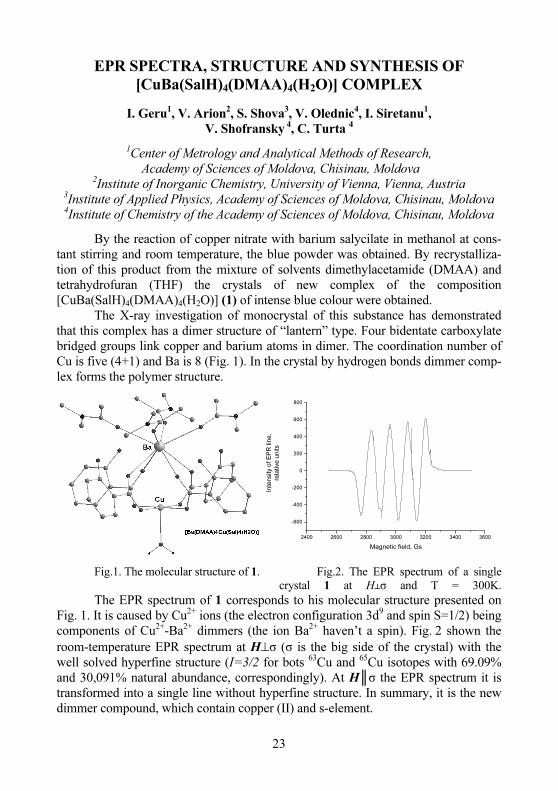
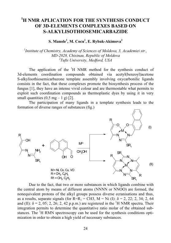
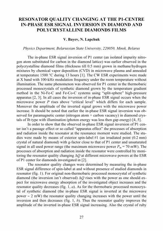
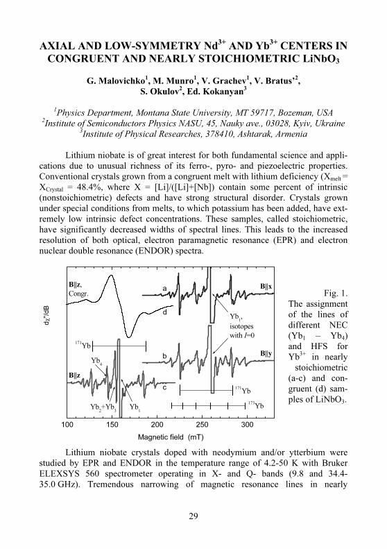
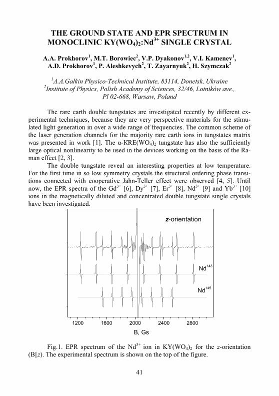
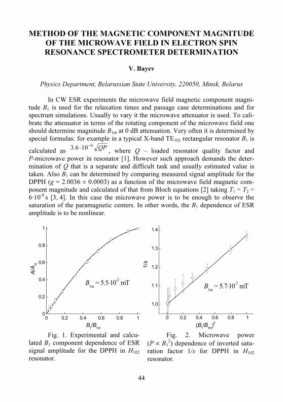