magnetic resonance imaging of the fetus - mary rutherford imaging
TRANSCRIPT

Magnetic Resonance Imaging of the fetus
Mary A RutherfordPerinatal Imaging Group, MRC Clinical Sciences Centre
Imperial [email protected]
The Moonbeam Trust

Overview
Practicalities and Sequences
Clinical applicationsNon Central Nervous System
Central Nervous System(CNS)
Research and development: studies in IUGR pregnancies

Relatively new techniqueComplimentary to ultrasound
Main application for CNSMotion is a major problem
Initially paralysed fetus (French studies)Sedation mother in some units (non UK)
Fetal MR imaging

Philips 1.5 Tesla magnet
Pregnant patient in left lateral tiltEar protectionFan on to keep cool
Cardiac coil- 5 channelsAround abdomen
We wow use a new cardiac coil with 32 channels.Gives better coverage and higher signal to noise

The fetus is often in continual motion which can be captured using a fetal cine sequence
(Hayat et al AJNR 2011 )

Fetal MR sequences
Fast single shot T2 weighted image acquisitionImage slice in less than 1 second

T1 weighted imaging
* Malamateniou et al Radiology 2011
PreviousT1W spin echo acquired with breathhold
Recently developedSNAPIR*

Clinical non CNS imaging
Chest
Abdomen
Renal
Liver
Skeletal
Cardiac
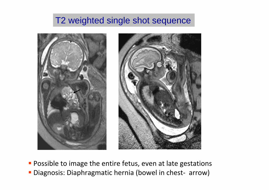
Possible to image the entire fetus, even at late gestationsDiagnosis: Diaphragmatic hernia (bowel in chest‐ arrow)
T2 weighted single shot sequence

Diaphragmatic herniaAssess contents of chest
Stomach (arrowhead)
liver
Assess lung signal intensity and volumeVisually (arrows)
Objectively
Use to predict respiratory outcome

Fetal hydrops
Pleural effusion
Compressed lung
Ascites
Subcutaneous oedema

Abdominal Imaging
Ultrasound: Renal pelvis dilatation (A)
Posterior urethral valves (B) in male fetus (C)
A B C

CNS imaging
Common indications
VentriculomegalyAgenesis of the corpus callosumCerebellar anomalies Congenital infection
Complications of twin pregnanciesAcute hypoxic event

Ventriculomegaly
Most common reason for MRI referralDetect any obvious cause for ventriculomegalye.g. haemorrhage /obstruction Identify additional anomaliesPredict outcome to counsel parents

Ventricular dilation with evidence of germinal matrix haemorrhage

Haemorrhage involves basal ganglia (arrow)
Ventricular dilation with evidence of germinal matrix haemorrhage

Posterior limb of internal capsule (PLIC) may be spared (arrow)
Motor outcome likely to be normal
Ventricular dilation with evidence of germinal matrix haemorrhage

Agenesis of the corpus callosum
May be referred with ventriculomegalyOccasionally as a parenchymal cyst

Agenesis corpus callosum (A)with interhemispheric cyst (B)(arrow)
A
B

Agenesis with interhemispheric cyst
Postnatal imagingT1 weighted showing cyst (arrow)

Postnatal diffusion tensor imaging (DTI)Showing white matter tracts circumventing cyst
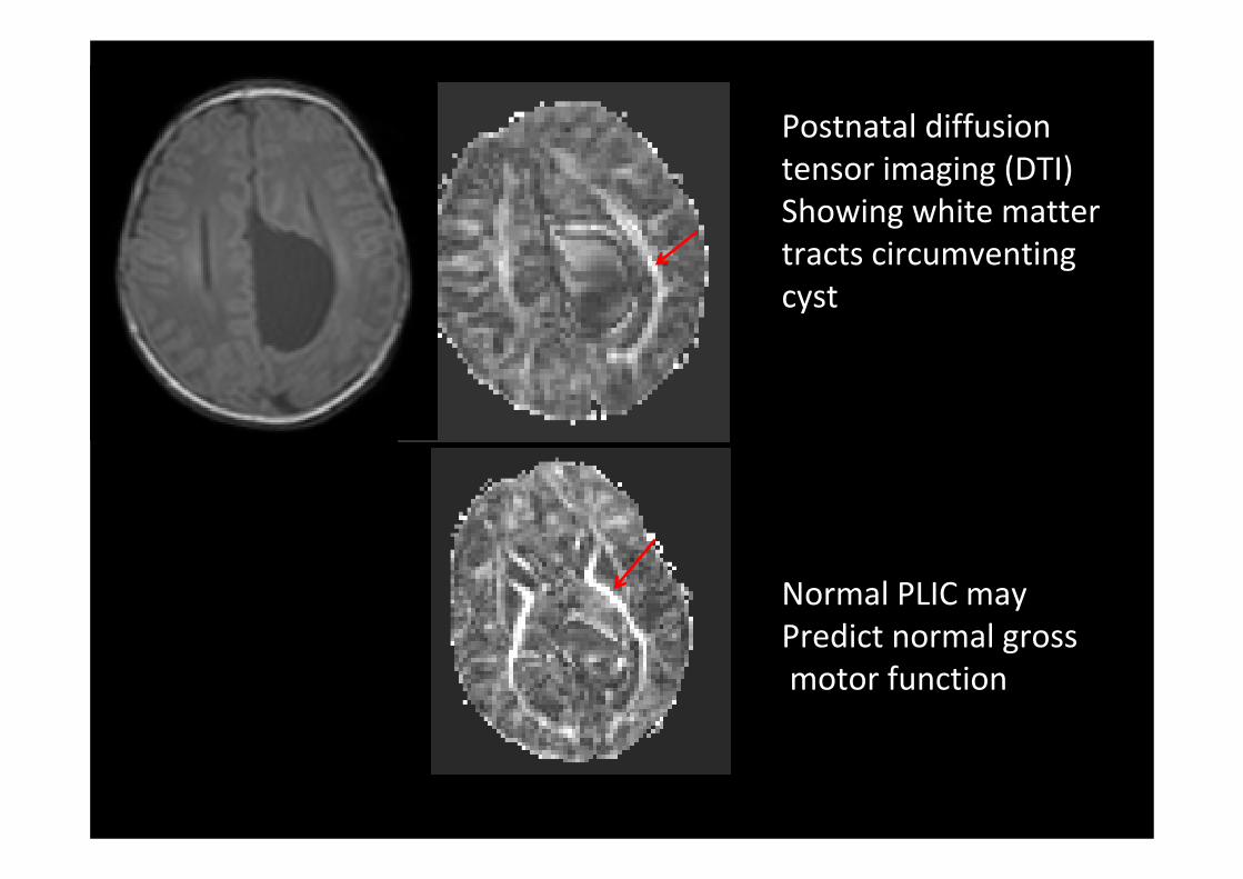
Postnatal diffusion tensor imaging (DTI)Showing white matter tracts circumventing cyst
Normal PLIC mayPredict normal grossmotor function

Cerebellar anomalies
Large cisterna magna Cerebellar hypoplasiaCerebellar haemorrhage

Not visible on sagittal or coronal images
26+3 weeks . Large cisterna magna10mm (A,B)
Bilateral irregular germinal matrix arrows (C)
Diagnosis: subependymal heterotopiafemale fetus
Repeat at 32 weeks GA
A
B
C

Mother asymptomaticMaternal grandmother epileptic
Enlarged cisterna magna
Diagnosis:X linked subependymal
heterotopia with enlargedcisterna magna
Mother’s images

Research and development
Improving image quality

Fetal motion
Single slices fast acquisition ‐ not artefacted
But fetus moves in and out of plane during acquisition
Rotation of fetal headbetween slices

Snapshot image acquisition with Volume reconstruction (SVR)*
Aim
Provide high signal to noise, high resolution 3‐Dimensional volumetric datasets of the brain in presence of fetal motion
* Jiang et al 2007

(a) (b) (c)
(d) (e) (f)
(g) (h) (i)
(j) (k) (l)
Registered3D fetal data
Ex utero data3 Tesla
Dynamic scan Single shot 1 loop
Combined loops

(a) (b) (c)
(d) (e) (f)
(g) (h) (i)
(j) (k) (l)
Registered3D fetal data
Ex utero data3 Tesla
Dynamic scan Single shot 1 loop
Combined loops

(a) (b) (c)
(d) (e) (f)
(g) (h) (i)
(j) (k) (l)
Registered3D fetal data
Ex utero data3 Tesla
Dynamic scan Single shot 1 loop
Combined loops

(a) (b) (c)
(d) (e) (f)
(g) (h) (i)
(j) (k) (l)
Registered3D fetal data
Ex utero data3 Tesla
Dynamic scan Single shot 1 loop
Combined loops

30 week fetus 1.5 Tesla 30 week preterm 3 Tesla

Quantifying normal brain development
20 weeks 32 weeks

Manual Segmentation of normal fetal brain n=18
Hayat 2007

log10
4.4000
4.6000
4.8000
5.0000
5.2000
5.4000
17 19 21 23 25 27 29 31 33 35
GA (weeks)
log1
0(Vo
lum
e)
Gestational Age (weeks)
Cerebral pa
0
50000
100000
150000
200000
250000
17 19 21 23 25 27 29 31 33 35
GA (weeks)
Volu
me
(mm
3)
Cerebral Parenchyma Volume and Log Volume against gestational age
Volume (m
m cub
ed)
Gestational Age (weeks)
r = 0.99 p < 0.05r = 0.99 p < 0.05
volume
Hayat 2007

Brain images may look normal
High risk of mortality and short term morbidity
Longer term neurodevelopmental impairments
adult onset hypertension, diabetes.
The fetus with intrauterine growth restriction (IUGR)

The fetus with IUGR
Cohort of IUGR fetuses = 40Estimated fetal weight < 5th centile.
Graded by Doppler studiesUmbilical artery (UA) pulsatility index >95th C
As above with PI <5th centile in middle cerebral artery
Absent end diastolic flow (EDF) in UA
Reversed EDF in UA
Absent or reversed “a” wave in ductus venosus and/or
pulsatilility in the umbilical vein
Damodaram (in prep.)

Growth parameters(ultrasound) in IUGR

Umbilical Artery Doppler
positive end diastolic flow (normal pregnancy)
absent end diastolic flow (growth restricted pregnancy)

The Placenta in IUGRReduced volume in IUGR
Placental volume and fetal weight
Damodaram et al 2012

Body volume in IUGR
IUGR =20Normal =19
Damodaram (2012)

The fetus with IUGR
Is there any evidence of organ “sparing” ?

The fetus with IUGR: liver volume
p <0.001
Decreased glucose transfer and hepatic storageDecreased IGF-1 productionLink to later adult disease ; type 2 diabetes
Damodaram (2012.)

An increase in the brain:liver ratio predicted perinatal mortality with an AUC of 0.89 (p =0.001, 95 CI = 0.78 – 1.0)
A brain:liver ratio above 3.0 was associated with a 3.3 fold increase in relative risk of perinatalmortality (95% CI = 1.68 – 6.47).
The fetus with IUGR: liver volume

The fetus with IUGR: renal volume
p <0.001
Decreased nephron numberInappropriate activation of renin-angiotensinAdult onset hypertension
Damodaram (2012
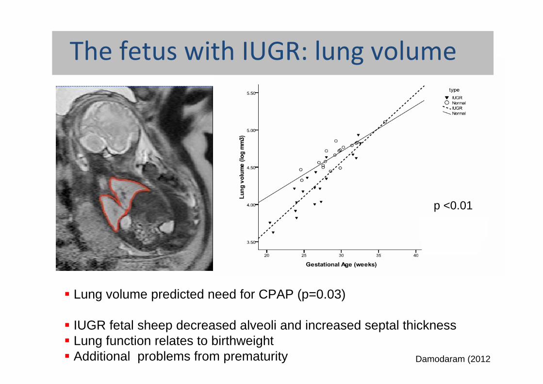
The fetus with IUGR: lung volume
Lung volume predicted need for CPAP (p=0.03)
IUGR fetal sheep decreased alveoli and increased septal thicknessLung function relates to birthweightAdditional problems from prematurity
p <0.01
Damodaram (2012

The fetus with IUGR: cardiac volume
Doppler studies show impaired cardiac function in human fetal IUGRRat model shows incresaed apoptosis and decreased cardiomyocytes
? Predisposition to cardiac ischaemia in adult life
p <0.01
Damodaram (2012)

The fetus with IUGR: thymus volume
p <0.01
? Stress mediated thymic involution via hypothalamic-piuitary-adrenal axis
Damodaram (2012)

Results: Fetal Brain VolumeThe Fetus with IUGR: brain volume
Damodaram (2012)
IUGR n=29Normal n=45
p<0.05 corrected for GA
Increased reduction with increased severity of IUGR p<0.001

The fetus with IUGR:ventricularvolume
Damodaram (in prep.)

Extracerebral space/brain volume
Damodaram (in prep.)Measures of BPD will overestimate brain size
p<0.001

The fetus with IUGR
Is there any evidence of organ “sparing” ? NO

The fetus with IUGR: cortical folding
Is there evidence of abnormal maturation of the cortex?
Assessed cortical folding visually with 8 point score

The fetus with IUGR: cortical folding
No difference in cortical folding
Damodaram

Automatic cortical segmentation
Courtesy of Paul Aljabar
Optimisation of an automatic segmentation technique would allow quicker analysis of group date for research or individual data for clinical practice

Fetal MR proton spectroscopy
Metabolic informationObtain chemical peaks Choline, Creatine, N Acetyl Aspartate, Lactate, Myo‐inositol
Technical difficultiesMotion
Small region of interest
Not possible to differentiate tissue types reliably‐ use large region of interest

MR spectroscopy
In vivo assessment of brain metabolism in normal and IUGR fetuses n=28 (controls 46)
Large voxel 2x2x2 cm3
70% success rate
Story et al 2011

24 week normal fetal control.
Spectrum obtained with TE= 144 ms
Lactate
N Acetyl AspartateCreatine
Choline

Spectrum obtained with (TE) 36 ms. A large bifid lactate peak is present at 1.3 ppmNo NAA visible at 2ppm
Lactate
23 week fetus with IUGR
ChoCr

NAA reduced in IUGR
Story et al 2012
p=0.001

Reduced NAA in brain of IUGR fetus
NAA is synthesised in neuronal mitochondria, thentransported to oligodendrocytes
Reduced NAA may be sign of immaturity but may also reflect mitochondrial dysfunction
Rat models of IUGR demonstrate mitochondrial dysfunction in brain other organs e.g pancreas
Use MRS to quantify NAA in rat IUGR model and then assess mitochondria phenotypes and function.

Overview
Practicalities and Sequences
Clinical applicationsNon CNS
CNS
Research and development in IUGR pregnancies

To all the staff in the Robert Steiner MR Unit and the Centre for Fetal Care, Queen Charlotte’s Hospital.
Shuzhou Jiang Joanna AllsopTayyib Hayat Lisa StoryAlpa Patel Amy McGuinessDaniel Rueckert Paul AljabarJo Hajnal Mellisa DamadoramSailesh Kumar Prachi PatkeeRuwan Wimalasundera Elisenda Eixarch
The Moonbeam Trust